AM Bugaj-TPDT
13
Photochemical & Photobiological Sciences Dynamic Article Links Cite this: Photochem. Photobiol. Sci., 2011, 10, 1097 www.rsc.org/pps PERSPECTIVE Targeted photodynamic therapy – a promising strategy of tumor treatment Andrzej M. Bugaj*† Received 9th June 2010, Accepted 7th March 2011 DOI: 10.1039/c0pp00147c Targeted therapy is a new promising therapeutic strategy, created to overcome growing problems of contemporary medicine, such as drug toxicity and drug resistance. An emerging modality of this approach is targeted photodynamic therapy (TPDT) with the main aim of improving delivery of photosensitizer to cancer tissue and at the same time enhancing specificity and efficiency of PDT. Depending on the mechanism of targeting, we can divide the strategies of TPDT into “passive”, “active” and “activatable”, where in the latter case the photosensitizer is activated only in the target tissue. In this review, contemporary strategies of TPDT are described, including new innovative concepts, such as targeting assisted by peptides and aptamers, multifunctional nanoplatforms with navigation by magnetic field or “photodynamic molecular beacons” activatable by enzymes and nucleic acid. The imperative of introducing a new paradigm of PDT, focused on the concepts of heterogeneity and dynamic state of tumor, is also called for. 1. Introduction Photodynamic therapy (PDT) is a minimally invasive method that destroys target cells in the presence of oxygen when light irradiates a photosensitizer, generating reactive oxygen species (mainly sin- glet oxygen), causing destruction of cellular targets through direct cellular damage, vascular shutdown and activation of an immune College of Health, Beauty and Education, Pozna´ n, Poland. E-mail: [email protected]; Tel: +48 61 877 07 27; Tel: +48 501645004 (mobile) † Address for correspondence: Os. Jagiello´ nskie 3 m. 9, 61-224, Pozna´ n, Poland. Andrzej M. Bugaj Andrzej M. Bugaj obtained his Ph.D. in pharmaceutical sci- ences. He has worked in many scientific centers in Europe, such as the Biophysics and PDT Group of Prof. Johan Moan from Institute for Can- cer Research in Rikshospitalet- Radiumhospitalet Medical Cen- ter in Oslo, Cancer Photobiology Group of Prof. Thierry Patrice from Department of Laser Ther- apy in University Hospital Center in Nantes, and – as a holder of French Government Fellowship – in the Laboratory of Photobiology from Mus´ eum National d’Histoire Naturelle in Paris, under the direction of Prof. Ren´ e Santus and Prof. Patrice Morli` ere. The research interests of Dr. Bugaj are focused on molecular and cellular mechanisms of photodynamic therapy. response against targeted cells. Current clinical applications of PDT include the treatment of numerous cancerous and non- cancerous diseases such as age-related macular degeneration or endometriosis. For over 30 years, the use of PDT for treatment of bacterial and fungal infections has been in practice. 1–4 Although some photosensitizers used in PDT reveal certain tumor selectivity, it is noteworthy that their preferential accumu- lation in tumors is itself not a guarantee of selective photoinduced tumor damage and successful PDT. The photosensitizers accu- mulate also in healthy tissues, resulting in uncomfortable adverse effects, such as phototoxic and photoallergic reactions. To avoid this obstacle, a new approach for drug delivery in PDT, called targeted photodynamic therapy (TPDT), has been developed. The aim of TPDT is a specific action towards well-defined targets or biologic pathways that, when inactivated, cause regression or inhibition of the disease process. 1,2 The commonly applied strategy to increase the specific accu- mulation of photosensitizers at the target site is encapsulation or attachment of photosensitizers to molecules or molecular constructs which improve affinity of these dyes to the target tissues. 1 This strategy, called targeting, is usually divided into “passive” and “active” ones (Fig. 1). Passive targeting is promoting of drug entry into the tumor cells determined by physicochemical factors of drug carrier, such as material composition, size and surface properties (e.g. electric charge) and by pathophysiological factors of the organism, such as tumor microenvironment as well as enhanced permeability and retention (EPR) effect, whereas active targeting involves drug delivery to the specific target sites based on molecular recognition. 1,3 The third strategy of targeting photosensitizers to the tumor cells is the use of photosensitizers alone or attached to carrier systems, which create active forms and produces cytotoxic effects only at the site of the lesion. Some authors suggested terming such delivery systems as “active” in This journal is © The Royal Society of Chemistry and Owner Societies 2011 Photochem. Photobiol. Sci., 2011, 10, 1097–1109 | 1097 Downloaded by ESP0075 on 25/04/2013 13:29:05. Published on 06 May 2011 on http://pubs.rsc.org | doi:10.1039/C0PP00147C View Article Online / Journal Homepage / Table of Contents for this issue
-
Upload
independent -
Category
Documents
-
view
0 -
download
0
Transcript of AM Bugaj-TPDT
Photochemical &Photobiological Sciences
Dynamic Article Links
Cite this: Photochem. Photobiol. Sci., 2011, 10, 1097
www.rsc.org/pps PERSPECTIVE
Targeted photodynamic therapy – a promising strategy of tumor treatment
Andrzej M. Bugaj*†
Received 9th June 2010, Accepted 7th March 2011DOI: 10.1039/c0pp00147c
Targeted therapy is a new promising therapeutic strategy, created to overcome growing problems ofcontemporary medicine, such as drug toxicity and drug resistance. An emerging modality of thisapproach is targeted photodynamic therapy (TPDT) with the main aim of improving delivery ofphotosensitizer to cancer tissue and at the same time enhancing specificity and efficiency of PDT.Depending on the mechanism of targeting, we can divide the strategies of TPDT into “passive”,“active” and “activatable”, where in the latter case the photosensitizer is activated only in the targettissue. In this review, contemporary strategies of TPDT are described, including new innovativeconcepts, such as targeting assisted by peptides and aptamers, multifunctional nanoplatforms withnavigation by magnetic field or “photodynamic molecular beacons” activatable by enzymes and nucleicacid. The imperative of introducing a new paradigm of PDT, focused on the concepts of heterogeneityand dynamic state of tumor, is also called for.
1. Introduction
Photodynamic therapy (PDT) is a minimally invasive method thatdestroys target cells in the presence of oxygen when light irradiatesa photosensitizer, generating reactive oxygen species (mainly sin-glet oxygen), causing destruction of cellular targets through directcellular damage, vascular shutdown and activation of an immune
College of Health, Beauty and Education, Poznan, Poland.E-mail: [email protected]; Tel: +48 61 877 07 27;Tel: +48 501645004 (mobile)† Address for correspondence: Os. Jagiellonskie 3 m. 9, 61-224, Poznan,Poland.
Andrzej M. Bugaj
Andrzej M. Bugaj obtained hisPh.D. in pharmaceutical sci-ences. He has worked in manyscientific centers in Europe,such as the Biophysics andPDT Group of Prof. JohanMoan from Institute for Can-cer Research in Rikshospitalet-Radiumhospitalet Medical Cen-ter in Oslo, Cancer PhotobiologyGroup of Prof. Thierry Patricefrom Department of Laser Ther-apy in University Hospital Centerin Nantes, and – as a holder of
French Government Fellowship – in the Laboratory of Photobiologyfrom Museum National d’Histoire Naturelle in Paris, under thedirection of Prof. Rene Santus and Prof. Patrice Morliere. Theresearch interests of Dr. Bugaj are focused on molecular and cellularmechanisms of photodynamic therapy.
response against targeted cells. Current clinical applications ofPDT include the treatment of numerous cancerous and non-cancerous diseases such as age-related macular degeneration orendometriosis. For over 30 years, the use of PDT for treatment ofbacterial and fungal infections has been in practice.1–4
Although some photosensitizers used in PDT reveal certaintumor selectivity, it is noteworthy that their preferential accumu-lation in tumors is itself not a guarantee of selective photoinducedtumor damage and successful PDT. The photosensitizers accu-mulate also in healthy tissues, resulting in uncomfortable adverseeffects, such as phototoxic and photoallergic reactions. To avoidthis obstacle, a new approach for drug delivery in PDT, calledtargeted photodynamic therapy (TPDT), has been developed. Theaim of TPDT is a specific action towards well-defined targetsor biologic pathways that, when inactivated, cause regression orinhibition of the disease process.1,2
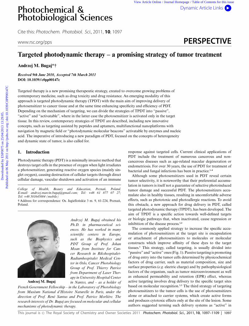
The commonly applied strategy to increase the specific accu-mulation of photosensitizers at the target site is encapsulationor attachment of photosensitizers to molecules or molecularconstructs which improve affinity of these dyes to the targettissues.1 This strategy, called targeting, is usually divided into“passive” and “active” ones (Fig. 1). Passive targeting is promotingof drug entry into the tumor cells determined by physicochemicalfactors of drug carrier, such as material composition, size andsurface properties (e.g. electric charge) and by pathophysiologicalfactors of the organism, such as tumor microenvironment as wellas enhanced permeability and retention (EPR) effect, whereasactive targeting involves drug delivery to the specific target sitesbased on molecular recognition.1,3 The third strategy of targetingphotosensitizers to the tumor cells is the use of photosensitizersalone or attached to carrier systems, which create active formsand produces cytotoxic effects only at the site of the lesion. Someauthors suggested terming such delivery systems as “active” in
This journal is © The Royal Society of Chemistry and Owner Societies 2011 Photochem. Photobiol. Sci., 2011, 10, 1097–1109 | 1097
Dow
nloa
ded
by E
SP00
75 o
n 25
/04/
2013
13:
29:0
5.
Publ
ishe
d on
06
May
201
1 on
http
://pu
bs.r
sc.o
rg |
doi:1
0.10
39/C
0PP0
0147
CView Article Online / Journal Homepage / Table of Contents for this issue

Fig. 1 Schematic presentation of passive and active TPDT. Photosensitiz-ers alone or attached to non-liganded carriers can reach tumors selectivelythrough the “leaky” vasculature surrounding the tumors (EPR effect –passive targeting). The ligands grafted at the surface of carriers allowactive targeting by binding to receptors overexpressed by tumor cells orangiogenic endothelial cells (according to ref. 13 and 14).
contrast to “passive” ones, whose role is solely selective transportof photosensitizers to the target sites.3 However, it seems moreprecise to call these systems “activatable”, because over the siteof lesion they are photodynamically inactive. Moreover, it avoidsconfusion with the traditional differentiation between “passive”and “active” targeting systems, which is commonly used inpharmaceutical sciences.4
2. Biodegradable nanocarriers in passive TPDT
Passive targeting makes use of the morphological and physiologi-cal differences between normal and tumor tissue to deliver the drugto a target site or utilize localized delivery (Fig. 1).5 The tumor vas-culature is very different from the normal vasculature. Unlike thetight endothelial lining in normal tissues, blood vessels in tumorshave gaps as large as 600–800 nm between adjacent endothelialcells.5 The tumor vessels are often dilated and convoluted, theymay have fenestrations and discontinuous membranes. This defec-tive vascular architecture coupled with poor lymphatic drainageinduces an enhanced permeability and retention effect (EPR), bywhich photosensitizers attached to macromolecules can selectivelyaccumulate the tumor interstitium. Another contributor to passivetargeting is the unique microenvironment surrounding tumorcells. Fast-growing, hyperproliferative cancer cells show a highmetabolic rate, and the supply of oxygen and nutrients is usuallynot sufficient for the cancer cells to maintain growth. Therefore,tumor cells use glycolysis to obtain extra energy, resulting in an
acidic environment. Finally, cancer cells express and release uniqueenzymes such as matrix metalloproteinases, which are implicatedin their movement and survival mechanism.5–7
Dramatic increase in tumor drug accumulation – usually tenfoldor greater – can be achieved when a drug is delivered by ananoparticle. Interest in nanoparticles as drug carriers has beenincreased in recent years. According to common resolution ofthe International Organisation of Standardisation and of theEuropean Standardisation Committee, nanoparticles may bedefined as objects with all three external dimensions in the sizerange from approximately 1 to 100 nm.8 In nanomedicine sizedimensions of 1–1000 nm are included; this is due to the factthat in medicine nanotechnology aims to improve and optimizematerial properties for their interaction with cells and tissue toallow, for example, passive tumor targeting or to improve thebioavailability. This approach makes use of nanoscale materialslarger than 100 nm.9 Nanoparticles in a mean diameter of 100 nmshow prolonged blood circulation and poor extravasation and arenot cleared by reticuloendothelial and phagocytic systems. Hence,drugs encapsulated in nanoparticles can easily accumulate in theorganism and its pharmacokinetic parameters such as eliminationhalf-life and volume of distribution often have significantly highervalues when compared to the free drug.10,11 More importantly,nanoparticles can selective accumulate in tumor cells due to theEPR effect. A “leaky”, highly fenestrated endothelial wall oftumor vasculature allows selective uptake of nanoparticles bytumor tissue, in contrast to vasculature of healthy tissues beinga primary delivery barrier for nanoparticles due to limited poresize. Even nanoparticles greater in size than 100 nm, which areeasily cleared by the reticuloendothelial system (RES) and willnot leave normal blood vessels, will tend to accumulate in tumorswhich have relatively “leaky” vascular beds.10–12 Moreover, bycontrast with many free photosensitizers which – due to theirsmall size – can freely diffuse from the tumor cells to the bloodvessels, so that concentration of these drugs in tumor tissue mayrapidly decrease below the effective concentration, the nanocar-riers cannot easily diffuse back into the blood stream becauseof their large size, resulting in their progressive accumulation inthe tumor tissue 13,14 (Fig. 1). For these reasons, nanoparticlesare largely applied in TPDT. Traditionally, these constructs canbe classified by material composition into biodegradable andnon-biodegradable.3,15
2.1. Biodegradable nanoparticles
Biodegradable nanoparticles have received a lot of attention dueto their possibility of controlling the drug release, versatility inmaterial manufacturing processes and high drug loading.15,16 Theyare made of organic natural or synthetic substances that aredegraded in the biological environment due to enzyme-catalyzedhydrolysis and hence release the photosensitizers. The chemicaland physical structure of these materials can be tailored to accom-modate photosensitizers with varying degrees of hydrophobicity,molecular weight, charge and pH.17
Liposomes, which are the most intensively investigated familyof drug carriers, are uni- or multilamellar lipid vesicles in sizeof 50–150 nm allowing incorporation of both hydrophilic and hy-drophobic substances to improve their drugabililty. Several studiesdemonstrated a high and fast accumulation of liposomes in tumor
1098 | Photochem. Photobiol. Sci., 2011, 10, 1097–1109 This journal is © The Royal Society of Chemistry and Owner Societies 2011
Dow
nloa
ded
by E
SP00
75 o
n 25
/04/
2013
13:
29:0
5.
Publ
ishe
d on
06
May
201
1 on
http
://pu
bs.r
sc.o
rg |
doi:1
0.10
39/C
0PP0
0147
C
View Article Online

tissues. Lasalle et al. studied pharmacologic effects of Foslip,a formulation of 5,10,15,20-tetrakis(meso-hydroxyphenyl)chlorin(m-THPC) incorporated in liposomes based on the mixtureof dipalmitoylphosphatidylcholine and dipalmitoylphosphatidyl-glycerol (DPPC/DPPG), on EMT6 tumor bearing mice.18 Afterintravenous administration of m-THPC in this formulation ata dose 0.3 mg kg-1 the volume of distribution was about 4-fold higher compared to those observed by Jones et al.19 forstandard preparation Foscan R© (709 and 172 ml kg-1, respec-tively), suggesting that some tissues preferentially accumulateFoslip.18 The tumor m-THPC concentration reached its max-imal values at 72 h after administration while in the case ofFoscan R© maximal tumor concentrations were attained 24–48 hafter administration.19,20 However, the best tumor response wasestimated for a drug-light interval of 6 h, for which photosensitizerwas present both in vasculature and tumor cells18 similar to theresults obtained for Foscan R©19 suggesting that the presence ofm-THPC in both endothelial and parenchyma cells is requiredfor optimal PDT efficiency. The release of m-THPC from Foslipliposomes was slower than that from Visudyne R© liposomes whichare made of more fluid lipids: dimyristoylphosphatidylcholine/eggphosphatidylglycerol.21
Conventional liposomes, mainly composed of phospholipidsand cholesterol, often exhibit a plasma half-life too short forefficient uptake by tumor cells, because of rapid clearing by RES,and because of disintegration due to lipid exchange with bloodplasma lipoproteins and due to molecular interaction betweenliposome components.22 Thus, during the past decade, the interestof polymer-based drug delivery systems has grown dramaticallywith the advent of biodegradable polymers, which are degraded inthe biological environment and hence release the photosensitizers.The resulting constructs may have different structures, includingmicelles and dendrimers.3,15
Block copolymer micelles emerge as more attractive drugdelivery systems than liposomes, due to their higher stabilityand small uniform particle size which accomplishes their passivetargeting due to the EPR effect and prevents their recognitionby macrophages and protein, prolonging their circulation time inblood.23,24 They are typically spherical nanosized (diameter 10–100 nm) supramolecular assemblies of amphiphilic copolymers, inwhich the drug may be either confined to a cavity surrounded bya polymer membrane (nanocapsules) or uniformly dispersed in amatrix (nanospheres). The core of these micelles is a loading spacethat accommodates hydrophobic drugs and the hydrophilic outershell facilitates dispersal of the micelles in water.25
Among synthetic polymers for preparing these micelles,poly(ethylene glycol) (PEG), polylactide (PLA), and their copoly-mer poly(D,L-lactide-co-glycolide) (PLGA) have been studiedespecially due to their versatility, mechanical strength, biocom-patibility, bioresorbability, high drug loading and possibility ofcontrolling the drug release. Cohen et al. encapsulated 5,10,15,20-tetrakis(meso-hydroxyphenyl)porphyrin (m-THPP) into PEG-PLA copolymer to obtain micelle nanoparticles (a size of about30.6 ± 3.3 nm) which exhibited phototoxic effect towards head andneck cancer cells.26 The nanoparticles loaded with 5 and 10% of m-THPP revealed <30% dark toxicity and >90% phototoxicity at amicelle concentration 2–20 mg L-1 compared to non-treated cells.Effect of free m-THPP was not studied. No significant cytotoxiceffect both for light (l = 420 nm) and for nanoparticles without
photosensitizer alone were observed.24 Master et al. encapsulatedhydrophobic silicon phthalocyanine (Pc4) in PEG-PCL micelles(a mean diameter 73–103 nm) revealing at concentration 400nM upon irradiation with red light, a significant phototoxiceffect (p < 0.01) towards MCF-7c3 human breast cancer cellsas compared with analogous effect of standard Pc4 formulationin dimethyl formamide (DMF).27 In contrast to delivery in DMFsolution, after which photosensitizer was preferentially localizedin mitochondria, endoplasmic reticulum and Golgi apparatus,Pc4 delivered to cell cultures in PEG-PCL nanoparticles waspartially distributed to lysosomes. It demonstrates the promiseof this carrier for tumor-targeted delivery of Pc4 for site-selectivePDT.27
Although synthetic polymers might be preferable to use asdrug delivery systems due to the possibility of adjusting theirmechanical properties and degradation kinetics appropriately forvarious applications, natural polymers such as agar, albumin,alginate, chitin, chitosan, collagen, cyclodextrins, dextran andgelatin remain attractive because they are relatively inexpen-sive, readily available and capable of a multitude of chemicalmodifications.17,28,29 The use of natural biodegradable polymersto deliver photosensitizers will continue to be an area of active re-search despite the advance in synthetic biodegradable polymers.17
Chitosan, the product of partial deacetylation of the naturalpolysaccharide chitin, presents enhanced tumor target specificityand high ability for encapsulating hydrophobic photosensitizerinto the multicore of nanoscale particles.30 Lee et al. preparedthe PpIX encapsulating chitosan-based nanoparticles with averagesize of 290 nm, which were rapidly taken up by SCC7 (squamouscell carcinoma) cells and did not reveal dark cytotoxicity towardsthese cells while following irradiation with visible light they werehighly phototoxic. In SCC-tumor bearing mice PpIX-chitosan-based nanoparticles exhibited enhanced tumor specificity andincreased therapeutic efficacy compared to free PpIX.31 Recently,Hu et al. demonstrated significantly higher uptake of chlorin e6encapsulated into stearic acid-grafted chitosan micelles by A-549lung cancer cells in vitro when compared with uptake of freephotosensitizer. The average micelle size (302–330 nm) decreasedby 10% with increase of drug content from 5 to 20%.32 Similarly,alginates, the polysaccharides isolated from brown algae, might beuseful for the sustained and localized delivery of photosensitizers.33
Khdair et al. showed that encapsulation of methylene blue inalginate nanoparticles containing anionic surfactant Aerosol R©OT(dioctyl sodium sulfosuccinate; an average diameter of 79 nm)enhanced its anticancer photodynamic efficiency in vitro.34
The new class of biodegradable non-polymeric nanoparticlesconsists of solid lipid nanoparticles (SLN), which are particlesof solid lipid matrix with an average diameter in the nanometrerange (150–170 nm). Their excellent physical stability, protectionof incorporated labile drugs from degradation, controlled drugrelease (fast or sustained), good tolerability and site-specifictargeting make SLN good candidates for TPDT.35,36 Despite this,the results of studies performed to date are less promising. Asshown by Kuchler et al., incorporation of Nile blue in SLNdecreased its penetration through pig skin 4–6-fold comparedto dendritic core multishell formulation. It was probably due tointeractions between lipids of SLN and skin.36
Dendrimers are another family of particulate carriers whichhave aroused increasing interest.25 They are regularly branched
This journal is © The Royal Society of Chemistry and Owner Societies 2011 Photochem. Photobiol. Sci., 2011, 10, 1097–1109 | 1099
Dow
nloa
ded
by E
SP00
75 o
n 25
/04/
2013
13:
29:0
5.
Publ
ishe
d on
06
May
201
1 on
http
://pu
bs.r
sc.o
rg |
doi:1
0.10
39/C
0PP0
0147
C
View Article Online

three-dimensional tree-like structures composed of a central coremolecule with a number of functional groups attached to repeatedpolymer branches organized in concentric layers called “genera-tions” and terminated with surface functional groups which toa considerable degree determine the dendrimer’s physicochemicalproperties.37,38 Dendrimers can host a variety of carrier molecules,both hydrophobic and hydrophilic. A reasonable cost of manufac-turing, good toxicological profile and biocompatibility as well astheir controlled multivalency provide attachment of a variety oftargeting compounds in a well-defined manner, distinguish themfrom other nano-sized species used for TPDT. Such formulationsmay significantly improve the circulation time of photosensitizersand their accumulation in hyperpermeable lesions due to the EPReffect.39 Battah et al. prepared well-defined dendritic moleculesfor delivery of aminolevulinic acid (ALA), natural precursor ofphotosensitizing porphyrin PpIX. This delivery vehicle carrying 18molecules of ALA resulted in increased production of PpIX and inhigher phototoxicity towards tumorigenic PAM 212 keratinocytesand A431 human epidermoides when compared with free ALA.40
In a murine tumor model the dendrimers induced sustainedporphyrin production for over 24 h while the porphyrins inducedby free ALA revealed concentration maxima between 3 and 4h.41 The obtained ALA dendrimers (molecular weight: 3679)are too low to elicit the EPR effect which can improve tumorselectivity for larger dendrimers and are cleared relatively rapidlyfrom mouse circulation. The rate of release of ALA residuesby dendrimer enzymatic hydrolysis (25% at 3 h and 40% at 24h after intraperitoneal administration to mice in dose 200 mgkg-1) within the cells may be the rate-limiting step for porphyrinproduction.41
It is also possible to encapsulate photosensitizers into den-drimers. The slow degradation of such complexes can give riseto prolonged release in vivo. Kojima et al. developed two PEG-attached dendrimers derived from poly(amidoamine) (PAMAM)and polypropylene imine (PPI) dendrimers (average molecularweight: 2000) to encapsulate rose bengal and PpIX. The PEG-PPIdendrimers held both these molecules in a more stable mannerthan PEG-PAMAM ones, probably due to higher hydrophobicity.The dendrimers of PpIX encapsulated in PEG-PPI revealed moreefficient phototoxic effect towards HeLa cells when compared tofree PpIX.42
In contrast to free PpIX, which reaches cell interior bymembrane diffusion, the dendrimeric PpIX with cationic andanionic groups entered cells by endocytosis and remained localizedin lysosomes.43 The cationic dendrimers entered Lewis lung carci-noma (LLC) cells 22–25 times more rapidly and revealed higherphototoxicity 230 times towards these cells than anionic ones.The observed differences of cell uptake and phototoxicity of bothdendrimeric compounds was probably due to different electro-static association of cell membranes. The cationic dendrimer couldstrongly adsorb on negatively charged membrane components (e.g.glycoproteins) through electrostatic interactions, while the anionicones could reveal lower affinity to these membrane components,due to electrostatic repulsion. As a consequence, positively chargeddendrimers may be more strongly bound by plasma membranesand may cause photodamage of these membranes more vigorouslythan anionic ones, although quantum yields of 1O2 productionwere similar for both positively and negatively charged PpIXdendrimers and for free PpIX (0.49, 0.41 and 0.45, respectively).43
Compared to free PpIX, its cationic dendrimer revealed a photo-toxic effect 20 times higher towards LLC cell plasma membranes,probably connected with lipid peroxidation, protein cross-linking,loss of ionic homeostasis and release of hydrolases to cytoplasmfrom damaged lysosomes. Thus cationic dendrimer porphyrinsseem to be a new class of promising PDT photosensitizers.43
To improve the passive targeting of this promising PDTsystem, cationic dendrimer porphyrins have been assembled withlinear copolymers of poly(ethylene glycol)-poly(a,b-aspartic acid)to obtain micellar constructs with a diameter of ca. 55 nmwhich prolongs circulation time of carriers in the blood andincreases their tumor accumulation by EPR. The PDT efficiencyof such dendrimer porphyrins was 40-fold higher than freedendrimers, probably due to decreased tendency of these micellardendrimers to aggregate, resulting in a higher yield of singletoxygen production.44 Nishiyama et al. synthesized dendrimericphthalocyanines with diameter of ca. 50 nm which accumulated inthe endosomes and efficiently induced phototoxic effects towardshuman adenocarcinoma cells in vitro and towards A549 tumorsin mice without inducing skin phototoxicity. The development ofdendrimers as carriers with smart functions may be a key to furtheradvance the clinical application of PDT.45
2.2. Non-biodegradable nanoparticles
Compared to biodegradable polymeric carrier system, non-biodegradable nanoparticles have several advantages, due to theirfacile synthesis, ease of functionalization, and biocompatibility.3,15
These nanoparticles act as catalysts to produce free radicals fromdissolved oxygen. They are not destroyed by the treatment process,thus they may be used repeatedly with adequate activation. Theirparticle size, shape, porosity and monodispersibility can be easilycontrolled during preparation, and exquisite control over poresize allows oxygen diffusion in and out of the particle but is notgood for the drug to escape. Moreover, they are not susceptibleto microbial attack.3,15 Most non-biodegradable nanoparticles areceramic- or metallic-based.
Silica-based nanoparticles have successfully encapsulated pho-tosensitizers such as m-THPC,46 Fotolon R©47 and PpIX.48 Royet al. demonstrated significantly higher uptake of 2-devinyl-2-(1-hexyloxyethyl)pyropheophorbide (HPPH) incorporated inultrafine organically modified silica-based nanoparticles (diameter~30 nm) by HeLa cells when compared with free photosensitizer.49
The methylene blue-encapsulating silica nanoparticles with a meandiameter of 105 nm were able to induce photodamage of HeLa cellsunder irradiation with light of 635 nm and revealed near-infraredfluorescence within the xenograft tumor in mice.50
Gold nanoparticles are promising nanocarriers for therapeu-tics. Cheng et al. synthesized conjugates of pegylated gold-nanoparticles with average diameter 5.0 nm, which can act aswater-soluble and biocompatible “cages” allowing delivery ofhydrophobic photosensitizers to its site of PDT action. Pc4conjugated with these nanoparticles reached the skin tumors in amurine model through a passive process and the time of maximumdrug accumulation has been reduced to only <2 h, compared to 2days for the free photosensitizer. When the pegylated nanoparticlescirculate in the body they can escape uptake by the RES. Thissuggests that the pegylation of polymer-photosensitizer constructsmay improve their tumor targeting.51
1100 | Photochem. Photobiol. Sci., 2011, 10, 1097–1109 This journal is © The Royal Society of Chemistry and Owner Societies 2011
Dow
nloa
ded
by E
SP00
75 o
n 25
/04/
2013
13:
29:0
5.
Publ
ishe
d on
06
May
201
1 on
http
://pu
bs.r
sc.o
rg |
doi:1
0.10
39/C
0PP0
0147
C
View Article Online

Wieder et al. reported the development of delivery systems basedon gold nanoparticles whereby the phthalocyanine photosensitizerwas bound to the surface of the nanoparticle. These conjugateswere easily taken up into HeLa cells, and upon irradiation,a decrease in cell viability by 57% was observed comparedto the non-irradiated cells, while free phthalocyanine decreasedviability of irradiated HeLa cells only by 26% in comparison tointact cells.52 This significant improvement in PDT efficiency isprobably due to the 50% enhancement of singlet oxygen quantumyield observed for the phthalocyanine-nanoparticle conjugates ascompared to the free photosensitizer. These results suggest thatgold nanoparticle conjugates hold great potential as a deliveryvehicle for photosensitizers in PDT.52
Among non-biodegradable organic polymers applied fordrug delivery, the best characterized are co-polymers of N-2-hydroxypropyl methacrylamide (HPMA). These co-polymers usedas drug carriers possess a size <10 nm and circulate in theblood system for prolonged period of time and by means of EPRthey localize to tumors effectively and selectively.53,54 Conjugatesof drugs with HPMA co-polymers have in vivo high antitumoractivity. Detailed studies were performed of pharmacokinetics andphotodynamic activity of HPMA conjugates with meso-chlorin e6monoethylene diamine (m-Ce6). These studies demonstrated thatHPMA substantially improves biodistribution of m-Ce6 and inthe process improves the therapeutic index of PDT against ovariancarcinoma cells.55
The major limitation of passive targeting is poor transfectionefficiency at the target site after systemic administration. Thelower size particles (<5 nm) are rapidly excreted through the renalfiltration system and therefore cannot maintain stable circulationin the bloodstream. Furthermore, the EPR effect is stronglyinfluenced by heterogeneity of tumor morphology and physiology,because permeability of vasculature varies both within and amongtumors. For instance, in very young tumors which have not yetdeveloped a vascular system, the use of passive targeting wasineffective.56 Thus, to overcome these shortages and to improveuptake of photosensitizers by treated cells, active targeting hasbeen employed.57
3. Carrier systems for active TPDT
Active targeting consists in transporting drugs to target cells usingspecific ligands which bind to appropriate receptors expressed atthe target site. Targeting ligands are chosen to bind to receptorsoverexpressed by tumor cells or tumor vasculature and notexpressed by normal cells (Fig. 1). Moreover, targeted receptorsshould be expressed homogeneously on all targeted cells.13,58
Photosensitizers linked to peptides that possess high affinity tocell receptors can enhance accumulation of these dyes in tumortissues via receptor-mediated endocytosis. A range of peptidesequences have been used successfully to direct photosensitizersto target both tumor vessel and tumor cell receptors.57,59
3.1. Tumor vessel-targeted PDT with use of peptide carriersystem; antiangiogenic factors
There are several advantages of targeting the tumor vasculatureas compared to targeting tumor cells (Fig. 1). Targeting thevasculature allows physiological barriers that prohibit dissemi-
nation of photosensitizer through the tumor to be overcome anddiminishes secondarily acquired drug resistance due to limitedsusceptibility of neovascular endothelial cells to undergoingphenotypic variations. Furthermore, destroying the vasculaturedecreases the growth and metastatic capabilities of the tumor.Finally, the tumor vasculature is not specific for the type ofcancer.60
Hypoxia and other mechanisms, such as genetic mutations,oxidative and mechanical stress or glucose deprivation, inducea variety of growth factors and cytokines able to stimulateangiogenesis.61 Angiogenesis, characterized by the invasion, mi-gration and proliferation of endothelial cells to degrade thebasement membrane and to form a new lumen structure, appearsto be one of the crucial steps in tumor translation to the metastaticform, capable of spreading to other parts of the body. Thus,targeting of angiogenesis has become a large area of focus forcancer therapeutics.53 The main angiogenic targets explored inTPDT are vascular endothelial growth factor receptor (VEGFR)and avb3-integrin.
Receptors for growth factors are often overexpressed on cancercells, representing an excellent target for specific photosensitizerdelivery systems. Vascular endothelial growth factor (VEGF) isconsidered to be the key mediator of angiogenesis in cancer.Thus, after conjugation with photosensitizer it could potentiate thevascular effect of PDT that is thought to play a major part in tumoreradication. The conjugation of 5-(4-carboxyphenyl)-10,15,20-triphenyl chlorin (TPC) to a ATWLPPR heptapeptide, specific forthe VEGF co-receptor neurophilin-1, significantly enhanced celluptake and photodynamic activity when compared to free TPC.In nude mice xenografted with U87 human malignant glioma cellsexpressing VEGF receptors, the conjugated photosensitizer couldtarget not only angiogenic endothelial cells but also tumor cells.62
The avb3-integrin, a heterodimeric transmembrane glycoproteinreceptor, is over-expressed in many tumor cells, such as osteo-carcinoma, neuroblastoma and lung carcinoma. Chaleix et al.synthesized four porphyrin derivatives bearing the avb3-integrinligand RGD tripeptide. Three of these porphyrin derivativesrevealed photodynamic activity on K562 leukemia cells to a degreecomparable to that of Photofrin R©. The same authors describedthe synthesis of a cyclic peptide containing the RGD sequenceand showing an increased affinity for integrins. Carboxy-glucosylporphyrins coupled to this peptide showed the same efficiency for1O2 production as hematoporphyrin.63
Hu et al. developed a TPDT system by conjugating factor VIIprotein with verteporfin (VP).64 Factor VII (fVII) is a naturalligand for the receptor tissue factor (TF) with high affinity andspecificity. The reason for targeting TF for the development ofTPDT is that TF is a common but specific target on angiogenictumor vascular endothelial cells (VEC) and many types of tumorcells, including solid tumors and leukemia. PDT with use offVII-VP conjugates could selectively kill TF-expressing breastcancer cells and VEGF-stimulated angiogenic human umbilicalvein endothelial cells (HUVEC) but had no side-effects on non-TF expressing non-stimulated cells. The PDT effect toward mousebreast cancer cells was 3–4-fold greater when compared withthe effect of free photosensitizer. Since TF is expressed in manytypes of cancer cells including leukemic cells and, selectively, onangiogenic tumor VEC, TPDT using fVII conjugates could havebroad therapeutic applications for cancer treatment.64
This journal is © The Royal Society of Chemistry and Owner Societies 2011 Photochem. Photobiol. Sci., 2011, 10, 1097–1109 | 1101
Dow
nloa
ded
by E
SP00
75 o
n 25
/04/
2013
13:
29:0
5.
Publ
ishe
d on
06
May
201
1 on
http
://pu
bs.r
sc.o
rg |
doi:1
0.10
39/C
0PP0
0147
C
View Article Online

3.2. Tumor cell-targeted PDT with use of peptide carrier system
Cell proliferation markers are significant targets for cancer ther-apeutics, as many of these markers are highly overexpressed incertain tumor cells. Targeting highly expressed ligands and theirreceptors is a promising area that can ensure the elimination ofhighly malignant tumors and metastatic cells that have not becomelarge enough to induce angiogenesis.57
Upon systemic administration, many photosensitizers canbe readily incorporated into lipoproteins such as low-densitylipoproteins (LDL) with the receptors being more abundant intumor tissues than in the surrounding normal cells. Loadedwith photosensitizers LDL can be targeted to both neovascularendothelial cells and tumor cells displaying a high expression ofLDL receptors due to increased cell proliferation. The role of LDLreceptors as carrier molecules to improve phototoxicity has beeninvestigated using various photosensitizers, including hematopor-phyrin derivative, zinc phthalocyanine, benzoporphyrin derivativeand chlorin e6 (Ce6).16 Zheng et al. successfully reconstituted theconjugates of pyropheophorbide a with cholesterol oleate anddemonstrated that this photosensitizer reconstituted LDL can beinternalized via LDL by human hepatoblastoma G2 tumor cells.65
The use of lipoproteins as carriers for photosensitizer deliveryto target tumor tissues imposes certain limitations, connectedwith redistribution in the blood, depending on dynamics ofinteractions between photosensitizers and blood components,which are not yet fully understood.59,66 Furthermore, such a modeof delivery predetermines to a large degree the subsequent subcel-lular distribution and thereby its sites of action. An increasinglypopular alternative approach is conjugating photosensitizers tomonoclonal antibodies.16,59
Antibody-targeted PDT is an established technique that im-proves photosensitizer delivery through photosensitizer conju-gation to targeting antibodies. The antibodies then deliver thephotosensitizers to specific antigens over-expressed on target cells.Despite promising results and years of progress, antibody-targetedPDT has yet to see clinical implementation.
Hydrophilic photosensitizers are most suitable for photoim-munotherapy because of their solubility in water. Vrouenraetset al. revealed phototoxicity of hydrophilic derivative of meso-5,10,15,20-tetrakis(N-methyl-4-pyridyl)porphine (TMPyP4) con-jugated with monoclonal antibody 425, recognizing epidermalgrowth factor receptors towards vulvar cells, contrary to freephotosensitizer which was not efficiently taken up by these cells.67
Bhatti et al. synthesized conjugates of pyropheophorbide a withsingle-chain Fv antibody fragments, which led to significantregression of breast cancer tumors upon irradiation.68
If photosensitizer-antibody conjugates have been the subjectof the most intense investigations in the past, they appearedto present some major limitations: large size and thus poortumor penetration, nonspecific uptake of the antibody moleculesby liver and reticulo-endothelial system and, often, the absenceof cellular internalization. One challenge is that the antibodiesmust have a low photosensitizer-to-antibody conjugation ratioto maintain targeting function.16,59 Thus, research has focusedon the targeting of receptors – rather than antigens – that arepreferentially expressed in tumor tissues. In spite of numerousencouraging in vitro results with use of proteins as ligands,peptides are molecules that have widely been described for targeted
therapy and that now appear to be interesting candidates forTPDT.16
The most established cell proliferation targets used for activelytargeting photosensitizers include human epidermal growth factorreceptor (EGFR) and transferrin receptors.59
Receptors for growth factors are often overexpressed in cancercells, representing excellent targets for specific photosensitizer de-livery systems. Epidermal growth factor receptor (EGFR) is widelyexpressed in many human tumors, particularly in glioblastomamultiforme and in many epithelial tumors, such as head and neck,breast, renal cell or esophageal cancers.69,70 This makes EGFR animportant target for treatment of the type of cancers given aboveand epidermal growth factor (EGF) – a potent mitogenic andangiogenesis-stimulating factor – a potential drug carrier. The con-jugate of disulfochloride aluminium phthalocyanine with mouseEGF were seven times more phototoxic against human breastcarcinoma cell line MCF-7 than free disulfochloride aluminiumphthalocyanine.71 As human EGF, in contrast to that of mice, maylose its biological activity due to presence of two amino groupsin the lysyl residue after direct conjugation to photosensitizer,Gijsens et al. conjugated tin(IV) chlorin e6 monoetylene diamine(SnCe6(ED)) with EGF through human serum albumin (HSA)as a linker.72 This conjugate showed a potent phototoxicity (IC50
= 63 nM) towards MDA-MB-468 human breast adenocarcinomacells dependent on EGF, because free SnCe6(ED) and SnCe6(ED)conjugated only to HSA revealed no phototoxic effect againstthese cells.72
Transferrin is a blood plasma glycoprotein for delivery ofionic iron. It is especially useful in targeting to cancer cells,since many cancer cells overexpress receptors for this proteinon their surface. Bioconjugates composed of transferrin andhematoporphyrin were found to induce phototoxicity in ery-throleukemic cells and the surviving cells did not reveal re-sistance to subsequent treatment with these conjugates.59 Thealuminium phthalocyanine tetrasulfonate encapsulated in dis-tearoyl phosphatidylethanolamine-PEG liposomes conjugated totransferrin developed 10-fold higher photodynamic effect thanfree photosensitizer, while the same photosensitizer in non-targeted liposomes revealed no photodynamic activity, whereasanalogous transport of hypericin by transferrin-coupled liposomeswas impossible due to instability of sensitizer in the liposomalmembrane.73
Rahimipour et al. described the coupling of protoporphyrin IXto peptides acting as gonadotropin-releasing hormone (GnRH)agonists or antagonists with the goal to selectively target GnRH re-ceptors as assessed in vitro by the assays with use of radioligands.74
The GnRH receptors are largely overexpressed in prostate andbreast tumors.74 The affinity of photosensitizer-peptide conjugateswas found to be lower than that of the corresponding peptides,however their photodynamic activity was increased about 1.5-foldin GnRH-expressed pituitary gonadotrope cells when comparedwith unconjugated PpIX. Interestingly, in addition to their tumor-targeting properties the peptide-photosensitizer conjugates actedon the luteineizing hormone levels.74
According to Oleinick and Evans, the photosensitizers ofgreatest interest in PDT bind to various cytoplasmic membranesbut are not found in the cell nucleus and do not bind to DNA.75
Although it is believed that the key targets of singlet oxygenoxidative damage in PDT are mitochondria,76,77 some authors
1102 | Photochem. Photobiol. Sci., 2011, 10, 1097–1109 This journal is © The Royal Society of Chemistry and Owner Societies 2011
Dow
nloa
ded
by E
SP00
75 o
n 25
/04/
2013
13:
29:0
5.
Publ
ishe
d on
06
May
201
1 on
http
://pu
bs.r
sc.o
rg |
doi:1
0.10
39/C
0PP0
0147
C
View Article Online

claim the cell nucleus is a more sensitive site for 1O2 damage thanother cell organelles.78 On the other hand, some photosensitizersmay concentrate near the cell nucleus79 and bind to nucleicacids.80,81 Apart from this, PDT may induce DNA damage and cellmutagenicity, the extent of which is dependent on the properties ofphotosensitizer,82–86 of cellular repair mechanisms85,86 and of targetgene.87
Proximity of photosensitizer to the nucleus by around 20 nmcorresponding to average intracellular diffusion distance of 1O2
producing during PDT effect88 results in enhanced toxicity oftumor cells.89 Hence, creation of 1O2 in close proximity to thecancer cell DNA would dramatically increase the odds of tumorcells. Therefore, efforts have been made to increase photosensitizerdelivery to the cell nucleus. One of the results of these efforts arenuclear localization signal peptides (NLS-peptides).90
3.3. Nuclear localization signal peptides (NLS-peptides)
The proteins to be imported into the nucleus must have a NLS oran analogous amino acid sequence, be resistant to proteolysis,and be able to be translocated to the nucleus in their nativeconformation without requirement of molecular chaperones orfolding enzymes. To date, different interesting NLS peptides havebeen synthesized, such as loligomers, branched peptides incorpo-rating identical proteins arms coding for functional domains whichcan guide nuclear uptake of photosensitizers.91 Incorporation ofCe6 into loligomers enhanced its phototoxicity 400-fold towardsChinese hamster ovarian cells when compared with effect of freephotosensitizer, although it was unclear if this enhancement was aconsequence of nuclear localization.92 Sobolev’s group designeda series of Ce6 conjugates with NLS endosomolytic peptidesenabling to circumvent lysosomal trafficking. The most efficientphotosensitizing agent was Ce6-insulin adduct, containing thecoding sequence of NLS. This conjugate efficiently targeted thecell nucleus and its phototoxicity was 2400-fold higher than thatof free Ce6.93
3.4. Aptamers
Similar to peptide-directed targeting, aptamer-based nucleic acidtargeting seems to be a promising and powerful targeting tech-nique. Aptamers are short nucleotides that fold into well-definedthree-dimensional architectures, enabling specific binding to extra-and intracellular targets as well as to membrane constituents andreceptors.94 In contrast to antisense oligonucleotides and smallinterfering RNAs (siRNAs), aptamers bind to existing protein andnon-protein (e.g. aminoglycosides or theophyllin95) targets withhigh affinity and specificity, analogous to monoclonal antibodies.They have the advantage of smaller size, ease of isolation and lackof immunogenicity. Moreover, aptamers are structurally stableacross a wide range of storage conditions, maintaining the abilityto form their unique tertiary structures.96,97
Shieh et al. prepared conjugates of G-quadruplex AS1411aptamers with TMPyP4.97 These conjugates revealed substantiallyhigher affinity to MCF7 breast cancer cells when comparedwith normal epithelial cells. After irradiation with blue light thephotodamage to MCF7 cells was larger than to M10 epithelialcells. These results indicated that use of aptamer-photosensitizer
complexes interacting uniquely with nucleolin on the cell surfacemay be a potential tactic in cancer therapy.97
Ferreira et al. designed aptamers that are only internalized byepithelial cancer cells and can be precisely activated by light to killsuch cells.98 Phototoxic DNA aptamers were selected to bind tounique short O-glycan-peptide signatures on the surface of breast,colon, lung, ovarian and pancreatic cancer cells. These surfaceantigens are not present on normal epithelial cells but are internal-ized by cancer cells thus providing a focused mechanism for theirintracellular delivery. When modified with Ce6, these aptamersexhibited a >500-fold increase in phototoxicity compared to thefree photosensitizer and were non-cytotoxic towards cells lackingO-glycan-peptide markers.98 Thus, aptamers can serve as deliveryvehicles in precisely routing cytotoxic carriers into epithelial cancercells, from which arise the majority of cancers.
Despite their advantages, fewer functional aptamers have beenidentified compared with antibodies. Thus, many oligonucleotideaptamers that are important for cancer research are not yetavailable. The susceptibility of aptamers to nuclease degradationis their major pitfall. Although the incorporation of chemicallymodified nucleotides at specific points along the nucleotide chainincreases resistance to nucleases, this also makes the chemicalsynthesis of functional aptamers difficult and costly.2 As anattractive alternative, many research groups highlighted the utilityof vitamins as targeting ligands for sensitizer delivery.
3.5. Folic acid
Among vitamins, folic acid seems to hold better promise in TPDT.It is stable in storage and circulation, inexpensive, non-toxicand non-immunogenic. Folic acid has a high affinity for folatereceptors which are up-regulated in numerous cancers, such asovary, kidney, lung, breast and brain carcinomas, and at the sametime are absent in most normal tissues. Moreover, folic acid canbe easily conjugated with PDT sensitizers. Schneider et al. syn-thesized conjugates of monocarboxylic acid tetraphenylporphyrinwith folic acid, which were taken up by KB nasopharyngeal cells7-fold as much as free photosensitizer. These conjugates showedalso significant photodynamic effects against KB cells while freetetraphenylporphyrin showed no photodynamic action at the sameconditions.99
Stevens et al. synthesized folate receptor-targeted SLN (amean diameter <200 nm) as a carrier for lipophilic derivativeof hematoporphyrin in folate receptor overexpressing tumorcells. The results of in vitro study showed that introductionof folic acid into hematoporphyrin-stearylamine SLN greatlyincreases phototoxicity and cellular uptake in FR-positive KBcells when compared with non-functionalized nanoparticles. Ad-ditional pharmacokinetic and photodynamic effect studies arenecessary.100
As was mentioned above, precise control of intracellular site of1O2 production may be essential for cytotoxic effect of PDT. Tocreate the possibility of such control, a new class of photosensitiz-ers has been developed, called activatable photosensitizers.
4. Activatable photosensitizers
Activatable photosensitizers may be turned on by a wide varietyof molecular stimuli, resulting in increased cytotoxic singlet
This journal is © The Royal Society of Chemistry and Owner Societies 2011 Photochem. Photobiol. Sci., 2011, 10, 1097–1109 | 1103
Dow
nloa
ded
by E
SP00
75 o
n 25
/04/
2013
13:
29:0
5.
Publ
ishe
d on
06
May
201
1 on
http
://pu
bs.r
sc.o
rg |
doi:1
0.10
39/C
0PP0
0147
C
View Article Online

oxygen generation. They can more potently and specifically killdiseased cells that differ from normal cells with respect to theirenvironment, enzyme expression, or nucleic acid expression.4,101,102
4.1. Environmental activatable photosensitizers
The concept of stimuli-sensitive delivery systems is based onthe fact that tumors usually have a lower extracellular pH thanhealthy tissues. Delivery systems attached with pH sensitive orthermosensitive components would have the ability to response tolocal physiological stimuli such as pathology-associated changesin local pH and/or temperature. Shieh et al. encapsulated m-THPC in pH-sensitive micelles based poly(2-ethyl-2-oxazoline)-b-poly(D,L-lactide) diblock copolymer. In comparison with therelease at pH 5.0, the photosensitizer release from micelles at pH7.4 was effectively suppressed. In vivo, the PDT effect was similar tothat exhibited by free m-THPC, but encapsulated photosensitizerhad less skin phototoxicity.103 Rijcken et al. incorporated solketal-substituted phthalocyanine into thermosensitive micelles madeof PEG-HPMA. However, the obtained formulation was hardlytaken up by cells.104
Environmental activation is an important factor in controllingthe singlet oxygen generation of photosensitizers. McDonnell etal. synthesized a series of pH-activatable photosensitizers basedon electron transfer. These photosensitizers were demonstratedto effectively kill cells.105 This approach was extended to developphotoinduced electron transfer quenchers that are only active in ahydrophobic environment. The result of this study was a constructconsisting of a photosensitizer, a modulatable photoinducedelectron transfer quencher, and a protein-targeting ligand that di-rected this activatable photosensitizer to the inositol triphosphatereceptors in cells. The photoinduced electron transfer quencherbecame inactive upon binding in the hydrophobic pockets ofcellular proteins. This approach allows inactivation of specificproteins in living cells.106
Another approach to photosensitizer activation was to use twodifferent control points, effectively functioning as a photosensi-tizer activation logic controller.107 This activatable photosensitizerwas designed to respond to two important physiological parame-ters – sodium ion concentration and pH – but only when both thehydrogen and sodium ion concentration were high. In this case,iodinated bodipy was attached to crown ether for sodium ion-induced photoinduced electron transfer as well as pyridyl groupsfor conferring pH sensitivity. This activatable photosensitizer wasshown to undergo a >6-fold increase in singlet oxygen at low pHand high sodium ion concentration, but no increase in low pHalone and only partial increase in high concentration of sodiumions alone.107
Further research in this field brought development of newclasses of photosensitizers, called “photodynamic molecularbeacons”.4,101,102,108–110
4.2. Photodynamic molecular beacons
The concepts of photodynamic molecular beacons (PMB) is anextension of the approach of molecular beacons that use Forsterresonance energy transfer (FRET) principle for controlling emis-sion in response to target activation. By combining molecularbeacons with PDT, it is possible to enable cancer biomarker-
controlled production of singlet oxygen with unprecedented PDTselectivity. The PMB consists of photosensitizer, quencher anddisease-specific linker, keeping them in close proximity so thatthe photosensitizer is quenched due to FRET. When the linkerinteracts with target molecules, photosensitizer and quencher areseparated one from another and the first can be photoactivated.4,106
Among the linkers described in the literature, the ones whichdemonstrated the highest efficiency are the openable and cleavablelinkers (Fig. 2).
Fig. 2 Activatable photosensitizers with openable and cleavable linker. Inthe case of openable linker, the moieties of photosensitizer and quencherare separated from each other due to a change of conformation of thelinker without cleavage (A), while in the case of a cleavable one boththese moieties are released because of cleavage of the linker in the cellularenvironment (B).101
Openable linkers are designed in order to keep the photosensi-tizer and quencher as close as possible to the biological target;then both moieties are separated using the better affinity ofthe linker moiety towards the triggered molecule. The openablelinkers keep their entire integrity but are bound to the targetedmolecules which allow the restoration of 1O2 generation. Asopenable linkers nucleic acids, due to robust synthesis and well-characterized chemical structure, demonstrate the possibility ofreliable and precise control of photosensitizer activation. Since allcancerous and many non-cancerous diseases are connected withgene mutations or altered gene expression, nucleic acid activatablephotosensitizers could form the basis of PDT.4,101,102
Although some photosensitizers can increase singlet oxygenproduction simply upon direct binding to nucleic acids,4 to realizethe benefits of acid sequence-specific targeting, a functionalizedphotosensitizer design is required. Clo et al. have developed aPMB based on the DNA reverse hybridization strategy.111 In thisapproach, photosensitizer pyropheophorbide-a is linked to anoligonucleotide sequence sharing the same sequence as the target.Upon addition of a complementary oligonucleotide conjugatedwith Black Hole Quencher 3 (BHQ3), one of a new class ofdesigned high-efficiency quenching dyes, the two strands hybridize,forcing the photosensitizer and quencher into close contact andattenuating the 1O2 signal. Upon interaction with the target nucleicacid, the photosensitizer-linked strand is displaced, resultingin photosensitizer unquenching and 1O2.111 To ensure efficientdisplacement of the photosensitizer strand, a longer quencher
1104 | Photochem. Photobiol. Sci., 2011, 10, 1097–1109 This journal is © The Royal Society of Chemistry and Owner Societies 2011
Dow
nloa
ded
by E
SP00
75 o
n 25
/04/
2013
13:
29:0
5.
Publ
ishe
d on
06
May
201
1 on
http
://pu
bs.r
sc.o
rg |
doi:1
0.10
39/C
0PP0
0147
C
View Article Online

strand and target strand may be used, facilitating the formationof the activated state of this photosensitizer. For example, apyropheophorbide-a was held in place next to a carotenoidquencher by a 6-base stem with a loop portion specific for thecRaf-1 oncogene. Upon incubation with cRaf-1 expressing cells,PMB entry into the cells was observed and was dependent on thepresence of the pyropheophorbide a photosensitizer.110 Zhu et al.have designed a new kind of 1O2 production probe by linkingnon-covalently a ssDNA aptamer-photosensitizer moiety withsingle-walled carbon nanotubes (SWNT). The aptamer coupledto Ce6 was able to perform 1O2 production upon photosensitizerirradiation. Due to the p-stacking between the aptamer andSWNT, the energy transfer between Ce6 and SWNT led to 98%1O2 quenching, however production of 1O2 was restored in thepresence of thrombin.112
Contrary to openable linkers, cleavable ones aim to release themoieties of photosensitizer and quencher (Fig. 2). Enzymes, par-ticularly proteases, are excellent targets for such photosensitizersdue to well-characterized catalytic activity. Smaller amino acidpeptide sequences that are cleaved by proteases can form thebioactive linker of activatable photosensitizers. The first exampleof such a photosensitizer geared toward pure PDT purposesused the short peptide approach with a specific amino acidsequence targeting caspase-3, an enzyme involved in apoptosis.The “beacon” consisted of the photosensitizer pyropheophorbide-a, a bioactive linker of a specific amino acid sequence, and aquencher – carotenoid or BHQ3. Upon incubation with caspase-3,the peptide portion of the activatable photosensitizers was cleavedand singlet oxygen production increased 4-fold. Because singletoxygen generation is dependent on irradiation intensity, using agreater light dose may induce apoptosis and caspase activationeven with a well-quenched photosensitizer.108
The drawback of caspase-targeted PMB is that they do not pref-erentially target a disease-associated enzyme. Thus, it is necessaryto find specific cleavable peptide linkers to target tumor-associatedproteases. One result of these finding is that an activatable photo-sensitizer targeting matrix metalloproteinase 7 (MMP-7), whichis associated in many cancers, was developed. Upon incubationof this photosensitizer with MMP-7 the production of singletoxygen was 19-fold higher, corresponding to the same productioninduced by quencher-free construct. Inhibition of MMP-7 or lackof its expression in treated cells, as well as modification of linkeramino acid sequence, resulted in disappearance of PDT activity.109
Chen et al. have reported the concept of a new PMB based onboth folate receptor driving accumulation within cancer cells andthe ability of PMB to be activated in the presence of MMPs.This construct should benefit the advantages of the ability ofphotodynamic beacons to be cytotoxic only within the targetedarea.113
Activatable photosensitizers have progressed remarkably in ashort period of time, but much work is required so they can fulfiltheir potential.
5. Multifunctional nanoplatforms in TPDT
A big challenge and opportunity for contemporary PDT aremultifunctional nanoplatforms in which multiple functionalitiesof therapeutics, targeting, stimuli responsiveness and imagingcan be integrated in one nanoparticle to achieve a more potent
target response.114 Reddy et al. have reported the applicationof a multifunctional nanoplatform for TPDT. In this study,polyacrylamide nanoparticles (with an average particle diameterof 40 nm) for targeting brain tumors were used for encapsulatingPhotofrin R© with iron oxide as the imaging agent. A tumor-homingpeptide, F3, which selectively targets tumor cells and angiogenicvasculature, was attached to the nanoparticle surface through aPEG spacer. The F3 targeting moiety significantly enhanced thetumor nanoparticle localization; considerable magnetic resonanceimaging contrast enhancement was achieved in intracranial rat 9L gliomas following intravenous nanoparticle administration. Animproved treatment efficiency was observed in the animals, whichexhibited a significantly enhanced overall survival in comparisonto the animals treated with Photofrin R© encapsulated in non-targeted nanoparticles or with free Photofrin R©.115
An ambitious goal of drug delivery is external “remote control”of the drug carriers in order to achieve the goal of maximumtarget specificity. One way to achieve this goal is introductionof magnetic properties in drug carrier, following manipulationof pharmacokinetic and pharmacodynamic properties with anexternal magnetic field. Cinteza et al. synthesized multifunctionalnanocarrier system, demonstrating combined functions of amagnetophoretically guided drug together with PDT, consistingof polymeric micelles of phosphatidylethanolamine–poly(ethyleneglycol) (PE-PEG) loaded with the photosensitizer HPPH and mag-netic Fe3O4 nanoparticles with an average diameter of 8 nm.116 Theaverage diameter of empty micelles was about 13 nm and of onesloaded with both HPPH and magnetic nanoparticles was about35 nm while average diameters of micelles loaded only with pho-tosensitizer or ferric oxide were about 19 and 24 nm, respectively.The in vitro study showed that this novel nanoplatform providesa possibility of magnetically guided delivery of photosensitizerto target HeLa cells, and revealed high stability of nanocarrierand high retention of HPPH whose phototoxicity was similarto that of HPPH in Tween-80 micelles and remained unalteredupon magnetic nanoparticle coencapsulation. Thus, incorporatinga magnetic moiety in a nanocarrier formulation can offer anadditional degree of freedom for targeted drug delivery andconsequent therapeutic efficiency.116 Moreover, such a magneticcomponent of the nanoplatforms may allow combination of PDTwith hyperthermia.117 Other multifunctional nanoplatforms werealso designed, in which PDT was combined with radiotherapy118 orhyperthermia,119 as well as PMB for simultaneous treatment andresponse monitoring (PDT with a built-in apoptosis sensor),120
however these constructs lack cancer-targeting capabilities.
6. Summary
The increasing cases of cell resistance towards conventionalchemotherapeutic drugs and non-specific toxicity of drugs onhealthy tissues give impetus to the development of new therapeuticmethods. One of these methods is TPDT. Further explorationof this strategy that targets the photosensitizers to diseased cellsenhancing the treatment outcomes of PDT may lead to new andimproved treatments for a variety of cancers and other diseases.2,121
On the other hand, in the face of exceeding progressionof contemporary medicine, exploring pathological processes atcellular and molecular levels, it is an imperative to revise oldparadigms of PDT. A human solid neoplasia should be regarded
This journal is © The Royal Society of Chemistry and Owner Societies 2011 Photochem. Photobiol. Sci., 2011, 10, 1097–1109 | 1105
Dow
nloa
ded
by E
SP00
75 o
n 25
/04/
2013
13:
29:0
5.
Publ
ishe
d on
06
May
201
1 on
http
://pu
bs.r
sc.o
rg |
doi:1
0.10
39/C
0PP0
0147
C
View Article Online

as an intricate yet poorly organized “organoid” whose functionis maintained by a dynamic interplay between neoplastic andhost cells.7 Tumors develop their unique anatomical structureand build physiological barriers that reduce the penetration andtransport of anti-cancer drugs, especially macromolecular agents.These barriers include poor blood flow in large tumors, tumorcapillary wall permeability, elevated interstitial fluid pressure,stroma causing poor diffusion in the interstitium, modified fluidityof cancer cell membrane and heterogeneous antigen expression.6,7
The availability of oxygen is a critical feature for obtaining thedesired photosensitization. Moreover, the presence of hypoxia intumor tissue may induce a variety of pro-angiogenic cytokinesand decrease extracellular pH, influencing in this manner thecytotoxic effect of photosensitizers.7 Thus, a new paradigm of PDTshould be focused on the concepts of heterogeneity and dynamicstate of tumor, whose morphology and physiology can intensivelychange during progression of disease and be strongly influencedby environment factors.1–4
Abbreviations
ALA 5-Aminolevulinic acidBHQ3 Black Hole Quencher 3Ce6 Chlorin e6DMF Dimethyl formamideDPPC Dipalmitoyl phosphatidylcholineDPPG Dipalmitoyl phosphatidylglycerolEGFR Receptor of endothelial growth factorEPR Enhanced permeability and retentionFR Folate receptorFRET Forster resonance energy transferfVII Conjugating factor VIIGnRH Gonadotropin-releasing hormoneHPMA N-2-Hydroxypropyl methacrylamideHPPH 2-Devinyl-2-(1-hexyloxyethyl)pyropheophorbideHUVEC Human umbilical vein endothelial cellsLLC Lewis lung cancerm-Ce6 meso-Chlorin e6 monoethylene diamineMMP-7 Matrix metalloproteinase 7m-THPC 5,10,15,20-Tetrakis(meso-hydroxyphenyl)chlorinm-THPP 5,10,15,20-Tetrakis(meso-hydroxyphenyl)porphyrinNLS Nuclear localization signalPAMAM Poly(amidoamine)Pc4 Silicon phthalocyanine 4PCL PolycaprolactonePDT Photodynamic therapyPE PhosphatidylethanolaminePEG Poly(ethylene glycol)PLA PolylactidePLGA Poly(D,L-lactide-co-glycolide)PMB Photodynamic molecular beaconsPPI Polypropylene imineRES Reticuloendothelial systemSCC Squamous cell carcinomasiRNA Small interfering RNASLN Solid lipid nanoparticlesSnCe6(ED) Tin(IV) chlorin e6 monoetylene diamineSWNT Single-walled carbon nanotubesTF Tissue factor
TMPyP4 meso-5,10,15,20-Tetrakis-(N-methyl-4-pyridyl)porphine
TPC 5-(4-Carboxyphenyl)-10,15,20-triphenyl chlorinTPDT Targeted photodynamic therapyVEC Vascular endothelial cellsVEGF Vascular endothelial growth factorVP Verteporfin
References
1 N. Solban, I. Rizvi and T. Hasan, Targeted photodynamic therapy,Lasers Surg. Med., 2006, 38, 522–531.
2 S. Verma, G. M. Watt, Z. Mai and T. Hasan, Strategies for enhancedphotodynamic therapy effects, Photochem. Photobiol., 2007, 83, 996–1005.
3 D. K. Chatterjee, L. S. Fong and Y. Zhang, Nanoparticles inphotodynamic therapy: an emerging paradigm, Adv. Drug DeliveryRev., 2008, 60, 1627–1637.
4 J. F. Lovell, T. W. B. Liu, J. Chen and G. Zheng, Activatablephotosensitizers for imaging and therapy, Chem. Rev., 2010, 110,2839–2857.
5 R. Misra, S. Acharya and S. K. Sahoo, Cancer nanotechnology:application of nanotechnology in cancer therapy, Drug DiscoveryToday, 2010, 15, 842–850.
6 O. Tredan, C. M. Galmarini, K. Patel and I. F. Tannock, Drugresistance and the solid tumor microenvironment, J. Natl. CancerInst., 2007, 99, 1441–1454.
7 G. Baronzio, A. Baronzio, E. Crespi and I. Freitas, Effects of tumormicroenvironment on hyperthermia, photodynamic and nanotherapy,in Cancer Microenvironment and Therapeutic Implications, ed. G.Baronzio, G. Fiorentini and C. R. Cogle, Springer, Berlin, 2009, Chap.10, pp. 181–201.
8 G. Lovestam, H. Rauscher, G. Roebben, B. S. Kluttgen, N. Gibson,J-P. Putaud and H. Stamm, Considerations on a definition of nano-material for regulatory purposes, JRC Reference Report, Luxembourg2010 (EUR 24403 EN), p. 13.
9 V. Wagner, B. Hussig, S. Gaisser, and A-K. Bock, Nanomedicine:Drivers for development and possible impacts, JRC Scientific andTechnical Reports, Luxembourg 2008 (EUR 23494 EN) p. 16.
10 S-D. Li and L. Huang, Pharmacokinetics and biodistribution ofnanoparticles, Mol. Pharmaceutics, 2008, 5, 496–504.
11 A. H. Faraji and P. Wipf, Nanoparticles in cellular drug delivery,Bioorg. Med. Chem., 2009, 17, 2950–2962.
12 D. S. Kohane, Microparticles and nanoparticles for drug delivery,Biotechnol. Bioeng., 2007, 96, 203–209.
13 F. Danhier, O. Feron and V. Preat, To exploit the tumor microen-vironment: Passive and active tumor targeting of nanocarriers foranti-cancer drug delivery, J. Controlled Release, 2010, 148, 135–146.
14 F. Danhier, B. Ucakar, N. Magotteaux, M. E. Brewster and V. Preat,Active and passive tumor targeting of a novel poorly soluble cyclindependent kinase inhibitor, Int. J. Pharm., 2010, 392, 20–28.
15 D. Bechet, P. Couleaud, C. Frochot, M-L. Viriot, F. Guilleminand M. Barberi-Heyob, Nanoparticles as vehicles for delivery ofphotodynamic therapy agents, Trends Biotechnol., 2008, 26, 612–621.
16 Y. N. Konan, R. Gurny and E. Allemann, State of the art in thedelivery of photosensitizers for photodynamic therapy, J. Photochem.Photobiol., B, 2002, 66, 89–106.
17 W-T. Li, Nanotechnology-based strategies to enhance the efficacy ofphotodynamic therapy for cancers, Curr. Drug Metab., 2009, 10, 851–860.
18 H-P. Lassalle, D. Dumas, S. Grafe, M-A. D’Hallewin, F. Guilleminand L. Bezdetnaya, Correlation between in vivo pharmacokinetics,intratumoral distribution and photodynamic efficiency of liposomalmTHPC, J. Controlled Release, 2009, 134, 118–124.
19 H. J. Jones, D. I. Vernon and S. B. Brown, Photodynamic therapy effectof m-THPC (Foscan) in vivo: correlation with pharmacokinetics, Br.J. Cancer, 2003, 89, 398–404.
20 Q. Peng, J. Moan, L. W. Ma and J. M. Nesland, Uptake, localization,and photodynamic effect of meso-tetra(hydroxyphenyl)porphine andits corresponding chlorin in normal and tumor tissues of mice bearingmammary carcinoma, Cancer Res., 1995, 55, 2620–2626.
1106 | Photochem. Photobiol. Sci., 2011, 10, 1097–1109 This journal is © The Royal Society of Chemistry and Owner Societies 2011
Dow
nloa
ded
by E
SP00
75 o
n 25
/04/
2013
13:
29:0
5.
Publ
ishe
d on
06
May
201
1 on
http
://pu
bs.r
sc.o
rg |
doi:1
0.10
39/C
0PP0
0147
C
View Article Online

21 B. Pegaz, E. Debefve, J. P. Ballini and G. Wagnieres et al., Photothrom-bic activity of m-THPC-loaded liposomal formulations: pre-clinicalassessment on chick chorioallantoic membrane model, Eur. J. Pharm.Sci., 2006, 28, 134–140.
22 A. S. L. Derycke and P. A. M. de Witte, Liposomes for photodynamictherapy, Adv. Drug Delivery Rev., 2004, 56, 17–30.
23 K. Kataoka, A. Harada and Y. Nagasaki, Block copolymer micellesfor drug delivery: design, characterization and biological significance,Adv. Drug Delivery Rev., 2001, 47, 113–131.
24 C. F. van Nostrum, Polymeric micelles to deliver photosensitizers forphotodynamic therapy, Adv. Drug Delivery Rev., 2004, 56, 9–16.
25 F. Marcucci and F. Lefoulon, Active targeting with particulate drugcarriers in tumor therapy: fundamentals and recent progress, DrugDiscovery Today, 2004, 9, 219–228.
26 E. M. Cohen, H. Ding, C. W. Kessinger, C. Khemtong, J. Gaoand B. D. Sumer, Polymeric micelle nanoparticles for photodynamictreatment of head and neck cancer cells, Otolaryngol.–Head. NeckSurg., 2010, 143, 109–115.
27 A. M. Master, M. E. Rodriguez, M. E. Kenney, N. L. Oleinick and A.S. Gupta, Delivery of the photosensitizer Pc 4 in PEG-PCL micellesfor in vitro PDT studies, J. Pharm. Sci., 2010, 99, 2386–2398.
28 X. Wang, Y. Wang, Z. G. Chen and D. M. Shin, Advances of cancertherapy by nanotechnology, Cancer Res. Treat., 2009, 41, 1–11.
29 R. Tong and J. J. Cheng, Anticancer polymeric nanomedicines, Polym.Rev., 2007, 47, 345–81.
30 M. N. V. Ravi Kumar, R. A. A. Muzzarelli, C. Muzzarelli, H. Sashiwaand A. J. Domb, Chitosan chemistry and pharmaceutical perspectives,Chem. Rev., 2004, 104, 6017–6084.
31 S. J. Lee, K. Park and Y-K. Oh et al., Tumor specificity andtherapeutic efficacy of photosensitizer-encapsulated glycol chitosan-based nanoparticles in tumor-bearing mice, Biomaterials, 2009, 30,2929–2939.
32 F-Q. Hu, X-H. Jiang, X. Huang, X-L. Wu, H. Yuan, X-H. Wei andY-Z. Du, Enhanced cellular uptake of chlorine e6 mediated by stearicacid-grafted chitosan oligosaccharide micelles, J. Drug Targeting,2009, 17, 384–391.
33 A. D. Augst, H. J. Kong and D. J. Mooney, Alginate hydrogels asbiomaterials, Macromol. Biosci., 2006, 6, 623–633.
34 A. Khdair, B. Gerard, H. Handa, G. Mao, M. P. V. Shekhar and J.Panyam, Surfactant-polymer nanoparticles enhance the effectivenessof anticancer photodynamic therapy, Mol. Pharmaceutics, 2008, 5,795–807.
35 S. A. Wissing, O. Kayser and R. H. Muller, Solid lipid nanoparticlesfor parenteral drug delivery, Adv. Drug Delivery Rev., 2004, 56, 1257–1272.
36 S. Kuchler, M. R. Radowski, T. Blaschke, M. Dathe, J. Plendl,R. Haag, M. Schafer-Korting and K. D. Kramer, Nanoparticlesfor skin penetration enhancement – a comparison of a dendriticcore-multishell-nanotransporter and solid lipid nanoparticles, Eur. J.Pharm. Biopharm., 2009, 71, 243–25.
37 S. H. Medina and M. E. H. El-Sayed, Dendrimers as carriers fordelivery of chemotherapeutic agents, Chem. Rev., 2009, 109, 3141–3157.
38 Y. Cheng, Z. Xu, M. Ma and T. Xu, Dendrimers as drug carriers:applications in different routes of drug administration, J. Pharm. Sci.,2008, 97, 123–143.
39 R. K. Tekade, P. V. Kumar and N. K. Jain, Dendrimers in oncology:an expanding horizon, Chem. Rev., 2009, 109, 49–87.
40 S. Battah, S. Balaratnam, A. Casas, S. O’Neill, C. Edwards, A.Batlle, P. Dobbin and A. J. MacRobert, Macromolecular deliveryof 5-aminolaevulinic acid-containing dendrimers for photodynamictherapy, Mol. Cancer Ther., 2007, 6, 876–885.
41 A. Casas, S. Battah, G. Di Venosa, P. Dobbin, L. Rodriguez, H.Fukuda, A. Batlle and A. J. MacRobert, Sustained and efficientporphyrin generation in vivo using dendrimer conjugates of 5-ALAfor photodynamic therapy, J. Controlled Release, 2009, 135, 136–143.
42 C. Kojima, Y. Toi, A. Harada and K. Kono, Preparation ofpoly(ethylene glycol)-attached dendrimers encapsulating photosensi-tizers for application to photodynamic therapy, Bioconjugate Chem.,2007, 18, 663–670.
43 N. Nishiyama, H. R. Stapert, G-D. Zhang, D. Takasu, D-L. Jiang, T.Nagano, T. Aida and K. Kataoka, Light-harvesting ionic dendrimersporphyrins as new photosensitizers for photodynamic therapy, Bio-conjugate Chem., 2003, 14, 58–66.
44 G-D. Zhang, A. Harada, N. Nishiyama, D-L. Jiang, H. Koyama, T.Aida and K. Kataoka, Polyion complex micelles entrapping cationicdendrimer porphyrin: effective photosensitizer for photodynamictherapy of cancer, J. Controlled Release, 2003, 93, 141–150.
45 N. Nishiyama, Y. Nakagishi and Y. Morimoto et al., Enhancedphotodynamic cancer treatment by supramolecular nanocarrierscharged with dendrimer phthalocyanine, J. Controlled Release, 2009,133, 245–251.
46 F. Yan and R. Kopelman, The embedding of meta-tetra(hydroxyphenyl-)chlorin into silica nanoparticle platformsfor photodynamic therapy and their singlet oxygen production andpH-dependent optical properties, Photochem. Photobiol., 2003, 78,587–591.
47 H. Podbielska, A. Ulatowska-Jarza, G. Muller, I. Hołowacz, J. Bauerand U. Bindig, Silica sol–gel matrix doped with Photolon moleculesfor sensing and medical therapy purposes, Biomol. Eng., 2007, 24,425–433.
48 L. M. Rossi, P. R. Silva, L. L. Vono, A. U. Fernandes, D. B. Tada andM. S. Baptista, Protoporphyrin IX nanoparticle carrier: preparation,optical properties and singlet oxygen generation, Langmuir, 2008, 24,12534–12538.
49 I. Roy, T. Y. Ohulchanskyy, H. E. Pudavar, E. J. Bergey, A. R.Oseroff, J. Morgan, T. J. Dougherty and P. N. Prasad, Ceramic-basednanoparticles entrapping water-insoluble photosensitizing anticancerdrugs: a novel drug-carrier system for photodynamic therapy, J. Am.Chem. Soc., 2003, 125, 7860–7865.
50 X. He, X. Wu, K. Wang, B. Shi and L. Hai, Methylene blue-encapsulated phosphonate-terminated silica nanoparticles for silmul-taneous in vivo imaging and photodynamic therapy, Biomaterials,2009, 30, 5601–5609.
51 Y. Cheng, A. C. Samia, J. D. Meyers, I. Panagopoulos, B. Fei and C.Burda, Highly efficient drug delivery with gold nanoparticle vectorsfor in vivo photodynamic therapy of cancer, J. Am. Chem. Soc., 2008,130, 10643–10647.
52 M. E. Wieder, D. C. Hone, M. J. Cook, M. M. Handsley, J.Gavrilovic and D. A. Russell, Intracellular photodynamic therapywith photosensitizer-nanoparticle conjugates: cancer therapy using a‘Trojan horse’, Photochem. Photobiol. Sci., 2006, 5, 727–734.
53 J. Kopecek, P. Kopeckova, T. Minko and Z-R. Lu, HPMA copolymer-anticancer drug conjugates: design, activity, and mechanism of action,Eur. J. Pharm. Biopharm., 2000, 50, 61–81.
54 S. Sadekar, A. Ray, M. Janat-Amsbury, C. M. Peterson and H.Ghandehari, Comparative biodistribution of PAMAM dendrimersand HPMA copolymers in ovarian-tumor-bearing mice, Biomacro-molecules, 2011, 12, 88–96.
55 M. Tijerina, P. Kopeckova and J. Kopecek, Correlation of subcellularcompartmentalization of hpma copolymer-mce6 conjugates withchemotherapeutic activity in human ovarian carcinoma cells, Pharm.Res., 2003, 20, 728–737.
56 I. J. Majoros, C. R. Williams and J. R. Baker Jr., Current dendrimerapplications in cancer diagnosis and therapy, Curr. Top. Med. Chem.,2008, 8, 1165–1179.
57 J. D. Byrne, T. Betancourt and L. Brannon-Peppas, Active targetingschemes for nanoparticle systems in cancer therapeutics, Adv. DrugDelivery Rev., 2008, 60, 1615–1626.
58 A. Beduneau, P. Saulnier and J-P. Benoit, Active targeting of braintumors using nanocarriers, Biomaterials, 2007, 28, 4947–4967.
59 W. M. Sharman, J. E. van Lier and C. M. Allen, Targeted photo-dynamic therapy via receptor mediated delivery systems, Adv. DrugDelivery Rev., 2004, 56, 53–76.
60 S. Kumar and C. Li, Targeting of vasculature in cancer and otherangiogenic diseases, Trends Immunol., 2001, 22, 129.
61 P. Carmeliet, Angiogenesis in health and diseases, Nat. Med., 2003, 9,653–660.
62 L. Tirand, C. Frochot and R. Vanderesse et al., A peptide competingwith VEGF165 binding on neurophilin-1 mediates taegeting of achlorin-type photosensitizer and potentiates its photodynamic activityin human endothelial cells, J. Controlled Release, 2006, 111, 153–164.
63 V. Chaleix, V. Sol, M. Guiloton, R. Granet and P. Krausz, Efficientsynthesis of RGD-containing cyclic peptide–porphyrin conjugates byring-closing metathesis on solid support, Tetrahedron Lett., 2004, 45,5295–5299.
64 Z. Hu, B. Rao, S. Chen and J. Duanmu, Targeting tissue factor ontumour cells and angiogenic vascular endothelial cells by factor VII-
This journal is © The Royal Society of Chemistry and Owner Societies 2011 Photochem. Photobiol. Sci., 2011, 10, 1097–1109 | 1107
Dow
nloa
ded
by E
SP00
75 o
n 25
/04/
2013
13:
29:0
5.
Publ
ishe
d on
06
May
201
1 on
http
://pu
bs.r
sc.o
rg |
doi:1
0.10
39/C
0PP0
0147
C
View Article Online

targeted verteporfin photodynamic therapy for breast cancer in vitroand in vivo in mice, BMC Cancer, 2010, 10, 235–248.
65 G. Zheng, H. Li, M. Zhang, S. Lund-Katz, B. Chance and J. D.Glickson, Low-density lipoprotein reconstituted by pyropheophor-bide cholesteryl oleate as target-specific photosensitizer, BioconjugateChem., 2002, 13, 392–396.
66 H. J. Hopkinson, D. I. Vernon and S. B. Brown, Identification andpartial characterization of an unusual distribution of the photosensi-tizer meta-tetrahydroxyphenyl chlorin (temoporfin) in human plasma,Photochem. Photobiol., 1999, 69, 482–488.
67 M. B. Vrouenraets, G. M. W. Visser and C. Loup et al., Targetingof a hydrophilic photosensitizer by use of internalizing monoclonalantibodies: a new possibility for use in photodynamic therapy, Int. J.Cancer, 2000, 88, 108–114.
68 M. Bhatti, G. Yahioglu, L. R. Milgrom, M. Garcia-Maya, K. A.Chester and M. P. Deonarain, Targeted photodynamic therapy withmultiply-loaded recombinant antibody fragments, Int. J. Cancer,2007, 122, 1155–1163.
69 J. J. Laskin and A. B. Sandler, Epidermal growth factor receptor: apromising target in solid tumours, Cancer Treat. Rev., 2004, 30, 1–17.
70 A. Levitzki and S. Klein, Signal transduction therapy of cancer, Mol.Aspects Med., 2010, 31, 287–329.
71 S. V. Lutsenko, N. B. Feldman and G. V. Finakova et al., Targetingphthalocyanines to tumor cells using epidermal growth factor conju-gates, Tumor Biol., 1999, 20, 218–224.
72 A. Gijsens, L. Missiaen, W. Merlevede and P. de Witte, Epidermalgrowth factor-mediated targeting of chlorin e6 selectively potentiatesits photodynamic activity, Cancer Res., 2000, 60, 2197–2202.
73 A. Gijsens, A. Derycke, L. Missiaen, D. De Vos, J. Huwyler, A. Eberleand P. de Witte, Targeting of the photocytotoxic compound AlPcS4 toHeLa cells by transferrin conjugated PEG-liposomes, Int. J. Cancer,2002, 101, 78–85.
74 S. Rahimipour, N. Ben-Aroya and K. Ziv et al., Receptor-mediatedtargeting of a photosensitizer by its conjugation to gonadotropin-releasing hormone analogues, J. Med. Chem., 2003, 46, 3965–3974.
75 N. L. Oleinick and H. H. Evans, The photobiology of photody-namic therapy: cellular targets and mechanisms, Radiat. Res., 1998,150(Suppl.), S146–S156.
76 D. Kessel and Y. Luo, Photodynamic therapy: A mitochondrialinducer of apoptosis, Cell Death Differ., 1999, 6, 28–35.
77 J. Morgan and A. R. Oseroff, Mitochondria-based photodynamicanti-cancer therapy, Adv. Drug Delivery Rev., 2001, 49, 71–86.
78 K. M. Wagstaff and D. A. Jans, Nuclear drug delivery to target tumourcells, Eur. J. Pharmacol., 2009, 625, 174–180.
79 A. A. Rosenkranz, D. A. Jans and A. R. Sobolev, Targeted intracellu-lar delivery of photosensitizers to enhance photodynamic efficiency,Immunol. Cell Biol., 2000, 78, 452–464.
80 S. Mettath, B. R. Munson and R. K. Pandey, DNA interactionand photocleavage properties of porphyrins containing cationicsubstituents at the peripheral position, Bioconjugate Chem., 1999, 10,94–102.
81 S. Tada-Oikawa, S. Oikawa, J. Hirayama, K. Hirakawa and S.Kawanishi, DNA damage and apoptosis induced by photosensitiza-tion of 5,10,15,20-tetrakis (n-methyl-4-pyridyl)-21H,23H-porphyrinvia singlet oxygen generation, Photochem. Photobiol., 2009, 85, 1391–1399.
82 B. B. Noodt, J. Moan, E. Kvam and H. B. Steen, No correlationbetween DNA strand breaks and HPRT mutation induced byphotochemical treatment in V79 cells, Mutat. Res. Lett., 1994, 323,75–79.
83 E. Kvam, K. Berg and H. B. Steen, Characterization of singlet oxygen-induced guanine residue damage after photochemical treatment offree nucleosides and DNA, Biochim. Biophys. Acta, Gene Struct.Expression, 1994, 1217, 9–15.
84 J. A. Woods, N. J. Traynor, L. Brancaleon and H. Moseley, The effectof Photofrin on DNA strand breaks and base oxidation in HaCaTkeratinocytes: a comet assay study, Photochem. Photobiol., 2004, 79,105–113.
85 F. I. McNair, B. Marples, C. M. L. West and J. V. Moore, A cometassay of DNA damage and repair in K562 cells after photodynamictherapy using haematoporphyrin derivative, methylene blue and meso-tetrahydroxyphenylchlorin, Br. J. Cancer, 1997, 75, 1721–1729.
86 A. K. Haylett, T. H. Ward and J. V. Moore, DNA damage and repairin gorlin syndrome and normal fibroblasts after aminolevulinic acid
photodynamic therapy: a comet assay study, Photochem. Photobiol.,2003, 78, 337–341.
87 H. H. Evans, M-F. Hornig, M. Ricanati, J. Thorn Deahl and N.L. Oleinick, Mutagenicity of photodynamic therapy as compared toUVC and ionizing radiation in human and murine lymphoblast celllines, Photochem. Photobiol., 1997, 66, 690–696.
88 J. Moan, On the diffusion length of singlet oxygen in cells and tissues,J. Photochem. Photobiol., B, 1990, 6, 343–344.
89 A. S. Sobolev, D. A. Jans and A. A. Rosenkranz, Targeted intracellulardelivery of photosensitizers, Prog. Biophys. Mol. Biol., 2000, 73, 51–90.
90 R. Schneider, L. Tirand, C. Frochot, R. Vanderesse, N. Thomas, J.Gravier, F. Guillemin and M. Barberi-Heyob, Recent improvementsin the use of synthetic peptides for a selective photodynamic therapy,Anti-Cancer Agents Med. Chem., 2006, 6, 469–488.
91 K. Sheldon, D. Liut, J. Ferguson and J. Garijepy, Loligomers: Designof de novo peptide-based intracellular vehicles, Proc. Natl. Acad. Sci.U. S. A., 1995, 92, 2056–2060.
92 S. K. Bisland, D. Singh and J. Gariepy, Potentiation of chlorine6 photodynamic activity in vitro with peptide-based intracellularvehicles, Bioconjugate Chem., 1999, 10, 982–992.
93 T. V. Akhlynina, D. A. Jans and A. A. Rosenkranz et al., Nucleartargeting of chlorin e6enhances its photosensitizing activity, J. Biol.Chem., 1997, 272, 20328–20331.
94 E. Dausse, S. Da Rocha Gomes and J-J. Toulme, Aptamers: a newclass of oligonucleotides in the drug discovery pipeline?, Curr. Opin.Pharmacol., 2009, 9, 602–607.
95 T. Hermann and D. J. Patel, Adaptive recognition by nucleic acidaptamers, Science, 2000, 287, 820–825.
96 P. Ray and R. R. White, Aptamers for targeted drug delivery,Pharmaceuticals, 2010, 3, 1761–177.
97 Y-A. Shieh, S-J. Yang, M-F. Wei and M-J. Shieh, Aptamer-basedtumor-targeted drug delivery for photodynamic therapy, ACS Nano,2010, 4, 1433–1442.
98 C. S. M. Ferreira, M. C. Cheung, S. Missalidis, S. Bisland and J.Gariepy, Phototoxic aptamers selectively enter and kill epithelialcancer cells, Nucleic Acids Res., 2008, 37, 866–876.
99 R. Schneider, F. Schmitt, C. Frochot, Y. Fort, N. Lourette, F.Guillemin, J-F. Muller and M. Barberi-Heyob, Design, synthesis,and biological evaluation of folic acid targeted tetraphenylporphyrinas novel photosensitizers for selective photodynamic therapy, Bioorg.Med. Chem., 2005, 13, 2799–2808.
100 P. J. Stevens, M. Sekido and R. J. Lee, Synthesis and evaluation ofa hematoporphyrin derivative in a folate receptor-targeted solid-lipidnanoparticle formulation, Anticancer Res., 2004, 24, 161–166.
101 M. Verhille, P. Couleaud, R. Vanderesse, D. Brault, M. Barberi-Heyoband C. Frochot, Modulation of photosensitization processes for animproved targeted photodynamic therapy, Curr. Med. Chem., 2010,17, 3925–3943.
102 T. W. B. Liu, J. Chen and G. Zheng, Peptide-based molecu-lar beacons for cancer imaging and therapy, Amino Acids, 2010,DOI: 10.1007/s00726-010-0499-1.
103 M-J. Shieh, C-L. Peng, W-L. Chiang, C-H. Wang, C-Y. Hsu, S-J. J. Wang and P-S. Lai, Reduced skin photosenstivity with meta-tetra(hydroxyphenyl)chlorin-loaded micelles based on a poly(2-ethyl-2-oxazoline)-b-poly(D,L-lactide) diblock copolymer in vivo, Mol.Pharmaceutics, 2010, 7, 1244–1253.
104 C. J. F. Rijcken, J-W. Hofman, F. van Zeeland, W. E. Hennink andC. F. van Nostrum, Photosensitizer-loaded biodegradable polymericmicelles: preparation, characterisation and in vitro PDT efficacy, J.Controlled Release, 2007, 124, 144–153.
105 S. O. McDonnell, M. J. Hall, L. T. Allen, A. Byrne, W. M. Gallagherand D. F. O’Shea, Supramolecular photonic therapeutic agents, J. Am.Chem. Soc., 2005, 127, 16360–16361.
106 T. Yogo, Y. Urano and A. Mizushima et al., Selective photoinactiva-tion of protein function through environment-sensitive switching ofsinglet oxygen generation by photosensitizer, Proc. Natl. Acad. Sci. U.S. A., 2008, 105, 28–32.
107 S. Ozlem and E. U. Akkaya, Thinking outside the silicon box:molecular and logic as an additional layer of selectivity in singletoxygen generation for photodynamic therapy, J. Am. Chem. Soc., 2009,131, 48.
108 G. Zheng, J. Chen, K. Stefflova, M. Jarvi, H. Li and B. C. Wilson,Photodynamic molecular beacon as an activatable photosensitizerbased on protease-controlled singlet oxygen quenching and activation,Proc. Natl. Acad. Sci. U. S. A., 2007, 104, 8989–8994.
1108 | Photochem. Photobiol. Sci., 2011, 10, 1097–1109 This journal is © The Royal Society of Chemistry and Owner Societies 2011
Dow
nloa
ded
by E
SP00
75 o
n 25
/04/
2013
13:
29:0
5.
Publ
ishe
d on
06
May
201
1 on
http
://pu
bs.r
sc.o
rg |
doi:1
0.10
39/C
0PP0
0147
C
View Article Online

109 P-C. Lo, J. Chen and K. Stefflova et al., Photodynamic molecular trig-gered by fibroblast activation protein on cancer associated fibroblastsfor diagnosis and treatment of epithelial cancer, J. Med. Chem., 2009,52, 358–368.
110 J. Chen, J. F. Lovell, P. Lo, K. Stefflova, M. Niedre, B. C. Wilsonand G. Zheng, A tumor mRNA-triggered photodynamic molecularbeacon based on oligonucleotide hairpin control of singlet oxygenproduction, Photochem. Photobiol. Sci., 2008, 7, 775–781.
111 E. Clo, J. W. Snyder, N. V. Voigt, P. R. Ogilby and K. V. Gothelf, DNA-programmed control of photosensitized singlet oxygen production, J.Am. Chem. Soc., 2006, 128, 4200–4201.
112 Z. Zhu, Z. Tang, J. A. Philips, R. Yang, H. Wang and W. Tan,Regulation of singlet oxygen generation using single-walled carbonnanotubes, J. Am. Chem. Soc., 2008, 130, 10856–10857.
113 J. Chen, K. Stefflova, M. Warren, J. Bu, B. C. Wilson and G. Zheng,Rational design of a receptor-targeted photodynamic molecularbeacon for the multilevel control of singlet oxygen production andPDT activity in cancer cells, Proc. SPIE Int. Soc. Opt. Eng., 2007,6449, 1–9.
114 M. Akbulut, S. M. D’Addio, M. E. Gindy and R. K. Prud’homme,Novel methods of targeted drug delivery: the potential of multifunc-tional nanoparticles, Expert Rev. Clin. Pharmacol., 2009, 2, 265–282.
115 G. R. Reddy, M. S. Bhojani and P. McConville et al., Vascular targetednanoparticles for imaging and treatment of brain tumors, Clin. CancerRes., 2006, 12, 6677–6686.
116 L. O. Cinteza, T. Y. Ohulchanskyy, Y. Sahoo, E. J. Bergey, R. K.Pandey and P. N. Prasad, Diacyllipid Micelle-Based Nanocarrier forMagnetically Guided Delivery of Drugs in Photodynamic Therapy,Mol. Pharmaceutics, 2006, 3, 415–423.
117 S. Balivada, R. S. Rachakatla and H. Wang et al., A/C magnetichyperthermia of melanoma mediated by iron(0)/iron oxide core/shellmagnetic nanoparticles: a mouse study, BMC Cancer, 2010, 10, 119–128.
118 W. Chen and J. Zhang, Using nanoparticles to enable simultaneousradiation and photodynamic therapy for cancer treatment, J. Nanosci.Nanotechnol., 2006, 6, 1159–1166.
119 M. Zhang, T. Murakami, K. Ajima, K. Tsuchida, A. S. D. San-danayaka, O. Ito, S. Iijima and M. Yudasaka, Fabrication ofZnPc/protein nanohorns for double photodynamic and hyperthermiccancer phototherapy, Proc. Natl. Acad. Sci. U. S. A., 2008, 105, 14773–14778.
120 K. Stefflova, J. Chen, D. Marotta, H. Li and G. Zheng, Photodynamictherapy agent with a built-in apoptosis sensor for evaluating itsown therapeutic outcome in situ, J. Med. Chem., 2006, 49, 3850–3856.
121 C. R. Rovaldi, A. Pievsky, N. A. Sole, P. M. Friden,D. M. Rothstein and P. Spacciapoli, Photoactive porphyrinderivative with broad-spectrum activity against oral pathogensin vitro, Antimicrob. Agents Chemother., 2000, 44, 3364–3367.
This journal is © The Royal Society of Chemistry and Owner Societies 2011 Photochem. Photobiol. Sci., 2011, 10, 1097–1109 | 1109
Dow
nloa
ded
by E
SP00
75 o
n 25
/04/
2013
13:
29:0
5.
Publ
ishe
d on
06
May
201
1 on
http
://pu
bs.r
sc.o
rg |
doi:1
0.10
39/C
0PP0
0147
C
View Article Online