Ridge Preservation Using a Novel Enzyme-Treated Xenograft ...
Upload
independentCategory
view
7download
0
Alveolar ridge preservation with guidedbone regeneration and a synthetic bonesubstitute or a bovine-derived xenograft:a randomized, controlled clinical trial
Nikos MardasVivek ChadhaNikolaos Donos
Authors’ affiliations:Nikos Mardas, Vivek Chadha, Nikolaos Donos,Periodontology Unit, UCL – Eastman DentalInstitute, London, UK.
Corresponding author:Prof. Nikolaos Donos, DDS, MS, FTHE,FDSRCS(Engl) PhDPeriodontology UnitDepartment of Clinical ResearchUCL – Eastman Dental InstituteUniversity College London256 Gray’s Inn RoadLondon, WC1X8LD, UKTel.: þ 44 20 7915 1075Fax: þ 44 20 7915 1137e-mail: [email protected]
Key words: alveolar ridge preservation, bone grafts, guided bone regeneration
Abstract
Objectives: The aim of this randomized, controlled clinical trial was to compare the
potential of a synthetic bone substitute or a bovine-derived xenograft combined with a
collagen membrane to preserve the alveolar ridge dimensions following tooth extraction.
Methods: Twenty-seven patients were randomized into two treatment groups following
single tooth extraction in the incisor, canine and premolar area. In the test group, the
alveolar socket was grafted with Straumann Bone Ceramics
(SBC), while in the control
group, Bio-Osss
deproteinized bovine bone mineral (DBBM) was applied. In both groups,
a collagen barrier was used to cover the grafting material. Complete soft tissue coverage
of the barriers was not achieved. After 8 months, during re-entry procedures and before
implant placement, the horizontal and vertical dimensions of the residual ridge were
re-evaluated and trephine biopsies were performed for histological analysis in all patients.
Results: Twenty-six patients completed the study. The bucco-lingual dimension of the
alveolar ridge decreased by 1.1 � 1 mm in the SBC group and by 2.1 � 1 in the DBBM
group (Po0.05). Both materials preserved the mesio-distal bone height of the ridge. No
differences in the width of buccal and palatal bone plate were observed between the two
groups. The histological analysis showed new bone formation in the apical part of the
biopsies, which, in some instances, was in direct contact with both SBC and DBBM particles.
The coronal part of the biopsies was occupied by a dense fibrous connective tissue
surrounding the SBC and DBBM particles.
Conclusion: Both biomaterials partially preserved the width and the interproximal bone
height of the alveolar ridge.
Tooth extraction normally results in a
significant resorption of the alveolar ridge.
The bone resorption process is initiated
immediately after extraction, leading to
an average 40–60% decrease in the hori-
zontal and vertical dimensions of the al-
veolar ridge, during the first 2 years
(Johnson 1969). The majority of postex-
traction bone loss is more evident on the
buccal aspect of the ridge (Pietrovski &
Massler 1967) and occurs mainly within
the first 3 months (Johnson 1969; Schropp
et al. 2003).
In order to preserve the original ridge
dimensions following extraction, various
bone grafts and substitutes have been sug-
gested for grafting of the postextraction
socket (Wang et al. 2004), such as auto-
genous bone (Becker et al. 1994), deminer-
alized freeze-dried bone allograft (Becker
et al. 1994, 1996; Froum et al. 2002),
mineralized freeze-dried bone allograft
(Feuille et al. 2003), deproteinized bovine
bone (Artzi et al. 2000), alloplastic poly-
mers (Gross 1995; Serino et al. 2003) and
bioactive glasses (Froum et al. 2002).
Date:Accepted 5 December 2009
To cite this article:Mardas N, Chadha V, Donos N. Alveolar ridgepreservation with guided bone regeneration and asynthetic bone substitute or a bovine-derived xenograft:a randomized, controlled clinical trial.Clin. Oral Impl. Res. 21, 2010; 688–698.doi: 10.1111/j.1600-0501.2010.01918.x
688 c� 2010 John Wiley & Sons A/S

Although some of these bone substitutes
were able to preserve postextraction alveo-
lar ridge dimensions to some extent, the
quantity and the quality of the bone tissue
formed in the socket have been variable
and their presence often interfered with the
normal healing process (Becker et al. 1994,
1996; Froum et al. 2002; Heberer et al.
2008). In recent preclinical studies in dogs,
the placement of deproteinized bovine bone
in combination with collagen in fresh ex-
traction sockets demonstrated delayed in-
itial socket healing in terms of new bone
formation (Araujo et al. 2009) but also
resulted in better preservation of the di-
mensions of the alveolar ridge than non-
grafted sites after 6 months of healing
(Araujo & Lindhe 2009).
The use of membranes according to the
guided tissue regeneration (GTR) principle,
either alone (Lekovic et al. 1998; Bartee
2001) or in combination with a bone sub-
stitute (Brugnami et al. 1996; Iasella et al.
2003; Barone et al. 2008), has also been
evaluated for alveolar ridge preservation,
with positive results. The combination
treatment was based on the assumption
that while a membrane will act as a barrier
against the expected epithelial down-
growth into the extraction socket, the
grafting material may be useful to prevent
possible membrane collapse and to en-
hance new bone formation through os-
teoinduction and/or osteoconduction.
Although current clinical trends support
the use of resorbable membranes, specific
combinations of membranes and bone sub-
stitutes that would have yielded optimal
results in immediate socket preservation
have not yet been identified. Cross-linked
collagen barriers have been extensively
used for guided bone regeneration due to
the specific physicochemical properties
such as haemostatic function that allows
wound stabilization, chemotactic effect
over gingival fibroblasts and permeability
that allows nutrient transfer (Rothamel
et al. 2004). The combination of resorbable
porcine or bovine collagen membranes and
bovine xenografts is one of the most pro-
mising combinations since it has been
found to be effective for bone regeneration
around titanium implants (Zitzmann et al.
1997; Hammerle & Lang 2001), alveolar
ridge augmentation (Zitzmann et al. 2001)
and alveolar ridge preservation (Barone
et al. 2008). However, in a recent rando-
mized, controlled clinical trial comparing
ridge dimensions and histologic character-
istics, the above-mentioned combination
was found to be inferior in terms of new
bone formation to a combination of allo-
graft ‘‘putty’’ combined with a calcium
sulphate barrier (Vance et al. 2004).
Straumann Bone Ceramics
(SBC) is a
new biphasic ceramic bone substitute,
which is composed of a combination of
hydroxyapatite (HA) and b-tricalcium
phosphate (b-TCP). The HA constitutes
the main mineral component of bone, and
at physiological pH, is the least soluble of
the naturally occurring calcium phosphate
salts. For this reason, it is resistant to
physiologic resorption (Govindaraj et al.
1999) and has been suggested for alveolar
ridge augmentation (El Deeb & Holmes
1989; Mercier et al. 1992), ridge preserva-
tion (Quinn & Kent 1984), as well as fillers
in periodontal defects (Kenney et al. 1986).
Although HA is well tolerated, histological
reports in biopsies have questioned its os-
teoconductive properties and its ability to
promote bone regeneration on a predictable
basis (Beirne et al. 1986; Stahl & Froum
1987). Unlike HA, tricalcium phosphate is
resorbable (Breitbart et al. 1995). It has
been suggested as an osteoconductive ma-
terial able to provide a matrix where new
bone can be deposited, and as it resorbs
slowly, it is replaced by new bone (Bucholz
et al. 1987; Niedhart et al. 2001). How-
ever, this replacement does not occur in a
1 : 1 ratio, resulting in less bone formation
compared with the volume of tricalcium
phosphate absorbed and often in soft tissue
encapsulation (Froum & Stahl 1987).
Therefore, the objective of combining the
insoluble HA with b-TCP is that HA
would maintain the space (scaffold func-
tion) while the b-TCP would resorb, while
at the same time promoting bone regenera-
tion. In recent human controlled trials,
SBC has been found to produce similar
amounts of newly formed bone when com-
pared with a bovine xenograft for grafting of
the maxillary sinus (Cordaro et al. 2008;
Froum et al. 2008) or for periodontal re-
generation (Zafiropoulos et al. 2007).
The aim of this study was to clinically
and histologically evaluate healing of fresh
extraction sockets resulting from applica-
tion of either SBC or deproteinized bovine
bone mineral (DBBM) in combination with
collagen membranes.
Material and methods
Study population
Thirty patients participated in this rando-
mized, controlled, clinical trial, which took
place in UCL Eastman Dental Institute,
Clinical Investigation Center, during the
period March 2006 to May 2008. The study
was conducted in accordance with the ethi-
cal principles founded in the Declaration of
Helsinki and the International Conference
on Harmonisation (ICH) for Good Clinical
Practice (GCP), awarded an ISO 14155 and
approved by the relevant independent com-
mittee on the Ethics of Human Research of
University College London.
The patients were evaluated for initial
study eligibility during an initial screening
visit. The study subjects were selected
based on the following inclusion criteria:
age between 18 and 75 years; good general
health; the presence of a hopeless tooth in
the mandibular or the maxillary incisor,
canine or pre-molar region requiring extrac-
tion and would be suitable for replacement
by a dental implant; the tooth to be ex-
tracted has at least one neighbouring tooth;
and subject had voluntarily signed the in-
formed consent.
In addition, patients were not admitted
to the study or were excluded if any of the
following exclusion criteria were present:
pregnancy or lactating period; chronic
treatment with any medication known to
affect oral status and bone turnover or
contraindicate surgical treatment within 1
month of baseline visit; concomitant antic-
oagulant therapy; any known diseases (not
including controlled diabetes mellitus); in-
fections or recent surgical procedures
within 30 days of study initiation; HIV or
hepatitis; administration of any other in-
vestigational drug within 30 days of study
initiation; limited mental capacity or lan-
guage skills or suffering from a known
psychological disorder; heavy smoking
(410 cigarettes per day); uncontrolled or
untreated periodontal disease; full-mouth
plaque level (FMPL) 430% at the enrol-
ment visit; severe bruxism; acute endodon-
tic lesion in the test tooth or in the
neighbouring areas; and major part of the
buccal or palatal osseous wall damaged or
lost following tooth extraction.
The subjects were randomly assigned to
the test or the control group by a computer-
generated table. A balanced randomly
Mardas et al �Alveolar ridge preservation
c� 2010 John Wiley & Sons A/S 689 | Clin. Oral Impl. Res. 21, 2010 / 688–698

permuted block approach was used to pre-
pare the randomization tables in order to
avoid unequal balance between the two
treatments. The subjects were randomized
according to smoking habits.
Baseline data and pre-surgical treatment
Demographic information, medical and
dental history was recorded at the enrol-
ment visit. Full-mouth plaque score
(FMPS) to confirm FMPL of o30% was
obtained at the enrolment visit. All sub-
jects underwent a rigorous oral hygiene
regimen including any periodontal treat-
ment (when it was indicated) before study
initiation.
The baseline data were collected just
before tooth extraction and included prob-
ing pocket depth (PPD), gingival recession
(REC) and bleeding upon probing (BOP)
measured at six sites (mid-buccal, buccal,
disto-buccal, lingual, mid-lingual and
disto-lingual) adjacent to the extraction
teeth by a single calibrated examiner using
a UNC-15 probe with a light probing force.
Intrasurgical measurements of the alveolarridge, surgical treatment and postoperativecare
By means of intracrevicular incisions mini-
mally extended to the neighbouring teeth,
a full-thickness mucoperiosteal flap was
elevated 3–4 mm from the buccal/lingual
bone crest in the area of the tooth to be
extracted. No vertical releasing incisions
were used and an effort was made to pre-
serve the interproximal papillae. The tooth
was atraumatically extracted by means of
periotomes, attempting to preserve the
surrounding osseous walls as much as
possible. The removal of residual pathology
and granulation tissue was performed by
means of bone curettes. In case a bony wall
was severely damaged or completely lost
during the extraction procedure, the patient
was excluded from the study.
Following tooth extraction, the following
intraoperative measurements of residual
ridges dimensions were taken using a
UNC-15 probe (Fig. 1a and b):
� Bucco-lingual/palatal width of the
alveolar ridge at its most central part
(B-L/P).
� Width of the buccal (Bbw) and the
palatal/lingual (P/Lbw) bone plate at
its most central part.
� Distance of the alveolar bone crest at
the mesial-central (Mbh) and distal-
central (Dbh) aspects of the socket
relative to the cementum–enamel junc-
tion or the restoration margin of the
neighbouring teeth.
After completion of the intrasurgical
measurements, the randomization envel-
ope was opened and the assigned treatment
(test or control) was revealed to the sur-
geon. In the test group, the extraction
socket was loosely filled with SBC (Strau-
mann AG, Basel, Switzerland, granule size
400–1000 mm) (Fig. 2), while in the control
group the extraction socket was filled with
DBBM (Bio-Osss
; Geistlich Biomaterials,
Wollhusen, Switzerland, granule size 250–
1000 mm). Both grafting materials were
previously rehydrated in blood and saline
and the sockets were filled up to the level
of the buccal and lingual/palatal bone plate.
In both groups, a resorbable bi-layer col-
lagen barrier (Bio-Gide, Geistlich, Basel,
Switzerland) with a dimension of
25 � 25 mm was trimmed and adapted to
cover the grafting material (Fig. 3). The
membrane was placed with a double-layer
technique. The first layer was placed with
the rough side facing the entrance of the
socket, followed by a second layer where
the rough side was facing the upper smooth
side of the first layer. The flaps were
coronally replaced and secured by vertical
mattress and horizontal cross mattress su-
tures (Gore-Texs
, W. L. Gore & Associates
Inc., Flagstaff, AZ, USA) in order to cover
the biomaterials as much as possible with-
out, however, being able to achieve their
complete coverage (Fig. 4).
Systemic antibiotics (amoxicillin 500 mg
and metronidazole 400 mg) were adminis-
tered three times per day for the first post-
operative week. In case of a reported allergy
to penicillin, 500 mg of erythromycin and
metronidazole 400 mg were administered.
Fig. 1. (a) Intraoperative measurements of the residual ridge dimensions: B-L/P (white dotted line), Bbw and
P/Lbw (small white arrows). (b) Intraoperative measurements of the residual ridge dimensions: Mbh (white
arrow) and Dbh (black arrow).
Fig. 2. Socket filled with SBC.
Mardas et al �Alveolar ridge preservation
690 | Clin. Oral Impl. Res. 21, 2010 / 688–698 c� 2010 John Wiley & Sons A/S

For postoperative pain control, paracetamol
500 mg was subscribed upon patient dis-
cretion. All the patients were instructed to
refrain from tooth brushing in the operated
area and rinse with a 0.2% chlorhexidine-
digluconate mouthwash, two times per
day, for the first two postoperative weeks.
Any removable temporary prostheses were
not worn for the first 2–3 weeks and sub-
sequently were adjusted to relieve any
pressure elicited to the wound area. The
sutures were removed after 14 days and
wound-healing assessment together with
prophylaxis were provided at 1, 2, 4, 8
and 16 weeks following the operation. In
addition, the FMPS was reviewed at 16 and
32 weeks postoperatively.
Re-entry operation for implant placementand biopsy harvesting
Following 8 months of healing and before
implant placement, the FMPS and the
PPD, REC and BOP were measured at six
sites (mid-buccal, buccal, disto-buccal, lin-
gual, mid-lingual and disto-lingual) at the
teeth adjacent to the extraction site using a
UNC-15 probe with a light probing force.
Following flap elevation, the same intrao-
perative measurements of the residual ridge
dimensions were taken using a UNC-15
probe. By means of a trephine burr (dia-
meter 2.6–2.8 mm, length: 6 mm), an in-
termediate osteotomy for placement of the
dental implant (Straumann Standard Plus –
SLActive) was performed in such a way
that a tissue biopsy from the central part of
the augmented alveolar ridge was har-
vested. At the same time, the resistance
of bone tissue during the trephination pro-
cess was evaluated by the operator and
recorded according to three categories: (a)
hard, (b) normal and (c) soft. The dental
implant placement was then completed
according to the standard procedures (Figs
5 and 6). If an osseous fenestration/dehis-
cence occurred during implant placement,
guided bone regeneration was performed
simultaneously using the same type of
biomaterials as those used for socket pre-
servation. The flaps were then coronally
positioned to fully cover the regeneration
materials by means of 5/0 and 6/0 non-
resorbable sutures (Gore-Texs
, W. L. Gore
& Associates Inc.).
According to local hospital protocols,
systemic antibiotics (3 g amoxicillin) were
administered 1 h before dental implant pla-
Fig. 3. Bilayer collagen membrane Bio-Gides
placed over the bone substitute.
Fig. 4. Suturing with vertical mattress and horizontal cross mattress Gore-Texs
sutures. The barrier left
partially exposed in the middle part of the defect.
Fig. 5. Ridge preservation site at 8 months following the alveolar ridge preservation surgery.
Mardas et al �Alveolar ridge preservation
c� 2010 John Wiley & Sons A/S 691 | Clin. Oral Impl. Res. 21, 2010 / 688–698
cement. In case of a reported allergy to
penicillin, 3 g of erythromycin was admi-
nistered 1 h before dental implant place-
ment. Paracetamol was prescribed for pain
control upon patient discretion. All the
patients were instructed to refrain from
tooth brushing in the operated area and
rinse with a 0.2% chlorhexidine-digluco-
nate mouthwash, two times per day, for
the first 2 postoperative weeks. Temporary
prostheses (if removable) were not used for
the first 2–3 weeks and were adjusted
subsequently to relieve any pressure from
the wound area. The sutures were removed
after 14 days and wound-healing assess-
ment together with prophylaxis were pro-
vided at 2, 4 and 8 weeks following the
operation.
Histological evaluation
The tissue biopsies were processed together
with the trephine burr. All biopsies were
placed in individual containers with 10%
buffered formalin and fixated for at least 7
days. After fixation, the specimens were
thoroughly rinsed in running water and
dehydrated in ascending concentrations of
ethanol (50%, 70% and 100%). After de-
hydration, the biopsy was embedded in
methacrylate and the exact cutting–grind-
ing technique (Donath & Breuner 1982)
was used for the preparation of the histolo-
gical specimens. One or two sections from
the most central part of the trephine biopsy
with an approximate thickness of 70–
100mm were obtained from the biopsy
and stained with toluidine blue for histolo-
gical analysis.
In the present study, a histomorpho-
metric analysis was not applicable because
the dimensions of the obtained biopsies
varied, either due to destruction during
the harvesting procedure or due to fracture
of the apical border of the biopsy. In addi-
tion, the thickness of the obtained biopsy
(together with the trephine burr) did not
allow for more than one central section
during the histological processing. There-
fore, only a qualitative analysis of the
biopsies was performed.
Sample size estimation, data collection andstatistical analysis
The sample size was based on the assump-
tion that a difference in the bucco-lingual/
palatal width (the primary outcome) of
1.5 mm is clinically relevant. Based on a
standard deviation of 1.2 mm (Vance et al.
2004), a size of 12 in each group will result
in 80% power to detect such a difference in
means (using an independent-sample t-
test) at an a-level of 5%. A sample size of
approximately 15 per group was selected to
allow for drop-outs.
All the periodontal and intrasurgical
measurements were made by a single,
blinded previously calibrated examiner
other than the surgeon, who was also not
aware of the treatment assignment (test or
control). The reproducibility of the exam-
iner was verified by duplicate measure-
ments in 10 randomly selected patients,
with a minimum of a 15-min interval
between measurements. A 98% agreement
within � 1 mm was achieved.
All data were entered into a computer
database, proofed for entry errors and
loaded in the SAS statistical software pack-
age for analysis. Significance was set to be
at Po0.05. Differences between groups
were assessed at each measurement inter-
val and in particular at the final examina-
tion. This was achieved using parametric
methods if the relevant assumptions (i.e.
normally distributed data with approxi-
mately equal variances) were fulfilled. If
the assumptions were not fulfilled, non-
parametric tests were used instead. More
specifically, these methods were applied as
follows:
Parametric: Independent-samples t-tests
for differences in means between groups
at each measurement occasion or paired
t-tests for the differences within each group
between baseline and 8 months.
Non-parametric: Mann–Whitney U-test
for differences in medians between the two
groups at each measurement interval and
the Wilcoxon test for the differences within
each group between baseline and 8 months.
Fisher’s exact test was used for differences
between groups in categorical or dichoto-
mous variables (gender and smoking
habits).
Results
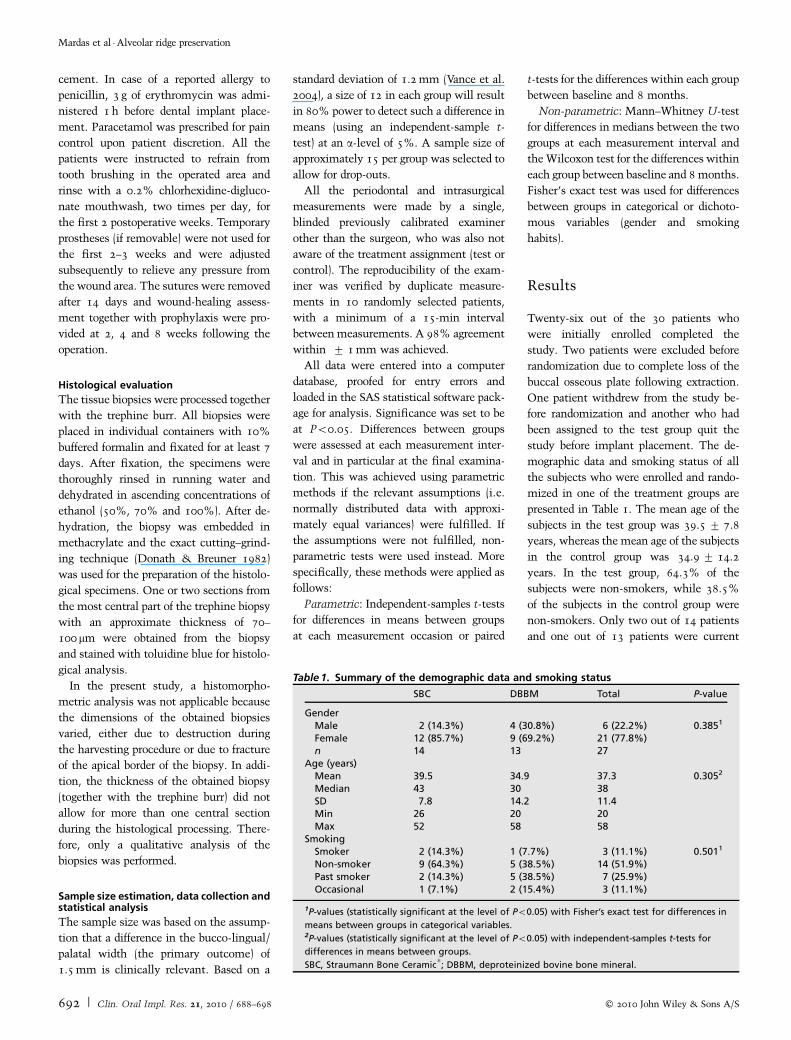
Twenty-six out of the 30 patients who
were initially enrolled completed the
study. Two patients were excluded before
randomization due to complete loss of the
buccal osseous plate following extraction.
One patient withdrew from the study be-
fore randomization and another who had
been assigned to the test group quit the
study before implant placement. The de-
mographic data and smoking status of all
the subjects who were enrolled and rando-
mized in one of the treatment groups are
presented in Table 1. The mean age of the
subjects in the test group was 39.5� 7.8
years, whereas the mean age of the subjects
in the control group was 34.9� 14.2
years. In the test group, 64.3% of the
subjects were non-smokers, while 38.5%
of the subjects in the control group were
non-smokers. Only two out of 14 patients
and one out of 13 patients were current
Table 1. Summary of the demographic data and smoking status
SBC DBBM Total P-value
GenderMale 2 (14.3%) 4 (30.8%) 6 (22.2%) 0.3851
Female 12 (85.7%) 9 (69.2%) 21 (77.8%)n 14 13 27
Age (years)Mean 39.5 34.9 37.3 0.3052
Median 43 30 38SD 7.8 14.2 11.4Min 26 20 20Max 52 58 58
SmokingSmoker 2 (14.3%) 1 (7.7%) 3 (11.1%) 0.5011
Non-smoker 9 (64.3%) 5 (38.5%) 14 (51.9%)Past smoker 2 (14.3%) 5 (38.5%) 7 (25.9%)Occasional 1 (7.1%) 2 (15.4%) 3 (11.1%)
1P-values (statistically significant at the level of Po0.05) with Fisher’s exact test for differences in
means between groups in categorical variables.2P-values (statistically significant at the level of Po0.05) with independent-samples t-tests for
differences in means between groups.
SBC, Straumann Bone Ceramics
; DBBM, deproteinized bovine bone mineral.
Mardas et al �Alveolar ridge preservation
692 | Clin. Oral Impl. Res. 21, 2010 / 688–698 c� 2010 John Wiley & Sons A/S

smokers in the test and the control groups,
respectively.
Each of the 26 patients who completed
the study contributed one extraction site.
The distribution of the extracted teeth
between the two groups in these patients
is presented in Table 2.
The healing following the ridge preserva-
tion procedure was uneventful overall. Few
patients in both groups reported minor
postoperative pain or discomfort, localized
oedema and in some cases exfoliated graft
particles were observed. All the patients
presented with membrane exposure at the
first postoperative week that, in most
cases, became larger during the second
week. At the fourth postoperative week,
most of the collagen barrier had been re-
sorbed, and at the 16th postoperative week,
complete closure of the postextraction
socket was observed with newly formed
keratinized mucosa.
Periodontal clinical indices and plaquelevels
The periodontal clinical indices at the
neighbouring teeth are presented in Table
3. At baseline, the mean PPD at neighbour-
ing teeth was statistically higher in the
control group. Moreover, at the 8-month
visit, the PPD was significantly reduced in
the control group but remained stable in
the test group. No statistically significant
differences in REC and BOP measurements
were observed between the groups at any of
the observation periods. Both treatments
equally preserved the baseline level of the
free gingival margin at the neighbouring
teeth following the ridge preservation
procedure.
Intrasurgical measurements of the alveolarridge
The bucco-lingual/palatal width (B-L/P)
The mean and range values of the bucco-
lingual/palatal width of the alveolar ridge
are presented in Table 4. The baseline B-L/
P of the alveolar ridge was comparable
between the two groups, while a statisti-
cally significant reduction of this dimen-
sion was observed in both groups. In the
SBC group, the mean B-L/P at baseline was
8.1� 1 mm and decreased to 7� 1.1 mm
at 8-month follow-up, presenting a reduc-
tion of the B-L/P of 1.1� 1 mm. In the
DBBM group, the mean B-L/P at baseline
was 9� 1.6 mm and decreased to
6.9� 1.9 mm at 8-month follow-up, pre-
senting a reduction of the B-L/P of
2.1� 1 mm. The mean reduction of the
B-L/P from baseline to 8 months was
statistically significantly lower in the SBC
group (Po0.05).
Width of the buccal and palatal/lingual boneplate (Bbw, P/Lbw)
The mean and range values of the width of
the buccal and palatal bone plate measured
in the most central part are presented in
Table 5. In 4/13 cases in the SBC group and
in 3/13 cases from the DBBM group, the
regenerated bone was completely inte-
grated into the buccal and palatal/lingual
bone plates and the examiner was unable to
distinguish the borders between the aug-
mented sites and the buccal bone plate. In
the SBC group, the baseline Bbw and L/
Pbw were 0.9� 0.3 and 1.3� 0.5 mm,
respectively, while at 8-month follow-up,
the Bbw and L/Pbw were 0.4� 0.5 and
1� 0.6 mm, respectively. In the DBBM
group, the baseline Bbw and L/Pbw
were 1.2� 0.6 and 1.2� 0.4 mm, respec-
tively, while at 8-month follow-up, the
Bbw and L/Pbw were 1.2� 1 mm and
1.3� 0.6 mm, respectively. The mean
changes of Bbw and L/Pbw from baseline
Table 2. Tooth extraction distribution between the two groups
Central incisor Lateral incisor Canine Premolars Total
Maxilla SBC 6 1 1 1 9Maxilla DBBM 7 5 12Mandible SBC 1 3 4Mandible DBBM 1 1Total 14 1 1 10 26
SBC, Straumann Bone Ceramics
; DBBM, deproteinized bovine bone mineral.
Table 3. Periodontal clinical indices on neighbouring teeth: mean PPD and REC
Baseline 8 months Difference P-valuenn
PPDSBC 2.2 � 0.2 2.2 � 0.3 � 0 � 0.4 0.66
n¼ 14 n¼ 13 n¼ 13 n¼ 13DBBM 2.6 � 0.4 2.2 � 0.3 � 0.3 � 0.4 0.011nn
n¼ 13 n¼ 13 n¼ 13 n¼ 13P-valuen 0.003n 0.697RECSBC 0.4 � 0.6 0.4 � 0.5 � 0 � 0.1 0.54
n¼ 14 n¼ 13 n¼ 13 n¼ 13DBBM 0.3 � 0.4 0.4 � 0.5 0.1 � 0.2 0.157
n¼ 13 n¼ 13 n¼ 13 n¼ 13P-valuen 0.34 0.7
nStatistically significant P-values (o0.05) with independent-samples t-tests (n) for differences in means
between groups.nnStatistically significant P-values (o0.05) with paired t-tests (nn) for the differences within each group
between baseline and 8 months.
SBC, Straumann Bone Ceramics
; DBBM, deproteinized bovine bone mineral; REC, gingival recession;
PPD, probing pocket depth.
Table 4. The bucco-lingual/palatal width of the alveolar ridge (mm; means/(median)� SD)
Baseline 8 months Difference P-valuenn
SBC 8.1/(8) � 1 7/(7) � 1.1 � 1.1/(� 1) � 1 0.0039nn
DBBM 9/(9) � 1.6 6.9/(7) � 1.9 � 2.1/(� 2) � 1 0.0002nn
P-valuen 0.117 0.809 0.017n
N 13 13 13
nStatistically significant P-values (o0.05) with the Mann–Whitney U-test for the difference in bucco-
lingual/palatal width of the alveolar ridge between SBC and BDX groups.nnStatistically significant P-values (o0.05) with the Wilcoxon signed rank test for the differences
within each group between baseline and 8 months.
SBC, Straumann Bone Ceramics
; DBBM, deproteinized bovine bone mineral.
Mardas et al �Alveolar ridge preservation
c� 2010 John Wiley & Sons A/S 693 | Clin. Oral Impl. Res. 21, 2010 / 688–698

to 8 months were not statistically signifi-
cant between the two groups (P40.05).
The mean and range values of the dis-
tance of the alveolar bone crest at the
mesial-central (Mbh) and distal-central
(Dbh) aspects of the socket to the relative
cementum–enamel junction or the re-
storation margin of the neighbouring teeth
are presented in Table 5. In 2/13 cases in
the SBC group, the Dbh values were not
available due to the absence of the relevant
tooth distal of the extraction socket. In
1/13 cases from the DBBM group, the
Mbh value was not available due to the
absence of the relevant tooth mesial of the
extraction socket. In the SBC group, the
baseline Mbh and Dbh were 3.4 � 0.8
and 3.1 � 0.4 mm, respectively, while at
8-month follow-up, the Mbh and Dbh
were 3 � 1.2 and 3.4 � 1.6 mm, respec-
tively. In the DBBM group, the baseline
Mbh and Dbh were 3.1 � 0.9 and
3.2 � 0.7 mm, respectively, while at 8-
month follow-up, the Mbh and Dbh
were 3.3 � 1.1 and 3.5 � 1.4 mm, re-
spectively. The mean changes of the
Mbh and Dbh from baseline to 8 months
were not statistically significant between
the two groups (P40.05).
The resistance of bone tissue during the
trephination process was similar in both
groups (Table 6). In all patients in the SBC
group, a dental implant of adequate dimen-
sions was placed with satisfactory initial
stability. In one patient from the DBBM
group, implant placement was not possible
due to insufficient primary stability. Dur-
ing implant placement, 9/13 implants in
the SBC group and 8/12 implants in the
DBBM group presented with either dehis-
cence or fenestration defects and required
additional bone augmentation to allow im-
plant placement in a predetermined pros-
thetically driven position (Table 7). Four
implants in each group were placed with-
out any additional bone augmentation pro-
cedure.
Histological analysis
Twelve specimens from each group were
available for histological analysis. In
one specimen from each group, it was not
possible to perform the histological
procedure.
In the SBC group, variable new bone
formation was observed. The newly formed
bone was observed mainly at the apical part
of the biopsy and was mainly woven, with
more lamellar bone occurring only in iso-
lated instances (Figs 7 and 8). The amount
of bone marrow differed significantly be-
tween individuals. In several specimens,
the SBC particles were in direct contact
with the newly formed bone (Figs 7 and 8).
In the coronal part of the biopsy, the SBC
particles were surrounded by dense con-
nective tissue composed of various forms of
fibroblasts, collagen fibres and blood ves-
sels, with no signs of inflammation. In
some specimens, areas of mineral apposi-
tion of the connective tissue fibres were
seen (Figs 7 and 8). No active resorption of
the SBC particles was observed.
Similar histological characteristics were
observed in the biopsies at the DBBM group
(Figs 9 and 10). New bone formation was
mainly limited in the apical part of the
biopsy, where newly formed bone of either
the woven or the mature lamellar type was
observed in direct contact with the DBBM
particles. No signs of active resorption of
the DBBM particles were observed. In the
coronal part of the biopsies, the particles
were surrounded by a dense connective
tissue, with no signs of inflammation.
Discussion
The aim of the various ridge preservation
procedures, involving grafting of immedi-
ate postextraction sockets, is to prevent
alveolar ridge atrophy and maintain ade-
quate dimensions of bone in order to facil-
itate implant placement in prosthetically
driven positions or to maintain an accepta-
ble ridge contour in areas of aesthetic con-
cern. This randomized, controlled clinical
trial compared the potential of a synthetic
bone substitute (SBC) and a bovine-derived
xenograft (DBBM), both in combination
with a collagen barrier according to the
GTR principle, to preserve alveolar ridge
dimensions and promote osseous healing
of postextraction alveolar sockets in the
Table 5. Bbw, L/Pbw, Mbh and Dbh of the alveolar ridge (mm; mean (median)� SD)
Baseline 8 months Difference P-valuenn
BbwSBC 0.9 (1) � 0.3 0.4 (0) � 0.5 � 0.4 (0) � 0.5 0.125
n¼ 13 n¼ 9 n¼ 9 n¼ 9DBBM 1.2 (1) � 0.6 1.2 (1) � 1 � 0.1(0) � 0.7 1
n¼ 13 n¼ 10 n¼ 10 n¼ 10P-valuen 0.096 0.097 0.317L/PbwSBC 1.3 (1) � 0.5 1 (1) � 0.6 � 0.3(0) � 0.9 0.375
n¼ 13 n¼ 13 n¼ 13 n¼ 13DBBM 1.2 (1) � 0.4 1.3 (1) � 0.6 0.1 (0) � 0.7 1
n¼ 13 n¼ 13 n¼ 13 n¼ 13P-valuen 0.38 0.33 0.286MbhSBC 3.4 (3) � 0.8 3 (3) � 1.2 � 0.4 (� 1) � 1 0.312
n¼ 13 n¼ 13 n¼ 13 n¼ 13DBBM 3.1 (3) � 0.9 3.3 (3) � 1.1 0.2 (0) � 0.7 0.687
n¼ 12 n¼ 12 n¼ 12 n¼ 12P-valuen 0.328 0.775 0.09DbhSBC 3.1 (3) � 1.4 3.4 (3) � 1.6 0.3 (0) � 0.6 0.375
n¼ 11 n¼ 11 n¼ 11 n¼ 11DBBM 3.2 (3) � 0.7 3.5 (3) � 1.4 0.3 (0) � 1.3 0.531
n¼ 13 n¼ 13 n¼ 13 n¼ 13P-valuen 0.526 0.857 0.951
nP-values (statistically significant at the level of Po0.05) with the Mann–Whitney U-test for the
difference in the bucco-lingual/palatal width of the alveolar ridge between SBC and BDX groups.nnP-values (statistically significant at the level of Po0.05) with the Wilcoxon signed rank test for the
differences within each group between baseline and 8 months.
SBC, Straumann Bone Ceramics
; DBBM, deproteinized bovine bone mineral.
Table 6. Frequency distribution of treatedsites according to the resistance of bonetissue during the trephination process
Hard Medium Soft N
SBC 2 (15.4%) 6 (46.1%) 5 (38.5%) 13DBBM 2 (15.4%) 6 (46.1%) 5 (38.5%) 13
SBC, Straumann Bone Ceramics; DBBM, de-
proteinized bovine bone mineral.
Mardas et al �Alveolar ridge preservation
694 | Clin. Oral Impl. Res. 21, 2010 / 688–698 c� 2010 John Wiley & Sons A/S

incisor, canine and bicuspid areas.
Although a decrease in the bucco-lingual
dimension of the alveolar ridge was ob-
served in both groups, both materials
equally preserved all the other clinical
dimensions of the site and supported new
bone formation in postextraction sockets,
allowing the placement of dental implants.
The results of this study are in agreement
with previous controlled studies where si-
milar combinations of bone grafts or sub-
stitutes with resorbable barriers were
successfully used for alveolar ridge preser-
vation (Iasella et al. 2003; Vance
et al. 2004; Barone et al. 2008). However,
complete preservation of the pre-extraction
ridge dimensions should not be anticipated,
even when alveolar ridge preservation tech-
niques involving postextraction socket
grafting are applied. In the present study,
a bucco-lingual width reduction of the
alveolar ridge was observed in both groups,
confirming previous clinical and preclinical
reports that postextraction healing is al-
ways characterized by osseous resorption
and significant contour changes especially
in the horizontal plane of the residual
alveolar ridge (Schropp et al. 2003; Araujo
& Lindhe 2005). These changes may be
limited but not avoided when grafting of
the socket is utilized (Iasella et al. 2003;
Barone et al. 2008; Araujo & Lindhe 2009;
Araujo et al. 2009).
In our study, the bucco-lingual dimen-
sion of the coronal part of the socket
decreased by 1.1� 1 mm in the SBC group
and by 2.1� 1 mm in the DBBM group.
Similar postextraction alveolar ridge resorp-
tion was observed in previous randomized,
controlled clinical trials where extraction
sockets were treated with either a porcine
xenograft and a collagen barrier (Barone
et al. 2008) or freeze-dried bone and a
collagen membrane (Iasella et al. 2003)
and compared with the healing of ‘‘empty’’
untreated extraction sockets (for a review,
see Darby et al. 2009). One of the limita-
tions of the present study is the absence of
a negative control group including patients
in whom unassisted socket healing would
have been followed for a similar period of
time and therefore the lack of a negative
control group does not allow a complete
evaluation of the overall effectiveness of
the two biomaterials.
In the present investigation, full-thick-
ness buccal and palatal/lingual muco-peri-
osteal flaps were raised to facilitate the
placement of the barrier membranes over
sound alveolar bone. It has been previously
advocated that in full-thickness muco-peri-
osteal flaps, the bone–periosteum continu-
ity is disrupted and a marginal bone
resorption of approximately 1 mm should
be anticipated (Moghaddas & Stahl 1980).
Based on this, it has been suggested that in
cases of postextraction ridge preservation,
flapless techniques should be utilized, be-
cause flap reflection may initiate further
bone resorption in addition to that natu-
rally occurring in the bundle bone of the
Table 7. Frequency distribution of residual peri-implant defects in the test and controlgroup following implant placement
Pristine Dehiscence Fenestration Failure N
SBC 4 (31%) 6 (46%) 3 (23%) 13DBBM 4 (31%) 7 (53.7%) 1 (7.7%) 1 (7.7%) 13
SBC, Straumann Bone Ceramics
; DBBM, deproteinized bovine bone mineral.
Fig. 6. Implant placement in the ridge preservation site at 8 months following the alveolar ridge preservation
surgery.
Fig. 7. Photomicrograph of a test specimen [Straumann Bone Ceramics
(SBC)] at the apical portion of the
socket. Residual SBC particles are integrated into newly formed bone. Toluidine blue staining, original
magnification � 10.
Mardas et al �Alveolar ridge preservation
c� 2010 John Wiley & Sons A/S 695 | Clin. Oral Impl. Res. 21, 2010 / 688–698

alveolar socket as a result of postextraction
healing. Whether or not the adaption of a
less extensive flap elevation or even a
flapless approach would have resulted in
further reduction of postextraction bone
resorption remains to be investigated;
therefore, further research is necessary to
define the most effective surgical protocol
for alveolar ridge preservation.
Although all the other alveolar ridge di-
mensions were similar between the two
groups, SBC was associated with statistically
significantly less reduction of the bucco-
lingual width. A sound biologic explanation
for the observed difference remains un-
known, but it should not be attributed solely
to a different ability of the two biomaterials
to promote bone formation because no other
clinical or histological parameter differed
between the two groups. Pre-operative fac-
tors such as the different distribution of the
extraction sites (four mandibular teeth in the
SBC group versus one in the DBBM group),
together with possible differences in the
condition of soft and hard tissues immedi-
ately after tooth extraction that were not
evaluated in this study, may be responsible
for this observation.
At 8 months following an alveolar ridge
preservation operation, the bucco-lingual
width was 7� 1.1 mm and 6.9� 1.9 mm
in the SBC and DBBM groups, respectively.
Considering that an alveolar ridge width
between 7 and 8 mm is usually necessary
for the placement of a standard diameter
implant, such postextraction ridge dimen-
sions allowed implant placement in all but
one case. However, in nine out of the 13
cases in the SBC group and in eight out of
12 cases in the DBBM group guided bone
regeneration to treat residual dehiscence or
fenestration defects around the implants
was necessary. These findings were not in
agreement with Barone et al. (2008), where
implant placement was uncomplicated in
the sockets that were treated with a porcine
xenograft and a collagen barrier. Different
implant and augmentation materials and
implant placement protocol, as well as the
intraoral distribution of the treated sites,
may be responsible for this different out-
come.
To our knowledge, this is one of the few
prospective randomized, controlled trials
(Barone et al. 2008) in which clinical and
histological data are correlated following
comparison of two different grafting mate-
rials for alveolar ridge preservation accord-
ing to the GTR principle. From the
histological point of view, both biomater-
ials promoted new bone formation, possi-
bly by osteoconduction at the apical and
the middle part of the socket while the
Fig. 8. Higher magnification of the same test specimen [Straumann Bone Ceramics
(SBC)]. Residual SBC
particles are integrated either into newly formed woven bone or into connective tissue. Toluidine blue staining,
original magnification � 20.
Fig. 9. Photomicrograph of a control specimen [deproteinized bovine bone mineral (DBBM)] at the apical
portion of the socket. Residual DBBM particles are integrated into newly formed bone and dense connective
tissue. Toluidine blue staining, original magnification � 10.
Fig. 10. Higher magnification of the same control specimen [deproteinized bovine bone mineral (DBBM)].
Residual DBBM particles are integrated into newly formed woven bone. Toluidine blue staining, original
magnification � 20.
Mardas et al �Alveolar ridge preservation
696 | Clin. Oral Impl. Res. 21, 2010 / 688–698 c� 2010 John Wiley & Sons A/S

coronal and central part of the socket was
mainly occupied by graft particles sur-
rounded by dense connective tissue, even
at 8 months following the alveolar ridge
preservation surgery. The histological re-
sults of the present study corroborate pre-
vious preclinical reports in dogs, where it
was concluded that both DBBM and SBC
are potential osteoconductive scaffolds to
support GBR for the treatment of dehis-
cence peri-implant defects (Schwarz et al.
2007). However, previous human histolo-
gical reports presented similar or even bet-
ter healing response after grafting of the
socket with deproteinized xenografts in
combination (Vance et al. 2004; Barone
et al. 2008) or not (Artzi et al. 2000;
Heberer et al. 2008) with barrier mem-
branes. Similar promising human histolo-
gical results have been published when
SBC was used for sinus augmentation
(Cordaro et al. 2008; Froum et al. 2008).
Although the moderate histological results
of the present study cannot be directly
compared with previous human histologi-
cal reports, as histomorphometric analysis
was not applicable in this study, several
factors may have contributed to the lack of
complete osseous regeneration of the alveo-
lar socket. The early membrane exposure
that took place in our study may have
compromised bone formation in the event
that these barriers became infected at a
later stage (Nowzari et al. 1995). However,
no obvious clinical signs of infections such
as suppuration were observed in this study
or in any of the above-mentioned studies
where the central part of the membrane
was also left uncovered. Although the
membrane was still visible in both groups
at the second postoperative week, at the
fourth postoperative week, most of the
collagen membrane had been resorbed and
possibly had lost barrier function (Donos
et al. 2004). Whether or not an early
resorption of the collagen barrier has influ-
enced bone formation in the coronal part of
the socket is an issue that needs further
investigation. However, it is logical to
assume that the use of barriers with de-
layed resorption time would provide an
elongated barrier function that would
have promoted further new bone formation
in the extraction socket by inhibiting the
connective tissue proliferation into the
socket area for a longer period of time
(Mardas et al. 2003; Donos et al. 2005).
In the present study, no effort was made
to select a predetermined type of socket
(Juodzbalys et al. 2008). The extraction
sockets in this study presented with differ-
ent soft tissue quantities, qualities and
gingival tissue biotypes as well as with
different anatomical and dimensional char-
acteristics of the hard tissue compartment.
Obviously, some of these characteristics,
together with several other factors (e.g.
smoking, reason for extraction, tooth loca-
tion, etc.), may influence the final out-
come of any socket preservation procedure
and may be important in making the deci-
sion of whether or not a ridge preservation
technique is indicated. Although many of
these factors were evaluated in this study,
the small unequal numbers of subjects in
each category (Tables 1 and 2) did not allow
any conclusions from this set of data.
Additional studies based on large patient
samples are necessary in order to identify
the specific trends and risk parameters that
should be evaluated before any alveolar
ridge preservation procedure as prognostic
factors of their effectiveness.
Conclusion
Both biomaterials partially preserved the
width and the interproximal bone height
of alveolar ridge. Both biomaterials sup-
ported implant placement at 8 months
following the ridge preservation procedure.
Acknowledgements: The authors
wish to express their gratitude to all
the clinical and research staff at the
Periodontal Research Unit at the
Eastman Clinical Investigation Center
(ECIC) and the laboratory technicians of
UCL, Eastman Dental Institute, involved
in the study. The materials for this study
were kindly supplied by Institut
Straumann, Basel, Switzerland.
References
Araujo, M.G., Linder, E. & Lindhe, J. (2009) Effect
of a xenograft on early bone formation in extrac-
tion sockets: an experimental study in dog. Clin-
ical Oral Implants Research 20: 1–6.
Araujo, M.G. & Lindhe, J. (2005) Dimensional ridge
alterations following tooth extraction. An experi-
mental study in the dog. Journal of Clinical
Periodontology 32: 212–218.
Araujo, M.G. & Lindhe, J. (2009) Ridge preservation
with the use of Bio-Oss collagen: a 6-month study
in the dog. Clinical Oral Implants Research 25:
433–440.
Artzi, Z., Tal, H. & Dayan, D. (2000) Porous bovine
bone mineral in healing of human extraction sock-
ets. Part 1: histomorphometric evaluations at 9
months. Journal of Periodontology 71: 1015–1023.
Barone, A., Aldini, N.N., Fini, M., Giardino, R.,
Calvo Guirado, J.L. & Covani, U. (2008) Xeno-
graft versus extraction alone for ridge preservation
after tooth removal: a clinical and histomorpho-
metric study. Journal of Periodontology 79:
1370–1377.
Bartee, B.K. (2001) Extraction site reconstruction for
alveolar ridge preservation. Part 2: membrane-
assisted surgical technique. Journal of Oral Im-
plantology 27: 194–207.
Becker, W., Becker, B.E. & Caffesse, R. (1994) A
comparison of demineralized freeze-dried bone
and autologous bone to induce bone formation in
human extraction sockets. Journal of Perio-
dontology 65: 1128–1133.
Becker, W., Urist, M., Becker, B.E., Jackson, W.,
Parry, D.A., Bartold, M., Vincenzzi, G., De
Georges, D. & Niederwanger, M. (1996) Clinical
and histologic observations of sites implanted
with intraoral autologous bone grafts or allografts.
15 human case reports. Journal of Periodontology
67: 1025–1033.
Beirne, O.R, Curtis, T.A. & Greenspan, J.S. (1986)
Mandibular augmentation with hydroxyapatite.
Journal of Prosthetic Dentistry 55: 362–367.
Breitbart, A.S., Staffenberg, D.A., Thorne, C.H.M.,
Glat, P.M., Cunningham, N.S., Reddi, A.H.,
Ricci, J. & Steiner, G. (1995) Tricalcium phos-
phate and osteogenin: a bioactive onlay bone graft
substitute. Plastic Reconstructive Surgery 96:
699–708.
Brugnami, F., Then, P.R., Moroi, H. & Leone, C.W.
(1996) Histologic evaluation of human extraction
sockets treated with demineralized freeze-dried
bone allograft (DFDBA) and cell occlusive mem-
brane. Journal of Periodontology 67: 821–825.
Bucholz, R.W., Carlton, A. & Holmes, R.E. (1987)
Hydroxyapatite and tricalcium phosphate bone
graft substitute. Orthopaedics Clinics North
America 18: 323–334.
Cordaro, L., Bosshardt, D.D., Palattella, P., Rao, W.,
Serino, G. & Chiapasco, M. (2008) Maxillary
sinus grafting with Bio-Oss or Straumann bone
ceramic: histomorphometric results from a rando-
mized controlled multicenter clinical trial. Clin-
ical Oral Implants Research 19: 796–803.
Darby, I., Chen, S.T. & Buser, D. (2009) Ridge
preservation techniques for implant therapy. The
International Journal of Oral & Maxillofacial
Implants 24: 260–271.
Mardas et al �Alveolar ridge preservation
c� 2010 John Wiley & Sons A/S 697 | Clin. Oral Impl. Res. 21, 2010 / 688–698

Donath, K. & Breuner, G. (1982) A method for the
study of undecalcified bones and teeth with at-
tached soft tissues. The Sage-Schliff (sawing and
grinding) technique. Journal of Oral Pathology
11: 318–326.
Donos, N., Bosshardt, D.D., Lang, N.P., Graziani,
F., Tonetti, M.S., Karring, T. & Kostopoulos, L.
(2005) Bone Formation by Enamel Matrix Pro-
teins and Xenografts: an experimental study in the
rat ramus. Clinical Oral Implants Research 16:
140–146.
Donos, N., Lang, N.P., Karoussis, I.K., Bosshardt,
D., Tonetti, M. & Kostopoulos, L. (2004) Effect of
GBR in combination with deproteinized bovine
bone mineral and/or enamel matrix proteins on
the healing of critical-size defects. Clinical Oral
Implants Research 15: 101–111.
El Deeb, M. & Holmes, R.E. (1989) Zygomatic and
mandibular augmentation with proplast and por-
ous hydroxyapatite in rhesus monkeys. The Inter-
national Journal of Oral and Maxillofacial
Surgery 47: 480–487.
Feuille, F., Knapp, C.I., Brunsvold, M.A. & Mello-
nig, J.T. (2003) Clinical and histologic evaluation
of bone replacement grafts in the treatment of
localized alveolar ridge defects. Part 1: mineralized
freeze-dried bone allograft. International Journal
of Periodontics and Restorative Dentistry 23:
29–35.
Froum, S., Cho, S.C., Rosenberg, E., Rohrer, M. &
Tarnow, D. (2002) Histological comparison of
healing extraction sockets implanted with bioac-
tive glass or demineralized freeze-dried bone allo-
graft: a pilot study. Journal of Periodontology 73:
94–102.
Froum, S. & Stahl, S.S. (1987) Human intraosseous
healing responses to the placement of tricalcium
phosphate ceramic implants. II. 13 to 18 months.
Journal of Periodontology 58: 103–109.
Froum, S.J., Wallace, S.S., Cho, S.C., Elian, N. &
Tarnow, D.P. (2008) Histomorphometric compar-
ison of a biphasic bone ceramic to anorganic
bovine bone for sinus augmentation: 6- to 8-
month postsurgical assessment of vital bone for-
mation. A pilot study. International Journal of
Periodontics and Restorative Dentistry 28:
273–281.
Govindaraj, S., Costantino, P.D. & Friedman, C.D.
(1999) Current use of bone substitute in maxillo-
facial surgery. Facial Plastic Surgery 15: 73–81.
Gross, J. (1995) Ridge preservation using HTR
synthetic bone following tooth extraction. Gen-
eral Dentistry 43: 364–377.
Hammerle, C.H. & Lang, N.P. (2001) Single stage
surgery combining transmucosal implant place-
ment with guided bone regeneration and biore-
sorbable materials. Clinical Oral Implants
Research 12: 9–18.
Heberer, S., Al-Chawaf, B., Hildebrand, D., Nelson,
J.J. & Nelson, K. (2008) Histomorphometric ana-
lysis of extraction sockets augmented with Bio-
Oss Collagen after a 6-week healing period: a
prospective study. Clinical Oral Implants Re-
search 19: 1219–1225.
Johnson, K. (1969) A study of the dimensional
changes occurring in the maxilla following
tooth extraction. Australian Dental Journal 14:
241–254.
Iasella, J.M., Greenwell, H., Miller, R.L., Hill, M.,
Drisko, C., Bohra, A.A. & Scheetz, J.P. (2003)
Ridge preservation with freeze-dried bone allograft
and a collagen membrane compared to extraction
alone for implant site development: a clinical and
histologic study in humans. Journal of Perio-
dontology 74: 990–999.
Juodzbalys, G., Sakavicius, D. & Wang, H.L. (2008)
Classification of extraction sockets based upon
soft and hard tissue components. Journal of Perio-
dontology 79: 413–424.
Kenney, E.B., Lekovic, V., Sa Ferreira, J.C., Han, T.,
Dimitrijevic, B. & Carranza, F.A. Jr (1986) Bone
formation within porous hydroxyapatite implants
in human periodontal defects. Journal of Perio-
dontology 57: 76–83.
Lekovic, V., Camargo, P.M., Klokkevold, P.R.,
Weinlaender, M., Kenney, E.B., Dimitrijevic, B.
& Nedic, M. (1998) Preservation of alveolar bone
in extraction sockets using bioabsorbable mem-
branes. Journal of Periodontology 69: 1044–1049.
Mardas, N., Kostopoulos, L., Stavropoulos, A. &
Karring, T. (2003) Osteogenesis by guided tissue
regeneration and demineralised bone matrix. Jour-
nal of Clinical Periodontology 30: 176–183.
Mercier, P., Huang, .H., Cholewa, J. & Djokovic, S.
(1992) A comparative study of the efficacy and
morbidity of five techniques for ridge augmenta-
tion of the mandible. The International Journal of
Oral and Maxillofacial Surgery 50: 210–217.
Moghaddas, H. & Stahl, S.S. (1980) Alveolar bone
remodeling following osseous surgery. A clinical
study. Journal of Periodontology 51: 376–381.
Niedhart, C., Maus, U., Redmann, E. & Siebert,
C.H. (2001) In vivo testing of beta-tricalcium
phosphate cement for osseous reconstruction.
Journal of Biomaterials Research 15: 530–537.
Nowzari, H., London, R. & Slots, J. (1995) The
importance of periodontal pathogens in guided
periodontal tissue regeneration and guided bone
regeneration. The Compendium of Continuing
Education in Dentistry 16: 1042–1046.
Pietrovski, J. & Massler, M. (1967) Alveolar ridge
resorption following tooth extraction. Journal of
Prosthetic Dentistry 17: 21–27.
Quinn, J.H. & Kent, J.N. (1984) Alveolar ridge
maintenance with solid nonporous hydroxyapa-
tite root implants. Oral Surgery, Oral Medicine
and Oral Pathology 58: 511–521.
Rothamel, D., Schwarz, F., Sculean, A., Herten, M.,
Scherbaum, W. & Becker, J. (2004) Biocompat-
ibility of various collagen membranes in cultures
of human PDL fibroblasts and osteoblast-
like cells. Clinical Oral Implants Research 15:
443–449.
Schropp, L., Wenzel, A., Kostopoulos, L. & Karring,
T. (2003) Bone healing and soft tissue contour
changes following single-tooth extraction: a clin-
ical and radiographic 12-month prospective study.
International Journal of Periodontics and Re-
storative Dentistry 23: 313–323.
Schwarz, F., Herten, M., Ferrari, D., Wieland, M.,
Schmitz, L., Engelhardt, E. & Becker, J. (2007)
Guided bone regeneration at dehiscence-type
defects using biphasic hydroxyapatiteþbeta tri-
calcium phosphate (Bone Ceramic) or a collagen-
coated natural bone mineral (Bio-Oss Collagen):
an immunohistochemical study in dogs. The
International Journal of Oral and Maxillofacial
Surgery 36: 1198–1206.
Serino, G., Biancu, S., Iezzi, G. & Piattelli, A.
(2003) Ridge preservation following tooth extrac-
tion using a polylactide and polyglycolide sponge
as space filler: a clinical and histological study in
humans. Clinical Oral Implants Research 14:
651–668.
Stahl, S.S. & Froum, S.J. (1987) Histologic and
clinical responses to porous hydroxyapatite im-
plants in human periodontal defects. Three to
twelve months post-implantation. Journal of
Periodontology 58: 689–695.
Vance, G.S., Greenwell, H., Miller, R.L., Hill, M.,
Johnston, H. & Scheetz, J.P. (2004) Comparison
of an allograft in an experimental putty carrier and
a bovine-derived xenograft used in ridge preserva-
tion: a clinical and histologic study in humans.
The International Journal Oral & Maxillofacial
Implants 19: 491–497.
Wang, H.L., Kiyonobu, K. & Neiva, R.F. (2004)
Socket augmentation: rationale and technique.
13: 286–296.
Zafiropoulos, G.G., Hoffmann, O., Kasaj, A., Will-
ershausen, B., Weiss, O. & Van Dyke, T.E. (2007)
Treatment of intrabony defects using guided
tissue regeneration and autogenous spongiosa
alone or combined with hydroxyapatite/beta-
tricalcium phosphate bone substitute or bovine-
derived xenograft. Journal of Periodontology 78:
2216–2225.
Zitzmann, N.U., Naef, R. & Scharer, P. (1997)
Resorbable versus non resorbable membranes in
combination with Bio-Oss for guided bone regen-
eration. The International Journal Oral & Max-
illofacial Implants 12: 844–852.
Zitzmann, N.U., Scharer, P. & Marinello, C.P.
(2001) Long-term results of implants treated
with guided bone regeneration: a 5-year prospec-
tive study. The International Journal of Oral &
Maxillofacial Implants 16: 355–366.
Supporting Information
Additional supporting information
may be found in the online version of
this article:
The Consort E-Flowchart Aug. 2005
Table S1. Supporting information in
accordance with the CONSORT
Statement 2001 checklist used in
reporting randomized trials.
Please note: Wiley-Blackwell is
not responsible for the content or
functionality of any supporting
materials supplied by the authors.
Any queries (other than missing
material) should be directed to the
corresponding author for the article.
Mardas et al �Alveolar ridge preservation
698 | Clin. Oral Impl. Res. 21, 2010 / 688–698 c� 2010 John Wiley & Sons A/S
Copyright © 2022 FDOKUMEN