Cytoskeleton actin-binding proteins in clinical behavior of ...

Altered Secondary Myogenesis in Transgenic Animals Expressing the Neural Cell Adhesion Molecule under the Control of a Skeletal Muscle ot-Actin Promoter Sam Fazeli,* Dominic J. Wells, * Carl Hobbs,* and Frank S. Walsh* • Department of Experimental Pathology, UMDS, Guy's Hospital, London SE1 9RT, United Kingdom; and*Department of Veterinary Basic Sciences, The Royal Veterinary College, London NW1 0TU, United Kingdom
Abstract, The majority of skeletal muscle fibers are generated through the process of secondary myogene- sis. Cell adhesion molecules such as NCAM are thought to be intricately involved in the cell-cell interactions between developing secondary and primary myotubes. During secondary myogenesis, the expression of NCAM in skeletal muscle is under strict spatial and temporal control. To investigate the role of NCAM in the regulation of primary-secondary myotube interac- tions and muscle fusion in vivo, we have examined mus- cle development in transgenic mice expressing the 125-kD muscle-specific, glycosylphosphatidylinositol- anchored isoform of human NCAM, under the control of a human skeletal muscle a-actin promoter that is ac- tive from about embryonic day 15 onward. Analysis of developing muscle from transgenic animals revealed a significantly lower number of myofibers encased by
basal lamina at postnatal day 1 compared with non- transgenic littermates, although the total number of de- veloping myofibers was similar. An increase in muscle fiber size and decreased numbers of VCAM-l-positive secondary myoblasts at postnatal day 1 was also found, indicating enhanced secondary myoblast fusion in the transgenic animals. There was also a significant de- crease in myofiber number but no increase in overall muscle size in adult transgenic animals; other measure- ments such as the number of nuclei per fiber and the size of individual muscle fibers were significantly in- creased, again suggesting increased secondary myoblast fusion. Thus the level of NCAM in the sarcolemma is a key regulator of cell-cell interactions occurring during secondary myogenesis in vivo and fulfills the prediction derived from transfection studies in vitro that the 125- kD NCAM isoform can enhance myoblast fusion.
V ERTEBRATE skeletal myogenesis occurs in at least two distinct steps. Myoblasts align and fuse syn- chronously to form primary myotubes (Kelly and
Zacks, 1969; Ontell and Kozeka, 1984a,b). Secondary myo- tubes then begin to form under the basal lamina of pri- mary myotubes. With time, secondary myotubes become indistinguishable from primary myotubes, as they separate and acquire their own basal lamina. Primary myotubes then act as a scaffold for further rounds of myogenesis. In this way the majority of adult muscle fibers are composed of secondary myotubes.
A number of cell adhesion molecule (CAM) 1 families have been implicated in myoblast fusion and myotube in-
Address all correspondence to Professor F.S. Walsh, Department of Ex- perimental Pathology, UMDS, Guy's Hospital, London Bridge, London SEI 9RT, United Kingdom. Tel.: 0171-955 4574. Fax: 0171-403 8883.
1. Abbreviat ions used in this paper: CAM, cell adhesion molecule; EDL, extensor digitorum longus; GPI, glycosylphosphatidylinositol; H and E, haematoxylin and eosin; hNCAM, human neural cell adhesion molecule; MSD, muscle-specific domain; NCAM, neural cell adhesion molecule; PND, postnatal day; SOL, soleus; VCAM-1, vascular cell adhesion mole- cule-l; VLA-4, very late antigen-4.
teractions (Walsh and Doherty, 1994). These include vari- ous members of the cadherin family of calcium-dependent adhesion molecules (Knudsen, 1990; Donalies et al. 1991; Takeichi, 1991; Hahn and Covault, 1992; Mege et al., 1992; Moore and Walsh, 1993; Zeschnigk et al., 1995), the inte- grin family of cell-cell and cell-matrix adhesion molecules (Menko and Boettiger, 1987; Rosen et al., 1992), and neu- ral cell adhesion molecule (NCAM) (Covault and Sanes, 1986; Dickson et al., 1990; Knudsen et al., 1990; Tassin et al., 1991; Peck and Walsh, 1993).
NCAM is a member of the immunoglobulin superfamily of adhesion molecules. Use of alternative splicing gives rise to the main NCAM protein isoforms of 180, 140, and 120 kD (Walsh and Doherty, 1991). These differ in the length of their intracellular domains such that the trans- membrane 180-kD isoform has a larger domain than the 140-kd isoform, while the 120-kd size class are attached to the cell membrane via a glycosylphosphatidylinositol (GPI) anchor. Additional diversity in structure is found in the ex- tracellular portion of NCAM including the muscle specific domain (MSD) producing a 125-kd isoform of NCAM that is preferentially expressed in skeletal muscle (Dickson et al., 1987; Thompson et al., 1989), and a 10-amino acid
© The Rockefeller University Press, 0021-9525/96/10/241/11 $2.00 The Journal of Cell Biology, Volume 135, Number 1, October 1996 241-251 241
on March 13, 2016
jcb.rupress.orgD
ownloaded from
Published October 1, 1996
exon between exons 7 and 8, called VASE (Small and Akeson, 1990; Walsh et al., 1992).
NCAM expression in skeletal muscle is developmentally regulated. The 180- and 140-kd isoforms are expressed in the myotome (Thiery et al., 1982; Lyons et al., 1992). After fusion the expression of these isoforms is repressed and the GPI-linked isoform (125 kD) is uniformly expressed on the surface of primary myotubes. As secondary myo- genesis begins, however, NCAM is found concentrated at the site of membrane apposition of primary and secondary myotubes (Covault and Sanes, 1986), possibly indicating a role for NCAM in the control of secondary myogenesis. NCAM expression increases in parallel with myotube for- mation (Moore et al., 1987) and is subsequently down-reg- ulated in the adult (Moore and Walsh, 1985; Covault and Sanes, 1986). Perturbation of neuromuscular activity in the chick, by in vivo injection of botulinum toxin, leads to changes in the normal pattern of expression of NCAM and N-cadherin and is also accompanied by an arrest of sec- ondary myogenesis (Fredette et al., 1993). A direct corre- lation between the changes in CAM expression and the ef- fects on myogenesis cannot, however, be made since blockade of neuromuscular activity may have a number of other unknown consequences.
Many studies on the role of CAMs in muscle develop- ment have involved the use of in vitro cell culture systems (Dickson et al., 1990; Knudsen et al., 1990; Tassin et al., 1991; Hahn and Covault, 1992; Mege et al., 1992; Peck and Walsh, 1993). For NCAM, transfeetion-based studies us- ing the C2 muscle cell line have provided data that show a role in the regulation of myoblast fusion (Peck and Walsh, 1993). Expression of the 125-kd, GPI-anchored NCAM isoform in particular led to a dramatic enhancement of fu- sion. It clearly would be of interest to be able to test whether CAMs have a similar role to play in vivo. There are presently a number of possible strategies for directly assessing the function of molecules in vivo. Homologous recombination can be used to generate animals unable to make the proteins of interest. Alternatively, ectopic ex- pression of a molecule, or modified forms of it, may give insights into its function. We chose the second strategy to test the hypothesis that the levels of NCAM in developing myotubes may regulate primary-secondary myotube in- teractions, which is an important step in the development of the full complement of myofibers and myoblast fusion. As myofiber interactions likely involve the operation of a number of adhesion molecules including VCAM-1 and VLA4 (Rosen et al., 1992) and different cadherins such as N-cadherin (Hahn and Covault, 1992) and M-cadherin (Donalies et al., 1991; Moore and Walsh, 1993, Zeschnigk et al., 1995), it seemed unlikely that removal of NCAM ex- pression by homologous recombination methods would have dramatic functional consequences. Also, since NCAM is expressed in the developing and adult nervous system and heart, any observed skeletal muscle phenotype in ani- mals lacking a functional NCAM gene may be secondary to perturbations at other sites. Mice in which the NCAM gene has been inactivated using homologous recombina- tion have been produced (Cremer et al., 1994), but analy- sis of myogenesis has not yet been reported.
Expression of the 125-kd isoform of human NCAM (hNCAM), under the control of the skeletal c~-actin pro-
rooter, recapitulates some aspects of the developmental profile of expression of the endogenous isoform. Both the transgene and the endogenous isoform are expressed and colocalize in developing myotubes. While the expression of the transgene persists into adulthood, endogenous NCAM expression is down-regulated postnatally associ- ated with the development of neuromuscular interactions (Moore and Walsh, 1985; Covault and Sanes, 1986). Mus- cles in adult transgenic animals were found to have a sig- nificantly reduced number of myofibers but myofiber size and the number of nuclei per fiber were increased. The re- duction in muscle fiber number in the adult was found to be the result of an impairment of secondary myotube sep- aration from primary myotubes beginning at PND1. A sig- nificant depletion in VCAM-l-posi t ive secondary myo- blasts was also found at this age which, taken together with the increase in myofiber size and the number of nuclei per fiber, was indicative of enhanced fusion induced by ex- pression of the transgene.
Material and Methods
Generation of Transgenic Animals The SV-40 't ' intron was cloned into the Bluescript vector (pBSK II+: Stratagene, La Jolla, CA), the multiple cloning site of which had been pre- viously modified to aid construction (pBSV40). A cDNA for the 125-kd, GPI-anchored isoform of hNCAM (with ]line AV21] or without ]lines AN5 and 14] the VASE exon ]Barton et al., 1988; Small and Akeson, 1990]) was then directionally cloned into pBSV40. The human skeletal muscle-specific a-actin promoter (-2.2kbHSA-CAT, a gift from Dr. Edna Hardeman, Children's Medical Research Institute, Sydney, Australia; Brennan and Hardeman, 1993), containing 2.2 kb of the 5' region of the gene, was cloned upstream of the NCAM cDNA (pACT125; see Fig. 1). The fragment for microinjection was released with BssHIl and purified by gel electrophoresis and glass milk extraction (Qiaex; QIAGEN, Hilden, Germany). DNA was injected into a pronucleus of fertilized oocytes of C57B1/10 x CBA/J at 1-2 ng/l~l, and the eggs cultured overnight. Two cell embryos were then implanted into pseudopregnant recipients and the Go progeny were analyzed by PCR and Southern blotting of genomic DNA, as described below.
Polymerase Chain Reaction PCR analysis was routinely used to detect the presence of the transgene. PCR was performed using 5 'AACCCGCTCCTTCT' ITGGTCAACG 3'
Figure 1. Schemat ic r e p r e s e n t a t i o n of the express ion casse t te used for gene ra t i on of t r ansgen ic animals . N C A M c D N A encod- ing the GPI-anchored isoform including the MSD region and ei- ther with or without the VASE exon were cloned downstream of the 2.2-kb ct-actin promoter, in a modified pBSK II+ vector (Stratagene). The construct also includes the SV-40 't' intron and polyadenylation signals. The fragment used for injection was lib- erated by digestion with BssHII. Arrows indicate the position of the forward (+) and reverse ( - ) primers used for PCR analysis of genomic DNA. Southern analysis was performed on Bglll- digested genomic DNA, resulting in a 1-kb product specific to the transgene.
The Journal of Cell Biology, Volume 135, 1996 242
on March 13, 2016
jcb.rupress.orgD
ownloaded from
Published October 1, 1996

as forward primer corresponding to position -466 to -442 in the ct-actin promoter (Brennan and Hardeman, 1993) and 5'CCCTGGCTGGGA- ACAATATCCACC 3' as the reverse primer corresponding to +226 to +202 in the NCAM cDNA (see Fig. 1 A), giving a product of 692 bp (data not shown). For use in Northern blot analysis (see below), a DNA frag- ment was obtained by reverse transcriptase PCR (Cavicchioli et al., 1991) of mouse muscle RNA extracted using the NP-40 lysis method (Sambrook et al., 1989). Primers complementary to positions 406-429 (forward: 5 'CCATCTACAACGCCAACATCGACG 3') and 1613-1639 (reverse: 5'AATTCCAAGGACTCCTGTCCAATA 3') of mouse NCAM cDNA encoding the GPI-anchored, 120-kd isoform (Barbas et al., 1988) were used.
Southern Blotting Genomic DNA was obtained by proteinase K digestion of tail biopsies fol- lowed by phenol/chloroform extraction (Sambrook et al., 1989). 10 p,g of DNA was digested overnight with BglII, which liberates a 1-kb fragment specific to the transgene (see Fig. 2 A) and fractionated by agarose gel electrophoresis. DNA concentration was estimated spectrofluorometri- cally and equal loading of tracks was assessed by ethidium bromide stain- ing of the gel. After alkaline denaturation, the DNA was transferred to Biodyne A membranes (Pall, Portsmouth, UK) by capillary transfer and covalently bound to the membrane by UV irradiation. After prehybridiza- tion of membranes, a gel-purified BglII fragment internal to the hNCAM cDNA and labeled with [32p]dCTP by nick translation (Boehringer Mann- heim, Mannheim, Germany), was used for hybridization at 65°C over- night. The hybridization buffer was 6 × SSC, 5% Denhardt's, 0.5% SDS, and 100 ~g/ml herring sperm DNA. After hybridization, the membrane was washed three times with 1 × SSC and 0.5% SDS at 65°C.
Northern Analysis mRNA from transgenic animals and control 7-d denervated and nonde- nervated hind leg muscles was obtained using the Fast Track mRNA puri- fication kit (Invitrogen, Leek, Holland). 1 I~g of mRNA was loaded on a 2.2 M formaldehyde agarose gel and transferred to Biodyne A membranes by capillary transfer (Sambrook et al., 1989). Equal loading of samples was checked by ethidium bromide staining of the gel. The remainder of the
procedure was essentially the same as that for Southern blots, except for the hybridization probe which was generated by RT-PCR (see above).
Western Blotting Tissue samples were homogenized in 10 mM Tris-HCL (pH 7.4), 1 mM EDTA, 1% NP-40 containing 5% aprotinin, and 1 mM PMSF. After incu- bation for 20 min at 4°C, the homogenate was spun at 14,000 g for 15 min. The supernatant was used for protein estimation (BCA; Pierce, Rockford, IL) and further analysis. SDS-PAGE and Western transfer were carried out using standard protocols. Briefly, proteins were fractionated on a 7.5 % resolving gel and transferred to reinforced nitrocellulose membrane (Schleicher and Schuell, Dassel, Germany). Membranes were routinely stained with Ponceau S to ensure equal protein loading and transfer.
The membrane was blocked in 4% (wt/vol) casein in PBS for 1 h, and then incubated with a primary antibody to hNCAM (NCC-Lu-243; Hirano et al., 1989) for 1 h. After washing, membranes were incubated with HRP-conjugated rabbit anti-mouse Ig (Bio-Rad Laboratories, Rich- mond, CA) for 1 h. Enhanced chemiluminescence (ECL; Amersham In- ternational, Amersham, UK) was used to visualize immunoreactive bands.
Immunohistochemical Analysis of Muscle Sections Monoclonal antibodies A4.74 and N3.36 reacting with specific myosin heavy chains (gifts from Dr. Simon Hughes, Randall Institute, Kings Col- lege, London) were used to stain type IIa and all type II fibers, respec- tively (Hughes and Blau, 1992; Hughes et al., 1993). Conditioned culture media for both antibodies were used at a dilution of 1:10. NCAM expres- sion from the human transgene was visualized using the hNCAM-specific antibody ERIC1 (Bourne et al., 1991) at 1:100 on frozen sections and at 1: 50 for paraffin-embedded sections pretreated by microwaving (McCor- mick et al., 1993). No reactivity was found on nontransgenic muscle under any circumstances. Monoclonal antibodies to laminin (Sigma Chemical Co., Poole, UK) were used at 1:1,500. Rat anti-mouse VCAM-1 and VLA-4 (Serotec, Bicester, UK) were used at 1:50 dilution. Polyclonal anti- body to mouse M-cadherin was generated by immunizing rabbits with a fusion protein corresponding to the amino-terminal region of the extracel- lular domain. It was used at a 1:100 dilution. Rabbit anti-rat and rabbit
Figure 2. Analysis o f t ransgenic status and express ion level o f founder mice. Three founder lines were analyzed in this series of experi- ments: AN14, AV21, and AN5. (A) Sou thern blot analysis o f these lines revealed that lines AN14 and AV21 each conta ined >100 cop- ies of the t ransgene, whereas line AN5 conta ined ,--~30 copies. The ar row indicates the 1-kb f ragment ob ta ined by BglII digest ion of 10 l~g of genomic D N A , • ~2 hybridized to a r andom pr imed a-- P- labeled BgllI f ragment ob ta ined by digest ion of h N C A M c D N A . Copy num- bers were es t imated by compar i son of hybridizat ion signal with a serial di lut ion of the purif ied probe. (B) Immunob lo t analysis of h N C A M express ion in adult skeletal muscle ob ta ined f rom each line. 40 Ixg prote in was loaded in each well and pro te in loads were checked by Ponceau S staining of ni t rocel lulose m e m b r a n e s before ant ibody reaction. Lines AN14 and AV21 express approximate ly fivefold higher levels of h N C A M (125 kD, arrow) than line AN5. The blot also demons t ra t e s the lower level of express ion in a p redom- inantly slow muscle (SOL, S) c o m p a r e d with an almost exclusively fast muscle (EDL, E). The smaller bands observed in some of the samples are likely to be degrada t ion products . (C) m R N A levels in adult quadriceps, examined by Nor the rn blot analysis, show that lines AN14 and AN5 express fivefold higher and equal levels o f N C A M than the endogenous levels in 7-d dene rva t ed (DN) nont rans- genic muscle. O n e major m R N A species of ~3 .4 kb was de tec ted in t ransgenic muscle in contras t to the ~2 .7 -kb band in the nont rans- genic dene rva t ed muscle. No N C A M m R N A was de tec ted in n o n d e n e r v a t e d control muscle (CON). 1 txg of purif ied m R N A was loaded onto each lane and the m R N A load examined by e th id ium bromide staining of the gel. The blot was hybridized to a mouse p robe ob- ta ined by R T - P C R of mouse RNA, giving a f ragment equivalent to the BgllI f ragment used for Sou thern blotting.
Fazeli et al. Skeletal Muscle NCAM 243
on March 13, 2016
jcb.rupress.orgD
ownloaded from
Published October 1, 1996
anti-mouse secondary antibodies conjugated with FITC (Dako, UK) were used at 1:50.
For laminin staining of paraffin-embedded sections, the tissue was pre- digested with 0.1% trypsin in 0.1% calcium chloride at pH 7.8 at 37°C for 20 min. Biotinylated class-specific secondary antibodies (Sigma Chemical Co.) were used to reduce nonspecific staining. Immunoreactivity was visu- alized using a streptavidin biotin-HRP system (ABC; Dako A/S, Copen- hagen, Denmark) followed by 1 mg/ml 3'3'-diaminobenzidine (DAB) as the chromogen. Sections stained for laminin were enhanced by addition of 0.025% cobalt chloride and 0.02% nickel ammonium sulfate to the DAB reagent (DeJong et al., 1985).
Morphometric Analysis of Adult Skeletal Muscle The whole hind limb of PND1 animals or extensor-digitorum longus (EDL) and soleus (SOL) muscles of adult transgenic and nontransgenic littermates were dissected, placed in Cryoembed (Bright, Huntingdon, UK), and frozen in liquid nitrogen cooled isopentane. 5-1xm cryostat sec- tions were obtained from the mid-belly region of each muscle and either processed for immunohistochemistry or stained with haematoxylin and eosin (H and E). The radii of 200 fibers per section were measured using a camera lucida arrangement in conjunction with a digitizer pad. A personal computer (86B; Hewlett-Packard Co., Palo Alto, CA) was used with an IMAGAN basic quantitation program. Mean fiber radii and maximum fi- ber diameter over the minor axis were computed from closed loop and line length measurements of laminin-stained, paraffin-embedded sections, respectively. The data were then ranked and a frequency histogram gener- ated. All analyses were double blind and confined to transgenic and non- transgenic littermates of the same sex. To reactivate endogenous NCAM gene expression, the hind limb was denervated by sectioning of the sciatic nerve. Muscle biopsies were obtained 7 d later, at which time the endoge- nous mouse NCAM expression is high (Covault and Sanes, 1985; Moore and Walsh, 1986). To estimate muscle size, the perimeter of each section was traced and the area was calculated. The total number of fibers in each muscle was calculated by counting the number of fibers in an area of 0.7 mm 2, corresponding to the sum of the area of eight different regions cho- sen randomly, and extrapolated using the area of the muscle section. The number of nuclei in each section, stained with H and E, was estimated in a similar way.
Results
Expression of hNCAM in Transgenic Animals and Analysis of Transgene Expression
We have used a human skeletal muscle a-actin promoter fused to a hNCAM cDNA encoding the 125-kd, GPI- anchored isoform (with and without the product of the VASE exon) to achieve high level expression of this iso- form in the skeletal muscle of transgenic mice. PCR analy- sis initially was used to detect the presence of the trans- gene in founder mice. The PCR primers were specific to the transgene, with the forward primer in the promoter re- gion and the reverse primer in the 5' region of the NCAM cDNA (Fig. 1). All founder mice were also verified to be transgenic by Southern blot analysis. Three founder lines were analyzed in this study: AN14, AV21, and AN5, and a representative Southern blot is shown (Fig. 2 A).
The level of protein expression in these lines, as deter- mined by Western blot analysis, is shown in Fig. 2 B. Im- munoblot analysis also showed differing levels of expres- sion in the three lines in individual muscle types. In the predominantly fast EDL muscle there was higher expres- sion than in the SOL which is a slow muscle. Fiber type differences in hNCAM expression can also be observed on immunostained sections of adult muscle (see below). Northern blotting revealed one major mRNA band in the AN14 and AN5 lines of ~3.4 kb, which is larger than that observed in the nontransgenic denervated muscle (~2.7
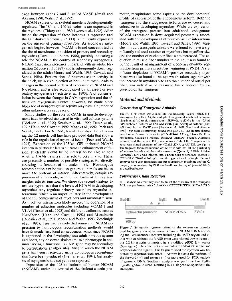
Figure 3. Immunoblot analysis of hNCAM espression under the control of the skeletal muscle a-actin promoter in line AN14. (A) A predominant band of 125 kd was detected in NP-40 extracts of skeletal muscle (M) and to a much lesser extent in heart (H) and gut (G) tissue. No detectable expression was obtained in spleen (S), kidney (K), liver (L), and brain (B). Each lane was loaded with 40 I~g of total protein. (B) The developmental profile of hNCAM expression is similar to that obtained from the et-actin promoter detailed in previous reports (Brennan and Hardeman, 1993). Expression is first detectable at El5 after which the ex- pression level increases (E17, PNDz). Each embryonic time point is represented by extracts of two embryos. Each lane was loaded with 40 Ixg of protein. The smaller bands present in some lanes are degradation products of the full-length hNCAM protein re- vealed by overloading to visualize the low levels of transgene expressed at embryonic stages. Adult (Ad) muscle was from PND 24.
kb, Fig. 2 C). This difference is attributable to the splicing of the SV-40 't ' intron at the 3' end of the cDNA in the transgene (Fig. 1), which does not affect the size of the protein product but increases the RNA size.
The level of hNCAM mRNA in line AN5 is approxi- mately equal to the level of endogenous NCAM mRNA in denervated adult muscle from nontransgenic animals (Fig. 2 C); denervation has been previously shown to reactivate N-CAM gene expression (Covault and Sanes, 1985; Moore and Walsh, 1985, 1986; Daniloff et al., 1986). Comparison of protein expression levels in lines AN14 and AV21 with line AN5 (Fig. 2 B) indicates about a fivefold higher level of hNCAM in the former than in line AN5. Thus, lines AN14 and AV21 express approximately fivefold higher levels of NCAM protein than is found in denervated mus- cle from nontransgenic mice. The profile of hNCAM ex- pression in various tissues from adult animals of line AN14 is shown in Fig. 3 A. The highest level of expression is in skeletal muscle, with heart and gut the only other tissues showing detectable expression. The developmental profile
The Journal of Cell Biology, Volume 135, 1996 244
on March 13, 2016
jcb.rupress.orgD
ownloaded from
Published October 1, 1996

of expression of the transgene in these lines (Fig. 3 B) re- vealed an increase in expression between PND1 and adult muscle such that at PND1 the expression level of the transgene in lines AN14 and AV21 is estimated to be ap- proximately equal to that of the endogenous protein. Our data are similar to the published profile and developmen- tal expression of the skeletal e~-actin promoter (Muscat and Kedes, 1987; Brennan and Hardeman, 1993). The ex- pression of the transgene remains high while the endoge- nous gene is switched off in adult innervated muscle (Moore and Walsh, 1985; Covault and Sanes, 1986).
Immunohistochemical Analysis of Muscle Cryosections
Immunohistochemical analysis showed that hNCAM was detected at the sarcolemma on primary myotubes at E15 (Fig. 4 A). During secondary myogenesis, hNCAM is also found expressed at higher levels at sites of primary-sec- ondary myotube apposition than on the rest of the sarco- lemma (Fig. 4 C). Fig. 4 also shows the expression of the endogenous mouse NCAM protein (Fig. 4, B and D) which colocalizes with the transgene.
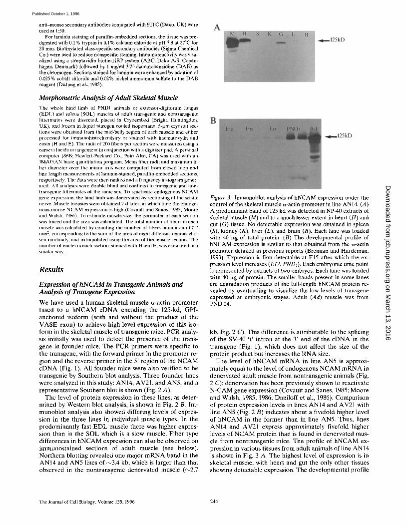
Adult EDL muscle from transgenic animals displayed clear fiber type variations in the expression of the trans- gene (Fig. 5). An antibody (A4.74) that recognizes MHC isoforms specific to type IIa fibers (Fig. 5 B) clearly indi-
cates that these fibers have a reduced level of expression of hNCAM compared with l ib fibers (Fig. 5 A). We have calculated that the mouse EDL is composed almost exclu- sively of type II fibers, with a ratio of about 30:70 of IIa/IIb subtypes, In the SOL, which is composed of both type IIa and I fibers, a more uniform staining was observed (data not shown). Taken together with the results obtained from immunoblot analysis (Fig. 2 C), these observations indi- cate a higher expression level in type l ib fibers compared with type IIa or type I fibers. The apparent intracellular staining for hNCAM is similar to that of the endogenous protein in developing, denervated, or regenerating adult muscle (Moore and Walsh, 1986).
Impaired Secondary Myogenesis in Transgenic Mice
Fig. 6 shows examples of laminin-stained muscle fibers from transgenic (Fig. 6 A) versus nontransgenic (Fig. 6 B) animals. The results of fiber counts obtained from four sets of muscle samples from transgenic and nontransgenic littermates are shown in Fig. 6, C and D. At E19 both groups of animals were found to have similar numbers of laminin encased fibers (Fig. 6 C), and H and E-stained muscle fibers (Fig. 6 D). Thus, fiber numbers were 578 ___ 23 for transgenic and 532 -+ 57 for nontransgenic animals for laminin and 577 _+ 52 and 531 _+ 41, respectively, for H
Figure 4. Immunohistochem- ical detection of NCAM in developing muscle. Immuno- staining with human specific antibody ERIC1 revealed muscle-specific expression of the transgene at El5 (A). The endogenous isoform of NCAM (detected with anti- body H28) is also found ex- pressed in myotubes, but higher levels of NCAM are present in nerve (arrowhead, B). Immunostaining of PND1 EDL muscle with hNCAM-specific antibody (C) and mouse NCAM-spe- cific antibody (D) revealed a similar expression profile of the two isoforms in muscle. Both human and mouse NCAM are found concen- trated at sites of myotube ap- position (arrows). Bar (A and B), 100 ~m; (C and D), 10 I~m.
Fazeli et al. Skeletal Muscle NCAM 245
on March 13, 2016
jcb.rupress.orgD
ownloaded from
Published October 1, 1996
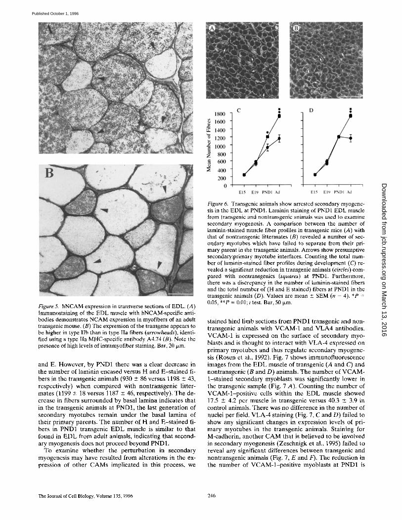
Figure 5. hNCAM expression in transverse sections of EDL. (A) Immunostaining of the EDL muscle with hNCAM-specific anti- bodies demonstrates NCAM expression in myofibers of an adult transgenic mouse. (B) The expression of the transgene appears to be higher in type Ifb than in type Ila fibers (arrowheads), identi- fied using a type IIa MHC-specific antibody A4.74 (B). Note the presence of high levels of intramyofiber staining. Bar, 20 Ixm.
and E. However, by PND1 there was a clear decrease in the number of laminin encased versus H and E-s ta ined fi- bers in the transgenic animals (930 +__ 86 versus 1198 + 43, respectively) when compared with nontransgenic litter- mates (1199 +__ 18 versus 1187 +__ 46, respectively). The de- crease in fibers surrounded by basal lamina indicates that in the transgenic animals at PND1, the last generation of secondary myotubes remain under the basal lamina of their primary parents. The number of H and E-s ta ined fi- bers in PND1 transgenic E D L muscle is similar to that found in E D L from adult animals, indicating that second- ary myogenesis does not proceed beyond PND1.
To examine whether the perturbation in secondary myogenesis may have resulted from alterations in the ex- pression of other CAMs implicated in this process, we
Figure 6. Transgenic animals show arrested secondary myogene- sis in the EDL at PND1. Laminin staining of PND1 EDL muscle from transgenic and nontransgenic animals was used to examine secondary myogenesis. A comparison between the number of laminin-stained muscle fiber profiles in transgenic mice (A) with that of nontransgenic littermates (B) revealed a number of sec- ondary myotubes which have failed to separate from their pri- mary parent in the transgenic animals. Arrows show presumptive secondary:primary myotube interfaces. Counting the total num- ber of laminin-stained fiber profiles during development (C) re- vealed a significant reduction in transgenic animals (circles) com- pared with nontransgenics (squares) at PND1. Furthermore, there was a discrepancy in the number of laminin-stained fibers and the total number of (H and E stained) fibers at PND1 in the transgenic animals (D). Values are mean +_ SEM (n = 4). *P = 0.05, **P = 0.01; t test. Bar, 50 Ixm.
stained hind limb sections from PND1 transgenic and non- transgenic animals with VCAM-1 and V L A 4 antibodies. VCAM-1 is expressed on the surface of secondary myo- blasts and is thought to interact with VLA-4 expressed on primary myotubes and thus regulate secondary myogene- sis (Rosen et al., 1992). Fig. 7 shows immunofluorescence images from the E D L muscle of transgenic (A and C) and nontransgenic (B and D) animals. The number of VCAM- 1-stained secondary myoblasts was significantly lower in the transgenic sample (Fig. 7 A). Counting the number of VCAM-l-pos i t ive cells within the E D L muscle showed 17.5 --- 4.2 per muscle in transgenic versus 40.3 -2-_ 3.9 in control animals. There was no difference in the number of nuclei per field. VLA-4 staining (Fig. 7, C and D) failed to show any significant changes in expression levels of pri- mary myotubes in the transgenic animals. Staining for M-cadherin, another CAM that is believed to be involved in secondary myogenesis (Zeschnigk et al., 1995) failed to reveal any significant differences between transgenic and nontransgenic animals (Fig. 7, E and F). The reduction in the number of VCAM-l-pos i t ive myoblasts at PND1 is
The Journal of Cell Biology, Volume 135, 1996 246
on March 13, 2016
jcb.rupress.orgD
ownloaded from
Published October 1, 1996
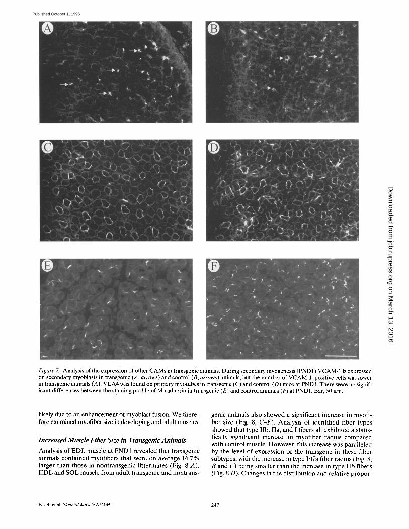
Figure 7. Analysis of the expression of other CAMs in transgenic animals. During secondary myogenesis (PND1) VCAM-1 is expressed on secondary myoblasts in transgenic (A, arrows) and control (B, arrows) animals, but the number of VCAM-l-positive cells was lower in transgenic animals (,4). VLA4 was found on primary myotubes in transgenic (C) and control (D) mice at PND1. There were no signif- icant differences between the staining profile of M-cadherin in transgenic (E) and control animals (F) at PND1. Bar, 50 txm.
likely due to an enhancement of myoblast fusion. We there- fore examined myofiber size in developing and adult muscles.
Increased Muscle Fiber Size in Transgenic Animals
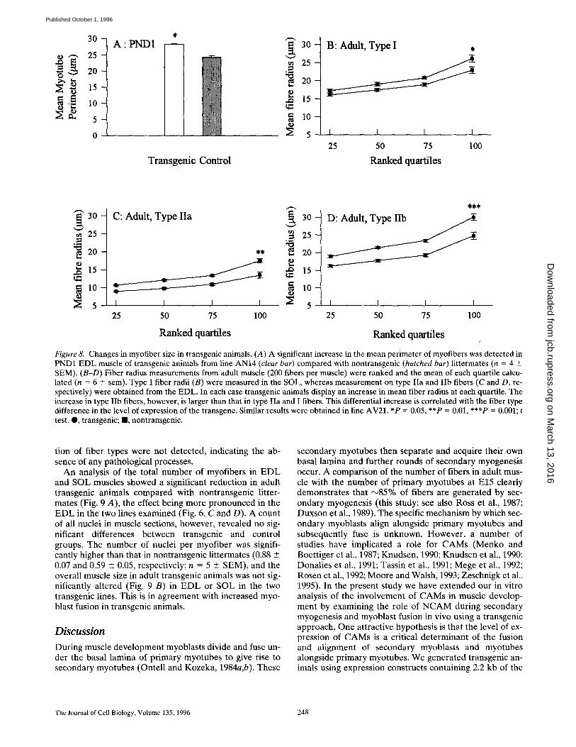
Analysis of EDL muscle at PND1 revealed that transgenic animals contained myofibers that were on average 16.7% larger than those in nontransgenic littermates (Fig. 8 A). EDL and SOL muscle from adult transgenic and nontrans-
genic animals also showed a significant increase in myofi- ber size (Fig. 8, C-E). Analysis of identified fiber types showed that type IIb, IIa, and I fibers all exhibited a statis- tically significant increase in myofiber radius compared with control muscle. However, this increase was paralleled by the level of expression of the transgene in these fiber subtypes, with the increase in type I/IIa fiber radius (Fig. 8, B and C) being smaller than the increase in type IIb fibers (Fig. 8 D). Changes in the distribution and relative propor-
Fazeli et al. Skeletal Muscle NCAM 247
on March 13, 2016
jcb.rupress.orgD
ownloaded from
Published October 1, 1996
3 0 -
2 5 -
20
A" PND1
Transgenic Control
" ~ 3 0 -
~ 2 5
20
,.~ 15
B: Adult, Type I ,
I I I I
25 50 75 100
Ranked quartiles
~ 30 -
"~" 25 -
2 0 -
o 1 5 -
5
C: Adult, Type IIa
I I I I
25 50 75 100
Ranked quartiles
i 3 0 -
• 2 5
20
1 5 -
g lO- I I I I
25 50 75 100
Ranked quartiles
Figure 8. Changes in myofiber size in transgenic animals. (A) A significant increase in the mean perimeter of myofibers was detected in PND1 EDL muscle of transgenic animals from line AN14 (clear bar) compared with nontransgenic (hatched bar) littermates (n = 4 _+ SEM). (B-D) Fiber radius measurements from adult muscle (200 fibers per muscle) were ranked and the mean of each quartile calcu- lated (n = 6 +_ sem). Type I fiber radii (B) were measured in the SOL, whereas measurement on type IIa and lib fibers (C and D, re- spectively) were obtained from the EDL. In each case transgenic animals display an increase in mean fiber radius at each quartile. The increase in type IIb fibers, however, is larger than that in type IIa and I fibers. This differential increase is correlated with the fiber type difference in the level of expression of the transgene. Similar results were obtained in line AV21. *P = 0.05, **P = 0.01, ***P = 0.001; t test. Q, transgenic; II, nontransgenic.
tion of fiber types were not detected, indicating the ab- sence of any pathological processes.
An analysis of the total number of myofibers in EDL and SOL muscles showed a significant reduction in adult transgenic animals compared with nontransgenic litter- mates (Fig. 9 A), the effect being more pronounced in the EDL in the two lines examined (Fig. 6, C and D). A count of all nuclei in muscle sections, however, revealed no sig- nificant differences between transgenic and control groups. The number of nuclei per myofiber was signifi- cantly higher than that in nontransgenic littermates (0.88 +- 0.07 and 0.59 ± 0.05, respectively; n = 5 +- SEM), and the overall muscle size in adult transgenic animals was not sig- nificantly altered (Fig. 9 B) in EDL or SOL in the two transgenic lines. This is in agreement with increased myo- blast fusion in transgenic animals.
Discussion During muscle development myoblasts divide and fuse un- der the basal lamina of primary myotubes to give rise to secondary myotubes (Ontell and Kozeka, 1984a,b). These
secondary myotubes then separate and acquire their own basal lamina and further rounds of secondary myogenesis occur. A comparison of the number of fibers in adult mus- cle with the number of primary myotubes at El5 clearly demonstrates that ~85% of fibers are generated by sec- ondary myogenesis (this study; see also Ross et al., 1987; Duxson et al., 1989). The specific mechanism by which sec- ondary myoblasts align alongside primary myotubes and subsequently fuse is unknown. However, a number of studies have implicated a role for CAMs (Menko and Boettiger et al., 1987; Knudsen, 1990; Knudsen et al., 1990; Donalies et al., 1991; Tassin et al., 1991; Mege et al., 1992; Rosen et al., 1992; Moore and Walsh, 1993; Zeschnigk et al., 1995). In the present study we have extended our in vitro analysis of the involvement of CAMs in muscle develop- ment by examining the role of NCAM during secondary myogenesis and myoblast fusion in vivo using a transgenic approach. One attractive hypothesis is that the level of ex- pression of CAMs is a critical determinant of the fusion and alignment of secondary myoblasts and myotubes alongside primary myotubes. We generated transgenic an- imals using expression constructs containing 2.2 kb of the
The Journal of Cell Biology, Volume 135, 1996 248
on March 13, 2016
jcb.rupress.orgD
ownloaded from
Published October 1, 1996

A
h~
2000
1500
1000
500
AN14 AV21
E L Sol. EDL Sol.
B
O
2.0
1.5
1.0
0.5
0.0
AN14 AV21 0 u t
E L Sol. EDL Sol.
iiii Figure 9. Adult transgenic mice have unaltered muscle size, but fewer fibers. (A) The total number of myofibers in each muscle section was counted by extrapolation from the number of fibers in 0.7 mm 2 of each section, using cross-sectional area measure- ments. A significant reduction in the number of myofibers was found in the EDL of adult transgenic mice of both lines AN14 and AV21. A smaller reduction was also observed in the soleus (Sol). These data parallel the differences in expression level found in the two muscles. (B) Measurements of the cross-sec- tional area of muscle sections from transgenic (solid bars) and nontransgenic (hatched bars) mice revealed no significant differ- ence between the two groups (n = 6, -+ SEM for all measure- ments). *P = 0.05, **P = 0.01; t test. I , transgenic; ~], nontrans- genic.
regulatory region of the human skeletal muscle tx-actin gene fused to hNCAM cDNA encoding a 125-kd, GPI- linked isoform of NCAM. The NCAM cDNA also con- tained the MSD region and either included or excluded the product of the VASE exon. The tx-actin promoter has some advantages for this type of work since it directs spe- cific high level expression in myotubes and not myoblasts. Furthermore, the expression level from this promoter gradually increases between El5 and postnatal week 2 re- suiting in high level transgene expression at times of devel- opment and plasticity of skeletal muscle.
There are presently no specific markers of secondary myotubes that would allow their unequivocal identifica- tion. However, we took advantage of the fact that second- ary myotubes develop under the basal lamina of their "parent" primary myotubes. The number of laminin en- cased fibers was therefore compared with the number of fibers counted on H and E-stained sections of developing EDL muscle. Fibers enclosed in basal lamina are either
primary myotubes with or without an associated second- ary myotube or are separated secondary myotubes. Thus, if secondary myotube separation was hindered, a discrep- ancy should occur between the total number of fibers (H and E sections) and the number of fibers enclosed by basal lamina as defined by laminin staining. There was a signifi- cant decrease in the number of fibers surrounded by basal lamina in the transgenic animals at PND1, whereas the to- tal number of fibers was unaltered. The expression levels of a number of other CAMs believed to be involved in sec- ondary myogenesis, such as VCAM-1, VLA-4, and M-cad- herin, were also studied in the hNCAM-expressing trans- genic animals. No change was found in the distribution or the level of expression of VLA-4 and M-cadherin. How- ever, there was a decreased number of VCAM-l-posi t ive secondary myoblasts at PND1, indicating a depletion of this population of cells. These findings thus indicate that the precise control of NCAM expression levels at primary- secondary myotube appositions is likely to be an impor- tant step in the regulation of secondary myoblast fusion and myotube separation. The hNCAM-expressing trans- genic animals showed no obvious signs of muscular dys- function and an examination of various muscles showed no obvious abnormalities. Cryosections of EDL and SOL muscles from transgenic and nontransgenic animals were compared by morphometric analysis. Adult animals from lines AV21 and AN14 revealed a significant decrease in the total number of fibers in each muscle examined. This was accompanied by an overall increase in myofiber size first detected at PND1. This increase is likely due to en- hanced fusion of secondary myoblasts into secondary myotubes, as a result of the increased expression of NCAM on myotube surfaces, and the observation of a lower number of secondary myoblasts in transgenic ani- mals from VCAM-1 analysis is in agreement with this conclusion. It has been found previously, using a transfec- tion-based approach, that overexpression of the 125-kd hNCAM isoform in C2 muscle cells results in enhanced fu- sion (Dickson et al., 1990; Peck and Walsh, 1993). Thus, the present data confirm the in vitro observations.
Detailed analysis of line AN14 showed that the decrease in fiber number and the increased fiber size were associ- ated with fiber type variations in hNCAM expression. The fast EDL muscle is composed of 70% type l ib fibers and these showed the highest transgene expression. These fi- bers were found to exhibit a larger change in fiber number and size compared with the predominantly slow SOL mus- cle, which showed a lower expression of the transgene. Fi- ber type variations in hNCAM expression levels were evi- dent as early as El8.
AV21 animals express an alternative isoform of hNCAM, namely one that contains the 10-amino acid product of the alternatively spliced VASE exon in its extracellular do- main. This transcript is thought to be specific to cardiac and neuronal tissues, although low levels of VASE con- taining NCAM isoforms have been detected in skeletal muscle (Anderson et al., 1993). The adhesive function of NCAM is not markedly influenced by the presence of the VASE exon (Doherty et al., 1992), though NCAM tran- scripts containing the VASE exon have been shown to be less effective in promoting neurite outgrowth in vitro (Doherty et al., 1992). Indeed, this isoform of NCAM is
Fazeli et al. Skeletal Muscle NCAM 249
on March 13, 2016
jcb.rupress.orgD
ownloaded from
Published October 1, 1996

thought to be partly responsible for the developmental loss of responsiveness of neurons to NCAM. Recent ob- servations have also indicated that, in neuronal cells, intra- cellular signaling by NCAM (without VASE) is at least partly through the activation of the FGF receptor (Will- iams et al., 1994). NCAM transcripts containing the VASE exon appear to be unable to activate this pathway. There was no difference between the muscle phenotype of ani- mals overexpressing NCAM, irrespective of whether they contained the VASE exon or not.
In summary, we have found that increased expression of the 125-kd, GPI-anchored form of NCAM in skeletal mus- cle under the control of the skeletal muscle c~-actin pro- moter results in a developmental perturbation of second- ary myogenesis first in that they do not effectively separate from primary myotubes, and second there is an enhancement of secondary myoblast fusion. The data indi- cate that the expression of NCAM in the sarcolemma, along with other CAMs, plays an important role during skeletal myogenesis. The strategy developed here will be useful in examining the role of other CAMs in myogenesis. In particular, members of the cadherin family such as N- and M-cadherin are of interest as truncated (dominant nega- tive) forms would most likely affect myoblast fusion.
This work was supported by the Wellcome Trust and the Muscular Dys- trophy Group of Great Britain.
Received for publication 12 January 1996 and in revised form 12 July 1996.
References
Anderson, A.M., M. Olsen, D. Zhernosekov, H. Gaardsvoll, L. Krog, D. Linne- mann, and E. Bock. 1993. Age-related changes in expression of the neural- cell adhesion molecule in skeletal muscle: a comparative study of newborn, adult and aged rats. Biochem. J. 290:641-648.
Barbas, J.A., J-C. Chaix, M. Steinmetz, and C. Goridis. 1988. Differential splic- ing and alternative polyadenylation generates distinct NCAM transcripts and proteins in the mouse. E M B O (Eur. Mol. Biol. Organ.) J. 7:625-632.
Barton, C.H., G. Dickson, H.J. Gower, L.H. Rowett, W. Putt, V. Elsom, S.E. Moore, C. Goridis, and F.S. Walsh. 1988. Complete sequence and in vitro ex- pression of a tissue-specific phosphatidylinositol-linked NCAM isoform from skeletal muscle. Development (Camb.). 104:165-173.
Bourne, S.P., K. Patel, F.S. Walsh, C.J. Popham, H.B. Coakham, and J.T. Kemshead. 1991. A monoclonal antibody (ERIC-I) raised against retino- blastoma that recognises the neural cell adhesion molecule (NCAM) ex- pressed on brain and tumours arising from the neuroectoderm. J. Neuro- Oncol. 10:111-119.
Brennan, K.J., and E.C. Hardeman. 1993. Quantitative analysis of the human a-skeletal actin gene in transgenic mice. J. Biol. Chem. 268:719-725.
Cavicchioli, L.. T.P. Flanigan, J.G. Dickson, G. Vantini. R. Dal Toso, M. Fusco, F.S. Walsh, and A. Leon. 1991. Choline acetyl-transferase messenger RNA expression in developing and adult rat brain: regulation by nerve growth fac- tor. Mot, Brain Res. 9:319-325.
Covault, J., and J.R. Sanes. 1985. Neural cell adhesion molecule (NCAM) accu- mulates in denervated and paralysed muscle. Proc. Natl. Acad. Sci. USA. 82: 4544-4548.
Covault, J., and J.R. Sanes. 1986. Distribution of NCAM in synaptic and extra- synaptic portions of developing and adult skeletal muscle. J. Cell Biol. 102: 716-730.
Cremer, H , R. Lange, A. Christoph, M. Plomann, G. Vopper, J. Roes, R. Brown, S. Baldwin, P. Kraemer, S. Scheff et al. 1994. Inactivation of the NCAM gene in mice results in size reduction of the olfactory bulb and defi- cits in spatial learning. Nature (Lond.). 367:455-459.
Daniloff, J.K., G. Levi, M. Grumet, F. Rieger, and G.M. Edelman. 1986. Al- tered expression of neural cell adhesion molecules induced by nerve injury and repair. J. Cell Biol, 103:929-945.
DeJong, A.S.H., M. Van Kessel-van Vark, and A.K. Raap. 1985. Sensitivity of various visualization methods for peroxidase and alkaline phosphatase activ- ity in immunoenzyme histochemistry. Histochem. J. 17:1119-1130.
Diekson, G., H.J. Gower, C.H. Barton. H.M. Prentice, V.L. Elsom, R.D. Cox, C.A. Quinn, W. Putt, and F.S. Walsh. 1987. Human muscle neural cell adhe- sion molecule (NCAM) identification of a muscle specific sequence in the extracellular domain. Cell. 50:1119-1130.
Dickson. G., D. Peck, S.E. Moore, C.H. Barton, and F.S. Walsh. 1990. En- hanced myogenesis in NCAM transfected mouse myoblasts. Nature (Lond.). 344:348-351.
Doherty, P., C.E.C.K. Moolenaar, S.V. Ashton, R.J.A.M. Michalides, and F.S. Walsh. 1992. The VASE exon downregulates the neurite promoting activity of NCAM 140. Nature (Lond.). 356:791-793.
Donalies, M., M. Kramer, M. Ringwald, and A. Starzinski-Powitz. 1991. Ex- pression of M-cadherin, a member of the cadherin multigene family, corre- lates with differentiation of skeletal muscle cells. Proc. Natl. Acad. Sci. USA. 88:8024-8028.
Duxson, M.J., Y. Usson, and A.J. Harris. 1989. The origin of secondary myo- tubes in mammalian skeletal muscles: ultrastructural studies. Development (Camb.). 107:743-750.
Fredette, B., U. Rutishauser, and L. Landmesser. 1993. Regulation and activity- dependence of N-cadherin, NCAM isoforms, and polysialic acid on chick myotubes during development. J. Cell Biol. 123:1867-1888.
Hahn, C.G., and J. Covault. 1992. Neural regulation of N-cadherin gene expres- sion in developing and adult skeletal muscle..L Neurosci. 12:4677-4687.
Hirano, T., S. Hirohashi, T. Kunii, M. Noguchi, Y. Shimosato, and Y. Hayata. 1989. Quantitative distribution of cluster 1 small cell lung cancer antigen in cancerous and noncancerous tissues, cultured cells and sera. Jap. J. Can. Res. 80:348-355.
Hughes, S.M., and H.M. Blau. 1992. Muscle fiber pattern is independent of cell lineage in postnatal rodent development. Cell, 68:659-671.
Hughes, S.M., M. Cho, I. Karsch-Mizrachi, M. Travis, L. Silberstein, L.A. Lein- wand, and H.M. Blau. 1993. Three slow myosin heavy chains sequentially ex- pressed in developing mammalian skeletal muscle. Dev. BioL 158:183-199.
Kelly, A.M., and S.J. Zacks. 1969. The histogenesis of rat intercostal muscle. J. Cell Biol. 42:135-153.
Knudsen, K.A. 1990. Cell adhesion molecules in myogenesis. Curr. Opinion in Cell Biol. 2:902-906.
Knudsen, K.A., S. McElwee, and L. Myers. 1990. A role for the neural cell ad- hesion molecule, NCAM, in myoblast interaction during myogenesis. Dev. Biol. 138:159-168.
Lyons, G., R. Moore, O. Yahara, M.E. Buckingham, and F.S. Walsh. 1992. Ex- pression of NCAM isoforms during skeletal myogenesis in the mouse em- bryo. Dev. Dynamics. 194:94--104.
McCormick, D., H. Chong, C. Hobbs, C. Datta, and P.A. Hall. 1993. Detection of the Ki-67 antigen in fixed and wax-embedded sections with the mono- clonal antibody MIB1. Histopathology (Oxf) . 22:355-360.
Mege, R.M., D. Goudou, C. Diaz, M. Nicolet, L. Garcia, G. Geraud, and F. Rieger. 1992. N-cadherin and NCAM in myoblast fusion: compared Iocalisa- tion and effect of blockade by peptides and antibodies. Z Cell Sci. 103:897-906.
Menko, A.S., and D. Boettiger. 1987. Occupation of the extracellular matrix re- ceptor, integrin, is a control point for myogenic differentiation. Cell, 51:51-57.
Moore, S.E., and F.S. Walsh. 1985. Specific regulation of NCAM/D2-CAM cell adhesion molecule during skeletal muscle development. EMBO (Ear. MoL Biol. Organ.)Z 4:623-630.
Moore, S.E., and F.S. Walsh. 1986. Nerve dependent regulation of neural cell adhesion molecule expression in skeletal muscle. Neurosciences. 18:499-505.
Moore, R., and F.S. Walsh. 1993. The cell adhesion molecule M-cadherin is spe- cifically expressed in developing and regenerating, but not denervated skele- tal muscle. Development (Camb.). 117:1409-1420.
Moore, S.E., J. Thompson, V. Kirkness, J.G. Dickson, and F.S. Walsh. 1987. Skeletal muscle neural cell adhesion molecule (NCAM): changes in protein and mRNA species during myogenesis of cell lines. ,L Cell Biol. 105:1377- 1386.
Muscat, G.E.O., and L. Kedes. 1987. Multiple 5'-flanking regions of the human alpha-skeletal actin gene synergistically modulate muscle-specific expres- sion. MoL Cell. Biol. 7:4089-4099.
Ontelt, M., and K. Kozeka. 1984a. The organogenesis of murine striated mus- cle: a cytoarchitectural study. Am. Z Anat. 171:133-148.
Ontell, M., and K. Kozeka. 1984b. Organogenesis of the mouse extensor digi- torum longus muscle: a quantitative study. Am. J. Anat. 171:149-161.
Peck, D., and F.S. Walsh. 1993. Differential effects of over expressed NCAM isoforms on myoblast fusion. ,L Cell Biol. 123:1587-1595.
Rosen, G.D., J.R. Sanes, R. Lachance, J.M. Cunningham, J. Roman, and D.C. Dean. 1992. Roles for the integrin VLA-4 and its counter receptor VCAM-1 in myogenesis. Cell. 69:1107-1119.
Ross, J.J., J.M. Duxson. and A.J. Harris. 1987. Formation of primary and sec- ondary myotubes in rat lumbrical muscles. Development (Camb.). 100:383-394.
Sambrook, J., E.F. Fritsch. and T. Maniatis. 1989. Molecular Cloning: A Labo- ratory Manual. 2nd ed., Cold Spring Harbor Laboratory, Cold Spring Har- bor, NY. 545 pp.
Small, S.J., and R. Akesou. 1990. Expression of the unique NCAM VASE exon is independently regulated in distinct tissues during development. Z Cell Biol. 111:2089-2096.
Takeichi, M. 199l. Cadherin cell adhesion receptors as a morphogenetic regula- tor. Science (Wash.). 251:1451-1455.
Tassin, A.-M., R.-M. Mege, D. Goudon. G.M. Edelman, and F. Rieger. 1991. Modulation of expression and cell surface distribution of NCAM during myogenesis in vitro. Neurochem. Int~ 18:97-106.
Thiery, J.-P., J.-L. Duband, U. Rutishauser, and G.M. Edelman. 1982. Cell ad- hesion molecules in early chick embryogenesis. Proc. Natl, Acad. Sci. USA. 79:6737-6741.
The Journal of Cell Biology, Volume 135, 1996 250
on March 13, 2016
jcb.rupress.orgD
ownloaded from
Published October 1, 1996

Thompson, J., G. Dickson, S.E. Moore, H.J. Gower, W. Putt, J. Kenimer, and F.S. Walsh. 1989. Alternative splicing of the neural cell adhesion molecule (NCAM) gene generates variant extracellular domain structure in skeletal muscle and brain. Genes & Dev. 3:348-357.
Walsh, F.S., and P. Doherty. 1991. Structure and function of the gene for neural cell adhesion molecule. Semin. Neurosci. 3:271-284.
Walsh, F.S., and P. Doherty. 1994. Cell biology of muscle. In Disorders of Vol- untary Muscle. J. Walton, G. Karpati, and D. Hilton-Jones, editors. Churchill Livingston, Edinburgh. 35~il.
Walsh, F.S.. J. Furness, S.E. Moore, S.V. Ashton, and P. Doherty. 1992. Use of
the NCAM-VASE exon by neurones is associated with a specific down-regu- lation of NCAM dependent neurite outgrowth in the developing cerebellum and hippocampus. J. Neurochem. 59:1959-1962.
Williams, E.J., J. Furness, F.S. Walsh, and P. Doherty. 1994. Activation of the FGF receptor underlies neurite outgrowth stimulated by L1, N-CAM, and N-cadherin. Neuron. 13:583-594.
Zeschnigk, M., D. Kozian, C. Kuch, M. Schmoll and A. Starzinksi-Powitz. 1995. Involvement of M-cadherin in terminal differentiation of skeletal muscle cells. J. Cell Sci. 108:2973-2981.
Fazeli et al. Skeletal Muscle NCAM 251
on March 13, 2016
jcb.rupress.orgD
ownloaded from
Published October 1, 1996
Copyright © 2022 FDOKUMEN