Alterations in regional homogeneity of resting state brain activity in internet gaming addicts...
8
RESEARCH Open Access Alterations in regional homogeneity of resting-state brain activity in internet gaming addicts Guangheng Dong 1* , Jie Huang 1 and Xiaoxia Du 2 Abstract Backgrounds: Internet gaming addiction (IGA), as a subtype of internet addiction disorder, is rapidly becoming a prevalent mental health concern around the world. The neurobiological underpinnings of IGA should be studied to unravel the potential heterogeneity of IGA. This study investigated the brain functions in IGA patients with resting-state fMRI. Methods: Fifteen IGA subjects and fourteen healthy controls participated in this study. Regional homogeneity (ReHo) measures were used to detect the abnormal functional integrations. Results: Comparing to the healthy controls, IGA subjects show enhanced ReHo in brainstem, inferior parietal lobule, left posterior cerebellum, and left middle frontal gyrus. All of these regions are thought related with sensory-motor coordination. In addition, IGA subjects show decreased ReHo in temporal, occipital and parietal brain regions. These regions are thought responsible for visual and auditory functions. Conclusions: Our results suggest that long-time online game playing enhanced the brain synchronization in sensory-motor coordination related brain regions and decreased the excitability in visual and auditory related brain regions. Keywords: Internet addiction disorder, Internet gaming addiction, Resting-state fMRI Background Internet addiction disorder (IAD) is usually defined as the inability of an individual to control his or her use of the internet. This inability will eventually cause psycho- logical, social, and/or work difficulties [1-4]. As the world’ s fastest growing ‘ addiction’ , IAD has been consid- ered as a serious public health issue [1,5,6]. Although significant prevalence estimations and associations with adverse consequences have been addressed [1,7-11], few studies focused on the neurobiological underpinnings of IAD [12-14]. Thus, investigations on the neurobiological basis of IAD are necessary and important. Discovery of the biomarkers of IAD will provide valuable information on unraveling the underpinnings of IAD and on asses- sing proper treatment strategies. IAD is also considered as a behavioral addiction and may share similar neuropsychological (i.e., development of euphoria, craving, and tolerance) and personality characteristics with other addictions [15], especially be- havioral addiction, for example, gambling addiction [16]. Internet addiction consists at least three subtypes: Inter- net gaming addiction (IGA), sexual preoccupations, and email/text messaging [12,14]. Comparing to other sub- types of IAD, internet gaming addiction, the most im- portant subtype of IAD, exhibits some specific features such as role-playing in the virtual world. Few studies have addressed the brain functional changes in people who are addicted to the internet games. Previous studies showed that mental disorders could change the spontan- eous activity in the brain [17-19]. Researchers believed that internet addiction is an impulse disorder or is at least related to it [20]. Thus, IGA may share similar neuropsychological and personality characteristics with other mental disorders or behavioral addictions [15,16]. * Correspondence: [email protected] 1 Department of Psychology, Zhejiang Normal University, 688 of Yingbin Road, Jinhua city, Zhejiang Province, P.R.China Full list of author information is available at the end of the article © 2012 Dong et al.; licensee BioMed Central Ltd. This is an Open Access article distributed under the terms of the Creative Commons Attribution License (http://creativecommons.org/licenses/by/2.0), which permits unrestricted use, distribution, and reproduction in any medium, provided the original work is properly cited. Dong et al. Behavioral and Brain Functions 2012, 8:41 http://www.behavioralandbrainfunctions.com/content/8/1/41
-
Upload
independent -
Category
Documents
-
view
1 -
download
0
Transcript of Alterations in regional homogeneity of resting state brain activity in internet gaming addicts...
Dong et al. Behavioral and Brain Functions 2012, 8:41http://www.behavioralandbrainfunctions.com/content/8/1/41
RESEARCH Open Access
Alterations in regional homogeneity ofresting-state brain activity in internet gamingaddictsGuangheng Dong1*, Jie Huang1 and Xiaoxia Du2
Abstract
Backgrounds: Internet gaming addiction (IGA), as a subtype of internet addiction disorder, is rapidly becoming aprevalent mental health concern around the world. The neurobiological underpinnings of IGA should be studied tounravel the potential heterogeneity of IGA. This study investigated the brain functions in IGA patients withresting-state fMRI.
Methods: Fifteen IGA subjects and fourteen healthy controls participated in this study. Regional homogeneity(ReHo) measures were used to detect the abnormal functional integrations.
Results: Comparing to the healthy controls, IGA subjects show enhanced ReHo in brainstem, inferior parietallobule, left posterior cerebellum, and left middle frontal gyrus. All of these regions are thought related withsensory-motor coordination. In addition, IGA subjects show decreased ReHo in temporal, occipital and parietal brainregions. These regions are thought responsible for visual and auditory functions.
Conclusions: Our results suggest that long-time online game playing enhanced the brain synchronization insensory-motor coordination related brain regions and decreased the excitability in visual and auditory related brainregions.
Keywords: Internet addiction disorder, Internet gaming addiction, Resting-state fMRI
BackgroundInternet addiction disorder (IAD) is usually defined asthe inability of an individual to control his or her use ofthe internet. This inability will eventually cause psycho-logical, social, and/or work difficulties [1-4]. As theworld’s fastest growing ‘addiction’, IAD has been consid-ered as a serious public health issue [1,5,6]. Althoughsignificant prevalence estimations and associations withadverse consequences have been addressed [1,7-11], fewstudies focused on the neurobiological underpinnings ofIAD [12-14]. Thus, investigations on the neurobiologicalbasis of IAD are necessary and important. Discovery ofthe biomarkers of IAD will provide valuable informationon unraveling the underpinnings of IAD and on asses-sing proper treatment strategies.
* Correspondence: [email protected] of Psychology, Zhejiang Normal University, 688 of YingbinRoad, Jinhua city, Zhejiang Province, P.R.ChinaFull list of author information is available at the end of the article
© 2012 Dong et al.; licensee BioMed Central LCommons Attribution License (http://creativecreproduction in any medium, provided the or
IAD is also considered as a behavioral addiction andmay share similar neuropsychological (i.e., developmentof euphoria, craving, and tolerance) and personalitycharacteristics with other addictions [15], especially be-havioral addiction, for example, gambling addiction [16].Internet addiction consists at least three subtypes: Inter-net gaming addiction (IGA), sexual preoccupations, andemail/text messaging [12,14]. Comparing to other sub-types of IAD, internet gaming addiction, the most im-portant subtype of IAD, exhibits some specific featuressuch as role-playing in the virtual world. Few studieshave addressed the brain functional changes in peoplewho are addicted to the internet games. Previous studiesshowed that mental disorders could change the spontan-eous activity in the brain [17-19]. Researchers believedthat internet addiction is an impulse disorder or is atleast related to it [20]. Thus, IGA may share similarneuropsychological and personality characteristics withother mental disorders or behavioral addictions [15,16].
td. This is an Open Access article distributed under the terms of the Creativeommons.org/licenses/by/2.0), which permits unrestricted use, distribution, andiginal work is properly cited.
Dong et al. Behavioral and Brain Functions 2012, 8:41 Page 2 of 8http://www.behavioralandbrainfunctions.com/content/8/1/41
Potential changes in brain are expected in IGA patientsby analogy with other mental disorders.Resting-state functional MRI has been increasingly
used to investigate the integration of neural activities ata resting state when no task is performed [21]. Althoughresting-state fMRI is a relatively new technique, manyexciting findings have been reported in the past severalyears (for a review see [22]). Nowadays, this techniquehas been successfully used to detect abnormal functionalintegration in some mental disorders [23,24]. Regionalhomogeneity (ReHo) is a widely used method in resting-state fMRI studies [21,25,26]. ReHo measures the func-tional coherence of a given voxel with its nearest neigh-bors, which can be used to evaluate resting-state brainactivities based on the hypothesis that significant brainactivities would more likely occur in clusters than in asingle voxel [27]. ReHo index could be regarded as ameasurement for investigating human brain activities inthe resting state and may be useful for revealing thecomplexity of human brain function [28,29].Online game playing requires players staring at the
computer screen and enduring the sound of the gamefor a long time. Previous studies have shown that noisecould cause hearing problem [30-33], and long-timehypertension visual attention could cause serious visualacuity decrease or vision loss [34]. Thus, the long-timegame playing might impair the visual or hearing func-tions and these changes could be detected in relevantbrain regions. In this study, we hypothesized that long-time online game playing changed the brain functions inthe regions that are related with vision or hearing. Toaddress this issue, we employed resting-state fMRI toexamine the spontaneous fluctuations of brain activitiesin IGA subjects in this study.
Methods and procedureSubjectWe examined a group of 15 (16–1) male IAD partici-pants (i.e., male, right handed, non-smokers) aged24.2 ± 3.5 years (mean ± SD) and a group of 14 (15–1)age matched, healthy males aged 24.6 ± 3.8 years (mean ±SD) (i.e., right handed, non-smokers). Our study focusedon male subjects because the prevalence of IAD in menis much higher than that in women [35]. IAD subjectswere recruited through advertisements. All participantsunderwent structured psychiatric interviews (M.I.N.I.)performed by an experienced psychiatrist with an ad-ministration time of approximately 15 minutes. TheMINI was designed to meet the need for a short but ac-curate structured psychiatric interview for multicenterclinical trials and epidemiology studies [36]. All partici-pants were free of Axis I psychiatric disorders listed inM.I.N.I. Depression was further assessed using the Beck
Depression Inventory [37]. Anyone who scored morethan 5 would be excluded from our study.All IAD subjects had a diagnosis of Young’s online
internet addiction test [38], which consists of 20 itemsassociated with online internet use including psycho-logical dependence, compulsive use, and withdrawal, aswell as related problems in school or work, sleep, family,and time management. For each item, a graded responseis selected from 1= “Rarely” to 5 = “Always”, or “Doesnot Apply”. The IAT is proved to be a valid and reliableinstrument that can be used in classifying IAD [39,40].People who scored more than 50 were considered to ex-perience occasional or frequent problems because of theinternet. Those who scored more than 80 were consid-ered to have significant problems in their lives [38]. Inthe present study, the threshold cut-off we used is 80 inthe internet addiction test. Estimates of the size of thegroup of ‘addicted gamers’ are defined by applying vari-ous cut-off points to scales measuring symptoms ofinternet addiction [41,42]. In present study, we addedsome specific limitations on the established measures ofinternet addiction, such as ‘you spend most of your timeplaying online games (>80%)’.The controls were also measured with the same
process. Controls scored lower than 20 in Young’s test(16.3 ± 4.3 mean ± SD). All participants were medicationfree and were instructed not to use any substance ofabuse, including coffee, on the day of the scan. No par-ticipant had previous experience with cocaine ormarijuana. The human investigation committee of Zhe-jiang normal university approved this research. All sub-jects signed a written informed consent.
Scanning processThe ‘resting state’ was defined as no specific cognitivetask during the fMRI scan. Participants were requiredsimply to keep still, close their eyes and not to think ofanything systematically [27,28]. Magnetic resonance im-aging scanning MRI data were acquired using a SiemensTrio 3 T scanner (Siemens, Erlangen, Germany) in East-China Normal University. Participants lay supine withhead snugly fixed by belt and foam pads to minimizehead movement. The resting-state functional imageswere acquired by using an echo-planar imaging se-quence with the following parameters: interleaved, 33axial slices, thickness = 3.0 mm, in-plane resolution = 64*64, repetition time = 2000 ms, echo time = 30 ms, flipangle = 90, field of view = 220* 220 mm, 240 volumes(8 min). Structural images were collected using a T1-weighted three-dimensional spoiled gradient-recalled se-quence was acquired covering the whole brain (176 slices,repetition time = 1700 ms, echo time TE= 3.93 ms, slicethickness = 1.0 mm, skip = 0 mm, flip angle = 15, inversion
Dong et al. Behavioral and Brain Functions 2012, 8:41 Page 3 of 8http://www.behavioralandbrainfunctions.com/content/8/1/41
time 1100 ms, field of view = 240*240 mm, in-plane reso-lution = 256* 256).
Preprocessing of image dataPreprocessing and ReHo process were using the softwareDPARSF (Data Processing Assistant for Resting-StatefMRI. www.restfmri.net), a MATLAB toolbox for ‘pipe-line’ data analysis of resting-state fMRI [43,44]. DPARSFis based on some functions in SPM and REST. The mainpre-processing steps and parameters are: The first 10volumes of each functional time series were discardedfor participant adaptation to the scanning. Then, theimage preprocessing, including slice timing, head motioncorrection and spatial normalization were conducted.After the procedure of head motion correction, thevalues for translation (mm) and rotation (degrees) wereobtained at each time point in six parameters (three fortranslation and three for rotation) for each participant.Participants (1 from IAD group and 2 from controlgroup) with head motion more than 2.0 mm maximumdisplacement in any direction of x, y, and z or 1° of anyangular motion throughout the course of scan wereexcluded from further analysis. The fMRI data weretemporally band-pass filtered (0.01-0.08 Hz) to reducethe low-frequency drift and physiological high-frequencyrespiratory and cardiac noise. Then the time-series ofimages of each subject were motion-corrected using aleast squares approach and a six-parameter (rigid body)linear transformation. Individual ReHo map was gener-ated by calculating Kendall’s coefficient concordance(with value from 0 to 1) of time series of a given voxelwith those of its nearest neighbors (26 voxels) in avoxel-wise way. Spatial smoothing was conducted on theReHo maps with a Gaussian kernel of 4 * 4 * 4 mm3
full-width at half-maximum. The intracranial voxels
Table 1 Brain areas of ReHo difference between two groups (
Main areas Hemisphere Peak MN
Increased ReHo in IAD
Right Brainstem R 3, -6, -15
Left Brainstem L −3, -21,
Inferior Parietal Lobule R 63, -30, 3
Inferior Parietal Lobule L −60, -36
Cerebellum Posterior Lobe L −48, -33
Middle Frontal Gyrus L −60, 27,
Decreased ReHo in IAD
Inferior Temporal Gyrus L −54, -42
Superior Temporal Gyrus L −39, 33
Occipital Lobe, limbic lobe L −24, -63
Parietal Lobe (Postcentral) L −63 ,-6,
Middle Cingulate Gyrus L 0, -15, 39
Voxel size = 3*3*3 mm. p< 0.05 FDR corrected and at least 10 voxels.
were extracted to make a mask [43]. For standardizationpurposes, each individual ReHo map was divided by itsown mean ReHo within the mask.
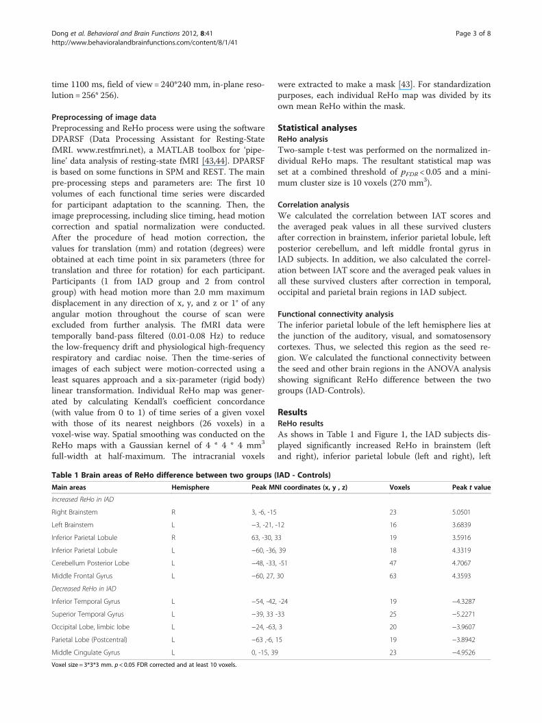
Statistical analysesReHo analysisTwo-sample t-test was performed on the normalized in-dividual ReHo maps. The resultant statistical map wasset at a combined threshold of pFDR < 0.05 and a mini-mum cluster size is 10 voxels (270 mm3).
Correlation analysisWe calculated the correlation between IAT scores andthe averaged peak values in all these survived clustersafter correction in brainstem, inferior parietal lobule, leftposterior cerebellum, and left middle frontal gyrus inIAD subjects. In addition, we also calculated the correl-ation between IAT score and the averaged peak values inall these survived clusters after correction in temporal,occipital and parietal brain regions in IAD subject.
Functional connectivity analysisThe inferior parietal lobule of the left hemisphere lies atthe junction of the auditory, visual, and somatosensorycortexes. Thus, we selected this region as the seed re-gion. We calculated the functional connectivity betweenthe seed and other brain regions in the ANOVA analysisshowing significant ReHo difference between the twogroups (IAD-Controls).
ResultsReHo resultsAs shows in Table 1 and Figure 1, the IAD subjects dis-played significantly increased ReHo in brainstem (leftand right), inferior parietal lobule (left and right), left
IAD - Controls)
I coordinates (x, y , z) Voxels Peak t value
23 5.0501
-12 16 3.6839
3 19 3.5916
, 39 18 4.3319
, -51 47 4.7067
30 63 4.3593
, -24 19 −4.3287
-33 25 −5.2271
, 3 20 −3.9607
15 19 −3.8942
23 −4.9526
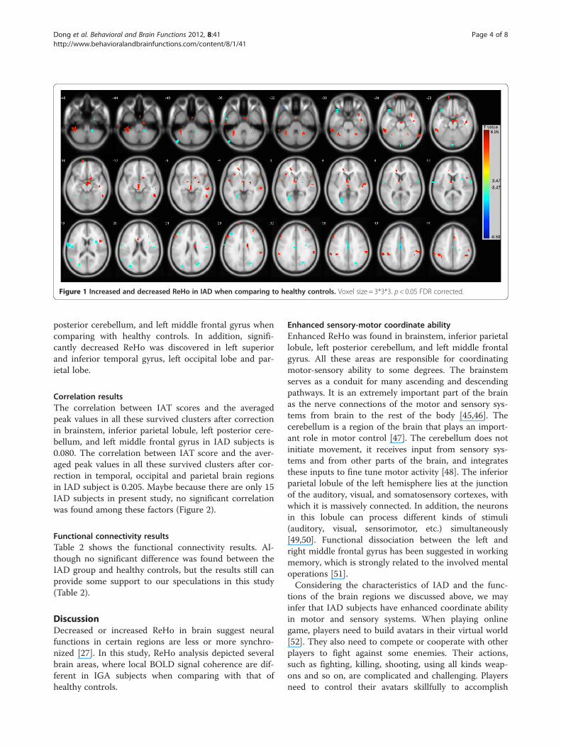
Figure 1 Increased and decreased ReHo in IAD when comparing to healthy controls. Voxel size = 3*3*3. p< 0.05 FDR corrected.
Dong et al. Behavioral and Brain Functions 2012, 8:41 Page 4 of 8http://www.behavioralandbrainfunctions.com/content/8/1/41
posterior cerebellum, and left middle frontal gyrus whencomparing with healthy controls. In addition, signifi-cantly decreased ReHo was discovered in left superiorand inferior temporal gyrus, left occipital lobe and par-ietal lobe.
Correlation resultsThe correlation between IAT scores and the averagedpeak values in all these survived clusters after correctionin brainstem, inferior parietal lobule, left posterior cere-bellum, and left middle frontal gyrus in IAD subjects is0.080. The correlation between IAT score and the aver-aged peak values in all these survived clusters after cor-rection in temporal, occipital and parietal brain regionsin IAD subject is 0.205. Maybe because there are only 15IAD subjects in present study, no significant correlationwas found among these factors (Figure 2).
Functional connectivity resultsTable 2 shows the functional connectivity results. Al-though no significant difference was found between theIAD group and healthy controls, but the results still canprovide some support to our speculations in this study(Table 2).
DiscussionDecreased or increased ReHo in brain suggest neuralfunctions in certain regions are less or more synchro-nized [27]. In this study, ReHo analysis depicted severalbrain areas, where local BOLD signal coherence are dif-ferent in IGA subjects when comparing with that ofhealthy controls.
Enhanced sensory-motor coordinate abilityEnhanced ReHo was found in brainstem, inferior parietallobule, left posterior cerebellum, and left middle frontalgyrus. All these areas are responsible for coordinatingmotor-sensory ability to some degrees. The brainstemserves as a conduit for many ascending and descendingpathways. It is an extremely important part of the brainas the nerve connections of the motor and sensory sys-tems from brain to the rest of the body [45,46]. Thecerebellum is a region of the brain that plays an import-ant role in motor control [47]. The cerebellum does notinitiate movement, it receives input from sensory sys-tems and from other parts of the brain, and integratesthese inputs to fine tune motor activity [48]. The inferiorparietal lobule of the left hemisphere lies at the junctionof the auditory, visual, and somatosensory cortexes, withwhich it is massively connected. In addition, the neuronsin this lobule can process different kinds of stimuli(auditory, visual, sensorimotor, etc.) simultaneously[49,50]. Functional dissociation between the left andright middle frontal gyrus has been suggested in workingmemory, which is strongly related to the involved mentaloperations [51].Considering the characteristics of IAD and the func-
tions of the brain regions we discussed above, we mayinfer that IAD subjects have enhanced coordinate abilityin motor and sensory systems. When playing onlinegame, players need to build avatars in their virtual world[52]. They also need to compete or cooperate with otherplayers to fight against some enemies. Their actions,such as fighting, killing, shooting, using all kinds weap-ons and so on, are complicated and challenging. Playersneed to control their avatars skillfully to accomplish
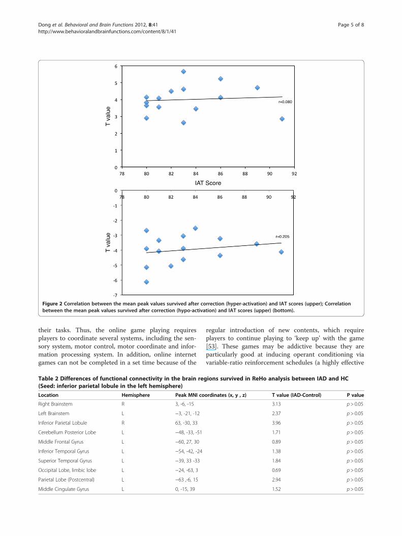
Figure 2 Correlation between the mean peak values survived after correction (hyper-activation) and IAT scores (upper); Correlationbetween the mean peak values survived after correction (hypo-activation) and IAT scores (upper) (bottom).
Dong et al. Behavioral and Brain Functions 2012, 8:41 Page 5 of 8http://www.behavioralandbrainfunctions.com/content/8/1/41
their tasks. Thus, the online game playing requiresplayers to coordinate several systems, including the sen-sory system, motor control, motor coordinate and infor-mation processing system. In addition, online internetgames can not be completed in a set time because of the
Table 2 Differences of functional connectivity in the brain reg(Seed: inferior parietal lobule in the left hemisphere)
Location Hemisphere Peak MNI co
Right Brainstem R 3, -6, -15
Left Brainstem L −3, -21, -12
Inferior Parietal Lobule R 63, -30, 33
Cerebellum Posterior Lobe L −48, -33, -51
Middle Frontal Gyrus L −60, 27, 30
Inferior Temporal Gyrus L −54, -42, -24
Superior Temporal Gyrus L −39, 33 -33
Occipital Lobe, limbic lobe L −24, -63, 3
Parietal Lobe (Postcentral) L −63 ,-6, 15
Middle Cingulate Gyrus L 0, -15, 39
regular introduction of new contents, which requireplayers to continue playing to ‘keep up’ with the game[53]. These games may be addictive because they areparticularly good at inducing operant conditioning viavariable-ratio reinforcement schedules (a highly effective
ions survived in ReHo analysis between IAD and HC
ordinates (x, y , z) T value (IAD-Control) P value
3.13 p> 0.05
2.37 p> 0.05
3.96 p> 0.05
1.71 p> 0.05
0.89 p> 0.05
1.38 p> 0.05
1.84 p> 0.05
0.69 p> 0.05
2.94 p> 0.05
1.52 p> 0.05
Dong et al. Behavioral and Brain Functions 2012, 8:41 Page 6 of 8http://www.behavioralandbrainfunctions.com/content/8/1/41
conditioning paradigm [54]). From what we discussedabove, we may infer that the long-term game playingenhanced coordinate ability among motor, sensory andinformation processing systems in brain.
Impaired visual and auditory functionConsistent with our hypothesis, the IAD subjects showdecreased ReHo in temporal, occipital and parietal brainregions when comparing with the healthy controls.These regions are thought to be responsible for visualand auditory functions. The superior temporal gyruscontains several important structures of the brain, in-cluding the location of the primary auditory cortex,which is responsible for the sensation of sound [55]. In-ferior temporal gyrus is one of the higher levels of theventral stream of audio and visual processing, associatedwith the representation of complex object features[56,57]. The occipital lobe is the visual processing centerof the mammalian brain containing most of the anatom-ical region of the visual cortex [58,59]. The parietal lobeintegrates sensory information from different modalities,particularly determining spatial sense and navigation[60-62].The decreased ReHo in visual and auditory related
brain regions might suggest the decreased synchro-nization in IGA subjects. Considering the features ofIGA, we can infer that this may be the result of long-time game playing. The gaming process requires playersto pay full attention to each subtle change in the screen,to endure the noisy sound of the game for a long time(usually more than 10 hours a day). The visual and audi-tory related brain regions have been stimulated for a longtime, which made them hardly to be excited or have adecreased excitability. As we mentioned in the introduc-tion section, long time hypertension of visual attentioncan impair subjects’ visual functions and noise can im-pair their hearing abilities [17-19]. Thus, we may inferthat the long-time game playing (expose to visual andauditory stimuli) may impair player’s visual and auditoryability, which could be indexed by the decreased ReHo inthis study.
LimitationOur results may sound more persuasive if we include agroup of video game addicts as another control group.From comparisons between IGA subjects and videogame addicts, we may find some specific features ofIGA. In addition, if we had vision and hearing tests onthe subjects before scan and take these variables asregressors in data analysis, we could get better scientificresults. These issues will be emphasized in our futurestudies. Third, correlation and functional connectivityresults cannot provide strong support to the conclusionsabout the impairment in visual auditory ability. Future
studies should involve more participants to investigatethis issue.
ConclusionsFrom what we discussed above, we can conclude thatlong-time online game playing enhanced the sensory-motor coordinate ability and impaired participants’ vis-ual and auditory functions. All these results can helppolicy-makers or individuals understand the influence ofinternet gaming addiction. First, this research can helpus understand the mechanism of addiction, find if itshares characteristics with other types of addictions; Sec-ond, this research revealed the influence of internetgames, especially its negative aspects to our brainmechanism.
Competing interestsThe authors do not have an affiliation with or financial interest in anyorganization that might pose a competing interest.
Authors’ contributionsGD took part in designing the research and the manuscript preparation. JHanalyzed the data. XD collected the data and took part in manuscriptpreparation. All authors read and approved the final manuscript.
AcknowledgementsThis research was supported by National Science Foundation of China(30900405). The funder had no role in research design, data collection andanalysis, decision to publish, or preparation of the manuscript.
Author details1Department of Psychology, Zhejiang Normal University, 688 of YingbinRoad, Jinhua city, Zhejiang Province, P.R.China. 2Department of Physics,Shanghai Key Laboratory of Magnetic Resonance, East China NormalUniversity, Shanghai, P.R.China.
Received: 23 March 2012 Accepted: 16 August 2012Published: 18 August 2012
References1. Young K: Internet addiction: the emergence of a new clinical disorder.
Cyberpsychol Behav 1998, 1(3):237–244.2. Davis RA: Cognitive-behavioral model of pathological Internet use.
Comput Hum Behav 2001, 17(2):187–195.3. Young KS, Rogers RC: The relationship between depression and internet
addiction. Cyberpsychol Behav 1998, 1(1):25–28.4. Young KS: Internet addiction: the emergence of a new clinical disorder.
Cyberpsychol Behav 1998, 1(3):237–244.5. van den Eijnden RJJM, Spijkerman R, Vermulst AA, van Rooij TJ, Engels
RCME: Compulsive Internet Use Among Adolescents: BidirectionalParent–child Relationships. J Abnorm Child Psychol 2010, 38(1):77–89.
6. Dong G, Lu Q, Zhou H, Zhao X: Precursor or sequela: pathologicaldisorders in people with Internet addiction disorder. PLoS One 2011,6(2):e14703.
7. Kim JH, Lau CH, Cheuk K-K, Kan P, Hui HLC, Griffiths SM: Predictors ofheavy Internet use and associations with health-promoting and healthrisk behaviors among Hong Kong university students. J Adolesc 2010,33(1):215–220.
8. Niemz K, Griffiths M, Banyard P: Prevalence of pathological Internet useamong university students and correlations with self-esteem, theGeneral Health Questionnaire (GHQ), and disinhibition. CyberpsycholBehav 2005, 8(6):562–570.
9. Dong G, Huang J, Du X: Enhanced reward sensitivity and decreased losssensitivity in Internet addicts: an fMRI study during a guessing task.J Psychiatr Res 2011, 45(11):1525–1529.
Dong et al. Behavioral and Brain Functions 2012, 8:41 Page 7 of 8http://www.behavioralandbrainfunctions.com/content/8/1/41
10. Dong G, Devito E, Huang J, Du X: Diffusion tensor imaging revealsthalamus and posterior cingulate cortex abnormalities in internetgaming addicts. J Psychiatr Res 2012, 46(9):1212–1216.
11. Dong G, Zhou H, Zhao X: Male Internet addicts show impaired executivecontrol ability: evidence from a color-word Stroop task. Neurosci Lett2011, 499(2):114–118.
12. Block JJ: Prevalence Underestimated in Problematic Internet Use Study.CNS Spectr 2006, 12:14–15.
13. Liu T, Potenza MN: Problematic Internet Use: Clinical Implications. CNSSpectr 2007, 12(6):453–466.
14. Dong G, Lu Q, Zhou H, Zhao X: Impulse inhibition in people with internetaddiction disorder: electrophysiological evidence from a Go/NoGo study.Neurosci Lett 2010, 485(2):138–142.
15. Grant JE, Potenza MN, Weinstein A, Gorelick DA: Introduction to BehavioralAddictions. Am J Drug Alcohol Abuse 2010, 36(5):233–241.
16. Potenza MN, Leung H-C, Blumberg HP, Peterson BS, Fulbright RK, LacadieCM, Skudlarski P, Gore JC: An fMRI Stroop Task Study of VentromedialPrefrontal Cortical Function in Pathological Gamblers. Am J Psychiatry2003, 160(11):1990–1994.
17. Peng DH, Jiang KD, Fang YR, Xu YF, Shen T, Long XY, Liu J, Zang YF:Decreased regional homogeneity in major depression as revealed byresting-state functional magnetic resonance imaging. Chin Med J (Engl)2011, 124(3):369–373.
18. Paakki JJ, Rahko J, Long X, Moilanen I, Tervonen O, Nikkinen J, Starck T,Remes J, Hurtig T, Haapsamo H, et al: Alterations in regional homogeneityof resting-state brain activity in autism spectrum disorders. Brain Res2010, 1321:169–179.
19. Wu T, Long X, Zang Y, Wang L, Hallett M, Li K, Chan P: Regionalhomogeneity changes in patients with Parkinson’s disease. Hum BrainMapp 2009, 30(5):1502–1510.
20. Beard KW, Wolf EM: Modification in the proposed diagnostic criteria forInternet addiction. Cyberpsychol Behav 2001, 4(3):377–383.
21. Liu Y, Wang K, Yu C, He Y, Zhou Y, Liang M, Wang L, Jiang T: Regionalhomogeneity, functional connectivity and imaging markers ofAlzheimer’s disease: a review of resting-state fMRI studies.Neuropsychologia 2008, 46(6):1648–1656.
22. Fox MD, Raichle ME: Spontaneous fluctuations in brain activity observedwith functional magnetic resonance imaging. Nat Rev Neurosci 2007, 8(9):700–711.
23. Deco G, Jirsa VK, McIntosh AR: Emerging concepts for the dynamicalorganization of resting-state activity in the brain. Nat Rev Neurosci 2011,12(1):43–56.
24. van den Heuvel MP, Hulshoff Pol HE: Exploring the brain network: areview on resting-state fMRI functional connectivity. EurNeuropsychopharmacol 2010, 20(8):519–534.
25. Zhang Z, Liu Y, Jiang T, Zhou B, An N, Dai H, Wang P, Niu Y, Wang L, ZhangX: Altered spontaneous activity in Alzheimer's disease and mildcognitive impairment revealed by Regional Homogeneity. Neuroimage2011, 59(2):1429–1440.
26. Wang L, Song M, Jiang T, Zhang Y, Yu C: Regional homogeneity of theresting-state brain activity correlates with individual intelligence. NeurosciLett 2011, 488(3):275–278.
27. Zang Y, Jiang T, Lu Y, He Y, Tian L: Regional homogeneity approach tofMRI data analysis. Neuroimage 2004, 22(1):394–400.
28. You H, Wang J, Wang H, Zang YF, Zheng FL, Meng CL, Feng F: Alteredregional homogeneity in motor cortices in patients with multiple systematrophy. Neurosci Lett 2011, 502(1):18–23.
29. Wu T, Zang Y, Wang L, Long X, Li K, Chan P: Normal aging decreasesregional homogeneity of the motor areas in the resting state. NeurosciLett 2007, 423(3):189–193.
30. Bovo R, Ciorba A, Martini A: Environmental and genetic factors in age-related hearing impairment. Aging Clin Exp Res 2011, 23(1):3–10.
31. Van Eyken E, Van Camp G, Van Laer L: The complexity of age-relatedhearing impairment: contributing environmental and genetic factors.Audiol Neurootol 2007, 12(6):345–358.
32. Fransen E, Lemkens N, Van Laer L, Van Camp G: Age-related hearingimpairment (ARHI): environmental risk factors and genetic prospects. ExpGerontol 2003, 38(4):353–359.
33. Pilati N, Ison MJ, Barker M, Mulheran M, Large CH, Forsythe ID, Matthias J,Hamann M: Mechanisms contributing to central excitability changesduring hearing loss. Proc Natl Acad Sci USA 2012, 109(21):8292–8297.
34. DellaCroce JT, Vitale AT: Hypertension and the eye. Curr Opin Ophthalmol2008, 19(6):493–498.
35. Report on Chinese youth internet addiction disorder. In. Beijing: Chinese Youthassociation of Internet; 2008:1–17.
36. Lecrubier Y, Sheehan DV, Weiller E, Amorim P, Bonora I, Harnett Sheehan K,Janavs J, Dunbar GC: The Mini International Neuropsychiatric Interview(MINI). A short diagnostic structured interview: reliability and validityaccording to the CIDI. European Psychiatry 1997, 12(5):224–231.
37. Beck AT, Ward CH, Mendelson M, Mock J, Erbaugh J: An Inventory forMeasuring Depression. Arch Gen Psychiatry 1961, 4(6):561–571.
38. Internet Addiction Test (IAT). http://netaddiction.com/.39. Widyanto L, McMurran M: The psychometric properties of the internet
addiction test. Cyberpsychol Behav 2004, 7(4):443–450.40. Widyanto L, Griffiths MD, Brunsden V: A psychometric comparison of
the Internet Addiction Test, the Internet-Related Problem Scale, andself-diagnosis. Cyberpsychol Behav Soc Netw 2011, 14(3):141–149.
41. Gentile D: Pathological Video-Game Use Among Youth Ages 8to 18: A National Study (vol 20, pg 594, 2009). Psychol Sci 2009,20(6):785–785.
42. Lemmens JS, Valkenburg PM, Peter J: Development and validation ofa game addiction scale for Adolescents. Media Psychology 2009,12(1):77–95.
43. Yan C-G, Zang Y-F: DPARSF: A MATLAB Toolbox for “Pipeline” DataAnalysis of Resting-State fMRI. Front Syst Neurosci 2010, 4:13.
44. Song XW, Dong ZY, Long XY, Li SF, Zuo XN, Zhu CZ, He Y, Yan CG, Zang YF:REST: A Toolkit for Resting-State Functional Magnetic ResonanceImaging Data Processing. PLoS One 2011, 6(9):e25031.
45. Angeles Fernandez-Gil M, Palacios-Bote R, Leo-Barahona M, Mora-Encinas JP:Anatomy of the brainstem: a gaze into the stem of life. Semin UltrasoundCT MR 2010, 31(3):196–219.
46. Hurley RA, Flashman LA, Chow TW, Taber KH: The brainstem: anatomy,assessment, and clinical syndromes. J Neuropsychiatry Clin Neurosci 2010,22(1):1–7. iv.
47. Stoodley CJ, Valera EM, Schmahmann JD: Functional topography of thecerebellum for motor and cognitive tasks: an fMRI study. Neuroimage2011, 59(2):1560–1570.
48. De Zeeuw CI, Hoebeek FE, Bosman LW, Schonewille M, Witter L, KoekkoekSK: Spatiotemporal firing patterns in the cerebellum. Nat Rev Neurosci2011, 12(6):327–344.
49. Kalenine S, Buxbaum LJ, Coslett HB: Critical brain regions for actionrecognition: lesion symptom mapping in left hemisphere stroke. Brain2010, 133(11):3269–3280.
50. Kiriyama I, Miki H, Kikuchi K, Ohue S, Matsuda S, Mochizuki T: Topographicanalysis of the inferior parietal lobule in high-resolution 3D MR imaging.AJNR Am J Neuroradiol 2009, 30(3):520–524.
51. Carter RM, O’Doherty JP, Seymour B, Koch C, Dolan RJ: Contingencyawareness in human aversive conditioning involves the middle frontalgyrus. NeuroImage 2006, 29(3):1007–1012.
52. Van Rooij AJ, Schoenmakers TM, Vermulst AA, Van den Eijnden RJ, Van deMheen D: Online video game addiction: identification of addictedadolescent gamers. Addiction 2010, 106(1):205–212.
53. Charlton JP, Danforth IDW: Distinguishing addiction and highengagement in the context of online game playing. Comput Hum Behav2007, 23(3):1531–1548.
54. Wallace P: The psychology of the Internet. Cambridge: Cambridge UniversityPress; 1999.
55. Robins DL, Hunyadi E, Schultz RT: Superior temporal activation inresponse to dynamic audio-visual emotional cues. Brain Cogn 2009,69(2):269–278.
56. Lewald J, Staedtgen M, Sparing R, Meister IG: Processing of auditorymotion in inferior parietal lobule: evidence from transcranial magneticstimulation. Neuropsychologia 2011, 49(2):209–215.
57. Allison T, McCarthy G, Nobre A, Puce A, Belger A: Human extrastriatevisual cortex and the perception of faces, words, numbers, and colors.Cereb Cortex 1994, 4(5):544–554.
58. Tam EW, Widjaja E, Blaser SI, Macgregor DL, Satodia P, Moore AM: Occipitallobe injury and cortical visual outcomes after neonatal hypoglycemia.Pediatrics 2008, 122(3):507–512.
59. Kojima H, Suzuki T: Hemodynamic change in occipital lobe during visualsearch: visual attention allocation measured with NIRS. Neuropsychologia2010, 48(1):349–352.
Dong et al. Behavioral and Brain Functions 2012, 8:41 Page 8 of 8http://www.behavioralandbrainfunctions.com/content/8/1/41
60. Battelli L, Alvarez GA, Carlson T, Pascual-Leone A: The role of the parietallobe in visual extinction studied with transcranial magnetic stimulation.J Cogn Neurosci 2009, 21(10):1946–1955.
61. Yin TC: The parietal lobe and visual attention. J Psychiatr Res 1978,14(1–4):261–266.
62. Yin TC, Mountcastle VB: Mechanisms of neural integration in the parietallobe for visual attention. Fed Proc 1978, 37(9):2251–2257.
doi:10.1186/1744-9081-8-41Cite this article as: Dong et al.: Alterations in regional homogeneity ofresting-state brain activity in internet gaming addicts. Behavioral andBrain Functions 2012 8:41.
Submit your next manuscript to BioMed Centraland take full advantage of:
• Convenient online submission
• Thorough peer review
• No space constraints or color figure charges
• Immediate publication on acceptance
• Inclusion in PubMed, CAS, Scopus and Google Scholar
• Research which is freely available for redistribution
Submit your manuscript at www.biomedcentral.com/submit