Adv Funct Mater 2012 Freudenberg 22,1391-1398
8
www.afm-journal.de FULL PAPER © 2012 WILEY-VCH Verlag GmbH & Co. KGaA, Weinheim 1 www.MaterialsViews.com wileyonlinelibrary.com Uwe Freudenberg, Jens-Uwe Sommer,* Kandice R. Levental, Petra B. Welzel, Andrea Zieris, Karolina Chwalek, Katja Schneider, Silvana Prokoph, Marina Prewitz, Ron Dockhorn, and Carsten Werner* research has revealed that, together with the provision of morphogens and the presentation of adhesion ligands, [2] the mechanical characteristics of extracel- lular matrices have a decisive influence on cell fate, provoking the development of materials with effective physical prop- erties. [3] This interplay of biomolecular and biophysical signals thus defines an obvious, but until now unmet, need for a new generation of biomaterials that can be selectively and independently tuned for biomolecular properties and physical material parameters. A conceptual basis to address this need is currently missing. As such, we have developed a rational design approach relying on mean field concepts to guide the design of biofunctional matrices. Considering the decisive role of electro- static interactions in functional assem- blies of living matter we selected a system that allows for a far-reaching modulation of structure-determining forces: crosslinking a hydrophilic and flexible, multi-armed polymer (with four-armed, amino-termi- nated poly(ethylene glycol) (starPEG) as an example system known for its anti-adhesive characteristics towards proteins, [4] with a multifunctional, highly charged crosslinker (such as heparin (HEP) or a similarly charged glycosaminoglycan), which can function as a multivalent binding site capable of complexing a plethora of important bioactive molecules. [5] We explored whether and how the combination of the particular gel components permits varying the physical and biomolecular characteristics of the swollen materials independently. Based on the successful experimental verification of the theo- retical predictions and the functionalization of starPEG-heparin gels with adhesive ligand peptides (such as the integrin- binding arginine-glycine-aspartic acid sequence (RGD))) and morphogens (vascular endothelial growth factor (VEGF), bone morphogenetic protein-2 (BMP-2)) through covalent and non- covalent conjugation schemes we were able to illustrate the resulting options for two selected example systems: studying the interplay of matrix elasticity and growth factor presenta- tion in inducing the pro-angiogenic state of human endothe- lial cells and promoting osteogenic differentiation of human mesenchymal stem cells we identified effective combinations of matrix parameters and demonstrated exciting options for the fully matrix controlled direction of the cells, i.e., removed the Using Mean Field Theory to Guide Biofunctional Materials Design Cell-instructive characteristics of extracellular matrices (ECM) resulting from a subtle balance of biomolecular and biophysical signals must be recapitulated in engineered biomaterials to facilitate regenerative therapies. However, no material explored so far allows the independent tuning of the involved molecular and phys- ical cues due to the inherent correlation between biopolymer concentration and material properties. Addressing the resulting challenge, a rational design strategy for ECM-inspired biohybrid hydrogels based on multi-armed poly(ethylene glycol) and heparin, adapting a mean field approach to identify conditions at which the balance of elastic, electrostatic, and excluded volume forces results in constant heparin concentrations within swollen polymer networks with gradually varied physical properties is introduced. Applying heparin-based biofunctionalization schemes, multiple distinct combinations of matrix parameters could be identified to effectively stimulate the pro-angiogenic state of human endothelial cells and the differentiation of human mesenchymal stem cells. The study demonstrates the power of joint theoretical and experimental efforts in creating bioactive mate- rials with specifically and independently controllable characteristics. DOI: 10.1002/adfm.201101868 Dr. U. Freudenberg, Dr. K. R. Levental, Dr. P. B. Welzel, A. Zieris, K. Chwalek, K. Schneider, S. Prokoph, M. Prewitz, Prof. C. Werner Leibniz Institute of Polymer Research Dresden (IPF) Max Bergmann Center of Biomaterials Dresden (MBC) Hohe Str. 6, 01069 Dresden, Germany E-mail: [email protected] Dr. U. Freudenberg, Dr. K. R. Levental, Dr. P. Welzel, A. Zieris, K. Chwalek, K. Schneider, S. Prokoph, M. Prewitz, Prof. C. Werner Technische Universität Dresden Center for Regenerative Therapies Dresden (CRTD) Tatzberg 47 01307 Dresden, Germany Prof. J.-U. Sommer, R. Dockhorn Leibniz Institute of Polymer Research Dresden (IPF) Hohe Str. 6, 01069 Dresden, Germany; E-mail: [email protected] Prof. J.-U. Sommer, R. Dockhorn Technische Universität Dresden Institute for Theoretical Physics Zellescher Weg 17, 01069 Dresden, Germany 1. Introduction Biomaterials that mimic extracellular matrices (ECM), and thereby enable control over cell fate decisions through exog- enous signals, are vital for regenerative therapies. [1] Recent Adv. Funct. Mater. 2012, DOI: 10.1002/adfm.201101868
-
Upload
tu-dresden -
Category
Documents
-
view
7 -
download
0
Transcript of Adv Funct Mater 2012 Freudenberg 22,1391-1398

www.afm-journal.de
FULL P
APER
www.MaterialsViews.com
Uwe Freudenberg , Jens-Uwe Sommer ,* Kandice R. Levental , Petra B. Welzel , Andrea Zieris , Karolina Chwalek , Katja Schneider , Silvana Prokoph , Marina Prewitz , Ron Dockhorn , and Carsten Werner *
Using Mean Field Theory to Guide Biofunctional Materials Design
Cell-instructive characteristics of extracellular matrices (ECM) resulting from a subtle balance of biomolecular and biophysical signals must be recapitulated in engineered biomaterials to facilitate regenerative therapies. However, no material explored so far allows the independent tuning of the involved molecular and phys-ical cues due to the inherent correlation between biopolymer concentration and material properties. Addressing the resulting challenge, a rational design strategy for ECM-inspired biohybrid hydrogels based on multi-armed poly(ethylene glycol) and heparin, adapting a mean fi eld approach to identify conditions at which the balance of elastic, electrostatic, and excluded volume forces results in constant heparin concentrations within swollen polymer networks with gradually varied physical properties is introduced. Applying heparin-based biofunctionalization schemes, multiple distinct combinations of matrix parameters could be identifi ed to effectively stimulate the pro-angiogenic state of human endothelial cells and the differentiation of human mesenchymal stem cells. The study demonstrates the power of joint theoretical and experimental efforts in creating bioactive mate-rials with specifi cally and independently controllable characteristics.
1. Introduction
Biomaterials that mimic extracellular matrices (ECM), and thereby enable control over cell fate decisions through exog-enous signals, are vital for regenerative therapies. [ 1 ] Recent
© 2012 WILEY-VCH Verlag GmbH & Co. KGaA, Weinheim
DOI: 10.1002/adfm.201101868
Dr. U. Freudenberg , Dr. K. R. Levental , Dr. P. B. Welzel , A. Zieris , K. Chwalek , K. Schneider , S. Prokoph , M. Prewitz , Prof. C. Werner Leibniz Institute of Polymer Research Dresden (IPF)Max Bergmann Center of Biomaterials Dresden (MBC)Hohe Str. 6, 01069 Dresden, GermanyE-mail: [email protected] Dr. U. Freudenberg, Dr. K. R. Levental, Dr. P. Welzel, A. Zieris, K. Chwalek, K. Schneider, S. Prokoph, M. Prewitz, Prof. C. WernerTechnische Universität DresdenCenter for Regenerative Therapies Dresden (CRTD)Tatzberg 47 01307 Dresden, Germany Prof. J.-U. Sommer , R. Dockhorn Leibniz Institute of Polymer Research Dresden (IPF)Hohe Str. 6, 01069 Dresden, Germany;E-mail: [email protected] Prof. J.-U. Sommer , R. Dockhorn Technische Universität DresdenInstitute for Theoretical PhysicsZellescher Weg 17, 01069 Dresden, Germany
Adv. Funct. Mater. 2012, DOI: 10.1002/adfm.201101868
research has revealed that, together with the provision of morphogens and the presentation of adhesion ligands, [ 2 ] the mechanical characteristics of extracel-lular matrices have a decisive infl uence on cell fate, provoking the development of materials with effective physical prop-erties. [ 3 ] This interplay of biomolecular and biophysical signals thus defi nes an obvious, but until now unmet, need for a new generation of biomaterials that can be selectively and independently tuned for biomolecular properties and physical material parameters. A conceptual basis to address this need is currently missing. As such, we have developed a rational design approach relying on mean fi eld concepts to guide the design of biofunctional matrices. Considering the decisive role of electro-static interactions in functional assem-blies of living matter we selected a system that allows for a far-reaching modulation
of structure-determining forces: crosslinking a hydrophilic and fl exible, multi-armed polymer (with four-armed, amino-termi-nated poly(ethylene glycol) (starPEG) as an example system known for its anti-adhesive characteristics towards proteins, [ 4 ] with a multifunctional, highly charged crosslinker (such as heparin (HEP) or a similarly charged glycosaminoglycan), which can function as a multivalent binding site capable of complexing a plethora of important bioactive molecules. [ 5 ] We explored whether and how the combination of the particular gel components permits varying the physical and biomolecular characteristics of the swollen materials independently.
Based on the successful experimental verifi cation of the theo-retical predictions and the functionalization of starPEG-heparin gels with adhesive ligand peptides (such as the integrin-binding arginine-glycine-aspartic acid sequence (RGD))) and morphogens (vascular endothelial growth factor (VEGF), bone morphogenetic protein-2 (BMP-2)) through covalent and non-covalent conjugation schemes we were able to illustrate the resulting options for two selected example systems: studying the interplay of matrix elasticity and growth factor presenta-tion in inducing the pro-angiogenic state of human endothe-lial cells and promoting osteogenic differentiation of human mesenchymal stem cells we identifi ed effective combinations of matrix parameters and demonstrated exciting options for the fully matrix controlled direction of the cells, i.e., removed the
1wileyonlinelibrary.com

FULL
PAPER
www.afm-journal.dewww.MaterialsViews.com
Figure 1 . A rational design concept is used to combine star-shaped, end-functionalized poly(ethylene glycol) (starPEG) and heparin in hybrid networks. Schematic representation of the interplay between elastic forces ( Π el ) ( ≈ crosslinking degree), excluded volume ( Π ev ), and electrostatic effects ( Π ch ) maintaining the swelling and heparin concentration of the swollen matrices ( ∏ el = ∏ ch + ∏ el ). Top: electrostatic effects (arrows) dominate the swelling at low salt situations, where the screening is less effective (Debye length ≈ 300 nm); increasing starPEG/heparin ratio γ ( ≈ crosslinking) increases the number of starPEG molecules and therefore the excluded volume effects (cylinders). Bottom: excluded volume effects dominate the swelling at high salt situations, where strong screening occurs (Debye length ≈ 0.2 nm). The interplay of both extension forces (electrostatic and excluded volume) with the varying retraction force (elastic force) is considered within our mean fi eld model of swelling equilibrium to predict swelling and utilize a rational design concept to control hydrogel composition.
need for the supplementation of the fl uid media with soluble morphogens.
2. Results
2.1. Mean Field Theory to Explore the Force Balance in Binary Hydrogel Systems
The concentration of the highly charged crosslinker com-ponent in the swollen network ( ̄cHEP ) can be assumed to be determined by the interplay of three forces: 1) the elastic (“retraction”) force, which is caused by the covalent linkages to the fl exible, hydrophilic component; this force is balanced by 2) the excluded volume (“expansion”) force, and 3) the electro-static (“expansion”) force ( Figure 1 ). Both the elastic retraction and the excluded volume force increase with increasing molar ratio of the gel components ( γ ). To further explore the impact of these forces on the properties of the gel system in aqueous media it is instructive to consider their contributions as they are affected by the ionic strength. There, the “retraction” force (elastic = Π el ) is balanced by either excluded volume ( Π ev ) or electrostatic (charging = Π ch ) effects if high (strong screening of electrical charges, excluded volume effects are pronounced,
2 wileyonlinelibrary.com © 2012 WILEY-VCH Verlag GmbH & Co. KGaA, Wei
η = 2.4) or low salt conditions (low screening of electrical charges, electrostatic interactions are pronounced, η = 20) are applied, respec-tively (for details see Figure 1 and Supporting Information). [ 6 ] If the elastic force is compen-sated by the electrostatic interactions only (low salt conditions), i.e.,
�el = �ch (1)
the concentration of the highly charged crosslinker follows as
c̄HEP ≈
(1 − γc
γ
)3/2
γ 1/2
(2)
where γ c denotes the molar ratio of the com-ponents defi ning the gelation threshold ( γ c = 0.7, see Figure 1 and Supporting Informa-tion). Thus, the concentration of the highly charged crosslinker in the swollen gel was concluded to be a monotonously increasing function of γ (see Figure 2 a,d). If the excluded volume effects compensate for the elastic force (high salt conditions), i.e.,
�el = �ev (3)
the concentration of the charged crosslinker is given by
c̄HEP ≈ δ
γ
(1 − γc
γ
)3/5
(4)
where δ is the ratio of the molar volumes (of e.g., heparin/starPEG). This function is non-monotonous and increases at low values of γ , while it decreases for larger values of γ (see Figure 2 b,d). At physiological salt conditions (phosphate buff-ered saline (PBS)), these two opposing trends can be taken into account by a numerical solution of the equilibrium condition Π el = Π ch + Π ev ( η = 4.2, see Supporting Information). In fact, our analysis revealed that the interplay of the two combined expansion forces and the elastic (retraction) force (Figure 2 d) results in a nearly constant concentration of the highly charged crosslinker (e.g., heparin) in the swollen gels at physiological salt conditions over a wide range of γ (see Figure 2 c,d). It is important to note that this desired behavior results from the combination of a highly charged crosslinker and a hydrophilic, fl exible but uncharged polymer component, i.e., the combi-nation of electrostatic and excluded volume repulsion. How-ever, localization of the different repulsive forces is required to produce this unique effect and polymer network systems of evenly distributed ionizable moieties cannot be adjusted to produce a similar behavior. The elaborated concept per-mits one to predict the behavior of gel materials formed from various different building blocks: hydrogels made of starPEG with higher molecular weight or heparin with lower molecular weight were shown to similarly contain nearly constant heparin concentrations at physiological ionic strength (see Supporting
nheim Adv. Funct. Mater. 2012, DOI: 10.1002/adfm.201101868

FULL P
APER
3
www.afm-journal.dewww.MaterialsViews.com
wileyonlinelibrary.com© 2012 WILEY-VCH Verlag GmbH & Co. KGaA, Weinheim
Figure 2 . Hydrogel properties vs. starPEG/heparin ratio ( γ ). a–c) Concentration of heparin and starPEG in the equilibrium swollen state (in mg mL − 1 ) under a) low salt conditions (electrostatic interactions dominate), b) high salt conditions (excluded volume effects dominate), and c) physiological salt conditions (PBS), which results in nearly constant heparin concentration for a wide range of γ . d) Comparison of the volume fraction of heparin (in v/v) in the equilibrium swollen state (see Experimential Section), derived from experimental data (dots) and the mean fi eld approach (lines, see Supporting Information) using γ c = 0.7 and Q 0 = 25 (see Supporting Information). e) Storage modulus at physiological salt conditions shows that the mechanical prop-erties of the hydrogel are adjustable over a large range by varying γ . f) Degree of swelling at physiological salt conditions. g) Volume concentration of the adhesion ligand RGD covalently immobilized within the hydrogel at physiological salt conditions (PBS). h) Volume concentration of the growth factor VEGF immobilized to heparin within the gel at physiological salt conditions; immobilized amounts of RGD and VEGF correlate well to the heparin concentration at physiological salt conditions [see (c,g, and h)]. The data give mean values and standard deviations based on a minimum of three repeats.
Adv. Funct. Mater. 2012, DOI: 10.1002/adfm.201101868

FULL
PAPER
4
www.afm-journal.dewww.MaterialsViews.com
Information Figure 1). Since the level of the heparin concentra-tion depends on the dimension of the building blocks, variation of the heparin levels can be achieved as well.
2.2. starPEG-Heparin Gels with Decoupled Biomolecular and Physical Characteristics
Experimental studies of the exemplarily investigated starPEG-heparin system thoroughly confi rmed constant heparin levels of PBS-swollen gels with varying physical properties (see Figure 2 c,e,f), validating the rational design strategy described above. Storage moduli ranging from soft ( ≈ 500 Pa) to stiff ( ≈ 15 kPa) (see Figure 2 e) were successfully produced by increasing γ . This allows the adaptation of the gel matrices to the physical conditions of soft and hard tissues ranging in elas-ticity from brain to osteoid-like bone tissue at a nearly invariant heparin concentration. [ 7 ]
Heparin, with its uniquely high affi nity to various sign-aling molecules, [ 5 ] was used for the subsequent biomolecular functionalization of the gels. We experimentally determined the binding of vascular endothelial growth factor (VEGF) to materials with varying γ in physiological electrolyte solutions. As expected, the amount of VEGF, when conjugated to the PBS-swollen gels under similar conditions, was equal across the range of γ and thus faithful to the heparin concentration of the swollen matrices (see Figure 2 h). Furthermore, the sim-ilar release characteristics of three different gel types ( γ = 1.5, 3, and 6, see Supporting Information Figure 2) demonstrated that VEGF upload and release are not affected by the degree of crosslinking of the network, which is in line with the estimated mesh size and the protein dimensions. [ 8 ] Binding and release of BMP-2 was found to be similarly independent of the network characteristics ( γ = 1.5, 3, and 6, see Supporting Information Figure 2). Thus, the hydrogel matrix allows for the customized uploading and long-term delivery of heparin-binding factors, such as VEGF and BMP-2.
In addition to tuning the delivery of morphogens, the control of adhesiveness is crucial in controlling cell fate decisions. [ 9 , 10 ] The covalent conjugation of adhesion ligands similarly showed an excellent correlation of the degree of functionalization with the heparin concentration ( ≈ 0.5 mol RGD per mol heparin (see Figure 2 g)). As larger molecules are restricted from entering the network [ 8 ] and the repellent character of PEG prevents non-specifi c protein adsorption, the cell adhesivity of our gels can be tuned from “non-adhesive” towards an optimized ligand den-sity for different applications. [ 10 ]
Adjusting different heparin concentrations in the gels through the choice of the building blocks offers an additional, effective means for fi ne-tuning the heparin-based biofunction-alization of the hydrogel materials (see Supporting Information Figure 1).
2.3. Modulation of starPEG-Heparin Hydrogels to Trigger Cell Fate Decisions In Vitro
To illustrate the applicability of the hydrogel system for dissecting distinct signaling mechanisms in cell fate
wileyonlinelibrary.com © 2012 WILEY-VCH Verlag
decisions, we created different sets of customized mate-rials to direct primary human endothelial cells (isolated from the umbilical vein, human umbilical vein endothe-lial cells (HUVECs)) morphogenesis and primary human mesenchymal stem cells (MSCs) differentiation in culture experiments.
Angiogenesis, the formation of new capillary blood vessels, is a key process required for the proper integration of almost any engineered tissue construct and desired in the therapeutic augmentation of wound healing and functional reconstitution. As the various tissues of interest exhibit a myriad of biochem-ical and biophysical properties (e.g., different stiffness, ECM composition, growth factor environment, [ 11 ] ) it is important to develop materials capable of effectively stimulating ang-iogenesis in a multitude of environments. Endothelial cells were shown to migrate and form networks of loosely associ-ated cellular structures in vitro before undergoing tube forma-tion. [ 9 ] Here, we utilized starPEG-heparin hydrogels to identify combinations of biophysical and biochemical matrix proper-ties that best promoted morphological changes of endothelial cells being indicative of a capillary-like network formation. [ 12 ] We found that the morphology of the endothelial cells is sig-nifi cantly dependent on both the degree of biofunctionality and the mechanical properties of the hydrogels. On soft and inter-mediate elastic hydrogels ( γ = 1.5 and 3) functionalized with VEGF, HUVECs preferentially migrated and elongated to form a network of tube-like structures on less adhesive gels (func-tionalized with low RGD concentrations; Figure 3 c, quantifi ed as a signifi cant increase in the aspect ratio of the cell shapes [ 13 ] and highlighted with arrows), whereas on more adhesive gels, cell spreading and the formation of a monolayer occurred. In contrast, HUVECs plated on stiffer hydrogels ( γ = 6) required a higher RGD concentration (solution concentration applied for conjugation = 20 μ g mL − 1 ) to allow adequate cell attachment and tube-like structure formation (Figure 3 c). In all cases, presenta-tion of VEGF through the hydrogels resulted in a higher aspect ratio of the cell shapes (e.g., a morphology indicating elonga-tion and network formation of the cells) as compared to the hydrogels without any growth factor incorporation (Figure 3 c). These results illustrate, that the gradual and independent vari-ation of synergistically acting matrix characteristics produce multiple sets of effective combinations (panels in Figure 3 c, highlighted in blue). Thus, our approach provides custom-ized materials that can be tuned to fi t a distinct environment allowing one to promote pro-angiogenic response within dif-ferent tissues.
To further illustrate the potential of precisely tuned biomate-rials for directing a desired cell behavior, we utilized starPEG-heparin hydrogels as a culture carrier system to promote human MSC differentiation. As recent research has shown, MSCs can differentiate in vitro into several anchorage-dependent lineages, including adipogenic, myogenic, and osteogenic, when given the appropriate soluble and insoluble cues. [ 14 , 15 ]
Capitalizing on the affi nity of various growth factors to heparin, we sought to determine whether starPEG-heparin hydrogels could deliver the heparin-binding cytokine bone mor-phogenetic protein-2 (BMP-2) to MSCs to promote osteogen-esis. In parallel, by tuning the matrix stiffness independently of the growth factor and RGD concentration, we intended to
GmbH & Co. KGaA, Weinheim Adv. Funct. Mater. 2012, DOI: 10.1002/adfm.201101868

FULL P
APER
5
www.afm-journal.dewww.MaterialsViews.com
wileyonlinelibrary.com© 2012 WILEY-VCH Verlag GmbH & Co. KGaA, Weinheim
Figure 3 . a) Independent tuning of mechanical (indicated by the storage modulus) and biochemical (indicated by the constant heparin concentration) prop-erties with varying γ . b) Heparin is the bioactive component of the hydrogel material mediating cell adhesion and provision of growth factors. c) HUVECs elongate to form a network of tube-like structures (arrows indicate cells with a high aspect ratio as a representative example) on starPEG-heparin hydrogels with independently varying VEGF and RGD incorporation and storage modulus. Images shown are confocal immunofl uorescence images of CD31 (green, endothelial cell marker), actin (red), and DAPI (blue) of HUVECs plated for 20–24 h and are representative of results from 3 independent experiments. Mean values and standard error of the mean of three experiments in which > 20 cells each is shown ( ∗ p < 0.05 (signifi cant), ∗ ∗ p < 0.01 (highly signifi cant), p is the p -value of probability). d) Stiff, BMP-2 loaded hydrogels promote osteogenic differentiation to a greater extend than softer BMP-2 loaded hydrogels. Left:% of ALP activity of MSCs cultivated on RGD-modifi ed soft ( γ = 1.5) and stiff ( γ = 6) hydrogels functionalized with and without BMP-2 are shown. Mean values and standard deviation of n = 4 donors normalized to MSCs cultivated on tissue culture plastic are shown ( ∗ p < 0.05, ∗ ∗ p < 0.01). Right: Representative images of ALP-stained MSCs cultivated on RGD-modifi ed hydrogels further functionalized with and without BMP-2 are shown. ALP is shown in red.
Adv. Funct. Mater. 2012, DOI: 10.1002/adfm.201101868
FULL
PAPER
6
www.afm-journal.dewww.MaterialsViews.com
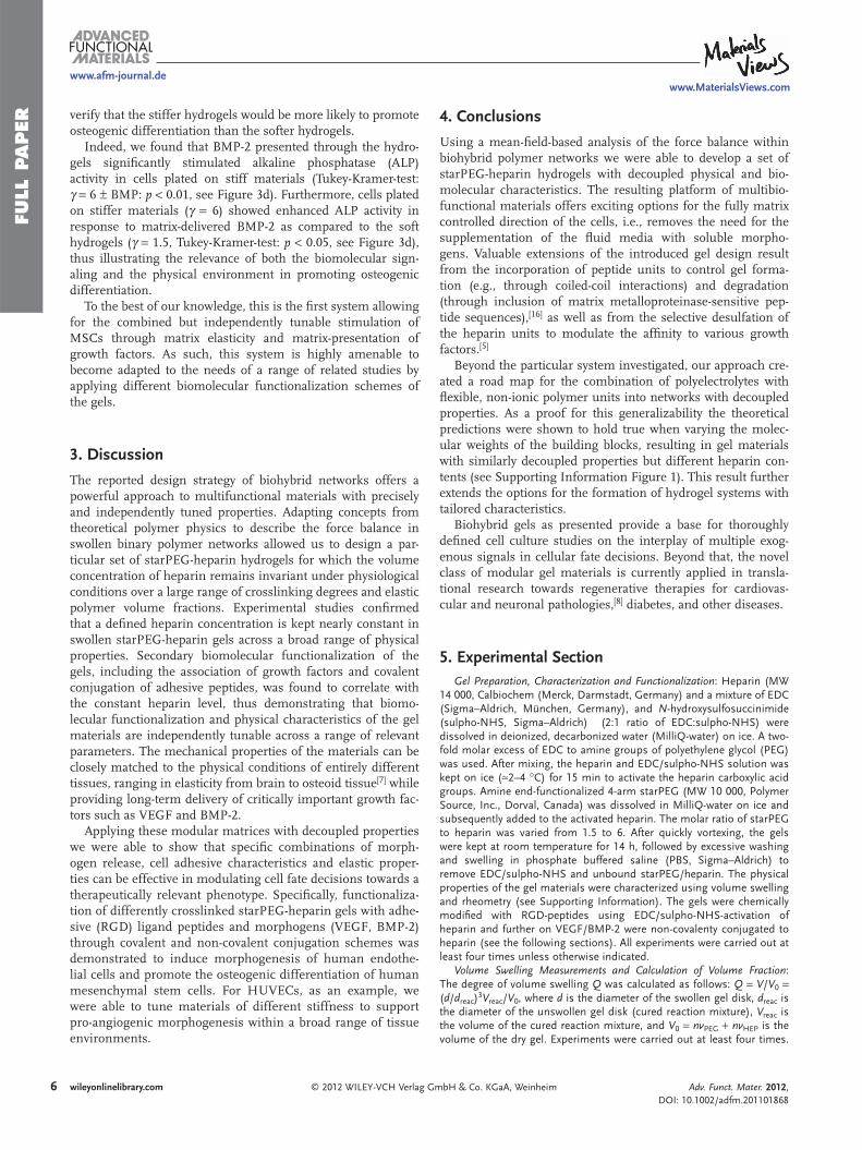
verify that the stiffer hydrogels would be more likely to promote osteogenic differentiation than the softer hydrogels.
Indeed, we found that BMP-2 presented through the hydro-gels signifi cantly stimulated alkaline phosphatase (ALP) activity in cells plated on stiff materials (Tukey-Kramer-test: γ = 6 ± BMP: p < 0.01, see Figure 3 d). Furthermore, cells plated on stiffer materials ( γ = 6) showed enhanced ALP activity in response to matrix-delivered BMP-2 as compared to the soft hydrogels ( γ = 1.5, Tukey-Kramer-test: p < 0.05, see Figure 3 d), thus illustrating the relevance of both the biomolecular sign-aling and the physical environment in promoting osteogenic differentiation.
To the best of our knowledge, this is the fi rst system allowing for the combined but independently tunable stimulation of MSCs through matrix elasticity and matrix-presentation of growth factors. As such, this system is highly amenable to become adapted to the needs of a range of related studies by applying different biomolecular functionalization schemes of the gels.
3. Discussion
The reported design strategy of biohybrid networks offers a powerful approach to multifunctional materials with precisely and independently tuned properties. Adapting concepts from theoretical polymer physics to describe the force balance in swollen binary polymer networks allowed us to design a par-ticular set of starPEG-heparin hydrogels for which the volume concentration of heparin remains invariant under physiological conditions over a large range of crosslinking degrees and elastic polymer volume fractions. Experimental studies confi rmed that a defi ned heparin concentration is kept nearly constant in swollen starPEG-heparin gels across a broad range of physical properties. Secondary biomolecular functionalization of the gels, including the association of growth factors and covalent conjugation of adhesive peptides, was found to correlate with the constant heparin level, thus demonstrating that biomo-lecular functionalization and physical characteristics of the gel materials are independently tunable across a range of relevant parameters. The mechanical properties of the materials can be closely matched to the physical conditions of entirely different tissues, ranging in elasticity from brain to osteoid tissue [ 7 ] while providing long-term delivery of critically important growth fac-tors such as VEGF and BMP-2.
Applying these modular matrices with decoupled properties we were able to show that specifi c combinations of morph-ogen release, cell adhesive characteristics and elastic proper-ties can be effective in modulating cell fate decisions towards a therapeutically relevant phenotype. Specifi cally, functionaliza-tion of differently crosslinked starPEG-heparin gels with adhe-sive (RGD) ligand peptides and morphogens (VEGF, BMP-2) through covalent and non-covalent conjugation schemes was demonstrated to induce morphogenesis of human endothe-lial cells and promote the osteogenic differentiation of human mesenchymal stem cells. For HUVECs, as an example, we were able to tune materials of different stiffness to support pro-angiogenic morphogenesis within a broad range of tissue environments.
wileyonlinelibrary.com © 2012 WILEY-VCH Verlag G
4. Conclusions
Using a mean-fi eld-based analysis of the force balance within biohybrid polymer networks we were able to develop a set of starPEG-heparin hydrogels with decoupled physical and bio-molecular characteristics. The resulting platform of multibio-functional materials offers exciting options for the fully matrix controlled direction of the cells, i.e., removes the need for the supplementation of the fl uid media with soluble morpho-gens. Valuable extensions of the introduced gel design result from the incorporation of peptide units to control gel forma-tion (e.g., through coiled-coil interactions) and degradation (through inclusion of matrix metalloproteinase-sensitive pep-tide sequences), [ 16 ] as well as from the selective desulfation of the heparin units to modulate the affi nity to various growth factors. [ 5 ]
Beyond the particular system investigated, our approach cre-ated a road map for the combination of polyelectrolytes with fl exible, non-ionic polymer units into networks with decoupled properties. As a proof for this generalizability the theoretical predictions were shown to hold true when varying the molec-ular weights of the building blocks, resulting in gel materials with similarly decoupled properties but different heparin con-tents (see Supporting Information Figure 1). This result further extends the options for the formation of hydrogel systems with tailored characteristics.
Biohybrid gels as presented provide a base for thoroughly defi ned cell culture studies on the interplay of multiple exog-enous signals in cellular fate decisions. Beyond that, the novel class of modular gel materials is currently applied in transla-tional research towards regenerative therapies for cardiovas-cular and neuronal pathologies, [ 8 ] diabetes, and other diseases.
5. Experimental Section Gel Preparation, Characterization and Functionalization : Heparin (MW
14 000, Calbiochem (Merck, Darmstadt, Germany) and a mixture of EDC (Sigma–Aldrich, München, Germany), and N -hydroxysulfosuccinimide (sulpho-NHS, Sigma–Aldrich) (2:1 ratio of EDC:sulpho-NHS) were dissolved in deionized, decarbonized water (MilliQ-water) on ice. A two-fold molar excess of EDC to amine groups of polyethylene glycol (PEG) was used. After mixing, the heparin and EDC/sulpho-NHS solution was kept on ice ( ≈ 2–4 ° C) for 15 min to activate the heparin carboxylic acid groups. Amine end-functionalized 4-arm starPEG (MW 10 000, Polymer Source, Inc., Dorval, Canada) was dissolved in MilliQ-water on ice and subsequently added to the activated heparin. The molar ratio of starPEG to heparin was varied from 1.5 to 6. After quickly vortexing, the gels were kept at room temperature for 14 h, followed by excessive washing and swelling in phosphate buffered saline (PBS, Sigma–Aldrich) to remove EDC/sulpho-NHS and unbound starPEG/heparin. The physical properties of the gel materials were characterized using volume swelling and rheometry (see Supporting Information). The gels were chemically modifi ed with RGD-peptides using EDC/sulpho-NHS-activation of heparin and further on VEGF/BMP-2 were non-covalenty conjugated to heparin (see the following sections). All experiments were carried out at least four times unless otherwise indicated.
Volume Swelling Measurements and Calculation of Volume Fraction : The degree of volume swelling Q was calculated as follows: Q = V / V 0 = ( d / d reac ) 3 V reac / V 0 , where d is the diameter of the swollen gel disk, d reac is the diameter of the unswollen gel disk (cured reaction mixture), V reac is the volume of the cured reaction mixture, and V 0 = nv PEG + nv HEP is the volume of the dry gel. Experiments were carried out at least four times.
mbH & Co. KGaA, Weinheim Adv. Funct. Mater. 2012, DOI: 10.1002/adfm.201101868

FULL P
APER
www.afm-journal.dewww.MaterialsViews.com
The volume fraction of heparin (see Figure 2d) was calculated according to the following equation: c HEP = c HEP ( m / v )/ ρ HEP ( m / v ), where ρ HEP is the density of heparin with 2.14 g mL − 1 .
Rheological Measurements : Oscillating measurements on the swollen gel disks (PBS) were carried out on a rotational rheometer (ARES LN2, TA Instruments, Eschborn, Germany), fi tted with a parallel plate geometry (plate diameter = 25 mm). Frequency sweeps were carried out at 25 ° C in a shear frequency range of 10 − 1 –10 − 2 rad s − 1 with a strain amplitude of 2%.
Modifi cation of the Gels with RGD Peptides : PBS-swollen hydrogels were treated with EDC/sulpho-NHS solution (50 m M EDC and 25 m M sulpho-NHS dissolved in 1/15 M phosphate buffer, 4 ° C) to activate the heparin carboxylic acid groups for 45 min followed by fl ushing in borate buffer (100 m M , pH 8.0, 4 ° C). Next, the samples were immersed in cyclo(Arg-Gly-Asp-dTyr-Lys) (RGD) (50 mg mL − 1 , Peptides International, Louisville, KY, USA) dissolved in borate buffer (100 m M , pH 8.0) for 2 h (room temperature) followed by excessive washing in PBS. Quantifi cation of RGD-peptide in the gels was performed by acidic hydrolysis and subsequent HPLC analysis as described elsewhere. [ 17 ]
Uptake and Release of VEGF/BMP-2 : Uptake and release of VEGF 165 (PeproTech GmbH, Hamburg, Germany) or BMP-2 (R&D Systems, Minneapolis, USA) were characterized using enzyme-linked immunosorbent assay (ELISA). Surface-bound gels ( n = 3) were placed in custom-made incubation chambers. VEGF solution (200 mL with a concentration of 1 μ g mL − 1 ) or BMP-2 (200 mL with a concentration of 1, 2.5 or 6.8 μ g mL − 1 ) were added per cm 2 . The VEGF or BMP-2 solution was removed, followed by washing with PBS. Each of these solutions was collected and assayed in duplicates using an ELISA Quantikine kit (R&D Systems, Minneapolis, USA). After immobilization using a protein concentration of 1 μ g mL − 1 (for VEGF) or 6.8 μ g mL − 1 (for BMP-2), the growth factors were allowed to release from these gels at 22 ° C into 250 μ L cm − 2 of the either serum-free endothelial cell growth medium (for VEGF) or DMEM (for BMP-2). For additional information, the reader is referred to Zieris et al. [ 18 ]
Endothelial Cell Culture Experiments : Human endothelial cells from the umbilical cord vein (HUVECs) were collected according to the procedure suggested by Weis et al. [ 19 ] 50 000 cells per cm 2 (passage 1 to 4) were seeded onto RGD- and VEGF-functionalized hydrogels in complete ECGM containing 2% calf serum. After 18 h cells were washed, fi xed and stained with monoclonal mouse anti-CD31 (BD Bioscience, Heidelberg, Germany) and AlexaFluor 546 goat anti-mouse secondary (Invitrogen) antibodies. Actin was visualised by AlexaFluor 633-labelled Phalloidin (Invitrogen). Confocal images were taken, and ImageJ software [ 20 ] was used to quantify the aspect ratio of individual cells as delineated by the actin and CD31 staining similar to that which was published previously. [ 21 ] The aspect ratio was determined by calculating the ratio of the major axis of individual cells to their minor axis, with a higher aspect ratio indicating more elongated cells. [ 13 ] Aspect ratios are recorded as means ± standard deviation of > 20 cells per experiment done in three repeats and statistically analyzed by a two-way analysis of variation test followed by Bonferroni post testing. p values less than 0.05 were considered statistically signifi cant. HUVECs isolated according to the above-mentioned protocol were shown to form tubes on Matrigel (see Supporting Information Figure 3).
Mesenchymal Stem Cell Culture Experiments : Human bone marrow-derived MSCs were isolated from healthy male donors (Caucasians 21–35 years of age). The study was approved by the institutional Review Board of the Medical Faculty at the University Hospital of Technische Universität Dresden. MSCs were expanded and characterized as described previously. [ 22 ] In brief, bone marrow mononuclear cells were isolated by Percoll (D = 1.073 g mL − 1 ) density gradient fractionation. For characterization, 1 × 10 6 of these cells were analyzed in a colony forming unit-fi broblast assay. Herein, cells were cultured for 14 days in NH expension medium (MACS Media for non-hematopoietic stem cells, Miltenyi Biotec, Bergisch Gladbach, Germany) and colonies were counted afterwards. Bone marrow derived mononuclear cells were seeded in a plastic tissue culture fl ask containing Dulbecco’s-modifi ed Eagle’s medium- low glucose
© 2012 WILEY-VCH Verlag GmAdv. Funct. Mater. 2012, DOI: 10.1002/adfm.201101868
supplemented with 10% fetal bovine serum (DMEM + 10% (v/v) FBS) for expansion. After 2 to 3 days non-adherent cells were removed by washing with PBS containing human serum albumin (0.5%) and fresh medium was added.
Adherent cells were cultured until they reached 90% confl uence. Cells were then harvested and further characterized by immune staining and fl ow cytometry analysis (stained positive for CD166, CD105, CD90, and CD73 and negative for CD45, CD34, and CD14) and by osteoblast (ALP activity quantifi cation and von Kossa staining for matrix mineralization) and adipocyte differentiation assays (lipid accumulation was quantifi ed using oil red staining). Based on this characterization the plastic-adherent bone marrow derived mononuclear cell fraction is referred to as mesenchymal stromal cells (MSCs).
MSCs were cultured in DMEM (low glucose) supplemented with FBS (10% (v/v)) and maintained in a humidified atmosphere of CO 2 (5%). For hydrogel experiments, passage 2 MSCs were plated at a density of 15 000 cells cm − 2 on RGD functionalized starPEG-heparin scaffolds that were preincubated over night with 1.9 μ g BMP-2 (R&D Systems) per scaffold. The medium (DMEM + 10% (v/v) FBS) was refreshed after 24 h and on day 5 it was changed to serum-free DMEM + supplement containing insulin, transferrin and selenous acid (ITSTM Premix, BD Biosciences). Termination and processing was done on day 7 to determine ALP enzyme activity and to stain for ALP distribution within the culture. Therefore, the cells were washed with PBS and lysed by freezing in H 2 O containing Triton X-100 (1% (v/v), Sigma). The cell lysates were incubated with p -nitrophenol phosphate and the reaction was monitored at 37 ° C for 16 min by measuring the absorbance at a wavelength of 410 nm. The reaction mixture was prepared by adding sodium borate-NaOH (5 mL of 50 m M , pH 9.8) to p -nitrophenol phosphate (5 mL of 16 m M ) and MgCl 2 (20 μ L of 1 M ). The ALP activity was normalized to the total amount of protein in the lysate by using the macro BCA protein assay (Thermo Scientific) to obtain specific ALP activities. ALP activity data (normalized to controls grown on tissue culture plastic) are recorded as means ± standard deviation of n = 4 donors. Significance was tested applying the Tukey-Kramer multiple comparison test. Additionally, qualitative ALP assessment was performed by processing the samples for histochemical detection based on the manufacturer’s protocol (Sigma).
Supporting Information Supporting Information is available from the Wiley Online Library or from the author.
Acknowledgements U.F. and J.-U.S contributed contributed equally to this work. The authors thank Roland Vogel for advice on rheometry (Leibniz Institute of Polymer Research Dresden), Martin Bornhäuser and Katrin Müller (University Hospital “Carl Gustav Carus”, Technische Universität Dresden, Dresden, Germany) for advice and support on the culture of mesenchymal stem cells, Phillip Seib (Tufts University, Medford, MA, USA), and Ilya Levental (Max Planck Institute for Cell Biology and Genetics, Dresden, Germany) for valuable discussions. U.F. and C.W. were supported by the Deutsche Forschungsgemeinschaft (DFG) through grants WE 2539-7/1, SFB 655 and FOR/EXC999, and by the Leibniz Association. K.R.L., S.P. and C.W. were supported by the Seventh Framework Programme of the European Union through the Integrated Project ANGIOSCAFF. K.S. and C.W. were supported by the Bundesministerium für Bildung, Forschung und Technologie (BMBF) through grant 01 GN 0946.
Received: August 11, 2011 Revised: November 18, 2011
Published online:
7wileyonlinelibrary.combH & Co. KGaA, Weinheim

FULL
PAPER
8
www.afm-journal.de
[ 1 ] a) M. P. Lutolf , J. A. Hubbell , Nat. Biotechnol. 2005 , 23 , 47 ; b) G. Chan , D. J. Mooney , Trends Biotechnol. 2008 , 26 , 382 ; c) E. S. Place , N. D. Evans , M. M. Stevens , Nat. Mater. 2009 , 8 , 457 ; d) N. Huebsch , D. J. Mooney , Nature 2009 , 462 , 426 ; e) M. P. Lutolf , P. M. Gilbert , H. M. Blau , Nature 2009 , 462 , 433 ; f) D. A. Wang , S. Varghese , B. Sharma , I. Strehin , S. Fermanian , J. Gorham , D. H. Fairbrother , B. Cascio , J. H. Elisseeff , Nat. Mater. 2007 , 6 , 385 ; g) N. Peppas , Z. Hilt , A. Khademhosseini , R. Langer , Adv. Mater. 2006 , 18 , 1345 .
[ 2 ] a) A. M. Kloxin , A. M. Kasko , C. N. Salinas , K. S. Anseth , Science 2009 , 324 , 59 ; b) H. J. Lee , C. Yu , T. Chansakul , N. S. Hwang , S. Varghese , S. M. Yu , J. H. Elisseeff , Tissue Eng. Part. A 2008 , 14 , 1843 ; c) L. Cao , P. R. Arany , Y. S. Wang , D. J. Mooney , Bioma-terials 2009 , 30 , 4085 ; d) C. Fischbach , R. Chen , T. Matsumoto , T. Schmelzle , J. S. Brugge , P. J. Polverini , D. J. Mooney , Nat. Methods 2007 , 4 , 855 ; e) T. P. Kraehenbuehl , L. S. Ferreira , P. Zammaretti , J. A. Hubbell , R. Langer , Biomaterials 2009 , 30 , 4318 ; f) S. T. Lee , J. I. Yun , Y. S. Jo , M. Mochizuki , A. J. van der Vlies , S. Kontos , J. E. Ihm , J. M. Lim , J. A. Hubbell , Biomaterials 2010 , 31 , 1219 ; g) E. Fong , D. A. Tirrell , Adv. Mater. 2010 , 22 , 5271 .
[ 3 ] a) D. E. Discher , D. J. Mooney , P. W. Zandstra , Science 2009 , 324 , 1673 ; b) D. E. Discher , P. Janmey , Y. L. Wang , Science 2005 , 310 , 1139 ; c) R. K. Assoian , E. A. Klein , Trends Cell. Biol. 2008 , 18 , 347 ; d) Y.-W. Lin , C.-M. Cheng , P. R. LeDuc , C.-C. Chen , PLoS ONE 2009 , 4 , e4293 ; e) S. Nemir , J. L. West , Ann. Biomed. Eng. 2010 , 38 , 2 .
[ 4 ] a) J. K. Tessmar , A. M. Goepferich , Macromol. Biosci. 2007 , 7 , 23 ; b) N. Peppas , Z. Hilt , A. Khademhosseini , R. Langer , Adv. Mater. 2006 , 18 , 1345.
[ 5 ] I. Capila , R. J. Linhardt , Angew. Chem. Int. Ed. Engl. 2002 , 41 , 391 [ 6 ] a) P. J. Flory , J. Rehner , J. Chem. Phys. 1943 , 11 , 521 ; b) H. M. James ,
E. Guth , J. Chem. Phys. 1947 , 15 , 669 ; c) T. Tanaka , M. Shibayama , A. Onuki , in Responsive Gels: Volume Transitions I , (Ed: K. Dusek ), Springer , Berlin Heidelberg, Germany 1993 .
[ 7 ] a) A. J. Engler , S. Sen , H. L. Sweeney , D. E. Discher , Cell 2006 , 126 , 677 ; b) I. Levental , P. C. Georges , P. A. Janmey , Soft Matter 2006 , 3 , 299 .
wileyonlinelibrary.com © 2012 WILEY-VCH Verlag G
www.MaterialsViews.com
[ 8 ] U. Freudenberg , A. Hermann , P. B. Welzel , K. Stirl , S. C. Schwarz , M. Grimmer , A. Zieris , W. Panyanuwat , S. Zschoche , D. Meinhold , A. Storch , C. Werner , Biomaterials 2009 , 30 , 5049 .
[ 9 ] a) D. E. Ingber , J. Folkman , J. Cell Biol. 1989 , 109 , 317 ; b) T. Maciag , J. Kadish , L. Wilkins , M. B. Stemerman , R. Weinstein . J. Cell Biol. 1982 , 94 , 511 .
[ 10 ] a) A. Engler , L. Bacakova , C. Newman , A. Hategan , M. Griffi n , D. Discher , Biophys. J. 2004 , 86 , 617 ; b) J. C. Liu , D. A. Tirrell , Biomacromolecules 2008 , 9 , 2984 .
[ 11 ] a) M. P. Lutolf , J. A. Hubbell , Nat. Biotechnol. 2005 , 23 , 4 ; b) M. P. Lutolf , P. M. Gilbert , H. M. Blau , Nature 2009 , 462 , 433.
[ 12 ] Y. Kubota , H. K. Kleinman , G. R. Martin , T. J. Lawley . J. Cell Biol. 1988 , 107 , 1589 .
[ 13 ] J. P. Califano , C. A. Reinhart-King . Cell Mol. Bioeng. 2008 , 1 , 122 . [ 14 ] A. J. Engler , S. Sen , H. L. Sweeney , D. E. Discher , Cell 2006 , 126 ,
677 . [ 15 ] a) M. F. Pittenger , A. M. Mackay , S. C. Beck , R. K. Jaiswal ,
R. Douglas , J. D. Mosca , M. A. Moorman , D. W. Simonetti , S. Craig , D. R. Marshak , Science 1999 , 284 , 143 ; b) R. McBeath , D. M. Pirone , C. M. Nelson , K. Bhadriraju , C. S. Chen , Dev. Cell 2004 , 6 , 483 ; c) A. M. Osyczka , P. S. Leboy , Endocrinology 2005 , 146 , 3428 .
[ 16 ] a) M. Tsurkan , K. R. Levental , U. Freudenberg , C. Werner , Chem. Commun. 2010 , 46 , 1141 ; b) K. Chwalek , K. R. Levental , M. V. Tsurkan , A. Zieris , U. Freudenberg , C. Werner , Biomaterials 2011 , 32 , 9649 .
[ 17 ] K. Salchert , T. Pompe , C. Sperling , C. Werner , J. Chromatogr. A 2003 , 1005 , 113 .
[ 18 ] A. Zieris , S. Prokoph , P. B. Welzel , M. Grimmer , K. R. Levental , W. Panyanuwat , U. Freudenberg , C. Werner , J. Mater. Sci. Mater. Med. 2010 , 21 , 914 .
[ 19 ] J. R. Weis , B. Sun , G. M. Rodgers , Thromb. Res. 1991 , 61 , 171 . [ 20 ] a) W. S. Rasband , ImageJ, U. S. National Institutes of Health,
Bethesda, Maryland, USA, http://imagej.nih.gov/ij/, 1997–2011 . [ 21 ] R. Sumagin , C. W. Brown III , I. H. Sarelius , M. R. King , Ann. Biomed.
Eng. 2008 , 36 , 580 . [ 22 ] J. Oswald , S. Boxberger , B. Jorgensen , S. Feldmann , G. Ehninger ,
M. Bornhauser , C. Werner , Stem Cells 2004 , 22 , 377 .
mbH & Co. KGaA, Weinheim Adv. Funct. Mater. 2012, DOI: 10.1002/adfm.201101868