The transcription factor MEF2C mediates cardiomyocyte hypertrophy induced by IGF-1 signaling
Upload
independentCategory
view
5download
0
Basic Res Cardiol 100: 161 – 170 (2005)DOI 10.1007/s00395-004-0506-3 ORIGINAL CONTRIBUTION
Armelle SeroseBernard PrudhonAndré SalmonMarie-Agnès DoyennetteMarc Y. FiszmanYves Fromes
Administration of insulin-like growth factor-1(IGF-1) improves both structure and functionof delta-sarcoglycan deficient cardiac musclein the hamster
� Abstract Dilated cardiomyopathies (DCM) are due to progressive dilata-tion of the cardiac cavities and thinning of the ventricular walls and leadunavoidably to heart failure. They represent a major cause for heart trans-plantation and, therefore, defining an efficient symptomatic treatment forDCM remains a challenge. We have taken advantage of the hamster strainCHF147 that displays progressive cardiomyopathy leading to heart failure totest whether stimulation of a hypertrophic pathway could delay the processof dilatation.
Six month old CHF147 hamsters were treated with IGF-1 so that we couldcompare the efficacy of systemic administration of human recombinant IGF-1 protein (rh IGF-1) at low dose to that of direct myocardial injections of aplasmid DNA containing IGF-1 cDNA (pCMV-IGF1).
IGF-1 treatment did not induce a significant variation of ventricle mass,but preserved left ventricular (LV) wall thickness and delayed dilatation ofcardiac cavities when compared to non-treated hamsters. Together with thisreduction of dilatation, we also noted a reduction in the amount of intersti-tial collagen. Furthermore, IGF-1 treatment induced beneficial effects on car-diac function since treated hamsters presented improved cardiac output andstroke volume, decreased end diastolic pressure when compared to non-treated hamsters and also showed a trend towards increased contractility(dP/dtmax).
This study provides evidence that IGF-1 treatment induces beneficialstructural and functional effects on DCM of CHF147 hamsters, hence makingthis molecule a promising candidate for future gene therapy of heart failuredue to DCM.
� Keys words Dilated cardiomyopathy – heart failure – CHF147 hamsters –gene therapy – IGF-1
BRC
506
A. Serose · B. Prudhon · A. Salmon M.-A. Doyennette · M. Y. Fiszman Y. Fromes, MD, PhD (�) Institute de myologie – Inserm U582Groupe Hospitalier Pitié-Salpêtrière47, Boulevard de l’Hôpital75651 Paris Cedex 13, FranceE-Mail: [email protected]
Received: 3 August 2004Returned for 1. revision: 23 August 20041. Revision received: 24 September 2004Returned for 2. revision: 10 October 20042. Revision received: 26 October 2004Accepted: 15 November 2004Published online: 22 December 2004
Introduction
Heart failure is a major issue in public health care due toits frequency and severity. Dilated cardiomyopathies(DCM) are one of the most frequent causes for heart fail-ure and are characterized by left and/or right ventriculardilatation associated with systolic dysfunction. Two
mechanisms are responsible for this progression tochronic heart failure: thinning of the ventricular wallsand dilatation of the ventricles. Moreover, myocardialcontraction is decreased as cardiac cavities dilate and asthere is a loss of contractile units due to cardiac wallsthinning. Thus, the heart is unable to maintain its output.Even though a genetic cause of DCM has been identifiedin about 30% of the cases, it, nevertheless, remains true

that heart failure results in common deregulations inde-pendent of the etiology.
Without etiologic diagnosis, clinicians set up sympto-matic treatments. In the acute phase, beta-adrenergicagonists are used to increase inotropism and overcomeacute heart failure episodes. At the chronic phase of heartfailure, this treatment is often composed of diuretics that reduce volume overload, angiotensin-convertingenzyme inhibitors, beta-blockers and anti-aldosteronetreatments [20]. So far, the aim of drug therapy in DCMis to attenuate symptoms of the failing heart; however, nolong-term positive inotropic treatments are available.Thus, DCM is the most common and frequent cause ofheart failure. Therefore, it seems essential to develop newtherapies for DCM treatment.
The availability of animal models of DCM is essentialfor the development of in vivo therapeutic approaches.One such model of inherited cardiomyopathy is theCHF147 hamster, which has a large deletion of the delta-sarcoglycan gene [10]. This deletion leads to the absenceof delta-sarcoglycan transcripts and of the delta-sarco-glycan protein, which is a structural protein of all musclecell membranes [24]. This model displays both morpho-logical and functional cardiac alterations.
It has been shown that re-expression of the delta-sar-coglycan gene before the state of heart failure can slowthe development of cardiac lesions [11]. Heart failuredue to DCM induces modifications in neuro-endocrinepathways; especially the GH/IGF-1 axis is altered [1, 2, 15,19]. Along this line, growth hormone (GH) was proposedas a potential treatment. The efficiency of GH treatmentwas assessed at early (4 months) and late (10 months)phases of the disease in the CHF147 hamster [23]. GHtreatment improved left ventricular dysfunction at 4months in CHF147 hamsters by increasing contractilityand reducing wall stress but had little beneficial effect at10 months in severe left ventricular heart failure.
In this study, we propose to stimulate a hypertrophicresponse to compensate dilatation of the cavities and toincrease cardiac muscle wall thickness. To achieve thisgoal, we used insulin-like growth factor-1 (IGF-1), a GHeffector. IGF-1 is involved in cardiomyocyte prolifera-tion, differentiation and cell survival [25]. Reiss et al.showed that total heart weight of transgenic mice over-expressing IGF-1 in myocardium is increased by 50%and cardiomyocyte number is as well increased by 20 to50% [21]. IGF-1 is also known for its role in regulation ofstructure and function of adult myocardium [31]. More-over, IGF-1 is able to improve myocardial contractility byincreasing the synthesis of cardiac contractile proteins[30, 31].
In this report, we compare IGF-1 treatment of DCM inadult hamster, either by IGF-1 expressed before injectionand delivered as a recombinant protein (rh IGF-1) or bydirect myocardial injection of a plasmid DNA containingthe IGF-1 cDNA. We provide evidence that both strate-
gies have beneficial structural and functional effects onDCM. We show that IGF-1 induced a thickening of thecardiac walls that slows cardiac dilatation and thatimproves global cardiac performance.
Materials and methods
� Animals and experimental protocols
Male Syrian hamsters (CHF147 and wild type) were usedfor the experiments. Wild type (healthy group) Syrianhamsters (strain Rj:AURA) were purchased from ElevageJanvier, France. CHF147 hamsters were bred at the localanimal facility of the Institut de Myologie and were fed onnormal chow diet after six weeks of age. The animalswere maintained at 20 °C with 10/14 hours light/darkcycles and free access to food and water was provided.Care of the animals was in compliance with the Guide ofthe Care and Use of Laboratory Animals, published by theNational Institute of Health (National Institute of Healthpublication no. 85–23, revised 1996). Wild type Syrianhamsters and CHF147 non-treated hamsters were used toestablish reference groups. The 6-month old CHF147hamsters were randomized into four groups: a controlgroup was sham operated receiving an osmotic pumpfilled with saline (Sham 1 group, n = 3), a group in whichanimals were treated with recombinant human IGF-1 (rh IGF-1; Chemicon International, Inc.) at a total doseof 22 µg (rh IGF-1 treated group, n = 7) which was deliv-ered by an osmotic pump (Alzet, model 2004) implantedunder the skin, a control group was sham operatedreceiving saline injection (Sham 2 group, n = 3) and afourth group in which animals were treated by genetransfer with the IGF-1 cDNA (pCMV-IGF1 treatedgroup, n = 6). The two last groups of animals were oper-ated as follows. Hamsters were anesthetized with keta-mine (75 mg/kg), xylazine (15 mg/kg) and midazolam(0.75 mg/kg) administered intraperitoneally, intubated,and ventilated (SAR-830/P ventilator, Bioseb, France).IGF-1 cDNA was injected through a small laparotomyand a left ventral phrenotomy. A total dose of 500 to 650 µg of IGF-1 cDNA was injected at multiple sites intothe left ventricular wall using a 31 Gauge needle. All ani-mals were sacrificed with an overdose of anesthetics after35 days of treatment.
� Plasmid vector pCMV-IGF1
The vector used in this study is a plasmid DNA, pCMV-IGF1, containing the cytomegalovirus (CMV) promoterand a cDNA encoding a rat preprohormone form of IGF-1. This plasmid has a size of 4356 bp and was produced,
162 Basic Research in Cardiology, Vol. 100, No. 2 (2005)© Steinkopff Verlag 2005

after amplification, by a Gigapreps kit (Qiagen) using theprotocol given by the manufacturer. Measuring IGF-1secretion following transfection of HEK293 cells assessedthe functional property of the plasmid. IGF-1 secretionby pCMV-IGF1 transfected myocytes was detected byimmunohistochemistry using polyclonal anti-IGF-1(1:1500, Chemicon International) primary antibody oncryostat sections.
� Measure of LV function
We used a cardiac loop analysis computer system withspecific software iox v.1.7. (Emka, France) with a pres-sure and volumetric system (spr-SERIES 1.4 FRENCH p-v conductance catheter, Millar Instruments) to per-form LV pressure-volume loop analysis. The systemallows beat-by-beat monitoring of heart performance inhamsters, and measurements and analysis were per-formed as follows. The ultra-miniature catheters wereused in closed-chest surgical procedures, introducedthrough the right carotid artery and properly positionedin the left ventricular cavity. Pressure and volume meas-urements were recorded from a single instrument. Thecalculated hemodynamic parameters serve as indicatorsof cardiovascular performance in response to differentinterventions (occlusions). Pressure-volume loops wereestablished by variation of cardiac load after compres-sion of the inferior vena cava. The end-systolic pressure-volume relation (ESPVR) was calculated to represent LVglobal systolic function.
� Morphological analysis of the cardiac muscle
Macroscopy
Animals were weighed before evaluation of cardiac func-tion. Hearts were quickly harvested and perfused with anormal saline solution under low-pressure conditions.Ventricles were also weighed after heart removal andatrium dissection. Ventricular weight-to-body weightratio was calculated and is given in mg/g. Thickness ofinter ventricular septum and posterior wall, as well ascardiac diameters, were measured on cardiac cryostatsections.
Histology
Hearts were removed and atriums were dissected. Thenthe ventricles were cut in half along the cardiac transver-sal axis. Each half was frozen in liquid nitrogen cooled in isopropanol and stored at –80 °C. Cryostat sections (8 µm) were cut and mounted on glass slides for histo-logical analysis. Four different histochemical stainingwere realized: hematoxylin-eosin (H&E), Sirius red (SR),
succinyl deshydrogenase (SDH) and von Kossa nitrate.Microscopy observations were performed with a NikonEclipse E800 microscope (Nikon). Image acquisition wascarried out using a Nikon Digital camera (Nikon).
Fibrosis on SR slides was quantified with Lucia Gversion 4.61 software (Nikon). Nine heart sections werequantified in the healthy group, five in the non-treatedgroup, four in the rh IGF-1 treated group and ten in thepCMV-IGF1 treated group.
Immunohistochemistry
Six micrometers cryostat sections of left ventricles werecut for immunohistochemical analysis in order to exam-ine collagen and contractile proteins. The following pri-mary antibodies were used: polyclonal anti-collagen type1 (1:30 Chemicon International), monoclonal anti-tro-ponin I (1:2000, Chemicon International), monoclonalanti-myosin heavy chain (1:1500, NOQ7.5.4D clone,Sigma). We also looked at the cytosquelettal alterationsin the cardiomyocytes using the monoclonal anti-dys-trophin (1:40, NCL-DYS1 clone, Novocastra) primaryantibody. All slides were mounted with Vectashield withDAPI (Vector Laboratories). Microscopy observationswere performed with a Leica DMR microscope (Leica)and image acquisition was carried out using a Sony 3CCDcolor video camera (Nikon) and TRIBVN ICS software(ICG).
Macrophages and CD8 lymphocytes were detectedusing monoclonal anti-rat macrophages (1:800, Bio-source International) and monoclonal anti-rat CD8 lym-phocytes (1:500, PharMingen International) primaryantibodies.
� Statistics
Morphometric and functional results are presented asmean ± standard deviation and fibrosis results corre-spond to the percentage of fibrosis on the surface of theslide. Experimental data were analyzed pair-wise bet-ween control and treated groups. Statistical significancebetween two measurements was determined by theANOVA method using the Stat View software. Values ofp < 0.05 were considered significant.
A. Serose et al. 163Effects of IGF-1 on DCM

164 Basic Research in Cardiology, Vol. 100, No. 2 (2005)© Steinkopff Verlag 2005
Table 1 Macroscopical analysis
Healthy Non-treated Sham 1 rh IGF-1 Sham 2 pCMV-IGF1n = 19 3 3 7 3 6
VW (mg) 529.0 � 93.1 505.9 � 64.9 599.9 � 26.7 597.0 � 64.1 544.4 � 50.7 498.8 � 91.04
BW (g) 155.5 � 17.5 101.7 � 27.41 141.6 � 11.6 136.1 � 16.31, 2 132.4 � 14.5 120.8 � 16.31
VW/BW (mg/g) 3.41 � 0.56 5.09 � 0.64 1 4.25 � 0.28 4.39 � 0.231, 2 4.12 � 0.09 4.13 � 0.491, 2
D (mm) 8.38 � 0.36 10.36 � 0.631 10.49 � 0.47 8.91 � 0.481, 2, 4 10.46 � 0.81 8.29 � 0.262, 3, 5
PW (mm) 1.92 � 0.15 2.17 � 0.34 2.21 � 0.40 2.93 � 0.251, 2, 4 2.17 � 0.30 2.31 � 0.24 1, 3
IVS (mm) 1.38 � 0.32 1.62 � 0.27 1.62 � 0.28 2.12 � 0.201, 2, 4 1.54 � 0.14 2.04 � 0.321, 2, 5
PW/IVS 1.44 � 0.27 1.34 � 0.07 1.36 � 0.06 1.39 � 0.14 1.41 � 0.11 1.15 � 0.14 1, 2, 3, 5
rh IGF-1 IGF-1 protein treated group; pCMV-IGF1 IGF-1 plasmid treated group; VW ventricle weight, in mg, BW body weight, in g, VW/BW ventricle weight-to-body weightratio, in mg/g, D cardiac diameter, in mm, PW posterior wall thickness, in mm, IVS inter-ventricular septum thickness, in mm, PW/IVS PW-to-IVS thickness ratio; cardiac diam-eters and thickness measurements were done on cryostat heart sections, in mm; 1 = p < 0.05 vs. healthy group, 2 = p < 0.05 vs. non-treated group; 3 = p < 0.05 vs. rh IGF-1 treated group, 4 = p < 0.05 vs. Sham 1 group, 5 = p < 0.05 vs. Sham 2 group
Fig. 1 Cytosquelettal alteration. Anti-dystrophin immunostaining (in red) on trans-verse heart sections of healthy (a), non-treated (b), rh IGF-1 treated (c) and pCMV-IGF1 treated (d) hamsters. Plain arrows show immunonegative areas with reference
to immunopositive stained areas (dotted arrows). Magnification 40x, bar = 50 �m,nuclei are visualized by DAPI staining (in blue)

Results
� Myocardium treatment using IGF-1 induces beneficialeffects on cardiac morphology
Cardiac dilatation is slowed down and wall thickness isincreased after IGF-1 treatment
Ventricular weights (VW), body weights (BW) and VW/BW ratios are reported in Table 1. The VW/BW ratio issignificantly increased in all CHF147 animals, whethertreated or non-treated, when compared to healthy ani-mals. However, in the protein treated group, there is atrend towards a further increase of VW/BW ratio withreference to the sham-operated animals. As expected,non-treated animals have significantly increased cardiacdiameters when compared to healthy hamsters. How-ever, cardiac diameters are significantly less increased inthe protein treated group as well as in the plasmid treatedanimals compared to their respective counterparts. Meancardiac diameters remain in the range of those of thehealthy group (Table 1).
We also measured the thickness of the posterior wall(PW) and of the inter-ventricular septum (IVS) and cal-
culated the PW/IVS ratio (Table 1). Treated animalsshowed significantly increased thickness of both PW andIVS which, in the case of the rh IGF-1 treated group, givesa ratio indistinguishable with that of the healthy animals,however significantly different from the non-treatedgroup. Hence, systemic delivery of the recombinant IGF-1 protein maintains a balanced shape of cardiac mor-phology. In contrast, the PW/IVS ratio in the pCMV-IGF1 treated group is statistically decreased compared tothe healthy group PW/IVS ratio.
Structure preservation of the myocardium fibrosis afterIGF-1 treatment
All CHF147 animals, whether treated or non-treated, dis-play important localized lesions, such as necrosis, calci-fications and interstitial fibrosis, as evidenced with his-tochemical staining. Anti-dystrophin immunostainingof healthy heart sections shows a normal localization ofthe dystrophin in the sub-sarcolemmal space (Fig. 1a),whereas a partial loss of dystrophin in the non-treatedgroup can be observed (Fig. 1b). IGF-1 treated hamsterspresent a more heterogeneous profile with some positiveareas as well as negative areas (Fig. 1c). Similar findingscan be observed in the plasmid treated group strength-
A. Serose et al. 165Effects of IGF-1 on DCM
Fig. 2 Myocardial fibrosis. A Collagen type 1 immunostaining (in red) on heart sec-tions of healthy (a), non-treated (b), rh IGF-1 treated (c) and pCMV-IGF1 treated (d)hamsters; magnification 16x; bar = 100 �m, nuclei are visualized by DAPI staining(in blue). B Histological and immunohistochemical analysis of myocardial fibrosis,Sirius red staining on heart sections of healthy (a), non-treated (b), rh IGF-1 treated(c) and pCMV-IGF1 treated (d) hamsters; magnification 1x; bar = 1 mm. C Myocar-dial fibrosis quantification, in percentage of total heart section; *= p < 0.05 vs.healthy group, †= p < 0.05 vs. non-treated group
ening the findings observed in the protein treated group(Fig. 1d). Interestingly, outside of the fibrotic lesions,there is no disorganization of the cardiac tissue, as evi-denced by single and double immunolabeling of tro-ponin-I and myosin heavy chain (data not shown).
We also analyzed the type of fibrosis, as shown in Fig. 2A. Immunohistochemical staining reveals largedeposits of collagen type I. The extent of fibrosis was fur-ther analyzed with the use of Sirius red staining (Fig. 2B).No pathological fibrosis was detected in healthy hamster(Fig. 2B, a) while non-treated hamsters displayed animportant fibrotic scar, essentially located in the medialarea of the heart between endocardium and epicardium(Fig. 2B, b). In treated animals, the fibrotic scar was muchmore limited (Fig. 2B, c and d) and this is particularly evi-dent for the pCMV-IGF1 treated group, where the Siriusred stained region was not any more concentrated in themedial area (Fig. 2B, d). When the surface area of the Sir-ius red stained tissue was precisely measured, this con-clusion held true and the extent of the fibrotic area wassignificantly decreased in pCMV-IGF-1 treated animalswhen compared to the non-treated animals (Fig. 2C).Hence, IGF-1 treatment probably prevents fibrosis fromfurther spreading.
At that point, we felt that it was important to checkwhether injection of the plasmid DNA had induced eitheran inflammatory or an immunological response. Indeed,some inflammation could be observed since a limitednumber of macrophages was seen; however, no CD8+lymphocytes were detected, ruling out the possibility thatan immunological response could have had any adverseeffect (data not shown).
� Functional analysis of the effects of IGF-1 genetransfer into the myocardium
CHF147 hamsters display overt signs of chronic heartfailure
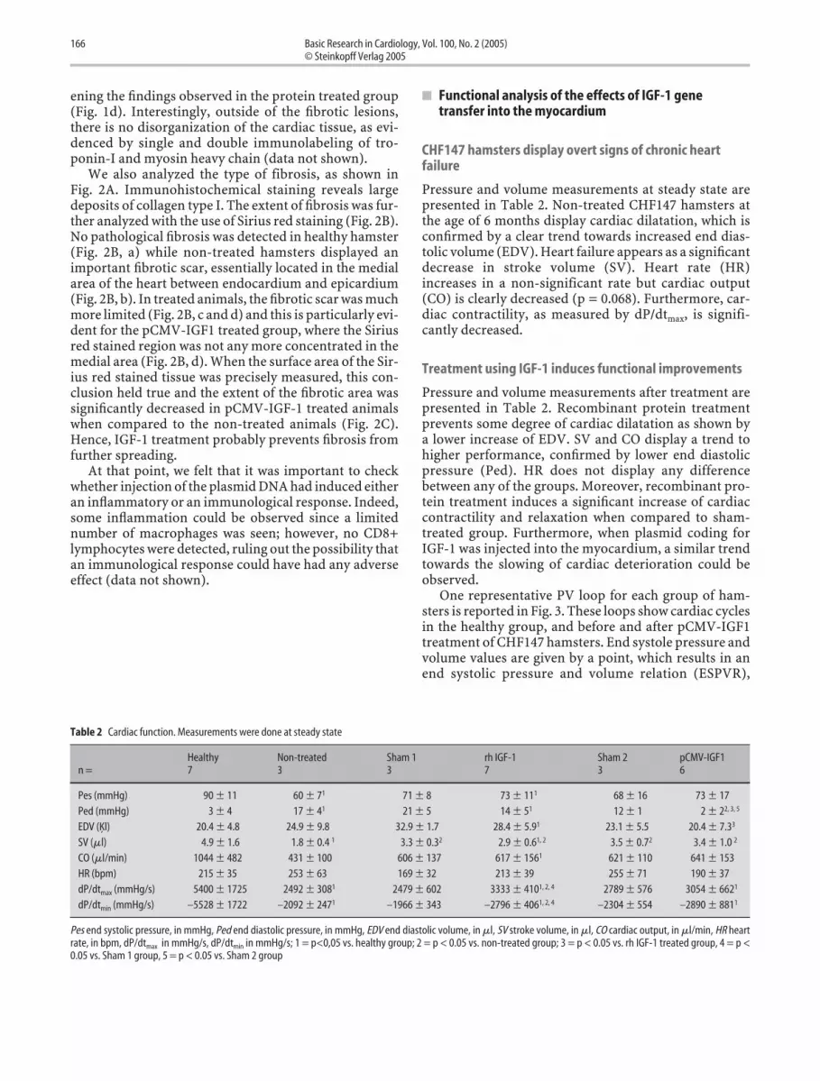
Pressure and volume measurements at steady state arepresented in Table 2. Non-treated CHF147 hamsters atthe age of 6 months display cardiac dilatation, which isconfirmed by a clear trend towards increased end dias-tolic volume (EDV). Heart failure appears as a significantdecrease in stroke volume (SV). Heart rate (HR)increases in a non-significant rate but cardiac output(CO) is clearly decreased (p = 0.068). Furthermore, car-diac contractility, as measured by dP/dtmax, is signifi-cantly decreased.
Treatment using IGF-1 induces functional improvements
Pressure and volume measurements after treatment arepresented in Table 2. Recombinant protein treatmentprevents some degree of cardiac dilatation as shown by a lower increase of EDV. SV and CO display a trend tohigher performance, confirmed by lower end diastolicpressure (Ped). HR does not display any differencebetween any of the groups. Moreover, recombinant pro-tein treatment induces a significant increase of cardiaccontractility and relaxation when compared to sham-treated group. Furthermore, when plasmid coding forIGF-1 was injected into the myocardium, a similar trendtowards the slowing of cardiac deterioration could beobserved.
One representative PV loop for each group of ham-sters is reported in Fig. 3. These loops show cardiac cyclesin the healthy group, and before and after pCMV-IGF1treatment of CHF147 hamsters. End systole pressure andvolume values are given by a point, which results in anend systolic pressure and volume relation (ESPVR),
166 Basic Research in Cardiology, Vol. 100, No. 2 (2005)© Steinkopff Verlag 2005
Table 2 Cardiac function. Measurements were done at steady state
Healthy Non-treated Sham 1 rh IGF-1 Sham 2 pCMV-IGF1n = 7 3 3 7 3 6
Pes (mmHg) – 90 � 11 – 60 � 71 – 71 � 8 – 73 � 111 – 68 � 16 – 73 � 17Ped (mmHg) – 3 � 4 – 17 � 41 – 21 � 5 – 14 � 51 – 12 � 1 – 2 � 22, 3, 5
EDV (Ķl) – 20.4 � 4.8 – 24.9 � 9.8 – 32.9 � 1.7 – 28.4 � 5.91 – 23.1 � 5.5 – 20.4 � 7.33
SV (�l) – 4.9 � 1.6 – 1.8 � 0.4 1 – 3.3 � 0.32 – 2.9 � 0.61, 2 – 3.5 � 0.72 – 3.4 � 1.0 2
CO (�l/min) –1044 � 482 – 431 � 100 – 606 � 137 – 617 � 1561 – 621 � 110 – 641 � 153HR (bpm) – 215 � 35 – 253 � 63 – 169 � 32 – 213 � 39 – 255 � 71 – 190 � 37dP/dtmax (mmHg/s) –5400 � 1725 –2492 � 3081 –2479 � 602 –3333 � 4101, 2, 4 –2789 � 576 –3054 � 6621
dP/dtmin (mmHg/s) –5528 � 1722 –2092 � 2471 –1966 � 343 –2796 � 4061, 2, 4 –2304 � 554 –2890 � 8811
Pes end systolic pressure, in mmHg, Ped end diastolic pressure, in mmHg, EDV end diastolic volume, in �l, SV stroke volume, in �l, CO cardiac output, in �l/min, HR heartrate, in bpm, dP/dtmax in mmHg/s, dP/dtmin in mmHg/s; 1 = p<0,05 vs. healthy group; 2 = p < 0.05 vs. non-treated group; 3 = p < 0.05 vs. rh IGF-1 treated group, 4 = p <0.05 vs. Sham 1 group, 5 = p < 0.05 vs. Sham 2 group

whose slope is representative of cardiac contractility. InFig. 3, the slope of ESPVR is equal to 20.73 for the healthygroup. ESPVR slope value is 0.19 in the non-treatedgroup and 10.41 in the pCMV-IGF1 treated group.
Discussion
IGF-1 is known to play a key role in cardiac function andcardiac growth [22] and several observations have sug-gested a possible link between an alteration of its path-way and congestive heart failure due to DCM [1, 2, 15,19]. For example, IGF-1 serum levels are decreased inheart failure and Vasan et al. have shown that the IGF-1
A. Serose et al. 167Effects of IGF-1 on DCM
Fig. 3 Representative example of PVloops. PV loops are obtained after car-diac preload variation; A healthy group,B non-treated group, C pCMV-IGF1treated group; bold loops correspond toPV loop at steady state, dotted red line= end systolic pressure volume relation(ESPVR) for healthy group, dotted greenline = ESPVR for non-treated group,blue line = ESPVR for pCMV-IGF1treated group

serum level was inversely related to the risk for conges-tive heart failure in elderly people without a previousmyocardial infarction [12, 15, 26, 29]. These results weretaken as an indication that IGF-1 could be considered asa marker of heart failure severity. Moreover, it has beenshown that IGF-1 administration causes hypertrophy innormal hearts and severely dysfunctional heart couldalso undergo hypertrophy, which improves cardiacfunction [7]. On the other hand, the effects of low dose ofIGF-1 on heart failure are not well documented.
Delaughter et al., using transgenic mice overexpress-ing IGF-1 in the heart, have shown that long-term expo-sure to IGF-1 leads to the development of pathologicalcardiac hypertrophy, in contrast to short-term exposure,which induces physiological hypertrophy [5]. In theirstudy, they proposed to limit temporal exposure to IGF-1 in order to deliver the potential benefits of IGF-1 whileavoiding its deleterious side effects.
Systemic administration of the recombinant humanIGF-1 protein to 6 month-old CHF147 hamsters led to adecreased cardiac dilatation and a significant hemody-namic improvement. This result vindicates the hypothe-sis that IGF-1 treatment can delay the process of cardiacdilatation and improve cardiac function. However, anumber of studies have indicated that a high IGF-1 serumlevel could be linked to an increased risk of cancer [3, 9].To avoid this problem, we decided to target IGF-1 deliv-ery to the heart by injecting into the myocardium a plas-mid DNA encoding IGF-1. As expected, no differencecould be detected in the IGF-1 serum level betweeninjected and noninjected animals, and this result is inagreement with previous observations obtained with ani-mals receiving local delivery of IGF-1 either in skeletal orcardiac muscle [4, 28].
At a macroscopic level, a significant reduction in bodyweights was observed between healthy, CHF147 non-treated or treated groups. This difference can reflect bothcardiac cachexy and the myopathic lesions that reducethe muscular mass. Upon microscopic observation, wenoted differences in the left ventricular shape as reflectedby posterior wall-to-inter ventricular septum (PW-to-IVS) ratios. Protein treatment restores a close to normalratio, whereas the pCMV-IGF1 treated group displayssome asymmetry in wall thickness. These differences areprobably due to the two different methods of adminis-tration used and also to the fact that IGF-1 produced bytransfected myocardium has a spatially limited actionwhile rh IGF-1 is available to the entire myocardium.Lastly, myocardial fibrosis showed a trend towardsdecrease in rh IGF-1 treated animals, the decrease reach-ing significance in the pCMV-IGF1 treated group whencompared to non-treated hamsters.
At the functional level, we noted a trend towardsincreased cardiac output in the pCMV-IGF1 treatedgroup when compared to the non-treated group and nostatistical difference when compared to the sham-oper-
ated group. As heart rate is not different between thenon-treated and the treated groups, the improvement ofcardiac output can be due to an increase of stroke vol-ume. Moreover, we observed an increase of contractility(dP/dtmax) and relaxation (dP/dtmin) in the rh IGF-1group and a trend in the pCMV-IGF1 group when com-pared to the sham-treated group. The improvement ofcardiac contractility after pCMV-IGF1 treatment can alsobe seen in the P/V loops, by an increase of the ESPVRslope. Our immunohistochemical data suggest that thecomplete loss of dystrophin and the associated proteinscould lead to a lack of force transmission in diseased ani-mals as the sharp decrease of the ESPVR slope shows onthe functional data. The improved contractility in IGF-1treated animals could be explained at least in part by thepreservation of the dystrophin related membrane com-plex. Thus, we suggest that beside the initial defect of thesarcoglycan, secondary loss of associated protein such asdystrophin might explain the secondary degradation ofthe cardiac function.
In conclusion, this study showed that a local myocar-dial treatment using IGF-1 is able to induce diffuse andbalanced modifications that allow an increase of the car-diac function. Hence, IGF-1 appears to be a promisingmolecule for the treatment of heart failure in DCM. Nev-ertheless, it could be interesting to test other moleculesimplicated in the IGF-1 pathway, which could have sim-ilar beneficial effects on heart failure than IGF-1.
Binding of IGF-1 activates the IGF-1 receptor whichleads to the activation of two signaling pathways: the Ras-Raf-MEK-ERK pathway and the phosphatidylinositol 3-kinase (PI3K)-Akt pathway [8]. It has been shown thatthe pathway downstream of Ras is inactivated duringhypertrophy in contrast to the PI3K-Akt pathway, whichis activated. It is also known that Akt1 activity is requiredfor IGF-1-mediated hypertrophy, and its activation issufficient to induce muscle hypertrophy [8]. Mc Mullenet al. showed that the activation of the IGF-1/PI3Kp110�/Akt is able to induce physiological cardiac hyper-trophy [17]. Another study conducted by Chao showedthat Akt activation is both necessary and sufficient formany of the beneficial effects of IGF-1 in cardiomyocytes[4].
However, the use of Akt presents drawbacks com-pared to IGF-1. First, Chao et al. have compared the abil-ity of adenovirus coding for IGF-1 (Ad IGF-1) or a con-stitutively active Akt (Ad myr-Akt) to protect cardio-myocytes from hypoxia-induced apoptosis. Theydemonstrated that Ad IGF-1 exerts both autocrine andparacrine protection of cardiomyocytes from hypoxia-induced apoptosis. In contrast, Ad myr-Akt protectedcardiomyocytes only in an autocrine manner. Hence, theuse of Akt in the treatment of the myocardium willrequire a higher efficacy of transduction than IGF-1,which will probably be an important hurdle for clinicalapplication due to the problem of transduction efficacy.
168 Basic Research in Cardiology, Vol. 100, No. 2 (2005)© Steinkopff Verlag 2005

Moreover, Akt has been described as an oncogene [18,27] and is implicated in cell proliferation [6, 16]. Thus aconstitutive activation of this molecule would probablyhave unwanted side effects. The use of IGF-1 allows aphysiologic regulation of the gene expression that willnot be possible with Akt, which is downstream of IGF-1in the molecular pathway.
In conclusion, we have chosen a strategy aimed atinducing a correction of the DCM syndromes, in contrastto Kawada et al. who tried to correct the gene defect usinga transduction of the delta-sarcoglycan gene but did notstop the evolution of the pathology [13, 14]. Here, we
have shown that IGF-1 treatment leads to morphologicaland functional improvements of DCM that slow the evo-lution of the pathology. Our study also shows that thegene therapy approach allows similar results to beobtained as in protein treatment. However, whetherlong-term treatment using IGF-1 could display differ-ences between recombinant protein and plasmid is stillopen.
Acknowledgment We thank Andrée Rouche for her assistance for fibro-sis quantitative analysis. We also thank Claire Carrion for her assistanceon the ELISA assay for IGF-1 dosing. We acknowledge AssociationFrançaise contre les Myopathies (AFM) for their financial support.
A. Serose et al. 169Effects of IGF-1 on DCM
1. Anwar A, Gaspoz JM et al. (2002) Effect ofcongestive heart failure on the insulin-like growth factor-1 system. Am J Cardiol90 (12):1402–1405
2. Broglio, F, Fubini A et al. (1999) Activityof GH/IGF-I axis in patients with dilatedcardiomyopathy. Clin Endocrinol (Oxf)50 (4):417–430
3. Chan JM, Stampfer MJ et al. (1998)Plasma insulin-like growth factor-I andprostate cancer risk: a prospective study.Science 279 (5350):563–566
4. Chao W, Matsui T et al. (2003) Strategicadvantages of insulin-like growth factor-I expression for cardioprotection. J GeneMed 5 (4):277–286
5. Delaughter MC, Taffet GE et al. (1999)Local insulin-like growth factor I expres-sion induces physiologic, then patho-logic, cardiac hypertrophy in transgenicmice. Faseb J 13 (14):1923–1929
6. Diez C, Nestler M et al. (2001) Down-regulation of Akt/PKB in senescent car-diac fibroblasts impairs PDGF-inducedcell proliferation. Cardiovasc Res 49(4):731–740
7. Duerr RL, Huang S et al. (1995) Insulin-like growth factor-1 enhances ventricularhypertrophy and function during theonset of experimental cardiac failure. JClin Invest 95 (2):619–627
8. Glass DJ (2003) Molecular mechanismsmodulating muscle mass. Trends MolMed 9 (8):344–350
9. Hankinson SE, Willett WC et al. (1998)Circulating concentrations of insulin-likegrowth factor-I and risk of breast cancer.Lancet 351 (9113):1393–1396
10. Hunter EG, Hughes V et al. (1984) Car-diomyopathic hamsters, CHF 146 andCHF 147: a preliminary study. Can J Phys-iol Pharmacol 62 (11):1423–1428
11. Ikeda Y, Gu Y et al. (2002) Restoration ofdeficient membrane proteins in the car-diomyopathic hamster by in vivo cardiacgene transfer. Circulation 105 (4):502–508
12. Juul A (2003) Serum levels of insulin-likegrowth factor I and its binding proteins inhealth and disease. Growth Horm IGF Res13 (4):113–170
13. Kawada T, Nakazawa M et al. (2002) Res-cue of hereditary form of dilated cardio-myopathy by rAAV-mediated somaticgene therapy: amelioration of morpho-logical findings, sarcolemmal permeabil-ity, cardiac performances, and the prog-nosis of TO- 2 hamsters. Proc Natl AcadSci USA 99 (2):901–906
14. Kawada T, Sakamoto A et al. (2001) Mor-phological and physiological restorationsof hereditary form of dilated cardiomy-opathy by somatic gene therapy. BiochemBiophys Res Commun 284 (2):431–435
15. Kontoleon PE, Anastasiou-Nana MI et al.(2003) Hormonal profile in patients withcongestive heart failure. Int J Cardiol 87(2-3):179–183
16. Lawlor MA, Alessi D.R (2001) PKB/Akt: akey mediator of cell proliferation, sur-vival and insulin responses? J Cell Sci 114(Pt 16):2903–2910
17. McMullen JR, Shioi T et al. (2003) Phos-phoinositide 3-kinase(p110alpha) plays acritical role for the induction of physio-logical, but not pathological, cardiachypertrophy. Proc Natl Acad Sci USA 100(21):12355–12360
18. Mirza AM, Kohn AD et al. (2000) Onco-genic transformation of cells by a con-ditionally active form of the pro-tein kinase Akt/PKB. Cell Growth Differ11 (6):279–292
19. Osterziel KJ, Ranke MB et al. (2000) Thesomatotrophic system in patients withdilated cardiomyopathy: relation ofinsulin-like growth factor-1 and its alter-ations during growth hormone therapy to cardiac function. Clin Endocrinol(Oxf) 53 (1):61–68
20. RALES_study_group (1996) Effectivenessof spironolactone added to an angio-tensin-converting enzyme inhibitor and aloop diuretic for severe chronic conges-tive heart failure (the Randomized Aldac-tone Evaluation Study [RALES]). Am JCardiol 78 (8):902–907
21. Reiss K, Cheng W et al. (1996) Overex-pression of insulin-like growth factor-1in the heart is coupled with myocyte pro-liferation in transgenic mice. Proc NatlAcad Sci USA 93 (16):8630–8635
22. Ren J, Samson WK et al. (1999) Insulin-like growth factor I as a cardiac hormone:physiological and pathophysiologicalimplications in heart disease. J Mol CellCardiol 31 (11):2049–2061
23. Ryoke T, Gu Y et al. (1999) Progressivecardiac dysfunction and fibrosis in thecardiomyopathic hamster and effects ofgrowth hormone and angiotensin-con-verting enzyme inhibition. Circulation100 (16):1734–1743
24. Sakamoto A, Ono K et al. (1997) Bothhypertrophic and dilated cardiomyo-pathies are caused by mutation of thesame gene, delta-sarcoglycan, in ham-ster: an animal model of disrupteddystrophin-associated glycoprotein com-plex. Proc Natl Acad Sci USA 94(25):13873–1388
25. Samarel AM (2002) IGF-1 Overexpres-sion rescues the failing heart. Circ Res 90(6):631–633
26. Schulze PC, Gielen S et al. (2003) Muscu-lar levels of proinflammatory cytokinescorrelate with a reduced expression ofinsulinlike growth factor-I in chronicheart failure. Basic Res Cardiol 98(4):267–274
27. Shin I, Yakes FM et al. (2002) PKB/Aktmediates cell-cycle progression by phos-phorylation of p27(Kip1) at threonine 157and modulation of its cellular localiza-tion. Nat Med 8 (10):1145–1152
References

28. Takahashi T, Ishida K et al. (2003) IGF-Igene transfer by electroporation pro-motes regeneration in a muscle injurymodel. Gene Ther 10 (8):612–620
29. Vasan RS, Sullivan LM et al. (2003) Seruminsulin-like growth factor I and risk forheart failure in elderly individuals with-out a previous myocardial infarction: theFramingham Heart Study. Ann InternMed 139 (8):642–648
30. Vetter U, Kupferschmid C et al. (1988)Insulin-like growth factors and insulinincrease the contractility of neonatal ratcardiocytes in vitro. Basic Res Cardiol 83(6):647–654
31. Wang PH (2001) Roads to survival:insulin-like growth factor-1 signalingpathways in cardiac muscle. Circ Res 88(6):552–554
170 Basic Research in Cardiology, Vol. 100, No. 2 (2005)© Steinkopff Verlag 2005
Copyright © 2022 FDOKUMEN