Acute Hepatitis E Virus Superinfection Increases Mortality in ...
17
Page 1/17 Acute Hepatitis E Virus Superinfection Increases Mortality in Patients with Cirrhosis Sang Soo Lee ( [email protected] ) Gyeongsang National University Changwon Hospital, Gyeongsang National University School of Medicine Jin-Kyu Cho Gyeongsang National University School of Medicine and Gyeongsang National University Hospital Hee Jin Kim Gyeongsang National University Changwon Hospital, Gyeongsang National University School of Medicine Ra Ri Cha Gyeongsang National University Changwon Hospital, Gyeongsang National University School of Medicine Jae Min Lee Gyeongsang National University Changwon Hospital, Gyeongsang National University School of Medicine Hyun Jin Kim Gyeongsang National University Changwon Hospital, Gyeongsang National University School of Medicine Woon Tae Jung Gyeongsang National University School of Medicine, Gyeongsang National University Hospital Ok-Jae Lee Gyeongsang National University School of Medicine, Gyeongsang National University Hospital Jung Woo Choi Gyeongsang National University School of Medicine, Gyeongsang National University Hospital Ho Jin Son Gyeongsang National University Changwon Hospital, Gyeongsang National University School of Medicine Hankyu Jeon Gyeongsang National University Changwon Hospital, Gyeongsang National University School of Medicine Research Article Keywords: hepatitis E virus, chronic liver disease, cirrhosis, acute-on-chronic liver failure, mortality
-
Upload
khangminh22 -
Category
Documents
-
view
0 -
download
0
Transcript of Acute Hepatitis E Virus Superinfection Increases Mortality in ...

Page 1/17
Acute Hepatitis E Virus Superinfection IncreasesMortality in Patients with CirrhosisSang Soo Lee ( [email protected] )
Gyeongsang National University Changwon Hospital, Gyeongsang National University School ofMedicineJin-Kyu Cho
Gyeongsang National University School of Medicine and Gyeongsang National University HospitalHee Jin Kim
Gyeongsang National University Changwon Hospital, Gyeongsang National University School ofMedicineRa Ri Cha
Gyeongsang National University Changwon Hospital, Gyeongsang National University School ofMedicineJae Min Lee
Gyeongsang National University Changwon Hospital, Gyeongsang National University School ofMedicineHyun Jin Kim
Gyeongsang National University Changwon Hospital, Gyeongsang National University School ofMedicineWoon Tae Jung
Gyeongsang National University School of Medicine, Gyeongsang National University HospitalOk-Jae Lee
Gyeongsang National University School of Medicine, Gyeongsang National University HospitalJung Woo Choi
Gyeongsang National University School of Medicine, Gyeongsang National University HospitalHo Jin Son
Gyeongsang National University Changwon Hospital, Gyeongsang National University School ofMedicineHankyu Jeon
Gyeongsang National University Changwon Hospital, Gyeongsang National University School ofMedicine
Research Article
Keywords: hepatitis E virus, chronic liver disease, cirrhosis, acute-on-chronic liver failure, mortality

Page 2/17
Posted Date: October 5th, 2021
DOI: https://doi.org/10.21203/rs.3.rs-934083/v1
License: This work is licensed under a Creative Commons Attribution 4.0 International License. Read Full License
Page 3/17
AbstractBackground: Although acute hepatitis E is not fatal in healthy individuals, it is unclear whether hepatitis Esuperinfection increases mortality in patients with pre-existing liver disease. Thus, we investigated theprognosis of patients with acute hepatitis E according to their cirrhosis diagnosis, and the prognosisaccording to the development of acute-on-chronic liver failure (ACLF) in patients with cirrhosis andchronic liver disease (CLD).
Methods: This study included 74 consecutive patients who were diagnosed with acute viral hepatitis Ebetween January 2007 and December 2019. Of them, 39 patients without CLD, 13 patients with non-cirrhotic CLD, and 22 patients with cirrhotic CLD were analyzed.
Results: Among the 74 patients with HEV infection, 7 (9.5%) died within 180 days: 5 with underlyingcirrhosis (71.4%) and 2 without cirrhosis (28.6%). The 180-day mortality was signi�cant higher forpatients with cirrhosis than for patients without cirrhosis (22.7% vs. 3.8%, P = 0.013). The age- and sex-adjusted proportional-hazard model revealed an approximately 8-fold increase in 180-day mortality risk inpatients with cirrhosis compared to patients without cirrhosis. In addition, development of hepatitis Evirus-related ACLF due to acute liver function deterioration in patients with pre-existing CLD or cirrhosisworsened the 180-day mortality rate.
Conclusions: Our �ndings suggest that the acute hepatitis E mortality rate was low in heathy individualsbut higher in patients with cirrhosis, and especially high in those with ACLF.
BackgroundThe burden of disease attributed to hepatitis E virus (HEV) genotypes 1 and 2 is estimated to be20 million incident infections, resulting in 70,000 deaths per year among developing countries [1]. Fromseroprevalence data, two million infections of HEV genotypes 3 and 4 have also been estimated indeveloped countries [2–4].
In the past, HEV infection was perceived as a disease limited to developing countries, as HEV genotypes 1and 2 spread via the fecal-oral route. However, locally acquired HEV infections attributed to genotypes 3and 4 have emerged among patients in most high-income countries who have not travelled to developingregions [5]. The incidence of hepatitis E infection varies both between and within countries and alsochanges over time. For example, the incidence of hepatitis E infection is particularly more common inFrance than in other European countries, but also varies between 0.3% and 4.6% within France itself [6]. Inhigh-income Asian countries such as Korea and Japan, locally acquired hepatitis E is an emerginginfection. HEV is still recognized as a rare cause of acute viral hepatitis in Korea (2%) [7], but it is nolonger rare in the southeast of Korea (20%) [8].
Hepatitis E superinfection is a concern in patients with chronic liver disease (CLD). In particular, acutehepatitis E can be a potential precipitating factor for acute-on-chronic liver failure (ACLF). Small case
Page 4/17
series from developing countries have reported that superinfection of hepatitis E in patients with pre-existing CLD is associated with high mortality [9–11]. Most previous studies of HEV-related ACLF havebeen conducted in low- and middle-income countries in Asia and Africa [12], and have reported short-termmortality rates ranging between 0–67%, with a median of 34% [11]. In contrast, there are few studies onHEV-related ACLF in Asian and Western high-income countries, where genotypes 3 and 4 predominate. Inone European study of 343 patients with decompensated CLD, only 11 patients had acute HEV infectionand three of those (27%) died within 6 months [13].
In Korea, several case series of acute hepatitis E genotypes 3 and 4 have been reported [8, 14–16].However, there are no reports on the prognosis of locally acquired HEV infection in CLD in Korea, a high-income country in Asia. Therefore, the present study aimed to compare the prognosis of patients withacute hepatitis E according to their cirrhosis diagnosis in Korea. In addition, we compared the prognosisaccording to the occurrence of HEV-related ACLF in patients with cirrhosis and CLD.
Patients And Methods
Study populationA total of 77 consecutive patients who were clinically diagnosed with acute hepatitis E infection at theGyeongsang National University Changwon Hospital and Gyeongsang National University Hospital fromJanuary 2007 to December 2019 were identi�ed. The exclusion criteria were as follows: (1) Non-Koreanforeigners (n = 2); (2) normal value of aminotransferase (n = 1); (3) human immunode�ciency virus co-infection (n = 0); (4) pregnant women (n = 0), and (5) age < 18 years (n = 0). Among the remaining 74patients with acute hepatitis E, 52 individuals without cirrhosis and 22 with cirrhosis were �nallyanalyzed. The study was approved by the Institutional Review Boards of Gyeongsang National UniversityChangwon Hospital (IRB File No. 2018-07-009) and Gyeongsang National University Hospital (IRB File No.2014-04-028) and was conducted in accordance with the principles of the Declaration of Helsinki (1975).The need for informed consent was waived due to the retrospective design of this study, as determined byboth Institutional Review Boards.
Data gathering and de�nitionClinical information on concomitant diseases such as hypertension, diabetes, chronic kidney disease, andmalignancy were retrieved from electronic health records. Laboratory data including levels of white bloodcells, hemoglobin, platelets, total bilirubin, aspartate aminotransferase, alanine aminotransferase,creatinine, albumin, sodium, and international normalized ratio, were collected. Patients’ histories werereviewed to identify exposure to risk factors including uncooked or undercooked meat consumption,sexual exposure, travel history, alcohol consumption, previous blood transfusions, and clinical symptoms.
Acute hepatitis E infection was de�ned as positive detection of anti-HEV IgM [Dia.Pro, Milan, Italy] and/orpositive HEV RNA in real-time reverse transcription PCR assay, as previously described, and acute illnesswith typical symptoms of acute hepatitis and/or abnormal liver function tests [8].
Page 5/17
Liver cirrhosis was de�ned as evidence of portal hypertension, manifested as splenomegaly, ascites,varices, or hepatic encephalopathy, and compatible imaging �ndings accompanied by thrombocytopenia(< 100,000/µL). CLD was de�ned as the persistent deterioration of liver function over 6 months with thepresence of one or more of the following diseases: alcoholic liver disease, chronic hepatitis B or C,autoimmune hepatitis, and cirrhosis. To investigate the effect of ACLF development on mortality inpatients with HEV infection, we used two widely accepted de�nitions of ACLF by the Asia–Paci�cAssociation for the Study of Liver (APASL) and the European Association for the Study of the Liver(EASL). According to the EASL-chronic liver failure criteria for ACLF (EASL-ACLF), ACLF was de�ned asthe development of acute deterioration (acute onset of overt ascites, hepatic encephalopathy,gastrointestinal hemorrhage, and bacterial infection) in cirrhosis patients who were at risk of organfailure and high short-term mortality based on the Consortium Acute-on-Chronic Liver Failure in Cirrhosisstudy [17]. Organ failure was de�ned according to the Chronic Liver Failure-Organ Failure score [18], andinvolved the following: liver failure (total bilirubin level of ≥ 12 mg/dL); kidney failure (serum creatininelevel of ≥ 2.0 mg/dL and/or requiring renal support therapy); cerebral failure (grade III or IV hepaticencephalopathy based on West Haven criteria); coagulation failure (international normalized ratio > 2.5);circulation failure (treatment with vasoconstrictors to maintain arterial blood pressure or inotropes toimprove cardiac output); and respiratory failure (PaO2/FiO2 ≤ 200 or SpO2/FiO2 ≤ 214). APASL-ACLFwas de�ned as an acute hepatic insult manifesting as jaundice (serum bilirubin ≥ 5 mg/dL) andcoagulation dysfunction (international normalized ratio ≥ 1.5) complicated within 4 weeks by overtascites and/or hepatic encephalopathy in patients with CLD [19]. Both patients diagnosed with ACLF atadmission and those who developed ACLF during their hospital stay were included in the analysis.
StatisticsContinuous variables were summarized as medians (interquartile ranges) and categorical variables asfrequencies (percentages). Comparisons between groups were performed using the Mann–Whitney U testfor continuous variables, and Fisher’s exact test for categorical variables. Survival rates in all patientsclassi�ed by cirrhosis diagnosis and in patients with cirrhosis and/or CLD classi�ed by ACLF wereestimated by the Kaplan-Meier method and compared using the log-rank test. We used a Cox proportionalhazards regression model to examine the predictors associated with 180-day mortality. The risk wasexpressed as a hazard ratio (HR) and 95% con�dence interval (CI). The association between cirrhosis and180-day mortality was evaluated via multivariate analysis after adjusting for age and sex. A P-value < 0.05 was considered statistically signi�cant for all analyses. Data handling and statistical operationswere performed using PASW Statistics, version 18 (SPSS Inc., Chicago, IL, USA).
Results
Patient characteristicsA total of 74 patients were diagnosed with acute hepatitis E based on serological investigations andclinical presentations over the course of the study period. Of these 74 patients, 22 (29.7%) patients with

Page 6/17
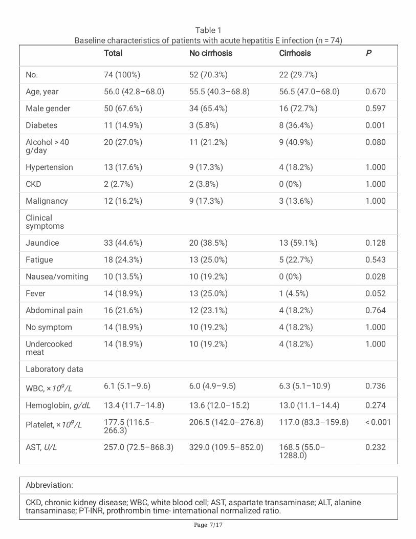
cirrhosis and 52 (70.3%) without cirrhosis were analyzed (Table 1). The proportion of diabetes was higherin patients with cirrhosis (36.4%) than in those without (5.8%, P = 0.001). However, there was nosigni�cant difference in age, sex, alcohol consumption, hypertension, chronic kidney disease, andmalignancy between the two groups.
Page 7/17
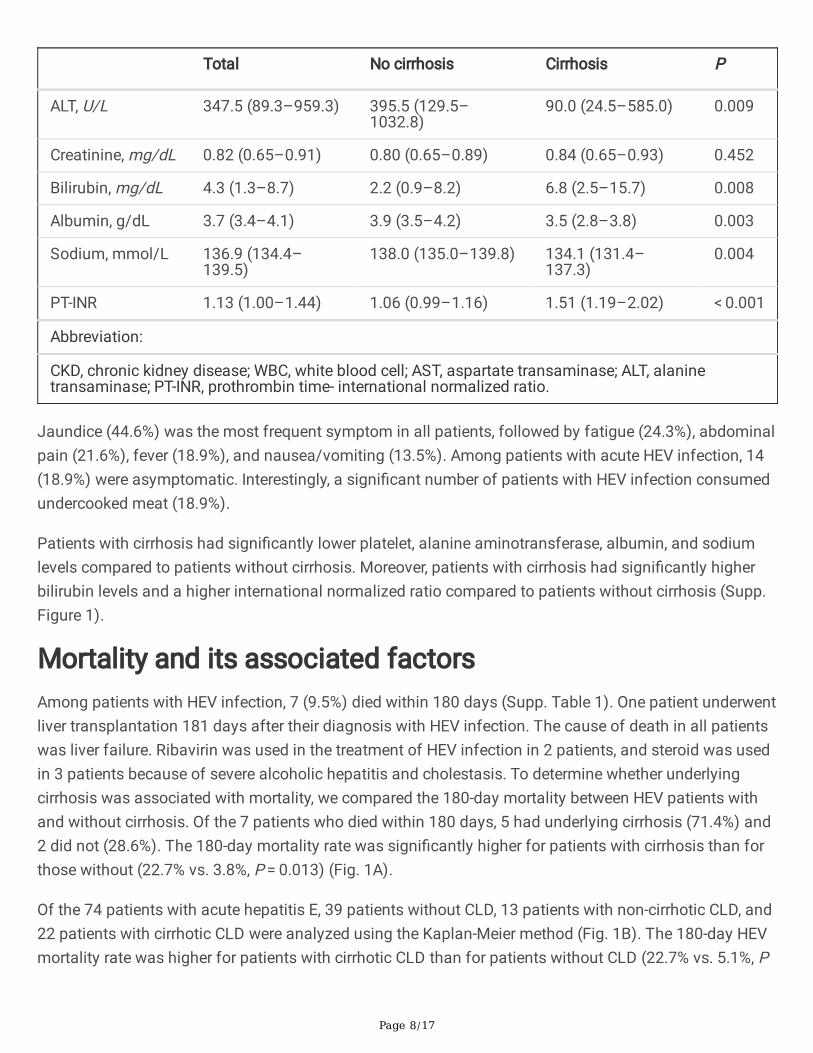
Table 1Baseline characteristics of patients with acute hepatitis E infection (n = 74)
Total No cirrhosis Cirrhosis P
No. 74 (100%) 52 (70.3%) 22 (29.7%)
Age, year 56.0 (42.8–68.0) 55.5 (40.3–68.8) 56.5 (47.0–68.0) 0.670
Male gender 50 (67.6%) 34 (65.4%) 16 (72.7%) 0.597
Diabetes 11 (14.9%) 3 (5.8%) 8 (36.4%) 0.001
Alcohol > 40g/day
20 (27.0%) 11 (21.2%) 9 (40.9%) 0.080
Hypertension 13 (17.6%) 9 (17.3%) 4 (18.2%) 1.000
CKD 2 (2.7%) 2 (3.8%) 0 (0%) 1.000
Malignancy 12 (16.2%) 9 (17.3%) 3 (13.6%) 1.000
Clinicalsymptoms
Jaundice 33 (44.6%) 20 (38.5%) 13 (59.1%) 0.128
Fatigue 18 (24.3%) 13 (25.0%) 5 (22.7%) 0.543
Nausea/vomiting 10 (13.5%) 10 (19.2%) 0 (0%) 0.028
Fever 14 (18.9%) 13 (25.0%) 1 (4.5%) 0.052
Abdominal pain 16 (21.6%) 12 (23.1%) 4 (18.2%) 0.764
No symptom 14 (18.9%) 10 (19.2%) 4 (18.2%) 1.000
Undercookedmeat
14 (18.9%) 10 (19.2%) 4 (18.2%) 1.000
Laboratory data
WBC, ×109/L 6.1 (5.1–9.6) 6.0 (4.9–9.5) 6.3 (5.1–10.9) 0.736
Hemoglobin, g/dL 13.4 (11.7–14.8) 13.6 (12.0–15.2) 13.0 (11.1–14.4) 0.274
Platelet, ×109/L 177.5 (116.5–266.3)
206.5 (142.0–276.8) 117.0 (83.3–159.8) < 0.001
AST, U/L 257.0 (72.5–868.3) 329.0 (109.5–852.0) 168.5 (55.0–1288.0)
0.232
Abbreviation:
CKD, chronic kidney disease; WBC, white blood cell; AST, aspartate transaminase; ALT, alaninetransaminase; PT-INR, prothrombin time- international normalized ratio.
Page 8/17
Total No cirrhosis Cirrhosis P
ALT, U/L 347.5 (89.3–959.3) 395.5 (129.5–1032.8)
90.0 (24.5–585.0) 0.009
Creatinine, mg/dL 0.82 (0.65–0.91) 0.80 (0.65–0.89) 0.84 (0.65–0.93) 0.452
Bilirubin, mg/dL 4.3 (1.3–8.7) 2.2 (0.9–8.2) 6.8 (2.5–15.7) 0.008
Albumin, g/dL 3.7 (3.4–4.1) 3.9 (3.5–4.2) 3.5 (2.8–3.8) 0.003
Sodium, mmol/L 136.9 (134.4–139.5)
138.0 (135.0–139.8) 134.1 (131.4–137.3)
0.004
PT-INR 1.13 (1.00–1.44) 1.06 (0.99–1.16) 1.51 (1.19–2.02) < 0.001
Abbreviation:
CKD, chronic kidney disease; WBC, white blood cell; AST, aspartate transaminase; ALT, alaninetransaminase; PT-INR, prothrombin time- international normalized ratio.
Jaundice (44.6%) was the most frequent symptom in all patients, followed by fatigue (24.3%), abdominalpain (21.6%), fever (18.9%), and nausea/vomiting (13.5%). Among patients with acute HEV infection, 14(18.9%) were asymptomatic. Interestingly, a signi�cant number of patients with HEV infection consumedundercooked meat (18.9%).
Patients with cirrhosis had signi�cantly lower platelet, alanine aminotransferase, albumin, and sodiumlevels compared to patients without cirrhosis. Moreover, patients with cirrhosis had signi�cantly higherbilirubin levels and a higher international normalized ratio compared to patients without cirrhosis (Supp.Figure 1).
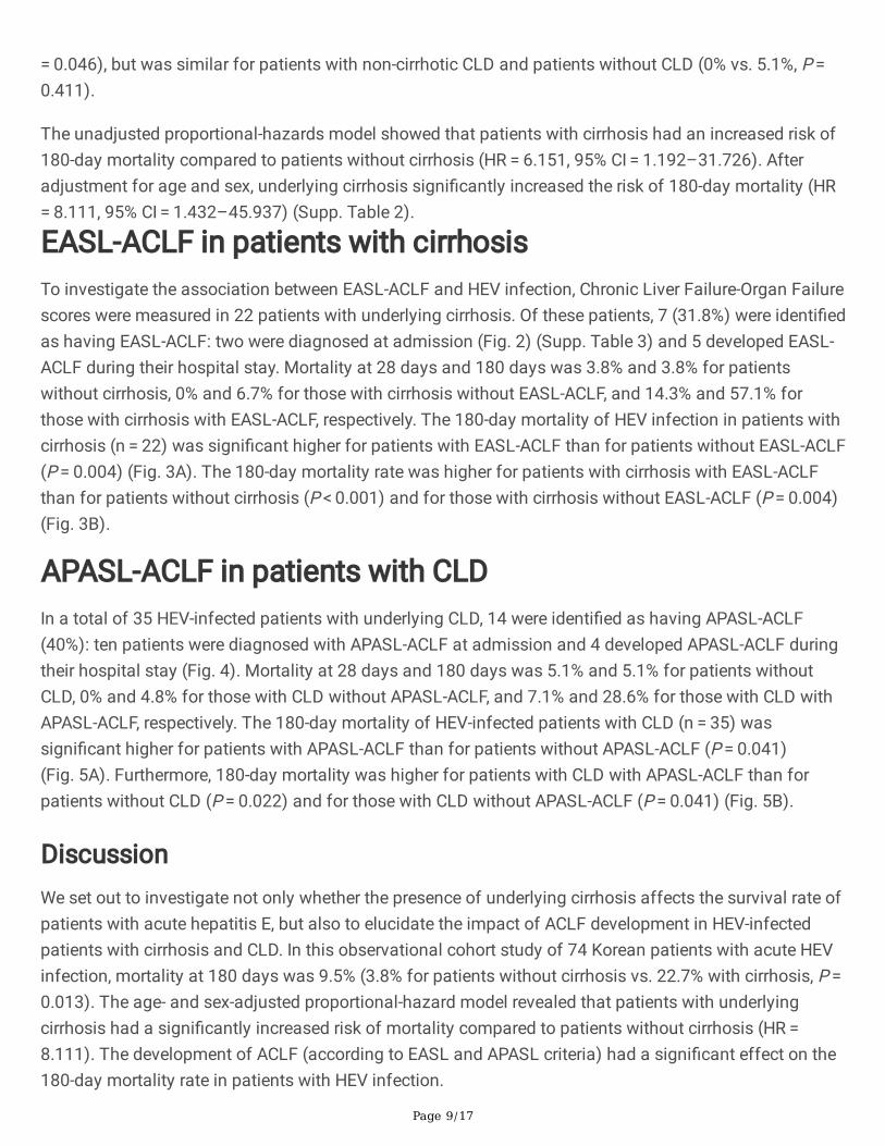
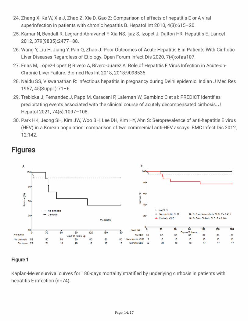
Mortality and its associated factorsAmong patients with HEV infection, 7 (9.5%) died within 180 days (Supp. Table 1). One patient underwentliver transplantation 181 days after their diagnosis with HEV infection. The cause of death in all patientswas liver failure. Ribavirin was used in the treatment of HEV infection in 2 patients, and steroid was usedin 3 patients because of severe alcoholic hepatitis and cholestasis. To determine whether underlyingcirrhosis was associated with mortality, we compared the 180-day mortality between HEV patients withand without cirrhosis. Of the 7 patients who died within 180 days, 5 had underlying cirrhosis (71.4%) and2 did not (28.6%). The 180-day mortality rate was signi�cantly higher for patients with cirrhosis than forthose without (22.7% vs. 3.8%, P = 0.013) (Fig. 1A).
Of the 74 patients with acute hepatitis E, 39 patients without CLD, 13 patients with non-cirrhotic CLD, and22 patients with cirrhotic CLD were analyzed using the Kaplan-Meier method (Fig. 1B). The 180-day HEVmortality rate was higher for patients with cirrhotic CLD than for patients without CLD (22.7% vs. 5.1%, P
Page 9/17
= 0.046), but was similar for patients with non-cirrhotic CLD and patients without CLD (0% vs. 5.1%, P = 0.411).
The unadjusted proportional-hazards model showed that patients with cirrhosis had an increased risk of180-day mortality compared to patients without cirrhosis (HR = 6.151, 95% CI = 1.192–31.726). Afteradjustment for age and sex, underlying cirrhosis signi�cantly increased the risk of 180-day mortality (HR = 8.111, 95% CI = 1.432–45.937) (Supp. Table 2).
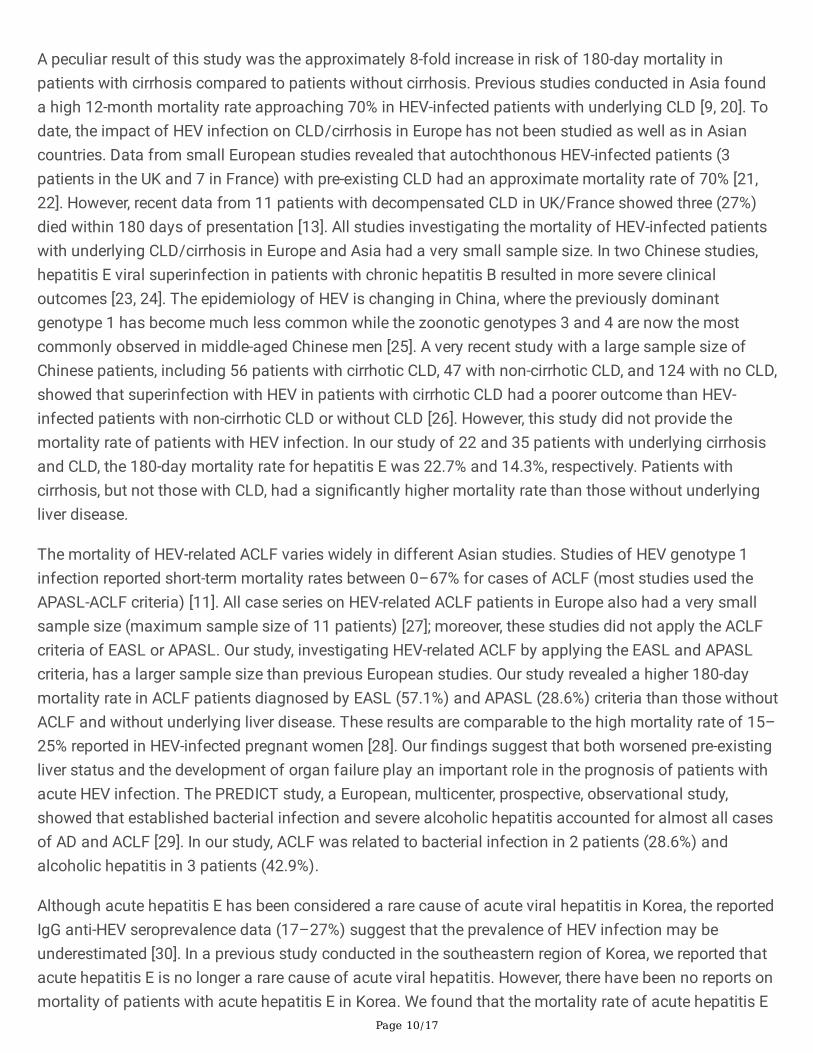
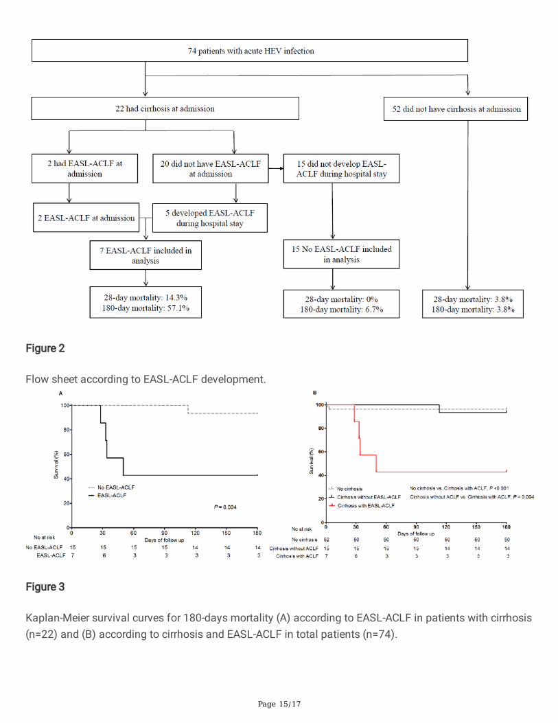
EASL-ACLF in patients with cirrhosisTo investigate the association between EASL-ACLF and HEV infection, Chronic Liver Failure-Organ Failurescores were measured in 22 patients with underlying cirrhosis. Of these patients, 7 (31.8%) were identi�edas having EASL-ACLF: two were diagnosed at admission (Fig. 2) (Supp. Table 3) and 5 developed EASL-ACLF during their hospital stay. Mortality at 28 days and 180 days was 3.8% and 3.8% for patientswithout cirrhosis, 0% and 6.7% for those with cirrhosis without EASL-ACLF, and 14.3% and 57.1% forthose with cirrhosis with EASL-ACLF, respectively. The 180-day mortality of HEV infection in patients withcirrhosis (n = 22) was signi�cant higher for patients with EASL-ACLF than for patients without EASL-ACLF(P = 0.004) (Fig. 3A). The 180-day mortality rate was higher for patients with cirrhosis with EASL-ACLFthan for patients without cirrhosis (P < 0.001) and for those with cirrhosis without EASL-ACLF (P = 0.004)(Fig. 3B).
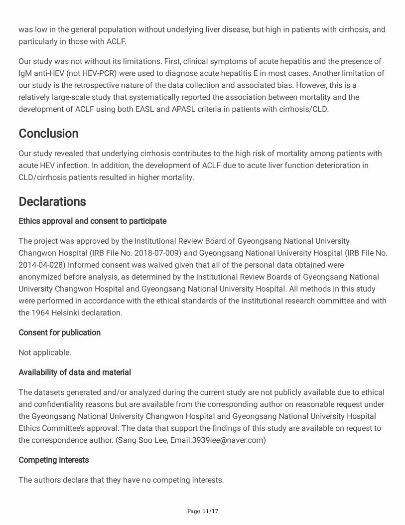
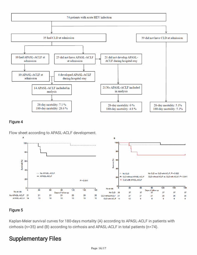
APASL-ACLF in patients with CLDIn a total of 35 HEV-infected patients with underlying CLD, 14 were identi�ed as having APASL-ACLF(40%): ten patients were diagnosed with APASL-ACLF at admission and 4 developed APASL-ACLF duringtheir hospital stay (Fig. 4). Mortality at 28 days and 180 days was 5.1% and 5.1% for patients withoutCLD, 0% and 4.8% for those with CLD without APASL-ACLF, and 7.1% and 28.6% for those with CLD withAPASL-ACLF, respectively. The 180-day mortality of HEV-infected patients with CLD (n = 35) wassigni�cant higher for patients with APASL-ACLF than for patients without APASL-ACLF (P = 0.041)(Fig. 5A). Furthermore, 180-day mortality was higher for patients with CLD with APASL-ACLF than forpatients without CLD (P = 0.022) and for those with CLD without APASL-ACLF (P = 0.041) (Fig. 5B).
DiscussionWe set out to investigate not only whether the presence of underlying cirrhosis affects the survival rate ofpatients with acute hepatitis E, but also to elucidate the impact of ACLF development in HEV-infectedpatients with cirrhosis and CLD. In this observational cohort study of 74 Korean patients with acute HEVinfection, mortality at 180 days was 9.5% (3.8% for patients without cirrhosis vs. 22.7% with cirrhosis, P = 0.013). The age- and sex-adjusted proportional-hazard model revealed that patients with underlyingcirrhosis had a signi�cantly increased risk of mortality compared to patients without cirrhosis (HR = 8.111). The development of ACLF (according to EASL and APASL criteria) had a signi�cant effect on the180-day mortality rate in patients with HEV infection.
Page 10/17
A peculiar result of this study was the approximately 8-fold increase in risk of 180-day mortality inpatients with cirrhosis compared to patients without cirrhosis. Previous studies conducted in Asia founda high 12-month mortality rate approaching 70% in HEV-infected patients with underlying CLD [9, 20]. Todate, the impact of HEV infection on CLD/cirrhosis in Europe has not been studied as well as in Asiancountries. Data from small European studies revealed that autochthonous HEV-infected patients (3patients in the UK and 7 in France) with pre-existing CLD had an approximate mortality rate of 70% [21,22]. However, recent data from 11 patients with decompensated CLD in UK/France showed three (27%)died within 180 days of presentation [13]. All studies investigating the mortality of HEV-infected patientswith underlying CLD/cirrhosis in Europe and Asia had a very small sample size. In two Chinese studies,hepatitis E viral superinfection in patients with chronic hepatitis B resulted in more severe clinicaloutcomes [23, 24]. The epidemiology of HEV is changing in China, where the previously dominantgenotype 1 has become much less common while the zoonotic genotypes 3 and 4 are now the mostcommonly observed in middle-aged Chinese men [25]. A very recent study with a large sample size ofChinese patients, including 56 patients with cirrhotic CLD, 47 with non-cirrhotic CLD, and 124 with no CLD,showed that superinfection with HEV in patients with cirrhotic CLD had a poorer outcome than HEV-infected patients with non-cirrhotic CLD or without CLD [26]. However, this study did not provide themortality rate of patients with HEV infection. In our study of 22 and 35 patients with underlying cirrhosisand CLD, the 180-day mortality rate for hepatitis E was 22.7% and 14.3%, respectively. Patients withcirrhosis, but not those with CLD, had a signi�cantly higher mortality rate than those without underlyingliver disease.
The mortality of HEV-related ACLF varies widely in different Asian studies. Studies of HEV genotype 1infection reported short-term mortality rates between 0–67% for cases of ACLF (most studies used theAPASL-ACLF criteria) [11]. All case series on HEV-related ACLF patients in Europe also had a very smallsample size (maximum sample size of 11 patients) [27]; moreover, these studies did not apply the ACLFcriteria of EASL or APASL. Our study, investigating HEV-related ACLF by applying the EASL and APASLcriteria, has a larger sample size than previous European studies. Our study revealed a higher 180-daymortality rate in ACLF patients diagnosed by EASL (57.1%) and APASL (28.6%) criteria than those withoutACLF and without underlying liver disease. These results are comparable to the high mortality rate of 15–25% reported in HEV-infected pregnant women [28]. Our �ndings suggest that both worsened pre-existingliver status and the development of organ failure play an important role in the prognosis of patients withacute HEV infection. The PREDICT study, a European, multicenter, prospective, observational study,showed that established bacterial infection and severe alcoholic hepatitis accounted for almost all casesof AD and ACLF [29]. In our study, ACLF was related to bacterial infection in 2 patients (28.6%) andalcoholic hepatitis in 3 patients (42.9%).
Although acute hepatitis E has been considered a rare cause of acute viral hepatitis in Korea, the reportedIgG anti-HEV seroprevalence data (17–27%) suggest that the prevalence of HEV infection may beunderestimated [30]. In a previous study conducted in the southeastern region of Korea, we reported thatacute hepatitis E is no longer a rare cause of acute viral hepatitis. However, there have been no reports onmortality of patients with acute hepatitis E in Korea. We found that the mortality rate of acute hepatitis E
Page 11/17
was low in the general population without underlying liver disease, but high in patients with cirrhosis, andparticularly in those with ACLF.
Our study was not without its limitations. First, clinical symptoms of acute hepatitis and the presence ofIgM anti-HEV (not HEV-PCR) were used to diagnose acute hepatitis E in most cases. Another limitation ofour study is the retrospective nature of the data collection and associated bias. However, this is arelatively large-scale study that systematically reported the association between mortality and thedevelopment of ACLF using both EASL and APASL criteria in patients with cirrhosis/CLD.
ConclusionOur study revealed that underlying cirrhosis contributes to the high risk of mortality among patients withacute HEV infection. In addition, the development of ACLF due to acute liver function deterioration inCLD/cirrhosis patients resulted in higher mortality.
DeclarationsEthics approval and consent to participate
The project was approved by the Institutional Review Board of Gyeongsang National UniversityChangwon Hospital (IRB File No. 2018-07-009) and Gyeongsang National University Hospital (IRB File No.2014-04-028) Informed consent was waived given that all of the personal data obtained wereanonymized before analysis, as determined by the Institutional Review Boards of Gyeongsang NationalUniversity Changwon Hospital and Gyeongsang National University Hospital. All methods in this studywere performed in accordance with the ethical standards of the institutional research committee and withthe 1964 Helsinki declaration.
Consent for publication
Not applicable.
Availability of data and material
The datasets generated and/or analyzed during the current study are not publicly available due to ethicaland con�dentiality reasons but are available from the corresponding author on reasonable request underthe Gyeongsang National University Changwon Hospital and Gyeongsang National University HospitalEthics Committee’s approval. The data that support the �ndings of this study are available on request tothe correspondence author. (Sang Soo Lee, Email:[email protected])
Competing interests
The authors declare that they have no competing interests.
Page 12/17
Funding
There was no �nancial support for this study.
Authors’ Contributions: Conception and design: JWC, HJS, and SSL
Data collection: HJ, HeeJK, RRC, JML, HyunJK, WTK, and OJL.
Data analysis and interpretation: JWC, HJS, and SSL
Manuscript writing: JWC, HJS, and SSL
Final approval of manuscript: All authors.
Acknowledgements
None.
Writing Assistance: We would like to thank Editage (www.editage.co.kr) for English language editing.There was no �nancial support for writing assistance.
References1. Rein DB, Stevens GA, Theaker J, Wittenborn JS, Wiersma ST: The global burden of hepatitis E virus
genotypes 1 and 2 in 2005. Hepatology 2012, 55(4):988–97.
2. Hewitt PE, Ijaz S, Brailsford SR, Brett R, Dicks S, Haywood B et al: Hepatitis E virus in bloodcomponents: a prevalence and transmission study in southeast England. Lancet 2014,384(9956):1766–73.
3. Bura M, Lagiedo M, Michalak M, Sikora J, Mozer-Lisewska I: Hepatitis E virus IgG seroprevalence inHIV patients and blood donors, west-central Poland. Int J Infect Dis 2017, 61:20–2.
4. European Association for the Study of the Liver. Electronic address eee, European Association for theStudy of the L: EASL Clinical Practice Guidelines on hepatitis E virus infection. J Hepatol 2018,68(6):1256–71.
5. Hoofnagle JH, Nelson KE, Purcell RH: Hepatitis E. N Engl J Med 2012, 367(13):1237–44.
�. Mansuy JM, Gallian P, Dimeglio C, Saune K, Arnaud C, Pelletier B et al: A nationwide survey ofhepatitis E viral infection in French blood donors. Hepatology 2016, 63(4):1145–54.
7. Jeong SH: Current status of hepatitis e virus infection in Korea. Gut Liver 2011, 5(4):427–31.
�. Oh HW, Cha RR, Lee SS, Lee CM, Kim WS, Jo YW et al: Comparing the Clinical Features andOutcomes of Acute Hepatitis E Viral Infections with Those of Acute Hepatitis A, B, and C Infections inKorea. Intervirology 2017, 60(3):109–17.
9. Kumar Acharya S, Kumar Sharma P, Singh R, Kumar Mohanty S, Madan K, Kumar Jha J, KumarPanda S: Hepatitis E virus (HEV) infection in patients with cirrhosis is associated with rapid
Page 13/17
decompensation and death. J Hepatol 2007, 46(3):387–94.
10. Hamid SS, Atiq M, Shehzad F, Yasmeen A, Nissa T, Salam A, Siddiqui A, Jafri W: Hepatitis E virussuperinfection in patients with chronic liver disease. Hepatology 2002, 36(2):474–8.
11. Kumar A, Saraswat VA: Hepatitis E and Acute-on-Chronic Liver Failure. J Clin Exp Hepatol 2013,3(3):225–30.
12. Sarin SK, Choudhury A, Sharma MK, Maiwall R, Al Mahtab M, Rahman S et al: Acute-on-chronic liverfailure: consensus recommendations of the Asian Paci�c association for the study of the liver(APASL): an update. Hepatol Int 2019, 13(4):353–90.
13. Blasco-Perrin H, Madden RG, Stanley A, Crossan C, Hunter JG, Vine L et al: Hepatitis E virus inpatients with decompensated chronic liver disease: a prospective UK/French study. AlimentPharmacol Ther 2015, 42(5):574–81.
14. Kim YM, Jeong SH, Kim JY, Song JC, Lee JH, Kim JW, Yun H, Kim JS: The �rst case of genotype 4hepatitis E related to wild boar in South Korea. J Clin Virol 2011, 50(3):253–6.
15. Yun H, Kim JS, Lee HJ, Jeong SH, Kim JS, Park SJ et al: The complete genome sequence andmolecular analysis of human hepatitis E virus genotype IV identi�ed from a Korean patient. ArchVirol 2010, 155(6):1003–8.
1�. Choi JY, Lee JM, Jo YW, Min HJ, Kim HJ, Jung WT, Lee OJ, Yun H, Yoon YS: Genotype-4 hepatitis E ina human after ingesting roe deer meat in South Korea. Clin Mol Hepatol 2013, 19(3):309–14.
17. Moreau R, Jalan R, Gines P, Pavesi M, Angeli P, Cordoba J et al: Acute-on-chronic liver failure is adistinct syndrome that develops in patients with acute decompensation of cirrhosis.Gastroenterology 2013, 144(7):1426–37, 37 e1-9.
1�. Jalan R, Saliba F, Pavesi M, Amoros A, Moreau R, Gines P et al: Development and validation of aprognostic score to predict mortality in patients with acute-on-chronic liver failure. J Hepatol 2014,61(5):1038–47.
19. Sarin SK, Choudhury A, Sharma MK, Maiwall R, Al Mahtab M, Rahman S et al: Correction to: Acute-on-chronic liver failure: consensus recommendations of the Asian Paci�c association for the studyof the liver (APASL): an update. Hepatol Int 2019, 13(6):826–8.
20. Ramachandran J, Eapen CE, Kang G, Abraham P, Hubert DD, Kurian G, Hephzibah J, Mukhopadhya A,Chandy GM: Hepatitis E superinfection produces severe decompensation in patients with chronicliver disease. J Gastroenterol Hepatol 2004, 19(2):134–8.
21. Dalton HR, Hazeldine S, Banks M, Ijaz S, Bendall R: Locally acquired hepatitis E in chronic liverdisease. Lancet 2007, 369(9569):1260.
22. Peron JM, Bureau C, Poirson H, Mansuy JM, Alric L, Selves J, Dupuis E, Izopet J, Vinel JP: Fulminantliver failure from acute autochthonous hepatitis E in France: description of seven patients with acutehepatitis E and encephalopathy. J Viral Hepat 2007, 14(5):298–303.
23. Cheng SH, Mai L, Zhu FQ, Pan XF, Sun HX, Cao H et al: In�uence of chronic HBV infection onsuperimposed acute hepatitis E. World J Gastroenterol 2013, 19(35):5904–9.
Page 14/17
24. Zhang X, Ke W, Xie J, Zhao Z, Xie D, Gao Z: Comparison of effects of hepatitis E or A viralsuperinfection in patients with chronic hepatitis B. Hepatol Int 2010, 4(3):615–20.
25. Kamar N, Bendall R, Legrand-Abravanel F, Xia NS, Ijaz S, Izopet J, Dalton HR: Hepatitis E. Lancet2012, 379(9835):2477–88.
2�. Wang Y, Liu H, Jiang Y, Pan Q, Zhao J: Poor Outcomes of Acute Hepatitis E in Patients With CirrhoticLiver Diseases Regardless of Etiology. Open Forum Infect Dis 2020, 7(4):ofaa107.
27. Frias M, Lopez-Lopez P, Rivero A, Rivero-Juarez A: Role of Hepatitis E Virus Infection in Acute-on-Chronic Liver Failure. Biomed Res Int 2018, 2018:9098535.
2�. Naidu SS, Viswanathan R: Infectious hepatitis in pregnancy during Delhi epidemic. Indian J Med Res1957, 45(Suppl.):71–6.
29. Trebicka J, Fernandez J, Papp M, Caraceni P, Laleman W, Gambino C et al: PREDICT identi�esprecipitating events associated with the clinical course of acutely decompensated cirrhosis. JHepatol 2021, 74(5):1097–108.
30. Park HK, Jeong SH, Kim JW, Woo BH, Lee DH, Kim HY, Ahn S: Seroprevalence of anti-hepatitis E virus(HEV) in a Korean population: comparison of two commercial anti-HEV assays. BMC Infect Dis 2012,12:142.
Figures
Figure 1
Kaplan-Meier survival curves for 180-days mortality strati�ed by underlying cirrhosis in patients withhepatitis E infection (n=74).
Page 15/17
Figure 2
Flow sheet according to EASL-ACLF development.
Figure 3
Kaplan-Meier survival curves for 180-days mortality (A) according to EASL-ACLF in patients with cirrhosis(n=22) and (B) according to cirrhosis and EASL-ACLF in total patients (n=74).
Page 16/17
Figure 4
Flow sheet according to APASL-ACLF development.
Figure 5
Kaplan-Meier survival curves for 180-days mortality (A) according to APASL-ACLF in patients withcirrhosis (n=35) and (B) according to cirrhosis and APASL-ACLF in total patients (n=74).
Supplementary Files

Page 17/17
This is a list of supplementary �les associated with this preprint. Click to download.
SuppFig1..tif
Supp.Table.docx