Activation of the rostromedial prefrontal cortex during the experience of positive emotion in the...
7
HUMAN NEUROSCIENCE ORIGINAL RESEARCH ARTICLE published: 20 December 2013 doi: 10.3389/fnhum.2013.00879 Activation of the rostromedial prefrontal cortex during the experience of positive emotion in the context of esthetic experience. An fNIRS study Ute Kreplin* and Stephen H. Fairclough School of Natural Science and Psychology, Liverpool John Moores University, Liverpool, UK Edited by: Nobuo Masataka, Kyoto University, Japan Reviewed by: Leonid Perlovsky, Harvard University and Air Force Research Laboratory, USA Hasan Ayaz, Drexel University, USA *Correspondence: Ute Kreplin, School of Natural Science and Psychology, Liverpool John Moores University, Tom Reilly Building, Byrom Street, Liverpool, L3 3AF, UK e-mail: [email protected] The contemplation of visual art requires attention to be directed to external stimulus properties and internally generated thoughts. It has been proposed that the medial rostral prefrontal cortex (rPFC; BA10) plays a role in the maintenance of attention on external stimuli whereas the lateral area of the rPFC is associated with the preservation of attention on internal cognitions. An alternative hypothesis associates activation of medial rPFC with internal cognitions related to the self during emotion regulation. The aim of the current study was to differentiate activation within rPFC using functional near infrared spectroscopy (fNIRS) during the viewing of visual art selected to induce positive and negative valence, which were viewed under two conditions: (1) emotional introspection and (2) external object identification. Thirty participants (15 female) were recruited. Sixteen pre-rated images that represented either positive or negative valence were selected from an existing database of visual art. In one condition, participants were directed to engage in emotional introspection during picture viewing. The second condition involved a spot-the-difference task where participants compared two almost identical images, a viewing strategy that directed attention to external properties of the stimuli. The analysis revealed a significant increase of oxygenated blood in the medial rPFC during viewing of positive images compared to negative images. This finding suggests that the rPFC is involved during positive evaluations of visual art that may be related to judgment of pleasantness or attraction. The fNIRS data revealed no significant main effect between the two viewing conditions, which seemed to indicate that the emotional impact of the stimuli remained unaffected by the two viewing conditions. Keywords: fNIRS, BA10, emotion, esthetics, prefrontal cortex INTRODUCTION The experience of viewing art is influenced by a modulation of attentional focus between external features of the stimuli and internal feelings/thoughts. Internal cognitive processes such as object recognition, memory recall and mental imagery facilitate content recognition during the viewing of visual art (Fairhall and Ishai, 2008). Recognition of familiar content evokes a pattern of activation in multiple extrastriate ventral and dorsal regions, the hippocampus, intra parietal sulcus and inferior frontal gyrus (Ishai et al., 2007; Fairhall and Ishai, 2008; Nadal et al., 2008; Nadal and Pearce, 2011). According to a MEG time frequency analysis, a peak of acitivity around 170 ms has been related to the commencement of coding for object identity and transformation of sensory code to cognitive processing has been associated with a peak of activity at approx. 170 ms during the observation of visual art (Munar et al., 2011). This process of feature extraction from visual art and the generation of associated thought and feelings have a distinct temporal window. The rostral prefrontal cortex (rPFC) may be an important site of activity during the processing of art; this region has been associated with higher order cognitive processes such as prospective memory (Volle et al., 2010; McDaniel et al., 2013), emotional regulation strategies (Amting et al., 2010; Viviani et al., 2010; Campbell-Sills et al., 2011) and sustained attention (Van Veen and Carter, 2006; Ernst et al., 2012). However, there is little consensus regarding the functional specificity and cytoar- chitecture of the prefrontal cortex (PFC), particularly the rostral area of the PFC. Ramnani and Owen (2004) suggested that the rPFC is activated when the outcomes of two or more separate cognitive operations require integration in the pursuit of a higher behavioral goal. Other accounts emphasized the involvement of medial rPFC during the processing of self-related information (Seitz et al., 2009; Denny et al., 2012). The gateway hypothesis (Burgess et al., 2003, 2007) was devel- oped to connect activation in the rPFC to higher-order self- referential processing and the evaluation of internally generated information. According to this model, rostromedial areas of the PFC (medial BA10) are implicated in the maintenance of atten- tion towards external stimuli whereas activation of the rostrolat- eral areas (lateral BA10) are associated with the preservation of Frontiers in Human Neuroscience www.frontiersin.org December 2013 | Volume 7 | Article 879 | 1
Transcript of Activation of the rostromedial prefrontal cortex during the experience of positive emotion in the...

HUMAN NEUROSCIENCEORIGINAL RESEARCH ARTICLE
published: 20 December 2013doi: 10.3389/fnhum.2013.00879
Activation of the rostromedial prefrontal cortex during theexperience of positive emotion in the context of estheticexperience. An fNIRS studyUte Kreplin* and Stephen H. Fairclough
School of Natural Science and Psychology, Liverpool John Moores University, Liverpool, UK
Edited by:
Nobuo Masataka, Kyoto University,Japan
Reviewed by:
Leonid Perlovsky, Harvard Universityand Air Force Research Laboratory,USAHasan Ayaz, Drexel University, USA
*Correspondence:
Ute Kreplin, School of Natural Scienceand Psychology, Liverpool JohnMoores University, Tom ReillyBuilding, Byrom Street, Liverpool, L33AF, UKe-mail: [email protected]
The contemplation of visual art requires attention to be directed to external stimulusproperties and internally generated thoughts. It has been proposed that the medial rostralprefrontal cortex (rPFC; BA10) plays a role in the maintenance of attention on externalstimuli whereas the lateral area of the rPFC is associated with the preservation of attentionon internal cognitions. An alternative hypothesis associates activation of medial rPFCwith internal cognitions related to the self during emotion regulation. The aim of thecurrent study was to differentiate activation within rPFC using functional near infraredspectroscopy (fNIRS) during the viewing of visual art selected to induce positive andnegative valence, which were viewed under two conditions: (1) emotional introspectionand (2) external object identification. Thirty participants (15 female) were recruited. Sixteenpre-rated images that represented either positive or negative valence were selectedfrom an existing database of visual art. In one condition, participants were directed toengage in emotional introspection during picture viewing. The second condition involveda spot-the-difference task where participants compared two almost identical images, aviewing strategy that directed attention to external properties of the stimuli. The analysisrevealed a significant increase of oxygenated blood in the medial rPFC during viewingof positive images compared to negative images. This finding suggests that the rPFCis involved during positive evaluations of visual art that may be related to judgment ofpleasantness or attraction. The fNIRS data revealed no significant main effect betweenthe two viewing conditions, which seemed to indicate that the emotional impact of thestimuli remained unaffected by the two viewing conditions.
Keywords: fNIRS, BA10, emotion, esthetics, prefrontal cortex
INTRODUCTIONThe experience of viewing art is influenced by a modulation ofattentional focus between external features of the stimuli andinternal feelings/thoughts. Internal cognitive processes such asobject recognition, memory recall and mental imagery facilitatecontent recognition during the viewing of visual art (Fairhall andIshai, 2008). Recognition of familiar content evokes a patternof activation in multiple extrastriate ventral and dorsal regions,the hippocampus, intra parietal sulcus and inferior frontal gyrus(Ishai et al., 2007; Fairhall and Ishai, 2008; Nadal et al., 2008;Nadal and Pearce, 2011). According to a MEG time frequencyanalysis, a peak of acitivity around 170 ms has been related to thecommencement of coding for object identity and transformationof sensory code to cognitive processing has been associated with apeak of activity at approx. 170 ms during the observation of visualart (Munar et al., 2011). This process of feature extraction fromvisual art and the generation of associated thought and feelingshave a distinct temporal window.
The rostral prefrontal cortex (rPFC) may be an importantsite of activity during the processing of art; this region has
been associated with higher order cognitive processes such asprospective memory (Volle et al., 2010; McDaniel et al., 2013),emotional regulation strategies (Amting et al., 2010; Viviani et al.,2010; Campbell-Sills et al., 2011) and sustained attention (VanVeen and Carter, 2006; Ernst et al., 2012). However, there islittle consensus regarding the functional specificity and cytoar-chitecture of the prefrontal cortex (PFC), particularly the rostralarea of the PFC. Ramnani and Owen (2004) suggested that therPFC is activated when the outcomes of two or more separatecognitive operations require integration in the pursuit of a higherbehavioral goal. Other accounts emphasized the involvement ofmedial rPFC during the processing of self-related information(Seitz et al., 2009; Denny et al., 2012).
The gateway hypothesis (Burgess et al., 2003, 2007) was devel-oped to connect activation in the rPFC to higher-order self-referential processing and the evaluation of internally generatedinformation. According to this model, rostromedial areas of thePFC (medial BA10) are implicated in the maintenance of atten-tion towards external stimuli whereas activation of the rostrolat-eral areas (lateral BA10) are associated with the preservation of
Frontiers in Human Neuroscience www.frontiersin.org December 2013 | Volume 7 | Article 879 | 1

Kreplin and Fairclough Emotion, esthetics and the rPFC
attention on internal cognitions; this functional differentiation isproposed to act as a gateway between the direction of attentiontowards external and internal stimuli (Burgess et al., 2007). Thecentral proposal of the model is that the rPFC is part of a systemthat allows conflicts to be resolved during ambiguous situations(where information activating relevant schemata has low trig-gering input) or increases activation of schemata in accordancewith higher-level goal representations (Cupchik et al., 2009; Volleet al., 2010; Henseler et al., 2011). Evidence for this functional dif-ferentiation has been observed during the comparison of shapes(Henseler et al., 2011), letters (Gilbert et al., 2005; Benoit et al.,2012) and the identification of features between two differentstimuli such as texture or aspects of geometric shapes (Volle et al.,2010).
The general function of the rPFC as described by the gate-way hypothesis is twofold. This area enables the activation ofschemata in a situation where no schema is sufficiently triggeredby incoming stimuli, e.g., if the stimulus is entirely novel and can-not be associated with existing information. Secondly, the rPFCenables attentional bias when many schema are simultaneouslyactivated (e.g., if a situation is very difficult or complex) or ifthere are a multitude of possible established outcomes withoutan obvious advantage to one of them (Burgess et al., 2005).The rPFC plays a key role in the goal-directed co-ordinationof stimulus-independent and stimulus-orientated cognitions insituations where established patterns of behavior are insuffi-cient. Stimulus-independent cognitions include introspection orcreative thoughts which are neither provoked by nor directedtoward external stimuli. Stimulus-oriented cognitions representthe opposite category, being provoked and oriented towards sen-sory input. The rPFC would be typically activated in situationsthat are novel or where a specific demand for it has been deter-mined (e.g., “I must pay special attention to. . .”, “I must thinkabout. . .”; Burgess et al., 2003, 2005, 2007; Gilbert et al., 2005;Volle et al., 2010; Benoit et al., 2012). The contemplation ofvisual art requires a shift from stimulus-dependent processingto those stimulus-independent processes that permits an assess-ment of stimuli as being esthetically pleasing or not. It maybe hypothesized that attention is directed to external propertiesof the stimulus (identification of physical properties within thepainting), the reinvestment of attention onto internally generatedthoughts (what does the artist want to say with this painting),and the reinvestment of attention onto subjective self or personalentity (what does the painting mean to me). It is plausible thatthe rPFC is involved in object identification as well as directingattention to those self-referential states that are relevant to estheticappreciation, but the roles of medial and lateral areas of the rPFCduring the contemplation of visual art as defined by the gatewayhypothesis remains unclear.
There is evidence connecting activity in the rPFC to aspectsof cognition that are implicit within emotional processing, e.g.,attention to emotion, emotion regulation, appraisal or interpreta-tion of emotion. For instance, Phan et al. (2002) reported strongconnections between the PFC (BA9/10) and the anterior cingulatecortex (ACC) and suggested that both areas of the PFC couldserve as top-down modulators of intense emotional responses (seealso Amting et al., 2010; Holroyd and Yeung, 2012). Evidence
for this interpretation stems from human lesion studies wheredamage to the PFC leads to socially inappropriate expressionsof emotions and impairment in making advantageous personallyrelevant decisions suggesting a lack of awareness/comprehensionof emotionally “loaded” situations (Damasio, 1999; Leopold et al.,2012; Maier and di Pellegrino, 2012). Similarly, activation of therPFC was related in a linear fashion with an emotion inductiontask that required different degrees of self-monitoring (identifyingwith the feelings/emotions depicted in a picture compared to justviewing a picture) (Herrmann et al., 2003; see Denny et al., 2012for a review).
Studies relating activity in the rPFC to the process of emotionalregulation present an alternative to the gateway hypothesis. Theregulation of emotions has been associated with activation of therPFC during the up- and down regulation of emotions (Mitchell,2011). Furthermore medial BA10 has been linked to activityrelated to the subjective self or personal entity (Amting et al.,2010; Denny et al., 2012). Seitz et al. (2009) suggested that nodesin the medial PFC participate in early processing of sensoryinformation and mediate the value judgment of the stimulusby assessing self-relevant meaning to the sensations. An estheticexperience, or more specifically the contemplation of visual art,is a highly subjective process. It is therefore important that theexperiences are self-referential, particularly if the viewer is nottrained in an art-related subject and bases his/her evaluation ofvisual art largely on personal experiences. Evidence supportingthis interpretation and the involvement of the medial rPFC wasprovided by Vessel et al. (2012) who reported an increase of acti-vation in the medial rPFC for those paintings subjectively judgedas most esthetically moving. This activation of the medial rPFCwas specific to the assessment of esthetic pleasure. This studysuggested that a highly-subjective emotional connection to visualart is important to the esthetic experiences and activation in therPFC was associated with this type of experience. Alternatively,the involvement of the lateral rPFC during esthetic experienceswas reported by Cupchik et al. (2009) who investigated howcognitive control/perceptual facilitation and the experience ofemotion contributed to esthetic perception. Participants in thisstudy were instructed to view a painting from either a pragmaticeveryday viewpoint or from an esthetic viewpoint. Pragmaticviewing was associated with activation of the fusiform gyrus andareas related to object recognition whereas the esthetic view-ing condition activated the insula and left lateral rPFC (BA10).The authors interpreted activation of the latter in terms ofstimulus-independent thought related to the contemplation ofvisual art.
The aim of the current study was to differentiate acti-vation within the rPFC using functional near infrared spec-troscopy (fNIRS) during the viewing of visual art selected toinduce positive and negative mood. Both categories of imagewere viewed under two conditions (emotional introspection andexternal object identification) designed to draw attention tostimulus-independent and stimulus-dependent features respec-tively. Images were viewed for 60 s overall and split into threeviewing periods (early, middle and late) for the analysis. Theextended viewing time was chosen because temporal differ-ences have been observed during activation of the PFC during
Frontiers in Human Neuroscience www.frontiersin.org December 2013 | Volume 7 | Article 879 | 2
Kreplin and Fairclough Emotion, esthetics and the rPFC
emotional experiences measured by fNIRS, where an overshootof activation into later periods was reported (León-Carrion et al.,2008), and during the experience of art (Munar et al., 2011).Our primary hypothesis was that emotional introspection wouldactivate lateral areas of the rPFC whereas viewing the image withan emphasis on external object identification would selectivelyactivate the medial rPFC. Our alternative hypothesis was thatemotional introspection would activate the medial rPFC as pre-dicted by the studies conducted by Seitz et al. (2009) and Vesselet al. (2012).
METHODSTHE SURVEYStandardized visual materials, such as the International AffectivePicture System (IAPS; Lang et al., 1993) are often used to provokeemotional experience under controlled conditions. The purposeof the survey exercise was to create a database of esthetic imageswith known psychological properties for experimental purposes.Sixty-three paintings including both representational and abstractimages were selected from online sources; all artists had sold orexhibited their work and agreed to allow their work to be usedin the survey exercise. Participants were asked to rate each imageon six components represented by 9-point Likert scales. Threecomponents represented cognitive components, these scales werelabeled: complexity (how complex was this image?), comprehen-sion (do you understand this image?) and novelty (how novel orunusual was the image?). The remaining three components repre-sented emotional factors including: valence (is the image positiveor negative?), activation (do you find the image stimulating) andattractiveness (do you find the image attractive or repellent?). Thecognitive components were derived from Silvia’s (2008) analysis ofinterest whilst emotional components were based upon the Self-Assessment Mannikin (Lang et al., 1993).
The survey was made available online and 1043 participants(63% female) with a mean age of 33 years. (s.d. 14.24) providedratings for the images. The resulting database was used to selectimages rated as positive/negative valence for the experimentalstudy.
EXPERIMENTAL STUDYParticipantsThirty right handed participants (15 female) were recruitedfrom an undergraduate population. Participants had a mean age22 years. (s.d. = 3.26 years) with no formal training in an art-related subject and no history of neurological disorder. Partici-pants were informed about the procedure and operating mode ofthe fNIRS prior to providing written consent. All procedures wereapproved by the University Research Ethics Committee prior todata collection.
Experimental taskThe stimuli were projected onto a white wall in front of theparticipants using E-prime 2.0 (PST Inc.); the image dimensionswere 2040 × 786 pixels. The viewing distance to the screen wasapprox. 1.90 m. Each stimulus was presented for 60 s and precededby a 60 s baseline consisting of a light grey screen with a fixation *.Following image presentation, participants were asked to provide
ratings for valence and complexity on the scales used in the sur-vey; ratings were recorded in e-prime via a keypad. The tasks weredesigned to induce different demands on attending to externalproperties of the stimulus (the spot-the-difference (SD) task) andto internal cognitions (the emotional introspection (EI) task).
We closely considered the prerequisites stated by Burgess et al.(2005) that a task requires to draw attention to external stimulusprocessing when designing the SD task. According to Burgesset al. (2005) an external attentional task requires: (1) that theinformation to be processed is currently available (i.e., presentin the sensory environment); (2) that the attention is directed toexternal stimuli or stimulus features; and (3) that the operationsinvolved prior to responding are relatively automatic or well-learnt. To elicit attending to external stimuli features during theSD task, all images were duplicated to form a pair and betweenthree and six aspects of the image on the right were modifiedusing PaintShopPro (Figure 1A). Instructions in the SD conditionstated: “If you see “SD” on the screen you will be asked to spotthe differences between two images. You will be able to see theanswers after the image”. Following the ratings in the SD taskparticipants were provided with the same picture but with thedifferences highlighted with a red circle. All images were used inthe SD task and the emotion induction task. Participants wereprompted with SD or E before the image appeared as indicatorwhich task to perform next during testing.
The EI condition was designed to initiate internal processeswith respect to: (1) the information attended to is being processedinternally; (2) that this information is self-generated or comesfrom a previously witnessed episode; and (3) that the response tobe made are triggered by these internal representations. Instruc-tions were as follows: “If you see an “E” on the screen youare asked to think about how the artwork makes you feel, i.e.,what emotions does it trigger in you? Does it make you feelsad/happy/angry etc.? Does it remind you of an emotional eventyou have experienced in the past? Don’t worry about the messagethe artist tried to bring across, just think about how the imagemakes YOU feel or if it makes you think of something that youfound emotional”. Participants were asked to write, using pen andpaper, brief notes about these associations following the picturepresentation. Participants were not asked to hand these notes tothe experimenter and took them away upon completion of testingto assure confidentiality. The images were presented as identicalpairs during the EI task to ensure consistency with the SD viewingcondition (Figure 1B). A practice trial was completed for eachcondition, and the opportunity to ask questions was given toinsure the instructions were fully understood before the start ofthe experiment. The presentation of positive/negative images andEI/SD was randomized.
Stimuli. Sixteen images (8 in a positive valence category and8 in a negative valence category) were selected according toratings of valence and complexity obtained during the survey(see section The Survey). Mean ratings for positive images were2.57 (+/− 1.27) and negative images 7.17 (+/− 1.3), whilstsubjective ratings of image complexity were constant betweenpositive and negative images (mean 4.58 +/− 2.22 and 4.07+/− 2.14 respectively).
Frontiers in Human Neuroscience www.frontiersin.org December 2013 | Volume 7 | Article 879 | 3

Kreplin and Fairclough Emotion, esthetics and the rPFC
FIGURE 1 | (A) Pictures used in the positive SD task (“BestAbstract” © Adrian Borda). The right image has slight differences tothe left image, differences are circled in red. (B) Two identicalimages used during negative EI (Monika Weiss “Elytron” 2003,
self-shot photography, performance, installation, sculpture and video.Courtesy the artist and Chelsea Art Museum, New York).Participants were asked to think about how the images made themfeel.
ProcedureParticipants were informed about the nature of the study uponarrival and provided written consent before the fNIRS device wasfitted. Participants were informed about the study tasks throughwritten instructions projected onto the wall in front of them. Par-ticipants had the opportunity to ask questions before and duringa practice trial for each task that was shown after the instructions.The experimental protocol began after the experimenter wassatisfied that the participant understood the experimental tasks.Participants were thanked for their participation following theexperiment and compensated with a £10 voucher for their time.
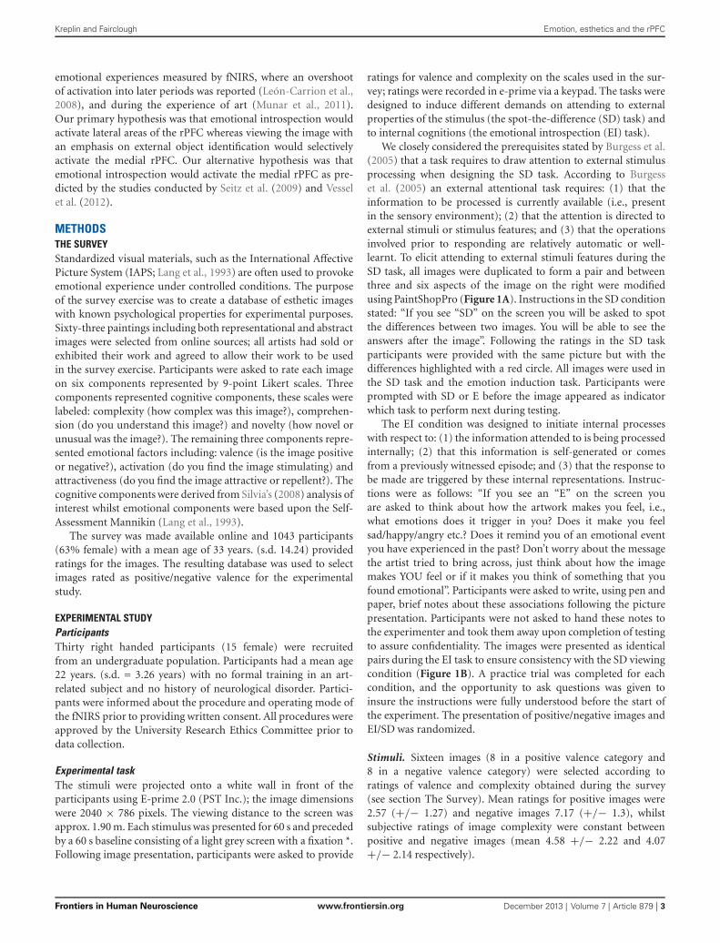
fNIRS data collectionfNIRS was recorded using fNIR Imager1000 and COBI datacollection suit (Biopac System Inc.) and is described in detailelsewhere (Ayaz et al., 2010). The system has a temporal resolutionof about 500 ms for one complete data acquisition cycle (about2 Hz). The 16 channel probe was placed on the forehead alignedto Fp1 and Fp2 of the international 10–20 system, and rotated sothat Fpz corresponded to the midpoint of the probe (Ayaz et al.,2006; Figure 2). Areas underlying the 16 voxels are right and leftsuperior and inferior frontal gyrii (BA10 and BA46). Cognitiveand emotional functions associated with BA10 and BA46 havebeen explored using fNIRS application similar to the device used
in this work (Plichta et al., 2006; Izzetoglu et al., 2007; León-Carrion et al., 2008).
fNIRS data were analyzed offline using fNIRS-Soft (Ayaz et al.,2010). Raw data was subjected to a Sliding-window Motion Arte-fact Rejection (SMAR) algorithm to remove motion artefacts andsaturated channels (Ayaz et al., 2010). Oxygenated haemoglobin(HbO) and deoxygenated haemoglobin (HHB) were calculatedusing the modified Beer-Lambert Law. A finite impulse responselinear phase low-pass filter, with order 20 and cut-off frequency of0.1 Hz was applied to attenuate high frequency noise, respirationand cardiac effects (Izzetoglu et al., 2007; Ayaz et al., 2010).Sixteen segments with durations of 60 s were extracted usingsynchronization markers. Segments were averaged according tocondition (Positive EI, Negative EI, Positive SD, Negative SD).
There is currently no strong consensus in the literature regard-ing the optimal feature of brain activation that can be derivedfrom fNIRS data. Research investigating emotional processesusing fNIRS has reported significant changes in the PFC for HbOalone (León-Carrion et al., 2008), for HHb alone (Ernst et al.,2012) or for both HbO and HHb (Glotzbach et al., 2011). Ithas been argued that HHb is sensitive to local haemodynamicchanges, less prone to influences from psychophysiological noise,such as breathing or heart rate and has a close association withthe blood oxygenation dependent (BOLD) signal obtained from
Frontiers in Human Neuroscience www.frontiersin.org December 2013 | Volume 7 | Article 879 | 4
Kreplin and Fairclough Emotion, esthetics and the rPFC
FIGURE 2 | The 16 voxels of the fNIRS probe located over the rPFC.
fMRI. However, HbO is the parameter which is less sensitive tovariation in probe placement due to head size and shape becauseHbO activation is more global compared to HHb activation(Wobst et al., 2001; Hoshi, 2005; Plichta et al., 2006). We decidedto calculate a compound score for oxygenation (Oxy = HbO –HHb) in order to capture both measures whilst controlling forchanges in blood volume (Ayaz et al., 2010).
RESULTSIndividual differences in the subjective evaluation of visual arthave been highlighted as a problem in previous studies (e.g.,Cupchik et al., 2009; Vessel et al., 2012). Our participantswere exposed to four groups of images (EI/positive, EI/negative,SD/positive, SD/negative), each of which contained four indi-vidual images. In order to control for individual differences, weselected only those three images from the sample of four thatwere most representative of their group designation in order tocreate the best representation of that image category for eachparticipant, i.e., the three images in the EI/negative valence groupwith maximum scores for valence (rated on a 9-point Likert scale;1 = positive, 9 = negative) were assumed to represent the bestexample of EI/negative valence for that particular person.
We conducted a manipulation check of the subjective valenceratings to assess differences between positive and negative imagesunder the two viewing conditions. A 2 (condition) × 2 (valence)analysis of variance (ANOVA) was conducted on subjective rat-ings from three images in each valence/viewing condition. A sig-nificant main effect for valence (F(1,29) = 720.50, p < 0.01,η2 = 0.96), and an interaction between condition and valence(F(1,29) = 5.06, p < 0.03, η2 = .014) was found. Negative images(mean 7.17, +/− 1.07) were rated as significantly more negativethan positive images (mean 2.57, +/− 0.85). Post-hoc analysis
showed no significant difference between the EI and SD conditionfor negative images. For positive images a marginal trend wasidentified with images in the EI (mean 2.57, +/− 0.85) conditionbeing rated as more positively than those viewed in the SDcondition (mean 2.98, +/− 0.78, p < .06).
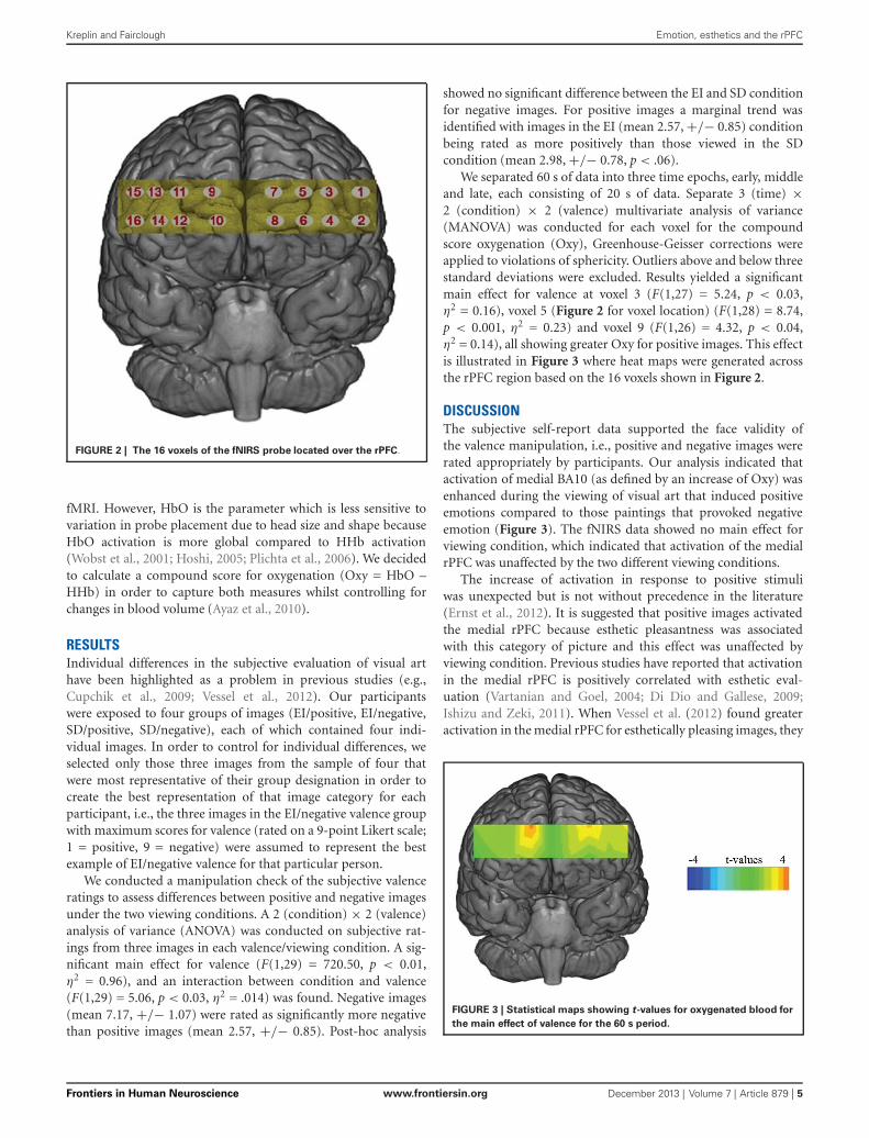
We separated 60 s of data into three time epochs, early, middleand late, each consisting of 20 s of data. Separate 3 (time) ×2 (condition) × 2 (valence) multivariate analysis of variance(MANOVA) was conducted for each voxel for the compoundscore oxygenation (Oxy), Greenhouse-Geisser corrections wereapplied to violations of sphericity. Outliers above and below threestandard deviations were excluded. Results yielded a significantmain effect for valence at voxel 3 (F(1,27) = 5.24, p < 0.03,η2 = 0.16), voxel 5 (Figure 2 for voxel location) (F(1,28) = 8.74,p < 0.001, η2 = 0.23) and voxel 9 (F(1,26) = 4.32, p < 0.04,η2 = 0.14), all showing greater Oxy for positive images. This effectis illustrated in Figure 3 where heat maps were generated acrossthe rPFC region based on the 16 voxels shown in Figure 2.
DISCUSSIONThe subjective self-report data supported the face validity ofthe valence manipulation, i.e., positive and negative images wererated appropriately by participants. Our analysis indicated thatactivation of medial BA10 (as defined by an increase of Oxy) wasenhanced during the viewing of visual art that induced positiveemotions compared to those paintings that provoked negativeemotion (Figure 3). The fNIRS data showed no main effect forviewing condition, which indicated that activation of the medialrPFC was unaffected by the two different viewing conditions.
The increase of activation in response to positive stimuliwas unexpected but is not without precedence in the literature(Ernst et al., 2012). It is suggested that positive images activatedthe medial rPFC because esthetic pleasantness was associatedwith this category of picture and this effect was unaffected byviewing condition. Previous studies have reported that activationin the medial rPFC is positively correlated with esthetic eval-uation (Vartanian and Goel, 2004; Di Dio and Gallese, 2009;Ishizu and Zeki, 2011). When Vessel et al. (2012) found greateractivation in the medial rPFC for esthetically pleasing images, they
FIGURE 3 | Statistical maps showing t-values for oxygenated blood for
the main effect of valence for the 60 s period.
Frontiers in Human Neuroscience www.frontiersin.org December 2013 | Volume 7 | Article 879 | 5
Kreplin and Fairclough Emotion, esthetics and the rPFC
speculated that intense esthetic experiences had high personalrelevance, which created a heightened integration of external(sensory/somatic) sensations and internal (evaluative/emotional)states, as the individual experienced an emotional connection tothe art. In support of this position, Seitz et al. (2009) suggestedthat the medial PFC is involved in the attribution of self-relevant,immediate and intuitive meaning. Therefore increased activationof the medial rPFC in the current study may have been theresult of self-monitoring of positive emotions or pleasantnessjudgements during the contemplation of art.
The medial area of the PFC has been implicated in emotionalprocessing in studies with a primary focus on emotion induction(Gray et al., 2002; Glotzbach et al., 2011; Euston et al., 2012;Lindquist and Feldman-Barrett, 2012). Increased prefrontal acti-vation and medial areas in particular, have been associated withself-regulatory strategies designed to minimize negative affect infMRI studies (for a review see Mitchell, 2011). However, studiesinvestigating self-regulatory strategies during emotional experi-ences have a tendency to focus on negative emotions such as fear(Glotzbach et al., 2011), anger, sadness, or disgust (Lindquist andFeldman-Barrett, 2012). Herrmann et al. (2003) used positiveand negative stimuli to investigate the change of Oxy in thePFC using fNIRS during two types of emotional induction, onewith a higher and one with a lower self-monitoring component.The task with the high self-monitoring component resulted inhigher levels of oxygenated blood in the medial rPFC regardlessof valence. This study suggests that the rPFC is highly sensitive toemotional induction tasks where an element of self-monitoring isimplicated. However, we found activation only for positive images,regardless of viewing condition, and it is unclear why negativeemotions would not have activated the rPFC unless the emotionalresponse was specific to esthetic pleasure or the beauty of thepicture.
The gateway hypothesis proposed a functional differentia-tion between activation for stimulus-dependent and stimulus-independent cognitions in the rPFC (Burgess et al., 2003, 2007).However, the current study found no evidence to support thisposition because the two viewing conditions had no significanteffect on activation in the rPFC. It could be argued that theSD task may not have been suitable to draw attention towardexternal properties of the stimulus. However the same task wassuccessful in eliciting the functional differentiation in rPFC acti-vation described by the gateway hypothesis in a previous study(Volle et al., 2010). Our stimuli consisted of complex visual artwhich contrasts with previous research that used simple geomet-ric patterns (Volle et al., 2010) or shapes (Henseler et al., 2011) todemonstrate the effect described by the gateway hypothesis. Theincreased complexity of our stimuli may account for the absenceof any effect on rPFC activation.
It could be argued that the EI task did not conform to the pre-requisites set out by Burgess et al. (2005) for internally generatedthoughts because the stimulus was present at all times. To invokeself-referential thoughts, participants were instructed to thinkabout how the images made them feel, how they felt connectedto them and what images or memories it provoked in them. Thepresence of the image throughout the introspective task may haveconfounded the result but we believe that the EI task used in the
current study was ecologically valid with respect to real-life behav-ior in gallery spaces. Previous findings have reported activation oflateral areas during the contemplation of visual art, and attributedthis to a focus on internally generated thought related to theartwork (Cupchik et al., 2009). However, these authors did notmanipulate their viewing conditions to investigate the gatewayhypothesis during the contemplation of art, but rather used thegateway hypothesis as a possible explanation of increased activityin the lateral rPFC. Nonetheless, future investigation may studythe gateway hypothesis during the contemplation of visual artusing a paradigm where the stimulus is not present during theEI task.
Future studies investigating the gateway hypothesis may bene-fit from an emotionally neutral condition during the contempla-tion visual art and the inclusion of a scale asking for subjectiveexperiences of esthetic pleasantness or beauty. Questions remain-ing unanswered regarding the absence of rPFC activation duringnegative emotions and the interaction between a self-relevantand other-relevant focus during the contemplation of art. Futureresearch may consider whether pictures with negative valence canbe esthetically pleasing and how this is related to rPFC activationwould be of benefit to neuroesthetics.
Our results suggested that emotional processing took prece-dence over differing viewing instruction during visual contem-plation of art. Emotional salience may have been brought to theforefront because of participants search for personal meaningof the art images. Thus participants will have drawn onto theirown evaluations and personal association of the art to form asubjective judgement about their value.
REFERENCESAmting, J. M., Greening, S. G., and Mitchell, D. G. V. (2010). Multiple mechanisms
of consciousness: the neural correlates of emotional awareness. J. Neurosci. 30,10039–10047. doi: 10.1523/JNEUROSCI.6434-09.2010
Ayaz, H., Izzetoglu, M., Platek, S. M., Bunce, S., Izzetoglu, K., Pourrezaei, K., et al.(2006). Registering fNIR data to brain surface image using MRI templates. Conf.Proc. IEEE Eng. Med. Biol. Soc. 1, 2671–2674. doi: 10.1109/IEMBS.2006.260835
Ayaz, H., Izzetoglu, M., Shewokis, P. A., and Onaral, B. (2010). Sliding-windowmotion artifact rejection for functional near-infrared spectroscopy. Conf. Proc.IEEE Eng. Med. Biol. Soc. 2010, 6567–6570. doi: 10.1109/iembs.2010.5627113
Benoit, R. G., Gilbert, S. J., Frith, C. D., and Burgess, P. W. (2012). Rostral prefrontalcortex and the focus of attention in prospective memory. Cereb. Cortex 22, 1876–1886. doi: 10.1093/cercor/bhr264
Burgess, P. W., Dumontheil, I., and Gilbert, S. J. (2005). “The gateway hypothesisof rostral prefrontal cortex (area 10) function,” in Measuring the Mind: Speed,Control and Age, eds D. L. Phillips and P. McLeod (Oxford: Oxford UniversityPress), 11, 217–2048. Retrieved from http://www.ncbi.nlm.nih.gov/pubmed/17548231
Burgess, P. W., Dumontheil, I., and Gilbert, S. J. (2007). The gateway hypothesisof rostral prefrontal cortex (area 10) function. Trends Cogn. Sci. 11, 290–298.doi: 10.1016/j.tics.2007.05.004
Burgess, P. W., Scott, S. K., and Frith, C. D. (2003). The role of the ros-tral frontal cortex (area 10) in prospective memory: a lateral versus medialdissociation. Neuropsychologia 41, 906–918. doi: 10.1016/s0028-3932(02)00327-5
Campbell-Sills, L., Simmons, A. N., Lovero, K. L., Rochlin, A. A., Paulus, M. P.,and Stein, M. B. (2011). NeuroImage functioning of neural systems supportingemotion regulation in anxiety-prone individuals. Neuroimage 54, 689–696.doi: 10.1016/j.neuroimage.2010.07.041
Cupchik, G. C., Vartanian, O., Crawley, A., and Mikulis, D. J. (2009). View-ing artworks: contributions of cognitive control and perceptual facilitation
Frontiers in Human Neuroscience www.frontiersin.org December 2013 | Volume 7 | Article 879 | 6

Kreplin and Fairclough Emotion, esthetics and the rPFC
to aesthetic experience. Brain Cogn. 70, 84–91. doi: 10.1016/j.bandc.2009.01.003
Damasio, A. (1999). The Feeling of What Happens: Body and Emotion Int He Makingof Consciouness. New York: Harvest Books.
Denny, B. T., Kober, H., Wager, T. D., and Ochsner, K. N. (2012). A meta-analysisof functional neuroimaging studies of self- and other judgments reveals a spatialgradient for mentalizing in medial prefrontal cortex. J. Cogn. Neurosci. 24, 1742–1752. doi: 10.1162/jocn_a_00233
Di Dio, C., and Gallese, V. (2009). Neuroaesthetics: a review. Curr. Opin. Neurobiol.19, 682–687. doi: 10.1016/j.conb.2009.09.001
Ernst, L. H., Weidner, A., Ehlis, A., and Fallgatter, A. J. (2012). Controlled attentionallocation mediates the relation between goal-oriented pursuit and approach –avoidance reactions to negative stimuli. Biol. Psychol. 91, 312–320. doi: 10.1016/j.biopsycho.2012.08.004
Euston, D. R., Gruber, A. J., and Mcnaughton, B. L. (2012). Review the role ofmedial prefrontal cortex in memory and decision making. Neuron 76, 1057–1070. doi: 10.1016/j.neuron.2012.12.002
Fairhall, S. L., and Ishai, A. (2008). Neural correlates of object indeterminacy inart compositions. Conscious. Cogn. 17, 923–932. doi: 10.1016/j.concog.2007.07.005
Gilbert, S. J., Frith, C. D., and Burgess, P. W. (2005). Involvement of rostral pre-frontal cortex in selection between stimulus-oriented and stimulus-independentthought. Eur. J. Neurosci. 21, 1423–1431. doi: 10.1111/j.1460-9568.2005.03981.x
Glotzbach, E., Mühlberger, A., Gschwendtner, K., Fallgatter, A. J., and Herrmann,M. J. (2011). Prefrontal brain activation during emotional processing: a func-tional near infrared spectroscopy study (fNIRS). Open Neuroimag. J. 5, 33–39.doi: 10.2174/1874440001105010033
Gray, J. R., Braver, T. S., and Raichle, M. E. (2002). Integration of emotion andcognition in the lateral prefrontal cortex. Proc. Natl. Acad. Sci. U S A 99, 4115–4120. doi: 10.1073/pnas.062381899
Henseler, I., Krüger, S., Dechent, P., and Gruber, O. (2011). A gateway system inrostral PFC? Evidence from biasing attention to perceptual information andinternal representations. Neuroimage 56, 1666–1676. doi: 10.1016/j.neuroimage.2011.02.056
Herrmann, M., Ehlis, A., and Fallgatter, A. (2003). Prefrontal activation throughtask requirements of emotional induction measured with NIRS. Biol. Psychol.64, 255–263. doi: 10.1016/s0301-0511(03)00095-4
Holroyd, C. B., and Yeung, N. (2012). Motivation of extended behaviors by anteriorcingulate cortex. Trends Cogn. Sci. 16, 122–128. doi: 10.1016/j.tics.2011.12.008
Hoshi, Y. (2005). Functional near-infrared spectroscopy: potential and limitationsin neuroimaging studies. Int. Rev. Neurobiol. 66, 237–266. doi: 10.1016/s0074-7742(05)66008-4
Ishai, A., Fairhall, S. L., and Pepperell, R. (2007). Perception, memory and aestheticsof indeterminate art. Brain Res. Bull. 73, 319–324. doi: 10.1016/j.brainresbull.2007.04.009
Ishizu, T., and Zeki, S. (2011). Toward a brain-based theory of beauty. PLoS One6:e21852. doi: 10.1371/journal.pone.0021852
Izzetoglu, M., Bunce, S. C., Izzetoglu, K., Onaral, B., and Pourrezaei, K.(2007). Functional brain imaging using near-infrared technology. IEEE Eng.Med. Biol. Mag. 26, 38–46. Retrieved from http://www.ncbi.nlm.nih.gov/pubmed/17672230 doi: 10.1109/memb.2007.384094
Lang, P. J., Greenwald, M. K., Bradley, M. M., and Hamm, A. O. (1993). Lookingat pictures: affective, facial, visceral and behavioral reactions. Psychophysiology30, 261–273. Retrieved from http://www.ncbi.nlm.nih.gov/pubmed/8497555doi: 10.1111/j.1469-8986.1993.tb03352.x
León-Carrion, J., Damas-López, J., Martín-Rodríguez, J. F., Domínguez-Roldán,J. M., Murillo-Cabezas, F., Barroso Y Martin, J. M., et al. (2008). Thehemodynamics of cognitive control: the level of concentration of oxygenatedhemoglobin in the superior prefrontal cortex varies as a function of performancein a modified Stroop task. Behav. Brain Res. 193, 248–256. doi: 10.1016/j.bbr.2008.06.013
Leopold, A., Krueger, F., dal Monte, O., Pardini, M., Pulaski, S. J., Solomon, J.,et al. (2012). Damage to the left ventromedial prefrontal cortex impacts affectivetheory of mind. Soc. Cogn. Affect. Neurosci. 7, 871–880. doi: 10.1093/scan/nsr071
Lindquist, K. A., and Feldman-Barrett, L. F. (2012). A functional architecture of thehuman brain: emerging insights from the science of emotion. Trends Cogn. Sci.16, 533–540. doi: 10.1016/j.tics.2012.09.005
Maier, M., and di Pellegrino, G. (2012). Impaired conflict adaptation in anemotional task context following rostral anterior cingulate cortex lesions inhumans. J. Cogn. Neurosci. 24, 2070–2079. 10p. 2 Color Photographs. doi: 10.1162/jocn_a_00266
McDaniel, M. A., Lamontagne, P., Beck, S. M., Scullin, M. K., and Braver, T. S.(2013). Dissociable neural routes to successful prospective memory. Psychol. Sci.24, 1791–1800. doi: 10.1177/0956797613481233
Mitchell, D. G. V. (2011). The nexus between decision making and emotionregulation: a review of convergent neurocognitive substrates. Behav. Brain Res.217, 215–231. doi: 10.1016/j.bbr.2010.10.030
Munar, E., Nadal, M., Castellanos, N. P., Flexas, A., Maestú, F., Mirasso, C., et al.(2011). Aesthetic appreciation: event-related field and time-frequency analyses.Front. Hum. Neurosci. 5:185. doi: 10.3389/fnhum.2011.00185
Nadal, M., Munar, E., and Capó, M. À. (2008). Towards a framework forthe study of the neural correlates. Spat. Vis. 21, 379–396. doi: 10.1163/156856808784532653
Nadal, M., and Pearce, M. T. (2011). The copenhagen neuroaesthetics conference:prospects and pitfalls for an emerging field. Brain Cogn. 76, 172–183. doi: 10.1016/j.bandc.2011.01.009
Phan, K. L., Wager, T., Taylor, S. F., and Liberzon, I. (2002). Functional neu-roanatomy of emotion: a meta-analysis of emotion activation studies in PETand fMRI. Neuroimage 348, 331–348. doi: 10.1006/nimg.2002.1087
Plichta, M. M., Herrmann, M. J., Baehne, C. G., Ehlis, A. C., Richter, M. M., Pauli,P., et al. (2006). Event-related functional near-infrared spectroscopy (fNIRS): arethe measurements reliable? Neuroimage 31, 116–124. doi: 10.1016/j.neuroimage.2005.12.008
Ramnani, N., and Owen, A. (2004). Anterior prefrontal cortex: insights intofunction from anatomy and neuroimaging. Nat. Rev. Neurosci. 5, 184–194.doi: 10.1038/nrn1343
Seitz, R. J., Franz, M., and Azari, N. P. (2009). Value judgments and self-controlof action: the role of the medial frontal cortexrole of the medial frontal cortex.Brain Res. Rev. 60, 368–378. doi: 10.1016/j.brainresrev.2009.02.003
Silvia, P. J. (2008). Interest—the curious emotion. Curr. Dir. Psychol. Sci. 17, 57–60.doi: 10.1111/j.1467-8721.2008.00548.x
Van Veen, V., and Carter, C. S. (2006). Conflict and cognitive control in the brain.Curr. Dir. Psychol. Sci. 15, 237–240. doi: 10.1111/j.1467-8721.2006.00443.x
Vartanian, O., and Goel, V. (2004). Neuroanatomical correlates of aesthetic prefer-ence for paintings. Neuroreport 15, 893–897. doi: 10.1097/00001756-200404090-00032
Vessel, E. A., Starr, G. G., and Rubin, N. (2012). The brain on art: intense aestheticexperience activates the default mode network. Front. Hum. Neurosci. 6:66.doi: 10.3389/fnhum.2012.00066
Viviani, R., Lo, H., Sim, E., Beschoner, P., Stingl, J. C., and Horn, A. B. (2010). Theneural substrate of positive bias in spontaneous emotional processing. PLoS One5:e15454. doi: 10.1371/journal.pone.0015454
Volle, E., Gilbert, S. J., Benoit, R. G., and Burgess, P. W. (2010). Specialization of therostral prefrontal cortex for distinct analogy processes. Cereb. Cortex 20, 2647–2659. doi: 10.1093/cercor/bhq012
Wobst, P., Wenzel, R., Kohl, M., Obrig, H., and Villringer, A. (2001). Linear aspectsof changes in deoxygenated hemoglobin concentration and cytochrome oxidaseoxidation during brain activation. Neuroimage 13, 520–530. doi: 10.1006/nimg.2000.0706
Conflict of Interest Statement: The authors declare that the research was con-ducted in the absence of any commercial or financial relationships that could beconstrued as a potential conflict of interest.
Received: 26 September 2013; paper pending published: 23 October 2013; accepted: 02December 2013; published online: 20 December 2013.Citation: Kreplin U and Fairclough SH (2013) Activation of the rostromedial prefrontalcortex during the experience of positive emotion in the context of esthetic experience.An fNIRS study. Front. Hum. Neurosci. 7:879. doi: 10.3389/fnhum.2013.00879This article was submitted to the journal Frontiers in Human Neuroscience.Copyright © 2013 Kreplin and Fairclough. This is an open-access article distributedunder the terms of the Creative Commons Attribution License (CC BY). The use, dis-tribution or reproduction in other forums is permitted, provided the original author(s)or licensor are credited and that the original publication in this journal is cited, inaccordance with accepted academic practice. No use, distribution or reproduction ispermitted which does not comply with these terms.
Frontiers in Human Neuroscience www.frontiersin.org December 2013 | Volume 7 | Article 879 | 7