acsnan_a2020m8v14n8p10168.pdf - ddd-UAB
19
Splenic Capture and In Vivo Intracellular Biodegradation of Biological-Grade Graphene Oxide Sheets Leon Newman, # Dhifaf A. Jasim, # Eric Prestat, Neus Lozano, Irene de Lazaro, Yein Nam, Bakri M. Assas, Joanne Pennock, Sarah J. Haigh, Cyrill Bussy,* and Kostas Kostarelos* Cite This: ACS Nano 2020, 14, 10168-10186 Read Online ACCESS Metrics & More Article Recommendations * sı Supporting Information ABSTRACT: Carbon nanomaterials, including 2D graphene- based materials, have shown promising applicability to drug delivery, tissue engineering, diagnostics, and various other biomedical areas. However, to exploit the benefits of these materials in some of the areas mentioned, it is necessary to understand their possible toxicological implications and long- term fate in vivo. We previously demonstrated that following intravenous administration, 2D graphene oxide (GO) nano- sheets were largely excreted via the kidneys; however, a small but significant portion of the material was sequestered in the spleen. Herein, we interrogate the potential consequences of this accumulation and the fate of the spleen-residing GO over a period of nine months. We show that our thoroughly characterized GO materials are not associated with any detectable pathological consequences in the spleen. Using confocal Raman mapping of tissue sections, we determine the sub-organ biodistribution of GO at various time points after administration. The cells largely responsible for taking up the material are confirmed using immunohistochemistry coupled with Raman spectroscopy, and transmission electron microscopy (TEM). This combination of techniques identified cells of the splenic marginal zone as the main site of GO bioaccumulation. In addition, through analyses using both bright-field TEM coupled with electron diffraction and Raman spectroscopy, we reveal direct evidence of in vivo intracellular biodegradation of GO sheets with ultrastructural precision. This work offers critical information about biological processing and degradation of thin GO sheets by normal mammalian tissue, indicating that further development and exploitation of GO in biomedicine would be possible. KEYWORDS: 2D materials, degradation, macrophage, toxicology, nanomedicine G raphene-based materials (GBMs) have potential for exciting applications including those related to medical technologies, such as improved drug delivery. 1−4 A key concern, however, is the implications of interactions of these engineered nanomaterials with the mammalian biological milieu. 5 Understanding the fate of GBMs after administration to the body and the materials’ tendencies to accumulate within organs and cells, especially those of the mononuclear phagocytic system (MPS), such as the lungs, liver and spleen, and the effects thereof, 6,7 are key in defining whether GBMs will be suitable for biomedical applications. Preferential accumulation of nanomaterials in one component of the MPS over another is typically influenced by the route of administration and the physicochemical characteristics of the materials administered. 8 We and others have reported the accumulation of functionalized GBMs in the spleen following intravenous (i.v.) 6,9 and intraperitoneal administration. 7 Splenic accumulation after i.v. administration is not restrictive only to GBMs; 6 it has been reported for various other nanomateri- als. 10,11 In terms of physicochemical characteristics, we showed that thicker GBMs such as multilayer graphene oxide (GO) sheets are more likely to be trapped within the capillary beds of the lungs compared with single-to-few-layer GO sheets, after systemic administration. 12 Single-to-few-layer GO sheets, on the other hand, have a greater capacity to be excreted via the urinary tract and for the small fraction of this material that remained in Received: April 24, 2020 Accepted: July 13, 2020 Published: July 13, 2020 Article www.acsnano.org © 2020 American Chemical Society 10168 https://dx.doi.org/10.1021/acsnano.0c03438 ACS Nano 2020, 14, 10168−10186 This is an open access article published under a Creative Commons Attribution (CC-BY) License, which permits unrestricted use, distribution and reproduction in any medium, provided the author and source are cited. Downloaded via UNIV AUTONOMA DE BARCELONA on February 8, 2021 at 12:18:42 (UTC). See https://pubs.acs.org/sharingguidelines for options on how to legitimately share published articles.
-
Upload
khangminh22 -
Category
Documents
-
view
0 -
download
0
Transcript of acsnan_a2020m8v14n8p10168.pdf - ddd-UAB

Splenic Capture and In Vivo IntracellularBiodegradation of Biological-Grade GrapheneOxide SheetsLeon Newman Dhifaf A Jasim Eric Prestat Neus Lozano Irene de Lazaro Yein Nam Bakri M AssasJoanne Pennock Sarah J Haigh Cyrill Bussy and Kostas Kostarelos
Cite This ACS Nano 2020 14 10168minus10186 Read Online
ACCESS Metrics amp More Article Recommendations sı Supporting Information
ABSTRACT Carbon nanomaterials including 2D graphene-based materials have shown promising applicability to drugdelivery tissue engineering diagnostics and various otherbiomedical areas However to exploit the benefits of thesematerials in some of the areas mentioned it is necessary tounderstand their possible toxicological implications and long-term fate in vivo We previously demonstrated that followingintravenous administration 2D graphene oxide (GO) nano-sheets were largely excreted via the kidneys however a smallbut significant portion of the material was sequestered in thespleen Herein we interrogate the potential consequences ofthis accumulation and the fate of the spleen-residing GO over aperiod of nine months We show that our thoroughly characterized GO materials are not associated with any detectablepathological consequences in the spleen Using confocal Raman mapping of tissue sections we determine the sub-organbiodistribution of GO at various time points after administration The cells largely responsible for taking up the material areconfirmed using immunohistochemistry coupled with Raman spectroscopy and transmission electron microscopy (TEM)This combination of techniques identified cells of the splenic marginal zone as the main site of GO bioaccumulation Inaddition through analyses using both bright-field TEM coupled with electron diffraction and Raman spectroscopy we revealdirect evidence of in vivo intracellular biodegradation of GO sheets with ultrastructural precision This work offers criticalinformation about biological processing and degradation of thin GO sheets by normal mammalian tissue indicating thatfurther development and exploitation of GO in biomedicine would be possibleKEYWORDS 2D materials degradation macrophage toxicology nanomedicine
Graphene-based materials (GBMs) have potential forexciting applications including those related to medicaltechnologies such as improved drug delivery1minus4 A key
concern however is the implications of interactions of theseengineered nanomaterials with the mammalian biologicalmilieu5 Understanding the fate of GBMs after administrationto the body and the materialsrsquo tendencies to accumulate withinorgans and cells especially those of the mononuclear phagocyticsystem (MPS) such as the lungs liver and spleen and the effectsthereof67 are key in defining whether GBMs will be suitable forbiomedical applicationsPreferential accumulation of nanomaterials in one component
of the MPS over another is typically influenced by the route ofadministration and the physicochemical characteristics of thematerials administered8 We and others have reported theaccumulation of functionalized GBMs in the spleen followingintravenous (iv)69 and intraperitoneal administration7 Splenic
accumulation after iv administration is not restrictive only toGBMs6 it has been reported for various other nanomateri-als1011 In terms of physicochemical characteristics we showedthat thicker GBMs such as multilayer graphene oxide (GO)sheets are more likely to be trapped within the capillary beds ofthe lungs compared with single-to-few-layer GO sheets aftersystemic administration12 Single-to-few-layer GO sheets on theother hand have a greater capacity to be excreted via the urinarytract and for the small fraction of this material that remained in
Received April 24 2020Accepted July 13 2020Published July 13 2020
Artic
le
wwwacsnanoorg
copy 2020 American Chemical Society10168
httpsdxdoiorg101021acsnano0c03438ACS Nano 2020 14 10168minus10186
This is an open access article published under a Creative Commons Attribution (CC-BY)License which permits unrestricted use distribution and reproduction in any mediumprovided the author and source are cited
Dow
nloa
ded
via
UN
IV A
UT
ON
OM
A D
E B
AR
CE
LO
NA
on
Febr
uary
8 2
021
at 1
218
42
(UT
C)
See
http
sp
ubs
acs
org
shar
ingg
uide
lines
for
opt
ions
on
how
to le
gitim
atel
y sh
are
publ
ishe
d ar
ticle
s
the body this was shown to accumulate predominantly withinthe spleen912
The spleen is a highly specialized organ it forms a major partof the MPS and has major functions in the innate and adaptivebranches of the immune system13 It is adapted to filtering bloodvia the strategic placement of an array of phagocytic cells such asspecialized macrophages These phagocytic cells act to ldquocleanrdquothe blood of foreign-body invaders and particulates The filteredblood is then returned to the main circulation via the efferentsplenic vein13 The spleen also contains large numbers oflymphoid cells and antigen-presenting cells such as dendriticcells If foreign-body invaders or particulates such as GBMs arerecognized by these cells as antigens they may induce antigen-specific immune responses1415 Besides immune-related func-tions the spleen also plays an important role in the maintenanceof a healthy red blood cell (RBC) population and the storage ofiron13
Accumulation of GBMs within the spleen followingadministration has been reported to be without toxicologicalconsequences7 At the same time however various reports havedetailed extensive immune responses characterized by upregu-lation of inflammatory markers as well as histopathologicalchanges6 following the administration of GBMs Suchdiscrepancies are often linked to the inhomogeneity inphysicochemical properties such as thickness lateral dimen-sions and levels of surface functionalization of the administeredGBMs between investigations816 Thick large or poorlyfunctionalized sheets are more likely to have detrimentalconsequences compared with thin small well-functionalizedand highly dispersed materials Variations in GBM character-istics can therefore determine the impact of the materials onbiological systems8 and consequently affect its applicability inbiomedical research particularly with regard to systemic drugdeliveryGO sheets are a promising class of GBMs that have been
investigated extensively in biomedicine like other GBMs theyare often complex and poorly characterized leading todiscrepancies in the literature in terms of toxicology One
aspect that is particularly sensitive to variations in physicochem-ical characteristics is the in vivo long-term fate and in particularthe biodegradability of the materials51718
The degradability of GO as well as related carbonnanomaterials has been reported in the literature under differentconditions mimicking biological processes Various studiesdescribe degradative processes mediated by oxidizing chem-icals19minus22 as well as free radicals (reactive oxygen species orreactive nitrogen species) generating enzymes including somethat are relevant to mammals171823minus26 Such studies havesuggested that the colloidal characteristics of GO dispersionsand the material characteristics that control these are importantdeterminants of the biodegradability of the materials17 Lowlevels of surface functionalization and increased thickness arefor instance likely to promote poor dispersibility andaggregationagglomeration of the materials thereby hinderingtheir enzyme-mediated biodegradation the contrary wouldpromote it1718 However these studies provide limitedinformation as to the potential of GBMs to degrade in vivoIn this respect the most advanced study so far describes the
biodegradability of carboxylated graphene (GO-like materials)over a period of three months in mice using confocal Ramanspectroscopy6 The authors reported the continuing butincomplete biodegradation of thick graphene-based aggregatesin different organs of the MPS with ensuing development ofassociated pathological features6 Noticeably the spleen wasshown to degrade the accumulated material at a faster ratecompared with other components of the MPS6
In order to develop GO as a potential vehicle for therapeuticsor diagnostics more work is required to determine the exactdegree of biodegradability of these materials in vivo In previousreports we demonstrated that after iv administration single-to-few-layer GO sheets are largely excreted via the kidneys andurinary tract927 We quantified the amount of material excretedas well as that retained in the body and examined the interactionof GO with the kidneys and its toxicological consequences27
Importantly we showed that a small yet significant proportionof this thin material was sequestered within the spleen Herein
Figure 1 Physicochemical characterization of GO prior to injection (a) AFM image with corresponding graphs showing the distribution ofsheet thickness and lateral dimensions (b) TEM (c) Raman spectroscopy with I(D)I(G) ratio (d) Electron diffraction pattern with thecorresponding SAED aperture micrograph given in Figure S1 Further physicochemical characterization is shown in Figure S2
ACS Nano wwwacsnanoorg Article
httpsdxdoiorg101021acsnano0c03438ACS Nano 2020 14 10168minus10186
10169
we build on these previous findings and report a much-neededlong-term study investigating both the impact and in vivobiodegradability of the portion of the small and thin GO sheetssequestered within the spleen over nine months following ivadministration in mice
RESULTS AND DISCUSSION
Two criteria are preferable in the development of engineerednanomaterials for biomedical applications a safe in vivo profilein the context of the designated application and the capacity foreffective or controlled biodegradation or body elimination5
Among GBMs GO sheets are the most studied oxidizedderivatives of sp2-hybridized carbon nanomaterials for bio-medical applications This is due to their relatively hydrophilicnature and amenability to surface functionalization28 Thesefeatures give GO sheets a strong capacity particularly regardingthe design of two-dimensional nanoscale carriers for drugs andother pharmaceutical agents22930 However informationconcerning the in vivo safety profile and fate over time of GOsheets is limited5 Considering our previous study whichdemonstrated that the spleen is the primary organ ofaccumulation of the GO dose fraction not excreted via the
Figure 2 Effect of GO on spleen structure H amp E-stained spleen sections (5 μm thick) are shown after injection of GO (25 5 and 10 mgkg)Dex 5 (negative control) and LPS 5mgkg (positive control) after (a) 24 h and (b) onemonth postinjection The first column is composed of20times magnification images Scale bars represent 100 μm The middle and last columns show 60times magnifications of red and white pulpsrespectively The scale bars represent 50 μm The inset images show 300times magnified regions with scale bars that represent 5 μm (c)Quantification of the total cells with brown pigmentation detected in spleen sections Data are represented as mean plusmn standard error (SE)Statistical significance was assessed via the KruskalminusWallis multiple comparison test and Dunnsrsquo posthoc test (p lt 005 p lt 0005 vs age-matched negative control) There were n = 3minus4 mice per group
ACS Nano wwwacsnanoorg Article
httpsdxdoiorg101021acsnano0c03438ACS Nano 2020 14 10168minus10186
10170
kidney after iv administration in mice9 the primary objective ofthe present study was to understand the impact of thisaccumulation on the spleen structure and function and to revealthe in vivo fate (ie biodegradability) of the GO sequesteredwithin the spleen
BIOLOGICAL-GRADE GO SHEETS HAVE IDEALFEATURES FOR BIOMEDICAL STUDIESVarious reports have detailed the importance of properlydefining and characterizing GBMs in biomedical research51631
Characteristics such as lateral dimensions thickness and surfacefunctionalization are key determinants of the impact of GBMson biological systems8 Following the production of an aqueousGO suspension an extensive physicochemical characterizationof the GO sheets was performed First atomic force microscopy(AFM) and transmission electron microscopy (TEM) wereused to confirm the synthesis of single-to-few-layer GO sheetswith lateral dimensions below 2 μm with a log-normaldistribution (Figure 1A and B) Previous studies recommendthat when injecting colloidal particles intravenously the materialdimensions should not exceed the diameter of a capillary (5minus7μm)32 This is because the materials must be able to journeythrough these narrow blood vessels following administrationwithout getting trapped for example in the lung capillarybed83233
Spectroscopic analysis was then performed and consistentwith that expected of GO an sp2-hybridized carbon hexagonallattice interspersed with oxygen functionalities Raman spec-troscopy showed enhanced scattering in the D band relative tothe G band (Figure 1C) which indicated the presence of defectswithin the graphitic backbone of GO This finding wascorroborated by the hexagonal electron-diffraction patternwhich showed diffusivity within the diffraction rings suggestiveof the presence of crystalline and noncrystalline regions withinthe GO sheets (Figure 1D) The corresponding electronmicrograph is provided in Figure S1 Attenuated totalreflectance Fourier-transform infrared spectroscopy (ATR FT-IR) indicated that these defective regions were occupied byvarious oxygen-containing functional groups (Figure S2A) Forinstance stretching vibrations of the OminusH bond were observedas a broad band between 3000 and 3700 cmminus134 these likelyoriginated from hydroxyl and carboxyl groups and residualtightly bound water molecules Aliphatic CminusH stretchingvibrations were observed as a minor band around 2815cmminus135 while stretching vibrations originating potentially fromCO groups present in carbonyl and carboxyl groups weredemonstrated by a sharp peak at approximately 1725 cmminus12834
Evidence of in-plane vibrations in cyclic aromatic networks werealso observed as very weak bands between 1500 and 1600cmminus12836 although OminusH bending modes in adsorbed watermolecules expected around 1625 cmminus1 may have alsocontributed2837 Bending vibrations of OminusH present inhydroxyls were detectable as a strong band around 1400cmminus12837 Evidence of ether groups could also be detected theyhave previously been reported to result in intense absorptionbands around 1045 and 965 cmminus1 which correspond to CminusOstretching vibrations2834 epoxides may finally have contributedto vibrations around 850 cmminus128 These inferences weresupported by thermogravimetric analysis (TGA) (Figure S2B)and X-ray photoelectron spectroscopy (XPS) (Figure S2C-i andC-ii) data TGA data also indicated that these defected regionsoccupied by oxygen-containing functionalities constitutedabout 40 ww of total GO Ultravioletminusvisible (UVminusvis)
(Figure S2D) and fluorescence (Figure S2E) spectroscopiesfurther revealed characteristic absorption and fluorescencecurves expected for GOPolar oxygen-containing functionalities as detected on our
GO material are known to minimize the thermodynamicconsequences of hosting hydrophobic carbon-based materials inthe hydrogen-bonding network of aqueous environments38 Inour case this allowed the formulation of a well-dispersed GOaqueous preparation Overall the characterization resultsdemonstrate the successful preparation of colloidal dispersionsof GO nanosheets suitable for iv administration833 Based onthe literature and our previous work small well-dispersed andthin GO sheets are less likely to cause adverse reactions due tophysical entrapment in capillaries and other small bloodvessels12 Small well-dispersed sheets are also more likely tobe excreted via the kidneys and urinary tract12 and regarding thesmall portion of material that remains within the body thesehave a greater chance of interacting with cells without causingdamage39
GO SHEETS HAVE LIMITED EFFECT ON SPLEENHISTOPATHOLOGYFollowing characterization and confirmation that the GO sheetswere of small lateral dimensions single-to-few layers andextensively surface oxidized dispersed material was adminis-trated to mice via iv injection Based on the materialphysicochemical features the GO sheets were expected toaccumulate over time in the spleen as previously reported by us9
and others740 independently However we could not find anystudy that examined the consequences of GO accumulation onthe splenic structure or function at the administered doses usedhere (25 5 or 10 mgkg)927
This prompted us to explore the possible effects of theaccumulated material first on spleen histology Using H amp Estaining we observed no histopathological changes or tissuedamages at either 24 h (Figure 2A) or one month (Figure 2B)after GO administration at doses of 25 5 or 10 mgkgMagnified regions of both red and white pulps werehistologically normal in GO-injected mice and showed noapparent difference compared with those of mice injected withthe vehicle control namely dextrose 5 (Dex 5 negativecontrol) In contrast mice injected with bacterial lipopolysac-charide 5 mgkg (LPS positive control used widely to studyacute toxicological responses)41 exhibited a reduction in theextent of hematoxylin staining especially in the red pulp whichis indicative of cell death These findings were consistent withother reports7 although some have demonstrated GO-inducedstructural changes associated with significant pathologicalimplications42 Differences in observations between studies arethought to be due to differences in the physicochemicalcharacteristics of the GBMs used particularly thickness lateraldimensions and dispersibility8
In addition to a normal histological appearance of the spleenwe observed that with increasing doses of GO (25 5 or 10 mgkg) there was an increase in the number of individual cellsshowing brown pigmentation in comparison to the controls(insets in Figure 2A (24 h) and 2B (1 month)) All cells thatvisually appeared brown were counted (Figure 2C) with nospecific exclusion criteria applied to reduce bias At 24 h afterGO administration increase in pigmentation was particularlyapparent in the red pulp andmarginal zone regions of the spleenThis increase in pigmentation in GO-treated animals comparedwith controls was more pronounced at one month compared
ACS Nano wwwacsnanoorg Article
httpsdxdoiorg101021acsnano0c03438ACS Nano 2020 14 10168minus10186
10171
Figure 3 Effect of GO on spleen function after 24 h and one month postinjection of GO at different concentrations (25 5 10 mgmL)compared with control mice Dex 5 (negative control) and LPS 5 mgkg-injected (positive control) mice (a) Blood smears stained with HampE at two magnifications 60times (scale bar 50 μm) and inset images 300times (scale bar 5 μm) The graph represents the percentage of deviant RBCs(reticulocytes abnormal and aged RBCs) from the total RBC counts the black arrows indicate some of the deviant RBCs detected in LPS-injected mice The graph was obtained after counting gt500 cells from each mouse (n = 3 per group) (b) Pearlsrsquo Persian blue-stained spleentissue sections (5 μm thick) at two magnifications 20times (scale bar 100 μm) and inset images 100times (scale bar 20 μm) Blue spots demonstrateregions of Persian blue-stained hemosiderin The graph represents the percentage of total imaged tissue that stained blue indicating thepresence of hemosiderin This was obtained bymeasuring the intensity of the blue-stained regions from 3 or 4 images from eachmouse capturedat 20times (n = 3 4 mice per group) by Fuji Image J analysis (c) Spleen T-lymphocyte cell counts CD4+ T-helper cell counts plot (left) andcytotoxic CD8+ cell count plot (right) Cell counting was performed by flow cytometry Three repeat counts were performed for each group ofmice (n = 3) (d) Gene expression levels of pro-inflammatory cytokines IL-6 IL-1β and TNF-α and gene expression levels of anti-inflammatorycytokines IL-10 and TGF-β (a) and (b) represent the spleen hematological function while (c) and (d) represent the spleen immunologicalfunctions In (a) (b) and (c) data are represented as meanplusmn standard error (SE) while for the RT-q-PCR in (d) data are represented as meanplusmn standard deviation (SD) Statistical significance was performed by using the KruskalminusWallis multiple comparison test and Dunnrsquos posthoctest while the Welch ANOVA and GamesminusHowellrsquos posthoc test was used in D In all cases statistics are p lt 005 p lt 001 p lt 0005 vsnegative control
ACS Nano wwwacsnanoorg Article
httpsdxdoiorg101021acsnano0c03438ACS Nano 2020 14 10168minus10186
10172
with 24 h At one month the presence of brown-pigmented cellswas also evident in the white pulp at the maximum dose (10mgkg) In our previous study a similar appearance of dark brownblack pigmented cells was noted in the red pulp of spleens ofmice injected with functionalized GO sheets ( fGO)9 Otherstudies have also reported dark local regions in theHampE stainedtissue sections of organs (including lungs and liver) followingadministration of GO or fGO43minus45 The dark local regions inthese studies were attributed to accumulation of foreign carbonnanostructures46 However at this level of information dark orbrown regions could be indicative of substances other than GOsuch as iron which is known to be stored in the spleen ashemosiderin preferentially in the red pulp4748 In addition tothe microarchitecture the general appearance of the wholespleen was examined (Figure S3) During the early stages ie 24h after injection there was a notable darkening of the spleensparticularly following the injection of the highest GO dosesWen et al demonstrated a similar dark black coloration of thewhole spleen in mice injected with fGO This darkening alsoincreased when the administrated dose (up to 16 mgkg)increased49
To further assess the possible impact of GO iv injection at thetissue level terminal deoxynucleotidyl transferase (TdT) dUTPnick-end labeling (TUNEL) staining was performed on spleensections (Figures S4 and S5) While the LPS-positive controlwas clearly inducing a higher number of TUNEL-positive cellscompared with the negative control GO was consistentlyinducing a higher number of TUNEL-positive apoptotic cells(about twice the amount found in negative control) for bothtime points tested only at the highest tested dose (ie 10 mgkg) Although the total number of TUNEL-positive apoptoticcells in GO groups was still within the norms expected for thespleen5051 but significantly lower than in the LPS control thesefindings were suggestive that 10 mgkg could likely be thelowest-observed-adverse-effect level (LOAEL) for the testedmaterial in this animal model
GO SHEETS HAVE LIMITED EFFECTS ON SPLENICFUNCTION
In order to further study the effect of GO on the spleen andclarify the possible risk associated with the 10 mgkg dose thetwo main splenic functions namely hematological andimmunological were interrogated The hematological functionconcerns primarily the ability of the red pulp macrophages toeliminate aberrant and old RBCs52minus56 Figure 3A presentsoptical microscopy images of H amp E-stained blood smears ofmice injected with GO at doses of 25 5 or 10 mgkg at 24 hand one month compared with those of mice injected with Dex5 (negative control) or 5 mgkg LPS (positive control) Blackarrows indicate deviant RBCs in a representative LPS-injectedmouse blood smear Quantification of the deviant RBCs isprovided as bar chart in the same figure Aberrant RBCsnormally do not account for more than 3 of total RBCs57 Thegraph therefore presents evidence of normal levels of aberrantRBCs (about 3)57 for all groups injected with GO regardlessof the concentrations at both 24 h and one month afteradministration The LPS-injected group (positive control)however exhibited elevated levels of abnormal RBCs (7)relative to the negative control (Dex 5) and significantly abovethe physiological level (3)57 This was expected as LPSadministration has been shown to result in increased RBCdeformation41
The other aspect of hematological function58 of the spleen isits capacity to serve as a reservoir for biologically importantmetals such as iron in the form of hemosiderin in red pulpmacrophages59minus61 Figure 3B presents Perlsrsquo Prussian bluestaining that was used to detect the hemosiderin content(stained in blue quantification presented in bar chart) in mousespleen sections at 24 h and one month after injection withdifferent concentrations of GO (25 5 or 10 mgkg) Asexpected the levels of hemosiderin increased with the age of themice (as seen in Dex 5-treated animals between the 24 h and30 day time points) As also expected there was a significantreduction in the amount of hemosiderin in LPS-treated micethis can be attributed to the marked cell death known to beinduced by LPS62 At 24 h and one month neither a decreasenor increase in hemosiderin was observed in GO-treated miceregardless of dose when compared with age-matched Dex 5-treated miceTaken together these results indicated that the spleen was
able tomaintain both a healthy RBC population (Figure 3A) andits iron-storage capability (Figure 3B) after GO administrationirrespective of the dose used Other studies have reported asimilar absence of changes in the overall spleen hematologicalparameters of mice injected with a single dose of GBMs of up to10 mgkg after 48 h43 and 10 days63 or even with doses as highas 60 mgkg after 24 h64 The same finding is true after repeatedadministration of 10 mgkg every 2 days for up to 22 days65 15mgkg every other day for 14 days66 or even after repeated oraladministration of high doses of GO (60 mgkg) every 24 h forfive consecutive days67
In addition to providing information about the hematologicalfunction of the spleen the Perlsrsquo Prussian blue staining providedadditional information concerning the identity of the brown-pigmented features previously observed in the splenic marginalzones of GO-treated mice (Figure 2) Most of the brownmaterial in the red pulp was stained blue and therefore wasconfirmed to be hemosiderin In contrast in cells of the splenicmarginal zone or their close vicinity brown material (unstainedby the Perlsrsquo Prussian blue dye) continued to be observed inspleen sections of GO-treated mice compared with controlgroups This further confirmed that the brown-pigmentedmaterial present in the marginal zones of GO-treated mice wasnot hemosiderin but more likely GOWe then investigated the immunological function of the
spleen (Figure 3C and D) Emphasis was put on cell-mediatedimmunity which was measured by determining the populationsof T lymphocytes specifically CD4+ and CD8+ cells as the majorimmune component of the splenic white pulp6869 Cell-mediated immunity has a crucial role in the recognition andelimination of antigenic substances such as abnormal cells andforeign matter15 Cell counts for both T helper CD4+ and Tcytotoxic CD8+ cells are presented in Figure 3C (representativeflow cytometry plots outlining CD4+ and CD8+ populations areprovided in Figure S6) The graph shows that there weremoderate changes in CD4+ and CD8+ cell populations in miceinjected with GO (25 5 and 10 mgkg) at both 24 h and onemonth after administration when compared with the age-matched negative controls (Dex 5) In contrast LPS-injectedmice showed a significant reduction in both cell populations(Figure 3C) These data were in agreement with the findings ofWang et al who showed no changes in CD4+ and CD8+ T-cellpopulations in spleens of mice injected with two different typesof graphene nanosheets at 1 mgkg after iv injection45
Interestingly the same study demonstrated that there was a
ACS Nano wwwacsnanoorg Article
httpsdxdoiorg101021acsnano0c03438ACS Nano 2020 14 10168minus10186
10173
significant rise in both CD4+ and CD8+ T cells afteradministration of multiwalled carbon nanotubes (CNTs) atthe same dose and that this rise persisted for more than 7 days45
Such results illustrate how different types of carbon nanoma-
terials (GBMs or CNTs) may interact with biological systems in
Figure 4 Localization and abundance of GO in spleen tissue at different time points following the administration of 75mgkgGO (a) ConfocalRaman correlation maps of GO Raman signatures detected in mice spleen sections at different time points (day 1 30 and 270) post-GOadministration The horizontal length scale bar represents 200 μmThe vertical heat scale bar (blackminuswhite) indicates 40minus80 correlation withspectra of starting material shown in Figure 1c (b) Overlay image of the immunostaining of MZM (green) and nuclei (blue) with bright fieldimages together with correlative Raman spectra of regions of interest at different time points (day 1 30 and 270) The white circles and theircorresponding numerical identities indicate different regions of interest where the Raman spectra were acquired The corresponding numberedspectra are inset in the respective images Scale bars represent 50 μm (c) The percentage of cell-sortedMZMs that screened Raman positive forGO over time (n = 20 spectra for each of three animals at each time point) (d) Apoptotic cell density detected as per the TUNEL assay for GOand Dex 5 treated groups at different time points (day 1 30 and 270) Statistical significances were tested using the KruskalminusWallis multiplecomparison test with Dunnrsquos posthoc test A full breakdown of the results of the statistical analysis for graphs C and D is provided in Tables S1and S2 respectively In all cases statistical significance was defined where p lt 005
ACS Nano wwwacsnanoorg Article
httpsdxdoiorg101021acsnano0c03438ACS Nano 2020 14 10168minus10186
10174
different ways and thereby result in different biologicaloutcomes5
To expand our study on the immunological function of thespleen we analyzed the gene expression profiles of variouscytokines including pro-inflammatory (IL-6 IL-1β and TNF-α) and anti-inflammatory (IL-10 andTGF- β)mediators Rise incytokine expression is typical evidence of CD4+ T-cell activationand is known to mediate CD8+ cellsrsquo and B-cellsrsquo functions14
Herein no significant differences in the levels of the pro-inflammatory cytokines IL-6 and TNF-α were detected at anytime point in mice injected with GO (25 5 or 10 mgkg)compared with mice injected with Dex 5 (Figure 3D) Thiswas in agreement with the findings of Wang et al who also didnot detect any change in cytokine levels following the ivinjection of graphene nanosheets even 1 week after admin-istration45 There was however a significant increase at 24 h inIL-1β expression for all GO-injected mice (Figure 3D) thisincrease was also noted by Orecchioni et al at 24 h after theincubation of GOwith human immune cells70 After one monthIL-1β levels returned to normal in mice treated with the lowestconcentrations of GO (25 and 5 mgkg) indicating that thiseffect was transient and due to a mild inflammatory responseoccurring soon after injection However at 10 mgkg and incontrast to the 25 and 5 mgkg doses IL-1β levels weresignificantly reduced at the one-month time point Noticeablythere was also a significant reduction in anti-inflammatorycytokine expression at 24 h in particular IL-10 in mice injectedwith 25 5 and 10 mgkg of GO and TGF-β in mice injectedwith 10mgkg of GO Levels of both IL-10 and TGF-βwere alsosignificantly reduced at the one-month time point for all testedGO dosesSuch cytokine suppressive effects of GO have been previously
reported by Sydlic et al following the administration of GO at 20mgkg71 The authors attributed such effects to a protectivemechanism associated with the adverse effects of GO71
Suppression of cytokine expression could also be due to directinhibitory effects of GO on secreting cells (eg primarilymacrophages in the case of TGF-β72 or monocytes andmacrophages in case of both IL-1073 and IL-1β74) by amechanism that does not involve secondary regulatory effects75
or decreased responsiveness of such cells76 T cells are the mainsource of IL-677 while TNF-α is primarily secreted bymacrophages but can also be produced by other cells such asCD4+ T cells T-cell populations were however unaltered in thepresent study (Figure 3C) reduction in cytokine expressionafter GO treatment may therefore indicate the involvement ofthe innate immune system in regulating the effects of GO70
More in-depth work is warranted to assess this possibility
GO SHEETS ACCUMULATE PREFERENTIALLY INMARGINAL ZONE MACROPHAGES AFTER IVADMINISTRATION BUT PROGRESSIVELY DISAPPEARFROM THE SPLEEN OVER NINE MONTHS
Following the determination of the impact of GO on mousespleen we analyzed the sub-splenic biodistribution of thematerial to reveal its long-term fate within this organ For thisexperiment we used 75mgkg of GO rather than themaximum10 mgkg dose used in the above experiments This selectionwas based on observations that 10 mgkg could inducemoderate but potentially significant modifications of theimmunological function of the spleen (Figure 3D) increasecell apoptosis despite being within a physiological range for
mouse spleen5051 (Figure S5) and our previous observations27
of uncharacteristic mouse growth curves at this dose after onemonth All observations suggested that 10 mgkg might be theLOAEL dose in mice for this material over the long termTherefore we selected a lower dose (ie 75 mgkg) to reducethe long-term risk to the animalsrsquo health This was important aswe intended to study biodegradation over nine months whilenot compromising our ability to detect and track the fate of GOduring this long period In agreement with this approach noimpact on the growth curve of the GO-injected mice wasobserved compared with the vehicle (Dex 5) control-injectedmice (Figure S7) over the nine-month periodUsing confocal Raman mapping to track the injected GO
sheets in spleen sections it was confirmed without ambiguitythat the observed brown material in the splenic marginal zonehad typical Raman spectroscopic fingerprints of GO (Figure 4Aday 1) Previous literature studies have reported similaraccumulation in this region for other engineered nanoparticlesincluding lipid-based nanoparticles1078 and CNT4079 In thespleen the marginal zone is densely populated with variousstrategically located phagocytic cells10 The next step wastherefore to determine the precise identity of the specific cellswithin the splenic marginal zone that sequestered GO Usingimmunofluorescence in correlation with conventional (point-and-shoot) Raman spectroscopy in spleen sections we foundthat GO was sequestered primarily within a subset of splenicmacrophages These were shown to be the marginal zonemacrophages (MZM) which stained positive for the ERTR-9antigen10 and were shown to contain GO by Raman analysis(Figure 4B) Preferential uptake of GO by MZMs was furtherconfirmed using fluorescence-activated cell sorting (FACS)followed by conventional Raman spectroscopy on isolatedsplenocyte subpopulations (Figure 4C and Figure S8)Within the spleen MZMsmake up a small fraction of the total
splenocytes and are situated throughout the middle and outerportion of the marginal zone80 In these regions of the spleenblood previously flowing under high pressure due to therestrictive diameter of the terminal arterioles emerges from thewhite pulp and into the sinuses of the marginal zone13 Due tothe open nature of this region there is a lack of resistance whichcauses the blood to experience a dramatic deceleration in flowvelocity resulting in a local decrease in blood pressure1013
Highly phagocytic MZMs take this opportunity to removeunwanted particulates including cellular debris and exogenousparticulates from the blood1013 We infer that GO sheetscirculating in the blood may have in the same way been taken upby the MZMs which have been repeatedly shown to internalizeengineered nanoparticles including liposomes1011 polystyrenemicrospheres10 or CNTs40 after iv administrationWe then attempted to probe the fate of the GO accumulated
in the marginal zone over time (from day 1 to day 270 afteradministration Figure 4A and B) Comparing spleen sectionsfrom GO-injected mice analyzed at the different time points weobserved a clear decrease in the abundance of detectable GOand a corresponding loss in the GO Raman spectral integrityfrom day 1 to day 270 Results from confocal Raman mapping ofspleen sections (Figure 4A) agreed with that of conventional(point and shoot) Raman spectroscopy on individualsplenocytes (Figure 4B) In both cases while the Ramansignature of GO was easy to identify at 24 h the same signaturecould hardly be detected at the later time points The greatestloss in GO-positive cells was observed during the first monthafter administration (between day 1 and day 30) but it
ACS Nano wwwacsnanoorg Article
httpsdxdoiorg101021acsnano0c03438ACS Nano 2020 14 10168minus10186
10175
continued to decrease over the remaining course of theexperiment (Figure 4C)Observation of loss of GO Raman signature may on one hand
be due to biodegradation of the material leaving only a traceamount of GO crystalline regions that were below the limit of
detection of Raman spectroscopy81 On the other hand it couldbe explained by the elimination of the material Indeed it hasbeen previously shown that nanomaterials traveling throughblood can be noncovalently attached to RBCs82 If this was thecase for GO the sheets would be held in the RBC reservoir of the
Figure 5 Splenic biodegradation of GO over nine months following iv administration at a dose of 75 mgkg (a) (i) Splenic sections of micethat were stained with H amp E scale bars represent 50 μm Inset images show the presence of GOmaterial in the vicinity of cells of the marginalzone scale bars represent 10 μm (ii) Average Raman spectra of GOpresent in physically homogenized spleen tissue at different time points n =10 ROI times 3 mice (iii) TEM micrographs of GO sequestered within the vesicular compartments of marginal zone splenocytes over time scalebars represent 1 μm The inset shows a magnification of the GO material at the respective time points scale bars represent 500 nm (iv)Representative electron diffraction pattern line profiles of GO at different time points postadministration with the respective diffractogramsinset scale bars represent 10 1nm (b) Average number of dark areas present in the splenic marginal zone at each time point (n = 5 ROI times 3mice) (p lt 005)(c) Evolution of the Raman I(D)I(G) ratio over ninemonths (n = 10ROItimes 3mice) Statistical significances for graphs B andC are given in Tables S3 and S4 respectively
ACS Nano wwwacsnanoorg Article
httpsdxdoiorg101021acsnano0c03438ACS Nano 2020 14 10168minus10186
10176
spleen andwould exit the organ carried by healthy RBCs withoutbeing captured by phagocytic cells However this hypothesiswould not fit with the biodistribution pattern observed hereinsince the majority of RBCs within the spleen are present in thered pulp13 and not in the marginal zone where GO appeared toaccumulate (Figure 3D) Our previous studies927 have alsoshown that a large portion of administrated GO sheets areexcreted via the urinary tract (without causing damage to thekidneys) soon after iv administration this would not bepossible if large amounts of GO were bound to RBCs Thissuggests that GO sheets are more likely relatively free and notcarried by RBCs when circulating in the blood leading to theircapture by MZMs when reaching the spleenAnother possibility that would explain an over-time
elimination would be GO sheets inducing significant tissuedamage upon accumulation which in turn would cause releaseof splenic content including iron (hemosiderin)83 and thecaptured GO However our results showed no detectabledamage to the spleen (or decrease of hemosiderin) for up to onemonth (Figures 2 and 3) when the greatest loss of GO signalwas observed Similarly no significant differences in the numberof apoptotic cells were found in the spleen of GO-injected miceeven after nine months (Figure 4D) although there was aninitial slight but statistically insignificant increase at day 1However the extent of cell death was within the norm expectedfor spleen tissue5051 In addition on examining the TUNEL-staining images it is apparent that apoptosis was notpredominant in the marginal zone (Figure S9) where thedensest accumulation of GO was observed at any time pointWithout completely negating the possibility of concomitant
elimination of the material via mechanisms that would requirefurther investigation the absence of compelling evidence tosupport elimination of the material leads us to conclude thatbiodegradative processes were the more likely leading cause forthe loss of GO Raman signature in spleen sections Thisconclusion is supported by several studies showing that thin andwell-dispersed GO sheets similar to those used in the presentstudy are more likely to undergo biodegradation compared withthicker and aggregated GO sheets in the presence of oxidativeenzymes1718 In particular myeloperoxidase an enzymetypically produced by granulocytes and macrophages includingMZMs was shown to be unable to degrade highly aggregatedGO but efficiently catalyzed degradation of well-dispersed GOmaterials17 such as those used herein Such studies emphasizethe key role of the hydrophilicity negative surface charge andcolloidal stability of aqueously dispersed GO in relation tobiodegradation In addition to this the spleen has beenpreviously shown to have a stronger capacity to biodegradeGBMs (especially GO-like carboxylated graphene)6 than othercomponents of the MPS such as the lungs and liver642
GO SHEETS BIODEGRADE IN THE MARGINAL ZONEMACROPHAGES OVER NINE MONTHSTo further characterize the progressive in vivo biodegradation ofGO over nine months we looked in more detail at the spleenmarginal zone cells in which GO sheets accumulate preferen-tially First using H amp E staining we found evidence of theconsistent presence of slightly darkened regions within themarginal zones of the spleen at all tested time points (Figure 5A-i) This agreed with the data presented in Figures 2minus4 in whichdark regions were observed across the whole spleen Howeverthese dark regions were present not only in GO-injected mice(Figure 5A-i) but also in the Dex 5-administered mice albeit
more sparingly (Figure S10) The number of dark regions wastherefore quantified (Figure 5B) and revealed a clear associationbetween the higher number of dark regions in the marginal zoneand GO administration compared with vehicle controlsImportantly and consistent with the idea of progressivedegradation of GO the number of marginal zone dark regionsin GO-treated mice decreased over time (more sharply duringthe first 30 days) reaching values similar to those found in Dex5-treated mice by day 270 (Figure 5B)To further confirm that progressive degradation of GO
entrapped in the spleen was taking place in GO-injected animalsconventional Raman spectroscopy was used to detect the typicalRaman fingerprints of GO in physically homogenized spleentissue of mice injected with GO at different time points and forup to nine month after administration This allowed us to probeGO biodegradation at the whole organ level to reveal the fullextent of degradation for all the GO sheets entrapped in thespleen In agreement with previous data (Figure 4) we foundthat the intensity of the Raman signature of GO dramaticallydecreased over time (Figure 5A-ii) By nine months the GORaman signature was barely recognizable lacking all typicalspectral features of GO crystals For reference the completeseries of averaged spectra (n = 30) over the nine month study isprovided in Figure S11Going further in the analysis of the collected Raman spectra
we extracted the peak intensity for the characteristic D vs Gbands of GBMs (D band at sim1350 cmminus1 G band at sim1580cmminus1) and calculated the I(D)I(G) intensity ratio for each timepoint (Figure 5C) The data revealed that over nine months theGO Raman signature in the spleen followed the expected three-stage classification of disorder for graphitic carbon crystals84
This theory is known as the amorphous trajectory of carbon anddescribes the transformation of sp2-hybridized carbon structuresinto amainly tetrahedral sp3 amorphous carbon phase84 Prior toinjection the GO material displayed a prominent D peak(Figure 1) which is due to the breathing modes of sp2-hybridized carbon bonds present in the structurersquos hexagonalrings In order for the D peak to be visualized charge carriersmust be inelastically scattered by a phonon and then undergo asecond elastic scattering by a defect within the material85 Thepresence of such a heightened D peak indicated therefore thatprior to injection theGO sheets were significantly defective froma crystal point of view This is expected considering theconditions of the modified Hummersrsquo method used to oxidizeand exfoliate graphite to form GO283486 From day 1 to day 7after administration the I(D)I(G) ratio of GO in the spleenbegan to rise (Figure 5C) indicating that the degree of defectswithin the material had increased as expected per the first stageof the amorphous trajectory of carbon and in correlation withpredictions of the TuinstraminusKoenin (TK) equation87 The TKequation relates the size of sp2-hybridized crystal domains withingraphitic materials as they become increasingly defective Theequation predicts that as the size of the sp2-hybridized crystaldomain decreases the I(D)I(G) ratio will increase until aminimum crystal domain size is reached After this point theI(D)I(G) is expected to decrease as the spectral peaks aregradually attenuated and the spectra are such that theassumptions of the TK equation no longer apply84
The rise in the I(D)I(G) ratio continued at a slow pace foranother week after injection (from day 7 to 14 Figure 5C) Thisslow pattern can be explained by the presence of oxidized debrisphysisorbed to the surface of GO sheets Such debris may havedegraded first thereby offering protection to the underlying GO
ACS Nano wwwacsnanoorg Article
httpsdxdoiorg101021acsnano0c03438ACS Nano 2020 14 10168minus10186
10177
sheet hence slowing the initial biodegradative process Thepresence of oxidized debris may be alluded to by the XPS OCatomic ratio (OC = 05) measured in the GOmaterial prior toinjection in animals A previous study88 showed that GOprepared through a modified Hummersrsquomethod often containssignificant quantities of physisorbed oxidized debris Thesedebris consist mainly of highly oxidized nanometer-sizedpolyaromatic molecules their presence on GO sheets can leadto high OC ratios of around 04 or above When such GOmaterials are subjected to base washing shown to remove thesephysisorbed oxidized debris significant reductions in the XPS-derived OC ratio to values of approximately 03 or below areobserved88 This is due to the lower abundance of oxygen in thesample following the removal of the oxidized debris by the basewashing The protective effect of these oxidized debris may alsobe enhanced by the in vivo-derived protein corona which isexpected to form around any nanomaterial on contact withbiological fluids such as bloodAfter day 14 the I(D)I(G) ratio was observed to decrease
(in particular between day 14 and 30 Figure 5C) as expected aspart of the second stage of the amorphous trajectory of carbon Itcan be explained by the increasing presence of defects within the2D structure of GO sheets which eventually results in thepresence of significant amounts of amorphous carbon structuresto such an extent that the assumptions of the TK equation nolonger apply8489 This inference is supported by studies in whichion beams of increasing energies were directed at graphenesheets In these cases the ions caused knock-on damageresulting in defects within the graphene sheets and consequentalterations in the Raman spectral features over time85 whichfollowed a similar pattern to what we observed herein for thespleen-bound GO The results of these ion-beam studies werealso discussed relative to the amorphous trajectory of carbon andpredictions of the TK equation85 Several enzyme- and cell-based studies investigating the biodegradation of GBMs alsodisplayed a similar pattern of Raman I(D)I(G) ratio changesalbeit under much shorter time frames1890
The presence of an increasing amount of defects and disorderwithin the spleen-bound GO may also explain the increasinglydifficult-to-detect GO Raman signals beyond 24 h (as observedin both conventional Raman spectroscopy (1 μm2 spot sizeFigure 4B Figure 5A-ii) and confocal Raman mapping (mm2
wide tissue section area Figure 4A)) Indeed as timeprogressed attenuation of the two characteristic Ramanscattering peaks continued to occur with subsequent decreasesin the I(D)I(G) ratio (Figure 5C) until both peaks were hardlydetectable By day 270 attenuation of both D and G bands wasconsistent across samples with limited exceptions indicatingthat the crystalline structure of the accumulated material hadbeen largely lost (Figure 5A-ii)We finally wanted to confirm visually that biodegradation of
GOwas occurring within the splenocytes and not extracellularlyBF-TEM was used to image GO directly within the ultra-structure of tissue-fixed cells An acceleration voltage of 80 kVwas used in order to reduce knock-on damage91 Cell-entrappedGO appeared to lose its characteristic appearance and becomeincreasingly defective over time (Figures 5A-iii and S12) Thiswas further validated by the evolution of the electron diffractionpatterns of intracellularly entrapped GO which becameincreasingly difficult to acquire (Figures 5A-iv and S13) Atday 1 it was relatively easy to locate intracellular GO structuresand acquire hexagonal diffraction patterns characteristic of GOwithin cellular compartments (Figure 5A-iii and A-iv) However
at longer time points there was an overall decrease in theabundance of material as well as the intensity measured for thediffraction peaks associated with GO This was attributed to aprogressive attenuation of the crystallinity of the GO to a moreamorphous structure due to biodegradation17 as discussedabove and supported by the Raman results (Figures 5C andS11) In further support of these findings evidence of theformation of holes within the planar structure of GO sheets wasidentified at day 30 (Figure S14) These formations were notapparent prior to injection or at early time points ie before day30 and could not be attributed to electron beam damage duringimage acquisition Beyond 30 days cell-entrapped GO sheetsappeared too damaged to allow TEM imaging of furtherdegradation (ie more advanced holey formation) of the planarstructure These observations and their time line may suggest astructural biodegradation-related GO transition state wherebydefects accumulate within the material and then erode awayresulting initially in the creation of large holes within the GOsheets and ultimately the complete loss of their planar structureImportantly the structural changes in GO planes observed inspleen sections could not be found in GO sheets incubated at 37degC in the dark for nine months (ie material not injected inanimals Figures S15 and S16) indicating that elements of thespleen biologicalmilieu are likely responsible for these structuralchangesSimilar results have been documented several times in vitro
when sp2-hybridized carbon nanomaterials including GBMswere exposed to oxidizing chemicals19 peroxidase familyenzymes171823 and other enzymes such as cellular nicotinamideadenine dinucleotide phosphate-oxidase (NADPH oxidase) orinduced nitrogen oxide synthase (iNOS)92 In terms of apossible mechanism by which the GO sheets were beingbiodegraded intracellularly based on the extensive workconcerning CNT degradation in macrophages93 it can beenvisioned that following phagocytosis in MZMs membrane-bound NADPH oxidase was activated and catalyzed theproduction of superoxide Superoxide dismutase could thenconvert the superoxide into hydrogen peroxide which couldreact with free radical nitric oxide to generate reactiveperoxynitrite Peroxidase enzymes such as myeloperoxidase(MPO) may also have a role in catalyzing production ofhypochlorite from peroxide and chloride ions even thoughmacrophages including MZMs contain lower levels of MPOthan other immune cells such as neutrophils94 Anotherpossibility is that Fe ions present within macrophages94
catalyzed the breakdown of peroxide to hydroxyl radicals As aresult of these different processes free radicals and reactivespecies including peroxynitrite hypochlorite and hydroxylradicals could then attack reactive sites such as epoxide groupspresent on the planes of GO sheets resulting in hole formation(as observed herein via TEM) and the eventual breakdown ofthe whole graphitic lattice Further research is howeverwarranted to confirm which molecular mechanism(s) is(are)leading to biodegradation of GO in the mouse spleenAn interesting and related question is the nature of the
resulting molecular byproducts of biodegradation and whetherthey could be eliminated or not Although we have notdetermined the molecular identity or fate of these in vivogenerated degradation products our study reveals that theirlikely production and presence did not obviously jeopardize thesplenic health or growth curve of mice Similarly and in supportof our findings previous studies have reported that the in vivoadministration of carbon nanomaterial biodegradation by-
ACS Nano wwwacsnanoorg Article
httpsdxdoiorg101021acsnano0c03438ACS Nano 2020 14 10168minus10186
10178
products generated in vitro lack toxic consequences for animalsubjects9596 However further studies would help determine theexact cellular mechanisms that catalyze the observed biode-gradation of GO as well as the precise molecular identity andfate of the resulting degradation byproducts for the latter usingGO synthesized from 13C-labeled graphite for instanceBesides the Raman TEM and electron diffraction properties
of GBMs probed herein the materialsrsquo photothermal propertiescould also be exploited in future studies to similarly revealbiodegradation this would particularly be relevant to reducedGO9798 Such a concept was previously applied to function-alized CNTs in 2D microglial cell cultures99 In this studyphotothermal imaging was used to track the fate of CNTs overtime revealing their progressive disappearance from thephagocytic cells The same technique could therefore beapplied to quantitatively determine the dynamics of biode-gradation of GBMs in biological systemsIn order to obtain more quantitative results regarding
biodegradation as most of the techniques used herein were atbest only providing semiquantitative assessment the use ofsimpler biological models such as in vitro 3D cell culturemodelsshould be explored In these models the appliedadministrateddose the delivered dose and the cellular dose of nanomaterialsmay be easier to determine Indeed 3D cell culture modelsrepresent a convenient midway point between 2D cell culturemodels (which does not replicate adequately cellminuscell and cellminusmatrix interactions or the complex diffusiontransport processesseen in 3D tissues)100101 and true in vivo animal modelsPrevious reports have demonstrated significant differences in theinteraction of nanomaterials with 2D cell cultures comparedwith 3D cell cultures100 This highlights the benefit of the latteras a more predictive and representative biological modelparticularly when quantitatively determining critical doses ofadministered materials and the dynamics of material degrada-tion in cells as recently reported for biosynthesized magneticiron nanoparticles102
CONCLUSIONSWe have provided evidence that following iv administration thesmall proportion of GO sheets that were not initially excretedbut accumulated in the spleen did not induce histopathologicdamage Importantly we report the maintenance of thefunctional (both hematological and immunological) integrityof the spleen at doses as high as 10 mgkg although at thehighest dose administered slight immunological variations wereobserved Using confocal Raman mapping as well asimmunohistochemistry and conventional Raman spectroscopyin combination to map the splenic distribution of GO weidentified that a subpopulation of splenic macrophages knownas the marginal zone macrophages were the predominant cellsresponsible for bioaccumulating the material Finally using anarray of techniques including Raman spectroscopy and TEMcoupled with electron diffraction we tracked the fate of thebioaccumulated GO over nine months and provide compellingevidence down to the ultrastructural level to suggest the in vivobiodegradation of GO to such an extent that the material lost itsdetectable graphitic crystalline structure This further suggeststhat the initial accumulation of GO in the spleen was a transientevent This important result supports the development ofthoroughly characterizedGO sheets for biomedical technologiesand is highly encouraging especially for systemic drug deliverypurposes where the biocompatibility and biodegradability ofnanocarriers are a key advantage103
EXPERIMENTAL SECTIONGraphene Oxide Preparation The oxidation of graphite was
based on a modified Hummersrsquo method86 Briefly a mixture of 08 g offlake graphite (Graflake 9580 Brazil) and 04 g of sodium nitrate(NaNO3) was maintained at approximately 0 degC using an ice bathThen 184 mL of 96 sulfuric acid (H2SO4) was added to the mixturewhile stirring followed by the gentle addition of 24 g of potassiumpermanganate (KMnO4) The mixture was taken from the ice bathAfter 30 min the mixture increased in viscosity and resulted in theformation of a dark-colored paste A volume of 37 mL of Milli-Q H2Owas added An increase in temperature to 80 degC was observed and thiscontinued until 98 degC The temperature was maintained at 98 degC for 30min After this duration the mixture was diluted with a volume of 112mL of warm Milli-Q H2O A 12 mL aliquot of 30 H2O2 was addedsubsequently The suspension was centrifuged and pelleted repeatedlyat 7690g for 20 min the supernatant was then removed and replacedwith Milli-Q H2O This process was repeated until the acidic pH of thesupernatant was neutralized (pH 7 20 degC) and a viscous orangebrowngel layer of single-to-few-layer GO appeared on top of the graphiteoxidation byproducts This gel-like layer was carefully extracted withwarm water to yield a dispersion of GO A known volume of thisaqueously dispersed material was freeze-dried and the dry materialweighed This allowed the concentration of the extracted GO materialto be determined The concentration of thematerial was found to be 24mgmL The aqueously dispersed GO was then subjected toultrasonication for 5 min (VWR 80W) to prepare the desired smallsheet size GO The internal batch reference of GO used in the presentstudy is GO (f1s) Samples were characterized via AFM TEM selectedarea electron diffraction (SAED) ATR FT-IR TGA XPS UVminusvis andfluorescence spectroscopy to confirm successful synthesis of single tofew layer small GO sheets
Atomic Force Microscopy Sample preparation was completed onfreshly cleaved mica treated with 40 μL of poly-L-lysine (Sigma-Aldrich UK) The poly-L-lysine was used to present a positively chargedsurface that would allow the adhesion of GO Aliquots (10 μL) of GO(100 μgmL) were transferred onto the mica-poly-L-lysine surface andleft to adsorb for 2 min Unbound structures were removed by gentlerinsing with 2mL ofMilli-QH2O and left to dry at room temperature Amultimode atomic force microscope (Bruker UK) was applied intapping mode in order to avoid damaging the samples for height (traceand retrace) and amplitude measurements Measurements wereperformed using an Otespa tapping mode tip (Bruker UK) Thefollowing parameters were employed scan rate 1 Hz lines per scan512 integral gain 1 proportional gain 5 amplitude set point 150 mV(maintained approximately constant between all measurements) Scanswere taken at 50 20 10 5 and 2 μm (aspect ratio 11) Postimageprocessing was completed using the BrukerNanoscope Analysis version14 software (Bruker UK) and included section analysis for measuringthe cross-sectional height of samples
Transmission Electron Microscopy for Material Character-ization A 20 μL aliquot of sample (200 μgmL) was placed on acarbon-coated copper grid (CF400-Cu) (Electron MicroscopyServices UK) and left to adsorb for 2 min Filter paper (Merk-Millipore UK) was used to gently remove the excess dispersedmaterialThen samples were observed with a FEI Tecnai 12 BioTWINmicroscope (FEI NL) at an acceleration voltage of 100 kV Imageswere taken with a Gatan Orius SC1000 CCD camera (Gatan UK) ForSAED of GO sheets material samples was prepared as described abovehowever on Quantifoil S 72 copper grids (Electron MicroscopyServices USA) SAED patterns were acquired on a FEI Talos 200X(FEI Eindhoven NL) operating at 80 kV and using electron dose ratesranging between 45 and 84 eminusmiddotAminus1 sminus1 This microscope was equippedwith a FEI Ceta CMOS camera Patterns were acquired from 05 μm2
areas corresponding to the 40 μm selected-area aperture Anacquisition time of 1 s was used
Raman Spectroscopy for Material Characterization Sampleswere prepared for analysis by drop casting 20 μL of GO (100 μgmL)dispersion onto a glass slide Samples were left to dry for at least 2 h at37 degC Spectra were collected using a DXR Raman microscope
ACS Nano wwwacsnanoorg Article
httpsdxdoiorg101021acsnano0c03438ACS Nano 2020 14 10168minus10186
10179
(Thermo Fisher Scientific UK) using a λ = 633 nm laser Spectra wereconsidered between 250 and 3500 cmminus1 enabling the visualization ofthe D and G scatter bands Prior to the measurements the Ramansystem was energy calibrated to a polystyrene reference material Thecalibrated instrument had an energy resolution of 25 cmminus1 Spectrawere collected at a laser power of 04 mW with a magnification lens of50times (numerical aperture 075) with a 25 s exposure time and averagedover five locations Raman spectra were corrected for tissueautofluorescence using a sixth-order polynomial baseline correctionall spectra were normalized to the G peak for I(D)I(G) calculationusing Origin Pro 851 software (Origin Lab USA)Attenuated Total Reflection Fourier Transform Infrared
Spectroscopy A drop of GO dispersion was placed into a Tensor27 FTIR machine (Bruker UK) and dried for 5 min at 60 degC until apowder remained Spectroscopic analysis was carried out between 750and 4000 cmminus1 Data processing was completed using OriginPro 851software (Origin Lab USA)Thermogravimetric Analysis Lyophilized GO (2 mg) was
weighed into a ceramic crucible set at 25 degC in a TGA 400thermogravimetric analyzer (PerkinElmer UK) Samples were initiallyheld at 100 degC for 5 min to reduce the presence of residual water Thetemperature was then increased at a rate of 10 degC per minute from 25 to995 degC with a nitrogen flow of 20 mLmin Measurements wereconsidered between 100 and 800 degC to avoid water interference Dataprocessing was completed using OriginPro 851 software (Origin LabUSA)X-ray Photoelectron Spectroscopy The chemical composition
of the GO sheets was studied by XPS at the NEXUS facility (the UKrsquosNational EPSRCXPSUsersrsquo Service hosted by nanoLAB inNewcastle-upon-Tyne) XPS spectra was recorded using a Thermo Theta ProbeXPS spectrometer with a monochromatic Al Kα source of 148668 eVThe survey XPS spectra were acquired with a pass energy (PE) of 200eV a 1 eV step size and a 50ms dwell time and averaged over five scansThe etching step was 90 s The high-resolution C 1s XPS spectra wereacquired with a PE of 40 eV a step size of 01 eV and a 100 ms dwelltime and averaged over 20 scans Spectra from insulating samples werecharge-corrected by shifting all peaks to the adventitious carbon C 1sspectral component with the binding energy set to 2846 eV TheCasaXPS software was used to process the spectra acquired at NEXUSProcessing steps included the C 1s XPS spectral deconvolution andassignment of the different deconvoluted spectral components as perthe binding energies at which they arise These assignments were madein accordance with NISTrsquos XPS databases πminusπ 290minus2902 OminusCO 2897minus2882 eV CO 2881minus2874 eV CminusO 2868minus2862 CminusC and CC 2846 eV The full width half-maximum of eachdeconvoluted peak other than that of the πminusπ was constrained to thesame extent and a Shirley background was taken The peak for CminusCCC was of an asymmetric line shape while the rest were of thestandard GaussianminusLorentzian line shapeUVVisible Spectroscopy The UVminusvis absorbance spectra of GO
were measured by a Varian Cary win UV 50 Bio spectrophotometer(Agilent Technologies UK) The GO samples were diluted in waterfrom 75 to 20 μLmL prior to measurement in a 1 mL glass cuvettewith a 1 cm path length Dual-beam mode and baseline correction wereused throughout the measurements to scan the peak wavelength andmaximum absorbance between 200 and 600 nmFluorescence Spectroscopy Fluorescence emission spectra of
GOwere measured over a concentration range between 75 and 200 μgmL using an LS-50B spectrofluorimeter (PerkinElmer UK) with bothexcitation and emission slits set at 20 nm The excitation wavelengthused was 525 nmAnimal Handling Procedures Eight-week-old C57BL6 mice
(181 plusmn 07 g) were obtained from Harlan (Oxfordshire UK) allowedto acclimatize for 1 week and maintained under a 12 h lightminusdark cycleat a steady temperature and humidity Mice were allowed access to foodand water ad libitum for the duration of the investigation Allexperiments were conducted in accordance with the UK Home OfficeCode of Practice (1989) for the housing and care of animals in scientificprocedures in accordance with prior approval from the UK HomeOffice (Project License Number P6C826A12) For short-term splenic
function and structural studies mice (n = 3) were intravenouslyadministered with GO (25 5 and 10 mgkg) or Dex 5 (negativecontrol) and were culled for the different experiments after 24 h and 1month LPS (E coli O127B8 L4516-1MG Sigma-Aldrich UK) (5mgkg) at 24 h was used as the positive control These mice weremaintained for only 24 h since the mice showed symptoms of distressand were therefore culled at this time point LPS is known to illicitinflammatory responses following injection and is commonly used as apositive control in this respect Relevant to our study LPS can induceincreased levels of red blood cell deformity pro-inflammatorycytokines and apoptosis4162 this is expected to result in alterationsto both structure and function of the spleen Administration of LPS as apositive control therefore allowed us to compare and gauge thepotential toxicological effects of GO on spleen structure and functionFor longer term degradation studies mice (n = 3) were administeredwith either 75 mgkg or Dex 5 (negative control) The massconcentrations of GO in the injection was 075 mgmL correspondingto 150 μg of GO per injection Mice were then culled at different timepoints (1 7 14 30 90 180 and 270 days following the singleadministration) via being subject to terminal anesthesia followed bycervical dislocation For all experiments mice were continuouslymonitored and weighed every 4 days during the first month and weeklythereafter
Spleen Histopathological Analysis Spleens of mice were fixedwith 4 paraformaldehyde and dehydrated through increasingconcentrations of alcohol (ethanol 50minus100) and embedded byparaffin wax Sections of 5 μm were cut on a microtome (Leica UK)stained with hematoxylin and eosin stain (H amp E) coverslipped withresinous mounting media and allowed to dry for 48 h at 37 degC Imageswere collected using a 20times objective in a 3D Histech Panoramic 250Flash slide scanner Images were processed and analyzed usingPannoramic Viewer (httpwww3dhistechcom) and FijiImageJsoftware (version 15c National Institutes of Health USA) All threemice were examined per condition
Blood Smear Analysis Blood was collected from mice via cardiacpuncture and immediately drawn into a capillary tube One drop ofblood (approximately 4 mm in diameter) was placed on a clean slide(near the end) The drop was spread by another glass slide placed at a45deg angle and pressed into the drop of blood allowing it to spread viacapillary action The slides were then fixed with cold methanol(precooled to minus20 degC) and allowed to dry at room temperature Thesamples were stained by H amp E stain and imaged as described aboveReticulocytes and abnormal and aged RBCs were considered as deviantRBCs52minus56104 and their percentage of the total RBCs was calculatedMore than 500 cells were counted for each mouse (n = 3 for eachgroup)
Perlsrsquo Persian Blue Analysis of Spleen Sections Paraffin-embedded sections of spleens were sectioned at 5 μm and thendehydrated as described above The sections were stained with a freshlyprepared mixture of aqueous solutions of 20 hydrochloric acid and10 potassium ferrocyanide trihydrate (Sigma-Aldrich UK) at avolume ratio of 11 followed by counter staining with nuclear fast red02 aqueous solution The slides were dehydrated through 95 and100 ethanol solutions respectively with two consecutive changes foreach concentration They were then coverslipped with resinousmounting media and left to dry overnight Images were collected asdescribed above The percentage of blue pigmentation was analyzed byFijiImageJ software (version 15c National Institutes of Health USA)This was obtained from three to four images from each mouse capturedat 20times after setting a threshold (n = 3 mice per group)
Spleen Single-Cell Suspension Preparation Half spleens wereextracted from mice and placed immediately in RPMI 1640 media(Sigma-Aldrich UK) supplemented with 10 FBS (Gibco ThermoFisher Scientific UK) and 1 penicillin and streptomycin (Sigma-Aldrich UK) Single-cell suspensions were obtained by gently passingthe spleens through a 100 μm cell strainer (BD Falcon cell sieve) Thecell suspension was centrifuged at 400g for 5 min and the supernatantwas discarded The cells were resuspended in fresh media and thecontaminating RBCs were removed by adding 3minus4mL of red blood celllysis buffer (Roche UK) and allowed to react at 4 degC for 5 min The cell
ACS Nano wwwacsnanoorg Article
httpsdxdoiorg101021acsnano0c03438ACS Nano 2020 14 10168minus10186
10180
suspension was then centrifuged (400g for 5 min) and the cells werepelleted while the supernatant was discarded The cell pellet was thenresuspended and washed in fresh media twice The splenocytes werecounted using a cytometer aliquoted and further processed as neededT-Lymphocyte Counts by Flow Cytometry Splenocytes were
first preincubated with anti-CD1632 antibodies at 2 μgmL(eBioscience UK ref 16-0161-85) for 20 min on ice in order toblock nonspecific binding sites The cells were washed (centrifuged at400g for 5 min) and then labeled with the antibody mixture of anti-CD4-PE at 025 μgmL (eBioscience UK ref 12-0041-82) and anti-CD8a at 025 μgmL PerCP-Cy55 (eBioscience UK ref 45-0081-82) was further incubated with the cells on ice for another 20 min Cellswere then washed and resuspended in buffer (1 sodium azide and 1FBS in 1times PBS) for acquisition Flow cytometry analysis was carried outusing a BD FACSVerse flow cytometer (Becton Dickinson OxfordUK) Single-color control samples were used to set compensation PEand PerCE-Cy55 fluorescence was detected with 57426 nm and 69050 nm band-pass filters respectively after excitation with a 488 nm bluelaser Data were analyzed and statistics were generated using theFACSuite v16 software (Becton Dickinson Oxford UK)Spleen Cytokine Gene Expression by RT-qPCR An Aurum
Total RNAminikit (Biorad UK) was used to extract the total RNA of 2times 106 isolated splenocytes The RNA concentration and quality wereanalyzed using UV spectrophotometry (BioPhotometer EppendorfUK) cDNA synthesis was performed from 1 μg of RNA sample with aniScript cDNA synthesis kit (Bio-Rad UK) according to themanufacturerrsquos instructions The protocol for reverse transcriptionwas set as follows 25 degC for 5 min 42 degC for 30 min 85 degC for 5 minand 4 degC for 5 min A 2 μL amount of cDNA sample was used for eachreal-time qPCR reaction performed with iQ SYBR Green Supermix(Bio-Rad UK) Experimental duplicates of each sample were run on aCFX-96 real time system (Bio-Rad UK) with the following protocol95 degC for 3min one cycle 95 degC for 10 s 60 degC for 30 s repeated for 40cycles Melt curve analysis was conducted at the end of the protocol toconfirm amplification of a single product β-Actin was used as ahousekeeping gene and gene expression levels were normalized to theDex 5-injected control group Primer sequences were as follows β-actin Fwd 5prime GACCTCTATGCCAACACAGT 3prime and Rv 5primeAGTACTTGCGCTCAGGAGGA 3prime IL-6 Fwd 5prime ATGGATG-CTACCAAACTGGA 3prime and Rv 5prime CCTCTTGGTTGAAGATATGA3prime TNFα Fwd 5prime CAGACCCTCACACTCAGATCATCT 3prime and Rv5prime CCTCCACTTGGTGGTTTGCTA 3prime IL-1β Fwd 5prime GGACAG-AATATCAACCAACAAGTGATA 3prime and Rv 5prime GTGTGCC-GTCTTTCATTACACAG 3prime IL-10 Fwd 5prime GGTTGCCAAGCC-TTATCGGA 3prime and Rv 5prime ACCTGCTCCACTGCCTTGCT 3primeTGF-β Fwd 5prime GACCAGCCGCCGCCGCAGG 3prime and Rv 5primeAGGGCTGTCTGGAGTCCTC 3primeConfocal RamanMapping of Tissue SectionsMice were culled
and spleens were immediately extracted and washed in 1times phosphatebuffered saline (PBS) (Sigma-Aldrich UK) One quarter of eachextracted spleen was immediately transversely immersed into theoptimal cutting temperature (OCT compound Tissue-Tek) mediaand then snap-frozen in isopropanol (Sigma-Aldrich UK) precooled toliquid nitrogen temperatures The spleens were cut into 5 μm thicksections via cryomicrotomy (Leica UK) and placed onto glass slidesThe tissue sections were then fixed using 300 μL of ice-cold methanol(Sigma-Aldrich UK) for 10min The sections were subsequently gentlywashed in 1times PBS twice in order to remove the OCT compoundMilli-Q water was used to remove any salt crystals from the PBS Thesample was redoused with 300 μL of ice-coldmethanol and dried for 1 hat 37 degC to dehydrate the sample Raman scanning was completed onregions of the spleen tissue including the red and white pulp regionsRaman scanning was completed using a polystyrene calibrated DXRxiRaman mapping system (Thermo Scientific UK) using the followingconditions λ = 633 nm 1 mW objective 50times (numerical aperture075) pixel size = 3 μm The software was used to generate Ramancorrelation maps based on the correlation of the acquired signal withthe spectra of the starting material shown in Figure 1C The instrumenthad an energy resolution of 25 cmminus1 Dex 5-treated animals were
analyzed in the same way to ensure no cross contamination as a qualitycheck
Immunofluorescence with Raman Spectroscopy Mice wereculled and the spleens were immediately extracted and washed in 1timesPBS (Sigma-Aldrich UK) One quarter of the spleen was immediatelytransversely immersed into the OCT compound and snap-frozen inisopropanol (Sigma-Aldrich UK) that was precooled to liquid-nitrogentemperatures The spleens were next sectioned into 5 μm sections viacryomicrotomy and the sections were collected onto glass slides(VWR UK) Afterward the tissue sections were fixed using 300 μL ofice-cold methanol (Sigma-Aldrich UK) for 10 min The methanol wasremoved and the fixed tissues were incubated in blocking buffer (1bovine serum albumin (fraction v Sigma-Aldrich UK) 01 TritonX100 in 1times PBS) for 1 h in a humidification chamber Tissue sectionswere washed in a washing buffer that consisted of 01 Triton X100 in1times PBS three times for 5 min each The washing buffer was removedand the tissue was incubated with the biotinylated Anti-SIGNRelated 1antibody [ER-TR9] (AB51819 Abcam UK) diluted in blockingbuffer to 33 μgmL for 2 h at room temperature in a humidificationchamber The antibody solution was removed and the tissue sectionswere gently washed with the washing buffer three times for 5 min eachThe tissue sections were incubated with the revealing agentFluorescein Avidin DCS (Vector Laboratories UK) which was dilutedto 22 μgmL with HEPES-buffered saline (pH 82 20 degC) for 2 h in ahumidification chamber The tissue was then washed with washingbuffer and excess liquid was removed using a cotton bud The tissuesections were coverslipped with Prolong gold antifade including DAPI(Thermo Fisher Scientific UK) The sample was left in the dark atroom temperature for a 24 h period Fluorescence microscopy wascompleted and the regions where ERTR-9 staining was detected weremarked on a customized grid Controls consisting of either the revealingagent (Fluorescein Avidin) alone without biotinylated antibody (toensure no signal was originating from the unspecific binding of therevealing agent) or tissues from animals treated with dextrose 5 wereincluded in this experiment in order to exclude false-positive readingsThe coverslips were later gently removed by incubating the slides in 1timesPBS overnight at room temperature Following this treatment thecoverslips could be removed via gravity care was taken not to damagethe fixed tissue The exposed tissues were gently washed with Milli-Qwater and left to dry at 37 degC for 1minus2 h Using the marked grids thesame regions identified by fluorescence microscopy were analyzed by aldquopoint-and-shootrdquo polystyrene calibrated DXR Raman microscope(Thermo Fisher Scientific UK) using a laser at λ = 633 nm 04 mW50times (numerical aperture of 075) and an exposure time of 25 s andaveraged over three readings the energy resolution of the instrumentwas 25 cmminus1 This allowed the colocalization of the Raman signature ofGO with the immunofluorescence signal of ERTR-9-positive marginalzone macrophages The spleens of at least three mice with two or moresplenic sections were imaged
Fluorescence-Activated Cell Sorting with Raman Spectros-copyA single-cell suspension of splenocytes was obtained as describedabove The isolated cells were then stained with the relevant antibodiesmentioned below as per the manufacturerrsquos instructions FACS wasthen performed on live cells on a BD Influx cell sorter (BD BiosciencesOxford UK) Fluorescently conjugated antibodies (Miltenyi BiotecLimited UK) were excited and their emission was collected under thefollowing conditions CD45-VioBlue violet laser band-pass 46050F480-v770-PE yellow-green laser 750LP CD11c-FITC blue laserband-pass 53040 CD209-PE yellow-green laser band-pass 58529CD169b red laser band-pass 67030 Cells were gated on an initialscatter gate to exclude debris Lymphocytes were identified as CD45+side scatter low and granulocytes were CD45+side scatter high F480+CD11cminus macrophages were gated from the granulocyte gate andCD11c+F480minus dendritic cells were gated from this population tooThe CD209+ and CD169b+ cells were identified as being F480minusCD11cminus and identified by plotting them against each other Baselinevoltages were set using unlabeled cells and positive populations wereidentified using full minus one (FMO) controls Cells were filteredthrough a 50 μmmesh prior to being sorted through a 100 μmdiameternozzle at 20 PSI pressure which generated a drop drive frequency of 38
ACS Nano wwwacsnanoorg Article
httpsdxdoiorg101021acsnano0c03438ACS Nano 2020 14 10168minus10186
10181
kHz Sort side streams were optimized to enable five separatepopulations to be collected simultaneously from each individualsample Up to 20 000 cells were collected depending on the frequencyof the cell subpopulation in the sample The event rate was maintainedat 2000minus3000 events per second to minimize aberrant dropletformation and ensure a high degree of purity in the sorted cells Cellswere collected into 15mL tubes containing 1times PBS and then depositedonto glass slides via a Cytospin apparatus using a centrifuge set to 350gThe cells were fixed onto the glass slide with a drop of ice coldmethanol Once dry the sample was analyzed via Raman microscopyusing a DXR Raman microscope (Thermo Fisher Scientific UK)calibrated to polystyrene with an energy resolution of 25 cmminus1Measurements used a laser at λ = 633 nm 04 mW 50times (numericalaperture of 075) and an exposure time of 25 s and averaged over threereadings The percentage of scanned cells in which GO was detectedwas determined at each time point n = three mice times 20 cells Cellsisolated from Dex 5-treated mice were processed in the same way andscanned to ensure there was no cross contamination of GO betweensamplesTUNEL Staining Spleen sections were embedded in paraffin wax
sectioned to 5 μm thickness on amicrotome (Leica UK) and placed onglass slides The sections then underwent deparafinization via twoimmersions in xylene for 5 min each followed by washing in 100methanol for 5 min The samples were subsequently rehydrated bypassing them through a series of decreasing concentrations of alcoholconcentrations (100 95 85 70 50) for 3 min at eachconcentration The sections were further washed in 085 NaCl for 5min The rehydrated sections were fixed in 4 formaldehyde (ref28908 Thermo Fisher Scientific UK) for 15 min The remainingformaldehyde was then removed by washing in PBS 1times (Sigma-AldrichUK) twice for 5 min each The excess PBS was removed gently withfilter paper being careful to not touch the sections The glass slides withtheir fixed sections were then placed on a flat surface and 10 μL of a 20μgmL proteinase-K solution was placed on each tissue section Theslides were left with the proteinase K solution for an optimized time of 8min Proteinase K was removed by washing with PBS for 5 min Thenthe tissue was covered with 100 μL of equilibrium buffer and left toequilibrate for 5min The equilibrium buffer was removed and 50 μL ofrTdT incubation buffer was added onto each slide and left for 60 min ina humidification chamber at 37 degC The slides were next immersed inphysiological saline (sodium chloride) 2times to terminate the reactionThe slides were then washed in PBS 1times (Sigma UK) twice for 5 minand then washed twice in Milli-Q water for another 5 min each Theslides were thereafter incubated in 4 Sudan black (in ethanol) for 10min to eliminate RBC-derived fluorescence followed by washing seventimes in PBS 1times (Sigma UK) for 3 min each and three washes in Milli-Q water for 3 min each The slides were coverslipped with Prolong goldantifade including DAPI (Thermo Fisher Scientific UK) and left atroom temperature in the dark for 24 h Images were collected using a20times objective in a 3D Histech Panoramic 250 Flash slide scannerImages were processed and analyzed using Pannoramic Viewer (httpwww3dhistechcom) and FijiImageJ software (version 15c Na-tional Institutes of Health USA) Three mice were examined percondition A total of five images of 300 μm2 were analyzed per mouseThe number of TUNEL-positive cells was blindly counted by threeindependent investigators and the counts of the three individuals wereaveraged for each imageRaman Spectroscopy on Homogenized Spleens For each
time point analyzed a quarter of the extracted and washed individualmurine spleens were homogenized mechanically onto a glass slide andleft to dry for 30minus60 min at room temperature and pressure This wascompleted in order to assess the state of the overall GO content of thespleen instead of cell-specific content Raman spectroscopy wasperformed on the tissue homogenates using a polystyrene calibratedDXR Raman microscope (Thermo Fisher Scientific UK) using a λ =633 nm laser a 50times objective lens (numerical aperture of 075) and alaser power of 04 mW with an exposure time of 25 s The energyresolution was 25 cmminus1 Raman scattering signals were acquired in atleast 10 different regions per mouse with three biological replicasRaman spectra were corrected for tissue autofluorescence using a sixth-
order polynomial baseline correction and the ratio of intensity of the Dand G peaks I(D)I(G) was calculated and averaged for n = 10 spectrafor each of the three mice We also scanned Dex 5-treated animals inthe same way to ensure no cross contaminations as a quality check
Histological Marginal Zone Analysis For each time point in thedegradation study a quarter of the spleen was fixed with 4paraformaldehyde and dehydrated through increasing concentrationsof ethanol (50minus100) and embedded in paraffin wax Sections of 5 μmthickness were cut and collected onto glass slides and then stained withH amp E stain The stained sections were then coverslipped with resinousmounting media and allowed to dry for 48 h at 37 degC Images werecollected using a 20times objective and a 3D Histech Panoramic 250 Flashslide scanner Images were processed and analyzed to determine theaverage number of dark regions in the splenic marginal zone (n = 5marginal zones for each of the three mice) using Panoramic Viewer(httpwww3dhistechcom) and FijiImageJ software (version 15cNational Institutes of Health USA)
TEM on Splenic Tissue Sections For each time point analyzed aquarter of each of the extracted and washed murine spleens was fixed inparaformaldehyde (4 (wv))glutaraldehyde (25 (wv)) (Sigma-Aldrich UK) for 24 h Following fixation specimens were cut into 2minus3mm blocks using a sterile surgical blade Each specimen was washedseveral times with deionized water submitted to a second fixation withreduced osmium tetroxide (OsO4) (Agar Scientific UK) for 90 minthen rinsed with deionized water and dehydrated through a series ofconcentration-graded ethanol (Thermo Fisher Scientific UK)solutions 30 50 70 90 and 100 The specimens wereincubated at each concentration for 15 min dilutions were made inMilli-Q water This was followed by the incubation of the specimen in100 acetone (Thermo Fisher Scientific UK) for 15 min Eachspecimen was infiltrated with increasing concentrations of polypropy-lene TAAB 812 hard resin (TAAB Laboratories Ltd UK) (25 5075 and 100 using acetone as the diluent) for 12 h at each gradeNext specimens were left in 100 resin for a further 6 h at roomtemperature Specimens were thereafter orientated in the embeddingmold filled with fresh 100 resin such that transversal sectioning of thespleen sample would be possible The resin (with the orientatedsample) was then cured at 60 degC for 48 h to allow resin polymerizationTEM imaging was performed on ultrathin sections (approximately 40minus70 nm thickness) obtained using an Ultracut E ultramicrotome(Reichert-Jung Austria) and a diamond knife (Diatome 45deg LeicaUK) Ultrathin sections were collected onto coatless 200-mesh thincopper 305 mm grids (Electron Microscopy Services USA) andobserved under a FEI Tecnai T-12 BioTWIN TEM (FEI EindhovenNL) equipped with an Orius CCD SC100 camera (Gatan UK) at 100kV The use of this microscope and grids allowed efficient observationof the whereabouts and tracking of the GO within tissue We alsoscanned Dex 5-treated animals in the same way to ensure no crosscontamination as a quality check
Electron Diffraction of GO in Tissue Sections Tissues werefixed and resin-embedded and ultrathin sections were prepared forTEM as described above In this experiment the ultrathin sectionswere collected onto Quantifoil S 72 copper grids (ElectronMicroscopy Services USA) TEM and SAED shown in Figures 5A-ivS13 and S14 were performed on a FEI Talos 200X (FEI EindhovenNL) operating at 80 kV and using electron dose rates ranging between45 and 84 eminusmiddotAminus1 sminus1 TEM images and SAED patterns were acquiredusing a FEI Ceta CMOS camera on an area of 314 times 10minus2 and 05 μm2
corresponding to the selected-area apertures of 10 and 40 μmrespectively An acquisition time of 1 s was used for the collection ofTEM images and SAED patterns However for the acquisition of SAEDpatterns with a 10 μm selected-area aperture the acquisition time wasset to 8 s
Statistical Analysis All experimental data are represented as meanplusmn standard error (SE) or mean plusmn standard deviation (SD) as stated ineach section with at least n = 3 4 Statistical significance wasdetermined by the KruskalminusWallis nonparametric test with Dunnrsquosposthoc test or a MannminusWhitney U test for pairwise comparisons whereappropriate For RT-qPCR data Levenersquos test for homogeneity ofvariances was performed if it showed significance (p lt 005) then
ACS Nano wwwacsnanoorg Article
httpsdxdoiorg101021acsnano0c03438ACS Nano 2020 14 10168minus10186
10182
Welch ANOVA and a Games-Howellrsquos posthoc test were performedSPSS software version 220 was used to perform this analysis In allexperiments probability values of lt005 were considered significant
ASSOCIATED CONTENTsı Supporting InformationThe Supporting Information is available free of charge athttpspubsacsorgdoi101021acsnano0c03438
Tables providing information regarding statistical robust-ness of the results presented in Figures 4CD and 5BCfigures presenting additional physicochemical character-ization data of starting GO material including FT-IRTGA XPS fluorescence and absorbance additional datarelated to the analysis of the structure and function of thespleen following GO administration 9-month growthcurve of mice administered with GO or Dex 5information on the abundance of GO in cells of thespleen determined by FACS images of TUNEL stainingof murine sections over 9 months additional informationregarding the biodegradation of GO including additionalhistological images Raman spectra TEM micrographsand SAED diffractograms data regarding physicochem-ical characterization of GOmaterial incubation in water at37 degC in the dark for 9 months (PDF)
AUTHOR INFORMATIONCorresponding AuthorsCyrill Bussy minus Nanomedicine Lab National Graphene Instituteand Faculty of Biology Medicine amp Health The University ofManchester Manchester M13 9PT United Kingdomorcidorg0000-0001-8870-443X Email cyrillbussy
manchesteracukKostas Kostarelos minus Nanomedicine Lab National GrapheneInstitute and Faculty of Biology Medicine amp Health TheUniversity of Manchester Manchester M13 9PT UnitedKingdom Catalan Institute of Nanoscience and Nanotechnology(ICN2) Barcelona 08193 Spain orcidorg0000-0002-2224-6672 Email kostaskostarelosmanchesteracuk
AuthorsLeon Newman minus Nanomedicine Lab National GrapheneInstitute and Faculty of Biology Medicine amp Health TheUniversity of Manchester Manchester M13 9PT UnitedKingdom
Dhifaf A Jasim minus Nanomedicine Lab National GrapheneInstitute and Faculty of Biology Medicine amp Health TheUniversity of Manchester Manchester M13 9PT UnitedKingdom
Eric Prestat minus Department of Materials School of NaturalSciences The University of Manchester Manchester M13 9PLUnited Kingdom
Neus Lozano minus Nanomedicine Lab National Graphene Instituteand Faculty of Biology Medicine amp Health The University ofManchester Manchester M13 9PT United Kingdom CatalanInstitute of Nanoscience and Nanotechnology (ICN2) Barcelona08193 Spain
Irene de Lazaro minus Nanomedicine Lab National GrapheneInstitute and Faculty of Biology Medicine amp Health TheUniversity of Manchester Manchester M13 9PT UnitedKingdom orcidorg0000-0002-2775-0173
Yein NamminusNanomedicine Lab National Graphene Institute andFaculty of Biology Medicine amp Health The University ofManchester Manchester M13 9PT United Kingdom
Bakri M Assas minus Lydia Becker Institute of Immunology andInflammation and Division of Infection Immunity andRespiratory Medicine School of Biological Sciences Faculty ofBiology Medicine and Health The University of ManchesterManchester M13 9PT United Kingdom Department ofImmunology Faculty of Applied Sciences King AbdulazizUniversity Jeddah 21589 Saudi Arabia
Joanne Pennock minus Lydia Becker Institute of Immunology andInflammation and Division of Infection Immunity andRespiratory Medicine School of Biological Sciences Faculty ofBiology Medicine and Health The University of ManchesterManchester M13 9PT United Kingdom
Sarah J Haigh minus Department of Materials School of NaturalSciences The University of Manchester Manchester M13 9PLUnited Kingdom orcidorg0000-0001-5509-6706
Complete contact information is available athttpspubsacsorg101021acsnano0c03438
Author ContributionsLN and DJ designed planned performed most of theexperimental work and data analysis and drafted themanuscriptEP performed and assisted in the analysis of TEM and electrondiffraction of GO in tissue sections NL prepared the GOsamples and helped in the physicochemical characterization ofthe samples IdL performed the RT-qPCR experiment and dataanalysis YN assisted in the TUNEL staining of the spleentissues MBA helped perform and gave advice on spleen cellsorting and spleen cytokine extractions JP helped in the initialstages of the project with planning the spleen cell experimentsextractions and flow cytometry SJH provided guidance onTEM experiments and discussed the findings CB and KKconceptualized the study designed planned and discussed thefindings reviewed and edited the manuscript and overallsupervised the work
Author ContributionsL Newman and DA Jasim contributed equally to this work
NotesThe authors declare no competing financial interest
ACKNOWLEDGMENTS
This work was partially supported by the EPSRC NOWNANODTC program and grants EPK0169461 and EPM0106191This work has received funding from the European UnionrsquosHorizon 2020 Research and Innovation Programme underGrant Agreement numbers GrapheneCore1 (696656) andGrapheneCore2 (785219) DJ and KK would like toacknowledge the support by the UKRI EPSRC from theProgramme Grant 2D-Health (EPP00119X1) The DTRAUSA (grant HDTRA1-12-1-0013) is also acknowledged forpartial support The authors would like to acknowledge the staffin the Faculty of Biology Medicine and Health EM Facilityparticularly Dr Aleksandr Mironov and Ms Samantha Forbesfor their expertise and assistance and the Wellcome Trust forequipment grant support to the EM Facility The University ofManchester Bioimaging Facility microscopes used in this studywere purchased with grants from the BBSRC Wellcome Trustand the University of Manchester Strategic Fund The authorswish to acknowledge Mr R Meadows from the BioimagingFacility The authors are also thankful toMr P Walker from theHistology Facility University of Manchester for his expertadvice and assistance in tissue histology The authors also thank
ACS Nano wwwacsnanoorg Article
httpsdxdoiorg101021acsnano0c03438ACS Nano 2020 14 10168minus10186
10183
Dr N Hodson from the Bio-AFM Facility for assistance andadvice regarding the AFM instrumentation
REFERENCES(1) Novoselov K S Falko V I Colombo L Gellert P R SchwabM G Kim K A Roadmap for Graphene Nature 2012 490 192minus200(2) Kostarelos K Novoselov K S Graphene Devices for Life NatNanotechnol 2014 9 744minus745(3) Sun X Liu Z Welsher K Robinson J T Goodwin A ZaricS Dai H Nano-Graphene Oxide for Cellular Imaging and DrugDelivery Nano Res 2008 1 203minus212(4) Placha D Jampilek J Graphenic Materials for BiomedicalApplications Nanomaterials 2019 9 1758(5) Kostarelos K Novoselov K S Materials Science Exploring theInterface of Graphene and Biology Science 2014 344 261minus263(6) Girish C M Sasidharan A Gowd G S Nair S Koyakutty MConfocal Raman Imaging Study Showing Macrophage MediatedBiodegradation of Graphene In Vivo Adv Healthcare Mater 2013 21489minus500(7) Yang K Wan J Zhang S Zhang Y Lee S-T Liu Z In VivoPharmacokinetics Long-Term Biodistribution and Toxicology ofPegylated Graphene In Mice ACS Nano 2011 5 516minus522(8) Bussy C Jasim D Lozano N Terry D Kostarelos K TheCurrent Graphene Safety Landscape - A Literature Mining ExerciseNanoscale 2015 7 6432minus6435(9) Jasim D A Menard-Moyon C Begin D Bianco AKostarelos K Tissue Distribution and Urinary Excretion of Intra-venously Administered Chemically Functionalized Graphene OxideSheets Chem Sci 2015 6 3952minus3964(10) Aichele P Zinke J Grode L Schwendener R A KaufmannS H Seiler P Macrophages of the SplenicMarginal Zone are Essentialfor Trapping of Blood-Borne Particulate Antigen but Dispensable forInduction of Specific T Cell Responses J Immunol 2003 171 1148minus1155(11) Liu D Mori A Huang L Large Liposomes ContainingGanglioside GM1 Accumulate Effectively In Spleen Biochim BiophysActa Biomembr 1991 1066 159minus165(12) Jasim D A Boutin H Fairclough M Menard-Moyon CPrenant C Bianco A Kostarelos K Thickness of FunctionalizedGrapheneOxide Sheets Plays Critical Role In Tissue Accumulation andUrinary Excretion A Pilot PetCt Study Appl Mater Today 2016 424minus30(13) Mebius R E Kraal G Structure and Function of the SpleenNat Rev Immunol 2005 5 606minus616(14) Zhu J Paul W E CD4 T Cells Fates Functions and FaultsBlood 2008 112 1557minus1569(15) Harty J T Tvinnereim A R White D W CD8+ T CellEffector Mechanisms In Resistance To Infection Annu Rev Immunol2000 18 275minus308(16)Wick P Louw-Gaume A E Kucki M Krug H F KostarelosK Fadeel B Dawson K A Salvati A Vazquez E Ballerini LTretiach M Benfenati F Flahaut E Gauthier L Prato M BiancoA Classification Framework for Graphene-Based Materials AngewChem Int Ed 2014 53 7714minus7718(17) Kurapati R Russier J Squillaci M A Treossi E Menard-Moyon C Del Rio-Castillo A E Vazquez E Samorigrave P PalermoV Bianco A Dispersibility-Dependent Biodegradation of GrapheneOxide By Myeloperoxidase Small 2015 11 3985minus3994(18) Kotchey G P Allen B L Vedala H Yanamala N KapralovA A Tyurina Y Y Klein-Seetharaman J Kagan V E Star A TheEnzymatic Oxidation of Graphene Oxide ACS Nano 2011 5 2098minus2108(19) Newman L Lozano N Zhang M Iijima S Yudasaka MBussy C Kostarelos K Hypochlorite Degrades 2D Graphene OxideSheets Faster Than 1D Oxidised Carbon Nanotubes and Nanohornsnpj 2D Mater Appl 2017 1 39(20) Bai H Jiang W Kotchey G P Saidi W A Bythell B JJarvis J M Marshall A G Robinson R A Star A Insight Into the
Mechanism of Graphene Oxide Degradation Via the Photo-FentonReaction J Phys Chem C 2014 118 10519minus10529(21) Zhou X Zhang Y Wang C Wu X Yang Y Zheng B WuH Guo S Zhang J Photo-Fenton Reaction of Graphene Oxide ANew Strategy To Prepare Graphene QuantumDots for DNACleavageACS Nano 2012 6 6592minus6599(22) Xing W Lalwani G Rusakova I Sitharaman B Degradationof Graphene By Hydrogen Peroxide Part Part Syst Charact 2014 31745minus750(23) Lalwani G Xing W Sitharaman B Enzymatic Degradation ofOxidized and Reduced Graphene Nanoribbons By Lignin Peroxidase JMater Chem B 2014 2 6354minus6362(24) Ding Y Tian R Yang Z Chen J Lu N NADPH Oxidase-Dependent Degradation of Single-Walled Carbon Nanotubes InMacrophages J Mater Sci Mater Med 2017 28 7(25) Elgrabli D Dachraoui W Marmier H d Menard-Moyon CBegin D Begin-Colin S Bianco A Alloyeau D Gazeau FIntracellular Degradation of Functionalized Carbon NanotubeIronOxide Hybrids Is Modulated By Iron ViaNrf2 Pathway Sci Rep 20177 40997(26) Lu N Li J Tian R Peng Y-Y Binding of Human SerumAlbumin to Single-Walled Carbon Nanotubes Activated NeutrophilsTo Increase Production of Hypochlorous Acid the Oxidant Capable ofDegrading Nanotubes Chem Res Toxicol 2014 27 1070minus1077(27) Jasim D A Murphy S Newman L Mironov A Prestat EMcCaffrey J Menard-Moyon C Rodrigues A F Bianco A HaighS Lennon R Kostarelos K The Effects of Extensive GlomerularFiltration of Thin Graphene Oxide Sheets On Kidney Physiology ACSNano 2016 10 10753minus10767(28) Rodrigues A F Newman L Lozano N Mukherjee S PFadeel B Bussy C Kostarelos K A Blueprint for the Synthesis andCharacterisation of Thin Graphene Oxide With Controlled LateralDimensions for Biomedicine 2D Mater 2018 5 No 035020(29) Georgakilas V Otyepka M Bourlinos A B Chandra V KimN Kemp K C Hobza P Zboril R Kim K S Functionalization ofGraphene Covalent and Non-Covalent Approaches Chem Rev 2012112 6156minus6214(30) Vacchi I A Spinato C Raya J Bianco A Menard-Moyon CChemical Reactivity of Graphene Oxide Towards Amines ElucidatedBy Solid-State NMR Nanoscale 2016 8 13714minus13721(31) Bianco A Cheng H-M Enoki T Gogotsi Y Hurt R HKoratkar N Kyotani T Monthioux M Park C R Tascon J M DZhang J All In the Graphene Family minus A RecommendedNomenclature for Two-Dimensional Carbon Materials Carbon 201365 1minus6(32) Ilium L Davis S S Wilson C G Thomas N W Frier MHardy J G Blood Clearance and Organ Deposition of IntravenouslyAdministered Colloidal Particles The Effects of Particle Size Natureand Shape Int J Pharm 1982 12 135minus146(33) Bussy C Ali-Boucetta H Kostarelos K Safety Considerationsfor Graphene Lessons Learnt FromCarbonNanotubesAcc Chem Res2013 46 692minus701(34) Jasim D Lozano N Kostarelos K Synthesis of Few-LayeredHigh-Purity Graphene Oxide Sheets From Different Graphite Sourcesfor Biology 2D Mater 2016 3 No 014006(35) Figueiredo J L Pereira M F R Freitas M M A Orfao J JM Modification of the Surface Chemistry of Activated CarbonsCarbon 1999 37 1379minus1389(36) Eigler S Dotzer C Hirsch A Enzelberger M Muller PFormation and Decomposition of CO2 Intercalated Graphene OxideChem Mater 2012 24 1276minus1282(37) Xu Y Bai H Lu G Li C Shi G Flexible Graphene Films Viathe Filtration of Water-Soluble Noncovalent Functionalized GrapheneSheets J Am Chem Soc 2008 130 5856minus5857(38) Raghav N Chakraborty S Maiti P K Molecular Mechanismof Water Permeation In a Helium Impermeable Graphene andGraphene Oxide Membrane Phys Chem Chem Phys 2015 1720557minus20562
ACS Nano wwwacsnanoorg Article
httpsdxdoiorg101021acsnano0c03438ACS Nano 2020 14 10168minus10186
10184
(39) Vranic S Rodrigues A F Buggio M Newman L White MR H Spiller D G Bussy C Kostarelos K Live Imaging of Label-Free Graphene Oxide Reveals Critical Factors Causing Oxidative-Stress-Mediated Cellular Responses ACS Nano 2018 12 1373minus1389(40) Chen S Xiong C Liu H Wan Q Hou J He Q Badu-Tawiah A Nie Z Mass Spectrometry Imaging Reveals the Sub-OrganDistribution of Carbon Nanomaterials Nat Nanotechnol 2015 10176minus182(41) Copeland S Warren H S Lowry S F Calvano S E RemickD Acute Inflammatory Response To Endotoxin In Mice and HumansClin Diagn Lab Immunol 2005 12 60minus67(42) Sasidharan A Swaroop S Koduri C K Girish C MChandran P Panchakarla L S Somasundaram V H Gowd G SNair S Koyakutty M Comparative In Vivo Toxicity OrganBiodistribution and Immune Response of Pristine Carboxylated andPegylated Few-Layer Graphene Sheets In Swiss Albino Mice A ThreeMonth Study Carbon 2015 95 511minus524(43) Qu G Wang X Liu Q Liu R Yin N Ma J Chen L HeJ Liu S Jiang G The Ex Vivo and In Vivo Biological Performances ofGraphene Oxide and the Impact of Surfactant On Graphene OxidersquosBiocompatibility J Environ Sci 2013 25 873minus881(44) Yang K Gong H Shi X Wan J Zhang Y Liu Z In VivoBiodistribution and Toxicology of Functionalized Nano-GrapheneOxide in Mice After Oral and Intraperitoneal AdministrationBiomaterials 2013 34 2787minus2795(45) Wang X Podila R Shannahan J H Rao A M Brown J MIntravenously Delivered Graphene Nanosheets and MultiwalledCarbon Nanotubes Induce Site-Specific Th2 Inflammatory ResponsesVia the Il-33St2 Axis Int J Nanomed 2013 8 1733minus1748(46) Li B Yang J Huang Q Zhang Y Peng C Zhang Y He YShi J Li W Hu J Fan C Biodistribution and Pulmonary Toxicity ofIntratracheally Instilled Graphene Oxide In Mice NPG Asia Mater2013 5 No e44(47) Suttie A W Histopathology of the Spleen Toxicol Pathol 200634 466minus503(48) Masuda T Satodate R Tsuruga K Kasai T QuantitativeAssessment of a Change of Hemosiderin Deposition With Age InSplenic Compartments of Rats Tohoku J Exp Med 1993 170 169minus179(49)Wen K P Chen Y C Chuang C H Chang H Y Lee C YTai N H Accumulation and Toxicity of Intravenously-InjectedFunctionalized Graphene Oxide In Mice J Appl Toxicol 2015 351211minus1218(50) SharronM Hoptay C E Wiles A A Garvin LM Geha MBenton A S Nagaraju K Freishtat R J Platelets Induce ApoptosisDuring Sepsis In a Contact-Dependent Manner That Is Inhibited ByGPIIBIIIA Bockade PLoS One 2012 7 No e41549(51) Figgett W A Fairfax K Vincent F B Le Page M A Katik IDeliyanti D Quah P S Verma P Grumont R Gerondakis SHertzog P OrsquoReilly L A Strasser A Mackay F The TACIReceptor Regulates T-Cell-Independent Marginal Zone B CellResponses Through Innate Activation-Induced Cell Death Immunity2013 39 573minus583(52) Schnitzer B Rucknagel D L Spencer H H Aikawa MErythrocytes Pits and Vacuoles as Seen With Transmission andScanning Electron Microscopy Science 1971 173 251minus252(53) Holroyde C P Gardner F H Acquisition of AutophagicVacuoles By Human Erythrocytes Physiological Role of the SpleenBlood 1970 36 566minus575(54) Dameshek W Hypersplenism Bull N Y Acad Med 1955 31113minus136(55) William B M Corazza G R Hyposplenism A ComprehensiveReview Part I Basic Concepts andCausesHematology 2007 12 1minus13(56) Doll D C List A F Yarbro J W Functional HyposplenismSouth Med J 1987 80 999minus1006(57) Russell E S Neufeld E F Higgins C T Comparison ofNormal Blood Picture of Young Adults From 18 Inbred Strains ofMiceExp Biol Med 1951 78 761minus766
(58) Gottlieb Y Topaz O Cohen L A Yakov L D Haber TMorgenstern A Weiss A Chait Berman K Fibach E Meyron-Holtz E G Physiologically Aged Red Blood Cells UndergoErythrophagocytosis In Vivo But Not In Vitro Haematologica 201297 994minus1002(59) Knutson M Wessling-Resnick M Iron Metabolism In theReticuloendothelial SystemCrit Rev Biochem Mol Biol 2003 38 61minus88(60) Kohyama M Ise W Edelson B T Wilker P R Hildner KMejia C Frazier W A Murphy T L Murphy K M Role for Spi-CIn the Development of Red Pulp Macrophages and Splenic IronHomeostasis Nature 2009 457 318minus321(61) Franken L Klein M Spasova M Elsukova A Wiedwald UWelz M Knolle P Farle M Limmer A Kurts C Splenic Red PulpMacrophages are Intrinsically Superparamagnetic and ContaminateMagnetic Cell Isolates Sci Rep 2015 5 12940(62) Nolan Y Vereker E Lynch AM Lynch M A Evidence ThatLipopolysaccharide-Induced Cell Death Is Mediated By Accumulationof Reactive Oxygen Species and Activation of P38 In Rat Cortex andHippocampus Exp Neurol 2003 184 794minus804(63) Wu H Shi H Wang Y Jia X Tang C Zhang J Yang SHyaluronic Acid Conjugated Graphene Oxide for Targeted DrugDelivery Carbon 2014 69 379minus389(64) Kanakia S Toussaint J D Mullick Chowdhury S TembulkarT Lee S Jiang Y P Lin R Z Shroyer K R Moore WSitharaman B Dose Ranging Expanded Acute Toxicity and SafetyPharmacology Studies for Intravenously Administered FunctionalizedGraphene Nanoparticle Formulations Biomaterials 2014 35 7022minus7031(65) Nurunnabi M Khatun Z Huh K M Park S Y Lee D YCho K J Lee Y-k In Vivo Biodistribution and Toxicology ofCarboxylated Graphene Quantum Dots ACS Nano 2013 7 6858minus6867(66) Chong Y Ma Y Shen H Tu X Zhou X Xu J Dai J FanS Zhang Z The In Vitro and In Vivo Toxicity of Graphene QuantumDots Biomaterials 2014 35 5041minus5048(67) Zhang D Zhang Z Liu Y Chu M Yang C Li W Shao YYue Y Xu R The Short- and Long-Term Effects of OrallyAdministered High-Dose Reduced Graphene Oxide Nanosheets OnMouse Behaviors Biomaterials 2015 68 100minus113(68) de Porto A P N A Lammers A J J Bennink R J ten BergeI J M Speelman P Hoekstra J B L Assessment of Splenic FunctionEur J Clin Microbiol Infect Dis 2010 29 1465minus1473(69) Langeveld M Gamadia L E ten Berge I J T-lymphocyteSubset Distribution In Human Spleen Eur J Clin Invest 2006 36250minus256(70) Orecchioni M Jasim D A Pescatori M Manetti R FozzaC Sgarrella F Bedognetti D Bianco A Kostarelos K Delogu LG Molecular and Genomic Impact of Large and Small LateralDimension Graphene Oxide Sheets On Human Immune Cells FromHealthy Donors Adv Healthcare Mater 2016 5 276minus287(71) Sydlik S A Jhunjhunwala S Webber M J Anderson D GLanger R In Vivo Compatibility of Graphene Oxide With DifferingOxidation States ACS Nano 2015 9 3866minus3874(72) Assoian R K Fleurdelys B E Stevenson H C Miller P JMadtes D K Raines E W Ross R Sporn M B Expression andSecretion of Type Beta Transforming Growth Factor By ActivatedHuman Macrophages Proc Natl Acad Sci U S A 1987 84 6020minus6024(73) Said E A Dupuy F P Trautmann L Zhang Y Shi Y El-Far M Hill B J Noto A Ancuta P Peretz Y Fonseca S G VanGrevenynghe J Boulassel M R Bruneau J Shoukry N H RoutyJ-P Douek D C Haddad E K Sekaly R-P Programmed Death-1-Induced Interleukin-10 Production By Monocytes Impairs CD4+ TCell Activation During HIV Infection Nat Med 2010 16 452minus459(74) Lopez-Castejon G Brough D Understanding the Mechanismof IL-1Beta Secretion Cytokine Growth Factor Rev 2011 22 189minus195
ACS Nano wwwacsnanoorg Article
httpsdxdoiorg101021acsnano0c03438ACS Nano 2020 14 10168minus10186
10185
(75) Malstrom C James S Inhibition of Murine Splenic andMucosal Lymphocyte Function By Enteric Bacterial Products InfectImmun 1998 66 3120minus3127(76) Ertel W Kremer J P Kenney J Steckholzer U Jarrar DTrentz O Schildberg F W Downregulation of ProinflammatoryCytokine Release In Whole Blood From Septic Patients Blood 199585 1341minus1347(77) Trinschek B Luessi F Haas J Wildemann B Zipp FWiendl H Becker C Jonuleit H Kinetics of IL-6 Production DefinesT Effector Cell Responsiveness To Regulatory T Cells In MultipleSclerosis PLoS One 2013 8 No e77634(78) Seiler P Aichele P Odermatt B Hengartner H ZinkernagelR M Schwendener R A Crucial Role of Marginal ZoneMacrophagesand Marginal Zone Metallophils In the Clearance of LymphocyticChoriomeningitis Virus Infection Eur J Immunol 1997 27 2626minus2633(79) Gherman C TudorM C Constantin B Flaviu T Stefan RMaria B Chira S Braicu C Pop L Petric R C Berindan-NeagoeI Pharmacokinetics Evaluation of Carbon Nanotubes Using FTIRAnalysis and Histological Analysis J Nanosci Nanotechnol 2015 152865minus2869(80) Birjandi S Z Ippolito J A Ramadorai A K Witte P LAlterations in Marginal Zone Macrophages and Marginal Zone B CellsIn Old Mice J Immunol 2011 186 3441minus3451(81) Schultz Z D Stranick S J Levin I W Tip Enhanced RamanSpectroscopy and Imaging An Apical Illumination Geometry ApplSpectrosc 2008 62 1173minus1179(82) Anselmo A C Gupta V Zern B J Pan D Zakrewsky MMuzykantov V Mitragotri S Delivering Nanoparticles To LungsWhile Avoiding Liver and Spleen Through Adsorption On Red BloodCells ACS Nano 2013 7 11129minus11137(83) Wang J Ma H Boor P J Ramanujam V M S Ansari G AS Khan M F Up-Regulation of Heme Oxygenase-1 in Rat SpleenAfter Aniline Exposure Free Radical Biol Med 2010 48 513minus518(84) Ferrari A C Robertson J Interpretation of Raman Spectra ofDisordered and Amorphous Carbon Phys Rev B Condens MatterMater Phys 2000 61 14095minus14107(85) Thomsen C Reich S Double Resonant Raman Scattering InGraphite Phys Rev Lett 2000 85 5214minus5217(86) Ali-Boucetta H Bitounis D Raveendran-Nair R Servant AVan den Bossche J Kostarelos K GrapheneOxide PurifiedGrapheneOxideDispersions Lack In VitroCytotoxicity and In Vivo PathogenicityAdv Healthcare Mater 2013 2 433minus441(87) Tuinstra F Koenig J L Raman Spectrum of Graphite J ChemPhys 1970 53 1126minus1130(88) Chen X Chen B Direct Observation Molecular Structure andLocation of Oxidation Debris On Graphene Oxide NanosheetsEnviron Sci Technol 2016 50 8568minus8577(89) Lucchese M M Stavale F Ferreira E H M Vilani CMoutinho M V O Capaz R B Achete C A Jorio A QuantifyingIon-Induced Defects and Raman Relaxation Length In GrapheneCarbon 2010 48 1592minus1597(90) Mukherjee S P Gliga A R Lazzaretto B Brandner BFielden M Vogt C Newman L Rodrigues A F Shao WFournier P M Toprak M S Star A Kostarelos K BhattacharyaK Fadeel B Graphene Oxide Is Degraded By Neutrophils and theDegradation Products are Non-Genotoxic Nanoscale 2018 10 1180minus1188(91) Smith B W Luzzi D E Electron Irradiation Effects In SingleWall Carbon Nanotubes J Appl Phys 2001 90 3509minus3515(92) Elgrabli D Dachraoui WMenard-Moyon C Liu X J BeginD Begin-Colin S Bianco A Gazeau F Alloyeau D CarbonNanotube Degradation In Macrophages Live Nanoscale Monitoringand Understanding of Biological Pathway ACS Nano 2015 9 10113minus10124(93) Yang M Zhang M Biodegradation of Carbon Nanotubes ByMacrophages Front Mater 2019 6 DOI 103389fmats201900225
(94) Hirayama D Iida T Nakase H The Phagocytic Function ofMacrophage-Enforcing Innate Immunity and Tissue Homeostasis IntJ Mol Sci 2018 19 92(95) Kagan V E Konduru N V Feng W Allen B L Conroy JVolkov Y Vlasova I I Belikova N A Yanamala N Kapralov ATyurina Y Y Shi J Kisin E R Murray A R Franks J Stolz DGou P Klein-Seetharaman J Fadeel B Star A et al CarbonNanotubes Degraded By Neutrophil Myeloperoxidase Induce LessPulmonary Inflammation Nat Nanotechnol 2010 5 354minus359(96)Osmond-McLeodM J Poland C AMurphy FWaddingtonL Morris H Hawkins S C Clark S Aitken R McCall M JDonaldson K Durability and Inflammogenic Impact of CarbonNanotubes Compared With Asbestos Fibres Part Fibre Toxicol 20118 15(97) Sahadev N Anappara A A Enhanced Photothermal Effect InReduced Graphene Oxide In Solid-State J Appl Phys 2017 122185103(98) Wu J Li Z Li Y Pettitt A Zhou F Photothermal Effects ofReduced Graphene Oxide On Pancreatic Cancer Technol Cancer ResTreat 2018 17 DOI 1011771533034618768637(99) Russier J Oudjedi L Piponnier M Bussy C Prato MKostarelos K Lounis B Bianco A Cognet L Direct Visualization ofCarbon Nanotube Degradation In Primary Cells By PhotothermalImaging Nanoscale 2017 9 4642minus4645(100) Lee J Lilly G D Doty R C Podsiadlo P Kotov N A InVitroToxicity Testing of Nanoparticles In 3DCell Culture Small 20095 1213minus1221(101) Nunes A S Barros A S Costa E C Moreira A F CorreiaI J 3D Tumor Spheroids as In VitroModels to Mimic In Vivo HumanSolid Tumors Resistance To Therapeutic Drugs Biotechnol Bioeng2019 116 206minus226(102) Curcio A Van deWalle A Serrano A Preveral S PechouxC Pignol D Menguy N Lefevre C T Espinosa A Wilhelm CTransformation Cycle of Magnetosomes In Human Stem Cells FromDegradation To Biosynthesis of Magnetic Nanoparticles Anew ACSNano 2020 14 1406minus1417(103) Lammers T Kiessling F Hennink W E Storm G DrugTargeting To Tumors Principles Pitfalls and (Pre-) Clinical ProgressJ Controlled Release 2012 161 175minus187(104) Lipson R L Bayrd E D Watkins C H ThePostsplenectomy Blood Picture Am J Clin Pathol 1959 32 526minus532
NOTE ADDED AFTER ASAP PUBLICATIONThis paper was published ASAP on July 28 2020 The caption toFigure 5 was corrected The revised paper was reposted onAugust 11 2020
ACS Nano wwwacsnanoorg Article
httpsdxdoiorg101021acsnano0c03438ACS Nano 2020 14 10168minus10186
10186
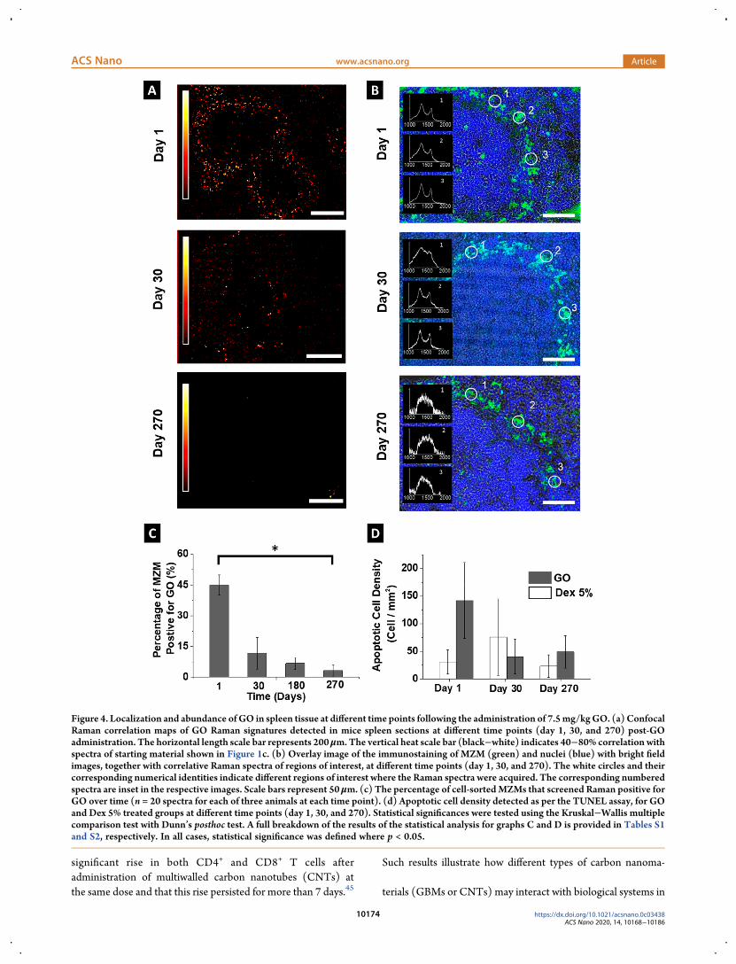
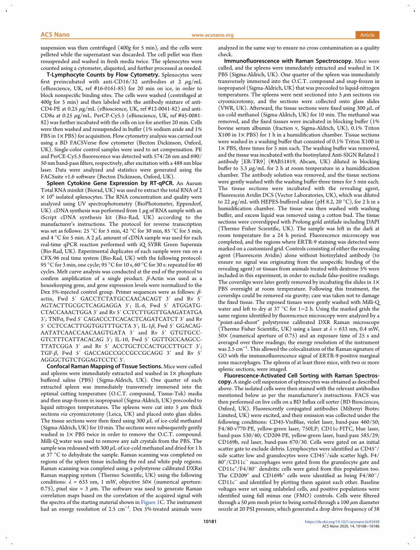