LDL Receptor-Related Protein 5 (LRP5) Affects Bone Accrual and Eye Development
Absence of p55 TNF Receptor Reduces Atherosclerosis,but Has No Major Effect on Angiotensin II InducedAneurysms in LDL Receptor Deficient MiceSofia Xanthoulea1, Melanie Thelen1, Chantal Pottgens1, Marion J. J. Gijbels1,2, Esther Lutgens2,
Menno P. J. de Winther1*
1 Department of Molecular Genetics, Cardiovascular Research Institute Maastricht, Maastricht University, Maastricht, The Netherlands, 2 Department of Pathology,
Cardiovascular Research Institute Maastricht, Maastricht University, Maastricht, The Netherlands
Abstract
Background: The aim of the current study was to investigate the role of p55 TNF Receptor (p55 TNFR), the main signalingreceptor for the pro-inflammatory cytokine tumor necrosis factor (TNF), in the development of two vascular disorders:atherosclerosis and angiotensin (Ang) II-induced abdominal aortic aneurysms (AAA).
Methodology/Principal Findings: p55 TNFR deficient mice were crossed to an LDL receptor deficient background and wereinduced for the development of either atherosclerosis or AngII-induced AAA, and compared to littermate controls, wild-typefor p55 TNFR expression. p55 TNFR deficient mice developed 43% smaller atherosclerotic lesions in the aortic sinusescompared to controls. Moreover, expression of CD68, a macrophage specific marker, exhibited a 50% reduction in the aorticarches. Decreased atherosclerosis correlated with a strong down-regulation in the expression of adhesion molecules, suchas VCAM-1 and ICAM-1, by p55 TNFR deficient endothelium. In addition, expression levels of the pro-inflammatory cytokinesand chemokines TNF, IL-6, MCP-1 and RANTES were significantly reduced in aortas of p55 TNFR deficient mice. In contrast, inthe AngII-induced model of AAA, p55 TNFR deficiency correlated with a slight trend towards increased aneurismal lethality,but the incidence of aortic rupture due to a dissecting aneurysm, and the expansion of the suprarenal aorta were notsignificantly different compared to controls.
Conclusion/Significance: We found that p55 TNFR expression promotes atherosclerosis, among other mechanisms, byenhancing expression of endothelial adhesion molecules, while it seems to have no major role in the development of AngII-induced AAA.
Citation: Xanthoulea S, Thelen M, Pottgens C, Gijbels MJJ, Lutgens E, et al. (2009) Absence of p55 TNF Receptor Reduces Atherosclerosis, but Has No Major Effecton Angiotensin II Induced Aneurysms in LDL Receptor Deficient Mice. PLoS ONE 4(7): e6113. doi:10.1371/journal.pone.0006113
Editor: Graham Pockley, University of Sheffield, United Kingdom
Received April 17, 2009; Accepted June 3, 2009; Published July 7, 2009
Copyright: � 2009 Xanthoulea et al. This is an open-access article distributed under the terms of the Creative Commons Attribution License, which permitsunrestricted use, distribution, and reproduction in any medium, provided the original author and source are credited.
Funding: This work was supported by the Netherlands Organization for Scientific Research NWO (VENI 016-056-124 to S. Xanthoulea, VIDI 917-66-329 to M.P.J. deWinther, VIDI 016-086-326 to E. Lutgens), the European Vascular Genomics Network (EVGN) and the Netherlands Heart Foundation (Established Investigator grantno. 2007T067). The funders had no role in study design, data collection and analysis, decision to publish, or preparation of the manuscript.
Competing Interests: The authors have declared that no competing interests exist.
* E-mail: [email protected]
Introduction
Vascular inflammatory processes, characterized by the accu-
mulation in the arterial wall of immune cells like monocytes/
macrophages and lymphocytes, are crucial events in the
pathogenesis of major vascular diseases such as atherosclerosis
and abdominal aortic aneurysms (AAA) [1,2]. Tumor necrosis
factor (TNF) is a major cytokine with a pivotal role in
orchestrating inflammatory responses and p55 TNFR mediates
the majority of TNF responses [3].
In atherosclerosis, TNF is generally considered to promote
plaque growth and progression since disease induction in TNF
deficient mice resulted in reduced development of atherosclerotic
lesions as well as their reduced progression towards more
advanced stages [4,5,6,7]. The role of the p55 TNFR in
atherogenesis is however less established. Total-body p55 TNFR
deficiency was shown to either have no effect on lesion size,
composition or features of plaque destabilization in very advanced
atherosclerosis [8], or to result in bigger lesions in C57BL/6 mice
fed an atherogenic diet, suggesting an athero-protective function
[9].
Regarding the cell-type specific role of this receptor, we have
recently shown that bone-marrow derived p55 TNFR promotes
atherosclerosis development by enhancing lesional foam-cell
formation and by promoting the expression of pro-atherosclerotic
chemokines, like MCP-1 [10]. In addition, by using a model of
carotid artery-to-carotid artery interposition grafting in which p55
TNFR deficient (but ApoE sufficient) carotid arteries were grafted
into ApoE deficient recipients, it was shown that arterial wall (i.e.
smooth muscle and endothelial cell) p55 TNFR expression is pro-
atherogenic [11]. However, as also indicated by the authors, this
study presented some limitations since it appeared that there was a
slight alloimmune reaction accelerating atherosclerosis, possibly
elicited by ApoE secreted by graft smooth muscle cells. This might
complicate the elucidation of the actual role of vascular wall p55
PLoS ONE | www.plosone.org 1 July 2009 | Volume 4 | Issue 7 | e6113
TNFR in atherogenesis. In addition, compared with the total-body
p55 TNFR deficiency studies, these results suggest the possibility
that p55 TNFR expression in different cell types might play
opposing roles in disease development.
The role of the TNF-p55 TNFR signaling in the development of
abdominal aortic aneurysms is not defined. Both serum [12] and
aneurismal tissues [13] from AAA patients have increased TNF
levels, implicating a role for this cytokine in disease pathogenesis.
However, to our knowledge, mouse models of AAA have not been
applied in either TNF or p55 TNFR deficient background.
In order to elucidate the role of p55 TNFR signaling in these
two vascular disorders, we have studied disease development in
total-body p55 TNFR deficient mice or their littermate wild-type
controls in a hyperlipidemic LDL receptor deficient background, a
well-established and physiologic model of these diseases. We show
that p55 TNFR expression promotes atherosclerosis in two
different sites prone to atherosclerotic lesion development like
the aortic sinuses and the aortic arches. Concomitant to additional
mechanisms identified previously [10], here we show that, in a
physiological setting, the pro-atherosclerotic action of this
important innate receptor is also mediated by inducing adhesion
molecule expression in endothelial cells and by enhancing
production of pro-atherosclerotic cytokines and chemokines at
the arterial wall. However, in a hyperlipidemia-accelerated
[15,16,17] AngII-induced model of AAA, the role of p55 TNFR
signaling appears to be secondary. Although we observed a trend
towards an increased aneurismal lethality in the p55 TNFR
deficient mice, differences were not significant. Moreover,
additional parameters of disease susceptibility like the incidence
of dissecting aneurysms and the suprarenal aortic diameters were
also not significantly affected by the absence of the receptor.
Results
p55 TNFR deficient mice develop smaller atheroscleroticplaques
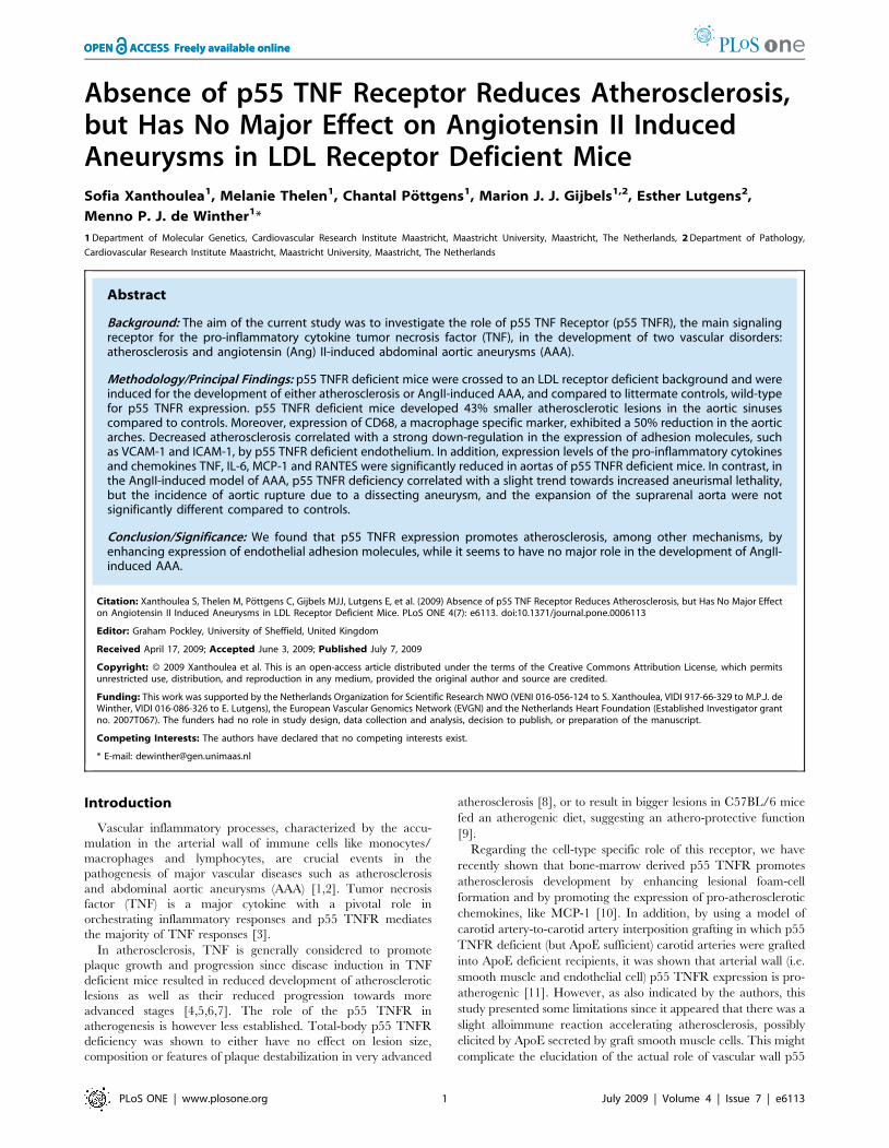
p55 TNFR deficient (p552/2LDLR2/2; n = 16) and littermate
control (p55+/+LDLR2/2; n = 18) mice were fed a high fat diet for
8 weeks and blood samples were collected at the beginning and at
the end of the feeding period. Plasma cholesterol and triglyceride
levels increased upon high fat diet but no significant differences
between the groups were observed. Body weights were also
comparable (Fig. 1A, B, C).
At sacrifice, the hearts and the aortic arches were isolated.
Assessment of atherosclerotic lesion size in the aortic sinus area
showed that p552/2LDLR2/2 mice developed 43% smaller
atherosclerotic plaques compared to p55+/+LDLR2/2 littermates
(Fig. 2A, B; p = 0.02). In addition, lesions were categorized for
severity with a scale from 1–3: (1) early lesions with fatty streaks
containing only macrophage derived foam-cells, (2) moderate
lesions characterized by the additional presence of a collagenous
cap, (3) advanced lesions with involvement of the media and
increased collagen content. p55+/+LDLR2/2 mice had 50% early
lesions, 29.6% moderate and 20.4% advanced lesions while these
percentages were 60.4%, 27.1% and 12.5% respectively for the
p552/2LDLR2/2 mice (Fig. 2C). Thus, p55 TNFR ablation
inhibits atherosclerosis development.
Next, RNA was isolated from aortic arches of p552/2LDLR2/2
and control mice and expression levels of the macrophage marker
CD-68 were determined. Expression levels of the housekeeping
genes cyclophilin and b-actin, used as controls, were similar
between the groups. Aortic arches of p552/2LDLR2/2 mice
showed approximately 50% less CD-68 macrophage-marker
expression compared to controls, suggesting reduced presence of
macrophages and thereby smaller plaques (Fig. 2D). Hence, p55
TNFR promotes the accumulation of macrophages and the
development of atherosclerotic lesions in two different atheroscle-
rosis prone sites as the aortic valve area and the aortic arches.
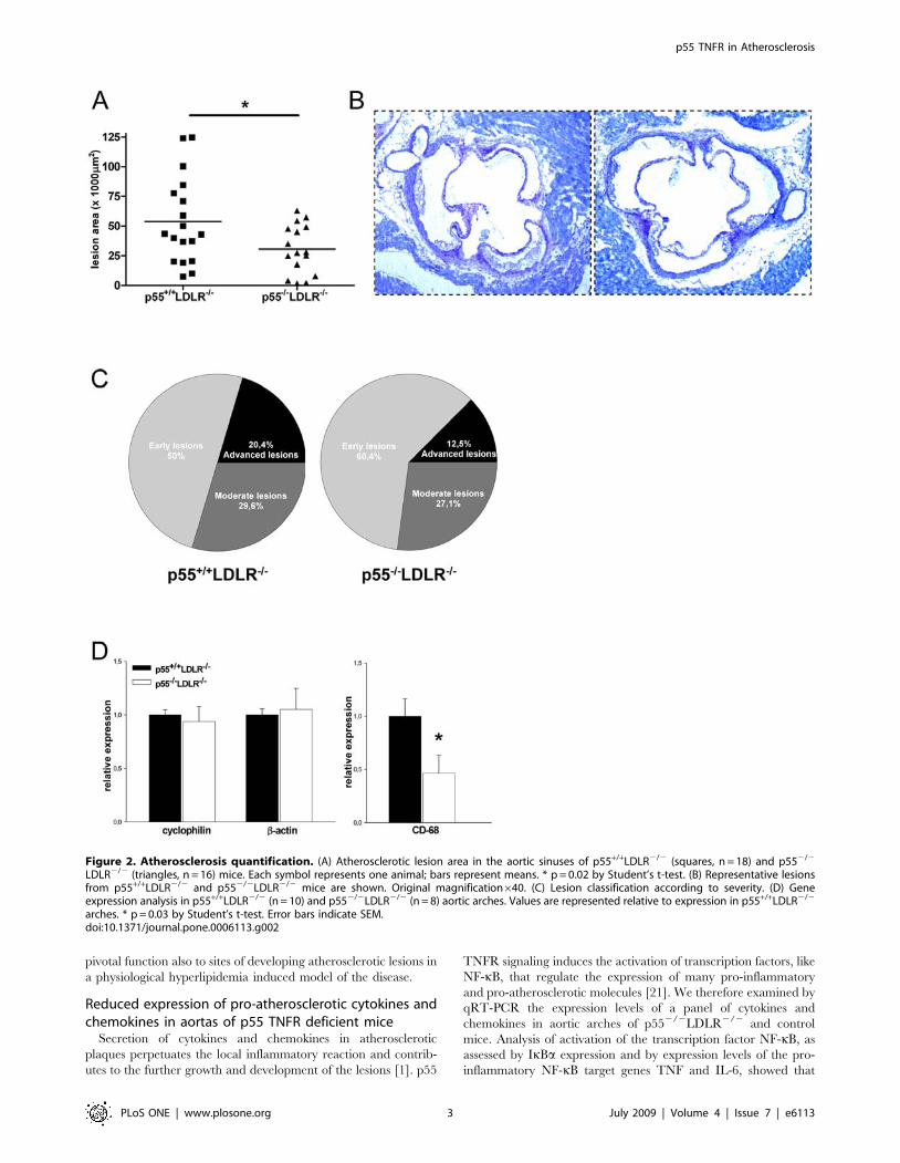
Reduced expression of adhesion molecules by p55 TNFRdeficient endothelium
Expression of adhesion molecules by the vascular endothelium
is thought to be one of the first events promoting atherogenesis,
and p55 TNFR has a crucial role in the TNF-induced expression
of adhesion molecules and consequent leukocyte organ infiltration
[18,19]. We thus examined by quantitative real-time PCR (qRT-
PCR) the expression levels of four adhesion molecules, VCAM-1,
ICAM-1, e-selectin and p-selectin, that are important for the
development of atherosclerosis [20] on RNA isolated from the
aortic arches of p552/2LDLR2/2 and control mice. mRNA levels
of all these adhesion molecules were significantly and approxi-
mately 50–60% reduced in aortas of p552/2LDLR2/2 mice
compared to controls (Fig. 3A). In addition, atherosclerotic lesions
were immunostained with an anti-VCAM-1 specific antibody and
the intensity of endothelial VCAM-1 staining was semi-quantita-
tively determined by two independent observers. This confirmed
the reduced expression of VCAM-1 by p55 TNFR deficient
endothelium in p552/2LDLR2/2 lesions (Fig. 3B, C). These
results are in line with previous findings regarding the crucial role
of p55 TNFR in the control of adhesion molecules expression by
endothelial cells at inflammatory sites [11,18,19], and extend this
Figure 1. Body weight and plasma lipid levels. (A) Body weight (B) plasma cholesterol and (C) plasma triglyceride levels in p55+/+LDLR2/2
(n = 18) and p552/2LDLR2/2 (n = 16) mice before and after 8 weeks of high fat feeding.doi:10.1371/journal.pone.0006113.g001
p55 TNFR in Atherosclerosis
PLoS ONE | www.plosone.org 2 July 2009 | Volume 4 | Issue 7 | e6113
pivotal function also to sites of developing atherosclerotic lesions in
a physiological hyperlipidemia induced model of the disease.
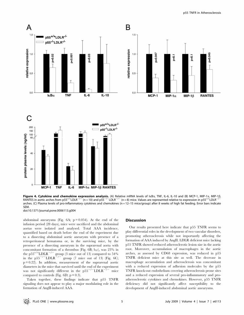
Reduced expression of pro-atherosclerotic cytokines andchemokines in aortas of p55 TNFR deficient mice
Secretion of cytokines and chemokines in atherosclerotic
plaques perpetuates the local inflammatory reaction and contrib-
utes to the further growth and development of the lesions [1]. p55
TNFR signaling induces the activation of transcription factors, like
NF-kB, that regulate the expression of many pro-inflammatory
and pro-atherosclerotic molecules [21]. We therefore examined by
qRT-PCR the expression levels of a panel of cytokines and
chemokines in aortic arches of p552/2LDLR2/2 and control
mice. Analysis of activation of the transcription factor NF-kB, as
assessed by IkBa expression and by expression levels of the pro-
inflammatory NF-kB target genes TNF and IL-6, showed that
Figure 2. Atherosclerosis quantification. (A) Atherosclerotic lesion area in the aortic sinuses of p55+/+LDLR2/2 (squares, n = 18) and p552/2
LDLR2/2 (triangles, n = 16) mice. Each symbol represents one animal; bars represent means. * p = 0.02 by Student’s t-test. (B) Representative lesionsfrom p55+/+LDLR2/2 and p552/2LDLR2/2 mice are shown. Original magnification640. (C) Lesion classification according to severity. (D) Geneexpression analysis in p55+/+LDLR2/2 (n = 10) and p552/2LDLR2/2 (n = 8) aortic arches. Values are represented relative to expression in p55+/+LDLR2/2
arches. * p = 0.03 by Student’s t-test. Error bars indicate SEM.doi:10.1371/journal.pone.0006113.g002
p55 TNFR in Atherosclerosis
PLoS ONE | www.plosone.org 3 July 2009 | Volume 4 | Issue 7 | e6113

these were significantly reduced in p552/2LDLR2/2 aortas
(Fig. 4A). In contrast, the anti-inflammatory cytokine IL-10 was
unaffected (Fig. 4A). In addition, expression levels of the pro-
atherosclerotic chemokines MCP-1 and RANTES were also
significantly lower in p552/2LDLR2/2 aortas compared to
controls (Fig. 4B). Expression levels of chemokines MIP-1a and
MIP-1b were reduced, but differences did not reach statistical
significance (Fig. 4B). Plasma concentrations of these inflammatory
cytokines and chemokines, measured at the end of the experiment,
indicated no significant differences between the groups (Fig. 4C).
Thus, absence of p55 TNFR expression leads to a reduced
expression of several pro-inflammatory and pro-atherosclerotic
cytokines and chemokines at sites prone to atherosclerotic lesion
development.
General characterisation of AngII infused miceTo determine the involvement of p55 TNFR in the AngII-
induced model of AAA formation, LDLR2/2 mice either wild-
type (p55+/+LDLR2/2) or deficient for p55 TNFR (p552/2
LDLR2/2) were fed a fat-enriched diet and infused with either
saline (n = 3/group) or AngII (1000 ng/kg/min; n = 13–14/group)
for 28 days. No differences in body weight were observed between
p55+/+LDLR2/2 and p552/2LDLR2/2 mice infused with
AngII, while plasma cholesterol levels were approximately 25%
reduced at the end of the study in p552/2LDLR2/2 mice
(Fig. 5A, B, C). To determine the effect of AngII infusion on
plasma concentrations of inflammatory cytokines and chemo-
kines, we performed a multi-cytokine analysis on plasma isolated
at the end of the experiment. Comparable to our previous
observations [10], plasma levels of MCP-1 were significantly and
approximately 30% reduced in p552/2LDLR2/2 mice, indi-
cating that p55 TNFR signaling is important for regulating
levels of MCP-1 in the circulation. Plasma levels of other
chemokines or cytokines did not show significant differences
between the groups (Fig. 5D).
p55 TNFR signaling does not significantly affect AngII-induced AAA formation
Neither genotype of saline-infused mice developed AAA. AngII
infusion resulted in lethality due to AAA rupture in 5 out of 13
p552/2LDLR2/2 within the first 10 days, while only one mouse
out of 14 from the p55+/+LDLR2/2 group died due to AAA
rupture, 26 days after the start of AngII administration. One
additional mouse from the p55+/+LDLR2/2 group died due to
ruptured thoracic aortic aneurysm (TAA) but presented no AAA
and thus was not included in the lethality caused by ruptured
Figure 3. Adhesion molecules expression analysis. (A) Relative mRNA levels of VCAM-1, ICAM-1, e-selectin and p-selectin in aortic arches fromp55+/+LDLR2/2 (n = 10) and p552/2LDLR2/2 (n = 8) mice. Values are represented relative to expression in p55+/+LDLR2/2 arches. (B)Immunohistochemical staining of VCAM-1 expression on sections from aortic valve areas indicating a less intense endothelial staining in p552/2
LDLR2/2 mice. Original magnification6200. (C) Staining quantification. * p = 0.01 by Student’s t-test. Error bars indicate SEM.doi:10.1371/journal.pone.0006113.g003
p55 TNFR in Atherosclerosis
PLoS ONE | www.plosone.org 4 July 2009 | Volume 4 | Issue 7 | e6113
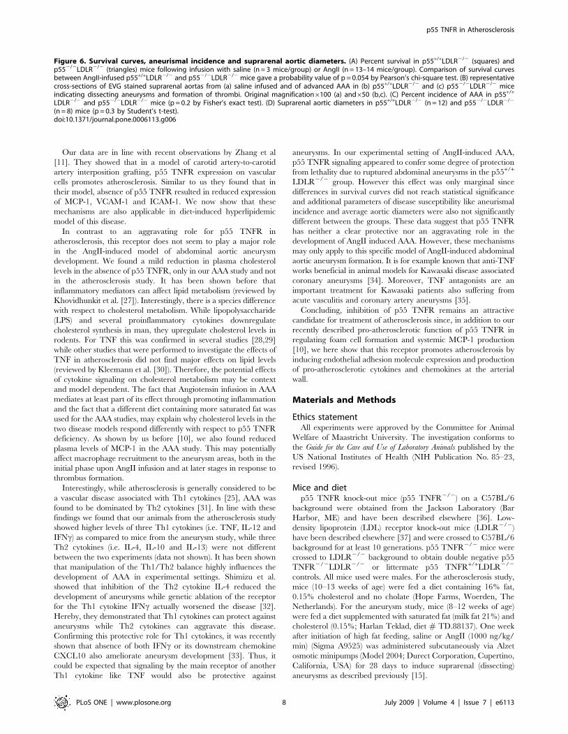
abdominal aneurysms (Fig. 6A; p = 0.054). At the end of the
infusion period (28 days), mice were sacrificed and the abdominal
aortas were isolated and analysed. Total AAA incidence,
quantified based on death before the end of the experiment due
to a dissecting abdominal aortic aneurysm with presence of a
retroperitoneal hematoma or, in the surviving mice, by the
presence of a dissecting aneurysm in the suprarenal aorta with
concomitant formation of a thrombus (Fig. 6B; b,c), was 23% in
the p55+/+LDLR2/2 group (3 mice out of 13) compared to 54%
in the p552/2LDLR2/2 group (7 mice out of 13) (Fig. 6C;
p = 0.22). In addition, measurement of the suprarenal aortic
diameters in the mice that survived until the end of the experiment
was not significantly different in the p552/2LDLR2/2 mice
compared to controls (Fig. 6D; p = 0.3).
Taken together, these findings indicate that p55 TNFR
signaling does not appear to play a major modulating role in the
formation of AngII-induced AAA.
Discussion
Our results presented here indicate that p55 TNFR seems to
play differential roles in the development of two vascular disorders,
promoting atherosclerosis while not importantly affecting the
formation of AAA induced by AngII. LDLR deficient mice lacking
p55 TNFR showed reduced atherosclerotic lesion size in the aortic
root. Moreover, accumulation of macrophages in the aortic
arches, as assessed by CD68 expression, was reduced in p55
TNFR deficient mice at this site as well. The decrease in
macrophage accumulation and atherosclerosis was concomitant
with a reduced expression of adhesion molecules by the p55
TNFR knock-out endothelium covering atherosclerosis prone sites
and a reduced expression of several pro-inflammatory and pro-
atherosclerotic cytokines and chemokines. However, p55 TNFR
deficiency did not significantly affect susceptibility to the
development of AngII-induced abdominal aortic aneurysms.
Figure 4. Cytokine and chemokine expression analysis. (A) Relative mRNA levels of IkBa, TNF, IL-6, IL-10 and (B) MCP-1, MIP-1a, MIP-1b,RANTES in aortic arches from p55+/+LDLR2/2 (n = 10) and p552/2LDLR2/2 (n = 8) mice. Values are represented relative to expression in p55+/+LDLR2/2
arches. (C) Plasma levels of pro-inflammatory cytokines and chemokines (n = 12–15 mice/group) after 8 weeks of high fat feeding. Error bars indicateSEM.doi:10.1371/journal.pone.0006113.g004
p55 TNFR in Atherosclerosis
PLoS ONE | www.plosone.org 5 July 2009 | Volume 4 | Issue 7 | e6113

We have previously shown that absence of p55 TNFR on
hematopoietic cells protects against atherosclerosis development
[10]. In a bone marrow transplantation setting we could determine
that p55 TNFR deficiency results in a reduced scavenger receptor
class A dependent uptake of modified lipoproteins by macrophag-
es, leading to smaller foam cells and subsequent smaller
atherosclerotic lesions. Moreover we showed that p55 TNFR
deficiency in hematopoietic cells results in lower plasma levels of
the crucial pro-atherosclerotic chemokine MCP-1, identifying an
additional potential mechanism leading to smaller lesions. In the
current study we extended these investigations to LDLR2/2
animals lacking p55 TNFR in all their cells. Hereby, we show that
p55 TNFR is important for the expression of adhesion molecules
on endothelial cells in atherosclerosis prone sites. An important
downstream mediator of p55 TNFR signaling is the transcription
factor NF-kB. In another recent study, we have shown that NF-kB
signaling in endothelial cells is imperative for the development of
atherosclerosis [14]. Using different mouse models targeting NF-
kB activation specifically in endothelial cells, we determined that
inhibition of NF-kB resulted in repressed expression of adhesion
molecules such as VCAM-1 and ICAM-1 by the vascular
endothelium and impaired macrophage recruitment to athero-
sclerotic plaques. Consequently, atherosclerotic lesions were
smaller and characterized by strongly reduced expression of
inflammatory cytokines, such as TNF and IL-6 and also reduced
expression of atherosclerosis associated chemokines, such as MCP-
1 and RANTES.
Different upstream receptors and pathways are responsible for
activating NF-kB in endothelial cells and previous studies have
shown that TLR receptors are upregulated in endothelial cells in
atherosclerosis prone sites and positively contribute to plaque
development [22,23,24]. In the present study we show that the
TNF-p55 TNFR pathway is also an important activator of NF-
kB and inducer of adhesion molecules in endothelial cells during
atherogenesis, since VCAM-1, ICAM-1, e-selectin and p-selectin
were reduced in the absence of p55 TNFR. Concomitant with a
reduced NF-kB activation in the aortic arches, as assessed by
IkBa expression, we also found reduced expression of TNF, IL-
6, MCP-1 and RANTES, all proven to be important in
atherosclerosis [25,26]. Since, these factors are also expressed by
macrophages we cannot exclude that their reduced levels are a
reflection of smaller atherosclerotic lesions, instead of being a
direct result of absence of p55 TNFR signaling. Plasma levels of
these inflammatory mediators were not affected by absence of
p55 (Fig. 4C) and showed a similar pattern as in the AAA study
(figure 5B).
Figure 5. General characterization of AngII infused mice. (A) Body weight (B) plasma cholesterol and (C) plasma triglyceride levels in p55+/+
LDLR2/2 and p552/2LDLR2/2 mice before (n = 13–14 mice/group) and after 5 weeks of high fat feeding (4 weeks of AngII infusion; n = 8–12 mice/group). (D) Plasma levels of pro-inflammatory cytokines and chemokines (n = 7–9 mice/group) after 5 weeks of high fat feeding (4 weeks of AngIIinfusion). * p,0.05 by Student’s t-test. Error bars indicate SEM.doi:10.1371/journal.pone.0006113.g005
p55 TNFR in Atherosclerosis
PLoS ONE | www.plosone.org 6 July 2009 | Volume 4 | Issue 7 | e6113

p55 TNFR in Atherosclerosis
PLoS ONE | www.plosone.org 7 July 2009 | Volume 4 | Issue 7 | e6113
Our data are in line with recent observations by Zhang et al
[11]. They showed that in a model of carotid artery-to-carotid
artery interposition grafting, p55 TNFR expression on vascular
cells promotes atherosclerosis. Similar to us they found that in
their model, absence of p55 TNFR resulted in reduced expression
of MCP-1, VCAM-1 and ICAM-1. We now show that these
mechanisms are also applicable in diet-induced hyperlipidemic
model of this disease.
In contrast to an aggravating role for p55 TNFR in
atherosclerosis, this receptor does not seem to play a major role
in the AngII-induced model of abdominal aortic aneurysm
development. We found a mild reduction in plasma cholesterol
levels in the absence of p55 TNFR, only in our AAA study and not
in the atherosclerosis study. It has been shown before that
inflammatory mediators can affect lipid metabolism (reviewed by
Khovidhunkit et al. [27]). Interestingly, there is a species difference
with respect to cholesterol metabolism. While lipopolysaccharide
(LPS) and several proinflammatory cytokines downregulate
cholesterol synthesis in man, they upregulate cholesterol levels in
rodents. For TNF this was confirmed in several studies [28,29]
while other studies that were performed to investigate the effects of
TNF in atherosclerosis did not find major effects on lipid levels
(reviewed by Kleemann et al. [30]). Therefore, the potential effects
of cytokine signaling on cholesterol metabolism may be context
and model dependent. The fact that Angiotensin infusion in AAA
mediates at least part of its effect through promoting inflammation
and the fact that a different diet containing more saturated fat was
used for the AAA studies, may explain why cholesterol levels in the
two disease models respond differently with respect to p55 TNFR
deficiency. As shown by us before [10], we also found reduced
plasma levels of MCP-1 in the AAA study. This may potentially
affect macrophage recruitment to the aneurysm areas, both in the
initial phase upon AngII infusion and at later stages in response to
thrombus formation.
Interestingly, while atherosclerosis is generally considered to be
a vascular disease associated with Th1 cytokines [25], AAA was
found to be dominated by Th2 cytokines [31]. In line with these
findings we found that our animals from the atherosclerosis study
showed higher levels of three Th1 cytokines (i.e. TNF, IL-12 and
IFNc) as compared to mice from the aneurysm study, while three
Th2 cytokines (i.e. IL-4, IL-10 and IL-13) were not different
between the two experiments (data not shown). It has been shown
that manipulation of the Th1/Th2 balance highly influences the
development of AAA in experimental settings. Shimizu et al.
showed that inhibition of the Th2 cytokine IL-4 reduced the
development of aneurysms while genetic ablation of the receptor
for the Th1 cytokine IFNc actually worsened the disease [32].
Hereby, they demonstrated that Th1 cytokines can protect against
aneurysms while Th2 cytokines can aggravate this disease.
Confirming this protective role for Th1 cytokines, it was recently
shown that absence of both IFNc or its downstream chemokine
CXCL10 also ameliorate aneurysm development [33]. Thus, it
could be expected that signaling by the main receptor of another
Th1 cytokine like TNF would also be protective against
aneurysms. In our experimental setting of AngII-induced AAA,
p55 TNFR signaling appeared to confer some degree of protection
from lethality due to ruptured abdominal aneurysms in the p55+/+
LDLR2/2 group. However this effect was only marginal since
differences in survival curves did not reach statistical significance
and additional parameters of disease susceptibility like aneurismal
incidence and average aortic diameters were also not significantly
different between the groups. These data suggest that p55 TNFR
has neither a clear protective nor an aggravating role in the
development of AngII induced AAA. However, these mechanisms
may only apply to this specific model of AngII-induced abdominal
aortic aneurysm formation. It is for example known that anti-TNF
works beneficial in animal models for Kawasaki disease associated
coronary aneurysms [34]. Moreover, TNF antagonists are an
important treatment for Kawasaki patients also suffering from
acute vasculitis and coronary artery aneurysms [35].
Concluding, inhibition of p55 TNFR remains an attractive
candidate for treatment of atherosclerosis since, in addition to our
recently described pro-atherosclerotic function of p55 TNFR in
regulating foam cell formation and systemic MCP-1 production
[10], we here show that this receptor promotes atherosclerosis by
inducing endothelial adhesion molecule expression and production
of pro-atherosclerotic cytokines and chemokines at the arterial
wall.
Materials and Methods
Ethics statementAll experiments were approved by the Committee for Animal
Welfare of Maastricht University. The investigation conforms to
the Guide for the Care and Use of Laboratory Animals published by the
US National Institutes of Health (NIH Publication No. 85–23,
revised 1996).
Mice and dietp55 TNFR knock-out mice (p55 TNFR2/2) on a C57BL/6
background were obtained from the Jackson Laboratory (Bar
Harbor, ME) and have been described elsewhere [36]. Low-
density lipoprotein (LDL) receptor knock-out mice (LDLR2/2)
have been described elsewhere [37] and were crossed to C57BL/6
background for at least 10 generations. p55 TNFR2/2 mice were
crossed to LDLR2/2 background to obtain double negative p55
TNFR2/2LDLR2/2 or littermate p55 TNFR+/+LDLR2/2
controls. All mice used were males. For the atherosclerosis study,
mice (10–13 weeks of age) were fed a diet containing 16% fat,
0.15% cholesterol and no cholate (Hope Farms, Woerden, The
Netherlands). For the aneurysm study, mice (8–12 weeks of age)
were fed a diet supplemented with saturated fat (milk fat 21%) and
cholesterol (0.15%; Harlan Teklad, diet # TD.88137). One week
after initiation of high fat feeding, saline or AngII (1000 ng/kg/
min) (Sigma A9525) was administered subcutaneously via Alzet
osmotic minipumps (Model 2004; Durect Corporation, Cupertino,
California, USA) for 28 days to induce suprarenal (dissecting)
aneurysms as described previously [15].
Figure 6. Survival curves, aneurismal incidence and suprarenal aortic diameters. (A) Percent survival in p55+/+LDLR2/2 (squares) andp552/2LDLR2/2 (triangles) mice following infusion with saline (n = 3 mice/group) or AngII (n = 13–14 mice/group). Comparison of survival curvesbetween AngII-infused p55+/+LDLR2/2 and p552/2LDLR2/2 mice gave a probability value of p = 0.054 by Pearson’s chi-square test. (B) representativecross-sections of EVG stained suprarenal aortas from (a) saline infused and of advanced AAA in (b) p55+/+LDLR2/2 and (c) p552/2LDLR2/2 miceindicating dissecting aneurysms and formation of thrombi. Original magnification6100 (a) and650 (b,c). (C) Percent incidence of AAA in p55+/+
LDLR2/2 and p552/2LDLR2/2 mice (p = 0.2 by Fisher’s exact test). (D) Suprarenal aortic diameters in p55+/+LDLR2/2 (n = 12) and p552/2LDLR2/2
(n = 8) mice (p = 0.3 by Student’s t-test).doi:10.1371/journal.pone.0006113.g006
p55 TNFR in Atherosclerosis
PLoS ONE | www.plosone.org 8 July 2009 | Volume 4 | Issue 7 | e6113

Genotyping and blood analysisPrimers for the wild-type or p55 TNFR deleted alleles were: 59-
TGTGAAAAGGGCACCTTTACGGC-39; 59-GGCTGCAGT-
CCACGCACTGG-39; 59-ATTCGCCAATGACAAGACGCT-
GG-39, and for the wild-type or LDLR deleted alleles were: 59-
CATATGCATCCCCAGTCTTTG-39; 59-ATAGATTCGCCC-
TTGTGTCC-39; 59-GCAGTGCTCCTCATCTGACTTG-39.
Cholesterol and triglyceride levels were determined on plasma
using enzymatic kits (Sigma-Aldrich; cat.no. 401 and 337). Plasma
cytokine levels in AngII infused mice were determined with the
Bio-Plex Mouse Cytokine 23-Plex Panel (cat.no:171-F11241; Bio-
Rad) according to manufacturer’s instructions.
Atherosclerotic lesion analysisConsecutive 7 mm sections of the heart the level of the
atrioventricular valves were collected and were stained with
toluidine blue for morphometric analysis and atherosclerosis
quantification as previously described [14]. For VCAM-1 staining,
sections were stained with anti-VCAM-1 antibody (BD).
Quantitative gene expressionRNA was isolated from aortic arches using RNeasy columns
with additional DNase digestion (Qiagen). cDNA synthesis was
performed using the iScriptTM cDNA synthesis kit (Bio-Rad) and
quantitative PCR was performed using the qPCR iQTM Custom
SYBR Green Supermic (Bio-Rad) on an iCycler thermal cycler
(Bio-Rad). Cyclophilin A was used to normalize RNA quantity.
Primer sequences are available upon request.
Quantification of aneurysms and morphometric analysisFor morphometric analysis of the AAA, the entire arterial tree
was perfused with 10 ml PBS containing 0.1 mg/ml sodium
nitroprusside (Sigma) following with 10 ml 1% paraformaldehyde
(Sigma). The arterial tree was isolated and fixed overnight in 1%
paraformaldehyde and embedded in paraffin. Cross sections
(4 mm) of the suprarenal area of the abdominal aorta were made
at 12 different levels at 200 mm intervals. Cross sections from each
level were stained with H&E and elastic-Van Gieson (EVG). Total
vessel area was quantified using the Leica Qwin V3.2.1 system and
DM 500B microscope.
On necropsies of unexpected deaths, death due to rupture of
aneurysm was qualified by presence of retroperitoneal hematoma
in addition to AAA. Aneurysmal incidence was quantified based
on such described deaths or, in the mice that survived until the end
of the experiment, in the presence of a dissecting aneurysm in the
suprarenal aorta with the concomitant formation of a thrombus.
All analyses were performed without prior knowledge of the
genotype.
Statistical analysisStatistical analyses were performed using Graphpad Prism
(Graphpad Software) or SigmaPlot t-tests. For lesion area and
characterization, differences were evaluated using a Welch
corrected t-test. For comparison of survival curves the Pearson’s
chi-square test was used. Percent incidence of AAA was analyzed
by Fisher’s exact test. Data are expressed as means6SEM. A p
value of less than 0.05 is considered statistically significant.
Acknowledgments
We thank Linda Beckers for help with the AAA model and the Animal
Facility staff of Maastricht University for excellent care of the animals used
in this study.
Author Contributions
Conceived and designed the experiments: SX MJJG EL MPJdW.
Performed the experiments: SX MT CP. Analyzed the data: SX MT CP
MJJG EL MPJdW. Contributed reagents/materials/analysis tools: EL.
Wrote the paper: SX MT CP MJJG EL MPJdW.
References
1. Libby P (2002) Inflammation in atherosclerosis. Nature 420: 868–874.
2. Shimizu K, Mitchell RN, Libby P (2006) Inflammation and cellular immune
responses in abdominal aortic aneurysms. Arterioscler Thromb Vasc Biol 26:
987–994.
3. Wajant H, Pfizenmaier K, Scheurich P (2003) Tumor necrosis factor signaling.
Cell Death Differ 10: 45–65.
4. Branen L, Hovgaard L, Nitulescu M, Bengtsson E, Nilsson J, et al. (2004)
Inhibition of tumor necrosis factor-alpha reduces atherosclerosis in apolipopro-
tein E knockout mice. Arterioscler Thromb Vasc Biol 24: 2137–2142.
5. Canault M, Peiretti F, Mueller C, Kopp F, Morange P, et al. (2004) Exclusive
expression of transmembrane TNF-alpha in mice reduces the inflammatory
response in early lipid lesions of aortic sinus. Atherosclerosis 172: 211–218.
6. Boesten LS, Zadelaar AS, van Nieuwkoop A, Gijbels MJ, de Winther MP, et al.
(2005) Tumor necrosis factor-alpha promotes atherosclerotic lesion progression
in APOE*3-Leiden transgenic mice. Cardiovasc Res 66: 179–185.
7. Ohta H, Wada H, Niwa T, Kirii H, Iwamoto N, et al. (2005) Disruption of
tumor necrosis factor-alpha gene diminishes the development of atherosclerosis
in ApoE-deficient mice. Atherosclerosis 180: 11–17.
8. Blessing E, Bea F, Kuo CC, Campbell LA, Chesebro B, et al. (2004) Lesion
progression and plaque composition are not altered in older apoE2/2 mice
lacking tumor necrosis factor-alpha receptor p55. Atherosclerosis 176: 227–
232.
9. Schreyer SA, Peschon JJ, LeBoeuf RC (1996) Accelerated atherosclerosis in mice
lacking tumor necrosis factor receptor p55. J Biol Chem 271: 26174–26178.
10. Xanthoulea S, Gijbels MJ, van der Made I, Mujcic H, Thelen M, et al. (2008)
p55 Tumour necrosis factor receptor in bone marrow-derived cells promotes
atherosclerosis development in low-density lipoprotein receptor knock-out mice.
Cardiovasc Res.
11. Zhang L, Peppel K, Sivashanmugam P, Orman ES, Brian L, et al. (2007)
Expression of tumor necrosis factor receptor-1 in arterial wall cells promotes
atherosclerosis. Arterioscler Thromb Vasc Biol 27: 1087–1094.
12. Fiane AE, Videm V, Lingaas PS, Heggelund L, Nielsen EW, et al. (2003)
Mechanism of complement activation and its role in the inflammatory
response after thoracoabdominal aortic aneurysm repair. Circulation 108:
849–856.
13. Hamano K, Li TS, Takahashi M, Kobayashi T, Shirasawa B, et al. (2003)
Enhanced tumor necrosis factor- alpha expression in small sized abdominal
aortic aneurysms. World J Surg 27: 476–480.
14. Gareus R, Kotsaki E, Xanthoulea S, van der Made I, Gijbels MJJ, et al. (2008)
Endothelial Cell-Specific NF-[kappa]B Inhibition Protects Mice from Athero-
sclerosis. Cell Metabolism 8: 372–383.
15. Daugherty A, Manning MW, Cassis LA (2000) Angiotensin II promotes
atherosclerotic lesions and aneurysms in apolipoprotein E-deficient mice. J Clin
Invest 105: 1605–1612.
16. Deng GG, Martin-McNulty B, Sukovich DA, Freay A, Halks-Miller M, et al.
(2003) Urokinase-type plasminogen activator plays a critical role in angiotensin
II-induced abdominal aortic aneurysm. Circ Res 92: 510–517.
17. Manning MW, Cassi LA, Huang J, Szilvassy SJ, Daugherty A (2002) Abdominal
aortic aneurysms: fresh insights from a novel animal model of the disease. Vasc
Med 7: 45–54.
18. Mackay F, Loetscher H, Stueber D, Gehr G, Lesslauer W (1993) Tumor necrosis
factor alpha (TNF-alpha)-induced cell adhesion to human endothelial cells is
under dominant control of one TNF receptor type, TNF-R55. J Exp Med 177:
1277–1286.
19. Neumann B, Machleidt T, Lifka A, Pfeffer K, Vestweber D, et al. (1996) Crucial
role of 55-kilodalton TNF receptor in TNF-induced adhesion molecule
expression and leukocyte organ infiltration. J Immunol 156: 1587–1593.
20. Galkina E, Ley K (2007) Vascular adhesion molecules in atherosclerosis.
Arterioscler Thromb Vasc Biol 27: 2292–2301.
21. Xanthoulea S, Curfs DM, Hofker MH, de Winther MP (2005) Nuclear factor
kappaB signaling in macrophage function and atherogenesis. Curr Opin Lipidol
16: 536–542.
22. Bjorkbacka H, Kunjathoor VV, Moore KJ, Koehn S, Ordija CM, et al. (2004)
Reduced atherosclerosis in MyD88-null mice links elevated serum cholesterol
levels to activation of innate immunity signaling pathways. Nat Med 10:
416–421.
23. Michelsen KS, Wong MH, Shah PK, Zhang W, Yano J, et al. (2004) Lack of
Toll-like receptor 4 or myeloid differentiation factor 88 reduces atherosclerosis
and alters plaque phenotype in mice deficient in apolipoprotein E. Proc Natl
Acad Sci U S A 101: 10679–10684.
p55 TNFR in Atherosclerosis
PLoS ONE | www.plosone.org 9 July 2009 | Volume 4 | Issue 7 | e6113
24. Mullick AE, Soldau K, Kiosses WB, Bell TA 3rd, Tobias PS, et al. (2008)
Increased endothelial expression of Toll-like receptor 2 at sites of disturbed
blood flow exacerbates early atherogenic events. J Exp Med 205: 373–383.
25. Tedgui A, Mallat Z (2006) Cytokines in atherosclerosis: pathogenic and
regulatory pathways. Physiol Rev 86: 515–581.
26. Zernecke A, Shagdarsuren E, Weber C (2008) Chemokines in Atherosclerosis.
An Update. Arterioscler Thromb Vasc Biol.
27. Khovidhunkit W, Kim MS, Memon RA, Shigenaga JK, Moser AH, et al. (2004)
Effects of infection and inflammation on lipid and lipoprotein metabolism:
mechanisms and consequences to the host. J Lipid Res 45: 1169–1196.
28. Feingold KR, Serio MK, Adi S, Moser AH, Grunfeld C (1989) Tumor necrosis
factor stimulates hepatic lipid synthesis and secretion. Endocrinology 124:
2336–2342.
29. Memon RA, Grunfeld C, Moser AH, Feingold KR (1993) Tumor necrosis factor
mediates the effects of endotoxin on cholesterol and triglyceride metabolism in
mice. Endocrinology 132: 2246–2253.
30. Kleemann R, Zadelaar S, Kooistra T (2008) Cytokines and atherosclerosis: a
comprehensive review of studies in mice. Cardiovasc Res 79: 360–376.
31. Schonbeck U, Sukhova GK, Gerdes N, Libby P (2002) T(H)2 predominant
immune responses prevail in human abdominal aortic aneurysm. Am J Pathol161: 499–506.
32. Shimizu K, Shichiri M, Libby P, Lee RT, Mitchell RN (2004) Th2-predominant
inflammation and blockade of IFN-gamma signaling induce aneurysms inallografted aortas. J Clin Invest 114: 300–308.
33. King VL, Lin AY, Kristo F, Anderson TJ, Ahluwalia N, et al. (2009) Interferon-gamma and the interferon-inducible chemokine CXCL10 protect against
aneurysm formation and rupture. Circulation 119: 426–435.
34. Hui-Yuen JS, Duong TT, Yeung RS (2006) TNF-alpha is necessary forinduction of coronary artery inflammation and aneurysm formation in an
animal model of Kawasaki disease. J Immunol 176: 6294–6301.35. Newburger JW, Fulton DR (2007) Kawasaki Disease. Curr Treat Options
Cardiovasc Med 9: 148–158.36. Pfeffer K, Matsuyama T, Kundig TM, Wakeham A, Kishihara K, et al. (1993)
Mice deficient for the 55 kd tumor necrosis factor receptor are resistant to
endotoxic shock, yet succumb to L. monocytogenes infection. Cell 73: 457–467.37. Ishibashi S, Brown MS, Goldstein JL, Gerard RD, Hammer RE, et al. (1993)
Hypercholesterolemia in low density lipoprotein receptor knockout mice and itsreversal by adenovirus-mediated gene delivery. J Clin Invest 92: 883–893.
p55 TNFR in Atherosclerosis
PLoS ONE | www.plosone.org 10 July 2009 | Volume 4 | Issue 7 | e6113
Copyright © 2022 FDOKUMEN