Characteristics and dynamics of surfzone transverse finger bars

AAV-Mediated Delivery of Zinc Finger NucleasesTargeting Hepatitis B Virus Inhibits Active ReplicationNicholas D. Weber1,2, Daniel Stone1, Ruth Hall Sedlak2, Harshana S. De Silva Feelixge1,
Pavitra Roychoudhury1, Joshua T. Schiffer1,3,4, Martine Aubert1, Keith R. Jerome1,2,5*
1 Vaccine and Infectious Disease Division, Fred Hutchinson Cancer Research Center, Seattle, Washington, United States of America, 2 Department of Laboratory Medicine,
University of Washington, Seattle, Washington, United States of America, 3 Division of Allergy and Infectious Disease, Department of Medicine, University of Washington,
Seattle, Washington, United States of America, 4 Clinical Research Division, Fred Hutchinson Cancer Research Center, Seattle, Washington, United States of America,
5 Department of Microbiology, University of Washington, Seattle, Washington, United States of America
Abstract
Despite an existing effective vaccine, hepatitis B virus (HBV) remains a major public health concern. There are effectivesuppressive therapies for HBV, but they remain expensive and inaccessible to many, and not all patients respond well.Furthermore, HBV can persist as genomic covalently closed circular DNA (cccDNA) that remains in hepatocytes even duringotherwise effective therapy and facilitates rebound in patients after treatment has stopped. Therefore, the need for aneffective treatment that targets active and persistent HBV infections remains. As a novel approach to treat HBV, we havetargeted the HBV genome for disruption to prevent viral reactivation and replication. We generated 3 zinc finger nucleases(ZFNs) that target sequences within the HBV polymerase, core and X genes. Upon the formation of ZFN-induced DNAdouble strand breaks (DSB), imprecise repair by non-homologous end joining leads to mutations that inactivate HBV genes.We delivered HBV-specific ZFNs using self-complementary adeno-associated virus (scAAV) vectors and tested their anti-HBVactivity in HepAD38 cells. HBV-ZFNs efficiently disrupted HBV target sites by inducing site-specific mutations. Cytotoxicitywas seen with one of the ZFNs. scAAV-mediated delivery of a ZFN targeting HBV polymerase resulted in complete inhibitionof HBV DNA replication and production of infectious HBV virions in HepAD38 cells. This effect was sustained for at least 2weeks following only a single treatment. Furthermore, high specificity was observed for all ZFNs, as negligible off-targetcleavage was seen via high-throughput sequencing of 7 closely matched potential off-target sites. These results show thatHBV-targeted ZFNs can efficiently inhibit active HBV replication and suppress the cellular template for HBV persistence,making them promising candidates for eradication therapy.
Citation: Weber ND, Stone D, Sedlak RH, De Silva Feelixge HS, Roychoudhury P, et al. (2014) AAV-Mediated Delivery of Zinc Finger Nucleases Targeting HepatitisB Virus Inhibits Active Replication. PLoS ONE 9(5): e97579. doi:10.1371/journal.pone.0097579
Editor: Haitao Guo, Drexel University College of Medicine, United States of America
Received February 24, 2014; Accepted April 21, 2014; Published May 14, 2014
Copyright: � 2014 Weber et al. This is an open-access article distributed under the terms of the Creative Commons Attribution License, which permitsunrestricted use, distribution, and reproduction in any medium, provided the original author and source are credited.
Funding: Portions of this work were funded by NIH supported Martin Delaney Collaboratory grant U19 AI 096111. NDW was supported by NIH InterdisciplinaryTraining Grant postdoctoral fellowship T32 CA080416. The funders had no role in study design, data collection and analysis, decision to publish, or preparation ofthe manuscript.
Competing Interests: The authors have declared that no competing interests exist.
* E-mail: [email protected]
Introduction
Hepatitis B virus (HBV) remains a significant cause of morbidity
and mortality worldwide [1]. Long-term chronic infection with
HBV carries a poor prognosis as it frequently leads to the
development of cirrhosis and hepatocellular carcinoma (HCC) [2].
For those unable to spontaneously clear HBV infection, antiviral
drugs can be used to inhibit viral replication, delaying long-term
liver damage [3]. Unfortunately, HBV reactivation is common due
to the persistence in hepatocytes of episomal covalently closed
circular DNA (cccDNA), the template for HBV viral replication
and the source for viral reactivation. In chronically infected
individuals, cccDNA is able to persist, and while antiviral therapies
can reduce hepatic cccDNA they do not entirely eliminate it [4].
Curative therapy strategies for chronically infected patients
should disrupt or eliminate residual hepatic cccDNA, and thus
prevent HBV reactivation. One potential approach involves
eliminating or modifying viral sequences enough to sufficiently
disrupt HBV gene functions. This could be achieved by
introducing double strand breaks (DSBs) into viral DNA using
targeted endonucleases. DNA DSBs are repaired through the
process of non-homologous end joining, which is error prone, and
repeated DNA cleavage will eventually yield mutations at a
targeted cleavage site [5]. Alternatively, episomal DNA that is
linearized from DSBs may be susceptible to direct degradation by
cellular DNases, which could result in reduced viral replication or
virus elimination. This strategy of targeted gene disruption has
been proposed as a novel anti-viral therapy [6–8]. Indeed, several
DSB-inducing enzymes, including zinc finger nucleases (ZFNs),
transcription activator-like effector nucleases (TALENs), homing
endonucleases (HEs), and the CRISPR/Cas system could be used
in such an approach.
High specificity is a requirement of targeted endonucleases so as
to avoid toxicity and other negative outcomes caused by the
cleavage of genomic sequences with similarity to the target
sequences, known as ‘‘off-target’’ sites. Off-target activity of
endonucleases depends on their cleavage efficiency, the length of
the target sequence, and the ability to recognize the correct
sequence with high specificity [9]. Efforts are being made to
improve these characteristics for some of the targeted endonucle-
PLOS ONE | www.plosone.org 1 May 2014 | Volume 9 | Issue 5 | e97579

ase platforms [10]. Several in-depth analyses focused solely on off-
target activity of targeted endonucleases have been performed
[11], particularly for the CRISPR/Cas system [12–14]. Indeed, it
is essential to assay the off-target activity of enzymes that possess
successful mutagenic capability at their intended sites, especially in
the case of antiviral enzymes that might be systemically delivered
and thus expressed in a large number of cells. There exist
bioinformatics programs for identifying potential off-target sites
[12,15], and high-throughput sequencing methods allow for many
sites to be queried for potential mutagenesis following treatment
with endonucleases.
Several reports have shown that targeted endonucleases can
disrupt viral DNA sequences from HBV, HIV, HPV, HSV and
HTLV [16–21]. In fact, antiviral effects against HBV have been
achieved through the use of ZFNs [17] and TALENs [22,23]. The
next steps in preparing a robust antiviral therapy based on virus-
specific targeted endonucleases for clinical application involve the
development of a practical and efficient delivery method and a
clear absence of off-target activity.
Materials and Methods
Cell cultureHuman embryonic kidney (HEK) 293T cells and HepAD38
cells [24] were grown in DMEM (Invitrogen) supplemented with
10% FBS. HepAD38 cells are derived from HepG2 and can
replicate HBV from a single integrated 1.1 length copy of a
genotype D ayw serotype HBV genome under the control of the
tetracycline responsive (tet-off) promoter. HBV replication in
HepAD38 cells was suppressed in some experiments by the
addition of 0.5 mg/ml doxycycline (dox). HepAD38 cells were
grown on poly-L-lysine coated plates.
ZFN target site heterogeneity analysisA list of 3847 Genbank HBV genome sequences from the
Hepatitis Virus Database (http://s2as02.genes.nig.ac.jp/) was
aligned using clustalx [25]. A phylogenetic analysis of the HBV
clustal alignment was performed with GeneiousPro (www.
geneious.com) and 440 genotype A, 2233 genotype B or C, and
674 genotype D sequences were identified based on sequence
clustering. A clustalx alignment of either the 3847 total sequences
or the separate genotype (A, B&C, or D) sequences was used for
sequence heterogeneity studies of the 3 ZFN target sites, and logo
plots were generated using GeneiousPro.
Zinc finger nucleasesZinc finger nuclease pairs that target HBV sequences in open
reading frame (ORF) P/ORF X (ZFN1), ORF P/ORF C (ZFN2)
and ORF P (ZFN3) were custom generated by Sigma Life Science.
To minimize off-target effects, each ZFN pair requires FokI
heterodimerization for DNA cleavage [26]. Genomic locations
and target sequences for ZFNs 1–3 are shown (Figure 1A, B). Each
ZFN showed target site cleavage activity in a yeast-based assay
(data not shown).
In vitro ZFN activity assayTarget sites for ZFN1, ZFN2 and ZFN3 were cloned into the
EcoRI/BamHI sites of the plasmid pRRL.SFFV.RS-GFP as
previously described [16]. Target sites were placed between the
green fluorescent protein (GFP) Turbo start codon and the GFP
ORF to knock down GFP expression through gene disruptions
that result in frame-shift mutations. The PEST amino acid
sequence from mouse ornithine decarboxylase was fused to the C-
terminus of the GFP gene in order to cause rapid proteasomal
degradation of the GFP resulting in enhanced protein turnover,
and sensitive detection of changes in gene expression [27].
Following treatment with a proteasome inhibitor, MG132
(Calbiochem), non-mutated, fully expressed GFP can be visualized
in cells due to accumulation. A total of 4.56105 HEK 293T cells
were plated in 12-well plates. The following day cells were
transfected with 500 ng of each reporter plasmid with or without
1000 ng of each ZFN expression plasmid pair
(pCMV.ZFNA.BGHpA & pCMV.ZFNB.BGHpA) using poly-
ethylenimine (PEI) (1 mg/ml) (PolySciences, Inc.) at a 4:1 (ml
PEI:mg plasmid DNA) ratio. At 72 hours post-transfection, 1 mM
MG132 was added and 4 hours later GFP expression was
analyzed by microscopy.
Figure 1. HBV-ZFN target sites. (a) HBV rcDNA genome mapshowing HBV ORFs and ZFN target site locations. (b) HBV target sitesequence heterogeneity for HBV-ZFN pairs 1–3 across 3847 completeHBV genotype A–H sequences found in Genbank. For each ZFN pair, thetarget sequence, consensus sequence logo plot and nucleotideRate4Site (R4S) scores are shown. ZFN spacer nucleotides arehighlighted in red and divergent nucleotides between the ZFN targetsite and the consensus sequence are bold and underlined. Rate4Sitescores are graded from low (white) to high (black) sequenceheterogeneity. Single nucleotide polymorphisms present in theHepAD38 genomic HBV sequence are shown above each target sitein blue. ORF – open reading frame; R4S – Rate4Site; ZFN – zinc fingernuclease.doi:10.1371/journal.pone.0097579.g001
AAV-Delivered Zinc Finger Nucleases Inhibit HBV
PLOS ONE | www.plosone.org 2 May 2014 | Volume 9 | Issue 5 | e97579

Polymerase chain reaction (PCR) amplicon sequenceanalysis
For analysis of gene disruption in HEK 293T or HepAD38
cells, DNA was isolated using the DNeasy Blood & Tissue Kit
(Qiagen). ZFN target sites were amplified using Platinum Pfx DNA
polymerase (Invitrogen) and PCR primers flanking the target
sequence (Table S1). PCR products were then sub-cloned using
the Zero Blunt TOPO PCR cloning kit (Invitrogen). HBV-ZFN
target sites present in the plasmids pRRL.SFFV.ZFN1-GFP,
pRRL.SFFV.ZFN2-GFP and pRRL.SFFV.ZFN3-GFP were am-
plified using primers SFFV-F and TurboGFP-R (Table S1). HBV-
ZFN target sites present in the integrated HBV sequence within
HepAD38 cells were amplified using PCR primers ZFN1/3-F and
ZFN1/3-R for target sites ZFN1 and ZFN3, or PCR primers
ZFN2-F and ZFN2-R for target site ZFN2 (Table S1). TOPO-
cloned PCR products were transformed into One Shot Top10
Escherichia coli (Invitrogen) for clonal analysis and individual
colonies were picked for plasmid purification from which the
clonal ZFN target sites were sequenced using T7 or SP6
sequencing primers.
DNA mutagenesis detectionDNA isolated using the DNeasy Blood & Tissue Kit (Qiagen)
was used to amplify PCR products for ZFN sites 1 & 3, or ZFN site
2 using Platinum Pfx DNA polymerase and primers listed in Table
S1. The Surveyor nuclease assay was performed using the
Surveyor Mutation Detection Kit (Transgenomic, Omaha, NE)
according to the manufacturer’s instructions. NcoI digestion was
used to detect the disruption of an NcoI site (CCATGG) contained
in ZFN target site 3. This was done by adding 1 ml NcoI (New
England Biolabs) and 1 ml NEB buffer #4 to 8 ml PCR amplicon,
and incubating at 37C for 1 hr. Gel fragments were visualized on
2% agarose gels and band intensities were quantified using ImageJ
software.
To evaluate cccDNA, DNA was isolated using a modified
HIRT procedure [28] and digested with ATP-dependent Plasmid-
safe DNase (Epicenter Biotechnologies, Madison, WI) according to
the manufacturer’s instructions.
scAAV vector plasmidsThe plasmid pscAAV-CMV-GFP (previously called pscAAV-
GFP) expresses humanized Renilla reniformas GFP (hrGFP) from
the human cytomegalovirus (hCMV) promoter and has been
described before [29]. The plasmid pscAAV-EFS-GFP was
generated by PCR amplifying a previously described short EF1a(EFS) promoter [30] using the primers EFS-F and EFS-R (Table
S1) and cloning it as an AscI-XhoI fragment into the plasmid
pscAAV-eGFP [31] to replace the full length EF1a promoter. The
plasmid pscAAV-EFS-pA was generated by deleting the Acc65I-
BsrGI eGFP fragment from the plasmid pscAAV-EFS-GFP. The
plasmid pscAAV-EFS-mCherry was generated by PCR amplifying
the mCherry gene with primers mCherry-1 and mCherry-2 (Table
S1) and cloning it as a XhoI-NotI fragment into pscAAV-EFS-pA.
HBV-ZFN-expressing scAAV vectors pscAAV-EFS-ZFN1A,
pscAAV-EFS-ZFN1B, pscAAV-EFS-ZFN2A, pscAAV-EFS-
ZFN2B, pscAAV-EFS-ZFN3A and pscAAV-EFS-ZFN3B were
generated by cloning HBV-ZFN PCR fragments into the plasmid
pscAAV-EFS-GFP as HindIII/NotI fragments. PCR fragments
for HBV-ZFN1A, 2A & 3A were amplified using primers ZFN-F
and ZFN-R1 (Table S1). PCR fragments for HBV-ZFN1B, 2B &
3B were amplified using primers ZFN-F and ZFN-R2 (Table S1).
Production of AAV vectorsscAAV vectors were generated by transiently transfecting HEK
293T cells using PEI according to the method of Choi et al [32].
Briefly, HEK 293T cells were transfected with a scAAV vector
expression plasmid, a plasmid that expresses the AAV capsid
protein (pRepCap) and a helper plasmid that expresses adenovirus
helper proteins (pHelper). pRepCap plasmids that contain the
AAV cap gene from serotypes 1, 2, 4, 6, 8 or 9 were used to
package scAAV vectors. At 72 hours post-transfection, cells were
collected and re-suspended in AAV lysis buffer (50 mM Tris,
150 mM NaCl, pH 8.5) before freeze-thawing 4 times. AAV
lysates were filtered through a 0.45 mm filter and stored at 280uC.
scAAV quantificationAll scAAV vector stocks were quantified by quantitative PCR
(qPCR) according to the method of Fagone et al [33] using
linearized plasmid DNA as a standard. scAAV reporter viruses
were titered using primers that amplify regions of hrGFP, eGFP or
mCherry genes (Table S1). scAAV HBV-ZFN viruses were titered
using primers that amplify a conserved region of the FokI domain
within each HBV-ZFN (Table S1).
scAAV transduction analysisA total of 56105 HepAD38 cells were seeded in 6-well plates.
The following day, indicated scAAV vectors were added to cells at
a multiplicity of infection (MOI) ranging between 100 and 5000
vector genomes/cell. Transduction of infected cells was analyzed
by flow cytometry and fluorescence microscopy for GFP and/or
mCherry expression at 72 hours post-infection.
ZFN toxicity and viability assaysA total of 46104 HepAD38 cells were seeded in 96-well plates.
Two days later they were treated with indicated ZFN-expressing
or control scAAV vectors at a total MOI of 10000 vector
genomes/cell. In samples with all three ZFN pairs combined, the
total scAAV MOI was 10000 (low) or 30000 (high) vector
genomes/cell. In samples treated with one ZFN subunit alone the
total MOI was 5000 genomes/cell. At 48 hours post-transduction,
cell viability was measured by MTT assay (Cayman Chemical)
according to the manufacturer’s instructions. Controls were
untreated cells and cells treated with 0.1% Triton X-100. For a
time-course assay on the effect of scAAV-ZFN treatment on cell
viability, 16106 HepAD38 cells were plated in 60 mm dishes. The
following day, cells were treated with indicated ZFN-expressing or
control scAAV vectors at a total MOI of 10000 vector genomes/
cell and dox was removed. At days 1, 3, 5, 7, 9, 11, and 14, cells
were photographed and passaged to new dishes with equivalent
numbers of cells plated for each treatment group, except for
samples in which the treatment was significantly toxic (Z2A/Z2B,
Z2A/Z3B), in which case the entire cell fraction was passaged to a
new dish.
Off-target analysisThe off-target predictor program PROGNOS [15] was utilized
to determine the closest potential off-target sites in the human
genome. The top two off-target sites for ZFN1A/B, top three sites
for ZFN2A/B, and top two sites for ZFN3A/B were chosen for
off-target analysis (Table S2). PCR primers (Table S1) were
designed to amplify each of the seven off-target sites from
HepAD38 cells that had been treated with the corresponding
scAAV-ZFN pairs, as well as the on-target HBV sites. PCR
amplicons were purified with a PCR clean-up kit (QIAgen), pooled
together in equimolar ratios, and sequenced by single molecule
AAV-Delivered Zinc Finger Nucleases Inhibit HBV
PLOS ONE | www.plosone.org 3 May 2014 | Volume 9 | Issue 5 | e97579

real time sequencing (SMRT) (Pacific Biosciences) at the
University of Washington PacBio Sequencing Services (https://
pacbio.gs.washington.edu/). 15,437 reads filtered for size and
FASTQ quality score (SMRT Portal software, Pacific Biosciences)
were aligned to the 10 reference sequences (GeniousPro) in order
to detect targeted mutagenesis in the ZFN target sites and
potential off-target sites. An indel was defined as a deletion and/or
insertion of at least 2 nucleotides (nt) in the spacer region of the
target site. The average FASTA quality score per nt (in ASCII
encoding corresponding to Phred scores) [34] was calculated for
each read. Reads with an average score less than 80 (max ASCII
value 113 over all reads) were deemed low quality (LQ) and
removed. Reads that aligned poorly with the reference sequence
(too short, too long, or possessing differences from the reference
homogenously distributed across the entire read) were removed.
Finally, of the remaining reads, any that possessed a single nt
insertion, deletion or substitution located in the spacer region were
excluded. This was because single nt indels could not be
distinguished as homopolymer sequencing artifacts or byproducts
of ZFN activity or PCR amplification. The sequencing data has
been archived in the NIH Short Read Archive (accession
SRP040654).
HBV replication studiesHepAD38 cells grown in the presence of dox for 14 days were
treated with HBV-ZFN-expressing scAAV2 vectors (total MOI
10000 vector genomes/cell). Three days later, media was
removed, cells were rinsed with PBS, and fresh media without
dox was added to the cells. For replication studies conducted for 7
days HepAD38 cells were plated in 6-well plates 1 day prior to
scAAV-ZFN treatment without further passaging. As HepAD38
cell viability is significantly reduced upon prolonged culture,
replication studies conducted for 14 days used HepAD38 cells that
were plated in 60 mm dishes and passaged to new dishes with
equalized cell numbers and fresh medium every 2 days to maintain
cell viability.
HBV droplet digital PCR (ddPCR) assayLevels of HBV DNA in HepAD38-derived cellular DNA or
supernatants were analyzed by ddPCR at the University of
Washington, Department of Laboratory Medicine, Molecular
Virology Laboratory using a laboratory-developed assay. DNA
samples were extracted from cells or supernatant using the Roche
MagNA Pure 96 system and total HBV DNA was quantified using
primers specific for HBV ORF P/ORF S that were developed to
amplify sequences from genotypes A–H. Deoxyinosine (bold) was
incorporated in primers to allow for binding at heterogeneous sites
and locked nucleic acids (underlined) were incorporated in the
probe to stabilize structure and increase the melting temperature.
HBV quantitation was done via a modification of the qPCR
method described in [35], using the revised primers HBV-qPCR-F
GATGTITCTGCGGCGTTITATC and HBV-qPCR-R GAG-
GACAIACGGGCAACATAC, and probe HBV-qPCR-P 6FAM-
CATCCTGCTGCTATGCCTC-BHQ1 in a duplex droplet
digital PCR (ddPCR) assay for HBV and RPP30 on the QX100
ddPCR system (Bio-Rad Laboratories, Hercules, CA). RPP30 is a
ribonuclease reference gene for cell count that exists at 2 copies/
cell. The RPP30 primer and probe set were provided by Bio-Rad
Laboratories and have the following sequences: RPP30for 59-
GATTTGGACCTGCGAGCG-39, RPP30rev 59-
GCGGCTGTCTCCACAAGT-39, RPP30probe 59-Hex-
TCTGACCTGAAGGCTCTGCGCG-BHQ1-39
The ddPCR reaction mixture consisted of 12.5 ml of a 2X
ddPCR Mastermix (Bio-Rad), 1.25 ml of each 20X primer-probe
mix, and 10 ml of template DNA (previously digested with
HindIII) in a final volume of 25 ml. Twenty ml of each reaction
mixture was loaded onto a disposable plastic cartridge (Bio-Rad)
with 70 ul of droplet generation oil (Bio-Rad) and placed in the
droplet generator (Bio-Rad). The droplets generated from each
sample were transferred to a 96-well PCR plate and PCR
amplification was performed on a 2720 Thermal Cycler (Applied
Biosystems, Carlsbad, CA) with the following conditions: 94uC for
10 minutes, 40 cycles of 94uC for 30 seconds and 60uC for 1
minute, followed by 98uC for 10 minutes and ending at 4uC. After
amplification, the plate was loaded onto the droplet reader (Bio-
Rad) and the droplets from each well of the plate were
automatically read at a rate of 32 wells/hour. Data were analyzed
with QuantaSoft analysis software and quantitation of target
molecules presented as copies/ml of PCR reaction. For quantifi-
cation of cellular HBV levels, values were divided by cellular
RPP30 levels to calculate HBV copies/cell.
StatisticsThe Mann–Whitney test was used to compare HBV levels
between HepAD38 treatment groups. A two-sided test of
proportions was used to compare rates of targeting by the 3
ZFN pairs as determined by SMRT sequencing.
Results
HBV-specific ZFNsIn order to verify that HBV-specific ZFN pairs could have
broad activity against multiple HBV genotypes, we analyzed
sequence conservation and the relative evolutionary rates of each
ZFN target site across 3847 HBV sequences found in Genbank
(Figure 1B). ZFN target sites were highly conserved with only 1
(ZFN1, ZFN2) or 3 (ZFN3) differences found in the targeted DNA
binding domains from the consensus sequence (Figure 1B, bold
and underlined). A Rate4site analysis [36], which predicts the
likelihood of evolution having occurred at a given position, showed
that ZFN target sites 1 and 3 had the lowest levels of sequence
evolution. Conservation of the target sites within sequences of
genotypes A, B&C, or D was found to be high across the three
genotype groups, except for ZFN2 with genotype A (Figure S1).
While designing HBV-ZFN pairs, sequences with similarity to
the HBV target sites were searched for in the human genome to
detect potential off-target sequences. No off-target sites were found
with fewer than 7 mismatches from the HBV site for the three
ZFN pairs chosen. The number of off-target sites with 7 or 8
mismatches was minimal (ZFN1: 0; ZFN2: 10; ZFN3: 2). ZFN2
was determined to have the most potential off-target sites. This
was because the right zinc finger protein of ZFN2 contained only 5
zinc fingers, while all others contained 6 zinc fingers, resulting in a
recognition sequence of 15 instead of 18 nt for the right half of the
target site.
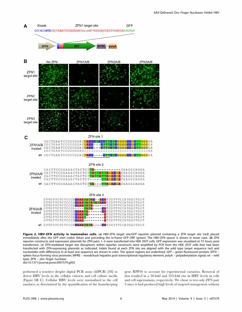
HBV-ZFN activity in mammalian cellsTo determine the activity of our HBV-ZFNs in mammalian
cells we used a GFP reporter assay [16] to detect disruption of
each ZFN target site. Three reporter plasmids each containing an
HBV-ZFN target site between the GFP start codon and ORF
(Figure 2A) were generated. The plasmids were individually
transfected together with combinations of ZFN-expressing plas-
mids in order to monitor frame-shift mutagenesis of circular
double-stranded DNA substrates in HEK 293T cells. A reduction
in GFP expression was seen for each target site plasmid when
treated with its corresponding ZFN pair (Figure 2B), suggesting
that sequence-specific gene disruption was responsible. To confirm
AAV-Delivered Zinc Finger Nucleases Inhibit HBV
PLOS ONE | www.plosone.org 4 May 2014 | Volume 9 | Issue 5 | e97579

mutagenesis, the ZFN target sequences were amplified by PCR
from cell DNA extracts. Clonal PCR amplicons were sequenced
and both deletions and insertions were found within the target sites
of all 3 ZFNs ranging from a 4 nt insertion to a 23 nt deletion
(Figure 2C). Disruptive frame-shift mutations were introduced
within the target sites of all reporter constructs specifically at the
spacer regions where the ZFNs are expected to cleave. As such, we
concluded that our HBV-ZFNs were able to efficiently disrupt
gene expression in mammalian cells.
scAAV-mediated delivery of HBV-ZFNsHigh levels of transgene delivery into target cells would be
essential for therapeutic benefit. Viral vectors such as adeno-
associated virus (AAV) vectors are capable of transducing
hepatocytes both in vitro and in vivo at high levels without
substantial toxicity. Self-complementary AAV (scAAV) vectors
have been shown to achieve higher levels of transduction and
greater transgene expression than single-stranded AAV (ssAAV)
vectors [37]. Therefore, we generated a panel of reporter- or ZFN-
expressing scAAV vector constructs to maximize delivery
(Figure 3A). scAAV-mediated gene delivery was then analyzed
in the HepAD38 cell line, an in vitro model for HBV replication, by
flow cytometry. Depending on the application, different AAV
capsids could be generated according to which serotype provides
the most efficient delivery. Therefore, capsids from AAV serotypes
1, 2, 4, 6, 8 and 9 were all tested in order to determine which
capsid would be most effective for transduction of the HepAD38
cell line. scAAV2 provided the most efficient gene delivery
(Figure 3B).
To determine whether scAAV vectors could be used to deliver
both halves of a ZFN to a significant number of hepatocytes, we
co-infected HepAD38 cells with 2 scAAV reporter viruses
(scAAV2-EFS-GFP and scAAV2-EFS-mCherry). Up to 96% of
HepAD38 expressed both GFP and mCherry (Figure 3C). We
observed significant co-expression even after 6 days post-
transduction with over 70% of cells still positive for both GFP
and mCherry (Figure 3D). These data suggest that scAAV can be
used to efficiently deliver both halves of a therapeutic ZFN for a
significant length of time.
scAAV-HBV-ZFN-mediated cellular toxicityWe analyzed the effects of transduction with scAAV-HBV-ZFN
on HepAD38 cell viability. While scAAV infection at an MOI of
10000 genomes/cell reduced cell viability slightly, the viability of
cells co-transduced with ZFN pairs containing ZFN2A and/or
ZFN2B was below the other ZFN pairs (Figure 4A). Additionally,
cells transduced with ZFN2A or ZFN2B had lower viability when
compared to the other ZFNs individually from pair 1 or 3
(Figure 4B). In two-week-long viability assays, co-transduction with
GFP and mCherry or ZFN pair 1 or 3 had no discernable effect on
cell survival over 14 days in culture (Figure 4C). On the other
hand, mismatch pair ZFN2A/3B caused reduced levels of cell
survival, and ZFN pair 2 showed significant levels of toxicity at
days 5 and 7 post-transduction (Figure 4C). By day 9 all cells in
this treatment group had died. Thus, expression of either ZFN2A
or ZFN2B alone reduced HepAD38 cell viability, and in
combination their expression was lethal.
scAAV-HBV-ZFN-mediated DNA mutations in HepAD38cells
Dox inhibits HBV replication in HepAD38 cells. While
replication is stopped, the only substrate for HBV gene disruption
is the single integrated copy of the HBV genome. Three days after
ZFN delivery by scAAV2 vectors in cells incubated with dox,
DNA extracted from the cells was analyzed for mutations through
Surveyor nuclease digestion or NcoI digestion (Figure 5A–C).
Surveyor analysis, a method for identifying DNA mutations at a
specific site, showed that 9.8%, 34% and 28% of ZFN target sites
1, 2 and 3 were mutated, respectively, when treated with their
corresponding ZFN pairs (Figure 5A–B, Table 1). ZFN site 3
contains an NcoI restriction enzyme site 10 nucleotides away from
the center of the ZFN cleavage site, which could be lost upon large
DNA deletions at the cleavage site (Figure 5D). After NcoI
digestion of the ZFN3 target site PCR amplicon, quantification of
undigested PCR product showed that 2.1% of the DNA was
resistant to digestion by NcoI in ZFN3A/B-treated cells
(Figure 5C). This indicated that the NcoI site was lost in a small
fraction of HBV genomes following treatment with ZFN3A/B.
HepAD38 cells were also treated with all 3 HBV-specific ZFNs at
one-third the MOI for each ZFN pair, resulting in gene disruption
in 20% and 8% of ZFN sites 2 and 3, respectively (Figure 5A–B).
Clonal amplicon sequencing of the target sites revealed insertions
and deletions at each of the ZFN sites (Figure 5D, Table 1) when
treated with their corresponding ZFN pairs. Overall, a significant
level of ZFN target site mutation was seen after HBV-ZFN
treatment.
In order to analyze the ability of the ZFN pairs to target and
mutate HBV cccDNA in HepAD38 cells, similar experiments
were run in which prior to treatment with scAAV-HBV-ZFN,
cccDNA production was induced by removing dox. Dox was then
added at the same time as treatment with the scAAV-HBV-ZFN
to prevent new cccDNA production and ensure that any mutations
detected in cccDNA would not reflect newly synthesized cccDNA
derived from mutated integrated HBV sequences. Cellular DNA
extracts were treated with ATP-dependent Plasmid-safe DNase in
order to isolate cccDNA. When Surveyor analysis or clonal PCR
amplicon sequencing was performed, evidence of site-specific
mutations could not be reproducibly detected (data not shown).
Off-target mutagenesisTo analyze the fidelity of the ZFNs to their cognate HBV target
sites, we selected seven potential off-target sites contained in the
human genome with 9 or fewer mismatches from the HBV sites
(Table S2). Using single molecule real time (SMRT) sequencing,
we sequenced PCR amplicons from cell extracts following
treatment with scAAV2-ZFNs. Reads were eliminated for being
too low quality (LQ), the wrong size, possessing significant
differences from the reference sequences, or for containing a
single nucleotide indel in the target site spacer region. Amplicons
for the genomic HBV on-target ZFN target sites were included in
the sequencing run. These revealed frequencies of mutagenesis of
16%, 43%, and 24% for site 1, 2, and 3, respectively, when treated
with their corresponding ZFN pair (Table 2, top). From the 9,290
filtered sequencing reads for the seven off-target sites treated with
their corresponding ZFN pair (Table 2, bottom), the presence of
indels larger than 1 nt were found in the target site of only 4 reads
(Figure S2). Thus, we concluded that the HBV-specific ZFNs
caused extremely minimal levels of off-target mutagenesis at these
sites in the HepAD38 cell line.
Inhibition of HBV replication after ZFN treatment ofHepAD38 cells
To test the effect of ZFNs on HBV replication, dox was
removed 3 days after treatment with scAAV2-ZFNs (Figure 6A).
Upon removal of dox, an intact HBV genome will initiate
replication, producing high levels of cellular relaxed circular DNA
(rcDNA) and secretion of new virions. Seven days later, we
AAV-Delivered Zinc Finger Nucleases Inhibit HBV
PLOS ONE | www.plosone.org 5 May 2014 | Volume 9 | Issue 5 | e97579
performed a sensitive droplet digital PCR assay (ddPCR) [38] to
detect HBV levels in the cellular extracts and cell culture media
(Figure 6B–C). Cellular HBV levels were normalized to the cell
numbers as determined by the quantification of the housekeeping
gene RPP30 to account for experimental variation. Removal of
dox resulted in a 30-fold and 323-fold rise in HBV levels in cells
and cell supernatants, respectively. We chose to test only ZFN pair
3 since it had produced high levels of targeted mutagenesis without
Figure 2. HBV-ZFN activity in mammalian cells. (a) HBV-ZFN target site/GFP reporter plasmid containing a ZFN target site (red) placedimmediately after the GFP start codon (blue) and preceding the in-frame GFP ORF (green). The HBV-ZFN spacer is shown in lower case. (b) ZFNreporter constructs and expression plasmids for ZFN pairs 1–3 were transfected into HEK 293T cells. GFP expression was visualized at 72 hours posttransfection. (c) ZFN-mediated target site disruptions within reporter constructs were amplified by PCR from the HEK 293T cells that had beentransfected with ZFN-expressing plasmids as indicated. Indels found at each ZFN site are aligned with the wild type target sequence (wt) andnucleotides with differences in at least one sequence are shown in color. The spacer regions are underlined. GFP – green fluorescent protein; SFFV –spleen focus-forming virus promoter; WPRE – woodchuck hepatitis post-transcriptional regulatory element; polyA – polyadenylation signal; wt – wildtype; ZFN – zinc finger nuclease.doi:10.1371/journal.pone.0097579.g002
AAV-Delivered Zinc Finger Nucleases Inhibit HBV
PLOS ONE | www.plosone.org 6 May 2014 | Volume 9 | Issue 5 | e97579

affecting cell viability. When cells treated with ZFN pair 3 were
analyzed following dox removal, we detected no significant
increase in cellular or supernatant levels of HBV.
We next tested the duration of the antiviral effect of the ZFNs in
HepAD38 during 14 days following dox removal and tested both
ZFN pairs 1 and 3 (Figure 6D–E). Analysis of cellular and
supernatant HBV levels showed that while treatment with ZFN
pair 1 produced a mild reduction in HBV levels over 14 days,
treatment with ZFN pair 3 produced a sustained suppression of
HBV levels over the course of the experiment.
Discussion
As a different approach from current anti-viral drug therapies,
we are developing a strategy to eliminate replication-competent
virus from chronic HBV patients. Here, we present data on the use
of ZFNs to generate DSBs in unique DNA sequences within HBV
genes. We predict that the introduction of targeted DSBs will
result in mutagenesis within the HBV genes and prevent viral gene
expression and viral replication. Previously, duck HBV and
human HBV have been targeted with zinc finger proteins
[39,40] and ZFNs [17]. We have applied a similar technology
toward targeting HBV and have utilized scAAV delivery vectors to
achieve high levels of DNA mutagenesis with minimal off-target
effects.
AAV vectors have safely been used to target liver cells in a
number of animal models [41–43] and clinical trials [44,45].
scAAV vectors achieve high transduction in multiple cell types and
increased transgene expression over other AAV vectors such as
single-stranded AAV [37], which have previously been used to
effectively deliver ZFNs to the mouse liver [46]. However, the
smaller packaging capacity of scAAV only allows for one ZFN
subunit, thus requiring the co-transduction of two scAAV vectors
to deliver a full ZFN pair. We were able to efficiently co-transduce
over 95% of HepAd38 cells in culture, and scAAV co-transduction
had a limited effect on cell viability at the experimental MOI.
Additionally, AAV vectors of different serotypes can easily be
constructed as needed depending on the application. In vivo
transduction of close to 100% of mouse hepatocytes with AAV8
vectors has been shown at doses that produce no adverse toxicity
[47–49]. Therefore, we anticipate that a high level of co-
transduction can be achieved in the liver when the optimal vector
is administered intravenously. The recent development of a
chimeric AAV serotype with highly efficient human hepatocyte-
tropic transduction when administered to a humanized murine
model [50] will be very useful in future in vivo murine HBV
experiments. Moreover, it has been reported that infection with
HBV increases levels of AAV transduction in the liver both in vitro
and in vivo [51].
In our ZFN target site/GFP reporter assay using transient
plasmid transfections, ZFN pairs 1, 2 and 3 were able to efficiently
knock down GFP expression in a manner that was dependent on
site-specific gene disruption. However in HepAD38 cells, ZFN
pairs 2 and 3 showed markedly higher targeted mutagenesis levels
Figure 3. scAAV vector transduction of HepAD38 cells. (a) scAAV vector construct organization. (b) Cells were transduced with scAAV vectorscontaining a CMV-hrGFP-pA expression cassette at a MOI of 500 (open bars) or 5000 (closed bars) vector genomes/cell, or (c) were co-transduced withscAAV2-EFS-GFP and scAAV2-EFS-mCherry vectors at indicated MOI. Cells were analyzed for GFP and mCherry expression by flow cytometry at72 hours post infection. (d) HepAD38 co-transduction persistence. Cells were transduced with scAAV2-EFS-eGFP and/or scAAV-EFS-mCherry at MOI5000 genomes/vector/cell and co-transduction levels were monitored by flow cytometry at 2 and 6 days post transduction. ITR – inverted terminalrepeat; CMV – cytomegalovirus immediate early promoter; EFS – elongation factor-1a short promoter; hrGFP – humanized Renilla green fluorescentprotein; eGFP – enhanced green fluorescent protein; pA – polyadenylation signal; ZFN – zinc finger nuclease.doi:10.1371/journal.pone.0097579.g003
AAV-Delivered Zinc Finger Nucleases Inhibit HBV
PLOS ONE | www.plosone.org 7 May 2014 | Volume 9 | Issue 5 | e97579

than ZFN pair 1. When all 3 ZFN pairs were delivered together
this became even more apparent as no gene disruptions were
detected at ZFN site 1 by Surveyor assay or clonal amplicon
sequencing. This may be because ZFN pair 1 is a less efficient
cleaver than ZFN pairs 2 and 3. Another possible explanation is
that the ZFN site 1 in HepAD38 cells contains 4 mismatches from
the target sequence for which the ZFN was designed whereas ZFN
sites 2 and 3 only contain 1 and 2 mismatches, respectively
(Figure 1B, blue letters). Alternatively, it could be because ZFN site
1 is less accessible than ZFN sites 2 and 3 due to epigenetic
modification.
Previous studies have shown that the expression of ZFN pairs
can lead to cellular toxicity that is likely mediated by non-specific
cleavage at similar off-target binding sites [52,53]. Although our
ZFN pairs contain FokI nuclease domains that require hetero-
dimerization for cleavage [26], which is thought to minimize the
levels of ZFN-derived toxicity, toxicity still occurred for ZFN2.
During our cell viability analysis we found that ZFN pair 2 was
cytotoxic to HepAD38 cells, killing transduced cells by day 9 post-
infection. Although ZFN2 did not exhibit off-target cleavage in the
three sites chosen for analysis, this does not guarantee that there is
not non-specific cleavage at other off-target sites in the human
genome. ZFN targeting is known to involve context dependency
for sequence binding [53], and off-target cleavage may occur more
efficiently at sites with less sequence homology than the closest
matches that are generated by tools such as PROGNOS.
Particularly because of the smaller recognition sequence of
ZFN2B compared to the other zinc finger recognition domains
(15 vs. 18 nt), the quantity of near-matches throughout the
genome is substantially higher for ZFN pair 2. An unbiased
method for detecting off-target cleavage could potentially identify
sites where cleavage occurs that we were unable to determine
Figure 4. scAAV-ZFN induced cytotoxicity. (a) HepAD38 cells were transduced with scAAV2 vectors expressing GFP and mCherry reportergenes, ZFN pairs 1, 2 or 3, or mismatched ZFN pairs at a total MOI of 10000 genomes/cell, or all 3 ZFN pairs at a total MOI of 10000 (low) and 30000(high) genomes/cell. At 48 hours post transduction, cell viability was measured by MTT assay and reported as percent of control. (b) HepAD38 cellstransduced with scAAV2 vectors expressing individual ZFN half sites at a MOI of 5000 genomes/cell were also analyzed. (c) Untreated cells and cellstreated with scAAV2 vectors expressing reporter genes, ZFN pairs 1, 2 or 3 or a mismatch ZFN pair were monitored for cell viability at 3, 5, 7 and 14days post transduction. eGFP – enhanced green fluorescent protein; ZFN – zinc finger nuclease.doi:10.1371/journal.pone.0097579.g004
AAV-Delivered Zinc Finger Nucleases Inhibit HBV
PLOS ONE | www.plosone.org 8 May 2014 | Volume 9 | Issue 5 | e97579

using PROGNOS, and potentially help to explain the toxic effect
of ZFN2. The requirement for FokI to heterodimerize in order to
cleave DNA makes it surprising that expression of ZFN2A or
ZFN2B alone was toxic to cells. In fact, all groups containing one
or both of ZFN2A or ZFN2B had reduced viability. Off-target
cleavage by FokI homodimers could explain the toxicity. It is also
possible that ZFN2A and ZFN2B are able to bind to specific DNA
sequences and act as transcriptional inhibitors in a manner that
induces cellular toxicity, without necessarily inducing DSBs.
To analyze the possibility of off-target mutagenic activity by the
ZFN pairs, a high-throughput sequencing method was employed.
SMRT sequencing allowed 10 separate PCR amplicons pooled on
an individual chip to be sequenced resulting in over 104 high
quality reads. By aligning the sequence reads to reference
sequences, indels at the HBV target sites or off-target sites could
easily be detected. The degree of targeted mutagenesis observed in
the three HBV sites was in agreement with earlier results obtained
by clonal sequencing and the Surveyor assay (Tables 1 and 2). The
higher number of reads provided by SMRT allowed more precise
quantification of targeted mutagenesis, which showed significant
differences between the three ZFN pairs, such that HBV
mutagenesis by Z2 & Z3 . Z1. These targeting efficiencies likely
stem from the number of mismatches between the HepAD38
HBV sequence and the designed target sites (1, 2, and 4, for Z2,
Z3 and Z1, respectively). All seven of the off-target sites showed
either no or exceedingly small levels of mutagenesis. Based on the
large number of reads, we can confidently say that the upper limit
for off-target cleavage was 0.16% for any of the off-target sites.
Moreover, the 4 reads indicating potential mutagenesis (out of
9290) all had small indels in regions with high sequence repetition
(Figure S2) indicating that they could have been homopolymer-
associated sequencing errors that happened to fall within the off-
target site.
Despite only inducing mutations in 24–28% of genomic HBV
target sites at 3 days post-delivery, ZFN pair 3 was able to knock
down HBV replication and production of infectious HBV almost
entirely by day 7. The Surveyor assay likely underestimates the
true mutation rate [54,55], so our actual rate of mutagenesis at day
3 may have been higher. It is also likely that in addition to
disrupting genomic HBV sequences in HepAd38 cells, ZFNs can
generate DSBs in both cccDNA and rcDNA species, which
contributes to the knockdown of HBV replication seen in ZFN-
pair-3-treated cells. However, it must be noted that we were
unable to obtain reproducible evidence of cccDNA disruption in
HepAD38 cells. Since we were able to achieve a complete and
sustained knockdown of HBV replication with a single dose of
Figure 5. ZFN-induced target site disruption in HepAD38 cells.(a, b) Cells were transduced with scAAV2 vectors expressing ZFN pairs1, 2, 3 or all three pairs together (1–3) at a total MOI of 10000 genomes/cell. The presence of mutations was analyzed in DNA isolated fromtransduced cells 72 hours later using the Surveyor nuclease assay. (c)For ZFN site 3, an analysis of DNA mutagenic events disrupting theinternal NcoI cleavage site was also performed. Above the gel images,the sizes of PCR amplicons and the cleavage products produced uponSurveyor nuclease cleavage (indicating mutations at the indicatedtarget site) or NcoI cleavage are shown. bp – base pairs; UC – untreatedcontrol; ZFN – zinc finger nuclease. Bands indicating mutations arehighlighted with an asterisk and the percentage of ZFN-mediated DNAmutation for each targeted site is indicated. (d) DNA mutations thatwere detected at ZFN target sites 1, 2 and 3 within HepAD38 HBVsequences are shown above the wild-type ZFN site. Nucleotides withdifferences in at least one sequence are shown in color. Spacer regionsare underlined. The rates at which DNA mutations were detected arelisted in Table 1. wt – wild type.doi:10.1371/journal.pone.0097579.g005
AAV-Delivered Zinc Finger Nucleases Inhibit HBV
PLOS ONE | www.plosone.org 9 May 2014 | Volume 9 | Issue 5 | e97579

scAAV2-ZFN3A/3B, we did not attempt to use multiple ZFN
doses. In an in vivo model of HBV replication where a successful
outcome may be more dependent on higher levels of HBV gene
disruption, it may be desirable to give multiple doses of therapeutic
AAV vector. We have used mathematical modeling to predict the
likelihood of success for our approach in eliminating HBV
infections and predicted that multiple therapeutic doses of HBV-
specific enzymes will likely be needed [56]. Of note, at day 7 the
mismatched pair of ZFN2A/3B showed a significant inhibition of
HBV replication compared to the untreated control (Figure 6B–
C), although not to the extent of ZFN pair 3. This knockdown was
observed for all scAAV-treated samples, indicating that infection
with the scAAV vector regardless of its payload has an inhibitory
effect on HBV.
In a clinical setting, cccDNA would be the major target for
HBV-specific ZFNs in hepatocytes of infected patients receiving
concurrent antiviral drug therapy. It will be important to
determine the efficiency with which site-specific endonucleases
can target cccDNA. Despite reports that cccDNA can be targeted
at certain sites by TALENs [22,23], we believe the question of
cccDNA cleavage and mutation efficiency requires continued
attention. Bloom et al. published evidence of TALEN-mediated
mutations in the cccDNA of HepG2.2.15 cells [23] and Chen et al.
showed mutations in cccDNA in Huh7 cells transfected with linear
HBV DNA [22]. However, neither group presented data showing
cccDNA mutation in HepAD38 cells. It may be that the cccDNA
present in HepAD38 cells is not amenable to cleavage, that it is not
present in sufficient quantities to detect mutations, or that ZFNs
are not as effective as TALENs. Our data would suggest that
cccDNA is a more difficult substrate for targeted mutation than
plasmid DNA or integrated DNA, and a more detailed analysis of
its susceptibility to HBV-specific endonucleases would highly
benefit this field of research. In vivo cccDNA takes on a highly
organized chromatinized structure often referred to as the HBV
minichromosome [57–59]. Due to the specific interactions
between histones and cccDNA within the minichromosome it is
unclear the degree to which target sites will be accessible for
endonuclease-mediated cleavage. Aubert et al. [21] demonstrated
that chromatin modifiers such as histone deacetylase inhibitors
served to increase the mutagenic activity of targeted endonucleases
on episomal herpes simplex virus DNA, and similar approaches
might improve the mutagenesis of HBV cccDNA.
In order to detect HBV levels, we developed a sensitive ddPCR
assay that can accurately detect sequences from HBV genotypes
A–H. This involved using a primer/probe combination that is
routinely used in a Taqman qPCR assay by the University of
Washington Molecular Virology Laboratory to detect HBV in
patient sera. This primer/probe combination is targeted to HBV
ORF P/S and is used to detect infectious HBV in serum. In our
HepAD38 cellular DNA extract samples, this assay will also detect
genomic (integrated) HBV, rcDNA and cccDNA. Therefore, the
experimental data must be interpreted with caution. In cellular
DNA samples, we normalized HBV DNA values to the human
gene RPP30, and thus values likely represent the total number of
rcDNA and cccDNA molecules per cell.
In summary, we have developed an effective strategy to inhibit
replication of HBV via HBV-specific ZFNs. We were able to
efficiently deliver therapeutic ZFNs to HBV-infected hepatocytes
using scAAV vectors, a platform for gene delivery that is adaptable
to in vivo-scale HBV treatments, without substantial toxicity.
Moreover, we have analyzed off-target activity of our three HBV-
specific ZFNs at seven potential off-target sites without detecting
any significant mutagenesis. The antiviral activity of our AAV-
delivered therapeutic ZFNs was significant for ZFN pair 3, which
reduced HBV DNA production to near-baseline levels and was
sustained for up to two weeks. Taken together, these results
support the feasibility of a ZFN-based strategy for targeting
hepatitis B virus.
Table 1. Summary of HBV-ZFN-induced mutagenesis in HepAD38 cells.
ZFN site ZFN treatmentTotal mutations (colonysequencing) Insertions (size nt) Deletions (size nt) Other
Surveyor assay (%indels)*
1 Uninfected 0/18 0%
1A/B 5/20 0 4 (4, 7, 16, 26) 1 (HR) 9.8%
2A/B 0%
3A/B 0/21
2A/3B 0/17
all 1–3 0/62 0 0 0 0%
2 Uninfected 0/12 0%
1A/B 0%
2A/B 6/15 4 (2, 2, 4, 4) 2 (7, 23) 0 34%
3A/B 0%
2A/3B 0/20
all 1–3 5/64 4 (2, 3, 4, 4) 1 (29) 0 20%
3 Uninfected 0/18 0%
1A/B 0/20 0%
3A/B 3/21 2 (2, 5) 1 (69) 0 28%
2A/3B 0/17
all 1–3 1/62 0 1 (16) 0 8%
*% indel = (12(12(a+b)/(a+b+c))‘0.5)*100; where a and b = cut bands, c = uncut band.doi:10.1371/journal.pone.0097579.t001
AAV-Delivered Zinc Finger Nucleases Inhibit HBV
PLOS ONE | www.plosone.org 10 May 2014 | Volume 9 | Issue 5 | e97579

Ta
ble
2.
SMR
Tse
qu
en
cin
gre
sult
ssh
ow
ing
on
-tar
ge
tan
do
ff-t
arg
et
acti
vity
of
HB
V-Z
FNs.
ZF
Np
air
tre
atm
en
ta
mp
li-c
on
chro
mo
som
elo
cati
on
mis
-ma
tch
rea
ds
eli
min
ate
dre
ma
inin
gre
ad
sto
tal
ind
els
inse
rts
de
leti
on
so
the
rta
rge
tin
g(%
)
LQ
po
or
ali
gn
sin
gle
in-
de
lin
TS
On
-ta
rge
tH
BV
site
s
Z1
HB
V_
Z1
HB
V_
14
41
47
99
13
33
97
05
57
88
25
61
21
5.8
*
Z2
HB
V_
Z2
HB
V_
23
27
13
27
41
77
38
28
82
77
11
20
05
94
43
31
73
43
.3*
Z3
HB
V_
Z3
HB
V_
13
34
23
14
71
12
82
23
53
21
30
22
3.8
*
Off
-ta
rge
tsi
tes
Z1
Z1
Ch
14
chr1
4_
74
70
71
99
92
46
42
53
69
84
20
58
10
10
0.0
5
Z1
Ch
9ch
r9_
11
42
46
41
09
61
89
57
04
44
90
00
00
Z2
Z2
Ch
5ch
r5_
11
16
07
94
08
17
29
19
65
12
41
45
80
00
00
Z2
Ch
Xch
rX_
13
91
73
04
77
20
84
19
45
25
61
78
20
00
00
Z2
Ch
Y,
Z2
Ch
15
chrY
_2
75
76
15
7,
chr1
5_
84
92
35
24
77
93
13
13
26
63
31
01
00
.16
Z3
Z3
Ch
4ch
r4_
13
45
02
13
78
12
79
11
82
06
11
35
00
00
0
Z3
Ch
15
chr1
5_
34
29
85
24
82
08
32
53
38
17
17
75
21
10
0.1
1
LQ,
low
qu
alit
y;T
S,ta
rge
tsi
te;
*Sig
nif
ican
ceb
etw
ee
nZ
1an
dZ
3(p
,0
.01
),Z
1an
dZ
2(p
,0
.00
1),
Z2
and
Z3
(p,
0.0
01
).d
oi:1
0.1
37
1/j
ou
rnal
.po
ne
.00
97
57
9.t
00
2
AAV-Delivered Zinc Finger Nucleases Inhibit HBV
PLOS ONE | www.plosone.org 11 May 2014 | Volume 9 | Issue 5 | e97579

Supporting Information
Figure S1 HBV target site sequence heterogeneity.Sequence heterogeneity for HBV-ZFN pairs 1–3 across 440
genotype A, 2233 genotype B&C, or 674 genotype D HBV
sequences found in Genbank. For each ZFN pair the target
sequence, logo plots, and consensus sequences obtained by
aligning the target sequences with the genotype A, genotype
B&C, or genotype D HBV sequences are shown. ZFN spacer
nucleotides are highlighted in red and divergent nucleotides
between the ZFN target site and the consensus sequence are bold
and underlined. Single nucleotide polymorphisms present in the
HepAD38 genomic HBV sequence are shown above each target
Figure 6. Levels of HBV DNA present in ZFN-treated HepAD38 cells and levels of secreted infectious HBV. (a) For experiments shown inpanels b and c, HepAD38 cells in the presence of dox were transduced with ZFN- or control-expressing scAAV2 vectors (total MOI 10000 genomes/cell) and 3 days later dox was removed from culture medium to enable HBV replication. Cells were left in culture for a further 7 days before HBVgenomic levels were quantified in cells (b) and infectious HBV levels were quantified in supernatants (c) by ddPCR. (d–e) HepAD38 cells werepassaged every two to three days over the course of 14 days following treatment with ZFN- or control-expressing scAAV vectors and monitored forcellular (d) and supernatant (e) HBV levels by ddPCR. ddPCR – droplet digital polymerase chain reaction; dox – doxycycline; eGFP – enhanced greenfluorescent protein; scAAV – self-complementary adeno-associated virus; UC – untreated control; ZFN – zinc finger nuclease. *p#0.05.doi:10.1371/journal.pone.0097579.g006
AAV-Delivered Zinc Finger Nucleases Inhibit HBV
PLOS ONE | www.plosone.org 12 May 2014 | Volume 9 | Issue 5 | e97579

site in blue. Logo plots and consensus sequences were obtained
with the use of GeneiousPro. ZFN – zinc finger nuclease.
(TIF)
Figure S2 Instances of indels in off-target site spacerregions. Alignments with wt reference sequences of the 4
sequence reads of off-target sites containing indels in the spacer
region. These were taken from a total of 9290 total reads of off-
target sites obtained from PCR amplicons generated from DNA
from cells that had been treated with ZFN-expressing scAAV2
vectors as indicated. Spacer regions are underlined. wt – wild type;
ZFN – zinc finger nuclease.
(TIF)
Table S1 PCR primer sets.(DOCX)
Table S2 Three on-target and 7 off-target ZFN sites.*Target sequence, red underlined nucleotides show spacer regions,
lowercase letters show mismatches from HBV sites, blue
nucleotides show variations contained in the HepAD38 HBV
genome that are different from the designed ZFN sites.
(DOCX)
Acknowledgments
We would like to thank Dr. Henry Fechner for kindly providing the
plasmid pscAAV-GFP, Dr. Lorne Tyrrell for providing us with the
HepAD38 cell line with permission from Dr. Ju-Tao Guo of Drexel
University, and Dr. Andy Scharenberg and Dr. David Rawlings for critical
discussions. We thank Harlan Pietz and Kevin Kwong for technical
assistance. We also thank Dave Swan and Dr. Tomer Hertz for assistance
with Rate4Site analyses.
Author Contributions
Conceived and designed the experiments: NDW DS MA KRJ. Performed
the experiments: NDW DS RHS HSDSF. Analyzed the data: NDW DS
RHS HSDSF PR JTS MA KRJ. Wrote the paper: NDW DS KRJ.
References
1. Lee WM (1997) Hepatitis B virus infection. N Engl J Med 337: 1733–1745.
2. Beasley RP, Hwang LY, Lin CC, Chien CS (1981) Hepatocellular carcinoma
and hepatitis B virus. A prospective study of 22 707 men in Taiwan. Lancet 2:
1129–1133.
3. Dienstag JL (2008) Hepatitis B virus infection. N Engl J Med 359: 1486–1500.
4. Werle-Lapostolle B, Bowden S, Locarnini S, Wursthorn K, Petersen J, et al.
(2004) Persistence of cccDNA during the natural history of chronic hepatitis Band decline during adefovir dipivoxil therapy. Gastroenterology 126: 1750–
1758.
5. Carroll D (2011) Genome engineering with zinc-finger nucleases. Genetics 188:
773–782.
6. Schiffer JT, Aubert M, Weber ND, Mintzer E, Stone D, et al. (2012) Targeted
DNA mutagenesis for the cure of chronic viral infections. J Virol 86: 8920–8936.
7. Stone D, Kiem HP, Jerome KR (2013) Targeted gene disruption to cure HIV.
Curr Opin HIV AIDS 8: 217–223.
8. Weber ND, Aubert M, Dang CH, Stone D, Jerome KR (2014) DNA cleavage
enzymes for treatment of persistent viral infections: Recent advances and thepathway forward. Virology doi: 10.1016/j.virol.2013.12.037.
9. Handel EM, Cathomen T (2011) Zinc-finger nuclease based genome surgery: it’sall about specificity. Curr Gene Ther 11: 28–37.
10. Boissel S, Jarjour J, Astrakhan A, Adey A, Gouble A, et al. (2014) megaTALs: arare-cleaving nuclease architecture for therapeutic genome engineering. Nucleic
Acids Res 42: 2591–2601.
11. Sander JD, Ramirez CL, Linder SJ, Pattanayak V, Shoresh N, et al. (2013) In
silico abstraction of zinc finger nuclease cleavage profiles reveals an expandedlandscape of off-target sites. Nucleic Acids Res 41: e181.
12. Cradick TJ, Fine EJ, Antico CJ, Bao G (2013) CRISPR/Cas9 systems targetingbeta-globin and CCR5 genes have substantial off-target activity. Nucleic Acids
Res 41: 9584–9592.
13. Cho SW, Kim S, Kim Y, Kweon J, Kim HS, et al. (2014) Analysis of off-target
effects of CRISPR/Cas-derived RNA-guided endonucleases and nickases.Genome Res 24: 132–141.
14. Fu Y, Foden JA, Khayter C, Maeder ML, Reyon D, et al. (2013) High-frequencyoff-target mutagenesis induced by CRISPR-Cas nucleases in human cells. Nat
Biotechnol 31: 822–826.
15. Fine EJ, Cradick TJ, Zhao CL, Lin Y, Bao G (2013) An online bioinformatics
tool predicts zinc finger and TALE nuclease off-target cleavage. Nucleic AcidsRes doi: 10.1093/nar/gkt1326.
16. Aubert M, Ryu BY, Banks L, Rawlings DJ, Scharenberg AM, et al. (2011)Successful targeting and disruption of an integrated reporter lentivirus using the
engineered homing endonuclease Y2 I-AniI. PLoS One 6: e16825.
17. Cradick TJ, Keck K, Bradshaw S, Jamieson AC, McCaffrey AP (2010) Zinc-
finger nucleases as a novel therapeutic strategy for targeting hepatitis B virusDNAs. Mol Ther 18: 947–954.
18. Tanaka A, Takeda S, Kariya R, Matsuda K, Urano E, et al. (2013) A noveltherapeutic molecule against HTLV-1 infection targeting provirus. Leukemia
27: 1621–1627.
19. Mino T, Mori T, Aoyama Y, Sera T (2013) Gene- and protein-delivered zinc
finger-staphylococcal nuclease hybrid for inhibition of DNA replication of
human papillomavirus. PLoS One 8: e56633.
20. Grosse S, Huot N, Mahiet C, Arnould S, Barradeau S, et al. (2011)Meganuclease-mediated Inhibition of HSV1 Infection in Cultured Cells. Mol
Ther 19: 694–702.
21. Aubert M, Boyle NM, Stone D, Stensland L, Huang ML, et al. (2014) In vitro
Inactivation of Latent HSV by Targeted Mutagenesis Using an HSV-specific
Homing Endonuclease. Mol Ther Nucleic Acids 3: e146.
22. Chen J, Zhang W, Lin J, Wang F, Wu M, et al. (2014) An Efficient Antiviral
Strategy for Targeting Hepatitis B Virus Genome Using Transcription
Activator-Like Effector Nucleases. Mol Ther 22: 303–311.
23. Bloom K, Ely A, Mussolino C, Cathomen T, Arbuthnot P (2013) Inactivation of
hepatitis B virus replication in cultured cells and in vivo with engineered
transcription activator-like effector nucleases. Mol Ther 21: 1889–1897.
24. Ladner SK, Otto MJ, Barker CS, Zaifert K, Wang GH, et al. (1997) Inducibleexpression of human hepatitis B virus (HBV) in stably transfected hepatoblas-
toma cells: a novel system for screening potential inhibitors of HBV replication.
Antimicrob Agents Chemother 41: 1715–1720.
25. Chenna R, Sugawara H, Koike T, Lopez R, Gibson TJ, et al. (2003) Multiple
sequence alignment with the Clustal series of programs. Nucleic Acids Res 31:3497–3500.
26. Miller JC, Holmes MC, Wang J, Guschin DY, Lee YL, et al. (2007) An
improved zinc-finger nuclease architecture for highly specific genome editing.
Nat Biotechnol 25: 778–785.
27. Kitsera N, Khobta A, Epe B (2007) Destabilized green fluorescent protein
detects rapid removal of transcription blocks after genotoxic exposure.
Biotechniques 43: 222–227.
28. Arad U (1998) Modified Hirt procedure for rapid purification of extrachromo-
somal DNA from mammalian cells. Biotechniques 24: 760–762.
29. Fechner H, Sipo I, Westermann D, Pinkert S, Wang X, et al. (2008) Cardiac-
targeted RNA interference mediated by an AAV9 vector improves cardiac
function in coxsackievirus B3 cardiomyopathy. J Mol Med 86: 987–997.
30. Zychlinski D, Schambach A, Modlich U, Maetzig T, Meyer J, et al. (2008)
Physiological promoters reduce the genotoxic risk of integrating gene vectors.
Mol Ther 16: 718–725.
31. Kota J, Chivukula RR, O’Donnell KA, Wentzel EA, Montgomery CL, et al.
(2009) Therapeutic microRNA delivery suppresses tumorigenesis in a murine
liver cancer model. Cell 137: 1005–1017.
32. Choi VW, Asokan A, Haberman RA, Samulski RJ (2007) Production of
recombinant adeno-associated viral vectors for in vitro and in vivo use. CurrProtoc Mol Biol Chapter 16: Unit 16 25.
33. Fagone P, Wright JF, Nathwani AC, Nienhuis AW, Davidoff AM, et al. (2012)
Systemic errors in quantitative polymerase chain reaction titration of self-
complementary adeno-associated viral vectors and improved alternativemethods. Hum Gene Ther Methods 23: 1–7.
34. Cock PJ, Fields CJ, Goto N, Heuer ML, Rice PM (2010) The Sanger FASTQ
file format for sequences with quality scores, and the Solexa/Illumina FASTQ
variants. Nucleic Acids Res 38: 1767–1771.
35. Loeb KR, Jerome KR, Goddard J, Huang M, Cent A, et al. (2000) High-
throughput quantitative analysis of hepatitis B virus DNA in serum using the
TaqMan fluorogenic detection system. Hepatology 32: 626–629.
36. Mayrose I, Graur D, Ben-Tal N, Pupko T (2004) Comparison of site-specificrate-inference methods for protein sequences: empirical Bayesian methods are
superior. Mol Biol Evol 21: 1781–1791.
37. McCarty DM, Monahan PE, Samulski RJ (2001) Self-complementary
recombinant adeno-associated virus (scAAV) vectors promote efficient trans-duction independently of DNA synthesis. Gene Ther 8: 1248–1254.
38. Sedlak RH, Jerome KR (2013) Viral diagnostics in the era of digital polymerase
chain reaction. Diagn Microbiol Infect Dis 75: 1–4.
39. Hoeksema KA, Tyrrell DL (2010) Inhibition of viral transcription using designed
zinc finger proteins. Methods Mol Biol 649: 97–116.
40. Zimmerman KA, Fischer KP, Joyce MA, Tyrrell DL (2008) Zinc finger proteins
designed to specifically target duck hepatitis B virus covalently closed circular
DNA inhibit viral transcription in tissue culture. J Virol 82: 8013–8021.
AAV-Delivered Zinc Finger Nucleases Inhibit HBV
PLOS ONE | www.plosone.org 13 May 2014 | Volume 9 | Issue 5 | e97579

41. Niemeyer GP, Herzog RW, Mount J, Arruda VR, Tillson DM, et al. (2009)
Long-term correction of inhibitor-prone hemophilia B dogs treated with liver-directed AAV2-mediated factor IX gene therapy. Blood 113: 797–806.
42. Harding TC, Koprivnikar KE, Tu GH, Zayek N, Lew S, et al. (2004)
Intravenous administration of an AAV-2 vector for the expression of factor IX inmice and a dog model of hemophilia B. Gene Ther 11: 204–213.
43. Mount JD, Herzog RW, Tillson DM, Goodman SA, Robinson N, et al. (2002)Sustained phenotypic correction of hemophilia B dogs with a factor IX null
mutation by liver-directed gene therapy. Blood 99: 2670–2676.
44. Nathwani AC, Tuddenham EG, Rangarajan S, Rosales C, McIntosh J, et al.(2011) Adenovirus-associated virus vector-mediated gene transfer in hemophilia
B. N Engl J Med365: 2357–2365.45. Manno CS, Pierce GF, Arruda VR, Glader B, Ragni M, et al. (2006) Successful
transduction of liver in hemophilia by AAV-Factor IX and limitations imposedby the host immune response. Nat Med 12: 342–347.
46. Li H, Haurigot V, Doyon Y, Li T, Wong SY, et al. (2011) In vivo genome
editing restores haemostasis in a mouse model of haemophilia. Nature 475: 217–221.
47. Wang L, Wang H, Bell P, McMenamin D, Wilson JM (2012) Hepatic genetransfer in neonatal mice by adeno-associated virus serotype 8 vector. Hum
Gene Ther 23: 533–539.
48. Chen CC, Sun CP, Ma HI, Fang CC, Wu PY, et al. (2009) Comparative studyof anti-hepatitis B virus RNA interference by double-stranded adeno-associated
virus serotypes 7, 8, and 9. Mol Ther 17: 352–359.49. Ho KJ, Bass CE, Kroemer AH, Ma C, Terwilliger E, et al. (2008) Optimized
adeno-associated virus 8 produces hepatocyte-specific Cre-mediated recombi-nation without toxicity or affecting liver regeneration. Am J Physiol Gastrointest
Liver Physiol 295: G412–419.
50. Lisowski L, Dane AP, Chu K, Zhang Y, Cunningham SC, et al. (2014) Selection
and evaluation of clinically relevant AAV variants in a xenograft liver model.Nature 506: 382–386.
51. Hosel M, Lucifora J, Michler T, Holz G, Gruffaz M, et al. (2014) Hepatitis B
virus infection enhances susceptibility towards adeno-associated viral vectortransduction in vitro and in vivo. Hepatology doi: 10.1002/hep.26990.
52. Pattanayak V, Ramirez CL, Joung JK, Liu DR (2011) Revealing off-targetcleavage specificities of zinc-finger nucleases by in vitro selection. Nat Methods
8: 765–770.
53. Gabriel R, Lombardo A, Arens A, Miller JC, Genovese P, et al. (2011) Anunbiased genome-wide analysis of zinc-finger nuclease specificity. Nat
Biotechnol 29: 816–823.54. Qiu P, Shandilya H, D’Alessio JM, O’Connor K, Durocher J, et al. (2004)
Mutation detection using Surveyor nuclease. Biotechniques 36: 702–707.55. Otto EA, Helou J, Allen SJ, O’Toole JF, Wise EL, et al. (2008) Mutation analysis
in nephronophthisis using a combined approach of homozygosity mapping, CEL
I endonuclease cleavage, and direct sequencing. Hum Mutat 29: 418–426.56. Schiffer JT, Swan DA, Stone D, Jerome KR (2013) Predictors of hepatitis B cure
using gene therapy to deliver DNA cleavage enzymes: a mathematical modelingapproach. PLoS Comput Bio 9: e1003131.
57. Shi L, Li S, Shen F, Li H, Qian S, et al. (2012) Characterization of nucleosome
positioning in hepadnaviral covalently closed circular DNA minichromosomes.J Virol 86: 10059–10069.
58. Pollicino T, Belloni L, Raffa G, Pediconi N, Squadrito G, et al. (2006) HepatitisB virus replication is regulated by the acetylation status of hepatitis B virus
cccDNA-bound H3 and H4 histones. Gastroenterology 130: 823–837.59. Levrero M, Pollicino T, Petersen J, Belloni L, Raimondo G, et al. (2009) Control
of cccDNA function in hepatitis B virus infection. J Hepatol 51: 581–592.
AAV-Delivered Zinc Finger Nucleases Inhibit HBV
PLOS ONE | www.plosone.org 14 May 2014 | Volume 9 | Issue 5 | e97579
Copyright © 2022 FDOKUMEN