Centromeric Protein B Null Mice Are Viable with No Apparent Abnormalities
Upload
independentCategory
view
1download
0
A tandemly repetitive centromeric DNA sequence of the fish Hopliasmalabaricus (Characiformes: Erythrinidae) is derived from 5S rDNA
Cesar Martins1, Irani Alves Ferreira1, Claudio Oliveira1, Fausto Foresti1 & PedroManoel Galetti Jr21Departamento de Morfologia, UNESP-Universidade Estadual Paulista, Instituto de Biociencias, CEP 18618-000, Botucatu, SP, Brazil (E-mail: [email protected]; Phone/Fax: +55-14-38116264); 2Departamentode Genetica e Evolucao, Universidade Federal de Sao Carlos, Centro de Ciencias Biologicas e da Saude, CEP13565-905, Sao Carlos, SP, Brazil
Received 15 June 2005; Accepted 31 August 2005
Key words: 5S rDNA, 5S rDNA variant, centromeric DNA, fish, Hoplias malabaricus, satellite DNA
Abstract
A substantial fraction of the eukaryotic genome consists of repetitive DNA sequences that include satellites,minisatellites, microsatellites, and transposable elements. Although extensively studied for the past threedecades, the molecular forces that generate, propagate and maintain repetitive DNAs in the genomes arestill discussed. To further understand the dynamics and the mechanisms of evolution of repetitive DNAs invertebrate genome, we searched for repetitive sequences in the genome of the fish species Hoplias mala-baricus. A satellite sequence, named 5SHindIII-DNA, which has a conspicuous similarity with 5S rRNAgenes and spacers was identified. FISH experiments showed that the 5S rRNA bona fide gene repeats wereclustered in the interstitial position of two chromosome pairs of H. malabaricus, while the satellite5SHindIII-DNA sequences were clustered in the centromeric position in nine chromosome pairs of thespecies. The presence of the 5SHindIII-DNA sequences in the centromeres of several chromosomes indi-cates that this satellite family probably escaped from the selective pressure that maintains the structure andorganization of the 5S rDNA repeats and become disperse into the genome. Although it is not feasible toexplain how this sequence has been maintained in the centromeric regions, it is possible to hypothesize thatit may be involved in some structural or functional role of the centromere organization.
Introduction
An interesting feature of eukaryote genome is thepresence of a substantial fraction of duplicatedDNA sequences, most of them composed by non-coding sequences that include satellite, minisatel-lite and microsatellite sequences, and transposableelements. Although studied extensively for the pastthree decades, the molecular forces that propagateand maintain repetitive DNAs in the genome arestill discussed. Among whole sequenced genomesthe repetitive areas remains as gaps because of thedifficulty in their correct positioning and array inthe genome. However, the role of these DNAs ingenome organization and evolution, and their
impact on speciation has been frequently reported(Charlesworth, Snlegowski and Stephan, 1994).The variation in genome size of different eukary-otes is often reported to differences in the amountof repeated DNA sequences (Cavalier-Smith,1985; Brenner et al., 1993). Recently advances onstudies concerning non-coding repetitive DNAsequences have shown that such sequences areextremely important in the structural and func-tional organization of the genome.
Among vertebrate species, studies about repeti-tive sequences have been mainly directed to theunderstanding of the structure and organization ofsatellite DNA and ribosomal DNA (rDNA)repeats. Satellite DNA families can correspond to
Genetica (2006) 127:133–141 � Springer 2006DOI 10.1007/s10709-005-2674-y
10–20% of some mammalian genomes (Beridze,1986) and are usually species-specific or found to beconserved within closely related species. They areuseful for molecular cytogenetic analysis, such asthe identification of homologous chromosomes andchromosomal abnormalities by in situ hybridiza-tion. The molecular organization, chromosomallocation, and possible functions of satellite DNAshave been studied in several groups of animals(Singer, 1982; Clabby et al., 1996). These studieshave indicated that satellite-like repetitive DNAsequences may play an important role at thechromosomal and nuclear level (Singer, 1982;Larin, Fricker and Tyler-Smith, 1994; Sart et al.,1997).
Although usually considered as a single biolog-ical species, the taxonomy ofHoplias malabaricus ispoorly understood. Growing evidence has pointedto the karyotypic diversity of H. malabaricus,showing interpopulational differences in the diploidnumber and chromosomemorphology, as well as insex chromosome systems (Bertollo, Takahashi andMoreira-Filho, 1978, 1983; Dergam and Bertollo,1990; Bertollo, Moreira-Filho and Fontes, 1997a,Bertollo et al., 1997b; Lopes et al., 1998; Bertolloand Mestriner, 1998; Born and Bertollo, 2000).Specimens with a putative hybrid karyotype havenot been found when distinct chromosomal forms(cytotypes) are sympatric (Bertollo et al., 2000).Since the fish H. malabaricus has shown to be aninteresting model species for cytogenetic andevolutionary studies, we have investigated repetitivesequences in the genome of this fish. A centromericsatellite DNA family originated from the 5S rDNArepeats was isolated and its composition and orga-nization in the genome of H. malabaricus studied.
Material and methods
Animals, DNA isolation, DNA digestion, PCRand cloning
Genomic DNA of five individuals of Hopliasmalabaricus from the Araqua river (Botucatu, SP,Brazil) was extracted according to standard phe-nol–chloroform procedures (Sambrook andRussel, 2001). Restriction enzyme digestions of thegenomic DNA were conducted with the endonuc-leases HindIII, MspI, PstI, HaeIII, EcoRI, PvuII,ScaI, RsaI and SpeI. The endonuclease HindIII
generated a band of approximately 350 bp thatwas purified from agarose gel for cloning. PCRamplifications of 5S rDNA were performed usingprimers A (5¢-TAC GCC CGA TCT CGT CCGATC-3¢) and B (5¢-CAG GCT GGT ATG GCCGTA AGC-3¢) designed from the 5S rRNA se-quence of rainbow trout (Komiya and Takemura,1979) to amplify the 5S rRNA gene and the non-transcribed spacer (NTS) (hereafter refereed as 5SrDNA repeat). A standard PCR reaction wasperformed using 150 pmoles of each primer, 20 ngof genomic DNA, 1x Taq buffer, 200 lM of eachdNTP and 2 U of Taq polymerase in a finalreaction volume of 50 ll. Cycling times were asfollows: 94�C of denaturation for 5 min, 35 cyclesof 95�C for 1 min, 63�C for 30 s and 72�C for1 min, with post-cycling extension at 72�C for7 min. The PCR-amplified products were visual-ized in 1% agarose gels. The HindIII-DNA frag-ments and the PCR-generated 5S rDNA repeatswere linked in the plasmids pUC18 HindIII-BAP(Amersham Bioscience) and pGEM-T (Promega),respectively, and used to transform DH5a E. colicompetent cells (Invitrogen).
Sequencing and sequence analysis
The clones obtained from the 5S rDNA–PCRproducts (five clones) and the HindIII-DNA frag-ments (five clones) were sequenced on anABI Prism377 DNA sequencer (Perkin-Elmer) with the ABIPrism BigDye Terminator Cycle Sequencing ReadyReaction Kit (Perkin-Elmer). Nucleotide sequenceswere subjected to BLASTN (Altschul et al., 1990)searches at the National Center for BiotechnologyInformation (NCBI), web site (http://www.ncbi.nlm.nih.gov/blast), and the sequencealignment was performed using Clustal W(Thompson, Higgins and Gibson, 1994) andchecked by hands. UPGMA based phylogeneticanalyses and Kimura’s 2-parameter genetic dis-tances were determined using the software MEGA2.1 (Kumar et al., 2001). Bootstrap resembling(Felsenstein, 1985) was applied to assess support forindividual nodes using 1000 replicates.
Southern blot hybridization
The genomic organization of the isolated sequenceswas investigated by Southern blot-hybridization.
134
Genomic DNA (8 lg) was partially (10 and30 min) and overnight (14 h) digested with 45 U ofthe endonuclease HindIII, submitted to gel-elec-trophoresis in 1% agarose and Southern-trans-ferred to Hybon-N nylon membrane (Southern,1975). The enzymeHindIII was used since has onlyone cleaving site in both isolated sequence classes.Filters containing the immobilized DNA wereprobed with cloned monomeric units of thePCR-generated 5S rDNA and with HindIII-DNAsequences. To avoid cross-hybridization betweenthe 5S rDNA and the HindIII-DNA, the filterhybridization was carried out with the kit ECL-direct nucleic acid labeling and detection system(Amersham Bioscience) using high stringencyconditions. The probes were denatured at 95�C,covalently labeled with the enzyme horseradishperoxidase and hybridized for 14 h to filterimmobilized target DNA in a hybridization buffer(6 M urea/50% formamide/0.5 M NaCl) at 42�C.After the hybridization the filters were washed in6 M urea/0.4% SDS/0.5�SSC buffer at 42�C andthe hybridized DNA detected bychemiluminescence.
Chromosome analyses
Mitotic chromosomes were prepared from anteriorkidney cells with in vivo colchicine treatment(Bertollo, Takahashi andMoreira-Filho, 1978) andwere submitted to fluorescence in situ hybridization(FISH) (Pinkel et al., 1986) and C-banding(Sumner, 1972). The same probes employed in theSouthern hybridizations (5S rDNA repeat andHindIII-DNA fragment) were used for FISH. Theprobes were labeled by nick translation with biotin-14-dATP (Bionick labelling system-Invitrogen).The hybridization was performed in three strin-gency conditions: 35, 50 or 65% of formamide. Themetaphase chromosome slides were incubated withRNase (40 lg/ml) for 1.5 h at 37�C. After dena-turation of chromosomal DNA in 70% formamide/2�SSC for 4 min at 70�C, hybridization mixturescontaining 100 ng of denatured probe, 10 mg/mldextran sulfate, 2�SSC and 35, 50 or 65% offormamide were dropped on the slides and thehybridization was performed overnight at 37�C.Hybridization washes included 2�SSC and 35, 50or 65% of formamide at 37�C and 2�SSC and4�SSC at room temperature. Detection of hybrid-
ized probes was carried out with avidin–FITCconjugate (Sigma) followedby two rounds of signal-amplification. After each amplification step, theslides were washed in the blocking buffer (1.26%NaHCO3, 0.018% sodium citrate, 0,0386% triton,1%non-fat driedmilk) at 42�C.Chromosomeswerecounterstained with propidium iodide, and theslides were mounted with antifade (Oncor).
Results
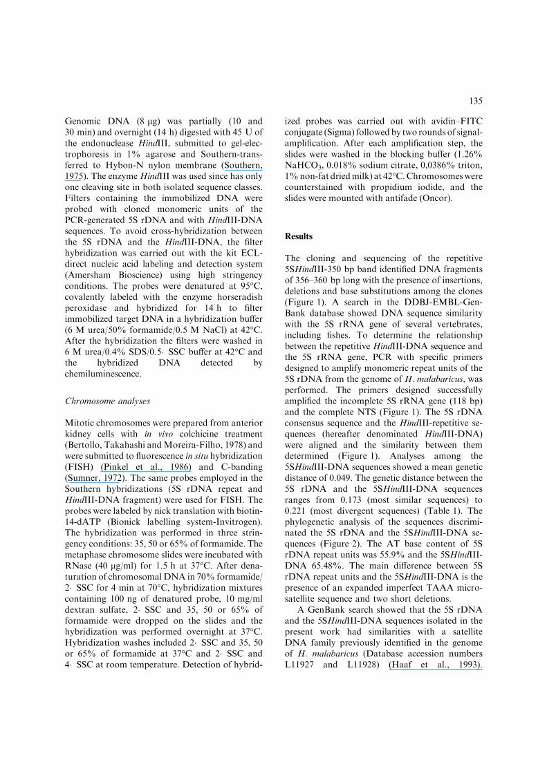
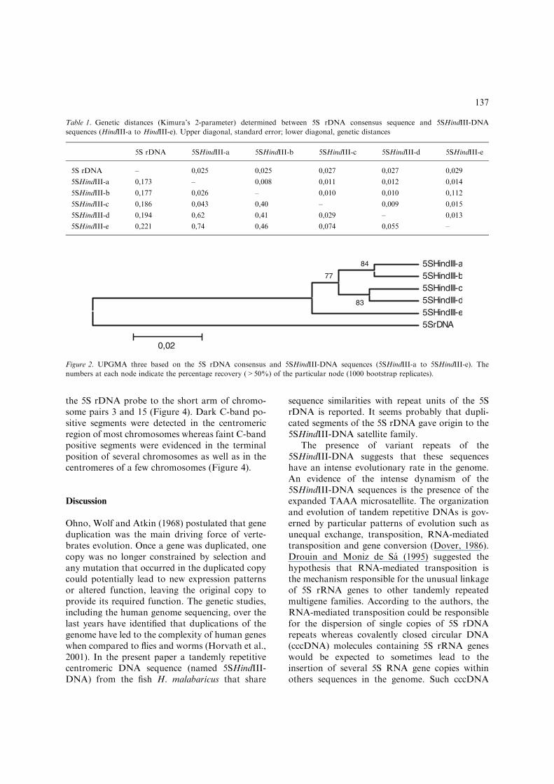
The cloning and sequencing of the repetitive5SHindIII-350 bp band identified DNA fragmentsof 356–360 bp long with the presence of insertions,deletions and base substitutions among the clones(Figure 1). A search in the DDBJ-EMBL-Gen-Bank database showed DNA sequence similaritywith the 5S rRNA gene of several vertebrates,including fishes. To determine the relationshipbetween the repetitive HindIII-DNA sequence andthe 5S rRNA gene, PCR with specific primersdesigned to amplify monomeric repeat units of the5S rDNA from the genome of H. malabaricus, wasperformed. The primers designed successfullyamplified the incomplete 5S rRNA gene (118 bp)and the complete NTS (Figure 1). The 5S rDNAconsensus sequence and the HindIII-repetitive se-quences (hereafter denominated HindIII-DNA)were aligned and the similarity between themdetermined (Figure 1). Analyses among the5SHindIII-DNA sequences showed a mean geneticdistance of 0.049. The genetic distance between the5S rDNA and the 5SHindIII-DNA sequencesranges from 0.173 (most similar sequences) to0.221 (most divergent sequences) (Table 1). Thephylogenetic analysis of the sequences discrimi-nated the 5S rDNA and the 5SHindIII-DNA se-quences (Figure 2). The AT base content of 5SrDNA repeat units was 55.9% and the 5SHindIII-DNA 65.48%. The main difference between 5SrDNA repeat units and the 5SHindIII-DNA is thepresence of an expanded imperfect TAAA micro-satellite sequence and two short deletions.
A GenBank search showed that the 5S rDNAand the 5SHindIII-DNA sequences isolated in thepresent work had similarities with a satelliteDNA family previously identified in the genomeof H. malabaricus (Database accession numbersL11927 and L11928) (Haaf et al., 1993).
135

However, no previous relationship was describedbetween that satellite sequence and the 5S rDNAsequences.
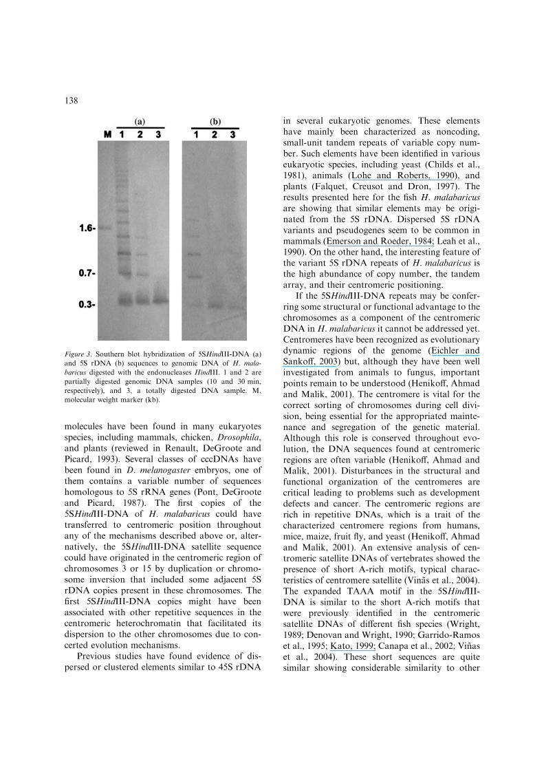
Southern blot hybridization analyses wereconducted using DNA digested with the restrictionendonuclease HindIII, selected for its recognitionsite within the repetitive isolated sequences. Theenzyme HindIII has only one cleaving site in bothisolated sequences. Membrane hybridization usingthe 5SHindIII-DNA and 5S rDNA isolated se-quences as probes showed that these repetitive
sequences are tandemly arrayed in the genome ofH. malabaricus (Figure 3).
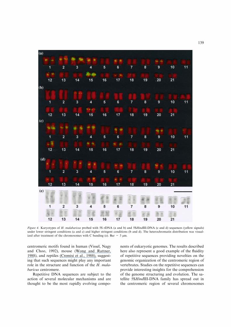
FISH was carried out using the 5S rDNA andthe 5SHindIII-DNA sequences as probes. Underlow stringent conditions (35% of formamide) bothprobes hybridized in the centromeric region of 18chromosomes and near the centromeres in the shortarm of chromosome pairs 3 and 15 (Figure 4).Under high stringent conditions (50 and 65%formamide) the 5SHindIII-DNA probe hybridizedto the centromeric region of 18 chromosomes and
Figure 1. Alignment of the 5S rDNA consensus sequence and 5SHindIII-DNA repeat units (5SHindIII-a to 5SHindIII-e) identified
in the genome of H. malabaricus. The 5S rRNA gene sequence regions are in boldface type, the primers used to obtain the 5S
rDNA sequences are underlined and the arrows indicate the direction of the PCR amplification. Hyphens represent gaps, dots base
identity, and N non-defined nucleotides. The sequences are deposited in GenBank under the accession numbers AY624052-
AY624061.
136
the 5S rDNA probe to the short arm of chromo-some pairs 3 and 15 (Figure 4). Dark C-band po-sitive segments were detected in the centromericregion of most chromosomes whereas faint C-bandpositive segments were evidenced in the terminalposition of several chromosomes as well as in thecentromeres of a few chromosomes (Figure 4).
Discussion
Ohno, Wolf and Atkin (1968) postulated that geneduplication was the main driving force of verte-brates evolution. Once a gene was duplicated, onecopy was no longer constrained by selection andany mutation that occurred in the duplicated copycould potentially lead to new expression patternsor altered function, leaving the original copy toprovide its required function. The genetic studies,including the human genome sequencing, over thelast years have identified that duplications of thegenome have led to the complexity of human geneswhen compared to flies and worms (Horvath et al.,2001). In the present paper a tandemly repetitivecentromeric DNA sequence (named 5SHindIII-DNA) from the fish H. malabaricus that share
sequence similarities with repeat units of the 5SrDNA is reported. It seems probably that dupli-cated segments of the 5S rDNA gave origin to the5SHindIII-DNA satellite family.
The presence of variant repeats of the5SHindIII-DNA suggests that these sequenceshave an intense evolutionary rate in the genome.An evidence of the intense dynamism of the5SHindIII-DNA sequences is the presence of theexpanded TAAA microsatellite. The organizationand evolution of tandem repetitive DNAs is gov-erned by particular patterns of evolution such asunequal exchange, transposition, RNA-mediatedtransposition and gene conversion (Dover, 1986).Drouin and Moniz de Sa (1995) suggested thehypothesis that RNA-mediated transposition isthe mechanism responsible for the unusual linkageof 5S rRNA genes to other tandemly repeatedmultigene families. According to the authors, theRNA-mediated transposition could be responsiblefor the dispersion of single copies of 5S rDNArepeats whereas covalently closed circular DNA(cccDNA) molecules containing 5S rRNA geneswould be expected to sometimes lead to theinsertion of several 5S RNA gene copies withinothers sequences in the genome. Such cccDNA
Table 1. Genetic distances (Kimura’s 2-parameter) determined between 5S rDNA consensus sequence and 5SHindIII-DNA
sequences (HindIII-a to HindIII-e). Upper diagonal, standard error; lower diagonal, genetic distances
5S rDNA 5SHindIII-a 5SHindIII-b 5SHindIII-c 5SHindIII-d 5SHindIII-e
5S rDNA – 0,025 0,025 0,027 0,027 0,029
5SHindIII-a 0,173 – 0,008 0,011 0,012 0,014
5SHindIII-b 0,177 0,026 – 0,010 0,010 0,112
5SHindIII-c 0,186 0,043 0,40 – 0,009 0,015
5SHindIII-d 0,194 0,62 0,41 0,029 – 0,013
5SHindIII-e 0,221 0,74 0,46 0,074 0,055 –
5SHindIII-a 5SHindIII-b 5SHindIII-c 5SHindIII-d 5SHindIII-e 5SrDNA
84
83
77
0,02
Figure 2. UPGMA three based on the 5S rDNA consensus and 5SHindIII-DNA sequences (5SHindIII-a to 5SHindIII-e). The
numbers at each node indicate the percentage recovery (>50%) of the particular node (1000 bootstrap replicates).
137
molecules have been found in many eukaryotesspecies, including mammals, chicken, Drosophila,and plants (reviewed in Renault, DeGroote andPicard, 1993). Several classes of cccDNAs havebeen found in D. melanogaster embryos, one ofthem contains a variable number of sequenceshomologous to 5S rRNA genes (Pont, DeGrooteand Picard, 1987). The first copies of the5SHindIII-DNA of H. malabaricus could havetransferred to centromeric position throughoutany of the mechanisms described above or, alter-natively, the 5SHindIII-DNA satellite sequencecould have originated in the centromeric region ofchromosomes 3 or 15 by duplication or chromo-some inversion that included some adjacent 5SrDNA copies present in these chromosomes. Thefirst 5SHindIII-DNA copies might have beenassociated with other repetitive sequences in thecentromeric heterochromatin that facilitated itsdispersion to the other chromosomes due to con-certed evolution mechanisms.
Previous studies have found evidence of dis-persed or clustered elements similar to 45S rDNA
in several eukaryotic genomes. These elementshave mainly been characterized as noncoding,small-unit tandem repeats of variable copy num-ber. Such elements have been identified in variouseukaryotic species, including yeast (Childs et al.,1981), animals (Lohe and Roberts, 1990), andplants (Falquet, Creusot and Dron, 1997). Theresults presented here for the fish H. malabaricusare showing that similar elements may be origi-nated from the 5S rDNA. Dispersed 5S rDNAvariants and pseudogenes seem to be common inmammals (Emerson and Roeder, 1984; Leah et al.,1990). On the other hand, the interesting feature ofthe variant 5S rDNA repeats of H. malabaricus isthe high abundance of copy number, the tandemarray, and their centromeric positioning.
If the 5SHindIII-DNA repeats may be confer-ring some structural or functional advantage to thechromosomes as a component of the centromericDNA in H. malabaricus it cannot be addressed yet.Centromeres have been recognized as evolutionarydynamic regions of the genome (Eichler andSankoff, 2003) but, although they have been wellinvestigated from animals to fungus, importantpoints remain to be understood (Henikoff, Ahmadand Malik, 2001). The centromere is vital for thecorrect sorting of chromosomes during cell divi-sion, being essential for the appropriated mainte-nance and segregation of the genetic material.Although this role is conserved throughout evo-lution, the DNA sequences found at centromericregions are often variable (Henikoff, Ahmad andMalik, 2001). Disturbances in the structural andfunctional organization of the centromeres arecritical leading to problems such as developmentdefects and cancer. The centromeric regions arerich in repetitive DNAs, which is a trait of thecharacterized centromere regions from humans,mice, maize, fruit fly, and yeast (Henikoff, Ahmadand Malik, 2001). An extensive analysis of cen-tromeric satellite DNAs of vertebrates showed thepresence of short A-rich motifs, typical charac-teristics of centromere satellite (Vinas et al., 2004).The expanded TAAA motif in the 5SHindIII-DNA is similar to the short A-rich motifs thatwere previously identified in the centromericsatellite DNAs of different fish species (Wright,1989; Denovan and Wright, 1990; Garrido-Ramoset al., 1995; Kato, 1999; Canapa et al., 2002; Vinaset al., 2004). These short sequences are quitesimilar showing considerable similarity to other
Figure 3. Southern blot hybridization of 5SHindIII-DNA (a)
and 5S rDNA (b) sequences to genomic DNA of H. mala-
baricus digested with the endonucleases HindIII. 1 and 2 are
partially digested genomic DNA samples (10 and 30 min,
respectively), and 3, a totally digested DNA sample. M,
molecular weight marker (kb).
138
centromeric motifs found in human (Vissel, Nagyand Choo, 1992), mouse (Wong and Rattner,1988), and reptiles (Cremisi et al., 1988), suggest-ing that such sequences might play any importantrole in the structure and function of the H. mala-baricus centromere.
Repetitive DNA sequences are subject to theaction of several molecular mechanisms and arethought to be the most rapidly evolving compo-
nents of eukaryotic genomes. The results describedhere also represent a good example of the fluidityof repetitive sequences providing novelties on thegenomic organization of the centromeric region ofvertebrates. Studies on the repetitive sequences canprovide interesting insights for the comprehensionof the genome structuring and evolution. The sa-tellite 5SHindIII-DNA family has spread out inthe centromeric region of several chromosomes
Figure 4. Karyotypes of H. malabaricus probed with 5S rDNA (a and b) and 5SHindIII-DNA (c and d) sequences (yellow signals)
under lower stringent conditions (a and c) and higher stringent conditions (b and d). The heterochromatin distribution was visual-
ized after treatment of the chromosomes with C banding (e). Bar = 5 lm.
139
and has been favored during the evolution due to apossible role of the repetitive sequences of thecentromeric region for the centromere structureand function.
Acknowledgements
The authors thank FAPESP (Fundacao de Am-paro a Pesquisa do Estado de Sao Paulo) andCNPq (Conselho Nacional de DesenvolvimentoCientıfico e Tecnologico) for financial support.
References
Altschul, S.F., W. Gish, W. Miller, E.W. Myers & D.J. Lipman,
1990. Basic local alignment search tool. J. Mol. Biol. 215:
403–410.
Beridze, R., 1986. Satellite DNA. Springer, Berlin, Heidelberg,
New York.
Bertollo, L.A.C. & C.A. Mestriner, 1998. The X1X2Y sex
chromosome system in the fish Hoplias malabaricus. II.
Meiotic analyses. Chromosome Res. 6: 141–147.
Bertollo, L.A.C., C.S. Takahashi & O. Moreira-Filho, 1978.
Karyotypic studies of two allopatric populations of the genus
Hoplias (Pisces, Erythrinidae). Brazil. J. Genet. 2: 17–37.
Bertollo, L.A.C., C.S. Takahashi & O. Moreira-Filho, 1983.
Multiple sex chromosomes in the genus Hoplias (Pisces,
Erythrinidae). Cytologia 48: 1–12.
Bertollo, L.A.C., O. Moreira-Filho & M.S. Fontes, 1997a.
Karyotypic diversity and distribution in Hoplias malabari-
cus (Pisces, Erythrinidae): Cytotypes with 2n. 40 chromo-
somes. Brazil. J. Genet. 20: 237–242.
Bertollo, L.A.C., M.S. Fontes, A.S. Fenocchio & J. Cano,
1997b. The X1X2Y sex chromosome system in the fish
Hoplias malabaricus. I. G-, C- and chromosome replication
banding. Chromosome Res. 5: 493–499.
Bertollo, L.A.C., G.G. Born, J.A. Dergam, A.S. Fenocchio &
O. Moreira-Filho, 2000. A biodiversity approach in the
Neotropical fish Hoplias malabaricus. Karyotypic survey,
geographic distribution of cytotypes and cytotaxonomic
considerations. Chrom. Res. 8: 603–613.
Born, G.G. & L.A.C. Bertollo, 2000. An XX/XY sex chromo-
some system in a fish species, Hoplias malabaricus, with a
polymorphic NOR-bearing X chromosome. Chromosome
Res. 8: 111–118.
Brenner, S., G. Elgar, R. Sandford, A. Macrae, B. Venkatesh &
S. Aparicio, 1993. Characterization of the pufferfish (Fugu)
genome as a compact model vertebrate genome. Nature
6452: 265–268.
Canapa, A., P.N. Cerioni, M. Barucca, E. Olmo & V. Caputo,
2002. A centromeric satellite DNA may be involved in
heterochromatin compactness in gobiid fishes. Chrom. Res.
10: 297–304.
Cavalier-Smith, T., 1985. The Evolution of Genome Size.
Wiley, New York.
Charlesworth, B., P. Snlegowski & W. Stephan, 1994. The
evolutionary dynamics of repetitive DNA in eukaryotes.
Nature 371: 215–220.
Childs, G., R. Maxson, R.H. Cohn & L. Kedes, 1981. Orphons:
dispersed genetic elements from tandem repetitive genes of
eukaryotes. Cell 23: 651–663.
Clabby, C., U. Goswami, F. Flavin, N.P. Wilkins, J.A.
Houghton & R. Powell, 1996. Cloning, characterization
and chromosomal location of a satellite DNA from the
Pacific oyster, Crassostrea gigas. Gene 168: 205–209.
Cremisi, F., R. Vignali, R. Batistoni & G. Barsacchi, 1988.
Heterochromatic DNA in Triturus (Anphibia, Urodela) II.
A centromeric satellite DNA. Chromosoma 97: 204–211.
Denovam, E.M. & J.M. Wright, 1990. A satellite DNA family
from pollock (Pollachius virens). Gene 87: 279–283.
Dergam, J.A. & L.A.C. Bertollo, 1990. Karyotypic diversi¢cat-
ion in Hoplias malabaricus (Osteichthyes, Erythrinidae) of
the Sao Francisco and Alto Parana basins, Brazil. Brazil. J.
Genet. 13: 755–766.
Dover, G.A., 1986. Linkage disequilibrium and molecular drive
in the rDNA gene family. Genetics 122: 249–252.
Drouin, G. & M. de Moniz Sa, 1995. The concerted evolution
of 5S ribosomal genes linked to the repeat units of other
multigene families. Mol. Biol. Evol. 12: 481–493.
Eichler, E.E. & D. Sankoff, 2003. Structural Dynamics of
eukaryotes chromosome evolution. Science 301: 793–797.
Emerson, B.M. & R.G. Roeder, 1984. Isolation and genomic
arrangement of active and inactive forms of mammalian 5S
RNA genes. J. Biol. Chem. 259: 7916–7925.
Falquet, J., R. Creusot & M. Dron, 1997. Molecular analysis of
DNA homologous to IGS subrepeats. Plant Physiol.
Biochem. 35: 611–622.
Felsenstein, J., 1985. Confidence limits on phylogenies: An
approach using the bootstrap. Evolution 39: 783–791.
Garrido-Ramos, M.A., M. Jamilena, R. Lozano, C. Ruiz Rejon
& M. Ruiz Rejon, 1995. The EcoRI centromeric satellite
DNA of the Sparidae family (Pisces, Perciformes) contains
a sequence motive common to other vertebrates centro-
meric satellite DNAs. Cytogen. Cell Genet. 71: 345–351.
Haaf, T., M. Schmid, C. Steinlein, P.M. Galetti & H. Willard,
1993. Organization and molecular cytogenetics of a satellite
DNA family from Hoplias malabaricus (Pisces, Erythrini-
dae). Chromosome Res. 1: 77–86.
Henikoff, S., K. Ahmad & H.S. Malik, 2001. The centromere
paradox: stable inheritance with rapidly evolving DNA.
Science 293: 1098–1102.
Horvath, J.E., J.A. Bailey, D.P. Locke & E.E. Eichler, 2001.
Lessons from the human genome: transitions between
euchromatin and heterochromatin. Hum. Mol. Genet. 10:
2215–2223.
Kato, M., 1999. Structural bistability of repetitive DNA
elements featuring CA/TG dinucleotide steps and mode of
evolution of satellite DNA. Eur. J. Biochem. 265: 204–209.
Komiya, H. & S. Takemura, 1979. Nucleotide sequence of 5S
ribosomal RNA from rainbow trout (Salmo gairdnerii)
liver. J. Biochem. 86: 1067–1080.
Kumar, S., K. Tamura, I. Jakobsen & M. Nei, 2001. MEGA:
Molecular evolutionary genetic analysis. Bioinformatics 12:
1244–1249.
Larin, Z., M.D. Fricker & C. Tyler-Smith, 1994. De novo
formation of several features of a centromere following
140
introduction of a Y alphoid YAC into mammalian cells.
Hum. Mol. Genet. 3: 689–695.
Leah, R., S. Frederiksen, J. Engberg & P.D. Sorensen, 1990.
Nucleotide sequence of a mouse 5S rRNA variant gene.
Nucl. Acids Res. 18: 7441–7441.
Lohe, A.R. & P.A. Roberts, 1990. An unusual Y chromosome
of Drosophila simulans carrying amplified rDNA spacer
without RNA genes. Genetics 125: 399–406.
Lopes, P.A., A.J. Alberdi, J.A. Dergam & A.S. Fenocchio,
1998. Cytotaxonomy of Hoplias malabaricus (Osteichthyes,
Erythrinidae) in the Aguapey river (Province of Corrientes,
Argentina). Copeia 1998: 485–487.
Ohno, S., U. Wolf & N.B. Atkin, 1968. Evolution from fish to
mammals by gene duplication. Hereditas 59: 169–187.
Pinkel, D., T. Straume & J. Gray, 1986. Cytogenetic analysis
using quantitative, high-sensitivity, fluorescence hybridiza-
tion. Proc. Natl. Acad. Sci. 83: 2934–2938.
Pont, G., F. DeGroote & G. Picard, 1987. Some extrachromo-
somal circular DNAs from Drosophila embryos are homol-
ogous to tandemly repeated genes. J.Mol. Biol. 195: 447–451.
Renault, S., F. DeGroote & G. Picard, 1993. Identification of
short tandemly repeated sequences in extrachromosomal
circular DNAs from Drosophila melanogaster embryos.
Genome 36: 244–254.
Sambrook, J. & D.W. Russel, 2001. Molecular Cloning. A
Laboratory Manual Third Edition. Cold Spring Harbor
Laboratory Press, Cold Spring Harbor, New York.
Sart, D., M.R. Cancilla, E. Earle, J.I. Mao, R. Saffery, K.M.
Tainton, P. Kalitsis, J. Martyn, A.E. Barry & K.H. Choo,
1997. A functional neo-centromere formed through activa-
tion of a latent human centromere and consisting of non-
alpha-satellite DNA. Nature Genet. 16: 144–153.
Singer, M.F., 1982. Highly repetitive sequences in mammalian
genomes. Int. Rev. Cytol. 76: 67–112.
Southern, E.M., 1975. Detection of specific sequences among
DNA fragments separated by gel electrophoresis. J. Mol.
Biol. 98: 503–517.
Sumner, A.T., 1972. A simple technique for demonstrating
centromeric heterochromatin. Exp. Cell Res. 75: 304–306.
Thompson, J.D., D.G. Higgins & T.J. Gibson, 1994. Clustal W:
improving the sensitivity of progressive multiple sequence
alignment through sequence weighting, position-specific
gap penalties and weight matrix choice. Nucl. Acids Res.
22: 4673–4680.
Vinas, A., M. Abuın, B.G. Pardo, P. Martınez & L. Sanchez,
2004. Characterization of a new HpaI centromeric satellite
DNA in Salmo salar. Genetica 121: 81–87.
Vissel, B., A. Nagy & K.H.A. Choo, 1992. A satellite III
sequence shared by human chromosomes 13, 14 and 21 that
is contiguous with alpha satellite DNA. Cytogen. Cell
Genet. 61: 81–86.
Wong, A.K.C. & J.B. Rattner, 1988. Sequence organization
and cytological localization of the minor satellite of mouse.
Nucl. Acids Res. 16: 11645–11661.
Wright, J.M., 1989. Nucleotide sequence, genomic organization
and the evolution of a major repetitive DNA family in
tilapia (Oreochromis mossambicus/hornorum). Nucl. Acids
Res. 17: 5071–5079.
141
Copyright © 2022 FDOKUMEN