A STUDY ON VADHA UBHAKATHAM
166
A STUDY ON VADHA UBHAKATHAM Dissertation Submitted To THE TAMIL NADU DR.M.G.R Medical University Chennai – 32 In Partial fulfillment for The Award of Degree of DOCTOR OF MEDICINE (SIDDHA) (Branch – I Pothu Maruthuvam) Department of Pothu Maruthuvam Government Siddha Medical College Palayamkottai – 627 002 September 2007
-
Upload
khangminh22 -
Category
Documents
-
view
0 -
download
0
Transcript of A STUDY ON VADHA UBHAKATHAM
A STUDY ON
VADHA UBHAKATHAM
Dissertation Submitted To
THE TAMIL NADU DR.M.G.R Medical University
Chennai – 32
In Partial fulfillment for The Award of Degree of
DOCTOR OF MEDICINE (SIDDHA) (Branch – I Pothu Maruthuvam)
Department of Pothu Maruthuvam
Government Siddha Medical College
Palayamkottai – 627 002
September 2007
ACKNOWLEDGEMENT
I am extremely greatful to my Lord almighty who empowered me
with his blessings and grace to complete my dissertation work
successfully
It is my pleasure and privilege to acknowledge the blessings and
encouragement of my parents and brothers without which the
completion of this study would not have been possible.
I take this opportunity to express gratitude and
acknowledgement to the Vice-chancellor, The Tamilnadu Dr. M.G.R
medical university, Chennai, and The Director, The joint director,
Directorate of Indian Medicine and Homeopathy, Chennai.
I would be very thankful to Dr.M. Dhinakaran M.D(s) principal
GSMC, Palayamkottai, and Dr.R.Devarajan M.D(s) Vice principal
GSMC Palayamkottai, for having permitted to make use of the
facilities available in this institution to bring out the dissertation a
successfully one.
I am really indepted to Dr.K.R.Revathy M.D (s)., Former Vice
Principal & Head, P.G.Department of Pothu Maruthuvam,
Government Siddha Medical college, palayamkottai for his
valuable guidance and encouragement in selecting this topic.
It is my privilege to record my deep sense of gratitude to
Dr.A.Prema M.D(s)., Head, Post Graduate Department of Pothu
Maruthuvam, Government Siddha Medical College for her devoted
guidance and for providing basic infrastructural needs for use patients
admitted in the ward, without her constant and authertic support,
this study would not have seen the light of day
I express my thanks to Dr.S.Mohan M.D(s)., Lecturer,
P.G.Maruthuvam Branch for his valuable guidance
I express my gratitudes to Dr.S.Chitra M.D(s)., Assistant
Lecturer, P.G.Maruthuvam Branch for his valuable guidance.
My sincere thanks to Dr.S.Justus Antony M.D(s)., for his
guidance and support during this study.
I owe special gratitude to Dr.M.R.Vairamuthu Raja M.B.B.S.,
M.D., Professor, Modern Medicine Department,TVMCH for his
valuable guidance in modern aspect of approach to this study.
I extend my special gratitude to Dr.Arumuga Pandian @
S. Mohan M.B.B.S.,M.D., Professor, Modern Medicine Department,
Government Siddha Medical college for his valuable guidance in
modern aspect of approach to this study.
I express my thanks to Dr.J. Joseph Doss M.B.B.S., P.hD.,
Former Head of the Department and Mr.M.Kalaivannan M.Sc.,
M.Phil., Lecturer and staffs of pharmacology Department for their
keen co-operation in electing the pharmacological evaluation of the
trial medicine
I also thanks to Mrs. N. Nagaprema M.Sc., M.phil., Head of
the Department and staffs of Biochemistry Department for their
cooperation in eliciting the Biochemical analysis of the trial medicine
I express my thanks to librarian Mrs.T.Poonkodi M.A., M.L.I.S.,
for permitting me to utilize the college library for my dissertation
work.
Finally I express my deep thanks to Selwyn’s Broad Band Net
Café & Staffs of this center for their kind co-operation.
1
INTRODUCTION
Medicine as every one knows is not merely as science but an
art as well. There are different systems of medicine in the world
according to their way of life with their geographical conditions.
The siddha system of medicine is one of the two pillars of
Indian system of medicine, the other one is ayurvedha, siddha
system of medicine is dedicated by siddhars to the human society
to live long & free from disease.
Siddhars were men of highly cultured, intellectual and
spiritual faculties combined with supernatural powers.
The siddha system considers not merely body alone but also
the soul.
The siddha system considers body as a whole, made up of five
basic elements namely.
1. Prithivi
2. Appu
3. Theyu
4. Vayu
5. Agayam
These five elements are considered as the fundamental principles
of all creations of god. This can be stated in the sathaga naadi as
“ghug;gh G+jike;J kz;ePH NjA
ghpthA thfha ike;jpdhNy
Nrug;gh rlkhr;R.......”
Neha; ehly; Neha; Kjy; ehly; jpul;L - 1
Traditional system of medicine all over the world believed
above.
2
“mz;lj;jpYs;sNj gpz;lk;
gpz;lj;jpYs;sNj mz;lk;
mz;lKk; gpz;lKk; xd;W
mwpe;J jhd; ghHf;Fk; NghJ”
- rl;lKdp epfz;L
According to this the natural forces acting in and through the
various organs of the human body are intimately related to the
similar or corresponding forces acting in and through the organism
of the universe.
According to siddha literatures the total number of diseases
which affects the mankind is 4448. One among them is vadha
ubhakatham.
The evidence of the disease Vadha ubhakatham is selected
from yugi vaidhya chinthamani – 800.
The author has chosen the disease ‘Vadha Ubhakatham’ for
this dissertation work. It is now common in occurrence due to
prevalence of madhumega noi as their complications. The disease is
correlated with the ‘peripheral neuritis’ in allopathic view.
Principles of Treatment
“ cw;wtd; jPHg;ghd; kUe;Jior; nry;thndd;
wg;gdhw; $w;Nw kUe;J”
- jpUf;Fws;
When patient, doctor, pharmacist and nurse all act in co-
ordination the disease will be cured.
In this unique system treatment is based upon the principles
of arusuvai, mukkutram and panchaboothas. Also the seasional
variation (Paruvakalam), personal habits (Nall ozhukkam) etc., are
also considered.
3
Siddha medicines are classified mainly into two major
divisions.
• Internal medicine – 32
• External medicine – 32
Pathiyam is a medical advice which includes dietary
restrictions and life style modifications according to the diseased
conditions.
The unshakable belief among the people regarding the vadha
disese that it can be effectively managed only by siddha medicine
than other systems of medicine also initiates the author to choose it
for dissertation.
The author’s choice of medicine for the clinical study were
Pathiriver chooranam 5 gm , B.d with hot water
Gunapadam Mooligai Vahuppu
Medicated vennai for external use.
Sarabendhirar Pitha roha sihitchai
So the author hopes that this dissertation work will arise new
horizons in the field of siddha medicines in managing vadha
disease.
4
AIM AND OBJECTIVES
The chief aim of this study about vadha ubhakatham is to
establish the disease scientifically, because
In siddha medicine vadham is considered as the most effective
cause of disease. It is common in India and other countries.
As the patients are disturbed both functionally and
emotionally, diagnosing the disease with proper siddha and modern
parameters is necessary to evaluate the disease because if it is not
diagnosed and treated it may leads to so many complications.
The disease vadha ubhakatham produces Gnawing sensation
over legs and hands, numbness, paresthesia, sensation of walking
on cotton wool, pain in the extremities, polyphagia and giddiness.
The purpose of the author is to elucidate a good medicine
from ancient siddha literatures and to create hope and faith in their
treatments.
The author extent a correlation of aetiology, signs and
symptoms of vadha ubhakatham in siddha aspect with peripheral
neuritis in modern science.
There are various causes for vadha ubhakatham. The
patients with vadha ubhakatham were selected for dissertation
work that had (madhumegam) diabetes mellitus anaemia (B12
defeciency) nutrional disorder and alcoholic abuse.
To collect various siddha literatures aetilogy pathology,
clinical features, diagnosis, prognosis, complications and the
treatment by making use of siddha concept.
To express the unique diagnostic method mentioned by
siddhars to know how the disease vadha ubhakatham deranges the
5
normal muththadhukkal, poripulangal, udal kattugal, and envagai
thervugal and naadi nadai.
To have an idea about the incidence of disease with age, sex,
habit, socio economic status, family history and climate conditions.
To have an idea about paruva kalam, environmental changes,
Thinai, Thegi for this disease.
To have a detailed clinical investigations.
To have a clinical trial on vadha ubhakatham with Pathiri ver
chooranam internally and Medicated vennai externally.
To apply the principle of management as adviced by siddhars,
recording the cause, prevention and treatment of disease.
To evaluate the biochemical and pharmacological effects of
the trial medicines.
To use modern parameters to confirm the diagnosis and to
access the prognosis of the disease.
To give yoga therapy (Mayurasanam) along with medicines for
relaxation of brain centers to achieve good results.
To apply the principle of pathiyam specific to this disease.
6
ABSTRACT
Keeping in mind the need for bringing out an effective siddha
therapy for “Vadha Ubhakatham” since the commonest disease in
the society, number of sufferers increasing day by day so I have
selected this disease for my dissertation work.
I studied the clinical course of the disease on basis of siddha
literature with the help of diagnostic methods mentioned by
siddhars and how the disease alter the normal conditions of uyir
thathukkal, poripulangal, seven udal kattugal and envagai
thervugal.
Signs and symptoms of “vadha ubhakatham” with peripheral
neuropathy in modern science.
20 out patients and 20 in patients of either sex were selected
and administered with the trail medicine.
I studied the incidence of the disease with age, sex, socio-
economic, status, family history and seasonal variation.
I utilized the modern parameters for confirmation and
prognosis of the disease.
I had clinical trail on vadha ubhakatham with
Pathiri ver chooranam (Internally)
Medicated vennai (Externally)
The trial medicine was subjected to bio-chemical and
pharmacological analysis.
At the end of the trial study the majority of the cases showed
good results.
7
REVIEW OF LITERATURES
SIDDHA ASPECTS
According to siddha system of medicine a human being is
composed of 96 basic principles. Among them the first thirty is
considered very vital and the rest are considered to be the extension
of the first thirty principles. This not only consists of the physical
principles components of the human body but also the mental,
intellectual components like passions, qualities, knowledge,
functions of the sense organs and motor organs and their
coordination.
Pancha pootha Theory
The five elements vinn, kall, anal, punal, and mann are the
basis for the world and the human being. These five elements are in
subtle states (Suthchuma nilai). They manifest, into gross state
(sthula nilai) and become visible. The manifestation of the five
elements from the subtle state to gross state are called as
panchapootha panchi karanam.
Three humours theory
The three “humours” as described in siddha medicine is a
golden line continuous in physiology, pathology and treatment. The
three humours are vadham, pitham and kabham, whose balance is
essential for the maintenance of good health. These are also made
up of five elements.
Vadham is formed by the combination of vayu and akash (kall
and vinn).
Pitham is formed by the theyu (anal)
8
Kabham is formed by the combination of prithvi and appu
(mann & punal)
These three humours exist in the ratio of 1: ½ : ¼ when these
three humours are in equilibrium they are called as uyir thathu
while they get deranged they are called kutras or doshas.
When an individual has predominant vadha he is said to have
a vadha temperament. Like wise other two, this temperament of
body is determined during fertilization.
Aetiopathology of disease
Siddha humoural pathology explains that all diseases are
caused due to the derangement of the three humours. When they
get deranged they bring about diseases peculiar to their influence.
The humours by themselves are not the producers of diseases while
functioning normally. But they give rise to diseases if they are
vitiated by other factors. This is quoted in Thirukkural as
“kpfpDk; FiwapDk; Neha; nra;Ak; E}NyhHtsp
Kjyh vz;zpa %d;W”
- jpUf;Fws;
Any change in the proportion of the three humours is sure to
bring about disease but the maintenance of their normal proportion
gives vitality to the organism and assures the preservation of health
and longevity of life.
9
EXPLANATION ABOUT TRIDHOSHAS
FORMATION OF TRIDHOSHAS
The formation of vadham, pitham and kabham has been
explained in kannusamiyam as follows.
“,Ug;ghd ehb vogNjhBuh
apukhd Njfj;jpy; Vyg; - ngUehb
xf;fj rkj;njhopiy A+f;fjr thAf;fs;
jf;fgb nad;Nw rhUk;”
“rhUe;jrehb jd;dpy; %yk; %d;W
NgUkplk; gpq;fiyAk; gpd;dYld; - khWk;
ciuf;ftpuw; fhw;nwhl;LzHj;JNk ehrp
Tiur; RopNah ikaj;jpy; te;J”
“te;j fiy %d;wpy; thAthk-ghdDld;
je;j gpuhzd; rkhDk; re;jKwf;
$l;LwT Nurpj;jy; $Wk; thjk; gpj;jk;
ehl;Lq;fgNk ahk; ehL”
- fz;Zrhkpak;
Abanan in conjuction with edagalai produces vadham.
Piranan in conjuction with pingalai produced pitham.
Samanan in conjuction with suzhumunai produces kabham
Thus vadham, pitham and kabham are formed.
10
VADHAM
Synonyms:
The term vadham denotes vayu, dryness, pain, flatulence &
lightness.
Definition:
The three basic factors vadham, pitham and kabham working
in physiological conditions are called muththadhukkal (or) uyir
thattukkal. Among the three, the biological air humour is called
vadham. In terms of etymology it means that which moves thing it
is the motivating force behind the other two humours which are
considered to be lame incapable of movement without it.
Vadha may be increased (or) decreased when the equilibrium
state is disturbed; if vadha is altered the other two are also altered
leading to vadha disease.
According to this the human body is composed of 72,000
naadi, narambugal among this 72,000 the ten are prominently
naadies (dhasanaadies) of this ten naadies edagalai, pingalai,
shuzhumunai are known as moolathara naadies.
Among the ten vayus five are important, they are piranan,
abanan, viyanan udhanan, samanan.
These three humours are thadhu (i.e) vadha, pitha & kabha
are the functional principles in the composition and substance of
the body.
11
Location of vadham
Vadham lives in
“ nespe;jpl;l thjkgh dj;ij gw;wp
epiwe;jpilar; NrHe;Je;jpf; fPNo epd;W
FspHe;jpl;l %ykJ} nloe;J fhkf;
nfhilapilaiag; gw;wpnaoq; Fzj;ijg; ghNu
Fzkhd ntYk;ig Nkw;nwhf; if-ehb
epzkhd nghUe;jplKk; Nuhkf; fhYk;
epiwthfp khq;fpr nky;yhk; gue;J
fhy; fhl;b thjnkq;Fk; fyq;Fe; jhNd”
- tapj;jpa rjfk; 55>56
Abanan
Stools
Edakalai
Undhiyin keel moolam
Kaamakodi
Hip bone and Joints
Skin
Nerves
Hair follicles
Muscles
“ cz;b rikj;jpbw; $l;LwT Flw;gFjp
jpz;bw nyz;b nrtpfwq;F - tpz;l
njhLTzHT Njhw;Wtpf;Fk; NjhypLg;gpy; CWk;
tpLtpyplkhk; tspf;F”
- kUj;Jt jdpg;ghly;
“mwpe;jpLk; thjk; mlq;F kyj;jpdpy;”
“ehnkd;w thjj;Jf;F fpUg;gplNk Nfsha;
ehgpf;F fPnod;W etpyyhFk;”
-Neha; ehly; Neha; Kjdhly; jpul;L

12
According to the maruthuva thanippadal vadha lives in the
digestive system, bones, ear, thigh, skin and hip, below navel
region.
Nature of vadham:
xOq;Fld; jhNjo; %r;rhq;fp ,aq;f
vOr;rp ngw vg;gzpTkhw;w – voe;jphpa
Ntfk; Gyd;fSf;F Nktr; RWRWg;G
thf;fspf;Fk; khe;jHf;F thA
- kUj;Jt jdpg;ghly;
Natural properties of vadham
Giving briskness
Respiration
Functioning the mind thought & body
Regulation of fourteen physiological reflexes.
Uniform functioning of the seven udal thathukkal.
Strengthening the five sensory organs.
Qualities of vadham
Hardness
Dryness
Lightness
Coolness
Mobility
Subtleness
Opposite qualities of vadham
Softness
Greasy
Heaviness
13
Hotness
Stable
Solid State
Functions of vadham
Body ache
Pricking pain
Tearing pain
Nerve weakness
Shivering
Mental distress
Dryness
Movements
Weakness
Joint pain
Traumatic pain
Dislocation of joints
Weakness of organs
Pilo – erection
Paralysis of limbs
Polydipsia
Severe pain in leg and thigh muscles bony pricking pain
Constipation
Unable to do flexion and extension of the limbs
All tastes to be like astringent
Excess salivation
Darkness of skin, eyes and urine.
Description of vadham
The siddha classical texts divide the general principles of
vadham into ten subsidiary forms that differ from one another by

14
their location in the body (anatomical) and by their particular
functions (Physiological).
They are
1. Piranan (Heart Centre) - Uyirkkal
It corresponds to the cardiac plexus and refers to the chest. It
maintains the action of the heart the functioning of the mental
faculities of perception and concentrations and also cares for the
arteries, veins & nerves. It regulates the respiration & digestion. It
otherwise called as “Uyirkkal”
2. Abaanan (Moolaadharam centre) – Kezhnokkukal
It corresponds to the pelvic plexus and controls the excretion.
It is focused in the lower part of the gut and also occupies the sites
in the bladder and gential organ. It has a tendency to travel
downwards. It moves in the whole gentio urinarytract and
regulates the defecation, micturation, mensuration parturition and
ejaculation.
3. Viyanan (Fore head centre) - Paravukal
It corresponds to the nasociliary plexus at the root of the nose
and base of the skull and controls the will. It helps in the
circulation of energy through out the entire nervous system and the
movements of various parts of the body. It also transports
nutrients and blood through out the entire body. It is also know as
“paruvakal”.
4. Udhanan (Throat centre) - Melnokkukal
This corresponds to the pharyngeal plexus in the throat
region and controls speech, and breathing. It is also responsible for
15
the physiological reflex actions like vomiting, hiccup, cough etc., It
is otherwise named as melnokukaal.
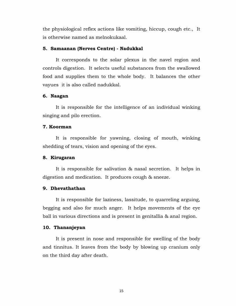
5. Samaanan (Nerves Centre) - Nadukkal
It corresponds to the solar plexus in the navel region and
controls digestion. It selects useful substances from the swallowed
food and supplies them to the whole body. It balances the other
vayues it is also called nadukkal.
6. Naagan
It is responsible for the intelligence of an individual winking
singing and pilo erection.
7. Koorman
It is responsible for yawning, closing of mouth, winking
shedding of tears, vision and opening of the eyes.
8. Kirugaran
It is responsible for salivation & nasal secretion. It helps in
digestion and medication. It produces cough & sneeze.
9. Dhevathathan
It is responsible for laziness, lassitude, to quarreling arguing,
begging and also for much anger. It helps movements of the eye
ball in various directions and is present in genitallia & anal region.
10. Thananjeyan
It is present in nose and responsible for swelling of the body
and tinnitus. It leaves from the body by blowing up cranium only
on the third day after death.

16
PITHAM
The term pitham denotes gastric juice, bile, energy, heat,
anger etc,.
Location of pitham
Head, heart, bladder, abdomen, umblicus, stomach, saliva,
sweat, blood, eyes and skin are the sites of the pitham.
Effects of vitiated pitham
Excessive heat in the body, improper digestion, excessive
sweat, giddiness, syncope and immoral behaviour are some of the
ill effects of vitiated pitham.
Types of pitham
According to the functions and location, pitham is classified
in to five types. They are,
Anar Pitham - It is located in the stomach and intestines
and is responsible for proper digestion.
Ranjaga Pitham - It is located in the intestine and is
responsible for the colour of blood.
Sadhaga Pitham - It is located in the heart and controls the
functions of the body.
Alosaga Pitham - It is located in the eyes and is responsible
for proper vision
Prasaga Pitham - It is located in the skin and is responsible
for the complexion of skin

17
KABHAM
Location of kabham
It is located in the tongue, chest, blood, bone marrow, bones
nerves, brain, large intestine, eyes and joints.
Functions of kabham
The important functions of kabham are maintaining the
viscosity and proper functioning of the joints.
Effects of vitiated kabham
Pain in the long bones, dysfunction of joints improper
digestion, excessive sleep and inhibition of under stating capacity.
Types of kabham
Avalambagam :
It is located in the heart and it controls the other four
kabham.
Kilethagam :
It is located in the stomach and is responsible for proper
digestion.
Pothagam :
It is located in the tongue and helps to feel the sensation of
taste.
Tharpagam :
It is present in the head and keeps the eyes cool.
Santhegam :
It is present in the joints and is responsible for proper functioning of the joints.

18
Fate of three humours
“ mwpe;jpLk; thjklq;F kyj;jpdpy;
gphpe;JLk; gpj;jk; NguhQ; ryj;jpdpy;
kwpe;jpLk; ikak; trpf;Fk; tpe;Jtpy;”
From this it is clear that the three humours can be
discharged through the following routs.
Vadham - faeces
Pitham - Urine
Kabham - Semen / Suronidham
The features of exaggeration of vadham
Body weakness and darkness
Shivering
Constipation
Dimmunition of immunity
Giddiness
Insomnia
Laziness
The Features of diminution of vadham
Body ache
Hoarseness of voice
Loss of memory
Semi Consciousness
Difficulty to do any work
Paleness and coolness of body
Anorexia
19
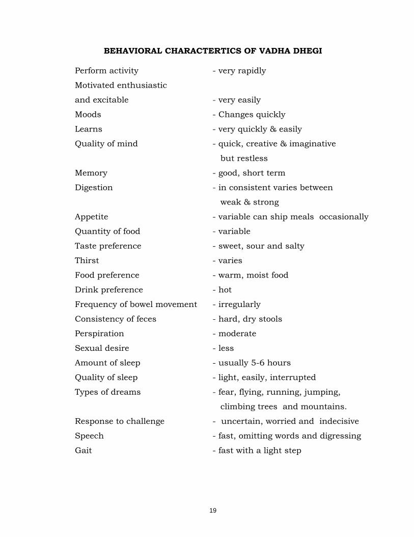
BEHAVIORAL CHARACTERTICS OF VADHA DHEGI
Perform activity - very rapidly
Motivated enthusiastic
and excitable - very easily
Moods - Changes quickly
Learns - very quickly & easily
Quality of mind - quick, creative & imaginative
but restless
Memory - good, short term
Digestion - in consistent varies between
weak & strong
Appetite - variable can ship meals occasionally
Quantity of food - variable
Taste preference - sweet, sour and salty
Thirst - varies
Food preference - warm, moist food
Drink preference - hot
Frequency of bowel movement - irregularly
Consistency of feces - hard, dry stools
Perspiration - moderate
Sexual desire - less
Amount of sleep - usually 5-6 hours
Quality of sleep - light, easily, interrupted
Types of dreams - fear, flying, running, jumping,
climbing trees and mountains.
Response to challenge - uncertain, worried and indecisive
Speech - fast, omitting words and digressing
Gait - fast with a light step
20
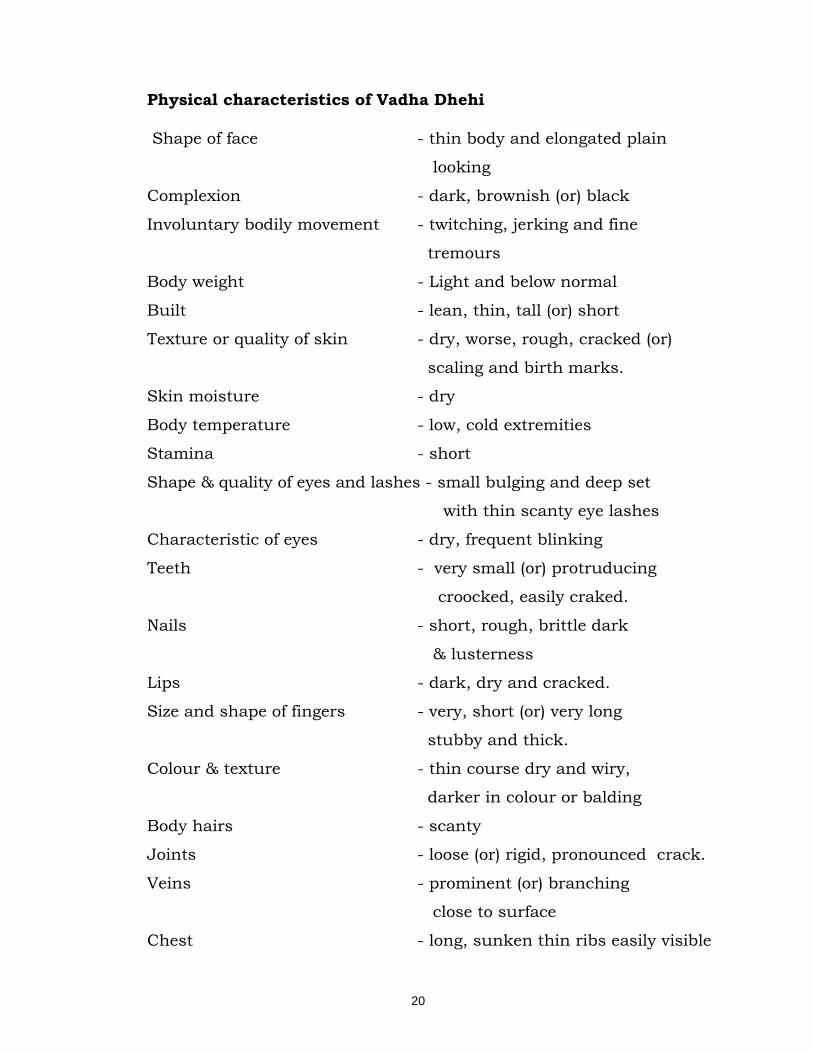
Physical characteristics of Vadha Dhehi
Shape of face - thin body and elongated plain
looking
Complexion - dark, brownish (or) black
Involuntary bodily movement - twitching, jerking and fine
tremours
Body weight - Light and below normal
Built - lean, thin, tall (or) short
Texture or quality of skin - dry, worse, rough, cracked (or)
scaling and birth marks.
Skin moisture - dry
Body temperature - low, cold extremities
Stamina - short
Shape & quality of eyes and lashes - small bulging and deep set
with thin scanty eye lashes
Characteristic of eyes - dry, frequent blinking
Teeth - very small (or) protruducing
croocked, easily craked.
Nails - short, rough, brittle dark
& lusterness
Lips - dark, dry and cracked.
Size and shape of fingers - very, short (or) very long
stubby and thick.
Colour & texture - thin course dry and wiry,
darker in colour or balding
Body hairs - scanty
Joints - loose (or) rigid, pronounced crack.
Veins - prominent (or) branching
close to surface
Chest - long, sunken thin ribs easily visible

21
AETIOLOGY
The common aetiological factors for all types of vadha disease
are applicable for vadha ubhakatham also.
Though yugi has not mentioned any specific cause for vadha
ubhakatham he has summarized the causative factors for all types
of vadha diseases as.
“vd;dNt thjk;jh nzz;g jhFk;
,fj;jpNy kdpjHfSf; nfa;A khW
kpd;dNt nghd;jida NrhuQ; nra;J
nghpNahHfs; gpuhkziu J}\zpj;Jk;
tz;z Njtw; nrhj;jpw; NrhuQ; nra;J
khjh gpjh FUit kwe;j NgHf;Fk;
fs;sNt Ntjj;ij epe;ij nra;jhy;
fhaj;jpw; fye;jpLNk thje;jhNd”
- A+fp itj;jpa rpe;jhkzp 243
According to this poem, dacoity, abusing bolymen, exploiting
properties of charities, forget the parents and teachers and
desecrating holy scripts will cause disturbance of vadham.
“jhndd;w frg;NghL JtHg;G iwg;G
rhjfkha; neQ;RfpDQ; rikj;j td;dk;
Mndd;w thwpdJ nghrpj;j yhYk;
Mfhaj; NjwyJ Fbj;j yhYk;
ghndw;w gfYwf;f kpuh tpopg;G
gl;bdpNa kpfl;tWjy; ghunka;jy;
Njndd;w nkhopahH Nkw; rpe;ijahjy;
rPf;fpukha; thjkJ nrdpFe; jhNd”
- A+fp itj;jpa rpe;jhkzp 244
22
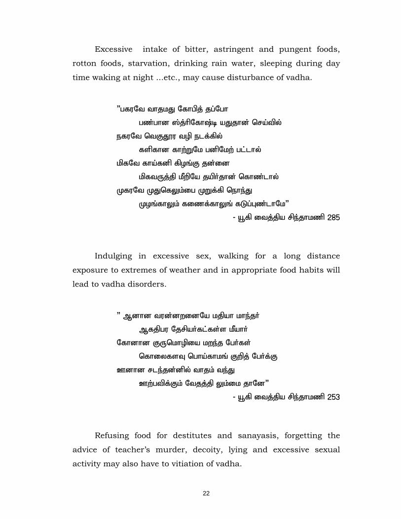
Excessive intake of bitter, astringent and pungent foods,
rotton foods, starvation, drinking rain water, sleeping during day
time waking at night ...etc., may cause disturbance of vadha.
“gfuNt thjkJ Nfhgpj; jg;Ngh
gz;ghd ];j;hpNfh\;b aJjhd; nra;tpy;
efuNt ntFJ}u top elf;fpy;
fspfhd fhw;WNk gdpNkw; gl;lhy;
kpfNt fha;fdp fpoq;F jd;id
kpftUj;jp kPwpNa japHjhd; nfhz;lhy;
KfuNt KJnfYk;ig KWf;fp nehe;J
Koq;fhYk; fizf;fhYq; fLg;Gz;lhNk”
- A+fp itj;jpa rpe;jhkzp 285
Indulging in excessive sex, walking for a long distance
exposure to extremes of weather and in appropriate food habits will
lead to vadha disorders.
“ Mdhd tud;dwidNa kjpah khe;jH
Mfjpgu NjrpaHfl;fs;s kPahH
Nfhdhd FUnkhopia kwe;j NgHfs;
nfhiyfsT ngha;fhkq; Fwpj; NgHf;F
Cdhd rle;jd;dpy; thjk; te;J
Cw;gtpf;Fk; Ntjj;jp Yk;ik jhNd”
- A+fp itj;jpa rpe;jhkzp 253
Refusing food for destitutes and sanayasis, forgetting the
advice of teacher’s murder, decoity, lying and excessive sexual
activity may also have to vitiation of vadha.

23
Vadha kanma varalaru
E}nyd;d thjk; te;j tif jhNdJ
Ez;ikaha;f; fd;kj;jpd; tiffiaf; NfS
fhdNy Njhd;wpaJ fUg;g NjJ
iffhypy; Klf;fpaJ tPf;fNkJ
NfhypNy gLfpd;w tpUl;rkhd
Foe;ij kue;jid ntl;l Nky; Njhy; rPty;
ehypNy rPtnre;J fhy; Kwpj;jy;
ey;y nfhk;G jil kwpj;;jy; eypj;jy; jhNd
-mf];jpaH fd;k fhz;lk; 300 > ghly; 56
Accordingly barrenning the land of young green, trees, laming
living beings, chopping the branches of a living tree, removing the
barks and leaves of trees may lead to vadha diseases.
ghhpdpw; gag;g;gl;lhYk; gyUld; Nfhgpj;jhYk;
fhnudr; fUfpNahbf; fokuj; Juj;jpdhYk;
VHngW jdJ neQ;rpd; kpfj;Jf;f kile;jpl;lhYk;
ghhpa fhw;wpdhYk; glhPYk; thjk; fhzhk;
- guuhr Nrfuk;
According to this verse fear, anger, sorrow, excessive work &
climate changes lead to vitiation of vadham.
Sabapathi kaioyedu attributes the causes of vadha diseases to
in appropriate food habits exposure to cold and indulgence in
excessive sex.
tspjU fha;fpoq;F tiutpyh japyy; Nfhio
KdpjapH Nghd; kpFf;F Kiwapyh cz;b
FspHjU tspapw; Nwfq;Fdpg;Gw tpytH ngz;bH

24
fspjU Kaf;fk; ngw;NwhH fbnray; fUtpahky;
- rghgjp ifNaL
In agastiyar Gunavagadam
“njhy;iy nra;a ,d;Wk; ntF thjNeha;fs;
njhy;Yyfpy; khe;jUf;Ff; fhz;gJz;L
vy;iyapy;iy thjNeha; NeHik jd;id
,ay;ghf mwpe;jplNt tpguq; NfNs”
“ tptuklh mrjp rd;dp %is NehT
tphpthd %isaJ kpUJthfp
mtdpjdpy; jplkhfg; NghtjhYk;
mgg;Nd %j;jpuf; Fz;bf;fha; tpahjpahYk;
jtKdptH jPHfhf;if NkfNuhfk;
jd;ikAs;s Kj;jz;Lf; nfhb tpahjp
mtkpyhg; ghpr euk;gge;jq;fz;hla;
mDFklh thj Neha; MFk; ghNu”
“mWFklh khkpre;jpd; tpahjpahYk;
mg;gNd #jfj;jpd; ngUf;fhYk;
Fzkpy;yh ,urk; tq;fk; jpd;dhYk;
Fb nfLj;j thjkJ cz;lhkg;gh”
- mf];jpaH Fzthflk;
Brain disease
Renal disease
Sexually transmitted disease
Disease of the vertibral column & spinal cord
Compression of sensory nerves
Menorrhogia
Taking improperly prepared medicine of mercury and lead will
cause vadha disease.
25
Other Causes:
1. Consumption of bitter, astringent
2. Eating previously cooked food
3. Drinking polluted water
4. Changing sleep rhythm
5. Excessive starvation
6. Excessive rest
7. Walking long distance
8. Living in chill environment
9. Excessive consumption of tubers, fruits, curd etc.,
Alteration of udal vanmai
Udalvanmai is described as iyarkai vanmai, cheyarkai vanmai
and kaala vanmai
Iyarkai vanmai is considered with three gunangal (Sathuva,
rajo, thamo gunam)
Kaala vanmai is considered with age, season, most of the
vadha disese occurs in old age because the kaala vanmai is
diminshed in old age.
Changes in cheyarkai vanmai also play a major role. Wrong
postures, style and improper foods, life style modification
causes the peripheral neuritis.
“thjNk Gspg;G Ntz;Lk; ....”
- ,uj;jpdr; RUf;f ehb
“GspJtHtpQ; Rq;fwpahw; G+hpf;Fk; thjk; .....”
- fz;Zrhkpak
Gspg;G> JtHg;G mjpfKs;s czTfshy; thjk; kpFjpgLk;
26
SIGNS AND SYMPTOMS OF VADHA DISEASES
The signs and symptoms of vadha diseases have been given in
many siddha classical text books as follows.
Pricking pain, dull aching pain termours, palpitation, spasm,
dryness, dehydration, dislocation of joints, weakness of the body,
paralysis, constipation, oliguria, excessive thirst horpliation,
difficulty in flexion and extension of limbs, astringent taste in the
mouth and excretions like stools, urine, tears and sweat are black
in colour.
In Agasthiyar 2000
“thjj;jpd; FzNknjd;dpy;
kaf;Fe; jpnaq;Fk; kyH rptf;Fk;
ghjq; FspHe;J rUthq;fk;gw;wp elf;F Kfq;fUf;Fq;
rPjj;JlNd tapW Gz;zhQ; rphpg;Gj; Je;njwp %r;rhk;
Nghje; jz;zPHjhd; thq;Fk; GfOk; gQ;rFzkhNk “
Giddiness
Stabbing pain in the face
Redness of eyes
Peptic ulcer
Abdominal distension
Joints pain in upper & lower limbs.
Numbness in the limb
Oliguria
Drowsiness
Chillness of body
27
Agasthiyar Naadi
“nrhy;yNt thj kJ kPwpw;hy;
NrhHtile;j thA tpdhy; Njfnkq;Fk;
nky;y iffhy; mrjp Az;lhFk;
nka;Klq;Fk; epkpu nthz;zhj; jpkph; cz;lhFk; “
- mf];jpah; ehb
Weakness of the limbs
Sluggishness
Stiffness
Numbness
In Theraiyar Vaagadam
“ thjtPW md;dkpwq;fhJ fLg;Gz;lhk; tz;zKz;lhk;
NkhJ fl;LNuhfk; RuKz;lh kpUkYkh Kwq;fhnjd;Wk;
XJ#hpa thjkiyf;F eLf;fKz;lhk; nghUs;fsha;j;
jPnjdNt euk;gprpj;J re;Jfs; NjhYq; fLf;Fe; jhNd”
- NjiuaH thflk;
Loss of appetite
Back ache
Fever
Cough
Sleeplessness
Shivering
Pain in the joints
“ jf;f thA Nfhgpj;jhy; re;JTise;J jiyNehT
kpf;f %hp nfhl;lhtp tpl;lq; nfhpT kyq;fl;Lk;
xf;f euk;G jhd; Klf;F KyHe;J tha; ePUwptpLk;
kpf;f FspUk; eLf;fkhFk; NkdpFd;wp tpLq;fhNz”
- NjiuaH thflk;
Pain in the joints

28
Head ache
Excessive yawning
Constipation
Burning sensation of the body
Paralysis
Excessive salivation
Chillness
Tremour
In Padhinen siddharkal naadi sasthiram
“ Nktpa thjQ; nra;Aq; Fzj;jpid tpsk;gf; Nfsha;
jhtpa tapW ke;je; re;J fhy; nghUj;J nehe;E
Nrtpa jhJ ehrk; rpWj;Jld; rpWePH tPok;
fhtpaq; ft;tpdhNs kykJ RUfpf;fhZk;”
- jpul;Lehb - gjpnzd; rpj;jHfs;
ehb rh];jpuk;
“thjj;jpd; FzNk njd;dpy; tapwJ nghUkpfhZk;
jhjj;jpy; Nkdp if fhy; re;JNk fLg;G fhZk;
jPje;J rpWePH jhJQ; nrwpe;Jld; fLj;J tPok;
NghJj;j thjnkd;W Gfd;wdH KdptH jhNk”
“fhzg;gh thj kPwpy; fhy; iffs; nghUj;jp NehpLk;
G+zg;gh Fly; Gul;Lk; kyQ;ryk; nghUkp fl;Lk;
Czg;gh FspUq; fha;r;r Ylk;ngy;yk; Fj;J tha;T
tPzg;gh FjkpUFk; NtHitAk; NtHf;Fk; ehNs”
- gjpnzd; rpj;jHfs; ehb rh];jpuk;
The vitiation of vadha results in dyspepsia, pain in the joints
& dysuria constipation, abdominal colic, fever ,rigor, body ache and
excessive sweating.

29
CLASSIFICATION OF VADHA DISEASES
In classification of vadha diseases we can find contradictory
view regarding the number, in various books.
In yoogivaidhya chinthamani – 800
“vd;dNt thjkJ vz;gjhFk;”
Eighty types of vadha diseases are described
In “Agasthiyar Rathina Surukam” – 500 Eighty four types of
vadha diseases are reported
“kw;wNk thjNuhfk; tifA
vz;gj;J ehNy”
- mf];jpaH uj;jpd RUf;fk;
But while concluding the number has been given as eighty
four by the yoogi vaidhya chinthamani – 800.
“Mkg;gh thjnkz;gj;J ehY
mjDila Fzh fzq;fylq;fhf”
- A+fp itj;jpa rpe;jhkzp
In Astaanga sangiragam and noi naadal noi mudhal naadal
thirattu part I. Vadha diseses have been classified as 85 types on
the symptamatology and involvement and different parts of the
body.
In Theraiyar Vaagadam 81 types of vadha diseases been
described
30
“Dhanvandhiri vaidhiyam” and “Jeeva Rakshamiraham” 88
types of vadha diseases have been noted.
“Agasthiyar 2000” forty types of vadha diseases are in the
upper half of the body and in forty lower half of the body and the
total number in 80.
“vz;gJ thjkhF kpU tif
ez;GW miuf;F NkNy ehw;gJ thjkhFk;
gz;Nru iuf;F fPNo
tz;L NrH FoypdhNs thjj;jpd; tiffs; jhNd”
- mf];jpaH 2000
In Bohar Vaidhiyam 700, 80 vadha diseases are told.
“ thr;nrd;wthjk; vd;gJTk;
NghFk;”
- NghfH itj;jpak; 700
In Agasthiyar Gurumuni – 235, 85 vadha diseases have been
reported.

31
CLINICAL FEATURES OF VADHA UBHAKATHAM
Uba means Subnormal
Katham means Pain full condition.
Yugi in yugi vaidhya chinthamnai, has described the clinical
features as follows.
Vadha ubhakatham is one of the vadha disease.
“Mz;ikahq; fhNyhL ifAQ; re;Jk;
mq;fnky;yh kpfkpjpHj;Jr; rhzpjhDk;
G+z;ikaha;g; G+rpdJ NghNy fhZk;
Guz;Ljhd; tpWtpWj;Jg; Gskhfpg;
ghz;ikahq; fhyirA K\;zkhfpg;
grpj;JNk kpfehzp eil nfhlhJ
thz;ikahk; thl;lKW kaf;f khFk;
thjTgje; jd;id tFj;jthNw”
- A+fp itj;jpa rpe;jhkzp
“Mz;ikahq; fhNyhL ifAQ; re;Jk;
mq;fnky;yh kpfjpkpHe;Jr;:
An increased feeling of numbness is vigorously present in the
legs & in the hands joints and all over the body.
“ rhzpjhDk; G+z;ikaha; G+rpdJ
NghNy fhZk;”
A sensation of smearing the dung of animals as armour.
Guz;Ljhd; tpWtpWj;Jg; Gskhfp

32
Hastening of numbness with gnawing sensation..
ghz;ikahq; fhyirA K\;zkhfpg;
A sense of heat in the leg during movements due to burning
sensation.
grpj;JNk kpfehzp eil nfhlhJ
thz;ikahk; thl;lKW kaf;f khFk;
As the worsening of numbness progress they have difficulty in
walking with delusion & drowsiness.
thjTgje; jd;id tFj;jthNw
All the above symptoms are called vadha ubhakatham.
Summary
An increased feeling of numbness and a sensation smearing
the dung of animals is vigorously present in the legs and in the
hands joints and all over the body. The numbness a condition of in
sensibility increases in its density with restleness of the body. As a
result a sense of heat in the feet become prominent. As the
worsening progresses they are difficult in walking, with mental
delusion and drowsiness.
33
MUKKUTRA VERUPAADUGAL
Pathogenesis
1. By any one (or) other etiological factors vadham is vitiated
first.
2. Then it affects the other dhoshas pitham and kabham which
are in three equilibrium.
3. And then the ten vayus, seven udarkattugal and other
structures are also affected according to the severity of the
illness.
4. By the affection of piranan wheezing cough, dyspnoea, nasal
congestion and indigestion may occur.
5. By the vitiation of abaanan constipation, oliguria and
menstrual disorders may occur.
6. By the affection of udhanan heart, chest and eyes are affected
hiccup vomiting & heart burns are formed.
7. By the vitiation of viyanan muscle wasting loss of sensation,
giddiness coma, body ache, numbness, itcing & tingling
sensation are formed.
8. By the affection of samanan disturbances of other vayus
abdominal distension, anorexia, malnutrition and indigestion
may occur.
9. When saaram is affected anorexia, laziness lassitude
weakness and dryness of the skin are formed.
10. When senneer is affected nerve weakness, dryness, mental
disorder, haematuria, jaundice anaemia, anorexia,
spleenomegaly & skin diseases may occurs.

34
11. When oon is affected muscle wasting dropsy, body ache,
oedema, weakness of the five sensory organs are formed.
12. When kozhuppu is affected body debility body ache, joints
pain, spleenomegaly, tiredness may occur.
13. When enbu is affected arthritis, joint pain, osteophytic
formation and other bone diseases are formed.
14. When moolai is affected blurring of vision, ulcres, heaviness
burning micturation, the body & bone diseases may occurs.
15. When sukkilam is affected lustfulness urinary calculus,
bleeding during coitus orchitis and diseases of genitalia are
formed.
16. When pitham is affected anorexia, anaemia, indigestion,
blurring vision, dryness and darkness of skin. Vomiting,
giddiness, burning sensation of the body and difficulty to do
works are formed.
17. When kabham is affected respiratory disorders, indigestion,
tastelessness, burning sensation of eyes and joint disease
may occurs.
In vadha disease abanan, viyanan, samanan, naagan,
koorman, dhevathathan are affected, generally saaram, senneer,
oon kozhuppu enbu and moolai are also affected one by one.
Among the five types of pitha the rhythm of sadhaha pitham
gets affected mainly causing difficulty in performing the activities of
daily life. (i.e.,) difficulty in walking etc., Among the five types of
kabham, tharpagam is affected mainly in Madhumegam patients..

35
Relationship between tridhoshas suvai and panchabootham:
There are six types of suvai. Each suvai is formed by the
combination of two boothas.
Inippu (Sweet) – Earth + water
Pulippu (Sour) – Earth + fire
Uppu (Saline) – Fire + water
Kaippu (Bitter) – Air + space
Karppu (Pungent) – Air + fire
Thuvarpu (Astringent ) – Earth + Air
They are also classified into 2 types of veeriyam (Potency)
Veppa veeriyam
Seetha veeriyam
Veppa veeriyam suvigal – Pullipu, karpu, uppu
Seetha veeriyam suvigal – Inippu, thuvarpu, kaippu
By knowing the veeriyam medicines are administered so that
the deranged kutrams are normalized
36
PINIYARI MURAIMAI (DIAGNOSIS)
Pini means the disease which affect the body and any interruption
of the normal functions of any body part.
Ari means identify
Muraimai means rules
Piniyarai murimai is the method of diagnosing the disease
affecting the people is based upon the following aspects
I. Poriyalarithal
II. Pulanarithal
III. Vinathal
IV. Envagai thervugal
V. Naadi Paritchai
I. Poriaal aridhal
The physician should examine the patient’s porigal by his
porigal.
Mei - feels all types of sensations
Vaai - for knowing taste
Kan - meant for vision
Mooku - for knowing the smell
Sevi - for hearing
II. Pulannal aridhal
The physician should examine the patient’s pulangal by his
pulangal
Hearing - ear
Vision - eye
37
Taste - tongue
Sensation - skin
Smell - nose
III. Vinadhal (Interrogation)
The physician should interrogate about the patient’s name,
age, sex, occupation, native, socio economic status, dietary habits
prone to any allergers, complaints history of previous illness,
history of habits and frequency of attacks. If the patient is in the
stage of inability to speak or a child to physician should interrogate
the details with his immediate relatives who are taking care of him.
Naadi (Pulse)
Sparisam (Palpation)
Naa (Tongue)
Niram (Colour of the body)
Mozhi (Speech)
Vizhi (Eye examination)
Malam (Motion examination)
Moothiram (Urine Examination)
Envagai thervugal in the siddha literature gunvagada naadi is
as follows.
“juzpAs;s tpahjp jd;id al;lhq;fj;jhy;
jhdwpa Ntz;LtJ NaNjh ntd;dpy;
jpuzpa NjhH ehbfz;fs; rj;jj;NjhL
Njfj;jpdJ ghprk; tUzk; ehf;F
38
apuzky %j;jpukhkpitf; nsl;Lk;
apjk; glNt jhd; ghHj;Jf; FwpAq; fz;L
GydUshy; nghpNahHfs; ghjk; Nghw;wpg;
gz;G jtwhky; gz;bjw; nra;tPNu”
- Fzthflehb
Envagai Thervugal
Eight different kinds of tests to be applied or attend by a
physician before arriving a correct diagnosis. These are also called
attavitha parichai (or) attathana parikshai.
Envagai thervugal is considered as physician’s instruments.
“ehb ghprk; ehepwk; nkhop tpop
kyk; Kj;jpukpit kUj;JtuhAjk;”
“ nka;Fwp epwe;njhdp tpop ehtpU kyk; iff;Fwp”
- NjiuaH
In agasthiyar vallathi 600, Envagai thervugal has been
mentioned as atta vidha paritchai
“njhf;fYw;W ml;ltpj ghPl;ir jd;id
Jyf;fKWk; gz;bjNu njspthfk;
gFj;jwpa ehbia eP gpbj;J ghU
gfHfpd;w thHj;ijiag;ghH ehit ghU
tFf;fhpa Njfkij njhl;Lg; ghU
tskhd ruPuj;jpd; epwj;ij njhl;Lg; ghU
rfpf;fhpa kyj;ijg;ghh; ryj;ijg; ghU
rhHe;j tpop js;sg;ghHj;J njsptha;f; fhNd”
- mfj;jpaH ty;yhjp 600

39
According to thirumoolar, feeling the naadi in various sites are mentioned as
jhJ KiwNfs; jdpj; jFjp re;NjhL xJW fhkpa Ke;jpneL khHG fhJ neL%f;F fz;lk; fuk;GUtk; NghJWKr;rp Gfo; gj;Jk; ghHj;jpNl
- jpU%yH ehb
Formation of Naadi
(Naadi) + (Vayu) = Uyirthathu
Idakalai + Abanan = Vadham
Pinkalai + Pranan = Pitham
Suzhumunai + samanan = Kabham
Normally the three humours of vadham, pitham and kabham exist the ratio 1: ½ : ¼ . The derangement in these ratio leads to various disease entities and is best diagnosed by feeling the naadi.
1. Naadi (Pulse)
Naadi is the vital force. Any changes in the three dhoshas are best diagnosed feeling the naadi. Naadi is an important observation for diagnosis and prognosis. Naadi is responsible for the existence of life and can be felt one inch below the wrist on the radial side by means of palpation with the tips of index, middle ring finger corresponding to vadham, pitham and kabham site to feel naadi and its procedure is
“fhpKf dbia tho;j;jpf; ifjdpy; ehb ghHf;fpy; ngUtpuyq;Fyj;jpy; gpbj;jb eLNt njhl;lhy; xUtpu Nyhby; thjk; caHeL tpuypw; gpj;jk; jpUtpuy; %d;wp Nyhby; rpNyj;Jk ehb jhNd”
- mfj;jpaH ehb

40
“ toq;fpa thjk; khj;jpiu nahd;whfpy;
joq;fpa gpj;je;jd;dpyiuthrp
moq;Fq;fge;jh dlq;fpNa fhNyhby;
gpwq;fpa rPtHf;Fg; gprnfhd;Wkpy;iyNa “
- Fzthflehb
Character of pulse “thfpdp yd;dq; Nfhop kapnyd elf;Fk; thjk;
Vfpa thikal;il apitnad elf;Fk; gpj;jk;
Nghfpa jtis ghk;G Nghythk; Nrj;Jke;jhd;
Mfpa ehb %d;Wk; kHj;jpbw; rd;dpahNk”
From this poem it is evident that the character of vadha naadi
resembles the walk of goose, hen, and peacock. Pitha resembles
the walk of tortoise and leech and kabha resembles the walk of frog
snake etc.,
In cases of vadha diseases the following stages of naadi are
seen.
Vatha Pitha Nadi “ nghUshd thjj;jpy; gpj;jQ; NrHe;J
nghUe;J Fzq;fsh K\;zthA rj;jp
nrhpahik Gspj;Njg;gk; nghUkw; ePhpw;
rptg;G kyk; gpbj;jYUe;jhJ el;lk;
fUthd NjfkjpYisr;ry; Nrhk;gy;
iffhy; jwpg;G ehf;frf;F kd;dk;
ghpthd T+z; Fiwjy; Urp Nflhjy;
gyNehAk; tUj;jp itf;Fk; ghq;FjhNd”
- rjf ehb
“jpUj;jkhk; thje;NjhNl jPq;nfhU gpj;jQ; Nrw;
nghUj;Jfs; NkYk; nehe;J NghjNt gpbf;Fk;”
- Nehapd; rhuk;
41
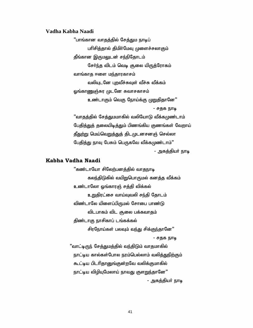
Vadha Kabha Naadi “ghq;fhd thjj;jpy; Nrj;Jk ehbg;
ghprpj;jhy; jpkpHNkT Kisr;ryhFk;
jPq;fhd ,UkYld; re;epNjhlk;
NrHe;j tplk; ntb #iy apUj;Nuhfk;
thq;fhj <is ke;jhufhrk;
typAlNd GwtPr;RTs; tPr;R tPf;fk;
Xq;fhZQ;Ru KlNd Rthrfhrk;
cz;lhFk; ntF Neha;f;F KWjpjhNd”
- rjf ehb
“thjj;jpy; Nrj;Jkkhfpy; typNahL tPf;fKz;lhk;
Ngjpj;Jj; jiyapbj;Jk; gpzq;fpa Fzq;fs; Ntwha;
jPJw;W nka;ntWj;Jj; jplKldrdQ; nry;yh
Ngjpj;J ehT NgRk; ngUfNt tPf;fKz;lhk;”
- mfj;jpaH ehb
Kabha Vadha Naadi “fz;lhNah rpNyw;gdj;jpy; thjehb
fye;jpLfpy; tapWnghUky; fdj;j tPf;fk;
cz;lhNyh Xq;fhuQ; rj;jp tpf;fy;
cWjpul;ir tha;Ttyp re;jp Njhlk;
tpz;lhNy apisg;gpUky; Nrhig ghz;L
tplghfk; tpl #iy gf;fthjk;
jpz;lhF ehrpfhg; lq;ff;fy;
rpuNeha;fs; gyTk; te;J rpf;Fe;jhNd”
- rjf ehb
“thl;bUe; Nrj;Jkj;jpy; te;jpLk; thjkhfpy;
ehl;ba fhy;fs;Nghy euk;ngy;yhk; typj;Jepw;Fk;
$l;ba gplhpjhDq;Fd;wNt typf;Fkhfpy;
ehl;ba tpopANkyha; ehtJ FsWe;jhNd”
- mfj;jpaH ehb

42
In all vadha ubhakatham patients vadha, kabha, thontha
naadi was noted.
Selection of hand for both ehndYk; GUlHf;nfy;yhk; ehbjhd; tyf;ifahFk;
NjdndDk; kltHf;nfy;yhe; jplk;ngw tplf;if rpj;Nj
- itj;jpa rhurq;fpufk;
This poem says that pulse should be seen on the right hand
for males and left hand for females.
This is because of the position of nabikoormam. It is upwards
in females and downwards in males.
Sparisam (Skin)
Examination of skin can be made out of inspection and touch
it reveals whether the skin is warm, chill, dry, weeping, rough,
smooth, hard, tender, presence of ulcers, swelling, wrinkles hair,
pigmentation etc.,
Naa-tongue
The following should be noted colour, clearness, pallor, any
coating excessive salivation, dryness, ulceration, fissures,
thickening any growths, dents, conditions of teeth, and gums,
speech any deviation of tongue etc.,
Niram (Complexion)
Colour of skin, flushing pallor discolouration etc., should be
noted.
Mozhi (Speech)
Disorder of speech tone, hoarseness, laughter, slurring,
incoherent, speech making unusual noices etc., should be noted.
43
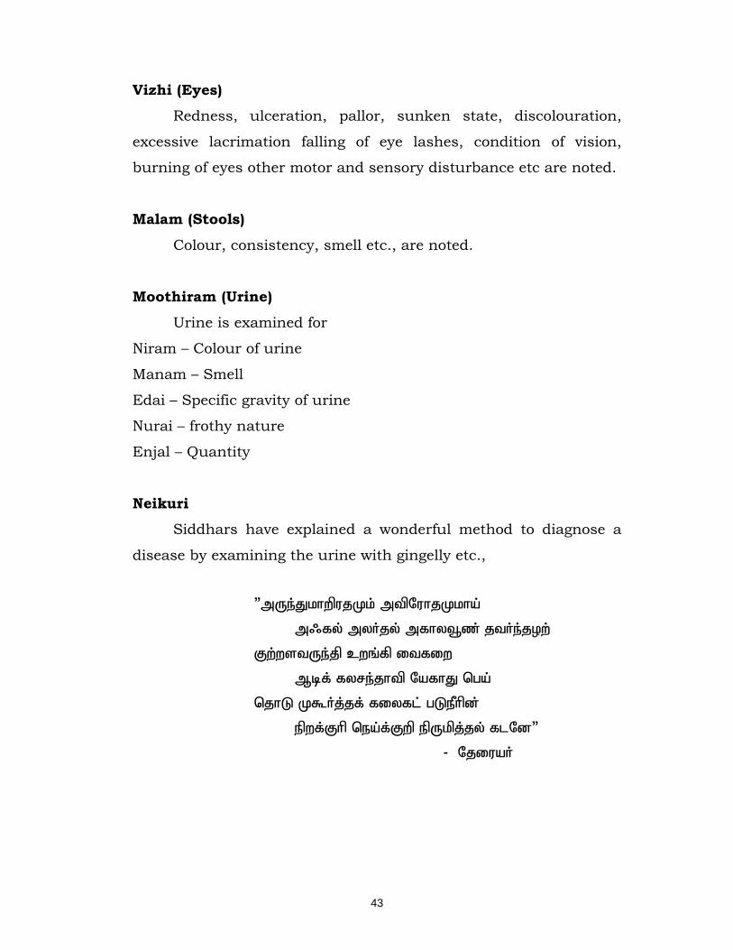
Vizhi (Eyes) Redness, ulceration, pallor, sunken state, discolouration,
excessive lacrimation falling of eye lashes, condition of vision,
burning of eyes other motor and sensory disturbance etc are noted.
Malam (Stools)
Colour, consistency, smell etc., are noted.
Moothiram (Urine)
Urine is examined for
Niram – Colour of urine
Manam – Smell
Edai – Specific gravity of urine
Nurai – frothy nature
Enjal – Quantity
Neikuri Siddhars have explained a wonderful method to diagnose a
disease by examining the urine with gingelly etc.,
“mUe;JkhwpujKk; mtpNuhjKkha;
m‡fy; myHjy; mfhyT+z; jtHe;jow;
Fw;wstUe;jp cwq;fp itfiw
Mbf; fyre;jhtp NafhJ nga;
njhL K$Hj;jf; fiyfl; gLePhpd;
epwf;Fhp nea;f;Fwp epUkpj;jy; flNd”
- NjiuaH
44
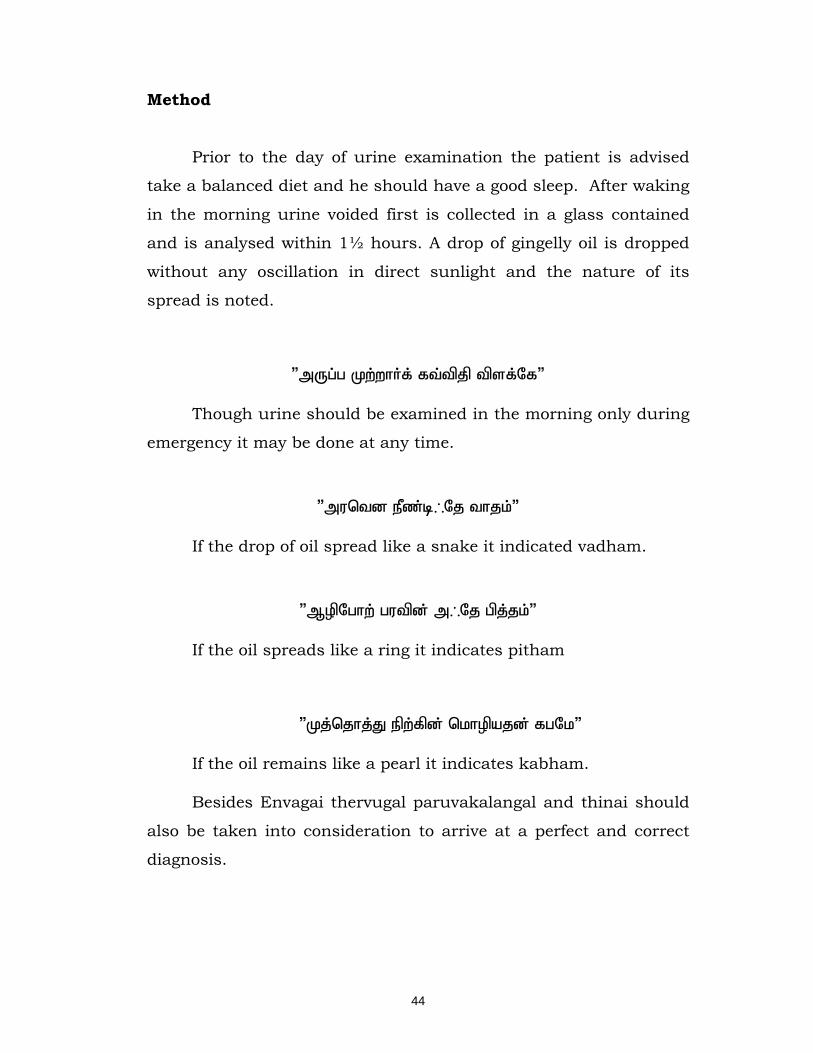
Method
Prior to the day of urine examination the patient is advised
take a balanced diet and he should have a good sleep. After waking
in the morning urine voided first is collected in a glass contained
and is analysed within 1½ hours. A drop of gingelly oil is dropped
without any oscillation in direct sunlight and the nature of its
spread is noted.
“mUg;g Kw;whHf; ft;tpjp tpsf;Nf”
Though urine should be examined in the morning only during
emergency it may be done at any time.
“muntd ePz;b0Nj thjk;”
If the drop of oil spread like a snake it indicated vadham.
“MopNghw; gutpd; m0Nj gpj;jk;”
If the oil spreads like a ring it indicates pitham
“Kj;njhj;J epw;fpd; nkhopajd; fgNk”
If the oil remains like a pearl it indicates kabham.
Besides Envagai thervugal paruvakalangal and thinai should
also be taken into consideration to arrive at a perfect and correct
diagnosis.
45
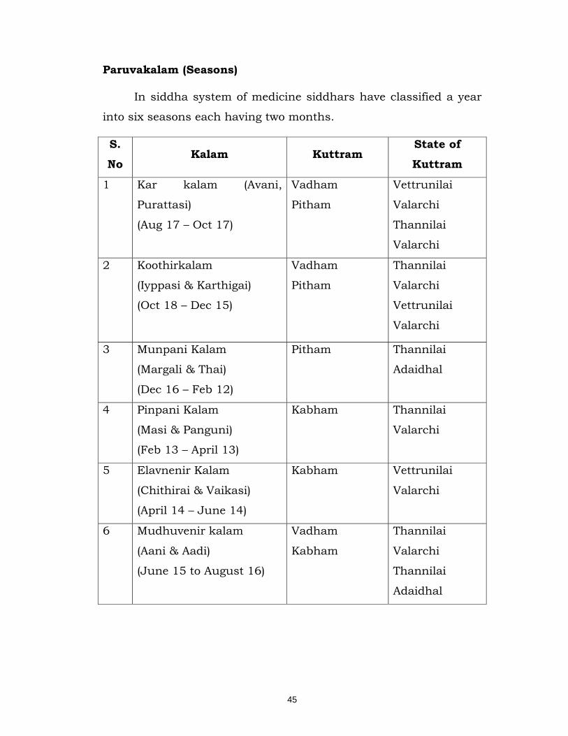
Paruvakalam (Seasons)
In siddha system of medicine siddhars have classified a year
into six seasons each having two months.
S.
No Kalam Kuttram
State of
Kuttram
1 Kar kalam (Avani,
Purattasi)
(Aug 17 – Oct 17)
Vadham
Pitham
Vettrunilai
Valarchi
Thannilai
Valarchi
2 Koothirkalam
(Iyppasi & Karthigai)
(Oct 18 – Dec 15)
Vadham
Pitham
Thannilai
Valarchi
Vettrunilai
Valarchi
3 Munpani Kalam
(Margali & Thai)
(Dec 16 – Feb 12)
Pitham Thannilai
Adaidhal
4 Pinpani Kalam
(Masi & Panguni)
(Feb 13 – April 13)
Kabham Thannilai
Valarchi
5 Elavnenir Kalam
(Chithirai & Vaikasi)
(April 14 – June 14)
Kabham Vettrunilai
Valarchi
6 Mudhuvenir kalam
(Aani & Aadi)
(June 15 to August 16)
Vadham
Kabham
Thannilai
Valarchi
Thannilai
Adaidhal
46
Environmental changes
(a) Seasonal changes of humours
Humours ↑ ↑↑ →
Vadham Mudhuvenir Kalam Kaarkalam Koodhirkalam
Pitham Kaarkalam Koodhirkalam Munpanikalam
Kabham Pinpanikalam Elavnil kalam Mudhuvenil
kalam
↑ - Thannilai valarchi
↑↑ - Piranilai valarchi
→ - Thannilai adaithal
Regional changes of humours
Thinai (land and place)
The geographical distribution of the land is classified into five
regions.
Kurunji - Mountain and surroundings - Kabha disease
Mullai - Forest and adjacent area - Pitha disease
Marutham - Fields and surroundings - No disease will occur
Neithal - Sea and surroundings - Vadha disease
Palai - Desert and its surroundings - Mukkuttra disease
Each region has its own characters which influence the
inhabitants physical mental, economic, occupational and cultural
activities. In each region some ailments are common in neithal
nilam. Palai nilam is a common place for all type of disease and
marutha nilam is good for all types of treatment and health.
47
ASSOCIATED DISEASES
Regarding to Diagnostic aspect of siddha system
According to Maan murugheeyam this disease vadha
ubhakatham is mentioned as follows.
“nkd;Nwy; Nkdp ntspwy; nkypjy;
typikapoj;jy; Ritnfly; jsHr;rp
capHg;G FWfy; vDkpit nay;yhk;
FWjpf; Fiwapd; nghJ Fwpnad;g
KJF jz;bd; euk;Gfs; jhf;Fjy;
if fhy; Fj;jy; jpkpHjy; gpwTk;
nfhba FUjpf; Fiwtpid j;jpLNk”
- Mjhuk;-khd;KUfpak; ghly; 33-83> 84-85
This disease due to disorders of the haemopoietic system,
especially due to anaemia (In peripheral neuritis one of the cause is
anaemia)
The symptoms are
1. The pallor of the skin and mucous membrane
2. Loss of body weight
3. Fatigue
4. Loss of taste sensation
5. Burning and pricking sensation over arms, palms legs and
soles.

48
ghz;LNeha; (In anaemia) “nkd;Nwy; Nkdp ntspwy; nkypjy;
typikapoj;jy; Ritnfly; jsHr;rp
....
if fhy; Fj;jy; jpkpHjy; gpwTk;
- Mjhuk;-khd;KUfpak; ghly; 33-83> 84-85
njhONeha; (In leprosy) “Fj;jy; Nehjy; vDkpiw
thrpapd; tUk; njhONehapd; Fwpnad nkhop”
- khd; KUfpak; ghly; 3977
G+g;GwTk; G+g;G Kbtpy; (Post - Menopausal) “jiytyp the;jp jaf;fk; kaf;fk;
ntspwy; cly; ghhpj;jpLjy;
kpFJapy; Nfhly; JapyhapUj;jy;
clk;G typj;jy; tapW Nehjy;
iffhy; cisjy; fLj;jy; tPq;fy;
fz;fs; Fopjy; fhl;rp kiwjy;
ngU%r;R vwpjy; Ngrpa gpwTk;
jdpj;Jf; fye;Jk; jiytU nkd;g
G+itaH G+g;G epw;Fq;fhy;”
- khd; KUfpak; ghly; 1056

49
DIFFERENTIAL DIAGNOSIS
thjfh;\;zk;
ghh;fpd;w ghjTs; shbapw; rhzp
gjpj;Jitj; jJNghyg; ghu nkq;Fk;
Nfhh;fpd;w Fjpiu euk;Gq; fhy;f nsq;Fk;
nfhbjhd ghukha;j; jpkph;g;Gz; lhfp
thh;f;fpd;w thh;j;jijfs; jhd; kpfNt nra;j
thWfpD epkpUfpDk; jrq;nfhlhky;
Vh;fpd;w fhYisf;Fk; thjfh;\;zk;
<jyw kpy;yhjhh;f; nfa;Jq; fhNs
Dung coated sensation in both feet numbness and heaviness
along course of sciatic nerve. In ability to maintain the upright
posture. Lower limbs pain . This is the signs and symptoms of
Vadhakarshanam.
In Vadhaubhakatham
Complaints such as numbness and heaviness along course of
sciatic nerve. In ability to maintain the upright posture is not
present. Thus it differentiated from Vadhakarshanam
fu];jk;g thjk;
va;jpNa tphpTjdp ypuz;L fhYk;
,irj;jTs; sbjdpD nkhpe;J nehthk;
IjpNa af;fpdpapy; itj;jhw; Nghy
mq;fnky;yh kpfntSj; Jyj;jp ahFk;
ngha;jpNa GiujdpNy GOf;f Sh;jy;
NghyNt kpfT+h;e;J eilnfh lhJ
ifjpNa thAyhTw; Wly;f df;Fk;
fzf;fhd fu];jk;gq; fUjpg; ghNu
50
Burning sensation in both lower limbs, generalised paleness
of body sensory disturbance, difficulty in walking, heaviness of the
body , this is the signs and symptoms of Karasthamba vadham
In Vadhaubhakatham Heaviness of the body is not present thus it differentiated
from the Karasthambhavatham
CU];jk;gk;
Mnkd;w thjkJ cs;s lf;fp
mbnjhilj;jhd; Fuq;fpuz;L kztha;g; gw;wpf;
fhnkd;w iufhypy; tpuYQ; Rw;wp
fdj;JNk rhzpaJ nghjpe;jhw; Nghy
Njnkd;w rpue;jdpNy ghu Kz;lha;j;
Njfnkq;F %jpNa jpkpUz; lhFk;
ehnkd;w elf;nfhzh nthUf;f khfp
fyp%U]; ek;gkJ eWFq; fhNz
The disease is specially localized to gluteal and thigh region.
Around waist, hip, legs and fingers there is a feeling of dung coated
sensation, heaviness of head, generalised bulkiness and numbness.
The walk is diminshed . These are the sings and symptoms of
voorusdhambham. Which is due to decreased vadham
In Vadha Ubhakatham It is not specified for gluteal and thigh region. Heaviness of
head is not present thus it differentiated from
Oorusthambavadham.
51
NOI NEEKAM / PARIKARAM
Treatment of vadha ubhakatham
In siddha system of medicine the main aim of the treatment is
removal of udalpinigal (due to alterations of uyir thadhukkal and
udal thadhukkal) and ulappinigal (due to alternation of mind).
Treatment is not only for removal of disease but for the prevention
and improving the body condition also. This said to as follow.
• Kaapu
• Neekam
• Niriappu
Ayyan Thiruvalluvar says about physicians duty as “study the
disease, spy the cause, seek subsiding ways and do what is proper
and effective” the man wellversed in medical lore would measure
the patients, disease and time before the healing work begins.
“ Neha; ehb Neha; Kjy; ehb mJjzpf;Fk;
tha; ehb tha;g;gr;nray;”
- jpUf;Fws;
“ cw;whd; sTk; gpzpasTq; fhyKq;
fw;whd; fUjpr; nray;”
- jpUf;Fws;
So its essential to know the disease the cause, the nature of
the patient, severity of illness, the seasons and time of occurence
must be observed clearly.
The treatment is divieded into three types in siddha system of
medicine namely.

52
Dheva Maruthuvam
Maanida Maruthuvam
Asura Maruthuvam
Dheva Maruthuvam
It includes medicines, made out of organic and inorganic ores
such as chunnam, parpam, chenduram, kalangu, kattu, mathirai,
melugu.
Manida Maruthuvam
It is the medicine method made out of plants such as
kudineer, churanam, surasam, pittu, vadagam, ilaham.
Asura Maruthuvam
It is named after the method of treatment. It include aruvai
(surgery) keeral (scratching of blood vessels and abcess) attai vedal
(Leech therapy) kuridhi vangal (blood letting therapy) kombu kattal
(setting of bone with support).
Purgatives
It corrects the vitiated vadham
“Ngjpahy; thjk; jhOk;”
Vellai ennai – 15ml at early morning is given one day before
starting the main treatment usually otherwise we used Nila vagai
chooranam 10gm with hot water at bed time.
“XJfpd;w kyf;fl;il nahopa itj;jhy;
clypYs;s thijnayh nkhLq;fpg; NghFk;”
53
Medicines
The anti vadha medicines both internal medicines and
external applications are given to relieve the symptoms and
strengthen the affected parts.
Pathiyam
Pathiyam is also an important part of treatment. It is divided
into three types namely
1. Echcha pathiyam
2. Kadum Pathiyam
3. Migakkadum Pathiyam
Uppila pathiyam is also mentioned in many ancient siddha
literatures especially for vadha disease.
“gj;jpaj;jp dhNy gyDz;lhFk; kUe;J
gj;jpaq;fs; Nghdhy; gyd;NghFk; - gj;jpaj;jpy;
gj;jpaNk ntw;wpjUk; gz;bjUf; fhjypdhw;
gj;jpaNk cj;jpnad;W ghH”
- Njiuah; akf ntz;ah
“NfSQ; rpWgapW gRtpd; ghy; japH nea;ahFk;
thUkh Jj;J tiuAl gUg;ghFk; - ike;jNd
Glyq;fha; tiu fhak; Nernkhw;w
rpWfPiuahk; ney;ypf;fha; J}Jtis gUg;ghk; ghNu”
- mf];jpaH ty;yhjp 600
These diet regimen will produce harmful effects so they
should be avoided.
54
Diet and advise
It must be easily digestible
Avoid fat contents and spicy foods
Diet must contain adequate fibre contents to facilitate easy
evacuation of bowels
Add food stuffs which neutralise vadham
Diabetic patients avoid sweets and carbohydrate food.
NrHf;f Ntz;bait
,UKiw tbj;j fQ;rp> fj;jphp gpQ;R> mtiu gpQ;R> mj;jp gpQ;R>
nts;shl;L fwp> fhil> fTjhhp> cLk;G> Klf;fWj;jhd;> mWfPiu>
nghd;dhq;fhzp> J}JNtis> Ntisf; fPiu> %f;fpul;il> gUg;G
tiffspy; Jtiu ey;yJ.
Mfhg; nghUl;fs;
Riu> G+Riz> nts;shp> Gliy> gPHf;F> nkhr;ir> fhuhkzp> cSe;J>
nfhs;S> fLF>
FspHe;j jiuapy; gLj;jy; $lhJ.
55
MODERN ASPECT
Theoretical View of the Dissertation Topic in Modern Aspect
ANATOMY The peripheral nervous system is formed by neurons
and their processes present in all regions of the body. Neurons or
nerve cells are structural and functional unit of the nervous
system. It provides the links from and to the real world. Its ghostly
white nerves thread through virtually every part of the body
enabling the CNS to receive information and carry out it's
decisions. It includes all neural structures outside the
brain and spinal cord that is the sensory receptors, peripheral
nerves and their associated ganglian and efferent motor endings.
Structure of neuron:-
The nerve cell is like any other cells in the body having
almost all the organelles in the cytoplasm. However it is different
from other cells because of the presence of the processes and
absence of centrosomes (centrosome function is formation of cilia
and flagellae and it forms the spindle of fibrillary protein during
mitosis). The neuron is made up of,
1. Nerve Cells
2. Dendrites
3. Axon
The nerve cell body is also called as soma or perikaryon. The
dentrite and axon together form the processes. Each neuron has
only one axon. The axon arises from axon Hillock of soma.
The dendrites may be absent or if present it may be one or
many in number. In general, the dendrites are short processes and
the axons are long processes. The dendrites and axons are usually
called nerve fibers.
56
1. Nerve cell body The nerve cell body is irregular in shape and. like any other
cell it is constituted by a mass of cytoplasm called
neuroplasm covered a cell membrane. The cytoplasm contains
a large nucleus, Nissl bodies, neurofibrils, mitochondria and
golgi apparatus.
(i) Nucleus Each neuron has only one nucleus in the nerve cell body. It is
located in the central part. It has one or two nucleoli which
are prominent. The nucleus does not contain centrosome. So
the nerve cell can not multiply like the other cells.
(ii) Nissl bodies
Nissl bodies or granules are basophilic in nature. These
granules are present through out soma except axon Hillock.
These bodies are responsible for spotted appearance of soma
after suitable staining. The nissl granules flow into the
dendrites from soma, but not into axon. By this axons can be
distinguished from the dendrites.
The nissl bodies are organelles responsible for synthesis of
proteins. The formed proteins in soma are transported to the axon
by axonal flow.
The number of nissl bodies vary with the condition of the
nerve. During fatigue or injury of neuron, these bodies fragment
and disappear by a process called chromatolysis.
iii. Neurofibrils
These are thread like structures present in the form of
network in soma and the processes. These consist of
microfilaments and microtubules.
57
iv. Mitochondria It forms the powerhouse of the nerve cell where ATP is
produced which is energy rich compound.
Golgi apparatus is concerned with package of
proteins into granules.
2. Dendrites
The dendrites are the branched processes of the
neuron and are branched repeatedly. The dendrite may be
present or absent. The dendrites have nissl granules and
neurofibrils.
Dendrites are conductive in nature and transmit impulses
towards the nerve cell body.
3. Axon
The axon is the longer process of the nerve cell.
This arises from axon Hillock of the nerve cell body and is devoid
of nissl granules. The axon may extend for a long distance away
from the nerve cell body. The length of the longest axon is about
one metre.
(i) Structure of axon
With in a nerve, each axon is surrounded by a delicate
layer of loose connective tissue called endoneurium, which
also encloses the fibre’s associated myelin and or neurilemma
sheath. Group of fibers are bound into bundles or fascicles by a
coarser connective tissue, wrapping the perineurium. Finally, all
the vesicles are enclosed by a tough fibrous sheath, the
epineurium, to form the nerve.
Neuron processes constitute only a small fraction of a nerve's
myelin and the protective connective tissue wrappings. Blood
vessels and lymphatic vessels and also found within a nerve.

58
Internal structure of axon:- Axon cylinder:-
The axon has long central core of cytoplasm called
axoplasm. The axoplasm is covered by membrane called axolemma.
Axoplasm contains mitochondria. neurofibrils and axoplasmic
vesicles. Most of the axons are insulated by myelin sheath called as
myelinated nerve fiber. Those, without myelin are called non-
myelinated nerve fibers.
Myelin sheath
It does not form a continuous sheath and is absent
at regular intervals. The area where myelin sheath absent is called
node of Ranvier. It is responsible for white colour of nerve fibers.
Neurilemma :
Surrounding the myelin sheath, there is thin membrane
called as neurilemmal sheath. This is also called as neurilemma or
sheath of Schwann. This contains Schwann cells which have
flattened and elongated nuclei. One nucleus is present in each
internode of axon. The nucleus is situated between myelin sheath
and neurilemma.
Schwann cells (Lemmocytes):-
Schwann cells are satellite cells of the peripheral nerous
system all peripheral axons are ensheathed by them and are
separted from the endoneurrium by the schwann cell plasma
membrnace schwann cells participate in the supply of metabolises
and trophic factors 10 axons in the maintenance of the iomic state
of the periaxonal space and possibly to the distribution of rotrans
mitters, also to the sitting of sodium channels along the axolemma.
59
PHYSIOLOGY Nervous system controls all the activities of the body. It is
quicker than the other control system in the body namely the
endocrine system. Primarily the nervous system is divided in to
central and peripheral nervous system. The central nervous system
includes brain and spinal cord.
Peripheral nervous system divides into somatic and
autonomic nervous system. The somatic nervous system controls
the movements of the body by acting on the skeletal muscles. The
autonomic or involuntary nervous system is concerned with
regulation of visceral or vegetative functions. It consists of
sympathetic and parasympathetic division. Groups of neuronal cell
bodies in peripheral nervous system are called as ganglia.
Classification of Neurons:-
The neurons classified by three different methods which are
1. Depending upon the number of poles divided in to unipolar,
bipolor and multipolar neurons.
2. Depending upon the function divided into motor and sensory
neurons.
3. Depending upon the length of axon, divided into.
Golgi type I neurons and
Golgi type II neurons.
According to their function.
1. Motor (efferent or effector) neurons.
2. Sensory (afferent (or) receptor) neurons.
3. Connecting neurons.
60
Axons of the motar neurons transmit impulses from the
CNS to stimulate muscles (or) glandular tissue.
The axons of sensory neurons transmit impulses to
areas of the brain or spinal cord from the periphery.
Connecting neurons which occur only in the grey matter
of the brain and spinal cord convey incoming stimuli to
neurons of various integrating centers of the CNS.
Neurons are designed to initiate receive and react to stimuli
transmit impulse process and store information neuronal activity
results in a wide variety of responses ranging from a simple reflex
to complex behaviors requiring central co-ordination.
Nerve Degeneration and Regeneration of nerve
When a peripheral nerve is cut the part of the nerve separated
from the cell body shows as series of chemical and physical
degenerative changes. At the same time the fibers of the proximal
stump of the nerve those still attached to their cell bodies, grow
distally toward the separated part of the nerve these changes
constitute the process of regeneration.
Conduction in Peripheral nerves When a nerve is stimulated, the sum of its action potentials
the compound action placed on the surface. Since action potentials
travel at different diameters. The compound action potential
measured at a distance from the point of stimulation is a complex
mixture with a least four sequential waves of different amplitude
and velocity.
A number of different factors govern conduction velocities of
nerve fibers is non myelinted fibers the action potential. Sweeps
continuously over the axolemma as depolarization of one area of

61
membrane triggers depolarization of adjacent areas. The rate of
spread is proportional to the nodes ranvier, from which ionic
currents spreads to other nodes is sequence.
During nodal excitation the permeability of the expose nodal
axolemma to sodium ions. In creases rapidly and these ions flow in
along their concentration gradient generating longitudinal ionic
currents inside the internodal axon to the next node. In this
condition, the conduction velocity is increase from about 1 m/s is
unmyelination axons to 60 – 70 m.s is the larges myelinated fibers.
Functions of the Neurons
The functions of the nerve cells are to receive, initiate and
conduct messages' known as nerve impulses. An impulse is a
combination of a -mechanical, chemical, or electronical change at
some point in the immediate environment of the neuron.These
changes consist of rapid fluxes of ions across the plasma
membrane, against a back ground of steady, trans-membrane
electrical potential difference.
The resting potential
The neuron is identical to the trans-membrane potential in
non-excitable cells. In most neurons it is about 80mv, inside
negative. The resting potential can change either by graded
potentials or action potentials. Graded potentials occur mainly
across the membranes of dendrites and somata; they are typically
transient increases or decreases in resting potential, (i.e) the cell is
relatively hyperpolarized or depolarized. Action potentials are
transient complete reversals of polarity across the membranes of
axons.
Graded potential variations
May be excitatory or inhibitory to the neuron. When
excitatory, they accompany an increased permeability to sodium or

62
calcium ions, which flow down their concentration gradient into the
cell, progressively depolarizing the membranes towards zero
potential. Inhibitory stimuli, believed to act mainly by an increasing
inflow of negatively charged chloride ions, tend to increase: the
membrane potential (hyper polarization) opposing or reducing the
total excitatory state.
The action potential It is seen in peripheral nerves in contrast a brief complete,
reversal of polarity, due to the influx of sodium ions, followed by a
rapid return to the resting potential as potassium ions flow out the
whole process being completed in about 5 milli seconds. The action
potential spreads rapidly; but unlike graded potentials, its size and
timing do not alter. Graded and action potential is functionally
inter-related at particular regions of neuronal surfaces.
The Action potential transmission
Action potential is propogated to the terminal region of the
nerve / synaptic region where it triggers the release of
transmitter.
The initiates a synaptic potential in the motor neuron.
The depolarizing currents occur between the nodes of
Ranvier and Saltatory conduction.
At the nodes voltage - gated channels open reducing an
action potential.
In myelinated fibers the inter nodal distance increases
which increasing fiber diameter, thus conduction increases
in proportion to the fiber diameter.
Conduction velocity in unmyelinated fibres is proportional
to the square root of the fiber diameter
63
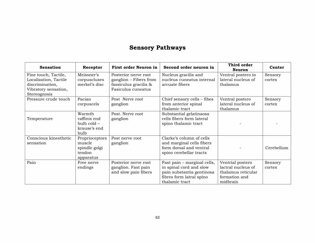
Sensory Pathways
Sensation Receptor First order Neuron in Second order neuron in Third order Neuron Center
Fine touch, Tactile, Localization, Tactile discrimination, Vibratory sensation, Stereognosis
Meissner’s corpuscluses merkel’s disc
Posterior nerve root ganglion – Fibers from fassiculus gracilis & Fasiculus cuneatus
Nucleus gracilis and nucleus cuneatus internal arcuate fibers
Ventral postero in lateral nucleus of thalamus
Sensory cortex
Pressure crude touch
Pacian corpuscels
Post Nerve root ganglion
Chief sensory cells – fibes from anterior spinal thalamic tract
Ventral postero lateral nucleus of thalamus
Sensory cortex
Temperature
Warmth raffinis end bulb cold – krause’s end bulb
Post. Nerve root ganglion
Substantial gelatinaosa cells fibers form lateral spino thalamic tract
-
-
Conscious kinesthetic sensation
Proprioceptors muscle spindle golgi tendon apparatus
Post nerve root ganglion
Clarke’s column of cells and marginal cells fibers form dorsal and ventral spino cerebellar tracts
- Cerebellum
Pain Free nerve endings
Posterior nerve root ganglion. Fast pain and slow pain fibers
Fast pain – marginal cells, in spinal cord and slow pain substantia gentinosa fibres form latral spino thalamic tract
Ventrial postero lactral nucleus of thalamus reticular formation and midbrain
Sensory cortex
64
Presynaptic events of neuromuscular transmission
When the action potential reaches the presynaptic nerve terminal,
voltage-gated calcium channels open, allowing influx of calcium ions
(Ca2+). This triggers release of acetylcholine from presynaptic vesicles
into the synaptic cleft.
Active zone
The active zone is that specialized area of the presynaptic
nerve terminal membrane visualized by freeze fracture electron
microscopy that contains a series of particles aligned in two parallel
rows. The particles are thought to represent the L-type voltage-
gated Ca2+ channels (VGCCs) activated by motor nerve
depolarization, which trigger the release of acetylcholine within
the presynaptic vesicles in to the synaptic cleft.
Postsynaptic events of neuromuscular transmission
The binding of two acetylcholine molecules, to each
acetylcholine receptor opens a channel within the receptor,
allowing Na+ influx and generating small, sub thresh hold endplate
potentials at the post synaptic membrane known as miniature
endplate potentials (MEPPs). The amplitude of the summated
endplate potential (EPP) for each muscle fiber is proportional to the
total number of MEPPs generated by the activation of many
different acetylcholine receptors at the same time. When a
sufficient number of receptors activated simultaneously, the
endplate potential becomes large enough to trigger an action
potential. The action potential then propagates along the muscle
sarcoplasmic membrane to the T-tubule system, leading to the
release of Ca2+ from the Sarcp Plasmic reticulum, ultimately
resulting in muscle contraction.
65
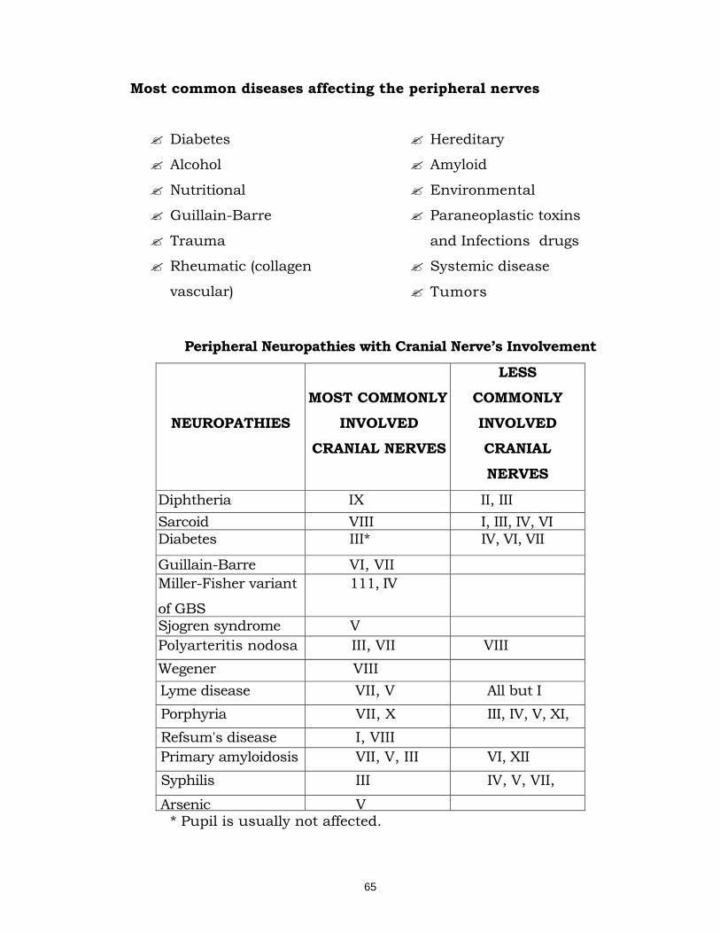
Most common diseases affecting the peripheral nerves
Diabetes
Alcohol
Nutritional
Guillain-Barre
Trauma
Rheumatic (collagen
vascular)
Hereditary
Amyloid
Environmental
Paraneoplastic toxins
and Infections drugs
Systemic disease
Tumors
Peripheral Neuropathies with Cranial Nerve’s Involvement
NEUROPATHIES
MOST COMMONLY
INVOLVED
CRANIAL NERVES
LESS
COMMONLY
INVOLVED
CRANIAL
NERVES
Diphtheria IX II, III Sarcoid VIII I, III, IV, VI Diabetes III* IV, VI, VII
Guillain-Barre VI, VII Miller-Fisher variant
of GBS
111, IV
Sjogren syndrome V Polyarteritis nodosa III, VII VIII Wegener VIII Lyme disease VII, V All but I Porphyria VII, X III, IV, V, XI, Refsum's disease I, VIII Primary amyloidosis VII, V, III VI, XII Syphilis III IV, V, VII,
Arsenic V * Pupil is usually not affected.
66
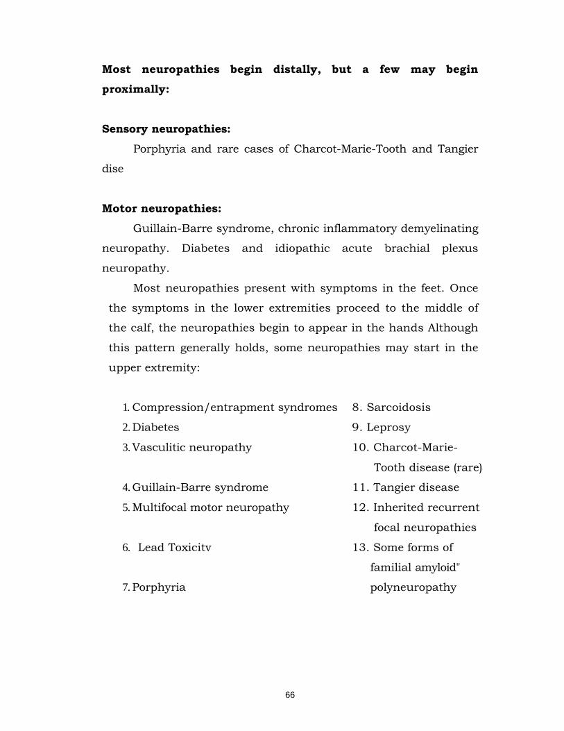
Most neuropathies begin distally, but a few may begin proximally:
Sensory neuropathies:
Porphyria and rare cases of Charcot-Marie-Tooth and Tangier
dise
Motor neuropathies:
Guillain-Barre syndrome, chronic inflammatory demyelinating
neuropathy. Diabetes and idiopathic acute brachial plexus
neuropathy.
Most neuropathies present with symptoms in the feet. Once
the symptoms in the lower extremities proceed to the middle of
the calf, the neuropathies begin to appear in the hands Although
this pattern generally holds, some neuropathies may start in the
upper extremity:
1. Compression/entrapment syndromes 8. Sarcoidosis
2. Diabetes 9. Leprosy
3. Vasculitic neuropathy 10. Charcot-Marie-
Tooth disease (rare)
4. Guillain-Barre syndrome 11. Tangier disease
5. Multifocal motor neuropathy 12. Inherited recurrent
focal neuropathies
6. Lead Toxicitv 13. Some forms of
familial amyloid"
7. Porphyria polyneuropathy

67
DISORDERS OF PERIPHERAL NERVES
Peripheral nerves are made up of axons, which represent
elongated processes originating from neurons in the anterior horn
cells of the spinal cord and dorsal root ganglia. These are
enveloped in a series of Schwann cells which form the fatty
myelin sheath. Pathological process may affect cell bodies, the
myelin sheath, or the connective tissue and blood vessels of
peripheral nerves. Although these processes cannot be
clinically distinguished, knowledge of the pathological nature of a
neuropathy is helpful in assessing prognosis and deciding on
treatment. The clinical classification of peripheral nerve lesions
comprises involvement of one or more individual peripheral nerves
or a generalized polyneuropathy.
Peripheral neuropathy and diseases of the lower motor neuron
Peripheral neuropathy manifests as paraesthesiae (pins and
needles) pain, to severe pain. Actual sensory impairment may
range from slight numbness with hypersensitivity to painful
stimuli to gross loss of pain perception. In others the picture is
essentially a motor one almost to the exclusion of sensory
phenomena. The only constant physical finding is depression or
absence of reflexes it is unlikely that he is suffering from a
peripheral neuropathy.
Lower motor neuron (LMN)
The motor cells of the cranial nerve nuclei of the brainstem
and the anterior horn cells of the spinal cord form the lower motor
neurons. The axons from these cells reach the mixed peripheral
nerve through the anterior nerve roots and supply the motor and

68
plates of a group of muscles. The anterior horn cell its axon and
the group of muscle fibers, it supplies comprise a lower motor
unit. All muscles are supplied by several such motor units, the
lower motor neuron is an integral part of the spinal reflex arc. It is
the final common path for all motor activity (both voluntary and
reflex) of all muscles. Integrity of the LMN is essential for
maintaining the normal nutrition and size of the muscle fiber
and when the LMN is damaged the corresponding muscles
undergo atrophy.
Signs of LMN lesions
1. Loss of all movements - voluntary and reflex.
2. Loss of tone - Flaccidity
3. Wasting - which appears within 2-3 weeks of the lesion
4. Loss of tendon reflexes and the corresponding superficial
reflexes
5. The atrophy muscles undergo contractures.
6. Denervated muscle fibres contract spontaneously contraction
of single muscle fibres is called fibrillation. This not visible, but
can be detected by electromyography, groups of muscle fibres
which are damaged but which are still capable of contracting
spontaneously give rise to visible fasciculations.
7. Electrical activity of the muscle is altered and this can be
detected by electromyography. The pattern of motor loss depends
on the site of affection of the LMN.Lesions of the anterior horn
cells or the anterior root give rise to segmental loss of
function. Lesions in the peripheral nerve give rise to paralysis
of the muscles supplied by that nerve often peripheral nerve
lesions are associated with sensory disturbances.
69
PERIPHERAL NEUROPATHY
Alternative Names
Peripheral Neuritis.
Definition
Peripheral neuropathy is failure of the peripheral nerves
that carry information to and from the brain and Spinal cord.
This produces symptoms like pain, loss of sensation, and
inability to control muscles, ("peripheral" - nerves beyond the
brain and spinal cord; "Neuro"- nerves; "Pathy"- abnormal)
Peripheral neuropathy may manifest as paraesthesia (pins
and needles) pain, muscle atrophy, sensory loss and cluminess
usually due to a mixture of slight loss of dexterity and sensory
impairment make the diagnosis more obvious even though the
aetiology often remains obscure.
In all age groups a very detailed family history is vital.
Some times several members of a family are found to have the
same disease, masquerading under several different diagnostic
labels. If there is any doubt it is worth examining other family
members to establish whether or not an inherited disorder is
present. A detailed history of all drugs used with in the previous
two years and any possible chemical exposure, diabetus mellitus,
dietary habits, previous surgical procedures and alcohol intake
should be included.
70
PATHOLOGY
The pathological reaction of peripheral nervous system in
response to injury may be in the form of one of the three types of
degeneration causing “Peripheral Neuropathy”. The three types of
degenerations are
1. Wallerian degeneration
2. Axonal degeneration
3. Segment demyelination
Degrees of injury When a nerve fibre is injured various changes occur in the
nerve fibre and nerve cell body. All these changes are together
called the degenerative changes. The injury may occur due to the
obstruction of blood flow, crushing of nerve fibre transaction of the
fibre and by local injection of toxic substances in to the nerve.
1. First degree of injury In this the axon is not destroyed but it losses the function for
a short time. This is caused by the pressure. Over a nerve for a
short period leadings to occlusion of blood flow and hypoxia. The
function of nerve fibre returns with in few hours to few weeks.
2. Second degree injury
This occurs due to the severe, prolonged pressures applied
over the nerve fibre. The endoneural tube remains intact.
3. Third degree of injury In this the endoneural tubes are interrupted.
4. Fourth degree of injury This initiates the degenerative changes which includes the
chemical and physical changes.
71
Changes during degeneration (a) Changes in nerve cell body
This nissl granule disintegrates into fragments by
chromatolysis process. The Golgi apparatus is disintegrated. The
cell body swells due to accumulation of fluid and become round. In
this case the neuron atropies and disappears completely.
(b) Changes in the nerve fibres The degenerative changes in the distal cut end of the nerve
fibre are named after the discoverer called as wallerian
degeneration.
The degeneration at proximal cut end along with degeneration
of cell body is called “retrograde degeneration”
Reaction to injury
1. Wallerian degeneration It occurs after transection of the axon which may be as a
result of knife wounds. Compression, traction and ischemia.
Following transection initially there is accumulation of granules in
the proximal and distal ends of the transection sites. Subsequently
the axon and myelin sheath distal to the transaction sites. Undergo
disintegration up to the next node of ranvier followed by
phagocytosis. The process of regeneration occurs by sprouting of
axons and proliferation of schwanncells from the proximal end.
2. Axonal degeneration
In axonal degeneration of the axon beings at the peripheral
terminal and proceeds backward towards the nerve cell body.
Cytoplasmic disintegration induced by injury of the distal axon lie
that portion of the axon farthest from the neuronal cell body is
called “axonal degeneration” Degeneration beings at the single point
72
within the axon and progresses to involve more symnts within few
days after the injury all portions of the distal axon begin the
process of disintegration.
Concurrently the axolemma becomes discontinuous and
electrical propagation is interrupted. The myelin sheath
disintegrates and axon fragments into small oxal segments at all
points distal into the site of injury. If the injry is in the immediate
proximity of the neuronal cell body the latter degenerates, otherwise
the cell body enlarges with a central eosinopilic region and
displacement of the nissl substances to the periphery of the cell,
termed axonal traction or chromatolytes.
Schwann cells actively participate in axonal degeneration and
regeneration. Shortly after the onset of disintegration of the axonal
cylinder. Schwann cells proliferate and encircle the axonal remains.
Hacrophanges are recruited to the site of axonal degeneration and
adhere to the outer surface of the fibre.
They participate in the phagocytosis of myelin and axonal
debris. The debris is removed over a period of weeks as foamy
macrophages migrate towards vascular spaces.
Evidence implicating fast axonal transport in the process of
axonal degeneration more slowly than short axons, implying that
material is depleted more rapidly in shorter axons. The interval
from injury to total axonal fragmentation is generally less than 2
weeks. Thus an acute axonopathy is by abundant degenerating
fibers while a chronic axonopathy features fiber loss.
The peripheral nervous system has a large capacity for
regeneration if the injurious agent is eliminated. There are two
patterns of axonal sprouting and regrowth. The first and most rapid
is sprouting of intact axons contiguous to site by denervation. The
sprouting occurs at the distal stump of the injured axon and
73
results in the formation of growth cones. Growth cones contains
elevated levels of growth associated protein (GAP – 43) other growth
factors including nerve growth factor (NGF) facilitate axonal
regeneration NGF plays dual role in axonal regeneration, interacting
with both axonal growth cones and schwann cells.
Shortly in axonal degeneration the cell body undergoes
chromatolysis. There is Schwann cells proliferation in the region of
axonal degeneration. The loss of axonal integrity occurs probably as
a result of some primary metabolic christarbance with in axon
itself. Changes similar to those seen is wallerian degeneration are
present but regenerative reaction is limited or absent.
3. Segmental demyelination
Segmental demyelination is similar to demyelination within
the brain. It is the demyelination between two consecutive nodes of
raniver, leaving a denoted axon segment. The axon however
remains intact Schwann cell proliferation. This results in
remyelination of the affected axon. Repeated episodes of
demylination and remyelintaion are associated with concentric
proliferation of Schwann cells around axons producing onion bulbs
found in hypertrophic neuropathy.
Clinical symptomatology
Clinical features may be due to affection of the motor,
sensory, or autonomic fibres separately or in combination. 1.
Sensory Phenomena
It may be positive, i.e. Irritative in nature or negative, i.e
due to loss of function.
74
Positive phenomena
i. Spontaneous pain
This occurs due to irritation of the small fibers which
carry pain or due to loss of large fibers which usually
inhibit the pain sensation carried by the small fibres,
ii. Paraesthesiae in the form of pins and needles,
iii. Contact dysesthesia : - where contact with the affected, Part
elicits a disturbing sensation,
iv. Hyperalgesia: increased appreciation of pain resulting from
decrease in the sensory threshold for pain,
v. Burning feet: - this is a frequent symptom but its
mechanism is not clear. It may occur in neurpathies,
accompanying alcoholism, diabetes and beriberi,
vi. Lancinating pains: - These are sharp and paroxysmal
pains occurring usually in the transverse plane in the
lower limbs in patients suffering from tabes dorsalis and
diabetes.
Negative Phenomena
Numbness is an important negative symptom which
indicates that at least 50 percent of the nerve fibres are destroyed.
Patients may have a feeling as though they are walking on
cotton wool. They may have unsteadiness of gait, particularly in
the dark due to loss of joint and position sense. Non healing
painless ulcers and neuropathic joints develop due to loss of
protective reflexes when the protective influence of pain is absent,
the joints undergo minor and major trauma due to over use and
injuries.
Degenerative and destructive changes of joint structures
develop, leading to disorganization of the joint (Charcot's joints) such
joints may occur in severe peripheral neuropathy. The pattern of
75
sensory loss differs depending up on the site of affection. When a
single peripheral nerve is affected the sensory and motor
abnormalities occur in the distribution of that nerve (mononeuritis)
(ex. Neural Leprosy). In classical Polyneuropathies there is
symmetrical distal sensory loss in the extremities described as
"gloved and stocking" anesthesia. 2. Motor Phenomena
These take the form of weakness, muscle wasting,
fasciculations and cramps, compared to disease affecting the
anterior horn cells, fasciculations are much less marked.
The tenden reflexes are lost early in Peripheral neuropathies
which affect the large fibres. The reflexes may be spared in
neuropathies which affect the small fibres alone. (eg) Neural leprosy. 3. Autonomic dysfunction
This manifests as loss of sweating postural hypo tension and
trophic disturbances and impotence.
APPROACH TO A PATIENTS WITH A CLINICAL FEATURES
The clinical points which are most helpful in diagnosis are
given below:
1. Rate of Onset: Acute : less than 1 week.
Subacute : less than 1 month
Chronic : more than 1 month.
2. Type of nerve fibre involved
Motor, Sensory, autonomic, or mixed.
3. Distribution
Proximal, distal or diffuse.
4. Painful neuropathies
Alcohol and nutritional deficiencies, DM,
hereditary sensory neuropathy, arsenic.
76
Loss of tenden reflexes is a -frequent in Peripheral
neuropathy, and usually first affects the ankle jerks. Sensory
symptoms and sensory loss in symmetrical neuropathies are
usually distal in distribution, giving rise to the "Glove and
stocking" pattern of involvement.
In the first, the impairment predominantly affects joint
position sense, and vibration and touch - pressure
sensibility, corresponding to a predominant loss of function in
the larger myelinated nerve fibres.
In the second pattern of selective sensory loss, pain and
temperature sensibility are predominantly affected, often
associated with loss of autonomic function, corresponding to a
predominant loss of autonomic function, corresponding to a
predominant loss of small myelinated and unmyelinated axons.
Trophic changes may complicate Peripheral neuropathies.
Paraesthesiae are a frequent feature in Peripheral
neuropathy. These are usually of a tingling nature ('Pins and
needles ') but may involve thermal sensation, most often with a
burning quality. The paraesthesiae may be aggravated by
touching or rubbing the skin.
Peripheral neuropathy can note any disease of the
peripheral nervous system (PNS). Involvement of a single
peripheral nerve, it is a syndrome characterized by disturbances of
the functions of peripheral nerves due to varied aetiology. In
this section we are concerned with inherited,infective,
metabolic,deficiency, toxic and obscure disorders in which the
peripheral nerves are diffusely damaged.The patients with
peripheral neuritis were selected for dissertation work who had
diabetus mellitus, B12 deficiency, alcoholic abuse , Nutritional
disorder.
77
Deficiency neuropathy Cyanocobalamin ( B12 deficiency) Definition
A progressive disease due to deficiency of vitamin B12 and
associated with pernicious anaemia, in which white matter of
the spinal cord degenerates, the effects being particularly in the
posterior and lateral columns. There is an associated leads to
peripheral neuropathy.
Symptoms
In most cases the symptoms appear indiously and without
any recognized existing cause. The first symptom is usually
numbness or tingling in the feet and a slight sensation of the same
kind in the fingers. This often the sensation in the feet is one of
swelling or coldness, or as if walking on cotton wool; and in few
cases unsteadiness in walking is at first the only complaint.
Examination
At the time shows the weakness of the toes or in dorsiflexion
of the feet, diminution or absence of the ankle-jerk, probably an
extensor plantar reflex, and a variable degree of sensory loss. The
superficial sensory loss is at first only over the feet, then it spreads
up to a cover a 'sock' area, and later has 'stocking' distribution, pain
and temperature impairment meanwhile being added to it.
Alcoholic peripheral neuropathy
Alcoholism is perhaps the commonest cause of peripheral
neuritis. It is preceded by chronic gastritis due to constant drinking
and as a result, there is failure of absorption of vitamin B1 by the
damaged stomach leading to neuritis. The onset is insidious with
tingling and numbness in the lower limbs followed by weakness
and globar wasting of the muscles which is symmetrical in
distribution. There is generally hypoaesthesia of the skin,
with hyperaesthesia of the deeper structures, like tenderness
78
over the calf muscles. The tendon jerks are lost. The gait is
characteristically of 'steppage' type due to bilateral foot-drop. It
may occur and recovery may take years even with adequate
vitamin replacement and abstention from alcohol.
DIABETIC NEUROPATHY
Definition
Diabetic neuropathy may be defined as a non-
inflammatory, non specific pathological or functional disorder of
the peripheral nervous system.
Introduction
Diabetic neuropathy (DN) is one of the commonest
metabolic complications of diabetes. (De-calvi, first showed the
relationship between DM and peripheral nerve damage in 1864).
It reviews current knowledge of the aetiology of diabetic
neuropathy and the outcomes and limitations of previous trials
and discusses future directions for the investigation of its
prevention and treatment proposed mechanisms for the
development of diabetic neuropathy have been widely studied. It
has been shown that there is improvement of nerve function
associated with some short term clinical trials of treatments that
address a number of possible etiologic pathways. However, with
the expectation of the diabetes control and complications Trial
(DCCT long term trials with adequate statistical power to
evaluate clinical outcome points have not been conducted. The
changes in nerve function are similar in most of the clinical trials.
For instance, in four clinical trials directed at separate
mechanisms / improved glucose control, high myo-inositol diet,
therapy with an aldose reductase inhibitor, and therapy with
supplementary (lambda - linolenic acid) a similar improvement in

79
peroneal motor velocity of 1 - 2 m/s is observed. This implies that
each of the proposed mechanisms contributes equally to the
development of neuropathy or that there is some redundancy to
their mechanisms.
Improvement with treatment has been measured with
several markers including nerve conduction velocity, quantitative
sensory testing, autonomic function testing, and morphometric
changes, morphometry will predict improvement in long-term
clinical outcomes such as impaired sensation, painful
neuropathy insensitive feet, neuropathic ulceration and
amputation. Neuropathy trials must consider present knowledge
about complications in general and neuropathy specially.
Patients with mild to moderate neuropathy with
presumably more metabolic than structural neuropathy would
be referred subjects, and follow up of 3-5 years is likely to be
needed. During the past 20 years, numerous clinical trials have
evaluated the efficacy of different agents for the treatment of
diabetic neuropathy.
Pathology of Human Diabetic neuropathy They can occur in motor, sensory and autonomic nerves.
There is a significant relationship between clinical measure of
neuropathic severity and myelinated fibre loss. Perhaps the prime
site of intermediate tissue damage resides in endoneural
microvasculature. Endoneurial microangiopathy has been
related, it various studies, to the severity of diabetes mellitus.
Pathological features
- Axonal degeneration of both myelinated and
unmyelinated fibres.
Early: Axon shrinkage
Later: Axonal fragmentation ; regeneration
80
- Thickening of schwann cell basal lamina
- Patchy, segmental demyelination
- Thickening of basement membrane and microthrombi in
intraneural
Capillaries
Aetiopathogenesis of diabetic neuropathy
Factors involved are,
- Direct insult
- Nerve perfusion
- Regenerative capacity
- Auto immune
Studies in neuropathic patients using vasodilator approach
have been extensive. Recent studies have noted alleviation of
symptoms using prostaglandin analog II prost and angiotension
converting enzymes inhibitor (ACEI) lisinopril. At present however
nitric acid (Na) prostaglandin (PGI2) Angiotension II (All) and
endothelin (CET) are the factors appearing to play significant role in
aetiopathogenesis.
Metabolic factors in diabetes which contribute to the decrease
of blood flow to endoneurium. They are likely to be dependent on
hyperglycemia mainly, even though insulin may also play a part.
The major metabolic changes caused by hyperglycemia are
increased poly of pathway flux, elevated oxygen free radical
formation, and advanced glycosylation. In addition, impaired
insulin action / hyperglycemia may contribute to defective carnitine
and w-6 essential fatty acid (EFA) metabolism.

81
Endothelial abnormalities
ET All Na PGl2
ET ACEI Nitrates
Prostanoid Angiotension VSM Reactivity
Antagonists
Analogues Antagonists
Nerve bloof flow and axogenation
Neuron and Schwann cell dysfunction
Nerve degeneration Tropic agents
The twin problems of chronic hyperglycemia and endoneural
hypoxia results in excessive oxidative stress. Peripheral nerves
have chain breaking antioxidants suppressing free radical chain
oxidation by molecular oxygen and comprises of supra - oxide
dismutase (SOD), catalase, glutathione peroxidase and
gluthione reductase. Of great interest is the fall of gluthione and
its related enzymes to only 10% in diabetic rodent's peripheral
nerve. The potential sources of free radical generation in diabetes
are ischemia, hyperglycemia, increased mitochondrial leak,
catecholamine oxidation and leukocytes.
Hyperglycemia damages microvessels via rheologic
mechanisms, advanced glycation end products (AGE) and reduction
in nitric oxide, resulting in endoneurial hypoxia and multifocal
glycation and lipid peroxidation.
The role of leukocytes in human diabetic neuropathy is
suggested by the presence of round cell infilteration in acute
autonomic neuropathy and subacute proximal neuropathy.
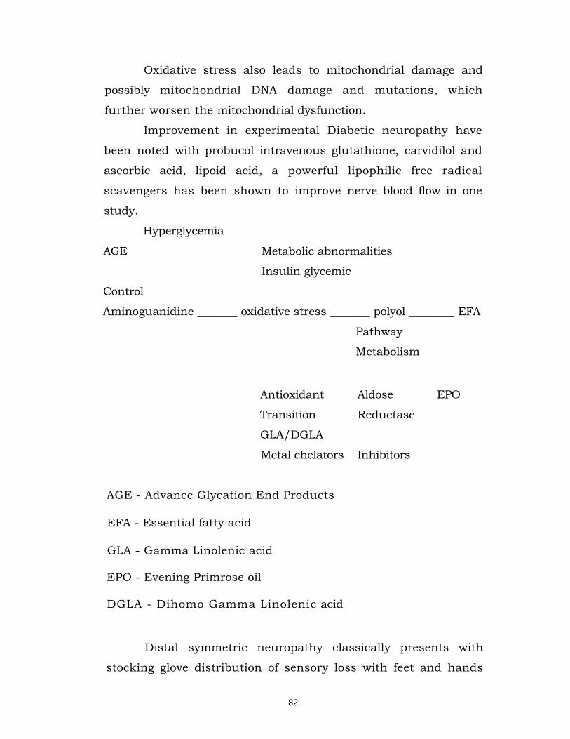
82
Oxidative stress also leads to mitochondrial damage and
possibly mitochondrial DNA damage and mutations, which
further worsen the mitochondrial dysfunction.
Improvement in experimental Diabetic neuropathy have
been noted with probucol intravenous glutathione, carvidilol and
ascorbic acid, lipoid acid, a powerful lipophilic free radical
scavengers has been shown to improve nerve blood flow in one
study.
Hyperglycemia
AGE Metabolic abnormalities
Insulin glycemic
Control
Aminoguanidine _______ oxidative stress _______ polyol ________ EFA
Pathway
Metabolism
Antioxidant Aldose EPO
Transition Reductase
GLA/DGLA
Metal chelators Inhibitors
AGE - Advance Glycation End Products
EFA - Essential fatty acid
GLA - Gamma Linolenic acid
EPO - Evening Primrose oil
DGLA - Dihomo Gamma Linolenic acid
Distal symmetric neuropathy classically presents with
stocking glove distribution of sensory loss with feet and hands

83
presenting earliest with more severe signs and symptoms. This
indicates that neuron with the longest axons are the most
vulnerable.
NGF and neurotrophin - 3 are both found to prevent at
least some of the diabetes associated metabolic changes and is
an important step in understanding the pathogenesis of
diabetic neuropathy and may have practical repercussions for
the development of therapy and prophylaxis.
Pathogenesis of diabetic neuropathy
The pathogenesis of diabetic neuropathy remains poorly
understood and is split between the metabolic and vascular
theories. Pathological changes include a loss of myelinated fibres
and microvascular abnormalities, particularly basement
membrane thickening and endothelial cell hyperplasia.
The metabolic theory suggests that diabetic neuropathy
arises as a consequence of chronic hyperglycemia. The
diabetes control and complications trial research group
demonstrated that glycaemic control reduced the progression
of neuropathy in type 1 diabetes and this is also seen in
patients after pancreatic transplants.The most clearly defined
inducers of nerve damage include activation of the polyol
pathway, advanced glycation and oxidative stress, although the
transducers linking these abnormalities to tissue malfunction
and damage and unknown.
84
Diagnosis of Diabetic peripheral neuropathy In our diabetes centre and high risk factor ulcer clinic, all
patients with foot problems have a foot screen that includes a
neuropathic and vascular assessment. The neuropathy disability
score uses a Nuerotip, tuning fork, hot and cold rods and a tendon
hammer. This simple assessment uses a scoring system in which
the patient scores one point for each incorrect test and an extra
point if the Achilles tendon reflexes are not determined with
reinforcement. The maximum score for each foot is 5 points, and a
score of greater than 6 out of 10 suggests neuropathy.
A 10 g Semmes Weinstein monofilament is used on the
plantar surface of the great toe, first, third, and fifth metatarsal
heads and heal, and the dorsum of the foot, patients who cannot
feel the touch of a log monofilament are also at risk. This
relatively inexpensive device is useful too for determining who
is at risk of foot ulceration.
Vibration perception threshold (VPT) is also commonly
measured using a neurothesiometer. A 'VPT' score of 25 volts on
the pulp of great toe indicates neuropathy.
Peripheral neuropathy can be diagnosed using nerve
conduction studies and quantitative sensory testing. The
neuropathy symptom score can be used to assess the severity of
neuropathic pain.
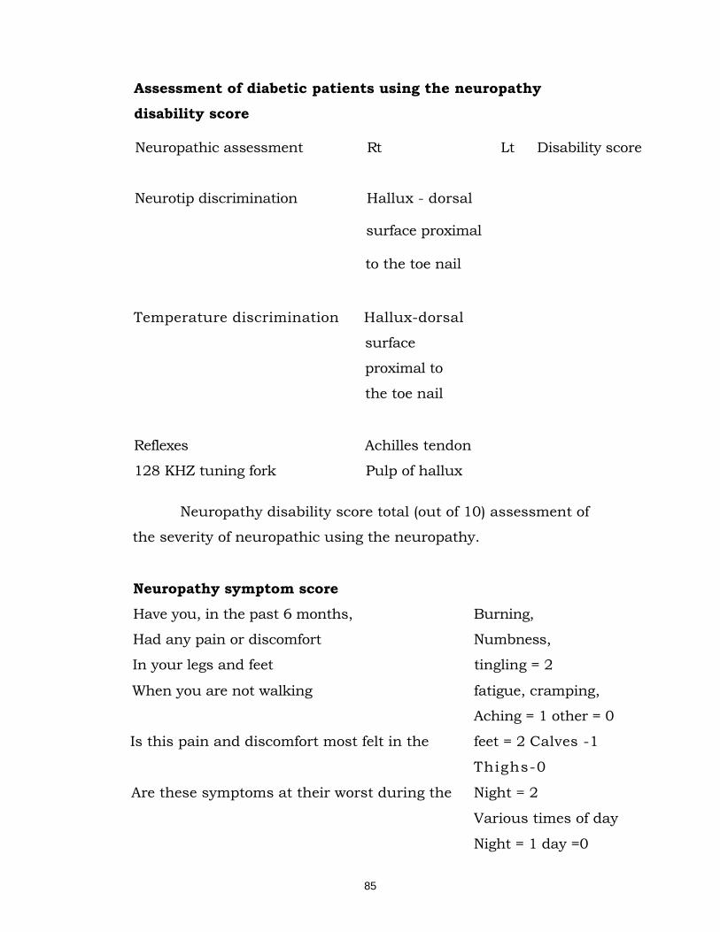
85
Assessment of diabetic patients using the neuropathy disability score
Neuropathic assessment Rt Lt Disability score
Neurotip discrimination Hallux - dorsal
surface proximal
to the toe nail
Temperature discrimination Hallux-dorsal
surface
proximal to
the toe nail
Reflexes Achilles tendon
128 KHZ tuning fork Pulp of hallux
Neuropathy disability score total (out of 10) assessment of
the severity of neuropathic using the neuropathy.
Neuropathy symptom score
Have you, in the past 6 months, Burning,
Had any pain or discomfort Numbness,
In your legs and feet tingling = 2
When you are not walking fatigue, cramping,
Aching = 1 other = 0
Is this pain and discomfort most felt in the feet = 2 Calves -1
Thighs-0
Are these symptoms at their worst during the Night = 2
Various times of day
Night = 1 day =0
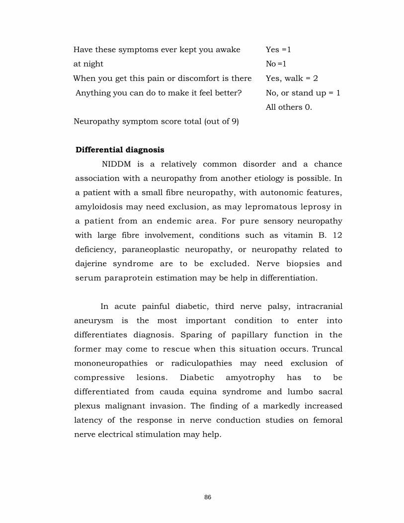
86
Have these symptoms ever kept you awake Yes =1
at night No =1
When you get this pain or discomfort is there Yes, walk = 2
Anything you can do to make it feel better? No, or stand up = 1
All others 0.
Neuropathy symptom score total (out of 9)
Differential diagnosis
NIDDM is a relatively common disorder and a chance
association with a neuropathy from another etiology is possible. In
a patient with a small fibre neuropathy, with autonomic features,
amyloidosis may need exclusion, as may lepromatous leprosy in
a patient from an endemic area. For pure sensory neuropathy
with large fibre involvement, conditions such as vitamin B. 12
deficiency, paraneoplastic neuropathy, or neuropathy related to
dajerine syndrome are to be excluded. Nerve biopsies and
serum paraprotein estimation may be help in differentiation.
In acute painful diabetic, third nerve palsy, intracranial
aneurysm is the most important condition to enter into
differentiates diagnosis. Sparing of papillary function in the
former may come to rescue when this situation occurs. Truncal
mononeuropathies or radiculopathies may need exclusion of
compressive lesions. Diabetic amyotrophy has to be
differentiated from cauda equina syndrome and lumbo sacral
plexus malignant invasion. The finding of a markedly increased
latency of the response in nerve conduction studies on femoral
nerve electrical stimulation may help.
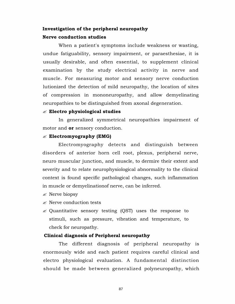
87
Investigation of the peripheral neuropathy Nerve conduction studies
When a patient's symptoms include weakness or wasting,
undue fatiguability, sensory impairment, or paraesthesiae, it is
usually desirable, and often essential, to supplement clinical
examination by the study electrical activity in nerve and
muscle. For measuring motor and sensory nerve conduction
lutionized the detection of mild neuropathy, the location of sites
of compression in mononeuropathy, and allow demyelinating
neuropathies to be distinguished from axonal degeneration.
Electro physiological studies
In generalized symmetrical neuropathies impairment of
motor and or sensory conduction.
Electromyography (EMG)
Electromyography detects and distinguish between
disorders of anterior horn cell root, plexus, peripheral nerve,
neuro muscular junction, and muscle, to dermire their extent and
severity and to relate neurophysiological abnormality to the clinical
context is found specific pathological changes, such inflammation
in muscle or demyelinationof nerve, can be inferred.
Nerve biopsy
Nerve conduction tests
Quantitative sensory testing (QST) uses the response to
stimuli, such as pressure, vibration and temperature, to
check for neuropathy.
Clinical diagnosis of Peripheral neuropathy
The different diagnosis of peripheral neuropathy is
enormously wide and each patient requires careful clinical and
electro physiological evaluation. A fundamental distinction
should be made between generalized polyneuropathy, which
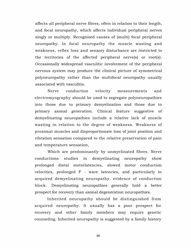
88
affects all peripheral nerve fibres, often in relation to their length,
and focal neuropathy, which affects individual peripheral nerves
singly or multiply. Recognized causes of (multi) focal peripheral
neuropathy. In focal neuropathy the muscle wasting and
weakness, reflex loss and sensory disturbance are restricted to
the territories of the affected peripheral nerve(s) or root(s).
Occasionally widespread vasculitic involvement of the peripheral
nervous system may produce the clinical picture of symmetrical
polyneuropathy rather than the multifocal neuropathy usually
associated with vasculitis.
Nerve conduction velocity measurements and
electromyography should be used to segregate polyneuropathies
into those due to primary demyelination and those due to
primary axonal generation. Clinical feature suggestive of
demyelinating neuropathies include a relative lack of muscle
wasting in relation to the degree of weakness. Weakness of
proximal muscles and disproportionate loss of joint position and
vibration sensation compared to the relative preservation of pain
and temperature sensation,
Which are predominantly by unmyelinated fibres. Nerve
conductions studies in demyelinating neuropathy show
prolonged distal motorlatencies, slowed motor conduction
velocities, prolonged F - wave latencies, and particularly in
acquired demyelinating neuropathy, evidence of conduction
block. Demyelinating neuropathies generally hold a better
prospect for recovery than axonal degeneration neuropathies.
Inherited neuropathy should be distinguished from
acquired neuropathy. It usually has a poor prospect for
recovery and other family members may require genetic
counseling. Inherited neuropathy is suggested by a family history
89
of neuropathy or parent consanguinity. It tends to evolve slowly,
and even marked degrees of weakness may not excite complaint
by the patient. Positive primary symptoms, such as paraesthesias,
spontaneous pain (or) sexual sensations suggest an acquired
neuropathy, although features do occur in metachromatic
leucodystrophy and disease. Often the inherited basis for a
patient’s neurology can only be firmly established after clinical or
electrical examination of relatives.
Clinical features may help in the often difficult distinction
and lesions of roots, plexuses, and peripheral nerves, lesions and
brachial or lumbo - sacral plexuses are suggested by patterns
blackness, reflex loss, and sensory disturbance which cannot be
generated to lesions of a single nerve root or peripheral nerve.
Relation between plexus and multiple root lesions may be
important on clinical grounds; however, the involvement of axon
nerve fibers in plexus lesions tends to produce a warm, red foot.
Further more, sensory nerve action potentials are stored in root
lesions, because the dorsal root ganglion or general branch of the
sensory axon is not affected. Proximal muscle weakness tends to
be a feature of lesions involving rectus or roots, rather than a
feature or lesions restricted to neural nerves. Lesions of single
nerve roots may be distal from those of individual peripheral
nerves by the topical distribution of cutaneous sensory
disturbance.
Treatment for peripheral neuropathy
We are still looking for a way to heal nerves damaged by
neuropathy. The treatment aims to make the symptoms of
neuropathy better; therapy for peripheral neuropathy differs
depending on the cause.
90
For example
Therapy for peripheral neuropathy caused by diabetes
involves control of the diabetes. Tight control of glucose is
important to prevent progression. As blood sugar is brought under
control, symptoms may worsen temporarily. However, improved
control often relieves pain and prevents or delays further problems.
Good foot care is also essential part of treatment for neuropathy
affecting the lower extremities.
Treatment for diabetic neuropathy is includes - drugs to
relieve pain and treat neurological problems.
- Relief of pain - Hot packs or soaks or infra red light with care
- Topical creams apply
- Dietary modification
- Prevention of foot drop and wrist drop and contractures by
splints or sand bags.
- Daily massage and passive movements as soon as calf
muscles are less tender.Care should be taken not to over
stain weak muscles.
- Check the feet daily
- Weakness - Occupational rehabiliation
- Nerve stimulation techniques
- Specific: - Adequate control of diabetes mellitus
Alcoholic Polyneuropathy:
Treatment of polyneuropathy in general combined with that
of alcohol addition, the aim being the gradual withdrawl of alcohol
& fully balanced dile.
B12 Defeciency Polyneuropathy:
General treatment combined with full, balanced diet,
parentral intake of vit B12

91
Nutiritional Defeciency: General Treatement combined with rich nutrition diet.
Expectations (prognosis)
The outcome greatly depends on the cause of the
neuropathy. In cases where a medical condition can be identified
and treated, the prognosis may be excellent.
Prevention
Neuropathy can be prevented, at least in some cases. For
example: The Diabetes control and complications trial studied
complications in people on tight control of blood sugar level
prevents the development of neuropathy in diabetics and
decreases the severity of symptoms. Take care of soles and hands.
In addition, regular foot care can prevent a small
infection from progressing. Maintaining ideal weight, regular
exercise. Follow good habits avoid alcohol and cigarette

92
MATERIALS AND METHODS
“Vadha ubhakatham” a type among 80 types of vadham has
been dealt in the text book of yugi vaithya chindhamani vadha
ubhakatham is siddha aspect and correlates with peripheral
neuritis in modern medicine. There are various causes for vadha
ubhakatham. The author had chosed the patients with diabetis
mellitus, anaemia (B12 defeciency), nutritional disorder, patients
with alcoholic abuse.
Selection of the patients
For this clinical study 20 patients of both sexes and of varying
age groups suffering from vada ubhakatham selected and admitted
in the in patient ward, another twenty patients were treated in out
patient’s ward of pothu maruthuvam, Post Graduate -department of
Government siddha medical college, palayamkottai.
This clinical study was carried under the careful supervision
and monitoring by professor, reader, and lecturer, Assistant
lecturer of the department.
In this study the detailed clinical history, family history,
related past history, personal habits, occupation were taken.
For this purpose the case sheets were prepared, based on
both siddha concept and modern concept were maintained
separately for all patients.
The Cases selected on the basis of
1. Gnawing sensation over legs, hands & all over the body
2. Numbness
3. Burning sensation present in the Upper & lower limbs and all
over the body
93
4. Polyphagia
5. Restricted movements
6. Tingling sensation
7. Giddiness
8. Dung coated senstaion over soles
9. Paresthesia
10. Pricking pain with pin and needle sensation.
11. Disturbed sensory perception of touch, temperture and pain
12. Difficulties to hold chappals
13. Difficulties to grasp things
14. Trophic ulcers
Investigations
The symptoms of vadha ubhakatham were more or less
correlated with peripheral neuritis in allopathic view. So
investigations meant for peripheral neuritis were done for vadha
ubhakatham also
Blood
Total count
Difference Count
ESR
Hb
Peripheral smear
Cytology
Complete haemogram
Sugar (fasting, PP)
Urea
Creatinine
Cholesterol
Liver function test
Serum protein
Thyroid profile
94
Skin Clipping AFB
Urine Albumin Sugar Deposit
Motion Ova Cyst Occult Blood
Others Nerve conduction study
On the basis of these investigations modern diagnosis and a
parallel siddha disgnosis was made with the help of the following
criteria the mukkurta nilaigal, envagai thervugal, 7 udal kattugal,
nilam, kalam, vayathu etc.,
Investigations were found to be useful in assessing the
progress of the disease and prognosis of the patient.
Selection of the trial drug According to tridhosa theory laxatives were first given to
normalize the vitiated vadham. For this Nilavagai chooranam
10gms with hot water at bed time was recomended before starting
the specific treatment.
The drugs (Pathiriver chooranam and Medicated vennai)
selected for the dissertation were subjected to pharmocological and
biochemical analysis of the respected Department of Government
Siddha Medical College, Palayamkottai.
Individual case sheet and proforma for each patient was
maintained inthe in patient ward.
All the inpatient is advised to come to the out patient ward at
regular intervals for further follow up after the discharge.
95
RESULTS AND OBSERVATION
The results were observed with respect of the following criteria
by clinical study on 20 – Inpatients and 20 – out patients
1. Sex distribution
2. Age distribution
3. Kaalam distribution
4. Constitution of the body (Dhehi)
5. Gunam distribution
6. Religion distribution
7. Occupational status
8. Socio economic status
9. Habits distribution
10. Dietary pattern
11. Paruvakaalam distribution
12. Thinai distribution
13. Aetiological Factors
14. Clinical manifestations
15. Associated diseases
16. Kosam distribution
17. Mode of Onset
18. Duration of Illness
19. Derangement of mukkurtram
A. Derangement of vadham
B. Derangement of pitham
C. Derangement of kabham
20. Ezhu udal kattugal
21. Envagai Thervugal
22. Duration of Treatment
23. Sensory system and Reflxes
24. Grading of results
25. Investigation tables

96
1. SEX DISTRIBUTION: Table 1 illustrates the distribution of Sex
Table 1
S. No
Sex In-Patients Out-Patients
No.of.Cases Percentage No.of.Cases Percentage
1. Male 9 45% 13 65%
2. Female 11 55% 7 35%
For this dissertation study 20 In-patients and 20 Out-patients
were selected.
Out of this In-patients 9 patients (45%) were males and 11
patients (65%) were females.
In Out-patients 13 patients (65%) were males and 7 (35%)
were females.
2. AGE DISTRIBUTION:
Table 2 illustrates the distribution of Age
Table 2
S.
No
Age in
years
In-Patients Out-Patients
No.of.Cases Percentage No.of.Cases Percentage
1. 21-30 - - 1 5%
2. 31-40 - - 2 10%
3. 41-50 2 10% 5 25%
4. 51-60 9 45% 3 15%
5. 61-70 6 30% 9 45%
6. 71-80 3 15% - -
10% of the In-patients were observed in the age group
between 41-50 years. 45% of the In-patients were observed in the
age group between 51-60 years. 30% of the In-patients were
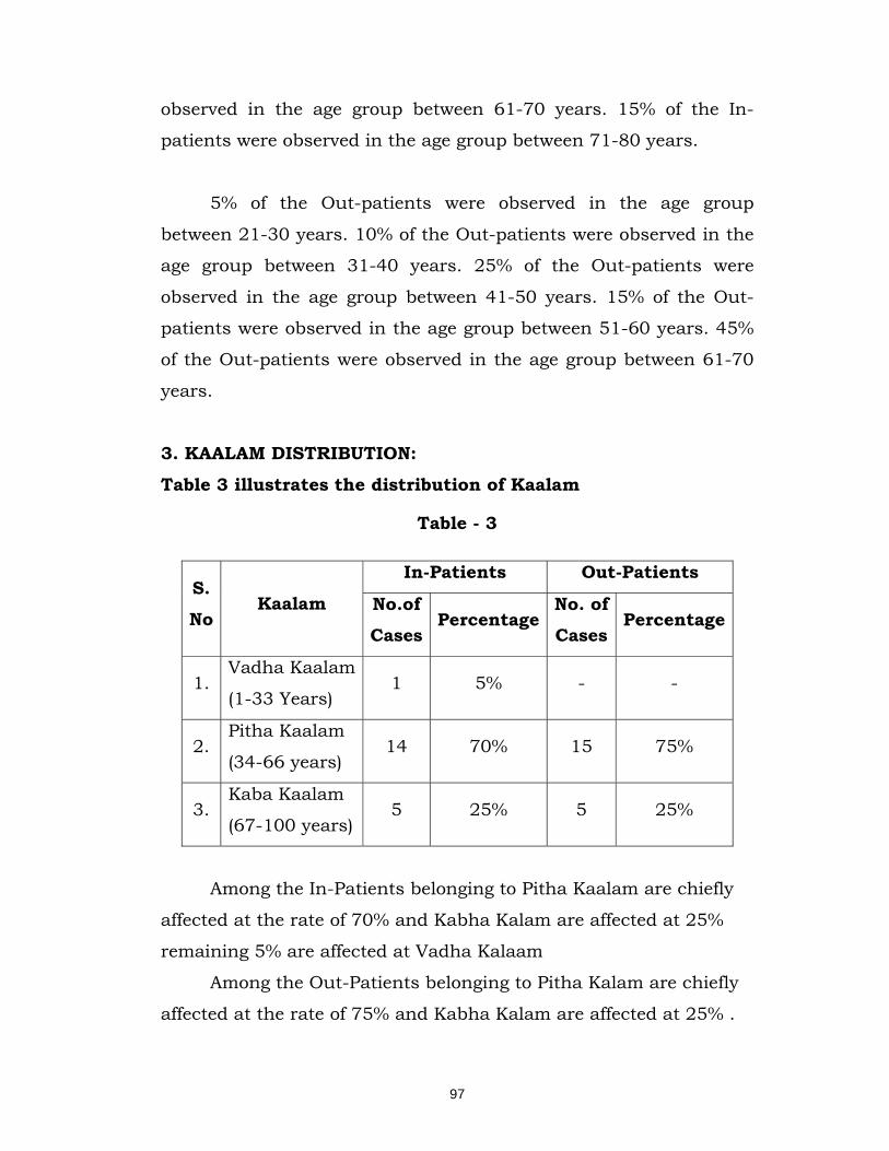
97
observed in the age group between 61-70 years. 15% of the In-
patients were observed in the age group between 71-80 years.
5% of the Out-patients were observed in the age group
between 21-30 years. 10% of the Out-patients were observed in the
age group between 31-40 years. 25% of the Out-patients were
observed in the age group between 41-50 years. 15% of the Out-
patients were observed in the age group between 51-60 years. 45%
of the Out-patients were observed in the age group between 61-70
years.
3. KAALAM DISTRIBUTION:
Table 3 illustrates the distribution of Kaalam
Table - 3
S. No
Kaalam
In-Patients Out-Patients
No.of Cases
Percentage No. of Cases
Percentage
1. Vadha Kaalam
(1-33 Years) 1 5% - -
2. Pitha Kaalam
(34-66 years) 14 70% 15 75%
3. Kaba Kaalam
(67-100 years) 5 25% 5 25%
Among the In-Patients belonging to Pitha Kaalam are chiefly
affected at the rate of 70% and Kabha Kalam are affected at 25%
remaining 5% are affected at Vadha Kalaam
Among the Out-Patients belonging to Pitha Kalam are chiefly
affected at the rate of 75% and Kabha Kalam are affected at 25% .

98
4. CONSTITITUTION OF THE BODY (DHEHI): Table 4 illustrates the distribution of Dhehi
Table 4
S.
No Dhehi
In-Patients Out-Patients
No.of Cases
Percentage No.of Cases
Percentage
1. Vadha Dhehi 5 25% 4 20%
2. Pitha Dhehi 2 10% 4 20%
3. Kaba Dhehi 3 15% 2 10%
4. Thontha Dhehi 10 50% 10 50%
Among the In-Patients Thontha dhehi are at the rate of 50%
and 25% of patients are Vadha dhehi, and 15% are kabha dhehi
remaining patients are Pitha dhehi.
Among the Out-Patients Thotha dhehi are at the rate of 50%
and 20% of patients are Vadha dhehi, and 20% of Pitha dhehi,
remaining patients are Kabha dhehi.
5. GUNAM DISTRIBUTION:
Table 5 illustrates the distribution of Gunam Table 5
S.
No Gunam
In-Patients Out-Patients
No.of. Cases
Percentage No.of. Cases
Percentage
1. Sathuva Gunam - - - -
2. Rajo Gunam 20 100% 20 100%
3. Thamo Gunam - - - -
In both In-Patients and Out-Patients cent percentage of the
cases had Rajo Gunam.
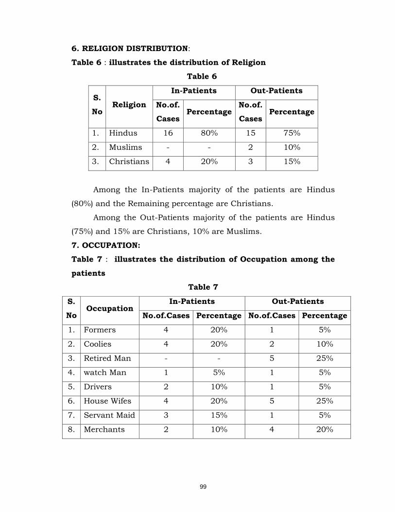
99
6. RELIGION DISTRIBUTION:
Table 6 : illustrates the distribution of Religion
Table 6
S.
No Religion
In-Patients Out-Patients
No.of. Cases
Percentage No.of. Cases
Percentage
1. Hindus 16 80% 15 75%
2. Muslims - - 2 10%
3. Christians 4 20% 3 15%
Among the In-Patients majority of the patients are Hindus
(80%) and the Remaining percentage are Christians.
Among the Out-Patients majority of the patients are Hindus
(75%) and 15% are Christians, 10% are Muslims.
7. OCCUPATION: Table 7 : illustrates the distribution of Occupation among the
patients
Table 7
S.
No Occupation
In-Patients Out-Patients
No.of.Cases Percentage No.of.Cases Percentage
1. Formers 4 20% 1 5%
2. Coolies 4 20% 2 10%
3. Retired Man - - 5 25%
4. watch Man 1 5% 1 5%
5. Drivers 2 10% 1 5%
6. House Wifes 4 20% 5 25%
7. Servant Maid 3 15% 1 5%
8. Merchants 2 10% 4 20%
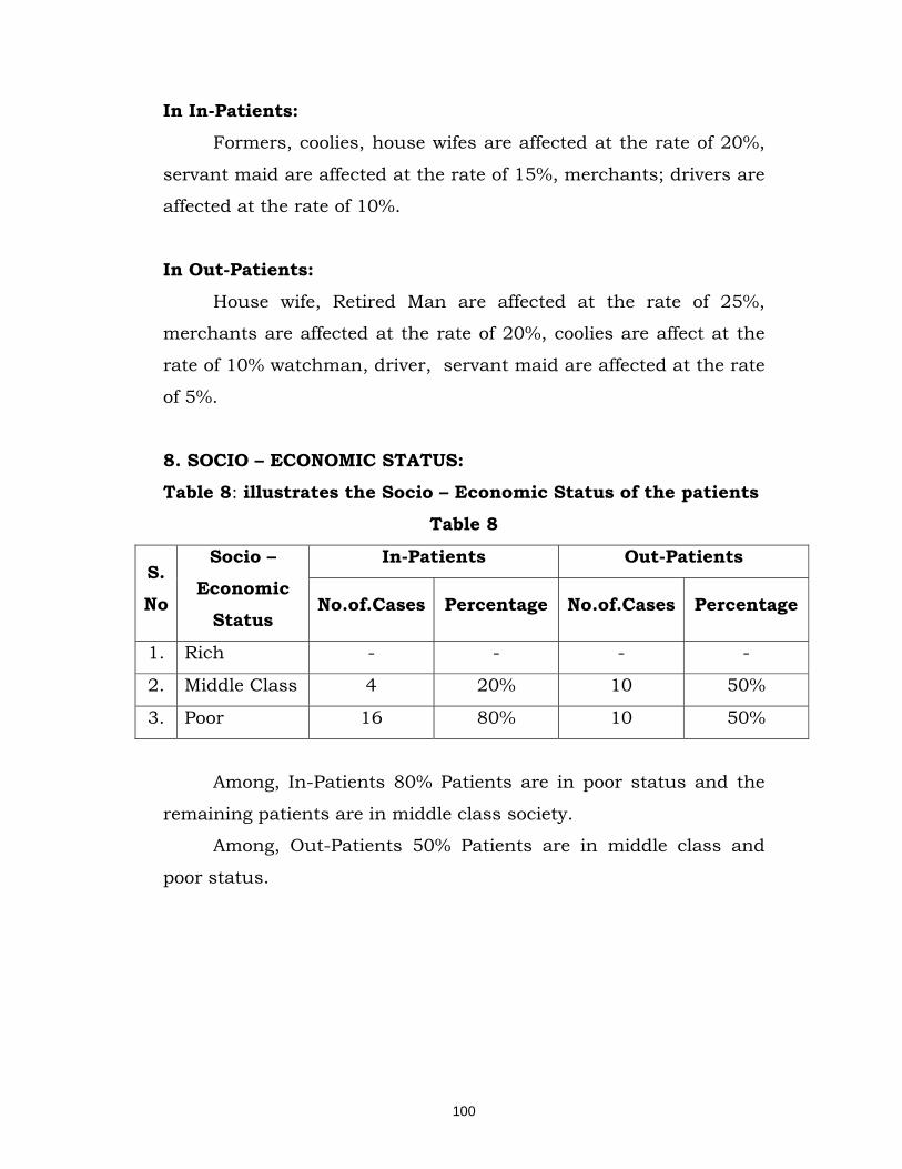
100
In In-Patients: Formers, coolies, house wifes are affected at the rate of 20%,
servant maid are affected at the rate of 15%, merchants; drivers are
affected at the rate of 10%.
In Out-Patients:
House wife, Retired Man are affected at the rate of 25%,
merchants are affected at the rate of 20%, coolies are affect at the
rate of 10% watchman, driver, servant maid are affected at the rate
of 5%.
8. SOCIO – ECONOMIC STATUS:
Table 8: illustrates the Socio – Economic Status of the patients
Table 8
S.
No
Socio –
Economic Status
In-Patients Out-Patients
No.of.Cases Percentage No.of.Cases Percentage
1. Rich - - - -
2. Middle Class 4 20% 10 50%
3. Poor 16 80% 10 50%
Among, In-Patients 80% Patients are in poor status and the
remaining patients are in middle class society.
Among, Out-Patients 50% Patients are in middle class and
poor status.
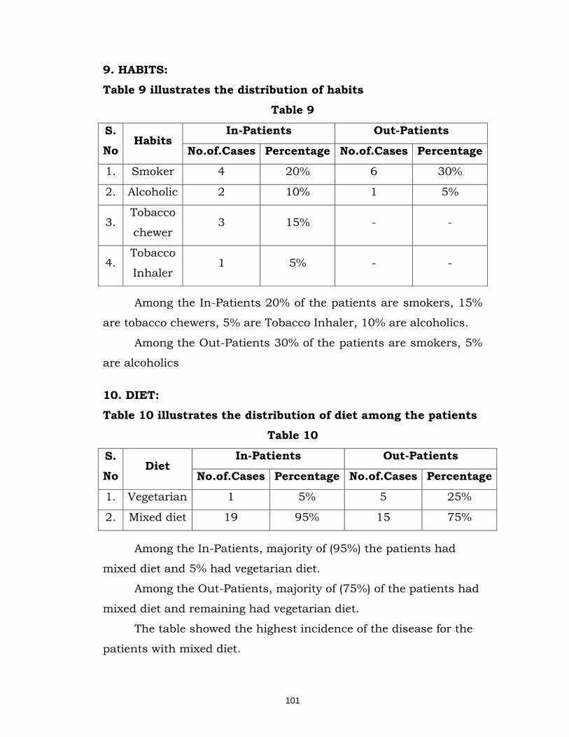
101
9. HABITS: Table 9 illustrates the distribution of habits
Table 9
S. No
Habits In-Patients Out-Patients
No.of.Cases Percentage No.of.Cases Percentage
1. Smoker 4 20% 6 30%
2. Alcoholic 2 10% 1 5%
3. Tobacco
chewer 3 15% - -
4. Tobacco
Inhaler 1 5% - -
Among the In-Patients 20% of the patients are smokers, 15%
are tobacco chewers, 5% are Tobacco Inhaler, 10% are alcoholics.
Among the Out-Patients 30% of the patients are smokers, 5%
are alcoholics
10. DIET: Table 10 illustrates the distribution of diet among the patients
Table 10
S.
No Diet
In-Patients Out-Patients
No.of.Cases Percentage No.of.Cases Percentage
1. Vegetarian 1 5% 5 25%
2. Mixed diet 19 95% 15 75%
Among the In-Patients, majority of (95%) the patients had
mixed diet and 5% had vegetarian diet.
Among the Out-Patients, majority of (75%) of the patients had
mixed diet and remaining had vegetarian diet.
The table showed the highest incidence of the disease for the
patients with mixed diet.
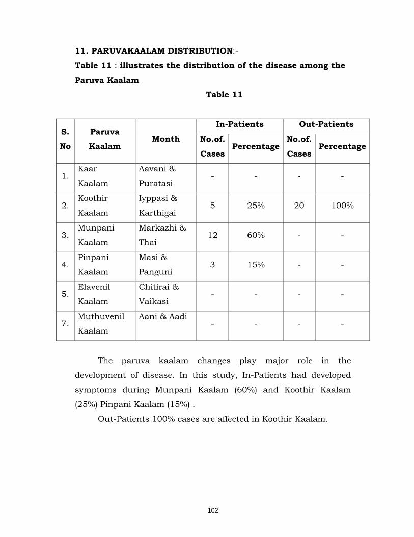
102
11. PARUVAKAALAM DISTRIBUTION:-
Table 11 : illustrates the distribution of the disease among the
Paruva Kaalam
Table 11
S.
No
Paruva
Kaalam Month
In-Patients Out-Patients
No.of. Cases
Percentage No.of. Cases
Percentage
1. Kaar
Kaalam
Aavani &
Puratasi - - - -
2. Koothir
Kaalam
Iyppasi &
Karthigai 5 25% 20 100%
3. Munpani
Kaalam
Markazhi &
Thai 12 60% - -
4. Pinpani
Kaalam
Masi &
Panguni 3 15% - -
5. Elavenil
Kaalam
Chitirai &
Vaikasi - - - -
7. Muthuvenil
Kaalam
Aani & Aadi - - - -
The paruva kaalam changes play major role in the
development of disease. In this study, In-Patients had developed
symptoms during Munpani Kaalam (60%) and Koothir Kaalam
(25%) Pinpani Kaalam (15%) .
Out-Patients 100% cases are affected in Koothir Kaalam.
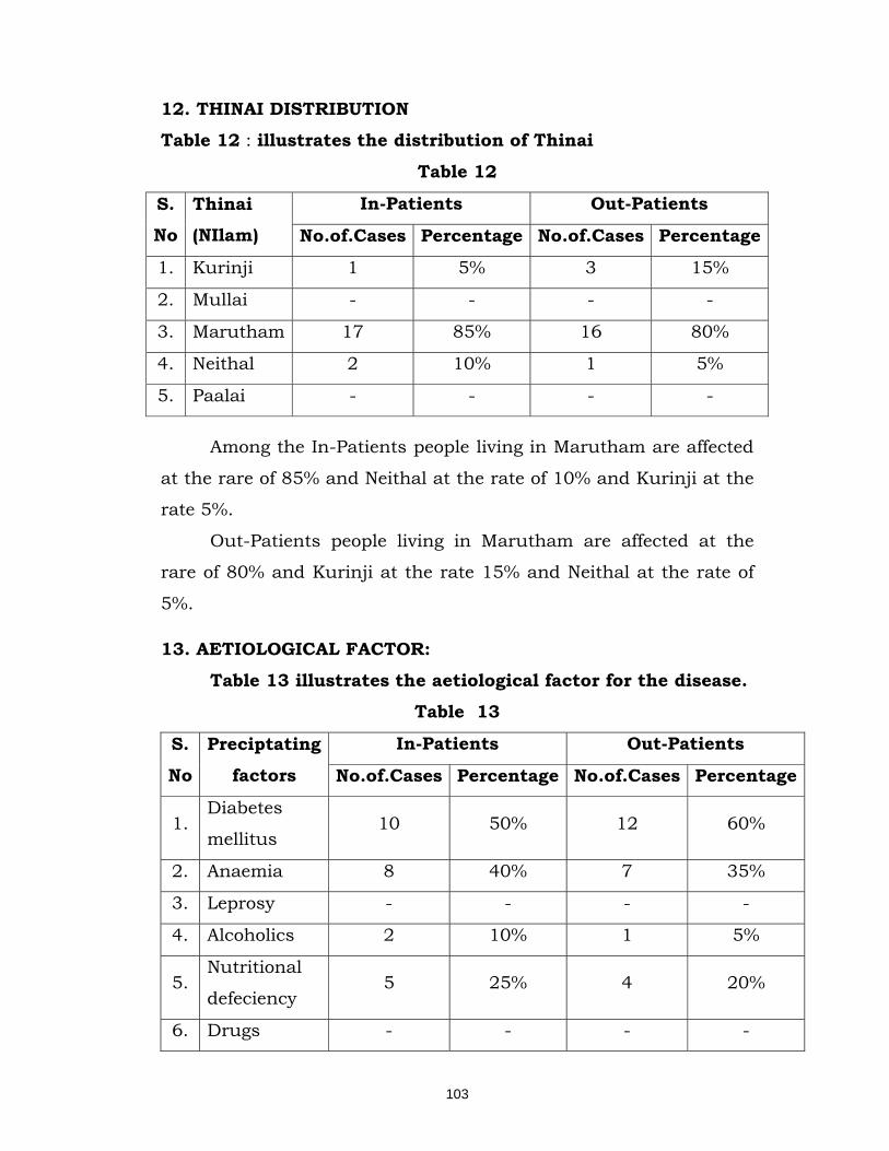
103
12. THINAI DISTRIBUTION Table 12 : illustrates the distribution of Thinai
Table 12
S. No
Thinai (NIlam)
In-Patients Out-Patients
No.of.Cases Percentage No.of.Cases Percentage
1. Kurinji 1 5% 3 15%
2. Mullai - - - -
3. Marutham 17 85% 16 80%
4. Neithal 2 10% 1 5%
5. Paalai - - - -
Among the In-Patients people living in Marutham are affected
at the rare of 85% and Neithal at the rate of 10% and Kurinji at the
rate 5%.
Out-Patients people living in Marutham are affected at the
rare of 80% and Kurinji at the rate 15% and Neithal at the rate of
5%.
13. AETIOLOGICAL FACTOR:
Table 13 illustrates the aetiological factor for the disease. Table 13
S.
No
Preciptating
factors
In-Patients Out-Patients
No.of.Cases Percentage No.of.Cases Percentage
1. Diabetes
mellitus 10 50% 12 60%
2. Anaemia 8 40% 7 35%
3. Leprosy - - - -
4. Alcoholics 2 10% 1 5%
5. Nutritional
defeciency 5 25% 4 20%
6. Drugs - - - -
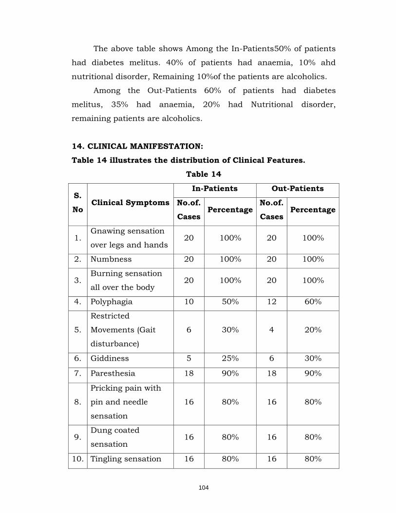
104
The above table shows Among the In-Patients50% of patients
had diabetes melitus. 40% of patients had anaemia, 10% ahd
nutritional disorder, Remaining 10%of the patients are alcoholics.
Among the Out-Patients 60% of patients had diabetes
melitus, 35% had anaemia, 20% had Nutritional disorder,
remaining patients are alcoholics.
14. CLINICAL MANIFESTATION:
Table 14 illustrates the distribution of Clinical Features.
Table 14
S.
No Clinical Symptoms
In-Patients Out-Patients
No.of.
Cases Percentage
No.of.
Cases Percentage
1. Gnawing sensation
over legs and hands 20 100% 20 100%
2. Numbness 20 100% 20 100%
3. Burning sensation
all over the body 20 100% 20 100%
4. Polyphagia 10 50% 12 60%
5.
Restricted
Movements (Gait
disturbance)
6 30% 4 20%
6. Giddiness 5 25% 6 30%
7. Paresthesia 18 90% 18 90%
8.
Pricking pain with
pin and needle
sensation
16 80% 16 80%
9. Dung coated
sensation 16 80% 16 80%
10. Tingling sensation 16 80% 16 80%
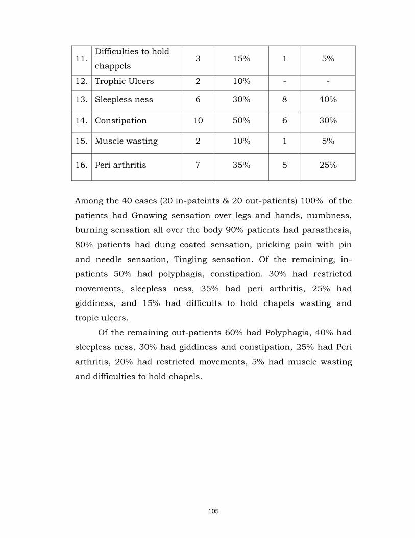
105
11. Difficulties to hold
chappels 3 15% 1 5%
12. Trophic Ulcers 2 10% - -
13. Sleepless ness 6 30% 8 40%
14. Constipation 10 50% 6 30%
15. Muscle wasting 2 10% 1 5%
16. Peri arthritis 7 35% 5 25%
Among the 40 cases (20 in-pateints & 20 out-patients) 100% of the
patients had Gnawing sensation over legs and hands, numbness,
burning sensation all over the body 90% patients had parasthesia,
80% patients had dung coated sensation, pricking pain with pin
and needle sensation, Tingling sensation. Of the remaining, in-
patients 50% had polyphagia, constipation. 30% had restricted
movements, sleepless ness, 35% had peri arthritis, 25% had
giddiness, and 15% had difficults to hold chapels wasting and
tropic ulcers.
Of the remaining out-patients 60% had Polyphagia, 40% had
sleepless ness, 30% had giddiness and constipation, 25% had Peri
arthritis, 20% had restricted movements, 5% had muscle wasting
and difficulties to hold chapels.

106
15. ASSOCIATED DISEASES Table 15 illustrates the distribution of associated diseases
Table 15
S. No
Associated diseases
In-Patients Out-Patients
No.of.Cases Percentage No.of.Cases Percentage
1. Diabetes
mellitus 10 50% 12 60%
2. Hyper
tension 4 20% 4 20%
3. Osteo
arthritis 5 25% 2 10%
4. lumbar
Spondylosis 2 10% - -
5. Trophic
Ulcers 2 10% - -
6. Piles - - 1 5%
7. Anaemia 8 40% 7 35%
Among the In-Patients 50% of patients are affected by the
disease of Diabetes mellitus, 20% of patients are affected by hyper
tension, 25% of patients are affected by Osteo arthritis, 10% of
patients are affected by lumbar spondylosis and tropic ulcer. 40%
of patients are affected by anaemia.
Among the Out-Patients 60% of patients are affected by the
disease of Diabetes mellitus, 20% of patients are affected by hyper
tension, 10% of patients are affected by Osteo arthritis, 5% patients
affected by piles, 35% of patients are affected by anaemia.

107
16.MODE OF ONSET: Table 16 illustrates the mode of onset of the disease
Table 16
S. No
Mode of onset
In-Patients Out-Patients
No.of.Cases Percentage No.of.Cases Percentage
1. Acute - - 2 10%
2. Sub
acute 3 15% 8 40%
3. Chronic 17 85% 10 50%
Among the In-Patients 85% of cases were affected in Chronic
condition, 15% of cases were affected in sub acute condition.
Among the Out-Patients 50% of cases were affected in
Chronic condition, 40% of cases were affected in sub acute
condition, remaining 10% were affected in acute condition.
17. DURATION OF ILLNESS:
Table 16: illustrates the distribution of the Duration of Illness Table 17
S.
No
Duration of
Illness
In-Patients Out-Patients
No.of.
Cases Percentage
No.of.
Cases Percentage
1. Upto 1 month 1 5% 10 50%
2. 1 month – 6 months 11 55% 8 40%
3. 6 month – 1 year 4 20% 2 10%
4. Above 1 year 4 20% - -
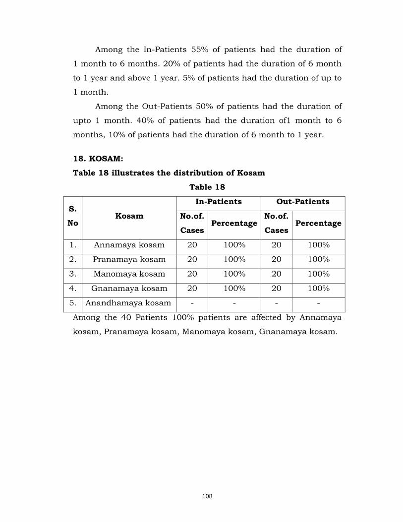
108
Among the In-Patients 55% of patients had the duration of
1 month to 6 months. 20% of patients had the duration of 6 month
to 1 year and above 1 year. 5% of patients had the duration of up to
1 month.
Among the Out-Patients 50% of patients had the duration of
upto 1 month. 40% of patients had the duration of1 month to 6
months, 10% of patients had the duration of 6 month to 1 year.
18. KOSAM:
Table 18 illustrates the distribution of Kosam
Table 18
S.
No Kosam
In-Patients Out-Patients
No.of.
Cases Percentage
No.of.
Cases Percentage
1. Annamaya kosam 20 100% 20 100%
2. Pranamaya kosam 20 100% 20 100%
3. Manomaya kosam 20 100% 20 100%
4. Gnanamaya kosam 20 100% 20 100%
5. Anandhamaya kosam - - - -
Among the 40 Patients 100% patients are affected by Annamaya
kosam, Pranamaya kosam, Manomaya kosam, Gnanamaya kosam.
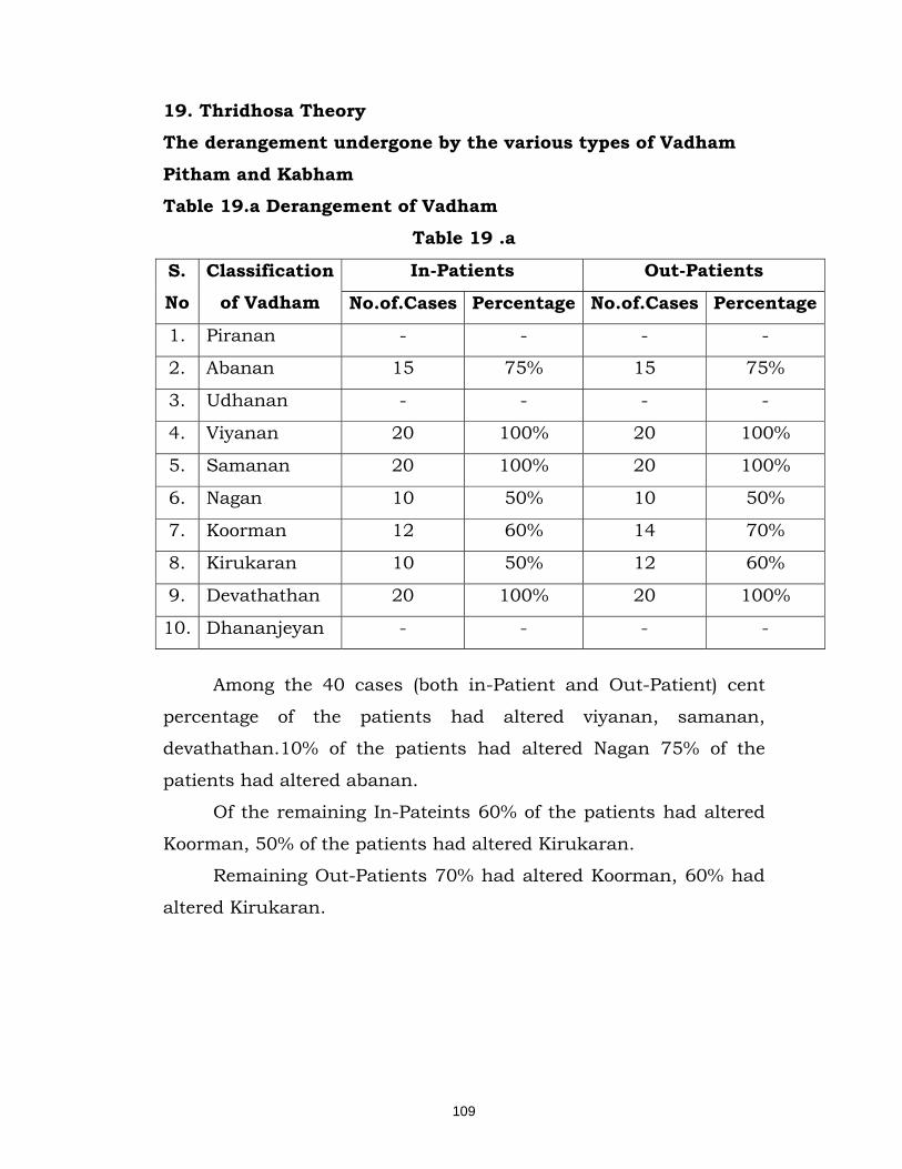
109
19. Thridhosa Theory The derangement undergone by the various types of Vadham
Pitham and Kabham Table 19.a Derangement of Vadham
Table 19 .a
S.
No
Classification
of Vadham
In-Patients Out-Patients
No.of.Cases Percentage No.of.Cases Percentage
1. Piranan - - - -
2. Abanan 15 75% 15 75%
3. Udhanan - - - -
4. Viyanan 20 100% 20 100%
5. Samanan 20 100% 20 100%
6. Nagan 10 50% 10 50%
7. Koorman 12 60% 14 70%
8. Kirukaran 10 50% 12 60%
9. Devathathan 20 100% 20 100%
10. Dhananjeyan - - - -
Among the 40 cases (both in-Patient and Out-Patient) cent
percentage of the patients had altered viyanan, samanan,
devathathan.10% of the patients had altered Nagan 75% of the
patients had altered abanan.
Of the remaining In-Pateints 60% of the patients had altered
Koorman, 50% of the patients had altered Kirukaran.
Remaining Out-Patients 70% had altered Koorman, 60% had
altered Kirukaran.
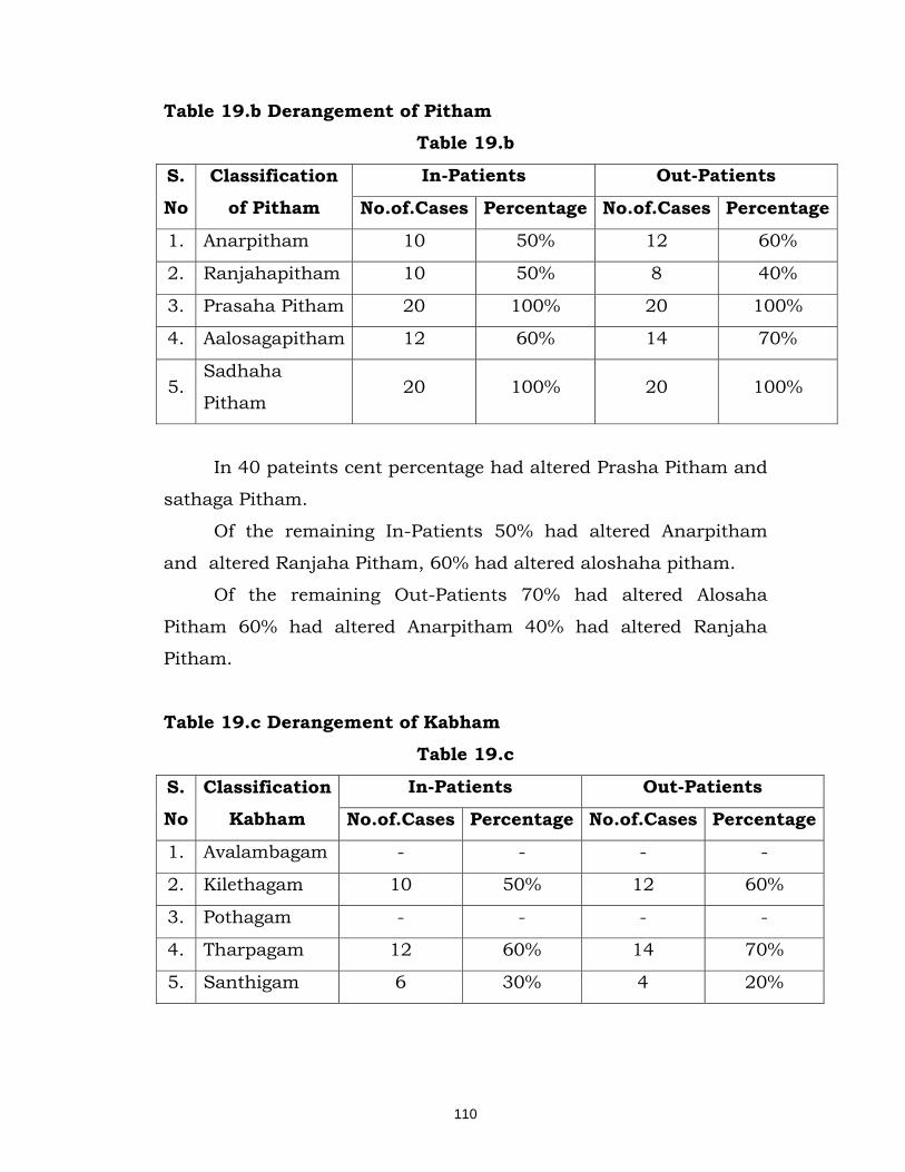
110
Table 19.b Derangement of Pitham Table 19.b
S.
No
Classification
of Pitham
In-Patients Out-Patients
No.of.Cases Percentage No.of.Cases Percentage
1. Anarpitham 10 50% 12 60%
2. Ranjahapitham 10 50% 8 40%
3. Prasaha Pitham 20 100% 20 100%
4. Aalosagapitham 12 60% 14 70%
5. Sadhaha
Pitham 20 100% 20 100%
In 40 pateints cent percentage had altered Prasha Pitham and
sathaga Pitham.
Of the remaining In-Patients 50% had altered Anarpitham
and altered Ranjaha Pitham, 60% had altered aloshaha pitham.
Of the remaining Out-Patients 70% had altered Alosaha
Pitham 60% had altered Anarpitham 40% had altered Ranjaha
Pitham.
Table 19.c Derangement of Kabham
Table 19.c
S.
No
Classification
Kabham
In-Patients Out-Patients
No.of.Cases Percentage No.of.Cases Percentage
1. Avalambagam - - - -
2. Kilethagam 10 50% 12 60%
3. Pothagam - - - -
4. Tharpagam 12 60% 14 70%
5. Santhigam 6 30% 4 20%
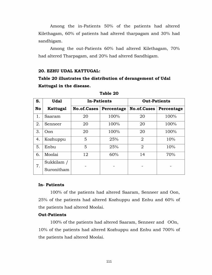
111
Among the in-Patients 50% of the patients had altered
Kilethagam, 60% of patients had altered tharpagam and 30% had
sandhigam.
Among the out-Patients 60% had altered Kilethagam, 70%
had altered Tharpagam, and 20% had altered Sandhigam.
20. EZHU UDAL KATTUGAL: Table 20 illustrates the distribution of derangement of Udal
Kattugal in the disease.
Table 20
S.
No
Udal
Kattugal
In-Patients Out-Patients
No.of.Cases Percentage No.of.Cases Percentage
1. Saaram 20 100% 20 100%
2. Senneer 20 100% 20 100%
3. Oon 20 100% 20 100%
4. Kozhuppu 5 25% 2 10%
5. Enbu 5 25% 2 10%
6. Moolai 12 60% 14 70%
7. Sukkilam /
Suronitham - - - -
In- Patients 100% of the patients had altered Saaram, Senneer and Oon,
25% of the patients had altered Kozhuppu and Enbu and 60% of
the patients had altered Moolai.
Out-Patients 100% of the patients had altered Saaram, Senneer and OOn,
10% of the patients had altered Kozhuppu and Enbu and 700% of
the patients had altered Moolai.
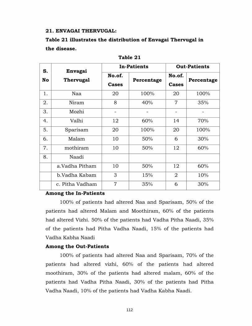
112
21. ENVAGAI THERVUGAL: Table 21 illustrates the distribution of Envagai Thervugal in
the disease. Table 21
S.
No
Envagai
Thervugal
In-Patients Out-Patients
No.of.
Cases Percentage
No.of.
Cases Percentage
1. Naa 20 100% 20 100%
2. Niram 8 40% 7 35%
3. Mozhi - - - -
4. Valhi 12 60% 14 70%
5. Sparisam 20 100% 20 100%
6. Malam 10 50% 6 30%
7. mothiram 10 50% 12 60%
8. Naadi
a.Vadha Pitham 10 50% 12 60%
b.Vadha Kabam 3 15% 2 10%
c. Pitha Vadham 7 35% 6 30%
Among the In-Patients 100% of patients had altered Naa and Sparisam, 50% of the
patients had altered Malam and Moothiram, 60% of the patients
had altered Vizhi. 50% of the patients had Vadha Pitha Naadi, 35%
of the patients had Pitha Vadha Naadi, 15% of the patients had
Vadha Kabha Naadi
Among the Out-Patients
100% of patients had altered Naa and Sparisam, 70% of the
patients had altered vizhi, 60% of the patients had altered
moothiram, 30% of the patients had altered malam, 60% of the
patients had Vadha Pitha Naadi, 30% of the patients had Pitha
Vadha Naadi, 10% of the patients had Vadha Kabha Naadi.

113
22. Duration of Treatment Table 22 illustrates the distribution of duration of Treatment
Table 22
S.
No
Duration of
Treatment
In-Patients Out-Patients
No.of Cases
Percentage No.of. Cases
Percentage
1. 30-40 days - - 7 35%
2. 41-50 days 19 95% 12 60%
3. 51-60 days 1 5% 1 5%
The duration of the treatment of the in-patients as follows.
41-50 days 19 patients (95%), 51-60 days 1 patient (5%)
Among the out-Patients
30-40 days 7 patients (35%), 41 – 50 days 11 patients (55%),
51-60 days 1 patient (5%)
23. SENSORY SYSTEM
Table 23.a: illustrates the distribution of the Sensory System
Table 23. a S.
No
Sensory
System
In-Patients Out-Patients
No.of.Cases Percentage No.of.Cases Percentage
1. Pain 2 10% - -
2. Temperature 4 20% - -
3. Touch 20 100% 20 100%
4. Position - - - -
5. Vibration 6 30% 6 30%
7. Steriognosis - - - -
8. Two point
discrimnation - - - -
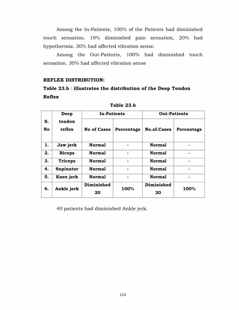
114
Among the In-Patients, 100% of the Patients had diminished
touch sensation. 10% diminished pain sensation, 20% had
hypothermia. 30% had affected vibration sense.
Among the Out-Patients, 100% had diminished touch
sensation. 30% had affected vibration sense
REFLEX DISTRIBUTION: Table 23.b : illustrates the distribution of the Deep Tendon
Reflex
Table 23.b
S.
No
Deep
tendon
reflex
In-Patients Out-Patients
No of Cases Percentage No.of.Cases Percentage
1. Jaw jerk Normal - Normal -
2. Biceps Normal - Normal -
3. Triceps Normal - Normal -
4. Supinator Normal - Normal -
5. Knee jerk Normal - Normal -
6. Ankle jerk Diminished
20 100%
Diminished
20 100%
40 patients had diminished Ankle jerk.

115
25. Clinical Improvement: Table 25 illustrates the distribution of clinical Improvement
Table 25
S. No
Result In-Patients Out-Patients
No.of.Cases Percentage No.of.Cases Percentage
1. Good 17 85% 16 80%
2. Fair 2 10% 3 15%
3. Poor 1 5% 1 5%
The results were graded as
Among the In-Patients 85% had showed good results, 10%
had showed fair results, 5% had showed poor results.
Among the out-Patients 80% had showed good results, 15%
had showed fair results, 5% had showed poor results.
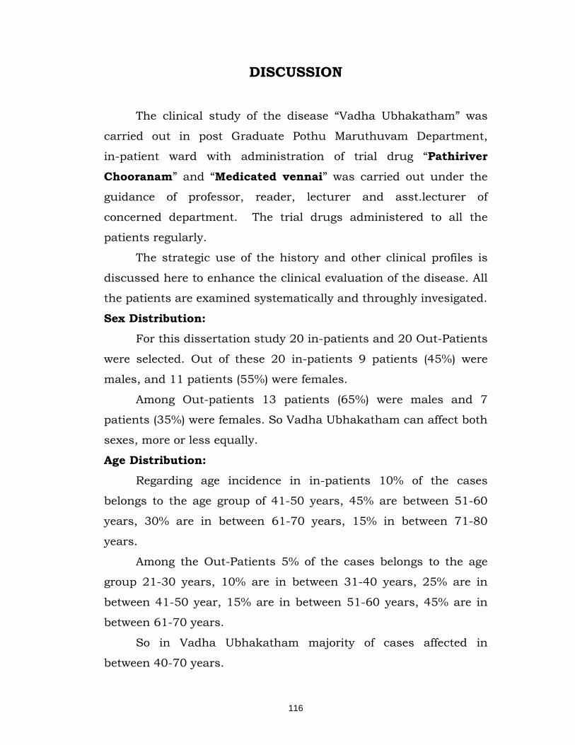
116
DISCUSSION
The clinical study of the disease “Vadha Ubhakatham” was
carried out in post Graduate Pothu Maruthuvam Department,
in-patient ward with administration of trial drug “Pathiriver
Chooranam” and “Medicated vennai” was carried out under the
guidance of professor, reader, lecturer and asst.lecturer of
concerned department. The trial drugs administered to all the
patients regularly.
The strategic use of the history and other clinical profiles is
discussed here to enhance the clinical evaluation of the disease. All
the patients are examined systematically and throughly invesigated.
Sex Distribution: For this dissertation study 20 in-patients and 20 Out-Patients
were selected. Out of these 20 in-patients 9 patients (45%) were
males, and 11 patients (55%) were females.
Among Out-patients 13 patients (65%) were males and 7
patients (35%) were females. So Vadha Ubhakatham can affect both
sexes, more or less equally.
Age Distribution:
Regarding age incidence in in-patients 10% of the cases
belongs to the age group of 41-50 years, 45% are between 51-60
years, 30% are in between 61-70 years, 15% in between 71-80
years.
Among the Out-Patients 5% of the cases belongs to the age
group 21-30 years, 10% are in between 31-40 years, 25% are in
between 41-50 year, 15% are in between 51-60 years, 45% are in
between 61-70 years.
So in Vadha Ubhakatham majority of cases affected in
between 40-70 years.

117
Kaalam: Among the 20 in-patients 70% of cases were in Pitha Kaalam
and 25% in Kabha Kaalam and remaining 5% in Vadha Kaalam.
Among 20 Out Patients 75% of cases were in Pitha Kaalam
and 25% in Kabha Kaalam.
Though “Vadha ubhakatham” can acquire in all Kaalam, but
majority of cases are affected in Pitha Kaalam.
Constitution of the Body:
Among the 20 in-patients 50% of the cases were thondha
thegi, 25% of the patients were Vadha thegi, 10% of the patients
were pitha thegi, 15% were kabha thegi.
Among 20 out-patients 50% of the cases were thondha thegi,
20% of the patients were Vadha thegi and Pitha thegi, and
remaining 10% of the patients were kabha thegi
So Vadha Ubhakatham can affect all thegis.
Gunam:
Among the 20 in-patients and 20 out-patients cent percent of
the cases had Rajo gunam.
So Vadha Ubhakatham is predominant in Rajo gunam
patients.
Religious Status: Out of 20 in-patients majority of the patients are Hindus
(80%), Christians are 15%.
Out of 20 out-patients majority of the patients are Hindus
(75%) Christians are 15%. Muslims are 10%.
Occupational Status:
Regarding occupational history among the in-patients House
wifes, Formers, Coolies are affected at the rate of 20%. Servant
maid are affected at the rate of 15%, Merchants & drivers are
118
affected at the rate of 10%. Remaining 5% of the affected patients
are watch man.
Among the out-patients House Wifes, retired man are affected
at the rate of 25% merchants are affected at the rate of 10%
watchman, drivers & servant maid are affected at the rate of 5%.
So Vadha Ubhakatham can affect all categories of people.
Socio Economic Status: Among the 20 in-patients 20% of the cases were in middle
class and 80% are poor people.
Among the 20 out-patients 50% of the cases were middle
class and poor status.
Habits:
Among the 20 in-patients 20% of the patients were smokers,
15% of the patients were tobacco chewers, 10% of the patients were
alcoholic 5% of the patients were tobacco inhaler.
Among the 20 out-patients, 30% of the patients were smoker,
5% of the patients were alcoholic.
So Vadha Ubhakatham may affect all bad habits patients.
Alcoholic abuse is one of the causes for neuritis.
Dietary Changes:
Among the 20 in-patients 95% of the patients were mixed diet
and the remaining are vegetarians.
Among the 20 out-patients 75% of the patients were mixed
diet and the remaining are vegetarians.
So “Vadha Ubhakatham” may affect both categories of people
but predominant among the mixed diet.
Paruvakaalam:
The paruvakaalam changes play major role in the
developement of the disease. In this study among the in-patients

119
25% of cases had developed symptoms during Koothir kaalam
(25%), Munpani Kaalam (60%), Pinpani Kaalam (15%).
Among the Out patients 100% of patients had developed
symptoms during Koothir Kaalam.
Thinai Reference: Among 20 in-patients most of the people living in Marutham
are affected at the rate of 85%, 10% of the pateints in Neidhal, 5%
of the patients are in Kurinji.
Among 20 out-patients most of the people living in Marutham
are affected at the rate of 80%, 15% of the patients in Kurinji, 5% of
the patients are in Neidhal.
Marutham is the area where the severity of the disease is less,
but these incidences may be due to the alterations in the food,
habit and other activities.
Aetiological Factors: Among 20 in patients 50% of patients were affected by
diabetes mellitus. 40% of patients were affected by anaemia;
remaining 10% of patients were affected by alcohol intake.
Among 20 out patients 60% of patients were affected by
diabetes mellitus. 35% of patients affected by anaemia, remaining
5% of patients were affected by alcoholic intake.
Literary evidence indicates that the disease Vadha
Ubhakatham is common in diabetes mellitus.
Associated diseases:
In this study among the in-patients 50% of the cases were
affected by diabetes mellitus, 20% had hyper tension, 25% had
Osteo arthritis, and 10% had lumbar spondylosis and trophic
ulcers.
120
Among the out-patients 60% of the patients were affected by
diabetes mellitus, 20% had hyper tension, 10% had Osteo arthritis,
5% had piles.
Mode of Onset:
Among the In-Patients majority of cases (85%) were presented
in chronic condition, remaining 15% were in sub acute condition.
Among the Out-patients 50% of cases were presented in
chronic condition. 40% of cases were presented in the subacute
condition remaining 10% were in acute stage.
So majority of chronic condition needs hospitalization other
condition can cure in out-patient ward.
In Vadha ubhakatham majority of the cases were reported in
the chronic condition.
Duration of Illness:
In In-Patients 20% were affected by the duration of above 6
month to 1 year and above 1 year. 55% were above 1 month to 6
months, 55 were upto 1 month duration affected.
In out-patients 50% were affected by the duration upto 1
month and 40% were above 1 month to 6 months. Remaining 10%
were above 1 month – 1 year.
In Vadha Ubhakatham majority of the cases were reported in
above 1month – 6 months.
In long duration the patients needs hospitalization.
Observations of Clinical features of Vadha Ubhakatham are as
follows:
Clinical Manifestation:
Among the 40 cases (20 in-pateints & 20 out-patients) 100%
of the patients had Gnawing sensation over legs and hands,
numbness, burning sensation all over the body 90% patients had
parasthesia, 80% patients had dung coated sensation, pricking
121
pain with pin and needle sensation, Tingling sensation. Of the
remaining, in-patients 50% had polyphagia, constipation. 30% had
restricted movements, sleepless ness, 35% had peri arthritis, 25%
had giddiness, 15% had difficults to hold chapels wasting and
tropic ulcers.
Of the remaining out-patients 60% had Polyphagia, 40% had
sleepless ness, 30% had giddiness and constipation, 25% had Peri
arthritis, 20% had restricted movements, 5% had muscle wasting
and difficulties to hold chapels.
Thridhosa Theory
A . Disturbances in Vadham:
Among the 40 cases (both in-Patient and Out-Patient) cent
percentage of the patients had altered viyanan, samanan,
devathathan.10% of the patients had altered Nagan 75% of the
patients had altered abanan.
Of the remaining In-Pateints 5% had altered Piranan &
altered Udhanan, 60% of the patients had altered Koorman, 50% of
the patients had altered Kirukaran.
Remaining Out-Patients 70% had altered Koorman, 60% had
altered Kirukaran.
Affected Abannan produced Poly urea and constipation.
Affected Viyanan produced numbness, tingling sensation, loss
of sensation, burning sensation, body pain, muscle wasting are
formed.
Affected samanan produces derangement of other vayus.
Affected naagan was found to the derangement in who were
mentally depression and sluggishness.
Affected Koorman produced diminished vision.
Affected Kirukaran produce increase the appetite.
122
Affected Devathathan produces fatigue and tiredness and
disturbances of sleep rhythm.
B. Disturbances in Pitham
Among the in-Patients 50% of the patients had altered Anar
pitham, 50 % of the patients had altered Ranchaga pitham, 60% of
patients had altered Alosaham, 100% had altered Prasaham and
Sathaham.
Among the out-Patients 60% had altered Anar pitham, 40%
had altered Ranchaga pitham, 70% had altered Alosaham, 100%
had altered Prasaham and Sathaham.
Affected Anarpitham produces increase the appetite.
Affected Ranchaga Pitham produces dyspnoea and reduced
heamoglobin.
Affected Prasakapitham produces dryness and darkness of
the skin and burning sensation of the body.
Affected Sathaha Pitham produces difficulty in attaining to
their regular duties.
Affected Alosaha pitham diminished vision.
C. Disturbance in Kabam: Among the in-Patients 50% of the patients had altered
Kilethagam, 60% of patients had altered tharpagam and 25% had
sandhigam.
Among the out-Patients 60% had altered Kilethagam, 70%
had altered Tharpagam, and 10% had altered Sandhigam.
Affected Kelethagam produce increase appetite.
Affected pothaham produces tastelessness
Affected tharpagam produces burning sensation in the eyes.
Affected Sandhiham produces Pain and difficult to flex and
extend joints.
123
Ezhu Udal Kattugal: In- Patients,
100% of the patients had altered Saaram, Senneer and Oon.
60% of the patients had altered Moolai.
25% of the patients had altered Kozhuppu and Enbu
Out-Patients,
100% of the patients had altered Saaram, Senneer and Oon.
70% of the patients had altered Moolai.
10% of the patients had altered Kozhuppu and Enbu
Affected Saaram produces depression, Roughness to skin
increased appetite and lazinees. Affected Senneer produces
reduction is haemoglopin, nerve weakness and pallor. Affected Oon
produces weakness of 5 sensory orgen, bodyache and muscle
wasting. Affected Kozhuppu produces restricted movements.
Affected Enbu produces pain and swelling in knee joint. Affected
Moolai produces bluring of vision.
Envagai Thervugal: Among the In-Patients,
100% of patients had altered Naa and Sparisam, 50% of the
patients had altered Malam and Moothiram, 60% of the patients
had altered Vizhi, 50% of the patients had Vadha Pitha Naadi, 35%
of the patients had Pitha Vadha Naadi and 15% of the patients had
Vadha Kabha Naadi.
Among the Out-Patients,
100% of patients had altered Naa and Sparisam, 70% of the
patients had altered vizhi, 60% of the patients had altered
moothiram, 30% of the patients had altered malam, 60% of the
patients had Vadha Pitha Naadi, 30% of the patients had Pitha
Vadha Naadi and 10% of the patients had Vadha Kabha Naadi.
124
Affected Naa denotes dryness of tongue and pallor. Affected
Niram denotes pallor of eye skin, nailas and all over body. Affected
Vizhi denotes burning sensation of eye, pallor of eye diminshed
vision. Affected Sparisam denotes burning sensation and numbness
of body. Affected Malam denotes constipation. Neekuri was found to
be diabetic patient’s urine denotes odur of honey. Edai was
increased, Manam ant flies were found to the attracted urine Nurai-
frothy. Neikuri had showed in inpatients 5% vadha neer, 45% pitha
neer and 50% kabha neer. In outpatients 10% vadha neer, 30%
pitha neer and 60% kabha neer.
Duration of Treatment
The patients were treated in the hospital for maximum 40
days. The prognosis and result of the treatment was assessed on
the basis of subjective feeling and objective parameters
Sensory system Among the In-Patients, 100% of the Patients had diminished
touch sensation. 10% diminished pain sensation, 20% had
hypothermia. 30% had affected vibration sense.
Among the Out-Patients, 100% had diminished touch
sensation. 30% had affected vibration sense
Reflexes 40 patients had diminished Ankle jerk.
Investigation
Routine haematological and urological investigation was done,
during the time of admission and discharge for all cases.
Haematological investigation had showed raised Blood sugar
and reduction in haemoglobin.
After the end of this regimen, raised haemoglobin and
reduction of blood sugar recorded.

125
Special Investigation Nerve conduction study was done for 2 patients it showed
sensory disturbence the symptoms are reduced after treatment.
Results
All the 20 inpatients and 20 outpatients were treated with
trial medicine. The results were assessed on the basis of subjective
and objective improvement of symptoms and a sence of well being
at the end of the treatment the results were categorized as follows.
Good : Complete subsitence of gnawing sensation,
numbness, pricking pain, normal apptite and free movements.
Fair : Relief of gnawing sensation, numbness,
pricking pain, normal apptite and some time restricted movements.
Poor : No imporvement
Among the in-patients 85% of the patients showed good
results 10% patients showed fair results remaining 5% of patients
showed poor results.
Among the out patients 80% of patients showed good results
15% of patients shows fair results remaining 5% of patients shows
poor results.

126
SUMMARY
The author feels privileged for carrying out the clinical study
on the disease “Vadha Ubakatham” perhaps the incidence of Vadha
Ubhakatham is increasing now a day. The disease is correlated with
“Peripheral Neuritis” in allopathic view. It is now common in
occurence due to prevalence of Madhu megam and their
complications, and dietry irregularities
So the author had worked on this dissertation study with
deep devotion and keen observation
For the disease Vadha Ubhakatham internal medicine
pathiriver choornam is given. Medicated vennai applied externally.
The medicines for the treatment of this disease have been
chosen from the traditional siddha literatures, after extensively
going through various siddha literatures. The medicines are
prepared according to the norms strictly confirmed to siddhar’s
ethics.
The aetiology, pathology, classifications, clinical features,
diagnosis complications differential diagnosis, treatment were
collected from a number of literatures both in Siddha system as
well as in modern system of medicine.
In this study 20 patients of either sex and of varying age
groups as in-patients and 20 patients as out-patient were selected
and treated with the trial drugs for this study.
The selection of cases and management of cases during
admission and after treatment was carreid out under the
supervision of Professor, Reader, Lecturer, and Asst. Lecturer of
Post graduate Pothu Maruthuvam Department.
In order to normalize the provoked Vadha humour, laxative
drug was given to all patients on the first day. The next day the trial
127
drugs were given to all the patients. From the observations and
results, the author had en-lights that vadha ubhakatham was
common in the following aspects.
The disease was more common in females.
Maximum incidence of age between 50 – 70 years i.e in pitha
kaalam the incidence of the disease was higher in koothirkaalam
(25% ip cases and 100% op cases) & Munpani Kaalam (60% ip
cases)
The disease due to vitated vadham which subsequently leads
to alterations in other dhosas.
Signs and symptoms along with various other factors
mentioned in the case sheets were elaborately discussed in the
previous chapter.
The siddha diagnosis were made with the help of Envagai
thervugal especially naadi and neikuri. Naddi Nadai of the patients
was found to be vadha pitham, vatha kabham and pitha vadham.
Majority of the patients had this disease due to nutritional
disorder another one is diabetes mellitus. In poor status the
patients having lake of nutritional elements in their diet. The
author has find out the cause from dietery habits majority of
patients are in poor status.
Regarding modern aspects Vadha ubhakatham is
differentiated from other pathologies by the use of routine blood
examination, blood sugar, blood urea, serum cholesterol, urine,
motion examination. Nerve conduction study was done in 2
patients for diagnosis and to follow the progress of the patients.
So the diagnosis, of the vadha ubhakatham was made out by
analyzing clinical signs, symptoms and differential diagnosis.
The effcacies of the drugs “Pathiriver chooranam, Medicated
vennai” were studied and observed during the period of this

128
research. The diabetic patients are also benefited by consuming
this internal medicine.
Daily monitoring of diabetic status was made by Urine
analysis. In adition to that drugs every patients was advised to Yoga
therapy (Mayurasanam) for better results.
Clinically there were marked reduction of pain, numbness,
Gnawsing sensation, and tingling sensation burning sensation
along and reduced blood sugar, blood urea, and increased Hb%
that were noted.
Pharmacological evaluation of the drug pathiriver chooranam
has showed analgesic, anti inflammatory (acute and chronic) and
Hypo glycemic action. Medicated vennai has anti – inflammatory
action.
Bio chemical analysis showed the drugs have the presence of
ferrous iron, unsaturated compound, reducing sugar and amino
acid. The clinical improvement is graded as good, fair, poor on the
basis of symptoms relieved and the results observed during this
study
The in-patients are discharged after satisfactory, clinical
improvement and advised to continue the treatment in out-patient
department.
No side effects, adverse effects and complications were
observed during these studies.

129
CONCLUSION
In this clinical trial results were found to be satisfactory in
80% of cases.
The trial drugs have excellent pharmacological action.
The trial drugs were free from side effects and adverse
effects.
Further follow up of all these patients showed excellent
reduction of their symptoms and a sense of well being.
The trial medicine Pathiri ver chooranam has the taste
Thuvarppu.
Suvai - Thuvarppu
Thanmai - Thatpam
Pirivu - Inippu
The taste Thuvarppu has skin soothening effect and blood
purifying effect, as per siddha maruthuvanga surukkam.
And the trial medicine has it thanmai as thatpam all the
thatpam naturated drug has cooling effect to body so it acts well
against burning sensation.
After ingestion while the trial medicine reacts the gastric guice
it changes to Inippu, at this stage Inippu cures the vadha disease
and it gives strength to 5 sensory organs. From this study Pathiri
ver Chooranam acts as antivadha drug on the basis of Suvai,
Thanmai, Pirivu and Edhir urai.
So Vadha Ubhakatham is well controllable with Pathiriver
Chooranam and Medicated vennai.

130
PREPARATION OF PATHIRI VER CHOORANAM Ingredients
Pathiriver - 1 k.g
Method Pathiriver is dried and purified then nicely powdered and filter
through cloth.
Dose
5 gms twice daily.
Adjuvant
Hot water
Indication Madhumegam, Burning sensation in eye, ear, leg and hands,
Eczema, pitham.
Expiry
6 months from the date of preparation.
Reference
Gunapadam - Mooligai Vaguppu Page No 665
Properties of the Pathiri Pathiri
Common Tamil Name : Pathiri
Synonyms : Kanni, Paadalimaram, Paadalam,
Punkali.
Botanical Name : Stereospermum suaveolens
Family : Bignoniaceae
Parts used : Leaf, flower Bark, root
Suvai : Thuvarppu (Astringent)
Thanmai : Thatpam (Cold)
Pirivu : Inippu (Sweat)
Actions : Diuretic,f ebrifuge, anti inflammatory,
antibacterial, tonic

131
Chemical Constituents : glycoside, albuminous,
saccharine and 6-0 glucosyl
cutellariein, dinatin – 7-
glucuronide
Uses:
“ghjphpapd; NtHFze;jhd; ghHf;fpy; kJNkfk;
XJfug; ghDoiy NahbtpLk; - khNjNfs;
fz;nzwpT fhnjhpT ifnahpT fhnyhpTk;
epz;zakha;g; NghFk; eprk;”
Pathiri ver used for madhumegam, , burning sensation in eye,
ear, hand and leg, eczema, pitham. Decoction of roots used in
intermittent and puperperal fevers and affections of the brain.
Preparation of Medicated vennai
Ingredients
Illandai ilai - 100gms
Cheek-kai ilai - 100gms
Musumusukkai ilai - 100gms
Karuva ilai - 100gms
Nelli ilai - 100gms
cow’s milk - 500ml
Method Take the juice of the above leaves. Add cow’s milk to the leaf
juice. Boil this mixture and cool. Keep this solution overnight to
facilitate fermentation. Next morning the fermented solution is
stirred well and a buttery mass will rise up then, that is collected
in a clean bowel. This butter is used for external application
preservate in a water.
Dose
Butter 5gms external application for 3 days.
132
Indications Mehathinal undana erivu, pithathinal undana erivu.
Expiry 3 months
Reference Sarabendhirar pitha roha sihitchai P.No 199
Properties of the Individual component
Illandai
Common Tamil Name : Illandai
Synonyms : Illandai, kulladhi, kulvali, kole,
korkodi, vadhari
Botanical Name : Ziziphus manuritiana var,
Maurtina
Family : Rhamnaceae
Parts used : Leaf, fruit, bark, root, root bark,
whole plant
Suvai : Thuvarppu, Inippu
Thanmai : Thatpam (Cold)
Pirivu : Inippu (Sweat)
Therapeutic Actions : Emollient, cooling and refrigent
Chemical Constituents:
Leafs contain, tannin and a crystallizable principle, ziziphic
acid and sugar
Uses:
Leaves are given cooling and refrigent, effect. Burning
sensation present in body, sacbies, pitham, Megam, also cured.

133
Cheek-kai Common Tamil Name : Cheek-kai
Synonyms : Cheek-kai, cheyakai
Botanical Name : Acacia sinuata
Family : Mimosaceae
Parts used : Leaf, and pod
Suvai : Pulippu
Thanmai : Thatpam (Cold)
Pirivu : Karppu
Therapeutic Actions : Externally detergent and astringent
Chemical Constituents:
Alkaloid saponins : 11.2%
Malic acid : 12.75%
Resin : 1%
Glucose : 13.9%
Gum and colouring matter : 21.5%
Grude fibre : 22%
Ash : 3.75%
Uses:
This plant has cleaning properties and protects the skin
against micro organism.
Musumuskaai
Common Tamil Name : Musumuskkai
Synonyms : Iyleyam, erukurangin kai,
mosumuskkai
Botanical Name : Melothira moderaspatana
Family : Cucurbitaceae
Parts used : Leaves and Root

134
Suvai : Thuvarppu
Thanmai : Veppam
Pirivu : Karppu
Therapeutic Actions : Expectorant, astringent
Chemical Constituents : Columbin
Uses: Leaves used in vitiligo and biliousness, kabham, cough.
Karuva Ilai
Common Tamil Name : Karuva Ilai
Synonyms : Lavanga pathiri
Botanical Name : Cinnamomum verum
Family : Lauracea
Suvai : Karppu
Thanmai : Veppam
Pirivu : Karppu
Therapeutic Actions : Astringent, Stimulant, carminative
Chemical Constituents : Eugenol, cinnamaledhyde,
benzaldehyde, pinene, phellandrene,
diterpene, safrole, methyleugenol
borneol, geraniol, caryophyllene.
Uses:
Leaves used for anti inflammatory effect. It if also useful in
mehasuram, kabha suram, asthma, vomiting.
135
Nelli Common Tamil Name : Nelli
Synonyms : Aamalaham, Aalaham, Aambal,
amariham, thaththari, thaththiri,
koranham, merudhu pala,
meethundhu.
Botanical Name : Phyllanthus emblica
Family : Euphorbiaceae
Parts used : Leaves, flowers, bark, root , seeds
and pod.
Suvai : Pulippu, Thuvarppu and Inippu
Thanmai : Thatpam
Pirivu : Inippu
Therapeutic Actions : Astringent, cooling diuretic
Chemical Constituents : Trigalloyglucose, terchebin, covilagin,
ellagic acid, phyllembic acid in the
chief constituent, 3-0 gallated
prodelphinidin and procyanidin
Uses:
Leaves are useful in the inflammation useful in sinusities,
vomiting, giddiness.
Cow’s milk Synonyms
Sanskrit : Dughaksheera
English : Milk
Arab : Habit
Persian : Sher
Hindu
Marathi Dudh
Kanada
136
Tamil & Telugu : Palu
Malayalam : Musu, Pala
Characters Cow milk is an opaque white, (or) yellowish, white, emulsion,
alkaline fluid, a little, more viscus, than water, taste is sweet,
odour, faint and peculiar kept for a long time.
Specific gravity is between 1027 to 1037 under the microscope numerous minutes fat globules are
seen floating in the form an emulsion.
The mild settles out in to 3 layers. The layer at the bottom of
the vessel contains bacteria, cells and dirt. That at middle contains
milk, plasma and small amount of fat (or) cream and a considerable
number of bacteria and fat globules.
Composition of milk per 100ml Components Percentage
Salts - 0.5%
Fat - 3.5%
Non fat - 8.0%
Casain - 2.8 % Solids of these proteins
lactalumina - 0.7%
Sugar - 4.5%
Total solids - 12.0%
Water - 88.0%
Action
Milk is generally considered cooling, nutritive, strengthening
and vitalizing also demulcent and emollient.
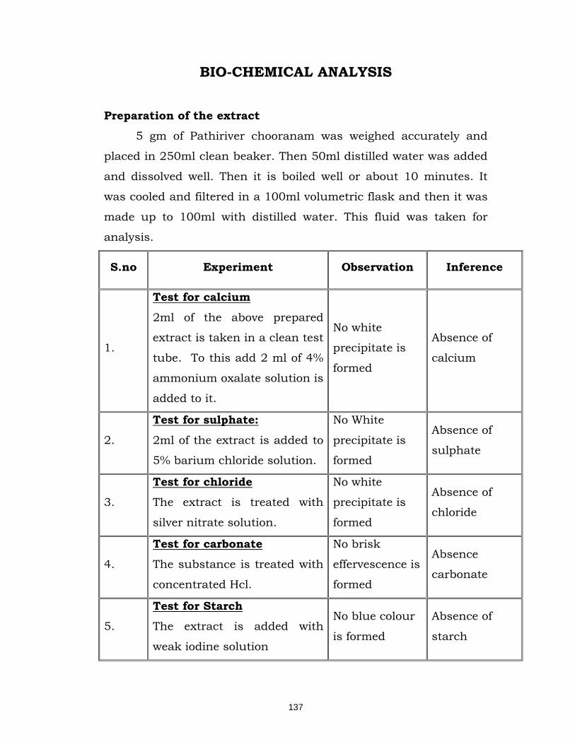
137
BIO-CHEMICAL ANALYSIS
Preparation of the extract
5 gm of Pathiriver chooranam was weighed accurately and
placed in 250ml clean beaker. Then 50ml distilled water was added
and dissolved well. Then it is boiled well or about 10 minutes. It
was cooled and filtered in a 100ml volumetric flask and then it was
made up to 100ml with distilled water. This fluid was taken for
analysis.
S.no Experiment Observation Inference
1.
Test for calcium
2ml of the above prepared
extract is taken in a clean test
tube. To this add 2 ml of 4%
ammonium oxalate solution is
added to it.
No white
precipitate is
formed
Absence of
calcium
2.
Test for sulphate:
2ml of the extract is added to
5% barium chloride solution.
No White
precipitate is
formed
Absence of
sulphate
3.
Test for chloride
The extract is treated with
silver nitrate solution.
No white
precipitate is
formed
Absence of
chloride
4.
Test for carbonate
The substance is treated with
concentrated Hcl.
No brisk
effervescence is
formed
Absence
carbonate
5.
Test for Starch The extract is added with
weak iodine solution
No blue colour
is formed
Absence of
starch
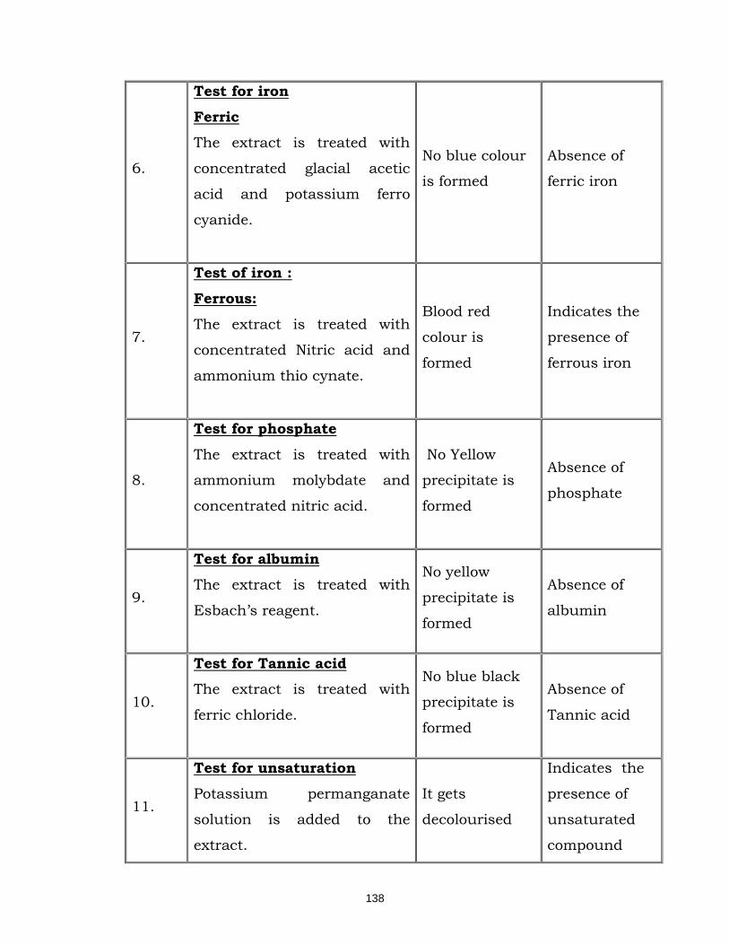
138
6.
Test for iron
Ferric
The extract is treated with
concentrated glacial acetic
acid and potassium ferro
cyanide.
No blue colour
is formed
Absence of
ferric iron
7.
Test of iron :
Ferrous:
The extract is treated with
concentrated Nitric acid and
ammonium thio cynate.
Blood red
colour is
formed
Indicates the
presence of
ferrous iron
8.
Test for phosphate
The extract is treated with
ammonium molybdate and
concentrated nitric acid.
No Yellow
precipitate is
formed
Absence of
phosphate
9.
Test for albumin The extract is treated with
Esbach’s reagent.
No yellow
precipitate is
formed
Absence of
albumin
10.
Test for Tannic acid The extract is treated with
ferric chloride.
No blue black
precipitate is
formed
Absence of
Tannic acid
11.
Test for unsaturation
Potassium permanganate
solution is added to the
extract.
It gets
decolourised
Indicates the
presence of
unsaturated
compound
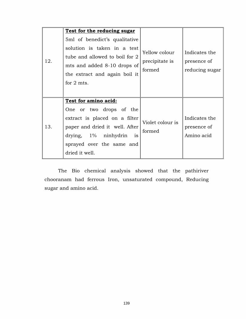
139
12.
Test for the reducing sugar
5ml of benedict’s qualitative
solution is taken in a test
tube and allowed to boil for 2
mts and added 8-10 drops of
the extract and again boil it
for 2 mts.
Yellow colour
precipitate is
formed
Indicates the
presence of
reducing sugar
13.
Test for amino acid:
One or two drops of the
extract is placed on a filter
paper and dried it well. After
drying, 1% ninhydrin is
sprayed over the same and
dried it well.
Violet colour is
formed
Indicates the
presence of
Amino acid
The Bio chemical analysis showed that the pathiriver
chooranam had ferrous Iron, unsaturated compound, Reducing
sugar and amino acid.
140
PHARMACOLOGICAL ANALYSIS
APPENDIX-III
ACUTE ANTI-INFLAMMATORY STUDY ON
PATHIRIVER CHOORANAM BY HIND-PAW METHOD
IN ALBINO RATS
AIM
To study the acute Anti-inflammatory effect of PATHIRIVER
Chooranam.
PREPARATION OF THE TEST DRUG
1 gm of PATHIRIVER Chooranam was dissolved in 10 ml of
water dose of 2 ml was given to each rat. This 2 ml contains 10mg
of the test drug.
PROCEDURE
Nine healthy albino rats weighing 100 - 150 gm were
taken and divided into three groups, each consisting of 3 rats.
First group was kept as control by giving distilled water of
2ml / 100mg of body weight. The second group was given
Ibuprofen as dose of 20mg / 100g of body weight. The third
group received the test drug PATHIRIVER Chooranam of 200 mg /
kg body weight.
Before administration of test drug, the hind-paw volume of all
rats was measured. This was done by dipping the hind-paw up to
tibio - tarsal junction, in to a mercury plethysmography. While
dipping the hind-paw, by pulling the syringe piston, the level of
141
mercury in the center small tube was made to coincide with red
marking and reading was noted from the plethysmograph.
Soon after the measurement, the drugs were administrated
orally. One hour later, a subcutaneous injection of 0.1ml of 1%
w/v carrageen in water was made into planter surface of both
hind-paw of each rat.
Three hours after carrageenin injection, the hind-paw volume
was measured once again. The difference between the initial and
final volume was calculated and compared.
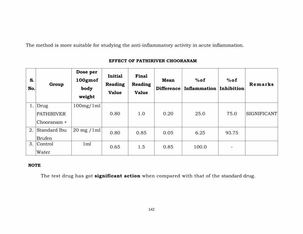
142
The method is more suitable for studying the anti-inflammatory activity in acute inflammation.
EFFECT OF PATHIRIVER CHOORANAM
S.
No. Group
Dose per 100gmof
body weight
Initial Reading
Value
Final Reading
Value
Mean
Difference
%of
Inflammation
%of
Inhibition Remarks
1. Drug
PATHIRIVER
Chooranam +
100mg/1ml
0.80 1.0 0.20 25.0 75.0 SIGNIFICANT
2. Standard Ibu
Brufen
20 mg /1ml 0.80 0.85 0.05 6.25 93.75
3. Control
Water
1ml 0.65 1.5 0.85 100.0 -
NOTE
The test drug has got significant action when compared with that of the standard drug.
143
CHRONIC ANTI-INFLAMMATORY ACTIVITY ON
RATS BY COTTON -PALLETS GRANULOMA METHOD
AIM
To study the chronic anti-inflammatory activity of the
drug, PATHIRIVER CHOORANAM in the rats by cotton pellets
implantation (granuloma) methods.
PREPARATION OF THE TEST DRUG
1 gm of PATHIRIVER Chooranam was dissolved in 10 ml of
water. A dose 1ml was given to each rat. This 1ml contains 10 mg
of test drug.
PROCEDURE
Cotton pellets each weighing 10mg were prepared and
sterilized in the autoclave for about one hour under 15 pounds
atmospheric pressure.9 albino rats weighing between 150-200gms
were selected and divided into 3 groups each containing 3 rats.
Each rat was anaesthetized with ether and cotton pellets were
implanted subcutaneously in the groin two on each side.
From the day of implantation first group was kept as control
by giving distilled water 2 ml/100mg and second group of animals
received PATHIRIVER Chooranam in a dose of 100mg / kg of body
weight. The standard group of animals received paracetamol in a
dose of 20mg / 100gm of body weight.
On the eighth- day the rats were sacrificed and the pellets
were removed, dried to concordant weight and weighed. They were
put in an incubator at 60 - 80°C and then the weight of the
granulation tissue was determined separately.
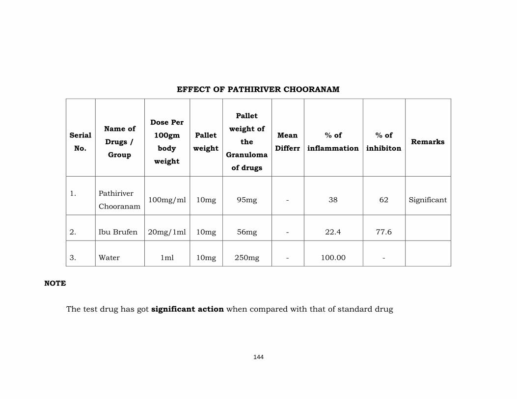
144
EFFECT OF PATHIRIVER CHOORANAM
Serial
No.
Name of
Drugs /
Group
Dose Per
100gm
body
weight
Pallet
weight
Pallet
weight of
the
Granuloma
of drugs
Mean
Differr
% of
inflammation
% of
inhibiton Remarks
1. Pathiriver
Chooranam 100mg/ml 10mg 95mg - 38 62 Significant
2. Ibu Brufen 20mg/1ml 10mg 56mg - 22.4 77.6
3. Water 1ml 10mg 250mg - 100.00 -
NOTE
The test drug has got significant action when compared with that of standard drug

145
ANALGESIC ACTIVITY OF PATHIRIVER
CHOORANAM
AIM
To study the analgesic effect of PATHIRIVER Chooranam on
albino rat by hot water bath method.
DRUG PREPARATION
1gm of PATHIRIVER Chooranam was dissolved in 10ml of
distilled water and 1ml was given to each rat. This 1 ml contains
100 mg of PATHIRIVER Chooranam.
PROCEDURE
Three groups of healthy albino rats of both sexes were
selected, each group having three rats. Each rat is weighing
between 80-100 gm. The hot water bath is maintained at the
temperature of 55°c.
The tail was dip into the bath, and time taken for each rat to
remove its tail from the hot water bath was noted. The rat which
takes more than 5 seconds for removal of its tail from hot water
bath was excluded from the experiment.
First group was kept as control by giving distilled water of
1ml/100mg of body weight.
The second group was given paracetamol 20mg/100gm of
body weight and kept as standard. Third group was given test
drug PATHIRIVER Chooranam 200mg/kg of body weight.
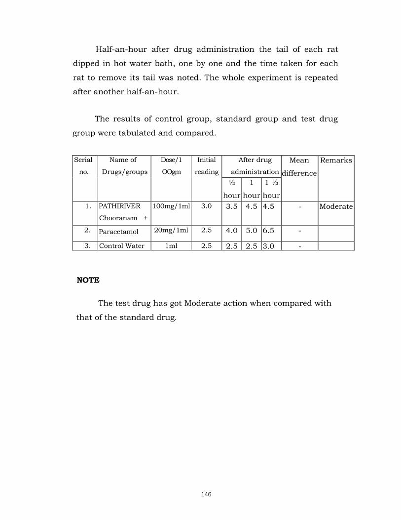
146
Half-an-hour after drug administration the tail of each rat
dipped in hot water bath, one by one and the time taken for each
rat to remove its tail was noted. The whole experiment is repeated
after another half-an-hour.
The results of control group, standard group and test drug
group were tabulated and compared.
Serial
no. Name of
Drugs/groups Dose/1
OOgm
Initial
reading
After drug
administration Mean
difference
Remarks
½
hour
1
hour
1 ½
hour 1. PATHIRIVER
Chooranam +
100mg/1ml 3.0 3.5 4.5 4.5 - Moderate
2. Paracetamol 20mg/1ml 2.5 4.0 5.0 6.5 -
3. Control Water 1ml 2.5 2.5 2.5 3.0 -
NOTE
The test drug has got Moderate action when compared with
that of the standard drug.
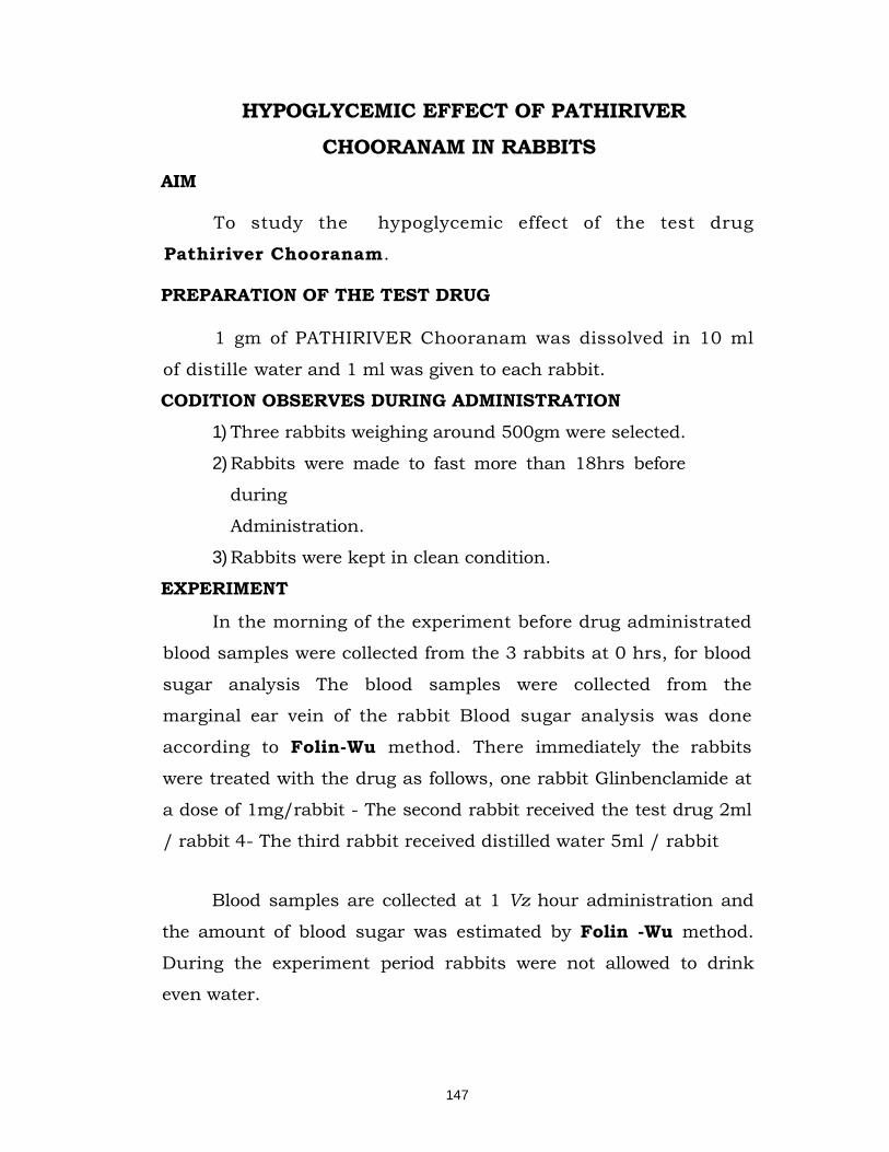
147
HYPOGLYCEMIC EFFECT OF PATHIRIVER
CHOORANAM IN RABBITS AIM
To study the hypoglycemic effect of the test drug
Pathiriver Chooranam.
PREPARATION OF THE TEST DRUG
1 gm of PATHIRIVER Chooranam was dissolved in 10 ml
of distille water and 1 ml was given to each rabbit.
CODITION OBSERVES DURING ADMINISTRATION
1) Three rabbits weighing around 500gm were selected.
2) Rabbits were made to fast more than 18hrs before
during
Administration.
3) Rabbits were kept in clean condition.
EXPERIMENT
In the morning of the experiment before drug administrated
blood samples were collected from the 3 rabbits at 0 hrs, for blood
sugar analysis The blood samples were collected from the
marginal ear vein of the rabbit Blood sugar analysis was done
according to Folin-Wu method. There immediately the rabbits
were treated with the drug as follows, one rabbit Glinbenclamide at
a dose of 1mg/rabbit - The second rabbit received the test drug 2ml
/ rabbit 4- The third rabbit received distilled water 5ml / rabbit
Blood samples are collected at 1 Vz hour administration and
the amount of blood sugar was estimated by Folin -Wu method.
During the experiment period rabbits were not allowed to drink
even water.
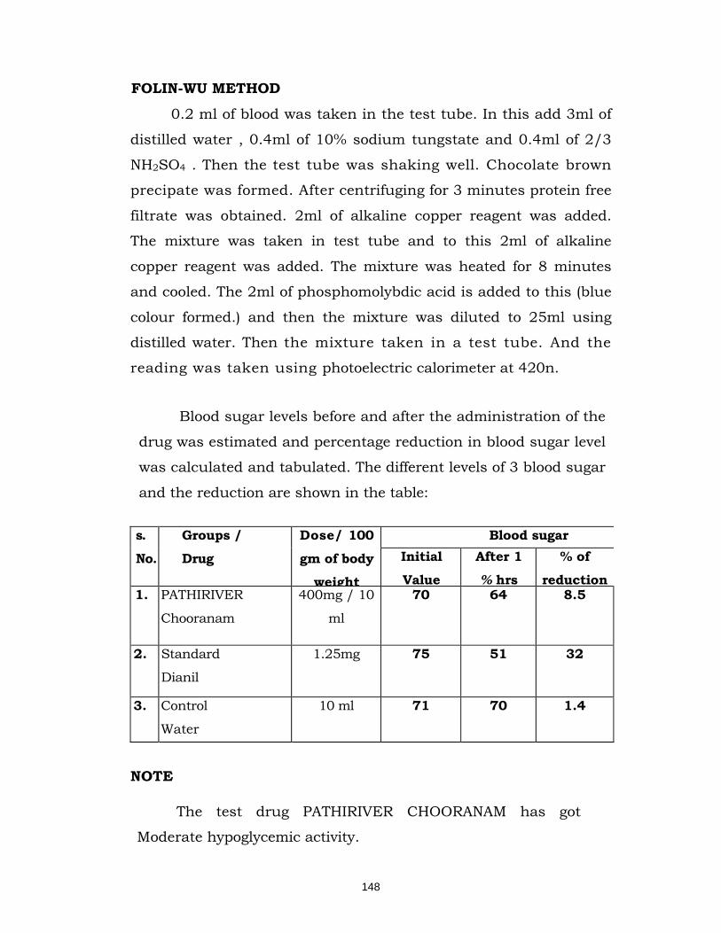
148
FOLIN-WU METHOD
0.2 ml of blood was taken in the test tube. In this add 3ml of
distilled water , 0.4ml of 10% sodium tungstate and 0.4ml of 2/3
NH2SO4 . Then the test tube was shaking well. Chocolate brown
precipate was formed. After centrifuging for 3 minutes protein free
filtrate was obtained. 2ml of alkaline copper reagent was added.
The mixture was taken in test tube and to this 2ml of alkaline
copper reagent was added. The mixture was heated for 8 minutes
and cooled. The 2ml of phosphomolybdic acid is added to this (blue
colour formed.) and then the mixture was diluted to 25ml using
distilled water. Then the mixture taken in a test tube. And the
reading was taken using photoelectric calorimeter at 420n.
Blood sugar levels before and after the administration of the
drug was estimated and percentage reduction in blood sugar level
was calculated and tabulated. The different levels of 3 blood sugar
and the reduction are shown in the table:
s.
No.
Groups /
Drug
Dose/ 100
gm of body
weight
Blood sugar Initial
Value
After 1
% hrs
% of
reduction 1. PATHIRIVER
Chooranam
400mg / 10
ml
70 64 8.5
2. Standard
Dianil
1.25mg 75 51 32
3. Control
Water
10 ml 71 70 1.4
NOTE
The test drug PATHIRIVER CHOORANAM has got
Moderate hypoglycemic activity.

149
ACUTE ANTI INFLAMMATORY STUDY OF MEDICATED
VENNAI (EXTERNALLY)
AIM
To study the anti inflammatory (acute) activity of
MEDICATED VENNAI.
PROCEDURE
Acute anti-inflammatory activity of Medicated Vennai was
studied in healthy albino rats, weighing between 100-150 gems. For
studying acute-inflammation rat hind-paw technique was used.
Six albino rats were selected and divided into two groups.
Each containing three rats. To the first group distilled water was
given and kept as control. Before the application of the drug, the
hind-paw volumes of all rats were measured. This was done by
dipping the hind-paw up to the tibio-tarsal junction in mercury
plethysmography.
Subcutaneous injection of 0.1 ml of 1% carragenin (w/v) in
water was made into plantar surface of both the hind-paw of each
rat. To the test group Medicated Vennai was topically applied
frequently over the inflamed surface in a thin layer. To the control
group, no drug was applied over the inflamed surface. One-and-half
the injection the hind-paw volume was measured once again. The
difference between the initial and final volumes would show the
amount of inflammation.
Taking the volume in the control group as 100% of
inflammation or anti-inflammatory effect of the group is calculated.
Tabulation of the results observed.
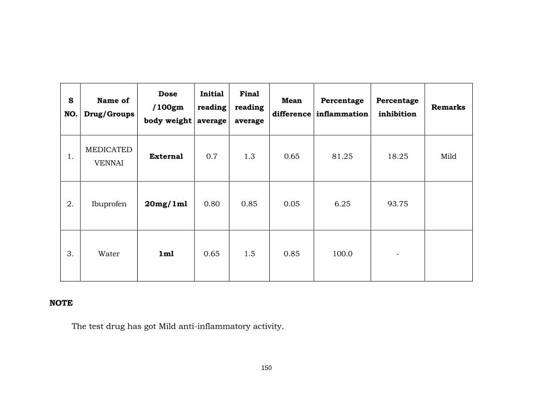
150
S
NO.
Name of
Drug/Groups
Dose
/100gm
body weight
Initial
reading
average
Final
reading
average
Mean
difference
Percentage
inflammation
Percentage
inhibition Remarks
1. MEDICATED
VENNAI External 0.7 1.3 0.65 81.25 18.25 Mild
2. Ibuprofen 20mg/1ml 0.80 0.85 0.05 6.25 93.75
3. Water 1ml 0.65 1.5 0.85 100.0 -
NOTE
The test drug has got Mild anti-inflammatory activity.
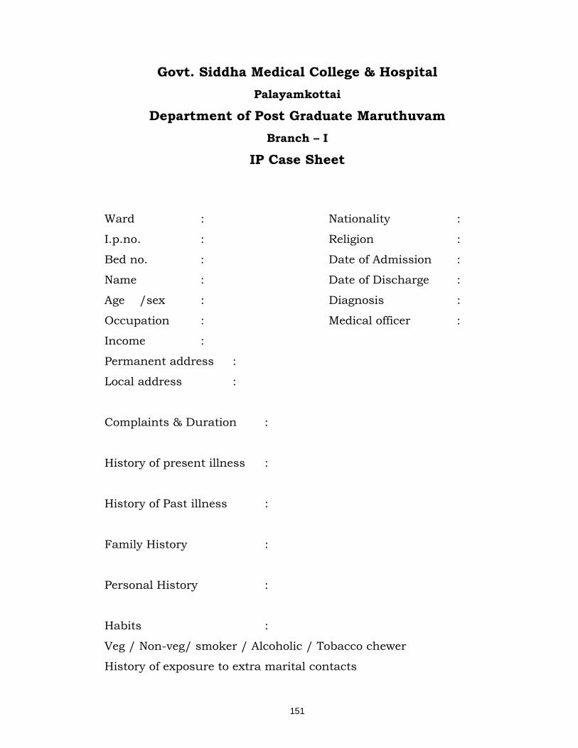
151
Govt. Siddha Medical College & Hospital Palayamkottai
Department of Post Graduate Maruthuvam Branch – I
IP Case Sheet
Ward : Nationality :
I.p.no. : Religion :
Bed no. : Date of Admission :
Name : Date of Discharge :
Age /sex : Diagnosis :
Occupation : Medical officer :
Income :
Permanent address :
Local address :
Complaints & Duration :
History of present illness :
History of Past illness :
Family History :
Personal History :
Habits :
Veg / Non-veg/ smoker / Alcoholic / Tobacco chewer
History of exposure to extra marital contacts
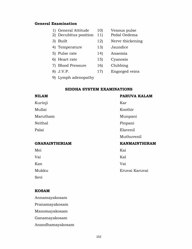
152
General Examination 1) General Attitude 10) Venous pulse 2) Decubitus position 11) Pedal Oedema 3) Built 12) Nerve thickening 4) Temperature 13) Jaundice 5) Pulse rate 14) Anaemia 6) Heart rate 15) Cyanosis 7) Blood Pressure 16) Clubbing 8) J.V.P. 17) Engorged veins 9) Lymph adenopathy
SIDDHA SYSTEM EXAMINATIONS
NILAM PARUVA KALAM
Kurinji Kar
Mullai Koothir
Marutham Munpani
Neithal Pinpani
Palai Elavenil
Muthuvenil
GNANAINTHIRIAM KANMAINTHIRAM Mei Kai
Vai Kal
Kan Vai
Mukku Eruvai Karuvai
Sevi
KOSAM
Annamayakosam
Pranamayakosam
Manomayakosam
Ganamayakosam
Anandhamayakosam
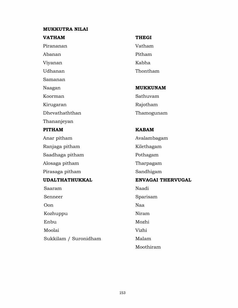
153
MUKKUTRA NILAI VATHAM THEGI
Pirananan Vatham
Abanan Pitham
Viyanan Kabha
Udhanan Thontham
Samanan
Naagan MUKKUNAM
Koorman Sathuvam
Kirugaran Rajotham
Dhevathaththan Thamogunam
Thananjeyan
PITHAM KABAM
Anar pitham Avalambagam
Ranjaga pitham Kilethagam
Saadhaga pitham Pothagam
Alosaga pitham Tharpagam
Pirasaga pitham Sandhigam
UDALTHATHUKKAL ENVAGAI THERVUGAL Saaram Naadi
Senneer Sparisam
Oon Naa
Kozhuppu Niram
Enbu Mozhi
Moolai Vizhi
Sukkilam / Suronidham Malam
Moothiram
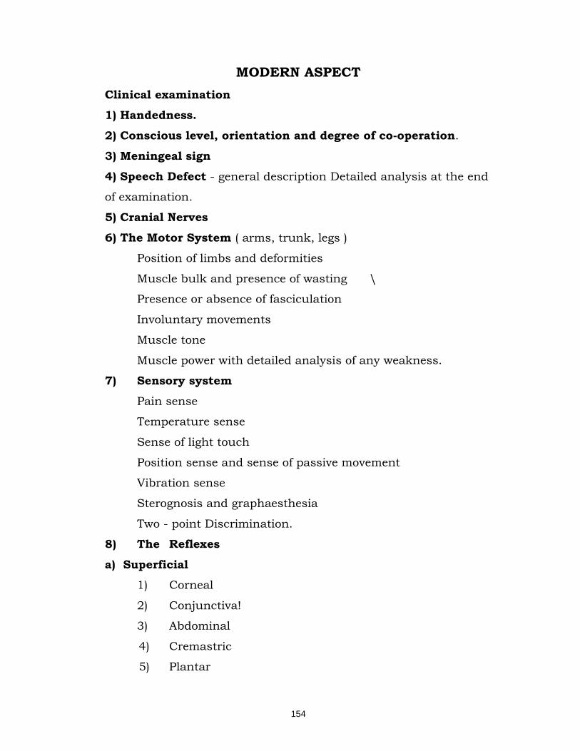
154
MODERN ASPECT Clinical examination
1) Handedness.
2) Conscious level, orientation and degree of co-operation.
3) Meningeal sign
4) Speech Defect - general description Detailed analysis at the end
of examination.
5) Cranial Nerves
6) The Motor System ( arms, trunk, legs )
Position of limbs and deformities
Muscle bulk and presence of wasting \
Presence or absence of fasciculation
Involuntary movements
Muscle tone
Muscle power with detailed analysis of any weakness.
7) Sensory system
Pain sense
Temperature sense
Sense of light touch
Position sense and sense of passive movement
Vibration sense
Sterognosis and graphaesthesia
Two - point Discrimination.
8) The Reflexes
a) Superficial 1) Corneal
2) Conjunctiva!
3) Abdominal
4) Cremastric
5) Plantar
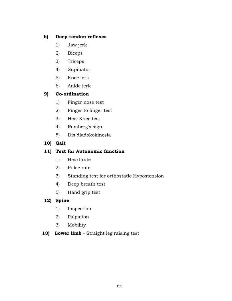
155
b) Deep tendon reflexes
1) Jaw jerk
2) Biceps
3) Triceps
4) Supinator
5) Knee jerk
6) Ankle jerk
9) Co-ordination
1) Finger nose test
2) Finger to finger test
3) Heel Knee test
4) Romberg's sign
5) Dis diadokokinesia
10) Gait 11) Test for Autonomic function
1) Heart rate
2) Pulse rate
3) Standing test for orthostatic Hypostension
4) Deep breath test
5) Hand grip test
12) Spine
1) Inspection
2) Palpation
3) Mobility
13) Lower limb - Straight leg raising test

156
Investigation Blood
Total Count Sugar
Differential Count Urea
ESR Creatinine
Hb Cholesterol
Complete haemogram Liver function test
Peripheral smear for cytology Serum protein
Haemoparasite Skin Clipping
AFB
Motion Urine
Ova Albumin
Cyst Sugar
Occult Blood Deposit
Nerve Conduction Study
Differential Diagnosis
Case Summary
Diagnosis
Treatment
Prognosis
Diet
Advices
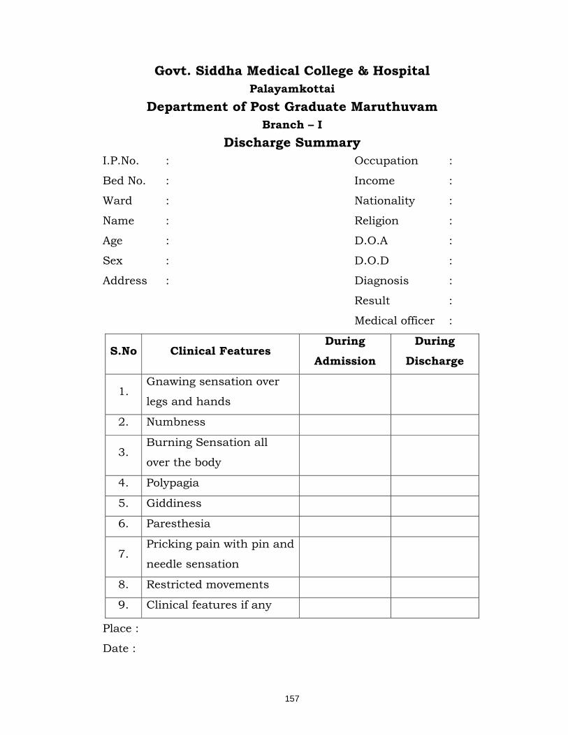
157
Govt. Siddha Medical College & Hospital Palayamkottai
Department of Post Graduate Maruthuvam Branch – I
Discharge Summary I.P.No. : Occupation :
Bed No. : Income :
Ward : Nationality :
Name : Religion :
Age : D.O.A :
Sex : D.O.D :
Address : Diagnosis :
Result :
Medical officer :
S.No Clinical Features During
Admission
During
Discharge
1. Gnawing sensation over
legs and hands
2. Numbness
3. Burning Sensation all
over the body
4. Polypagia
5. Giddiness
6. Paresthesia
7. Pricking pain with pin and
needle sensation
8. Restricted movements
9. Clinical features if any
Place :
Date :

158
Govt. Siddha Medical College & Hospital Palayamkottai
Department of Post Graduate Maruthuvam Branch – I
OP Case Sheet OP No : Treatment starting date :
Name : End of Treatment date :
Age / Sex : No. of days treated :
Occupation : Diagnosis :
Income : Results :
Address : Medical Officer :
Complaints and Duration:
General Examination
Temperature : Anaemia :
Blood Pressure : Jaundice :
Heart rate : Cyanosis :
Pulse rate : Clubbing :
Respiratory rate : Others if any :
In siddha aspects Nilam : Dhehi :
Mukkunam : Ezhu Udal Kattugal :
Imporikal : Mukkutram :
Kanmendhriyam : Envagai Thervugal :
Kosam : Neerkuri :
Paruvakalam : Neikuri :
Mukkunam :

159
Examination of Sensory system Pain sense
Temperature sense
Sense of light touch
Position sense and sense of passive movement
Vibration sense
Sterognosis and graphaesthesia
Two - point Discrimination
Ankle joint Reflex
Investigation
Blood
Total Count : Sugar :
Differential Count : Urea :
ESR : Creatinine :
Hb : Cholesterol :
Complete haemogram : Liver function test :
Peripheral smear for cytology Serum protein
Haemoparasite Skin Clipping
AFB
Motion Urine
Ova Albumin
Cyst Sugar
Occult Deposit
Nerve Conduction Study

160
BIBLIOGRAPHY
Siddha Reference Books
Agasthiar Naadi Sasthiram – Mr. S.B. Ramachandran, 2002 print
Agasthiar – 2000 Dr.S. Venkat rajan, reprint 1998
Agasthiar Vallathy – 600 – R.C. Mohan, reprint April 2001
Agasthiyar Kanma Kandam - Mr. S.B. Ramachandran, May
1996
Anuboga vaidhya Deva Ragasiam – Mr.J. Seetha Ram Prasad,
1991 print
Dr.S.N.Yoga Narasimhan Medicinal plants– 2000 Ist print
Gosaye Anuboga Vaidhya brama Ragasiyam - Mr. S.B.
Ramachandran, March 1999
Gunapadam Muligai Vaguppu – Murugesa Mudaliyar reprint
1998.
Gunavagadam Noyinsaram – Dr.S. Prema and Dr.S.
Arangarasan, 2006 reprint
Manmurikiyam Kaiyettu Pirathi
Medicinal plants of India vol.II, Vol III, Vol IV
Noinadal Noi Muthal Nadal Vol.I 2003 & Vol II 2003 Reprint –
Dr.M.Shanmugavelu
161
Prana Raksharmirtha sindhu – T.R. Mahadeva Pandithar
Nov1999 print
Sarabendhirar pitha roha sihitchai IInd print 1990
Shitcha Rathina theepam – Mr.C.Kannusamy Pillai 8th Edition
Siddha Maruthuvanga Churrukkam 2003 Reprint by
K.S.Uthamarayan
Siddha maruthuvam - 2005 reprint – Dr.K.M.Kuppusamy
Mudaliyar
Thanvandri Nigandu – 300 by Thanvandari
Tamil Dictionary – by T.V.Sambasivam pillai, Vol - V
Thirukkural – By Thiruvalluvar
Thirumoolar Naadi nool by Thirumoolar
Udal Thathuvam – Dr. Venugopal H.P.I.M, 2003 reprint
Yugi vaithya chinthamani – Ist Edition – 1998 R.Thiyagarajan
Yugi Muni Vaithya Kaviyam – by R.C.Mohan, March 2002
reprint
Uyir Kakkum Siddha Maruthuvam – Aathma Ratchamirtha
Sara Sangiraham, Oct – 2002 print
162
MODERN REFERENCE BOOKS
Brian’s Disease of Nervous System – 10th edition
Davidson’s Principle and Practice of Medicine’s 20th edition
Essentials of Human Physiology – by K. sembulingam, IInd
edition
Golwalla Medicine For Students – 17th edition
Gray’s Anatomy – 13th edition
Harrison’s Principles of internal medicine – Vol I & II, 14th
edition
How to Examine a patient by Menino –De- Soza
Human Anatomy – by T.S.Renga Nathan 5th edition
Hutchison’s Clinical Methods – Micheal Swash – 20th edition
Manual of Practical Medicine – by R.Alagappan, III edition
Neuro Anatomy – A.K.Datta First print 1995
Neurological Differential Diagnosis - By John Patton 14th
reprint 1996
Neurology secrets - by Loren A Rolak, IInd edition
Oxford Text Book of Medicine Vol.III
Text Book of Medicine by Cecil – Vol I 19th edition
Text Book of Medicine - by K.V.Krishna das, IIIrd edition
Text book of Pathology – by Harsh Mohan