A CLINICOEPIDEMIOLOGICAL STUDY ON ADVERSE ...
115
1 A CLINICOEPIDEMIOLOGICAL STUDY ON ADVERSE CUTANEOUS DRUG REACTIONS Dissertation Submitted in fulfillment of the university regulations for MD DEGREE IN DERMATOLOGY, VENEREOLOGY AND LEPROSY (BRANCH XII A) THE TAMILNADU DR.M.G.R. MEDICAL UNIVERSITY CHENNAI
-
Upload
khangminh22 -
Category
Documents
-
view
3 -
download
0
Transcript of A CLINICOEPIDEMIOLOGICAL STUDY ON ADVERSE ...
1
A CLINICOEPIDEMIOLOGICAL STUDY ON
ADVERSE CUTANEOUS DRUG REACTIONS
Dissertation Submitted in
fulfillment of the university regulations for
MD DEGREE IN
DERMATOLOGY, VENEREOLOGY AND
LEPROSY
(BRANCH XII A)
THE TAMILNADU DR.M.G.R. MEDICAL UNIVERSITY
CHENNAI

2
APRIL 2012
CERTIFICATE
This is to certify that this dissertation entitled ‘A CLINICO
EPIDEMIOLOGICAL STUDY ON ADVERSE CUTANEOUS DRUG
REACTIONS’ submitted by Dr. Koregol Savita to The Tamil Nadu
Dr.M.G.R. Medical University, Chennai is in partial fulfillment of the
requirement for the award of M.D., [DERMATO VENEREO LEPROLOGY]
and is a bonafide research work carried out by her under direct supervision and
guidance.
Dr. A. S. Krishnaram. M.D.,D.D., Dr. D. Amal Raja. M.D.,D.V., Professor and Head Professor and Head Department of Dermatology, Department of STD, Madurai Medical College & Madurai Medical College & Government Rajaji Hospital, Government Rajaji Hospital, Madurai. Madurai.
DECLARATION
3
I, Dr. Koregol Savita solemnly declare that I carried out this work on ‘A
CLINICOEPIDEMIOLOGICAL STUDY ON ADVERSE CUTANEOUS
DRUG REACTIONS’’ at Department Of Dermatology, Government Rajaji
Hospital during the period of Oct 2009 – Sep 2011.
I also declared this bonafide work or a part of this work was not submitted
by me or any other for any award, degree and diploma to any university, board
either in India or abroad.
This is submitted to The Tamil Nadu Dr. M.G.R Medical University,
Chennai in partial fulfillment of the rules and regulation for M.D.,[D.V.L]
Degree examination.
Govt. Rajaji Hospital. Dr. Koregol Savita. Madurai.
ACKNOWLEDGEMENT
I am extremely thankful to Dr. A. EDWIN JOE., M.D, Dean, Madurai
Medical College, and Medical superintendent, Government Rajaji Hospital,
Madurai for permitting me to use the hospital materials for this study.
4
I express my sincere and heartfelt gratitude to Prof. Dr. A.S.Krishnaram
M.D.,D.D., Professor and Head of the Department of Dermatology, Madurai
Medical College, Madurai, for his excellent guidance and supervision for this
dissertation work. His commitment, devotion and perfection in work gave me
the drive for completing the project successfully. I profoundly thank Prof. Dr. S.
Krishnan, M.D.,D.D., who has always guided me, by example and valuable
words of advice through the conduct of the study and also during my
postgraduate course.
My heartful thanks to my Prof. Dr. G. Geetharani, M.D.,DNB, for her
valuable support and guidance throughout the study. I proudly thank Prof. Dr.
D. Amal Raja, M.D.,D.V., Head of the Department of Venereology for his
valuable guidance.
I express my deep sense of gratitude and thanks to my teachers Dr.
Senthil kumar, Dr. Kothandaraman, Dr. Sathesh, Dr. Balaji Adityan
Assistant professors, for their valuable guidance, timely advise, and constant
encouragement.
I would also like to acknowledge my thanks to all the Assistant
professors, of Department of STD, for their constant support during the period of
my study.

5
I would like to convey my regards to my family members who have
always stood by me in my career.
I owe a lot of thanks to my patients without whom this study would not
have been possible , and the authors, who have worked on this subject, from
whose wisdom and experience, I have been benefitted immensely.
CONTENTS

6
S.NO. TITLES PAGE NO.
1. INTRODUCTION
2. AIM OF THE STUDY
3. REVIEW OF LITERATURE
4. MATERIALS AND METHODS
5. OBSERVATION AND RESULTS
6. DISCUSSION
7. SUMMARY
8. CONCLUSION
9. BIBLIOGRAPHY
10. CLINICAL PHOTOGRAPHS
11. PROFORMA
12. MASTER CHART
13. KEY TO MASTER CHART

7
INTRODUCTION
A drug may be defined as a chemical substance, or combination of
substances, administered for the investigation, prevention or treatment of
diseases or symptoms, real or imagined. An adverse drug reaction (ADR) may
be defined as an undesirable clinical manifestation resulting from administration
of a particular drug; this includes reactions due to overdose, predictable side
effects and unanticipated adverse manifestations. ADR can also be defined as
‘an appreciably harmful or unpleasant reaction, resulting from an intervention
related to the use of a medicinal product, which predicts hazard from future
administration and warrants prevention or specific treatment, or alteration of the
dosage regimen, or withdrawal of the product’. ADRs are underreported and are
an underestimated cause of morbidity and mortality; it has been estimated that
ADRs represent the fourth to the sixth leading cause of death[1],[2]
Adverse Cutaneous Drug Reactions (ACDRs) are probably the most
frequent manifestations of all drug sensitivity reactions although their incidence
is difficult to determine. Very few published studies[3],[4] have assessed the
epidemiological and clinical features of drug reactions in India and still fewer in
South India[3]. Hence this study was undertaken to assess the pattern and clinical
features of ACDRs and common drugs causing them in South Tamil nadu.
8
AIM OF THE STUDY
1. To study the clinicoepidemiological pattern of various adverse cutaneous drug
reactions in patients attending SKIN OPD, Government Rajaji Hospital.
2. To study the common drugs causing adverse cutaneous drug reactions.

9
REVIEW OF LITERATURE
EPIDEMIOLOGICAL ASPECTS OF ADVERSE CUTANEOUS DRUG
REACTIONS:
Incidence:
The exact incidence and prevalence of cutaneous adverse drug reactions
are unknown, but the overall prevalence is believed to be less than 1% of the
general population. Estimates between 2.0% to 2.4% have been reported
worldwide.[68] Out of this 7% is due to antibiotics. An incidence of 0.38% has
been reported from India .[69]
Immunosuppressed patients are most frequently affected.[1]
Age:
Though ADRs can affect any age group ADRs occur in 6–17% of children
admitted to specialist paediatric hospitals ,those over 60 years of age comprise
only 12% of the population in the USA, 33% of all drugs are prescribed for this
age group,[1] [70] and the elderly have a significantly higher incidence of ADRs,
related to decreased organ reserve capacity, altered pharmacokinetics and
pharmacodynamics, and polypharmacy.
Gender:
Women are more likely than men to develop ADRs.[1]
10
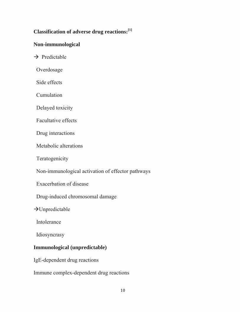
Classification of adverse drug reactions:[1]
Non-immunological
Predictable
Overdosage
Side effects
Cumulation
Delayed toxicity
Facultative effects
Drug interactions
Metabolic alterations
Teratogenicity
Non-immunological activation of effector pathways
Exacerbation of disease
Drug-induced chromosomal damage
Unpredictable
Intolerance
Idiosyncrasy
Immunological (unpredictable)
IgE-dependent drug reactions
Immune complex-dependent drug reactions
11
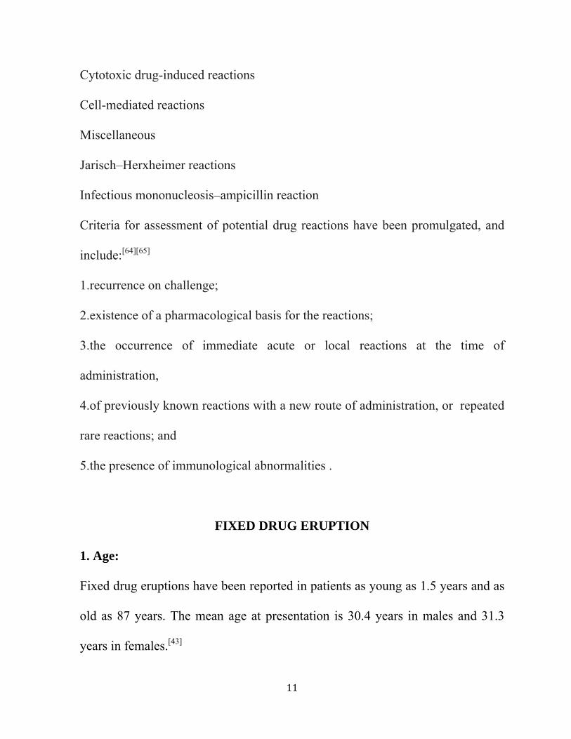
Cytotoxic drug-induced reactions
Cell-mediated reactions
Miscellaneous
Jarisch–Herxheimer reactions
Infectious mononucleosis–ampicillin reaction
Criteria for assessment of potential drug reactions have been promulgated, and
include:[64][65]
1.recurrence on challenge;
2.existence of a pharmacological basis for the reactions;
3.the occurrence of immediate acute or local reactions at the time of
administration,
4.of previously known reactions with a new route of administration, or repeated
rare reactions; and
5.the presence of immunological abnormalities .
FIXED DRUG ERUPTION
1. Age:
Fixed drug eruptions have been reported in patients as young as 1.5 years and as
old as 87 years. The mean age at presentation is 30.4 years in males and 31.3
years in females.[43]

12
2. Gender :
One large study of 450 patients by mahboob et al.[43] revealed a male to female
ratio of 1:1.1 for fixed drug eruptions.
3.Race:
Fixed drug eruptions have no known racial predilection. A genetic susceptibility
to developing a fixed drug eruption with an increased incidence of HLA-B22 is
possible. [44]
4. Pathophysiology:
Although the exact mechanism is unknown, recent research suggests a cell-
mediated process that initiates both the active and quiescent lesions. The process
may involve an antibody-dependent, cell-mediated cytotoxic response. CD8+
effector/memory T cells play an important role in reactivation of lesions with re-
exposure to the offending drug[45]
The offending drug is thought to function as a hapten that preferentially binds to
basal keratinocytes, leading to an inflammatory response. Through liberation of
cytokines such as tumor necrosis factor-alpha, keratinocytes may locally up-
regulate expression of the intercellular adhesion molecule-1 (ICAM1).The up-
regulated ICAM1 has been shown to help T cells (CD4 and CD8) migrate to the
site of an insult.
The newly arriving and residential CD8 cells likely perpetuate tissue damage by

13
their production of the inflammatory cytokines interferon-gamma and tumor
necrosis factor-alpha. CD8 cells isolated from active lesions have also been
shown to express alpha E beta 7, a ligand for E-cadherin, which may further
contribute to the lymphocyte’s ability to localize to the epidermis. Other cell
surface molecules, such as CLA/alpha4beta1/CD4a, that bind E-selectin/vascular
cellular adhesion molecule-2/ICAM1 help to further attract CD8 cells to the
area.
Changes in cell surface markers allow vascular endothelium to select CD4 cells
for migration into active lesions. These regulatory CD4 cells likely produce
interleukin 10, which has been shown to help suppress immune function,
resulting in a resting lesion.As the inflammatory response dissipates, interleukin
15 expression from keratinocytes is thought to help ensure the survival of CD8
cells, helping them fulfill their effector memory phenotypes. Thus, when
reexposure to the drug occurs, a more rapid response develops in the exact
location of any prior lesions.[45]
5. History:
The initial eruption is often solitary and frequently located on the lip or genitalia.
Rarely, the eruption may be intraoral. Lesions are commoner on the limbs than
on the trunk; the hands and feet, genitalia and perianal areas are favoured sites.
Perioral and periorbital lesions may occur. Genital and oral mucous membranes

14
may be involved in association with skin lesions, or alone. With the initial fixed
drug eruption attack, a delay of up to 2 weeks may occur from the initial
exposure of the drug to the development of the skin lesion.Skin lesions develop
over a period of hours but require days to become necrotic. Lesions may persist
from days to weeks and then fade slowly to residual oval hyperpigmented
patches.[46]
The eruption may initially be morbilliform, scarlatiniform or erythema
multiforme-like; urticarial, nodular or eczematous lesions are less common.
A fixed drug eruption characteristically recurs in the same site or sites each time
the drug is administered; with each exposure, however, the number of involved
sites may increase. Subsequent reexposure to the medication results in a
reactivation of the site, with inflammation occurring within 30 minutes to 16
hours. The reactivation of old lesions also may be associated with the
development of new lesions at other sites.
Patients may not be cognizant that a drug, nutritional supplement, over the
counter medication, or, rarely, food (eg, fruits, nuts) triggered the skin problem.
They may be convinced that an insect, particularly a spider, may be the culprit.
A careful history is required to elicit the fact that a drug has been taken and is
temporally related to the onset of the eruption. Medications taken episodically,
such as pain relievers, antibiotics, or laxatives, are often to blame. When able to

15
be identified, patients often report ingestion of one the following types of
medications:[67]
• Analgesics
• Muscle relaxants
• Sedatives
• Anticonvulsants
• Antibiotics
In the case of isolated male genital fixed drug eruption (often affecting only the
glans penis), the drugs most commonly implicated in one series were co-
trimoxazole (trimethoprim– sulfamethoxazole), tetracycline and ampicillin .
Cross-sensitivity to related drugs may occur, such as between phenylbutazone
and oxyphenbutazone, between tetracycline type drugs, and between
anticonvulsants .
Local symptoms may include pruritus, burning, and pain.Systemic symptoms are
uncommon, but fever, malaise, nausea, diarrhea, abdominal cramps, anorexia,
and dysuria have been reported.
Further questioning may reveal prior episodes of fixed drug eruption, atopic
disease, or other past drug reactions. Family history may render a history of
atopy, drug reactions, or diabetes mellitus.
16
Several cases of fixed drug eruption on the genitalia have been reported in
patients who were not ingesting the drug but whose sexual partner was taking
the offending drug and the patient was exposed to the drug through sexual
contact.
6. Types: Several variants of fixed drug eruption have been described, based on
their clinical features and the distribution of the lesions.These include the
following:[47]
• Pigmenting fixed drug eruption
• Generalized or multiple fixed drug eruption
• Linear fixed drug eruption
• Wandering fixed drug eruption
• Nonpigmenting fixed drug eruption
• Bullous fixed drug eruption
• Eczematous fixed drug eruption
• Urticarial fixed drug eruption
• Erythema dyschromicum perstans–like fixed drug eruption
• Vulval & Oral fixed drug eruption
7. Differential diagnosis:
• Bullous Pemphigoid
• Cellulitis
17
• Drug-Induced Bullous Disorders
• Eczema
• Erythema Annulare Centrifugum
• Erythema Multiforme
• Herpes Simplex
• Lichen Planus
• Lichen Planus Actinicus
• Discoid lupus erythematoses
• Pemphigus Vulgaris , Pityriasis Rosea
• Postinflammatory Hyperpigmentation
8. Complications:
Hyperpigmentation is the most likely complication of a fixed drug eruption
(FDE). The potential for infection exists in the setting of multiple, eroded
lesions. Generalized eruptions have been reported following topical and oral
provocation testing.[48]
9. Prognosis:
The prognosis is very good, and an uneventful recovery should be expected. No
deaths due to fixed drug eruption have been reported. Residual
hyperpigmentation is very common, but this is less likely with the
nonpigmenting variant. As healing occurs, crusting and scaling are followed by

18
pigmentation, which may be very persistent and occasionally extensive,
especially in pigmented individuals; pigmentation may be all that is visible
between attacks.[49]
MACULOPAPULAR RASH
A drug reaction is suspected when a maculopapular eruption begins within 4 to
12 days of beginning a new medicine. Macules predominate initially, then
become confluent patches with papular areas within the patches.[17]
Proposed mechanism :
Morbilliform drug eruption is a non-immediate type IV allergic reaction
involving drug-specific T cells (CD4+) with direct cytotoxic effects and release
of pro-inflammatory factors.[1]
Clinical features: Most reactions develop within 7 to 8 days, though reactions to
aminopenicillins may develop over a longer period (>8 days) & in phenytoin it
takes around 3 weeks . This interval is the time needed for an immunological
(cell-mediated) delayed-type hypersensitivity reaction to occur.The eruption is
characteristically polymorphic, with confluent areas on the trunk, papular areas
on the arms, and purpuric areas on the feet.In maculopapular rash, features range
from a normal epidermis to local areas of basal cell degeneration with
eosinophilic infiltrat Dermis showed congested vessels, mild to moderate
19
perivascular/periappendageal infiltrate of mononuclear cells.[66]
Moderate pruritus, a low-grade fever, and general malaise may be present.
Mucous membranes are typically spared, and lymphadenopathy is mild if
present.
The eruption generally fades over 1 to 2 weeks without complication. Post-
inflammatory desquamation is common. Antibiotics (sulphonamides,
aminopenicillins, cephalosporins) and anticonvulsants are most commonly
implicated.[17]
Chemotherapeutic agents, particularly cytarabine, dacarbazine, hydroxyurea,
paclitaxel, and procarbazine, have also been associated with maculopapular
eruption. Epidermal growth factor receptor inhibitors (EGFRs; e.g., cetuximab)
also have a propensity to trigger cutaneous eruptions.
Treatment:
Prompt cessation of the causative drug results in resolution over 1-2 weeks. No
specific treatment is required but topical corticosteroids or oral antihistamines
may give symptomatic relief of itch.[17]
URTICARIA
Urticaria is a skin rash, also called hives, or nettle rash, which is often
accompanied by swelling and itching of the skin.

20
Classification:
-Immune-mediated
IgE-mediated: penicillin
Complement and immune complex mediated: penicillin, immunoglobulins,
whole blood.
-Nonallergic urticarial ACDR
Analgesics/NSAIDs inhibit/block cyclooxygenase in prostaglandin synthesis
Radio contrast media
ACE inhibitors: inhibition of kinin metabolism
Calcium channel blockers
Drugs releasing histamine[13]
Clinical features:
Time from Initial Drug Exposure to Appearance of Urticaria : IgE-Mediated
Initial sensitization, usually 7–14 days; urticaria may occur while the drug is still
being administered or after it is discontinued. In previously sensitized
individuals, usually within minutes or hours. Skin Symptoms consist of Pruritus,
burning of palms/ soles, auditory canal. With airway edema, difficulty breathing.
Drug-induced urticaria may be allergic (immunologically mediated) or
pseudoallergic (nonimmunologically mediated).
Common drugs: Penicillins , Cephalosporins , Sulphonamides , Cytostatic

21
agents, ACE inhibitors, Calcium channel blockers
Drug-induced urticaria is seen in association with anaphylaxis, angio-oedema
and serum sickness.[9]
Differential diagnosis :
Is of acute edematous red pruritic plaque(s):
a) Allergic contact dermatitis (poison ivy, poison oak dermatitis)
b) Cellulitis
c) insect bite(s).
Management :
The offending drug should be identified and withdrawn as soon as possible.
Prevention: Previously Sensitized Individuals. The patient should carry
information listing drug sensitivities (wallet card, bracelet). Radiographic
Contrast Media Avoid use of contrast media known to have caused prior
reaction. If not possible, pretreat patient with antihistamine and prednisone (1
mg/kg) 30–60 min before contrast media exposure.
Treatment of Acute Severe Urticaria/Anaphylaxis :
Epinephrine : 0.3–0.5 mL of a 1:1000 dilution subcutaneously, repeated in 15–
20 min. Maintain airway, Intravenous access, Antihistamines H1 blockers or
H2 blockers or combination.
Systemic Glucocorticoids : Intravenous Hydrocortisone or methylprednisolone

22
for severe symptoms. Oral Prednisone 70 mg, tapering by 10 or 5 mg daily over
1–2 weeks, is usually adequate.[13]
ERYTHRODERMA
It is most frequently caused by antibiotics, antiepileptic drugs and NSAIDs. [50]
Drug-induced erythroderma accounts for about 10% of all erythrodermas.
Definition: generalized erythema of the skin (more than 90% of the body surface
area) accompanied by a variable degree of scaling.
Pathogenesis: The rise in adhesion molecule expression (VCAM-1, ICAM-1, E-
selectin and P-selectin) seen in exfoliative dermatitis stimulates dermal
inflammation, which may lead to epidermal proliferation and increased
production of inflammatory mediators. The complex interaction of cytokines and
cellular adhesion molecules such as interleukin-1, -2 and -8; intercellular
adhesion molecule-I (ICAM-I); and tumor necrosis factor (TNF) results in
significantly elevated epidermal turnover rate, leading to above normal mitotic
rate. The amount of germinative cells increases and the transit time of
keratinocytes through the epidermis decreases, causing loss of more cellular
material from the surface.
Clinical features:
The pattern observed is erythematous patches, which increase in size and

23
coalesce to form extensive areas of erythema, and eventually spread to involve
most of the skin surface.Some studies have shown sparing of the nose and
paranasal areas, and this has been described as the "nose sign".
The epidermis appears thin, giving a glossy appearance to the skin. Once
erythema has been established, white or yellow scales develop that progress to
give the skin a dry appearance with a dull scarlet and grey hue. Induration and
thickening of the skin from edema and lichenification may provoke a sensation
of severe skin tightness in the patient. The skin is bright red, dry, scaly and
warm to touch.
Some patients may experience involvement of their palms and soles, with hair
loss and nail shedding. Involved nails are thick, lusterless, dry, brittle, and show
ridging of the nail plate. Subungual hyperkeratosis, distal onycholysis, splinter
hemorrhages occur; and sometimes, the nails may shed. Shelley described
alternating bands of nail plate discontinuity and leukonychia in drug-induced
erythroderma.
Laboratory investigations:
Laboratory findings in the erythrodermic patient are usually nonspecific.
Common abnormalities are mild anemia, leukocytosis with eosinophillia,
elevated sedimentation rate, decreased serum albumin, increased uric acid,
abnormal serum protein electrophoresis with polyelevation in the gamma
24
globulin region and elevated IgE levels.
Management:
To identify & stop the offending drug. Systemic corticosteroids , nutritional
replacement, fluid and electrolyte losses. Local skin care measures should be
employed, such as oatmeal baths as well as wet dressings to weeping or crusted
sites followed by the application of bland emollients and low potency
corticosteroids. Known precipitants and irritants are to be avoided.
Secondary infections are treated with antibiotics. Edema in dependent areas,
such as in periorbital and pedal areas, may require diuretics.
Hemodynamic or metabolic instability should be addressed adequately. Serum
protein, electrolyte and blood urea levels should be monitored.[51]
URTICARIA WITH ANGIOEDEMA
In the past this was called giant urticaria or angioneurotic edema or Quincke's
edema. Angioedema is a swelling of the deep layers of the subcutaneous or
submucosal tissue or both. Most commonly it occurs on the lips, tongue, face,
hands or feet.
The oedema is caused by an increase in capillary leakage as a result of
inflammatory mediators. This can be a manifestation of Type I allergic
reactions; or a consequence of deficiency in C1-esterase inhibitor or because of
failure to metabolise mediators such as bradykinin. Drugs are implicated in all
25
three mechanisms, although C1-esterase inhibitor deficiency is usually
hereditary.
Drug most commonly implicated:
ACE-inhibitors , Bupropion , vaccines , selective serotonin reuptake inhibitors
(SSRIs) , COX-II inhibitors , angiotensin II antagonists , other antidepressants ,
non-steroidal anti-inflammatory drugs (NSAIDs), statins , proton pump
inhibitors.
Management:
Primary management is to ensure an adequate airway. Adrenaline,
antihistamines, and corticosteroids may be needed. Endotracheal intubation or
tracheotomy is required in severe cases.[52]
DRESS
[Drug Rash with Eosinophilia & Systemic Symptoms]
Synonym:
Drug Hypersensitivity Syndrome[DHS]
drug-induced delayed multiorgan hypersensitivity syndrome (didmohs)
Definition: Long-lasting papulopustular or erythematous skin eruption often
progressing to exfoliative dermatitis, with fever, lymphadenopathy, and visceral
involvement (hepatitis, pneumonitis, myocarditis, pericarditis, nephritis).
Incidence/prevalence:For phenytoin, carbamazepine, and phenobarbital, the

26
incidence of DRESS has been estimated to 1 reaction per 5,000 to 10,000
exposures.
Race: Reactions to antiepileptic drugs may be higher in black individuals.
Etiology:Most commonly: antiepileptic drugs (phenytoin, carbamazepine,
phenobarbital; cross-sensitivity among the three drugs is common) and
sulfonamides (antimicrobial agents, dapsone, sulfasalazine).
Less commonly: allopurinol, gold salts, sorbinil, minocycline, zalcitabine,
calcium-channel blockers, ranitidine, thalidomide, mexiletine.
Pathogenesis :
Some patients have a genetically determined inability to detoxify the toxic arene
oxide metabolic products of anticonvulsant agents. Slow N-acetylation of
sulfonamide and increased susceptibility of leukocytes to toxic hydroxylamine
metabolites are associated with higher risk of hypersensitivity syndrome.
Clinical manifestation:
Onset : 2–6 weeks after drug is initially used, and later than most other serious
skin reactions.
Prodrome: Fever, rash ,malaise.
Skin Lesions:
Early : Morbilliform eruption on face, upper trunk, upper extremities; cannot
be distinguished from exanthematous drug eruption. May progress to
27
generalized exfoliative dermatitis/erythroderma, especially if drug is not
discontinued. Eruption becomes infiltrated with edematous follicular
accentuation. Facial edema (especially periorbitally) is characteristic. Dermal
edema may result in blister formation. Sterile folliculocentric as well as
nonfollicular pustules may occur. Eruption may become purpuric on legs.
Scaling and/or desquamation may occur with healing.
Distribution : Symmetric, almost always on trunk and extremities. Lesions may
become confluent and generalized.
Mucous Membranes: Cheilitis, erosions, erythematous pharynx, enlarged tonsils.
General Examination: Elevated temperature (drug fever).
Lymph Nodes:Lymphadenopathy frequent ± tender; usually due to benign
lymphoid hyperplasia. Involvement of liver, heart, lungs, joints, muscles,
thyroid, brain also occurs.
Laboratory examinations :
Hemogram and Chemistries: Eosinophilia (30% of cases), Leukocytosis,
Mononucleosis like atypical lymphocytes. Signs of hepatitis and nephritis.
Histology of skin shows Lymphocytic infiltrate, dense and diffuse or
superficial and perivascular ± Eosinophils or dermal edema. In some cases,
bandlike infiltrate of atypical lymphocytes with epidermotropism, simulating
cutaneous T cell lymphoma. Benign lymphoid hyperplasia of lymph nodes.
28
Uncommonly atypical lymphoid hyperplasia, pseudolymphoma.
Liver - Eosinophilic infiltrate or granulomas. Kidney -Interstitial nephritis.
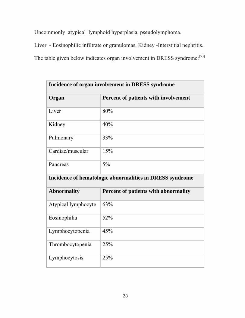
The table given below indicates organ involvement in DRESS syndrome:[53]
Incidence of organ involvement in DRESS syndrome
Organ Percent of patients with involvement
Liver 80%
Kidney 40%
Pulmonary 33%
Cardiac/muscular 15%
Pancreas 5%
Incidence of hematologic abnormalities in DRESS syndrome
Abnormality Percent of patients with abnormality
Atypical lymphocyte 63%
Eosinophilia 52%
Lymphocytopenia 45%
Thrombocytopenia 25%
Lymphocytosis 25%

29
Diagnosis:
Proposed Diagnostic Criteria:[54]
(1) Cutaneous drug eruption;
(2) hematologic abnormalities (eosinophilia >1500/μL or atypical lymphocytes);
(3) systemic involvement [adenopathies >2 cm in diameter or hepatitis (SGOT
>2 N) or interstitial nephritis or interstitial pneumonitis or carditis]. Diagnosis is
confirmed if three criteria are present.
Differential diagnosis :
Early: That of morbilliform eruptions, can mimic early measles or rubella.
Later: Serum sickness, drug-induced vasculitis, Henoch-Schönlein purpura,
cryoglobulin-associated vasculitis, vasculitis associated with infection, and
collagen vascular diseases.
Rash Plus Lymphadenopathy : Rubella, primary EBV or CMV mononucleosis
syndrome.
Course and prognosis:
Rash and hepatitis may persist for weeks after drug is discontinued. In patients
treated with systemic glucocorticoids, rash and hepatitis may recur as
glucocorticoids are tapered. Lymphadenopathy usually resolves when drug is
withdrawn; however, rare progression to lymphoma has been reported. Rarely,
patients die from systemic hypersensitivity such as with eosinophilic
30
myocarditis. Clinical findings recur if drug is given again.
Management:
Identify and discontinue the offending drug.
Symptomatic Treatment :
Oral antihistamine to alleviate pruritus.
Glucocorticoids :
Topical High-potency topical glucocorticoids applied twice a day are usually
helpful in relieving cutaneous symptoms of pruritus but do not alter systemic
hypersensitivity.
Systemic : Prednisone (0.5 mg/kg per day) usually results in rapid improvement
of symptoms and laboratory parameters. [54]
Future Drug Therapy:
Cross-sensitivity between various aromatic antiepileptic drugs occurs, making it
difficult to select alternative anticonvulsant therapy.
Prevention:
The individual must be aware of his or her specific drug hypersensitivity and
that other drugs of the same class can cross-react. These drugs must never be
readministered. Patient should wear a medical alert bracelet.
31
STEVEN JOHNSON SYNDROME
SJS is a severe illness of usually sudden onset, associated with marked
constitutional symptoms of high fever, malaise, myalgia, arthralgia and
extensive erythema multiforme of the trunk, with occasional skin blisters and
erosions covering less than 10% of the body’s surface area. [1]
The skin lesions are variable in extent, and consist of typical maculopapular
lesions of erythema multiforme, bullous or, rarely, pustular lesions. New crops
of lesions develop over a period of 10 days, or sometimes 3–4 weeks. Mucous
membranes:
Oral: extensive bulla formation, erosions and a greyish white membrane,
so that the mouth and lips show characteristic haemorrhagic crusting . [7]
Eyes: a severe catarrhal or purulent conjunctivitis can be seen. Corneal
ulceration, anterior uveitis or panophthalmitis may occur. The eye
changes often regress completely, but synechiae, corneal opacities and
rarely blindness are possible sequelae.
Genital : retention of urine due to involvement of the bladder. [1]
Respiratory symptoms may occur, and often the radiological changes within the
lungs are far greater than the symptoms. Abnormalities of liver function may be
present. Renal involvement with haematuria or even renal tubular necrosis has
been reported and may lead to progressive renal failure. [25]
32
Untreated, this disease used to have a mortality of 5–15% from infection,
toxaemia or renal damage, but the mortality rate is now lower. The eruption
usually heals without sequelae, although the eyes may be permanently damaged.
Eruptive melanocytic naevi are reported to develop after SJS.
Complications :
Acute: Massive fluid and electrolyte loss (3–4L/day) ,prerenal renal
failure,Bacterial infection and septicaemia, hypercatabolism: insulin resistance,
diffuse interstitial pneumonitis, mucous membrane involvement
Chronic :Ocular complications (up to 35%) Conjunctivitis, ectropion or
entropion, corneal scarring , Symblepharon,Sjögren-like sicca syndrome
Other mucous membrane involvement :Oesophageal stricture ,Phimosis. Vaginal
synechiae, Orogenital ulcers
Miscellaneous: Wound infection, pigmentary changes, nail dystrophy,
hypohidrosis, scarring alopecia, contractures, development of melanocytic
naevi.[1]
Management is similar to toxic epidermal necrolysis and has been discussed
subsequently.

33
TOXIC EPIDERMAL NECROLYSIS
TEN: The incidence of erythema multiforme, SJS and TEN was estimated at 1.8
cases per million person years for patients aged between 20 and 64 years; the
incidence for patients aged less than 20 years, and 65 years or more, increased to
7 and 9 cases per million person-years, respectively. The incidence of TEN was
estimated at 0.5 per million per year.[1]
Aetiology
Immunology
In general, CD4 T cells predominate in the upper dermis, while epidermal CD8
T cells and macrophages are variable and Langerhans’ cells virtually disappear.
Keratinocytes express HLA-DR and ICAM-1, and there is endo- thelial cell
ICAM-1, vascular cell adhesion molecule 1 (VCAM-1) and E-selectin
expression. CD3 activated T cells expressing the skin-homing receptor
(cutaneous leukocyte antigen; CLA) in both skin and peripheral blood parallel
the severity of the disease, and tumour necrosis factor-α (TNF-α), IFN-γ and
interleukin-2 (IL-2) are overexpressed in peripheral blood mononuclear cells,
suggesting an important role for T cells in TEN . Prominent involvement of the
monocyte–macrophage lineage, including factor XIIIa, HLA-DR, dendrocytes
and CD68, Mac387, macrophages, before, during and especially after epidermal
necrosis has been reported ,with dense labelling of the epidermis for TNF-α.
34
Mechanisms mediating keratinocyte death:
Cytokines released by activated lymphocytes and/or from keratinocytes
may contribute to apoptosis in TEN. Activated lymphocytes may induce
apoptosis via an interaction between Fas-ligand (FasL), expressed on the surface
of and secreted by lymphocytes, and Fas antigen (CD95), expressed by
keratinocytes after exposure to IFN-γ. There is keratinocyte overexpression of
Fas antigen in drug-induced erythema multiforme, SJS and TEN . Moreover,
peripheral blood mononuclear cells from TEN and SJS patients secreted high
levels of soluble(s) FasL on stimulation with the causal drug . Increased sFasL
levels precede skin detachment in patients with SJS and TEN . Bcl-2, a protein
known to block apoptosis, is strongly expressed along the basal layer and in the
dermal infiltrate both in erythema multiforme and SJS/TEN; thus Fas-mediated
cell death may be partially suppressed by Bcl-2 protein .
The matrix metalloproteinase MMP2 has a significant role in epidermal
detachment, inflammation and re-epithelialization.
Drugs implicated in toxic epidermal necrolysis :
Antibiotics: Sulphonamides : Co-trimoxazole , Sulfadoxine, Sulfadiazine,
Sulfasalazine
Penicillins : Amoxicillin Ampicillin
Cephalosporins
35
Non-steroidal anti-inflammatory drugs :
Phenylbutazone
Oxyphenabutazone
Oxicam-derivatives
Clinical features:
Typically sheet like erosions involve more than 30% of the body surface with
widespread purpuric macules or flat atypical target lesions, and there is severe
involvement of conjunctival, corneal, irideal, buccal, labial and genital mucous
membranes.[27]
Prodromal period: Flu-like symptoms (malaise, fever, rhinitis and
conjunctivitis), sometimes accompanied by difficulty in urination, which usually
lasts 2–3 days; however, it may last from 1 day to 3 weeks before signs of skin
involvement develop.
Acute phase : Persistent fever, severe mucous membrane involvement and
generalized epidermal sloughing to leave large, raw, painful areas, and lasts
from 8 to 12 days. There may be an initial ‘burning’ maculopapular, urticarial or
erythema multiforme-like eruption. Most frequently, the initial individual skin
lesions form poorly defined macules with darker purpuric or blistering centres,
progressively merging on the chin, upper parts of chest and back.
Nikolsky’s sign, the ability to extend the area of superficial sloughing by gentle

36
lateral pressure on the surface of the skin at an apparently unaffected site, may
be positive.
Detachment of the full thickness of the epidermis at sites of pressure or trauma,
such as the back, shoulders or buttocks, leaves a dark red oozing dermis.
However, the entire skin surface may be involved, with up to 100% of the
epidermis sloughing off. Only the hairy portion of the scalp is never affected.
The process tends to occur in waves, over a 3- to 5-day period (sometimes a
week), but involvement of the whole of the body surface occurs within 24 h in
approximately 10% of cases.
Mucous membranes (particularly the buccal, and less commonly the
conjunctival, genital, perianal, nasal, tracheal, bronchial, pharyngeal and
oesophageal membranes) are involved in nearly all patients (85–95%). Urethritis
develops in up to two-thirds of patients, and may lead to urinary retention.
Stomatitis and mucositis lead to impaired oral intake with consequent
malnutrition and dehydration. Intestinal involvement has been documented .
Healing occurs by re-epithelialization; this may occur within a few days on the
anterior thorax, but is slower on the back and at intertrigi- nous areas. Most
patients’ skin lesions are completely healed in about 3–4 weeks, but mucosal
lesions take longer and the glans penis may take up to 2 months to heal over.
37
Investigations:
Increase in serum aminotransferases and serum amylase overt hepatitis.
Anaemia and lymphopenia with a selective and transient depletion of CD4+ T
lymphocytes. Neutropenia is observed in approximately 30% of patients, and
thrombocytopenia in 15%; eosinophilia is very unusual. Hypophosphataemia ,
hyperglycaemia,raised urea and creatinine levels, subclinical interstitial oedema
is often noted on early chest X-rays.[1]
Prognosis :
There is an appreciable mortality as a result of TEN, increasing from 5% in SJS,
to 10–15% in transitional SJS–TEN and 30–40% in TEN.
SCORTEN prognosis score:[1]
Parameters:
1.Age > 40 years
2.Presence of a malignancy
3.Epidermal detachment > 30%
4.Heart rate > 120/min
5.Bicarbonate < 20 mmol/L
6.Urea > 10 mmol/L
7.Glycaemia > 14 mmol/L
1 point awarded for each parameter; SCORTEN derived by totalling scores[1]
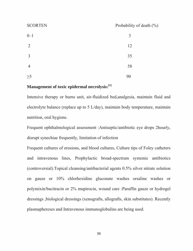
38
SCORTEN Probability of death (%)
0–1 3
2 12
3 35
4 58
≥5 90
Management of toxic epidermal necrolysis:[1]
Intensive therapy or burns unit, air-fluidized bed,analgesia, maintain fluid and
electrolyte balance (replace up to 5 L/day), maintain body temperature, maintain
nutrition, oral hygiene.
Frequent ophthalmological assessment :Antiseptic/antibiotic eye drops 2hourly,
disrupt synechiae frequently, limitation of infection
Frequent cultures of erosions, and blood cultures, Culture tips of Foley catheters
and intravenous lines, Prophylactic broad-spectrum systemic antibiotics
(controversial).Topical cleansing/antibacterial agents 0.5% silver nitrate solution
on gauze or 10% chlorhexidine gluconate washes orsaline washes or
polymixin/bacitracin or 2% mupirocin, wound care :Paraffin gauze or hydrogel
dressings ,biological dressings (xenografts, allografts, skin substitutes). Recently
plasmaphereses and Intravenous immunoglobulins are being used.

39
ERYTHEMA MULTIFORME
Erythema multiforme is an acute, self-limited, and sometimes recurring skin
condition that is considered to be a type IV hypersensitivity reaction associated
with certain infections, medications, and other various triggers.
Erythema multiforme may be present within a wide spectrum of severity. The
papules evolve into pathognomonic target lesions or iris lesions that appear
within a 72-hour period and begin on the extremities . Lesions remain in a fixed
location for at least 7 days and then begin to heal.
The clinical description is as follows:[8]
• Erythema multiforme minor - Typical target or raised, edematous papules
distributed acrally.
• Erythema multiforme major - Typical targets or raised, edematous papules
distributed acrally with involvement of one or more mucous membranes;
epidermal detachment involves less than 10% of total body surface area.
Erosions of the oral mucosa may result in difficulty in eating, drinking, or
opening the mouth. Conjunctival involvement may cause lacrimation,
photophobia, burning eyes, or visual impairment. Genital lesions are painful and
may result in urinary retention; painful micturition due to genitourinary tract
ulceration may also occur. Shortness of breath or difficulty in breathing may
occur due to tracheobronchial epithelial involvement.
40
Drug hypersensitivity:
The keratinocyte is the ultimate target of this disease process, with keratinocyte
necrosis being the earliest pathologic finding.
Patients frequently display an altered metabolism of the responsible drug, and
are considered to be slow acetylators, both genotypically and phenotypically.
Drug metabolism is directed toward the alternative pathway of oxidation by the
cytochrome P-450 system, resulting in increased production of reactive and
potentially toxic metabolites. Affected individuals have a defect in the ability to
detoxify these reactive metabolites, which may then behave as haptens by
binding covalently to proteins on the surface of epithelial cells. This may then
induce the immune response, leading to the severe skin reaction.[17]
Sulfa drugs are the most common triggers (30%) followed by the
anticonvulsants, including barbiturates, carbamazepine, phenytoin, and valproic
acid.
Causative antibiotics include penicillin, ampicillin, tetracyclines, amoxicillin,
cefotaxime, cefaclor, cephalexin, ciprofloxacin,erythromycin, minocycline,
sulfonamides, trimethoprim-sulfamethoxazole, and vancomycin.
Other drugs : albendazole, arsenic, bromofluorene, quinine, cimetidine,
clofibrate, corticosteroids, diclofenac, didanosine, dideoxycytidine,
diphosphonate, estrogen, etretinate, fluconazole, gabapentin, granulocyte-
41
macrophage colony-stimulating factor (GM-CSF), hydralazine, indapamide,
indinavir, mefloquine, methotrexate, meprobamate, mercurials, minoxidil,
nifedipine, nevirapine, nystatin, nonsteroidal anti-inflammatory drugs
(NSAIDs), phenolphthalein,pyritinol, progesterone, potassium iodide, sulindac,
suramin, saquinavir, thiabendazole, thiouracil, terbinafine, theophylline,
verapamil, and dihydrocodeine phosphate.[13]
Two additional rare clinical forms of erythema multiforme have been
reported: [8]
i) Continuous erythema multiforme manifests as a prolonged course with
overlapping attacks and may be associated with systemic administration of
glucocorticoids.
ii) Persistent erythema multiforme has a protracted clinical course over months,
is commonly associated with atypical skin lesions and is commonly resistant to
conventional treatment. It has been reported in association with inflammatory
bowel disease (IBD), occult renal carcinoma, persistent or reactivated Epstein-
Barr virus (EBV) infection, and HSV infection.
42
ACNEIFORM ERUPTIONS
This pattern of eruption represents a small percentage of drug-induced skin
eruptions. Clinically it presents as a papulo-pustular inflammatory eruption on
the face and upper trunk resembling acne vulgaris.
Drug-induced acne should be considered if :
• The onset is sudden
• There is worsening of existing acne
• Comedones are absent
• Monomorphic appearance of the papules and pustules
• There is an exposure to a potentially responsible drug
Few drugs can cause acneiform eruption historically, corticosteroids have been a
common cause. However, the new EGF receptor (EGFR) antagonists used in
cancer treatment, such as erlotinib, are associated with an acneiform eruption
that carries a good prognostic factor for the treatment of the underlying
condition as evidence of effect on another organ that expresses EGFR.
Withdrawal of the culprit drug and treatment with tetracycline or erythromycin
antibiotics is often all that is needed in terms of treatment[13].
Adrenocorticotrophic hormone (ACTH), corticosteroids , dexamethasone in
neurosurgical patients, anabolic steroids for body building , androgens (in
females), oral contraceptives, iodides and bromides may produce acneiform
43
eruptions.
Isoniazid may induce acne, especially in slow inactivators of the drug . Other
drugs implicated in the production of acneiform rashes include dantrolene ,
danazol , quinidine , lithium and azathioprine.[14]
The fact that acneiform eruptions do not affect prepubertal children indicates
that previous hormonal priming is a necessary prerequisite. In cases in which the
offending agent cannot be discontinued topical tretinoin may be helpful[17].
DRUG INDUCED PHOTOSENSITIVITY
Phototoxic reactions : Ultraviolet light activates the photosensitizing drug to
emit energy that may damage adjacent skin tissue resulting in an intensified
sunburn with skin peeling. Factors influencing the intensity and incidence of
drug-induced phototoxicity include:
1) the concentration, absorption, and pharmacokinetics of the drug. Higher doses
of lipophilic drugs (e.g., amiodarone) known to cause this reaction have a higher
incidence, and
2) the “dose” of sunlight (i.e., quantity and spectrum of sunlight).
Phototoxicity is characterized by a rapid onset of erythema, pain, prickling, or
burning sensation of areas exposed to the sun, with peak symptoms occurring
12-24 hours after the initial exposure. The hallmark of this reaction is the
44
appearance of a sunburn-like reaction on areas of skin with the greatest exposure
to sunlight. These reactions do not involve the immune system; therefore, prior
exposure or sensitization to a drug is not necessary for this reaction to occur.[58]
Photoallergic reactions :
Drug induced photoallergy is less common than phototoxicity, and requires
prolonged or prior exposure to the photosensitizing drug. As the name suggests,
this type of reaction is immune mediated. UV light reacts with the drug to
produce an immunogenic stimuli known as a hapten. This hapten combines with
a tissue antigen producing a cell mediated immune response resulting in a skin
reaction. This reaction requires a latency period following drug exposure for the
immunological memory response to develop after the first drug contact.
Subsequent exposures to the drug can elicit a more rapid reaction. Photoallergic
reactions are not dose dependent.
Photoallergic reactions are characterized by solar urticaria with eczema-like
dermatitis and erythema. Light exposed areas on the skin are the predominant
location of the reaction. These eruptions usually disappear spontaneously upon
removal of the offending drug.[58]
45
LICHENOID DRUG ERUPTIONS
Commonly caused due to HMG CoA reductase inhibitors , gold salts, beta
blockers, antimalarials, thiazide diuretics, furosemide, spironolactone, and
penicillamine.[59]
Clinicalfeatures:
• Extensive rash distributed symmetrically over the trunk and limbs
• Photodistribution – the rash is predominantly in areas exposed to the sun
• Rash may be scaly resembling eczema or psoriasis
• Wickham’s striae are usually absent
• Nail and mucous membrane (e.g., mouth) involvement is uncommon
• More likely to resolve leaving marked pigmentation
Prevention and Treatment : [59]
To stop the underlying offending drug
1. Topical steroids such as clobetasol propionate and betamethasone
propionate ointments .
2. Hydrocortisone foam can be used.
3. Steroid injections into affected areas may be useful for localised disease.
4. Systemic steroids may also be used.
46
ACUTE GENERALIZED EXANTHEMATOUS PUSTULOSIS [AGEP]
It is characterized by the rapid appearance of many pustules, which are sterile
and located subcorneally in the epidermis; the patients have fever, leukocytosis,
and sometimes also an eosinophilia. In >90% of the cases it is caused by drug
intake, in particular aminopenicillins, anticonvulsants and anti-inflammatory
drugs. Lethality in AGEP is 1% for older patients.
The most striking feature of AGEP is the short interval between the drug
administration and the onset of the disease.[60]
The main histopathological findings in AGEP are spongiform superficial
pustule, papillary edema, polymorphous perivascular infiltrate with eosinophils
and leucocytoclastic vasculitis with fibrinoid deposits.
Use of systemic steroid in AGEP is justified.[60]
TOXIC ACRAL ERYTHEMA
Acral erythema is also known as palmoplantar erythrodysesthesia or hand-foot
syndrome. It manifests as painful erythema of the palms and soles, with or
without bullae. These symptoms can be preceded by dysaesthesia . The pain
from this rash may be so severe that daily activities are limited. If recognised
early, the usual course of acral erythema is desquamation followed by re-
epithelialization. The exact mechanism is unknown, but it is postulated that the
47
skin of the hands and feet favour a higher level of certain chemotherapy drugs
which causes direct toxicity to the skin cells.[61]
Associated drugs include: Cytarabine , Docetaxel , Doxorubicin , Fluorouracil,
Cyclophosphamide , Daunorubicin , Vincristine , Vinblastin.
DRUG INDUCED SWEET’S SYNDROME LIKE ERUPTION
It is rare and represent overall less than 5% of all cases. Approximately 50 cases
have been reported, mostly as isolated clinical cases. Sitjas et al[62]. observed in
a retrospective study of 30 patients with SS that 7 of them had received a new
treatment before the rash occurred (non-steroidal anti-inflammatory drugs
(NSAIDS), penicillin, carbamazepine). However, these drugs were often given
for another confounding cause, especially an infection.
Delay is highly variable, ranging from several days to, exceptionally, 2 years ,
but mostly within 7 days. Clinical presentation and histology are quite similar to
the idiopathic form. Hyperleucocytosis could be less frequent during the drug-
induced form. Evolution is favourable after drug withdrawal. Fever vanishes
within one to three days, eruption within 3–30 days under corticosteroid
ointment and one week with oral corticosteroid therapy. Relapses occur in case
of drug reintroduction.[62]
48
CUTANEOUS REACTIONS TO ANTINEOPLASTIC AND
CHEMOTHERAPEUTIC DRUGS
Alopecia: is the most common adverse skin manifestation of the
chemotherapeutic treatment. There are two types of drug-induced alopecia: the
anagen effluvium and the telogen effluvium. In the anagen effluvium hair loss
occurs due to the sudden interruption of the mitotic activity of the hair matrix,
one to two weeks after the start of chemotherapy, leading to lack of hair
production or its thinning (Pohl- Pinkus constrictions). They involve the hair,
eyebrows, beard, axillary and pubic hair. It is dose-dependent and reversible. In
the telogen effluvium, hairs move prematurely to a resting phase with
subsequent loss of normal hair.[40]
The antineoplastic agents that most frequently cause the anagen effluvium lead
to diffuse hair loss, of sudden onset, from 7 to 10 days after the start of
chemotherapy. Hair loss becomes more pronounced about 1 to 2 months after
the start of treatment.
Trichomegaly and hair curling :
Hair alterations with acceleration of growth and shaft changes are observed with
the use of epidermal growth factor receptor inhibitors (EGFR)
Ungual, subungual, and periungual alterations :
Nail alterations can present with a reduction of the nail growth speed, fragility,
49
lines of discoloration (Mees' lines), transverse depressions (Beau's lines),
hyperpigmentation, onycholysis with subungual aseptic abcesses,
photoonycholysis, paronychia, and pyogenic granulomas of the periungual folds.
Hyperpigmentation can occur due to the use of cyclophosphamide, hydroxyurea,
fluoropirimidines, such as 5-fluorouracil (5-FU) and specially anthracyclines
like doxorubicin and daunorubicin . Painful onycholysis and subungual abcesses
are due to the use of taxanes (docetaxel/paclitaxel) and anthracyclines
(doxorubicin). Ingrown nails, paronychia, and pyogenic granuloma are
associated with the use of tyrosine kinase inhibitors of the epidermal growth
factor receptor (EGFR), such as erlotinib and gefitinib. The fenestration or
avulsion of the lamina should be considered when abscesses that involve more
than 50% of the nail bed are present.[40]
Other adverse effects are as follows :
Neutrophilic eccrine hidradenitis
Eccrine squamous syringometaplasia
Acral erythema or palmoplantar erythrodysesthesia syndrome
Toxic erythema caused by chemotherapeutic drugs
Acneiform eruption
Mucous membrane alterations
Stomatitis

50
DRUG INDUCED PSEUDOLYMPHOMA SYNDROME
Pseudolymphoma syndrome (PS) consists of the triad of fever, generalised rash
and lymphadenopathy. In addition malaise, hepatosplenomegaly, arthralgia,
congestive cardiac failure, eosinophilia, thrombocytopenia and blood dyscrasias
may be present.
Diphenylhydantion, tridione and phenobarbitone etc. can produce a peculiar
response of reticuloendothelial system resulting in PS.PS may be either
hypersensitivity reaction or possibly a genetically determined enzymatic defect
as seen in primaquin sensitivity. PS may present as generalised exfoliative
dermatitis. PS may have generalised or localised lesions and may result from
non-anticonvulsant drugs. Histopathology may reveal mycosis fungoides or
sezary like syndrome.[63]
DRUG INDUCED BULLOUS DISORDER
Approximately 10% of cases of pemphigus are drug-induced or drug-triggered.
The skin lesions are flaccid blisters which break easily and often only erosions
+/- crusting are seen. The Nikolsky sign can be positive. In drug-induced
pemphigus, mucous membranes are only involved in 10-15% of cases whereas
drug-triggered pemphigus is indistinguishable from classic pemphigus.[57]
Skin biopsy shows the typical separation of individual skin cells seen in

51
pemphigus. Direct immunofluoresence is positive in 90% of drug related
pemphigus, compared to 100% in idiopathic classic disease. Circulating
antidesmoglein autoantibodies are only detected in the blood in 70% of drug-
induced pemphigus.
The onset of drug related pemphigus can be weeks to months after the drug was
started. Resolution occurs after drug withdrawal in drug-induced pemphigus but
not if drug-triggered.
Drug-induced pemphigus is caused by drugs with a thiol group such as:
D-penicillamine , Captopril ,Gold sodium thiomalate , Pyritinol
Drug-triggered pemphigus follows nonthiol drug use including:
Antibiotics especially betalactams, rifampicin , Pyrazolone derivatives ,
Nifedipine , Propranolol , Piroxicam, Phenobarbital.[57]
DRUG INDUCED LUPUS ERYTHEMATOSUS
Drug-induced lupus erythematosus is an autoimmune disorder that is brought on
by a reaction to medication.The most common medications known to cause
drug-induced lupus include: isoniazid, hydralazine, and procainamide.[56]
Other medications known to cause drug-induced lupus, include:
Anti-seizure medications , Capoten , Chlorpromazine , Etanercept, Infliximab ,
Methyldopa , Minocycline , Penicillamine ,Quinidine , Sulfasalazine

52
Symptoms tend to occur after taking the drug for at least 3 to 6 months.
Persons with drug-induced lupus erythematosus may have symptoms that affect
the joints (arthritis), heart, and lungs. Other symptoms associated with SLE,
such as lupus nephritis and nervous system (neurological) disease, are rare.[56]
DRUG INDUCED PSEUDOPORPHYRIA
Skin signs include skin fragility and photosensitivity. Tense blisters form at the
sites of minor trauma on sun exposed skin, bursting early to leave scabs and
erosions. The blisters are most often seen on the hands and feet. They sometimes
heal with some scar formation and milia.A sunburn type rash may also occur.[55]
Pseudoporphyria is due to drugs which interact with sunlight to cause a
phototoxic reaction in the skin. These include:[55]
• Non steroidal anti-inflammatory drugs e.g. naproxen
• Antibiotics: doxycyline, nalidixic acid
• Diuretics: chlorthalidone, bumetanide, furosemide and hydrochlorthiazide
• Retinoids – isotretinoin, etretinate, acitretin
• Oral contraceptives
It is important to withdraw the suspected agent where possible and avoid
unnecessary exposure to strong light.Sun protection measures may include UVB
and UVA blocking sunscreens.Symptoms usually resolve within several weeks
but sometimes they are persistent.[55]

53
MATERIALS AND METHODS
This study was a prospective, observational study conducted at the skin
OPD, Government Rajaji Hospital, Madurai Medical College, Madurai during
the period from October 2009-September 2011[24 months].
Inclusion criteria:
All consecutive consenting patients diagnosed clinically as a case of
adverse cutaneous drug reaction of all age groups, of all genders, during the
study period were included in the study.
Exclusion criteria:
Non consenting patients, and patients with morphological pattern of
adverse cutaneous drug reaction, but were unable to provide an exact history of
drug intake.
Patients satisfying the criteria were included in the study and demographic
details were recorded. A detailed clinical history including duration, site of
onset, symptoms, drug history, family history were elicited. A complete general
examination, systemic examination and dermatological examination were made.
Digital photographs were taken.The morphology and distribution of skin lesions,
concomitant affection of mucosa, hair, nails, palms, soles, genital involvement
was meticulously recorded and presence of any other associated diseases were
noted.
54
OBSERVATIONS AND RESULTS:
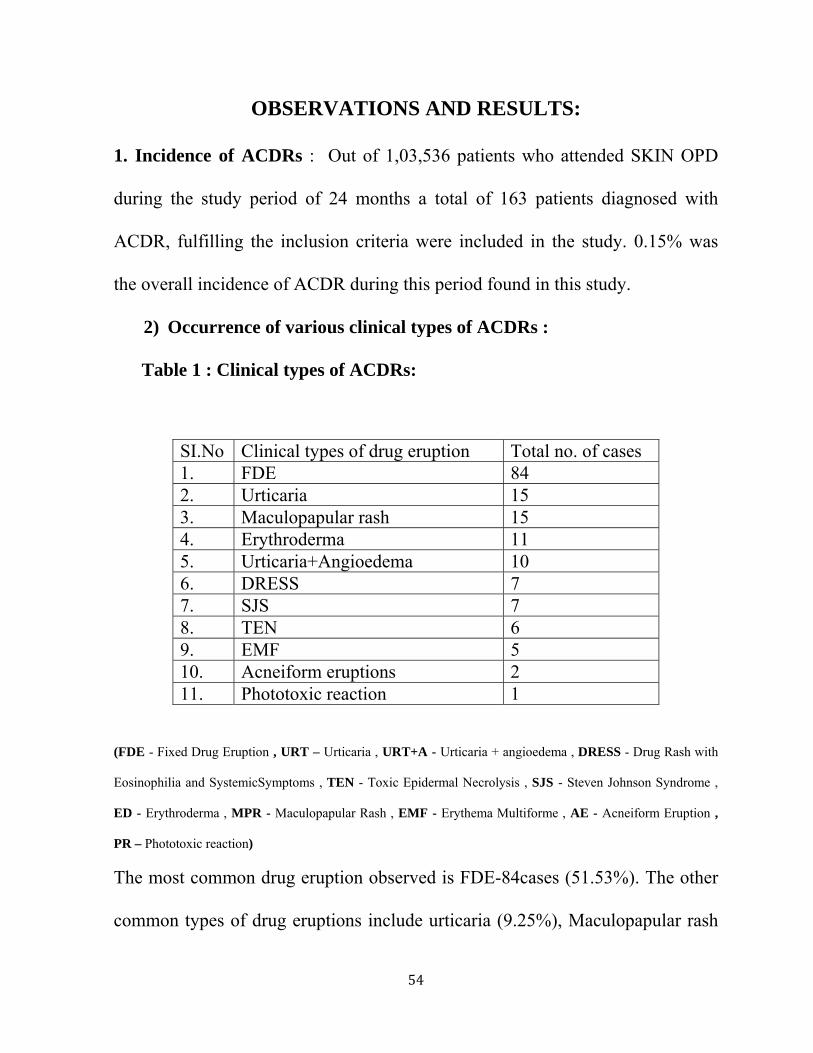
1. Incidence of ACDRs : Out of 1,03,536 patients who attended SKIN OPD
during the study period of 24 months a total of 163 patients diagnosed with
ACDR, fulfilling the inclusion criteria were included in the study. 0.15% was
the overall incidence of ACDR during this period found in this study.
2) Occurrence of various clinical types of ACDRs :
Table 1 : Clinical types of ACDRs:
(FDE - Fixed Drug Eruption , URT – Urticaria , URT+A - Urticaria + angioedema , DRESS - Drug Rash with
Eosinophilia and SystemicSymptoms , TEN - Toxic Epidermal Necrolysis , SJS - Steven Johnson Syndrome ,
ED - Erythroderma , MPR - Maculopapular Rash , EMF - Erythema Multiforme , AE - Acneiform Eruption ,
PR – Phototoxic reaction)
The most common drug eruption observed is FDE-84cases (51.53%). The other
common types of drug eruptions include urticaria (9.25%), Maculopapular rash
SI.No Clinical types of drug eruption Total no. of cases 1. FDE 84 2. Urticaria 15 3. Maculopapular rash 15 4. Erythroderma 11 5. Urticaria+Angioedema 10 6. DRESS 7 7. SJS 7 8. TEN 6 9. EMF 5 10. Acneiform eruptions 2 11. Phototoxic reaction 1

(9.25
angio
(3.08
in tab
3
cases
cases
in 7th
0
5
10
15
20
25
30
35
%) and Er
oedema (6
%), Acnei
ble 1.
) Age dist
Figure 1
As show
s) and 5th (
s) decades,
decade &
0
5
0
5
0
5
0
5
21 2
rythroderm
6.17%), D
iform erup
tribution o
: Age dist
wn in figur
(32 cases)
20 cases e
1 case wa
20
293
A
ma (6.74%)
DRESS (4
tions (1.23
of various
ribution o
re 1 the hi
decades f
each were
s seen in 8
33 32
2
AGE DIS
55
). The othe
.32%), SJ
3%) and ph
s ACDRs:
of various
ighest num
followed c
seen in 2nd
8th decade.
20
7
STRIBUT
er ACDRs
JS (4.32%
hototoxic r
ACDRs
mber of ca
closely by
d and 6th d
1
TION
seen were
%), TEN
reaction (0
ases were s
3rd(29 cas
ecade. 7 ca
Total n
e urticaria
(3.7%), E
0.61%) as
seen in 4th
ses) and 1
ases were
no. of cases
with
EMF
seen
h (33
st(21
seen

56
4) Gender distribution in ACDRs:
Table 3: Gender distribution in various ACDRs
There were a total of 82 male and 81 female cases with a gender ratio of 1.01:1.
The gender distribution for various drug eruptions is shown in Table 3.
As show in table 3 male patients constituted 43 cases of FDE, 8 cases of
urticaria, 8 cases of MPR, 5 cases of ED, 2 cases of urticaria with angloedema. 3
cases of DRESS, 4 cases of TEN, 5 cases of SJS, 2 cases of EMF, 1 case of AE.
Female patients constituted 41 cases of FDE, 7 cases of urticaria, 7 cases of
MPR, 6 cases of ED, 8 cases of urticaria with angloedema. 4 cases of DRESS, 2
cases of TEN, 2 cases of SJS, 3 cases of EMF, 1 case of AE, 1 case of PR.
57
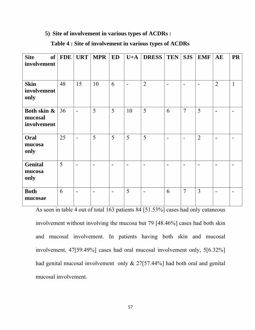
5) Site of involvement in various types of ACDRs :
Table 4 : Site of involvement in various types of ACDRs
Site of involvement
FDE URT MPR ED U+A DRESS TEN SJS EMF AE PR
Skin involvement only
48 15 10 6 - 2 - - - 2 1
Both skin & mucosal involvement
36 - 5 5 10 5 6 7 5 - -
Oral mucosa only
25 - 5 5 5 5 - - 2 - -
Genital mucosa only
5 - - - - - - - - - -
Both mucosae
6 - - - 5 - 6 7 3 - -
As seen in table 4 out of total 163 patients 84 [51.53%] cases had only cutaneous
involvement without involving the mucosa but 79 [48.46%] cases had both skin
and mucosal involvement. In patients having both skin and mucosal
involvement, 47[59.49%] cases had oral mucosal involvement only, 5[6.32%]
had genital mucosal involvement only & 27[57.44%] had both oral and genital
mucosal involvement.
6
Tab
no of
Urtic
case o
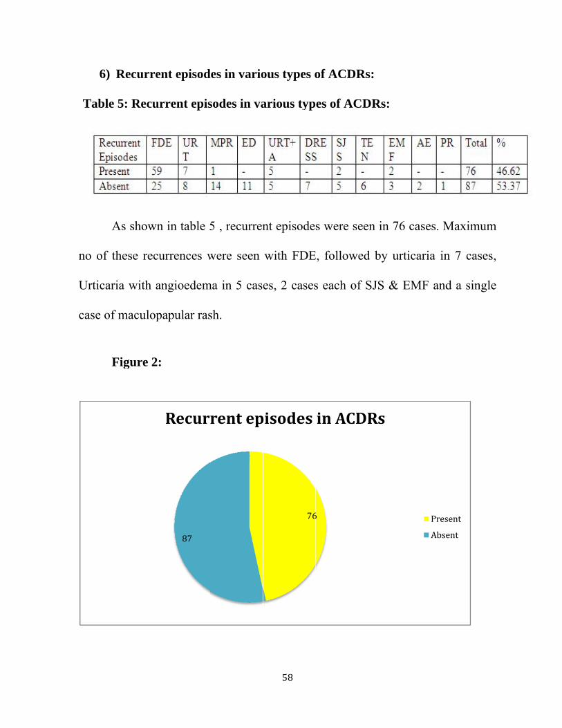
6) Recurre
le 5: Recu
As shown
f these rec
aria with a
of maculop
Figure 2
ent episod
urrent epis
n in table 5
currences w
angioedem
papular ras
:
87
Recur
des in vario
sodes in va
5 , recurren
were seen
ma in 5 cas
sh.
rent ep
58
ous types o
arious typ
nt episode
with FDE
es, 2 cases
76
pisodes
of ACDRs
es of ACD
s were see
E, followed
s each of S
6
in ACDR
s:
DRs:
en in 76 ca
d by urtica
SJS & EM
Rs
ses. Maxim
aria in 7 ca
MF and a si
Present
Absent
mum
ases,
ingle
59
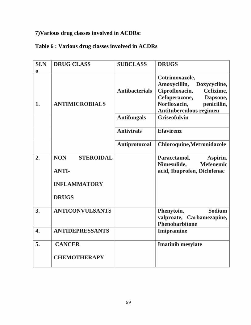
7)Various drug classes involved in ACDRs:
Table 6 : Various drug classes involved in ACDRs
SI.No
DRUG CLASS SUBCLASS DRUGS
1.
ANTIMICROBIALS
Antibacterials
Cotrimoxazole, Amoxycillin, Doxycycline, Ciprofloxacin, Cefixime, Cefoperazone, Dapsone, Norfloxacin, penicillin, Antituberculous regimen
Antifungals Griseofulvin
Antivirals Efavirenz
Antiprotozoal Chloroquine,Metronidazole
2. NON STEROIDAL
ANTI-
INFLAMMATORY
DRUGS
Paracetamol, Aspirin, Nimesulide, Mefenemic acid, Ibuprofen, Diclofenac
3. ANTICONVULSANTS Phenytoin, Sodium valproate, Carbamezapine, Phenobarbitone
4. ANTIDEPRESSANTS Imipramine
5. CANCER
CHEMOTHERAPY
Imatinib mesylate

60
In our study the above five classes of drugs were encountered as shown in table
6. A total of 108 [66.25%] cases due to antimicrobials were seen out of these 92
cases were due to intake of antibacterials, 10 cases were due to antivirals, 5
cases were due to antiprotozoal, single case was due to antifungal. 28 [17.17%]
cases were due to Non steroidal anti-inflammatory drugs,25 [ 15.33%] cases
were due to anticonvulsants and a single case[ 0.61%]each was due to
antidepressants and chemotherapy.
61
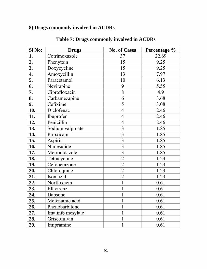
8) Drugs commonly involved in ACDRs
Table 7: Drugs commonly involved in ACDRs Sl No: Drugs No. of Cases Percentage % 1. Cotrimoxazole 37 22.69 2. Phenytoin 15 9.25 3. Doxycycline 15 9.25 4. Amoxycillin 13 7.97 5. Paracetamol 10 6.13 6. Nevirapine 9 5.55 7. Ciprofloxacin 8 4.9 8. Carbamezapine 6 3.68 9. Cefixime 5 3.08 10. Diclofenac 4 2.46 11. Ibuprofen 4 2.46 12. Penicillin 4 2.46 13. Sodium valproate 3 1.85 14. Piroxicam 3 1.85 15. Aspirin 3 1.85 16. Nimesulide 3 1.85 17. Metronidazole 3 1.85 18. Tetracycline 2 1.23 19. Cefoperazone 2 1.23 20. Chloroquine 2 1.23 21. Isoniazid 2 1.23 22. Norfloxacin 1 0.61 23. Efavirenz 1 0.61 24. Dapsone 1 0.61 25. Mefenamic acid 1 0.61 26. Phenobarbitone 1 0.61 27. Imatinib mesylate 1 0.61 28. Griseofulvin 1 0.61 29. Imipramine 1 0.61
62
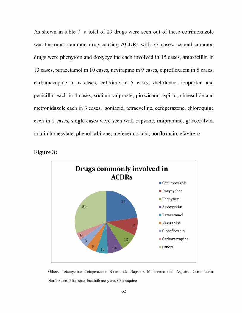
As shown in table 7 a total of 29 drugs were seen out of these cotrimoxazole
was the most common drug causing ACDRs with 37 cases, second common
drugs were phenytoin and doxycycline each involved in 15 cases, amoxicillin in
13 cases, paracetamol in 10 cases, nevirapine in 9 cases, ciprofloxacin in 8 cases,
carbamezapine in 6 cases, cefixime in 5 cases, diclofenac, ibuprofen and
penicillin each in 4 cases, sodium valproate, piroxicam, aspirin, nimesulide and
metronidazole each in 3 cases, Isoniazid, tetracycline, cefoperazone, chloroquine
each in 2 cases, single cases were seen with dapsone, imipramine, griseofulvin,
imatinib mesylate, phenobarbitone, mefenemic acid, norfloxacin, efavirenz.
Figure 3:
Others- Tetracycline, Cefoperazone, Nimesulide, Dapsone, Mefenemic acid, Aspirin, Griseofulvin,
Norfloxacin, Efavirenz, Imatinib mesylate, Chloroquine
37
15
15
13109
8
6
50
Drugs commonly involved in ACDRs
Cotrimoxazole
Doxycycline
Phenytoin
Amoxycillin
Paracetamol
Nevirapine
Ciprofloxacin
Carbamezapine
Others

63
9]Drugs and ACDRs caused by them: Table 8: Drugs and ACDRs caused by them
(CTX- Cotrimoxazole, DOXY-Doxycycline, PHE- Phenytoin, AX- Amoxycillin, NVP-Nevirapine, PCM-
Paracetamol, CBZ-Carbamezapine, CPX-Ciprofloxacin, CFX-Cefixime, DIC-Diclofenac, IBP-Ibuprofen, SV-
Sodium valproate, PXM-Piroxicam.)
As shown in table 8 it was observed that Cotrimoxazole was found to cause
FDE in 26 cases, urticaria in 5 cases, urticaria with angioedema in 3 cases , SJS
and EMF in a single case, Phenytoin was found to cause maculopapular rash in 8
cases , DRESS in 5 cases, TEN and SJS each in a single case.Amoxycillin was
found to cause FDE in 9 cases, Urticaria, SJS, Erythroderma and Maculopapular
rash each in a single case. Nevirapine in 2 cases of TEN, 3 cases of
erythroderma,4 cases of maculopapular rash. Paracetamol was seen in 4 cases of
FDE, 2cases each of urticaria, urticaria with angioedema & 1 case each of TEN
& EMF: total of 10 cases.Carbamezapine was seen in 2 cases each of FDE &
Erythroderma and in 1 case each of TEN & SJS : total of 6 cases.Cefixime was

64
seen with 3 cases of FDE, 1 case each of Urticaria, DRESS & maculo- papular
rash. Diclofenac sodium & Ibuprofen were associated with 4 cases each of FDE.
Sodium valproate : 1 case of FDE, 2 cases of maculopapular rash, total of 3
cases. Piroxicam : 2 cases of FDE , a case of urticaria :total of 3 cases.
10) Site of involvement for common individual drugs in ACDRs:
Table 9: Site of involvement for common individual drugs Sl No: Drugs Cutaneous
involvement Both skin
and mucosa
Total Percentage %
1. Cotrimoxazole 12 15 37 22.69 2. Doxycycline 6 9 15 9.25 3. Phenytoin 6 9 15 9.25 4. Amoxycillin 6 7 13 7.9 5. Paracetamol 6 4 10 6.13 6. Nevirapine 3 6 9 5.55 7. Carbamezapine 4 2 6 3.68 8. Ciprofloxacin 7 1 8 4.9 9. Cefixime 1 4 5 3.08 10. Diclofenac 2 2 4 2.46 11. Ibuprofen 2 2 4 2.46 12. Sodium
valproate 2 1 3 1.85
13 Piroxicam 2 1 3 1.85 As shown in table 9 Cotrimoxazole was the most common drug causing
cutaneous in12 and both involvement in 15 cases, followed by doxycycline and
phenytoin each in 6 cases of cutaneous and 9 cases of both. Amoxycillin in 6
cutaneous and both in 7 cases. Paracetamol in 6 cutaneous and both in 4
cases.Nevirapine in 3 cutaneous cases and 6 of both cases. Carbamezapine in 4

65
cutaneous and 2 case of both. Ciprofloxacin in 7 cases of cutaneous and 1 case
of both, Cefixime in 1 case of cutaneous and 4 cases of both, Diclofenac and
Ibuprofen each in 2 cases in both categories, Sodium valproate and Piroxicam
each in 2 cases of cutaneous and 1 case of both skin and mucosal involvement.
11)Associated diseases in ACDRs: Table 10 : Underlying associated diseases in ACDRs
Associated disease No. of cases 1. Upper respiratory tract infection 32(19.63%) 2.Seizure disorder 24(14.72%) 3. Fever & Headache 18(11.04%) 4.HIV 10(6.13%) 5. Lower respiratory tract infection 9(5.52%) 6. Dental caries & Gingivitis 9(5.52%) 7. Sinusitis 8(4.9%) 8.Diabetes mellitus [DM] 7(4.29%) 9.Hypertension [HTN] 6(3.68%) 10. Urinary tract infection 6(3.68%) 11.HTN+DM 4 (2.4%) 12. Arthritis & Bursitis 4 (2.4%) 13. Acute & Chronic suppurative otitis media
4 (2.4%)
14. Tuberculous meningitis 3(1.84%) 15. Malaria 3(1.84%) 16. Acute diarrhea 3(1.84%) 17. Typhoid 3(1.84%) 18. Myalgia 3(1.84%) 19. Pyoderma 2(1.22%) 20.Leprosy 1 (0.61%) 21. Depression 1 (0.61%) 22. Chronic lymphocytic leukemia 1 (0.61%) 23. Epididymoorchitis 1 (0.61%) 24.External hordeolum 1 (0.61%) TOTAL 163
66
As shown in table 10, maximum number of cases were seen with patients taking
drugs for underlying upper respiratory tract infections: 32(19.63%), followed by
seizure disorders 24(14.72%) cases, then followed by fever and headache
18(11.04%) cases, HIV was seen in 10(6.13%) cases, equal no. of patients that is
9(5.52%) cases were seen for both lower respiratory tract infections and dental
caries & gingivitis. Among chronic long standing diseases Diabetes mellitus was
seen in 7(4.29%) cases, Hypertension was seen in 6(3.68%) cases, both diabetes
and hypertension were seen in 4(2.4%) cases. Urinary tract infections were seen
in 6(3.68%) cases, 4(2.4%) cases each of arthritis and bursitis & acute and
chronic suppurative otitis media were seen.
3(1.84%) cases each of tuberculous meningitis, Malaria, acute diarrhea, myalgia
& typhoid were seen. 2(1.22%) cases of pyoderma were seen. Single cases each
of leprosy, depression, chronic lymphocytic leukemia, epididymoorchitis &
external hordeolum were seen.
12] Family history in ACDRs:
None of the cases had any history of family members with ACDRs.

67
DISCUSSION
1) Incidence of ACDRs:
Out of 1,03,536 total patients during the study period of 2 years, 163 were
diagnosed as ACDRs which constitutes 0.15%. The incidence of ACDRs has
been found to be 2.6% in a study by chatterjee et al[42] which is higher than our
study
2) Occurrence of various clinical types of ACDRs :
The commonest ACDR in our study was FDE seen in 84 [51.53%]
patients which is greater than a study by Thappa et al[3] where it was 28 [31.1%]
patients. Maculopapular rash was seen in 15 [9.25%] patients in our study which
is lesser than the study done by Thappa et al[3] where it was 12.2%. Urticaria
was seen in 15 [9.25%] patients in our study which is greater than in the study
by Thappa et al [3]where it was 7.8% . Erythroderma in our study included
11[6.74%] cases but according to Thappa et al[3] it was 3.3% which is lesser than
our study. Urticaria with angioedema constituted 10[6.17%] cases but Thappa et
al[3] recorded 1.1% which is lesser than our study. DRESS cases were 7 [4.32%]
in number but in a study by Shear et al[41] 9% of cases were due to DRESS
which is higher than our study. SJS constituted 7[4.32%] cases , but was only
3% in a study by Raksha MP et al[4]. TEN cases were 6 [3.7%] in number but
only 1% in a study by Raksha MP et al[4] which is lesser than our study. EMF
68
cases were 5[3.08%] in our study but 6.7% in the study by Thappa et al[3] which
is twice the number of cases when compared to our study. Acneiform eruptions
were 2 [1.23%] in number whereas Thappa et al[3] recorded 3.3% which is
greater than our study.
3) Age distribution of various ACDRs:
The age of the patients ranged from 3 months to 75 years. The majority of
patients (33 patients or 20.24%) fall in the age group from 31-40 years followed
by age group 41-50 years (32 patients or 19.63%). In a study by Ruchika
Nandha et al [15] the maximum number of cases was seen in the age group 21-30
years (25.27%) followed by the age group 31-40 years (23.07%) which is
greater than our study.
4) Gender distribution in ACDRs:
In our study, almost equal involvement was noted , 82 patients were
males and 81 were females [male to female ratio was 1.01:1] out of 163 total
cases , whereas during the study done by Ruchika Nandha et al[15] of the total 91
cases reported 47 (51.7%) were females and 44 (48.3%) were males. The male
to female ratio was 0.93:1, which is slightly lesser than ours.
Predominance of males was reported in few studies. Equal ratio has also been
reported in other studies.[23]

69
5) Site of involvement in various types of ACDRs :
Out of total 163 patients 84 [51.53%] cases had only cutaneous
involvement without involving the mucosa but 79 [48.46%] cases had both skin
and mucosal involvement. In patients having both skin and mucosal
involvement, 47[59.49%] cases had oral mucosal involvement only, 5[6.32%]
had genital mucosal involvement only & 27[57.44%] had both oral and genital
mucosal involvement which is higher than in a study by Faisal et al[35] where
about 32.7% of the patients (68/208) had mucosal involvement, the
manifestations of which varied according to the type of rash.
6) Recurrent episodes in various types of ACDRs :
Out of 163 patients 76 [46.62%] patients had recurrent episodes which is
higher than in a study by Thappa et al[3] where of the 90 consecutive patients, 25
had consumed the same drug earlier, 13 had a similar cutaneous reaction earlier
and 12 had no reactions .
7) Various Drug classes involved in ACDRs:
In our study the various drug classes encountered were:
a] Antimicrobials : Cotrimoxazole, Amoxycillin , Ciprofloxacin , Nevirapine ,
Cefixime ,Tetracycline, Cefoperazone, Griseofulvin, Norfloxacin, Efavirenz,
Dapsone- 108 [66.25%] cases.

70
b] NSAIDs: Paracetamol, Diclofenac, Ibuprofen, Piroxicam, Aspirin,
Nimesulide, Mefenemic acid-28 [17.17%] cases
c] Anticonvulsants: Phenytoin, Carbamezapine, Sodium valproate-25 [15.33%]
cases
d] Antidepressants: Imipramine -1 [0.61%] case.
e] Cancer chemotherapy: Imatinib mesylate-1 [0.61%] case.
The common offending drug groups in a study by chatterjee et al[42]were
antimicrobials (34.10%) which is lesser than our study, anticonvulsants
(32.88%) which is higher than our study, anti-inflammatory drugs (21.51%)
which is greater than our study.Similar incidence as in our study was seen with
antidepressants and chemotherapy drugs.
8) Drugs commonly involved in ACDRs :
Commonest drug causing ACDR was Cotrimoxazole recorded in 37 cases,
in a study by Vander Linden et al[19]the most frequent reactions were observed in
patients receiving a trimethoprim-sulfonamide combination , which is similar to
our study,followed by Phenytoin and Doxycycline each in 15 (9.25%) cases, but
Thappa et al[3] observed 7.8% cases for Phenytoin which is lesser than our study.
Amoxycillin in 13 cases was the 3rd common drug but in a study by khushwaha
et al[30] it was observed that amoxicillinwas 2ndmost common drug, Nevirapine
in 9 cases, Paracetamol was involved in 10 cases and was the 5th commonest
71
drug in our study but in a study by Naldi et al[30] paracetamol was ranked at
no.8 to cause ACDRs. Carbamezapine was the 6th commonest drug in our study
which is in contrast to a study by Ding et al[29] where Carbamazepine was the
most common drug causing severe ACDR. Ciprofloxacin in 6 cases, Cefixime
in 5, Diclofenac in 4, Ibuprofen in 4, Sodium valproate , Metronidazole,
Piroxicam, Aspirin & Nimesulide in 3( 1.85%) cases each where as Thappa et
al[3] recorded 2.2% for Metronidazole which is greater than our study.
Tetracycline & Cefoperazone in 2 [1.23%] cases each. Mefenemic acid,
Griseofulvin, Dapsone, Norfloxacin & Efavirenz each were incriminated in
1[.61%] case, but in a study by Thappa et al[3] Griseofulvin was accounted for
3.3% cases, which is much greater than our study.
9) Drugs and ACDRs caused by them: a) Cotrimoxazole was found to cause FDE in 26 cases,urticaria in 5 cases,
urticaria with angioedema in 3 cases , SJS and EMF in a single case.In a study
by Thappa et al[3] Cotrimoxazole was the leading causative agent (29.5%) in
FDE cases which is consistent with our study.
b) Phenytoin was found to cause maculopapular rash in 8 cases, DRESS in 5
cases, TEN and SJS each in a single case whereas in a study by Noel et al[34]
phenytoin caused maximum no of cases of Maculopapular rash followed by
TEN and erythroderma.
72
c) Amoxycillin was found to cause FDE in 9 cases, Urticaria, SJS, Erythroderma
and Maculopapular rash each in a single case, whereas in a study by S Ghosh et
al[30], the drug which was attributed to cause maximal number of maculopapular
rashes, was amoxicillin.
d) Nevirapine was implicated in 2 cases of TEN, 3 cases of erythroderma,4 cases
of maculopapular rash: total of 9 cases, wheras in a study by Ananworanich et
al[32] nevirapine most commonly caused maculopapular rash ,TEN.
e) Paracetamol was seen in 4 cases of FDE, 2cases each of urticaria, urticaria
with angioedema & 1 case each of TEN & EMF: total of 10 cases.
Noel et al[34] recorded 2 cases of urticaria , 1 case each of FDE and SJS, which is
lesser than our study.
f) Carbamezapine was seen in 2 cases each of FDE & Erythroderma and in 1
case each of TEN & SJS: total of 6 cases, whereas in a study by Hung et al[31]
the anticonvulsant carbamazepine (CBZ) frequently causes adverse cutaneous
drug reactions (ACDRs), including maculopapular eruption (MPE),
hypersensitivity syndrome (HSS), Stevens-Johnson syndrome (SJS) and toxic
epidermal necrolysis (TEN).
g) Cefixime was seen with 3 cases of FDE, 1 case each of Urticaria, DRESS &
maculopapular rash whereas in a study by Shipley et al Cefixime caused
urticaria more frequently.[39]

73
h) Diclofenac sodium & Ibuprofen were associated with 4 cases each of FDE
which is consistent with a study by Stern S et al where similar incidence was
found.[37]
i) Sodium valproate : 1 case of FDE, 2 cases of maculopapular rash, total of 3
cases, whereas in a study by Patricia et al[38]there were no confirmed serious
cutaneous diagnoses in 1,504 new valproate users.
j) Piroxicam : 2 cases of FDE , a case of urticaria :total of 3 cases, whereas in a
study by Stern S et al[37] Piroxicam was seen to cause TEN.
10) Site of involvement for common individual drugs in ACDRs:
Cotrimoxazole was the most common drug causing cutaneous in 12 and
both involvement in 15 cases, which is similar to a study by Arora et al[27],
followed by doxycycline and phenytoin each in 6 cases of cutaneous and 9 cases
of both. Amoxycillin in 6 cutaneous and both in 7 cases. Paracetamol in 6
cutaneous and both in 4 cases.Nevirapine in 3 cutaneous cases and 6 of both
cases. Carbamezapine in 4 cutaneous and 2 case of both. Ciprofloxacin in 7
cases of cutaneous and 1 case of both, Cefixime in 1 case of cutaneous and 4
cases of both, Diclofenac and Ibuprofen each in 2 cases in both categories,
Sodium valproate and Piroxicam each in 2 cases of cutaneous and 1 case of both
skin and mucosal involvement. Specfic references involving cutaneous and
mucosal involvement for other drugs were not found.

74
11) Associated diseases in ACDRs:
Maximum number of cases were seen with patients taking drugs for
underlying upper respiratory tract infections; 32(19.63%), followed by seizure
disorders 24(14.72%) cases, then followed by fever and headache 18(11.04%)
cases, HIV was seen in 10(6.13%) cases, equal no. of patients that is 9(5.52%)
cases were seen for both lower respiratory tract infections and dental caries and
gingivitis.
Among chronic long standing diseases Diabetes mellitus was seen in
7(4.29%) cases, Hypertension was seen in 6(3.68%) cases; both diabetes and
hypertension were seen in 4(2.4%) cases. Urinary tract infections were seen in
6(3.68%) cases, 4(2.4%) cases each of arthritis and bursitis & acute and chronic
suppurative otitis media were seen.
3(1.84%) cases each of tuberculous meningitis, Malaria, acute diarrhea, myalgia
& typhoid were seen. 2(1.22%) cases of pyoderma were seen. Single cases each
of leprosy, depression, chronic lymphocytic leukemia, epididymoorchitis &
external hordeolum were seen.
In a study by Thappa et al[3] history of a previous systemic illness was
present in 44 patients (48.9%); 12 (13.3%) had a major illness while the
remaining 32 (35.6%) had a minor one.
Hachem et al[18] reported an incidence of 12% of drug-related rashes among 85
75
HIV-infected patients which is greater than that found in our study.
12) Family history in ACDRs:
None of the patients in our study had a positive family history whereas
according to a recently published study of ACDR during a 5year period by
Fernandez et al[16], the risk for developing an adverse cutaneous reaction to
drugs in patients with a positive family history for these reactions is 14%
compared to 1.2% for those without a family history. In this study only 23% of
the patients with an ACDR had a positive personal or family history.

76
SUMMARY
1) Incidence:
Out of all patients attending SKIN OPD during the study period of 24
months 163 patients were diagnosed with ACDR, which constituted to 0.15 %.
2) Occurrence of various clinical types of ACDRs :
Out of the total 163 patients FDE was the most common ACDR seen in 84
cases, Urticaria and Maculopapular rash in 15 cases each, Erythroderma in 11
cases, Urticaria with angioedema in 10 cases, DRESS & SJS in 7 cases, TEN in
6 patients, EMF in 5 patients, Acneiform eruptions in 2 cases and a single case
of phototoxic reaction.
3) Age distribution in various ACDRs:
The maximum no. of cases that is 39.87% of patients were falling between
31-50 years of age group. The youngest patient was 3 months old & oldest
patient was 75 years.
4) Gender distribution in ACDRs:
An almost equal incidence of occurrence in both males & females was
noted, with a slight male predominance.82 cases were males & 81 cases were
females.
77
5) Site of involvement in various types of ACDRs :
84 patients had only cutaneous involvement.Both cutaneous and mucosal
involvement was seen in 79 out of 163 patients. Out of these 49 had oral
mucosal involvement only, 5 had genital mucosal involvement only, 24 had
both oral and genital mucosal involvement.
6) Recurrent episodes in various types of ACDRs:
Recurrent episodes were seen in 76 patients out of 163. DRESS, TEN,
Erythroderma, Acneiform eruption and phototoxic reaction patients never had
any previous episodes of recurrences.
7] Various drug classes involved in ACDRs:
The commonest drug class involved was antimicrobials with 11 drugs,
followed by NSAIDs with 7 drugs, next common was anticonvulsants with 3
drugs and least common was seen with antidepressants and cancer chemotherapy
with a single drug.
8) Drugs commonly involved in ACDRs:
Cotrimoxazole was the commonest drug causing ACDR which was found
in 37 cases followed by Phenytoin and Doxycycline in 15 cases each,
Amoxycillin in 13cases , Paracetamol in 10 cases , Nevirapine in 9 cases ,
Ciprofloxacin in 8 cases, Carbamezapine in 6 cases, Cefixime in 5 cases,

78
Diclofenac, penicillin and Ibuprofen in 4 cases each, Sodium valproate ,
Piroxicam , Aspirin ,Metronidazole& Nimesulide in 3 cases each. Tetracycline,
Chloroquine, Isoniazid & Cefoperazone in 2 cases each. Mefenemic acid,
Griseofulvin, Norfloxacin, Dapsone, Phenobarbitone, Imipramine, Imatinib
mesylate & Efavirenz each were incriminated in 1 case each.
9) Drugs and ACDRs caused by them:
Cotrimoxazole was found in 26 FDE cases, 5 cases of urticaria, 3 cases of
urticaria with angioedema, 1 case each of SJS and EMF.Phenytoin in 8 cases of
maculopapular rash , 5 cases of DRESS, 1 case of TEN, 1 case of
SJS.Amoxycillin was seen in 9 cases of FDE, 1 case each of urticaria, SJS,
Erythroderma, Maculopapular rash.Nevirapine was implicated in 2 cases of
TEN, 3 cases of erythroderma,4 cases of maculopapular rash.Paracetamol was
seen in 2 cases each of FDE, urticaria, urticaria with angioedema & 1 case each
of TEN & EMF.Carbamezapine was seen in 2 cases each of FDE &
Erythroderma and in 1 case each of TEN & SJS .Cefixime was seen with 3 cases
of FDE, 1 case each of Urticaria, DRESS & maculopapular rash.Diclofenac
sodium & Ibuprofen were associated with 4 cases each of FDE.Sodium
valproate : 1 case of FDE, 2 cases of maculopapular rash, total of 3
cases.Piroxicam : 2 cases of FDE ,a case of urticaria .

79
10) Site of involvement for common individual drugs:
Cotrimoxazole was the most common drug causing cutaneous in 12 and
both involvement in 15 cases, followed by doxycycline and phenytoin each in 6
cases of cutaneous and 9 cases of both. Amoxycillin in 6 cutaneous and both in
7 cases. Paracetamol in 6 cutaneous and both in 4 cases.Nevirapine in 3
cutaneous cases and 6 of both cases. Carbamezapine in 4 cutaneous and 2 case
of both. Ciprofloxacin in 7 cases of cutaneous and 1 case of both, Cefixime in 1
case of cutaneous and 4 cases of both, Diclofenac and Ibuprofen each in 2 cases
in both categories, Sodium valproate and Piroxicam each in 2 cases of cutaneous
and 1 case of both skin and mucosal involvement.
11) Associated diseases in ACDRs:
Maximum number of cases were seen with patients taking drugs for
underlying upper respiratory tract infections; 32(19.63%), followed by seizure
disorders 24(14.72%) cases, then followed by fever and headache 18(11.04%)
cases, HIV was seen in 10(6.13%) cases, equal no. of patients that is 9(5.52%)
cases were seen for both lower respiratory tract infections and dental caries &
gingivitis. Among chronic long standing diseases Diabetes mellitus was seen in
7(4.29%) cases, Hypertension was seen in 6(3.68%) cases, both diabetes and
hypertension were seen in 4(2.4%) cases. Urinary tract infections were seen in
80
6(3.68%) cases, 4(2.4%) cases each of arthritis and bursitis & acute and chronic
suppurative otitis media were seen.
3(1.84%) cases each of tuberculous meningitis, malaria, acute diarrhea, myalgia
& typhoid were seen. 2(1.22%) cases of pyoderma were seen. Single cases each
of leprosy, depression, chronic lymphocytic leukemia, epididymoorchitis &
external hordeolum were seen.
12) Family history in ACDRs:
There was no positive family history in any of the cases.
81
CONCLUSION
A total of 163 patients out of 1,03,536 were diagnosed with ACDRs. The
incidence of Cutaneous adverse drug reaction in this study was 0.15% during
this study period of 2 years.
Maximum number of the patients were in the 4thand 5th decade, closely
followed by patients in 1st and 3rd decades.Both the genders were almost equally
involved with a slight predominance of male patients.
Fixed drug eruption was the commonest type of ACDR followed by Urticaria
and Maculopapular rash, next closely followed by erythroderma .
Out of all the ACDRs 84 cases had exclusive cutaneous involvement only. Both
skin and mucosa were involved in rest of the 79 cases. Out of these 79 cases 47
had only oral mucosal involvement, 5 had genital mucosal involvement only and
27 had both the mucosa involved. Recurrent episodes were seen in 76 patients
of all the total cases.
The commonest class of drug causing ACDR was antimicrobials, among
these antimicrobials antibacterials were the commonest subclass involved. Non
steroidal anti-inflammatory drugs were the second commonest followed by
anticonvulsants. Other groups which caused ACDR were antidepressants and
cancer chemotherapy.
Amongst all the drugs Cotrimoxazole was the commonest drug causing
82
ACDR followed by equal involvement of phenytoin and doxycycline .
Among the individual ACDRs, FDE was most commonly caused by
Cotrimoxazole. Mucosal involvement was most commonly seen with
Cotrimoxazole followed by Phenytoin and doxycycline in succession. None of
the patients had a positive family history of drug reaction.
Upper respiratory tract infection was the most common underlying
systemic disorder following which patients had consumed the drug and
developed ACDR.

83
BIBLIOGRAPHY
1] Breathnach S M. Drug reactions. In, Burns T, Breathnach S, Cox N, Griffiths
C (ed). Rook’s textbook of dermatology, 8th edition, Blackwell publishing. 2010;
75.1-77.
2] Klassen C D. Principles of toxicology and treatment of poisoning. In,
Hardman G J, Limbird L E (ed). Goodman’s & Gilman’s The Pharmacological
Basis of Therapeutics, 9th edition. Mc Graw Hill. 1996; 63-75.
3] Pudukadan D, Thappa D M.Adverse cutaneous drug reactions: Clinical
pattern and causative agents in a tertiary care center in South India.Indian J
Dermatol Venereol Leprol. 2004;70:20-4.
4] Raksha MP, Marfatia YS. Clinical study of cutaneous drug eruptions in 200
patients. Indian J Dermatol Venereol. 2008;74:430.
5] Sharma VK, Sethuraman G, Kumar B. Cutaneous adverse drug reactions:
clinical pattern and causative agents- a 6 year series from Chandigarh, India. J
Postgrad Med. 2001;47:95-9.
6]Esen Ozkaya – Bayazit. Specific site involvement in fixed drug eruption.
J Am Acad Dermatol, 2003;49:1003-07.
7] BA Mehta, A Raka, V Sinha. Oral manifestations in Steven Johnson
syndrome. waent.org/archives/2011;4
84
8]Johnson S.ErythemaMultiforme. Available from: emedicine. medscape.
com/article/1122915-overview Accessed on 2011 Jul 29
9]Shipley D, Ormerod AD. Drug-induced urticaria. Recognition and treatment.
Am J Clin Dermatol. 2001;2:151-8.
10]Davis E, Green C, Taylor S, Williamson P, David R, Munir P.Adverse Drug
Reactions in Hospital In-Patients: A Prospective Analysis of 3695 Patient-
Episodes. Ploson 2009;4:e4439.
11]Lazarou J, Brue H, Pomeranz, Corey P. Incidence of adverse drug reactions
in hospitalized patients. JAMA 1998;279:1200-1205.
12]Padmaja U, Adhikari P, Pereira P .A Prospective Analysis of Adverse Drug
Reactions in a South Indian Hospital. 2009[Journal (On-line/Unpaginated)]
13] Wolff K. Adverse Cutaneous drug reactions. In, Wolff K, Johnson R(ed).
Fitzpatrick’s Colour Atlas and Synopsis of Clinical Dermatology, 6th edition. Mc
Graw Hill. 2009; 553-79.
14]Firas Al Niami. Salford royal foundation trust. Drug eruptions in
dermatology:Acneiform eruptions. Available from:
www.medscape.com/viewarticle/744497_16
15] Ruchika Nandha, Anita Gupta , Arif Hashmi. Cutaneous adverse drug
reaction in a tertiary care teaching hospital : A NorthIndian perspective.
International Journal of applied & Basic Medical Research 2011 ;1: 50-53

85
16] Fernandez M, Rosales S, Dubon C, Topete R.Incidence and risk factors for
cutaneous adverse drug reactions in an intensive care unit. Rev. invest.
clín. 2005;57
17]Brahmi N, Routier E, Peyron N, Tronqouy A, Jasson C.A three-year-analysis
of fixed drug eruptions in hospital settings in France.European journal of
dermatology.2010;20:461-4.
18) Mendiratta V, Mittal S, Jain A, Chander R. Mucocutaneous manifestations
in
children with HIV infection. Indian J Dermatol Venereol.2010;76:458-466.
19]Svensson C, Cowen E, Gaspari A. Cutaneous Drug Reactions Experimental
Therapeutics Pharmacol Rev . 2000;53:357–79.
20]AkhyaniM, Ghodsi Z, Toosi , Dabbaghian H. Erythroderma: A clinical
study of 97 cases.BMC Dermatology.2005;5:45-55.
21]Lambert WC , Okoduwa C, Kubeyinje E, Eitokpah A, Sinha S, Chen W.
Indian Journal of dermatology.Erythroderma :Review of a potentially life -
threatening dermatosis. 2009;54:1-6.
22]Assessment of maculopapular rash . Available at:bestpractice.bmj.com/best-
practice/monograph/774/.../aetiology.html. Accessed on 22 Jun 2011
86
23]Nayak S, Acharjya B. Adverse cutaneous drug reaction. Indian J Dermatol.
2008;53:2-8.
24] Revuz JE, Roujen JC. Toxic epidermal necrolysis. Cutaneous Medicine and
Surgery.1995; 704-711.
25]Strom BL, Carson JL, Halpern AC, Synder ES, Shaw M, Tilson HH.
A Population-Based Studyof Stevens-Johnson Syndrome Incidence and
Antecedent Drug Exposures. Arch Dermatol 1991;127: 831-8.
26]Kanani A, Schellenberg R, Warrington R. Urticaria and angioedema
Allergy, Asthma & Clinical Immunology. 2011; 7:1-10.
27]Arora VK, Venubabu K.Cotrimoxazole Induced Toxic Epidermal Necrolysis
in a Suspected Case of Pneumocystis Carinii Pneumonia with Human Immuno
Deficiency Virus Infection. Indian J Chest Dis Allied Sci . 1998; 40 : 125-129
28]Kalmar J, James W. Oral Manifestations of Drug Reactions . Available at:
emedicine.medscape.com/article/1080772-overview Accessed on: mar 11 2009
29]Ding WY, Lee CK, Choon SE. Cutaneous adverse drug reactions seen in a
tertiary hospital in Johor, Malaysia. Int J Dermatol. 2010 ;49(7):834-41.
30]S Ghosh, Leelavathi D Acharya, Padma GM Rao. Study and evaluation of
the various cutaneous adverse drug reactions in Kasturba hospital, Manipal.
87
Indian J Pharm Sc 2006;68:212-15.
31] Hung, Shuen-Iu, Chung, Wen-Hung, Jee, Shiou-Hwa. Genetic susceptibility
to carbamazepine-induced cutaneous adverse drug reactions Pharmacogenetics
& Genomics. 2006 ;16 :297-306
32]Ananworanich J, Zewlan M, Umaporn S. Incidence and risk factors for rash
in Thai patients randomized to regimens with nevirapine, efavirenz or both
drugs. J Int AIDS Soc 2005 ;19:185-192
33]Mukhyaprana M, Smitha P, Mishra P, Palaian� S. Cellulitis-like fixed drug
eruption attributed to paracetamol (acetaminophen) .�Dermatology Online
Journal 11 : 24.
34]Noel MV, Sushma M, Guido S. Cutaneous adverse drug reactions in
hospitalized patients in a tertiary care center. Indian J Pharmacol .2004;36:292-
5.
35]Faisel K, Al-Ghanem F, Al-Mutairi � N.Spectrum of Cutaneous Adverse
Drug Reactions seen �in the Emergency Department (ED): �A Prospective
Study from Kuwait. Journal of emergency medicine trauma and acute care.
2006;6.
36]Esen öZkaya-Bayazit. Specific site involvement in fixed drug eruption.
88
J Am Acad Dermatol. 2003 ;49 :1003-7 .
37] Stern S, Bigby M An Expanded Profile of Cutaneous Reactions to
Nonsteroidal Anti-inflammatory Drugs Reports to a Specialty-based System for
Spontaneous Reporting of Adverse Reactions to Drugs. JAMA 1984;252:1433-
37.
38]P Tennis, Stern S. Risk of serious cutaneous disorders after initiation of use
of phenytoin, carbamazepine,or sodium valproate: A record linkage study.
Neurology 1997; 49: 542-46.
39]Shipley D, Ormerod A.D.Drug-Induced Urticaria: Recognition and
Treatment . American Journal of Clinical Dermatology. 2001;2 :151-8.
40] Antonio J, Brandt H, Moure E, Pereira G. Adverse mucocutaneous reactions
to chemotherapeutic agents - part I. An. Bras. Dermatol. 2010;85.
41]Shear N, Spielberg S. Anticonvulsant hypersensitivity syndrome, in-vitro
assessment of risk. J Clin Invest 1988;82:1826-32.
42] Chatterjee S, Ghosh AP, Barbhuiya J, Dey SK. Adverse cutaneous drug
reactions: A one year survey at a dermatology outpatient clinic of a tertiary care
hospital. Indian J Pharmacol 2006;38:429-31.
43]Mahboob A, Haroon TS. Drugs causing fixed eruptions: a study of 450 cases.
89
Int J Dermatol.1998;37:833-8.
44] Butler D, Elston D. Fixed Drug Eruptions. Available at : [emedicine.
medscape. com/article/1336702-overview]Accessed on: mar 11 2009
45]Teraki Y, Shiohara T. IFN-gamma-producing effector CD8+ T cells and IL-
10
producing regulatory CD4+ T cells in fixed drug eruption. J Allergy Clin
Immunol. 2003;112:609-15.
46] Bolognia JL, Jorizzo JL, Rapini RP. Fixed drug eruptions. In: Dermatology.
London, England: Mosby; 2003:344-5.
47] Katoulis AC, Bozi E, Kanelleas A, et al. Psoriasiform fixed drug eruption
caused by nimesulide. Clin Exp Dermatol. 2009;34:e360-1.
48] Gonzalo-Garijo MA, de Argila D, Rodriguez-Nevado I. Generalized reaction
after patch testing with metamizol. Contact Dermatitis. 2001;45:180.
49] Tornero P, De Barrio M, Baeza ML, Herrero T. Cross-reactivity among p-
amino group compounds in sulfonamide fixed drug eruption: diagnostic value of
patch testing. Contact Dermatitis. 2004;51:57-62.
50]Breathnach SM. Drug reactions. In: Champion RH, Burton I, Burns DA
Breathnach SM editors. Textbook of Dermatology. 4 th ed. Oxford: Blackwell
Scientific Publications; 1988. p. 3370-1.
51]Okoduwa C, W C Lambert,Schwartz R, Kubeyinje E, Eitokpah A, Sinha S,
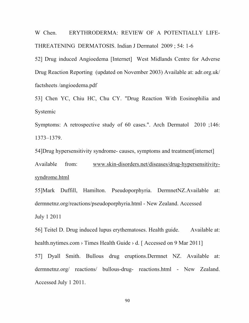
90
W Chen. ERYTHRODERMA: REVIEW OF A POTENTIALLY LIFE-
THREATENING DERMATOSIS. Indian J Dermatol 2009 ; 54: 1-6
52] Drug induced Angioedema [Internet] West Midlands Centre for Adverse
Drug Reaction Reporting (updated on November 2003) Available at: adr.org.uk/
factsheets /angioedema.pdf
53] Chen YC, Chiu HC, Chu CY. "Drug Reaction With Eosinophilia and
Systemic
Symptoms: A retrospective study of 60 cases.". Arch Dermatol 2010 ;146:
1373–1379.
54]Drug hypersensitivity syndrome- causes, symptoms and treatment[internet]
Available from: www.skin-disorders.net/diseases/drug-hypersensitivity-
syndrome.html
55]Mark Duffill, Hamilton. Pseudoporphyria. DermnetNZ.Available at:
dermnetnz.org/reactions/pseudoporphyria.html - New Zealand. Accessed
July 1 2011
56] Teitel D. Drug induced lupus erythematoses. Health guide. Available at:
health.nytimes.com › Times Health Guide › d. [ Accessed on 9 Mar 2011]
57] Dyall Smith. Bullous drug eruptions.Dermnet NZ. Available at:
dermnetnz.org/ reactions/ bullous-drug- reactions.html - New Zealand.
Accessed July 1 2011.
91
58]Moore DE. Drug-induced cutaneous photosensitivity: incidence, mechanism,
prevention and management. Drug Saf. 2002;25:345-72.
59]Delwyn Dyall-Smith. Lichenoid drug eruption.Dermnet NZ. Available
dermnetnz.org/reactions/lichenoid-drug-eruption.html - New Zealand. Accessed
November 9, 2010.
60]Criton S, Sofia B. Acute generalised exanthematous pustulosis. Indian J
Dermatol Venereol Leprol 2001;67:93-5
61] Baack B, Burgdorf W . Chemotherapy-induced acral erythema. J Am Acad
Dermatol24 : 457–61.
62]Sitjas D, Puig L, Cuatrecasas M, De Moragas JM. Acute febrile neutrophilic
dermatosis (Sweet’s syndrome). Int J Dermatol 1993; 32: 261–268.
63]Mittal RR, Jain C, Walia R, Chopra A. Drug-induced pseudolymphoma
syndrome. Indian J Dermatol Venereol Leprol 1995;61:94-5.
64] Venning GR. Validity of anecdotal reports of suspected adverse drug
reactions:
the problem of false alarms. BMJ 1982; 284: 249–52.
65] Stern RS, Wintroub BU. Adverse drug reactions: reporting and evaluating
cuta- neous reactions. Adv Dermatol 1987; 2: 3–18.
66]Sehgal S, Balachandran C, Shenoi SD. Clinical study of cutaneous drug
reactions in 80 patients. Indian J Dermatol Venereol Leprol 2003;69:6-7

92
67] Shear N, Knowles S, Shapiro L. Cutaneous reactions to drugs. In, Wolff K,
Goldsmith L, Katz S, Gilchrest B, Paller A, Leffell D (ed). Fitzpatrick’s
Dermatology in General Medicine, 7th edition, McGraw Hill. 2008; 355- 63
68] Arndt KA, Jick H. Rates of cutaneous reactions to drugs. A report from the
Boston
Collaborative Drug Surveillance Program.JAMA 1976; 235: 918– 22.
69] Sharma VK, Sethuraman G, Kumar B. Cutaneous adverse drug reactions:
clinical pattern and causative agents. A 6 year series from Chandigarh, India.
J Postgrad Med 2001; 47: 95–9.
70] Col N, Fanale JE, Kronholm P. The role of medication noncompliance and
adverse drug reactions in hospitalizations of the elderly.
Arch Intern Med 1990; 150: 841–5.
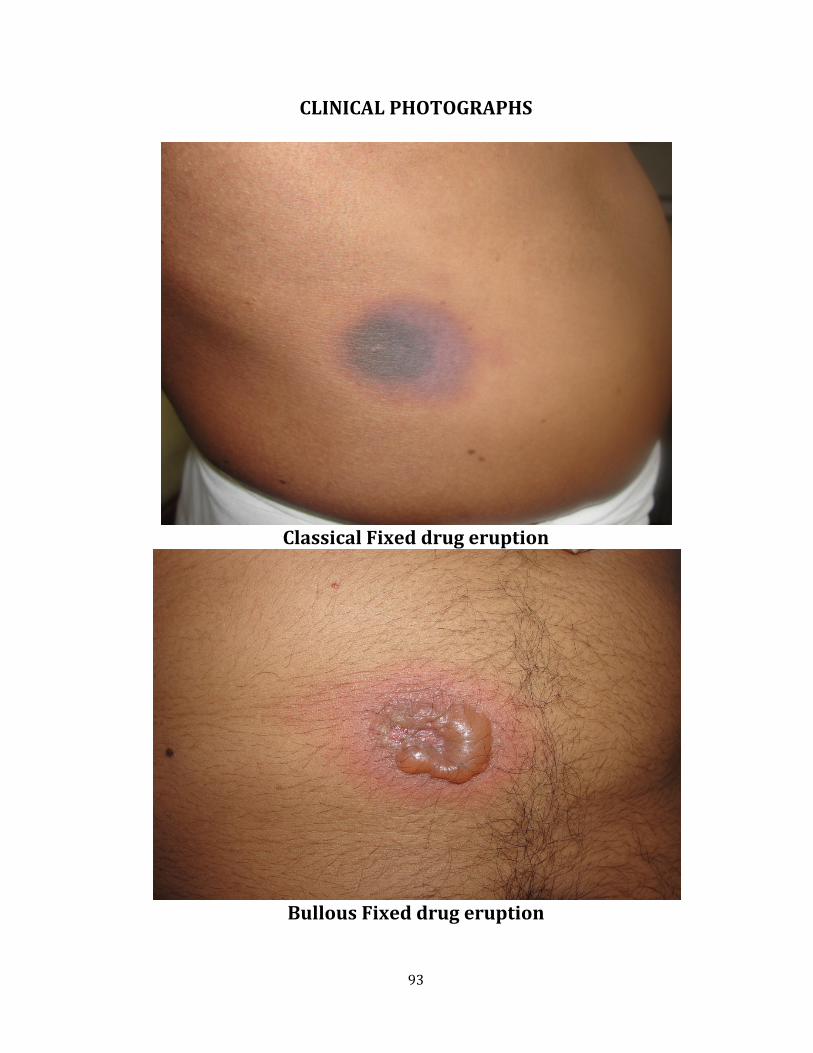
93
CLINICAL PHOTOGRAPHS
Classical Fixed drug eruption
Bullous Fixed drug eruption

Gene
mu
eralised
ucosal Fi
94
Fixed dr
ixed drug
rug erupt
g eruptio
tion
on
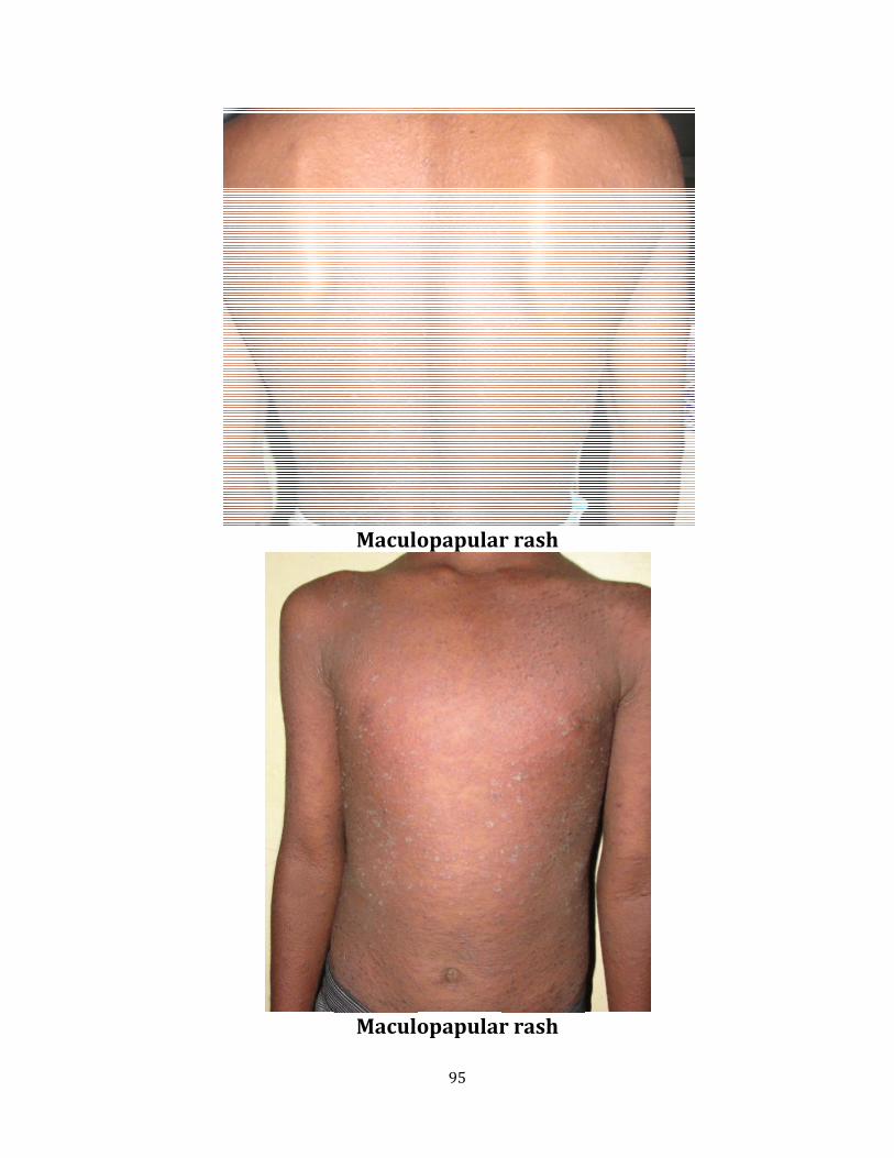
Maculo
Maculo
95
opapular
opapular
r rash
r rash
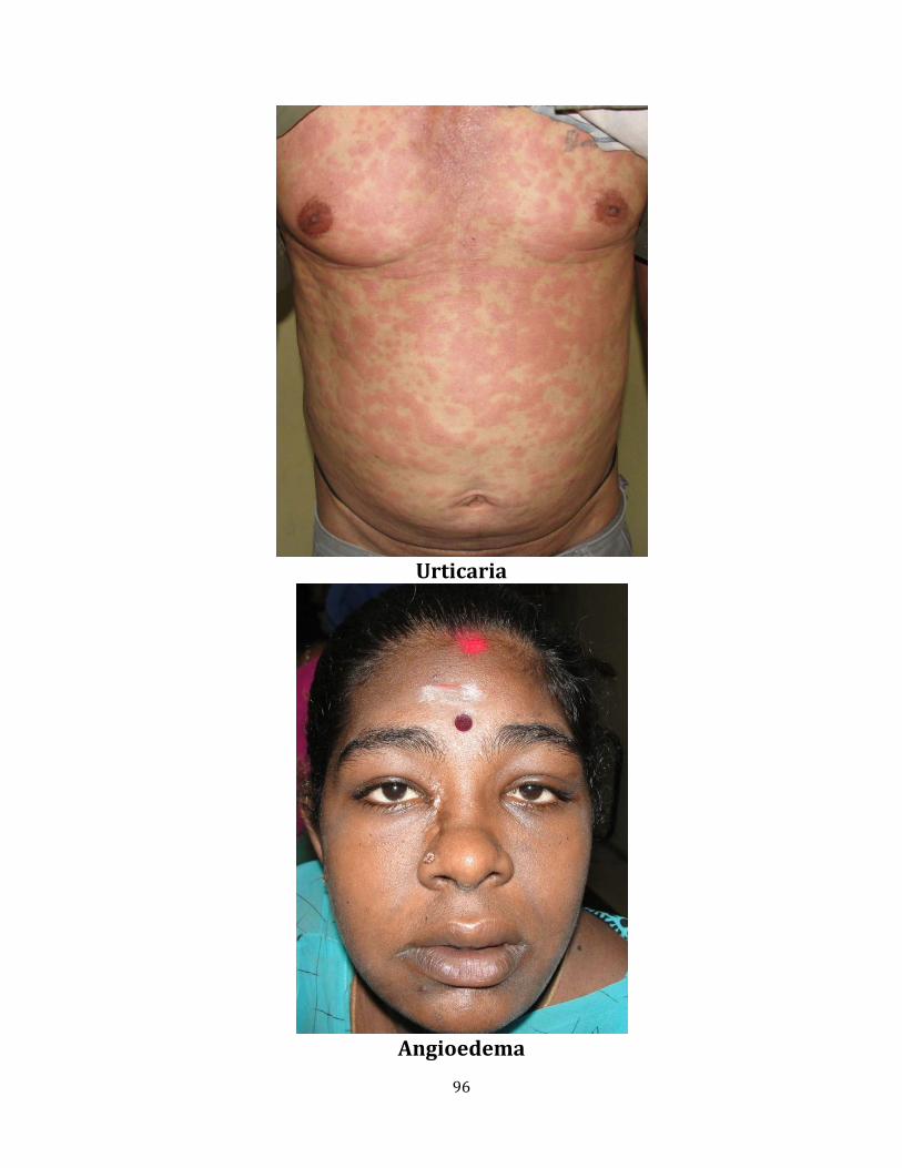
96
Urticaria
Angioedema
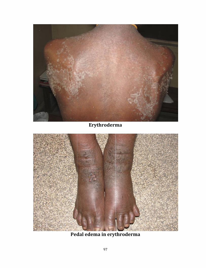
Ped
Ery
dal edem
97
ythroderm
ma in eryt
ma
throdermma

98
Drug rash with eosinophilia and systemic symptoms (dress)
Acral involvement in dress

99
Steven Johnson Syndrome
Erythema Multiforme
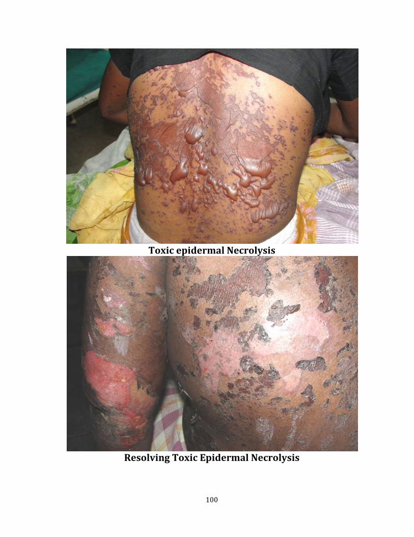
T
Resolv
oxic epid
ving Toxic
100
dermal N
c Epiderm
Necrolysis
mal Necr
s
rolysis
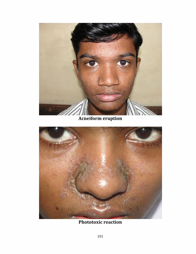
101
Acneiform eruption
Phototoxic reaction

102
PROFORMA
Name:
Age:
Gender :
Address:
DETAILS ABOUT THE PRESENTING CONDITION:
-Time elapsed between drug intake & onset of first sign/symptom:
-Time since onset of first sign/symptom:
-Type of lesion patient developed:
-Presence of mucosal involvement:
ABOUT THE DRUG:
-Probable drug which caused the onset:
-Mode of drug administration:
-Type of drug taken[allopathic/homeopathic/ayurvedic]:
-Nature of illness for which drug was taken:
-Total number of dose of drug taken:
-Any previous h/o drug interactions:
-Whether taken over the counter/prescribed by RMP:
-Precipitating factors if any:
103
CONTRIBUTORY FACTORS:
-Any associated systemic illness:
-If the drug was taken for systemic illness:
-Any family h/o drug interactions:
-Associated secondary complications:
-Scalp/hair/nail involvement:
-Similar episodes in the past:
-If yes then details about the condition:
-Extracutaneous manifestations:
-HIV status of the patient:
DERMATOLOGICAL EXAMINATION:
SYSTEMIC EXAMINATION:
CVS:
RS:
P/A:
CNS:
RELEVANT INVESTIGATIONS CARRIED OUT:
Complete hemogram:
Liver function test:
104
Renal function test:
Tzanck smear:
FINAL DIAGNOSIS:
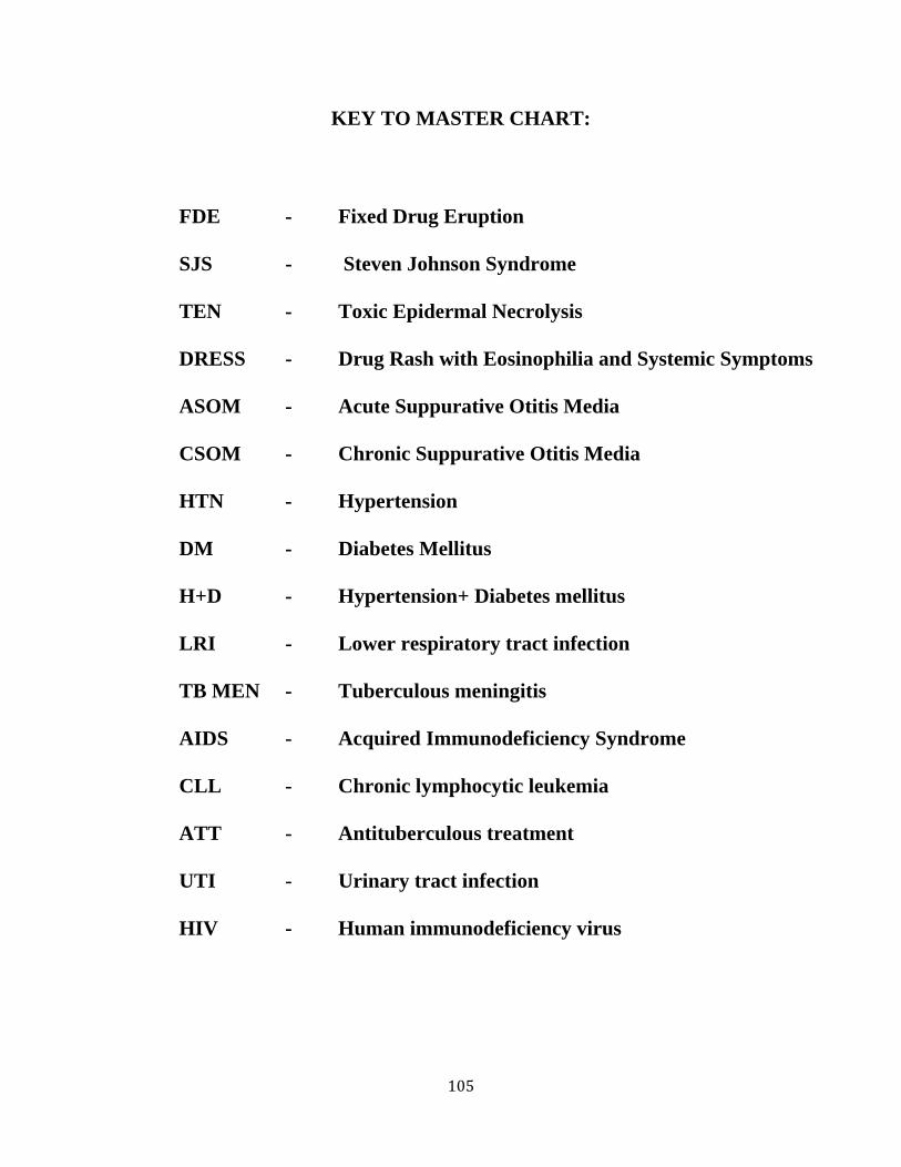
105
KEY TO MASTER CHART:
FDE - Fixed Drug Eruption
SJS - Steven Johnson Syndrome
TEN - Toxic Epidermal Necrolysis
DRESS - Drug Rash with Eosinophilia and Systemic Symptoms
ASOM - Acute Suppurative Otitis Media
CSOM - Chronic Suppurative Otitis Media
HTN - Hypertension
DM - Diabetes Mellitus
H+D - Hypertension+ Diabetes mellitus
LRI - Lower respiratory tract infection
TB MEN - Tuberculous meningitis
AIDS - Acquired Immunodeficiency Syndrome
CLL - Chronic lymphocytic leukemia
ATT - Antituberculous treatment
UTI - Urinary tract infection
HIV - Human immunodeficiency virus

106
A CLINICOEPIDEMIOLOGICAL STUDY ON ADVERSE
CUTANEOUS DRUG REACTIONS
INTRODUCTION:An adverse drug reaction (ADR) may be defined as an
undesirable clinical manifestation resulting from administration of a particular
drug; this includes reactions due to overdose, predictable side effects and
unanticipated adverse manifestations.
Very few published studies have assessed the epidemiological and clinical
features of drug reactions in India and still fewer in South India. Hence this
study was undertaken to assess the pattern and clinical features of ACDRs in
South Tamilnadu.
AIM OF THE STUDY:
1. To study the clinicoepidemiological pattern of various adverse cutaneous
drug reactions in patients attending SKIN OPD, Government Rajaji Hospital.
2. To study the common drugs causing adverse cutaneous drug reactions.
MATERIALS AND METHODS:
This study was conducted at the skin OPD, Government Rajaji Hospital,
Madurai Medical College, Madurai during the period from October 2009-
September 2011 [24 months].All consecutive consenting patients diagnosed
clinically as a case of ACDR of all age groups, of all genders were included in
the study.
RESULTS AND CONCLUSION:
A total of 163 patients out of 1,03,536 were diagnosed with ACDRs.
Maximum number of the patients were in the 4thand 5th decade. Fixed drug
eruption was the commonest type of ACDR followed by Urticaria and
Maculopapular rash, next closely followed by erythroderma . Out of all the
ACDRs 84 cases had exclusive cutaneous involvement only. Both skin and
mucosa were involved in rest of the 79 cases. The commonest class of drug
causing ACDR was antimicrobials. Other classes were anticonvulsants,
NSAIDs, antidepressants & cancer chemotherapy.Cotrimoxazole was the
commonest drug causing ACDR. Among the individual ACDRs, FDE was
most commonly caused by Cotrimoxazole. Among the individual ACDRs,
FDE was most commonly caused by Cotrimoxazole. Mucosal involvement
was most commonly seen with Cotrimoxazole followed by Phenytoin and
doxycycline in succession. Mucosal involvement was most commonly seen
with Cotrimoxazole. None of the patients had a positive family history of
ACDR.
Upper respiratory tract infection was the most common underlying systemic
disorder following which patients had consumed the drug and developed
ACDR.
Key words: ACDRs, Fixed Drug Eruption, Cotrimoxazole.

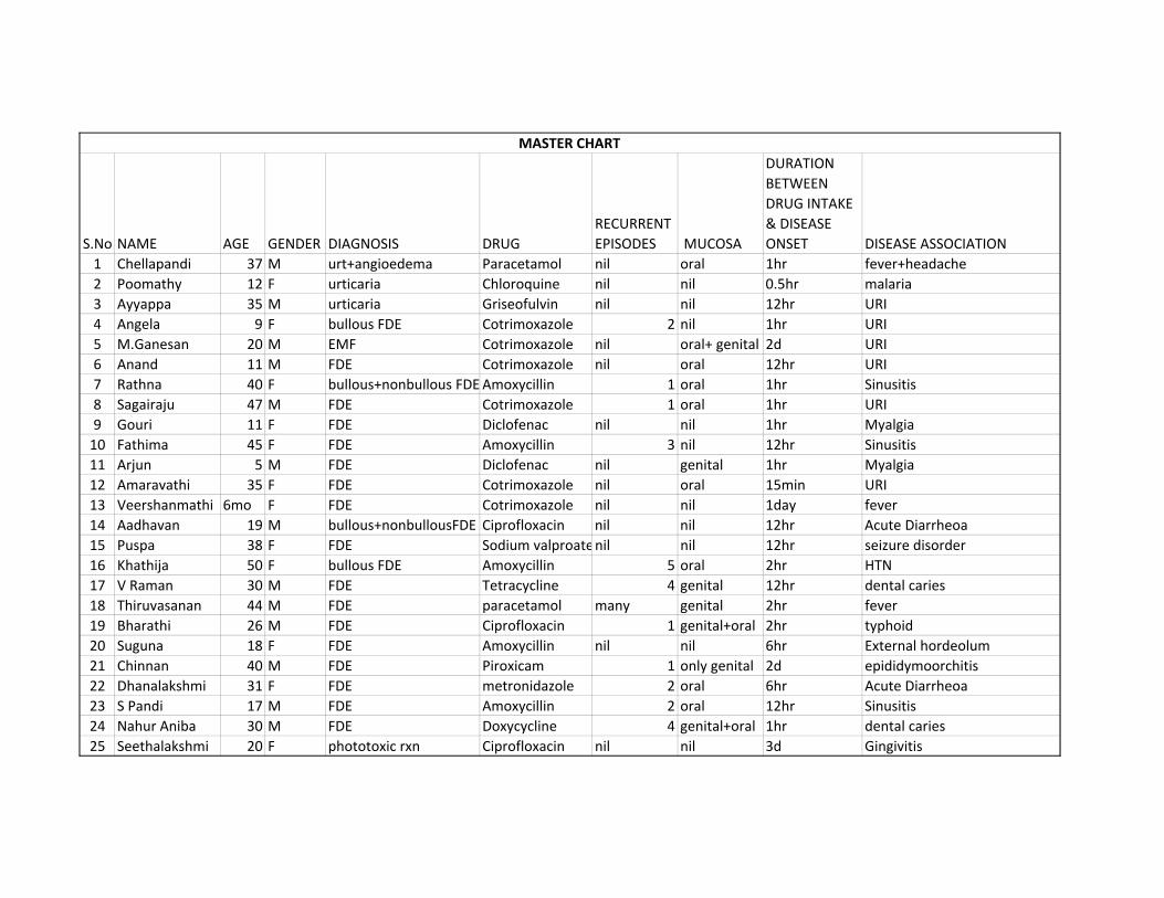
S.No NAME AGE GENDER DIAGNOSIS DRUGRECURRENT EPISODES MUCOSA
DURATION BETWEEN DRUG INTAKE & DISEASE ONSET DISEASE ASSOCIATION
1 Chellapandi 37 M urt+angioedema Paracetamol nil oral 1hr fever+headache2 Poomathy 12 F urticaria Chloroquine nil nil 0.5hr malaria3 Ayyappa 35 M urticaria Griseofulvin nil nil 12hr URI4 Angela 9 F bullous FDE Cotrimoxazole 2 nil 1hr URI5 M.Ganesan 20 M EMF Cotrimoxazole nil oral+ genital 2d URI6 Anand 11 M FDE Cotrimoxazole nil oral 12hr URI7 Rathna 40 F bullous+nonbullous FDEAmoxycillin 1 oral 1hr Sinusitis8 Sagairaju 47 M FDE Cotrimoxazole 1 oral 1hr URI9 Gouri 11 F FDE Diclofenac nil nil 1hr Myalgia10 Fathima 45 F FDE Amoxycillin 3 nil 12hr Sinusitis11 Arjun 5 M FDE Diclofenac nil genital 1hr Myalgia12 Amaravathi 35 F FDE Cotrimoxazole nil oral 15min URI13 Veershanmathi 6mo F FDE Cotrimoxazole nil nil 1day fever 14 Aadhavan 19 M bullous+nonbullousFDE Ciprofloxacin nil nil 12hr Acute Diarrheoa15 Puspa 38 F FDE Sodium valproatenil nil 12hr seizure disorder16 Khathija 50 F bullous FDE Amoxycillin 5 oral 2hr HTN17 V Raman 30 M FDE Tetracycline 4 genital 12hr dental caries18 Thiruvasanan 44 M FDE paracetamol many genital 2hr fever 19 Bharathi 26 M FDE Ciprofloxacin 1 genital+oral 2hr typhoid20 Suguna 18 F FDE Amoxycillin nil nil 6hr External hordeolum21 Chinnan 40 M FDE Piroxicam 1 only genital 2d epididymoorchitis22 Dhanalakshmi 31 F FDE metronidazole 2 oral 6hr Acute Diarrheoa23 S Pandi 17 M FDE Amoxycillin 2 oral 12hr Sinusitis24 Nahur Aniba 30 M FDE Doxycycline 4 genital+oral 1hr dental caries25 Seethalakshmi 20 F phototoxic rxn Ciprofloxacin nil nil 3d Gingivitis
MASTER CHART
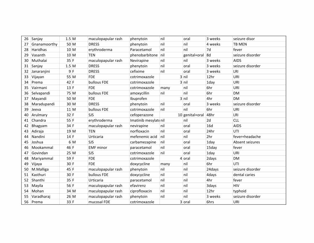
26 Sanjay 1.5 M maculopapular rash phenytoin nil oral 3 weeks seizure disor27 Gnanamoorthy 50 M DRESS phenytoin nil nil 4 weeks TB MEN28 Haridhas 10 M erythroderma Paracetamol nil nil 7d fever 29 Vasanth 10 M TEN phenobarbitone nil genital+oral 8d seizure disorder30 Muthalai 35 F maculopapular rash Nevirapine nil nil 3 weeks AIDS31 Sanjay 1.5 M DRESS phenytoin nil oral 3 weeks seizure disorder32 Janaranjini 9 F DRESS cefixime nil oral 3 weeks LRI33 Vijayan 55 M FDE cotrimoxazole 3 nil 12hr URI34 Prema 42 F bullous FDE cotrimoxazole 3 nil 1day URI35 Vairmani 13 F FDE cotrimoxazole many nil 6hr URI36 Selvapandi 75 M bullous FDE amoxycillin nil nil 6hr DM37 Mayandi 50 M FDE Ibuprofen 3 nil 4hr DM38 Maradupandi 30 M DRESS phenytoin nil oral 3 weeks seizure disorder39 Jeeva 11 M bullous FDE cotrimoxazole nil nil 6hr URI40 Arulmary 32 F SJS cefoperazone 10 genital+oral 48hr LRI41 Chandra 55 F erythroderma Imatinib mesylatenil nil 2d CLL42 Bhagyam 34 F maculopapular rash nevirapine nil oral 16d AIDS43 Adiraja 19 M TEN norfloxacin nil oral 24hr UTI44 Nandini 14 F Urticaria mefenemic acid nil nil 2hr fever+headache45 Joshua 6 M SJS carbamezapine nil oral 1day Absent seizures46 Mookammal 46 F EMF minor paracetamol nil oral 15day fever 47 Govindan 25 M SJS cotrimoxazole nil oral 1day URI48 Mariyammal 59 F FDE cotrimoxazole 4 oral 2days DM49 Vijaya 30 F FDE doxycycline many nil 6hr UTI50 M.Malliga 45 F maculopapular rash phenytoin nil nil 24days seizure disorder51 Kasthuri 30 F bullous FDE doxycycline nil nil 4days dental caries52 Shanthi 35 F Urticaria paracetamol nil nil 4hr fever 53 Mayila 56 F maculopapular rash efavirenz nil nil 3days HIV54 Mohan 34 M maculopapular rash ciprofloxacin nil nil 12hr typhoid55 Varadharaj 26 M maculopapular rash phenytoin nil nil 3 weeks seizure disorder56 Prema 33 F mucosal FDE cotrimoxazole 3 oral 6hrs URI
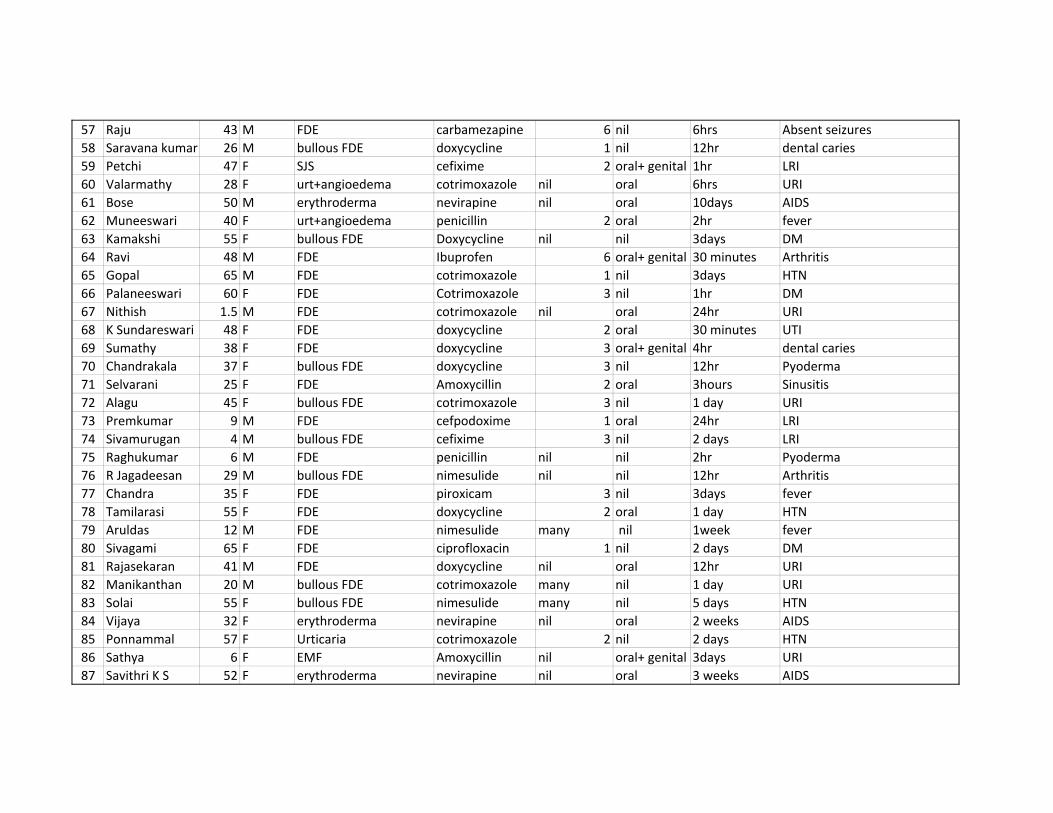
57 Raju 43 M FDE carbamezapine 6 nil 6hrs Absent seizures58 Saravana kumar 26 M bullous FDE doxycycline 1 nil 12hr dental caries59 Petchi 47 F SJS cefixime 2 oral+ genital 1hr LRI60 Valarmathy 28 F urt+angioedema cotrimoxazole nil oral 6hrs URI61 Bose 50 M erythroderma nevirapine nil oral 10days AIDS62 Muneeswari 40 F urt+angioedema penicillin 2 oral 2hr fever 63 Kamakshi 55 F bullous FDE Doxycycline nil nil 3days DM64 Ravi 48 M FDE Ibuprofen 6 oral+ genital 30 minutes Arthritis65 Gopal 65 M FDE cotrimoxazole 1 nil 3days HTN66 Palaneeswari 60 F FDE Cotrimoxazole 3 nil 1hr DM67 Nithish 1.5 M FDE cotrimoxazole nil oral 24hr URI68 K Sundareswari 48 F FDE doxycycline 2 oral 30 minutes UTI69 Sumathy 38 F FDE doxycycline 3 oral+ genital 4hr dental caries70 Chandrakala 37 F bullous FDE doxycycline 3 nil 12hr Pyoderma71 Selvarani 25 F FDE Amoxycillin 2 oral 3hours Sinusitis72 Alagu 45 F bullous FDE cotrimoxazole 3 nil 1 day URI73 Premkumar 9 M FDE cefpodoxime 1 oral 24hr LRI74 Sivamurugan 4 M bullous FDE cefixime 3 nil 2 days LRI75 Raghukumar 6 M FDE penicillin nil nil 2hr Pyoderma76 R Jagadeesan 29 M bullous FDE nimesulide nil nil 12hr Arthritis77 Chandra 35 F FDE piroxicam 3 nil 3days fever 78 Tamilarasi 55 F FDE doxycycline 2 oral 1 day HTN79 Aruldas 12 M FDE nimesulide many nil 1week fever 80 Sivagami 65 F FDE ciprofloxacin 1 nil 2 days DM81 Rajasekaran 41 M FDE doxycycline nil oral 12hr URI82 Manikanthan 20 M bullous FDE cotrimoxazole many nil 1 day URI83 Solai 55 F bullous FDE nimesulide many nil 5 days HTN84 Vijaya 32 F erythroderma nevirapine nil oral 2 weeks AIDS85 Ponnammal 57 F Urticaria cotrimoxazole 2 nil 2 days HTN86 Sathya 6 F EMF Amoxycillin nil oral+ genital 3days URI87 Savithri K S 52 F erythroderma nevirapine nil oral 3 weeks AIDS

88 Janaranjini 6 F DRESS cefoperazone nil oral 2 weeks LRI89 Amutha 24 F maculopapular rash phenytoin 2 oral 1 month seizure disorder90 Prakash 35 M SJS phenytoin nil oral+ genital 15 days seizure disorder91 Rathna 50 F urt+angioedema cotrimoxazole nil oral 12hr URI92 Lokanathan 25 M Urticaria ciprofloxacin nil nil 4hr typhoid93 Panneer selvam 52 M erythroderma carbamezapine nil nil 1 month seizure disorder94 Nagarani 20 F acneiform eruptions ATT nil nil 3 months TB MEN95 Prabhakar 21 M acneiform eruptions ATT nil nil 20 days TB MEN96 Azhagar Raj 48 M Urticaria cotrimoxazole nil nil 4hr URI97 Kashi 70 M Urticaria metronidazole nil nil 6hr H+D98 Pandi 36 M urt+angioedema doxycycline nil oral+ genital 1hr dental caries99 Eswari 17 F DRESS phenytoin nil oral 2 weeks seizure disorder100 Amaravathi 38 F FDE paracetamol nil nil 3hours fever 101 Mahalingam 65 M FDE ibuprofen nil nil 30 minutes H+D102 Mohan 47 M FDE cotrimoxazole many oral+ genital 2hr URI103 Dinesh 22 M FDE ciprofloxacin 3 nil 3days Sinusitis104 Kavitha 25 F FDE amoxycillin nil nil 2 days ASOM105 Pasupathi 25 M SJS cefixime nil oral+ genital 2 days LRI106 Panju 43 F erythroderma sodium valproate nil oral 2 months seizure disorder107 Ayyanar 60 M urticaria piroxicam 3 nil 3hours H+D108 Chinnakani 40 M TEN phenytoin nil oral+ genital 1 month seizure disorder109 Anushya 17 F FDE penicillin 3 nil 3hours URI110 Mani 40 F bullous FDE cotrimoxazole 4 oral 6hrs URI111 Syed Ibrahim 40 M bullous FDE doxycycline 2 oral 7 hours dental caries112 Parvathi 24 F erythroderma chlorpromazine nil nil 1 month seizure disorder113 Vijaya 32 F FDE cotrimoxazole 6 nil 4hr URI114 Sumathy 30 F maculopapular rash phenytoin nil oral 4 weeks seizure disorder115 Ammani 6 F Urticaria chloroquine nil nil 2hr malaria116 Alagammal 23 F Urticaria Amoxycillin 4 nil 4hr ASOM117 Backiaraja 32 M maculopapular rash phenytoin nil nil 3 weeks seizure disorder118 Duraiammal 34 F urt+angioedema metronidazole 3 oral+ genital 4hr Acute Diarrheoa

119 Eswar 45 M erythroderma sodium valproate nil nil 6 weeks seizure disorder120 Thirupathy 16 M FDE cotrimoxazole many oral 4hr URI121 T K Ramesh 41 M FDE Amoxycillin nil nil 5 hours Sinusitis122 Vishith 13 M FDE cotrimoxazole 3 nil 6hrs URI123 Seema 10 F bullous FDE cotrimoxazole 4 oral 12hr URI124 Saraswathi 47 F FDE cotrimoxazole 2 nil 30 minutes fever 125 Murugan 37 M FDE NSAIDs 6 oral 2hr fever 126 Ayyava 30 M FDE cotrimoxazole 3 oral 8 hours URI127 Praveen 36 M Urticaria paracetamol many nil 6hrs fever+headache128 Waheeda banu 32 F urt+angioedema ciprofloxacin 4 oral 8 hours UTI129 Latha 25 F maculopapular rash phenytoin nil nil 3 weeks seizure disorder130 Umapathy 53 M erythroderma carbamezapine nil nil 2 months seizure disorder131 Sitalakshmi 45 F urt+angioedema cefixime 4 oral+ genital 3hours LRI132 Kannan 56 M EMF Tetracycline many oral 6hrs CSOM133 Vasanthi 45 F erythroderma dapsone nil nil 2 months leprosy134 Bhuvaneswari 65 F DRESS phenytoin nil nil 4 weeks seizure disorder135 Chinnakalai 45 M Urticaria Imipramine 6 nil 5 hours Depression136 Asoka 32 M FDE cotrimoxazole 2 nil 4hr UTI137 Thiyagaraj 43 M maculopapular rash nevirapine nil nil 2 weeks HIV138 Pavan 28 M EMF penicillin many oral+ genital 48hr Sinusitis139 Sangilikalai 54 M Urticaria cotrimoxazole many nil 12hr LRI140 Pandiarajan 34 M FDE Aspirin 3 nil 1hr fever+headache141 Visheeth 16 M FDE cotrimoxazole nil genital 2hr URI142 Sooryabiwi 60 F FDE doxycycline 4 oral 30 minutes UTI143 Krishnan 26 M SJS Amoxycillin nil oral+ genital 3hours Sinusitis144 Chinnasamy 30 M bullous FDE doxycycline nil oral 12hr dental caries145 Abdul Rahman 30 M maculopapular rash Amoxycillin nil nil 1 day fever 146 Lakshmi 23 F bullous FDE ibuprofen nil oral 4hr Myalgia147 Thiyagaraja 7 M FDE aspirin nil nil 7 hours fever 148 Seethalakshmi 50 F bullous+nonbullous FDE cotrimoxazole many oral+ genital 2hr URI149 Lakshmi 28 F bullous FDE diclofenac 3 oral 3hours bursitis
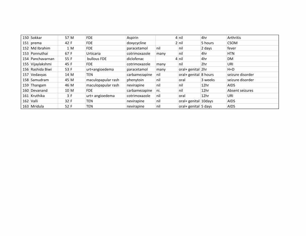
150 Sokkar 57 M FDE Aspirin 4 nil 4hr Arthritis151 prema 42 F FDE doxycycline 2 nil 5 hours CSOM152 Md Ibrahim 1 M FDE paracetamol nil nil 2 days fever 153 Ponnuthai 67 F Urticaria cotrimoxazole many nil 4hr HTN154 Panchavarnan 55 F bullous FDE diclofenac 4 nil 4hr DM155 Vijaylakshmi 45 F FDE cotrimoxazole many nil 2hr URI156 Rashida Biwi 53 F urt+angioedema paracetamol many oral+ genital 2hr H+D157 Vedavyas 14 M TEN carbamezapine nil oral+ genital 8 hours seizure disorder158 Samudram 45 M maculopapular rash phenytoin nil oral 3 weeks seizure disorder159 Thangam 46 M maculopapular rash nevirapine nil nil 12hr AIDS160 Devanand 10 M FDE carbamezapine ni. nil 12hr Absent seizures161 Kruthika 3 F urt+ angioedema cotrimoxazole nil oral 12hr URI162 Valli 32 F TEN nevirapine nil oral+ genital 10days AIDS163 Mridula 52 F TEN nevirapine nil oral+ genital 5 days AIDS