Conjugated linoleic acid in meat and meat products: A review
Upload
khangminh22Category
view
4download
0
Tasmanian Institute of Agriculture
A study of key meat spoilage bacteria
By
Tamsyn Stanborough
Master of Science
Submitted in fulfilment of the requirements for the
Doctor of Philosophy (Agriculture)
February 2018

i
Declaration of originality
This thesis contains no material which has been accepted for a degree or diploma by the
University or any other institution, except by way of background information and duly
acknowledged in the thesis, and to the best of my knowledge and belief no material
previously published or written by another person except where due acknowledgement is
made in the text of the thesis, nor does the thesis contain any material that infringes
copyright.
(Tamsyn Stanborough)
University of Tasmania
February 2018
Statement on authority of access
This thesis may be made available for loan and limited copying and communication in
accordance with the Copyright Act 1968.
(Tamsyn Stanborough)
University of Tasmania
February 2018
ii
Statement regarding published work contained in this thesis
The publishers of the papers comprising Chapters 2, 4 and 5 hold the copyright for that
content and access to the material should be sought from the respective journals. The
remaining non published content of the thesis may be made available for loan and limited
copying and communication in accordance with the Copyright Act 1968.
(Tamsyn Stanborough)
University of Tasmania
February 2018
Statement of ethical conduct
The research associated with this thesis abides by the international and Australian codes on
human and animal experimentation, the guidelines by the Australian Government's Office of
the Gene Technology Regulator and the rulings of the Safety, Ethics and Institutional
Biosafety Committees of the University.
(Tamsyn Stanborough)
University of Tasmania
February 2018
iii
Co-authorship statements
The following people and institutions contributed to the publication of work undertaken as
part of this thesis:
Author 1: Tamsyn Stanborough, Tasmanian Institute of Agriculture, University of Tasmania
(Candidate)
Author 2: Shane M. Powell, Tasmanian Institute of Agriculture, University of Tasmania
(Primary supervisor)
Author 3: Mark Tamplin, Tasmanian Institute of Agriculture, University of Tasmania (Co-
supervisor)
Author 4: P. Scott Chandry, Agriculture and Food, CSIRO (Co-supervisor)
Author 5: Narelle Fegan, Agriculture and Food, CSIRO (Co-supervisor)
Author 6: Tanoj Singh, Agriculture and Food, CSIRO (external collaborator)
Author 7: Randy Suryadinata, Manufacturing, CSIRO (external collaborator)
Author 8: Stewart D. Nuttall, Manufacturing, CSIRO (external collaborator)
DETAILS OF THE AUTHOR ROLES
Chapter 2: Insight into the genome of Brochothrix thermosphacta, a problematic meat
spoilage bacterium.
Published as: Stanborough T., Fegan N., Powell S.M., Tamplin M. and Chandry P.S., 2017.
Insight into the genome of Brochothrix thermosphacta, a problematic meat spoilage
bacterium. Applied and Environmental Microbiology, 83, e02786-16.
Author 1 contributed 70% (conceptualised the study, conducted experimental work,
analysed data and wrote the manuscript), authors 5, 2 and 3 each contributed 5% (assisted
iv
with study development and commented on the manuscript) and author 4 contributed 15%
(guided experimental design and study development, assisted with data analysis and
commented on the manuscript).
Chapter 3: Characterisation of a putative Brochothrix thermosphacta sortase A enzyme.
Submitted for publication as: Stanborough T., Suryadinata R., Fegan N., Powell S.M.,
Tamplin M., Nuttall S.D. and Chandry P.S. Characterisation of a putative Brochothrix
thermosphacta sortase A enzyme.
Author 1 contributed 70% (conceptualised the study, conducted all experimental work,
analysed data and wrote the manuscript), author 7 contributed 8% (assisted with
experimental design and commented on the manuscript), authors 5, 2 and 3 each
contributed 3% (commented on the manuscript), author 8 contributed 8% (guided
experimental design and study development and commented on the manuscript) and
author 4 contributed 5% (contributed to study development and commented on the
manuscript).
Chapter 4: Genomic and metabolic characterisation of spoilage-associated Pseudomonas
species.
Published as: Stanborough T., Fegan N., Powell S.M., Singh T., Tamplin M., and Chandry P.S.,
2018. Genomic and metabolic characterisation of spoilage-associated Pseudomonas species.
International Journal of Food Microbiology, 268: 61-72.
Author 1 contributed 70% (conceptualised the study, conducted all experimental work,
analysed data and wrote the majority of the manuscript), authors 5, 2 and 3 each

v
contributed 5% (assisted with the interpretation of results and commented on the
manuscript), author 6 contributed 8% (assisted with experimental design, data analysis and
writing of the methods section of the headspace analysis, and commented on the
manuscript) and author 4 contributed 7% (assisted with experimental design and
commented on the manuscript).
Chapter 5: Vibrioferrin production by the food spoilage bacterium Pseudomonas fragi.
Published as: Stanborough T., Fegan N., Powell S.M., Tamplin M., and Chandry P.S., 2018.
Vibrioferrin production by the food spoilage bacterium Pseudomonas fragi. FEMS
Microbiology Letters, 365(6), fnx279.
Author 1 contributed 75% (conceptualised the study, conducted all experimental work,
analysed data and wrote the manuscript), authors 5, 2 and 3 each contributed 5% (assisted
with the interpretation of results and commented on the manuscript) and author 4
contributed 10% (guided experimental design and commented on the manuscript).
We the undersigned agree with the above stated “proportion of work undertaken” for each
of the above published (or submitted) peer-reviewed manuscripts contributing to this
thesis:
Signed:
(Shane M. Powell)
Primary supervisor
School of Land and Food
University of Tasmania
(Prof. Holger Meinke)
Head of School
School of Land and Food
University of Tasmania
Date: February 2, 2018

vi
Publications and communications arising from this research
Journal publications
Stanborough T., Fegan N., Powell S.M., Tamplin M. and Chandry P.S., 2017. Insight into
the genome of Brochothrix thermosphacta, a problematic meat spoilage bacterium.
Applied and Environmental Microbiology, 83, e02786-16.
Stanborough T., Fegan N., Powell S.M., Singh T., Tamplin M., and Chandry P.S., 2018.
Genomic and metabolic characterisation of spoilage-associated Pseudomonas species.
International Journal of Food Microbiology, 268: 61-72.
Stanborough T., Fegan N., Powell S.M., Tamplin M., and Chandry P.S., 2018. Vibrioferrin
production by the food spoilage bacterium Pseudomonas fragi. FEMS Microbiology
Letters, 365(6), fnx279..
Conference presentations (poster format)
Stanborough T., Fegan N., Powell S.M., Tamplin M. and Chandry P.S., 2016. Comparative
genomics of the meat spoilage bacterium, Brochothrix thermosphacta. Australian
Society of Microbiology 2016 Annual Scientific Meeting, Perth, Australia.
Stanborough T., Suryadinata R., Fegan N., Powell S.M., Tamplin M., Nuttall S.D. and
Chandry P.S., 2016. Characterisation of a putative Brochothrix thermosphacta sortase
enzyme. Australian Society of Microbiology 2016 Victoria/Tasmania Bi-state Conference,
Launceston, Tasmania.
Stanborough T., Chandry P.S., Powell S.M., Tamplin M. and Fegan N., 2017. A putative
siderophore locus of Pseudomonas fragi: Solving an iron problem. International
Association of Food Protection 2017 Annual Meeting, Tampa, USA.

vii
Acknowledgements
First and foremost, I would like to acknowledge the funding support from the Australian
Meat Processor Corporation and CSIRO, without which this research would not have been
possible. I would like to express my sincere gratitude to all of my supervisors, Scott Chandry,
Narelle Fegan, Shane Powell and Mark Tamplin. Without their outstanding knowledge and
leadership, invaluable suggestions and constructive criticism, this work would not have been
possible. I am also truly grateful for the encouragement I received from all of my
supervisors, which helped me to stay positive and confident when times were tough.
My sincere appreciation goes to Stewart Nuttall for giving me the opportunity to conduct
the research from chapter 3 under his guidance at CSIRO Manufacturing, and I would also
like to thank Randy Suryadinata for his help and advice for this section. I am very grateful to
David Ratkowsky for the statistical help he provided for the work in chapter 6 and I would
like to thank Steve Petrovski for providing strains, plasmids and advice for the work in
chapter 5. I am also incredibly grateful to Ed Fox and Tanoj Singh for their help, advice and
insight, which were invaluable for improving the work in this thesis.
I have been very lucky to have been based for my research at CSIRO Agriculture and Food in
Werribee. The support and kindness I received from all of the staff members was amazing
and made my time thoroughly enjoyable.
Last but not least, I would like to thank my partner Barbara Koch for putting up with my
whinging and worrying, for her constant encouragement and advice, and for being the best
listener.

viii
Table of contents
Declaration of originality ........................................................................................................... i
Statement on authority of access .............................................................................................. i
Statement regarding published work contained in this thesis ............................................... ii
Statement of ethical conduct ................................................................................................... ii
Co-authorship statements ....................................................................................................... iii
Publications and communications arising from this research................................................ vi
Acknowledgements ................................................................................................................. vii
Abstract................................................................................................................................... xiii
Chapter 1 ................................................................................................................................... 1
Literature review and thesis objectives ................................................................................... 1
INTRODUCTION ....................................................................................................................... 1
COMPOSITION OF FRESH RED MEAT ...................................................................................... 3
PRE-SLAUGHTER HANDLING OF LIVESTOCK ........................................................................... 5
SLAUGHTER PROCESS AND CARCASS DRESSING AS INITIAL CONTAMINATION POINTS ........ 6
MEAT PRESERVATION METHODS............................................................................................ 7
Cooling .................................................................................................................................. 8
Packaging systems ................................................................................................................ 8
MEAT PROCESSING INTERVENTIONS .................................................................................... 10
PEF ...................................................................................................................................... 10
MEAT SPOILAGE .................................................................................................................... 12
COMPLEXITY OF SPOILAGE COMMUNITIES .......................................................................... 16
MEAT SPOILAGE MICROORGANISMS ................................................................................... 18
Brochothrix spp. ................................................................................................................. 22
Pseudomonas spp. .............................................................................................................. 24
THESIS OBJECTIVES ............................................................................................................... 27
REFERENCES .......................................................................................................................... 29
Chapter 2 ................................................................................................................................. 50

ix
Insight into the genome of Brochothrix thermosphacta, a problematic meat spoilage
bacterium................................................................................................................................. 50
ABSTRACT .............................................................................................................................. 50
Importance ......................................................................................................................... 51
INTRODUCTION ..................................................................................................................... 51
MATERIALS AND METHODS .................................................................................................. 54
Bacterial strains .................................................................................................................. 54
Strain identification ............................................................................................................ 56
DNA isolation, whole genome sequencing and gene annotation...................................... 57
Genome sequence similarity analyses ............................................................................... 58
Pan-genome analysis .......................................................................................................... 58
Phylogeny of Brochothrix strains ....................................................................................... 59
Identification of genes involved in cell metabolism .......................................................... 60
Virulence factor search in Brochothrix ............................................................................... 60
Biochemical tests of strains ................................................................................................ 61
Screening for biogenic amine production .......................................................................... 61
MIC testing of antibiotics ................................................................................................... 61
MTC testing of heavy metals .............................................................................................. 62
Accession numbers............................................................................................................. 62
RESULTS ................................................................................................................................. 62
Genomic features and intergenomic sequence similarity of strains ................................. 62
Pan-genome investigation ................................................................................................. 65
Phylogeny of B. thermosphacta strains ............................................................................. 68
Cell metabolism .................................................................................................................. 72
Resistance genes ................................................................................................................ 75
Presence of Listeria virulence genes in Brochothrix .......................................................... 77
Stress response genes ........................................................................................................ 80
DISCUSSION ........................................................................................................................... 82
Intergenomic sequence similarity of strains ...................................................................... 82
Pan-genome investigation ................................................................................................. 83
Cell metabolism .................................................................................................................. 85

x
Resistance genes ................................................................................................................ 88
Presence of Listeria virulence genes in Brochothrix .......................................................... 89
Stress response genes ........................................................................................................ 91
Conclusion .......................................................................................................................... 91
REFERENCES .......................................................................................................................... 92
Chapter 3 ............................................................................................................................... 105
Characterisation of a putative Brochothrix thermosphacta sortase A enzyme.................. 105
ABSTRACT ............................................................................................................................ 105
INTRODUCTION ................................................................................................................... 106
MATERIALS AND METHODS ................................................................................................ 109
Multiple sequence alignment of SrtA proteins ................................................................ 109
Cloning, expression and purification of BtSrtA construct ................................................ 109
In vitro thioacyl intermediate reactions ........................................................................... 110
In vitro bioconjugation ..................................................................................................... 111
Genome mining for and characterisation of BtSrtA substrate proteins .......................... 111
RESULTS ............................................................................................................................... 112
DISCUSSION ......................................................................................................................... 121
REFERENCES ........................................................................................................................ 124
Chapter 4 ............................................................................................................................... 130
Genomic and metabolic characterisation of spoilage-associated Pseudomonas species .. 130
ABSTRACT ............................................................................................................................ 130
INTRODUCTION ................................................................................................................... 131
MATERIALS AND METHODS ................................................................................................ 132
Bacterial isolates .............................................................................................................. 132
DNA isolation, genome sequencing, assembly and annotation ...................................... 136
Genomic analyses ............................................................................................................. 136
Determination of average nucleotide identity .............................................................. 136
Single Nucleotide Polymorphism (SNP) analysis ........................................................... 136
Pan-genome .................................................................................................................. 137
Identification of homologous genes .............................................................................. 137

xi
Metabolic analyses ........................................................................................................... 138
Sample preparation in headspace vials ......................................................................... 138
Determination of atmospheric gases and bacterial numbers in headspace vials ........ 139
Headspace analysis by solid phase micro-extraction-gas chromatography mass
spectrometry (SPME-GCMS) ......................................................................................... 140
RESULTS ............................................................................................................................... 142
Phylogeny ......................................................................................................................... 142
Pan-genome ..................................................................................................................... 146
Volatile analysis ................................................................................................................ 150
Esters ............................................................................................................................. 152
Ketones .......................................................................................................................... 154
Other volatile compounds ............................................................................................. 156
DISCUSSION ......................................................................................................................... 157
Genomic analyses ............................................................................................................. 157
VOC analysis ..................................................................................................................... 158
Conclusions....................................................................................................................... 162
REFERENCES ........................................................................................................................ 162
Chapter 5 ............................................................................................................................... 168
Vibrioferrin production by the food spoilage bacterium Pseudomonas fragi .................... 168
ABSTRACT ............................................................................................................................ 168
INTRODUCTION ................................................................................................................... 169
MATERIALS AND METHODS ................................................................................................ 171
Strains, plasmids and growth conditions ......................................................................... 171
Identification of siderophore gene cluster ...................................................................... 173
Generation of disruption mutants, genomic DNA isolation, and genome sequencing and
analysis ............................................................................................................................. 174
Phylogenetic tree of vibrioferrin sequences .................................................................... 175
RNA isolation and quantitative reverse transcription-polymerase chain reaction (qRT-PCR)
.......................................................................................................................................... 176
Chromeazurol S (CAS) supernatant assays....................................................................... 177
Growth experiments with 2,2’-bipyridyl and bovine apo-transferrin ............................. 177

xii
RESULTS AND DISCUSSION .................................................................................................. 178
Conclusion ........................................................................................................................ 188
REFERENCES ........................................................................................................................ 188
Chapter 6 ............................................................................................................................... 195
The effect of a low field strength pulsed electric field on the aerobic microbiota of beef 195
ABSTRACT ............................................................................................................................ 195
INTRODUCTION ................................................................................................................... 196
MATERIALS AND METHODS ................................................................................................ 197
Meat sampling and preparation ...................................................................................... 197
PEF equipment and treatment of meat ........................................................................... 198
Microbiological analysis ................................................................................................. 2199
Reversed phase-high performance liquid chromatography (RP-HPLC) ........................... 200
RESULTS AND DISCUSSION .................................................................................................. 200
REFERENCES ........................................................................................................................ 205
Chapter 7 ............................................................................................................................... 208
General Discussion ................................................................................................................ 208
REFERENCES ........................................................................................................................ 215
Appendix A ............................................................................................................................ 219
Appendix B............................................................................................................................. 227
Appendix C ............................................................................................................................. 230

xiii
Abstract
Fresh meat is a highly nutritious commodity, conducive to the rapid growth of
microorganisms and spoilage. Despite the economic and social importance associated with
preventing meat spoilage and minimising food waste, the physiology of many
microorganisms involved in meat spoilage remains poorly studied. Such fundamental
knowledge is of great importance to enable further improvements in the control of meat
contamination and spoilage, and to facilitate shelf-life extension. In this context, the broad
objective of this thesis was to improve current knowledge of the biology of important meat
spoilage bacteria, Brochothrix thermosphacta and psychrotrophic pseudomonads
Pseudomonas fragi and Pseudomonas lundensis.
In contrast to the well-studied and phylogenetically related pathogen Listeria
monocytogenes, genome analyses of B. thermosphacta were lacking. Thus, draft genomes of
12 B. thermosphacta strains were analysed and compared to genomes of Brochothrix
campestris and L. monocytogenes to identify genes that play a role in spoilage and
persistence of B. thermosphacta throughout the meat production chain. The 12 strains
shared a high degree of genomic similarity. Genes/pathways likely involved in the
production of organoleptically unpleasant compounds such as acetoin, butanediol and
isobutyric, isovaleric and 2-methylbutyric acid were identified in both Brochothrix species,
while amino acid decarboxylase genes were not found, and phenotypic testing confirmed
their absence. Orthologues of key Listeria virulence genes were absent from the Brochothrix
genomes, however auxiliary virulence genes such as factors involved in surface protein
anchoring (e.g. sortase A), and key stress response regulatory genes were identified,
establishing parallels to and differences from this related foodborne pathogen.
xiv
Gram-positive bacteria utilise class A sortases to attach a diversity of proteins to the cell
wall, including factors involved in the adhesion of pathogens to host cells and tissues. As a
starting point for understanding how B. thermosphacta interacts with its environment, a
truncated and tagged variant of the B. thermosphacta sortase A (His6-BtSrtA) was generated
and catalytic activity of His6-BtSrtA was investigated. His6-BtSrtA recognised and cleaved
LPXTG sorting motifs and attached SrtA pseudo-substrates to rhodamine-labelled tri-glycine,
demonstrating in vitro SrtA bioconjugation activity. Genome examination identified 11
potential SrtA substrate proteins, two of which contained protein domains associated with
adherence of pathogens to host extracellular matrix proteins and cells, suggesting the B.
thermosphacta SrtA may be indirectly involved in its attachment to meat surfaces.
Despite their importance as aerobic meat spoilers, genomic studies of the species P. fragi
and P. lundensis were missing, and limited knowledge existed of their metabolic potential at
the strain level. Thus, the genomes and metabolic activity of 13 P. fragi and seven P.
lundensis were analysed. Genome investigations showed that the 20 isolates may belong to
more than two species with possible spoilage potential; they revealed a high degree of
diversity among the P. fragi and indicated that genetic flexibility and diversity may be traits
of both species. Growth of the P. lundensis isolates on a beef paste was characterised by the
production of large amounts of 1-undecene, 5-methyl-2-hexanone and methyl-2-butenoic
acid, while P. fragi isolates produced extensive amounts of methyl and ethyl acetate. Some
of the P. fragi produced extremely low levels of volatile organic compounds, suggesting not
all strains have the same spoilage potential.
It is not understood how P. fragi competes so successfully with other bacteria in foods. As
little is known about iron uptake systems of P. fragi and iron is a limiting factor in many

xv
environments, the possibility of siderophore-mediated iron uptake as an iron acquisition
system for P. fragi was explored. A vibrioferrin siderophore gene cluster was identified in
the 13 P. fragi and experiments were conducted with a representative strain of this group
(F1801). Chromeazurol S assays showed P. fragi F1801 produced siderophores under iron
limitation. Disruption of the vibrioferrin receptor (pvuA) caused polar effects on
downstream vibrioferrin biosynthetic genes. This led to impaired siderophore production of
the ΔpvuA mutant and growth defects under severe iron-restriction, demonstrating that the
identified vibrioferrin-mediated iron acquisition system is required for growth of this
bacterium under iron-starvation.
Meat tenderness is an important quality attribute and considerable interest exists for the
development of novel technologies to improve meat tenderness. Low field strength pulsed
electric field (PEF) has come under investigation for its potential to tenderise meat due to its
ability to cause limited muscle cell disruption and enhanced proteolysis, which may also
promote meat spoilage owing to an increased availability of precursor metabolites of
microbial spoilage. A brief study was conducted to determine the effects of a low field
strength PEF (0.25 kV/cm, 3000 pulses) on the aerobic microbiota of beef. The microbial
load of naturally contaminated PEF-treated and -untreated samples was compared and
potential damage to muscle cells and enhanced proteolysis was assessed with reversed
phase-high performance liquid chromatography (RP-HPLC). Until three days post-treatment,
little difference was detected in the mean log cfu/g of treated and untreated samples.
Differences in the sample means were observed at later time points, and on day-eight and -
nine these differences were statistically significant (P<0.05), indicating that PEF may
promote microbial growth. RP-HPLC showed no differences in the profiles of phosphate
buffered saline-soluble compounds derived from the meat surface of treated and untreated

xvi
samples, demonstrating evidence for muscle cell leakage and enhanced proteolysis was not
obtained with this method.
In summary, the genomic and physiological studies of key meat spoilage bacteria that form
this thesis enhance knowledge of these poorly studied bacteria by: 1) providing insight into
genomic and metabolic diversity of strains of these bacteria 2) demonstrating how strains of
these bacteria may or may not contribute to spoilage by the production of various
malodourous compounds 3) providing a starting point for further studies on surface protein
attachment of B. thermosphacta and understanding mechanisms with which this bacterium
interacts with its environment and 4) by revealing a siderophore-mediated iron acquisition
system of P. fragi.

Chapter 1 – Literature review and thesis objectives
1
Chapter 1
Literature review and thesis objectives
INTRODUCTION
Australia’s red meat industry, comprising the beef, sheep and goat meat sectors, produces
$15 billion in earnings through the export of product to 100 countries and contributes $18
billion to Gross Domestic Product (Ernst and Young Global Limited, 2017). In 2016, Australia
was the world’s largest exporter of beef, the second largest exporter of sheep meat and the
third largest exporter of livestock (Ernst and Young Global Limited, 2017). Australia was also
the largest global exporter of goat meat in 2013 (most recent available data) (Ernst and
Young Global Limited, 2017). These accomplishments are notable considering the domestic
beef herd represents only 2%, and the domestic sheep flock 6%, of the global beef herd and
sheep flock sizes (Ernst and Young Global Limited, 2017).
Australia is also a leader in domestic consumption of meat (Ernst and Young Global Limited,
2017). In 2014, Australia was the largest consumer of meat (defined as beef, pork, chicken
and lamb) worldwide, with an estimated 90.21 kg of meat consumed per capita, and in 2015
and 2016, Australia’s estimated consumption of meat was only surpassed by the USA
(OECD, 2017). The success of the red meat industry in Australia is dependent on efficient
production and supply chains, understanding and meeting customer and consumer
demands and capitalising on market opportunities. However central to the success is the
sustainable production of safe, high quality meat.

Chapter 1 – Literature review and thesis objectives
2
Although meat quality can be a subjective topic, research has shown that flavour, colour,
texture, tenderness, juiciness and nutritive value of meat are considered important
attributes, but meat must also be safe (free from food-borne pathogens) and easy for the
consumer to handle (Aymerich et al., 2008, Font-I-Furnols and Guerrero, 2014, Troy and
Kerry, 2010). Because of its unique chemical and biological properties, fresh meat is highly
susceptible to spoilage, which is associated with organoleptic changes in meat colour,
flavour and texture, and the development of undesirable odours (Borch et al., 1996, Ingram
and Dainty, 1971). In addition to food safety requirements, these deteriorative changes
determine the shelf-life of meat, and can have a significant economic impact on both the
meat industry and the retail marketplace, cause food wastage and limit valuable food
sources for human consumption.
Spoilage of meat is influenced by a number of interrelated factors including microorganisms,
post-mortem activity of endogenous enzymes, storage temperature of the meat, levels of
atmospheric oxygen, light and dehydration of the meat (Zhou et al., 2010). The predominant
cause of spoilage is the propagation of spoilage bacteria and the accumulation of their
metabolites (Kakouri and Nychas, 1994, Nychas and Arkoudelos, 1990, Braun et al., 1999,
Stutz et al., 1991, Borch et al., 1996, Doulgeraki et al., 2012). Despite the economic and
social importance associated with preventing meat spoilage and minimising food waste,
basic aspects of the physiology of many of the bacteria that cause meat spoilage remain
poorly studied. Such fundamental knowledge is of great importance to enable further
improvements in the control of meat contamination and spoilage and ultimately to facilitate
shelf-life extension.

Chapter 1 – Literature review and thesis objectives
3
The broad focus of this literature review is microbial spoilage of fresh red meat. As the
nutrient source for spoilage bacteria, this review begins with the composition of red meat
followed by pre-slaughter aspects and meat preservation and processing technologies,
which play a crucial role in the control of contamination and growth of spoilage bacteria on
meat. The novel, non-thermal processing technology pulsed electric field, which has come
under investigation for its potential to improve quality attributes of meat, is discussed in
more detail. Thereafter, the review focuses on microbial activity leading to meat spoilage,
the complexity of microbial spoilage communities and major meat spoilage microorganisms,
in particular the key meat spoilage bacteria Brochothrix thermosphacta and psychrotrophic
pseudomonads Pseudomonas fragi and Pseudomonas lundensis.
COMPOSITION OF FRESH RED MEAT
Red muscle meat, defined as “the voluntary striated skeletal muscular tissue of red meat
animals”, is a nutrient-rich product (Roberts et al., 2005). Chemically the three main
components are water, protein and lipids, with lean muscle tissues comprising 71-74%
water (water activity 0.99), 20-22% protein and 3-8% lipids (Lambert et al., 1991). Total
muscle proteins are composed of contractile myofibrillar proteins actin, myosin, troponin
and tropomyosin; soluble sarcoplasmic proteins including myoglobin, haemoglobin and
glycolytic enzymes; and the stromal proteins collagen, elastin and reticulin (Smulders, 1986).
Lipids of both muscle and adipose tissue contain combinations of saturated, mono-
unsaturated and polyunsaturated fatty acids (Wood et al., 2008). Abundant fatty acids of
red meats include the saturated fatty acids palmitic and stearic acid, the mono-unsaturated
fatty acid oleic acid, and linoleic acid is one of the most abundant polyunsaturated fatty
acids (Wood et al., 2004). Low molecular weight soluble constituents of red meat include

Chapter 1 – Literature review and thesis objectives
4
glucose and glycolytic intermediates, glycogen, lactic acid, inosine monophosphate,
creatine, nucleotides, dipeptides and amino acids (Roberts et al., 2005). Typical
concentrations of these low molecular weight components in beef muscle of normal pH are
shown in Table 1.1.
Table 1.1: Concentrations of low-molecular weight soluble components in beef muscle of normal pHa
Component Concentration (mg/g)
Lactic acid 9.0
Creatine 5.5
Amino acids 3.5
Dipeptides 3.0
Inosine monophosphate 3.0
Nucleotides 1.0
Glycogen 1.0
Glucose-6-phosphate 0.2
Glucose 0.1
a (Jensen et al., 2004)
As a dietary component, red meat is an excellent source of protein, but it is also a valuable
source of iron, complex B vitamins (particularly vitamin B12), zinc, selenium and phosphorus
(Pereira and Vicente, 2013). Haem iron, which has a higher and more uniform absorption in
the gut than non-haem iron, represents the majority of total iron in meat, with haem iron
constituting 60 to 90% of total iron in beef (Ramos et al., 2009, Lombardi-Boccia et al., 2002,
Hurrell and Egli, 2010).
The oxygenation and oxidation status of the sarcoplasmic haem protein myoglobin plays an
important role in the appearance of red meat. Due to the anaerobic condition of uncut
muscle, myoglobin is present as non-oxygenated deoxymyoglobin, which contains iron in

Chapter 1 – Literature review and thesis objectives
5
the ferrous state (Fe2+), rendering the meat a purplish-red colour (Mancini and Hunt, 2005).
Upon cutting and air exposure, deoxymyoglobin is rapidly oxygenated in a process known as
blooming to oxymyoglobin (Mancini and Hunt, 2005). The iron atom in oxymyoglobin
remains in the ferrous form, but oxygen is attached, giving the meat the desired cherry red
colour (Mancini and Hunt, 2005). Oxymyoglobin is oxidised over time to metmyoglobin (iron
atom in the ferric form, Fe3+), resulting in brown discolouration of the meat, which is
generally associated with a lack of freshness (Faustman and Cassens, 1990).
PRE-SLAUGHTER HANDLING OF LIVESTOCK
Pre-slaughter handling of livestock can have a marked impact on microbial spoilage of meat,
as stressed or exercised animals prior to slaughter have reduced glycogen levels in muscles
(Miller, 2002). Post-mortem anaerobic glycolysis of glycogen to lactic acid generally results
in pH levels of fresh meats between 5.5 and 5.9 (Roberts et al., 2005). With reduced
glycogen in muscles, pH levels may only reach 6.4–6.8, resulting in Dark, Firm, Dry (DFD)
meat (Dave and Ghaly, 2011). DFD meat is not only characterised by high pH (>6.0), but also
by deficiencies in glucose and glycolytic intermediates, which ultimately results in
premature spoilage (Newton and Gill, 1981). The elevated pH of DFD meat facilitates growth
of bacteria with high spoilage potential such as Shewanella putrefaciens, which are
otherwise inhibited in growth at the normal ultimate pH of fresh meat (Samelis, 2006). In
addition, reduced glucose levels and deficiencies in alternative low molecular weight
compounds, results in early utilisation of amino acids, which are precursors of many
offensive metabolic end-products responsible for spoilage (Newton and Gill, 1981).

Chapter 1 – Literature review and thesis objectives
6
SLAUGHTER PROCESS AND CARCASS DRESSING AS INITIAL CONTAMINATION POINTS
It is generally accepted that muscle meat from healthy animals is a sterile product and
contamination of meat begins during the slaughter process (Samelis, 2006). Potential
contamination sources include the abattoir air, hides or fleeces of the animals, abiotic
surfaces or equipment within the plant, and the worker’s hands and clothes. Thus, as with
downstream steps in meat processing, good hygiene during the slaughter process is
imperative to minimise microbial contamination.
The hides or fleeces of the animals have a high abundance and diversity of microorganisms
including the normal hide or fleece microflora (e.g. micrococci, staphylococci and yeasts),
faecal microorganisms and microbes derived from soil and other environmental sources
(Roberts et al., 2005, Chandry, 2013, Chandry, 2016). The hide/fleece is separated from
underlying tissue, during which direct contamination of the carcass is possible and
considerable air-borne dissemination occurs (Bell, 1997). A recent study showed that 86% of
the bacteria in air samples derived from a bovine abattoir taken close to the hide puller, in
the chiller and just outside the slaughter floor were from the hides of the processed animals
(Chandry, 2016). More importantly, results of this work suggested that air-contamination
was a major source of carcass contamination with an average of 25% of the bacteria on the
carcasses derived from the air.
Following skinning of the animals, evisceration must be carefully performed to ensure the
organs, particularly the rumen and intestines, are not perforated, as the gastrointestinal
tract of ruminants can be a reservoir for human pathogenic Salmonella, shiga toxin-
producing Escherichia coli, Campylobacter and Listeria monocytogenes (Roberts et al., 2005,
Fegan et al., 2004, Fegan, 2011, Nightingale et al., 2004). The carcasses are then trimmed

Chapter 1 – Literature review and thesis objectives
7
and washed to remove physical debris and associated microorganisms. Cold water carcass
washing was shown to be relatively ineffective in removing microbial contamination and
mainly resulted in redistribution of microorganisms on the carcasses (Bell, 1997). However,
the antimicrobial potential of washing is improved when acid solutions such as lactic acid or
acetic acid solutions or high water temperatures are applied (Castillo et al., 1998, Castillo et
al., 1999, Brustolin et al., 2014, Carranza et al., 2013).
Following slaughter and dressing, the carcasses are chilled to prevent growth of mesophilic
bacteria, which include many of the pathogens, and to retard growth of psychrotrophic
microorganisms. This is considered the start of the cool chain (Roberts et al., 2005). Due to
the number of possible sources of microbial contamination, marked differences in the
microbial ecology of farms and abattoirs, and seasonal changes in the microbial
communities, the abundance and diversity of microorganisms on carcasses can vary
significantly (Samelis, 2006). Typically at this point, cattle and sheep carcasses carry
between 102-104 mesophile cfu/cm2, while the pyschrotrophic population of
microorganisms is a variable percentage of the mesophilic count (0.2–10%) (Roberts et al.,
2005). The next steps in meat processing include deboning, trimming and packaging, during
which knives, surfaces, equipment and the hands of the workers are further sources of
microbial contamination (Huynh et al., 2016).
MEAT PRESERVATION METHODS
To maximise the shelf-life of fresh meat, preservation measures such as product cooling and
the use of packaging systems are applied. Fresh meat can also be preserved by interventions
such as chemical treatments (salts, chlorine or 1-3% organic acid solutions) and irradiation.
Under investigation for meat preservation potential are various biological treatments (e.g.

Chapter 1 – Literature review and thesis objectives
8
bacteriocins, bacteriophages, plant extracts and essential oils), as well as novel technologies
like high pressure processing (Lambert et al., 1991, Zhou et al., 2010, Sohaib et al., 2016).
Meat processors may also employ multiple interventions sequentially, known as a “hurdle
approach”, to obtain microbiologically cleaner carcasses (Samelis, 2006). The following
section focuses on two of the main meat preservation methods, cooling and packaging.
Cooling
The most important and effective measure to preserve meat is the use of storage
temperatures below the optimum range of growth of many microorganisms. For example,
at 0 °C to 1 °C the growth rates of most microorganisms are reduced to half the rate
observed at 5 °C, and they can be further reduced at lower temperatures (Egan et al., 1988).
Meat freezes at around -1.5 °C to -2 °C and is commonly stored at temperatures both above
(-1.5 °C to 5 °C) and below the freezing point (-18 °C) (Zhou et al., 2010, Coombs et al.,
2017). For vacuum-packed primals that are transported to distant markets, -1.5 °C was
recommended as the optimum storage temperature (Gill et al., 1988), while fresh meats
close to retail outlets are generally stored at higher temperatures (around 2 °C), with
temperatures above 5 °C considered as abusive (Mills et al., 2014). To ensure fresh meat is a
safe commodity and to prevent early spoilage and shelf-life reduction, it is imperative that
the cool chain is maintained during distribution and retail storage through to cooking and
consumption.
Packaging systems
Packaging of meat also serves to extend its shelf-life. Commonly used packaging systems
include overwrap packaging for short term storage and/or retail display, vacuum-packaging
(VP) for primary cuts of red meat and modified atmosphere packaging (MAP) for longer

Chapter 1 – Literature review and thesis objectives
9
term storage of retail displays of meat (Chen et al., 2012). More recently, active packaging
(AP) and intelligent packaging (IP) systems have emerged (Fang et al., 2017). VP and MAP of
meat can inhibit growth of dominant aerobic spoilage bacteria such as Gram-negative
psychrotrophic Pseudomonas species, which have fast growth rates and high spoilage
potential (Chen et al., 2012). It is the lack of oxygen in vacuum-packaged meat that prevents
growth of these organisms, while the carbon dioxide present in the head space of modified
atmosphere packaged meat inhibits their growth (Gill and Jones, 1996). MAP can
encompass both aerobic and anaerobic conditions depending on the gas composition. In
order to minimise the oxidation of oxymyoglobin to metmyoglobin, oxygen levels are kept
either below 0.05% or at saturating levels (Faustman and Cassens, 1990). Cooked meat is
often stored in 70% nitrogen and 30% carbon dioxide (Smiddy et al., 2002), while fresh red
meat is commonly stored in MAP with 80% oxygen and 20% carbon dioxide (Chen et al.,
2012).
AP technology involves the use of specific compounds incorporated into the packaging
system that can interact with the products themselves or the environment surrounding the
food to extend the shelf-life of these products (Biji et al., 2015). These substances may serve
as oxygen-scavengers or -producers, carbon dioxide controllers, moisture controllers,
antimicrobials or odour regulators (Brody et al., 2008, Brody, 2009). IP systems use sensors
that monitor the environment of the packaged food and provide the consumer with
information on the condition of the meat product (Kerry et al., 2006, Yam et al., 2005). For
example, colour-changing sensors can accurately measure in real-time volatile amines
produced by spoilage microorganisms in the package head-space (Pacquit et al., 2007,
Pacquit et al., 2006).

Chapter 1 – Literature review and thesis objectives
10
MEAT PROCESSING INTERVENTIONS
Among important meat eating qualities, tenderness was determined as the most important,
due its influence on consumer choice of a particular meat cut and the re-purchase intent of
consumers (Miller et al., 2001, Troy and Kerry, 2010). Post-mortem aging is commonly used
to improve meat tenderness, but it is slow and expensive. Alternative interventions for
increasing meat tenderness include electrical stimulation, aitchbone hanging and
blade/needle tenderisation (Bolumar et al., 2013). More recently the novel technologies
high pressure processing and pulsed electric field (PEF) have come under investigation for
their potential to improve meat tenderness (Chen et al., 2012, Niemira, 2012, Jayasena et
al., 2015, Sohaib et al., 2016).
Two components influence meat tenderness: 1) background toughness owing to the
connective tissue in meat and 2) post-mortem shortening of the contractile apparatus of the
muscle (sarcomere) during rigor mortis (Bekhit et al., 2014a). While little can be done to
improve background toughness, shortening-induced toughness can be improved by
activating endogenous proteases (Bekhit et al., 2014a). During post mortem aging,
proteases such as cathepsins and calpains begin to degrade myofibrillar proteins (Huynh et
al., 2016). Their activity is dependent upon calcium levels and other enzymes, thus
processes that enhance (Ca2+ induction), activate (temperature and pH control) or extend
the length of activity (aging) of endogenous proteases can improve meat tenderness (Bekhit
et al., 2014a).
PEF
PEF-processing is the application of very short pulses (µs to ms range) of low to high electric
field intensity (0.1-50 kV/cm) to foods, which are either passed through or placed between

Chapter 1 – Literature review and thesis objectives
11
two electrodes (Buckow et al., 2013, Toepfl et al., 2014). The external electric fields cause an
increase in electric potential across cell membranes leading to membrane thinning and
eventually electroporation of cells, disruption of cell organelles and further structural
changes to foods (Zimmermann et al., 1976, Toepfl et al., 2014, Gudmundsson and
Hafsteinsson, 2001). An important parameter is the electric field strength (E), which can be
approximated by dividing the voltage (U) that is applied by the distance (d) between the
electrodes (E = U/d) (Buckow et al., 2013, Toepfl et al., 2007). While high field strength PEF
(20-50 kV/cm) is effective for microbial inactivation in fruit juices owing to the disruption of
microbial cell membranes (Buckow et al., 2013), it is not suitable as a decontamination
method for muscle foods. The absence of a protective cell wall and larger size renders
muscle cells highly susceptible to PEF-induced damage (Hülsheger et al., 1981, Grahl and
Märkl, 1996). Low field strength PEF is however considered to have potential to improve
meat tenderness due to limited muscle cell disruption, potential accelerated release of Ca2+
and proteases post-mortem and stimulation of the glycolysis process, all of which could
contribute to enhanced post-mortem proteolysis (Suwandy et al., 2015, Bekhit and Hopkins,
2014). An advantage of PEF over other available technologies is the ability to modify the
parameters of the technology to treat different cuts of meat, potentially increasing the
quality of low value cuts (Bekhit et al., 2014b). However, damage to muscle fibres and
enhanced proteolysis may promote microbial growth due to an increased availability of
nutrients and precursor metabolites of microbial spoilage. Despite the potential for spoilage
promotion, literature searches failed to identify studies addressing the impact of low field
strength PEF on the microorganisms that grow on red meat during storage.
Chapter 1 – Literature review and thesis objectives
12
MEAT SPOILAGE
Meat spoilage is primarily caused by the growth of microbial spoilage communities and the
production of their metabolites such as esters, organic acids, amines, sulphur compounds,
ketones and aldehydes (Ercolini et al., 2009, Montel et al., 1998, Samelis, 2006). The high
water activity and abundance of nutrients in fresh red meat enables microorganisms to
grow on its surface to ~109 cfu/cm2 (Ellis and Goodacre, 2001). When microbial loads reach
~107 cfu/cm2, off-odours can be detected due to microbial production of volatile
metabolites, and slime can be observed when numbers reach ~108 cfu/cm2 owing to
extensive bacterial production of extracellular polymeric substances (Nychas and Drosinos,
2014, Ingram and Dainty, 1971, Wang et al., 2017a).
Meat spoilage begins with the attachment of spoilage microorganisms to the meat surface.
Knowledge of the molecular aspects of this step remain largely unknown. Various factors
may influence the adhesion of microorganisms to the meat surface such as cell motility, cell
surface charge, temperature and length of contact time, as well as treatments to the surface
of meat such as salt treatments and chemical rinses (Piette and Idziak, 1992, Li and
McLandsborough, 1999, Fratamico et al., 1996, Rivas et al., 2006, Zulfakar et al., 2012,
Zulfakar et al., 2013a). Spoilage bacteria have a range of factors that could be involved in
specific and non-specific adhesion to meat tissue such as pili, flagella and adhesins;
however, evidence for the involvement of specific appendages or surface molecules in
attachment is missing. Studies using various model systems have shown that bacteria can
attach to muscle cells, as well as muscle extracellular matrix (ECM) proteins, and that
attachment is strain-dependent (Zulfakar et al., 2013b, Zulfakar et al., 2012, Frank, 2001,
Chagnot et al., 2013). However, bacterial attachment properties to actual meat may differ.

Chapter 1 – Literature review and thesis objectives
13
Recent work that investigated colonisation of skeletal muscle types by the pathogen E. coli
O157:H7, showed that at a cellular level bacterial adhesion occurred at the ECM (Chagnot et
al., 2017), suggesting that bacterial factors involved in adherence to ECM components could
play an important role in this process.
Microbial surface components recognising adhesive matrix molecules (MSCRAMMs)
mediate the attachment of pathogens to host tissues by interaction of MSCRAMMs with
ECM components such as collagen, fibronectin, laminin and elastin (Chagnot et al., 2012). In
Gram-positive bacteria, MSCRAMMs are covalently ligated to the peptidoglycan by sortase
enzymes (Heilmann, 2011). Both MSCRAMMs and sortase enzymes are not restricted to
pathogens (Muñoz-Provencio et al., 2012, Yu et al., 2016). Thus, molecular mechanisms of
adhesion of spoilage bacteria to ECM components of carcasses may occur via interactions
between MSCRAMMs and ECM components. Further, sortase enzymes in Gram-positive
spoilage bacteria may also play an important role by attaching MSCRAMMs to the surface of
these bacteria.
The important physicochemical alterations that take place during spoilage occur within the
aqueous phase of meat, which contains glucose, lactic acid and amino acids (Nychas and
Drosinos, 2014). Although the protein in meat is a major potential nutrient for
microorganisms, most bacteria do not express enzymes to break down complex substrates
when simpler compounds are present, and concentrations of low molecular weight soluble
compounds in meat are sufficient to support extensive microbial growth (Nychas and
Drosinos, 2014, Jensen et al., 2004). The majority of spoilage microorganisms preferentially
utilise glucose under both aerobic and anaerobic conditions (Nychas et al., 1988, Nychas and
Arkoudelos, 1990). The meat surface will begin to feel tacky and then slimy due to the
Chapter 1 – Literature review and thesis objectives
14
growth of microorganisms and their production of extracellular polymeric substances (Ellis
and Goodacre, 2001). When glucose levels sink and the diffusion gradient from the
underlying tissue to the surface is insufficient to meet microbial demands, other low
molecular weight substrates such as lactic acid and amino acids are attacked (Nychas et al.,
1988, Drosinos and Board, 1994, Ellis and Goodacre, 2001). The catabolism of amino acids
and other simple nitrogenous compounds such as urea and nucleotides is associated with
the production of particularly offensive metabolites such as ammonia, sulphur compounds,
amines, indole and scatole (Ellis and Goodacre, 2001). Degradation of meat proteins only
takes place at very late stages of spoilage (Nychas and Drosinos, 2014).
Spoilage of meat can occur on lean and adipose tissue and was shown in one study to be
essentially the same, with bacteria preferentially utilising low molecular weight compounds
on the surface of adipose tissue, which are likely derived from the serum of cut blood
vessels (Gill and Newton, 1980). Gill and Newton (1980) showed that when glucose is
exhausted from the surfaces of fat, amino acids are attacked and malodourous compounds
produced, demonstrating that lipolytic activity is not necessarily required for bacterial
spoilage of adipose tissue.
The specific characteristics of spoiled meat (discoloration, off-odours, slime and gas
production) depend on the microbes present (Remenant et al., 2015, De Filippis et al.,
2013). For example, greening of meat and drip is sometimes observed when lactic acid
bacteria (LAB) or Enterobacteriaceae are present due to their ability to produce H2S or H2O2,
as these molecules react with myoglobin to produce sulphmyoglobin and cholemyoglobin,
respectively (Borch et al., 1996, Remenant et al., 2015). Off-odours detected in spoiled meat
can be described as dairy, fatty, cheesy, sweet, fruity, putrid or sulphurous and are

Chapter 1 – Literature review and thesis objectives
15
attributable to the various volatile compounds produced by the microorganisms present
such as organic acids, fatty acids, esters, sulphur compounds, ketones, aldehydes, alcohols
and ammonia (Casaburi et al., 2015). For example, dairy or buttery aromas are associated
with pyruvate catabolism and the production of ketones such as diacetyl and acetoin by B.
thermosphacta (Smit et al., 2005). Fruity aromas can be linked to ester production and the
presence of Pseudomonas fragi, and putrid aromas to sulphur compounds and biogenic
amines (BA), which pseudomonads, Enterobacteriaceae and other members of spoilage
communities can produce (Casaburi et al., 2015). BA are primarily the consequence of
enzymatic decarboxylation of amino acids and their presence is considered a freshness
marker in meat (Vinci and Antonelli, 2002). Not only do BA such as putrescine and
cadaverine contribute to spoilage due their putrid aromas, but high levels of BA are toxic
and can cause headaches, rashes, palpitations, hypertension and nausea (Vinci and
Antonelli, 2002, Shalaby, 1996). Amino acid decarboxylases are found in the meat spoilage
bacteria Pseudomonas, Lactobacillus, Enterobacteriaceae and Clostridium species (Shalaby,
1996).
Gas production is another characteristic of meat spoilage and can be observed when
vacuum-packaged meat is contaminated with Clostridium estertheticum, Enterobacteriaceae
(Hafnia alvei, Serratia liquefaciens and Enterobacter aerogenes) and psychrotolerant LAB
species (Húngaro et al., 2016). These organisms cause blown pack spoilage (BPS), which is
characterised by putrid odours and the production of large amounts of gas (mostly
hydrogen and carbon dioxide) that causes gross distension of the packaging (Clemens et al.,
2010, Silva et al., 2011, Chaves et al., 2012, Húngaro et al., 2016).

Chapter 1 – Literature review and thesis objectives
16
COMPLEXITY OF SPOILAGE COMMUNITIES
Previously, the term specific spoilage organism was used to describe the single species
responsible for spoilage (Casaburi et al., 2015). Little was known about the diversity of
microbial communities and their dynamics on meat, as studies relied on culture-based
techniques with limited access to whole communities (Kiermeier et al., 2013). The
development of advanced molecular techniques enabling metagenomic and metabolomic
approaches has allowed a more complete description of microbial spoilage and led to the
understanding that spoilage activities can be strain, species or group specific and are
dynamic in nature (Nieminen et al., 2012, Ercolini et al., 2009, Ercolini et al., 2011, Kiermeier
et al., 2013, Ercolini et al., 2010). Furthermore, they are strongly influenced by initial
contamination numbers, the specific meat matrix, product packaging and storage
conditions, which select for the growth of a small fraction of the initial microbial load
(Remenant et al., 2015, De Filippis et al., 2013).
An example of the dynamic nature of microbial populations during meat storage was shown
by Kiermeier et al. (2013) with a study that investigated microbial communities and
population shifts on vacuum- and modified atmosphere packaged (100% CO2) lamb over an
85 day period. Gram-positive and Gram-negative aerobic bacteria were initially
predominant on vacuum-packaged lamb, but at later stages of storage a shift to Gram-
positive facultative anaerobic LAB was observed. In addition, the authors observed an
overall reduction in microbial community diversity during these later stages, as populations
of LAB became more dominant. Less microbial diversity was observed on modified
atmosphere packaged lamb samples, likely due to the strict anaerobic selective pressure.
Although the microbial community of modified atmosphere packaged lamb samples did not

Chapter 1 – Literature review and thesis objectives
17
shift as much during shelf-life as the communities on vacuum-packaged lamb, facultative
anaerobes also became dominant at late stages of storage.
Interactions between microorganisms, which may include competition, antagonism,
metabiosis and cell-to-cell communication (quorum sensing), are also important in
influencing the development of microbial spoilage communities (Gram et al., 2002). The
competition for nutrients and limiting compounds such as iron, acidification of the media or
the production of antimicrobials such as bacteriocins are all ways for microorganisms to
antagonise other microorganisms and gain a competitive advantage (Gram et al., 2002). For
example, members of the fluorescent group of Pseudomonas can not only compete very
efficiently for available iron, but these bacteria also produce both antimicrobial and
antifungal compounds (Ellis et al., 2000). LAB can also inhibit the growth of other
microorganisms by producing large amounts of lactic acid (resulting in acidification of the
meat medium) as well as bacteriocins (Jones et al., 2009, Casaburi et al., 2011).
Metabiosis is defined as “the reliance of one organism on another to produce a favourable
environment” (Gram et al., 2002). An example of metabiosis relevant to the development of
meat spoilage communities is the removal of headspace oxygen by Gram-negative aerobic
bacteria, enabling growth of anaerobic bacteria such as Clostridium spp., which can cause
BPS (Gram et al., 2002). Metabiosis can also be a situation where one microorganism
provides nutrients, thereby enhancing growth of another species (Gram et al., 2002). For
example proteolytic activity of pseudomonads in milk was suggested to be the cause of
enhanced growth and acid production of lactic acid bacteria (Cousin and Marth, 1977).
Cell-to-cell communication among bacteria is termed quorum sensing (QS) and involves self-
produced extracellular signal molecules that accumulate in the local environment to levels

Chapter 1 – Literature review and thesis objectives
18
required to activate transcription of specific genes (Whiteley et al., 2017). Via detection of
these signalling molecules, bacteria respond to changes in the surrounding bacterial
population and coordinate group behaviour (Ball et al., 2017). QS is omnipresent and
despite limited studies on QS in food and in particular meat environments, QS likely
participates in regulating processes involved in meat spoilage. Studies have shown that
various signalling molecules such as autoinducer-1, autoinducer-2 and N-acyl-homoserine
lactones can be detected in foods (milk, meat and vegetables) and that their concentrations
increased during storage (Liu et al., 2006, Pinto et al., 2007, Lu et al., 2004, Bruhn et al.,
2004, Skandamis and Nychas, 2012). Their production has been attributed to certain
members of spoilage communities including pseudomonads, Enterobacteriaceae and LAB
(Skandamis and Nychas, 2012). In addition, QS regulates spoilage traits such as attachment
and biofilm production, and the production of extracellular proteases, and the regulation of
these traits has been shown for spoilage species (Bai A and Rai Vittal, 2014, Liu et al., 2007,
Christensen et al., 2003). Elucidation of the role of QS signal molecules in meat spoilage will
be an important area for future research. Thus, knowledge of the microbiota present on
carcasses and understanding that these communities are variable, that their growth is
complex and governed not only by processing and storage conditions, but also influenced by
other microorganisms present through interactive behaviour, is necessary to understand the
spoilage process (Powell and Tamplin, 2012).
MEAT SPOILAGE MICROORGANISMS
Spoilage of fresh meat is mainly caused by Gram-negative (Pseudomonas, Shewanella and
Enterobacteriaceae) and some Gram-positive bacteria (LAB, B. thermosphacta and
clostridia) that dominate under different selective pressures (Pothakos et al., 2015). The

Chapter 1 – Literature review and thesis objectives
19
growth rates of yeast are generally considered too slow to give them a competitive
advantage against psychrotrophic bacteria on fresh meat, however they can dominate the
meat microbiota under conditions where the water activity has been reduced, and may be
of importance when antibacterial substances are used as preservation methods (Samelis,
2006, Nychas and Drosinos, 2014). Like yeasts, moulds can also be present on carcasses,
however they are usually not a spoilage concern for packaged, refrigerated fresh meat
(Lambert et al., 1991).
Table 1.2 shows gaseous atmosphere requirements of the main groups of spoilage bacteria
as well as information about their spoilage potential, the characteristic alterations they
cause to meat and pH requirements relevant to their growth on fresh red meat.
Determining these factors and attributes collectively for a group is problematic as the
manifestation of spoilage is species and strain-dependent, and likely to depend on bacterial
interactions, participation of bacteria that don’t directly contribute to spoilage and the
storage conditions that can influence the metabolic activity of the bacteria. Thus the
information in this table must be considered as a generalisation.

Chapter 1 – Literature review and thesis objectives
20
Table 1.2: Spoilage bacteria found in red meata.
Bacteria O2-requirement
pH-requirement CO2 sensitivity
Spoilage potential
Spoilage characteristics
General remarks
Pseudomonas spp. Aerobes
Progressively slower growth at lower pH values (Koutsoumanis et al., 2006).
High High Fruity, sulphurous and putrid off-odours
Dominant spoilage bacteria of aerobically stored meat
Shewanella putrefaciens
Facultative anaerobe
No growth below pH 6
Moderate Very high Sulphurous off-odours, some discolouration of meat
Spoilage of vacuum-packaged meat with high pH
Brochothrix thermosphacta
Facultative anaerobe
Poor anaerobic growth below pH 5.8
Moderate High
Green drip, meat discolouration, pungent cheesy, dairy odours
Spoilage of vacuum- and MAP-packaged meat when sufficient oxygen is present
Enterobacteriaceae Facultative anaerobes
Some anaerobic growth below pH 5.8
Moderate High
Green drip, meat discolouration, production of sulphurous, putrid odours
Spoilage of high pH, vacuum-packaged meat, BPS
Lactic acid bacteria Aerotolerant anaerobes
Can be the dominant bacteria on low and high pH meat stored anaerobically (Borch et al., 1996).
Low Low
Greening of meat, slime production, cheesy, malty, acidic off-odours
Usually dominant bacteria of vacuum-packaged meat
Clostridium spp.
Anaerobes, but highly resistant spores
Growth inhibition below pH 5.8 (Huynh et al., 2016).
Low High Production of large amounts of gas and putrid off-odours
Causative agents of BPS
a Table adapted from Huynh et al. (2016).
LAB that typically dominate the meat microbiota under anaerobic conditions include the
aciduric species Lactobacillus sakei, Lactobacillus curvatus, Leuconostoc carnosum,
Leuconostoc mesenteroides, Leuconostoc gelidum, Lactococcus raffinolyticus and non-
aciduric species Carnobacterium divergens and Carnobacterium maltaromaticum (Dainty
and Mackey, 1992, Samelis, 2006, Doulgeraki et al., 2012). LAB are generally considered to
have low spoilage potential due to slow growth rates and the production of less offensive
Chapter 1 – Literature review and thesis objectives
21
odours than other spoilage bacteria (Pothakos et al., 2015). However, they are associated
with slime production, known to cause greening of meat and produce cheesy, malty, acidic
off-odours (Nychas and Drosinos, 2014). Some LAB may contribute quite substantially to
spoilage, while others may play a negligible role and have a bio-protective function
(Pothakos et al., 2015).
Psychrotrophic Clostridium species are associated with spoilage of vacuum-packaged red
meat. Cl. estertheticum is a common causative agent of BPS (Silva et al., 2011, Yang and
Badoni, 2013, Broda et al., 1999, Broda et al., 2000) and Clostridium putrefaciens and
Clostridium algidicarnis have been implicated in bone-taint spoilage, which is characterised
by putrid odours from the internal sections of the muscle that are in contact with bone
(Doulgeraki et al., 2012). B. thermosphacta is also a Gram-positive meat spoilage bacterium
that produces malodourous compounds when sufficient oxygen is present (Nychas and
Drosinos, 2014), and will be discussed in more detail in the following section.
Enterobacteriaceae usually only account for a small proportion of the spoilage microbiota
(Nychas and Drosinos, 2014). Important meat spoilage members of this family include H.
alvei, which can be found in modified atmosphere and vacuum-packaged beef and is
associated with BPS (Chaves et al., 2012); S. liquefaciens, which is only found on high pH
meat (Dainty and Mackey, 1992); and Enterobacter agglomerans, which is found on
aerobically stored meat and meat in MAP (Samelis, 2006). Spoilage characteristics of
members of this family include greening of the meat and the production of putrid,
sulphurous odours (Doulgeraki et al., 2012, Nychas and Drosinos, 2014). Another frequently
mentioned Gram-negative meat spoilage bacterium is S. putrefaciens. This bacterium is
unable to grow at pH values under 6 at refrigeration temperatures, however on meat with

Chapter 1 – Literature review and thesis objectives
22
pH values greater than 6, S. putrefaciens can degrade cysteine and cystine resulting in H2S
production, even in the presence of glucose (Samelis, 2006).
Psychrotrophic Pseudomonas are the most important spoilage bacteria of aerobically stored
meat. Dominant meat spoilage members of this genus include Pseudomonas fluorescens as
well as the lesser studied species P. fragi and P. lundensis (Casaburi et al., 2011, Doulgeraki
et al., 2012), both of which are discussed in a later section. Moraxella and Acinetobacter
spp. can also constitute a major proportion of the aerobic bacteria on meat (Nychas and
Drosinos, 2014). However despite utilising amino acids as growth substrates they do not
produce malodourous volatile compounds and likely contribute indirectly to spoilage by
limiting the availability of oxygen and thereby enhancing spoilage activities of other bacteria
(Nychas and Drosinos, 2014, Samelis, 2006). For example, the facultative anaerobic
bacterium S. putrefaciens has a strictly respiratory metabolism and under oxygen limitation
will reduce alternative terminal electron acceptors, which can result in H2S production,
sulphmyoglobin formation and greening of meat (Nychas and Drosinos, 2014, Dawood et al.,
1998).
As some of the most important microorganisms associated with the spoilage of fresh meat,
but also some of the least studied, the following sections focus more closely on the bacteria
B. thermosphacta and psychrotrophic pseudomonads P. fragi and P. lundensis.
Brochothrix spp.
The Brochothrix genus comprises only two known species, B. thermosphacta and B.
campestris. Brochothrix are Gram-positive, non-motile, facultative anaerobic, fermentative,
non-spore forming rods (Stackebrandt and Jones, 2006). Based on 16S phylogeny,
Brochothrix has been assigned to the Listeriaceae family and Brochothrix and Listeria species

Chapter 1 – Literature review and thesis objectives
23
share many common features such as the same main fatty acids and menaquinones, meso-
diamino pimelic acid in the cell wall, a similar GC-content and catalase production
(Stackebrandt and Jones, 2006). Despite these similarities, there is no data to suggest that B.
thermosphacta or B. campestris have pathogenic potential.
B. thermosphacta was first isolated in 1951 from pork sausages and trimmings (Sulzbacher
and Mclean, 1951) and has since been identified as a common meat spoilage bacterium that
is ubiquitous throughout the meat production chain (Holley, 2014, Nychas et al., 2008,
Patterson and Gibbs, 1978, Stellato et al., 2016). B. thermosphacta tolerates low pH (down
to pH 5.5) and high salt concentrations (~10%), can grow at temperatures as low as -1.5 °C,
and can become the dominant spoilage microorganism when sufficient oxygen is present
(Gribble and Brightwell, 2013, Nowak et al., 2012). While glucose appears to be the
preferred substrate, this bacterium can ferment a wide array of substrates including ribose,
maltose, fructose, saccharose, trehalose, gentiobiose and N-acetylglucosamine (Nowak et
al., 2012, Samelis, 2006). Under anaerobic conditions, B. thermosphacta ferments glucose,
mainly producing lactic acid and ethanol as end-products (Labadie, 1999). Under aerobic
conditions B. thermosphacta metabolises various substrates and produces numerous end-
products including lactic acid, acetoin, acetic acid, isobutyric acid, isovaleric acid, 2,3-
butanediol, diacetyl, 3-methylbutanal, 2-methylpropanol and 3-methylbutanol (Nychas and
Drosinos, 2014). Thus, B. thermosphacta is capable of utilising different metabolic pathways
depending on the growth conditions, and anaerobic storage conditions prevent more
offensive metabolic activity of this bacterium (Hernández-Macedo et al., 2011a).
There is scarce and ambiguous data available regarding the proteolytic and lipolytic activity
of B. thermosphacta. These data indicate B. thermosphacta is a non-proteolytic bacterium

Chapter 1 – Literature review and thesis objectives
24
with limited lipolytic activity at cold temperatures (Labadie, 1999, Nowak et al., 2012,
Casaburi et al., 2014).
B. campestris has only ever been isolated from soil and grass (Gribble and Brightwell, 2013).
Like B. thermosphacta, this bacterium will generally not grow above 30 °C and its
biochemical activity profile is similar to that of B. thermosphacta (Talon et al., 1988). B.
campestris produces Brochocin-C, a bacteriocin effective against B. thermosphacta, Listeria
monocytogenes and other Gram-positive bacteria, which suggests it may be an interesting
candidate for the bio-preservation of meat and other foods (Siragusa and Cutter, 1993).
Very little data is published on this organism and its spoilage potential was only relatively
recently examined (Gribble and Brightwell, 2013). This work demonstrated that B.
campestris grows on low pH meat and can cause spoilage characterised by off-odours and
pronounced green discolouration of drip in vacuum-packaged lamb. The authors suggested
it is possible that B. campestris was falsely identified as B. thermosphacta in the past as
sufficient differential testing may not always have been carried out.
Pseudomonas spp.
Psychrotrophic pseudomonads are the most important meat spoilage bacteria under chilled,
aerobic conditions (Nychas and Drosinos, 2014). Historically their identification has been
difficult because of a lack of distinguishing bacteriological tests and unequivocal
identification has remained difficult due to limited heterogeneity of the 16S rRNA gene
sequences (Moore et al., 1996, Ercolini et al., 2007, Anzai et al., 2000, Yamamoto et al.,
2000, Arnaut-Rollier et al., 1999). Thus the exact nature and diversity of aerobic meat
spoilage communities has remained unclear. The well-studied bacterium P. fluorescens and

Chapter 1 – Literature review and thesis objectives
25
the lesser known members of the P. fragi subgroup, P. lundensis and P. fragi (Gomila et al.,
2015) are all considered important meat spoilage organisms.
Pseudomonas are Gram-negative, aerobic, motile rods with a purely respiratory metabolism
that can shorten the shelf-life of meat drastically due to their fast growth rates
(Koutsoumanis et al., 2008, Nychas and Drosinos, 2014). This group of bacteria
preferentially utilise glucose, which they metabolise via the Entner-Doudoroff pathway, an
alternative pathway to the Emben-Meyerhof-Parnas glycolytic pathway, to metabolise
glucose to pyruvate (Dainty and Mackey, 1992). Following glucose exhaustion,
pseudomonads switch to lactic acid utilisation (Nychas and Drosinos, 2014). Under glucose-
limiting conditions, Pseudomonas can also metabolise gluconate and 2-oxo-gluconate, both
catabolic products of glucose conversion via the Entner-Doudoroff pathway, giving these
bacteria an added competitive advantage because these substances are not readily used by
other microorganisms present on meat (Nychas and Arkoudelos, 1990, Gill, 1976).
Pseudomonas species are responsible for sweet, fruity odours early in spoilage (Ercolini et
al., 2010). At later stages when surface levels of glucose and lactate are depleted and amino
acids and other nitrogenous compounds are degraded, Pseudomonas generate putrid,
sulphury odours (Koutsoumanis et al., 2006, Dainty et al., 1985). Members of this genus
produce dimethyl sulphide during amino acid degradation but are unable to generate H2S
like Enterobacteriaceae and S. putrefaciens (Samelis, 2006). Pseudomonads also produce
ammonia during amino acid degradation, which is the main cause of increasing pH-values at
advanced stages of spoilage (Gill, 1976).
P. fragi is the most dominant and therefore important meat spoilage-related species of this
genus (Ercolini et al., 2010). This bacterium is lipolytic and also well-known for its ability to
Chapter 1 – Literature review and thesis objectives
26
spoil milk and milk products (De Jonghe et al., 2011). Besides the role it plays in food
spoilage, P. fragi has attracted attention as a source of lipases (Alquati et al., 2002, Hao Tran
and H. G. Crout, 1998); esterases of this bacterium have been used for the production of
fruity aromas for the development of food products (Lamer et al., 1996); and this
microorganism has potential for bioremediation (Adelowo et al., 2006), corrosion inhibition
of steel surfaces (Jayaraman et al., 1998) and plant growth promotion (Selvakumar et al.,
2009).
Spoilage characteristics of P. fragi include fruity, sickly sweet aromas ascribable to the
production of esters, and putrid, sulphur odours at late stages of spoilage associated with
dimethyl sulphide production (Ercolini et al., 2010, Casaburi et al., 2015). The proteolytic
activity of P. fragi may be strain-dependent (Ercolini et al., 2010). P. fragi is metabolically
diverse and able to catabolise creatine and creatinine, which may explain its position as such
a common meat spoiler (Drosinos and Board, 1994). Another explanation for the dominance
of this species was suggested to be its ability to efficiently use the different sources of iron
present in fresh meat (Labadie, 1999). Iron metabolism of P. fragi remains largely unstudied
although available data suggests that this bacterium has high iron requirements, does not
produce siderophores and may be capable of utilising foreign siderophore-iron complexes
(Champomier-Vergès et al., 1996, Ercolini et al., 2010). It was suggested that P. fragi saves
the energy otherwise required for siderophore expression, explaining its shorter generation
time in a meat medium compared to P. fluorescens (Labadie, 1999).
Far less is known about the bacterium P. lundensis that has been frequently isolated from
spoiled, refrigerated meat and was only defined as a bacterial species in 1986 (Molin et al.,
1986). P. lundensis is known for its strong proteolytic activity and like P. fragi, this bacterium

Chapter 1 – Literature review and thesis objectives
27
is a common spoilage agent of milk (De Jonghe et al., 2011). Little is known about how P.
lundensis contributes to meat spoilage although one strain in cheese was shown to produce
significant quantities of ketones, in particular 2-pentanone, 2-heptanone and 2-nonanone,
which are odour-active compounds that impart fruity, cheesy odours (Morales et al., 2005c).
Even though meat spoilage bacteria are responsible for substantial economic losses to the
meat and retail industry, they remain quantitatively less studied than pathogens and species
of biotechnological interest. As a result, less is known about the physiology of bacterial
spoilage species, and as is the case for P. lundensis, often very little is understood about
how these species contribute to spoilage.
THESIS OBJECTIVES
The goal of this thesis was to improve current knowledge of the biology of the important
meat spoilage bacteria B. thermosphacta and psychrotrophic pseudomonads P. fragi and P.
lundensis. Greater knowledge of the physiology of these key spoilage bacteria is important
in ensuring improvements in the control of meat contamination and spoilage, and in shelf-
life extension. In contrast to the well-studied meat pathogens, genomes of many of the
meat spoilage bacteria, including B. thermosphacta, P. fragi and P. lundensis, remain largely
unanalysed. Not only do genome sequences facilitate reliable identification of bacteria, but
access to the genome can provide crucial insight into the evolution of bacteria, their
metabolic potential, environmental adaptability, resistance mechanisms and more.
Furthermore, little is known about cellular pathways of spoilage bacteria critical to their
persistence in food processing environments such as stress response mechanisms, factors

Chapter 1 – Literature review and thesis objectives
28
involved in their colonisation of meat such as surface adhesion proteins, and fundamental
elements of their biology such as iron acquisition systems.
With the aim of identifying pathways that could play a role in spoilage and the persistence
of B. thermosphacta throughout the meat production chain, chapter 2 focused on analysing
genomes of different strains of this bacterium and comparing these to genomes of B.
campestris and L. monocytogenes. Physiological analyses of the different B. thermosphacta
strains were conducted with the aim of linking gene inventory discovered during the
genome analyses with phenotypic traits.
The foundation of chapter 3 was based on the identification of a putative sortase A gene
identified in the B. thermosphacta genome in chapter 2. In other Gram-positive bacteria,
sortase A plays a housekeeping role, attaching multiple proteins to the cell surface of these
bacteria including MSCRAMMs involved in the adhesion of pathogens to host ECM, and thus
facilitating interactions of these microbes with the human host. Knowledge of the
housekeeping sortase of B. thermosphacta and its substrate proteins provides a starting
point for understanding how this bacterium interacts with its environment. Therefore the
aim of this work was to characterise biochemically the putative sortase A of B.
thermosphacta and determine potential sortase A substrate proteins.
As a genus with considerable unresolved taxonomy, one of the aims of chapter 4 was to
provide insight into genomes of the psychrotrophic Pseudomonas species P. lundensis and P.
fragi, and investigate genomic diversity of different isolates of these species. Furthermore,
limited information is available about the odour-active compounds produced by P.
lundensis, and a thorough understanding of the metabolic potential of P. fragi at the strain
level is missing. Thus the second aim of this chapter was to compare the metabolic profiles

Chapter 1 – Literature review and thesis objectives
29
of the P. lundensis and P. fragi isolates during their growth on a beef matrix and where
possible link metabolic activity with gene repertoire.
P. fragi is one of the most dominant spoilage microorganisms, but how this bacterium
competes so successfully with other bacteria is not understood. As little is known about iron
uptake systems of P. fragi and iron is a limiting factor in many environments including meat,
where it is tightly bound to iron-binding proteins, the aim of chapter 5 was to explore the
possibility of siderophore-mediated iron uptake as an iron acquisition system of P. fragi.
Meat tenderness is an important quality attribute strongly influencing consumer choice.
Therefore considerable interest exists for the development of novel technologies which can
improve the tenderness of meat. As a processing technology with potential for meat
tenderisation, but also with a potential to enhance meat spoilage, the research chapters of
this thesis conclude with chapter 6, which aimed to determine the effects of a low field
strength PEF on the aerobic microbiota of beef.
REFERENCES
ADELOWO, O. O., ALAGBE, S. O. & AYANDELE, A. A. 2006. Time-dependent stability of used
engine oil degradation by cultures of Pseudomonas fragi and Achromobacter
aerogenes. African Journal of Biotechnology, 5, 2476-2479.
ALQUATI, C., DE GIOIA, L., SANTAROSSA, G., ALBERGHINA, L., FANTUCCI, P. & LOTTI, M.
2002. The cold-active lipase of Pseudomonas fragi. European Journal of Biochemistry,
269, 3321-3328.

Chapter 1 – Literature review and thesis objectives
30
ANZAI, Y., KIM, H., PARK, J. Y., WAKABAYASHI, H. & OYAIZU, H. 2000. Phylogenetic affiliation
of the pseudomonads based on 16S rRNA sequence. International Journal of
Systematic and Evolutionary Microbiology, 50 Pt 4, 1563-89.
ARNAUT-ROLLIER, I., VAUTERIN, L., DE VOS, P., MASSART, D. L., DEVRIESE, L. A., DE ZUTTER,
L. & VAN HOOF, J. 1999. A numerical taxonomic study of the Pseudomonas flora
isolated from poultry meat. Journal of Applied Microbiology, 87, 15-28.
AYMERICH, T., PICOUET, P. A. & MONFORT, J. M. 2008. Decontamination technologies for
meat products. Meat Science, 78, 114-129.
BAI A, J. & RAI VITTAL, R. 2014. Quorum sensing regulation and inhibition of exoenzyme
production and biofilm formation in the food spoilage bacteria Pseudomonas
psychrophila PSPF19. Food Biotechnology, 28, 293-308.
BALL, A. S., CHAPARIAN, R. R. & VAN KESSEL, J. C. 2017. Quorum sensing gene regulation by
LuxR/HapR master regulators in vibrios. Journal of Bacteriology, 199.
BEKHIT, A. E.-D. A., CARNE, A., HA, M. & FRANKS, P. 2014a. Physical interventions to
manipulate texture and tenderness of fresh meat: A review. International Journal of
Food Properties, 17, 433-453.
BEKHIT, A. E.-D. A. & HOPKINS, D. L. 2014. Enhancement of meat quality by pulsed electric
field application, Meat & Livestock Australia Limited.
BEKHIT, A. E.-D. A., VAN DE VEN, R., SUWANDY, V., FAHRI, F. & HOPKINS, D. L. 2014b. Effect
of pulsed electric field treatment on cold-boned muscles of different potential
tenderness. Food and Bioprocess Technology, 7, 3136-3146.
BELL, R. G. 1997. Distribution and sources of microbial contamination on beef carcasses.
Journal of Applied Microbiology, 82, 292-300.
Chapter 1 – Literature review and thesis objectives
31
BIJI, K. B., RAVISHANKAR, C. N., MOHAN, C. O. & SRINIVASA GOPAL, T. K. 2015. Smart
packaging systems for food applications: a review. Journal of Food Science and
Technology, 52, 6125-6135.
BOLUMAR, T., ENNEKING, M., TOEPFL, S. & HEINZ, V. 2013. New developments in
shockwave technology intended for meat tenderization: opportunities and
challenges. A review. Meat Science, 95, 931-9.
BORCH, E., KANT-MUERMANS, M.-L. & BLIXT, Y. 1996. Bacterial spoilage of meat and cured
meat products. International Journal of Food Microbiology, 33, 103-120.
BRAUN, P., FEHLHABER, K., KLUG, C. & KOPP, K. 1999. Investigations into the activity of
enzymes produced by spoilage-causing bacteria: a possible basis for improved shelf-
life estimation. Food Microbiology, 16, 531-540.
BRODA, D. M., LAWSON, P. A., BELL, R. G. & MUSGRAVE, D. R. 1999. Clostridium frigidicarnis
sp nov., a psychrotolerant bacterium associated with 'blown pack' spoilage of
vacuum-packed meats. International Journal of Systematic Bacteriology, 49, 1539-
1550.
BRODA, D. M., SAUL, D. J., LAWSON, P. A., BELL, R. G. & MUSGRAVE, D. R. 2000. Clostridium
gasigenes sp. nov., a psychrophile causing spoilage of vacuum-packed meat.
International Journal of Systematic and Evolutionary Microbiology, 50, 107-18.
BRODY, A. L. 2009. Innovations in fresh prepared meal delivery systems. Food Technology,
63, 84-86.
BRODY, A. L., BUGUSU, B., HAN, J. H., SAND, C. K. & MCHUGH, T. H. 2008. Innovative food
packaging solutions. Journal of Food Science, 73, R107-R116.
BRUHN, J. B., CHRISTENSEN, A. B., FLODGAARD, L. R., NIELSEN, K. F., LARSEN, T. O.,
GIVSKOV, M. & GRAM, L. 2004. Presence of acylated homoserine lactones (AHLs) and

Chapter 1 – Literature review and thesis objectives
32
AHL-producing bacteria in meat and potential role of AHL in spoilage of meat.
Applied and Environmental Microbiology, 70, 4293-4302.
BRUSTOLIN, J. C., DAL PISOL, A., STEFFENS, J., TONIAZZO, G., VALDUGA, E., DI LUCCIO, M. &
CANSIAN, R. L. 2014. Decontamination of pig carcasses using water pressure and
lactic acid. Brazilian Archives of Biology and Technology, 57, 954-961.
BUCKOW, R., NG, S. & TOEPFL, S. 2013. Pulsed electric field processing of orange juice: a
review on microbial, enzymatic, nutritional, and sensory quality and stability.
Comprehensive Reviews in Food Science and Food Safety, 12, 455-467.
CARRANZA, L. R., LOZANO, M. S. R., MEDINA, R. D. M., RODARTE, M. D. W., ESPINOSA, J. F.
N., CAMACHO, B. L. V. & MACEDO, R. E. F. 2013. Acetic acid as an intervention
strategy to decontaminate beef carcasses in mexican commercial slaughterhouse.
Food Science and Technology, 33, 446-450.
CASABURI, A., DE FILIPPIS, F., VILLANI, F. & ERCOLINI, D. 2014. Activities of strains of
Brochothrix thermosphacta in vitro and in meat. Food Research International, 62,
366-374.
CASABURI, A., NASI, A., FERROCINO, I., DI MONACO, R., MAURIELLO, G., VILLANI, F. &
ERCOLINI, D. 2011. Spoilage-related activity of Carnobacterium maltaromaticum
strains in air-stored and vacuum-packed meat. Applied and Environmental
Microbiology, 77, 7382-7393.
CASABURI, A., PIOMBINO, P., NYCHAS, G.-J., VILLANI, F. & ERCOLINI, D. 2015. Bacterial
populations and the volatilome associated to meat spoilage. Food Microbiology, 45,
Part A, 83-102.
CASTILLO, A., LUCIA, L. M., GOODSON, K. J., SAVELL, J. W. & ACUFF, G. R. 1998. Comparison
of water wash, trimming, and combined hot water and lactic acid treatments for

Chapter 1 – Literature review and thesis objectives
33
reducing bacteria of fecal origin on beef carcasses. Journal of Food Protection, 61,
823-828.
CASTILLO, A., LUCIA, L. M., GOODSON, K. J., SAVELL, J. W. & ACUFF, G. R. 1999.
Decontamination of beef carcass surface tissue by steam vacuuming alone and
combined with hot water and lactic acid sprays. Journal of Food Protection, 62, 146-
151.
CHAGNOT, C., AGUS, A., RENIER, S., PEYRIN, F., TALON, R., ASTRUC, T. & DESVAUX, M. 2013.
In vitro colonization of the muscle extracellular matrix components by Escherichia
coli O157:H7: the influence of growth medium, temperature and pH on initial
adhesion and induction of biofilm formation by collagens I and III. PLOS ONE, 8,
e59386.
CHAGNOT, C., LISTRAT, A., ASTRUC, T. & DESVAUX, M. 2012. Bacterial adhesion to animal
tissues: protein determinants for recognition of extracellular matrix components.
Cellular Microbiology, 14, 1687-1696.
CHAGNOT, C., VENIEN, A., RENIER, S., CACCIA, N., TALON, R., ASTRUC, T. & DESVAUX, M.
2017. Colonisation of meat by Escherichia coli O157:H7: investigating bacterial
tropism with respect to the different types of skeletal muscles, subtypes of
myofibres, and postmortem time. Frontiers in Microbiology, 8.
CHAMPOMIER-VERGÈS, M.-C., STINTZI, A. & MEYER, J.-M. 1996. Acquisition of iron by the
non-siderophore-producing Pseudomonas fragi. Microbiology, 142, 1191-1199.
CHANDRY, P. S. 2013. Metagenomic analysis of the microbial communities contaminating
meat and carcasses, Meat & Livestock Australia Limited.
CHANDRY, P. S. 2016. Metagenomic analysis to explore the mechanisms of carcass
contamination, Meat and Livestock Australia Limited.
Chapter 1 – Literature review and thesis objectives
34
CHAVES, R. D., SILVA, A. R., SANT’ANA, A. S., CAMPANA, F. B. & MASSAGUER, P. R. 2012.
Gas-producing and spoilage potential of Enterobacteriaceae and lactic acid bacteria
isolated from chilled vacuum-packaged beef. International Journal of Food Science &
Technology, 47, 1750-1756.
CHEN, J. H., REN, Y., SEOW, J., LIU, T., BANG, W. S. & YUK, H. G. 2012. Intervention
technologies for ensuring microbiological safety of meat: current and future trends.
Comprehensive Reviews in Food Science and Food Safety, 11, 119-132.
CHRISTENSEN, A. B., RIEDEL, K., EBERL, L., FLODGAARD, L. R., MOLIN, S., GRAM, L. &
GIVSKOV, M. 2003. Quorum-sensing-directed protein expression in Serratia
proteamaculans B5a. Microbiology, 149, 471-83.
CLEMENS, R. M., ADAM, K. H. & BRIGHTWELL, G. 2010. Contamination levels of Clostridium
estertheticum spores that result in gaseous spoilage of vacuum-packaged chilled
beef and lamb meat. Letters in Applied Microbiology, 50, 591-596.
COOMBS, C. E. O., HOLMAN, B. W. B., FRIEND, M. A. & HOPKINS, D. L. 2017. Long-term red
meat preservation using chilled and frozen storage combinations: a review. Meat
Science, 125, 84-94.
COUSIN, M. A. & MARTH, E. H. 1977. Lactic acid production by Streptococcus thermophilus
and Lactobacillus bulgaricus in milk precultured with psychrotrophic bacteria.
Journal of Food Protection, 40, 475-479.
DAINTY, R. H., EDWARDS, R. A. & HIBBARD, C. M. 1985. Time course of volatile compound
formation during refrigerated storage of naturally contaminated beef in air. Journal
of Applied Bacteriology, 59, 303-309.

Chapter 1 – Literature review and thesis objectives
35
DAINTY, R. H. & MACKEY, B. M. 1992. The relationship between the phenotypic properties
of bacteria from chill-stored meat and spoilage processes. Journal of Applied
Bacteriology, 73, 103s-114s.
DAVE, D. & GHALY, A. 2011. Meat spoilage mechanisms and preservation techniques: a
critical review. American Journal of Agricultural and Biological Sciences, 6, 486-510.
DAWOOD, Z., EHRENREICH, L. & BRÖZEL, V. S. 1998. The effect of molecular oxygen on
sulfite reduction by Shewanella putrefaciens. FEMS Microbiology Letters, 164, 383-
387.
DE FILIPPIS, F., LA STORIA, A., VILLANI, F. & ERCOLINI, D. 2013. Exploring the sources of
bacterial spoilers in beefsteaks by culture-independent high-throughput sequencing.
PLOS ONE, 8, e70222.
DE JONGHE, V., COOREVITS, A., VAN HOORDE, K., MESSENS, W., VAN LANDSCHOOT, A., DE
VOS, P. & HEYNDRICKX, M. 2011. Influence of storage conditions on the growth of
Pseudomonas species in refrigerated raw milk. Applied and Environmental
Microbiology, 77, 460-470.
DOULGERAKI, A. I., ERCOLINI, D., VILLANI, F. & NYCHAS, G.-J. E. 2012. Spoilage microbiota
associated to the storage of raw meat in different conditions. International Journal
of Food Microbiology, 157, 130-141.
DROSINOS, E. H. & BOARD, R. G. 1994. Metabolic activities of pseudomonads in batch
cultures in extract of minced lamb. Journal of Applied Bacteriology, 77, 613-620.
EGAN, A. F., EUSTACE, I. J. & SHAY, B. J. Meat packaging - Maintaining the quality and
prolonging the storage life of chilled beef, pork and lamb. Meat 88: proceedings of
Industry Day, part of the 34th International Congress of Meat Science and
Technology, 1988 Brisbane. 68-75.

Chapter 1 – Literature review and thesis objectives
36
ELLIS, D. I. & GOODACRE, R. 2001. Rapid and quantitative detection of the microbial spoilage
of muscle foods: current status and future trends. Trends in Food Science &
Technology, 12, 414-424.
ELLIS, R. J., TIMMS-WILSON, T. M. & BAILEY, M. J. 2000. Identification of conserved traits in
fluorescent pseudomonads with antifungal activity. Environmental Microbiology, 2,
274-284.
ERCOLINI, D., CASABURI, A., NASI, A., FERROCINO, I., DI MONACO, R., FERRANTI, P.,
MAURIELLO, G. & VILLANI, F. 2010. Different molecular types of Pseudomonas fragi
have the same overall behaviour as meat spoilers. International Journal of Food
Microbiology, 142, 120-131.
ERCOLINI, D., FERROCINO, I., NASI, A., NDAGIJIMANA, M., VERNOCCHI, P., LA STORIA, A.,
LAGHI, L., MAURIELLO, G., GUERZONI, M. E. & VILLANI, F. 2011. Monitoring of
microbial metabolites and bacterial diversity in beef stored under different
packaging conditions. Applied and Environmental Microbiology, 77, 7372-7381.
ERCOLINI, D., RUSSO, F., BLAIOTTA, G., PEPE, O., MAURIELLO, G. & VILLANI, F. 2007.
Simultaneous detection of Pseudomonas fragi, P. lundensis, and P. putida from meat
by use of a multiplex PCR assay targeting the carA gene. Applied and Environmental
Microbiology, 73, 2354-2359.
ERCOLINI, D., RUSSO, F., NASI, A., FERRANTI, P. & VILLANI, F. 2009. Mesophilic and
psychrotrophic bacteria from meat and their spoilage potential in vitro and in beef.
Applied and Environmental Microbiology, 75, 1990-2001.
ERNST AND YOUNG GLOBAL LIMITED 2017. State of the industry report: the Australian red
meat and livestock industry, Meat and Livestock Australia Limited.

Chapter 1 – Literature review and thesis objectives
37
FANG, Z. X., ZHAO, Y. Y., WARNER, R. D. & JOHNSON, S. K. 2017. Active and intelligent
packaging in meat industry. Trends in Food Science & Technology, 61, 60-71.
FAUSTMAN, C. & CASSENS, R. G. 1990. The biochemical basis for discoloration in fresh meat:
a review. Journal of Muscle Foods, 1, 217-243.
FEGAN, N. 2011. Understanding food safety concerns associated with E. coli in red meat
production. Food Safety and Food Security 2010. Yogyakarta, Indonesia: Universitas
Gadjah Mada.
FEGAN, N., VANDERLINDE, P., HIGGS, G. & DESMARCHELIER, P. 2004. Quantification and
prevalence of Salmonella in beef cattle presenting at slaughter. Journal of Applied
Microbiology, 97, 892-898.
FONT-I-FURNOLS, M. & GUERRERO, L. 2014. Consumer preference, behavior and perception
about meat and meat products: an overview. Meat Science, 98, 361-371.
FRANK, J. F. 2001. Microbial attachment to food and food contact surfaces. Advances in
Food and Nutrition Research, 43, 319-70.
FRATAMICO, P. M., SCHULTZ, F. J., BENEDICT, R. C., BUCHANAN, R. L. & COOKE, P. H. 1996.
Factors influencing attachment of Escherichia coli O157:H7 to beef tissues and
removal using selected sanitizing rinses. Journal of Food Protection, 59, 453-459.
GILL, C. O. 1976. Substrate limitation of bacterial growth at meat surfaces. Journal of Applied
Bacteriology, 41, 401-410.
GILL, C. O. & JONES, T. 1996. The display life of retail packaged pork chops after their
storage in master packs under atmospheres of N2, CO2 or O2 + CO2. Meat Science, 42,
203-213.
GILL, C. O. & NEWTON, K. G. 1980. Development of bacterial spoilage at adipose tissue
surfaces of fresh meat. Applied and Environmental Microbiology, 39, 1076-1077.

Chapter 1 – Literature review and thesis objectives
38
GILL, C. O., PHILLIPS, D. M. & HARRISON, J. C. L. Product temperature criteria for shipment
of chilled meats to distant markets. Refrigeration for Food and People, 1988 Paris:
International Institute Refrigeration. 40-47.
GOMILA, M., PEÑA, A., MULET, M., LALUCAT, J. & GARCÍA-VALDÉS, E. 2015. Phylogenomics
and systematics in Pseudomonas. Frontiers in Microbiology, 6, 214.
GRAHL, T. & MÄRKL, H. 1996. Killing of microorganisms by pulsed electric fields. Applied
Microbiology and Biotechnology, 45, 148-157.
GRAM, L., RAVN, L., RASCH, M., BRUHN, J. B., CHRISTENSEN, A. B. & GIVSKOV, M. 2002.
Food spoilage—interactions between food spoilage bacteria. International Journal of
Food Microbiology, 78, 79-97.
GRIBBLE, A. & BRIGHTWELL, G. 2013. Spoilage characteristics of Brochothrix thermosphacta
and campestris in chilled vacuum packaged lamb, and their detection and
identification by real time PCR. Meat Science, 94, 361-368.
GUDMUNDSSON, M. & HAFSTEINSSON, H. 2001. Effect of electric field pulses on
microstructure of muscle foods and roes. Trends in Food Science & Technology, 12,
122-128.
HAO TRAN, C. & H. G. CROUT, D. 1998. Hydrolytic enzymes in the synthesis of carba-sugars:
application of the lipase from Pseudomonas fragi. Journal of the Chemical Society,
Perkin Transactions 1, 1065-1068.
HEILMANN, C. 2011. Adhesion mechanisms of staphylococci. Advances in Experimental
Medicine and Biology, 715, 105-23.
HERNÁNDEZ-MACEDO, M. L., BARANCELLI, G. V. & CONTRERAS-CASTILLO, C. J. 2011.
Microbial deterioration of vacuum-packaged chilled beef cuts and techniques for

Chapter 1 – Literature review and thesis objectives
39
microbiota detection and characterization: a review. Brazilian Journal of
Microbiology 42, 1-11.
HOLLEY, R. A. 2014. Brochothrix In: TORTORELLO, M. L. & BATT, C. A. (eds.) Encyclopedia of
Food Microbiology (Second Edition). Oxford: Academic Press.
HÜLSHEGER, H., POTEL, J. & NIEMANN, E. G. 1981. Killing of bacteria with electric pulses of
high field strength. Radiation and Environmental Biophysics, 20, 53-65.
HÚNGARO, H. M., CATURLA, M. Y. R., HORITA, C. N., FURTADO, M. M. & SANT'ANA, A. S.
2016. Blown pack spoilage in vacuum-packaged meat: a review on clostridia as
causative agents, sources, detection methods, contributing factors and mitigation
strategies. Trends in Food Science & Technology, 52, 123-138.
HURRELL, R. & EGLI, I. 2010. Iron bioavailability and dietary reference values. The American
Journal of Clinical Nutrition, 91, 1461S-1467S.
HUYNH, L., JENSON, I., KAUR, M., KIERMEIER, A., KOCHARUNCHITT, C., MILES, D., ROSS, T.,
SUMNER, J. & VANDELINDE, P. 2016. Shelf life of Australian red meat - second
edition.
INGRAM, M. & DAINTY, R. H. 1971. Changes caused by microbes in spoilage of meats.
Journal of Applied Bacteriology, 34, 21-39.
JAYARAMAN, A., SUN, A. K. & WOOD, T. K. 1998. Characterization of axenic Pseudomonas
fragi and Escherichia coli biofilms that inhibit corrosion of SAE 1018 steel. Journal of
Applied Microbiology, 84, 485-92.
JAYASENA, D. D., KIM, H. J., YONG, H. I., PARK, S., KIM, K., CHOE, W. & JO, C. 2015. Flexible
thin-layer dielectric barrier discharge plasma treatment of pork butt and beef loin:
effects on pathogen inactivation and meat-quality attributes. Food Microbiology, 46,
51-57.

Chapter 1 – Literature review and thesis objectives
40
JENSEN, W. K., DEVINE, C. & DIKEMAN, M. 2004. Encyclopedia of meat sciences, Oxford,
Academic Press.
JONES, R. J., ZAGOREC, M., BRIGHTWELL, G. & TAGG, J. R. 2009. Inhibition by Lactobacillus
sakei of other species in the flora of vacuum packaged raw meats during prolonged
storage. Food Microbiology, 26, 876-881.
KAKOURI, A. & NYCHAS, G. J. E. 1994. Storage of poultry meat under modified atmospheres
or vacuum packs: possible role of microbial metabolites as indicator of spoilage.
Journal of Applied Bacteriology, 76, 163-172.
KERRY, J. P., O’GRADY, M. N. & HOGAN, S. A. 2006. Past, current and potential utilisation of
active and intelligent packaging systems for meat and muscle-based products: a
review. Meat Science, 74, 113-130.
KIERMEIER, A., TAMPLIN, M., MAY, D., HOLDS, G., WILLIAMS, M. & DANN, A. 2013. Microbial
growth, communities and sensory characteristics of vacuum and modified
atmosphere packaged lamb shoulders. Food Microbiology, 36, 305-315.
KOUTSOUMANIS, K., STAMATIOU, A., SKANDAMIS, P. & NYCHAS, G.-J. E. 2006. Development
of a microbial model for the combined effect of temperature and pH on spoilage of
ground meat, and validation of the model under dynamic temperature conditions.
Applied and Environmental Microbiology, 72, 124-134.
KOUTSOUMANIS, K. P., STAMATIOU, A. P., DROSINOS, E. H. & NYCHAS, G. J. E. 2008. Control
of spoilage microorganisms in minced pork by a self-developed modified atmosphere
induced by the respiratory activity of meat microflora. Food Microbiology, 25, 915-
921.
LABADIE, J. 1999. Consequences of packaging on bacterial growth. Meat is an ecological
niche. Meat Science, 52, 299-305.

Chapter 1 – Literature review and thesis objectives
41
LAMBERT, A. D., SMITH, J. P. & DODDS, K. L. 1991. Shelf-life extension and microbiological
safety of fresh meat - a review. Food Microbiology, 8, 267-297.
LAMER, S., LEBLANC, D., MORIN, A. & KERMASHA, S. 1996. Biogeneration of ethyl valerate
by whole cells of Pseudomonas fragi CRDA 037 in aqueous medium. Biotechnology
Techniques, 10, 475-478.
LI, J. & MCLANDSBOROUGH, L. A. 1999. The effects of the surface charge and
hydrophobicity of Escherichia coli on its adhesion to beef muscle. International
Journal of Food Microbiology, 53, 185-193.
LIU, M., GRAY, J. M. & GRIFFITHS, M. W. 2006. Occurrence of proteolytic activity and N-acyl-
homoserine lactone signals in the spoilage of aerobically chill-stored proteinaceous
raw foods. Journal of Food Protection, 69, 2729-37.
LIU, M., WANG, H. & GRIFFITHS, M. W. 2007. Regulation of alkaline metalloprotease
promoter by N-acyl homoserine lactone quorum sensing in Pseudomonas
fluorescens. Journal of Applied Microbiology, 103, 2174-2184.
LOMBARDI-BOCCIA, G., MARTINEZ-DOMINGUEZ, B., AGUZZI, A. & RINCON-LEON, F. 2002.
Optimization of heme iron analysis in raw and cooked red meat. Food Chemistry, 78,
505-510.
LU, L., HUME, M. E. & PILLAI, S. D. 2004. Autoinducer-2-like activity associated with foods
and its interaction with food additives. Journal of Food Protection, 67, 1457-62.
MANCINI, R. A. & HUNT, M. C. 2005. Current research in meat color. Meat Science, 71, 100-
121.
MILLER, M. F., CARR, M. A., RAMSEY, C. B., CROCKETT, K. L. & HOOVER, L. C. 2001. Consumer
thresholds for establishing the value of beef tenderness. Journal of Animal Science,
79, 3062-8.

Chapter 1 – Literature review and thesis objectives
42
MILLER, R. K. 2002. Factors affecting the quality of raw meat. Meat Processing. Woodhead
Publishing.
MILLS, J., DONNISON, A. & BRIGHTWELL, G. 2014. Factors affecting microbial spoilage and
shelf-life of chilled vacuum-packed lamb transported to distant markets: a review.
Meat Science, 98, 71-80.
MOLIN, G., TERNSTROM, A. & URSING, J. 1986. Pseudomonas lundensis, a new bacterial
species isolated from meat. International Journal of Systematic Bacteriology, 36,
339-342.
MONTEL, M. C., MASSON, F. & TALON, R. 1998. Bacterial role in flavour development. Meat
Science, 49, Supplement 1, S111-S123.
MOORE, E. R. B., MAU, M., ARNSCHEIDT, A., BOTTGER, E. C., HUTSON, R. A., COLLINS, M. D.,
VANDEPEER, Y., DEWACHTER, R. & TIMMIS, K. N. 1996. The determination and
comparison of the 16S rRNA gene sequences of species of the genus Pseudomonas
(sensu stricto) and estimation of the natural intrageneric relationships. Systematic
and Applied Microbiology, 19, 478-492.
MORALES, P., FERNÁNDEZ-GARCÍA, E. & NUÑEZ, M. 2005. Volatile compounds produced in
cheese by Pseudomonas strains of dairy origin belonging to six different species.
Journal of Agricultural and Food Chemistry, 53, 6835-6843.
MUÑOZ-PROVENCIO, D., RODRÍGUEZ-DÍAZ, J., COLLADO, M. C., LANGELLA, P., BERMÚDEZ-
HUMARÁN, L. G. & MONEDERO, V. 2012. Functional analysis of the Lactobacillus
casei BL23 sortases. Applied and Environmental Microbiology, 78, 8684-8693.
NEWTON, K. G. & GILL, C. O. 1981. The microbiology of DFD fresh meats: a review. Meat
Science, 5, 223-232.

Chapter 1 – Literature review and thesis objectives
43
NIEMINEN, T. T., KOSKINEN, K., LAINE, P., HULTMAN, J., SÄDE, E., PAULIN, L., PALORANTA,
A., JOHANSSON, P., BJÖRKROTH, J. & AUVINEN, P. 2012. Comparison of microbial
communities in marinated and unmarinated broiler meat by metagenomics.
International Journal of Food Microbiology, 157, 142-149.
NIEMIRA, B. A. 2012. Cold plasma decontamination of foods. Annual Review of Food Science
and Technology, 3, 125-142.
NIGHTINGALE, K. K., SCHUKKEN, Y. H., NIGHTINGALE, C. R., FORTES, E. D., HO, A. J., HER, Z.,
GROHN, Y. T., MCDONOUGH, P. L. & WIEDMANN, M. 2004. Ecology and transmission
of Listeria monocytogenes infecting ruminants and in the farm environment. Applied
and Environmental Microbiology, 70, 4458-4467.
NOWAK, A., RYGALA, A., OLTUSZAK-WALCZAK, E. & WALCZAK, P. 2012. The prevalence and
some metabolic traits of Brochothrix thermosphacta in meat and meat products
packaged in different ways. Journal of the Science of Food and Agriculture, 92, 1304-
1310.
NYCHAS, G.-J. E., SKANDAMIS, P. N., TASSOU, C. C. & KOUTSOUMANIS, K. P. 2008. Meat
spoilage during distribution. Meat Science, 78, 77-89.
NYCHAS, G. J. & ARKOUDELOS, J. S. 1990. Microbiological and physicochemical changes in
minced meats under carbon dioxide, nitrogen or air at 3°C. International Journal of
Food Science & Technology, 25, 389-398.
NYCHAS, G. J., DILLON, V. M. & BOARD, R. G. 1988. Glucose, the key substrate in the
microbiological changes occurring in meat and certain meat-products. Biotechnology
and Applied Biochemistry, 10, 203-231.

Chapter 1 – Literature review and thesis objectives
44
NYCHAS, G. J. E. & DROSINOS, E. H. 2014. Meat and poultry | Spoilage of meat. In:
TORTORELLO, M. L. & BATT, C. A. (eds.) Encyclopedia of Food Microbiology (Second
Edition). Oxford: Academic Press.
OECD 2017. Meat consumption (indicator).
PACQUIT, A., FRISBY, J., DIAMOND, D., LAU, K. T., FARRELL, A., QUILTY, B. & DIAMOND, D.
2007. Development of a smart packaging for the monitoring of fish spoilage. Food
Chemistry, 102, 466-470.
PACQUIT, A., LAU, K. T., MCLAUGHLIN, H., FRISBY, J., QUILTY, B. & DIAMOND, D. 2006.
Development of a volatile amine sensor for the monitoring of fish spoilage. Talanta,
69, 515-520.
PATTERSON, J. T. & GIBBS, P. A. 1978. Sources and properties of some organisms isolated in
two abattoirs. Meat Science, 2, 263-273.
PEREIRA, P. M. D. C. C. & VICENTE, A. F. D. R. B. 2013. Meat nutritional composition and
nutritive role in the human diet. Meat Science, 93, 586-592.
PIETTE, J. P. & IDZIAK, E. S. 1992. A model study of factors involved in adhesion of
Pseudomonas fluorescens to meat. Applied and Environmental Microbiology, 58,
2783-91.
PINTO, U. M., DE SOUZA VIANA, E., MARTINS, M. L. & VANETTI, M. C. D. 2007. Detection of
acylated homoserine lactones in gram-negative proteolytic psychrotrophic bacteria
isolated from cooled raw milk. Food Control, 18, 1322-1327.
POTHAKOS, V., DEVLIEGHERE, F., VILLANI, F., BJÖRKROTH, J. & ERCOLINI, D. 2015. Lactic acid
bacteria and their controversial role in fresh meat spoilage. Meat Science, 109, 66-
74.

Chapter 1 – Literature review and thesis objectives
45
POWELL, S. M. & TAMPLIN, M. L. 2012. Microbial communities on Australian modified
atmosphere packaged Atlantic salmon. Food Microbiology, 30, 226-232.
RAMOS, A., CABRERA, M. C., DEL PUERTO, M. & SAADOUN, A. 2009. Minerals, haem and
non-haem iron contents of rhea meat. Meat Science, 81, 116-9.
REMENANT, B., JAFFRÈS, E., DOUSSET, X., PILET, M.-F. & ZAGOREC, M. 2015. Bacterial
spoilers of food: behavior, fitness and functional properties. Food Microbiology, 45,
Part A, 45-53.
RIVAS, L., DYKES, G. A. & FEGAN, N. 2006. Attachment of shiga toxigenic Escherichia coli to
beef muscle and adipose tissue. Journal of Food Protection, 69, 999-1006.
ROBERTS, T. A., CORDIER, J. L., GRAM, L., TOMPKIN, R. B., PITT, J. I., GORRIS, L. G. M. &
SWANSON, K. M. J. 2005. Meat and meat products. In: ROBERTS, T. A., CORDIER, J. L.,
GRAM, L., TOMPKIN, R. B., PITT, J. I., GORRIS, L. G. M. & SWANSON, K. M. J. (eds.)
Micro-Organisms in Foods 6. Springer US.
SAMELIS, J. 2006. 9 - Managing microbial spoilage in the meat industry. In: BLACKBURN, C.
D. W. (ed.) Food Spoilage Microorganisms. Woodhead Publishing.
SELVAKUMAR, G., JOSHI, P., NAZIM, S., MISHRA, P. K., BISHT, J. K. & GUPTA, H. S. 2009.
Phosphate solubilization and growth promotion by Pseudomonas fragi CS11RH1
(MTCC 8984), a psychrotolerant bacterium isolated from a high altitude Himalayan
rhizosphere. Biologia, 64, 239-245.
SHALABY, A. R. 1996. Significance of biogenic amines to food safety and human health. Food
Research International, 29, 675-690.
SILVA, A. R., PAULO, É. N., SANT'ANA, A. S., CHAVES, R. D. & MASSAGUER, P. R. 2011.
Involvement of Clostridium gasigenes and C. algidicarnis in ‘blown pack' spoilage of

Chapter 1 – Literature review and thesis objectives
46
Brazilian vacuum-packed beef. International Journal of Food Microbiology, 148, 156-
163.
SIRAGUSA, G. R. & CUTTER, C. N. 1993. Brochocin-C, a new bacteriocin produced by
Brochothrix campestris. Applied and Environmental Microbiology, 59, 2326-2328.
SKANDAMIS, P. N. & NYCHAS, G.-J. E. 2012. Quorum sensing in the context of food
microbiology. Applied and Environmental Microbiology, 78, 5473-5482.
SMIDDY, M., PAPKOVSKY, D. & KERRY, J. 2002. Evaluation of oxygen content in commercial
modified atmosphere packs (MAP) of processed cooked meats. Food Research
International, 35, 571-575.
SMIT, G., SMIT, B. A. & ENGELS, W. J. M. 2005. Flavour formation by lactic acid bacteria and
biochemical flavour profiling of cheese products. FEMS Microbiology Reviews, 29,
591-610.
SMULDERS, F. J. M. 1986. Sensory meat quality and its assessment. Veterinary Quarterly, 8,
158-167.
SOHAIB, M., ANJUM, F. M., ARSHAD, M. S. & RAHMAN, U. U. 2016. Postharvest intervention
technologies for safety enhancement of meat and meat based products; a critical
review. Journal of Food Science and Technology, 53, 19-30.
STACKEBRANDT, E. & JONES, D. 2006. The Genus Brochothrix. In: M. DWORKIN, S. F., E.
ROSENBERG, K.-H. SCHLEIFER, E. STACKEBRANDT (ed.) The Prokaryotes. Springer US.
STELLATO, G., LA STORIA, A., DE FILIPPIS, F., BORRIELLO, G., VILLANI, F. & ERCOLINI, D. 2016.
Overlap of spoilage-associated microbiota between meat and the meat processing
environment in small-scale and large-scale retail distributions. Applied and
Environmental Microbiology, 82, 4045-4054.

Chapter 1 – Literature review and thesis objectives
47
STUTZ, H. K., SILVERMAN, G. J., ANGELINI, P. & LEVIN, R. E. 1991. Bacteria and volatile
compounds associated with ground beef spoilage. Journal of Food Science, 56, 1147-
1153.
SULZBACHER, W. L. & MCLEAN, R. A. 1951. The bacterial flora of fresh pork sausage. Food
Technology, 5, 7-8.
SUWANDY, V., CARNE, A., VAN DE VEN, R., BEKHIT, A. E.-D. A. & HOPKINS, D. L. 2015. Effect
of pulsed electric field on the proteolysis of cold boned beef M. Longissimus
lumborum and M. Semimembranosus. Meat Science, 100, 222-226.
TALON, R., GRIMONT, P. A. D., GRIMONT, F., GASSER, F. & BOEUFGRAS, J. M. 1988.
Brochothrix campestris sp. nov. International Journal of Systematic Bacteriology, 38,
99-102.
TOEPFL, S., HEINZ, V. & KNORR, D. 2007. High intensity pulsed electric fields applied for food
preservation. Chemical Engineering and Processing: Process Intensification, 46, 537-
546.
TOEPFL, S., SIEMER, C., SALDAÑA-NAVARRO, G. & HEINZ, V. 2014. 6 - Overview of pulsed
electric fields processing for food. In: SUN, D.-W. (ed.) Emerging Technologies for
Food Processing (Second Edition). San Diego: Academic Press.
TROY, D. J. & KERRY, J. P. 2010. Consumer perception and the role of science in the meat
industry. Meat Science, 86, 214-226.
VINCI, G. & ANTONELLI, M. L. 2002. Biogenic amines: quality index of freshness in red and
white meat. Food Control, 13, 519-524.
WANG, G.-Y., MA, F., WANG, H.-H., XU, X.-L. & ZHOU, G.-H. 2017. Characterization of
extracellular polymeric substances produced by Pseudomonas fragi under air and
modified atmosphere packaging. Journal of Food Science, 82, 2151-2157.

Chapter 1 – Literature review and thesis objectives
48
WHITELEY, M., DIGGLE, S. P. & GREENBERG, E. P. 2017. Progress in and promise of bacterial
quorum sensing research. Nature, 551, 313-320.
WOOD, J. D., ENSER, M., FISHER, A. V., NUTE, G. R., SHEARD, P. R., RICHARDSON, R. I.,
HUGHES, S. I. & WHITTINGTON, F. M. 2008. Fat deposition, fatty acid composition
and meat quality: A review. Meat Science, 78, 343-358.
WOOD, J. D., RICHARDSON, R. I., NUTE, G. R., FISHER, A. V., CAMPO, M. M., KASAPIDOU, E.,
SHEARD, P. R. & ENSER, M. 2004. Effects of fatty acids on meat quality: a review.
Meat Science, 66, 21-32.
YAM, K. L., TAKHISTOV, P. T. & MILTZ, J. 2005. Intelligent packaging: concepts and
applications. Journal of Food Science, 70, R1-R10.
YAMAMOTO, S., KASAI, H., ARNOLD, D. L., JACKSON, R. W., VIVIAN, A. & HARAYAMA, S.
2000. Phylogeny of the genus Pseudomonas: intrageneric structure reconstructed
from the nucleotide sequences of gyrB and rpoD genes. Microbiology, 146 ( Pt 10),
2385-94.
YANG, X. & BADONI, M. 2013. Substrate utilization during incubation in meat juice medium
of psychrotolerant clostridia associated with blown pack spoilage. Food
Microbiology, 34, 400-405.
YU, X., JAATINEN, A., RINTAHAKA, J., HYNÖNEN, U., LYYTINEN, O., KANT, R., ÅVALL-
JÄÄSKELÄINEN, S., VON OSSOWSKI, I. & PALVA, A. 2016. Human gut-commensalic
Lactobacillus ruminis ATCC 25644 displays sortase-assembled surface piliation:
phenotypic characterization of its fimbrial operon through in silico predictive analysis
and recombinant expression in Lactococcus lactis. PLOS ONE, 10, e0145718.
ZHOU, G. H., XU, X. L. & LIU, Y. 2010. Preservation technologies for fresh meat – a review.
Meat Science, 86, 119-128.

Chapter 1 – Literature review and thesis objectives
49
ZIMMERMANN, U., PILWAT, G., BECKERS, F. & RIEMANN, F. 1976. Effects of external
electrical fields on cell membranes. Bioelectrochemistry and Bioenergetics, 3, 58-83.
ZULFAKAR, S. S., WHITE, J. D., ROSS, T. & TAMPLIN, M. 2013a. Effect of pH, salt and chemical
rinses on bacterial attachment to extracellular matrix proteins. Food Microbiology,
34, 369-375.
ZULFAKAR, S. S., WHITE, J. D., ROSS, T. & TAMPLIN, M. L. 2012. Bacterial attachment to
immobilized extracellular matrix proteins in vitro. International Journal of Food
Microbiology, 157, 210-217.
ZULFAKAR, S. S., WHITE, J. D., ROSS, T. & TAMPLIN, M. L. 2013b. Cultured C2C12 cell lines as
a model for assessment of bacterial attachment to bovine primary muscle cells.
Meat Science, 94, 215-219.

Chapter 2 – Insight into the genome of Brochothrix thermosphacta, a problematic meat spoilage bacterium
50
Chapter 2
Insight into the genome of Brochothrix thermosphacta, a
problematic meat spoilage bacterium
Except for minor language edits this chapter has been published as:
Stanborough T., Fegan N., Powell S.M., Tamplin M. and Chandry P.S., 2017. Insight into the
genome of Brochothrix thermosphacta, a problematic meat spoilage bacterium. Applied and
Environmental Microbiology, 83, e02786-16.
ABSTRACT
Brochothrix thermosphacta is a dominant, but poorly studied meat spoilage organism. It is a
close relative of the foodborne pathogen Listeria monocytogenes and constitutes the
second genus in the Listeriaceae family. Here, the genomes of 12 B. thermosphacta strains
were sequenced, assembled into draft genomes, characterised and compared with the
genomes of Brochothrix campestris and L. monocytogenes. Phenotypic properties including
biogenic amine production, and antibiotic and heavy metal susceptibility were tested.
Comparative genomic analyses revealed a high degree of similarity among the B.
thermosphacta strains, with bacteriophage genes constituting a significant proportion of the
accessory genome. Genes for the production of malodourous compounds acetate, acetoin,
butanediol and fatty acids were found, as were stress response regulatory genes, which
likely play important roles in the spoilage process. Amino acid decarboxylases were not
identified in the genomes and phenotypic testing confirmed their absence. Orthologues of
Listeria virulence proteins involved in virulence regulation, intracellular survival and surface
protein anchoring were found, however, key virulence genes were absent. Analysis of

Chapter 2 – Insight into the genome of Brochothrix thermosphacta, a problematic meat spoilage bacterium
51
antibiotic susceptibility showed strains were sensitive to the four tested antibiotics, except
for one tetracycline resistant isolate with plasmid-mediated tetracycline resistance genes.
Strains tolerated higher levels of copper and cobalt than of cadmium, although not at
concentrations high enough to categorize the strains as resistant. This study provides insight
into the Brochothrix genome, links previous phenotypic data and data provided here to gene
inventory and identifies genes that may contribute to the persistence of this organism in the
food chain.
Importance
Despite increasing knowledge and advances in food preservation techniques, microbial
spoilage of foods causes substantial losses with negative social and economic consequences.
To better control the contamination and microbial spoilage of foods, fundamental
knowledge of the biology of key spoilage bacteria is crucial. As a common meat spoilage
organism, B. thermosphacta contributes substantially to spoilage-associated losses.
Nonetheless, this organism and in particular its genome, remain largely unstudied. This
study contributes to improving our knowledge of the Brochothrix genus. Spoilage-relevant
pathways and genes that may play a role in the survival of this organism in a food processing
environment were identified, linking previous phenotypic data and data provided here to
the gene inventory of Brochothrix, and establishing parallels and differences to the closely
related foodborne pathogen L. monocytogenes.
INTRODUCTION
Brochothrix thermosphacta is one of the most abundant spoilage organisms of fresh and
cured meats, fish, and fish products, due to its tolerance to high salt and low pH conditions,

Chapter 2 – Insight into the genome of Brochothrix thermosphacta, a problematic meat spoilage bacterium
52
its ability to grow at refrigeration temperatures and production of organoleptically
unpleasant compounds (Borch et al., 1996, Holley, 2014, Mamlouk et al., 2012). This Gram-
positive, fermentative bacterium can become the dominant spoilage species of modified
atmosphere- (MAP) and vacuum-packaged meats when sufficient oxygen is present (Ercolini
et al., 2011, Nowak et al., 2012, Mamlouk et al., 2012, Gribble and Brightwell, 2013).
Although aerobic spoilage is largely dominated by Gram-negative pseudomonads, B.
thermosphacta can also play an important role in shortening the shelf-life of aerobically
stored meat, particularly when bacteriostatic agents such as sulphites are used (Prieto et al.,
1993, Barlow and Kitchell, 1966, Grau et al., 1985, Banks et al., 1985, Stringer et al., 1995).
B. thermosphacta is considered a non-proteolytic spoilage organism and is associated with
spoilage characterised by cheesy, buttery or sour odours rather than putrefaction (Holley,
2014, Nychas and Drosinos, 2014). Under anaerobic conditions or oxygen limitation, B.
thermosphacta produces lactic acid and ethanol as primary end-products, with smaller
amounts of formate and acetate also produced (Labadie, 1999, Blickstad and Molin, 1984).
Under aerobic conditions, the end-products of its metabolism include lactic acid, acetic acid,
acetoin, diacetyl, 2,3-butanediol, ethanol, isobutyric acid, isovaleric acid and 2-
methylbutyric acid (Nychas and Drosinos, 2014, Blickstad and Molin, 1984). These fatty acids
are thought to be formed from amino acids and not by lipolysis (Holley, 2014). Information
on the lipolytic activity of B. thermosphacta strains is scarce and ambiguous, and the role of
lipolysis in meat spoilage caused by B. thermosphacta remains unclear (Collins-Thompson et
al., 1971, Papon and Talon, 1988, Nowak et al., 2012).
The Brochothrix genus comprises only two known species, B. thermosphacta and the lesser
known species B. campestris (Talon et al., 1988). Brochothrix are low GC-content bacteria

Chapter 2 – Insight into the genome of Brochothrix thermosphacta, a problematic meat spoilage bacterium
53
(Firmicutes) that have been placed in the Listeriaceae family, and members of the Listeria
and Brochothrix genera share many common features such as the same main fatty acids,
menaquinones, meso-diamino pimelic acid in the peptidoglycan, a similar GC-content and
catalase production (Stackebrandt and Jones, 2006). Their 16S rRNA gene sequences share
92.8 to 96.6% sequence similarity (Ludwig et al., 1984).
While B. campestris has only been isolated from soil and grass (Talon et al., 1988), B.
thermosphacta is thought to be ubiquitous throughout the meat production chain
(Patterson and Gibbs, 1978, Nychas et al., 2008, Holley, 2014). As a saprophytic organism
capable of growth at cold temperatures, B. thermosphacta shares its environmental niche
with a member of its sister taxon, L. monocytogenes, the foodborne pathogen and causative
agent of listeriosis. The persistence of microorganisms like B. thermosphacta and L.
monocytogenes in the food chain and other ecosystems reflects their ability to adapt to
numerous stresses such as osmotic, pH and temperature stress (Ratani et al., 2012).
Furthermore, the use of antibiotics in animal husbandry and the presence of heavy metals in
the environment, provides selective pressure for the survival and dominance of resistant
bacteria (Verraes et al., 2013). Even low levels of antibiotics and heavy metals are thought
to be sufficient to select and enrich bacteria carrying multiresistance plasmids (Gullberg et
al., 2014). Resistance to heavy metals appears to be one of the longest known
environmental adaptations of Listeria and although it remains unclear whether heavy metal
resistance contributes to overall fitness in food processing environments or foods, it is
interesting that heavy metal resistant Listeria strains are more common among food isolates
than those recovered sporadically (Ratani et al., 2012).
Chapter 2 – Insight into the genome of Brochothrix thermosphacta, a problematic meat spoilage bacterium
54
While genome characterization of L. monocytogenes and other Listeria species has been
extensive, until now, genomic analyses have focused only on bacteriophages of B.
thermosphacta (Kilcher et al., 2010). A recent study also used the genome sequences of B.
thermosphacta DSM 20171T and B. campestris DSM 4712T to investigate the dynamics of
genome evolution of the Listeria genus, but without any in-depth analysis of the Brochothrix
genomes (Chiara et al., 2015). To better understand the physiology of this problematic
spoilage organism, we have sequenced the genomes of 12 B. thermosphacta strains and
conducted comparative genomic analyses of these strains with B. campestris DSM 4712T.
Emphasis was placed on identifying genes and pathways that may play an important role in
food spoilage and persistence of this organism throughout the food chain. Numerous
virulence genes of Listeria are involved in the adhesion to mammalian cells and transition
from an environmental organism to human pathogen. As Brochothrix is a close relative of
Listeria that is commonly found associated with meat, we conducted a search for Listeria
virulence orthologues that may play a role in the adaptation of B. thermosphacta from a
farm-to-carcass ecosystem. To link the gene inventory identified in this study with
phenotypic properties, amino acid decarboxylase activity and antibiotic and heavy metal
tolerance of the strains were investigated.
MATERIALS AND METHODS
Bacterial strains
In total, 12 B. thermosphacta strains were used in this study (Table 2.1). B. campestris DSM
4712T (GenBank accession number AODH00000000.1) and L. monocytogenes 10403S
(GenBank accession number CP002002) (Edman et al., 1968, Bishop and Hinrichs, 1987)

Chapter 2 – Insight into the genome of Brochothrix thermosphacta, a problematic meat spoilage bacterium
55
were included for genomic comparisons. Strains 7803, 7804, 7806, 7807, 7808, 7809 and
7810 were isolated in 1993 during a meat spoilage study (data not published) by cutting 25
cm2 cores from the meat and placing them in sterile bags with 300 mL of Maximum
Recovery Diluent (MRD). These samples were macerated for 1 min using a Seward
Stomacher 400 set to 230 rpm and then stored in ice water. Samples of homogenate were
serially diluted in MRD and 50 µL of appropriate dilutions were plated onto non-selective
plate count agar (Oxoid).
Strains 7811, 7813, 7816 and 7818 were isolated for the purpose of this study. The
respective meat sources were kept at 4 °C until their used by date and then the strains were
isolated using the method of Gribble and Brightwell (Gribble and Brightwell, 2013).

Chapter 2 – Insight into the genome of Brochothrix thermosphacta, a problematic meat spoilage bacterium
56
Table 2.1: B. thermosphacta strains.
Strain
Meat sourcea
Genome
size (bp)
No. of
contigs
G+C
content
(%)
No. of protein-
coding genes
No. of
tRNAs
GenBank
accession no.
DSM
20171Tb Pork sausage 2,494,302 41 36.4 2,290 84 MDLK00000000
7803c Beef (MAP) 2,575,404 61 36.3 2,348 80 MDLL00000000
7804c Beef (MAP) 2,613,933 56 36.3 2,415 83 MDLU00000000
7806c Beef (air) 2,539,264 41 36.3 2,328 80 MDLM00000000
7807c Lamb (air) 2,482,452 31 36.4 2,277 91 MDLN00000000
7808c Lamb (air) 2,557,615 68 36.3 2,328 86 MDLO00000000
7809c Beef (air) 2,491,703 22 36.3 2,273 80 MDLV00000000
7810c Beef (air) 2,471,990 22 36.4 2,244 80 MDLP00000000
7811d Beef (air) 2,548,405 186 36.3 2,292 81 MDLT00000000
7813d Veal mince (MAP) 2,642,200 166 36.2 2,417 90 MDLQ00000000
7816d Lamb (air) 2,584,597 157 36.3 2,342 71 MDLR00000000
7818d Beef (air) 2,587,934 130 36.3 2,364 82 MDLS00000000
a MAP, modified atmosphere packaged. Air indicates storage under aerobic conditions.
b Type strain (isolated during a study on the microflora of fresh pork sausage by Sulzbacher
and Mclean (McLean and Sulzbacher, 1953, Sulzbacher and Mclean, 1951)).
c Strains were isolated in 1993 (meat sources obtained in Melbourne, Australia) during a
meat spoilage study (H. M. Craven and N. Baxter-Keene, CSIRO Agriculture and Food,
unpublished data).
d Strains were isolated for this study from meat sources obtained in Melbourne, Australia.
Strain identification
PCR was performed prior to whole genome sequencing to confirm all isolates were
Brochothrix strains. Primers were designed to target the glyceraldehyde-3-phosphate
dehydrogenase gene in Brochothrix, following alignment of this gene with Listeria
orthologues and the determination of conserved bases present only in the Brochothrix gene.

Chapter 2 – Insight into the genome of Brochothrix thermosphacta, a problematic meat spoilage bacterium
57
The forward primer bound to bases 611 to 632 of the gene on the plus strand. The reverse
primer targeted bases 107 to 125 of the gene on the minus strand.
Template preparation was performed by immersing a small single colony in 25 µL H2O,
heating the sample for 15 min at 95 °C, before centrifugation at 15,000xg for 2 min and
using 2 µL of supernatant as template. The amplification mixture also contained 0.25 µM of
each primer (gapdhfwd: 5’-GTAGTGTTTGGAACAATGTTAG-3‘, and gapdhrev: 5’-
CTGATGCTTCACAATTAGC-3’) and 1x HotstarTaq Master Mix (Qiagen). PCR amplification was
performed as follows: Initial denaturation at 95 °C for 15 min, followed by 30 cycles;
denaturation at 94 °C for 30 sec, annealing at 50 °C for 1 min, polymerization at 72 °C for 1
min; final extension at 72 °C for 4 min.
DNA isolation, whole genome sequencing and gene annotation
B. thermosphacta strains from Table 2.1 were grown for 18 h (+/- 2 h) in Tryptone Soya
Broth (TSB, Oxoid) at 25 °C. Genomic DNA was isolated with the Qiagen DNeasy Blood and
Tissue Kit according the manufacturer’s protocol for Gram-positive bacteria. Library
preparation and genome sequencing was carried out at The Ramaciotti Centre at the
University of NSW using the Nextera XT DNA library 300 bp paired end preparation kit and
was performed on the Illumina MiSeq platform (Illumina USA). A public Galaxy instance
supported by the CSIRO Bioinformatics Core and IM&T (Goecks et al., 2010) was used to run
the assembly software. FastQC:Read QC (version 0.10.1) was applied for quality control of
sequence reads and adaptor sequences and trailing bases were removed using Trimmomatic
(version 1.0.0) (Lohse et al., 2012). A minimum read length of 150 bases was chosen.
Assembly of the sequencing reads was performed using the SPAdes genome assembler tool
(version 3.5) with k-mer sizes of 21, 33, 55 and 77 (Bankevich et al., 2012). For annotation,
Chapter 2 – Insight into the genome of Brochothrix thermosphacta, a problematic meat spoilage bacterium
58
the genomes were submitted to RAST (version 2.0, FIGfam version: Release 70) (Overbeek
et al., 2014) and NCBI’s Prokaryotic Genome Automatic Annotation Pipeline (version 3.3).
Genome sequence similarity analyses
Overall genome relatedness indices, Orthologous Average Nucleotide Identity (OrthoANI)
(Lee et al., 2016) and digital DNA:DNA hybridization (DDH) estimates (Meier-Kolthoff et al.,
2013), were determined using the OrthoANI Tool (version 0.93, BLAST+) and Genome-to-
Genome Distance Calculator (GGDC) (version 2, BLAST+) with the recommended formula 2
(identities/high scoring segment pair lengths).
BLASTn comparisons were performed with BRIG (the BLAST Ring Image Generator
application) (Alikhan et al., 2011). B. thermosphacta DSM 20171T was used as the central
reference sequence, and 70 and 90% were chosen as the lower and upper identity
thresholds. Mauve (version 2.3.1) (Darling et al., 2004) and PHAST (PHAge Search Tool)
(Zhou et al., 2011) were used for identifying bacteriophage genes present in B.
thermosphacta DSM 20171T, but absent in the query genomes.
Pan-genome analysis
Pan-genome analysis was performed with USEARCH (version 8.1.1861) using a minimum
identity of 0.6. Seed sequences were parsed into tabular form with a custom script
(https://github.com/bioinformatics-deakin/gene-matrix-from-uclust3.py) and genes less
than 153 nucleotides in length (protein sequences <50 amino acids in length) were
eliminated from the analysis. The core genome of the 12 isolates was determined as the set
of genes shared by all strains, while the accessory genome was the set of genes present in a
subset of strains or in a single strain.

Chapter 2 – Insight into the genome of Brochothrix thermosphacta, a problematic meat spoilage bacterium
59
The accessory genome was split into high (genes shared by 10 or 11 of the isolates), medium
(genes common to at least three and up to nine strains) and low frequency coding
sequences (unique genes and genes shared by only two isolates). Functional annotation of
the translated seed sequences of these accessory groups was conducted with NCBI’s CD-
Search Tool to identify conserved domains using a mirrored COG (Clusters of Orthologous
Groups of proteins)-database with the default settings (e-value threshold of 0.01, maximum
number of hits of 500 and composition-corrected scoring was turned on). As each COG can
have one or more general category letter associations, the pool of functional categories was
compared for each accessory group. Due to the anticipated high number of non-assignable
(i.e. hypothetical proteins) bacteriophage gene products, PHAST was used to determine the
prophage regions in each of the 12 B. thermosphacta genomes. All genes within these
identified prophage regions were then classified as “bacteriophage” genes.
The abundance of general categories from COG-assignment and prophage genes
determined by PHAST were normalized for high, medium and low frequency genes by
dividing these values by the number of genes present in each frequency group. Statistical
analysis of the lengths of the core and accessory proteins was conducted using BoxPlotR as
implemented by http://boxplot.tyerslab.com/.
Phylogeny of Brochothrix strains
Progressive Mauve (Darling et al., 2010) was used to create whole genome alignments of B.
thermosphacta strains and B. campestris DSM 4712T. StripSubset removed LCBs smaller
than 2000 bases and Gblocks (version 0.91b) (Castresana, 2000, Talavera and Castresana,
2007) was run with the default settings to remove poorly aligned positions and divergent
regions of the alignments prior to phylogenetic analysis. Tree building was performed using

Chapter 2 – Insight into the genome of Brochothrix thermosphacta, a problematic meat spoilage bacterium
60
FastTree (version 2.1.7) (Price et al., 2009, Price et al., 2010) with the GTR Gamma model of
nucleotide evolution (determined by jModelTest (version 2.1.7) (Guindon and Gascuel,
2003, Darriba et al., 2012) as the most suitable model) and 1000 bootstrapping replicates.
The tree was rooted on B. campestris DSM 4712T.
Identification of genes involved in cell metabolism
The SEED Viewer was used to identify reactions for cellular processes in the B.
thermosphacta strains and B. campestris DSM 4712T. Pathway details were obtained using
KEGG pathway reconstructions and comparisons were accessed via The SEED Viewer
(Overbeek et al., 2005). Reconstructed KEGG pathways for which many reactions were
present and only single or a few steps were missing, were manually controlled for gap filling.
Genes not found within these models or gene candidates found to be missing critical protein
domains are reported as not identified in the genomes.
Virulence factor search in Brochothrix
A Brochothrix protein database was generated using all protein sequences from the 12 B.
thermosphacta strains and B. campestris DSM 4712T. Listeria virulence factors included in
this analysis were obtained from the table supplied for pathogenomic comparisons of
Listeria spp. from the Virulence Factors of Pathogenic Bacteria (VFPB) database
(http://www.mgc.ac.cn/cgi-bin/VFs/compvfs.cgi?Genus=Listeria). This list of 44 Listeria
virulence proteins were searched against the Brochothrix database using the scoring matrix
BLOSUM62 and the BLASTp algorithm. Proteins with statistically significant sequence
similarity (BLASTp cutoff scores of E<10-5 and bit scores = or >50) were subjected to domain
analysis using the InterproScanProgram and gene synteny was manually assessed.
Chapter 2 – Insight into the genome of Brochothrix thermosphacta, a problematic meat spoilage bacterium
61
Biochemical tests of strains
API 20E strips (BioMerieux) were used to test the following reactions: arginine, lysine and
ornithine decarboxylase activity, Voges-Proskauer reaction and mannitol fermentation. Two
strips per strain were inoculated as described by the manufacturer. One strip was incubated
at 25 °C for 24 h. The second strip was incubated at 25 °C for 30 h. Reactions were read
according to the product’s instructions. Quality control of the strips was tested using the
strain, Escherichia coli ATCC 25922.
Screening for biogenic amine production
An improved decarboxylase medium as described by Bover-Cid and Holzapfel, 1999 (Bover-
Cid and Holzapfel, 1999), was used with one adjustment: The pH of the media was set to pH
5.6 to better mimic the pH of raw meat. Strains were grown at 25 °C for 18 h (+/- 2 h) in
Nutrient Standard Broth (Oxoid) (with 1% precursor amino acids and 0.005% pyridoxal-5-
phosphate added). Strains were then streaked on the decarboxylase medium containing the
amino acids L-phenylalanine and L-tryptophan and the amino acid salts L-histidine
monohydrochloride (Sigma), L-lysine hydrochloride (BDH) and DL-ornithine
monohydrochloride (Sigma), and without amino acids (as controls) and were incubated for 7
d at 25 °C under aerobic and anaerobic conditions in parallel. Positive reactions increase the
pH of the medium, changing the color of the media from a yellow/brown to purple. E. coli
ATCC 25922 was used as a positive control for testing this system due to its known lysine
and ornithine decarboxylase activity.
MIC testing of antibiotics
Minimal inhibitory concentrations of tetracycline, erythromycin, penicillin G and
ciprofloxacin were established using Thermo Scientific Oxoid M.I.C.E Strips according to the

Chapter 2 – Insight into the genome of Brochothrix thermosphacta, a problematic meat spoilage bacterium
62
manufacturer’s protocol for Listeria with one exception: The plate incubation temperature
was 25 °C to allow the growth of Brochothrix.
MTC testing of heavy metals
The Maximum Tolerance Concentration (MTC) of three heavy metal salts (CoCl2, CdCl,
CuSO4) was tested using a modified plate method (Hu and Zhao, 2007). B. thermosphacta
strains were grown at 25 °C for 18 h (+/- 2 h) in TSB media. Subsequently, 100 µL of each
cultured strain were spread onto TSA plates containing no heavy metal (control plates), 50,
100, 200, 300, 400, 450, 500 and 600 µg/mL of CoCl2, 5, 10, 15, 30, 50, 70, 100 and 200
µg/mL of CdCl and 100, 200, 300, 350 and 400 µg/mL of CuSO4. Plates were incubated at 25
°C and checked for growth at daily intervals for up to 5 d. The MTC was determined as the
highest metal concentration at which the growth of bacteria was not altered compared to
their growth on control plates. MTCs are lower than MICs by approximately a factor of two
(Mergeay et al., 1985, Schmidt et al., 1991).
Accession numbers
Accession numbers of the 12 B. thermosphacta strains are listed in Table 2.1.
RESULTS
Genomic features and intergenomic sequence similarity of strains
Whole genome sequencing was undertaken for 12 B. thermosphacta isolates, including the
type strain B. thermosphacta DSM 20171T. Table 2.1 contains a summary of the assembly
and annotation metrics of these draft genomes. The 12 draft genomes were highly similar in
estimated size varying by approximately 1.7 kb and were between 2.47 and 2.64 Mb. Their

Chapter 2 – Insight into the genome of Brochothrix thermosphacta, a problematic meat spoilage bacterium
63
GC-content was between 36.2 and 36.4%, and the genomes harbored 2,244 to 2,417
predicted protein coding sequences.
A high degree of genomic sequence similarity is demonstrated among the B. thermosphacta
strains by the two calculated genomic indices (Tables A1 and A2). Genomic comparisons of
the B. thermosphacta isolates to B. thermosphacta DSM 20171T revealed OrthoANI
(Orthologous Average Nucleotide Identity) values between 98.99 and 99.54%
(recommended cut-off range for species delimitation is 95-96%), while GGDC DDH
(Genome-to-Genome Distance Calculator digital DNA:DNA hybridization) estimates, which
correlate well with wet lab DNA:DNA hybridization percentages, were between 90.7 and
93.1%. For B. campestris DSM 4712T and B. thermosphacta DSM 20171T, OrthoANI and
GGDC DDH values of 75.05 and 20.60% were obtained.
The genomic sequence similarity of B. thermosphacta isolates is also illustrated in a BLASTn
ring diagram (Figure 2.1), where the 11 Australian strains and B. campestris DSM 4712T were
compared to the central reference genome of B. thermosphacta DSM 20171T. The most
prominent region absent in the 11 Australian isolates was an approximately 37 kb region of
the type strain genome harboring phage genes.

Chapter 2 – Insight into the genome of Brochothrix thermosphacta, a problematic meat spoilage bacterium
64
Figure 2.1: B. thermosphacta strains share a high degree of genomic similarity. Shown is a
schematic of BLAST comparisons of nucleotide sequences of the draft genomes of B.
thermosphacta strains and B. campestris DSM 4712T (shown as concentric rings) to B.
thermosphacta DSM 20171T (central black ring). The GC-content of the genome of B.
thermosphacta DSM 20171T is shown in black outside of the inner ring. BLASTn matches are
coloured on a sliding scale from 70 to 90%, demonstrating the degree of sequence similarity
between the query strains (shown in the legend) and the B. thermosphacta type strain.
Spaces in the rings indicate regions of the type strain genome that were absent in the query
genomes. Bacteriophage proteins encoded in the type strain genome, but largely absent in
the query genomes, are depicted with a black arc in the outer ring.
Chapter 2 – Insight into the genome of Brochothrix thermosphacta, a problematic meat spoilage bacterium
65
Pan-genome investigation
Strain diversity was investigated with a pan-genome analysis. The pan-genome of the 12
strains comprised 3012 protein coding sequences, with 2012 core genes (66.8% of the pan-
genome) and 1000 accessory genes (30.8% of the pan-genome) (Figure 2.2A). With between
2,244 and 2,417 predicted protein coding genes identified in the draft genomes (Table 2.1),
the 2012 core genes constituted between 83 and 90% of the predicted proteome of each
isolate.
To investigate possible links between genes of the accessory genome and different
evolutionary processes influencing their frequency of occurrence, this group was separated
into high, medium and low frequency accessory genes. With 591 genes, the low frequency
accessory group was the largest, followed by the medium frequency group with 344 genes
and the high frequency group with 65 coding sequences (Figure 2.2A). Functional
annotation of their gene products, assigned a pool of 46, 109 and 223 categories to the high,
medium and low frequency accessory groups, respectively (Figure 2.2B). As prophage
typically contain significant numbers of hypothetical genes to which no annotation can be
assigned, bacteriophage-associated coding sequences were determined using PHAST. Of the
high frequency accessory genes, 6.2% were bacteriophage coding sequences, while 28.8%
and 33.5% of the medium and low frequency accessory genome were bacteriophage genes.
Thus, prophage coding sequences constituted a significant proportion of the accessory
genome, in particular the medium and low frequency groups.

Chapter 2 – Insight into the genome of Brochothrix thermosphacta, a problematic meat spoilage bacterium
66
Figure 2.2: Pan-genome analysis shows that bacteriophage genes represent a large
proportion of the accessory genome. (A) The pan-genome of the 12 B. thermosphacta
strains comprises 3012 genes, with the core genome constituting 66.8% of the genes, the
high frequency accessory group constituting 2.2%, the medium frequency accessory group
representing 11.4%, and the low frequency accessory group representing 17.2%. (B) The
relative abundance of COG functional categories for the high, medium and low frequency

Chapter 2 – Insight into the genome of Brochothrix thermosphacta, a problematic meat spoilage bacterium
67
accessory groups are shown. On the vertical axis, COG functional categories are depicted
with their letter association, while the horizontal axis indicates their relative abundance. The
relative abundance of prophage coding sequences is included at the bottom of the graph
and the corresponding label on the vertical axis has been marked with an asterisk
(*Bacteriophage) to highlight the different method by which these gene functions were
determined. To aid visualisation, the x-axis and the low and medium frequency bars
associated with bacteriophage coding sequences are truncated and the break points are
marked with //.
In the high frequency pool, the highest relative number of coding sequences were found to
be associated with transcription processes followed by carbohydrate transport and
metabolism and signal transduction mechanisms. In addition to bacteriophage genes, which
were notably the most abundant within the medium and low frequency accessory groups,
genes involved in the biogenesis of the cell envelope, defence mechanisms such as CRISPR
systems, transcription processes, and DNA replication, recombination and repair were also
abundant in both groups. Furthermore, genes with functions in signal transduction, and
carbohydrate and lipid transport and metabolism were well represented in the medium
frequency group.
Annotation of the draft genomes involved a limited amount of manual curation. Thus, to
help gauge the possible impact of miscalled or spurious open reading frames (ORFs)
identified by the gene calling algorithm, the statistics of the lengths of the proteins
belonging to the accessory group were determined (Figure A1: Appendix A). The median
length of core and accessory proteins was 273 and 202 amino acids respectively, and
individual groupings of accessory genes had median lengths of 214 (high frequency), 219

Chapter 2 – Insight into the genome of Brochothrix thermosphacta, a problematic meat spoilage bacterium
68
(medium frequency) and 189 (low frequency) residues, indicating possible miscalled ORFs in
the accessory genome.
Phylogeny of B. thermosphacta strains
To better understand the relationship among the geographically and temporally diverse
collection of strains, a phylogenetic tree of the 12 strains and B. campestris DSM 4712T was
inferred using alignments of orthologous genomic regions of the strains and an
approximated maximum likelihood approach. The resulting tree shows four distinct
phylogroups (groups 1 to 4) of strains (Figure 2.3). Genes specific to certain phylogroups,
which were identified during the pan-genome analysis, are listed to the right of this tree and
locus tags of these are given in Table 2.2.

Chapter 2 – Insight into the genome of Brochothrix thermosphacta, a problematic meat spoilage bacterium
69
Figure 2.3: Phylogenetic tree showing the relationships of the 12 B. thermosphacta strains
and accessory genes present or absent in these strains. The tree is based on alignments of
orthologous regions of the genomes and was generated using an approximated maximum
likelihood method. All interior node values are 100 or otherwise as marked and represent
local support values from 1,000 bootstrap replicates. The scale bar denotes the number of
substitutions per site. Branches that were truncated to aid visualisation are marked with //,
and the original branch length is indicated above. The four phylogroups of B. thermosphacta
strains are represented in coloured boxes, with the phylogroup number shown in boldface
type at the right of the box. Accessory genes present or absent in strains are shown to the
right of the tree and black boxes represent genes that are present, while white boxes
represent the absence of genes from the genome.

Chapter 2 – Insight into the genome of Brochothrix thermosphacta, a problematic meat spoilage bacterium
70
Table 2.2: Locus tags of coding sequencesa.
Locus tag(s) (GenBank accession no.)
Coding region B. thermosphactab B. campestris DSM 4712T
Cas1 BFR34_02990 BCAMP_RS03535
Cas2 BFR34_02995 BCAMP_RS03530
Cas9 BFR34_02985 –
Csn2 BFR34_03000 BCAMP_RS03525
Transketolase (N-terminal peptide)
Transketolase (C-terminal peptide)
BFR34_08475,
BFR34_08480 ––
DNA (cytosine-5-)methyltransferase BFR35_12330 (MDLQ00000000) –
Mannitol-1-phosphate-5-dehydrogenase BFR34_07330 BCAMP_RS09425
Glucokinase BFR34_10620 BCAMP_RS05020
Fructokinases BFR34_11880, BFR34_10080 BCAMP_RS00590
Pyruvate carboxylase BFR34_02675 BCAMP_RS02425
Lactate dehydrogenase BFR34_12075 BCAMP_RS09650
Pyruvate decarboxylase BFR34_04065 –
Alcohol dehydrogenase BFR34_11980 BCAMP_10295c
Acetaldehyde dehydrogenase BFR34_12025 BCAMP_RS09610
Pyruvate formate-lyase BFR34_00380 BCAMP_RS07680
Phosphate acetyltransferase BFR34_05080 BCAMP_RS07720
Acetate kinase BFR34_09255 BCAMP_RS05095
Acetolactate synthase BFR34_08185 BCAMP_RS01540
Acetolactate decarboxylase BFR34_08180 BCAMP_RS01545
Butanediol dehydrogenase BFR34_03815 BCAMP_RS09100
Acyl-CoA thioesterase BFR34_05700 BCAMP_RS02760
PotA
PotB
PotC
PotD
BFR34_09935,
BFR34_09930,
BFR34_09925,
BFR34_09920
BCAMP_RS06135,
BCAMP_RS06130,
BCAMP_RS06125,
BCAMP_RS06120
Lipase BFR34_09910 BCAMP_RS06110
Tet(L) BFR45_11435 (MDLT00000000) –
Alkylmercury lyase BFR36_11230 (MDLR00000000) –
Mercury(II)-responsive transcriptional
regulator BFR36_11260 (MDLR00000000) –
RNA polymerase sigma factor SigmaB BFR34_09145 BCAMP_RS05220
a – indicates that the gene was not identified.
b Gene locus tags are from B. thermosphacta DSM 20171T (GenBank accession number
Chapter 2 – Insight into the genome of Brochothrix thermosphacta, a problematic meat spoilage bacterium
71
MDLK00000000) unless otherwise specified in parentheses (accession numbers of other B.
thermosphacta genomes).
c This gene was identified in the sequence reported under the GenBank accession number
AODH00000000, but is missing from the RefSeq version of the B. campestris DSM 4712T
draft genome under accession number NZ_AODH00000000.1.
Group 1 isolates 7813 and 7818 formed a clade separated from the other B. thermosphacta
strains and these isolates were found to encode an immune defence restriction modification
protein (DNA (cytosine-5-)methyltransferase) unique to this group. Group 2 comprised the
three isolates 7806, 7807 and 7809. Absent in the genomes of group 1 and group 2 isolates
were genes for the two universally conserved CRISPR-prokaryotic immune defence proteins
Cas1 and Cas2, as well as genes for predicted Cas9 and Csn2 proteins. Group 2 strains were
also lacking two genes (N- and C-terminal peptides) for an additional transketolase (Tkt)
encoded in all of the other genomes. Group 3 comprised B. thermosphacta DSM 20171T and
a second isolate 7810, and group 4 contained the largest number of strains with five isolates
(7803, 7808, 7816, 7804 and 7811). While all of the group 3 and 4 strains carried genes for
the CRISPR proteins and Tkt peptide chains, a mannitol-1-phosphate-5-dehydrogenase
(M1P5D) gene encoded in all of the other strains, was absent in the genomes of group 4
isolates 7811, 7804 and 7816. These three strains also encoded a truncated mannitol-
specific phosphotransferase transporter and were missing an additional mannitol-uptake
gene, both located upstream of the M1P5D gene, and tested negative for mannitol
fermentation (Table 2.3).
Neither the meat environment nor the packaging type from which the strains were derived
appeared to be congruent with the phylogrouping of the strains.

Chapter 2 – Insight into the genome of Brochothrix thermosphacta, a problematic meat spoilage bacterium
72
Table 2.3: Phenotypic properties of B. thermosphacta isolatesb.
Strain
Mannitol fermentation abilitya
Antibiotic tolerance (MIC [µg/mL])c Heavy metal tolerance (MTC [µg/mL])d
Pen G Cip Tet Ery CdCl CoCl2 CuSO4
DSM 20171T + 0.12 0.12 0.25 0.02 10 200 200
7803 + 0.25 0.25 0.5 0.06 10 200 200
7804 – 0.5 0.25 0.5 0.06 15 200 200
7806 + 0.12 0.25 0.25 0.03 10 200 200
7807 + 0.5 0.25 0.25 0.06 10 200 200
7808 + 0.25 0.25 0.5 0.06 10 200 200
7809 + 0.25 0.12 0.25 0.06 15 200 200
7810 + 0.5 0.5 0.25 0.12 10 200 200
7811 – 0.5 0.25 32 0.12 10 200 200
7813 + 0.25 0.12 0.25 0.06 10 200 200
7816 – 0.25 0.25 0.5 0.06 10 200 200
7818 + 0.25 0.25 0.25 0.12 10 200 200
a + indicates an ability to ferment mannitol, and – indicates an inability to ferment mannitol.
b Voges-Proskauer tests were positive for all strains. Decarboxylase activity reactions were
negative for all strains with lysine, arginine, ornithine, histidine, tryptophan and
phenylalanine.
c Pen G, penicillin G; Cip, ciprofloxacin; Tet, tetracycline; Ery, erythromycin.
d The maximum tolerant concentrations (MTCs) are shown for the three heavy metal salts.
Cell metabolism
Inspection of the genomes of B. thermosphacta strains and B. campestris DSM 4712T
revealed a diverse repertoire of substrate-specific genes from the phosphotransferase
system (PTS) including glucose, maltose, sucrose, fructose, mannose, trehalose, cellobiose,
β-glucoside, mannitol, and N-acetylglucosamine specific EIIA, EIIB and EIIC genes. Ribose,
glycerol-3-phosphate, maltose/maltodextrin and inositol transporter genes were also
identified as well as lactate, gluconate and malate permease genes.

Chapter 2 – Insight into the genome of Brochothrix thermosphacta, a problematic meat spoilage bacterium
73
For both Brochothrix species, all genes required for glycolysis were found including genes
encoding the sugar-specific kinases glucokinase and fructokinase (Table 2.2, and Figure A2:
Appendix A). The full complement of genes required for the pentose-phosphate pathway
were also found (Figure A3: Appendix A), while coding sequences for only four of the eight
enzymes of the citrate cycle (tricarboxylic acid [TCA] cycle) were present (Figure A4:
Appendix A). Genes identified as absent included α-ketoglutarate dehydrogenase, succinate
dehydrogenase, succinate thiokinase and malate dehydrogenase genes. Furthermore, both
species encoded a pyruvate carboxylase (Table 2.2), while a fumarate reductase was not
found.
The Brochothrix genomes were examined for the presence of genes involved in the
conversion of pyruvate to various end-products, particularly those considered as
malodourous compounds, as described by pathways in KEGG (Figures A2 and A5: Appendix
A). Genes involved in the production of lactate, ethanol, acetate and formate were present
in both Brochothrix species and included genes for the enzymes lactate-dehydrogenase (EC
1.1.1.27), alcohol dehydrogenase (EC 1.1.1.1), aldehyde dehydrogenase (EC 1.2.1.3),
acetaldehyde dehydrogenase (EC 1.2.1.10), pyruvate formate-lyase (EC 2.3.1.54), phosphate
acetyltransferase (EC 2.3.1.8) and acetate kinase (EC 2.7.2.1) (Table 2.2). A pyruvate
decarboxylase (EC 4.1.1.1) gene was identified only in the draft genomes of the B.
thermosphacta strains (Table 2.2). Furthermore, in both species genes involved in
butanediol fermentation were found including α-acetolactate synthase (EC 2.2.1.6) and
acetolactate decarboxylase (EC 4.1.1.5) genes with contiguous locations for the production
of acetoin from pyruvate, and an (S,S)-butanediol dehydrogenase gene required for the
reduction of acetoin to 2,3-butanediol (EC 1.1.1.76) (Table 2.2, and Figure A5: Appendix A).
Chapter 2 – Insight into the genome of Brochothrix thermosphacta, a problematic meat spoilage bacterium
74
The production of acetoin, as determined by positive Voges-Proskauer reactions, was
observed for all B. thermosphacta strains (Table 2.3).
Amino acid decarboxylase genes were not identified in the B. thermosphacta strains or B.
campestris. Furthermore, lysine-, arginine, ornithine-, histidine-, phenylalanine- and
tryptophan decarboxylase activity was not detected in the 12 strains using both API 20E
strips and decarboxylase screening media (Table 2.3), while the control strain E. coli ATCC
25922 was clearly positive for lysine and ornithine decarboxylase reactions using both
testing procedures. Both Brochothrix species were however found to possess a homologous
gene cluster with conserved synteny in L. monocytogenes comprising
spermidine/putrescine-specific uptake genes potA, potB, potC and potD. The translated
products of these genes also have significant homology to the PotABCD components of the
spermidine/putrescine ABC transporter in Escherichia coli K-12 (Table 2.2).
As it is thought that free fatty acids result from amino acid metabolism in B. thermosphacta
(Holley, 2014), amino acid pathways were investigated. Genes were present in both
Brochothrix species for the production of isovaleryl coenzyme A (CoA) (3-methylbutanoyl-
CoA), isobutyryl-CoA and 2-methylbutanoyl-CoA from the degradation of valine, leucine and
isoleucine, respectively (Figure A6: Appendix A). These pathways involve the common
activity of a branched chain amino acid transaminase (EC 2.6.1.42), which catalyzes the
reaction between the amino acids and their α-keto acids. Further steps involve the activity
of a branched chain keto acid dehydrogenase (EC 1.2.4.4), a dihydrolipoyllysine-residue (2-
methylpropanoyl)transferase (EC 2.3.1.168) and a dihydrolipoyl dehydrogenase (EC 1.8.1.4)
for the conversion of the resulting 2-oxo acids to the respective branched chain fatty acyl-
CoAs. Furthermore, genes of the distal pathways of valine and isoleucine degradation were

Chapter 2 – Insight into the genome of Brochothrix thermosphacta, a problematic meat spoilage bacterium
75
identified in B. thermosphacta strains for the synthesis of propanoyl-CoA (Figure A6:
Appendix A). An acyl-CoA thioesterase gene was also identified in both Brochothrix species
(Table 2.2) for the hydrolysis of the acyl-CoAs to free fatty acids and coenzyme A.
A search for lipase and esterase genes revealed the presence of a lipase in B. thermosphacta
and B. campestris with 54% and 52% identity to lipases in Listeria species (Table 2.2), and
multiple esterase genes in both Brochothrix species.
Resistance genes
Numerous multi-drug resistance efflux transporters were annotated in the draft genomes.
As a close relative of L. monocytogenes and an organism that shares its environmental niche
with this foodborne pathogen, we were interested in testing the MICs of the strains to
antibiotics traditionally used to treat listeriosis such as penicillin, erythromycin and
tetracycline, as well as the therapeutically important fluoroquinolone ciprofloxacin, to which
L. monocytogenes shows decreased susceptibility (Allerberger and Wagner, 2010, Hof et al.,
1997). B. thermosphacta strains were sensitive to all four tested antibiotics, with the
exception of one isolate (7811) that displayed resistance to tetracycline (MIC was 32 µg/mL)
(Table 2.3). Inspection of the draft genome of this strain revealed the presence of a
tetracycline efflux transporter gene on a contig with notably high coverage (read coverage
approximately 14-fold greater than the average genome read coverage) (Table 2.2 and
Figure 2.3). This gene product was found to share more than 80% identity with known class
L Tet determinants and greater identity with determinants from the plasmid subgroup (88%
identity to Tet(L) protein from Bacillus subtilis plasmid pNS1981-Accession no. BAA00005)
than those from the chromosomal subgroup (83% identity to Bacillus subtilis GSY908 Tet(L)
protein Accession no. CAA30827) (Levy et al., 1999, Sakaguchi et al., 1988, Takayuki et al.,

Chapter 2 – Insight into the genome of Brochothrix thermosphacta, a problematic meat spoilage bacterium
76
1985). A tetR regulatory gene was also identified on this contig in close proximity to the
efflux gene.
Subsystems of the SEED predicted numerous genes in both Brochothrix species to be
involved in copper, arsenic, cobalt, zinc and cadmium homeostasis/resistance. Metal
resistance is commonly defined based on the metal tolerance levels of other bacteria and
until now data has been missing on metal tolerance levels of B. thermosphacta. The heavy
metal tolerance of the isolates was analysed by determining the MTCs of CoCl2, CdCl and
CuSO4 (Table 2.3). Overall, significantly higher concentrations of CuSO4 and CoCl2 were
tolerated compared to tolerated CdCl levels, however none of the observed MTCs were at
levels high enough to categorize the strains as resistant to these metals (Mullapudi et al.,
2008, McLauchlin et al., 1997, Najiah et al., 2009, Dekker et al., 2014). An MTC of 200 µg/mL
of CoCl2 and CuSO4 was observed for all of the strains. Although impaired, growth of the
isolates was still observed in the range of 200–500 µg/mL CoCl2, while 300-350 µg/mL was
the upper limit for growth on media containing CuSO4. For CdCl, the MTCs of the strains
were between 10–15 µg/mL and although impaired, growth of all isolates was still observed
at 50 µg/mL CdCl. At these higher concentrations, confluent growth was replaced by the
emergence of single colonies.
Examination of the draft genomes of the strains revealed differences in the number of
predicted copper, cobalt and cadmium resistance genes. The genomes of the strains
encoded one or two genes predicted to be involved in cadmium transport, although no
correlation was observed between the number of these genes and the tolerated cadmium
concentrations. The strains were also found to encode two putative cobalt transporter

Chapter 2 – Insight into the genome of Brochothrix thermosphacta, a problematic meat spoilage bacterium
77
genes and three or four genes with a predicted function in copper transport and
homeostasis.
Presence of Listeria virulence genes in Brochothrix
The genomes of Brochothrix were searched for orthologues of 44 well-characterised Listeria
virulence factors with the aim of identifying gene candidates that may play a role in the
contamination and growth on meat.
Key virulence genes hly (Listeriolysin O), actA (nucleation promoting factor), plcA, plcB
(membrane damaging phospholipases), inlA (Internalin A) and inlB (Internalin B) were
absent from the Brochothrix genomes. However, orthologues of other virulence factors
involved in virulence regulation, intracellular survival and surface protein anchoring were
identified (Table 2.4). Conserved synteny in the Brochothrix genomes was only observed for
eight of the 22 identified virulence gene orthologues. These included two factors involved in
adherence to mammalian cells (dltA and fbpA), one gene required for intracellular survival
(prsA2), one factor with a function in iron uptake (hbp2), homologous virR and virS two-
component regulatory genes, a threonine phosphatase (stpA) and the surface anchoring
factor, srtB (Table 2.4).

Chapter 2 – Insight into the genome of Brochothrix thermosphacta, a problematic meat spoilage bacterium
78
Table 2.4: Orthologues of L. monocytogenes virulence factors present in Brochothrix genomes according to their functional categorya.
Locus tag
Function Virulence factors Related genes
B. thermosphacta DSM 20171T
B. campestris DSM 4712T
Adherence
D-alanine-phosphoribitol ligase
dltA BFR34_01920 BCAMP_RS06480
Fibronectin-binding protein fbpA BFR34_07800 BCAMP_RS01735
Bile resistance Bile salt hydrolase bsh BFR34_01045 no hit
Enzyme Threonine phosphatase stp BFR34_07765 BCAMP_RS01770
Metalloprotease mpl no hit BCAMP_RS07830
Intracellular survival
Lipoate protein ligase A1 lplA1 BFR34_04540 BCAMP_RS04420
Oligopeptide-binding protein
oppA BFR34_01280 BCAMP_RS03095
Post-translation chaperone prsA2 BFR34_04575 BCAMP_RS10760
Sugar uptake system hpt BFR34_09035 BCAMP_RS05315
Iron uptake Haemoglobin binding protein
hbp2 BFR34_05250 BCAMP_RS12765
Invasion
Cell wall teichoic acid glycosylation protein
gtcA BFR34_03990 BCAMP_RS09170
Promoting entry protein lpeA BFR34_01515 BCAMP_RS03260
Peptidoglycan modification
OatA oatA BFR34_04360 BCAMP_RS07115
Regulation
Positive regulatory factor prfA BFR34_00370 no hit
LisR lisR BFR34_11190 BCAMP_RS07275
LisK lisK BFR34_11195 BCAMP_RS07280
VirR virR BFR34_09230 BCAMP_RS05130
VirS virS BFR34_09225 BCAMP_RS05135
Surface protein anchoring
Diacylglyceryl transferase lgt BFR34_06385 BCAMP_RS05615
Specific signal peptidase II lspA BFR34_07940 BCAMP_RS09260
Sortase A srtA BFR34_10065 BCAMP_RS06070
Sortase B srtB BFR34_05235 BCAMP_RS10940
a Rows containing orthologous genes with conserved synteny are highlighted in grey.
In L. monocytogenes the srtB gene (lmo2181) is located on a Fur-regulated operon, which
also encodes the haem-binding virulence protein Hbp2 (lmo2185), the cell wall anchored
haem-binding protein Hbp1 (lmo2186) and components of a ferric hydroxymate ABC
Chapter 2 – Insight into the genome of Brochothrix thermosphacta, a problematic meat spoilage bacterium
79
transporter (lmo2182, lmo2183, lmo2184) (Figure 2.4A) (Xiao et al., 2011). A conserved srtB
locus was found in the Brochothrix genomes (Figure 2.4A) as well as a well conserved Fur
box (Figure 2.4B) identified upstream of the first gene of this putative operon. In B.
thermosphacta the srtB locus encoded five open-reading frames, while in B. campestris DSM
4712T there were seven genes. Counterparts to the L. monocytogenes genes lmo2182 and
lmo2180, encoding a membrane ATPase of the ABC transporter and a soluble protein of
unknown function, were missing. The first gene of this putative operon was predicted to
carry a SrtB sorting signal (NPXTN) and encoded a protein in B. thermosphacta and B.
campestris that shared 51 and 44% identity with its L. monocytogenes counterpart Hbp1.
This gene was followed by a homologous hbp2 gene, which showed striking variability in
sequence and length among the B. thermosphacta strains and also between both species.
The evolution of this gene in B. campestris appears to have led to three separate hbp2
coding sequences. Downstream reading frames to hbp2 encoded proteins with homology to
Lmo2184 and Lmo2183, which are lipoprotein and permease components of a ferric
hydroxymate ABC transporter. The srtB operon in Brochothrix lacked an orthologue for
lmo2180 and terminated with srtB.

Chapter 2 – Insight into the genome of Brochothrix thermosphacta, a problematic meat spoilage bacterium
80
Figure 2.4: Schematic representation of the conserved synteny of the srtB locus in
Brochothrix. (A) In L. monocytogenes, the srtB operon contains genes for cell wall-
associated and secreted proteins (lmo2186 and lmo2185), sortase B that ligates them to the
peptidoglycan (lmo2181) and an ABC transporter (lmo2182 to 2184). Genes encoding an
ATP-binding component of the putative ABC transporter were absent from this region in
Brochothrix species. In B. thermosphacta, the length of the second gene of this locus varied
considerably among the isolates, while in B. campestris, the evolution of this gene led to
three separate coding regions. Numbers in parentheses indicate the number of spacer
nucleotides between the genes or bases that overlap. aa, amino acids. (B) A Fur box was
identified upstream of the first gene of the srtB locus in Brochothrix species.
Stress response genes
Another region of notable synteny identified in the Brochothrix genomes, which extended
beyond the Listeriaceae family to other Gram-positive organisms, was a locus encoding the
key stress regulator in L. monocytogenes, the alternative sigma factor, σB (Figure 2.5 and

Chapter 2 – Insight into the genome of Brochothrix thermosphacta, a problematic meat spoilage bacterium
81
Table 2.2). Although the highest sequence similarity between amino acid sequences of σB
and σB-related gene sequences from Brochothrix was shared with members of the Listeria
genus, the gene components of this genomic region were most similar to the respective
region in Staphylococcus aureus (Figure 2.5). In addition to the σB gene, a gene predicted to
encode an anti-sigma factor (rsbW) was found, as were anti-sigma factor antagonist (rsbV)
and RsbV-phosphatase (rsbU) genes. In contrast to L. monocytogenes, B. subtilis and S.
aureus, where the rsbU gene was followed by a significant spacer sequence that holds a
known σB-dependent promoter, the spacer region following the rsbU gene was notably
shorter in Brochothrix. The length of this spacer in B. thermosphacta was only 18 bp and 15
bp in B. campestris (Figure 2.5). Consequently, a σB-dependent promoter is likely not
present between these genes in Brochothrix due to the short length of the respective spacer
regions. The rsbU gene was followed by rsbV, rsbW and sigB genes. As is the case in S.
aureus, an rsbX gene was absent in Brochothrix genomes.

Chapter 2 – Insight into the genome of Brochothrix thermosphacta, a problematic meat spoilage bacterium
82
Figure 2.5: Conserved synteny of the sigB locus in Gram-positive bacteria. The conserved
synteny of the sigB locus identified in the Brochothrix genomes extends beyond the
Listeriaceae family to other Gram-positive organisms. In both Brochothrix species, this
region harboured predicted rsbU, rsbV, rsbW and sigB genes. Large spacer regions between
the rsbU and rsbV genes in S. aureus, B. subtilis and L. monocytogenes contain σB-dependent
promotors, which are likely not present in this intergenic region in Brochothrix species due
to the short length of the respective spacer regions. Grey arrows depict a hypothetical
protein coding sequence.
DISCUSSION
Intergenomic sequence similarity of strains
A high degree of genomic similarity was observed for the 12 B. thermosphacta strains using
genome-relatedness indices and BLASTn ring comparisons. Core genome phylogeny showed
that several intraspecies clades exist and that strains 7813 and 7818 from the group 1

Chapter 2 – Insight into the genome of Brochothrix thermosphacta, a problematic meat spoilage bacterium
83
lineage were clearly separated from the remaining 10 strains. The high degree of similarity
between the type strain and the 11 Australian isolates, and also among the Australian
strains, was surprising considering their geographic and temporal differences and the
different meat packaging types used for the meat sources of the isolates (Sulzbacher and
Mclean, 1951). Although overall, the temporal and in particular the geographic diversity of
the isolates was limited, the striking genomic similarity between the Australian isolates and
the type strain could be indicative that a high degree of genomic similarity is a feature of
this species. However, B. thermosphacta is an environmental organism and has been
isolated from a variety of foods (Holley, 2014), and therefore, sequencing of isolates from
alternative sources may shed more light of the overall intraspecies diversity.
Pan-genome investigation
The core genome of the strains contained an estimated 2012 coding sequences, which is
similar in size to the predicted core genome of the Listeria genus (1,994 core genes) (den
Bakker et al., 2010). Determination of the pan-genome from draft genomic data must be
considered an estimate given the variable number of contig break points and difficulties in
assembling large repeated regions. However, it can be anticipated that the number of genes
in the core genome will decrease as the numbers of available B. thermosphacta genomes
increase. The accessory genome was estimated to constitute 30.8% of the pan-genome of
the strains. While this illustrates a relatively limited amount of strain diversity, this number
was nonetheless higher than anticipated based on the BLASTn comparisons that we
conducted. A notable reduction in median sequence length of accessory proteins was
observed and may be the result of incorrect ORF assignments during the automated

Chapter 2 – Insight into the genome of Brochothrix thermosphacta, a problematic meat spoilage bacterium
84
annotation process. Hence, it is possible that the prediction of 30.8% accessory genome
content of the 12 strains, and therefore their strain diversity, is an overestimate.
The accessory genome was categorized into high, medium and low frequency genes in the
place of a typical binary analysis of core and accessory genomes, enabling a linkage of genes
in these groups to different evolutionary processes as suggested by Cordero and Polz, 2014
(Cordero and Polz, 2014). High frequency genes are thought to be primarily maintained by
vertical inheritance and homologous recombination, typically encode core metabolic
functions and are potentially involved in habitat-specific tasks (Cordero and Polz, 2014). In
line with these assumptions, the high frequency accessory pool in B. thermosphacta
contained genes involved in signal transduction, transcription regulation, and carbohydrate
uptake and metabolism. All of these processes may enable adaptation of this organism to
new environments and the uptake and utilisation of different carbon sources, and can be
explained by the saprophytic lifestyle of this bacterium. It is important to note that as the
genomes are drafts, it cannot be excluded that some of the genes from this group may in
fact belong to the core genome.
Bacteriophage genes represented the largest proportion of the medium and low frequency
genes. Also identified among the medium and low frequency pools were bacterial defence
systems including CRISPR and restriction modification elements. Interestingly, there was no
noticeable correlation between the presence of these genes and the abundance of
prophages in the isolates. Low frequency genes tend to be encoded in mobile regions of the
genome, allowing a higher rate of DNA turnover (Cordero and Polz, 2014), and congruent
with this hypothesis the low frequency group comprised genes conferring resistance to the
antibiotic tetracycline that were located on a putative plasmid.
Chapter 2 – Insight into the genome of Brochothrix thermosphacta, a problematic meat spoilage bacterium
85
Cell metabolism
In line with previous observations, the draft genome sequences of the B. thermosphacta
strains and B. campestris DSM 4712T indicated that these organisms should be able to utilise
a variety of carbon sources including C3-compounds such as glycerol (Stackebrandt and
Jones, 2006). Additionally, work from Chiara et al. (2015), which identified ethanolamine
utilisation genes (eut genes) in Brochothrix, as well as the annotation of the draft genomes
presented here suggest that this organism may also be capable of utilising this compound
(Chiara et al., 2015). Evidence suggests that the utilisation of ethanolamine, a major
constituent of lipids in eukaryotic and bacterial cells, may play a role in the coexistence of
bacteria like L. monocytogenes with other microorganisms that are incapable of using these
substrates (Tang et al., 2015).
Genes encoding enzymes for the major catabolic pathways, the glycolysis and pentose
phosphate pathways, were present in Brochothrix; however, four enzymes of the TCA cycle
appear to be absent. Previous reports have shown that Brochothrix does not have an
operational TCA cycle, and indeed, this organism must have alternate means to provide
amino acid precursors such as oxaloacetate. Like L. monocytogenes, it is possible that
pyruvate carboxylase-catalyzed carboxylation of pyruvate is the predominant reaction
leading to oxaloacetate in B. thermosphacta (Joseph and Goebel, 2007). Furthermore, both
the requirement of CO2 for oxaloacetate production and the absence of a decarboxylating
enzyme like malate dehydrogenase, which may be inhibited by elevated CO2-
concentrations, could help explain why CO2 has only a limited inhibitory effect on B.
thermosphacta.

Chapter 2 – Insight into the genome of Brochothrix thermosphacta, a problematic meat spoilage bacterium
86
B. thermosphacta has been shown to be capable of producing a number of organoleptically
unpleasant compounds such as acetoin, butanediol, and a variety of fatty acids and alcohols,
imparting cheesy, sour and malty odours that characterise its growth on meat (Casaburi et
al., 2014, Nychas and Drosinos, 2014, Casaburi et al., 2015). One study also investigated the
spoilage ability of B. campestris in vacuum-packaged lamb, demonstrating that this species
has the potential to cause spoilage due to the production of off-odours and discoloration of
drip in the packs (Gribble and Brightwell, 2013). Despite the multitude of studies
demonstrating the ability of B. thermosphacta to produce these malodorous substances, the
gene inventory responsible for their production has remained unknown. Under anaerobic
conditions, Brochothrix species likely engage a pyruvate formate-lyase for the
decarboxylation of pyruvate to acetyl-CoA and formate, which could initiate the
transformation of pyruvate to acetate via the sequential activity of phosphate
acetyltransferase and acetate kinase enzymes, and to ethanol via the sequential activity of
acetaldehyde dehydrogenase and alcohol dehydrogenase enzymes. These enzymes, in
addition to an identified lactate dehydrogenase, likely explain the production of lactate,
acetate, formate and ethanol by B. thermosphacta under anaerobic conditions, as
previously described (Grau, 1983). Under aerobic conditions pyruvate decarboxylase activity
may initiate both ethanol and acetate production with the reduction of acetaldehyde to
ethanol via alcohol dehydrogenase activity and the oxidation of acetaldehyde to acetate by
aldehyde dehydrogenase activity. Genes were also found in both species for the production
of key spoilage products acetoin and butanediol, which have been shown to be produced
under aerobic conditions when glucose is metabolized (Dainty and Hibbard, 1983). Diacetyl
production is also characteristic of spoilage caused by B. thermosphacta and it can be
formed via the non-enzymatic oxidative decarboxylation of acetoin (Dainty and Hibbard,

Chapter 2 – Insight into the genome of Brochothrix thermosphacta, a problematic meat spoilage bacterium
87
1983). All 12 strains demonstrated acetoin production in a Voges-Proskauer test, further
underlining the importance of these substances in spoilage caused by Brochothrix.
Previous analyses have shown conflicting results regarding the presence and activity of
secreted lipases of B. thermosphacta (Papon and Talon, 1988, Nowak et al., 2012, Collins-
Thompson et al., 1971). In both Brochothrix species, a lipase gene with significant homology
to lipases in Listeria species as well as numerous esterase genes were identified. Hence, the
gene inventory for the production of fatty acid end-products from lipolysis may be present
in Brochothrix. It is also possible that lipid metabolism in the form of uncharacterised or
poorly characterised lipid oxidation pathways may play a role in spoilage caused by this
organism. However, there is strong evidence for the production of fatty acid moieties from
amino acids (Dainty and Hofman, 1983, Dainty and Hibbard, 1983). In line with these
observations, genes were present in both species for the conversion of branched chain
amino acids leucine, valine and isoleucine to their branched chain acyl-CoA derivatives.
These steps, which are common for all three amino acids, involve the conversion of the
amino acids to their respective α-ketoacids and the subsequent conversion of the α-
ketoacids to acyl-CoAs. In addition, B. thermosphacta strains may be able to produce
propanoyl-CoA from distal leucine and valine degradation pathways. Thus, it is likely that
together with the activity of acyl-CoA thioesterases present in both species, Brochothrix
produce branched chain fatty acids isovaleric acid, isobutyric acid and 2-methylbutyric acid
from leucine, valine and isoleucine degradation, while propionic acid production by B.
thermosphacta may also be the result of valine and/or isoleucine metabolism.
The secretion of biogenic amines into the environment can contribute to spoilage due to
their putrid aroma and can have serious health implications (Yoshida et al., 2004, Shalaby,

Chapter 2 – Insight into the genome of Brochothrix thermosphacta, a problematic meat spoilage bacterium
88
1996). Biogenic amines are primarily produced by the decarboxylation of amino acids via
amino acid decarboxylase activity. A number of studies have demonstrated that different B.
thermosphacta strains are capable of producing various biogenic amines such as histamine,
tyramine, tryptamine, putrescine and cadaverine (Casaburi et al., 2014, Nowak and
Czyzowska, 2011, Paleologos et al., 2004). Nonetheless, amino acid decarboxylase genes
were not found in the genomes of the strains. Moreover, lysine-, arginine- and ornithine
reactions were negative using API 20E strips and an amino acid decarboxylase screen also
failed to detect decarboxylase activity of a wide range of target amino acid decarboxylases.
The B. thermosphacta strains from this study and B. campestris DSM 4712T may be reliant
on the uptake of polyamines such as spermidine or putrescine, via the homologous potA, B,
C and D genes, which enable spermidine-preferential uptake in E. coli (Igarashi and
Kashiwagi, 1999).
Considering the similarity of the biochemical profiles of B. thermosphacta and B. campestris,
it is not surprising that for the metabolic reactions analysed in this study the gene inventory
of both organisms is so similar. The presence of all genes required for acetoin and
butanediol formation also provides further evidence that B. campestris may be capable of
spoilage.
Resistance genes
Although B. thermosphacta is not a pathogen, antibiotic resistance genes in spoilage
bacteria increase the gene pool from which pathogens can acquire antibiotic resistance
traits (Verraes et al., 2013). Previously, both Brochothrix species were described as being
susceptible to a wide range of antibiotics (Stackebrandt and Jones, 2006). In this study, a
tetracycline resistant isolate (7811) was identified and found to possess a tetL gene, which

Chapter 2 – Insight into the genome of Brochothrix thermosphacta, a problematic meat spoilage bacterium
89
encode transmembrane, tetracycline efflux pumps in known tetL-carrying organisms such as
B. subtilis (Takayuki et al., 1985, Sakaguchi et al., 1988). This gene was located on a contig
with an approximately 14-fold higher read coverage than the average read coverage of the
genome, suggesting this contig may represent a high copy plasmid.
There is growing concern about bacterial cross-resistance to antibiotics, disinfectants and/or
heavy metals in various environments (Chapman, 2003, Langsrud et al., 2003, Baker-Austin
et al., 2006, Elhanafi et al., 2010). Until now, there has been no available data on the heavy
metal tolerance of B. thermosphacta strains. The MTCs of the 12 isolates to the tested
heavy metals were not at levels considered high enough to be resistant to these substances.
However, growth, although impaired, was still observed at much higher concentrations of
the metals and could be attributed to the presence of putative copper, cobalt and cadmium
resistance genes identified in the draft genomes. Considering heavy metal resistant Listeria
isolates are more common among food isolates than those recovered sporadically (Ratani et
al., 2012), further studies on heavy metal resistance of B. thermosphacta and other
spoilage-related organisms, which could also act as potential reservoirs for resistance
determinants, may be warranted.
Presence of Listeria virulence genes in Brochothrix
Notable among the Listeria virulence orthologues detected in the Brochothrix genomes was
the significant representation of factors involved in intracellular survival and surface protein
anchoring, while the majority of factors with a function in adherence and invasion of
mammalian host cells were not found. Homologs of virulence proteins involved in immune
modulation were also missing, which, considering the non-pathogenic nature of this
bacterium and specialized functions many of these proteins have, is not surprising.
Chapter 2 – Insight into the genome of Brochothrix thermosphacta, a problematic meat spoilage bacterium
90
Speculation can be made that some of the homologous virulence genes present in the
Brochothrix genomes are required for the attachment to and growth on animal carcasses.
For example, FbpA protein (fibronectin-binding protein) facilitates adhesion of Listeria to
host fibronectin (Osanai et al., 2013), while OppA (Oligopeptide-binding protein) enables
oligopeptide uptake with pleiotropic effects on growth at cold temperatures and PrsA2
(post-translocation secretion protein) contributes to the integrity of the cell wall and
bacterial resistance to osmotic stress in Listeria (Borezee et al., 2000, Cahoon and Freitag,
2014). In addition, sortase B (SrtB) plays an indirect role in virulence in L. monocytogenes
and haem/haemoglobin uptake by anchoring Hbp1 and Hbp2 to the peptidoglycan (Borezée
et al., 2001, Xiao et al., 2011). Hbp2 promotes intracellular survival by facilitating bacterial
escape from phagosomes in macrophages (Borezée et al., 2001) and while SrtB binds Hbp2
to the cell wall, a pool of Hbp2 is also secreted into the environment, where this protein
operates as a haemophore (Xiao et al., 2011). Due to the conserved nature of the srtB locus
and the finding of a Fur box upstream of this region in Brochothrix, it is likely that this locus
is iron-regulated and may contribute to the uptake of haem, facilitating the use of the
tightly bound iron present in animal carcasses.
The absence of both key Listeria virulence genes and conserved synteny for the majority of
virulence gene orthologues identified in the Brochothrix genomes, highlights the degree of
genetic divergence we detected between these organisms. A number of studies suggest that
Listeria species have evolved from a pathogenic ancestor and that non-pathogenic Listeria
members arose primarily via gene loss (den Bakker et al., 2010, Hain et al., 2006, Schmid et
al., 2005). Hence, it is possible that Brochothrix species have evolved from a more recent
non-pathogenic member of the Listeria genus, however, further phylogenetic studies would
be required to answer this intriguing question.

Chapter 2 – Insight into the genome of Brochothrix thermosphacta, a problematic meat spoilage bacterium
91
Stress response genes
In L. monocytogenes, σB plays a crucial role in the growth and survival at cold temperatures,
low pH, in oxidative and osmotic stress and carbon starvation (Becker et al., 1998, Becker et
al., 2000, Ferreira et al., 2001, Wemekamp-Kamphuis et al., 2004). It is also involved in
regulating a number of genes of virulence determinants including genes from the internalin
family (Wiedmann et al., 1998). It is tempting to speculate that σB and σB-related genes may
regulate gene expression in B. thermosphacta, facilitating survival and growth under high
salt conditions and the adaptation to refrigeration temperatures. The practical importance
of this key regulatory factor for the food industry has been previously recognised, as the
activation of σB results in increased stress resistance of the bacterial strains (Wemekamp-
Kamphuis et al., 2004). A better fundamental knowledge of the σB-activation process and σB-
regulon will be crucial for the effective design of food processing steps to ensure the stress
response is not activated, as its activation would have the consequence of enhanced
resistance of the bacteria to inactivation and food preservation treatments.
Conclusion
This study provides a detailed analysis of the B. thermosphacta genome, identifying
spoilage-relevant pathways and establishing parallels and differences to the closely related
foodborne pathogen L. monocytogenes. New insights into the gene inventory of Brochothrix
are provided and genes involved in meat spoilage pathways, which have been previously
described in the literature, are discussed. The limited genetic diversity of the isolates in this
study may reflect their niche specificity and therefore as more strains of B. thermosphacta
are sequenced, it will be interesting to see if this is true of the species as a whole.
Chapter 2 – Insight into the genome of Brochothrix thermosphacta, a problematic meat spoilage bacterium
92
REFERENCES
ALIKHAN, N.-F., PETTY, N. K., BEN ZAKOUR, N. L. & BEATSON, S. A. 2011. BLAST Ring Image
Generator (BRIG): simple prokaryote genome comparisons. BMC Genomics, 12, 402.
doi: 10.1186/1471-2164-12-402.
ALLERBERGER, F. & WAGNER, M. 2010. Listeriosis: a resurgent foodborne infection. Clinical
Microbiology and Infection, 16, 16-23.
BAKER-AUSTIN, C., WRIGHT, M. S., STEPANAUSKAS, R. & MCARTHUR, J. V. 2006. Co-
selection of antibiotic and metal resistance. Trends in Microbiology, 14, 176-182.
BANKEVICH, A., NURK, S., ANTIPOV, D., GUREVICH, A. A., DVORKIN, M., KULIKOV, A. S.,
LESIN, V. M., NIKOLENKO, S. I., PHAM, S., PRJIBELSKI, A. D., PYSHKIN, A. V., SIROTKIN,
A. V., VYAHHI, N., TESLER, G., ALEKSEYEV, M. A. & PEVZNER, P. A. 2012. SPAdes: a
new genome assembly algorithm and its applications to single-cell sequencing.
Journal of Computational Biology, 19, 455-477.
BANKS, J. G., DALTON, H. K., NYCHAS, G. J. & BOARD, R. G. 1985. Sulfite, an elective agent in
the microbiological and chemical changes occurring in uncooked comminuted meat
products. Journal of Applied Biochemistry, 7, 161-179.
BARLOW, J. & KITCHELL, A. G. 1966. A note on the spoilage of prepacked lamb chops by
Microbacterium thermosphactum. Journal of Applied Bacteriology, 29, 185-188.
BECKER, L. A., ÇETIN, M. S., HUTKINS, R. W. & BENSON, A. K. 1998. Identification of the gene
encoding the alternative sigma factor σB from Listeria monocytogenes and its role in
osmotolerance. Journal of Bacteriology, 180, 4547-4554.
BECKER, L. A., EVANS, S. N., HUTKINS, R. W. & BENSON, A. K. 2000. Role of σB in adaptation
of Listeria monocytogenes to growth at low temperature. Journal of Bacteriology,
182, 7083-7087.

Chapter 2 – Insight into the genome of Brochothrix thermosphacta, a problematic meat spoilage bacterium
93
BISHOP, D. K. & HINRICHS, D. J. 1987. Adoptive transfer of immunity to Listeria
monocytogenes. The influence of in vitro stimulation on lymphocyte subset
requirements. Journal of Immunology, 139, 2005-9.
BLICKSTAD, E. & MOLIN, G. 1984. Growth and end-product formation in fermenter cultures
of Brochothrix thermosphacta ATCC 11509T and two psychrotrophic Lactobacillus
spp. in different gaseous atmospheres. Journal of Applied Bacteriology, 57, 213-220.
BORCH, E., KANT-MUERMANS, M.-L. & BLIXT, Y. 1996. Bacterial spoilage of meat and cured
meat products. International Journal of Food Microbiology, 33, 103-120.
BOREZEE, E., PELLEGRINI, E. & BERCHE, P. 2000. OppA of Listeria monocytogenes, an
oligopeptide-binding protein required for bacterial growth at low temperature and
involved in intracellular survival. Infection and Immunity, 68, 7069-7077.
BOREZÉE, E., PELLEGRINI, E., BERETTI, J.-L. & BERCHE, P. 2001. SvpA, a novel surface
virulence-associated protein required for intracellular survival of Listeria
monocytogenes. Microbiology, 147, 2913-2923.
BOVER-CID, S. & HOLZAPFEL, W. H. 1999. Improved screening procedure for biogenic amine
production by lactic acid bacteria. International Journal of Food Microbiology, 53, 33-
41.
CAHOON, L. A. & FREITAG, N. E. 2014. Listeria monocytogenes virulence factor secretion:
don't leave the cell without a chaperone. Frontiers in Cellular and Infection
Microbiology, 4, 13 doi:10.3389/fcimb.2014.00013.
CASABURI, A., DE FILIPPIS, F., VILLANI, F. & ERCOLINI, D. 2014. Activities of strains of
Brochothrix thermosphacta in vitro and in meat. Food Research International, 62,
366-374.

Chapter 2 – Insight into the genome of Brochothrix thermosphacta, a problematic meat spoilage bacterium
94
CASABURI, A., PIOMBINO, P., NYCHAS, G.-J., VILLANI, F. & ERCOLINI, D. 2015. Bacterial
populations and the volatilome associated to meat spoilage. Food Microbiology, 45,
Part A, 83-102.
CASTRESANA, J. 2000. Selection of conserved blocks from multiple alignments for their use
in phylogenetic analysis. Molecular Biology and Evolution, 17, 540-552.
CHAPMAN, J. S. 2003. Disinfectant resistance mechanisms, cross-resistance, and co-
resistance. International Biodeterioration & Biodegradation, 51, 271-276.
CHIARA, M., CARUSO, M., D’ERCHIA, A. M., MANZARI, C., FRACCALVIERI, R., GOFFREDO, E.,
LATORRE, L., MICCOLUPO, A., PADALINO, I., SANTAGADA, G., CHIOCCO, D., PESOLE,
G., HORNER, D. S. & PARISI, A. 2015. Comparative genomics of Listeria sensu lato:
Genus-wide differences in evolutionary dynamics and the progressive gain of
complex, potentially pathogenicity-related traits through lateral gene transfer.
Genome Biology and Evolution, 7, 2154-2172.
COLLINS-THOMPSON, D. L., SØRHAUG, T., WITTER, L. D. & ORDAL, Z. J. 1971. Glycerol ester
hydrolase activity of Microbacterium thermosphactum. Applied Microbiology, 21, 9-
12.
CORDERO, O. X. & POLZ, M. F. 2014. Explaining microbial genomic diversity in light of
evolutionary ecology. Nature Reviews Microbiology, 12, 263-73.
DAINTY, R. H. & HIBBARD, C. M. 1983. Precursors of the major end products of aerobic
metabolism of Brochothrix thermosphacta. Journal of Applied Bacteriology, 55, 127-
133.
DAINTY, R. H. & HOFMAN, F. J. K. 1983. The influence of glucose concentration and culture
incubation time on end-product formation during aerobic growth of Brochothrix
thermosphacta. Journal of Applied Bacteriology, 55, 233-239.

Chapter 2 – Insight into the genome of Brochothrix thermosphacta, a problematic meat spoilage bacterium
95
DARLING, A. C. E., MAU, B., BLATTNER, F. R. & PERNA, N. T. 2004. Mauve: multiple
alignment of conserved genomic sequence with rearrangements. Genome Research,
14, 1394-1403.
DARLING, A. E., MAU, B. & PERNA, N. T. 2010. ProgressiveMauve: multiple genome
alignment with gene gain, loss and rearrangement. PLOS ONE, 5, e11147.
DARRIBA, D., TABOADA, G. L., DOALLO, R. & POSADA, D. 2012. jModelTest 2: more models,
new heuristics and parallel computing. Nature Methods, 9, 772-772.
DEKKER, L., OSBORNE, T. H. & SANTINI, J. M. 2014. Isolation and identification of cobalt- and
caesium-resistant bacteria from a nuclear fuel storage pond. FEMS Microbiology
Letters, 359, 81-84.
DEN BAKKER, H. C., CUMMINGS, C. A., FERREIRA, V., VATTA, P., ORSI, R. H., DEGORICIJA, L.,
BARKER, M., PETRAUSKENE, O., FURTADO, M. R. & WIEDMANN, M. 2010.
Comparative genomics of the bacterial genus Listeria: Genome evolution is
characterised by limited gene acquisition and limited gene loss. BMC Genomics, 11,
1-20.
EDMAN, D. C., POLLOCK, M. B. & HALL, E. R. 1968. Listeria monocytogenes L forms I.
Induction, maintenance, and biological characteristics. Journal of Bacteriology, 96,
352-357.
ELHANAFI, D., DUTTA, V. & KATHARIOU, S. 2010. Genetic characterization of plasmid-
associated benzalkonium chloride resistance determinants in a Listeria
monocytogenes strain from the 1998-1999 outbreak. Applied and Environmental
Microbiology, 76, 8231-8238.
ERCOLINI, D., FERROCINO, I., NASI, A., NDAGIJIMANA, M., VERNOCCHI, P., LA STORIA, A.,
LAGHI, L., MAURIELLO, G., GUERZONI, M. E. & VILLANI, F. 2011. Monitoring of

Chapter 2 – Insight into the genome of Brochothrix thermosphacta, a problematic meat spoilage bacterium
96
microbial metabolites and bacterial diversity in beef stored under different
packaging conditions. Applied and Environmental Microbiology, 77, 7372-7381.
FERREIRA, A., O'BYRNE, C. P. & BOOR, K. J. 2001. Role of σB in heat, ethanol, acid, and
oxidative stress resistance and during carbon starvation in Listeria monocytogenes.
Applied and Environmental Microbiology, 67, 4454-4457.
GOECKS, J., NEKRUTENKO, A. & TAYLOR, J. 2010. Galaxy: a comprehensive approach for
supporting accessible, reproducible, and transparent computational research in the
life sciences. Genome Biology, 11, 1-13.
GRAU, F. H. 1983. End products of glucose fermentation by Brochothrix thermosphacta.
Applied and Environmental Microbiology, 45, 84-90.
GRAU, F. H., EUSTACE, I. J. & BILL, B. A. 1985. Microbial flora of lamb carcasses stored at 0°C
in packs flushed with nitrogen or filled with carbon dioxide. Journal of Food Science,
50, 482-485.
GRIBBLE, A. & BRIGHTWELL, G. 2013. Spoilage characteristics of Brochothrix thermosphacta
and campestris in chilled vacuum packaged lamb, and their detection and
identification by real time PCR. Meat Science, 94, 361-368.
GUINDON, S. & GASCUEL, O. 2003. A simple, fast, and accurate algorithm to estimate large
phylogenies by maximum likelihood. Systematic Biology, 52, 696-704.
GULLBERG, E., ALBRECHT, L. M., KARLSSON, C., SANDEGREN, L. & ANDERSSON, D. I. 2014.
Selection of a multidrug resistance plasmid by sublethal levels of antibiotics and
heavy metals. MBio, 5.
HAIN, T., STEINWEG, C., KUENNE, C. T., BILLION, A., GHAI, R., CHATTERJEE, S. S., DOMANN,
E., KÄRST, U., GOESMANN, A., BEKEL, T., BARTELS, D., KAISER, O., MEYER, F., PÜHLER,
A., WEISSHAAR, B., WEHLAND, J., LIANG, C., DANDEKAR, T., LAMPIDIS, R., KREFT, J.,
Chapter 2 – Insight into the genome of Brochothrix thermosphacta, a problematic meat spoilage bacterium
97
GOEBEL, W. & CHAKRABORTY, T. 2006. Whole-genome sequence of Listeria
welshimeri reveals common steps in genome reduction with Listeria innocua as
compared to Listeria monocytogenes. Journal of Bacteriology, 188, 7405-7415.
HOF, H., NICHTERLEIN, T. & KRETSCHMAR, M. 1997. Management of listeriosis. Clinical
Microbiology Reviews, 10, 345-57.
HOLLEY, R. A. 2014. Brochothrix In: TORTORELLO, M. L. & BATT, C. A. (eds.) Encyclopedia of
Food Microbiology (Second Edition). Oxford: Academic Press.
HU, N. & ZHAO, B. 2007. Key genes involved in heavy-metal resistance in Pseudomonas
putida CD2. FEMS Microbiology Letters, 267, 17-22.
IGARASHI, K. & KASHIWAGI, K. 1999. Polyamine transport in bacteria and yeast. Biochemical
Journal, 344, 633-642.
JOSEPH, B. & GOEBEL, W. 2007. Life of Listeria monocytogenes in the host cells' cytosol.
Microbes and Infection, 9, 1188-1195.
KILCHER, S., LOESSNER, M. J. & KLUMPP, J. 2010. Brochothrix thermosphacta bacteriophages
feature heterogeneous and highly mosaic genomes and utilise unique prophage
insertion sites. Journal of Bacteriology, 192, 5441-5453.
LABADIE, J. 1999. Consequences of packaging on bacterial growth. Meat is an ecological
niche. Meat Science, 52, 299-305.
LANGSRUD, S., SIDHU, M. S., HEIR, E. & HOLCK, A. L. 2003. Bacterial disinfectant resistance-a
challenge for the food industry. International Biodeterioration & Biodegradation, 51,
283-290.
LEE, I., OUK KIM, Y., PARK, S.-C. & CHUN, J. 2016. OrthoANI: an improved algorithm and
software for calculating average nucleotide identity. International Journal of
Systematic and Evolutionary Microbiology, 66, 1100-1103.

Chapter 2 – Insight into the genome of Brochothrix thermosphacta, a problematic meat spoilage bacterium
98
LEVY, S. B., MCMURRY, L. M., BARBOSA, T. M., BURDETT, V., COURVALIN, P., HILLEN, W.,
ROBERTS, M. C., ROOD, J. I. & TAYLOR, D. E. 1999. Nomenclature for new
tetracycline resistance determinants. Antimicrobial Agents and Chemotherapy, 43,
1523-1524.
LOHSE, M., BOLGER, A. M., NAGEL, A., FERNIE, A. R., LUNN, J. E., STITT, M. & USADEL, B.
2012. RobiNA: a user-friendly, integrated software solution for RNA-Seq-based
transcriptomics. Nucleic Acids Research, 40, W622-W627.
LUDWIG, W., SCHLEIFER, K.-H. & STACKEBRANDT, E. 1984. 16S rRNA analysis of Listeria
monocytogenes and Brochothrix thermosphacta. FEMS Microbiology Letters, 25, 199-
204.
MAMLOUK, K., MACÉ, S., GUILBAUD, M., JAFFRÈS, E., FERCHICHI, M., PRÉVOST, H., PILET,
M.-F. & DOUSSET, X. 2012. Quantification of viable Brochothrix thermosphacta in
cooked shrimp and salmon by real-time PCR. Food Microbiology, 30, 173-179.
MCLAUCHLIN, J., HAMPTON, M. D., SHAH, S., THRELFALL, E. J., WIENEKE, A. A. & CURTIS, G.
D. W. 1997. Subtyping of Listeria monocytogenes on the basis of plasmid profiles and
arsenic and cadmium susceptibility. Journal of Applied Microbiology, 83, 381-388.
MCLEAN, R. A. & SULZBACHER, W. L. 1953. Microbacterium thermosphactum, spec nov; a
nonheat resistant bacterium from fresh pork sausage. Journal of Bacteriology, 65,
428-433.
MEIER-KOLTHOFF, J. P., AUCH, A. F., KLENK, H.-P. & GÖKER, M. 2013. Genome sequence-
based species delimitation with confidence intervals and improved distance
functions. BMC Bioinformatics, 14, 1-14.

Chapter 2 – Insight into the genome of Brochothrix thermosphacta, a problematic meat spoilage bacterium
99
MERGEAY, M., NIES, D., SCHLEGEL, H. G., GERITS, J., CHARLES, P. & VAN GIJSEGEM, F. 1985.
Alcaligenes eutrophus CH34 is a facultative chemolithotroph with plasmid-bound
resistance to heavy metals. Journal of Bacteriology, 162, 328-334.
MULLAPUDI, S., SILETZKY, R. M. & KATHARIOU, S. 2008. Heavy-metal and benzalkonium
chloride resistance of Listeria monocytogenes isolates from the environment of
turkey-processing plants. Applied and Environmental Microbiology, 74, 1464-1468.
NAJIAH, M., LEE, S. W., WENDY, W., TEE, L. W., NADIRAH, M. & FAIZAH, S. H. 2009.
Antibiotic resistance and heavy metals tolerance in gram-negative bacteria from
diseased american bullfrog (Rana catesbeiana) cultured in malaysia. Agricultural
Sciences in China, 8, 1270-1275.
NOWAK, A. & CZYZOWSKA, A. 2011. In vitro synthesis of biogenic amines by Brochothrix
thermosphacta isolates from meat and meat products and the influence of other
microorganisms. Meat Science, 88, 571-574.
NOWAK, A., RYGALA, A., OLTUSZAK-WALCZAK, E. & WALCZAK, P. 2012. The prevalence and
some metabolic traits of Brochothrix thermosphacta in meat and meat products
packaged in different ways. Journal of the Science of Food and Agriculture, 92, 1304-
1310.
NYCHAS, G.-J. E., SKANDAMIS, P. N., TASSOU, C. C. & KOUTSOUMANIS, K. P. 2008. Meat
spoilage during distribution. Meat Science, 78, 77-89.
NYCHAS, G. J. E. & DROSINOS, E. H. 2014. Meat and poultry | Spoilage of meat. In:
TORTORELLO, M. L. & BATT, C. A. (eds.) Encyclopedia of Food Microbiology (Second
Edition). Oxford: Academic Press.

Chapter 2 – Insight into the genome of Brochothrix thermosphacta, a problematic meat spoilage bacterium
100
OSANAI, A., LI, S.-J., ASANO, K., SASHINAMI, H., HU, D.-L. & NAKANE, A. 2013. Fibronectin-
binding protein, FbpA, is the adhesin responsible for pathogenesis of Listeria
monocytogenes infection. Microbiology and Immunology, 57, 253-262.
OVERBEEK, R., BEGLEY, T., BUTLER, R. M., CHOUDHURI, J. V., CHUANG, H.-Y., COHOON, M.,
DE CRÉCY-LAGARD, V., DIAZ, N., DISZ, T., EDWARDS, R., FONSTEIN, M., FRANK, E. D.,
GERDES, S., GLASS, E. M., GOESMANN, A., HANSON, A., IWATA-REUYL, D., JENSEN,
R., JAMSHIDI, N., KRAUSE, L., KUBAL, M., LARSEN, N., LINKE, B., MCHARDY, A. C.,
MEYER, F., NEUWEGER, H., OLSEN, G., OLSON, R., OSTERMAN, A., PORTNOY, V.,
PUSCH, G. D., RODIONOV, D. A., RÜCKERT, C., STEINER, J., STEVENS, R., THIELE, I.,
VASSIEVA, O., YE, Y., ZAGNITKO, O. & VONSTEIN, V. 2005. The subsystems approach
to genome annotation and its use in the project to annotate 1000 genomes. Nucleic
Acids Research, 33, 5691-5702.
OVERBEEK, R., OLSON, R., PUSCH, G. D., OLSEN, G. J., DAVIS, J. J., DISZ, T., EDWARDS, R. A.,
GERDES, S., PARRELLO, B., SHUKLA, M., VONSTEIN, V., WATTAM, A. R., XIA, F. &
STEVENS, R. 2014. The SEED and the Rapid Annotation of microbial genomes using
Subsystems Technology (RAST). Nucleic Acids Research, 42, D206-D214.
PALEOLOGOS, E. K., SAVVAIDIS, I. N. & KONTOMINAS, M. G. 2004. Biogenic amines
formation and its relation to microbiological and sensory attributes in ice-stored
whole, gutted and filleted Mediterranean Sea bass (Dicentrarchus labrax). Food
Microbiology, 21, 549-557.
PAPON, M. & TALON, R. 1988. Factors affecting growth and lipase production by meat
lactobacilli strains and Brochothrix thermosphacta. Journal of Applied Bacteriology,
64, 107-115.

Chapter 2 – Insight into the genome of Brochothrix thermosphacta, a problematic meat spoilage bacterium
101
PATTERSON, J. T. & GIBBS, P. A. 1978. Sources and properties of some organisms isolated in
two abattoirs. Meat Science, 2, 263-273.
PRICE, M. N., DEHAL, P. S. & ARKIN, A. P. 2009. FastTree: computing large minimum
evolution trees with profiles instead of a distance matrix. Molecular Biology and
Evolution, 26, 1641-1650.
PRICE, M. N., DEHAL, P. S. & ARKIN, A. P. 2010. Fasttree 2-approximately maximum-
likelihood trees for large alignments. PLOS ONE, 5, e9490.
PRIETO, M., GARCIALOPEZ, M. L., GARCIAARMESTO, M. R., OTERO, A., LOPEZ, T. M. &
MORENO, B. 1993. Factors affecting spoilage microflora succession on lamb
carcasses at refrigeration temperatures. Journal of Applied Bacteriology, 74, 521-
525.
RATANI, S. S., SILETZKY, R. M., DUTTA, V., YILDIRIM, S., OSBORNE, J. A., LIN, W., HITCHINS, A.
D., WARD, T. J. & KATHARIOU, S. 2012. Heavy metal and disinfectant resistance of
Listeria monocytogenes from foods and food processing plants. Applied and
Environmental Microbiology, 78, 6938-6945.
SAKAGUCHI, R., AMANO, H. & SHISHIDO, K. 1988. Nucleotide sequence homology of the
tetracycline-resistance determinant naturally maintained in Bacillus subtilis Marburg
168 chromosome and the tetracycline-resistance gene of B. subtilis plasmid
pNS1981. Biochimica et Biophysica Acta (BBA) - Gene Structure and Expression, 950,
441-444.
SCHMID, M. W., NG, E. Y. W., LAMPIDIS, R., EMMERTH, M., WALCHER, M., KREFT, J.,
GOEBEL, W., WAGNER, M. & SCHLEIFER, K.-H. 2005. Evolutionary history of the
genus Listeria and its virulence genes. Systematic and Applied Microbiology, 28, 1-18.

Chapter 2 – Insight into the genome of Brochothrix thermosphacta, a problematic meat spoilage bacterium
102
SCHMIDT, T., STOPPEL, R.-D. & SCHLEGEL, H. G. 1991. High-level nickel resistance in
Alcaligenes xylosoxydans 31A and Alcaligenes eutrophus KTO2. Applied and
Environmental Microbiology, 57, 3301-3309.
SHALABY, A. R. 1996. Significance of biogenic amines to food safety and human health. Food
Research International, 29, 675-690.
STACKEBRANDT, E. & JONES, D. 2006. The Genus Brochothrix. In: M. DWORKIN, S. F., E.
ROSENBERG, K.-H. SCHLEIFER, E. STACKEBRANDT (ed.) The Prokaryotes. New York:
Springer.
STRINGER, S. C., CHAFFEY, B. J., DODD, C. E. R., MORGAN, M. R. A. & WAITES, W. M. 1995.
Specific antibody-mediated detection of Brochothrix thermosphacta in situ in British
fresh sausage. Journal of Applied Bacteriology, 78, 335-340.
SULZBACHER, W. L. & MCLEAN, R. A. 1951. The bacterial flora of fresh pork sausage. Food
Technology, 5, 7-8.
TAKAYUKI, H., TAKAYUKI, I., NOBORU, T. & KENSUKE, F. 1985. Nucleotide sequence of the
tetracycline resistance gene of pTHT15, a thermophilic Bacillus plasmid: comparison
with staphylococcal TcR controls. Gene, 37, 131-138.
TALAVERA, G. & CASTRESANA, J. 2007. Improvement of phylogenies after removing
divergent and ambiguously aligned blocks from protein sequence alignments.
Systematic Biology, 56, 564-577.
TALON, R., GRIMONT, P. A. D., GRIMONT, F., GASSER, F. & BOEUFGRAS, J. M. 1988.
Brochothrix campestris sp. nov. International Journal of Systematic Bacteriology, 38,
99-102.
TANG, S., ORSI, R. H., DEN BAKKER, H. C., WIEDMANN, M., BOOR, K. J. & BERGHOLZ, T. M.
2015. Transcriptomic analysis of the adaptation of Listeria monocytogenes to growth

Chapter 2 – Insight into the genome of Brochothrix thermosphacta, a problematic meat spoilage bacterium
103
on vacuum-packed cold smoked salmon. Applied and Environmental Microbiology,
81, 6812-6824.
VERRAES, C., VAN BOXSTAEL, S., VAN MEERVENNE, E., VAN COILLIE, E., BUTAYE, P., CATRY,
B., DE SCHAETZEN, M.-A., VAN HUFFEL, X., IMBERECHTS, H., DIERICK, K., DAUBE, G.,
SAEGERMAN, C., DE BLOCK, J., DEWULF, J. & HERMAN, L. 2013. Antimicrobial
resistance in the food chain: a review. International Journal of Environmental
Research and Public Health, 10, 2643-2669.
WEMEKAMP-KAMPHUIS, H. H., WOUTERS, J. A., DE LEEUW, P. P. L. A., HAIN, T.,
CHAKRABORTY, T. & ABEE, T. 2004. Identification of sigma factor σB-controlled genes
and their impact on acid stress, high hydrostatic pressure, and freeze survival in
Listeria monocytogenes EGD-e. Applied and Environmental Microbiology, 70, 3457-
3466.
WIEDMANN, M., ARVIK, T. J., HURLEY, R. J. & BOOR, K. J. 1998. General stress transcription
factor σB and its role in acid tolerance and virulence of Listeria monocytogenes.
Journal of Bacteriology, 180, 3650-3656.
XIAO, Q., JIANG, X., MOORE, K. J., SHAO, Y., PI, H., DUBAIL, I., CHARBIT, A., NEWTON, S. M. &
KLEBBA, P. E. 2011. Sortase independent and dependent systems for acquisition of
haem and haemoglobin in Listeria monocytogenes. Molecular Microbiology, 80,
1581-1597.
YOSHIDA, M., KASHIWAGI, K., SHIGEMASA, A., TANIGUCHI, S., YAMAMOTO, K.,
MAKINOSHIMA, H., ISHIHAMA, A. & IGARASHI, K. 2004. A unifying model for the role
of polyamines in bacterial cell growth, the polyamine modulon. Journal of Biological
Chemistry, 279, 46008-46013.

Chapter 2 – Insight into the genome of Brochothrix thermosphacta, a problematic meat spoilage bacterium
104
ZHOU, Y., LIANG, Y., LYNCH, K. H., DENNIS, J. J. & WISHART, D. S. 2011. PHAST: a fast phage
search tool. Nucleic Acids Research, 39, W347-W352.

Chapter 3 – Characterisation of a putative Brochothrix thermosphacta sortase A enzyme
105
Chapter 3
Characterisation of a putative Brochothrix thermosphacta sortase A
enzyme
Submitted for publication as: Stanborough T., Suryadinata R., Fegan N., Powell S.M., Tamplin
M., Nuttall S.D. and Chandry P.S. Characterisation of a putative Brochothrix thermosphacta
sortase A enzyme.
ABSTRACT
Gram-positive bacteria utilise class A sortases to coat the surface of their cells with a
diversity of proteins that facilitate interactions with their environment and play
fundamental roles in cell physiology and virulence. A putative sortase A gene was identified
in the genome of the poorly studied meat spoilage bacterium Brochothrix thermosphacta.
To understand how this bacterium mediates interactions with its environment, an N-
terminal truncated, His-tagged variant of this protein (His6-BtSrtA) was expressed and
purified in a two-step purification process. Catalytic activity of recombinant His6-BtSrtA was
investigated, including sorting motif recognition of sortase substrate proteins and
bioconjugation activity. Furthermore, the B. thermosphacta genome was examined for the
presence of sortase A (SrtA) protein substrates. Recombinant His6-BtSrtA recognised and
cleaved LPXTG sorting motifs and attached SrtA pseudo-substrates to rhodamine-labelled
tri-glycine, demonstrating in vitro SrtA bioconjugation activity. Genome examination
identified 11 potential SrtA substrates, two of which contained protein domains associated
with adherence of pathogens to host extracellular matrix proteins and cells, suggesting the

Chapter 3 – Characterisation of a putative Brochothrix thermosphacta sortase A enzyme
106
B. thermosphacta SrtA may be indirectly involved in its attachment to meat surfaces. Thus,
further work in this area could provide crucial insight into molecular mechanisms involved in
the colonisation of meat by B. thermosphacta.
INTRODUCTION
Proteins present on the cell surface of microorganisms facilitate interaction of the microbes
with their environment, and play fundamental roles in microbial physiology and virulence.
Gram-positive bacteria employ sortase enzymes to coat the surface of their cells with
proteins (Spirig et al., 2011). These cysteine transpeptidases have two distinct, but
mechanistically related functions: 1) to directly attach proteins to the cell wall 2) to
assemble pili (Jacobitz et al., 2017).
Based on primary sequence, most sortase homologs can be assigned to one of six classes (A-
F) (Spirig et al., 2011). Sortase A (SrtA) proteins are considered to be versatile, capable of
attaching relatively large numbers of functionally distinct proteins to the cell wall, which has
led to the proposal that these enzymes fulfil a housekeeping role in the cell (Comfort and
Clubb, 2004). The Staphylococcus aureus SrtA (SaSrtA) is the archetypal SrtA protein,
responsible for the attachment of key virulence factors to the cell surface of this bacterium
including microbial surface components recognising adhesive matrix molecules
(MSCRAMMs) such as staphylococcal protein A (SpA), clumping factors A and B (ClfA and
ClfB), fibronectin-binding proteins A and B (FnBPA and FnBPB) and collagen adhesin CNA
(Schneewind et al., 1992, Mazmanian et al., 2000, Ghasemian et al., 2015, Kang et al., 2013).
SaSrtA recognises substrate proteins with the five amino acid sorting motif Leu-Pro-X-Thr-
Gly (LPXTG), where X is any amino acid (Kruger et al., 2004, Ton-That et al., 1999,

Chapter 3 – Characterisation of a putative Brochothrix thermosphacta sortase A enzyme
107
Mazmanian et al., 1999). Sorting motifs of sortase substrates are located at the C-terminal
end of the proteins and are followed by a hydrophobic stretch and a positively charged
cytoplasmic anchor to ensure these cell surface proteins are retained in the membrane
(Schneewind et al., 1992).
The transpeptidation reaction of the SaSrtA can be seen as a two-step process (Ton-That et
al., 1999, Jacobitz et al., 2017). In step one, SaSrtA cleaves the Thr-Gly bond of the LPXTG
motif, resulting in the formation of a semi-stable thioacyl intermediate, whereby the SaSrtA
is covalently attached to its protein substrate via its cysteine residue (Clancy et al., 2010).
The second half of the transpeptidation reaction involves the recognition of the cell wall
precursor, lipid II (Perry et al., 2002). Oligo-glycine of lipid II functions as a nucleophile to
dissociate the thioacyl intermediate and promote SaSrtA-catalysed linkage between the N-
terminal primary amino group of oligo-glycine and the Thr carboxyl group of the substrate
(Perry et al., 2002). The resulting protein-lipid II product, in which the components are
joined by a peptide bond, is then incorporated into the peptidoglycan via cell wall
synthesising transpeptidation and transglycosylation reactions (Jacobitz et al., 2017). The
chemistry of sortase-mediated ligation has been exploited for a range of biotechnological
applications including the attachment of fluorophores to drugs or antibodies, the
immobilisation of peptides to solid surfaces and the labelling of cell surface proteins on
living cells (Antos et al., 2016, Ritzefeld, 2014).
Class B, C, D, E and F sortases are associated with more specialised functions such as iron
acquisition (class B) (Maresso et al., 2006, Maresso and Schneewind, 2006, Mazmanian et
al., 2002, Mariscotti et al., 2009, Bierne et al., 2004), pilus growth (class C) (Wu et al., 2012,
Cozzi et al., 2012, Naziga and Wereszczynski, 2017) and sporulation (class D) (Marraffini and

Chapter 3 – Characterisation of a putative Brochothrix thermosphacta sortase A enzyme
108
Schneewind, 2006, Marraffini and Schneewind, 2007, Suryadinata et al., 2015). Differences
between the different classes of enzymes include the sorting motifs that they cleave and
nucleophile substrate specificity. For example, sortase B enzymes recognise an
NP(Q/K)(T/S)(N/G/S)(D/A) sequence, which differs notably from the canonical LPXTG motif
(Spirig et al., 2011). Class A and D sortases of Bacillus anthracis recognise the closely related
motifs LP[A/N/K]TG and LPNTA, respectively, although interestingly the enzymes function
non-redundantly (Marraffini and Schneewind, 2006, Spirig et al., 2011). Comparative
genome analyses also indicate that class E enzymes target LAXTG signals (Comfort and
Clubb, 2004), while class C and F sortases of Clostridium diphtheriae target the sorting
motifs LPLTG and LAFTG, respectively (Spirig et al., 2011). Common features of all
characterised sortases to date include the presence of a His-Cys-Arg catalytic triad and the
ability to catalyse a transpeptidation reaction, resulting in the attachment of cell surface
proteins carrying C-terminal sorting signals to an amino nucleophile (Jacobitz et al., 2017).
In a previous study (Stanborough et al., 2017a), a sortase gene predicted to encode a class A
sortase was identified in the genome of the poorly studied, psychrotrophic, Gram-positive,
meat spoilage bacterium B. thermosphacta. Knowledge of the housekeeping sortase of B.
thermosphacta and its substrate proteins provides a starting point for understanding how
this bacterium interacts with its environment. Therefore the aims of this study were to
generate recombinant B. thermosphacta SrtA (BtSrtA), investigate the in vitro catalytic
activity of this enzyme and to identify potential SrtA substrate proteins in the B.
thermosphacta genome.

Chapter 3 – Characterisation of a putative Brochothrix thermosphacta sortase A enzyme
109
MATERIALS AND METHODS
Multiple sequence alignment of SrtA proteins
MAFFT multiple sequence alignment of SrtA sequences from S. aureus (GenBank accession
number: WP_000759367), Listeria monocytogenes (GenBank accession number:
WP_003722751), and B. thermosphacta Bth-7807 (GenBank accession number: ODJ55388.1)
were performed in Geneious (Kearse et al., 2012) using MAFFT version 7.308 (Katoh and
Standley, 2013) and the default settings.
Cloning, expression and purification of BtSrtA construct
DNA manipulations were performed using standard protocols (Sambrook and Russell, 2001).
An N-terminal truncated srtA construct was generated by PCR amplification of the B.
thermosphacta Bth-7807 srtA (locus tag: BFR41_06585 in GenBank assembly:
GCA_001715635.1) starting from codon 31 (Figure 3.1) using the forward primer
BtNdeISrtA_31fwd (5’-aaaaaCATATGGCAAATACCAGCAAGGTTG-3’) and reverse primer
BtBamHISrtArev (5’- aaaaaGGATCCTTATTTGTGCTTGGTAACTT-3’). An in-house pET43
expression vector (AmpR) variant was used that was modified between the NdeI and XhoI
restriction sites to contain from 5’ to 3’ a His6-tag, a KpnI restriction site, a TEV-cleavage site,
BamHI and NheI restriction sites and a stop codon. The srtA gene fragment was cloned into
the vector using the BamHI and NheI sites to generate an N-terminally His6-tagged
recombinant protein (His6-BtSrtA).
The expression of His6-BtSrtA was performed in a BL21 (DE3) Rosetta Escherichia coli strain.
Cultures were grown for 18 h +/- 2 h in 2YT media (50 µg/mL ampicillin and 2% glucose
(w/v)) at 37 °C, diluted 1/100 in fresh 2YT media (50 µg/mL ampicillin and 2% glucose (w/v))
and cultured at 37 °C and 200 rpm until OD600 0.4-0.5. Cultures were then shifted to 16 °C

Chapter 3 – Characterisation of a putative Brochothrix thermosphacta sortase A enzyme
110
and expression of recombinant protein was induced with 1 mM isopropyl-ß-D-1-
thiogalactopyranoside for 20 h. Cells were harvested and resuspended in lysis buffer (50
mM HEPES pH 8, 300 mM NaCl, 2 mM MgCl2, 0.0025 units/µL Benzonase (Novagen), 100
µg/mL lysozyme and protease-inhibitor cocktail (Roche)). Lysates were subjected to two
freeze and thaw cycles (on dry ice and in a 22 °C water bath) followed by sonication on ice.
Cell debris and insoluble proteins were removed by centrifugation and clarified lysate was
run over a 5 mL His-Trap FF IMAC column (GE). Following extensive washes with Buffer A (50
mM Tris/HCl pH 8, 150 mM NaCl and 15 mM Imidazole), recombinant His6-BtSrtA was eluted
in a Buffer B (50 mM Tris/HCl pH 8, 150 mM NaCl, 250 mM Imidazole). The eluate was
further purified on a Superdex 75 16/60 gel-filtration chromatography column (GE),
resulting in the recovery of recombinant His6-BtSrtA in 50 mM Tris/HCl and 150 mM NaCl.
Aliquots of protein were kept at -80 °C.
In vitro thioacyl intermediate reactions
Reaction mixes contained 12 µM His6-BtSrtA and 4 µM of a peptide comprising the first 16
amino acids of the amyloid-β (Aβ1-16) peptide fused at the C-terminus to a variety of sortase
signal motifs (LPETG, LPQTGS, LPNTGS, LAETG). Reactions were performed in a HEPES buffer
(50 mM HEPES pH 8, 150 mM NaCl and 10 mM β-Mercaptoethanol) at 22 °C and
components were incubated for 5 h. Reaction products were separated by SDS-PAGE using
4-12% Bis-Tris gels (Invitrogen) and transferred onto nitrocellulose membranes for Western
Blot analysis of thioacyl intermediates with an antibody against Aβ (WO2). Equal loading
was determined using an anti-His5 antibody (Qiagen).

Chapter 3 – Characterisation of a putative Brochothrix thermosphacta sortase A enzyme
111
In vitro bioconjugation
Bioconjugation reactions were performed as previously described (Baer et al., 2014) with
some adjustments. Briefly, the reaction buffer consisted of 50 mM HEPES pH 8, 150 mM
NaCl and 10 mM β-Mercaptoethanol. Reaction mixes contained 60 µM His6-BtSrtA, 20 µM of
substrate protein and 2 mM of rhodamine-labelled nucleophile. Two recombinant substrate
proteins were used: 1) the Im7 immunity protein with the LPETGG peptide attached at its C-
terminus followed by a His6-tag. 2) 1E8 scFv fusion protein (variable heavy and variable light
antibody chains against the amyloid-β protein joined by a (Gly4Ser)3 linker attached at its C-
terminus to the LPETGG peptide. These substrate proteins and the rhodamine-labelled
nucleophiles (GGG-EDA-TAMRA, cadaverine-TAMRA, lysine-EDA-TAMRA and EDA-TAMRA)
were provided by CSIRO Manufacturing, and manufactured as previously described (Baer et
al., 2014, Juraja et al., 2006, Nisbet et al., 2013). Reactions were incubated at 22 °C for 5 h
and analysed by SDS-PAGE and Coomassie staining on 4-12% Bis-Tris gels (Invitrogen).
Following subsequent removal of Coomassie dye from the gels, rhodamine fluorescence was
measured at 605 nm with a VersaDoc MP Imaging System (Bio-Rad).
Genome mining for and characterization of BtSrtA substrate proteins
Genome mining for BtSrtA substrate proteins was performed with the CW-PRED tool
(Fimereli et al., 2012) available on http://bioinformatics.biol.uoa.gr/CW-PRED/. MAFFT
protein alignment with the default settings was used to determine percent identity between
the substrate candidate orthologues in B. thermosphacta Bth-7807 and B. thermosphacta
DSM 20171T. InterproScan analysis of substrate candidates was applied for protein
classification and to inform on predicted protein function.

Chapter 3 – Characterisation of a putative Brochothrix thermosphacta sortase A enzyme
112
RESULTS
The srtA gene of the meat spoilage bacterium B. thermosphacta consisted of 636
nucleotides encoding a 211 amino acid long protein with a calculated molecular weight of
23.4 kDa. Figure 3.1 shows a multiple sequence alignment of the BtSrtA with SrtA proteins
from other Gram-positive firmicutes, Listeria monocytogenes and S. aureus. The alignment
identified His127, Cys189 and Arg197 as the active site residues in the BtSrtA and the
TMHMM Server (version 2.0) predicted amino acids 6-25 to form the transmembrane
domain. The BtSrtA shared 47% sequence identity with the SrtA sequence of L.
monocytogenes, as determined by MAFFT protein alignment, while sequence identity
between the BtSrtA and SaSrtA was substantially lower (26%).
Figure 3.1: Multiple sequence alignment of SrtA proteins. Shown is a MAFFT multiple
sequence alignment of SrtA proteins from S. aureus, L. monocytogenes and B.
thermosphacta. Amino acids 6-25 of the BtSrtA, predicted to form transmembrane helices,
are underlined in red. To highlight the residues of the N-terminal truncated construct used
for recombinant expression (His6-BtSrtA), amino acids 31-211 of the BtSrtA are marked
below with a black line. Active site residues are highlighted in orange and the conserved
TLXTC motif of sortases is marked with a red box.

Chapter 3 – Characterisation of a putative Brochothrix thermosphacta sortase A enzyme
113
For biochemical characterisation of BtSrtA, an N-terminal His-tagged and truncated variant
of the enzyme, missing the transmembrane domain, was expressed (His6-BtSrtA) (Figure
3.1). This truncated variant showed good solubility as demonstrated by SDS-PAGE and
Western blot analysis of the clarified lysate (Figure 3.2A) and was purified using a two-step
purification process (Figure 3.2A and 3.2B). The protein was readily concentrated to ~20
mg/mL, a yield of ~40 mg/L of recombinant His6-BtSrtA from 500 mL of culture.
Figure 3.2: Expression and purification of recombinant His6-BtSrtA. (A) SDS-PAGE (top
panel) and Western blot analysis (bottom panel) of purification steps of His6-BtSrtA. A faint
dimeric species is visible at approximately 50 kDa, consistent with other purified
recombinant SrtA proteins (Baer et al., 2014). Lane MW: protein marker. (B) Purification of
His6-BtSrtA on a Superdex 75 16/60 gel-filtration chromatography column. The grey vertical
lines indicate the fractions that were collected and analysed in (A) in lane four.

Chapter 3 – Characterisation of a putative Brochothrix thermosphacta sortase A enzyme
114
To test catalytic activity and sorting motif recognition of His6-BtSrtA, the recombinant
enzyme was incubated with substrates consisting of the first 16 amino acids of the amyloid-
β (Aβ1-16) peptide fused at its C-terminus with the sorting signals LPETG, LPQTGS, LPNTGS
and LAETG. His6-BtSrtA-Aβ1-16 thioacyl intermediates were detected by Western blot using
an anti-Aβ antibody (Figure 3.3). Results of these experiments showed His6-BtSrtA formed
thioacyl intermediates with substrates containing the sorting signals LPETG, LPQTGS and
LPNTGS, while catalytic activity of His6-BtSrtA towards Aβ1-16-LAETG was not detected.
Figure 3.3: His6-BtSrtA recognises and cleaves LPXTG sorting motifs. Western blot analysis
of recombinant His6-BtSrtA incubated in the absence (lane five) or presence of Aβ1-16-
peptides fused to LPETG (lane one), LPQTGS (lane two) LPNTGS (lane three) or LAETG (lane
four) sorting signals. The Western blot was probed with an anti-Aβ antibody (top panel).
Equal loading was determined with an anti-His antibody (bottom panel).
In vitro bioconjugation activity of His6-BtSrtA was investigated with a previously described
model system (Baer et al., 2014), utilising recombinant Im7 protein fused at its C-terminus
with the LPETGG peptide followed by a His-tag (11.6 kDa), as a SrtA pseudo-substrate, and
rhodamine-labelled tri-glycine (GGG-EDA-TAMRA) to mimic oligo-glycine in the cross-bridge
Chapter 3 – Characterisation of a putative Brochothrix thermosphacta sortase A enzyme
115
of lipid II. Successful attachment of the Im7 protein to rhodamine was detected by
rhodamine fluorescence at 605 nm. Incubation of His6-BtSrtA and Im7-LPETGG alone,
resulted in a faint band visible at ~34 kDa (Figure 3.4), representing His6-BtSrtA-Im7-LPET
intermediates. More importantly, incubation of His6-BtSrtA and Im7-LPETGG in the presence
of GGG-EDA-TAMRA, resulted in dissociation of the His6-BtSrtA-Im7-LPET intermediates and
formation of the rhodamine-labelled Im7 end-products (Im7-LPET-TAMRA), as indicated by
the fluorescent band in the bottom panel.

Chapter 3 – Characterisation of a putative Brochothrix thermosphacta sortase A enzyme
116
Figure 3.4: His6-BtSrtA shows in vitro bioconjugation activity. His6-BtSrtA was tested for its
ability to catalyse conjugation between Im7-LPETGG and rhodamine-labelled tri-glycine
(GGG-EDA-TAMRA). Coomassie stained SDS-PAGE analysis (top panel) shows incubation of
His6-BtSrtA and Im7-LPETGG in the absence of GGG-EDA-TAMRA resulted in the formation
of His6-BtSrtA-Im7-LPETG intermediates (lane four), which are decreased when GGG-EDA-
TAMRA is added to the reaction (lane five). Together with this reduction of intermediate
products, a fluorescent band, indicating Im7-LPET-TAMRA end-products (lane five in bottom
panel, measured at 605 nm), was detected when all three reaction components were
present. Lane MW: protein marker.

Chapter 3 – Characterisation of a putative Brochothrix thermosphacta sortase A enzyme
117
To demonstrate that bioconjugation activity was not limited to the Im7 system, His6-BtSrtA
was incubated with another C-terminal LPETGG-tagged acceptor protein, the 1E8 scFv (~30
kDa), and GGG-EDA-TAMRA. In addition, conjugation activity was tested with the alternative
nucleophiles cadaverine, lysine and EDA. As seen with the Im7 system, formation of His6-
BtSrtA-1E8 scFv-LPET intermediates was observed in the absence of GGG-EDA-TAMRA
(Figure 3.5). Addition of the nucleophile GGG-EDA-TAMRA to the reaction mix, resulted in
the detection of fluorescent 1E8 scFv-LPET-TAMRA end-products and a concomitant
reduction in His6-BtSrtA-1E8 scFv-LPET intermediates. Although notably weaker, fluorescent
bands were also observed for reactions containing cadaverine-TAMRA, lysine-EDA-TAMRA
and EDA-TAMRA, indicating that these alternative nucleophiles also facilitated His6-BtSrtA-
mediated bioconjugation.
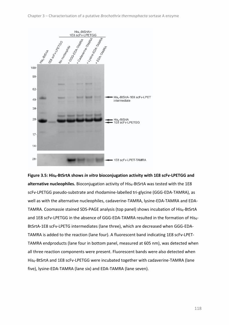
Chapter 3 – Characterisation of a putative Brochothrix thermosphacta sortase A enzyme
118
Figure 3.5: His6-BtSrtA shows in vitro bioconjugation activity with 1E8 scFv-LPETGG and
alternative nucleophiles. Bioconjugation activity of His6-BtSrtA was tested with the 1E8
scFv-LPETGG pseudo-substrate and rhodamine-labelled tri-glycine (GGG-EDA-TAMRA), as
well as with the alternative nucleophiles, cadaverine-TAMRA, lysine-EDA-TAMRA and EDA-
TAMRA. Coomassie stained SDS-PAGE analysis (top panel) shows incubation of His6-BtSrtA
and 1E8 scFv-LPETGG in the absence of GGG-EDA-TAMRA resulted in the formation of His6-
BtSrtA-1E8 scFv-LPETG intermediates (lane three), which are decreased when GGG-EDA-
TAMRA is added to the reaction (lane four). A fluorescent band indicating 1E8 scFv-LPET-
TAMRA endproducts (lane four in bottom panel, measured at 605 nm), was detected when
all three reaction components were present. Fluorescent bands were also detected when
His6-BtSrtA and 1E8 scFv-LPETGG were incubated together with cadaverine-TAMRA (lane
five), lysine-EDA-TAMRA (lane six) and EDA-TAMRA (lane seven).

Chapter 3 – Characterisation of a putative Brochothrix thermosphacta sortase A enzyme
119
To determine potential BtSrtA substrates, the genome of B. thermosphacta Bth-7807 was
mined for the presence of substrate proteins using a hidden Markov model-based tool for
accurate prediction of LPXTG and LPXTG-like cell wall proteins of Gram-positive organisms
(Fimereli et al., 2012). Results of this search revealed 11 SrtA substrate candidates, of which
the respective GenBank accession numbers and sorting motifs are included in Table 3.1. The
consensus sorting motif for the 11 proteins was LPXTG with this sequence of amino acids
followed by a glutamic acid in six, an aspartic acid in four and a valine in one of the proteins.
To investigate whether these substrate candidates are conserved among different strains of
this organism, the genome of the geographically and temporally diverse type strain, B.
thermosphacta DSM 20171T, was searched for orthologues of the 11 proteins. Not only did
this search show that the coding sequences of the 11 candidates were conserved in the type
strain genome with protein sequences sharing between 70% and 100% identity based on
MAFFT protein alignment, but 10 of the 11 sorting motifs of these proteins were identical to
those of the Bth-7807 orthologues. The B. thermosphacta DSM 20171T orthologue with the
GenBank accession number ODJ47926.1 was an exception, with the sorting motif of this
protein differing by one residue (LPKTGE). As the majority of these proteins were annotated
as hypothetical proteins, InterProScan analysis was performed to determine whether
protein family assignment and domains could be predicted that were missed in the
automated annotation (Table 3.1).

Chapter 3 – Characterisation of a putative Brochothrix thermosphacta sortase A enzyme
120
Table 3.1: SrtA substrate candidates identified in B. thermosphacta. Substrate
candidates
identified in
Bth-7807a
Sorting
motifb Protein classification
Orthologues
identified in
DSM 20171Tc
Identity of
orthologues
(%)
ODJ52574.1 LPHTGE Two adhesion domains including one fibrinogen
binding domain (Go-term: cell adhesion) ODJ50812.1 76
ODJ52759.1 LPSTGE Terpenoid cyclase/prenyltransferase ODJ51174.1 100
ODJ52782.1 LPQTGE None predicted ODJ51150.1 100
ODJ54194.1 LPVTGE None predicted ODJ48938.1 88
ODJ55230.1 LPKTGD WD40/YVTN repeat-like domain ODJ48437.1 85
ODJ55992.1 LPTTGD None predicted ODJ51226.1 70
ODJ56202.1 LPTTGD
Six adhesion domains including one fibrinogen
and four collagen binding domains/12
immunoglobulin-like folds (Go-term: cell
adhesion)
ODJ49110.1,
ODJ49111.1,
ODJ49112.1d
96
97
81
ODJ56210.1 LPETGE Leucine-rich repeat domain ODJ47926.1 96
ODJ57179.1 LPHTGD ParB/Sulfiredoxin-like domain ODJ50440.1 85
ODJ57328.1 LPKTGV None predicted ODJ48146.1 99
ODJ57540.1 LPKTGE Five MucBP domains ODJ47926.1 98
a Provided are the GenBank accession numbers of the identified proteins from B.
thermosphacta Bth-7807 (GenBank accession number of genome assembly:
GCA_001715635.1).
b Sorting motifs of the substrate candidates of B. thermosphacta Bth-7807.
c Provided are the GenBank accession numbers of the orthologues identified in B.
thermosphacta DSM 20171T (GenBank accession number of genome assembly:
GCA_001715655.1).
d Blastp analyses showed that while the substrate protein with the GenBank accession
number ODJ56202.1 in Bth-7807 is a large protein consisting of 2395 amino acids, and this
sequence is largely conserved in DSM 20171T, stop codons present in this genomic region in
DSM 20171T result in three coding sequences.
Functional domains for four of the 11 proteins were not found. Two proteins that were
assigned the Go-term “cell adhesion” for biological processes contained multiple N-terminal

Chapter 3 – Characterisation of a putative Brochothrix thermosphacta sortase A enzyme
121
located domains belonging to the adhesion domain superfamily, including a fibrinogen
binding domain in each, and four predicted collagen binding domains in one of these
proteins (GenBank accession number: ODJ56202.1). This protein also comprised 12 C-
terminal immunoglobulin-like folds. Other substrate candidates included a terpenoid
cyclase/prenyltransferase domain protein, a WD/40YVTN repeat-like domain protein, a
leucine-rich repeat domain protein, a ParB/Sulfiredoxin-like domain protein and a MucBP
(MUCin binding protein) domain protein comprising five MucBP domains.
DISCUSSION
The BtSrtA shares only moderate sequence homology with the SrtA of the closely related
food-borne pathogen L. monocytogenes and notably less sequence homology with the
archetypal SrtA from S. aureus. Despite substantial primary sequence divergence, this work
shows that the BtSrtA recognises and cleaves LPXTG motifs, which are sorting motifs
targeted by class A sortases. In contrast, a non-canonical LAETG motif, targeted by class E
sortases of Actinobacteria (Kattke et al., 2016), was not cleaved by the BtSrtA enzyme.
Together with the observed in vitro thioacyl intermediate activity, analysis of the sorting
motifs present in the 11 identified SrtA substrate candidates suggests that the BtSrtA targets
substrate proteins with the LPXTG motif, which in most cases is followed by a conserved
glutamic or aspartic acid residue.
Utilising a model system that was previously tested on SaSrtA (Baer et al., 2014), the BtSrtA
demonstrated in vitro bioconjugation activity with two different SrtA pseudo-substrates and
a variety of nucleophiles, although rhodamine-labelled tri-glycine was most effective. These
in vitro results indicate that like other SrtA enzymes of Gram-positive bacteria, this enzyme
Chapter 3 – Characterisation of a putative Brochothrix thermosphacta sortase A enzyme
122
could be capable of covalently linking substrate proteins to the cross-bridge of lipid II. As
genome examination identified 11 potential SrtA substrates that were conserved between
strain Bth-7807 and the geographically and temporally diverse type strain of this bacterium,
BtSrtA may also fulfil a housekeeping function, attaching multiple, functionally distinct
proteins to the cell wall.
Domain analysis of the substrate proteins revealed two proteins with multiple adhesion
domains including predicted fibrinogen and collagen binding domains. SaSrtA attaches a
variety of adhesion domain containing virulence proteins (MSCRAMMs) to the cell wall,
which are involved in the adherence to host extracellular matrix proteins and cells (Madani
et al., 2017, Tsompanidou et al., 2012, Roche et al., 2004). These include proteins with
fibrinogen, fibronectin and collagen binding domains such as ClfA, ClfB, FnBPA, FnBPB and
CNA (Piroth et al., 2008, Deivanayagam et al., 1999, Jemima Beulin and Ponnuraj, 2017). As
a non-pathogenic, meat spoilage bacterium, the two adhesion domain containing substrate
proteins of B. thermosphacta could be involved in the attachment and colonisation of this
bacterium to meat carcasses. Indeed, similar to the domain organisation of CNA, one of the
candidates contained N-terminal collagen binding domains and C-terminal repetitive
immunoglobulin-like folds (Jemima Beulin and Ponnuraj, 2017). A MucBP domain protein
was also among the identified sortase substrate proteins. Interestingly, cell-surface MucBP-
domain proteins are currently known to be present in Listeria and Lactobacillilaes species
only, and these proteins are assumed to function as adhesins binding to mucus material,
although few studies have been conducted to determine their genuine role (Nishiyama et
al., 2016, Bierne and Cossart, 2007). Although it is tempting to speculate that the MucBP
domain protein of B. thermosphacta may be involved in adherence of this bacterium to

Chapter 3 – Characterisation of a putative Brochothrix thermosphacta sortase A enzyme
123
animal mucus layers such as the gastrointestinal tract, B. thermosphacta is a psychrotrophic
bacterium and clear evidence does not exist that it inhabits these niches.
Among the identified BtSrtA substrates were also three factors with domains that are found
in a variety of different proteins and often implicated in protein-protein interactions (WD40
repeat-like domain, leucine-rich repeat domain and immunoglobulin-like fold), a terpenoid
cyclase/prenyl transferase substrate protein, which are enzymes involved in the synthesis of
terpenes, terpenoids and sterols, and a ParB/Sulfiredoxin-like domain containing protein,
which constitute a largely uncharacterised family of proteins.
Enzymes from psychrotrophic and psychrophilic microorganisms may offer novel
opportunities for biotechnological exploitation due to their high catalytic activity at low
temperatures, low thermostability and other unusual specificities (Feller, 2013). Therefore,
in addition to understanding more about the fundamental biology of a key meat spoilage
bacterium, the continued study of the BtSrtA may prove fruitful for future biotechnological
applications.
Together, the results of this study show that B. thermosphacta encodes a SrtA enzyme,
which targets substrate proteins that contain C-terminal LPXTG sorting motifs. Protein
homology with other SrtA proteins coupled with in vitro bioconjugation activity, suggests
the enzyme is capable of attaching these substrates to the cross-bridge of lipid II. Genome
examination identified 11 potential SrtA substrates, two of which contained protein
domains associated with the adherence of pathogens to host extracellular matrix proteins
and cells, suggesting the B. thermosphacta SrtA may be indirectly involved in its attachment
to meat surfaces. Thus, further work in this area may lead to an understanding of molecular
mechanisms involved in the colonisation of meat by B. thermosphacta.

Chapter 3 – Characterisation of a putative Brochothrix thermosphacta sortase A enzyme
124
REFERENCES
ANTOS, J. M., TRUTTMANN, M. C. & PLOEGH, H. L. 2016. Recent advances in sortase-catalyzed
ligation methodology. Current Opinion in Structural Biology, 38, 111-8.
BAER, S., NIGRO, J., MADEJ, M. P., NISBET, R. M., SURYADINATA, R., COIA, G., HONG, L. P. T.,
ADAMS, T. E., WILLIAMS, C. C. & NUTTALL, S. D. 2014. Comparison of alternative
nucleophiles for Sortase A-mediated bioconjugation and application in neuronal cell
labelling. Organic & Biomolecular Chemistry, 12, 2675-2685.
BIERNE, H. & COSSART, P. 2007. Listeria monocytogenes surface proteins: from genome
predictions to function. Microbiology and Molecular Biology Reviews, 71, 377-397.
BIERNE, H., GARANDEAU, C., PUCCIARELLI, M. G., SABET, C., NEWTON, S., GARCIA-DEL
PORTILLO, F., COSSART, P. & CHARBIT, A. 2004. Sortase B, a new class of sortase in
Listeria monocytogenes. Journal of Bacteriology, 186, 1972-1982.
CLANCY, K. W., MELVIN, J. A. & MCCAFFERTY, D. G. 2010. Sortase transpeptidases: insights
into mechanism, substrate specificity, and inhibition. Biopolymers, 94, 385-96.
COMFORT, D. & CLUBB, R. T. 2004. A comparative genome analysis identifies distinct sorting
pathways in Gram-positive bacteria. Infection and Immunity, 72, 2710-22.
COZZI, R., PRIGOZHIN, D., ROSINI, R., ABATE, F., BOTTOMLEY, M. J., GRANDI, G., TELFORD, J.
L., RINAUDO, C. D., MAIONE, D. & ALBER, T. 2012. Structural basis for group B
Streptococcus pilus 1 sortases C regulation and specificity. PLOS ONE, 7, e49048.
DEIVANAYAGAM, C. C. S., PERKINS, S., DANTHULURI, S., OWENS, R. T., BICE, T., NANAVATHY,
T., FOSTER, T. J., HOOK, M. & NARAYANA, S. V. L. 1999. Crystallization of ClfA and
ClfB fragments: the fibrinogen-binding surface proteins of Staphylococcus aureus.
Acta Crystallographica Section D, 55, 554-556.

Chapter 3 – Characterisation of a putative Brochothrix thermosphacta sortase A enzyme
125
FELLER, G. 2013. Psychrophilic enzymes: from folding to function and biotechnology.
Scientifica, 2013, 28.
FIMERELI, D. K., TSIRIGOS, K. D., LITOU, Z. I., LIAKOPOULOS, T. D., BAGOS, P. G. &
HAMODRAKAS, S. J. 2012. CW-PRED: A HMM-based method for the classification of
cell wall-anchored proteins of Gram-positive bacteria. In: MAGLOGIANNIS, I.,
PLAGIANAKOS, V. & VLAHAVAS, I. (eds.) Artificial Intelligence: Theories and
Applications: 7th Hellenic Conference on AI, SETN 2012, Lamia, Greece, May 28-31,
2012. Proceedings. Berlin, Heidelberg: Springer Berlin Heidelberg.
GHASEMIAN, A., NAJAR PEERAYEH, S., BAKHSHI, B. & MIRZAEE, M. 2015. The microbial
surface components recognising adhesive matrix molecules (MSCRAMMs) genes
among clinical isolates of Staphylococcus aureus from hospitalized children. Iranian
Journal of Pathology, 10, 258-264.
JACOBITZ, A. W., KATTKE, M. D., WERESZCZYNSKI, J. & CLUBB, R. T. 2017. 7 - Sortase
transpeptidases: structural biology and catalytic mechanism. In: KARABENCHEVA-
CHRISTOVA, T. (ed.) Advances in Protein Chemistry and Structural Biology. Academic
Press.
JEMIMA BEULIN, D. S. & PONNURAJ, K. 2017. Steered molecular dynamics study reveals
insights into the function of the repetitive B region of collagen- and fibrinogen-
binding MSCRAMMs. Journal of Biomolecular Structure and Dynamics, 35, 535-550.
JURAJA, S. M., MULHERN, T. D., HUDSON, P. J., HATTARKI, M. K., CARMICHAEL, J. A. &
NUTTALL, S. D. 2006. Engineering of the Escherichia coli Im7 immunity protein as a
loop display scaffold. Protein Engineering, Design and Selection, 19, 231-244.
KANG, M., KO, Y.-P., LIANG, X., ROSS, C. L., LIU, Q., MURRAY, B. E. & HÖÖK, M. 2013.
Collagen-binding microbial surface components recognising adhesive matrix

Chapter 3 – Characterisation of a putative Brochothrix thermosphacta sortase A enzyme
126
molecule (MSCRAMM) of gram-positive bacteria inhibit complement activation via
the classical pathway. The Journal of Biological Chemistry, 288, 20520-20531.
KATOH, K. & STANDLEY, D. M. 2013. MAFFT Multiple Sequence Alignment Software Version
7: improvements in Performance and Usability. Molecular Biology and Evolution, 30,
772-780.
KATTKE, M. D., CHAN, A. H., DUONG, A., SEXTON, D. L., SAWAYA, M. R., CASCIO, D., ELLIOT,
M. A. & CLUBB, R. T. 2016. Crystal structure of the Streptomyces coelicolor Sortase
E1 transpeptidase provides insight into the binding mode of the novel class E sorting
signal. PLOS ONE, 11, e0167763.
KEARSE, M., MOIR, R., WILSON, A., STONES-HAVAS, S., CHEUNG, M., STURROCK, S.,
BUXTON, S., COOPER, A., MARKOWITZ, S., DURAN, C., THIERER, T., ASHTON, B.,
MEINTJES, P. & DRUMMOND, A. 2012. Geneious Basic: an integrated and extendable
desktop software platform for the organization and analysis of sequence data.
Bioinformatics, 28, 1647-9.
KRUGER, R. G., OTVOS, B., FRANKEL, B. A., BENTLEY, M., DOSTAL, P. & MCCAFFERTY, D. G.
2004. Analysis of the substrate specificity of the Staphylococcus aureus sortase
transpeptidase SrtA. Biochemistry, 43, 1541-51.
MADANI, A., GARAKANI, K. & MOFRAD, M. R. K. 2017. Molecular mechanics of
Staphylococcus aureus adhesin, CNA, and the inhibition of bacterial adhesion by
stretching collagen. PLOS ONE, 12, e0179601.
MARESSO, A. W., CHAPA, T. J. & SCHNEEWIND, O. 2006. Surface protein IsdC and sortase B
are required for heme-iron scavenging of Bacillus anthracis. Journal of Bacteriology,
188, 8145-52.
Chapter 3 – Characterisation of a putative Brochothrix thermosphacta sortase A enzyme
127
MARESSO, A. W. & SCHNEEWIND, O. 2006. Iron acquisition and transport in Staphylococcus
aureus. Biometals, 19, 193-203.
MARISCOTTI, J. F., GARCIA-DEL PORTILLO, F. & PUCCIARELLI, M. G. 2009. The Listeria
monocytogenes sortase-B recognizes varied amino acids at position 2 of the sorting
motif. Journal of Biological Chemistry, 284, 6140-6.
MARRAFFINI, L. A. & SCHNEEWIND, O. 2006. Targeting proteins to the cell wall of
sporulating Bacillus anthracis. Molecular Microbiology, 62, 1402-1417.
MARRAFFINI, L. A. & SCHNEEWIND, O. 2007. Sortase C-mediated anchoring of BasI to the
cell wall envelope of Bacillus anthracis. Journal of Bacteriology, 189, 6425-36.
MAZMANIAN, S. K., LIU, G., JENSEN, E. R., LENOY, E. & SCHNEEWIND, O. 2000.
Staphylococcus aureus sortase mutants defective in the display of surface proteins
and in the pathogenesis of animal infections. Proceedings of the National Academy
of Sciences, 97, 5510-5515.
MAZMANIAN, S. K., LIU, G., TON-THAT, H. & SCHNEEWIND, O. 1999. Staphylococcus aureus
sortase, an enzyme that anchors surface proteins to the cell wall. Science, 285, 760-
763.
MAZMANIAN, S. K., TON-THAT, H., SU, K. & SCHNEEWIND, O. 2002. An iron-regulated
sortase anchors a class of surface protein during Staphylococcus aureus
pathogenesis. Proceedings of the National Academy of Sciences, 99, 2293-2298.
NAZIGA, E. B. & WERESZCZYNSKI, J. 2017. Molecular mechanisms of the binding and
specificity of Streptococcus pneumoniae sortase C enzymes for pilin subunits.
Scientific Reports, 7, 13119.

Chapter 3 – Characterisation of a putative Brochothrix thermosphacta sortase A enzyme
128
NISBET, R. M., NIGRO, J., BREHENEY, K., CAINE, J., HATTARKI, M. K. & NUTTALL, S. D. 2013.
Central amyloid-beta-specific single chain variable fragment ameliorates Abeta
aggregation and neurotoxicity. Protein Engineering, Design and Selection, 26, 571-80.
NISHIYAMA, K., SUGIYAMA, M. & MUKAI, T. 2016. Adhesion properties of lactic acid bacteria
on intestinal mucin. Microorganisms, 4, 34.
PERRY, A. M., TON-THAT, H., MAZMANIAN, S. K. & SCHNEEWIND, O. 2002. Anchoring of
surface proteins to the cell wall of Staphylococcus aureus. III. Lipid II is an in vivo
peptidoglycan substrate for sortase-catalyzed surface protein anchoring. Journal of
Biological Chemistry, 277, 16241-8.
PIROTH, L., QUE, Y.-A., WIDMER, E., PANCHAUD, A., PIU, S., ENTENZA, J. M. & MOREILLON,
P. 2008. The fibrinogen- and fibronectin-binding domains of Staphylococcus aureus
fibronectin-binding protein A synergistically promote endothelial invasion and
experimental endocarditis. Infection and Immunity, 76, 3824-3831.
RITZEFELD, M. 2014. Sortagging: a robust and efficient chemoenzymatic ligation strategy.
Chemistry – A European Journal, 20, 8516-8529.
ROCHE, F. M., DOWNER, R., KEANE, F., SPEZIALE, P., PARK, P. W. & FOSTER, T. J. 2004. The
N-terminal A domain of fibronectin-binding proteins A and B promotes adhesion of
Staphylococcus aureus to elastin. Journal of Biological Chemistry, 279, 38433-38440.
SAMBROOK, J. & RUSSELL, D. W. 2001. Molecular cloning : a laboratory manual, Cold Spring
Harbor, N.Y, Cold Spring Harbor Laboratory Press.
SCHNEEWIND, O., MODEL, P. & FISCHETTI, V. A. 1992. Sorting of protein A to the
staphylococcal cell wall. Cell, 70, 267-81.
SPIRIG, T., WEINER, E. M. & CLUBB, R. T. 2011. Sortase enzymes in Gram-positive bacteria.
Molecular Microbiology, 82, 1044-1059.

Chapter 3 – Characterisation of a putative Brochothrix thermosphacta sortase A enzyme
129
STANBOROUGH, T., FEGAN, N., POWELL, S. M., TAMPLIN, M. & CHANDRY, P. S. 2017. Insight
into the genome of Brochothrix thermosphacta, a problematic meat spoilage
bacterium. Applied and Environmental Microbiology, 83, e02786-16.
SURYADINATA, R., SEABROOK, S. A., ADAMS, T. E., NUTTALL, S. D. & PEAT, T. S. 2015.
Structural and biochemical analyses of a Clostridium perfringens sortase D
transpeptidase. Acta Crystallographica Section D, 71, 1505-1513.
TON-THAT, H., LIU, G., MAZMANIAN, S. K., FAULL, K. F. & SCHNEEWIND, O. 1999.
Purification and characterization of sortase, the transpeptidase that cleaves surface
proteins of Staphylococcus aureus at the LPXTG motif. Proceedings of the National
Academy of Sciences of the United States of America, 96, 12424-9.
TSOMPANIDOU, E., DENHAM, E. L., SIBBALD, M. J., YANG, X. M., SEINEN, J., FRIEDRICH, A.
W., BUIST, G. & VAN DIJL, J. M. 2012. The sortase A substrates FnbpA, FnbpB, ClfA
and ClfB antagonize colony spreading of Staphylococcus aureus. PLOS ONE, 7,
e44646.
WU, C., MISHRA, A., REARDON, M. E., HUANG, I. H., COUNTS, S. C., DAS, A. & TON-THAT, H.
2012. Structural determinants of Actinomyces sortase SrtC2 required for membrane
localization and assembly of type 2 fimbriae for interbacterial coaggregation and oral
biofilm formation. Journal of Bacteriology, 194, 2531-9.

Chapter 4 – Genomic and metabolic characterisation of spoilage-associated Pseudomonas species
130
Chapter 4
Genomic and metabolic characterisation of spoilage-associated
Pseudomonas species
Except for minor language edits and discussion of headspace gas analysis this chapter was
published as:
Stanborough T., Fegan N., Powell S.M., Singh T., Tamplin M., and Chandry P.S., 2018.
Genomic and metabolic characterisation of spoilage-associated Pseudomonas species.
International Journal of Food Microbiology, 268: 61-72.
ABSTRACT
Pseudomonas are common spoilage agents of aerobically stored fresh foods. Their ability to
cause spoilage is species- and may be strain-specific. To improve our understanding of the
meat and milk spoilage agents Pseudomonas fragi and Pseudomonas lundensis, we
sequenced the genomes of 12 P. fragi and seven P. lundensis isolates. These genomes
provided a dataset for genomic analyses. Key volatile organic compounds (VOCs) produced
or metabolised by the isolates were determined during their growth on a beef paste and
where possible, metabolic activity was associated with gene repertoire.
Genome analyses showed that the isolates included in this work may belong to more than
two Pseudomonas species with possible spoilage potential. Pan-genome analyses
demonstrated a high degree of diversity among the P. fragi and genetic flexibility and
diversity may be traits of both species. Growth of the P. lundensis isolates was characterised
by the production of large amounts of 1-undecene, 5-methyl-2-hexanone and methyl-2-
butenoic acid. P. fragi isolates produced extensive amounts of methyl and ethyl acetate and

Chapter 4 – Genomic and metabolic characterisation of spoilage-associated Pseudomonas species
131
the production of methyl esters predominated over ethyl esters. Some of the P. fragi
produced extremely low levels of VOCs, highlighting the importance of strain-specific
studies in food matrices.
INTRODUCTION
Psychrotrophic Pseudomonas play a major role in limiting the shelf-life of fresh foods stored
aerobically at cold temperatures (Remenant et al., 2015). Members of the Pseudomonas
fragi subgroup, P. fragi and P. lundensis (Gomila et al., 2015), are recognised as common
agents contributing to the spoilage of fresh meat and fish, and pasteurised and ultra-high-
temperature-treated milk (products) via the activity of heat-stable extracellular proteases
and lipases (Ercolini et al., 2007, Quigley et al., 2013, Tryfinopoulou et al., 2002, De Jonghe
et al., 2011). In the case of meat, their growth and utilisation of food nutrients such as
sugars and amino acids, and their release of both volatile and non-volatile metabolites such
as esters, ketones, alcohols, aldehydes organic acids, sulphur compounds and amines, cause
organoleptic changes to the meat resulting in spoilage (Ercolini et al., 2006, Samelis, 2006,
Montel et al., 1998).
P. fragi are associated with fruity off-odours due to the production of esters, in particular
short chain ethyl esters (Casaburi et al., 2015, Morales et al., 2005a). However, a number of
studies indicate that ester production of this bacterium is strain-dependent (Morales et al.,
2005a, Edwards et al., 1987). Other volatile compounds commonly associated with P. fragi
include ketones, alcohols, 1-undecene and sulphur compounds (Wang et al., 2017b, Ercolini
et al., 2010). The production of volatile sulphurous compounds with putrid aromas is

Chapter 4 – Genomic and metabolic characterisation of spoilage-associated Pseudomonas species
132
characteristic for Pseudomonas and occurs when glucose in meat is depleted and the amino
acid pool is attacked (Edwards et al., 1987).
Volatile analyses of pure cultures of P. lundensis are rare and thus little is known about the
contribution of this species to off-odours in meat spoilage. One study, which looked at the
volatile profile of a single strain of this species in cheese, showed that it produced significant
quantities of ketones, but that the production of alcohols, butanoic acid and the
hydrocarbons 1-undecene and cyclohexane were also noteworthy (Morales et al., 2005c).
Despite their biological and economic importance, the genomes of P. fragi and P. lundensis
remain largely unstudied. Furthermore, thorough understanding of their metabolic potential
at the strain level is missing, in particular for P. lundensis.
In this study, we sequenced the genomes of 17 red meat- and two raw milk-derived P. fragi
and P. lundensis isolates. To understand intra-species genetic diversity and provide insight
into the genomes, average nucleotide identity, phylogenetic and pan-genome analyses were
conducted. To investigate the heterogeneity of the bacteria in relation to their meat
spoilage potential, the main volatile organic compounds (VOCs) produced or metabolised by
the isolates during growth on a beef paste were assessed, and where possible, metabolic
activity was linked to gene repertoire.
MATERIALS AND METHODS
Bacterial isolates
Table 4.1 lists the isolates and type strains used in this study and provides their source
information.

Chapter 4 – Genomic and metabolic characterisation of spoilage-associated Pseudomonas species
133
Bacteria were isolated from meat sources by aseptically removing 25 g pieces and placing
them into sterile stomacher bags with 300 mL of maximum recovery diluent (MRD, Oxoid,
Basingstoke, UK). The samples were macerated for 1 min using a Seward Stomacher 400 set
to 230 rpm. Samples of homogenate were decimally diluted and 100 µL of appropriate
dilutions were spread on tryptone soya agar (TSA, Oxoid, Basingstoke, UK) or plate count
agar (PCA, Oxoid, Basingstoke, UK); plates were incubated at 22 °C for 24 h to 48 h. Isolation
of bacteria from spoiled milk was achieved by decimally diluting milk samples in MRD,
spreading 100 µL of appropriate dilutions on PCA and incubating the plates at 30 °C for 72 h.
Identification of the isolates was performed by a multiplex carA PCR as previously described
(Ercolini et al., 2007).

Chapter 4 – Genomic and metabolic characterisation of spoilage-associated Pseudomonas species
134
Table 4.1: Source and genome information of bacteria used in this study.
Organisms Species Sourcea
Total
sequence
length
(bp)
GC-content
(%)
No. protein coding
genes
No. of contigs
>1000 bp in length Genome coverage
GenBank accession
no.
F1786b P. fragi Raw cow’s milkc 4,976,899 59.7 4,441 23 120x NQKV00000000
F1791b P. fragi Beef steakd 5,143,683 58.2 4,641 44 25x NQKU00000000
F1792b P. fragi Beef steakd 5,013,810 58.3 4,488 45 20x NQKT00000000
F1793b P. fragi Beef steakd 4,970,618 58.4 4,460 43 30x NQKS00000000
F1794b P. fragi Lamb fillets (MAP)d 5,256,020 58.2 4,730 44 20x NQKR00000000
F1801b P. fragi Beef stripse 5,051,439 58.0 4,538 63 80x NQKQ00000000
F1813b P. fragi Beef topside roaste 5,210,542 58.1 4,658 42 78x NQKP00000000
F1815b P. fragi Beef topside roaste 4,979,003 58.4 4,479 44 36x NQKO00000000
F1816b P. fragi Lamb stripse 5,113,509 58.4 4,634 30 58x NQKN00000000
F1818b P. fragi Beef steake 5,298,866 59.2 4,797 93 45x NQKM00000000
F1820b P. fragi Minced lambe 5,215,977 58.1 4,745 53 70x NQKL00000000
F1821b P. fragi Minced beefe 4,975,134 58.4 4,478 57 40x NQKK00000000
L1788b P. lundensis Raw cow’s milkc 4,931,932 58.7 4,403 135 40x NQKJ00000000
L1802b P. lundensis Beef stripse 4,800,490 57.7 4,336 92 78x NQKI00000000
L1810b P. lundensis Beef steake 4,982,671 58.3 4,474 64 90x NQKH00000000
L1814b P. lundensis Beef topside roaste 5,016,267 58.7 4,455 64 30x NQKG00000000
L1817b P. lundensis Minced beefe 4,955,748 58.4 4,405 81 30x NQKF00000000
L1819b P. lundensis Beef steake 5,035,790 58.7 4,487 73 60x NQKE00000000
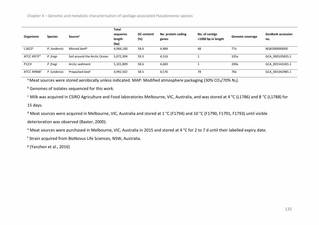
Chapter 4 – Genomic and metabolic characterisation of spoilage-associated Pseudomonas species
135
Organisms Species Sourcea
Total
sequence
length
(bp)
GC-content
(%)
No. protein coding
genes
No. of contigs
>1000 bp in length Genome coverage
GenBank accession
no.
L1822b P. lundensis Minced beefe 4,966,160 58.6 4,484 48 77x NQKD00000000
ATCC 4973Tf P. fragi Soil around the Arctic Ocean 5,072,304 59.3 4,516 1 105x GCA_900105835.1
P121g P. fragi Arctic sediment 5,101,809 58.6 4,683 1 200x GCA_001543265.1
ATCC 49968T P. lundensis Prepacked beef 4,992,502 58.5 4,576 78 70x GCA_001042985.1
a Meat sources were stored aerobically unless indicated. MAP: Modified atmosphere packaging (30% CO2/70% N2).
b Genomes of isolates sequenced for this work.
c Milk was acquired in CSIRO Agriculture and Food laboratories Melbourne, VIC, Australia, and was stored at 4 °C (L1786) and 8 °C (L1788) for
15 days.
d Meat sources were acquired in Melbourne, VIC, Australia and stored at 1 °C (F1794) and 10 °C (F1790, F1791, F1793) until visible
deterioration was observed (Baxter, 2000).
e Meat sources were purchased in Melbourne, VIC, Australia in 2015 and stored at 4 °C for 2 to 7 d until their labelled expiry date.
f Strain acquired from BioNovus Life Sciences, NSW, Australia.
g (Yanzhen et al., 2016)

Chapter 4 – Genomic and metabolic characterisation of spoilage-associated Pseudomonas species
136
DNA isolation, genome sequencing, assembly and annotation
Isolates were grown for 18 h in tryptone soya broth (TSB, Oxoid, Basingstoke, United
Kingdom) at 25 °C. Genomic DNA was isolated with the DNeasy Blood and Tissue Kit
(QIAGEN, Valencia, CA, USA) according the manufacturer’s protocol for Gram-negative
bacteria. Library preparation and genome sequencing was carried out at The Ramaciotti
Centre at the University of NSW using the Nextera XT DNA library 300 bp paired end
preparation kit, and was performed on the Illumina MiSeq platform (Illumina, San Diego, CA,
USA). Genome assembly and annotation was performed as previously described
(Stanborough et al., 2017a). GenBank accession numbers of the 19 genomes sequenced in
this study are listed in Table 4.1.
Genomic analyses
Determination of average nucleotide identity
Orthologous Average Nucleotide Identity (OrthoANI) was determined using the OrthoANI
Tool (version 0.93, Blast+) (Lee et al., 2016).
Single Nucleotide Polymorphism (SNP) analysis
SNP analysis was performed with the software kSNP3.0 (Gardner et al., 2015). Optimal k-
mer length was determined by the auxiliary script kChooser and was 19 for the P. lundensis
and 21 for the P. fragi genomes. The analysis of core SNPs (loci present in all input
genomes), rather than “all SNPs” or “majority SNPs”, was chosen as this method is better
suited to remove recombination/horizontally transferred genomic elements from the
analysis (Pettengill et al., 2016). Core SNP trees were estimated by parsimony and based on
a consensus of up to 100 equally parsimonious trees. Trees were drawn with Dendroscope
(version 3.5.9) (Huson et al., 2007). The P. fragi core SNP matrix was plotted as a

Chapter 4 – Genomic and metabolic characterisation of spoilage-associated Pseudomonas species
137
NeighborNet network using SplitsTree4 and uncorrected P-distances (version 4.14.2) (Huson
and Bryant, 2006). SplitsTree4 was used to calculate the δ score and Q-residual score as
measures of “treelikeness” of the phylogenetic distance data (Holland et al., 2002).
Pan-genome
Pan-genome analysis was performed with USEARCH (version 8.1.1861) using an all versus all
comparison with a minimum identity cut-off score of 0.6. Seed sequences were parsed into
tabular form with a custom script (https://github.com/bioinformatics-deakin/gene-matrix-
from-uclust3.py) and genes less than 153 nucleotides in length (proteins <50 amino acids in
length) were eliminated from the analysis. The core genome was determined as the set of
genes shared by all organisms, while the accessory genome was the set of genes present in
a subset of organisms or in a single organism. Functional annotation of the translated seed
sequences of the accessory genome was conducted with the NCBI CD-Search tool to identify
conserved domains using a mirrored COG (Clusters of Orthologous Groups of Proteins)
database with the default settings (E-value threshold of 0.01, maximum number of hits of
500, and composition-corrected scoring turned on). As each COG can have one or more
general category letter association, the pools of functional categories were compared. The
relative abundances of COG functional categories were determined by dividing the number
of genes assigned to each category by the total pool of functions determined for each
accessory genome. Principal component analysis of the pan-genome matrix was conducted
with a custom script (https://github.com/bioinformatics-deakin/PCA/blob/master/PCA.py).
Identification of homologous genes
A nucleotide database was generated in Geneious (version 9.0.5) (Kearse et al., 2012)
containing the genome sequences of the isolates and type strains included in Table 4.1.

Chapter 4 – Genomic and metabolic characterisation of spoilage-associated Pseudomonas species
138
Query proteins were searched against the database using the scoring matrix BLOSUM62 and
tblastn algorithm. Sequences with E-value scores of <10-5 and bit scores of ≥50 were further
subjected to protein domain analysis with the InterProScan tool. Query proteins and source
organisms are included in Table 4.2.
Table 4.2: Locus tags of gene homologs Query proteins
(GenBank accession numbers) Query organism
Locus tags of homologous genesa
P. fragi P. lundensis
UndA
(4WWJ_A)
Pseudomonas protegens
Pf-5 SAMN05216594_4735b TU74_08480c
AcuC
(NP_390849.1) Bacillus subtilis 168 SAMN05216594_2987b TU74_12385c
Methionine gamma lyase
(AAY92781.1)
Pseudomonas protegens
Pf-5 SAMN05216594_0122b TU74_09140c
a – indicates that a homologous sequence was not identified.
b GenBank accession number GCA_900105835.1
c GenBank accession number GCA_001042985.1
Metabolic analyses
Sample preparation in headspace vials
Large 1-2 kg beef samples were surface sterilized by cooking in boiling water for 10 min.
Cooked surface meat was removed aseptically and the raw, sterile inner was cut into small
pieces in a sterile manner. Cut meat pieces were mixed with 0.9% sterile NaCl solution (ratio
of 1 g:0.67 mL) and homogenized for 60 sec with a Sorvall Omni-Mixer to produce a smooth
meat paste. 3 g of sterile meat paste were aliquoted with a 100 mL sterile syringe into
sterilized 20 mL glass headspace vials (Grace Davidson Discovery Sciences, Rowville, VIC,
Australia).

Chapter 4 – Genomic and metabolic characterisation of spoilage-associated Pseudomonas species
139
Bacterial inocula were prepared by inoculating 3 mL of sterile TSB with single colonies and
incubating the cultures in a shaking water bath at 25 °C for 18 h. Per experiment, three
cultures were prepared for each isolate (n=3) on one day. To inoculate meat paste samples,
cultures were diluted 1:50 with sterile 0.9% NaCl solution and 50 µL of the diluted cultures,
containing approximately 106 cells, were distributed on the meat paste surface. For VOC
analysis, each inoculum was used to inoculate two meat paste samples (one sample for
headspace analysis and one sample for analysis of atmospheric gases and bacterial
enumeration). For the determination of bacterial growth and consumption/production of
atmospheric gases of low (F1794, F1815 and L1788) and high (F1801 and L1814) VOC-
producers over a 4 d incubation period, each inoculum was used to inoculate four samples
(one sample per day). Control meat paste samples were inoculated with 50 µL of diluted
sterile TSB. Three controls were frozen immediately at -20 °C to act as time point 0 controls
(C0) and three controls (Cd4) were incubated together with the inoculated vials for 4 d at 8
°C.
In anticipation of considerable biological variability, the VOC analysis was repeated two
times constituting a total of three independent experiments, each experiment conducted
with three biological replicates of the strains.
Determination of atmospheric gases and bacterial numbers in headspace vials
To analyse the levels of atmospheric gases (CO2, O2 and N2) in the 20 mL headspace vials
containing the inoculated samples and non-inoculated controls, a Shimadzu gas
chromatograph (GC model GC8A, Shimadzu Scientific Instruments (Oceania) Pty Ltd,
Rydalmere NSW, Australia) fitted with a CTR-1 column (180 cm long, 0.64 cm and 0.32 cm
diameter of outer and inner columns, respectively; Alltech Deerfield, IL. USA) was used. An

Chapter 4 – Genomic and metabolic characterisation of spoilage-associated Pseudomonas species
140
aliquot of 1 mL of headspace gas was withdrawn from the vials and injected using an on-
column injector (50 °C). Column and thermal conductivity detector were maintained at 30 °C
and 50 °C, respectively. Helium, at a constant pressure of 3 kg/cm2, was used as the carrier
gas. The Shimadzu GC was interfaced with the computer, running Delta 5.5 chromatography
Data System, using an analog-to-digital converter (Data Centre 4000). Peak areas obtained
for CO2, O2 and N2 were used to calculate relative amounts (%) of the composition gases and
the means and standard deviations of the three independent experiments were calculated.
The number of bacteria on the inoculated meat paste samples and non-inoculated controls
was determined by adding 10 mL of sterile MRD to the 20 mL headspace vials. Vial contents
were transferred to sterile stomacher bags and then crushed for 1 min at 230 rpm in a
Seward Stomacher 400. Samples of homogenate were decimally diluted, 100 µL of
appropriate dilutions were spread on TSA and incubated for 24 h to 48 h at 25 °C. Levels of
bacteria were enumerated for each sample, converted to log10 cfu/g, and the means and
standard deviations of the three independent experiments calculated.
Headspace analysis by solid phase micro-extraction-gas chromatography mass
spectrometry (SPME-GCMS)
Headspace analysis was used to analyse the VOCs in the inoculated meat paste samples and
non-inoculated controls. An Agilent Technologies gas chromatograph-mass spectrometer
system (GC model 6890N, MS model 5975 series B; Agilent Technologies Inc., Palo Alto, CA,
USA) equipped with a Combi PAL robotic auto sampler (CTC Analytics AG, Zwingen,
Switzerland) and a programmed temperature vaporization injector (PTV inlet set in splitless
mode, temperature 260 °C, splitless time 1 min; Gerstel, Mülheim, Germany) was used. The
SPME fiber, coated with carboxen/polydimethylsiloxane (CAR/PDMS StableFlex fibre, phase

Chapter 4 – Genomic and metabolic characterisation of spoilage-associated Pseudomonas species
141
thickness 85 µm, length 1 cm; Supelco, Bellefonte, Pa, USA), was preconditioned prior to
analysis at 270 °C for 1.5 h. 1 mL of internal standard solution (2-methyl-3-heptanone, 3.5
mg, and 2-ethyl butanoic acid, 7.1 mg, per 10 mL of methanol) was added to the samples.
The autosampler was operated under the following conditions: pre-incubation time of 5
min, pre-incubation and absorption temperature of 45 °C, absorption time of 30 min,
desorption time of 8 min (purge after 1 min), and desorption temperature of 260 °C. The
volatile compounds of the meat paste samples were chromatographed on a VF-WAXms
column (30 m length, 0.32 mm i.d., 1.0 mm film thickness, Agilent Technologies Australia,
Mulgrave, VIC, Australia) using a temperature gradient: 35 °C for 5 min, raised to 225 °C at a
rate of 10 °C/min and finally held at 225 °C for 10 min. Helium was used as carrier gas at a
constant flow rate of 1.6 mL/min. The MSD conditions were as follows: capillary direct
interface temperature of 280 °C, ionization energy of 70 eV, mass range of 35-300 amu; EM
voltage of autotune +200 V, with a scan rate of 5.27 scans/s.
VOCs were identified by comparison of mass spectra with the spectra in the NIST11
database (National Institute of Standards Technology; United States of America), and linear
retention indices determined using a set of saturated alkanes C7–C22 and spectra of
authentic standards. VOC profiles of the isolates were established by calculating the relative
amounts of VOCs as percentages based on the total peak area. To assess VOCs that played
key roles in the metabolism of the isolates on the beef paste, those present in trace
amounts (i.e. constituted <1% of the total volatile profile of the isolates) were not
considered further. The relative amounts of VOCs shown were calculated using the means
of the three independent experiments. Quantitative analysis was performed using an
internal standard methodology.

Chapter 4 – Genomic and metabolic characterisation of spoilage-associated Pseudomonas species
142
RESULTS
Phylogeny
In this study, the genomes of 12 P. fragi and seven P. lundensis isolates of red meat and raw
milk origin were sequenced and draft genome assemblies deposited in GenBank to provide
an analysis data set (Table 4.1). Metrics of the draft genomes of the isolates correlated well
with genome metrics of the respective type strains. Size estimation of the assemblies varied
between 4.97 Mb and 5.30 Mb for the P. fragi and 4.80 Mb and 5.04 Mb for the P. lundensis
isolates. The GC-content of the P. fragi and P. lundensis draft genomes ranged from 58% to
59.6% and 57.7% to 58.7%, respectively. Between 4,441 and 4,797 protein coding genes
were predicted for the P. fragi genomes, while 4,336 to 4,487 were identified in the P.
lundensis genomes. Genomic similarity of the isolates was investigated by pairwise
calculation of average nucleotide identity (ANI) (Figure 4.1) and included the publically
available genome sequences of the type strains and the environmental isolate P. fragi P121.
A high degree of variation was observed among the P. fragi, with P121 clearly distinct from
all other P. fragi with ANI values of 84.7% to 85.1% (Figure 4.1A). Three distinct P. fragi
groups were otherwise observed, including 1) a group of eight closely related isolates
(F1791, F1792, F1793, F1794, F1813, F1815, F1816 and F1821) as indicated by shared ANI
values above 98.9%, 2) a group that consisted of two isolates (F1786, F1818) and the type
strain with ANI values between 96.9% to 98.7%, and 3) a group consisting of two isolates
(F1801 and F1820) that shared 95.8% ANI. Seven of the eight P. lundensis were closely
related with ANI values between 97.7% and 99.9%, while low ANI values were determined
for isolate L1802 (90.3%-90.5%) (Figure 4.1B).

Chapter 4 – Genomic and metabolic characterisation of spoilage-associated Pseudomonas species
143
Figure 4.1: Pairwise comparison of the average nucleotide identity (ANI) of the genomes.
Heat maps show the pairwise ANI as determined for the P. fragi (A) and P. lundensis (B)
genomes. The colour scale is shown to the right of the heat maps.
To better understand the intra-species relationships, core SNPs were used to generate SNP
matrices for phylogenetic reconstruction (Figure 4.2). The P. fragi SNP matrix consisted of
11,192 SNPs, whereby 5,098 of these were unique to P. fragi P121. Poor branch support
values were obtained for many branches of the P. fragi SNP tree (Figure B1A: Appendix B),

Chapter 4 – Genomic and metabolic characterisation of spoilage-associated Pseudomonas species
144
indicating that a bifurcating tree was unlikely to accurately represent the ancestry.
Therefore, the SNP matrix was examined with a NeighborNet network with uncorrected
nucleotide distances (Figure 4.2A). The resultant network was not tree-like displaying a high
level of reticulation (δ-score = 0.1915, Q-residual score = 0.00035). This network revealed
three main groups of taxa with isolate P121 clearly distant from the others. These groups
were consistent with the groups identified by ANI calculations. The low numbers of unique
core SNPs and short network branches of the eight meat-derived Group 1 isolates
underlined their close phylogenetic relationship. The exact relationship between the three
Group 2 organisms and the two isolates from Group 3 could not be resolved. Interestingly,
despite their geographic and temporal diversity and their different isolation sources, the
soil-derived type strain and meat isolate F1818 were phylogenetically close. Moreover, the
third Group 2 isolate (F1786) was derived from milk, demonstrating the Group 2 diversity.

Chapter 4 – Genomic and metabolic characterisation of spoilage-associated Pseudomonas species
145
Figure 4.2: P. fragi and P. lundensis core SNP phylogenies. Coloured shading was used to
highlight the main groups of taxa. Parentheses at the branch tips indicate the number of
core SNPs exclusive to the isolates and type strains. (A) NeighborNet network analysis of P.
fragi core SNP matrix was plotted using uncorrected P-distances. The distance scale
indicates the number of nucleotide substitutions per number of SNPs. Truncated network
branch is marked with // to aid visualisation and the original branch length is indicated
above. (B) Mid-point rooted consensus parsimony cladogram of P. lundensis core SNPs.
Support values of all nodes were 1 as determined by Shimodiara-Hasegawa resampling test.
Numbers at the internal nodes represent the number of core SNPs that are shared
exclusively by the descendants of that node.

Chapter 4 – Genomic and metabolic characterisation of spoilage-associated Pseudomonas species
146
Of the 42,696 core SNPs identified for the P. lundensis, 32,085 were exclusive to L1802.
Resampling branch supports of the P. lundensis core SNP tree (Figure 4.2B) were high with
support values of 1 for all branch points. The tree topology showed two main groups of taxa
with isolate L1802 clearly separated from the other organisms. Cluster 1 was comprised of
two isolates (L1817 and L1810) and the type strain. With a high number of unique SNPs and
no additional shared alleles, the meat isolate L1822 was clearly distant from the other
Cluster 2 isolates. Cluster 2 also comprised two closely related meat isolates (L1814 and
L1819) with only five and six unique core SNPs, respectively, as well as one milk isolate
(L1788). A NeighborNet network derived from the core SNP matrix yielded a tree-like
network (δ score = 0.083, Q-residual score = 0.00054) confirming that a bifurcating tree
adequately describes their phylogeny (Figure B1B: Appendix B).
Pan-genome
A total of 7,378 genes comprised the pan-genome of the P. fragi, with 3,286 genes
identified as core genes, resulting in a core/pan-genome ratio of 45%. The P. lundensis pan-
genome comprised 5,769 genes. Of these, 3,537 were core genes, amounting to a core/pan-
genome ratio of 61%. The pan-genome matrices were used in a principal component
analysis (PCA) to assess clustering of the isolates based on their gene repertoire (Figure 4.3).
PCA of the P. fragi pan-matrix (Figure 4.3A) showed clustering correlated well with their
phylogeny and P121 was clearly separated from the other P. fragi. PCA of the P. lundensis
pan-genome matrix segregated some members of the same phylogenetic clusters and L1802
was clearly separated from the other isolates and the type strain (Figure 4.3B).
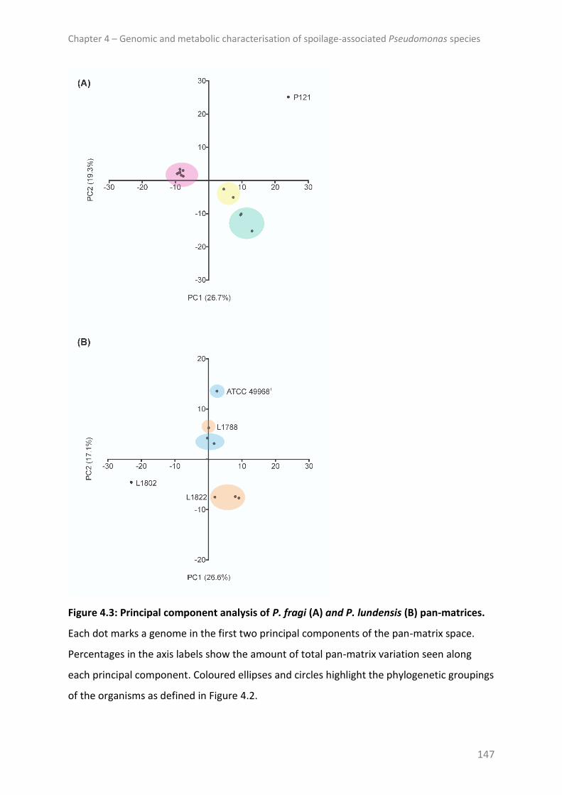
Chapter 4 – Genomic and metabolic characterisation of spoilage-associated Pseudomonas species
147
Figure 4.3: Principal component analysis of P. fragi (A) and P. lundensis (B) pan-matrices.
Each dot marks a genome in the first two principal components of the pan-matrix space.
Percentages in the axis labels show the amount of total pan-matrix variation seen along
each principal component. Coloured ellipses and circles highlight the phylogenetic groupings
of the organisms as defined in Figure 4.2.
Chapter 4 – Genomic and metabolic characterisation of spoilage-associated Pseudomonas species
148
The accessory genome was analysed to understand functional groups of genes responsible
for intra-species differences. A large percentage of genes of both accessory genomes were
assigned to the categories general function prediction and function unknown. Genes
involved in transcription and signal transduction mechanisms represented large proportions
of both accessory genomes (Figure 4.4). Other well represented functional groups (i.e. more
than 5% of the accessory pool) of the P. fragi accessory genome included carbohydrate and
amino acid transport, replication, recombination and repair, cell wall/membrane/envelope
biogenesis, inorganic ion transport and the mobilome (Figure 4.4A). A notably large
proportion of the P. lundensis accessory genes were assigned to the mobilome, and
replication, recombination and repair groups (Figure 4.4B). Additional well represented
categories were cell wall/membrane/envelope biogenesis and inorganic ion transport.

Chapter 4 – Genomic and metabolic characterisation of spoilage-associated Pseudomonas species
149

Chapter 4 – Genomic and metabolic characterisation of spoilage-associated Pseudomonas species
150
Figure 4.4: COG functional categories of accessory genomes. The relative abundances of
COG functional categories for the P. fragi (A) and P. lundensis (B) accessory genomes are
shown. On the vertical axis, COG functional categories are depicted with their letter
association, while the horizontal axis indicates their relative abundance.
Volatile analysis
To assess the main intra- and inter-species metabolic differences, the major VOCs produced
or metabolised by the isolates were investigated using a meat model system, enabling
controlled, reproducible conditions and sufficient replicates. Where possible, metabolic
activity was linked to gene repertoire. Isolates were inoculated onto precise amounts of
sterile beef paste in headspace vials and poor refrigeration conditions were simulated by
incubating the vials at 8 °C. Preliminary tests showed the transition to stationary phase
growth occurred around day 4 of incubation and oxygen levels on this day were reduced to
approximately 2% to 3%. Pseudomonads grow well in oxygen concentrations down to 2%,
but are progressively inhibited at lower concentrations (Clark and Burki, 1972). Therefore,
VOCs were determined on day 4 before oxygen levels were low enough to impact on the
growth of the isolates.
Samples were inoculated with log 6 cfu/g; after the 4-day incubation period numbers
increased to log 9.4-9.6 cfu/g, while oxygen in the headspaces of the inoculated samples
constituted between 1.8%-5.5% of the volume and carbon dioxide and nitrogen between
7.6%-10.1% and 86.2%-88.9%, respectively (Table B1: Appendix B).
A total of 16 VOCs (Figure 4.5) were found to be the major VOCs produced or metabolised
by the isolates. Among these were short and medium-length fatty acid esters, ketones,

Chapter 4 – Genomic and metabolic characterisation of spoilage-associated Pseudomonas species
151
hydrocarbons as well as a fatty acid, alcohol and a sulphur compound. Strong differences
were observed in the total amounts of detected VOCs between the isolates.
Figure 4.5: VOC profiles of inoculated meat paste samples and non-inoculated meat paste
controls. Scatter plot shows total amounts of the 16 VOCs detected in the inoculated
samples and non-inoculated controls prior to (C0) and after (Cd4) incubation at 8 °C for 4
days. Replicates from experiment 1 (Exp1), 2 (Exp2) and 3 (Exp3) are coloured in red, blue
and grey, respectively, while mean values are shown with black horizontal bars. Each sample
Chapter 4 – Genomic and metabolic characterisation of spoilage-associated Pseudomonas species
152
is represented by nine data points with overlapping potentially obscuring some points. Lines
above the data points demarcate between control samples and samples of the two species.
The heat map was generated with the means of the area ratios (%) of the 16 VOCs; the
colour scale shown to the right of the heat map indicates area ratio differences of 1%
between 0% and 25%.
Esters
Six different esters of short and medium chain fatty acids with equal carbon numbers were
identified. These included one ethyl ester (ethyl acetate), four linear methyl esters (methyl
acetate, methyl butanoate, methyl hexanoate and methyl octanoate) and 4-hexenyl
acetate. Methyl acetate was the only ester detected in all samples. Ethyl acetate was found
exclusively in samples inoculated with P. fragi, with the exception of L1802, which was the
only P. lundensis isolate for which this compound was found. Methyl butanoate was
detected in most samples with the exception of three P. fragi isolates (F1786, F1801 and
F1818). Methyl hexanoate and methyl octanoate were only identified in samples inoculated
with P. fragi F1792, F1793 and F1813, while 4-hexenyl-acetate only constituted more than
1% of the volatile profiles of P. lundensis L1802 and L1814 and the detected amounts of
these three esters were low (data not shown). Notably, the majority of the P. fragi
demonstrated extensive production of ethyl and methyl acetate (Figure 4.6). In marked
contrast, P. fragi isolates F1815 and F1794 as well as the P. fragi type strain produced
extremely low levels of these esters. For isolate F1815, ethyl acetate levels did not
constitute more than 1% of its volatile profile. Although the P. fragi and P. lundensis
genomes harboured a multitude of predicted lipase and esterase genes, no obvious genetic
variations were identified to explain the difference in levels of esters produced.

Chapter 4 – Genomic and metabolic characterisation of spoilage-associated Pseudomonas species
153
Figure 4.6: VOCs produced by P. fragi and P. lundensis isolates. Absolute amounts of key
VOCs of the inoculated samples compared to their detected amounts in non-inoculated
controls prior to (C0) and after (Cd4) incubation at 8 °C for 4 days. Replicates from
experiment 1 (Exp1), 2 (Exp2) and 3 (Exp3) are coloured in red, blue and grey respectively,
while the mean values are shown with black horizontal bars. Each sample is represented by
nine data points with overlapping potentially obscuring some points. Lines above the data
points demarcate between control samples and samples of the two species.

Chapter 4 – Genomic and metabolic characterisation of spoilage-associated Pseudomonas species
154
Ketones
Five different ketones constituted a significant proportion of the volatile profiles of samples,
including 5-methyl-2-hexanone, 2-nonanone, acetoin (3-hydroxy-2-butanone), 2-octanone
and 2,3-butandione. P. lundensis isolates L1802, L1814 and L1819 produced the largest
amounts of 5-methyl-2-hexanone and 2-nonanone (Figure 4.6), with particularly high levels
of 2-nonanone detected in Experiment 2, as observed for most of the organisms. In
contrast, comparatively low amounts of 2-nonanone were detected for P. lundensis L1788
and five P. fragi (F1815, F1816, F1820, F1821 and ATCC 4973T).
Mean values of the ketones 2-octanone and acetoin, which were present in non-inoculated
controls, notably decreased in all inoculated samples (Figure 4.7) and genome inspections
showed all genomes encoded a gene with homology to the acetoin utilisation protein (AcuC)
of Bacillus subtilis strain 168 (Table 4.2).

Chapter 4 – Genomic and metabolic characterisation of spoilage-associated Pseudomonas species
155
Figure 4.7: VOCs metabolised by P. fragi and P. lundensis isolates. Absolute amounts of
VOCs detected in reduced amounts in inoculated samples compared to non-inoculated
controls prior to (C0) and after (Cd4) incubation at 8 °C for 4 days. Replicates from
experiment 1 (Exp1), 2 (Exp2) and 3 (Exp3) are coloured in red, blue and grey respectively,
while the mean values are shown with black horizontal bars. Each sample is represented by
nine data points with overlapping potentially obscuring some points. Lines above the data
points demarcate between control samples and samples of the two species.

Chapter 4 – Genomic and metabolic characterisation of spoilage-associated Pseudomonas species
156
Other volatile compounds
The hydrocarbon 1-undecene and volatile fatty acid methyl-2-butenoic acid were identified
in all inoculated samples and amounts were notably higher in samples inoculated with P.
lundensis isolates (Figure 4.6). In line with the ubiquitous production of 1-undecene, an
undA homolog was identified in all genomes. The sequences of the P. lundensis and P. fragi
UndA proteins shared a high degree of homology with 73%-74% and 68%-72% identity with
the Pseudomonas protegens Pf-5 sequence, respectively (Table 4.2). The VOCs E-1,4-
undecadiene and 2-octanol were identified in non-inoculated controls and inoculated
samples, with a marked reduction in mean 2-octanol levels detected for all samples
inoculated with P. fragi and some samples inoculated with P. lundensis isolates (Figure 4.7).
Closely related isolates L1814 and L1819 were exceptions with little to no reduction in their
mean 2-octanol values, and an increase in levels of this compound were observed for one
replicate of each isolate from Experiment 2. Dimethyl sulphide was only found in notable
quantities in samples inoculated with P. lundensis L1822, although, a methionine gamma
lyase gene (EC 4.4.1.11) was identified in all of the genomes.
To better understand differences in levels of VOCs, in particular the striking absence of ester
production of P. fragi F1815, F1794 and ATCC 4973T, growth of three isolates with overall
low levels of VOC production (F1815, F1794, L1788) and growth of two isolates with high
levels of VOC production (F1801 and L1814) were monitored over a 4-day incubation period.
Percentages of atmospheric gases in the headspace vials were also measured prior to
sampling. Results in Figure B2 (Appendix B) show notable interspecies differences in oxygen
consumption, however intra-species differences were not observed. Moreover, major intra-

Chapter 4 – Genomic and metabolic characterisation of spoilage-associated Pseudomonas species
157
species differences, which might explain differences in levels of produced VOCs, were not
seen in growth rates of the isolates.
DISCUSSION
Genomic analyses
Unresolved taxonomy of the Pseudomonas genus complicates our understanding of the
ecology of aerobic meat spoilage communities. The results of our study, which included the
genomes of isolates identified using a multiplex PCR targeting the carbamoyl phosphate
synthase (carA) gene (Ercolini et al., 2007), as well as a number of publically available
genome sequences from NCBI, highlight this. The ANI for 11 of the P. fragi and one of the P.
lundensis isolates with their respective type strains was well below the suggested threshold
of 95%-96% for species demarcation (Richter and Rossello, 2009). More notably, based on
these recommendations for species delineation, the four distinct P. fragi groups determined
with pairwise calculation of ANI and SNP phylogeny, indicate that a larger diversity of
Pseudomonas microbiota may exist with possible spoilage potential. As taxonomy is
essential for identification and classification of bacterial species, it is not surprising that an
unambiguous, molecular-based identification does not yet exist for this group of bacteria.
To tackle this problem effectively, comprehensive comparative genomic and systematic
phylogenetic studies of spoilage-associated Pseudomonas species are required.
Although, the pan-genomes must be considered as rough estimates due to the nature of
draft genomes, estimates of the core to pan-genome ratios provided insight into the isolate
diversity. A relatively high degree of diversity was observed for the 14 P. fragi included in
this analysis (core/pan-genome ratio of 45%). Recent genome investigations of the meat

Chapter 4 – Genomic and metabolic characterisation of spoilage-associated Pseudomonas species
158
spoilage bacterium Brochothrix thermosphacta revealed a substantially higher core/pan-
genome ratio of 66% for 12 draft genomes of this organism (Stanborough et al., 2017a),
which is likely influenced by the smaller genome size of this bacterium. A similar degree of
diversity was observed for the eight analysed P. lundensis (core/pan-genome ratio of 61%).
Clustering of the pan-genome matrices did not reveal a correlation between the sources of
the isolates and type strains, and their gene inventory. The separation of P. fragi P121 and
P. lundensis L1802 from the other isolates indicates notable differences in their gene
repertoire, and the clear segregation of these two isolates from the respective type strains
in all genomic analyses suggests that taxonomic reclassification may be required.
Components of the accessory genome are significant contributors to genome evolution, but
they also confer phenotypes that are advantageous under specific conditions (Kung et al.,
2010). The accessory genomes of both species contained a large proportion of regulatory
genes (assigned to transcription and signal transduction groups), which suggests genetic
flexibility and diversity are traits of these organisms. While the large proportion of phage
and transposon-related genes (assigned to mobilome and replication, recombination and
repair groups) in the P. lundensis accessory genome indicates that bacteriophages and
mobile genetic elements are differentiating genetic features of this group, metabolic
diverseness appears to contribute to the overall diversity of the P. fragi, as carbohydrate
and amino acid transport and metabolism, and inorganic ion transport were well
represented functional groups.
VOC analysis
The utilisation of a meat paste model system in this study has its limitations, as these
conditions provide higher accessibility of nutrients, therefore promoting spoilage. Although

Chapter 4 – Genomic and metabolic characterisation of spoilage-associated Pseudomonas species
159
higher in abundance, the types of compounds available for microbial utilisation remain the
same. Therefore volatile metabolic end-products of the isolates observed in this work are
likely to be similar to those encountered during their growth and metabolism on beef cuts.
The volatile analysis showed that a number of characteristic species- and isolate-specific
VOCs were produced or metabolised during growth of the isolates on the meat paste, many
of which are associated with the undesirable characteristics of meat spoilage. A correlation
was not observed between the isolation sources of the bacteria and the types and amounts
of VOCs detected. In agreement with previous studies (Morales et al., 2005b, Edwards et al.,
1987), short chain fatty acid esters responsible for the fruity flavour defect dominated the
volatile profiles of most of the P. fragi. The results of a number of studies have led to the
suggestion that ethanol is the preferred substrate for ester synthesis (Casaburi et al., 2015,
Edwards et al., 1987). However, our results showed that methyl acetate was produced in
greater amounts than ethyl acetate and while small amounts of methyl butanoate,
hexanoate and octanoate were detected, ethyl esters other than ethyl acetate did not
constitute more than 1% of the total volatile profile of the strains. Thus, the enzyme(s)
involved in ester production may not be as substrate-specific as previously thought. Instead
the production of either ethyl or methyl esters may be a result of the availability of the
substrates in the matrix. Interestingly, two P. fragi isolates and the P. fragi type strain
consistently produced very low levels of methyl and ethyl acetate. This observation was all
the more intriguing considering ester production is a distinguishing taxonomic feature of the
type strain of this species (Breed et al., 1957). Their lack of ester production was not
accompanied by the production of alternative compounds, rather, these organisms showed
an overall low production of VOCs. These differences in metabolic activity could not be
easily explained by notably slower growth rates or discrepancies in gene content. As ester
Chapter 4 – Genomic and metabolic characterisation of spoilage-associated Pseudomonas species
160
production requires the availability and/or production of alcohols and fatty acids and the
activity of esterases, absence of these components, for example due to single enzyme
defects or deficiencies, could result in the absence of ester production (Ercolini et al., 2010).
We cannot exclude the possibility that these three organisms produced alternative odour-
active compounds, which were not detected with our methods, but these results raise the
question of whether strains of presumed spoilage species have similar meat-spoilage
potential. They also highlight the importance of strain-specific studies in the meat
environment to better understand the heterogeneity of strains of the same species as well
as the heterogeneous behaviour of strains in general.
Currently, very limited information is available about the production of odour-active VOCs
produced by P. lundensis strains. Therefore little is known about their contribution to meat
spoilage. Growth and metabolism of the P. lundensis isolates from this study was associated
in particular with the production of 1-undecene as well as the odour-active compounds 2-
nonanone and methyl-2-butenoic acid. Production of 1-undecene is considered to be
ubiquitous for members of the Pseudomonas genus (Rui et al., 2014) and its potential as a
marker compound for acceptability/degree of spoilage has been discussed (Dainty et al.,
1984). In this work, 1-undecene production was ubiquitously observed, although in notably
greater amounts for P. lundensis isolates. This may be attributed to activity of the identified
homologous UndA protein, which was shown to convert lauric acid to 1-undecene in P.
protegens Pf-5 (Rui et al., 2014). The ketone 2-nonanone, which has an earthy, dirty, dairy
odour, is commonly associated with the presence of pseudomonads including the species P.
fragi and P. lundensis (Morales et al., 2005b). Our study showed that 2-nonanone was a key
VOC of most of the P. fragi and all P. lundensis isolates. For three P. lundensis isolates, the
fruity odour VOC 5-methyl-2-hexanone, was produced in large amounts, suggesting that

Chapter 4 – Genomic and metabolic characterisation of spoilage-associated Pseudomonas species
161
strains of this species may also contribute substantially to fruity off-odours. Furthermore,
methyl-2-butenoic acid with its rancid, pungent odour was a characteristic volatile
metabolite of all P. lundensis isolates and to a lesser extent the P. fragi. A similar compound,
2-methyl butanoic acid, was previously found associated with aerobically stored beef and
Pseudomonas populations (Ercolini et al., 2011).
Pseudomonads metabolise acetoin (Edwards et al., 1987), and accordingly, a strong
reduction in mean acetoin levels compared to the control samples was observed, which may
be the result of the identified AcuC homolog. Comparisons with the control samples also
showed marked reductions in 2-octanone and 2-octanol levels for all P. fragi and some P.
lundensis strains, likely due to dehydrogenase activity of the bacteria. Interestingly, the
closely related isolates L1814 and L1819 showed little to no reduction in mean 2-octanol
levels and an increase was observed in replicates from Experiment 2, which may indicate an
ability of these isolates to produce this compound with its oily, earthy odour under
favouring conditions. The production of 2-octanol could result from specific methyl ketone
reductase activity of the isolates, as other Pseudomonas species were shown to be capable
of catalysing the reduction of 2-octanone to 2-octanol (Du et al., 2014). Although sulphurous
compounds are commonly associated with Pseudomonas at late stages of spoilage, they did
not represent major VOC of the strains’ volatile profiles. However, the presence of a
methionine gamma lyase gene in all genomes is indicative of an ability to degrade L-
methionine to methanethiol, which can in turn give rise to dimethyl sulphide via auto-
oxidation. Thus, under different experimental conditions dimethyl sulphide production of
the isolates may have been detected. It is possible that the homogenisation of the meat in
our study increased the availability of sugar compounds, and at the sampling time point
substantial degradation of sulphurous amino acids had not yet occurred.

Chapter 4 – Genomic and metabolic characterisation of spoilage-associated Pseudomonas species
162
Conclusions
The isolates included in this work may belong to more than two Pseudomonas species with
possible spoilage potential. Among the P. fragi, a high degree of genetic diversity was
observed, and the large proportion of regulatory genes in the accessory genomes, suggests
genetic flexibility and diversity are traits of both species. Most of the organisms produced
significant levels of odour-active compounds, however three P. fragi strains that produced
extremely low levels of VOCs, led us to question whether all strains of presumed spoilage
species have the same spoilage potential.
REFERENCES
BAXTER, N. J. 2000. The bacteria involved in meat spoilage and the effect of environmental
factors on their growth. Victoria University of Technology.
BREED, R. S., MURRAY, E. G. D. & SMITH, N. R. 1957. Bergey's Manual of Determinative
Bacteriology, Baltimore, Williams and Wilkins.
CASABURI, A., PIOMBINO, P., NYCHAS, G.-J., VILLANI, F. & ERCOLINI, D. 2015. Bacterial
populations and the volatilome associated to meat spoilage. Food Microbiology, 45,
Part A, 83-102.
CLARK, D. S. & BURKI, T. 1972. Oxygen requirements of strains of Pseudomonas and
Achromobacter. Canadian Journal of Microbiology, 18, 321-326.
DAINTY, R. H., EDWARDS, R. A. & HIBBARD, C. M. 1984. Volatile compounds associated with
the aerobic growth of some Pseudomonas species on beef. Journal of Applied
Bacteriology, 57, 75-81.

Chapter 4 – Genomic and metabolic characterisation of spoilage-associated Pseudomonas species
163
DE JONGHE, V., COOREVITS, A., VAN HOORDE, K., MESSENS, W., VAN LANDSCHOOT, A., DE
VOS, P. & HEYNDRICKX, M. 2011. Influence of storage conditions on the growth of
Pseudomonas species in refrigerated raw milk. Applied and Environmental
Microbiology, 77, 460-470.
DU, P.-X., WEI, P., LOU, W.-Y. & ZONG, M.-H. 2014. Biocatalytic anti-Prelog reduction of
prochiral ketones with whole cells of Acetobacter pasteurianus GIM1.158. Microbial
Cell Factories, 13, 84.
EDWARDS, R. A., DAINTY, R. H. & HIBBARD, C. M. 1987. Volatile compounds produced by
meat pseudomonads and related reference strains during growth on beef stored in
air at chill temperatures. Journal of Applied Bacteriology, 62, 403-412.
ERCOLINI, D., CASABURI, A., NASI, A., FERROCINO, I., DI MONACO, R., FERRANTI, P.,
MAURIELLO, G. & VILLANI, F. 2010. Different molecular types of Pseudomonas fragi
have the same overall behaviour as meat spoilers. International Journal of Food
Microbiology, 142, 120-131.
ERCOLINI, D., FERROCINO, I., NASI, A., NDAGIJIMANA, M., VERNOCCHI, P., LA STORIA, A.,
LAGHI, L., MAURIELLO, G., GUERZONI, M. E. & VILLANI, F. 2011. Monitoring of
microbial metabolites and bacterial diversity in beef stored under different
packaging conditions. Applied and Environmental Microbiology, 77, 7372-7381.
ERCOLINI, D., RUSSO, F., BLAIOTTA, G., PEPE, O., MAURIELLO, G. & VILLANI, F. 2007.
Simultaneous detection of Pseudomonas fragi, P. lundensis, and P. putida from meat
by use of a multiplex PCR assay targeting the carA gene. Applied and Environmental
Microbiology, 73, 2354-2359.

Chapter 4 – Genomic and metabolic characterisation of spoilage-associated Pseudomonas species
164
ERCOLINI, D., RUSSO, F., TORRIERI, E., MASI, P. & VILLANI, F. 2006. Changes in the spoilage-
related microbiota of beef during refrigerated storage under different packaging
conditions. Applied and Environmental Microbiology, 72, 4663-4671.
GARDNER, S. N., SLEZAK, T. & HALL, B. G. 2015. kSNP3.0: SNP detection and phylogenetic
analysis of genomes without genome alignment or reference genome.
Bioinformatics, 31, 2877-2878.
GARRITY, G. M., BELL, J. A. & LILBURN, T. 2015. Pseudomonadaceae. Bergey's Manual of
Systematics of Archaea and Bacteria. John Wiley & Sons, Ltd.
GOMILA, M., PEÑA, A., MULET, M., LALUCAT, J. & GARCÍA-VALDÉS, E. 2015. Phylogenomics
and systematics in Pseudomonas. Frontiers in Microbiology, 6, 214.
HOLLAND, B. R., HUBER, K. T., DRESS, A. & MOULTON, V. 2002. Delta plots: a tool for
analyzing phylogenetic distance data. Molecular Biology and Evolution, 19, 2051-9.
HUSON, D. H. & BRYANT, D. 2006. Application of phylogenetic networks in evolutionary
studies. Molecular Biology and Evolution, 23, 254-267.
HUSON, D. H., RICHTER, D. C., RAUSCH, C., DEZULIAN, T., FRANZ, M. & RUPP, R. 2007.
Dendroscope: an interactive viewer for large phylogenetic trees. BMC Bioinformatics,
8, 460.
JI, B., YANG, K., ZHU, L., JIANG, Y., WANG, H., ZHOU, J. & ZHANG, H. 2015. Aerobic
denitrification: a review of important advances of the last 30 years. Biotechnology
and Bioprocess Engineering, 20, 643-651.
KEARSE, M., MOIR, R., WILSON, A., STONES-HAVAS, S., CHEUNG, M., STURROCK, S.,
BUXTON, S., COOPER, A., MARKOWITZ, S., DURAN, C., THIERER, T., ASHTON, B.,
MEINTJES, P. & DRUMMOND, A. 2012. Geneious Basic: an integrated and extendable

Chapter 4 – Genomic and metabolic characterisation of spoilage-associated Pseudomonas species
165
desktop software platform for the organization and analysis of sequence data.
Bioinformatics, 28, 1647-9.
KUNG, V. L., OZER, E. A. & HAUSER, A. R. 2010. The Accessory Genome of Pseudomonas
aeruginosa. Microbiology and Molecular Biology Reviews, 74, 621-641.
LEE, I., OUK KIM, Y., PARK, S.-C. & CHUN, J. 2016. OrthoANI: an improved algorithm and
software for calculating average nucleotide identity. International Journal of
Systematic and Evolutionary Microbiology, 66, 1100-1103.
MOLIN, G., TERNSTROM, A. & URSING, J. 1986. Pseudomonas lundensis, a new bacterial
species isolated from meat. International Journal of Systematic Bacteriology, 36,
339-342.
MONTEL, M. C., MASSON, F. & TALON, R. 1998. Bacterial role in flavour development. Meat
Science, 49, Supplement 1, S111-S123.
MORALES, P., FERNANDEZ-GARCIA, E. & NUNEZ, M. 2005a. Production of volatile
compounds in cheese by Pseudomonas fragi strains of dairy origin. Journal of Food
Protection, 68, 1399-407.
MORALES, P., FERNANDEZ-GARCIA, E. & NUNEZ, M. 2005b. Volatile compounds produced in
cheese by Pseudomonas strains of dairy origin belonging to six different species.
Journal of Agricultural and Food Chemistry, 53, 6835-43.
MORALES, P., FERNÁNDEZ-GARCÍA, E. & NUÑEZ, M. 2005c. Volatile compounds produced in
cheese by Pseudomonas strains of dairy origin belonging to six different species.
Journal of Agricultural and Food Chemistry, 53, 6835-6843.
PETTENGILL, E. A., PETTENGILL, J. B. & BINET, R. 2016. Phylogenetic analyses of Shigella and
enteroinvasive Escherichia coli for the identification of molecular epidemiological

Chapter 4 – Genomic and metabolic characterisation of spoilage-associated Pseudomonas species
166
markers: whole-genome comparative analysis does not support distinct genera
designation. Frontiers in Microbiology, 6.
QUIGLEY, L., O'SULLIVAN, O., STANTON, C., BERESFORD, T. P., ROSS, R. P., FITZGERALD, G. F.
& COTTER, P. D. 2013. The complex microbiota of raw milk. FEMS Microbiology
Reviews, 37, 664-698.
REDONDO-NIETO, M., BARRET, M., MORRISSEY, J., GERMAINE, K., MARTÍNEZ-GRANERO, F.,
BARAHONA, E., NAVAZO, A., SÁNCHEZ-CONTRERAS, M., MOYNIHAN, J. A., MURIEL,
C., DOWLING, D., O’GARA, F., MARTÍN, M. & RIVILLA, R. 2013. Genome sequence
reveals that Pseudomonas fluorescens F113 possesses a large and diverse array of
systems for rhizosphere function and host interaction. BMC Genomics, 14, 54.
REMENANT, B., JAFFRÈS, E., DOUSSET, X., PILET, M.-F. & ZAGOREC, M. 2015. Bacterial
spoilers of food: behavior, fitness and functional properties. Food Microbiology, 45,
Part A, 45-53.
RICHTER, M. & ROSSELLO, R. 2009. Shifting the genomic gold standard for the prokaryotic
species definition. Proceedings of the National Academy of Sciences of the United
States of America, 106.
RUI, Z., LI, X., ZHU, X., LIU, J., DOMIGAN, B., BARR, I., CATE, J. H. D. & ZHANG, W. 2014.
Microbial biosynthesis of medium-chain 1-alkenes by a nonheme iron oxidase.
Proceedings of the National Academy of Sciences of the United States of America,
111, 18237-18242.
SAMELIS, J. 2006. 9 - Managing microbial spoilage in the meat industry. In: BLACKBURN, C.
D. W. (ed.) Food Spoilage Microorganisms. Woodhead Publishing.

Chapter 4 – Genomic and metabolic characterisation of spoilage-associated Pseudomonas species
167
STANBOROUGH, T., FEGAN, N., POWELL, S. M., TAMPLIN, M. & CHANDRY, P. S. 2017. Insight
into the genome of Brochothrix thermosphacta, a problematic meat spoilage
bacterium. Applied and Environmental Microbiology, 83, e02786-16.
TRYFINOPOULOU, P., TSAKALIDOU, E. & NYCHAS, G.-J. E. 2002. Characterization of
Pseudomonas spp. associated with spoilage of gilt-head sea bream stored under
various conditions. Applied and Environmental Microbiology, 68, 65-72.
WANG, G.-Y., WANG, H.-H., HAN, Y.-W., XING, T., YE, K.-P., XU, X.-L. & ZHOU, G.-H. 2017.
Evaluation of the spoilage potential of bacteria isolated from chilled chicken in vitro
and in situ. Food Microbiology, 63, 139-146.
YANZHEN, M., YANG, L., XIANGTING, X. & WEI, H. 2016. Complete genome sequence of a
bacterium Pseudomonas fragi P121, a strain with degradation of toxic compounds.
Journal of Biotechnology, 224, 68-69.

Chapter 5 – Vibrioferrin production by the food spoilage bacterium Pseudomonas fragi
168
Chapter 5
Vibrioferrin production by the food spoilage bacterium
Pseudomonas fragi
Except for minor language edits this chapter has been published as:
Stanborough T., Fegan N., Powell S.M., Tamplin M., and Chandry P.S., 2018. Vibrioferrin
production by the food spoilage bacterium Pseudomonas fragi. FEMS Microbiology
Letters, 365(6), fnx279.
ABSTRACT
Pseudomonas fragi is a meat and milk spoilage bacterium with high iron requirements;
however, mechanisms of iron acquisition remain largely unknown. The aim of this work was
to investigate siderophore production as an iron acquisition system for P. fragi. A
vibrioferrin siderophore gene cluster was identified in 13 P. fragi and experiments were
conducted with a representative strain of this group (F1801). Chromeazurol S assays showed
that P. fragi F1801 produced siderophores under iron starvation at optimum growth and
refrigeration temperature. Conversely, supplementation of low iron media with 50 µM FeCl3
repressed transcription of the vibrioferrin genes and siderophore production. Disruption of
the siderophore receptor (pvuA) caused polar effects on downstream vibrioferrin genes,
resulting in impaired siderophore production of the ΔpvuA mutant. Growth of this mutant
was compared to growth of a control strain (Δlip) with wild-type vibrioferrin genes in low
iron media supplemented with iron chelators 2,2’-bipyridyl or apo-transferrin. While 25 µM
2,2’-bipyridyl caused impaired growth of ΔpvuA, growth of the mutant was completely
Chapter 5 – Vibrioferrin production by the food spoilage bacterium Pseudomonas fragi
169
inhibited by 2.5 µM apo-transferrin, but could be restored by FeCl3 addition. In summary,
this work identifies a vibrioferrin-mediated iron acquisition system of P. fragi, which is
required for growth of this bacterium under iron-starvation.
INTRODUCTION
Iron is required for fundamental cellular processes such as respiration and the synthesis of
amino acids and DNA, and is therefore an essential element for the majority of
microorganisms (Ilbert and Bonnefoy, 2013). However, iron is in short supply in many
habitats. For example, in aerobic environments at neutral pH, Fe2+ is rapidly oxidised to Fe3+,
leading to the formation of insoluble hydroxide salts and rendering iron scarce in the
environment (Colombo et al., 2014). Similarly, in the human host and foods such as milk and
meat, iron is sequestered by iron-binding proteins including transferrin family proteins,
transferrin and lactoferrin, haemproteins myoglobin and haemoglobin, and ferritin (Skaar,
2010).
Microorganisms have evolved a variety of mechanisms to obtain their iron. In iron-rich
environments, iron acquisition occurs through energy-independent, low-affinity iron uptake
systems (Jones and Niederweis, 2010). In iron-limited environments, a common strategy
involves the excretion of small molecular weight iron-chelating compounds termed
siderophores (Noinaj et al., 2010). Fe3+-siderophore complexes that form outside the cell are
taken up in Gram-negative bacteria by specific outer membrane receptors, which are
supplied with energy for transport from the cytoplasmic membrane by the Ton system
(Faraldo-Gomez and Sansom, 2003).

Chapter 5 – Vibrioferrin production by the food spoilage bacterium Pseudomonas fragi
170
Excess iron causes toxic cellular effects, necessitating the tight regulation of iron uptake in
microorganisms. The ferric uptake regulator protein (Fur) regulates iron homoeostasis at the
transcription level in many Gram-negative and some Gram-positive organisms (Carpenter et
al., 2009). When intracellular Fe2+-concentrations exceed a certain threshold level, Fur binds
to specific regulatory DNA sequences, termed Fur-boxes, preventing RNA polymerase
binding to promotors (Troxell and Hassan, 2013). In contrast, scarcity of intracellular iron
causes Fur to lose its ability to bind to Fur-boxes, resulting in gene transcription. Thus, the
transcription of iron uptake genes is ultimately regulated by the concentration of
intracellular iron.
Pseudomonas have some of the best studied siderophore-mediated iron uptake systems
such as the pyoverdine siderophores of the fluorescent pseudomonads (Meyer and
Abdallah, 1978, Cox and Adams, 1985, Cezard et al., 2015, Trapet et al., 2016). In contrast,
Pseudomonas fragi has been considered a non-siderophore producing member of this genus
and is described as not producing siderophores in detectable amounts in Bergey’s Manual
(Garrity et al., 2005, Champomier-Vergès et al., 1996). P. fragi is a problematic meat and
milk spoilage bacterium with high iron requirements, but beyond its ability to scavenge
foreign Fe3+-siderophore complexes, very little is known about the strategies this bacterium
employs to obtain iron (Casaburi et al., 2015, De Jonghe et al., 2011, Champomier-Vergès et
al., 1996). Interestingly, in a recent study on plant growth-promoting bacteria, a putative P.
fragi isolate derived from the rhizosphere was shown to produce siderophores (Farh et al.,
2017), suggesting previous conclusions that this bacterium is a non-siderophore producer
may not have been accurate.

Chapter 5 – Vibrioferrin production by the food spoilage bacterium Pseudomonas fragi
171
In this work, siderophore production was investigated as an iron acquisition system for P.
fragi. The genomes of 13 P. fragi were examined for the presence of siderophore
biosynthetic gene clusters, which led to the identification of vibrioferrin biosynthesis and
vibrioferrin-mediated iron acquisition genes. This work describes experiments that
determine siderophore production for P. fragi F1801 and show a role for the siderophore in
iron metabolism of this bacterium under low iron conditions.
MATERIALS AND METHODS
Strains, plasmids and growth conditions
Strains, plasmids, and genome and gene sequences used in this study are included in Table
5.1.
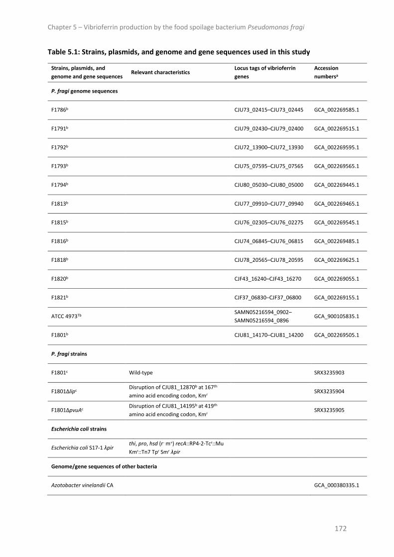
Chapter 5 – Vibrioferrin production by the food spoilage bacterium Pseudomonas fragi
172
Table 5.1: Strains, plasmids, and genome and gene sequences used in this study
Strains, plasmids, and
genome and gene sequences Relevant characteristics
Locus tags of vibrioferrin
genes
Accession
numbersa
P. fragi genome sequences
F1786b CJU73_02415–CJU73_02445 GCA_002269585.1
F1791b CJU79_02430–CJU79_02400 GCA_002269515.1
F1792b CJU72_13900–CJU72_13930 GCA_002269595.1
F1793b CJU75_07595–CJU75_07565 GCA_002269565.1
F1794b CJU80_05030–CJU80_05000 GCA_002269445.1
F1813b CJU77_09910–CJU77_09940 GCA_002269465.1
F1815b CJU76_02305–CJU76_02275 GCA_002269545.1
F1816b CJU74_06845–CJU76_06815 GCA_002269485.1
F1818b CJU78_20565–CJU78_20595 GCA_002269625.1
F1820b CJF43_16240–CJF43_16270 GCA_002269055.1
F1821b CJF37_06830–CJF37_06800 GCA_002269155.1
ATCC 4973Tb SAMN05216594_0902–
SAMN05216594_0896 GCA_900105835.1
F1801b CJU81_14170–CJU81_14200 GCA_002269505.1
P. fragi strains
F1801c Wild-type SRX3235903
F1801Δlipc Disruption of CJU81_12870b at 167th
amino acid encoding codon, Kmr SRX3235904
F1801ΔpvuAc Disruption of CJU81_14195b at 419th
amino acid encoding codon, Kmr SRX3235905
Escherichia coli strains
Escherichia coli S17-1 λpir thi, pro, hsd (r- m+) recA::RP4-2-Tcr::Mu
Kmr::Tn7 Tpr Smr λpir
Genome/gene sequences of other bacteria
Azotobacter vinelandii CA GCA_000380335.1
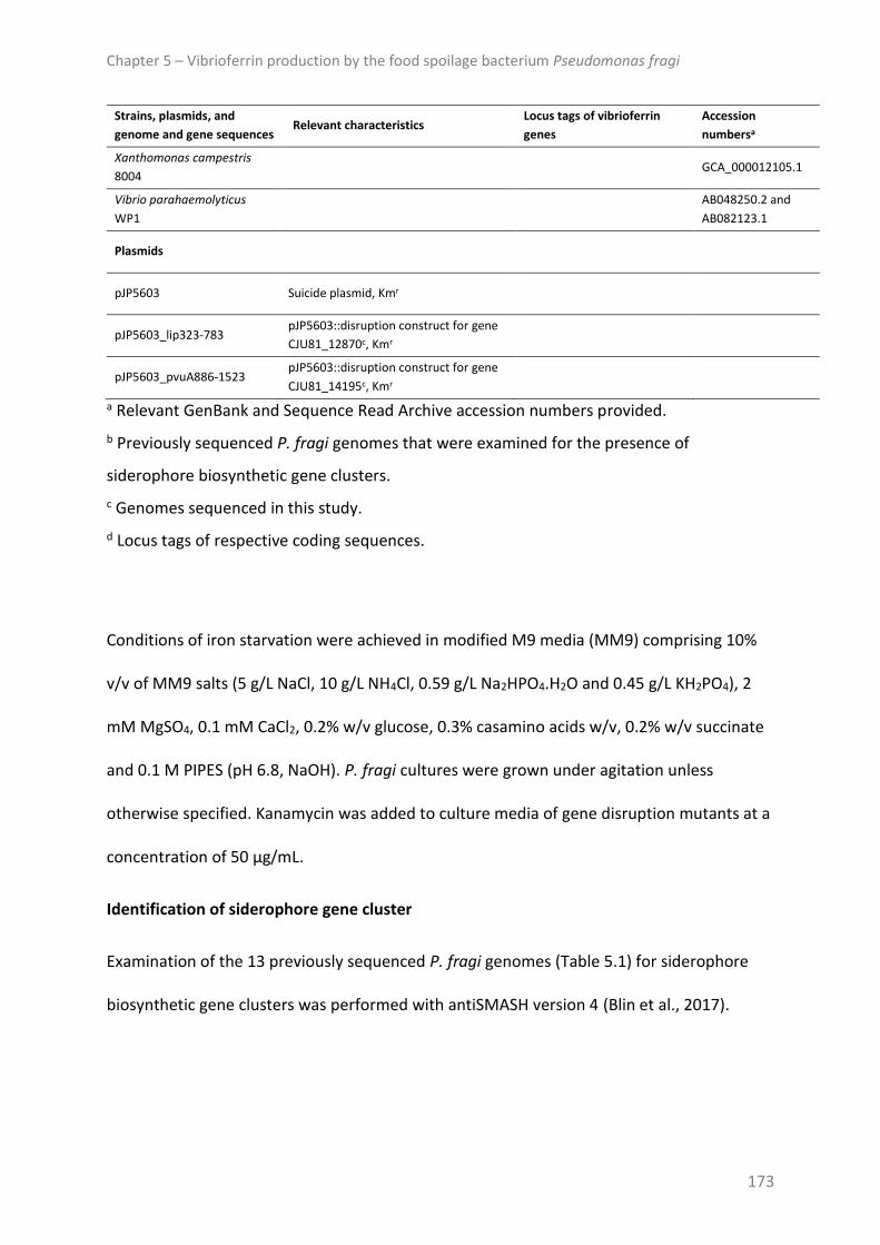
Chapter 5 – Vibrioferrin production by the food spoilage bacterium Pseudomonas fragi
173
Strains, plasmids, and
genome and gene sequences Relevant characteristics
Locus tags of vibrioferrin
genes
Accession
numbersa
Xanthomonas campestris
8004 GCA_000012105.1
Vibrio parahaemolyticus
WP1
AB048250.2 and
AB082123.1
Plasmids
pJP5603 Suicide plasmid, Kmr
pJP5603_lip323-783 pJP5603::disruption construct for gene
CJU81_12870c, Kmr
pJP5603_pvuA886-1523 pJP5603::disruption construct for gene
CJU81_14195c, Kmr
a Relevant GenBank and Sequence Read Archive accession numbers provided.
b Previously sequenced P. fragi genomes that were examined for the presence of
siderophore biosynthetic gene clusters.
c Genomes sequenced in this study.
d Locus tags of respective coding sequences.
Conditions of iron starvation were achieved in modified M9 media (MM9) comprising 10%
v/v of MM9 salts (5 g/L NaCl, 10 g/L NH4Cl, 0.59 g/L Na2HPO4.H2O and 0.45 g/L KH2PO4), 2
mM MgSO4, 0.1 mM CaCl2, 0.2% w/v glucose, 0.3% casamino acids w/v, 0.2% w/v succinate
and 0.1 M PIPES (pH 6.8, NaOH). P. fragi cultures were grown under agitation unless
otherwise specified. Kanamycin was added to culture media of gene disruption mutants at a
concentration of 50 µg/mL.
Identification of siderophore gene cluster
Examination of the 13 previously sequenced P. fragi genomes (Table 5.1) for siderophore
biosynthetic gene clusters was performed with antiSMASH version 4 (Blin et al., 2017).

Chapter 5 – Vibrioferrin production by the food spoilage bacterium Pseudomonas fragi
174
Generation of disruption mutants, genomic DNA isolation, and genome sequencing and
analysis
A gene disruption mutant was generated of the TonB-dependent outer membrane
siderophore receptor (ΔpvuA) by homologous recombination with a pJP5603 suicide vector
construct. This system results in a single cross-over event, whereby the pJP5603 vector
(Riedel et al., 2013) encoding a kanamycin resistance gene remains integrated in the
genome (Penfold and Pemberton, 1992). Thus, a control strain with wild-type vibrioferrin
genes and a disrupted lipase gene (Δlip) was generated for subsequent experimental
comparisons. The lip gene was chosen because the disruption of this gene would not affect
iron metabolism of the strain, nor would it be required for growth of the strain in the
culture media used in this work. Furthermore, because it is present as a single gene rather
than in an operon cluster, polar effects on downstream genes were highly unlikely.
DNA manipulations were performed using standard protocols (Sambrook and Russell, 2001).
Plasmid constructs were generated by ligating BamHI-digested pJP5603 and BamHI-
restriction fragments of the P. fragi F1801 lip and pvuA genes (Table 5.1), which were
amplified using primers included in Table C1 (Appendix C).
pJP5603 gene disruption constructs were transferred from Escherichia coli S17-1 λpir (Simon
et al., 1983) to P. fragi F1801 by biparental filter matings as previously described
(Windgassen et al., 2000) with some adjustments; recipient cells were grown at 30 °C and
the ratio of donor to recipient cells was 1:2 (5x10⁸ cfu to 109 cfu), respectively. After mating,
cells were spread on Pseudomonas agar containing cetrimide-fucidin-cephaloridine selective
supplement (Oxoid) for Pseudomonas and 50 µg/mL kanamycin sulphate to select for
transconjugants. Plates were incubated at 25 °C for 30 h.
Chapter 5 – Vibrioferrin production by the food spoilage bacterium Pseudomonas fragi
175
Strains (wild-type F1801, Δlip and ΔpvuA) were grown for 18 h in tryptone soya broth (TSB,
Oxoid) at 25 °C. Genomic DNA was isolated with the DNeasy Blood and Tissue Kit (QIAGEN)
according to the manufacturer’s protocol for Gram-negative bacteria. Library preparation
and genome sequencing were carried out at Queensland Health (Health Support
Queensland) using MiniSeq High Output kits with 150 cycles and the Illumina MiniSeq
System (Illumina). Genome assembly was performed as previously described (Stanborough
et al., 2017a). Confirmation of correct insertion of the vector was achieved by manual
inspection of the genomes with Geneious version 9.0.5 (Kearse et al., 2012). Snippy
(https://github.com/tseemann/snippy) was used to confirm an absence of additional
genetic variants in the Δlip and ΔpvuA mutants’ genomes by aligning sequence reads of
wild-type F1801, ΔpvuA and Δlip strains to the reference genome of F1801.
Sequence reads of wild-type F1801, Δlip and ΔpvuA strains were deposited in the Sequence
Read Archive and the respective accession numbers are provided in Table 5.1.
Phylogenetic tree of vibrioferrin sequences
MAFFT multiple sequence alignments (Katoh et al., 2002) of the vibrioferrin PvuA and PvsA-
PvsE protein sequences were performed using the default settings. The six multiple
alignments were concatenated and Gblocks version 0.91b (Castresana, 2000, Talavera and
Castresana, 2007) was run with the default settings to remove poorly aligned positions and
divergent regions of the alignments. Bayesian inference (BI), performed with MrBayes
version 3.2.6 (Ronquist and Huelsenbeck, 2003), was conducted with a run of 1000,000
generations and sampling every 1000. A mixed amino acid model analysis was set, enabling
the MCMC sampler to test all of the fixed rate models. MrBayes determined that the
Wheeler and Goldman (WAG) model of amino acid replacement (Whelan and Goldman,

Chapter 5 – Vibrioferrin production by the food spoilage bacterium Pseudomonas fragi
176
2001) had the best likelihood score and was chosen for the analysis. Convergence
parameters were assessed using Tracer version 1.6 (Rambaut et al., 2014) and the majority
rule consensus tree was rendered with Figtree version 1.4.2
(http://tree.bio.ed.ac.uk/software/figtree/). BIs were run three times to ensure
reproducibility of the resulting trees.
RNA isolation and quantitative reverse transcription-polymerase chain reaction (qRT-PCR)
RNA was isolated from 1 mL of mid-log phase iron-starved cultures (1.5x10⁸ cfu).
Stabilisation of RNA was achieved with RNAprotect Bacteria Reagent (QIAGEN) following the
manufacturer’s recommendations and total RNA was extracted with the RNeasy Mini Kit
(QIAGEN) according to the supplier’s instructions and stored in H2O at -80 °C. cDNA was
synthesised from 1 µg of total RNA using iScript gDNA Clear cDNA Synthesis Kit (Biorad) as
suggested by the manufacturer, however DNase treatment was increased from the
recommended 5 to 30 min. qRT-PCR was performed on an AriaMx Real-time PCR System
(Agilent) using the iTaq Universal SYBR Green Supermix (Biorad) as a fluorescence source.
PCR reaction mixes included 10 µL iTaq Universal SYBR Green Supermix (Biorad), 500 nM
forward and reverse primers and 1 µL template cDNA (diluted 1:10 in H2O) in a total volume
of 20 µL. Primer sequences (Table C1: Appendix C) were designed with Primer3 version 2.3.4
(Untergasser et al., 2012) . Acceptable primer efficiency was confirmed for each target gene
with standard curves, and melt-curve analysis of amplicons of each target gene was
performed to ensure amplification of single gene products. PCR conditions were as follows:
30 s at 95 °C followed by 40 cycles of 5 s at 95 °C and 30 sec at 60 °C. Melt-curve analysis
involved an incremental increase of 0.5 °C every 2 s from 65 to 95 °C. Each assay included a
non-template control, and each sample a control without reverse transcriptase. Raw data

Chapter 5 – Vibrioferrin production by the food spoilage bacterium Pseudomonas fragi
177
were analysed with Agilent AriaMx version 1.0 software and exported to excel for further
analysis. Ct-values were normalised to the isocitrate dehydrogenase gene (gene locus tag
CJU81_09775 in F1801, GenBank accession no. GCA_002269505.1) and relative expression
levels were calculated according to the 2ΔΔCt method.
Chromeazurol S (CAS) supernatant assays
CAS assay solution was prepared as previously described (Alexander and Zuberer, 1991).
Siderophore detection from culture supernatants was performed using the method of
Schwyn and Neilands (1987). Briefly, bacterial cultures were spun down at 2,500 x g for 5
min and the supernatants were filtered through a 0.20 µm cellulose acetate membrane
filter (Sartorius). Siderophores were detected in the supernatants by mixing equal volumes
of filtered supernatant and CAS solution. After allowing the solutions to equilibrate for 1.5 h,
a visible change in the colour of the mixture from blue to orange was considered a positive
reaction. To detect siderophore production at 25 °C, strains were grown in 4 mL MM9 for 18
h prior to supernatant collection. To monitor siderophore production at 4 °C, F1801 was
grown in 50 mL MM9 for 4 d and every 24 h 3 mL of culture were removed to measure
growth (A600), and detect siderophores by CAS supernatant assays. To monitor the increase
in siderophore levels over the 4 d, absorbance measurements of the CAS reaction mixtures
were performed at 630 nm.
Growth experiments with 2,2’-bipyridyl and bovine apo-transferrin
Single colonies were used to inoculate 3 mL MM9. Cultures were grown at 25 °C for 18 h. 20
µL of these cultures were used to inoculate 180 µL fresh MM9, supplemented as indicated
in the text with 2,2’-bipyridyl (Sigma-Aldrich), bovine apo-transferrin (Sigma-Aldrich) and/or

Chapter 5 – Vibrioferrin production by the food spoilage bacterium Pseudomonas fragi
178
sodium bicarbonate and FeCl3, in Greiner 96-well flat bottom microtitre plates (Sigma-
Aldrich). Growth was monitored (A600) using an EON microplate reader (BioTek).
RESULTS AND DISCUSSION
In this work, 13 previously sequenced P. fragi genomes were examined for the presence of
siderophore biosynthetic gene clusters. This search resulted in the identification of a
conserved gene cluster, homologs of which were shown to be responsible for vibrioferrin
biosynthesis and vibrioferrin-mediated iron uptake in the terrestrial bacteria Azotobacter
vinelandii and Xanthomonas spp., and in the marine bacterium Vibrio parahaemolyticus
(Pandey and Sonti, 2010, Yamamoto et al., 1994, Baars et al., 2016, Pandey et al., 2017)
(Figure 5.1). Similar to the vibrioferrin gene clusters in A. vinelandii and Xanthomonas
campestris, this ~10 kb region in P. fragi comprised seven open reading frames (Figure
5.1A). Previous work in Xanthomonas spp. and V. parahaemolyticus showed that five of
these genes (mhpE, pvsA, pvsB, pvsD and pvsE) are involved in the biosynthesis of
vibrioferrin, while pvuA is a TonB-dependent outer-membrane receptor and pvsC an inner-
membrane exporter for vibrioferrin (Tanabe et al., 2006, Funahashi et al., 2002, Fujita et al.,
2011, Pandey and Sonti, 2010). Locus tags of these genes in the 13 strains are shown in
Table 5.1. Analysis of the upstream region of this gene cluster revealed the presence of a 19
bp sequence, matching 14 of the 19 nucleotides of the X. campestris consensus Fur-box
(Blanvillain et al., 2007) and 11 of the 19 nucleotides of the Escherichia coli and
Pseudomonas aeruginosa consensus Fur-box. This sequence was located 129 bases
upstream of the mhpE start codon. Fur homologs were also identified in the P. fragi
genomes with the locus tag CJU81_16820 in F1801.

Chapter 5 – Vibrioferrin production by the food spoilage bacterium Pseudomonas fragi
179
To investigate the relatedness of the vibrioferrin genes between the 13 strains and A.
vinelandii, X. campestris and V. parahaemolyticus, a phylogenetic tree of the PvuA and PvsA-
PvsE sequences was determined by Bayesian inference (BI) (Figure 5.1B). Topology of the BI
consensus tree showed three clades of P. fragi vibrioferrin sequences. The largest of the
three clades, comprising closely related sequences of eight of the 13 strains, was clearly
separated from the other two clades, which share a more recent common ancestral
sequence. The sequences of the P. fragi type strain were found in one of these two clades
with those of two additional strains. Sequences of F1801, the P. fragi strain used for
subsequent experiments, were grouped together in the second of these clades with
vibrioferrin sequences of one other strain. In accordance with their phylogeny, sequences of
the P. fragi were closest to those of A. vinelandii, while those of X. campestris and V.
parahaemolyticus were substantially more distant.

Chapter 5 – Vibrioferrin production by the food spoilage bacterium Pseudomonas fragi
180
Figure 5.1: Vibrioferrin gene cluster identified in 13 P. fragi. (A) Schematic diagram of the
vibrioferrin gene cluster identified in P. fragi and conserved synteny of these genes in
known vibrioferrin producing bacteria A. vinelandii, X. campestris and V. parahaemolyticus.
Predicted biosynthesis genes are marked with an asterisk in the P. fragi genes. Numbers
shown in genes indicate percent sequence identity of proteins to respective homologs in P.
fragi F1801, as determined by MAFFT protein alignment. The putative Fur-box identified
upstream of the first gene in the P. fragi cluster is shown and consensus Fur-box sequences
of X. campestris and E. coli (identical to the P. aeruginosa consensus Fur-box) are included
for comparison. (B) Mid-point rooted phylogram of the consensus BI tree of the PvuA and
PvsA-PvsE sequences of the 13 P. fragi, A. vinelandii, X. campestris and V. parahaemolyticus.
P. fragi F1801 that was used in subsequent experiments is marked with an asterisk. The

Chapter 5 – Vibrioferrin production by the food spoilage bacterium Pseudomonas fragi
181
subtree shown in the box represents BI branching on a different scale to the tree. Posterior
probability values are shown as percentages at the nodes and scale bars denote the number
of substitutions per site.
Previous work suggested that P. fragi does not synthesise siderophores (Champomier-
Vergès et al., 1996), and utilising the succinate media (non-deferrated and deferrated
forms) presented in the work, P. fragi F1801 displayed little to no growth and siderophore
production of this strain was not observed (data not shown). Therefore in this study, a non-
deferrated, modified M9 medium (MM9) was used containing glucose that was not present
in the media used by Champomier-Verges et al. (1996). MM9 enabled both satisfactory
growth, as demonstrated by growth of F1801 at 25 °C, while still ensuring conditions of iron-
starvation, which can be seen by the pronounced increase in growth of F1801 in MM9
supplemented with 50 µM FeCl3 (Figure 5.2A).
The sensitive CAS supernatant assay was used to detect the presence of siderophores in
culture supernatants of F1801 grown at optimal growth temperature (25 °C) in MM9. The
CAS assay is based on the removal of iron from the chromogenic dye CAS by siderophores,
whereby a colour change is observed from blue to orange (Schwyn and Neilands, 1987).
Figure 5.2B shows that siderophores were clearly detected in cell-free supernatants of iron-
starved cultures of F1801. Reaction times for the exchange of iron from CAS were within 1.5
h, a time-frame consistent with carboxylate-type siderophores (Schwyn and Neilands, 1987).
In contrast, when F1801 was grown in MM9 supplemented with 50 mM FeCl3, siderophores
were largely absent from culture supernatants (Figure 5.2B). Furthermore, expression of the
vibrioferrin genes was heavily down-regulated (between 12- (pvsE) and 120-fold (pvsD)
reduction) relative to their expression in MM9 without iron-supplementation (Figure 5.2C),

Chapter 5 – Vibrioferrin production by the food spoilage bacterium Pseudomonas fragi
182
indicating repression of siderophore production under iron-rich conditions. These results are
in agreement with other studies, which showed that vibrioferrin production in A. vinelandii,
Xanthomonas and V. parahaemolyticus increased under iron-limitation, while under iron-
replete conditions vibrioferrin production ceased (Pandey and Sonti, 2010, Funahashi et al.,
2000, Pandey et al., 2017, McRose et al., 2017). Thus, together with the presence of a
putative Fur-box upstream of the vibrioferrin gene cluster, these results suggest Fur-
mediated regulation of the P. fragi vibrioferrin genes.
As a key meat and milk spoilage bacterium, P. fragi F1801 was tested for siderophore
production at refrigeration temperature by monitoring growth and the accumulation of
siderophores in supernatants at 4 °C over 4 days. Blue-coloured CAS solution has a
maximum absorption at 630 nm. Orange-coloured CAS reaction mixtures, in which
siderophores have removed iron from CAS, have essentially no absorbance at this
wavelength and there is a largely linear dependence of A630-values of CAS reaction mixtures
versus concentration of the chelator (Schwyn and Neilands, 1987). Figure 5.2D shows a
steady decrease in A630 values of CAS reaction mixtures over the 4 days, indicating an
accumulation of siderophores in culture supernatants of F1801. This accumulation
correlated well with cell growth of the strain, demonstrating siderophore production of P.
fragi at a temperature relevant to the storage of meat and milk.
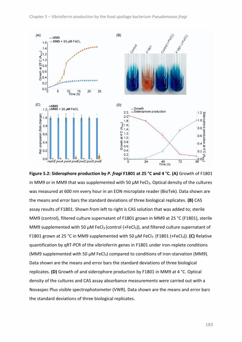
Chapter 5 – Vibrioferrin production by the food spoilage bacterium Pseudomonas fragi
183
Figure 5.2: Siderophore production by P. fragi F1801 at 25 °C and 4 °C. (A) Growth of F1801
in MM9 or in MM9 that was supplemented with 50 µM FeCl3. Optical density of the cultures
was measured at 600 nm every hour in an EON microplate reader (BioTek). Data shown are
the means and error bars the standard deviations of three biological replicates. (B) CAS
assay results of F1801. Shown from left to right is CAS solution that was added to; sterile
MM9 (control), filtered culture supernatant of F1801 grown in MM9 at 25 °C (F1801), sterile
MM9 supplemented with 50 µM FeCl3 (control (+FeCl3)), and filtered culture supernatant of
F1801 grown at 25 °C in MM9 supplemented with 50 µM FeCl3 (F1801 (+FeCl3)). (C) Relative
quantification by qRT-PCR of the vibrioferrin genes in F1801 under iron-replete conditions
(MM9 supplemented with 50 µM FeCl3) compared to conditions of iron-starvation (MM9).
Data shown are the means and error bars the standard deviations of three biological
replicates. (D) Growth of and siderophore production by F1801 in MM9 at 4 °C. Optical
density of the cultures and CAS assay absorbance measurements were carried out with a
Novaspec Plus visible spectrophotometer (VWR). Data shown are the means and error bars
the standard deviations of three biological replicates.
Chapter 5 – Vibrioferrin production by the food spoilage bacterium Pseudomonas fragi
184
To confirm a role for the vibrioferrin gene cluster in siderophore-mediated iron acquisition,
a gene disruption mutant of the TonB-dependent outer membrane siderophore receptor
(ΔpvuA) was generated, as well as a control strain with wild-type vibrioferrin genes and a
disrupted lipase gene (Δlip) for subsequent experimental comparisons. qRT-PCR analysis
revealed that expression of genes downstream of pvuA in the ΔpvuA mutant was markedly
reduced (between 7- (pvsE) and 128-fold (pvsA) reduction) relative to the Δlip control strain
(Figure 5.3A), indicating polar effects caused by integration of the suicide vector in the pvuA
gene. As our aim was to confirm a role for this gene cluster in siderophore-mediated iron
uptake, rather than to study the function of the vibrioferrin genes, which has been
addressed in other studies (Pandey and Sonti, 2010, Tanabe et al., 2003, Funahashi et al.,
2002, Fujita et al., 2011, Tanabe et al., 2006, Pandey et al., 2017), the ΔpvuA mutant was
used for further investigations.
A marked reduction in siderophore levels was detected in culture supernatants of ΔpvuA
compared to the Δlip control strain (Figure 5.3B). Gene deletion of pvuA, which encodes an
outer membrane vibrioferrin receptor, should result in an accumulation of siderophores in
culture supernatants. Therefore the observed reduction was likely due to the polar effects
on the downstream biosynthesis genes. Genetic variants were not found in genes or in non-
coding regions associated with the vibrioferrin genes or other iron-related genes of the
ΔpvuA mutant’s genome (Table C2: Appendix C), demonstrating that phenotypes of the
ΔpvuA mutant were solely due to the disruption of the target gene and associated polar
effects.
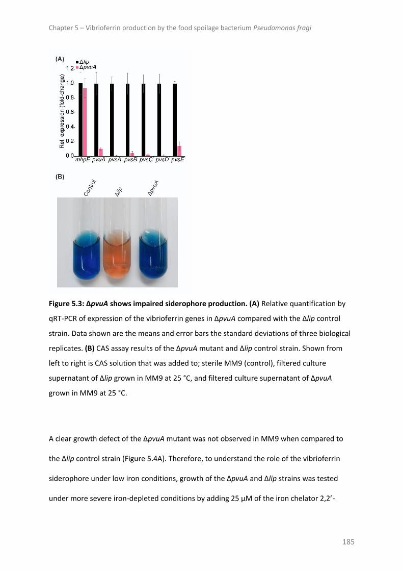
Chapter 5 – Vibrioferrin production by the food spoilage bacterium Pseudomonas fragi
185
Figure 5.3: ΔpvuA shows impaired siderophore production. (A) Relative quantification by
qRT-PCR of expression of the vibrioferrin genes in ΔpvuA compared with the Δlip control
strain. Data shown are the means and error bars the standard deviations of three biological
replicates. (B) CAS assay results of the ΔpvuA mutant and Δlip control strain. Shown from
left to right is CAS solution that was added to; sterile MM9 (control), filtered culture
supernatant of Δlip grown in MM9 at 25 °C, and filtered culture supernatant of ΔpvuA
grown in MM9 at 25 °C.
A clear growth defect of the ΔpvuA mutant was not observed in MM9 when compared to
the Δlip control strain (Figure 5.4A). Therefore, to understand the role of the vibrioferrin
siderophore under low iron conditions, growth of the ΔpvuA and Δlip strains was tested
under more severe iron-depleted conditions by adding 25 µM of the iron chelator 2,2’-

Chapter 5 – Vibrioferrin production by the food spoilage bacterium Pseudomonas fragi
186
bipyridyl (BP) to MM9. While growth of the Δlip control strain was unaffected at 25 µM BP,
addition of the ferrous iron chelator at these concentrations markedly impaired growth of
the ΔpvuA mutant, demonstrating that the siderophore is involved in iron acquisition under
low iron conditions.
To investigate whether the vibrioferrin siderophore could play a role in iron metabolism of
P. fragi in foods such as meat and milk, where iron is sequestered by high-affinity iron-
binding proteins, growth of ΔpvuA and Δlip strains was compared in MM9, to which bovine
apo-transferrin (apo-Tsf), as a model transferrin protein, was added. In all transferrin
proteins, the carbonate (or bicarbonate) of the binding cleft is the synergistic anion, without
which the protein loses its affinity for iron (Pakdaman and El Hage Chahine, 1997). Thus, to
ensure the specific binding of Fe3+ by apo-Tsf, the growth media was also supplemented
with 20 mM bicarbonate (BC) (Chung, 1984). BC at a concentration of 20 mM had no effect
on the strains’ growth when compared to their growth in MM9 without BC addition (Figure
5.4B). When the growth medium was supplemented with apo-Tsf at a concentration of 2.5
µM in the presence of 20 mM BC, growth, although slowed, was observed for the Δlip
control strain (Figure 5.4C). In contrast, growth of ΔpvuA was completely inhibited in the
presence of apo-Tsf and BC, but could be rescued by supplementation of this media with
100 µM FeCl3 (Figure 5.4D). These data suggest that the vibrioferrin siderophore may be
capable of removing ferric iron from transferrin proteins.

Chapter 5 – Vibrioferrin production by the food spoilage bacterium Pseudomonas fragi
187
Figure 5.4: Vibrioferrin siderophore plays a role under iron-starvation and scavenges iron
from transferrin. Growth of ΔpvuA and Δlip control strain in MM9, and in MM9
supplemented with 25 µM 2,2’-bipyridyl (BP) (A), in MM9, and in MM9 supplemented with
20 mM bicarbonate (BC) (B), in MM9 supplemented with 2.5 µM bovine apo-transferrin
(apo-Tsf) and 20 mM BC (C) and in MM9 supplemented with 2.5 µM apo-Tsf, 20 mM BC and
100 µM FeCl3 (D). Data shown for growth experiments in (A–D) are the means and error
bars the standard deviations of three biological replicates. All experiments were repeated a
minimum of three times with similar results observed.
Remarkably, despite considerable divergence between the six vibrioferrin protein sequences
shared by A. vinelandii and V. parahaemolyticus, the absence of an mhpE gene in V.
parahaemolyticus, and the absence of the vibrioferrin-uptake genes pvuA(1)-pvuE in A.

Chapter 5 – Vibrioferrin production by the food spoilage bacterium Pseudomonas fragi
188
vinelandii, both bacteria were shown to produce identical vibrioferrin compounds (Baars et
al., 2016, Yamamoto et al., 1994). Based on percent identities and the evolutionary
relationship of the P. fragi vibrioferrin sequences with A. vinelandii and V. parahaemolyticus
counterparts, it is likely that the P. fragi siderophore is an α-hydroxycarboxylate type
siderophore, similar to or identical to vibrioferrin.
Conclusion
A vibrioferrin gene cluster was identified in 13 strains of the food spoilage bacterium P. fragi
and siderophore production of P. fragi F1801 was observed. Based on the homology of the
identified genes, P. fragi likely excretes an α-hydroxycarboxylate type siderophore, similar
to or identical to vibrioferrin, which plays a role in iron-metabolism of this bacterium under
low iron conditions. More experiments are required to determine whether the siderophore
contributes to the successful growth and colonisation of milk and meat by P fragi; however,
these data suggest that the vibrioferrin siderophore is capable of removing ferric iron from
transferrin proteins.
REFERENCES
ALEXANDER, D. B. & ZUBERER, D. A. 1991. Use of chrome azurol S reagents to evaluate
siderophore production by rhizosphere bacteria. Biology and Fertility of Soils, 12, 39-
45.
BAARS, O., ZHANG, X., MOREL, F. M. M. & SEYEDSAYAMDOST, M. R. 2016. The siderophore
metabolome of Azotobacter vinelandii. Applied and Environmental Microbiology, 82,
27-39.

Chapter 5 – Vibrioferrin production by the food spoilage bacterium Pseudomonas fragi
189
BLANVILLAIN, S., MEYER, D., BOULANGER, A., LAUTIER, M., GUYNET, C., DENANCÉ, N.,
VASSE, J., LAUBER, E. & ARLAT, M. 2007. Plant carbohydrate scavenging through
TonB-dependent receptors: a feature shared by phytopathogenic and aquatic
bacteria. PLOS ONE, 2, e224.
BLIN, K., WOLF, T., CHEVRETTE, M. G., LU, X., SCHWALEN, C. J., KAUTSAR, S. A., SUAREZ
DURAN, H. G., DE LOS SANTOS, E. L. C., KIM, H. U., NAVE, M., DICKSCHAT, J. S.,
MITCHELL, D. A., SHELEST, E., BREITLING, R., TAKANO, E., LEE, S. Y., WEBER, T. &
MEDEMA, M. H. 2017. antiSMASH 4.0-improvements in chemistry prediction and
gene cluster boundary identification. Nucleic Acids Research.
CARPENTER, B. M., WHITMIRE, J. M. & MERRELL, D. S. 2009. This is not your mother's
repressor: the complex role of Fur in pathogenesis. Infection and Immunity, 77,
2590-2601.
CASABURI, A., PIOMBINO, P., NYCHAS, G.-J., VILLANI, F. & ERCOLINI, D. 2015. Bacterial
populations and the volatilome associated to meat spoilage. Food Microbiology, 45,
Part A, 83-102.
CASTRESANA, J. 2000. Selection of conserved blocks from multiple alignments for their use
in phylogenetic analysis. Molecular Biology and Evolution, 17, 540-552.
CEZARD, C., FARVACQUES, N. & SONNET, P. 2015. Chemistry and biology of pyoverdines,
Pseudomonas primary siderophores. Current Medicinal Chemistry, 22, 165-186.
CHAMPOMIER-VERGÈS, M.-C., STINTZI, A. & MEYER, J.-M. 1996. Acquisition of iron by the
non-siderophore-producing Pseudomonas fragi. Microbiology, 142, 1191-1199.
CHUNG, M. C.-M. 1984. Structure and function of transferrin. Biochemical Education, 12,
146-154.

Chapter 5 – Vibrioferrin production by the food spoilage bacterium Pseudomonas fragi
190
COLOMBO, C., PALUMBO, G., HE, J.-Z., PINTON, R. & CESCO, S. 2014. Review on iron
availability in soil: interaction of Fe minerals, plants, and microbes. Journal of Soils
and Sediments, 14, 538-548.
COX, C. D. & ADAMS, P. 1985. Siderophore activity of pyoverdin for Pseudomonas
aeruginosa. Infection and Immunity, 48, 130-138.
DE JONGHE, V., COOREVITS, A., VAN HOORDE, K., MESSENS, W., VAN LANDSCHOOT, A., DE
VOS, P. & HEYNDRICKX, M. 2011. Influence of storage conditions on the growth of
Pseudomonas species in refrigerated raw milk. Applied and Environmental
Microbiology, 77, 460-470.
FARALDO-GOMEZ, J. D. & SANSOM, M. S. P. 2003. Acquisition of siderophores in Gram-
negative bacteria. Nature Reviews Molecular Cell Biology, 4, 105-116.
FARH, M. E.-A., KIM, Y.-J., SUKWEENADHI, J., SINGH, P. & YANG, D.-C. 2017. Aluminium
resistant, plant growth promoting bacteria induce overexpression of Aluminium
stress related genes in Arabidopsis thaliana and increase the ginseng tolerance
against Aluminium stress. Microbiological Research, 200, 45-52.
FUJITA, M. J., KIMURA, N., SAKAI, A., ICHIKAWA, Y., HANYU, T. & OTSUKA, M. 2011. Cloning
and heterologous expression of the vibrioferrin biosynthetic gene cluster from a
marine metagenomic library. Bioscience, Biotechnology, and Biochemistry, 75, 2283-
2287.
FUNAHASHI, T., FUJIWARA, C., OKADA, M., MIYOSHI, S.-I., SHINODA, S., NARIMATSU, S. &
YAMAMOTO, S. 2000. Characterization of Vibrio parahaemolyticus manganese-
resistant mutants in reference to the function of the ferric uptake regulatory protein.
Microbiology and Immunology, 44, 963-970.

Chapter 5 – Vibrioferrin production by the food spoilage bacterium Pseudomonas fragi
191
FUNAHASHI, T., MORIYA, K., UEMURA, S., MIYOSHI, S.-I., SHINODA, S., NARIMATSU, S. &
YAMAMOTO, S. 2002. Identification and characterization of pvuA, a gene encoding
the ferric vibrioferrin receptor protein in Vibrio parahaemolyticus. Journal of
Bacteriology, 184, 936-946.
GARRITY, G. M., BELL, J. A. & LILBURN, T. 2005. Pseudomonadales Orla-Jensen 1921, 270AL.
In: BRENNER, D. J., KRIEG, N. R., STALEY, J. T., GARRITY, G. M., BOONE, D. R., DE VOS,
P., GOODFELLOW, M., RAINEY, F. A. & SCHLEIFER, K.-H. (eds.) Bergey’s Manual® of
Systematic Bacteriology: Volume Two The Proteobacteria Part B The
Gammaproteobacteria. Boston, MA: Springer US.
ILBERT, M. & BONNEFOY, V. 2013. Insight into the evolution of the iron oxidation pathways.
Biochimica et Biophysica Acta - Bioenergetics, 1827, 161-175.
JONES, C. M. & NIEDERWEIS, M. 2010. Role of porins in iron uptake by Mycobacterium
smegmatis. Journal of Bacteriology, 192, 6411-6417.
KATOH, K., MISAWA, K., KUMA, K.-I. & MIYATA, T. 2002. MAFFT: a novel method for rapid
multiple sequence alignment based on fast Fourier transform. Nucleic Acids
Research, 30, 3059-3066.
KEARSE, M., MOIR, R., WILSON, A., STONES-HAVAS, S., CHEUNG, M., STURROCK, S.,
BUXTON, S., COOPER, A., MARKOWITZ, S., DURAN, C., THIERER, T., ASHTON, B.,
MEINTJES, P. & DRUMMOND, A. 2012. Geneious Basic: an integrated and extendable
desktop software platform for the organization and analysis of sequence data.
Bioinformatics, 28, 1647-9.
MCROSE, D. L., BAARS, O., MOREL, F. M. M. & KRAEPIEL, A. M. L. 2017. Siderophore
production in Azotobacter vinelandii in response to Fe-, Mo- and V-limitation.
Environmental Microbiology, 19, 3595-3605.
Chapter 5 – Vibrioferrin production by the food spoilage bacterium Pseudomonas fragi
192
MEYER, J. M. & ABDALLAH, M. A. 1978. The fluorescent pigment of Pseudomonas
fluorescens: Biosynthesis, purification and physicochemical properties. Microbiology,
107, 319-328.
NOINAJ, J., GUILLIER, M., TRAVIS J. BARNARD & BUCHANAN, S. K. 2010. TonB-dependent
transporters: Regulation, structure, and function. Annual Review of Microbiology, 64,
43-60.
PAKDAMAN, R. & EL HAGE CHAHINE, J.-M. 1997. Transferrin. European Journal of
Biochemistry, 249, 149-155.
PANDEY, A. & SONTI, R. V. 2010. Role of the FeoB protein and siderophore in promoting
virulence of Xanthomonas oryzae pv. oryzae on rice. Journal of Bacteriology, 192,
3187-3203.
PANDEY, S. S., PATNANA, P. K., RAI, R. & CHATTERJEE, S. 2017. Xanthoferrin, the alpha-
hydroxycarboxylate-type siderophore of Xanthomonas campestris pv. campestris, is
required for optimum virulence and growth inside cabbage. Molecular Plant
Pathology, 18, 949-962.
PENFOLD, R. J. & PEMBERTON, J. M. 1992. An improved suicide vector for construction of
chromosomal insertion mutations in bacteria. Gene, 118, 145-146.
RAMBAUT, A., SUCHARD, M. A., XIE, D. & DRUMMOND, A. J. 2014. Tracer v1.6. Available
from http://tree.bio.ed.ac.uk/software/tracer/.
RIEDEL, T., ROHLFS, M., BUCHHOLZ, I., WAGNER-DÖBLER, I. & RECK, M. 2013. Complete
sequence of the suicide vector pJP5603. Plasmid, 69, 104-107.
RONQUIST, F. & HUELSENBECK, J. P. 2003. MrBayes 3: bayesian phylogenetic inference
under mixed models. Bioinformatics, 19, 1572-1574.

Chapter 5 – Vibrioferrin production by the food spoilage bacterium Pseudomonas fragi
193
SAMBROOK, J. & RUSSELL, D. W. 2001. Molecular cloning : a laboratory manual, Cold Spring
Harbor, N.Y, Cold Spring Harbor Laboratory Press.
SCHWYN, B. & NEILANDS, J. B. 1987. Universal chemical assay for the detection and
determination of siderophores. Analytical Biochemistry, 160, 47-56.
SIMON, R., PRIEFER, U. & PUHLER, A. 1983. A broad host range mobilization system for in
vivo genetic engineering: transposon mutagenesis in gram negative bacteria. Nature
Biotechnology, 1, 784-791.
SKAAR, E. P. 2010. The battle for iron between bacterial pathogens and their vertebrate
hosts. PLOS Pathogens, 6, e1000949.
STANBOROUGH, T., FEGAN, N., POWELL, S. M., TAMPLIN, M. & CHANDRY, P. S. 2017. Insight
into the genome of Brochothrix thermosphacta, a problematic meat spoilage
bacterium. Applied and Environmental Microbiology, 83, e02786-16.
TALAVERA, G. & CASTRESANA, J. 2007. Improvement of phylogenies after removing
divergent and ambiguously aligned blocks from protein sequence alignments.
Systematic Biology, 56, 564-577.
TANABE, T., FUNAHASHI, T., NAKAO, H., MIYOSHI, S.-I., SHINODA, S. & YAMAMOTO, S. 2003.
Identification and characterization of genes required for biosynthesis and transport
of the siderophore vibrioferrin in Vibrio parahaemolyticus. Journal of Bacteriology,
185, 6938-6949.
TANABE, T., NAKAO, H., KURODA, T., TSUCHIYA, T. & YAMAMOTO, S. 2006. Involvement of
the Vibrio parahaemolyticus pvsC gene in export of the siderophore vibrioferrin.
Microbiology and Immunology, 50, 871-6.
TRAPET, P., AVOSCAN, L., KLINGUER, A., PATEYRON, S., CITERNE, S., CHERVIN, C., MAZURIER,
S., LEMANCEAU, P., WENDEHENNE, D. & BESSON-BARD, A. 2016. The Pseudomonas

Chapter 5 – Vibrioferrin production by the food spoilage bacterium Pseudomonas fragi
194
fluorescens siderophore pyoverdine weakens Arabidopsis thaliana defence in favour
of growth in iron-deficient conditions. Plant Physiology.
TROXELL, B. & HASSAN, H. M. 2013. Transcriptional regulation by Ferric Uptake Regulator
(Fur) in pathogenic bacteria. Frontiers in Cellular and Infection Microbiology, 3, 59.
UNTERGASSER, A., CUTCUTACHE, I., KORESSAAR, T., YE, J., FAIRCLOTH, B. C., REMM, M. &
ROZEN, S. G. 2012. Primer3-new capabilities and interfaces. Nucleic Acids Research,
40, e115.
WHELAN, S. & GOLDMAN, N. 2001. A general empirical model of protein evolution derived
from multiple protein families using a maximum-likelihood approach. Molecular
Biology and Evolution, 18, 691-699.
WINDGASSEN, M., URBAN, A. & JAEGER, K.-E. 2000. Rapid gene inactivation in Pseudomonas
aeruginosa. FEMS Microbiology Letters, 193, 201-205.
YAMAMOTO, S., OKUJO, N., YOSHIDA, T., MATSUURA, S. & SHINODA, S. 1994. Structure and
iron transport activity of vibrioferrin, a new siderophore of Vibrio parahaemolyticus.
The Journal of Biochemistry, 115, 868-874.

Chapter 6 – The effect of a low field strength pulsed electric field on the aerobic microbiota of meat
195
Chapter 6
The effect of a low field strength pulsed electric field on the aerobic
microbiota of beef
ABSTRACT
This study aimed to investigate the effect of a low field strength pulsed electric field (PEF)
(0.25 kV/cm, 3000 pulses), with potential for beef tenderisation, on the aerobic microbiota
of beef stored at 4 °C. The microbial load of naturally contaminated PEF-treated and -
untreated samples was compared along with assessing potential damage to muscle cells and
enhanced proteolysis using reversed phase-high performance liquid chromatography (RP-
HPLC). For time points up to and including 3 days post-treatment, little difference was
detected in the mean log cfu/g of treated and untreated samples. Later time points
indicated that PEF may promote microbial growth, as differences in the sample means were
observed. On day-eight and -nine post-processing, these differences were statisically
significant (P<0.05). RP-HPLC showed no differences in the profiles of phosphate buffered
saline-soluble compounds derived from the meat surface of treated and untreated samples
2 hours post-treatment. In summary, the tested PEF-settings slightly enhanced growth of
the aerobic microbiota, but evidence for muscle cell leakage and enhanced proteolysis was
not obtained with the applied method.

Chapter 6 – The effect of a low field strength pulsed electric field on the aerobic microbiota of meat
196
INTRODUCTION
Meat tenderness is recognised as the most important meat eating quality attribute and
therefore it has a significant impact on the value of meat cuts and the re-purchase intent of
consumers (Miller et al., 2001, Troy and Kerry, 2010). This drives a demand for processing
technologies that can improve meat tenderness of low value cuts and ensure the
consistency of high value muscles.
The emerging food processing technology, pulsed electric field (PEF), refers to the use of
very short pulses (µs to ms range) of low to high electric field intensity (0.1-50 kV/cm) to
foods (Buckow et al., 2013, Toepfl et al., 2014). PEF-treatment induces external electric
fields, which cause an increase in the electric potential across cell membranes
(Zimmermann et al., 1976). If the electric field intensity exceeds a critical limit then
electroporation of cells, disruption of cell organelles and further structural changes to foods
can occur (Zimmermann et al., 1976, Toepfl et al., 2014, Gudmundsson and Hafsteinsson,
2001). While high field strength PEF (20-50 kV/cm) is effective for microbial inactivation in
fruit juices owing to the disruption of microbial cell membranes (Buckow et al., 2013), it is
not suitable as a decontamination method for muscle foods. The absence of a protective cell
wall and larger size renders muscle cells highly susceptible to PEF-induced damage
(Hülsheger et al., 1981, Grahl and Märkl, 1996). Low field strength PEF is however
considered to have potential to improve meat tenderness due to limited muscle cell
disruption, potential accelerated release of Ca2+ and proteases post-mortem and stimulation
of the glycolysis process, all of which could contribute to enhanced post-mortem proteolysis
(Suwandy et al., 2015, Bekhit and Hopkins, 2014). A real advantage of this technology over
other available methods is the ability to modify the processing parameters to treat different
Chapter 6 – The effect of a low field strength pulsed electric field on the aerobic microbiota of meat
197
meat cuts, which may be translated into quality upgrading of low value cuts (Bekhit et al.,
2014b).
Due to muscle cell damage and enhanced proteolysis, PEF-treatment could promote the
development of meat spoilage owing to an increased availability of nutrients and precursor
metabolites of microbial spoilage. Currently, the impact of low field strength PEF on the
microorganisms that grow on beef during storage is unknown. Microorganisms that play a
major role in the spoilage of aerobically stored, refrigerated meat are psychrotrophic
Pseudomonas, whose growth is promoted by oxygen and chilled temperatures (Gill and
Newton, 1978, Hernández-Macedo et al., 2011b). Mesophiles, although initially present on
carcasses, do not grow at chilled temperatures and are quickly outgrown by psychrotrophic
members of the microbiota (Gill and Newton, 1978). In this context, the aim of this work
was to investigate the effects of a low field strength PEF-treatment (0.25 kV/cm, 3000
pulses) with potential for beef tenderisation on the aerobic microbiota developing on beef
stored aerobically at 4 °C. The microbial load of naturally contaminated PEF-treated and -
untreated samples was compared over the storage period of 9 days and reversed phase-
high performance liquid chromatography (RP-HPLC) was used to determine potential
damage to muscle cells and connective tissue of samples following PEF-treatment.
MATERIALS AND METHODS
Meat sampling and preparation
Beef topside, muscle M. semimembranosus, was sourced from a local butcher. The muscle
was trimmed of excess fat and connective tissue and 20 samples of an average size of 65 x
30 x 25 mm were cut and weighed. Of the 20 portions, 10 of these pieces were randomly
Chapter 6 – The effect of a low field strength pulsed electric field on the aerobic microbiota of meat
198
chosen for PEF-treatment (i.e. 10 treated replicates) and 10 were randomly chosen as
untreated control samples (i.e. 10 control replicates).
PEF equipment and treatment of meat
A Diversified Technologies Power ModTM 25 kW Pulsed Electric Field System (Diversified
Technologies, Inc., Bedford, MA, USA) was used, consisting of an adjustable treatment
chamber and a modulator cabinet designed and manufactured by CSIRO for the application
of pulsed electric fields to solid foods. The height of the inside of the treatment chamber
was 40 mm, the gap between the parallel plate electrodes was set to 65 mm and Teflon
blocks were inserted in the sides of the chamber to define a width of 30 mm. To eliminate
temperature effects, the PEF-treatment chamber and the 20 beef portions were placed in
separate sterile Stomacher bags and cooled to approximately 1-2 °C in an ice slurry prior to
treatment. The PEF-treatment chamber was then dried of excess water and the samples
were placed into the treatment chamber ensuring there were no air pockets at the surface
of the electrodes.
Only one set of parameters were tested. These settings represented the harshest of a range
of previously trialled PEF-conditions with meat tenderisation potential that did not result in
temperature effects (Bolumar, T. G., personal communication): Square wave pulses of 10 μs
width at a peak voltage of 1,500 V were applied, resulting in an electrical field strength of
0.25 kV/cm. The pulse repetition rate was set to 100 Hz and PEF was applied to the meat for
a treatment time of 30 ms.
Following PEF-treatment, samples were removed from the chamber, placed in separate
sterile Stomacher bags and kept at 4 °C. Handling of the control samples was identical to
treated samples with the exception of the PEF-treatment.

Chapter 6 – The effect of a low field strength pulsed electric field on the aerobic microbiota of meat
199
Microbiological analysis
Microbiological analysis of the samples was carried out over a period of 9 d. Sampling took
place every 24 h, beginning at 2 h post PEF-treatment: 47.5 mL sterile Dulbecco A phosphate
buffered saline (PBS) (Oxoid, Basingstoke, UK) were added to the Stomacher bags containing
the samples. Samples were washed by vigorously shaking the bags for 30 s in a horizontal
direction. Washing buffer was then aspirated from the bags and ten-fold dilutions of the
buffer were prepared. 1 mL of the dilutions was spread in duplicate onto aerobic plate
count Petrifilm (3M, Maplewood, USA). To ensure good recovery and growth of
psychrotropic spoilage bacteria, petrifilms were incubated at 25 °C for 72 h.
To compare the aerobic populations of microorganisms between treated and untreated
samples with a limited number of samples, following aspiration of the washing buffer,
washed samples were not discarded. Instead samples were aseptically placed into fresh,
sterile Stomacher bags, returned to storage at 4 °C and sampled again as described at each
time point.
For each sample at each time point, cfu were enumerated and converted to log10 cfu/g of
the meat sample. Mean log10 cfu/g values were calculated for treated and untreated
samples for each time point and compared using a two-sample T-test (assuming unequal
variances). Statistical significance was assigned to comparisons with a P value below 0.05.
The Baranyi model (Baranyi and Roberts, 1994) was fitted to the growth curves of the
means of the treated and non-treated samples using DMFit (v3.5 [ComBase;
http://www.combase.cc/index.php/en/tools]) to calculate the kinetic growth parameters:
maximum growth rate (log10 cfu/h), maximum population density (log10 cfu/g) and lag time

Chapter 6 – The effect of a low field strength pulsed electric field on the aerobic microbiota of meat
200
(h). These parameters were compared using a two sample z-test with a significance level of
α<0.05 (critical z-score values: -1.96 and +1.96).
Reversed phase-high performance liquid chromatography (RP-HPLC)
PBS-soluble compounds derived from the washing buffer of the 10 treated and 10 untreated
samples 2 h post-treatment were analysed using RP-HPLC. Extracts were filtered using 0.2
µm cellulose acetate filters (Advantec MFS, Dublin, USA) before analysis. The filtered
extracts were chromatographed by an automated Shimadzu system (consisting of two LC-
10AD VP pumps, FCV-10AL VP mixer, DGU-14A degasser and SIL-10 VP auto injector;
Shimadzu Corp., Kyoto, Japan) fitted with a Vydac, Everest C18 column (250x4.6 mm 300A
5um; The Separations Group Inc, CA, USA). The samples were chromatographed using a
multi-step solvent gradient of solvent B (0.1% trifluoroacetic acid in 90% acetonitrile in
water) in solvent A (0.1% trifluoroacetic acid in deionised water). Samples were eluted using
a gradient elution method: the concentration of solvent B was increased from 27% to 32% in
the first 2 min, to 39% in the next 15 min, to 42.4% in the following 13 min, to 50% in the
subsequent 15 min, and finally increased to 60% in the next 3 min and maintained for 5 min.
The column temperature and solvent flow rate was maintained at 40 °C and 0.7 mL/min,
respectively. Eluate was monitored at 214 nm using a SPD-10A vp UV-VIS detector
(Shimadzu Corp., Kyoto, Japan). The chromatography system was interfaced with computer
Class VP 7.3 software (Shimadzu Corp., Kyoto, Japan).
RESULTS AND DISCUSSION
The sample means of the log cfu/g for PEF-treated and -untreated beef samples are plotted
in Figure 6.1 (n=10 for both treated and untreated beef samples). The initial microbial load

Chapter 6 – The effect of a low field strength pulsed electric field on the aerobic microbiota of meat
201
of the samples was determined two hours post-treatment, demonstrating that the observed
mean starting population density was 1.84 ± 0.28 log cfu/g and 1.88 ± 0.14 log cfu/g for the
treated and untreated samples, respectively (Table 6.1).
Figure 6.1: Growth promotion of aerobic microorganisms at late storage time points of
PEF-treated samples. Shown are the mean curves of aerobic microbial counts of aerobically
stored PEF-treated and -untreated beef samples (n=10 for both treated and untreated
samples) determined over the storage period of 9 d at 4 °C. The blue curve shows the means
of PEF-treated samples, while the red curve represents the means of untreated samples. On
day-eight and -nine post-treatment, the difference in the means of the log cfu/g of the PEF-
treated and -untreated beef samples were statistically significant. *: P value of 0.03, **: P
value of 0.01.

Chapter 6 – The effect of a low field strength pulsed electric field on the aerobic microbiota of meat
202
Table 6.1: Aerobic microbial counts and statistical comparison.
Log cfu/ga
T-test assuming
unequal variances
Time points (d) Treated samples Untreated samples P (T<=t) two-tail
0 1.84 ± 0.28 1.88 ± 0.14 0.74
1 1.68 ± 0.34 1.68 ± 0.12 0.10
2 1.93 ± 0.69 1.91 ± 0.36 0.95
3 3.19 ± 0.77 3.13 ± 0.36 0.84
4 4.56 ± 0.76 4.23 ± 0.43 0.25
5 5.75 ± 0.63 5.41 ± 0.42 0.17
6 6.67 ± 0.60 6.20 ± 0.47 0.07
7 7.24 ± 0.45 6.90 ± 0.38 0.08
8 7.74 ± 0.33 7.40 ± 0.33 0.03
9 8.12 ± 0.27 7.78 ± 0.19 0.01
a Values are means ± standard deviation of 10 replicates.
The sample means of the log cfu/g remained similar for times up to and including 3 days
post-processing, after which, the means of the treated samples were consistently higher
than those of the controls. After day-five, the means of the treated samples were more than
two-fold greater than those of the control samples at all time points, and on day-eight and -
nine post-treatment, statistically significant differences (P <0.05) in the sample means were
observed, indicating that the PEF-treatment may enhance microbial growth. Interestingly,
despite the potential stress conditions exerted by the PEF-treatment, no noticeable
difference in the lag phase of the growth curves was observed.
Using the Baranyi model, the growth curves were fitted to obtain estimates of the kinetic
growth parameters of the curves. For the fitted curves of the treated and untreated

Chapter 6 – The effect of a low field strength pulsed electric field on the aerobic microbiota of meat
203
samples, a lag parameter of 38.7 and 41.6 h, a maximum specific growth rate of 0.04 and
0.05 log cfu/h, and a maximum population density of 7.68 and 7.88 log cfu/g were observed,
respectively (Table 6.2). As demonstrated by z-scores within the critical z-score values (-1.96
and +1.96), these minor differences in the three growth parameters between the treated
and untreated samples were not statistically significant (Table 6.2).
Table 6.2: Kinetic growth parameters determined by DMFit and statistical comparison. Kinetic parameters Treated samples Untreated samples z-scorea
Lag parameter 41.6 38.7 0.21
Maximum growth rate 0.05 0.04 0.93
Maximum population density 7.88 7.68 0.69
a z-score: as calculated by a two-sample z-test.
Mild PEF-processing of muscle meat may result in differences in the types and amounts of
compounds that are present on the meat surface, such as myocyte constituents due to cell
disruption and peptides resulting from enhanced proteolytic activity (Gudmundsson and
Hafsteinsson, 2001, Suwandy et al., 2015). To investigate whether the microbial growth
promotion that was observed for the treated samples could be attributed to an increase in
available nutrients, PBS-soluble compounds derived from the beef sample surfaces two
hours post-treatment were analysed by RP-HPLC. Representative chromatograms of a
treated and untreated sample are shown in Figure 6.2. Comparison of the chromatogram
profiles showed that all peaks associated with the treated samples were also present in the
profiles of the untreated samples. Furthermore, significant differences in the mean peak
heights and peak areas of each peak were not found. As these results did not indicate

Chapter 6 – The effect of a low field strength pulsed electric field on the aerobic microbiota of meat
204
changes to the types and amounts of PBS-soluble compounds that were present on the
surfaces of the treated samples, no further analysis of the compounds of these peaks was
undertaken. It is possible that longer incubation of the samples post-treatment may be
required to detect PEF-induced cell leakage and enhanced proteolysis.
Figure 6.2: Representative RP-HPLC chromatograms of PBS-soluble compounds from the
surface of a PEF-treated and untreated beef sample show no notable profile changes. PBS-
soluble compounds derived from the surface of PEF-treated and -untreated beef samples 2
h post-treatment were analysed by RP-HPLC using a UV detector (214 nm). Representative
chromatograms of a treated sample (blue chromatogram) and an untreated sample (red
chromatogram) show that profiles were highly similar, indicating no detectable changes to
the types and amounts of PBS-soluble compounds that were present on the surfaces of the
treated samples.
In summary, the mild PEF-settings tested here had only a minor impact on the development
of the aerobic microbiota of the beef samples post-treatment. A slight growth enhancement
Chapter 6 – The effect of a low field strength pulsed electric field on the aerobic microbiota of meat
205
was observed for processed samples, however significant differences were only seen at late
time points, where the microbial counts were at levels high enough to characterise the meat
as spoiled (Nychas and Drosinos, 2014). Furthermore, no differences in the profiles of PBS-
soluble compounds derived from the meat surface of treated and untreated samples were
detected. Thus, cell leakage and enhanced proteolysis, as a result of PEF-processing, were
not observed with the PEF-conditions and methods used in this study. Due to the emerging
interest in this technology for its potential to improve quality attributes of meat, the minor
growth enhancement of aerobic microorganisms on treated samples observed in this work,
and the limited scope of this study, further work addressing the impact of low field strength
PEF on the microbiota of meat is warranted. In particular, a larger sample size enabling the
use of conventional microbiology testing, a greater range of tested PEF-settings, and testing
of different microbial subpopulations and longer incubation times following treatment
should be trialled.
REFERENCES
BARANYI, J. & ROBERTS, T. A. 1994. A dynamic approach to predicting bacterial growth in
food. International Journal of Food Microbiology, 23, 277-294.
BEKHIT, A. E.-D. A. & HOPKINS, D. L. 2014. Enhancement of meat quality by pulsed electric
field application, Meat & Livestock Australia Limited.
BEKHIT, A. E.-D. A., VAN DE VEN, R., SUWANDY, V., FAHRI, F. & HOPKINS, D. L. 2014. Effect
of pulsed electric field treatment on cold-boned muscles of different potential
tenderness. Food and Bioprocess Technology, 7, 3136-3146.

Chapter 6 – The effect of a low field strength pulsed electric field on the aerobic microbiota of meat
206
BUCKOW, R., NG, S. & TOEPFL, S. 2013. Pulsed electric field processing of orange juice: a
review on microbial, enzymatic, nutritional, and sensory quality and stability.
Comprehensive Reviews in Food Science and Food Safety, 12, 455-467.
GILL, C. O. & NEWTON, K. G. 1978. The ecology of bacterial spoilage of fresh meat at chill
temperatures. Meat Science, 2, 207-17.
GRAHL, T. & MÄRKL, H. 1996. Killing of microorganisms by pulsed electric fields. Applied
Microbiology and Biotechnology, 45, 148-157.
GUDMUNDSSON, M. & HAFSTEINSSON, H. 2001. Effect of electric field pulses on
microstructure of muscle foods and roes. Trends in Food Science & Technology, 12,
122-128.
HERNÁNDEZ-MACEDO, M. L., BARANCELLI, G. V. & CONTRERAS-CASTILLO, C. J. 2011.
Microbial deterioration of vacuum-packaged chilled beef cuts and techniques for
microbiota detection and characterization: a review. Brazilian Journal of
Microbiology, 42, 1-11.
HÜLSHEGER, H., POTEL, J. & NIEMANN, E. G. 1981. Killing of bacteria with electric pulses of
high field strength. Radiation and Environmental Biophysics, 20, 53-65.
MILLER, M. F., CARR, M. A., RAMSEY, C. B., CROCKETT, K. L. & HOOVER, L. C. 2001. Consumer
thresholds for establishing the value of beef tenderness. Journal of Animal Science,
79, 3062-8.
NYCHAS, G. J. E. & DROSINOS, E. H. 2014. Meat and poultry | Spoilage of meat. In:
TORTORELLO, M. L. & BATT, C. A. (eds.) Encyclopedia of Food Microbiology (Second
Edition). Oxford: Academic Press.

Chapter 6 – The effect of a low field strength pulsed electric field on the aerobic microbiota of meat
207
SUWANDY, V., CARNE, A., VAN DE VEN, R., BEKHIT, A. E.-D. A. & HOPKINS, D. L. 2015. Effect
of pulsed electric field on the proteolysis of cold boned beef M. Longissimus
lumborum and M. Semimembranosus. Meat Science, 100, 222-226.
TOEPFL, S., SIEMER, C., SALDAÑA-NAVARRO, G. & HEINZ, V. 2014. 6 - Overview of pulsed
electric fields processing for food. In: SUN, D.-W. (ed.) Emerging Technologies for
Food Processing (Second Edition). San Diego: Academic Press.
TROY, D. J. & KERRY, J. P. 2010. Consumer perception and the role of science in the meat
industry. Meat Science, 86, 214-226.
ZIMMERMANN, U., PILWAT, G., BECKERS, F. & RIEMANN, F. 1976. Effects of external
electrical fields on cell membranes. Bioelectrochemistry and Bioenergetics, 3, 58-83.

Chapter 7 – General discussion
208
Chapter 7
General discussion
Fresh meat is a highly nutritious commodity, conducive to the rapid growth of
microorganisms and spoilage. Despite the negative economic and social consequences
associated with meat spoilage, the physiology of many of the microorganisms involved in
meat spoilage remains poorly studied. The objective of this thesis was to improve current
knowledge of the biology of the important meat spoilage bacteria B. thermosphacta, P. fragi
and P. lundensis, which is critical to ensure improvements in the control of meat
contamination and spoilage, and to facilitate shelf-life extension.
Advances in sequencing technologies and the field of genomics has enabled rapid and
affordable whole genome sequencing and analysis of bacterial genomes. Not only do
genome sequences facilitate reliable identification of bacteria, but access to the genome
can provide crucial insight into the evolution of bacteria, their metabolic potential,
environmental adaptability, resistance mechanisms and more. Analysis of genome
sequences of the meat spoilage bacterium B. thermosphacta were lacking in the literature.
Therefore chapter 2 (Stanborough et al., 2017a) focused on analysing draft genomes of 12
strains of B. thermosphacta and compared these to genomes of B. campestris and L.
monocytogenes. Data from these analyses revealed a high degree of genomic similarity
among the 12 strains, with bacteriophage genes representing a large proportion of their
accessory genome. Particularly striking was the genomic similarity between the type strain
of this species and the other strains, considering both the temporal and geographic

Chapter 7 – General discussion
209
differences between them. In the future, sequencing of more strains of this organism will
shed light on the overall intraspecies diversity.
Links between previously published phenotypic data on B. thermosphacta, as well as data
provided during this study, and the gene inventory of this bacterium were explored. For
example B. thermosphacta is commonly associated with cheesy, buttery, sweaty odours,
and the production of the metabolites acetate, acetoin, butanediol and branched-chain
fatty acids isobutyric, isovaleric and 2-methylbutyric acid (Dainty and Hibbard, 1983, Dainty
and Hofman, 1983, Grau, 1983, Casaburi et al., 2014). Strong evidence existed for the
production of these branched-chain fatty acids from amino acids rather than lipolysis
(Dainty and Hibbard, 1983). The results of this study confirmed that genes for the
conversion of the branched-chain amino acids leucine, valine, and isoleucine to their
respective branched-chain fatty acid moieties were present in both Brochothrix species.
Genes for the production of acetoin, butanediol and additional metabolic end-products
from pyruvate such as ethanol, lactic acid and acetate were also identified in both species.
As a previous study showed that B. campestris can cause spoilage characterised by off-
odours and green drip (Gribble and Brightwell, 2013), and genes for the production of
acetoin and butanediol were also identified in the B. campestris genome in this work,
further emphasis should be placed on determining whether B. campestris also plays a role in
meat spoilage.
Contrary to available data, amino acid decarboxylase genes were not found in the
Brochothrix genomes and BA production by the B. thermosphacta strains was not observed.
As the production of BA by B. thermosphacta strains has been observed in previous studies
(Casaburi et al., 2014, Nowak and Czyzowska, 2011, Paleologos et al., 2004), it is likely that

Chapter 7 – General discussion
210
the production of these compounds by this bacterium is strain-dependent. Considering the
spoilage impact of BA like cadaverine and putrescine and the health implications associated
with the secretion of BA into food environments, further studies on BA production of B.
thermosphacta strains and the responsible gene repertoire are warranted. An interesting
starting point would be to examine the genomes of known BA-producing strains for the
presence of amino acid decarboxylase genes.
Analysis of antibiotic susceptibility of the 12 strains showed that the bacteria were sensitive
to the four tested antibiotics, except for one tetracycline-resistant strain with plasmid-
mediated tetracycline resistance genes. The strains were also tested for their tolerance to
copper, cobalt and cadmium, however, tolerance levels were not high enough to categorise
the strains as resistant. Literature searches identified only a limited number of studies on
the prevalence of resistance genes in non-pathogenic, meat-associated bacteria. Monitoring
of resistance genes in these bacteria should be addressed in the future for the following
reasons: 1) increasing concern about bacterial cross-resistance to antibiotics, disinfectants
and/or heavy metals, 2) the relative ease with which resistance genes can be transferred
between organisms, 3) the use of antibiotics in animal husbandry and, 4) B. thermosphacta
and other meat-associated bacteria share their environmental niche (i.e. food, food
processing environment, abattoir, farm) with human and livestock pathogens.
A search for L. monocytogenes virulence gene orthologues in the Brochothrix genomes
showed key virulence genes were absent, consistent with the lack of evidence to suggest
that Brochothrix have pathogenic potential. However, orthologues of other L.
monocytogenes virulence genes such as factors involved in surface protein anchoring (srtA,
srtB) and intracellular survival, as well as stress-response regulatory genes (σB and σB-related
Chapter 7 – General discussion
211
genes) were found, which could be involved in spoilage-relevant pathways and play a role in
the persistence of this organism in food processing environments. Knowledge of these
genes led to the work comprising chapter 3 and could also be applied for future studies,
especially considering genes such as σB, whose activation during food processing steps or
abiotic surface cleaning, could have consequences for increased stress resistance and
persistence of the bacteria in food and food processing environments.
The discovery of an orthologue of the L. monocytogenes srtA (involved in surface protein
anchoring) in the B. thermosphacta genomes formed the basis of the work presented in
chapter 3, in which the putative BtSrtA was characterised biochemically and potential
substrate proteins were determined. Successful expression and purification of a
recombinant, truncated version of this enzyme was achieved. Investigations of its catalytic
activity showed the BtSrtA was capable of attaching pseudo-substrates containing LPXTG
sorting motifs to a cell wall mimic, demonstrating in vitro SrtA bioconjugation activity. In
addition, genome examination of B. thermosphacta identified 11 potential BtSrtA
substrates, two of which contained protein domains associated with adherence of
pathogens to host extracellular matrix proteins and cells. Clearly the attachment of these
identified substrates by BtSrtA needs to be confirmed in future work, but this study provides
a robust starting point for investigating surface protein anchoring in B. thermosphacta. As
surface proteins mediate interactions of bacteria with their environment, and two of the
substrate proteins identified in this work contained conserved domains that are involved in
adhesion of pathogens to host cells and ECM, further work in this direction could provide
crucial insight into molecular mechanisms involved in the colonisation of meat by B.
thermosphacta.
Chapter 7 – General discussion
212
The most important meat spoilage bacteria under aerobic conditions are psychrotrophic
Pseudomonas species. In particular, the species P. fragi and P. lundensis constitute a
significant proportion of the aerobic spoilage microbiota, but remain surprisingly poorly
studied and genome analyses of these bacteria were missing. Therefore chapter 4
(Stanborough et al., 2018) aimed to provide insight into genomes of P. lundensis and P.
fragi. The genome analyses showed that the isolates included in this work may belong to
more than two Pseudomonas species with possible spoilage potential. These results
highlight the existing difficulty of species identification of this group and suggest that the
exact composition of aerobic meat spoilage communities remains to be elucidated. To
improve our understanding of psychrotrophic Pseudomonas species associated with meat,
future work will need to focus on increasing the availability of genome sequences of these
bacteria and systematic studies of the phylogeny of this group will be required.
As very limited information was available on the odour-active compounds produced by P.
lundensis, and a thorough understanding of the metabolic potential of P. fragi at the strain
level was missing, the second aim of this chapter was to compare the metabolic profiles of
the P. lundensis and P. fragi isolates during their growth on a beef matrix. Most of the
organisms produced significant levels of odour-active compounds, however three P. fragi
strains produced extremely low levels of VOCs, which suggested that not all strains of
presumed spoilage species have the same spoilage potential. These results demonstrate
that strain-specific studies in the meat environment are required to better understand the
heterogeneity of strains of the same species as well as the heterogeneous behaviour of
strains in general. The P. fragi isolates were associated in particular with the production of
ethyl and methyl esters, which is in line with previous studies on this bacterium (Casaburi et
al., 2015, Ercolini et al., 2010, Morales et al., 2005a). The P. lundensis isolates produced
Chapter 7 – General discussion
213
substantial amounts of 2-nonanone and methyl-2-butenoic acid, confirming that isolates of
this species can be significant producers of odour-active ketones (Morales et al., 2005b) and
demonstrating how isolates of this species may contribute to the spoilage of meat. 1-
undecene production was ubiquitous among isolates of both species, further confirming
that production of this compound is a feature of Pseudomonas species, and as such, 1-
undecene may have potential to be a marker compound for acceptability/degree of spoilage
(Rui et al., 2014, Dainty et al., 1984).
The work presented in chapter 5 (Stanborough et al., 2017b) was initiated following the
discovery of putative siderophore biosynthetic genes in the P. fragi genomes from chapter
4. P. fragi was thought not to produce siderophores (Champomier-Vergès et al., 1996) and is
described as not producing siderophores in detectable amounts in Bergey’s Manual (Garrity
et al., 2005). Not only is iron acquisition a fundamental aspect of the biology of
microorganisms, but efficient iron acquisition may play a role in the dominance of this
bacterial species in spoilage communities. Results of this chapter showed P. fragi likely
secretes an α-hydroxycarboxylate type siderophore, similar to or identical to vibrioferrin,
which plays a role in iron-metabolism of this bacterium under low iron conditions.
Remarkably, both A. vinelandii and V. parahaemolyticus were shown to produce vibrioferrin
siderophores with identical structures, despite considerable divergence between their
vibrioferrin protein sequences and the absence of the vibrioferrin-uptake genes in A.
vinelandii (Baars et al., 2016, Yamamoto et al., 1994). Next steps following on from this
research could be to purify and determine the chemical structure of the P. fragi
siderophore, which is required to understand its mechanism of iron coordination. To
investigate whether the siderophore contributes to the successful growth of P fragi on
meat, expression of the vibrioferrin genes should be tested during its growth on meat.

Chapter 7 – General discussion
214
Furthermore, mutants deficient in siderophore production could be used to determine
whether the vibrioferrin siderophore contributes to providing P. fragi with a competitive
advantage over other members of the meat microbiota. For example, growth of mutant and
wild type strains could be compared in single or multiorganism biofilms on meat.
Meat tenderness is considered the most important quality attribute contributing to
consumer choice and thus considerable interest exists for the development of novel
technologies, which can improve the tenderness of meat. As PEF is a processing technology
with potential for meat tenderisation, but also with a potential to enhance meat spoilage, a
small study was conducted on the effects of a low field strength PEF on the aerobic
microbiota of beef. The mild PEF-settings tested in this work had only a minor impact on the
total aerobic microbial counts of treated samples. A slight growth enhancement was
observed for PEF-processed samples, however significant differences were only seen at time
points, where the microbial counts were at levels high enough to characterise the meat as
spoiled. Furthermore, cell leakage and enhanced proteolysis as a result of PEF-processing
was not observed with the PEF-conditions and methods used in this work. Due to the
emerging interest in this technology for its potential to improve quality attributes of meat,
the minor growth enhancement of aerobic microorganisms on treated samples observed in
this work, and the overall limited scope of this study, further work addressing the impact of
low field strength PEF on the microbiota of meat is warranted. Larger sample sizes would be
required to enable a conventional approach to microbiology testing, and to account for
variations in the application of PEF and the heterogeneity of meat between samples. The
effects of low field strength PEF should also be tested on different subpopulations of the
meat microbiota and systematic testing of various PEF settings would be needed.
Additionally, a longer incubation time following treatment may reveal increased proteolysis.

Chapter 7 – General discussion
215
In conclusion, the genomic and physiological studies of key meat spoilage bacteria that form
this thesis enhance knowledge of these poorly studied bacteria by: 1) providing insight into
genomic and metabolic diversity of strains of these bacteria, 2) demonstrating how strains
of these bacteria may or may not contribute to spoilage by the production of various
malodourous compounds, 3) providing a starting point for further studies on surface protein
attachment of B. thermosphacta and understanding mechanisms with which this bacterium
interacts with its environment and 4) by revealing a siderophore-mediated iron acquisition
system of P. fragi.
REFERENCES
BAARS, O., ZHANG, X., MOREL, F. M. M. & SEYEDSAYAMDOST, M. R. 2016. The siderophore
metabolome of Azotobacter vinelandii. Applied and Environmental Microbiology, 82,
27-39.
CASABURI, A., DE FILIPPIS, F., VILLANI, F. & ERCOLINI, D. 2014. Activities of strains of
Brochothrix thermosphacta in vitro and in meat. Food Research International, 62,
366-374.
CASABURI, A., PIOMBINO, P., NYCHAS, G.-J., VILLANI, F. & ERCOLINI, D. 2015. Bacterial
populations and the volatilome associated to meat spoilage. Food Microbiology, 45,
Part A, 83-102.
CHAMPOMIER-VERGÈS, M.-C., STINTZI, A. & MEYER, J.-M. 1996. Acquisition of iron by the
non-siderophore-producing Pseudomonas fragi. Microbiology, 142, 1191-1199.

Chapter 7 – General discussion
216
DAINTY, R. H., EDWARDS, R. A. & HIBBARD, C. M. 1984. Volatile compounds associated with
the aerobic growth of some Pseudomonas species on beef. Journal of Applied
Bacteriology, 57, 75-81.
DAINTY, R. H. & HIBBARD, C. M. 1983. Precursors of the major end products of aerobic
metabolism of Brochothrix thermosphacta. Journal of Applied Bacteriology, 55, 127-
133.
DAINTY, R. H. & HOFMAN, F. J. K. 1983. The influence of glucose concentration and culture
incubation time on end-product formation during aerobic growth of Brochothrix
thermosphacta. Journal of Applied Bacteriology, 55, 233-239.
ERCOLINI, D., CASABURI, A., NASI, A., FERROCINO, I., DI MONACO, R., FERRANTI, P.,
MAURIELLO, G. & VILLANI, F. 2010. Different molecular types of Pseudomonas fragi
have the same overall behaviour as meat spoilers. International Journal of Food
Microbiology, 142, 120-131.
GARRITY, G. M., BELL, J. A. & LILBURN, T. 2005. Pseudomonadales Orla-Jensen 1921, 270AL.
In: BRENNER, D. J., KRIEG, N. R., STALEY, J. T., GARRITY, G. M., BOONE, D. R., DE VOS,
P., GOODFELLOW, M., RAINEY, F. A. & SCHLEIFER, K.-H. (eds.) Bergey’s Manual® of
Systematic Bacteriology: Volume Two The Proteobacteria Part B The
Gammaproteobacteria. Boston, MA: Springer US.
GRAU, F. H. 1983. End products of glucose fermentation by Brochothrix thermosphacta.
Applied and Environmental Microbiology, 45, 84-90.
GRIBBLE, A. & BRIGHTWELL, G. 2013. Spoilage characteristics of Brochothrix thermosphacta
and campestris in chilled vacuum packaged lamb, and their detection and
identification by real time PCR. Meat Science, 94, 361-368.
Chapter 7 – General discussion
217
LEBERT, I., BEGOT, C. & LEBERT, A. 1998. Growth of Pseudomonas fluorescens and
Pseudomonas fragi in a meat medium as affected by pH (5.8-7.0), water activity
(0.97-1.00) and temperature (7-25 degrees C). International Journal of Food
Microbiology, 39, 53-60.
MORALES, P., FERNANDEZ-GARCIA, E. & NUNEZ, M. 2005a. Production of volatile
compounds in cheese by Pseudomonas fragi strains of dairy origin. Journal of Food
Protection, 68, 1399-407.
MORALES, P., FERNANDEZ-GARCIA, E. & NUNEZ, M. 2005b. Volatile compounds produced in
cheese by Pseudomonas strains of dairy origin belonging to six different species.
Journal of Agricultural and Food Chemistry, 53, 6835-43.
NOWAK, A. & CZYZOWSKA, A. 2011. In vitro synthesis of biogenic amines by Brochothrix
thermosphacta isolates from meat and meat products and the influence of other
microorganisms. Meat Science, 88, 571-574.
PALEOLOGOS, E. K., SAVVAIDIS, I. N. & KONTOMINAS, M. G. 2004. Biogenic amines
formation and its relation to microbiological and sensory attributes in ice-stored
whole, gutted and filleted Mediterranean Sea bass (Dicentrarchus labrax). Food
Microbiology, 21, 549-557.
PENNACCHIA, C., ERCOLINI, D. & VILLANI, F. 2011. Spoilage-related microbiota associated
with chilled beef stored in air or vacuum pack. Food Microbiology, 28, 84-93.
REDONDO-NIETO, M., BARRET, M., MORRISSEY, J., GERMAINE, K., MARTÍNEZ-GRANERO, F.,
BARAHONA, E., NAVAZO, A., SÁNCHEZ-CONTRERAS, M., MOYNIHAN, J. A., MURIEL,
C., DOWLING, D., O’GARA, F., MARTÍN, M. & RIVILLA, R. 2013. Genome sequence
reveals that Pseudomonas fluorescens F113 possesses a large and diverse array of
systems for rhizosphere function and host interaction. BMC Genomics, 14, 54.

Chapter 7 – General discussion
218
RUI, Z., LI, X., ZHU, X., LIU, J., DOMIGAN, B., BARR, I., CATE, J. H. D. & ZHANG, W. 2014.
Microbial biosynthesis of medium-chain 1-alkenes by a nonheme iron oxidase.
Proceedings of the National Academy of Sciences of the United States of America,
111, 18237-18242.
STANBOROUGH, T., FEGAN, N., POWELL, S. M., SINGH, T., TAMPLIN, M. & CHANDRY, P. S.
2018. Genomic and metabolic characterization of spoilage-associated Pseudomonas
species. International Journal of Food Microbiology, 268, 61-72.
STANBOROUGH, T., FEGAN, N., POWELL, S. M., TAMPLIN, M. & CHANDRY, P. S. 2017a.
Insight into the genome of Brochothrix thermosphacta, a problematic meat spoilage
bacterium. Applied and Environmental Microbiology, 83, e02786-16.
STANBOROUGH, T., FEGAN, N., POWELL, S. M., TAMPLIN, M. & CHANDRY, P. S. 2017b.
Vibrioferrin production by the food spoilage bacterium Pseudomonas fragi. FEMS
Microbiology Letters, fnx279.
YAMAMOTO, S., OKUJO, N., YOSHIDA, T., MATSUURA, S. & SHINODA, S. 1994. Structure and
iron transport activity of vibrioferrin, a new siderophore of Vibrio parahaemolyticus.
The Journal of Biochemistry, 115, 868-874.
YOUSSEF, M. K., GILL, C. O., TRAN, F. & YANG, X. 2014. Unusual Compositions of Microflora
of Vacuum-Packaged Beef Primal Cuts of Very Long Storage Life. Journal of Food
Protection, 77, 2161-7.
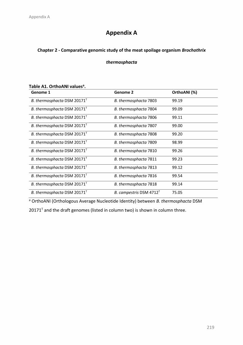
Appendix A
219
Appendix A
Chapter 2 - Comparative genomic study of the meat spoilage organism Brochothrix
thermosphacta
Table A1. OrthoANI valuesa. Genome 1 Genome 2 OrthoANI (%)
B. thermosphacta DSM 20171T B. thermosphacta 7803 99.19
B. thermosphacta DSM 20171T B. thermosphacta 7804 99.09
B. thermosphacta DSM 20171T B. thermosphacta 7806 99.11
B. thermosphacta DSM 20171T B. thermosphacta 7807 99.00
B. thermosphacta DSM 20171T B. thermosphacta 7808 99.20
B. thermosphacta DSM 20171T B. thermosphacta 7809 98.99
B. thermosphacta DSM 20171T B. thermosphacta 7810 99.26
B. thermosphacta DSM 20171T B. thermosphacta 7811 99.23
B. thermosphacta DSM 20171T B. thermosphacta 7813 99.12
B. thermosphacta DSM 20171T B. thermosphacta 7816 99.54
B. thermosphacta DSM 20171T B. thermosphacta 7818 99.14
B. thermosphacta DSM 20171T B. campestris DSM 4712T 75.05
a OrthoANI (Orthologous Average Nucleotide Identity) between B. thermosphacta DSM
20171T and the draft genomes (listed in column two) is shown in column three.
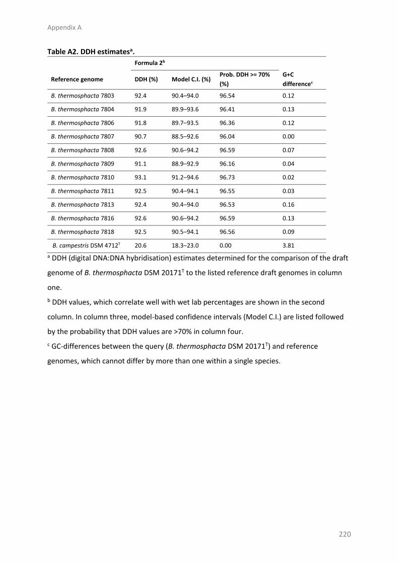
Appendix A
220
Table A2. DDH estimatesa.
Formula 2b
Reference genome DDH (%) Model C.I. (%) Prob. DDH >= 70%
(%)
G+C
differencec
B. thermosphacta 7803 92.4 90.4–94.0 96.54 0.12
B. thermosphacta 7804 91.9 89.9–93.6 96.41 0.13
B. thermosphacta 7806 91.8 89.7–93.5 96.36 0.12
B. thermosphacta 7807 90.7 88.5–92.6 96.04 0.00
B. thermosphacta 7808 92.6 90.6–94.2 96.59 0.07
B. thermosphacta 7809 91.1 88.9–92.9 96.16 0.04
B. thermosphacta 7810 93.1 91.2–94.6 96.73 0.02
B. thermosphacta 7811 92.5 90.4–94.1 96.55 0.03
B. thermosphacta 7813 92.4 90.4–94.0 96.53 0.16
B. thermosphacta 7816 92.6 90.6–94.2 96.59 0.13
B. thermosphacta 7818 92.5 90.5–94.1 96.56 0.09
B. campestris DSM 4712T 20.6 18.3–23.0 0.00 3.81
a DDH (digital DNA:DNA hybridisation) estimates determined for the comparison of the draft
genome of B. thermosphacta DSM 20171T to the listed reference draft genomes in column
one.
b DDH values, which correlate well with wet lab percentages are shown in the second
column. In column three, model-based confidence intervals (Model C.I.) are listed followed
by the probability that DDH values are >70% in column four.
c GC-differences between the query (B. thermosphacta DSM 20171T) and reference
genomes, which cannot differ by more than one within a single species.

Appendix A
221
Figure A1. Statistics of protein sequence lengths. Box plots of protein lengths of all
predicted coding regions in the pan-genome of the 12 B. thermosphacta strains are shown.
Crosses represent sample means and whiskers extend 1.5 times the interquartile. Outliers
are represented by circles. The number of protein sequences analysed are listed below each
plot.

Appendix A
222
Figure A2. KEGG map of the glycolysis/gluconeogenesis pathways and subsequent
pyruvate reactions in Brochothrix. Enzymes that are present in Brochothrix species are
shown in green boxes, while enzymes that are absent are represented in uncoloured boxes,
with the exception of a pyruvate decarboxylase (EC 4.1.1.1) gene, which was not identified in
the draft genome of B. campestris DSM 4712T.

Appendix A
223
Figure A3. KEGG map of the pentose-phosphate pathway in Brochothrix. Enzymes that are
present in Brochothrix species are shown in green boxes. Enzymes that are absent are
represented in uncoloured boxes.

Appendix A
224
Figure A4. KEGG map of the citrate cycle shows key enzymes are missing in Brochothrix.
Enzymes that are present in Brochothrix are shown in green boxes and enzymes that are
absent are represented in uncoloured boxes. Although not shown here, a gene encoding an
adihydrolipoamide S-succinyltransferase enzyme (EC 2.3.1.61) was also identified in the
genome of B. campestris DSM 4712T.

Appendix A
225
Figure A5. KEGG map of butanoate metabolism indicates that enzymes are present in
Brochothrix for the conversion of pyruvate to acetoin and 2,3-butanediol. Enzymes that are
present in Brochothrix species are shown in green boxes, while enzymes that are absent are
represented in uncoloured boxes.

Appendix A
226
Figure A6. KEGG map of valine, leucine and isoleucine degradation pathways indicates all
enzymes are present for the conversion of these amino acids to the acyl-CoA derivatives
isobutyryl-CoA, isovaleryl-CoA and 2-methylbutananoyl-CoA, and propanoyl-CoA. Enzymes
present in the B. thermosphacta strains are marked in green boxes and enzymes that are
absent are in uncoloured boxes. While all enzymes required for the conversion of valine,
leucine and isoleucine to their branched chain acyl-CoA derivatives are present in B.
campestris DSM 4712T, many of the enzymes downstream of these are missing including
enzymes required for propanoyl-CoA production.

Appendix B
227
Appendix B
Chapter 4 - Genomic and metabolic characterisation of spoilage-associated Pseudomonas
species
Table B1. Bacterial numbers of inoculated samples and non-inoculated controls, and percentages of atmospheric gases in headspace vials after 4 d of incubation at 8 °C.a
Samples Log cfu/g sample CO2 levels (%) O2 levels (%) N2 levels (%)
Mean SD Mean SD Mean SD Mean SD
C0 BLDb - 0.1 0.1 21.2 0.1 78.8 0.1
Cd4 5.37 1.08 0.2 0.1 20.3 0.2 79.4 0.3
F1786 9.59 0.05 9.5 0.2 2.3 0.6 88.1 0.6
F1791 9.50 0.11 9.0 0.8 2.6 0.2 88.4 1.0
F1792 9.39 0.09 8.3 0.5 3.2 1.7 88.5 1.6
F1793 9.48 0.02 7.6 0.4 4.7 1.5 87.7 1.9
F1794 9.50 0.06 8.8 0.9 2.8 1.1 88.4 1.4
F1801 9.44 0.13 9.1 0.8 2.7 0.5 88.2 1.1
F1813 9.46 0.04 8.8 0.7 1.9 0.1 89.4 0.8
F1815 9.53 0.12 8.7 0.6 2.7 1.3 88.6 1.1
F1816 9.37 0.04 8.8 0.6 2.3 1.0 88.9 1.2
F1818 9.41 0.06 9.3 0.5 3.1 1.4 87.6 1.1
F1820 9.41 0.05 8.7 0.8 2.7 0.9 88.6 0.6
F1821 9.47 0.04 9.2 0.3 2.8 0.9 88.0 0.9
ATCC 4973T 9.56 0.07 10.0 0.4 1.8 0.3 88.2 0.7
L1788 9.43 0.04 8.3 0.6 5.5 1.4 86.2 0.8
L1802 9.62 0.04 9.9 0.4 3.3 0.8 86.8 1.1
L1810 9.59 0.02 9.5 0.3 3.3 0.9 87.2 0.7
L1814 9.59 0.06 9.1 0.2 3.9 0.7 87.1 0.6
L1817 9.60 0.01 9.8 0.9 3.6 0.5 86.6 0.8
L1819 9.56 0.02 10.1 0.8 3.1 0.4 86.9 1.2
L1822 9.58 0.08 10.1 1.0 3.3 1.2 86.6 0.7
a Shown are the means and standard deviations (SD) of three independent experiments.
b BLD: Below the detection limit

Appendix B
228
Figure B1. Core SNP phylogenies of P. fragi and P. lundensis organisms. A Mid-point rooted
consensus parsimony cladogram of P. fragi core SNPs. Support values were determined by
Shimodiara-Hasegawa resampling test. B NeighborNet network analysis of P. lundensis core
SNP matrix was plotted using uncorrected P distances. The distance scale indicates the
number of nucleotide substitutions per number of SNPs. Truncated network branch is
marked with // to aid visualisation and the original branch length is indicated above.

Appendix B
229
Figure B2. Growth and percentages of atmospheric gases in headspaces of vials of low
(F1794, F1815 and L1788) and high (F1801 and L1814) VOC producers. Tested isolates,
indicated in the legends, were inoculated onto a sterile beef paste and samples were
incubated at 8 °C. The percentages of O2 (A), CO2 (B) and N2 (C) in the headspaces of the
vials were determined at 24 h intervals, prior to sampling of the vial contents to determine
the log cfu/g (D). Each point represents the mean and error bars show the standard
deviation of three experimental replicates.

Appendix C
230
Appendix C
Chapter 5 - Vibrioferrin production by the food spoilage bacterium Pseudomonas fragi
Table C1. Primers used in this study.
Primer name Primer sequencea
Primers used for generating gene disruption constructs
lip323fwd AAAAAGGATCCGTCCCAATCATGGCTCCGAA
lip783rev AAAAAGGATCCCAGGTGGTCCATTGCGTAGT
pvuA886fwd AAAAAGGATCCAGCTACCGCAATTTCGTCGTC
pvuA1523rev AAAAAGGATCCATCAGTGGTGGCGTTGTAGAC
qRT-PCR primers
icd481fwd AAAATGGGTGCCTGGGCTGC
icd623rev GTGGTGGTGCCGTCTTGAGC
aldo559fwd GCCGAACAGAACGCGCAATG
aldo664rev GGAGAGAATGCAGGCGGGCT
pvuA1898fwd CCTCGGCAGGCATCAGCAAC
pvuA2005rev CGCCTAAGGTGAGCGAACGC
pvsA162fwd GCTGCAGGTGATCGAGTGCG
pvsA282rev GGCTGTCGCGGTTTGCAGAT
pvsB1190fwd CCGGCTCCTTGCAACACTGG
pvsB1292rev TGCTGATTGGCCTCAAGCGC
pvsC1028fwd TGGTCAGCCAAATGTGCGGG
pvsC1127rev CGGGAAGTGATCATTAGAGGGCG
pvsD505fwd ACGCCGAAAAGCCGTGAAGG
pvsD657rev CGCCAGTTGCTGCAGGGTAC
pvsE915fwd CCATCACTTCCGCACGCCTG
pvsE1038rev CTTGGGCGTACACAGCTGGC
a Underlined sequences indicate BamHI restriction enzyme sites.

Appendix C
231
Table C2. Genetic variants present in ΔpvuA.
Contiga Positionb Variant type Refc Altd Evidencee FTYPEf
NQKQ01000013 14617 Complex ACAGG GCAGA GCAGA:17 ACAGG:1
Non-coding region
NQKQ01000051 87 Single nucleotide polymorphism
A G G:53 A:5 Non-coding region
a Accession numbers of the contigs the variants were found on in the draft genome of P.
fragi F1801 (Accession no. GCA_002269505.1).
b Position in the sequence, counting from 1.
c The nucleotide(s) in the reference.
d The alternate nucleotide(s) supported by the reads.
e Frequency counts for REF and ALT.
f Class of feature affected: CDS, tRNA, rRNA or non-coding region.
Copyright © 2022 FDOKUMEN