A Sequence of the CIS Gene Promoter Interacts Preferentially with Two Associated STAT5A Dimers: a...
10
1998, 18(10):5852. Mol. Cell. Biol. and Stany Chretien Patrick Mayeux, Catherine Lacombe, Sylvie Gisselbrecht Christian Beisenherz-Huss, Paule Varlet, Odile Muller, Frédérique Verdier, Raquel Rabionet, Fabrice Gouilleux, and STAT5B Biochemical Difference between STAT5A Associated STAT5A Dimers: a Distinct Interacts Preferentially with Two A Sequence of the CIS Gene Promoter http://mcb.asm.org/content/18/10/5852 Updated information and services can be found at: These include: REFERENCES http://mcb.asm.org/content/18/10/5852#ref-list-1 at: This article cites 35 articles, 21 of which can be accessed free CONTENT ALERTS more» articles cite this article), Receive: RSS Feeds, eTOCs, free email alerts (when new http://journals.asm.org/site/misc/reprints.xhtml Information about commercial reprint orders: http://journals.asm.org/site/subscriptions/ To subscribe to to another ASM Journal go to: on April 2, 2014 by guest http://mcb.asm.org/ Downloaded from on April 2, 2014 by guest http://mcb.asm.org/ Downloaded from
Transcript of A Sequence of the CIS Gene Promoter Interacts Preferentially with Two Associated STAT5A Dimers: a...

1998, 18(10):5852. Mol. Cell. Biol.
and Stany ChretienPatrick Mayeux, Catherine Lacombe, Sylvie GisselbrechtChristian Beisenherz-Huss, Paule Varlet, Odile Muller, Frédérique Verdier, Raquel Rabionet, Fabrice Gouilleux, and STAT5BBiochemical Difference between STAT5A Associated STAT5A Dimers: a DistinctInteracts Preferentially with Two A Sequence of the CIS Gene Promoter
http://mcb.asm.org/content/18/10/5852Updated information and services can be found at:
These include:
REFERENCEShttp://mcb.asm.org/content/18/10/5852#ref-list-1at:
This article cites 35 articles, 21 of which can be accessed free
CONTENT ALERTS more»articles cite this article),
Receive: RSS Feeds, eTOCs, free email alerts (when new
http://journals.asm.org/site/misc/reprints.xhtmlInformation about commercial reprint orders: http://journals.asm.org/site/subscriptions/To subscribe to to another ASM Journal go to:
on April 2, 2014 by guest
http://mcb.asm
.org/D
ownloaded from
on A
pril 2, 2014 by guesthttp://m
cb.asm.org/
Dow
nloaded from

MOLECULAR AND CELLULAR BIOLOGY,0270-7306/98/$04.0010
Oct. 1998, p. 5852–5860 Vol. 18, No. 10
Copyright © 1998, American Society for Microbiology. All Rights Reserved.
A Sequence of the CIS Gene Promoter Interacts Preferentially withTwo Associated STAT5A Dimers: a Distinct Biochemical
Difference between STAT5A and STAT5BFREDERIQUE VERDIER,1 RAQUEL RABIONET,1 FABRICE GOUILLEUX,1 CHRISTIAN BEISENHERZ-HUSS,2
PAULE VARLET,1 ODILE MULLER,1 PATRICK MAYEUX,1 CATHERINE LACOMBE,1
SYLVIE GISSELBRECHT,1 AND STANY CHRETIEN3*
Institut Cochin de Genetique Moleculaire (ICGM), Institut National de la Sante et de la Recherche Medicale(INSERM U363), Hopital Cochin, Universite Rene Descartes, F75014 Paris,1 and Institut National de
la Transfusion Sanguine (INTS-GIP), F75015 Paris,3 France, and Tumor Biology Center,Institute for Experimental Cancer Research and Department of Biology,
University of Freiburg, 79106 Freiburg, Germany2
Received 13 February 1998/Returned for modification 3 April 1998/Accepted 17 July 1998
Two distinct genes encode the closely related signal transducer and activator of transcription proteinsSTAT5A and STAT5B. The molecular mechanisms of gene regulation by STAT5 and, particularly, the require-ment for both STAT5 isoforms are still undetermined. Only a few STAT5 target genes, among them the CIS(cytokine-inducible SH2-containing protein) gene, have been identified. We cloned the human CIS gene andstudied the human CIS gene promoter. This promoter contains four STAT binding elements organized in twopairs. By electrophoretic mobility shift assay studies using nuclear extracts of UT7 cells stimulated witherythropoietin, we showed that these four sequences bound to STAT5-containing complexes that exhibiteddifferent patterns and affinities: the three upstream STAT binding sequences bound to two distinct STAT5-containing complexes (C0 and C1) and the downstream STAT box bound only to the slower-migrating C1 band.Using nuclear extracts from COS-7 cells transfected with expression vectors for the prolactin receptor, STAT5A,and/or STAT5B, we showed that the C1 complex was composed of a STAT5 tetramer and was dependent on thepresence of STAT5A. STAT5B lacked this property and bound with a stronger affinity than did STAT5A to thefour STAT sequences as a homodimer (C0 complex). This distinct biochemical difference between STAT5A andSTAT5B was confirmed with purified activated STAT5 recombinant proteins. Moreover, we showed that thepresence on the same side of the DNA helix of a second STAT sequence increased STAT5 binding and that onlyhalf of the palindromic STAT binding sequence was sufficient for the formation of a STAT5 tetramer. Again,STAT5A was essential for this cooperative tetrameric association. This property distinguishes STAT5A fromSTAT5B and could be essential to explain the transcriptional regulation diversity of STAT5.
STAT proteins are latent transcription factors containing aSrc homology 2 domain (SH2 domain) that become activatedby tyrosine phosphorylation. The binding of the STAT SH2domains to the phosphorylated cytokine receptors allows theirtyrosine phosphorylation by Jak kinases. After dimerizationand nuclear translocation, STAT dimers bind to specific DNAsequences, thereby allowing downstream gene regulation.Seven members of the STAT family have been described(STAT1a/b, STAT2, STAT3a/b, STAT4, STAT5A, STAT5B,and STAT6), and specific combinations are involved in thesignaling pathways of different cytokines. The association ofSTAT5 with various coactivators or repressors and the bindingof additional transcription factors to the promoter region oftarget genes, together with various combinations of STATdimers, could contribute to the diversity and specificity of thetranscriptional regulation of target genes (for reviews, see ref-erences 4, 11, and 26).
STAT5, originally named mammary gland factor (MGF),has been described as a positive regulator of the b-casein(bCAS) promoter by prolactin (29). In addition to prolactin,STAT5 proteins have been shown to be activated by erythro-
poietin (Epo), growth hormone, interleukin 2 (IL-2), IL-3,IL-5, IL-7, IL-9, IL-15, granulocyte-macrophage colony-stim-ulating factor (GM-CSF), thrombopoietin, and several othergrowth factors. However, only a few STAT5 target sequenceshave been identified, among them the bCAS promoter (8),the IL-2 receptor alpha (IL-2Ra) chain enhancer (15), p21(WAF1) (21), oncostatin M (34), the serine protease inhibitor2.1 (31), and the CIS gene promoter (20). Different MGFboxes located inside these promoters are implicated in tran-scriptional regulation following STAT5 activation.
Besides positive regulatory pathways, negative regulatorypathways such as phosphatases modulate the response to cyto-kines. The phosphatase SHP-1 has been shown to bind to boththe phosphorylated Epo receptor (Epo-R) and the Jak2 kinaseand to dephosphorylate these proteins, thereby leading to in-activation of Epo-R signaling (12). However, under certainconditions, STAT5 itself or carboxy-terminally truncated iso-forms of STAT5 act as negative regulators of gene transcrip-tion (1, 19, 23, 24, 30). Another negative regulatory mechanisminvolves the protein CIS. The CIS protein is rapidly induced inhematopoietic cells by IL-2, IL-3, GM-CSF, and Epo. CIScontains an SH2 domain in the central part of the moleculeand is associated with the cytoplasmic domains of the tyrosine-phosphorylated Epo-R and the b chain of IL-3R (35). CISoverexpression reduces the activation of promoters regulatedby STAT5. Moreover, STAT boxes were shown to be involved
* Corresponding author. Mailing address: Institut National de laTransfusion Sanguine (INTS-GIP), 6 rue Alexandre Cabanel, F75015Paris, France. Phone: 33 1 46 33 14 09. Fax: 33 1 46 33 92 97. E-mail:[email protected].
5852
on April 2, 2014 by guest
http://mcb.asm
.org/D
ownloaded from

in the Epo-dependent promoter activation of the murine CISpromoter in HEK 293 cells (20).
In this report, we identified different nuclear factors whichbound to the human CIS gene promoter. This complex combi-nation of transcriptional factors comprised STAT5A, STAT5B,an Sp1-related family protein, and at least three GGAA bind-ing proteins, among which one showed an Epo-dependentDNA binding capacity. Among the four STAT5 boxes of theCIS promoter, only one bound to a STAT5 tetramer. More-over, we showed that two STAT5A dimers can interact incooperative binding with two STAT binding sequences of lowor undetectable affinity to form a tetramer. This structure in-volves STAT5A but not STAT5B. The difference in the behav-ior of the two isoforms of STAT5 could be essential for thedifferential transcriptional regulation of STAT5 target genes.
MATERIALS AND METHODS
Reagents. Anti-STAT5 antibodies, directed against the amino terminus of thesheep STAT5A and recognizing both STAT5A and STAT5B, were producedas described previously (7). We produced specific antibodies directed againstSTAT5A and STAT5B by immunizing rabbits with peptides corresponding to the12 carboxy-terminal amino acids (AGLFTSARSSLS) of STAT5A or the 8 car-boxy-terminal amino acids of STAT5B (QWIPHAQS) coupled to keyhole limpethemocyanin (data not published). Rabbit polyclonal anti-Elf-1 and anti-Ets-1/2were purchased from Santa Cruz Biotechnology (Santa Cruz, Calif.). Antibodiesdirected against the amino-terminal domain of PU.1/Spi-1 were kindly providedby F. Moreau-Gachelin (Institut Curie, Paris, France). Anti-GABPa and -bantibodies were kindly provided by M. Negishi (National Institutes of Health,Bethesda, Md.) (33). Highly purified recombinant human Epo (specific activity,120,000 U/mg) was a generous gift from M. Brandt (Boehringer Mannheim), andprolactin was purchased from Sigma.
Isolation of human CIS promoter. The murine CIS cDNA from nucleotides111 to 1092 (GenBank accession no. MUSSH2DC) (35) was cloned by reversetranscriptase PCR with total RNA from IW32 erythroleukemia cells with a senseprimer (59-CTCCTTCCATCCCGCCGAA-39) and an antisense primer (59-CCTGCCTTGTTCTTGCTGGCA-39).
A human placenta genomic DNA library cloned in the cosmid pWE 15 (Clon-tech; reference no. HL 1095m) was screened with the murine CIS cDNA PCRprobe.
Cell cultures. The GM-CSF-dependent human megakaryoblastic cell line UT7(13) was passaged by dilution twice weekly in alpha minimal essential mediumcontaining 10% fetal calf serum containing 2.5 ng of GM-CSF per ml. COS-7cells (ECCAC no. 87021302) were maintained in Dulbecco’s modified Eagle’smedium supplemented with 10% fetal calf serum.
Preparation of nuclear extracts. Starved cells were stimulated at 37°C with 10U of Epo per ml and quickly chilled in ice-cold phosphate-buffered saline. Cellswere pelleted and solubilized with buffer A (buffer A: 20 mM HEPES; 10 mMKCl; 1 mM EDTA; 1 mM dithiothreitol [DTT]; 1 mM phenylmethylsulfonylfluoride; 0.1 mM Na2VO4; 0.2% Nonidet P-40; 10% glycerol; 1 mg each ofaprotinin, pepstatin, and leupeptin per ml, pH 7.9). Cell lysates were centrifugedat 20,000 3 g for 2 min, and the pellets were extracted with buffer B (buffer B:20 mM HEPES; 350 mM NaCl; 10 mM KCl; 1 mM EDTA; 1 mM DTT; 1 mMphenylmethylsulfonyl fluoride; 0.1 mM Na2VO4; 20% glycerol; 1 mg each ofaprotinin, pepstatin, and leupeptin per ml, pH 7.9) with 1 ml of buffer B for 5 3107 cells. The extracts were centrifuged at 20,000 3 g for 5 min, and supernatantswere quickly frozen and stored at 280°C.
Production of recombinant activated STAT5 protein in Sf9 cells. Sf9 cellsgrown in a spinner flask suspension culture were coinfected with baculovirusesencoding either STAT5A and Jak2 or STAT5B and Jak2 with a multiplicity ofinfection of 10 PFU/cell for each virus. At 60 h after infection, cells wereharvested and lysed for 15 min on ice in a buffer containing 10% glycerol, 20 mMHEPES (pH 7.9), 10 mM KCl, 0.2% Nonidet P-40, 1 mM EDTA, 0.1 mM sodiumvanadate, 2 mM DTT, 4-(2-aminoethyl)-benzesulfonyl fluoride, phenylmethyl-sulfonyl fluoride, leupeptin, and aprotinin. Proteins were loaded on a heparincolumn (Pharmacia) and eluted with an NaCl gradient in lysis buffer.
Electrophoretic mobility shift assay (EMSA). Two microliters of nuclear ex-tracts was mixed with 20 ml of binding buffer [10 mM Tris-HCl, 50 mM NaCl,1 mM EDTA, 1 mM DTT, 0.1% Nonidet P-40, 5% glycerol, 1 mg of bovineserum albumin per ml, 2 mg of poly(dI-dC) per ml, pH 7.5] containing 60,000cpm of end-labelled probe, and the mixture was incubated for 30 min at 4°C.Complexes were separated on 6% nondenaturing polyacrylamide gels in 0.253Tris-borate-EDTA and detected by autoradiography.
UV cross-linking of CIS12 probe to complexes C0 and C1. Two probes (A andB) were used in cross-linking experiments. Single-stranded oligonucleotides (25ng) CIS12 (antisense) and CIS2 (sense) (probe A) or CIS12 (sense) and CIS1(antisense) (probe B) were annealed. Flanking sequences were transcribed bythe Klenow fragment of DNA polymerase under standard conditions in the
presence of 250 mM dATP–dGTP–5-bromo-29-dUTP–80 mCi of [a-32P]dCTP(3,000 Ci/mmol). Probes were purified by Sephadex G-25 chromatography, and2.5 ng of probes was incubated with UT7 nuclear extracts under EMSA condi-tions. After separation of the different complexes on a 6% nondenaturing poly-acrylamide gel, the gel was irradiated for 15 min with UV light and autoradio-graphed. The bands were cut out and placed on the top of a sodium dodecylsulfate (SDS)–10% polyacrylamide gel. After migration of the gel, labelled pro-teins were detected by autoradiography.
Transfections and luciferase assays. All plasmids were purified by the Qiagenkit method (Qiagen, Inc.), and two different preparations of plasmid for eachconstruct were tested. Plasmids (1 mg/transfection) of different promoter-lucif-erase constructs cloned in the pGL2-basic vector (Promega) were introduced inCOS-7 cells together with 1 mg of the pCMV-gal plasmid (internal control fortransfection efficiency). After 24 h of culture with the appropriate medium, halfof the transfected cells were stimulated with 1 mg of prolactin per ml. Total cellextracts were prepared and used for determination of luciferase and b-galacto-sidase activities according to the manufacturer’s instructions (Promega kit). Finalluciferase activity results were obtained after normalization with b-galactosidaseactivity.
RESULTS
Localization and structural analysis of the human CIS gene5*-flanking region: constitutive and cytokine-induced nuclearfactors bound to the CIS gene promoter. The murine CIScDNA probe was used to screen a human genomic library asdescribed in Materials and Methods. A cosmid, COS-CIS, thatcontained the full-length CIS gene was isolated (data notshown). Eight hundred sixty-five base pairs of the murine CISgene promoter sequences have been previously reported byMatsumoto et al. (20). The nucleotide sequence of the 59-flanking region of the human CIS gene was compared with thissequence. Computer alignments revealed that regions con-served between the two species began at position 2646 of themurine CIS sequence (Fig. 1) and displayed 70% identity. Twoblocks of 50 to 60 nucleotides were highly conserved: nucleo-tides 2376 to 2324 and 2262 to 2198 (human sequence) had94 and 91% identity, respectively. Each conserved region con-tained two STAT binding consensus sequences (CIS3 and CIS4and CIS1 and CIS2 at positions 2361 and 2350 and 2249 and2232, respectively [Fig. 1]). These STAT boxes and the dis-tance between CIS3 and CIS4 (2 bases) and between CIS1 andCIS2 (8 bases) were totally conserved between the human andmurine species.
Double-stranded oligonucleotides corresponding to individ-ual STAT boxes or to a cluster of two STAT boxes were usedin gel retardation assays to characterize the nuclear proteinsable to bind to these sequences. All the names, sequences, andpositions of the oligonucleotides used in this study are indi-cated in Fig. 2. The four STAT boxes of the CIS gene promoterwere compared with the bCAS oligonucleotide sequence: theCIS1 sequence TTCTAGGAA was identical to the bCASSTAT binding site while the three other STAT boxes con-tained different nucleotides at positions 4 and 5.
EMSAs were performed with nuclear extracts from restingand Epo-stimulated UT7 cells. The same mobility complex(C0) was obtained with the bCAS and the CIS1 probes. Thiscomplex, which is known to contain a dimer of STAT5, wassupershifted with antibodies against STAT5. Thus, CIS1 boundspecifically to STAT5 but with a weaker affinity than bCAS(Fig. 3A; compare lanes 2 and 5). CIS3, CIS4, and CIS34 oli-gonucleotides displayed similar Epo-induced retarded bands: acomplex migrating like the STAT5-bCAS complex (C0) and aDNA-protein complex of lower mobility (C1) (Fig. 3B, lanes 8,10, and 12). A different pattern of protein-DNA complexes wasdetected with the CIS2 and CIS12 probes: the C0 complex wasabsent, but the Epo-induced complex C1 could be visualized,the CIS12 probe displaying a higher affinity for complex C1than CIS2 (Fig. 3B, lanes 4 and 6). Two prominent bands ofhigher mobility were also detected with these two probes. One
VOL. 18, 1998 BIOCHEMICAL DIFFERENCE BETWEEN STAT5A AND STAT5B 5853
on April 2, 2014 by guest
http://mcb.asm
.org/D
ownloaded from

band contained a constitutive C2 DNA-protein complex, andthe lower band contained at least two complexes of similarmobilities, C3 and C4 (Fig. 3B, lanes 3 to 6; see also Fig. 6A,lanes 2 [C4] and 3 [C31C4]), C3 being detected only in nuclearextracts from Epo-stimulated cells.
Identification of STAT5 nuclear factors. To identify the nu-clear factors bound to the CIS34 and CIS12 probes, we per-formed EMSA in the presence of an excess of unlabelledoligonucleotides and supershift assays with various antibodies.Competition assays with unlabelled bCAS oligonucleotideshowed that C2, C3, and C4 complexes were not affected (Fig.3C, lane 4). In contrast, Epo-induced C1 and C0 complexestotally disappeared, suggesting that the C1 and C0 complexesboth contained STAT5 (Fig. 3C, lanes 4 and 5), as confirmedby supershift experiments with STAT5-specific antibody (Fig.3C, lanes 6 and 7).
Two highly homologous STAT5 genes encoding STAT5Aand STAT5B are expressed in many tissues and cell lines (16,17, 25). To determine which STAT5 protein bound to theSTAT binding sequences of the CIS gene promoter, COS-7cells were cotransfected with vectors directing the expressionof STAT5A or STAT5B and the prolactin receptor. After pro-lactin stimulation, nuclear extracts (COS-STAT5A and COS-STAT5B) were tested for DNA binding activity. As STAT5Aand STAT5B were independently transfected, we verified thatboth proteins were expressed and phosphorylated at similarlevels. When EMSAs were performed with extracts from COS-7 cells transfected with STAT5A, strong C0 and C1 complexeswere detected with the CIS34 probe (Fig. 4A, lane 1). Incontrast, STAT5B-transfected cell extracts induced a C0-likecomplex (Fig. 4A, lane 2). When extracts from COS-7 cellstransfected with STAT5A and STAT5B were mixed, we de-tected the same retardation pattern as that with UT7 nuclearextracts (Fig. 4A, lane 3, and 3B, lane 12).
With the CIS12 probe, different results were obtained un-der the same EMSA conditions (COS-7 reconstituted mod-el): STAT5A bound strongly to the CIS12 probe and appearedas a C1 complex (Fig. 4A, lane 4) whereas a C0-like complexwas formed with COS-STAT5B nuclear extracts (Fig. 4A,lane 5). We also observed a faint C1 complex with STAT5B.However, when EMSA was performed with purified recombi-nant STAT5B, C1 complex was not detected (see below andFig. 5C, lane 5). COS-7 cells were then tested for endogenousSTAT5 expression by Western blot analysis with specific anti-STAT5A and anti-STAT5B antibodies. Both proteins weredetected at a low level (data not shown). Therefore, the weak C1
FIG. 2. (A) Synthetic oligonucleotides used in bandshift assays. Double-stranded oligonucleotide probes containing STAT binding sites were end labelled and testedfor protein binding by EMSA. Sequences and positions of the upper-strand oligonucleotides used as probes in EMSAs are indicated. The consensus STAT bindingsequence TTCNNNGAA is underlined. (B) Schematic representation of the 59-flanking region of the human CIS gene. Boxes and circles indicate the positions of STATand SP1 potential binding sequences, respectively. Positions of transcription start sites are indicated by arrows. CIS1 to CIS4 correspond to the names of the four STATbinding oligonucleotides described for panel A.
FIG. 1. Nucleotide sequence comparison of human and murine gene 59-flanking regions. The Kanehisa program was used to compare the human (upper)and murine (lower) 59-flanking sequences of the CIS gene. The murine sequencecorresponds to the sequence published by Matsumoto et al. (20). These se-quences are numbered such that 11 refers to the translation initiation codon ofthe CIS protein. Positions of transcription start sites are indicated by arrows. Theconsensus binding sites for STAT and SP1 are boxed.
5854 VERDIER ET AL. MOL. CELL. BIOL.
on April 2, 2014 by guest
http://mcb.asm
.org/D
ownloaded from

complex observed in COS-7 cells could result from endogenousSTAT5A expression.
When COS-STAT5A and COS-STAT5B extracts were mixed,a main C1 complex was detected (Fig. 4A, lane 6), as observedwith UT7 nuclear extracts (Fig. 3B, lane 6).
In conclusion, the CIS12 probe preferentially bound toSTAT5A and the C1 complex seemed to depend on the pres-ence of STAT5A. With the CIS2 probe, we observed the samecomplexes, C1 with STAT5A and C0 with STAT5B, althoughSTAT5B showed a stronger affinity for this probe than didSTAT5A (Fig. 4A, lanes 9 and 10). In the same conditions, theCIS1 probe bound only to STAT5B (Fig. 4A, lanes 7 and 8).
Finally, addition of UT7 nuclear extracts which containphosphorylated STAT5A/B heterodimers to COS-STAT5B ex-tracts led to the disappearance of the C0 complex and to anincrease of the C1 complex on the CIS12 probe (Fig. 4B,compare lanes 2 and 5). When COS-STAT5A nuclear extractswere mixed with UT7 nuclear extracts, the intensity of the C1band decreased while C2, C3, and C4 formation increased (Fig.4B, lanes 3 and 7). This could result from the high expressionin UT7 cells of proteins forming C2, C3, and C4 complexes.These proteins bind to a DNA sequence which overlaps with aSTAT binding site and could compete with STAT5 proteins forbinding to the CIS12 probe. Increasing the amount of COS-STAT5A and STAT5B nuclear extracts augmented the C1complex (Fig. 4B, compare lanes 5 with 6 and 7 with 8, respec-tively). On the other hand, increasing the amount of COS-STAT5B nuclear extracts resulted in the appearance of the C0complex (Fig. 4B, lane 6). In conclusion, these data suggestthat the C0 complex contains STAT5B homodimers whereasthe C1 complex is dependent on the binding of STAT5A.
To demonstrate the tetrameric structure of C1 complexes,we first analyzed the nature of the proteins bound to the CIS12probe by UV cross-linking. Two kinds of CIS12 probes wereused, probes A and B, which contained 5-bromo-29-dUTP inregard to CIS2 or CIS1, respectively (see Materials and Meth-ods and Fig. 5A). EMSAs performed with Epo-stimulated UT7extracts revealed that probe A bound the same complexes asdid the CIS2 probe and that probe B was able to also bind C0.This could be explained by the fact that CIS1 but not CIS2binds to complex C0 (Fig. 3A and B). The presence of 5-bro-mo-29-dUTP in regard to the CIS1 binding site could stabilizeC0 complexes on the CIS1 sequence. After UV irradiation ofthe gel, the complexes C1-probe A, C1-probe B, and C0-probeA were resolved on an SDS-polyacrylamide gel. In all cases,the cross-linked complexes appeared as a single band of about120 kDa which corresponded to the molecular mass of STAT5(90 kDa) after subtraction of the mass of the DNA probe(about 30 kDa for 35 bp) (Fig. 5B). These data indicate thatthe only protein of the C1 complex that binds to CIS12 DNAis STAT5.
To confirm that C1 was composed of a tetramer of STAT5A,we then performed EMSAs with purified recombinant STAT5Aand STAT5B proteins. Figure 5C shows that recombinantSTAT5A induced C1 complexes with the CIS12 probe, con-firming that no additional proteins were necessary for the for-mation of C1. Only the dimeric C0 complex was detected withrecombinant STAT5B. Finally, to demonstrate that the C1complex depended on interactions between two STAT5Adimers and did not result from the independent binding of twoSTAT5A dimers, we used deoxycholate, which disrupts proteininteractions, and tested this compound on STAT5 complex for-mation. Increasing the concentration of this detergent reducedthe amount of C1 complex (Fig. 5C, lanes 2 to 4). Altogether,these data demonstrate that STAT5 proteins are sufficient for
FIG. 3. Gel shift assays of DNA complexes bound to STAT binding oligo-nucleotides of the human CIS gene promoter. Nuclear extracts from Epo-stimu-lated UT7 cells were incubated with 32P-labelled probes. DNA-protein complexeswere analyzed in nondenaturing acrylamide gels and revealed by autoradiogra-phy. Free probes are not visible on these autoradiographs. (A) Identification ofDNA-protein complexes which bind to the CIS1 probe (lanes 1 to 3). bCASprobe was used as a control (lanes 4 and 5). An antibody directed against STAT5(7) was added to nuclear extracts in lane 3. The numbers above the panel showtime in minutes. (B) Identification of DNA-protein complexes which bind to theCIS2 (lanes 3 and 4), CIS12 (lanes 5 and 6), CIS3 (lanes 7 and 8), CIS4 (lanes9 and 10), and CIS34 (lanes 11 and 12) probes. bCAS probe was used as a control(lanes 1 and 2). The position and the name of each specific DNA-proteincomplex (C) are indicated on the right by an arrow. Numbers above the panelshow time in minutes. (C) Nuclear extracts from UT7 cells stimulated with Epofor 15 min were incubated with the clustered STAT binding probes CIS12 (lanes1, 4, and 6) and CIS34 (lanes 2, 5, and 7). Competition assays with a 50-foldmolar excess of unlabelled double-stranded bCAS probe were performed (lanes4 and 5). For lanes 6 and 7, anti-STAT5 serum was added to the EMSA reaction.
VOL. 18, 1998 BIOCHEMICAL DIFFERENCE BETWEEN STAT5A AND STAT5B 5855
on April 2, 2014 by guest
http://mcb.asm
.org/D
ownloaded from

the formation of a C1 complex and that this complex resultsfrom an interaction between two STAT5A dimers.
Analysis of C2, C3, and C4 complexes. The identity of theconstitutive C2 and C4 complexes and of the Epo-induced C3complex was first studied by competition assays with unlabelledoligonucleotides on the CIS12 probe (Fig. 6A). A 50-fold mo-lar excess of CIS12 oligonucleotide resulted in the completedisappearance of all C complexes (Fig. 6A, lane 4); similarresults were observed with an excess of CIS2 oligonucleotide,although competition was weaker for C2 and C4 complexes(Fig. 6A, lane 6); in contrast, the same amount of oligonucle-otides CIS1 and bCAS competed only for the binding ofSTAT5-containing complex C1 (Fig. 6A, lanes 5 and 7). Thefailure of oligonucleotides CIS1 and bCAS to compete for thebinding of C2, C3, and C4 to CIS12 confirmed that these three
complexes bound to the 2240-to-2223 sequence of the humanCIS gene promoter (Fig. 2B). The CIS12 sequence containstwo 59-GGAA-39 or 59-TTCC-39 motifs known to be the corebinding site of Ets factors. To determine if CIS12 was able tobind Ets proteins, an optimized Ets probe from the DrosophilaE74 promoter (3) was used as a competitor. This oligonucleo-tide competed for the binding of C2, C3, and C4 (Fig. 6A, lane8); similar results were obtained with the CIS2 probe (data notshown). As a control for specificity, we used as a competitorthe oligonucleotide EtsM, in which the Ets binding sequenceGGAA was mutated into a CCAA sequence (Fig. 6A, lane 9).In conclusion, the three complexes C2 to C4 recognize the mo-tif 59-GGAA-39 and could be related to different members ofthe Ets family or to GGAA binding proteins. The CIS2 STATbinding sequence overlaps with two Ets sequences (Ets-b and
FIG. 4. Specific binding patterns of STAT5A and STAT5B for the CIS probes. COS-7 cells transfected with expression vectors for the prolactin receptor, STAT5A,or STAT5B were treated with prolactin, and nuclear extracts were prepared (COS-STAT5A and COS-STAT5B, respectively). Bandshift assays were carried out with32P-labelled CIS34 probe (lanes 1 to 3 [A]), CIS12 probe (lanes 4 to 6 [A] and 1 to 9 [B]), CIS1 probe (lanes 7 and 8 [A]), or CIS2 probe (lanes 9 and 10 [A]) in thepresence of different combinations of nuclear extracts (COS-STAT5A, COS-STAT5B, and UT7) as indicated by a 1 in the figure (11 corresponds to a double quantityof nuclear extracts).
FIG. 5. (A and B) UV cross-linking of STAT5 complexes to CIS12 probe. Two double-stranded DNA probes comprising CIS1 and CIS2 were used. The firstcontained 5-bromo-29-dUTP in regard to the CIS2 binding site (probe A), and the other contained it in regard to CIS1 (probe B) (see Materials and Methods). (A)EMSAs were carried out with UT7 cell extracts stimulated for 15 min. The complexes C1-probe A, C1-probe B, and C0-probe B were cut out after UV irradiation ofthe gel. (B) These complexes were electrophoresed on an SDS–10% polyacrylamide gel, and the cross-linked complexes were visualized by autoradiography. Molecularmasses of the marker proteins are shown in kilodaltons. (C) Effect of the disruption of STAT5 protein interactions by deoxycholate (DOC). EMSA reactions wereperformed with the CIS12 probe in the presence of purified activated STAT5A (lanes 1 to 4) or STAT5B (lanes 5 to 8). Specific binding of STAT5 proteins to CIS12probe was determined by supershift assays with specific antibodies directed against STAT5A (lane 1) and STAT5B (lane 8). Different concentrations of deoxycholatewere added in the binding reaction mixtures as indicated. DNA-protein complexes were analyzed in nondenaturing acrylamide gels and revealed by autoradiography.
5856 VERDIER ET AL. MOL. CELL. BIOL.
on April 2, 2014 by guest
http://mcb.asm
.org/D
ownloaded from

Ets-c [Fig. 6B]) and is also involved in Epo-induced STAT5 C1complex formation as described above. However, competitionwith the Ets E74 oligonucleotide did not affect the formationof the C1 complex.
Identification of the C1 to C4 binding sites. In order todetermine the sites required for each protein-DNA interac-tion, we examined the effects of selected nucleotide modifica-tions in the two STAT binding sequences (CIS1 and CIS2) orin the three Ets sites (Ets-a, -b, and -c). For this purpose, mu-tated oligonucleotides, shown at the bottom of Fig. 6B, wereused as probes or competitors in bandshift assays with theCIS12 probe (Fig. 6B). As a result, C1 binding was not affectedby mutations which modified the NNN sequence of STAT5consensus (M1 mutant) or destroyed the 39 part of the palin-dromic proximal CIS2 STAT box and the Ets-c sequence (M2mutant) (Fig. 6B, lanes 2, 3, 9, and 10). However, the M3 andM5 mutants, containing GG-to-TT and TT-to-AA substitu-tions that affected either the 39 binding site of the distal CIS1STAT box and the Ets-a sequence (M3 mutant) or the 59 CIS1STAT-binding sequence (M5 mutant), evidenced a markedlydecreased affinity for the C1 complex (Fig. 6B, lanes 4, 6, 11,and 13). This M4 mutant, carrying a TT-to-CC substitutionthat affected the 59 binding part of the proximal STAT box(CIS2) and the Ets-b site, was unable to bind C1 (Fig. 6B, lane5). The faint C0 band detected with this mutant probablycorresponded to the binding of STAT5 to the CIS1 sequence(Fig. 6B, lanes 5 and 12). In conclusion, both the CIS1 distalSTAT box and the 59 half of the proximal STAT site seemed tocooperate to bind C1 with high affinity. Moreover, Ets-b wasalso involved in the binding of C2 and C3: the M4 mutant did
not bind C2 or C3 (Fig. 6B, lanes 5 and 12), and the M1 andM2 oligonucleotides, in which the Ets-b environment or theEts-b site itself was changed, did not compete for the bindingof C2 and C3 to the Ets-b sequence (Fig. 6B, lanes 9 and 12).Interestingly, the M2 mutation, in which the second guanine ofthe Ets-c motif was changed to an adenine, restored a new Etssequence with increased affinity for C2, C3, and C4 (Fig. 6B,lane 3). Using several anti-Ets antibodies, we identified C2 asGABPa/b-containing complex (data not shown).
A 500-bp human CIS gene promoter shows a basal tran-scriptional activity and is transactivated by STAT5. To deter-mine the role of STAT5A and STAT5B on the minimal CISpromoter, the 2404CIS-LUC construct was transfected inCOS-7 cells in combination with different expression vectorscarrying the prolactin receptor, STAT5A, and STAT5B (Fig.7). We compared the activity of the CIS gene promoter withthat of the bCAS gene promoter (bCAS-LUC) (5). Promoteractivities were examined in the absence or presence of prolac-tin stimulation. In the absence of STAT5, the bCAS-LUCconstruct was inactive; in contrast, the 2404CIS-LUC con-struct evidenced a basal activity which was not increased byprolactin (Fig. 7, lane 1). In the presence of STAT5, prolactintreatment stimulated the luciferase activity of both reporterconstructs to approximately the same level; however, if resultswere expressed in fold induction, the bCAS gene promoter wasmuch more inducible than the CIS gene promoter. This dif-ference results from the basal activity of the 2404CIS pro-moter in the absence of prolactin. Similar results were ob-tained for UT7 cells grown in the absence or presence of Epo(data not shown). Both STAT5A and STAT5B enhanced re-
FIG. 6. Analysis of DNA-protein complexes which bind to the CIS12 probe. Nuclear extracts from Epo-stimulated UT7 cells were incubated with a 32P-labelledCIS12 probe. (A) Identification of specific DNA-protein complexes by competition experiments. Unlabelled competitors (50-fold molar excess) were added to EMSAmixtures as follows: no competitor (lane 3) and double-stranded oligonucleotides CIS12, CIS1, and CIS2; bCAS; an optimized Ets probe from the Drosophila E74promoter (3); and EtsM, in which the Ets binding sequence GGAA was mutated to CCAA (lanes 4 to 9, respectively). A bCAS probe was used as a control (lane 1).Lane 2 contains nuclear extracts from unstimulated UT7 cells. (B) Mutated CIS12 oligonucleotide bandshift assays. (Top) Nuclear extracts from Epo-stimulated UT7cells were incubated with the 32P-labelled CIS12 probe (lanes 1 and 7 to 13) and with 32P-labelled mutated CIS12 M1 to M5 probes (lanes 2 to 6, respectively).Competition assays were performed for lanes 8 to 13 with a 50-fold molar excess of probes as indicated in the figure. (Bottom) Alignment of wild-type CIS12 andmutated M1 to M5 sequences. The consensus STAT binding sequence TTCNNNGAA is underlined, and the Ets domains are boxed.
VOL. 18, 1998 BIOCHEMICAL DIFFERENCE BETWEEN STAT5A AND STAT5B 5857
on April 2, 2014 by guest
http://mcb.asm
.org/D
ownloaded from

porter gene transcription, and STAT5B alone was more effi-cient than STAT5A (Fig. 8, lanes 2 and 3). The luciferaseactivity was identical in cells transfected with STAT5A andSTAT5B and in those with STAT5A alone (Fig. 7, lane 4). Thespecificity of STAT5 transactivation was demonstrated bythe expression of STAT5 dominant-negative mutants whichlacked the C-terminal transactivation region (STAT5-D749and STAT5-D754) (23). As shown in Fig. 7, lanes 5 and 6, basalexpression of 2404CIS-LUC was still observed in the presenceof these STAT5 dominant-negative mutants. Altogether, thesedata show that the 2404-to-21 CIS promoter region is con-stitutively active in the absence of cytokine stimulation andthat STAT proteins are responsible for the increased activityinduced by prolactin or Epo stimulation.
Similar studies were performed with the 2292CIS-LUC con-struct, which contained only the two proximal STAT5 sites(CIS1 and CIS2): in both UT7 and COS-7 cell lines, the levelof luciferase activity was reduced by about 50% in comparisonwith the activity of the 2404CIS-LUC construct. We observeda basal activity without hormone stimulation and similar levelsof STAT5 transactivation as observed with the 2404CIS-LUCconstruct. Different mutants of this construct in which the twoSTAT boxes were mutated (separately or together) were alsotested. However, no significant difference could be observeddue to the low level of activity of these constructs (data notshown).
DISCUSSION
A specific combination of transcriptional factors plays anessential role in the activation of genes induced by cytokines.The study of the CIS gene promoter has revealed the bindingof a specific association of ubiquitous and cytokine-inducedtranscription factors, among which we identified STAT5A andSTAT5B in close association with ubiquitous GABPa and -bmembers (band C2) (data not shown) and an Epo-inducedprotein (band C3). These GGAA proteins and STAT5 bind to
the same DNA region, and their binding sites overlap in the 59part of the CIS2 STAT box.
The promoter sequence at position 2258 to 2224 (CIS12),which contains two MGF boxes localized in the same DNAhelix face (8 bp between each element), is the most interestingbecause this DNA fragment is able to discriminate betweenSTAT5A and STAT5B binding. Indeed, in a reconstitutedCOS-7 model, we showed that the distal STAT consensus se-quence (CIS1) bound with a low affinity to STAT5B homo-dimer (C0) but not to STAT5A (Fig. 8, lanes 1 and 2); incontrast, the second element (CIS2) bound to a STAT5B ho-modimer with good affinity (C0) and weakly to a STAT5Atetramer (C1) (Fig. 8, lanes 3 and 4). We show for the first timethat a single STAT box can bind STAT5 only in a tetramericstructure. When EMSAs were performed with an oligonucle-otide containing these two clustered STAT elements (CIS12probe), we observed an increase in STAT5A binding (C1)revealing cooperative binding of two STAT5A dimers (Fig. 8,lane 5). This CIS12 probe bound with high affinity to STAT5Aand to STAT5B, and specific shifts were observed with eachSTAT5 member: there was a low-mobility DNA complex (C1band) for STAT5A, whereas the fast-migrating C0 proteincomplex contained STAT5B, demonstrating that STAT5B wasin a dimer organization (Fig. 8, lane 6). The formation of C1with STAT5A strongly suggested that only STAT5A tetramersbound to this probe with high affinity. Competition experi-ments as well as the use of recombinant STAT5 proteins fa-vored this STAT5A tetramerization. Moreover, disruption ofthe C1 complex generated with recombinant STAT5A proteinsby deoxycholate indicated a direct interaction between twodimers of STAT5A.
To understand how STAT5 interacted with the CIS12 DNAsequence, we determined the sequences required for the for-mation of the C1 complex with mutated DNA sequences.These studies indicated that the 39 part of the proximal CIS2STAT element was not necessary, while the CIS1 STAT boxincreased STAT5 binding on the CIS12 probe (Fig. 8, lane 7).
FIG. 7. STAT5 transactivates the minimal CIS gene promoter in a reconstituted COS-7 model. COS-7 cells were cotransfected with the CIS gene promoter(2404CIS-LUC) or with the bCAS gene promoter-luciferase reporter constructs (bCAS-LUC) (lanes 1 and 2) together with vectors encoding the prolactin receptorand STAT5A, STAT5B, or two dominant-negative forms of STAT5 which lack the C-terminal transactivation region (STAT5-D749 and STAT5-D754) (23), as indicatedin the figure (lanes 1 to 6). To normalize the transfections, a b-galactosidase gene was cotransfected in all cases. Luciferase activities were measured after cellstimulation with (COS-PRL-R Prolactin) or without (COS-PRL-R) prolactin. Values were normalized to the b-galactosidase activities and were the means of threeindependent transfection assays.
5858 VERDIER ET AL. MOL. CELL. BIOL.
on April 2, 2014 by guest
http://mcb.asm
.org/D
ownloaded from
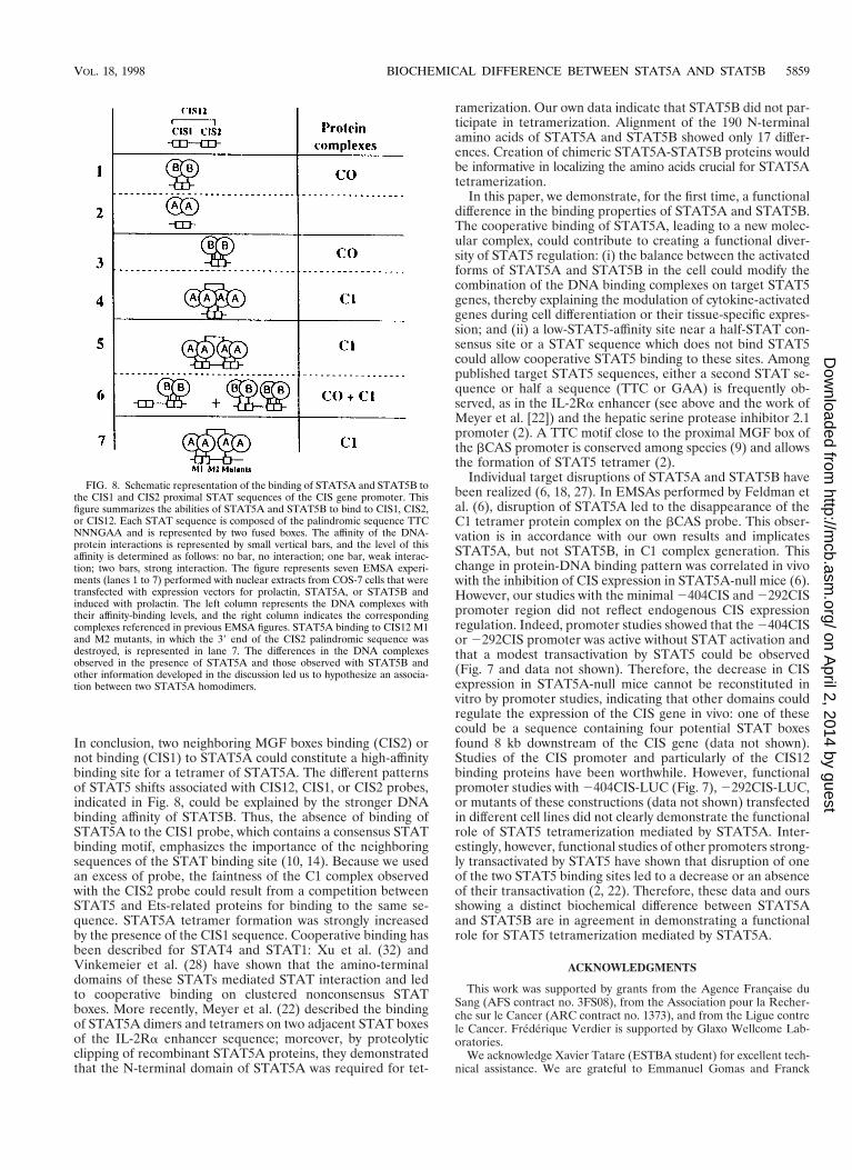
In conclusion, two neighboring MGF boxes binding (CIS2) ornot binding (CIS1) to STAT5A could constitute a high-affinitybinding site for a tetramer of STAT5A. The different patternsof STAT5 shifts associated with CIS12, CIS1, or CIS2 probes,indicated in Fig. 8, could be explained by the stronger DNAbinding affinity of STAT5B. Thus, the absence of binding ofSTAT5A to the CIS1 probe, which contains a consensus STATbinding motif, emphasizes the importance of the neighboringsequences of the STAT binding site (10, 14). Because we usedan excess of probe, the faintness of the C1 complex observedwith the CIS2 probe could result from a competition betweenSTAT5 and Ets-related proteins for binding to the same se-quence. STAT5A tetramer formation was strongly increasedby the presence of the CIS1 sequence. Cooperative binding hasbeen described for STAT4 and STAT1: Xu et al. (32) andVinkemeier et al. (28) have shown that the amino-terminaldomains of these STATs mediated STAT interaction and ledto cooperative binding on clustered nonconsensus STATboxes. More recently, Meyer et al. (22) described the bindingof STAT5A dimers and tetramers on two adjacent STAT boxesof the IL-2Ra enhancer sequence; moreover, by proteolyticclipping of recombinant STAT5A proteins, they demonstratedthat the N-terminal domain of STAT5A was required for tet-
ramerization. Our own data indicate that STAT5B did not par-ticipate in tetramerization. Alignment of the 190 N-terminalamino acids of STAT5A and STAT5B showed only 17 differ-ences. Creation of chimeric STAT5A-STAT5B proteins wouldbe informative in localizing the amino acids crucial for STAT5Atetramerization.
In this paper, we demonstrate, for the first time, a functionaldifference in the binding properties of STAT5A and STAT5B.The cooperative binding of STAT5A, leading to a new molec-ular complex, could contribute to creating a functional diver-sity of STAT5 regulation: (i) the balance between the activatedforms of STAT5A and STAT5B in the cell could modify thecombination of the DNA binding complexes on target STAT5genes, thereby explaining the modulation of cytokine-activatedgenes during cell differentiation or their tissue-specific expres-sion; and (ii) a low-STAT5-affinity site near a half-STAT con-sensus site or a STAT sequence which does not bind STAT5could allow cooperative STAT5 binding to these sites. Amongpublished target STAT5 sequences, either a second STAT se-quence or half a sequence (TTC or GAA) is frequently ob-served, as in the IL-2Ra enhancer (see above and the work ofMeyer et al. [22]) and the hepatic serine protease inhibitor 2.1promoter (2). A TTC motif close to the proximal MGF box ofthe bCAS promoter is conserved among species (9) and allowsthe formation of STAT5 tetramer (2).
Individual target disruptions of STAT5A and STAT5B havebeen realized (6, 18, 27). In EMSAs performed by Feldman etal. (6), disruption of STAT5A led to the disappearance of theC1 tetramer protein complex on the bCAS probe. This obser-vation is in accordance with our own results and implicatesSTAT5A, but not STAT5B, in C1 complex generation. Thischange in protein-DNA binding pattern was correlated in vivowith the inhibition of CIS expression in STAT5A-null mice (6).However, our studies with the minimal 2404CIS and 2292CISpromoter region did not reflect endogenous CIS expressionregulation. Indeed, promoter studies showed that the 2404CISor 2292CIS promoter was active without STAT activation andthat a modest transactivation by STAT5 could be observed(Fig. 7 and data not shown). Therefore, the decrease in CISexpression in STAT5A-null mice cannot be reconstituted invitro by promoter studies, indicating that other domains couldregulate the expression of the CIS gene in vivo: one of thesecould be a sequence containing four potential STAT boxesfound 8 kb downstream of the CIS gene (data not shown).Studies of the CIS promoter and particularly of the CIS12binding proteins have been worthwhile. However, functionalpromoter studies with 2404CIS-LUC (Fig. 7), 2292CIS-LUC,or mutants of these constructions (data not shown) transfectedin different cell lines did not clearly demonstrate the functionalrole of STAT5 tetramerization mediated by STAT5A. Inter-estingly, however, functional studies of other promoters strong-ly transactivated by STAT5 have shown that disruption of oneof the two STAT5 binding sites led to a decrease or an absenceof their transactivation (2, 22). Therefore, these data and oursshowing a distinct biochemical difference between STAT5Aand STAT5B are in agreement in demonstrating a functionalrole for STAT5 tetramerization mediated by STAT5A.
ACKNOWLEDGMENTS
This work was supported by grants from the Agence Francaise duSang (AFS contract no. 3FS08), from the Association pour la Recher-che sur le Cancer (ARC contract no. 1373), and from the Ligue contrele Cancer. Frederique Verdier is supported by Glaxo Wellcome Lab-oratories.
We acknowledge Xavier Tatare (ESTBA student) for excellent tech-nical assistance. We are grateful to Emmanuel Gomas and Franck
FIG. 8. Schematic representation of the binding of STAT5A and STAT5B tothe CIS1 and CIS2 proximal STAT sequences of the CIS gene promoter. Thisfigure summarizes the abilities of STAT5A and STAT5B to bind to CIS1, CIS2,or CIS12. Each STAT sequence is composed of the palindromic sequence TTCNNNGAA and is represented by two fused boxes. The affinity of the DNA-protein interactions is represented by small vertical bars, and the level of thisaffinity is determined as follows: no bar, no interaction; one bar, weak interac-tion; two bars, strong interaction. The figure represents seven EMSA experi-ments (lanes 1 to 7) performed with nuclear extracts from COS-7 cells that weretransfected with expression vectors for prolactin, STAT5A, or STAT5B andinduced with prolactin. The left column represents the DNA complexes withtheir affinity-binding levels, and the right column indicates the correspondingcomplexes referenced in previous EMSA figures. STAT5A binding to CIS12 M1and M2 mutants, in which the 39 end of the CIS2 palindromic sequence wasdestroyed, is represented in lane 7. The differences in the DNA complexesobserved in the presence of STAT5A and those observed with STAT5B andother information developed in the discussion led us to hypothesize an associa-tion between two STAT5A homodimers.
VOL. 18, 1998 BIOCHEMICAL DIFFERENCE BETWEEN STAT5A AND STAT5B 5859
on April 2, 2014 by guest
http://mcb.asm
.org/D
ownloaded from

Letourneur for DNA sequencing. We thank M. Negishi for anti-GABPa and -b antibodies.
REFERENCES
1. Azam, M., C. Lee, I. Strehlow, and C. Schindler. 1997. Functionally distinct iso-forms of STAT5 are generated by protein processing. Immunity 6:691–701.
2. Bergad, P. L., H. M. Shih, H. C. Towle, S. J. Schwarzenberg, and S. A. Berry.1995. Growth hormone induction of hepatic serine protease inhibitor 2.1transcription is mediated by a Stat5-related factor binding synergistically totwo gamma-activated sites. J. Biol. Chem. 270:24903–24910.
3. Bosselut, R., J. Levin, E. Adjadj, and J. Ghysdael. 1993. A single amino-acidsubstitution in the Ets domain alters core DNA binding specificity of Ets1 tothat of the related transcription factors Elf1 and E74. Nucleic Acids Res. 21:5184–5191.
4. Darnell, J. E., Jr. 1997. STATs and gene regulation. Science 277:1630–1635.5. Doppler, W., B. Groner, and R. K. Ball. 1989. Prolactin and glucocorticoid
hormones synergistically induce expression of transfected rat beta-caseingene promoter constructs in a mammary epithelial cell line. Proc. Natl. Acad.Sci. USA 86:104–108.
6. Feldman, G. M., L. A. Rosenthal, X. Liu, M. P. Hayes, A. Wynshaw-Boris,W. J. Leonard, L. Hennighausen, and D. S. Finbloom. 1997. STAT5A-de-ficient mice demonstrate a defect in granulocyte-macrophage colony-stimu-lating factor-induced proliferation and gene expression. Blood 90:1768–1776.
7. Gouilleux, F., C. Pallard, I. Dusanter-Fourt, H. Wakao, L. A. Haldosen, G.Norstedt, D. Levy, and B. Groner. 1995. Prolactin, growth hormone, eryth-ropoietin and granulocyte-macrophage colony stimulating factor induceMGF-Stat5 DNA binding activity. EMBO J. 14:2005–2013.
8. Gouilleux, F., H. Wakao, M. Mundt, and B. Groner. 1994. Prolactin inducesphosphorylation of Tyr694 of Stat5 (MGF), a prerequisite for DNA bindingand induction of transcription. EMBO J. 13:4361–4369.
9. Groner, B., and F. Gouilleux. 1995. Prolactin-mediated gene activation inmammary epithelial cells. Curr. Opin. Genet. Dev. 5:587–594.
10. Horvath, C. M., Z. Wen, and J. E. Darnell, Jr. 1995. A STAT protein domainthat determines DNA sequence recognition suggests a novel DNA-bindingdomain. Genes Dev. 9:984–994.
11. Ihle, J. N. 1996. STATs: signal transducers and activators of transcription.Cell 84:331–334.
12. Klingmuller, U., U. Lorenz, L. C. Cantley, B. C. Neel, and H. C. Lodish.1995. Specific recruitment of SH-PTP1 to the erythropoietin receptor causesinactivation of JAK2 and termination of proliferative signals. Cell 80:729–738.
13. Komatsu, N., H. Nakauchi, A. Miwa, T. Ishihara, M. Eguchi, M. Moroi, M.Okada, Y. Sato, H. Wada, Y. Yamata, T. Suda, and Y. Miura. 1991. Estab-lishment and characterization of a human leukemic cell line with megakaryo-cytic features: dependency on granulocyte-macrophage colony stimulatingfactor, interleukin 3, or erythropoietin for growth and survival. Cancer Res.51:341–345.
14. Lamb, P., H. M. Seidel, J. Haslam, L. Milocco, L. V. Kessler, R. B. Stein, andJ. Rosen. 1995. STAT protein complexes activated by interferon-gamma andgp130 signaling molecules differ in their sequence preferences and transcrip-tional induction properties. Nucleic Acids Res. 23:3283–3289.
15. Lecine, P., M. Algarte, P. Rameil, C. Beadling, P. Bucher, M. Nabholz, andJ. Imbert. 1996. Elf-1 and Stat5 bind to a critical element in a new enhancerof the human interleukin-2 receptor a gene. Mol. Cell. Biol. 16:6829–6840.(Author’s correction, 17:2351, 1997.)
16. Lin, J. X., J. Mietz, W. S. Modi, S. John, and W. J. Leonard. 1996. Cloningof human Stat5B. Reconstitution of interleukin-2-induced Stat5A andStat5B DNA binding activity in COS-7 cells. J. Biol. Chem. 271:10738–10744.
17. Liu, X., G. W. Robinson, F. Gouilleux, B. Groner, and L. Hennighausen.1995. Cloning and expression of Stat5 and an additional homologue (Stat5b)involved in prolactin signal transduction in mouse mammary tissue. Proc.Natl. Acad. Sci. USA 92:8831–8835.
18. Liu, X., G. W. Robinson, K. U. Wagner, L. Garrett, A. Wynshaw-Boris, andL. Hennighausen. 1997. Stat5a is mandatory for adult mammary gland de-velopment and lactogenesis. Genes Dev. 11:179–186.
19. Luo, G., and L. Yu-Lee. 1997. Transcriptional inhibition by Stat5. Differentialactivities at growth-related versus differentiation-specific promoters. J. Biol.Chem. 272:26841–26849.
20. Matsumoto, A., M. Masuhara, K. Mitsui, M. Yokouchi, M. Ohtsubo, H.Misawa, A. Miyajima, and A. Yoshimura. 1997. CIS, a cytokine inducibleSH2 protein, is a target of the JAK-STAT5 pathway and modulates STAT5activation. Blood 89:3148–3154.
21. Matsumura, I., J. Ishikawa, K. Nakajima, K. Oritani, Y. Tomiyama, J.Miyagawa, T. Kato, H. Miyazaki, Y. Matsuzawa, and Y. Kanakura. 1997.Thrombopoietin-induced differentiation of a human megakaryoblastic leu-kemia cell line, CMK, involves transcriptional activation of p21 (WAF1/Cip1) by STAT5. Mol. Cell. Biol. 17:2933–2943.
22. Meyer, W. K., P. Reichenbach, U. Schindler, E. Soldaini, and M. Nabholz.1997. Interaction of STAT5 dimers on two low affinity binding sites mediatesinterleukin 2 (IL-2) stimulation of IL-2 receptor alpha gene transcription.J. Biol. Chem. 272:31821–31828.
23. Moriggi, R., V. Gouilleux-Gruart, R. Jahne, S. Berchtold, C. Gartmann, X.Liu, L. Hennighausen, A. Sotiropoulos, B. Groner, and F. Gouilleux. 1996.Deletion of the carboxyl-terminal transactivation domain of MGF-Stat5 re-sults in sustained DNA binding and a dominant negative phenotype. Mol.Cell. Biol. 16:5691–5700.
24. Mui, A. L., H. Wakao, T. Kinoshita, T. Kitamura, and A. Miyajima. 1996.Suppression of interleukin-3-induced gene expression by a C-terminal trun-cated Stat5: role of Stat5 in proliferation. EMBO J. 15:2425–2433.
25. Mui, A. L., H. Wakao, A. M. O’Farrell, N. Harada, and A. Miyajima. 1995.Interleukin-3, granulocyte-macrophage colony stimulating factor and inter-leukin-5 transduce signals through two STAT5 homologs. EMBO J. 14:1166–1175.
26. Pellegrini, S., and I. Dusanter-Fourt. 1997. The structure, regulation andfunction of the Janus kinases (JAKs) and the signal transducers and activa-tors of transcription (STATs). Eur. J. Biochem. 248:615–633.
27. Udy, G. B., R. P. Towers, R. G. Snell, R. J. Wilkins, S. H. Park, P. A. Ram,D. J. Waxman, and H. W. Davey. 1997. Requirement of STAT5b for sexualdimorphism of body growth rates and liver gene expression. Proc. Natl.Acad. Sci. USA 94:7239–7244.
28. Vinkemeier, U., S. L. Cohen, I. Moarefi, B. T. Chait, J. Kuriyan, and J. E.Darnell, Jr. 1996. DNA binding of in vitro activated Stat1 alpha, Stat1 betaand truncated Stat1: interaction between NH2-terminal domains stabilizesbinding of two dimers to tandem DNA sites. EMBO J. 15:5616–5626.
29. Wakao, H., F. Gouilleux, and B. Groner. 1994. Mammary gland factor(MGF) is a novel member of the cytokine regulated transcription factor genefamily and confers the prolactin response. EMBO J. 13:2182–2191.
30. Wang, D., D. Stravopodis, S. Teglund, J. Kitazawa, and J. N. Ihle. 1996.Naturally occurring dominant negative variants of Stat5. Mol. Cell. Biol. 16:6141–6148.
31. Wood, T. J., D. Sliva, P. E. Lobie, F. Gouilleux, A. L. Mui, B. Groner, G.Norstedt, and L. A. Haldosen. 1997. Specificity of transcription enhancementvia the STAT responsive element in the serine protease inhibitor 2.1 pro-moter. Mol. Cell. Endocrinol. 130:69–81.
32. Xu, X., Y. L. Sun, and T. Hoey. 1996. Cooperative DNA binding and se-quence-selective recognition conferred by the STAT amino-terminal do-main. Science 273:794–797.
33. Yokomori, N., R. Kobayashi, R. Moore, T. Sueyoshi, and M. Negishi. 1995.A DNA methylation site in the male-specific P450 (Cyp 2d-9) promoter andbinding of the heteromeric transcription factor GABP. Mol. Cell. Biol. 15:5355–5362.
34. Yoshimura, A., M. Ichihara, I. Kinjyo, M. Moriyama, N. G. Copeland, D. J.Gilbert, N. A. Jenkins, T. Hara, and A. Miyajima. 1996. Mouse oncostatinM: an immediate early gene induced by multiple cytokines through theJAK-STAT5 pathway. EMBO J. 15:1055–1063.
35. Yoshimura, A., T. Ohkubo, T. Kigushi, N. A. Jenkins, D. J. Gilbert, N. G.Copeland, T. Hara, and A. Miyajima. 1995. A novel cytokine-inducible geneCIS encodes an SH2-containing protein that binds to tyrosine-phosphory-lated interleukin 3 and erythropoietin receptors. EMBO J. 14:2816–2826.
5860 VERDIER ET AL. MOL. CELL. BIOL.
on April 2, 2014 by guest
http://mcb.asm
.org/D
ownloaded from