
Tail-Heaviness, Asymmetry, and Profitability Forecasting by ...
Upload
khangminh22Category
view
1download
0
Citation: Zhu, Y.; Zhao, Y.; Wang, Y.
A Review of Three-Dimensional
Facial Asymmetry Analysis Methods.
Symmetry 2022, 14, 1414. https://
doi.org/10.3390/sym14071414
Academic Editor: Theodore E. Simos
Received: 7 June 2022
Accepted: 5 July 2022
Published: 9 July 2022
Publisher’s Note: MDPI stays neutral
with regard to jurisdictional claims in
published maps and institutional affil-
iations.
Copyright: © 2022 by the authors.
Licensee MDPI, Basel, Switzerland.
This article is an open access article
distributed under the terms and
conditions of the Creative Commons
Attribution (CC BY) license (https://
creativecommons.org/licenses/by/
4.0/).
symmetryS S
Review
A Review of Three-Dimensional Facial AsymmetryAnalysis MethodsYujia Zhu 1,2,3,4,5,6, Yijiao Zhao 1,2,3,4,5,6,* and Yong Wang 1,2,3,4,5,6,*
1 Center of Digital Dentistry, Department of Prosthodontics, Peking University School and Hospital ofStomatology, Beijing 100081, China; [email protected]
2 National Center of Stomatology, Beijing 100081, China3 National Clinical Research Center for Oral Diseases, Beijing 100081, China4 National Engineering Research Center of Oral Biomaterials and Digital Medical Devices, Beijing 100081, China5 Beijing Key Laboratory of Digital Stomatology, Beijing 100081, China6 NHC Research Center of Engineering and Technology for Computerized Dentistry, Beijing 100081, China* Correspondence: [email protected] (Y.Z.); [email protected] (Y.W.);
Tel.: +86-010-82195521 (Y.Z.); +86-8219-5553 (Y.W.)
Abstract: Three-dimensional symmetry and coordination are important factors in facial aesthetics,and analysis of facial asymmetry is the basis for clinical diagnosis, treatment, and doctor–patient com-munication. With the development of three-dimensional measurement and data analysis technology,facial asymmetry analysis methods are mainly based on facial anatomic landmarks, original-mirroralignment algorithm, facial anthropometric mask, and artificial intelligence. This review summarizesthe methods of three-dimensional facial asymmetry analysis, and current research progress in thefield. The advantages and limitations of various methods are analyzed and discussed to provide areference for oral clinical application.
Keywords: three-dimensional facial data; facial asymmetry analysis; landmarks; median sagittal plane
1. Introduction
Facial symmetry and coordination are important factors in aesthetics and attractive-ness [1–3]. Symmetry is represented in size, structure, and balanced distribution on bothsides of the face; however, due to congenital development, environment, disease, andother factors, there is no perfectly symmetrical human face [4,5]. In dental clinics, lowerdegrees of asymmetry are not easily detected, but severe facial asymmetry or facial de-fects can affect the physical and mental health of patients and often require treatmentby orthognathic surgery and orthodontics [6]. Asymmetry analysis is critical to surgicalplanning and preoperative evaluation in orthodontics, oral and maxillofacial surgery, andaesthetic prosthodontics, also important in plastic and reconstructive surgery, facial nerveparalysis and anthropologic evaluation [7–9]. The development of digital technology hasallowed the acquisition of facial data through three-dimensional facial scanning, computedtomographic (CT), cone-beam computed tomography (CBCT) and other three-dimensionalimaging technologies generating clinically valuable reference data. Analysis of facial sym-metry is also important in clinical diagnosis and doctor–patient communication whenevaluating the efficacy of preoperative and postoperative orthognathic surgery [10,11].Three-dimensional facial asymmetry analysis has become the focus of clinical research, andintelligent and automated evaluation methods are emerging.
The methods for facial asymmetry analysis are mainly based on anatomical landmarks,original-mirror alignment algorithms, template-mapping strategy, and artificial intelligence.Three-dimensional facial asymmetry analysis methods were shown in Table 1.
Symmetry 2022, 14, 1414. https://doi.org/10.3390/sym14071414 https://www.mdpi.com/journal/symmetry

Symmetry 2022, 14, 1414 2 of 10
Table 1. Three-dimensional facial asymmetry analysis methods.
Three-Dimensional Facial AsymmetryAnalysis Methods
Asymmetry Evaluation Index ApplicationConditions Advantages Limitation
facial anatomiclandmarks
distance to referenceplane method
asymmetry index(AI =
√(Ldx− Rdx)2 + (Ldy− Rdy)2 + (Ldz− Rdz)2 ),
asymmetry rate (Q = G−KG 100%)
determinethree-dimensional
anatomical landmarkmature, simple, and
intuitive
selection oflandmarks is
mostly based onmanual annotation;
reference planeaffect the
evaluation result
Euclidean distancematrix analysis
FDM (ratio of the total number of asymmetrical lines tothe total lines)
determinethree-dimensional
anatomical landmark
comprehensive,does not depend onthe facial reference
planes
selection oflandmarks is
mostly based onmanual annotation
original-mirroralignmentalgorithm
Landmark-basedmethod RMS
(√x2
1+x22+...+x2
nn ), MAD
(|x1 |+|x2 |+...+|xn |
n
),
MSD(
x1+x2+...+xnn
)original-mirror
alignment algorithm(PA)
good repeatabilityand accuracy
manually selectedlandmarks
Landmark-independent
method
original-mirroralignment algorithm
(ICP)
good repeatabilityand accuracy
manually selectedstable regions
template-mappingstrategy
densequasi-landmarksmapping to target
RMS(√
x21+x2
2+...+x2n
n
) original-mirroralignment algorithm
(template-mapping + PA)
automation,structured data,corresponding
quasi-landmarks
constructrepresentative
anthropometric mask
artificialintelligence
convolutional neuralnetwork (CNN)
facial symmetry score(MinClass + (MaxClass −MinClass) × PPredit)
N/A efficient andintelligent
the training costis high
2. Three-Dimensional Facial Asymmetry Analysis Method Based on FacialAnatomic Landmarks
Dental clinicians should first pay attention to important anatomical landmark features,such as the pronasale, pupil, and cheilion, when performing facial asymmetry analysis [12].The quantitative evaluation of facial asymmetry based on three-dimensional facial land-marks mainly includes the distance to reference plane method and Euclidean distancematrix analysis (EDMA).
In the distance to reference plane method, there are two indexes, asymmetry index(AI) and asymmetry rate. Both are commonly used indexes in dental clinics, and the expertchooses the index according to personal preference. There is a slight difference in thecalculation formula, but two indexes both belong to the calculation of the distance to thereference plane method.
The asymmetry index (AI) can be used to evaluate overall facial asymmetry andasymmetry of individual landmarks. The degree of landmark asymmetry is indicated bythe difference in the linear distance between the landmarks and the reference plane. In2013, Huang et al. used AI to quantify the degree of asymmetry of 16 facial landmarks in60 Chinese adults, and left–right asymmetry (x-direction) was crucial in clinical treatmentand was most easily detected by doctors and patients [13]. By calculating the distancefrom each landmark to the median sagittal plane and drawing a line chart, the left andright landmark deviations could be clearly described. The following formula was extendedto evaluate the asymmetrical representation of the face data in three-dimensional space:
AI =√(Ldx− Rdx)2 + (Ldy− Rdy)2 + (Ldz− Rdz)2, where Ldx, Rdx, Ldy, Rdy, Ldz,
and Rdz represent the distances from the homonymous left and right anatomical landmarksto the sagittal, coronal, and horizontal planes, respectively; reference planes are shownin Figure 1. Faces that are more symmetrical have a smaller difference from the left andright homonymous landmarks to the reference plane. The smaller difference betweentwo landmarks’ distances, the smaller the AI value while larger differences are associatedwith a greater AI value. In 2015, Xiong et al. using the asymmetry index by the averagedistance from the midpoint of bilateral landmarks and midline landmarks to the mediansagittal plane and evaluated the facial asymmetry of 60 young people and 30 edentulouspatients [14]. In 2016, Tian Kaiyue et al. used AI to evaluate the preoperative and post-operative facial symmetry of patients with skeletal Class III deformities and evaluatedthe digital design efficacy to correct the asymmetry deformity in patients with skeletalClass III deformities [15]. As a brief summary, the asymmetry index is relatively intuitive

Symmetry 2022, 14, 1414 3 of 10
for evaluating the asymmetry of facial data and landmarks. However, the premise ofcalculating this AI value is to determine the facial reference planes; therefore, the accuracyof the facial reference planes is a key factor affecting AI.
Symmetry 2022, 14, x FOR PEER REVIEW 3 of 10
tween two landmarks’ distances, the smaller the AI value while larger differences are as-sociated with a greater AI value. In 2015, Xiong et al. using the asymmetry index by the average distance from the midpoint of bilateral landmarks and midline landmarks to the median sagittal plane and evaluated the facial asymmetry of 60 young people and 30 edentulous patients [14]. In 2016, Tian Kaiyue et al. used AI to evaluate the preoperative and postoperative facial symmetry of patients with skeletal Class III deformities and eval-uated the digital design efficacy to correct the asymmetry deformity in patients with skel-etal Class III deformities [15]. As a brief summary, the asymmetry index is relatively intu-itive for evaluating the asymmetry of facial data and landmarks. However, the premise of calculating this AI value is to determine the facial reference planes; therefore, the accuracy of the facial reference planes is a key factor affecting AI.
Figure 1. Three-dimensional reference planes shown on a 3D face . Reprinted with permission from Ref. [13]. 2013, Huang, C.S.; Liu, X.Q.; Chen, Y.R.
The asymmetry rate is an established method for evaluating facial asymmetry, and is widely used [16–18]. It is expressed by the formula Q = 100%, where Q is the asym-metry rate, G is the larger linear distance from the measurement landmark on the left and right sides to the median sagittal plane, and K is the smaller one. A smaller Q value indi-cates that the two homonymous landmarks are more symmetrical, and a value of zero indicates complete symmetry. In 2014, Wang et al. applied the asymmetry rate to study the distance between the bilateral exocanthion and the cheilion line, and the distance be-tween the bilateral gonion and the median sagittal plane. By calculating the asymmetry rate, a value of less than 10% was regarded as relatively symmetric [19]. The asymmetry rate evaluates the degree of asymmetry from the landmark to the sagittal plane or the reference plane, ignoring the facial morphological asymmetry of the face in the depth di-mension; thus, it does not evaluate facial asymmetry in three-dimensional space. None-theless, the asymmetry rate is affected by the accuracy of the facial reference planes.
The Euclidean distance matrix analysis is a widely used morphological measurement method. In recent years, this method has been applied to the evaluation of facial asym-metry [20]. Based entirely on the three-dimensional spatial distribution characteristics of the anatomical facial landmarks, the EDMA method constructed the form difference ma-trix of the linear distance ratio between each landmark of the “left face” and “right face” and extracted the relevant ratios to construct the evaluation of the facial asymmetry index. The principle is to represent the shape and size of individuals through a form matrix (FM) generated by the Euclidean distances between landmarks in the geometric morphology of individuals. The form difference matrix (FDM) represents the morphological differences among individuals through the ratio of the corresponding matrix elements. Furthermore, the asymmetry of the three-dimensional facial region was evaluated by calculating the percentage of the total number of too large or too small matrix ratio elements. In 2008,
Figure 1. Three-dimensional reference planes shown on a 3D face. Reprinted with permission fromRef. [13]. 2013, Huang, C.S.; Liu, X.Q.; Chen, Y.R.
The asymmetry rate is an established method for evaluating facial asymmetry, andis widely used [16–18]. It is expressed by the formula Q = G−K
G 100%, where Q is theasymmetry rate, G is the larger linear distance from the measurement landmark on the leftand right sides to the median sagittal plane, and K is the smaller one. A smaller Q valueindicates that the two homonymous landmarks are more symmetrical, and a value of zeroindicates complete symmetry. In 2014, Wang et al. applied the asymmetry rate to study thedistance between the bilateral exocanthion and the cheilion line, and the distance betweenthe bilateral gonion and the median sagittal plane. By calculating the asymmetry rate, avalue of less than 10% was regarded as relatively symmetric [19]. The asymmetry rateevaluates the degree of asymmetry from the landmark to the sagittal plane or the referenceplane, ignoring the facial morphological asymmetry of the face in the depth dimension;thus, it does not evaluate facial asymmetry in three-dimensional space. Nonetheless, theasymmetry rate is affected by the accuracy of the facial reference planes.
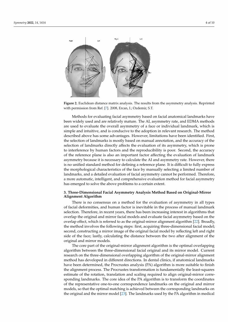
The Euclidean distance matrix analysis is a widely used morphological measurementmethod. In recent years, this method has been applied to the evaluation of facial asymme-try [20]. Based entirely on the three-dimensional spatial distribution characteristics of theanatomical facial landmarks, the EDMA method constructed the form difference matrixof the linear distance ratio between each landmark of the “left face” and “right face” andextracted the relevant ratios to construct the evaluation of the facial asymmetry index. Theprinciple is to represent the shape and size of individuals through a form matrix (FM)generated by the Euclidean distances between landmarks in the geometric morphology ofindividuals. The form difference matrix (FDM) represents the morphological differencesamong individuals through the ratio of the corresponding matrix elements. Furthermore,the asymmetry of the three-dimensional facial region was evaluated by calculating thepercentage of the total number of too large or too small matrix ratio elements. In 2008,Ercan et al. examined facial asymmetry among Turkish youths based on FDM ratio analysis.This population-level study found that there was asymmetry in the female face and thatthe shape of the left face had considerable impact on facial asymmetry [7], The resultsfrom the asymmetry analysis are shown in Figure 2. In 2014, Shuang et al. studied facialasymmetry among young women and men in Liaoning province of China using the EDMAmethod. The investigators recorded an FDM ratio lower than 0.95 and higher than 1.05as asymmetry, and calculated the percentage of the total number of asymmetrical lines tothe total lines [21]. The algorithm refers to the association between the landmarks; it isobjective and comprehensive and does not depend on the facial reference planes.
Symmetry 2022, 14, 1414 4 of 10
Symmetry 2022, 14, x FOR PEER REVIEW 4 of 10
Ercan et al. examined facial asymmetry among Turkish youths based on FDM ratio anal-ysis. This population-level study found that there was asymmetry in the female face and that the shape of the left face had considerable impact on facial asymmetry [7], The results from the asymmetry analysis are shown in Figure 2. In 2014, Shuang et al. studied facial asymmetry among young women and men in Liaoning province of China using the EDMA method. The investigators recorded an FDM ratio lower than 0.95 and higher than 1.05 as asymmetry, and calculated the percentage of the total number of asymmetrical lines to the total lines [21]. The algorithm refers to the association between the landmarks; it is objective and comprehensive and does not depend on the facial reference planes.
Figure 2. Euclidean distance matrix analysis. The results from the asymmetry analysis. Reprinted with permission from Ref. [7]. 2008, Ercan, I.; Ozdemir, S.T.
Methods for evaluating facial asymmetry based on facial anatomical landmarks have been widely used and are relatively mature. The AI, asymmetry rate, and EDMA methods are used to evaluate the overall asymmetry of a face or individual landmark, which is simple and intuitive, and is conducive to the adoption in relevant research. The method described above has some advantages. However, limitations have been identified. First, the selection of landmarks is mostly based on manual annotation, and the accuracy of the selection of landmarks directly affects the evaluation of its asymmetry, which is prone to interference by human factors and the reproducibility is poor. Second, the accuracy of the reference plane is also an important factor affecting the evaluation of landmark asym-metry because it is necessary to calculate the AI and asymmetry rate. However, there is no unified standard method for defining a reference plane. It is difficult to fully express the morphological characteristics of the face by manually selecting a limited number of landmarks, and a detailed evaluation of facial asymmetry cannot be performed. There-fore, a more automatic, intelligent, and comprehensive evaluation method for facial asym-metry has emerged to solve the above problems to a certain extent.
3. Three-Dimensional Facial Asymmetry Analysis Method Based on Original-Mirror Alignment Algorithm
There is no consensus on a method for the evaluation of asymmetry in all types of facial deformities, and human factor is inevitable in the process of manual landmark se-lection. Therefore, in recent years, there has been increasing interest in algorithms that overlap the original and mirror facial models and evaluate facial asymmetry based on the overlap effect, which is referred to as the original-mirror alignment algorithm [22]. Broadly, the method involves the following steps: first, acquiring three-dimensional facial model; second, constructing a mirror image of the original facial model by reflecting left and right side of the face; lastly, calculating the distance between the two after alignment of the original and mirror models.
Figure 2. Euclidean distance matrix analysis. The results from the asymmetry analysis. Reprintedwith permission from Ref. [7]. 2008, Ercan, I.; Ozdemir, S.T.
Methods for evaluating facial asymmetry based on facial anatomical landmarks havebeen widely used and are relatively mature. The AI, asymmetry rate, and EDMA methodsare used to evaluate the overall asymmetry of a face or individual landmark, which issimple and intuitive, and is conducive to the adoption in relevant research. The methoddescribed above has some advantages. However, limitations have been identified. First,the selection of landmarks is mostly based on manual annotation, and the accuracy of theselection of landmarks directly affects the evaluation of its asymmetry, which is proneto interference by human factors and the reproducibility is poor. Second, the accuracyof the reference plane is also an important factor affecting the evaluation of landmarkasymmetry because it is necessary to calculate the AI and asymmetry rate. However, thereis no unified standard method for defining a reference plane. It is difficult to fully expressthe morphological characteristics of the face by manually selecting a limited number oflandmarks, and a detailed evaluation of facial asymmetry cannot be performed. Therefore,a more automatic, intelligent, and comprehensive evaluation method for facial asymmetryhas emerged to solve the above problems to a certain extent.
3. Three-Dimensional Facial Asymmetry Analysis Method Based on Original-MirrorAlignment Algorithm
There is no consensus on a method for the evaluation of asymmetry in all typesof facial deformities, and human factor is inevitable in the process of manual landmarkselection. Therefore, in recent years, there has been increasing interest in algorithms thatoverlap the original and mirror facial models and evaluate facial asymmetry based on theoverlap effect, which is referred to as the original-mirror alignment algorithm [22]. Broadly,the method involves the following steps: first, acquiring three-dimensional facial model;second, constructing a mirror image of the original facial model by reflecting left and rightside of the face; lastly, calculating the distance between the two after alignment of theoriginal and mirror models.
The core part of the original-mirror alignment algorithm is the optimal overlappingalgorithm between the three-dimensional facial original and its mirror model. Currentresearch on the three-dimensional overlapping algorithm of the original-mirror alignmentmethod has developed in different directions. In dental clinics, if anatomical landmarkshave been determined, the Procrustes analysis (PA) algorithm is more suitable to finishthe alignment process. The Procrustes transformation is fundamentally the least-squaresestimate of the rotation, translation and scaling required to align original-mirror corre-sponding landmarks. The core idea of the PA algorithm is to transform the coordinatesof the representative one-to-one correspondence landmarks on the original and mirrormodels, so that the optimal matching is achieved between the corresponding landmarks onthe original and the mirror model [23]. The landmarks used by the PA algorithm in medical

Symmetry 2022, 14, 1414 5 of 10
image analysis are mainly anatomical landmarks with clinical significance, such as thepronasale, endocanthion, exocanthion, and pupil. Overlapping of the original and mirrormodels was achieved under the guidance of these landmarks with clear correspondingrelations. In 2016, Xiong et al. reported clinical evaluation research on the application ofthe “regional PA original-mirror algorithm” to construct the facial median sagittal plane ofpatients with mandibular deviation. This improves the accuracy of the PA original-mirroralgorithm by artificially screening and removing the landmarks related to facial asymmetry,which could meet the needs of symmetry analysis of clinical facial deformities in dentalclinics [24]. In 2020, Zhu et al. conducted a weighted Procrustes analysis (WPA) algorithmfor complex facial deformities to determine a three-dimensional facial median sagittalplane [25]. The feasibility and effectiveness of the WPA algorithm strategy in constructinga three-dimensional facial SRP were confirmed by applying this method to 40 patientswith three-dimensional facial deformities. For patients with asymmetry deformity in thelower face, the clinical adaptability of the SRP constructed by the six WPA algorithmshas been improved compared with the classical PA algorithm, achieving a result closeto the truth plane of experts. The WPA algorithm is important and beneficial for oralclinical diagnosis [26]. In another clinical scenario when landmarks are not determined,the iterative closest point (ICP) algorithm is more suitable to finish the original-mirroralignment. The ICP algorithm is currently one of the most common used algorithms foroverlapping 3D medical models. The ICP algorithm has two steps that are iterated untilconvergence or for a set number of iterations: (1) a corresponding point on the stationarysurface is estimated for each point on the other floating surface; (2) like the PA algorithm,the Procrustes transformation translation to align the floating surface to its correspondingpoints on the targe surface is estimated and applied. During the iterative process, thecorresponding point (the closest point) of stationary and floating surface is changed. Itsprinciple is the iterative matching calculation of the nearest spatial distance between thepoint clouds of the two models based on the least-squares method. The algorithm obtainsthe optimal matching position between the two models through multiple iterations of thespatial matrix transformation, which is called registration. Overlapping regions of theoriginal and mirror models, such as the frontal, upper, middle, and lower parts of the face,are manually selected to achieve the optimal matching effect in a clinical sense [12,27,28].
Different evaluation indicators are used to quantify facial asymmetry based on theoriginal and mirror model overlapping methods. In 2016, Ozsoy et al. compared root meansquare (RMS), mean absolute deviation (MAD), and mean signed distance (MSD). The calcu-
lation formulae for each indicator are as follows: RMS =
√x2
1+x22+...+x2
nn ;
MAD = |x1|+......+|xn |n ; MSD = x1+x2+...+xn
n [29], where x represents the nearest distancefrom a point on the original model to the mirror model after overlapping, and n representsthe total number of distances. The study found that RMS and MAD are more suitable indi-cators for the evaluation of the degree of facial asymmetry, while MSD does not contributeto the real morphological differences between the two models because it includes positiveand negative distance values. In 2017, Codari et al. adopted RMS as a quantitative indicatorof facial asymmetry to assess facial asymmetry in patients with facial paralysis by regionand found that this method had high repeatability and accuracy [8]. In 2019, Cassi et al.applied RMS to evaluate facial asymmetry in patients with hemifacial microsomia andnoted that this method could provide richer information for facial analysis, treatmentplanning, and follow-up [30].
RMS is widely used in the quantitative evaluation indicator of three-dimensional facialasymmetry based on the original-mirror alignment algorithm. Owing to its good repeata-bility and accuracy, this method can quantify the asymmetry of the entire or part of the face,which is more comprehensive for the evaluation of facial asymmetry. When original-mirroralignment is conducted using the PA or ICP algorithm, landmarks or relatively stableregions need to be manually selected by the operator. This inevitably introduces a degreeof subjectivity and potential for error. In addition, there is no corresponding relationshipbetween the point clouds of the original and mirror models in ICP algorithm, according

Symmetry 2022, 14, 1414 6 of 10
to the nearest principle to overlap the original and mirror models may underestimate thefacial asymmetry.
4. Three-Dimensional Facial Asymmetry Analysis Method Based onTemplate-Mapping Strategy
In recent years, facial asymmetry analysis method based on the template-mappingstrategy has received considerable attention and application [31–33]. The anthropometricmask (AM) that contains dense quasi-landmarks is deformed into the shape of the three-dimensional target facial point clouds. This imposes the vertices and topology of the AMonto the target. The AM consists of thousands or tens of thousands of spatially densequasi-landmarks that cover the entire facial region, which can be used as an extensionof the traditional facial anatomical landmarks. Dense quasi-landmarks can express facialgeometry in more detail and can be used for three-dimensional facial morphology analysisand symmetry analysis by mathematical and statistical methods. In 2011, Claes et al.constructed an AM with a dense sequence of point sets with approximately 10,000 points,and proposed the concept of weighted overlap of original and mirror masks. A three-dimensional facial median sagittal plane was constructed based on the PA alignmentalgorithm, and the RMSE was used to evaluate the three-dimensional facial symmetry.In 2012, Claes et al. created a distance map showing the amount and direction of facialsoft tissue changes in three patients based on AM, and the results showed that they couldmeet clinical expectations [34]. In 2013, Walters et al. applied AM to patients with hemi-mandibular hyperplasia and hemi-mandibular elongation anomalies and observed themandibular rotation direction and asymmetric magnitude in these patients, providing abasis for quantitative evaluation of facial asymmetry and design of orthognathic treatmentplans [35]. In 2019, the AM template and the related Meshmonk program were madeavailable to researchers for in-depth research in areas such as three-dimensional facialasymmetry evaluation and facial landmark determination [36,37].
RMS indices are used in both the template-mapping strategy and ICP algorithms whenevaluating three-dimensional facial asymmetry, although these are essentially differentmethods for calculating the RMS value. The RMS value of ICP algorithm calculate thenearest distance between original and mirror models; however, in the template-mappingstrategy, the distance between original and mirror corresponding quasi-landmarks on AMis calculated. Compared with other algorithms, the asymmetry analysis method basedon a facial anthropometric mask has the advantages of more points, a correspondingrelationship, and higher degree of automation, which compensate for the shortage of areasthat lack anatomic landmarks, and is helpful in the study of subtle changes in the face andin determining the advantages of the differences in the facial parts.
5. Three-Dimensional Facial Asymmetry Analysis Method Based onArtificial Intelligence
Artificial intelligence technology has a strong ability for feature extraction and learning.With the deepening application of this technology in face recognition, there has beenan increased interest in research in related medical fields, including facial asymmetryevaluation and automatic facial landmark detection. In 2019, Lin et al. established adeep convolutional neural network to extract three-dimensional (3D) contour features andassess the degree of facial symmetry in patients treated with orthognathic surgery. Thisincluded a deep convolutional neural network (CNN) for feature extraction and a regressionnetwork for prediction. The results showed that the 3D contour line-based features deeplearning system serves as a general, useful, automated, and human-like efficient decisiontool for objective assessment of facial symmetry before and after orthognathic surgery forthe improvement of treatment in clinical practice. In 2021, Lin et al. applied a transferlearning model with a convolutional neural network based on three-dimensional (3D)contour line features to evaluate facial symmetry before and after orthognathic surgery.The results showed that the mean postoperative facial symmetry score was 3.52, whichwas significantly better than the preoperative score (mean, 2.74). The mean degree of

Symmetry 2022, 14, 1414 7 of 10
improvement in facial symmetry after surgery was 21% [38]. In 2022, Zhu Yujia et al.proposed a method for automatically constructing a facial median sagittal plane based on adeep learning algorithm, a multi-view stacked hourglass convolutional neural network,and a mathematical model, which was the basis of three-dimensional facial symmetryevaluation [39]. Facial asymmetry analysis methods based on artificial intelligence havegreat potential by reducing dependency on doctors and improving clinical diagnosis andtreatment efficiency.
6. Discussion6.1. Automatic Determination of Facial Anatomical Landmarks Is Helpful to Improve theTraditional Method of 3D Facial Asymmetry Analysis Based on Landmarks
Facial landmarks play a vital role in the evaluation of asymmetry and are the basis forthe asymmetry analysis method of PA and WPA algorithms, and calculation of indicatorssuch as AI, asymmetry rate, and EDMA method. Following the traditional method, anatom-ical landmarks are defined by experts and human factors may affect this process. With theincreasing development of digital technology, there is an urgent need for rapid and auto-matic landmark-determination algorithms, facial asymmetry analysis method based on PAalignment of dense facial points and artificial intelligence to gradually become a researchhotspot. Wen et al. applied an AM template with 32 landmarks to the three-dimensionalfacial data of normal people and patients with mandibular deviation [40]. In general, theimpact on the overall facial asymmetry is minimal in the case of the maxilla because themaxilla is connected to the skull and is difficult to separate from the facial structures, so themandibular deviation patients were mostly evaluated. This approach obtained the faciallandmarks of the target model and achieved efficient and automatic landmark extraction.Zhu et al. applied a deep learning algorithm to study the position error automaticallydetermined 21 landmarks of three-dimensional facial data from 30 patients, and the resultsindicated that the average error was less than 2 mm, which could meet the needs of diagno-sis and analysis of a large amount of oral clinical data [39]. The automatic determination oflandmarks represents a promising future direction of research, and it can make up for theshortcomings of traditional methods affected by human factors and improve the efficiencyof clinical diagnosis and treatment.
6.2. Automatic Construction Method of the Median Sagittal Plane to Provide a More ReliableBaseline for 3D Facial Asymmetry Analysis
Median sagittal plane is the basis of 3D facial asymmetry analysis, especially thecalculation of facial asymmetry index based on landmarks and mirror the original modelin original-mirror alignment algorithm. The automatic construction methods of the mediansagittal plane based on 3D facial data include ICP original-mirror alignment algorithm andartificial intelligence. Among the original mirror-alignment algorithms, the ICP algorithmis a highly automated and repeatable method for analyzing facial asymmetry [8,28]. Theglobal or regional ICP registration algorithm makes full use of the rich morphologicalinformation of the three-dimensional face model, which does not require the manualdefinition of landmarks and treats all point clouds participating in the operation equally.The alignment between the original and mirror point clouds was completely automatic, andno matching relation was required manually. The method is efficient, automatic, and has acertain universality, and can be used for facial analysis in patients with unilateral cleft lipand palate, orthodontic patients, orthognathic patients, and teenagers [41–43]. Consideringthe experts’ habits and experience in clinical diagnosis and treatment, this method shouldbe used cautiously in specific cases. Important facial anatomic landmarks often receivemore attention and sometimes even play a decisive role. Therefore, an asymmetry analysismethod with reference to facial landmarks, such as the PA algorithm, has substantialstrengths. With the advent of the era of big data, deep learning algorithms have greatpotential in the automatic segmentation and recognition of 3D features, but the research onthe automatic construction of a median sagittal plane algorithm based on deep learning hasnot been reported. In 2019, Gao Lin et al. built a PRS-Net model based on the ShapeNet

Symmetry 2022, 14, 1414 8 of 10
dataset to realize the automatic construction of the symmetry plane of the 3D point clouddata, which provided an important reference for the automatic construction of the facialmedian sagittal plane [44]. In 2022, Zhu et al. construct a three-dimensional facial mediansagittal plane based on a deep learning algorithm in 30 patients with mandibular deviation.The angle error between the median sagittal plane constructed by the WPA algorithm andthe expert plane was less than 1.5◦ [45].
6.3. Future Direction
Digital and intelligent diagnosis and treatment will become increasingly available,and the automatic evaluation of three-dimensional facial asymmetry will provide thebasis for facial aesthetic analysis and design, allowing automated, accurate, and efficientdetermination of three-dimensional facial anatomical landmarks. Based on the non-rigidregistration principle, the anthropometric mask realizes deformation matching from thetemplate to the target model and constructs the corresponding average template for peopleof different ages, sex, race, and type of disease, improving the accuracy and robustness ofthe non-rigid registration. Artificial intelligence methods can automatically learn featuresfrom the three-dimensional facial data to obtain quantitative and objective indicators toassist doctors in clinical evaluations. This function can be integrated with diagnosticevaluation and surgical design software to evaluate the degree of improvement in patientasymmetry deformity after orthodontic and orthognathic treatment. It will also become anefficient doctor–patient communication tool.
7. Conclusions
Over the years, with the continuous improvement of facial data acquisition meansand people’s aesthetic needs, symmetry analysis has provided a basis for oral clinicaldiagnosis and analysis. Facial asymmetry analysis methods reviewed in the paper mainlyincluded the following: facial anatomic landmarks, original-mirror alignment algorithm,facial anthropometric mask, and artificial intelligence. Future studies are needed to developmore automatic, intelligent, and comprehensive evaluation method for three-dimensionalfacial asymmetry analysis.
Author Contributions: Y.Z. (Yujia Zhu) collected the information from literature and wrote themanuscript. Y.Z. (Yijiao Zhao) reviewed and edited the manuscript. Y.W. designed and conceptualizedthe review. All authors have read and agreed to the published version of the manuscript.
Funding: This research was funded by the National Natural Science Foundation of China, grantnumber 81870815, 82071171; Innovation Talents Program of Lanzhou, grant number 2019-RC-22.Open Subject Foundation of Peking University Hospital of Stomatology PKUSS20210202.
Institutional Review Board Statement: Not applicable.
Informed Consent Statement: Not applicable.
Data Availability Statement: Not applicable.
Conflicts of Interest: The authors declare no conflict of interest.
References1. Baudouin, J.Y.; Tiberghien, G. Symmetry, averageness, and feature size in the facial attractiveness of women. Acta Psychol. (Amst.)
2004, 117, 313–332. [CrossRef] [PubMed]2. Rhodes, G. The evolutionary psychology of facial beauty. Annu. Rev. Psychol. 2006, 57, 199–226. [CrossRef] [PubMed]3. Burriss, R.P.; Roberts, S.C.; Welling, L.L.M.; Puts, D.A.; Little, A.C. Heterosexual Romantic Couples Mate Assortatively for Facial
Symmetry, But Not Masculinity. Personal. Soc. Psychol. Bull. 2011, 37, 601–613. [CrossRef] [PubMed]4. Lum, V.; Goonewardene, M.S.; Mian, A.; Eastwood, P. Three-dimensional assessment of facial asymmetry using dense correspon-
dence symmetry, and midline analysis. Am. J. Orthod. Dentofac. Orthop. 2020, 158, 134–146. [CrossRef]5. Matthews, H.; de Jong, G.; Maal, T.; Claes, P. Static and Motion Facial Analysis for Craniofacial Assessment and Diagnosing
Diseases. Annu. Rev. Biomed. Data Sci. 2022. preprint online. [CrossRef]6. Thiesen, G.; Gribel, B.F.; Freitas, M.P.M. Facial asymmetry: A current review. Dental Press J. Orthod. 2015, 20, 110–125. [CrossRef]

Symmetry 2022, 14, 1414 9 of 10
7. Ercan, I.; Ozdemir, S.T.; Etoz, A.; Sigirli, D.; Tubbs, R.S.; Loukas, M.; Guney, I. Facial asymmetry in young healthy subjectsevaluated by statistical shape analysis. J. Anat. 2008, 213, 663–669. [CrossRef]
8. Codari, M.; Pucciarelli, V.; Stangoni, F.; Zago, M.; Tarabbia, F.; Biglioli, F.; Sforza, C. Facial thirds-based evaluation of facial asymmetryusing stereophotogrammetric devices: Application to facial palsy subjects. J. Craniomaxillofac. Surg. 2017, 45, 76–81. [CrossRef]
9. Arias, E.; Huang, Y.H.; Zhao, L.; Seelaus, R.; Patel, P.; Cohen, M. Virtual Surgical Planning and Three-Dimensional Printed Guidefor Soft Tissue Correction in Facial Asymmetry. J. Craniofac. Surg. 2019, 30, 846–850. [CrossRef]
10. Weinberg, S.M.; Naidoo, S.; Govier, D.P.; Martin, R.A.; Kane, A.A.; Marazita, M.L. Anthropometric precision and accuracy ofdigital three-dimensional photogrammetry: Comparing the Genex and 3dMD imaging systems with one another and with directanthropometry. J. Craniofac. Surg. 2006, 17, 477–483. [CrossRef]
11. Dindaroglu, F.; Kutlu, P.; Duran, G.S.; Gorgulu, S.; Aslan, E. Accuracy and reliability of 3D stereophotogrammetry: A comparisonto direct anthropometry and 2D photogrammetry. Angle Orthod. 2016, 86, 487–494. [CrossRef] [PubMed]
12. Meyer-Marcotty, P.; Stellzig-Eisenhauer, A.; Bareis, U.; Hartmann, J.; Kochel, J. Three-dimensional perception of facial asymmetry.Eur. J. Orthod. 2011, 33, 647–653. [CrossRef] [PubMed]
13. Huang, C.S.; Liu, X.Q.; Chen, Y.R. Facial asymmetry index in normal young adults. Orthod. Craniofac. Res. 2013, 16, 97–104.[CrossRef] [PubMed]
14. Xiong, Y.; Zhao, Y.; Yang, H.; Sun, Y.; Wang, Y. Accuracy assessment of Procrustes Analysis for Computing Mid-sagittalPlane of Three-dimensional Facial Data. In Proceedings of the 3rd Conference of the Optics-and-Photonics-Society-of-Singapore/International Conference on Optical and Photonic Engineering (icOPEN), Singapore, 14–16 April 2015.
15. Tian, K.Y.; Li, Q.Q.; Liu, X.J. Evaluation of the efficacy of digital design in the correction of mandibular prognathism. Chin. J.Stomatol. 2016, 51, 594–599.
16. Wang, X.; Zhang, Z.K. A study on the symmetry of horizontal section of face in Chinese beautiful population. J. Mod. Stomatol.1989, 8, 233–239.
17. Ras, F.; Habets, L.; Vanginkel, F.C.; Prahlandersen, B. Method for Quantifying Facial Asymmetry in 3 Dimensions UsingStereophotogrammetry. Angle Orthod. 1995, 65, 233–239.
18. Uechi, J.; Okayama, M.; Shibata, T.; Muguruma, T.; Hayashi, K.; Endo, K.; Mizoguchi, I. A novel method for the 3-dimensionalsimulation of orthognathic surgery by using a multimodal image-fusion technique. Am. J. Orthod. Dentofac. Orthop. 2006, 130,786–798. [CrossRef]
19. Wang, X. Digital Assisted Analysis and Treatment Effect Evaluation on Facial Asymmetry Deformity; China Medical University:Shenyang, China, 2014.
20. Ferrario, V.F.; Sforza, C.; Miani, A., Jr.; Serrao, G. A three-dimensional evaluation of human facial asymmetry. J. Anat. 1995,186, 103–110.
21. Qi, S.; Ren, F.; Zhai, G.; Zhu, B.; Ma, Q. Facial asymmetry study on young female Han nationality in Liaoning province. Chin. J.Forensic Med. 2014, 29, 556–558, (full article in Chinese, abstract in English).
22. Benz, M.; Laboureux, X.; Maier, T.; Nkenke, E.; Seeger, S.; Neukam, F.W.; Hausler, G. The symmetry of faces. In Proceedings of theInternational Fall Workshop on Vision Modeling and Visualization 2000, Erlangen, Germany, 20–22 November 2002; pp. 43–50.
23. Klingenberg, C.P.; Barluenga, M.; Meyer, A. Shape analysis of symmetric structures: Quantifying variation among individualsand asymmetry. Evolution 2002, 56, 1909–1920. [CrossRef]
24. Xiong, Y.X.; Zhao, Y.J.; Yang, H.F.; Sun, Y.C.; Wang, Y. Comparison Between Interactive Closest Point and ProcrustesAnalysis for Determining the Median Sagittal Plane of Three-Dimensional Facial Data. J. Craniofac. Surg. 2016, 27, 441–444.[CrossRef] [PubMed]
25. Zhu, Y.J.; Zhao, Y.J.; Zheng, S.W.; Wen, A.N.; Fu, X.L.; Wang, Y. A method for constructing three-dimensional face symmetryreference plane based on weighted shape analysis algorithm. J. Peking Univ. (Health Sci.) 2020, 53, 220–226, (full article in Chinese,abstract in English). [CrossRef]
26. Zhu, Y.J.; Zheng, S.W.; Yang, G.S.; Fu, X.L.; Xiao, N.; Wen, A.N.; Wang, Y.; Zhao, Y.J. A novel method for 3D face symmetryreference plane based on weighted Procrustes analysis algorithm. BMC Oral Health 2020, 20, 319. [CrossRef] [PubMed]
27. Verhoeven, T.J.; Nolte, J.W.; Maal, T.J.; Bergé, S.J.; Becking, A.G. Unilateral condylar hyperplasia: A 3-dimensional quantificationof asymmetry. PLoS ONE 2013, 8, e59391. [CrossRef]
28. Verhoeven, T.; Xi, T.; Schreurs, R.; Bergé, S.; Maal, T. Quantification of facial asymmetry: A comparative study of landmark-basedand surface-based registrations. J. Craniomaxillofac. Surg. 2016, 44, 1131–1136. [CrossRef]
29. Ozsoy, U. Comparison of Different Calculation Methods Used to Analyze Facial Soft Tissue Asymmetry: Global and Partial3-Dimensional Quantitative Evaluation of Healthy Subjects. J. Oral Maxillofac. Surg. 2016, 74, e1841–e1849. [CrossRef]
30. Cassi, D.; Battistoni, G.; Magnifico, M.; Di Blasio, C.; Pedrazzi, G.; Di Blasio, A. Three-dimensional evaluation of facial asymmetryin patients with hemifacial microsomia using stereophotogrammetry. J. Craniomaxillofac. Surg. 2019, 47, 179–184. [CrossRef]
31. Mao, Z.L.; Ju, X.Y.; Siebert, J.P.; Cockshott, W.P.; Ayoub, A. Constructing dense correspondences for the analysis of 3D facialmorphology. Pattern Recognit. Lett. 2006, 27, 597–608. [CrossRef]
32. Paysan, P.; Knothe, R.; Amberg, B.; Romdhani, S.; Vetter, T. A 3D Face Model for Pose and Illumination Invariant Face Recognition.In Proceedings of the 6th IEEE International Conference on Advanced Video and Signal Based Surveillance, Genoa, Italy,2–4 September 2009; pp. 296–301.

Symmetry 2022, 14, 1414 10 of 10
33. Claes, P.; Walters, M.; Vandermeulen, D.; Clement, J.G. Spatially-dense 3D facial asymmetry assessment in both typical anddisordered growth. J. Anat. 2011, 219, 444–455. [CrossRef]
34. Claes, P.; Walters, M.; Clement, J. Improved facial outcome assessment using a 3D anthropometric mask. Int. J. Oral Maxillofac.Surg. 2012, 41, 324–330. [CrossRef]
35. Walters, M.; Claes, P.; Kakulas, E.; Clement, J.G. Robust and regional 3D facial asymmetry assessment in hemimandibularhyperplasia and hemimandibular elongation anomalies. Int. J. Oral Maxillofac. Surg. 2013, 42, 36–42. [CrossRef] [PubMed]
36. White, J.D.; Ortega-Castrillón, A.; Matthews, H.; Zaidi, A.A.; Ekrami, O.; Snyders, J.; Fan, Y.; Penington, T.; Van Dongen, S.;Shriver, M.D.; et al. MeshMonk: Open-source large-scale intensive 3D phenotyping. Sci. Rep. 2019, 9, 6085. [CrossRef] [PubMed]
37. Ding, M.; Fan, Y.; Qin, M.; Claes, P.; Matthews, H.; Peng, H.; Zhao, Y.; Zhu, J. Facial Morphological Changes Following DentureTreatment in Children with Hypohidrotic Ectodermal Dysplasia. Pediatr. Dent. 2020, 42, 315–320. [PubMed]
38. Lo, L.J.; Yang, C.T.; Ho, C.T.; Liao, C.H.; Lin, H.H. Automatic Assessment of 3-Dimensional Facial Soft Tissue Symmetry Beforeand After Orthognathic Surgery Using a Machine Learning Model: A Preliminary Experience. Ann. Plast. Surg. 2021, 86,S224–S228. [CrossRef]
39. Zhu, Y.J.; Xu, Q.; Zhao, Y.J.; Zhang, L.; Fu, Z.W.; Wen, A.N.; Gao, Z.X.; Zhang, J.; Fu, X.L.; Wang, Y. Deep learning-assistedconstruction of three-demensional facial midsagittal plane. J. Peking Univ. (Health Sci.) 2022, 54, 134–139, (full article in Chinese,abstract in English). [CrossRef]
40. Wen, A.N.; Zhu, Y.J.; Zheng, S.W.; Xiao, N.; Gao, Z.X.; Fu, X.L.; Wang, Y.; Zhao, Y. Preliminary study on the method ofautomatically determining facial landmarks based on three-dimensional face template. Chin. J. Stomatol. 2022, 57, 358–365, (fullarticle in Chinese, abstract in English) [CrossRef]
41. Nkenke, E.; Lehner, B.; Kramer, M.; Haeusler, G.; Benz, S.; Schuster, M.; Neukam, F.W.; Vairaktaris, E.G.; Wurm, J. Determinationof facial symmetry in unilateral cleft lip and palate patients from three-dimensional data: Technical report and assessment ofmeasurement errors. Cleft Palate Craniofac. J. 2006, 43, 129–137. [CrossRef]
42. Djordjevic, J.; Toma, A.M.; Zhurov, A.I.; Richmond, S. Three-dimensional quantification of facial symmetry in adolescents usinglaser surface scanning. Eur. J. Orthod. 2014, 36, 125–132. [CrossRef]
43. Xiaojing, L.; Qianqian, L.; Xiaoxia, W.; Ying, H.; Zheng, X.; Zili, L. Automatic constructed MSP of 3D skull based on original-mirroralignment. Chin. J. Orthod. 2014, 21, 148–150, (full article in Chinese, abstract in English).
44. Gao, L.; Zhang, L.-X.; Meng, H.-Y.; Ren, Y.-H.; Lai, Y.-K.; Kobbelt, L. PRS-Net: Planar Reflective Symmetry Detection Net for 3DModels. IEEE Trans. Vis. Comput. Graph. 2021, 27, 3007–3018. [CrossRef]
45. Zhu, Y.J.; Fu, X.L.; Zhang, L.; Zheng, S.W.; Wen, A.N.; Xiao, N.; Wang, Y.; Zhao, Y.J. A mathematical algorithm of the facialsymmetry plane: Application to mandibular deformity 3D facial data. J. Anat. 2022, 240, 556–566. [CrossRef] [PubMed]
Copyright © 2022 FDOKUMEN