A progressive translational mouse model of human valosin-containing protein disease: The VCP R155H/+...
12
Slow development of ALS-like spinal cord pathology in mutant valosin-containing protein gene knock-in mice HZ Yin 1 , A Nalbandian 2 , C-I Hsu 1 , S Li 1 , KJ Llewellyn 2 , T Mozaffar 1,3 , VE Kimonis* ,2 and JH Weiss* ,1,4 Pathological features of amyotrophic lateral sclerosis (ALS) include, in addition to selective motor neuron (MN) degeneration, the occurrence of protein aggregates, mitochondrial dysfunction and astrogliosis. SOD1 mutations cause rare familial forms of ALS and have provided the most widely studied animal models. Relatively recent studies implicating another protein, TDP-43, in familial and sporadic forms of ALS have led to the development of new animal models. More recently, mutations in the valosin- containing protein (VCP) gene linked to the human genetic disease, Inclusion Body Myopathy associated with Paget’s disease of bone and frontotemporal dementia (IBMPFD), were found also to be associated with ALS in some patients. A heterozygous knock-in VCP mouse model of IBMPFD (VCP R155H/ þ ) exhibited muscle, bone and brain pathology characteristic of the human disease. We have undertaken studies of spinal cord pathology in VCP R155H/ þ mice and find age-dependent degeneration of ventral horn MNs, TDP-43-positive cytosolic inclusions, mitochondrial aggregation and progressive astrogliosis. Aged animals (B24–27 months) show electromyography evidence of denervation consistent with the observed MN loss. Although these animals do not develop rapidly progressive fatal ALS-like disease during their lifespans, they recapitulate key pathological features of both human disease and other animal models of ALS, and may provide a valuable new model for studying events preceding onset of catastrophic disease. Cell Death and Disease (2012) 3, e374; doi:10.1038/cddis.2012.115; published online 16 August 2012 Subject Category: Neuroscience Amyotrophic lateral sclerosis (ALS) is characterized by a number of pathological hallmarks, including selective motor neuron (MN) loss, intracellular inclusions, astrogliosis, and mitochondrial abnormalities. 1 Whereas most cases are sporadic, B10% are familial and some of those are caused by mutations in the SOD1 gene. Although transgenic mutant SOD1 animals, which provide the most widely studied models of the disease, display many of these features, 1,2 therapeutic efficacy in these animals has been poorly predictive of human efficacy, and it is not clear the degree to which studies in these models overexpressing a gene implicated in a small subset of human cases will prove applicable to human disease. An important breakthrough came with the relatively recent findings implicating roles of an unrelated protein, TDP-43 in familial as well as sporadic ALS. 3,4 TDP-43 containing inclusions are found in both forms of the disease, and mutations in TDP-43 are associated with some familial forms of ALS. Interestingly, TDP-43 pathology is also associated with forms of frontotemporal dementia (FTD), suggesting some mechanistic overlap between these neurodegenerative diseases. 3–6 Elucidation of the link between TDP-43 and ALS has led to the recent development of some new animal models. 7 Although the mechanism of injury is still unclear, 8 TDP-43 is normally a nuclear RNA/DNA-binding protein involved in multiple aspects of RNA metabolism, and, in disease, often appears in cytosolic inclusions. Suggesting a broad relevance of consequent mechanisms, a related RNA/DNA-binding protein, fused in sarcoma (FUS), is also associated with forms of FTD and ALS. 6 Mutations in an unrelated gene, valosin-containing protein (VCP), were previously found to be associated with a genetic disease, Inclusion Body Myopathy associated with Paget’s disease of bone and frontotemporal dementia (IBMPFD). 9 Features of ALS were found in 10% of individuals, 10 and VCP mutations have recently been linked to 1–2% of isolated familial ALS cases, which are associated with TDP-43 pathology. 11 In addition, two mouse models of IBMPFD have been produced, both reproducing muscle, bone and brain pathology characteristic of the human disease, including TDP-43-positive inclusions. 12,13 In one, nuclear clearance of TDP-43 was reported in B5% of spinal neurons. 13 1 Department of Neurology, University of California, Irvine, CA, USA; 2 Department of Pediatrics, Division of Genetics and Metabolism, University of California, Irvine, CA, USA; 3 Department of Orthopaedic Surgery, University of California, Irvine, CA, USA and 4 Department of Anatomy & Neurobiology, University of California, Irvine, CA, USA *Corresponding author: JH Weiss, Department of Neurology, University of California, 2101 Gillespie Building, Irvine, CA 92697-4292, USA. Tel: +949 824 6774; Fax: +949 824 1668; E-mail: [email protected] or VE Kimonis, Department of Pediatrics, Division of Genetics and Metabolism, University of California, 101 The City Drive South, ZC4482, Orange, CA 92868, USA. Tel: +714 456 5791; Fax: +714 4565 330; E-mail: [email protected] Received 05.4.12; revised 06.7.12; accepted 10.7.12; Edited by A Verkhratsky Keywords: motor neuron; SOD1; TDP-43; amyotrophic lateral sclerosis; animal models Abbreviations: ALS, amyotrophic lateral sclerosis; AMPA, a-amino-3-hydroxy-5-methyl-4-isoxazolepropionic acid; COX-IV, cytochrome oxidase subunit IV; EMG, electromyography; FTD, frontotemporal dementia; GFAP, glial fibrillary acidic protein; IBMPFD, Inclusion Body Myopathy associated with Paget’s disease of bone and frontotemporal dementia; MN, motor neuron; PDB, Paget’s disease of bone; SOD1, superoxide dismutase type 1; TDP-43, 43 kDa transactivation response (TAR) DNA- binding protein; UPS, ubiquitin–proteasome system; VCP, valosin-containing protein; WT, wild type Citation: Cell Death and Disease (2012) 3, e374; doi:10.1038/cddis.2012.115 & 2012 Macmillan Publishers Limited All rights reserved 2041-4889/12 www.nature.com/cddis
Transcript of A progressive translational mouse model of human valosin-containing protein disease: The VCP R155H/+...

Slow development of ALS-like spinal cord pathology inmutant valosin-containing protein gene knock-in mice
HZ Yin1, A Nalbandian2, C-I Hsu1, S Li1, KJ Llewellyn2, T Mozaffar1,3, VE Kimonis*,2 and JH Weiss*,1,4
Pathological features of amyotrophic lateral sclerosis (ALS) include, in addition to selective motor neuron (MN) degeneration, theoccurrence of protein aggregates, mitochondrial dysfunction and astrogliosis. SOD1 mutations cause rare familial forms of ALSand have provided the most widely studied animal models. Relatively recent studies implicating another protein, TDP-43, infamilial and sporadic forms of ALS have led to the development of new animal models. More recently, mutations in the valosin-containing protein (VCP) gene linked to the human genetic disease, Inclusion Body Myopathy associated with Paget’s disease ofbone and frontotemporal dementia (IBMPFD), were found also to be associated with ALS in some patients. A heterozygousknock-in VCP mouse model of IBMPFD (VCPR155H/þ ) exhibited muscle, bone and brain pathology characteristic of the humandisease. We have undertaken studies of spinal cord pathology in VCPR155H/þ mice and find age-dependent degeneration ofventral horn MNs, TDP-43-positive cytosolic inclusions, mitochondrial aggregation and progressive astrogliosis. Aged animals(B24–27 months) show electromyography evidence of denervation consistent with the observed MN loss. Although theseanimals do not develop rapidly progressive fatal ALS-like disease during their lifespans, they recapitulate key pathologicalfeatures of both human disease and other animal models of ALS, and may provide a valuable new model for studying eventspreceding onset of catastrophic disease.Cell Death and Disease (2012) 3, e374; doi:10.1038/cddis.2012.115; published online 16 August 2012Subject Category: Neuroscience
Amyotrophic lateral sclerosis (ALS) is characterized by anumber of pathological hallmarks, including selective motorneuron (MN) loss, intracellular inclusions, astrogliosis, andmitochondrial abnormalities.1 Whereas most cases aresporadic, B10% are familial and some of those are causedby mutations in the SOD1 gene. Although transgenic mutantSOD1 animals, which provide the most widely studied modelsof the disease, display many of these features,1,2 therapeuticefficacy in these animals has been poorly predictive of humanefficacy, and it is not clear the degree to which studies in thesemodels overexpressing a gene implicated in a small subset ofhuman cases will prove applicable to human disease.
An important breakthrough came with the relatively recentfindings implicating roles of an unrelated protein, TDP-43 in
familial as well as sporadic ALS.3,4 TDP-43 containing
inclusions are found in both forms of the disease, and
mutations in TDP-43 are associated with some familial forms
of ALS. Interestingly, TDP-43 pathology is also associated
with forms of frontotemporal dementia (FTD), suggesting
some mechanistic overlap between these neurodegenerative
diseases.3–6
Elucidation of the link between TDP-43 and ALS has led tothe recent development of some new animal models.7
Although the mechanism of injury is still unclear,8 TDP-43 is
normally a nuclear RNA/DNA-binding protein involved in
multiple aspects of RNA metabolism, and, in disease, often
appears in cytosolic inclusions. Suggesting a broad relevance
of consequent mechanisms, a related RNA/DNA-binding
protein, fused in sarcoma (FUS), is also associated with
forms of FTD and ALS.6
Mutations in an unrelated gene, valosin-containing protein(VCP), were previously found to be associated with a genetic
disease, Inclusion Body Myopathy associated with Paget’s
disease of bone and frontotemporal dementia (IBMPFD).9
Features of ALS were found in 10% of individuals,10 and VCP
mutations have recently been linked to 1–2% of isolated
familial ALS cases, which are associated with TDP-43
pathology.11 In addition, two mouse models of IBMPFD have
been produced, both reproducing muscle, bone and brain
pathology characteristic of the human disease, including
TDP-43-positive inclusions.12,13 In one, nuclear clearance of
TDP-43 was reported in B5% of spinal neurons.13
1Department of Neurology, University of California, Irvine, CA, USA; 2Department of Pediatrics, Division of Genetics and Metabolism, University of California, Irvine, CA,USA; 3Department of Orthopaedic Surgery, University of California, Irvine, CA, USA and 4Department of Anatomy & Neurobiology, University of California, Irvine, CA, USA*Corresponding author: JH Weiss, Department of Neurology, University of California, 2101 Gillespie Building, Irvine, CA 92697-4292, USA. Tel: +949 824 6774;Fax: +949 824 1668; E-mail: [email protected] VE Kimonis, Department of Pediatrics, Division of Genetics and Metabolism, University of California, 101 The City Drive South, ZC4482, Orange, CA 92868, USA.Tel: +714 456 5791; Fax: +714 4565 330; E-mail: [email protected]
Received 05.4.12; revised 06.7.12; accepted 10.7.12; Edited by A Verkhratsky
Keywords: motor neuron; SOD1; TDP-43; amyotrophic lateral sclerosis; animal modelsAbbreviations: ALS, amyotrophic lateral sclerosis; AMPA, a-amino-3-hydroxy-5-methyl-4-isoxazolepropionic acid; COX-IV, cytochrome oxidase subunit IV; EMG,electromyography; FTD, frontotemporal dementia; GFAP, glial fibrillary acidic protein; IBMPFD, Inclusion Body Myopathy associated with Paget’s disease of bone andfrontotemporal dementia; MN, motor neuron; PDB, Paget’s disease of bone; SOD1, superoxide dismutase type 1; TDP-43, 43 kDa transactivation response (TAR) DNA-binding protein; UPS, ubiquitin–proteasome system; VCP, valosin-containing protein; WT, wild type
Citation: Cell Death and Disease (2012) 3, e374; doi:10.1038/cddis.2012.115& 2012 Macmillan Publishers Limited All rights reserved 2041-4889/12
www.nature.com/cddis

We have undertaken the first study to date examining theevolution of spinal cord pathology in a mutant VCP mousemodel. Present studies use a novel heterozygous R155Hhuman VCP knock-in mouse model (VCPR155H/þ ), in whichthe mutant gene is expressed in physiological patterns andlevels. Clincially, these animals show mild progressiveweakness, but do not develop rapidly progressive ALS-likedisease. However, by 24 months, they have widespreaddenervation and neurogenic (as well as some myopathic)changes on electromyography (EMG), suggestive of a motorneuropathy/neuronopathy, and pathological examinationshows progressive age-dependent MN degeneration. Theseanimals also show age-dependent appearance of TDP-43-labeled cytosolic inclusions in MNs, and astrogliosis. MutantVCP knock-in animals provide a new model recapitulating keypathological features of ALS, which may yield insights intopathogenesis of both familial and sporadic forms of humandisease.
Results
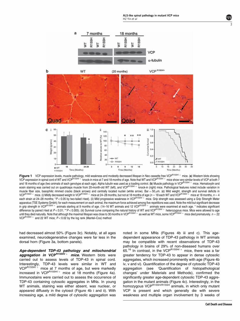
VCPR155H/þ heterozygous knock-in mice develop slowlyprogressive weakness. The Kimonis laboratory previouslygenerated and characterized a heterozygote knock-in mousewith the most common human VCP mutation (R155H)expressed at physiological levels.12 Examination of brain,muscle, and bone pathology in this VCPR155H/þ mouserevealed many of the features of human IBMPFD disease,including the progressive cytoplasmic accumulation of TDP-43 and ubiquitin-positive inclusions in muscle and thecerebral cortex.12 We have subsequently produced a slightlymodified VCPR155H/þ knock-in mouse model (which isidentical to the previously described model except for thelack of a Neomycin cassette) to assess and confirm thephenotype of the VCP mutant knock-in, without risk ofinterference from the Neo cassette. Like the Neo cassettecontaining strain,12 these Neo-free mice also express VCP atan endogenous level. Western blot studies of spinal cordtissue from these animals shows VCP levels to beindistinguishable from those in wild-type (WT) animals atboth 7 and 18 months of age (Figure 1a).
These Neo-free heterozygotes, used in the present studies,have similar brain, muscle, and bone pathology as the Neocassette containing heterozygotes, including the appearanceof age-dependent pathological changes in muscle (withbasophilic rimmed cracks and centrally located nuclei inmuscle fibers, Figure 1b). The bone, brain, and musclephenotype of these animals is more fully described in aseparate publication.14 Furthermore, these mice appearedgenerally normal in growth and behavior. They showed mildlydecreased weights after 24 months of age and mildprogressive weakness from B9 months of age (Figure 1ciand ii). Although their maximal lifespans are the same as WTmice (close to 30 months), their survival curves differ, withsome VCPR155H/þ mice dying prematurely and survival timebroadly distributed betweenB15 and 30 months (Figure 1ciii).
Although these mice manifest slowly developing butextensive ALS-like spinal cord pathology (as describedbelow), they do not develop a rapidly progressive ALS-likedisease (as is seen in mutant SOD1-overexpressing models),
and maintain generally normal appeareance and behaviorthroughout their lives. The absence of clinical ALS may beconsistent with human VCP-linked disease, in which ALS onlyoccurs in B10% of patients. The causes of the prematuredeaths in some of the VCPR155H/þ mice is not known, but mayresemble causes of death in affected humans, many of whomdie in their 40s–60s, often with cardiorespiratory failure.10
Notably, homozygosity of the R155H mutation is lethal, withthese animals dying by 21 days of age with indications ofabnormalities in several systems including muscle, brain andspinal cord (the complex phenotype of these animals will befully characterized in a separate publication).
EMG evidence of MN degeneration in aged VCPR155H/þ
mice. Needle EMG examination provides a valuable tool forthe discrimination between neurogenic and myopathiccauses of neuromuscular dysfunction. Neurogenic findingson EMG include fasciculation potentials and fibrillationpotentials in the acute phase, as well as increased amplitudeand duration of motor unit potentials with polyphasia andreduced recruitment. Myopathic changes include shortduration, small amplitude polyphasic potentials with normalrecruitment patterns.
EMG examination was carried out on four VCPR155H/þ
animals and four WT littermates (see Materials and methods).Evidence of widespread acute denervation was seen in theVCPR155H/þ mice (average age 24.98±0.41 months) but notin their WT littermates. These changes included varyingdegree of abnormal spontaneous activity in the form offibrillation and fasciculation potentials, and distinct reductionin recruitment and interference patterns. Motor unit analysiswas, however, complicated by the fact that both ‘myopathic’(narrow duration, small amplitude) potentials and neurogenic(large amplitude, broad duration) potentials were seen. Firingrates were generally increased, suggestive of a neurogenicrather than myopathic pattern of activation (Figure 2a).Notably, these distinctive abnormalities were seen in almostall the muscles examined in the VCPR155H/þ mice, but werenever seen in the WT mice (Figure 2b).
Age-dependent MN degeneration in VCPR155H/þ mice.MN degeneration was assessed in lumbar spinal cordsections from VCPR155H/þ and WT animals at various ages.MNs were labeled with toluidine blue, and immunocyto-chemically using an antibody to non-phosphorylated neuro-filament epitopes, SMI-32, which strongly labels MNs as wellas their neuritic processes.15 In WTs, virtually all MNsappeared healthy, with a small percentage showing mildatrophic changes in older animals (Figure 3a and b).However, in the VCPR155H/þ animals, there were clearage-dependent degenerative changes, which appeared tofollow distinct pathways. Some MNs showed progressivecellular and nuclear constriction, others showed cytoplasmicvacuolar changes, and some showed nuclear enlargementand chromatin loss (Figure 3a vii and viii). At late stages,MNs often appeared to be replaced by glial nodules(Figure 3a vii and viii, arrows). Surviving MNs were countedin SMI-32-labeled slices from VCPR155H/þ and WT siblings(Figure 3b), from three age groups (8±2, 20±2, 26±2months). By B20 months, MN numbers in VCPR155H/þ mice
ALS-like spinal pathology in mutant VCP miceHZ Yin et al
2
Cell Death and Disease
had decreased almost 50% (Figure 3c). Notably, at all agesexamined, neurodegenerative changes were far less in thedorsal horn (Figure 3a, bottom panels).
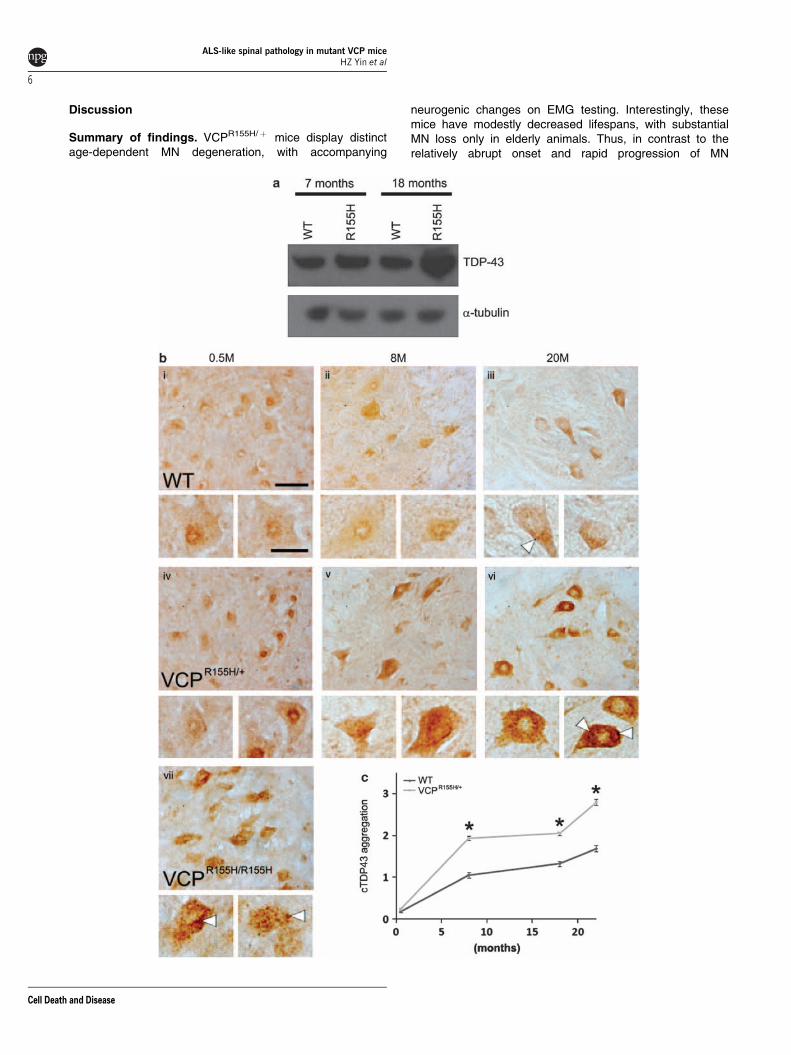
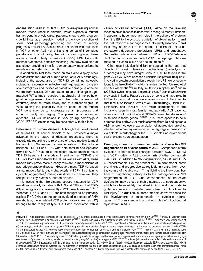
Age-dependent TDP-43 pathology and mitochondrialaggregation in VCPR155H/þ mice. Western blots werecarried out to assess levels of TDP-43 in spinal cord.Interestingly, TDP-43 levels were similar in WT andVCPR155H/þ mice at 7 months of age, but were markedlyincreased in VCPR155H/þ mice at 18 months (Figure 4a).Immunostains were carried out to assess the occurrence ofTDP-43 containing cytosolic aggregates in MNs. In youngWT animals, staining was either absent, was nuclear, orappeared diffusely in the cytosol (Figure 4b i and ii). Withincreasing age, a mild degree of cytosolic aggregation was
noted in some MNs (Figures 4b iii and c). This age-dependent appearance of TDP-43 pathology in WT animalsmay be compatible with recent observations of TDP-43pathology in brains of 29% of non-diseased humans over65.16 In contrast, in the VCPR155H/þ mice, there was a fargreater tendency for TDP-43 to appear in dense cytosolicaggregates, which increased prominently with age (Figure 4biv, v and vi). Quantification of the degree of cytosolic TDP-43aggregation (see ‘Quantification of histopathologicalchanges’ under Materials and Methods), confirmed thesignificantly greater age-dependent cytosolic TDP-43 aggre-gation in the mutant animals (Figure 4c). Interestingly, in thehomozygous VCPR155H/R155H animals, in which only mutantVCP is present and which generally die with severeweakness and multiple organ involvement by 3 weeks of
Figure 1 VCP expression levels, muscle pathology, mild weakness and modestly decreased lifespan in Neo cassette free VCPR155H/þ mice. (a) Western blots showingVCP expression in spinal cord of WT and VCPR155H/þ knock-in mice at 7 and 18 months of age. Note that WT and VCPR155H/þ mice show very similar levels of VCP at both 7and 18 months of age (two animals of each genotype at each age). Alpha tubulin was used as a loading control. (b) Muscle pathology in VCPR155H/þ mice. Hematoxylin andeosin staining was carried out on quadriceps muscle from 20-month-old WT (left), and VCPR155H/þ knock-in (right) mice. Pathological features noted include variation inmuscle fiber size, basophilic rimmed cracks (black arrows) and centrally located nucleii (white arrow). Bar¼ 50mm. (c) Mild weight, strength and survival deficits inVCPR155H/þ mice. (i) Mildly decreased weight in VCPR155H/þ mice at 24–28 months, but not at 18 months of age (n¼ 18 each WT and VCPR155H/þ mice at 18 months, n¼ 4each strain at 24–28 months; *Po0.05 by two-tailed t-test). (ii) Mild progressive weakness in VCPR155H/þ mice. Grip strength was assessed using a Grip Strength Meterapparatus (TSE Systems Gmbh); for each measurement on each animal, the maximum force achieved among five repetitions was used. Note the mild but significant decreasein grip strength in VCPR155H/þ animals starting at 9 months of age. (14–16 WT animals and 12 VCPR155H/þ animals were examined at each age. * indicates significantdifference by paired t-test at Po0.01; **Po0.005). (iii) Survival curve comparing the natural history of WT and VCPR155H/þ heterozygous mice. Mice were allowed to ageuntil they died naturally. Note that although the maximal lifespan was close to 30 months in VCPR155H/þ as well as WT mice, some VCPR155H/þ mice died prematurely. n¼ 22VCPR155H/þ and 22 WT mice; Po0.02 by the log rank (Mantel–Cox) method
ALS-like spinal pathology in mutant VCP miceHZ Yin et al
3
Cell Death and Disease

age, advanced TDP-43 pathology was evident in MNs of10-day-old animals, an age at which VCPR155H/þ miceshowed little TDP-43 pathology (Figure 4b vii).
We also carried out double immunofluorescence stains forTDP-43 along with the mitochondrial marker, COX-IV. Inyoung WT and VCPR155H/þ animals, COX-IV labeling wasdiffuse and speckled, as expected for mitochondria, and TDP-43 labeling was faint and nuclear or diffuse cytosolic (asdescribed above). In aged (B18 month) WT animals,mitochondria were still diffusely distributed, but there wassome minimal aggregation noted, which co-localized withareas of slight TDP-43 aggregation (Figure 5a i and ii).However, in the aged VCPR155H/þ animals, COX-IV labelingwas densely aggregated and co-localized strongly with TDP-43 (Figure 5a iii and iv). Examination of all MNs in sets of age-matched sections revealed little COX-IV aggregation up to 8months in either WT or VCPR155H/þ animals, but substantialco-aggregation of COX-IV and TDP-43 by 18 months only inthe VCPR155H/þ animals, with much milder age-dependentco-aggregation seen in WT animals (Figure 5b).
To further address the pathological similarities between theVCPR155H/þ animals and diverse forms of human ALS, wecarried out immunostains for ubiquitin (Figure 5c) as well asfor the autophagy related protein, SQSTM1/P-62 (Figure 5d),both of which are prominent constituents of cytosolic MNaggregates in most human ALS cases.17 Labeling with eithermarker revealed appearance of cytosolic aggregates similarto that seen with TDP-43, with far greater age-dependentcytosolic aggregation in the VCPR155H/þ animals than in WTanimals.
Age-dependent astrogliosis and 3-nitrotyrosine labelingin heterozygous R155H mice. Previous reports haveindicated progressive reactive astrogliosis in mutant SOD1as well as TDP-43 mouse models of ALS.7,18 Similarly, wesee a dramatic age-dependent increase in GFAP labeling inVCPR155H/þ animals, most prominent in ventral horn(Figure 6a and c i).
Paralleling this increased astrogliosis, we also noteincreases in 3-nitrotyrosine (3-NT) labeling, both within
Figure 2 EMG evidence of muscle denervation in VCPR155H/þ mice. EMG studies were carried out on four VCPR155H/þ animals (average age 24.98±0.41 months), incomparison with four WT littermates. (a) Traces show representative pathological changes, which were seen only in the VCPR155H/þ animals. (i) Fasciculation potential; (ii)fibrillation potentials; (iii) reduced motor unit recruitment. This is a neurogenic recruitment pattern. Firing frequency in these animals was increased at 50 Hz. (iv) Co-existingneurogenic and myopathic potentials. Motor units exhibit mixed morphology with small amplitude, polyphasic potentials, suggestive of a myogenic disturbance. However,recruitment frequencies are decreased, suggesting a neurogenic pattern. (b) Tabular chart showing frequency of various abnormalities seen on EMG from the sampling ofseven muscles in each animal. Note frequent abnormalities in VCPR155H/þ animals and absence of abnormalities in WT mice (MUP, motor unit potential)
ALS-like spinal pathology in mutant VCP miceHZ Yin et al
4
Cell Death and Disease

and between ventral horn MNs. Unlike the astrogliosis,this 3-NT labeling was most prominent at B20 months,decreasing at advanced ages when many MNs had degen-erated. As noted in our studies of mutant SOD1 animals,19,20
the 3-NT labeling was often particularly strong nearMN somata, falling off sharply with distance, possiblyconsistent with a role of ROS generated within MNs(Figure 6b and c ii).
Figure 3 Progressive age-dependent degenerative changes and loss of ventral horn MNs from heterozygous VCP knock-in (VCPR155H/þ ) mice. (a) Age-dependentdegenerative changes in ventral horn MNs VCPR155H/þ mice. Lumbar sections were stained with toluidine blue. Images show representative fields (400� ) from ventral hornof WT and sibling VCPR155H/þ mice at the indicated ages (±2 months). In the WTs, virtually all MNs appeared healthy, with mild atrophic changes noted in a small number ofthe older MNs. In contrast, in VCPR155H/þ animals, clear age-dependent degenerative changes are seen (panels vii and viii) with eccentric nuclei, fragmentation of dendrites,and some MNs showing cellular and nuclear constriction (stars), and others showing prominent swelling and vacuolar changes in cytoplasm (arrowheads). In some cases,degenerated MNs were replaced by glial nodules (arrows). Bottom panels (iv and ix) show low power views from 26–month-old spinal cords, with regions from ventral horn(VH) and dorsal horn (DH) highlighted. Arrows indicate high power views of regions indicated. Note the relative preservation of neuronal morphology in the DH region, despiteextensive degenerative changes in the VH region of the 26 month VCPR155H/þ mouse (panels viii, ix, and x). Comparative examination of VH and DH regions from 440 slicesfrom 26±2-month-old WT and VCPR155H/þ mice revealed similar condition of VH and DH neurons in all WT’s but distinctly greater degenerative changes in VH MNscompared to DH regions in all slices from VCPR155H/þ mice. Bar¼ 500mm (low power panels); 50mm (all other panels). (b) Age-dependent loss of ventral horn MNs inVCPR155H/þ mice. Lumbar sections were immunostained for the non-phosphoryylated neurofilament marker, SMI-32. Images show representative fields (400� ) from ventralhorn of WT and sibling VCPR155H/þ mice at the indicated ages (±2 months). Note the age-dependent atrophy and loss of MNs in the VCPR155H/þ animals. Bar¼ 50mm.(c) Quantification of MN cell loss. Surviving MNs were counted in SMI-32 immunostained slices from each condition. Values represent mean number of surviving ventral hornMNs per section (containing two ventral horns), as assessed under direct microscopic examination. Each data point reflects counts from 3–7 animals (for SMI-32); all ventralhorn MNs were counted in each of 10–15 slices for each animal. Note the modest age-dependent MN loss in WTs in contrast to the marked loss in the VCPR155H/þ mice.* indicates difference from WT animals at the same age by two-tailed t-test (Po0.025)
ALS-like spinal pathology in mutant VCP miceHZ Yin et al
5
Cell Death and Disease
Discussion
Summary of findings. VCPR155H/þ mice display distinctage-dependent MN degeneration, with accompanying
neurogenic changes on EMG testing. Interestingly, thesemice have modestly decreased lifespans, with substantialMN loss only in elderly animals. Thus, in contrast to therelatively abrupt onset and rapid progression of MN
ALS-like spinal pathology in mutant VCP miceHZ Yin et al
6
Cell Death and Disease
degeneration seen in mutant SOD1 overexpressing animalmodels, these knock-in animals, which express a mutanthuman gene in physiological patterns, show slowly progres-sive MN damage, possibly modeling the slow evolution ofspinal cord pathology preceding the onset of rapidlyprogressive clinical ALS in subsets of patients with mutationsin VCP or other ALS risk enhancing genes of incompletepenetrance. It is intriguing that with advancing age, theseanimals develop fairly extensive (450%) MNs loss withminimal symptoms, possibly reflecting the slow evolution ofpathology, providing time for compensatory mechanisms tomaintain adequate motor function.
In addition to MN loss, these animals also display othercharacteristic features of human spinal cord ALS pathology,including the appearance of TDP-43 containing cytosolicinclusions, evidence of mitochondrial aggregation, progres-sive astrogliosis and indices of oxidative damage in affectedventral horn tissues. Of note, examination of findings in age-matched WT animals revealed that virtually all the patholo-gical findings were not exclusive to the mutant animals, butoccurred, albeit far more slowly and to a milder degree, inWTs, raising the possibility that an effect of the mutantVCP gene may be to accelerate certain processes thatoccur normally with aging. The presence of advancedcytosolic TDP-43 inclusions in very young homozygousVCPR155H/R155H animals may be consistent with this idea.
Relevance to human disease. Although the developmentof mutant SOD1 animal models of ALS provided a majoradvance in the study of disease processes, there isuncertainty regarding the relevance of these models to mosthuman ALS. Subsequent characterization of the linkagebetween TDP-43 and FUS with both familial and sporadicforms of ALS5,6 has led to the recent development of novelTDP-43 based animal models. In addition, as TDP-43 andFUS are both associated with FTD as well as with ALS, thesemodels may prove more broadly relevant to mechanisms ofneurodegenerative disease. However, most TDP-43-basedanimal models fail to show characteristic TDP-43 containingcytosolic aggregates,7 raising questions as to how well theyrecapitulate key events of human disease.
It is intriguing that the disease spectrum caused by VCPmutations similarly includes both ALS and FTD and that TDP-43 pathology occurs prominently in VCP linked disease.9,11–13
Whereas TDP-43 and FUS are both thought to be primarilynuclear RNA/DNA-binding genes involved in aspects of RNAmetabolism, the unrelated VCP protein (also known as p97)belongs to the family of type II ATPase associated with a
variety of cellular activities (AAA). Although the relevantmechanism in disease is uncertain, among its many functions,it appears to have important roles in the delivery of proteinsfrom the ER to the cytosol, regulation of ubiquitination,21 andthe maturation of autophagosomes into autolysosomes,22 andthus may be crucial to the normal function of ubiquitin–proteasome-dependent proteolysis (UPS) and autophagy.Suggesting interactions between VCP and TDP-43-depen-dent mechanisms, either mutant VCP or autophagic inhibitionresulted in cytosolic TDP-43 accumulation.22
Other recent studies lend further support to the idea thatdefects in protein clearance mechanisms (proteolysis orautophagy) may have integral roles in ALS. Mutations in thegene UBQLN2, which encodes a ubiquitin-like protein, ubiquilin 2,involved in protein degradation through the UPS, were recentlyfound to be linked to forms of dominantly inherited, X-linked ALSand ALS/dementia.23 Similarly, mutations in optineurin24 and inSQSTM1 (which encodes the protein p62),25 both of which werepreviously linked to Paget’s disease of bone and have roles inUPS/autophagy pathways, were recently found to be linked torare familial or sporadic forms of ALS. Interestingly, ubiquilin 2,optineurin, and SQSTM1 are major components of theinclusions seen in most familial and sporadic ALS patients,often along with ubiquitin and TDP-43, even the absence ofmutations in these genes.17,23,24 Thus, there appears to be acommon final pathway for multiple forms of familial and sporadicALS, wherein cytosolic accumulation of aggregated proteins,whether via a primary enhancement of aggregate formation orvia defects in autophagy or the UPS, creates an environmentthat promotes neurodegeneration.
Emerging clues to common mechanisms of selective MNdegeneration in diverse forms of ALS. Comparison of theevolution of pathological changes between SOD1, TDP-43and VCP models of ALS provide hints of other commonal-ities. First, in addition to MN degeneration, SOD1 and TDP-43 based models, like the present VCP mutant model, showprominent and progressive astrogliosis in ventral horn overthe course of the disease,7,26 highlighting the likely contribu-tions of neighboring astrocytes to the pathogenesis of MNdegeneration in ALS. One consequence of astrocytedysfunction may be loss of their glutamate transport capacity,which has been widely described in ALS and may underlieglutamate receptor mediated (excitotoxic) contributions toMN injury.1 In addition, all three models show evidence forthe involvement of mitochondria in cytosolic aggre-gates,2,27,28 consistent with prominent roles of mitochondrialdysfunction in ALS.
Figure 4 Age-dependent increases in total spinal cord TDP-43 and its appearance in cytosolic inclusions in ventral horn MNs of VCPR155H/þ mice. (a) Western blotsshowing TDP-43 expression in spinal cord of WT and VCPR155H/þ knock-in mice at 7 and 18 months of age. Note that WT and VCPR155H/þ mice show very similar levels ofTDP-43 at 7 months of age, whereas TDP-43 levels were markedly increased in the VCPR155H/þ spinal cord at 18 months. Alpha tubulin was used as a loading control.(b) Immunocytochemical assessment of cytosolic TDP-43 aggregation as a function of age-representative photomicrographs. Lumbar sections were immunostained for TDP-43 and photographed (400� ). Representative fields are shown from ventral horn of WT (i, ii, and iii) and sibling VCPR155H/þ mice (iv, v, and vi) at the indicated ages(±2 months). In WT animals, faint and generally cytosolic or nuclear labeling was generally seen at young ages, with more prominent but generally still diffuse staining seen by20 months. In the VCPR155H/þ animals, cytosolic TDP-43 labeling was both stronger, and far more prone to appear as discrete inclusions or aggregates with increasing age(arrowheads). By way of comparison, we also show stains from young (0.5 months) homozygous VCPR155H/R155H animals (vii). Note the markedly accelerated appearance ofstrong cytosolic TDP-43 aggregation in MN from these young mice (arrowheads). Bar¼ 50 or 25mm (detail). (c) Quantification of cytosolic TDP-43 aggregation. Each MN inexamined sections was rated for cytosolic TDP-43 aggregation according to a five point scale as described (see Materials and methods). Each data point represents all MNs(4160) present in 5–10 ventral horn micrographs from each of 2–5 animals. * indicates difference from WT animals at the same age by two-tailed t-test (Po0.001)
ALS-like spinal pathology in mutant VCP miceHZ Yin et al
7
Cell Death and Disease

Figure 5 Colocalization of mitochondrial aggregates with TDP-43, and occurrence of ubiquitin and SQSTM1/ p62 aggregates in aging VCPR155H/þ MNs.(a) Representative images. Slices were double fluorescence immunostained for the mitochondrial marker, COX-IV (top) and TDP-43 (bottom) (400� ); inserts show overlay.Note in the 18-month-old WT animals, the COX-IV labeling is speckled and distributed throughout the cytoplasm. TDP-43 labeling shows mild early aggregation in a smallnumber of MNs, that co-localizes with some early mitochondrial aggregation (arrowheads). In contrast, in age-matched VCPR155H/þ animals, the COX-IV labeling is highlyaggregated and colocalizes with TDP-43 (arrows). Bar¼ 25mm. (b) Quantification of COX-IV and TDP-43 co-aggregation. Graph shows the % of examined MNs containingCOX-IV and TDP-43 co-aggregation at each age (each point based on examination of 440 MNs). Note the paucity of co-aggregation at 8 months in all animals, and the sharpincrease of co-aggregation in the VCPR155H/þ mice by 18 months. (c and d): Ubiquitin (c) and SQSTM1/ p62 (d) labeling (24-month-old animals) revealed similar patterns ofage-dependent appearance of cytosolic aggregates as seen for TDP-43. Arrows denote aggregates in VCPR155H/þ MNs. Bar¼ 25mm
ALS-like spinal pathology in mutant VCP miceHZ Yin et al
8
Cell Death and Disease
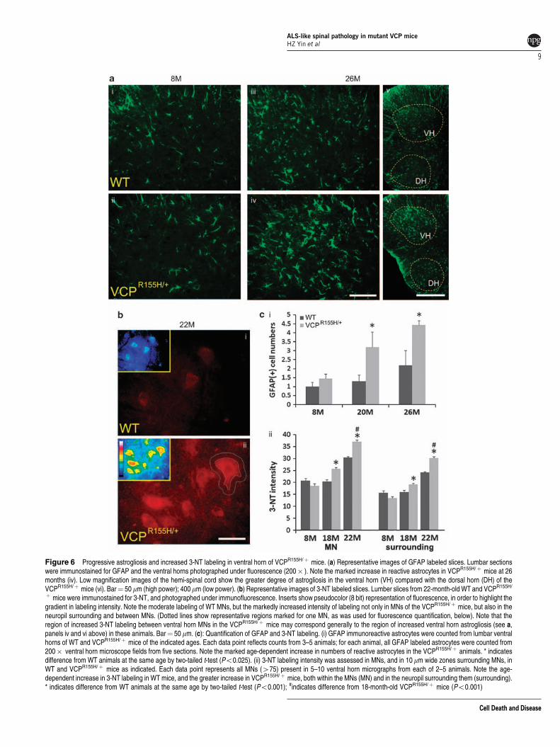
Figure 6 Progressive astrogliosis and increased 3-NT labeling in ventral horn of VCPR155H/þ mice. (a) Representative images of GFAP labeled slices. Lumbar sectionswere immunostained for GFAP and the ventral horns photographed under fluorescence (200� ). Note the marked increase in reactive astrocytes in VCPR155H/þ mice at 26months (iv). Low magnification images of the hemi-spinal cord show the greater degree of astrogliosis in the ventral horn (VH) compared with the dorsal horn (DH) of theVCPR155H/þ mice (vi). Bar¼ 50mm (high power); 400 mm (low power). (b) Representative images of 3-NT labeled slices. Lumber slices from 22-month-old WT and VCPR155H/
þ mice were immunostained for 3-NT, and photographed under immunofluorescence. Inserts show pseudocolor (8 bit) representation of fluorescence, in order to highlight thegradient in labeling intensity. Note the moderate labeling of WT MNs, but the markedly increased intensity of labeling not only in MNs of the VCPR155H/þ mice, but also in theneuropil surrounding and between MNs. (Dotted lines show representative regions marked for one MN, as was used for fluorescence quantification, below). Note that theregion of increased 3-NT labeling between ventral horn MNs in the VCPR155H/þ mice may correspond generally to the region of increased ventral horn astrogliosis (see a,panels iv and vi above) in these animals. Bar¼ 50mm. (c): Quantification of GFAP and 3-NT labeling. (i) GFAP immunoreactive astrocytes were counted from lumbar ventralhorns of WT and VCPR155H/þ mice of the indicated ages. Each data point reflects counts from 3–5 animals; for each animal, all GFAP labeled astrocytes were counted from200� ventral horn microscope fields from five sections. Note the marked age-dependent increase in numbers of reactive astrocytes in the VCPR155H/þ animals. * indicatesdifference from WT animals at the same age by two-tailed t-test (Po0.025). (ii) 3-NT labeling intensity was assessed in MNs, and in 10 mm wide zones surrounding MNs, inWT and VCPR155H/þ mice as indicated. Each data point represents all MNs (475) present in 5–10 ventral horn micrographs from each of 2–5 animals. Note the age-dependent increase in 3-NT labeling in WT mice, and the greater increase in VCPR155H/þ mice, both within the MNs (MN) and in the neuropil surrounding them (surrounding).* indicates difference from WT animals at the same age by two-tailed t-test (Po0.001); #indicates difference from 18-month-old VCPR155H/þ mice (Po0.001)
ALS-like spinal pathology in mutant VCP miceHZ Yin et al
9
Cell Death and Disease

A central question in the study of ALS pathogenesis is thushow a range of perturbations, likely reflecting combinations ofgenetic and environmental factors, may trigger a convergentphenotype with selective MN loss and surrounding astro-gliosis. Possible clues to the selectivity of MN loss may beprovided by studies examining factors underlying their highvulnerability to excitotoxic injury. Specifically, susceptibleMNs possess large numbers of unusual Ca2þ permeableAMPA channels,15,29 yet have little ability to buffer cytosolicCa2þ rises,30,31 such that they may be unusually prone tomitochondrial Ca2þ loading and consequent oxidativestress.19,32
Excessive oxidative stress could contribute to the oxidationand aggregation of various proteins. Indeed, in the case ofSOD1-linked disease, oxidative stress promotes the forma-tion of insoluble SOD1 multimers in affected spinal cordtissue.33,34 Interestingly, oxidative stress has also recentlybeen reported to promote the aggregation of TDP-43 and itsrecruitment into stress granules.35,36 Other recent studiessuggest that with both SOD137,38 and TDP-43,39,40 theaggregates may mediate injury in large part via interactionswith mitochondria.
Present findings of mitochondrial aggregation in the VCPmodel (analogous to that previously reported in SOD1 andTDP-43 models), along with evidence of increased oxidativestress (3-NT labeling) within and surrounding the MNs arealso compatible with a mechanism wherein disruption ofmitochondrial function, perhaps reflecting a combination ofretention of dysfunctional mitochondria due to defects inautophagy/mitophagy and deleterious interactions with mis-folded proteins, results in further oxidative stress, moreprotein aggregations and propagation of a disese process inwhich mitochondrial dysfunction, oxidative stress and proteinaggregation are all intimitely involved. Oxidative stress in andaround MNs could also contribute to the activation (and likelydysfunction) of ventral horn astrocytes seen in each of thethree ALS models (SOD1, TDP-43 and VCP).20,41
Conclusions
Present studies extend the range of animal models availableto study mechanisms underlying pathogenesis in ALS. Thepathological commonalities between human ALS and thethree animal models of genetic forms discussed (SOD1, TDP-43 and VCP) are striking, in that they all show, in addition topreferential MN loss, the appearance of aggregates in MNs,evidence of mitochondrial dysfunction and/or oxidative tissuedamage, and pronounced astrogliosis in the ventral horn.Thus, it appears that mutations in these three completelyunrelated genes can trigger a cascade of events resulting in arelatively characteristic pathological phenotype. A key aim ofongoing studies is thus to elucidate what ties these distinctforms of disease together and explains the convergentphenotype despite distinct ‘causes’. It is likely that theconvergent phenotype reflects attributes of the MNs them-selves and the nature of their interactions with surroundingcells like astrocytes. We suggest a possible factor: that MNs,in part because of their complement of excitatory receptorsand Ca2þ handling characteristics, are unusually prone tooxidative stress, which may promote aggregation and
misfolding of proteins. Primary defects in UPS/autophagypathways (as may occur with VCP, UBQLN2, SQSTM1, oroptineutin mutations) may similarly promote aggregateformation. The aggregates may disrupt mitochondrial func-tion, causing more oxidative stress and protein aggregation,setting in motion cascades that cause slowly developing MNdegeneration.
It is interesting that these mice, however, do not developclassical clinical ALS, in which disease (associated withsevere MN degeneration) progresses rapidly and contigu-ously from one or multiple sites. Perhaps clinical ALS occurswhen dysfunction of MNs and surrounding astrocytes in aregion of tissue exceeds a threshold necessary to maintaincellular energy balance and ionic homeostasis, resulting infeed forward cascades and propagation of the injury frontacross the compromised tissue.
Of note, human ALS is typically a disease of aging adultsand the VCP mutation may accelerate events (including TDP-43 aggregation and MN loss) that occur far more slowly withnormal aging. Might these VCP mice, in which extensive ALS-like patholological changes develop slowly over their lifespanswithout frank disease, thus provide valuable models for thestudy of ways in which ALS risk genes of partial penetrancemay accelerate age-associated changes and bring spinal cordtissue to a level of dysfunction that may on occasion triggeronset of characteristic rapidly progressive disease? Com-parative studies in these and other available ALS animalmodels may enable breakthroughs in the understanding ofcommon and essential mechanisms of disease and thetesting of therapeutic or preventative interventions.
Materials and MethodsVertebrate animals. This study was carried out in strict accordance with therecommendations in the Guide for the Care and Use of Laboratory Animals of theNational Institutes of Health. The protocol was approved by the Institutional AnimalCare and Use Committee of the University of California, Irvine (Protocol number:1997-1267). All procedures were terminal and all animals were deeply anethetizedbefore starting any procedures to minimize suffering.
VCPR155H/þ knock-in mice. In order to generate a VCP disease mousemodel, a genomic VCP fragment with 7.9 kb of upstream homology sequence and2.1 kb of downstream homology sequence was sub-cloned into a targeting vector.Site-directed mutagenesis using Quick-Change XL Site-Directed Mutagenesis Kit(Stratagene, La Jolla, CA, USA) was used to introduce the R to H mutation atamino acid position 155. The knock-in mouse model with the R155H VCPmutation was generated at InGenious Targeting Laboratory, Inc. (Stony Brook,NY, USA) through a Neomycin cassette insertion using 129/SvEv mice. Theexpression of mutant VCP was confirmed by RT-PCR using the following primersin the PCR reactions: forward 50-CACGGTGTTGCTAAAAGGAAAGAAAAG-30;reverse 30-CTGAAGAATCTCCAAACGTCCTGTAGC-50, after the RT reactions withthe reverse primer. These mice were backcrossed 46 times with the C57BL/6 strainbefore experiments were done to make sure that the majority (498%) of the geneticbackground of generated mice was of C57BL/6 origin. The Neomycin cassette wasdeleted by crossing with the Flp deletion mouse model. Genotyping is carried out asdescribed.12 Heterozygotes are bred with heterozygotes, such that half the progenyare heterozygotes, and a quarter each WT and homozygotes. For all studies, ageand gender matched WT siblings are used as controls.
Strength testing. Muscle strength of the forelimbs of mice was measuredusing a Grip Strength Meter apparatus (TSE Systems Gmbh, Hamburg,Germany). As described (Figure 1), 14–16 WT animals and 12 VCPR155H/þ
animals were examined at each age and the strongest force of 5 trails for eachanimal at each age was used for analysis. Statistical analyses were performed byStudent’s t-test.
ALS-like spinal pathology in mutant VCP miceHZ Yin et al
10
Cell Death and Disease

Tissue immunohistochemistry. Animals were perfused transcardiallywith phosphate-buffered saline (PBS), followed by 4% paraformaldehyde (PFA) for10 min. The brain and spinal cord were dissected and post fixed in 4% PFA for48 h and removed to 30% sucrose/PBS for another 2–3 days. Serial 20 mm frozensections were cut from the lumbar enlargement of spinal cord. For immunostain-ing, sections were blocked (10% HS and 0.3% Triton-X 100, 1 h) and exposed tomonoclonal SMI-32 (1 : 5000; Sternberger Monoclonals, Berkeley, CA, USA)or anti-cytochrome oxidase subunit IV (COX-IV; 1 : 100; ab14744; Abcam,Cambridge, MA, USA), or to polyclonal antibodies against TARDBP/ TDP-43(1 : 1000 for immunoperoxidase, 1 : 200 for immunofluorescence; ab41972; Abcam),glial fibrillary acidic protein (GFAP; 1 : 500; G9269; Sigma-Aldrich, St Louis,MO, USA), SQSTMI/p62 (1 : 3000; ab91526; Abcam) or 3-NT (1 : 250; 06-284;Upstate Biotechnology, Waltham, MA, USA) in blocking solution (48–72 h, 4 1C).Slices were then incubated with an appropriate biotinylated (Vector Laboratoroes,Burlingame, CA, USA), or fluorescent secondary antibody (DyLight 488 or 594fluorophores; Jackson ImmunoResearch, West Grove, PA, USA). Immunoperoxidaselabeling was visualized using ABC solution (Vector Labratories) and diaminobenzidine,and immunofluorescence labeling was visualized under fluorescence microscopy.Specificity of all immunostains was confirmed by deletion of primary antibody.
For toluidine blue staining, the slide was flooded with 0.02% Toluidine Blue O(Fisher Chemical, Hudson, NH, USA) for 1.5 min, washed, and flooded with 95%ethanol to remove excess dye. For muscle studies, quadriceps muscle wasembedded in cryo-sectioning media and 5–10mm cryostat sections produced andstained with hematoxylin and eosin.
Western blotting. Spinal cords were harvested and extracted using the NE-PER Nuclear and Cytoplasmic Extraction Kit (Thermo Scientific, Rockford, IL,USA). Protein concentrations were determined using the Nanodrop according tothe manufacturer’s protocols. Equal amount of proteins were separated on Bis-Tris4-12% NuPAGE gels, and the expression levels of proteins were analyzed bywestern blotting using specific antibodies against VCP (Affinity BioReagents,Golden, CO, USA) or TDP-43 (Abcam). Alpha tubulin (Sigma-Aldrich) was usedfor loading control.
Quantification of histopathological changes. Quantification ofmeasures (including MN survival, astrogliosis, cytosolic TDP-43 aggregation,and 3-NT labeling) was based upon compilations of data from a total of 28 animals(17 male, 11 female). In each study (comprising comparisons of age matchedgroups of WT and VCPR155H/þ mice), the gender compositions of the WT andmutant groups were identical.
Surviving MNs or reactive astrocytes were counted in ventral horn of lumbarspinal slices from each condition. MN numbers are presented as mean survivingMNs per section (comprising 2 ventral horns), and GFAP labeled reactive astrocytescalculated as numbers per unit area of ventral horn, normalized to the numberpresent in WTs of the youngest age. MNs with pyknotic nuclei, or with a markedlyatrophic, irregular or fragmented soma were not counted as alive.
Quantitative assessment of TDP-43 pathology was carried out by rating eachventral horn MN in examined sections according to a 5-point scale as follows: 0, nocytosolic TDP-43; 1, faint diffuse cytosolic label; 2, some granularity of cytosoliclabeling with no clear aggregate; 3, some clear cytosolic TDP-43 aggregation; and4, large and dense cytosolic TDP-43 aggregates.
For quantification of 3-NT labeling intensity, sections from matched WT andtransgenic animals were identically labeled and photographed, and two regionswere marked around each MN: one immediately outlining the circumfrence of theMN and another 10mm further out. The space between these regions was definedas the surround zone. After subtraction of background fluorescence, averagedintensity was calculated within each MN soma, and in the surround zone (8 bitintensity scale, Image J software, Public Domain software from the NIH).
EMG studies. Terminal EMG study was performed on these animals at anaverage age of 26.68±0.47 months for the WT (N-4) and 24.98±0.41 months forthe VCPR155H/þ mice (N¼ 4). Subdermal EEG electrodes were used for conductingEMG studies. The active and reference electrodes were paired together to act as aconcentric needle electrode for these recordings. In each animal, the following sevenmuscles were sampled: unilateral thoracic paraspinal, bilateral tibialis anterior, bilateralmedial gastrocnemius, and bilateral hamstrings. All recordings were made using aCadwell Sierra LT machine (Cadwell Laboratories, Kennewich, WA, USA). Patterns ofinsertional and spontaneous activity were noted along with patterns of motor unitpotentials evoked by movement of limbs caused by noxious footpad stimuli.
Conflict of InterestThe authors declare no conflict of interest.
Acknowledgements. This work was supported by National Institutes ofHealth grants NS36548 (JHW) and AR050236 (VEK), and a grant from theMuscular Dystrophy Association (VEK). We appreciate the work of Drs. Watts, Vesaand Badadani who performed the preliminary characterization studies of the R155Hknock-in mice.
1. Rothstein JD. Current hypotheses for the underlying biology of amyotrophic lateralsclerosis. Ann Neurol 2009; 65(Suppl 1): S3–S9.
2. Manfredi G, Xu Z. Mitochondrial dysfunction and its role in motor neuron degeneration inALS. Mitochondrion 2005; 5: 77–87.
3. Neumann M, Sampathu DM, Kwong LK, Truax AC, Micsenyi MC, Chou TT et al.Ubiquitinated TDP-43 in frontotemporal lobar degeneration and amyotrophic lateralsclerosis. Science 2006; 314: 130–133.
4. Sreedharan J, Blair IP, Tripathi VB, Hu X, Vance C, Rogelj B et al. TDP-43 mutations infamilial and sporadic amyotrophic lateral sclerosis. Science 2008; 319: 1668–1672.
5. Chen-Plotkin AS, Lee VM, Trojanowski JQ. TAR DNA-binding protein 43 inneurodegenerative disease. Nat Rev Neurol 2010; 6: 211–220.
6. Lagier-Tourenne C, Polymenidou M, Cleveland DW. TDP-43 and FUS/TLS: emergingroles in RNA processing and neurodegeneration. Hum Mol Genet 2010; 19: R46–R64.
7. Wegorzewska I, Baloh RH. TDP-43-Based Animal Models of Neurodegeneration: NewInsights into ALS Pathology and Pathophysiology. Neurodegener Dis 2011; 8: 262–274.
8. Baloh RH. TDP-43: the relationship between protein aggregation and neurodegenerationin amyotrophic lateral sclerosis and frontotemporal lobar degeneration. Febs J 2011; 278:3539–3549.
9. Watts GD, Wymer J, Kovach MJ, Mehta SG, Mumm S, Darvish D et al. Inclusion bodymyopathy associated with Paget disease of bone and frontotemporal dementia is causedby mutant valosin-containing protein. Nat Genet 2004; 36: 377–381.
10. Kimonis VE, Mehta SG, Fulchiero EC, Thomasova D, Pasquali M, Boycott K et al. Clinicalstudies in familial VCP myopathy associated with Paget disease of bone andfrontotemporal dementia. Am J Med Genet A 2008; 146A: 745–757.
11. Johnson JO, Mandrioli J, Benatar M, Abramzon Y, Van Deerlin VM, Trojanowski JQ et al.Exome sequencing reveals VCP mutations as a cause of familial ALS. Neuron 2010; 68:857–864.
12. Badadani M, Nalbandian A, Watts GD, Vesa J, Kitazawa M, Su H et al. VCP associatedinclusion body myopathy and paget disease of bone knock-in mouse model exhibits tissuepathology typical of human disease. PLoS One 2010; 5: pii: e13183.
13. Custer SK, Neumann M, Lu H, Wright AC, Taylor JP. Transgenic mice expressing mutantforms VCP/p97 recapitulate the full spectrum of IBMPFD including degeneration in muscle,brain and bone. Hum Mol Genet 2010; 19: 1741–1755.
14. Nalbandian A, Llewellyn KJ, Badadani M, Yin HZ, Nguyen C, Mukherjee J et al.A progressive translational mouse model of human VCP disease: The VCPR155H/þMouse. Muscle Nerve 2012; doi:10.1002/mus.23522.
15. Carriedo SG, Yin HZ, Weiss JH. Motor neurons are selectively vulnerable to AMPA/kainatereceptor-mediated injury in vitro. J Neurosci 1996; 16: 4069–4079.
16. Geser F, Robinson JL, Malunda JA, Xie SX, Clark CM, Kwong LK et al. Pathological 43-kDa transactivation response DNA-binding protein in older adults with and without severemental illness. Arch Neurol 2010; 67: 1238–1250.
17. Fecto F, Siddique T. Making Connections: Pathology and Genetics Link AmyotrophicLateral Sclerosis with Frontotemporal Lobe Dementia. J Mol Neurosci 2011; 45:663–675.
18. Tu PH, Raju P, Robinson KA, Gurney ME, Trojanowski JQ, Lee VM. Transgenic micecarrying a human mutant superoxide dismutase transgene develop neuronal cytoskeletalpathology resembling human amyotrophic lateral sclerosis lesions. Proc Natl Acad Sci USA1996; 93: 3155–3160.
19. Rao SD, Yin HZ, Weiss JH. Disruption of glial glutamate transport by reactive oxygenspecies produced in motor neurons. J Neurosci 2003; 23: 2627–2633.
20. Yin HZ, Tang DT, Weiss JH. Intrathecal infusion of a Ca2þ permeable AMPA channelblocker slows loss of both motor neurons and of the astrocyte glutamate transporter, GLT-1in a mutant SOD1 rat model of ALS. Exp Neurol 2007; 207: 177–185.
21. Halawani D, Latterich M. p97: the cell’s molecular purgatory? Mol Cell 2006; 22: 713–717.22. Ju JS, Fuentealba RA, Miller SE, Jackson E, Piwnica-Worms D, Baloh RH et al. Valosin-
containing protein (VCP) is required for autophagy and is disrupted in VCP disease. J CellBiol 2009; 187: 875–888.
23. Deng HX, Chen W, Hong ST, Boycott KM, Gorrie GH, Siddique N et al. Mutations inUBQLN2 cause dominant X-linked juvenile and adult-onset ALS and ALS/dementia.Nature 2011; 477: 211–215.
24. Maruyama H, Morino H, Ito H, Izumi Y, Kato H, Watanabe Y et al. Mutations of optineurin inamyotrophic lateral sclerosis. Nature 2010; 465: 223–226.
25. Fecto F, Yan J, Vemula SP, Liu E, Yang Y, Chen W et al. SQSTM1 Mutations in Familialand Sporadic Amyotrophic Lateral Sclerosis. Arch Neurol 2011; 68: 1440–1446.
ALS-like spinal pathology in mutant VCP miceHZ Yin et al
11
Cell Death and Disease

26. Philips T, Robberecht W. Neuroinflammation in amyotrophic lateral sclerosis: role of glialactivation in motor neuron disease. Lancet Neurol 2011; 10: 253–263.
27. Shan X, Chiang PM, Price DL, Wong PC. Altered distributions of Gemini of coiled bodiesand mitochondria in motor neurons of TDP-43 transgenic mice. Proc Natl Acad Sci USA2010; 107: 16325–16330.
28. Xu YF, Gendron TF, Zhang YJ, Lin WL, D’Alton S, Sheng H et al. Wild-type human TDP-43expression causes TDP-43 phosphorylation, mitochondrial aggregation, motor deficits, andearly mortality in transgenic mice. J Neurosci 2010; 30: 10851–10859.
29. Van Den Bosch L, Vandenberghe W, Klaassen H, Van Houtte E, Robberecht W. Ca(2þ )-permeable AMPA receptors and selective vulnerability of motor neurons. J Neurol Sci2000; 180: 29–34.
30. Alexianu ME, Ho BK, Mohamed AH, La Bella V, Smith RG, Appel SH. The role of calcium-binding proteins in selective motoneuron vulnerability in amyotrophic lateral sclerosis. AnnNeurol 1994; 36: 846–858.
31. Vanselow BK, Keller BU. Calcium dynamics and buffering in oculomotor neurones frommouse that are particularly resistant during amyotrophic lateral sclerosis (ALS)-relatedmotoneurone disease. J Physiol 2000; 525(Pt 2): 433–445.
32. Carriedo SG, Sensi SL, Yin HZ, Weiss JH. AMPA exposures induce mitochondrial Ca(2þ )overload and ROS generation in spinal motor neurons in vitro. J Neurosci 2000; 20:240–250.
33. Furukawa Y, Fu R, Deng HX, Siddique T, O’Halloran TV. Disulfide cross-linked proteinrepresents a significant fraction of ALS-associated Cu, Zn-superoxide dismutaseaggregates in spinal cords of model mice. Proc Natl Acad Sci USA 2006; 103: 7148–7153.
34. Niwa J, Yamada S, Ishigaki S, Sone J, Takahashi M, Katsuno M et al. Disulfide bondmediates aggregation, toxicity, and ubiquitylation of familial amyotrophic lateral sclerosis-linked mutant SOD1. J Biol Chem 2007; 282: 28087–28095.
35. Colombrita C, Zennaro E, Fallini C, Weber M, Sommacal A, Buratti E et al. TDP-43 isrecruited to stress granules in conditions of oxidative insult. J Neurochem 2009; 111: 1051–1061.
36. Ayala V, Granado-Serrano AB, Cacabelos D, Naudi A, Ilieva EV, Boada J et al. Cell stressinduces TDP-43 pathological changes associated with ERK1/2 dysfunction: implications inALS. Acta Neuropathol 2011; 122: 259–270.
37. Deng HX, Shi Y, Furukawa Y, Zhai H, Fu R, Liu E et al. Conversion to the amyotrophiclateral sclerosis phenotype is associated with intermolecular linked insoluble aggregates ofSOD1 in mitochondria. Proc Natl Acad Sci USA 2006; 103: 7142–7147.
38. Ferri A, Fiorenzo P, Nencini M, Cozzolino M, Pesaresi MG, Valle C et al. Glutaredoxin 2prevents aggregation of mutant SOD1 in mitochondria and abolishes its toxicity. Hum MolGenet 2010; 19: 4529–4542.
39. Duan W, Li X, Shi J, Guo Y, Li Z, Li C. Mutant TAR DNA-binding protein-43 inducesoxidative injury in motor neuron-like cell. Neuroscience 2010; 169: 1621–1629.
40. Braun RJ, Sommer C, Carmona-Gutierrez D, Khoury CM, Ring J, Buttner S et al.Neurotoxic 43-kDa TAR DNA-binding protein (TDP-43) triggers mitochondrion-dependentprogrammed cell death in yeast. J Biol Chem 2011; 286: 19958–19972.
41. Rao SD, Weiss JH. Excitotoxic and oxidative cross-talk between motor neurons and glia inALS pathogenesis. Trends Neurosci 2004; 27: 17–23.
Cell Death and Disease is an open-access journalpublished by Nature Publishing Group. This work is
licensed under the Creative Commons Attribution-NonCommercial-NoDerivative Works 3.0 Unported License. To view a copy of this license,visit http://creativecommons.org/licenses/by-nc-nd/3.0/
ALS-like spinal pathology in mutant VCP miceHZ Yin et al
12
Cell Death and Disease