A previously unobserved conformation for the human Pex5p receptor suggests roles for intrinsic...
12
BioMed Central Page 1 of 12 (page number not for citation purposes) BMC Structural Biology Open Access Research article A previously unobserved conformation for the human Pex5p receptor suggests roles for intrinsic flexibility and rigid domain motions in ligand binding Will A Stanley 1,2 , Niko V Pursiainen 3,4 , Elspeth F Garman 5 , André H Juffer 3,4 , Matthias Wilmanns 1 and Petri Kursula* 1,3 Address: 1 EMBL-Hamburg, c/o DESY, Notkestraβe 85, 22603 Hamburg, Germany, 2 Centre for Cellular and Molecular Biology, Council of Scientific and Industrial Research, Uppal Road, Hyderabad 500 007, India, 3 Department of Biochemistry, University of Oulu, FIN-90014, Oulu, Finland, 4 Biocenter Oulu, University of Oulu, FIN-90014, Oulu, Finland and 5 Laboratory of Molecular Biophysics, Department of Biochemistry, University of Oxford, Oxford OX1 3QU, UK Email: Will A Stanley - [email protected]; Niko V Pursiainen - [email protected]; Elspeth F Garman - [email protected]; André H Juffer - [email protected]; Matthias Wilmanns - [email protected]; Petri Kursula* - [email protected] * Corresponding author Abstract Background: The C-terminal tetratricopeptide (TPR) repeat domain of Pex5p recognises proteins carrying a peroxisomal targeting signal type 1 (PTS1) tripeptide in their C-terminus. Previously, structural data have been obtained from the TPR domain of Pex5p in both the liganded and unliganded states, indicating a conformational change taking place upon cargo protein binding. Such a conformational change would be expected to play a major role both during PTS1 protein recognition as well as in cargo release into the peroxisomal lumen. However, little information is available on the factors that may regulate such structural changes. Results: We have used a range of biophysical and computational methods to further analyse the conformational flexibility and ligand binding of Pex5p. A new crystal form for the human Pex5p C- terminal domain (Pex5p(C)) was obtained in the presence of Sr 2+ ions, and the structure presents a novel conformation, distinct from all previous liganded and apo crystal structures for Pex5p(C). The difference relates to a near-rigid body movement of two halves of the molecule, and this movement is different from that required to reach a ring-like conformation upon PTS1 ligand binding. The bound Sr 2+ ion changes the dynamic properties of Pex5p(C) affecting its conformation, possibly by making the Sr 2+ -binding loop – located near the hinge region for the observed domain motions – more rigid. Conclusion: The current data indicate that Pex5p(C) is able to sample a range of conformational states in the absence of bound PTS1 ligand. The domain movements between various apo conformations are distinct from those involved in ligand binding, although the differences between all observed conformations so far can be characterised by the movement of the two halves of Pex5p(C) as near-rigid bodies with respect to each other. Published: 11 April 2007 BMC Structural Biology 2007, 7:24 doi:10.1186/1472-6807-7-24 Received: 20 December 2006 Accepted: 11 April 2007 This article is available from: http://www.biomedcentral.com/1472-6807/7/24 © 2007 Stanley et al; licensee BioMed Central Ltd. This is an Open Access article distributed under the terms of the Creative Commons Attribution License (http://creativecommons.org/licenses/by/2.0 ), which permits unrestricted use, distribution, and reproduction in any medium, provided the original work is properly cited.
Transcript of A previously unobserved conformation for the human Pex5p receptor suggests roles for intrinsic...

BioMed CentralBMC Structural Biology
ss
Open AcceResearch articleA previously unobserved conformation for the human Pex5p receptor suggests roles for intrinsic flexibility and rigid domain motions in ligand bindingWill A Stanley1,2, Niko V Pursiainen3,4, Elspeth F Garman5, André H Juffer3,4, Matthias Wilmanns1 and Petri Kursula*1,3Address: 1EMBL-Hamburg, c/o DESY, Notkestraβe 85, 22603 Hamburg, Germany, 2Centre for Cellular and Molecular Biology, Council of Scientific and Industrial Research, Uppal Road, Hyderabad 500 007, India, 3Department of Biochemistry, University of Oulu, FIN-90014, Oulu, Finland, 4Biocenter Oulu, University of Oulu, FIN-90014, Oulu, Finland and 5Laboratory of Molecular Biophysics, Department of Biochemistry, University of Oxford, Oxford OX1 3QU, UK
Email: Will A Stanley - [email protected]; Niko V Pursiainen - [email protected]; Elspeth F Garman - [email protected]; André H Juffer - [email protected]; Matthias Wilmanns - [email protected]; Petri Kursula* - [email protected]
* Corresponding author
AbstractBackground: The C-terminal tetratricopeptide (TPR) repeat domain of Pex5p recognisesproteins carrying a peroxisomal targeting signal type 1 (PTS1) tripeptide in their C-terminus.Previously, structural data have been obtained from the TPR domain of Pex5p in both the ligandedand unliganded states, indicating a conformational change taking place upon cargo protein binding.Such a conformational change would be expected to play a major role both during PTS1 proteinrecognition as well as in cargo release into the peroxisomal lumen. However, little information isavailable on the factors that may regulate such structural changes.
Results: We have used a range of biophysical and computational methods to further analyse theconformational flexibility and ligand binding of Pex5p. A new crystal form for the human Pex5p C-terminal domain (Pex5p(C)) was obtained in the presence of Sr2+ ions, and the structure presentsa novel conformation, distinct from all previous liganded and apo crystal structures for Pex5p(C).The difference relates to a near-rigid body movement of two halves of the molecule, and thismovement is different from that required to reach a ring-like conformation upon PTS1 ligandbinding. The bound Sr2+ ion changes the dynamic properties of Pex5p(C) affecting its conformation,possibly by making the Sr2+-binding loop – located near the hinge region for the observed domainmotions – more rigid.
Conclusion: The current data indicate that Pex5p(C) is able to sample a range of conformationalstates in the absence of bound PTS1 ligand. The domain movements between various apoconformations are distinct from those involved in ligand binding, although the differences betweenall observed conformations so far can be characterised by the movement of the two halves ofPex5p(C) as near-rigid bodies with respect to each other.
Published: 11 April 2007
BMC Structural Biology 2007, 7:24 doi:10.1186/1472-6807-7-24
Received: 20 December 2006Accepted: 11 April 2007
This article is available from: http://www.biomedcentral.com/1472-6807/7/24
© 2007 Stanley et al; licensee BioMed Central Ltd. This is an Open Access article distributed under the terms of the Creative Commons Attribution License (http://creativecommons.org/licenses/by/2.0), which permits unrestricted use, distribution, and reproduction in any medium, provided the original work is properly cited.
Page 1 of 12(page number not for citation purposes)

BMC Structural Biology 2007, 7:24 http://www.biomedcentral.com/1472-6807/7/24
BackgroundTranslocation of most matrix proteins to the lumen of theperoxisome depends on a signal assembled mechanism[1], in which substrate proteins carrying a C-terminal per-oxisomal targeting signal type 1 (PTS1) tripeptide aretranslated on free ribosomes in the cytosol and recognisedby the soluble receptor protein, Pex5p (reviewed in [2,3]).While the N-terminal half of Pex5p has been found to beintrinsically unstructured [4,5], the C-terminal domain ofPex5p, hereafter referred to as Pex5p(C), consists almostentirely of tandemly repeated helix-loop-helix tetratr-icopeptide repeats (TPRs) which are commonly found asmediators of protein-protein interactions (reviewed in[6]). It is this C-terminal domain that recognises PTS1-bearing translocation substrates. Subsequent to Pex5p(C)– PTS1 recognition, the receptor – cargo complex dockswith other proteins at the peroxisomal membrane, isinternalised, the cargo dissociated, and the receptor recy-cled (reviewed in [2,3,7]).
Human Pex5p(C) has been studied previously by X-raycrystallography: bound to a consensus PTS1 peptide (PDBcode 1FCH, [8]), bound to a complete PTS1 cargo protein,sterol carrier 2 (SCP2), and in an unliganded state (2C0Land 2C0M respectively, [9]). The two liganded structuresof Pex5p(C) are found to share an almost identical con-formation but the apo-Pex5p(C) conformation is mark-edly different. While in PTS1-liganded structures, the TPRarray of Pex5p(C) is "ring-like", a more loose "snail-like"arrangement is found in apo-Pex5p(C) [5,9].
Here, we report a new crystal form of apo-Pex5p(C),obtained in a crystallisation condition containing SrCl2.Refinement of the crystal structure reveals a novel, previ-ously unobserved, conformation of Pex5p(C), furtherdemonstrating the flexibility of the TPR array. In addition,Pex5p(C) is found to coordinate a single Sr2+ ion withinan inter-TPR loop, at a site that coincides with the hingefor the rigid-body movement of the two molecular halveswith respect to one another. Synchrotron radiation circu-lar dichroism spectropolarimetry (SRCD) indicates littledifference in Pex5p(C) secondary structure in the presenceor absence of PTS1 peptides, thus supporting the notionof rigid body conformational changes in solution uponcargo loading. The conformations of liganded and apo-Pex5p(C) and the presence of the Sr2+ ion are discussedwith reference to a set of molecular dynamic (MD) simu-lations. The physiological relevance of the Sr2+ ion is ques-tionable, as proton induced X-ray emission spectroscopy(PIXE) data demonstrate that Pex5p(C) has negligibleaffinity for Sr2+ in solution – however, the Sr2+ ion foundin the crystal structure allows for a more detailed analysisof the propensity of this protein domain to explore con-formational space.
Results and discussionStructure and crystal packingThe structure of apo-Pex5p(C) in the new crystal formcould not be easily solved by molecular replacementusing our earlier structure [9] as a model, giving the firstindication of a conformational difference between thetwo crystal forms. However, structure solution was suc-cessful when using the two halves of Pex5p(C) as modelsseparately (Table 1, Figure 1A). The final model, contain-ing two Pex5p(C) molecules in the asymmetric unit, indi-cates that the main difference between the new structureand the previous apo-Pex5p(C) is a rigid-body movementof the two halves of the molecule with respect to eachother, the hinge region being located around residueSer485 (Figure 1B).
As in the previous apo-Pex5p(C) structure [9], the N-ter-minal half of the TPR domain is less ordered than the C-terminal region. In essence, out of all protein molecules inthe Pex5p(C) crystals, 50 % have their N-terminal halfvery disordered; in the present crystal form, this affectsone molecule in the asymmetric unit and in the previousform, two.
For both apo-Pex5p(C) crystal forms, 50 % of the proteinmolecules are well defined in electron density, except fora gap containing a part of the TPR4 segment. In theremaining Pex5p(C) molecules, the TPR segments 5–7and the C-terminal helical bundle are well defined,whereas the N-terminal TPR domains 1–3 could only bemodeled approximately. There is, however, no evidenceto support the unfolding of considerable parts of the TPRsegments. Previous [10] and current (see below) circulardichroism data show that the secondary structure contentsof both the liganded and unliganded receptor conforma-tions are almost identical. The highly flexible arrangementof TPR segments 1–3 in every other Pex5p(C) moleculefound in both of the apo-Pex5p(C) crystal forms suggeststhat there may be even more conformational freedomthan that observed in the available crystal structures.
The maximum difference in atomic coordinates betweenthe two apo-structures is in the range of 10 Å, which is ofapproximately the same magnitude as seen between theapo- and liganded structures [9]. A movie made from thetwo conformations (additional file 1) shows that the dif-ference seems to be caused by a sliding movementbetween the two halves, without any significant closingmotions.
The two Pex5p(C) monomers in the asymmetric unit arerelated by a dyad axis. The total buried surface area, calcu-lated by subtracting the accessible surface area of thedimer from the summed accessible areas of the mono-mers, is 2900 Å2 at the interface; this is by far the largest
Page 2 of 12(page number not for citation purposes)

BMC Structural Biology 2007, 7:24 http://www.biomedcentral.com/1472-6807/7/24
area contact within the crystal lattice. Interestingly, theprevious apo crystal form also shows the packing of suchapparent dimers within the crystal lattice, while the rest ofthe crystal packing is completely different. The Sr2+-bind-ing loop points towards neighbouring molecules, but theSr2+ ion is not directly involved in crystal contacts. It ispossible that in addition to the hinge movement centeredaround the Sr2+ ion binding site (Figure 1B), the rigidifica-tion of the loop (the B factors of the Sr2+ ion binding loopin the B monomer are among the lowest in the entirestructure) plays a role in the formation of the new crystalform.
Comparison with previous Pex5p(C) crystal structuresAnalysis of domain motions between the currently availa-ble Pex5p(C) structures using the program DynDom [11]demonstrates that, regardless of whether the PTS1 ligandis the YQSKL consensus peptide [8] or SCP2 protein [9],no potential motions can be found in liganded Pex5p(C),and the overall structure remains essentially invariant,with a whole protein backbone root mean square devia-tion (RMSD) best fit of 2.07 Å2. However, analysis ofPex5p(C) loaded with SCP2 [9] with apo-Pex5p(C)reveals significant motions, summarised in Table 2.Motions between holo-Pex5p(C) and both apo-Pex5p(C)conformations are, overall, rather similar, with whole pro-tein backbone RMSDs of 3.06 and 3.26 Å2, residues 337–518/523 comprising the fixed rigid body (the whole seg-ment of Pex5p(C) from the start of TPR1 to the end ofTPR5). A rotation of the mobile rigid body (TPR6 to theC-terminus of Pex5p(C)) of approximately 20°, centeredaround the loop connecting TPR5 with TPR6, is observedwith respect to the fixed rigid body. A significant closuremotion of 80–90%, as reported by DynDom, occurs
between either apo-Pex5p(C) to holo-Pex5p(C), repre-senting the previously described ''snail-like'' to ''ring-like''conformational change [9]. However, all of thesemotional similarities between the two apo-Pex5p(C) withholo-Pex5p(C) do not exclude domain motions betweenthe two apo-Pex5p(C) states (see Table 2). The closuremotion between the two apo-states is absent; nonetheless,an interdomain twist of 14.2°, centered at TPR5 isobserved. Thus, both apo-Pex5p(C) conformations can beconsidered similarly ''snail-like'' with respect to holo-Pex5p(C), and the apparent rotation axis relating the twoconformations is parallel to the line connecting the cent-ers of mass of the rigid domains.
From the above results, it can be concluded that a similarmovement is required for going from both apo conforma-tions to the liganded conformation. On the other hand,the rotation relating the two apo structures is completelydifferent. These results are visualised in Figure 2Avia theprincipal dynamic rotation axes, as reported by DynDom.The slightly different directions of closure motionsrequired for each apo-Pex5p(C) state to accommodate thePTS1 ligand (i.e. to go from the flexible "snail-like" con-formation to the rigidly defined "ring-like" conforma-tion) suggest that Pex5p(C) exists in a pre-existingequilibrium set of conformations allowing sufficientaccessibility for PTS1 binding.
At the PTS1-binding site, the rigid-body movementbetween the liganded conformation and either of the twoapo structures disrupts central interactions and pulls apartresidues involved in PTS1 binding (Figure 2B). Essen-tially, the tight cavity surrounding the PTS1 motif isopened up in the apo forms. Diagnostic to the conforma-
Table 1: X-ray data collection and refinement statistics
Data collection
space group P212121unit cell dimensions (Å) 53.7, 91.4, 119.9resolution (Å) 20-2.65 (2.8–2.65)<I/σ I> 11.7 (2.5)Rsym (%) 10.1 (58.1)completeness (%) 93.5 (95.3)redundancy 3.8 (3.8)RefinementRcryst/Rfree (%) 24.7/31.0rmsd bond legth (Å) 0.006rmsd bond angle (°) 0.9rmsd B factors (Å2) of bonded atomsmain chain 0.3side chain 0.6average B factors (Å2)monomer A, B 44, 72Sr ions 41water 24
Page 3 of 12(page number not for citation purposes)

BMC Structural Biology 2007, 7:24 http://www.biomedcentral.com/1472-6807/7/24
tional change is the disruption of the salt bridge formedbetween Arg557 of TPR7 and Glu383/Glu385 of TPR2.
Examining superimpositions of all available Pex5p(C)structures reveals interesting features in the location of the7C-loop and the N-terminal coiled segment (Figure 2C).The 7C-loop, between TPR7 and the C-terminal helicalbundle of Pex5p(C), has previously been demonstrated tobe a determinant in efficient Pex5p-mediated transloca-tion of PTS1 proteins [9,12]. This 7C-loop is clearlyresolved in two of the reported structures, Pex5p(C) incomplex with SCP2 (2C0L) and apo-Pex5p(C) (2C0M)[9] but is not visible in the new apo-Pex5p(C) structure
presented here or in the complex with the consensus PTS1peptide [8]. The position of the 7C-loop with the coiledsegment at the N-terminus of Pex5p(C) is notably differ-ent in the presence of the SCP2 ligand (Figure 2C), withparticular regard to its proximity with a highly taxonomi-cally conserved (data not shown) acidic motif, Phe326-Glu327-Glu328-Glu329-Asn330, here designated the"FEEEN motif". The FEEEN motif is visible in all structuresexcept the Pex5p(C)/SCP2 complex. While the FEEENmotif essentially contacts the 7C-loop in the ligandedPex5p(C) structures – in which the overall ring-like con-formation is present – the FEEEN motif and 7C-loop arepulled apart in the apo-Pex5p structures, to a distance ofabout 15 Å (Figure 2C). This feature can most clearly beseen in the previous apo-structure (2C0M), the one avail-able structure in which both the 7C-loop and the FEEENmotif are present. The preceding segment N-terminal tothe FEEEN motif (from Leu315 onwards) occupies a dif-ferent position on the surface of Pex5p(C) when the pro-tein is PTS1-loaded (Figure 2C). It is possible that thedrawing together of the FEEEN motif and 7C-loop as aresult of PTS1-loading of Pex5p(C) conveys a signal to theN-terminal intrinsically unstructured [4] domain ofPex5p, stimulating downstream folding/modification/interactions of Pex5p, for example, binding with proteinsof the peroxisomal membrane docking complex, such asPex14p, which has been shown to display a higher bind-ing affinity for cargo-loaded Pex5p than apo-Pex5p [13].To our knowledge, in vivo analysis of the function of theFEEEN motif is yet to be conducted.
Comparison with Pex5p(C) in solutionA hexapeptide derived from the C-terminus of humanSCP2, PGNAKL, has previously been demonstrated tobind to Pex5p(C) with Kd = 664 ± 37 nM using ITC [9]. Inthe same assay, the consensus peptide YQSKL, previouslyco-crystallised with Pex5p(C) [8], was found to bind withKd = 429 ± 12 nM (Figure 3). Thermodynamic parametersgoverning the interactions of these two peptides withPex5p(C) are shown in Table 3. Both peptides bind toPex5p(C) with 1:1 stoichiometry. For both peptides,binding is exothermic and enthalpy driven – the negativeentropy component (presumably indicative of the loss ofconformational freedom of the peptide, Pex5p(C), orboth, upon binding) is outweighed by the evolved heat, togive a favourable Gibbs free energy for spontaneous bind-ing. Binding affinities of a series of variations in theYQSKL peptide have previously been intensively tested bya fluorescence anisotropy assay [8,14,15], revealing thatYQSKL has a Kd value in the range of 70 ± 20 nM to 250 ±80 nM and that the variant sequence YQAKL has a slightlylower binding affinity. These results are in reasonableagreement with results presented here, and discrepanciesare likely to be related to the fidelity and conditions of theassays used.
The crystal structure of apo-Pex5p(C)Figure 1The crystal structure of apo-Pex5p(C). A. Structure of apo-Pex5p(C) elucidated from the novel crystal form. The bar at the bottom indicates the colour progression from N- to C-terminus. The N-terminal coiled segment is indicated with an "N", the TPR motifs are numbered 1–7 and the C-terminal 3-helix bundle is labelled "C". The position of the FEEEN motif is indicated by a red bar; the strontium ion with a red circle. The cylinder model above also highlights the FEEEN motif and strontium ion in red; the "*" indicates the PTS1-binding groove. B. Superposition of the new (blue) and old (cyan) apo-Pex5p(C) crystal structures. The C-terminal halves were superposed, to visualise the relative difference in the orientations between the N- and C-terminal halves. The Sr2+ ion is indicated in green, and it is clear that the hinge region for the observed movement lies in close vicinity to the Sr2+-binding site.
Page 4 of 12(page number not for citation purposes)
BMC Structural Biology 2007, 7:24 http://www.biomedcentral.com/1472-6807/7/24
Apo-Pex5p(C) was examined in solution by SRCD andcompared to Pex5p(C) saturated with synthetic PTS1 pep-tides (Figure 4). As noted above, the two available lig-anded Pex5p(C) crystal structures [8,9] have almostidentical conformation, regardless of whether the ligandis the consensus peptide or the SCP2 protein. SCRD spec-tra (Figure 4) for Pex5p(C) in the presence of the two pep-tides are virtually identical, with a helical content of 66.5± 2.2 %, as estimated by SELCON [16,17]. This value com-pares well with the helical content of the Pex5p(C) incomplex with the consensus peptide (Chain A of PDB1FCH, [8]), determined as 68.5 % by XTLSSTR [18]. Takentogether with previous data [10], this indicates that thestructure of Pex5p(C) in solution is very similar to that ofcrystalline Pex5p(C). SRCD spectra of Pex5p(C) ligandedwith PTS1 peptides and apo-Pex5p(C) (Figure 4) demon-strate that at the secondary structure level, liganded andapo-Pex5p(C) are almost identical. Two features of the α-helical spectrum result from π → π* exciton splitting – theminimum at ~208 nm (which is identical for apo- andholo-Pex5p(C)) (Figure 4) results from polarization par-allel to the helical axis, while the maximum at ~190 nmresults from polarization perpendicular to the helical axis[19]. Thus, the small differences in the spectra around 190nm may indicate subtle differences in the α-helical struc-ture arrangement. In summary, all our data are compati-ble with a model in which the two halves of Pex5p(C) aremobile, as (near-) rigid bodies, with respect to one
another, while at the same time, secondary structure con-tent is not significantly altered between the different con-formations.
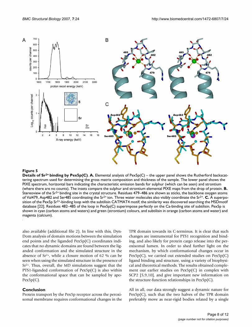
The Sr2+ ion binding siteCrystallisation of Pex5p(C) clearly demonstrated a prefer-ence for strontium – no crystals could be grown in thepresence of other divalent cations: magnesium, calcium,barium, cadmium, cobalt, copper, manganese, yttrium,iron, zinc, or nickel (data not shown). However, uponincubation in an excess of strontium in solution and sub-sequent removal of free strontium by dialysis, no specificbinding of the metal could be detected – an elementalanalysis by PIXE demonstrated the presence of a maxi-mum of 5 × 10⁻⁵ strontium ions per molecular of protein,calibrated against the X-ray fluorescence signal from sul-phur atoms present in Pex5p(C) (Figure 5A).
The strong electron density found in the crystal structurereveals that the single Sr2+ ion is coordinated by the back-bone oxygen atoms of three Pex5p(C) residues – Val479(2.52 Å), Asp482 (2.46 Å), and Ser485 (2.53 Å) – and bythree water molecules, at Sr-O distances of 2.36, 2.47, and2.81 Å (Figure 5B). Examination of other Sr2+ ion bindingproteins deposited in the PDB [20,21] does not suggestany clearly similar Sr2+ ion binding sites, although in gen-eral, a similar coordination system, forming a tetragonalbipyramid, can be observed.
Table 2: Summary of conformational differences between PTS1-liganded and apo-Pex5p(C).
DynDoma parameters Pex5p(C)/mSCP2b versus previous apo-Pex5p(C)c
Pex5p(C)/mSCP2b versus new apo-Pex5p(C)d
new apo-Pex5p(C)d versus previous apo-Pex5p(C)c
Whole protein residues 335–639 335–639 315–639Whole protein RMSD (Å2) 3.06 3.26 2.20Fixed domain residues 337–523 337–518 323–485Fixed domain RMSD (Å2) 1.40 2.32 0.71Rotation angle (°) 19.7 20.0 14.2Translation along axis (Å) -0.7 -0.1 0.1Bending residues 495–500 (end of TPR5 helix A) 523–524
(start of TPR6 helix A) 533–537 (end of TPR6 helix A)
519–520 (loop connection TPRs 5 and 6) 530–531 (middle of TPR6 helix A) 535–536 (end of TPR6 helix A) 588–601 (entire 7C-loop)
485–489 (loop connecting TPRs 4 and 5) 496–499 (end of TPR5 helix A)
% Closure motion 92.0 83.8 1.7
a As described in [11] and [47]b As described in [9], PDB indentifier 2C0L.c As described in [9], PDB indentifier 2C0M.d As described in this study, PDB identifier 2J9Q.
Table 3: Thermodynamic parameters governing PTS1 peptide binding to Pex5p(C).
peptide nb ΔH (kJ/mol) -TΔS (kJ/mol) ΔG(kJ/mol) Kd (nM)
PGNAKLa 1.00 -45.1 8.7 -36.4 664 ± 37YQSKL 1.00 -63.1 25.5 -37.6 429 ± 12
a Data from [9].b n = stoichiometry
Page 5 of 12(page number not for citation purposes)

BMC Structural Biology 2007, 7:24 http://www.biomedcentral.com/1472-6807/7/24
Page 6 of 12(page number not for citation purposes)
Comparison of the new structure and previously determined Pex5p(C) structuresFigure 2Comparison of the new structure and previously determined Pex5p(C) structures. A. Superposition of the new (blue) and old (cyan) apo-Pex5p(C) structures and the liganded structure (yellow). The arrows represent the rotation axes suggested by DynDom to describe the dynamic domain movements in the molecule. The green axis corresponds to the transi-tion between the two apo forms. The red axis relates the new apo structure to the liganded structure and the orange one the old apo structure to the liganded structure. B. Disruption of the PTS1 binding site in the apo structures, caused by the move-ment away of the N-terminal half-molecule. The new apo structure is in blue, the old apo structure in cyan, and the liganded structure in yellow. The C-terminus of SCP2 is shown in gray, entering the binding site. In the apo structures, for example, the salt bridge between Arg557 from the C-terminal half (TPR7) and Glu residues 383 and 385 from the N-terminal half (TPR2) is broken. The positions of the 4 asparagine residues that intimately interact with the PTS1 motif are also shown. C. Comparison of the FEEEN motif and the 7C loop in the new apo-Pex5p(C) conformation (blue) with the previously described apo-structure (cyan) and SCP2-bound (yellow) structure [9]. The Sr2+ ion in the new structure is indicated as a green sphere, and the FEEEN motif in red. The position of the 7C loop is also given.

BMC Structural Biology 2007, 7:24 http://www.biomedcentral.com/1472-6807/7/24
On the other hand, a search for small 3D motifs at the EBIMSDMotif server [22] indicates a large number of Ca2+ ionbinding sites found in various proteins that have a similarloop backbone conformation as seen in the Sr2+ ion bind-ing loop; the motif is called CATMAT4 in the database.One of them, that from subtilisin determined at 0.78-Åresolution [23], is shown in figure 5C for comparison.While it is possible that this loop in Pex5p(C) can binddivalent cations at low affinity, no physiological relevancefor such binding can be established at the moment. Thebinding of Sr2+ ion to the loop probably stabilises the con-formation of Pex5p(C) seen in our new crystal form.
Molecular dynamics simulations of apo-Pex5p(C)In order to elucidate the putative effect of the bound Sr2+
ion on the dynamic properties of Pex5p(C), 10-ns MDsimulations were carried out. The starting structure wasthe new crystal structure of apo-Pex5p(C) describedabove, and two runs were performed: with and withoutthe Sr2+ ion. An overall comparison of the trajectories bymeans of the RMSD between them indicates clearly thatthe simulations take different paths, depending on thepresence of Sr2+ (Figure 6A). At 1 ns, the RMSD reaches 4Å, reaching equilibrium at approximately 4.7 Å after 6 ns.
The end-point coordinates of the MD simulations wereanalysed with respect to their similarity to experimentalapo and liganded structures of Pex5p(C). A multiple struc-tural alignment was carried out using SSM [24], and theresulting RMSD values for 260 Cα atoms are shown intable 4. While the Sr2+-loaded structure remains ratherclose to the starting conformation, the conformation ofthe Sr2+-depleted structure after simulation indeed mostclosely resembles the conformation seen in the ligandedPex5p(C) structures.
The above observation was confirmed by carrying outmanual superposition of all structures (experimental apoand liganded structures plus the two simulation end-points), using residues 503–639 of the C-terminal half forsuperposing. Clearly, the removal of Sr2+ prior to simula-tion brings the simulation end-point very close to the lig-anded conformation (Figure 6B). A supplementary filecontaining a QuickTime movie of the superposition is
ITC analysis of the binding of the PTS1-containing peptide YQSKL to Pex5p(C)Figure 3ITC analysis of the binding of the PTS1-containing peptide YQSKL to Pex5p(C). The upper panels show the raw titra-tion data with ligand heats of dilution offset by 0.4 μJ/sec for clarity. The lower panels show the integrated enthalpies fit-ted to a single set of ligand binding sites in Origin 5.0.
Conformation of liganded and apo-Pex5p(C) in solutionFigure 4Conformation of liganded and apo-Pex5p(C) in solution. Shown are SRCD spectra of apo-Pex5p(C) (shown in black), Pex5p(C) in the presence of the consensus PTS1 peptide, YQSKL (red), and the SCP2-derived PTS1 peptide, PGNAKL (green).
Page 7 of 12(page number not for citation purposes)
BMC Structural Biology 2007, 7:24 http://www.biomedcentral.com/1472-6807/7/24
also available (additional file 2). In line with this, Dyn-Dom analysis of domain motions between the simulationend points and the liganded Pex5p(C) coordinates indi-cates that no dynamic domains are found between the lig-anded conformation and the simulated structure in theabsence of Sr2+, while a closure motion of 62 % can beseen when using the simulated structure in the presence ofSr2+. Thus, overall, the MD simulations suggest that thePTS1-liganded conformation of Pex5p(C) is also withinthe conformational space that can be sampled by apo-Pex5p(C).
ConclusionProtein transport by the Pex5p receptor across the peroxi-somal membrane requires conformational changes in the
TPR domain towards its C-terminus. It is clear that suchchanges are instrumental for PTS1 recognition and bind-ing, and also likely for protein cargo release into the per-oxisomal lumen. In order to shed further light on themechanism, by which conformational changes occur inPex5p(C), we carried out extended studies on Pex5p(C)ligand binding and structure, using a variety of biophysi-cal and theoretical methods. The results obtained comple-ment our earlier studies on Pex5p(C) in complex withSCP2 [5,9,10], and give important new information onthe structure-function relationships in Pex5p(C).
All in all, our data strongly suggest a dynamic nature forPex5p(C), such that the two halves of the TPR domainpreferably move as near-rigid bodies related by a single
Details of Sr2+ binding by Pex5p(C)Figure 5Details of Sr2+ binding by Pex5p(C). A. Elemental analysis of Pex5p(C) – the upper panel shows the Rutherford backscat-tering spectrum used for determining the gross matrix composition and thickness of the sample. The lower panel shows the PIXE spectrum, horizontal bars indicating the characteristic emission bands for sulphur (which can be seen) and strontium (where there are no counts). The insets compare the sulphur and strontium elemental PIXE maps from the drop of protein. B. Stereoview of the Sr2+ binding site in the crystal structure. Residues 479–486 are shown as sticks, the backbone oxygen atoms of Val479, Asp482 and Ser485 coordinating the Sr2+ ion. Three water molecules also visibly coordinate the Sr2+. C. A superpo-sition of the Pex5p Sr2+-binding loop with the subtilisin CATMAT4 motif; the similarity was discovered searching the MSDmotif database [22]. Residues 482–485 of the loop in Pex5p(C) superimpose perfectly on the Ca-binding site of subtilisin. Pex5p is shown in cyan (carbon atoms and waters) and green (strontium) colours, and subtilisin in orange (carbon atoms and water) and magenta (calcium).
Page 8 of 12(page number not for citation purposes)

BMC Structural Biology 2007, 7:24 http://www.biomedcentral.com/1472-6807/7/24
hinge around TPR5. The movement relating different apoconformations to each other is different from that relatingthe apo conformations to the liganded form. On the otherhand, unliganded Pex5p(C) closes up to adopt a "nearlyliganded" conformation in MD simulations, while theone having a single Sr2+ ion bound does not. This indi-cates a role for the Sr2+-binding loop in Pex5p(C) dynam-ics, although Sr2+ is unlikely to be a physiologicallyrelevant ligand. Rather, the results suggest that the morerigid conformation of the loop induced by the bound Sr2+
can restrict the movement between the two lobes of theTPR domain.
Based on the current and earlier data, three regions clearlyemerge as being putatively important for regulating thedynamics and ligand binding of Pex5p(C): the 7C-loop,the FEEEN motif, and the Sr2+-binding loop. The 7C-loopand the N-terminal region containing the FEEEN motifinteract in the liganded state, but not in the apo confor-mations. On the other hand, the hinge for rigid bodymovement between the N- and C-terminal halves ofPex5p(C) lies in close proximity of the Sr2+-binding siteseen in the present crystal structure. It is possible that theinteractions of these regions with other molecules in ornear the peroxisomal membrane affect the ability ofPex5p to bind and release its cargo properly.
To conclude, Pex5p(C) is able to adopt distinctly differentconformations in its apo state. These conformations donot involve changes in secondary structure content, butcan be mainly described as rigid-body movements abouta single hinge, located at the Sr2+ binding loop and the fol-lowing helix of TPR5. Furthermore, PTS1 ligand bindingalso occurs via near-rigid body movement of the twohalves of Pex5p(C), without detectable change in second-ary structure content. The origin (or hinge) for the lattermovement lies in the TPR5-6 segment. As can be seenfrom our Sr2+-liganded structure, it is possible that thebinding of a ligand to the region comprising TPR domains4–6 can affect the overall conformation of Pex5p(C), fur-ther changing its functional properties.
MethodsCrystallisation and data collectionPreparation of recombinant Pex5p(C), representing theC-terminal TPR domain of the long isoform of humanPex5p (residues 315 – 639) has been previously described[9]. Prior to crystallization, Pex5p(C) was dialysed against20 mM bis-Tris-propane, 20 mM KCl, 1 mM Tris-(2-car-boxyethyl)-phosphine hydrochloride (pH 7.0) and con-centrated to 8 mg/ml by centrifugal ultrafiltration.Crystals were obtained by mixing 1 μl protein with 1 μlreservoir solution, using the hanging drop vapour diffu-sion method at 277 K. Reservoir buffer conditions were
Molecular dynamics simulations in the presence and absence of SrFigure 6Molecular dynamics simulations in the presence and absence of Sr. A. A comparison between the Pex5p(C) structures along the simulation trajectories. The difference between the simulation in the presence and absence of the Sr2+ ion is plotted as a function of running time. B. Superpo-sition of the new apo structure (blue) with the old apo struc-ture (cyan), the SCP2-loaded structure (yellow), and the simulation end-states in the presence (green) and absence (orange) of the Sr2+ ion. Residues 503–639 were used for the superposition to highlight the orientation between the N- and C-terminal halves. The tail of SCP2 enters the binding cavity from below (yellow ribbon). Note the similarity between the liganded structure and the simulation without Sr2+. An animated presentation of the result can be found in the additional data file 2.
Page 9 of 12(page number not for citation purposes)

BMC Structural Biology 2007, 7:24 http://www.biomedcentral.com/1472-6807/7/24
optimised to 23% (w/v) PEG 3350, 100 mM Tris-HCl (pH8.75) and 10 mM SrCl2. Crystals grew within 10 days.
X-ray diffraction data were collected at the EMBL-Ham-burg synchrotron beamline X13 (DESY, Hamburg, Ger-many). Data were obtained from a single crystal capturedin a fibre loop and directly cooled to 100 K in a stream ofgaseous nitrogen. Data were processed and scaled usingXDS [25] and XDSi [26]. For cross-validation of subse-quent steps, 5% of the data were randomly selected andnot used in refinement. X-ray data collection statistics aresummarised in Table 1.
Structure solution, refinement, and analysisThe structure was solved by molecular replacement usingMOLREP [27]. No solution was found when using thewhole apo-Pex5p(C) domain as model, even though 2molecules were expected to be present in the asymmetricunit. This was a clear indication of a difference in confor-mation between apo-Pex5p(C) in the current and previ-ous crystal form [9]. The correct solution was found stepby step, by dividing the search model into two halves andsearching for the halves separately, with intermediaterefinement and rebuilding. For both molecules in theasymmetric unit, the C-terminal half (residues 466 to639) was found first, followed by the N-terminal half (res-idues 315 to 442). Refinement was carried out usingREFMAC5 [28], and model building was performed inCOOT [29]. When the built model was nearly complete,TLS parameters [30] were employed. TLS groups andparameters suggested by the TLSMD server [31] were alsoused during refinement. The refinement statistics arelisted in table 1. The final refined model, as well as thestructure factors, have been deposited at the Protein Databank and have accession code 2J9Q.
Structure comparison was carried out using DynDom[11], MSDMotif [22], SSM [24], and O [32]. Figures andmovies were made using DINO [33], PyMOL [34], POV-Ray [35], and the Morph server [36,37].
Isothermal titration microcalorimetry (ITC)Binding of a synthetic consensus PTS1 peptide YQSKL [8]to Pex5p(C) was analysed by ITC, as previously describedfor PGNAKL (representing the C-terminus of PTS1 proteinSCP2 [9]). Data were fitted using Origin 5.0 (MicroCal,Northampton, MA, USA).
Synchrotron radiation circular dichroism (SRCD)Pex5p(C) was dialysed against 10 mM potassium phos-phate (pH 7.4) and diluted to 1 mg/ml. When appropri-ate, samples were mixed with a 10-fold molar excess ofsynthetic PTS1 peptides (Sigma-Genosys, UK) YQSKL orPGNAKL. Spectra were measured on SRS beamline stationCD12 (CCLRC Daresbury, UK) [38] and data analysed asdescribed [10]. Peptide and buffer background were sub-tracted at the outset of data analysis. Secondary structureestimations from SRCD spectra were made using the SEL-CON software [16,17] and defined from crystal structuresusing XTLSSTR [18].
Proton induced X-ray emission (PIXE)Pex5p(C) at 80 μM(~3 mg/ml) was incubated with 1 mMSrCl2 for 3 hours at room temperature and subsequentlyexhaustively dialysed against 200 mM ammonium acetate(pH 7.4) and diluted to 2.1 mg/ml. 0.2 μl samples weredried on to mylar films of 2 μm thickness. Elemental anal-ysis was conducted at the National Ion Beam Centre, Uni-versity of Surrey, UK, using a 2.5-MeV proton beam of 3-μm diameter [39]. X-ray emission and Rutherford back-scattered proton spectra were measured as previouslydescribed [40], and the data were analysed using theGUPIX software [41].
Briefly, the Rutherford backscattering proton spectra wereanalysed to give the CNO composition of the matrix andits thickness, so the self absorption of the X-rays could becorrected for. All the other quantitative informationcomes from the X-ray spectra. The elemental maps weremade from scanning the proton beam in x and y direc-tions and collecting X-ray spectra at each point, setting asoftware gate around the sulphur and strontium peak.
Table 4: RMSDs (Å) for Cα atoms based on multiple structural alignment [24] of the experimental structures and the end-point structures from the MD simulations.
Pex5p(C)-apo1 (current) Pex5p(C)-apo2 Pex5p(C)-SCP2 MD with Sr MD w/o Sr
Pex5p(C)-apo1 (current) - 2.03 2.39 1.77 2.12
Pex5p(C)-apo2 - 2.31 2.28 2.61
Pex5p(C)-SCP2 - 2.46 2.09
MD with Sr - 2.66
MD w/o Sr -
Page 10 of 12(page number not for citation purposes)

BMC Structural Biology 2007, 7:24 http://www.biomedcentral.com/1472-6807/7/24
Then, the number of X-rays in the peak at that x-y positionwere sorted into a 2-D plot.
Molecular dynamics simulationsAs a starting point, the new crystal structure of Sr2+-boundapo-Pex5p(C) was used. An energy minimisation and a10-ns molecular dynamics (MD) simulation of the freeprotein in water was performed with the Gromacs 3.2.1package, both with and without a Sr2+ ion [42,43], usingthe OPLS-AA/L force field. The simulation box corre-sponded to a truncated dodecahedron, containing theprotein, single point charge water molecules [44], and Na+
ions to neutralize the system. The box size was determinedby setting the distance between the protein and the box to0.6 nm. Dummy hydrogen atoms were used. Weak tem-perature coupling (300 K) and pressure coupling (1.0 bar)were utilized [45] with coupling constants of 1.0 ps. Cou-lomb interactions were computed with the fast particlemesh Ewald summation (PME) with a grid spacing of 0.12nm and fourth-order interpolation. The LINCS algorithmwas employed to constrain bonds [46]. The centre of massmotion was removed on every step. The system wasenergy-minimised, followed by a short (10 ps) positionrestraining run for further equilibation. The 10-ns simula-tion was performed using the Brutus Cluster at the Univer-sity of Oulu, Finland.
AbbreviationsITC isothermal titration microcalorimetry
PIXE proton induced X-ray emission spectroscopy
PTS1 peroxisomal targeting signal type 1
SCP2 sterol carrier protein 2
SRCD synchrotron radiation circular dichroism spec-tropolarimetry
TPR tetratricopeptide repeat
Pex5p(C) C-terminal domain of human peroxin 5, thePTS1 receptor
RMSD root mean square deviation
Authors' contributionsWAS conceived of the study together with MW and PK,cloned and purified the protein, and carried out ITC,SRCD, crystallisation, diffraction data collection, and dataanalysis. NP and AJ carried out MD simulations, and EFGcarried out the PIXE analysis. PK solved the crystal struc-ture and carried out structure analysis. All authors partici-pated in drafting the manuscript and read and approvedthe final manuscript.
Additional material
AcknowledgementsWe thank Ashwani Verma & Nicole Schüller for assistance in the wet-lab, Dave Clarke & Alan Brown for support with SRCD experiments, and mem-bers of the Stanley family for remote data handling. SRS at CCLRC Dares-bury, UK is acknowledged for access to SRCD beamline station CD12, and EMBL/DESY for access to crystallography beamline X13. The National Ion Beam Centre, University of Surrey, UK is acknowledged for facility access. WAS is recipient of a Council for Scientific and Industrial Research (India) Visiting Fellowship. PK is an Academy Research Fellow (Academy of Fin-land). PK thanks Profs. Gunter Schneider and Ylva Lindqvist for kindly sup-porting his work.
References1. Schnell DJ, Hebert DN: Protein translocons: multifunctional
mediators of protein translocation across membranes. Cell2003, 112:491-505.
2. Holroyd C, Erdmann R: Protein translocation machineries ofperoxisomes. FEBS Lett 2001, 501:6-10.
3. Lazarow PB: Peroxisome biogenesis: advances and conun-drums. Curr Opin Cell Biol 2003, 15:489-497.
4. Carvalho AF, Costa-Rodrigues J, Correia I, Costa Pessoa J, Faria TQ,Martins CL, Fransen M, Sa-Miranda C, Azevedo JE: The N-terminalhalf of the peroxisomal cycling receptor Pex5p is a nativelyunfolded domain. J Mol Biol 2006, 356:864-875.
5. Stanley WA, Wilmanns M: Dynamic architecture of the peroxi-somal import receptor Pex5p. Biochem Biophys Acta in press.
6. D'Andrea LD, Regan L: TPR proteins: the versatile helix. TrendsBiochem Sci 2003, 28:655-662.
7. Erdmann R, Schliebs W: Peroxisomal matrix protein import:the transient pore model. Nat Rev Mol Cell Biol 2005, 6:738-742.
8. Gatto GJJ, Geisbrecht BV, Gould SJ, Berg JM: Peroxisomal target-ing signal-1 recognition by the TPR domains of human PEX5.Nat Struct Biol 2000, 7:1091-1095.
9. Stanley WA, Filipp FV, Kursula P, Schuller N, Erdmann R, Schliebs W,Sattler M, Wilmanns M: Recognition of a functional peroxisometype 1 target by the dynamic import receptor pex5p. Mol Cell2006, 24:653-663.
10. Stanley WA, Sokolova A, Brown A, Clarke DT, Wilmanns M, SvergunDI: Synergistic use of synchrotron radiation techniques forbiological samples in solution: a case study on protein-ligandrecognition by the peroxisomal import receptor Pex5p. JSynchrotron Radiat 2004, 11:490-496.
11. Hayward S, Berendsen HJ: Systematic analysis of domainmotions in proteins from conformational change: new
Additional file 1Morphing movie between the two experimentally determined conforma-tions of apo-Pex5p(C). An animation of the difference between the two apo conformations of apo-Pex5p(C). The molecule is coloured from the N-terminus (blue) to the C-terminus (red). The predicted motion has a slid-ing nature instead of being a closing motion.Click here for file[http://www.biomedcentral.com/content/supplementary/1472-6807-7-24-S1.gif]
Additional file 2A superposition of the experimental and simulated structures of Pex5p(C). This is an animated presentation of figure 6B. See legend for figure 6B for additional details.Click here for file[http://www.biomedcentral.com/content/supplementary/1472-6807-7-24-S2.mov]
Page 11 of 12(page number not for citation purposes)

BMC Structural Biology 2007, 7:24 http://www.biomedcentral.com/1472-6807/7/24
Publish with BioMed Central and every scientist can read your work free of charge
"BioMed Central will be the most significant development for disseminating the results of biomedical research in our lifetime."
Sir Paul Nurse, Cancer Research UK
Your research papers will be:
available free of charge to the entire biomedical community
peer reviewed and published immediately upon acceptance
cited in PubMed and archived on PubMed Central
yours — you keep the copyright
Submit your manuscript here:http://www.biomedcentral.com/info/publishing_adv.asp
BioMedcentral
results on citrate synthase and T4 lysozyme. Proteins 1998,30:144-154.
12. Shimozawa N, Zhang Z, Suzuki Y, Imamura A, Tsukamoto T, OsumiT, Fujiki Y, Orii T, Barth PG, Wanders RJ, Kondo N: Functional het-erogeneity of C-terminal peroxisome targeting signal 1 inPEX5-defective patients. Biochem Biophys Res Commun 1999,262:504-508.
13. Urquhart AJ, Kennedy D, Gould SJ, Crane DI: Interaction ofPex5p, the type 1 peroxisome targeting signal receptor, withthe peroxisomal membrane proteins Pex14p and Pex13p. JBiol Chem 2000, 275:4127-4136.
14. Gatto GJJ, Maynard EL, Guerrerio AL, Geisbrecht BV, Gould SJ, BergJM: Correlating structure and affinity for PEX5:PTS1 com-plexes. Biochemistry 2003, 42:1660-1666.
15. Maynard EL, Gatto GJJ, Berg JM: Pex5p binding affinities forcanonical and noncanonical PTS1 peptides. Proteins 2004,55:856-861.
16. Clarke DT, Jones GR: Extended circular dichroism measure-ments using synchrotron radiation show that the assemblyof clathrin coats requires no change in secondary structure.Biochemistry 1999, 38:10457-10462.
17. Sreerama N, Woody RW: A self-consistent method for theanalysis of protein secondary structure from circular dichr-oism. Anal Biochem 1993, 209:32-44.
18. King SM, Johnson WC: Assigning secondary structure fromprotein coordinate data. Proteins 1999, 35:313-320.
19. Woody RW: Theory of circular dichroism of proteins. In Circu-lar Dichroism and Conformational Analysis of Biomolecules Edited by: Fas-man GD. New York: Plenum; 1996:109-157.
20. Metalloprotein database and browser [http://metallo.scripps.edu]
21. Castagnetto JM, Hennessy SW, Roberts VA, Getzoff ED, Tainer JA,Pique ME: MDB: the Metalloprotein Database and Browser atThe Scripps Research Institute. Nucleic Acids Res 2002,30:379-382.
22. Golovin A, Oldfield TJ, Tate JG, Velankar S, Barton GJ, Boutselakis H,Dimitropoulos D, Fillon J, Hussain A, Ionides JM, John M, Keller PA,Krissinel E, McNeil P, Naim A, Newman R, Pajon A, Pineda J, RachediA, Copeland J, Sitnov A, Sobhany S, Suarez-Uruena A, SwaminathanGJ, Tagari M, Tromm S, Vranken W, Henrick K: E-MSD: an inte-grated data resource for bioinformatics. Nucleic Acids Res 2004,32:D211-6.
23. Kuhn P, Knapp M, Soltis SM, Ganshaw G, Thoene M, Bott R: The0.78 A structure of a serine protease: Bacillus lentus subtili-sin. Biochemistry 1998, 37:13446-13452.
24. Krissinel E, Henrick K: Secondary-structure matching (SSM), anew tool for fast protein structure alignment in three dimen-sions. Acta Crystallogr D Biol Crystallogr 2004, 60:2256-2268.
25. Kabsch W: Automatic processing of rotation diffraction datafrom crystals of initially unknown symmetry and cell con-stants. J Appl Cryst 1993, 26:795-800.
26. Kursula P: XDSi – a graphical interface for the data processingprogram XDS. J Appl Cryst 2004, 37:347-348.
27. Vagin A, Teplyakov A: MOLREP: an automated program formolecular replacement. J Appl Cryst 1997, 30:1022-1025.
28. Murshudov GN, Vagin AA, Dodson EJ: Refinement of macromo-lecular structures by the maximum-likelihood method. ActaCrystallogr D Biol Crystallogr 1997, 53:240-255.
29. Emsley P, Cowtan K: Coot: model-building tools for moleculargraphics. Acta Crystallogr D Biol Crystallogr 2004, 60:2126-2132.
30. Winn MD, Isupov MN, Murshudov GN: Use of TLS parametersto model anisotropic displacements in macromolecularrefinement. Acta Crystallogr D Biol Crystallogr 2001, 57:122-133.
31. Painter J, Merritt EA: Optimal description of a protein structurein terms of multiple groups undergoing TLS motion. ActaCrystallogr D Biol Crystallogr 2006, 62:439-450.
32. Jones TA, Zou JY, Cowan SW, Kjeldgaard M: Improved methodsfor building protein models in electron density maps and thelocation of errors in these models. Acta Crystallogr A 1991,47:110-119.
33. DINO: Visualizing structural biology [http://www.dino3d.org]34. The PyMOL molecular graphics system [http://
www.pymol.org]35. POV-Ray – the persistence of vision raytracer [http://
www.povray.org]36. The Yale Morph server [http://molmovdb.org/morph]
37. Krebs WG, Gerstein M: The morph server: a standardized sys-tem for analyzing and visualizing macromolecular motionsin a database framework. Nucleic Acids Res 2000, 28:1665-1675.
38. Clarke DT, Jones G: CD12: a new high-flux beamline for ultra-violet and vacuum-ultraviolet circular dichroism on the SRS,Daresbury. J Synchrotron Radiat 2004, 11:142-149.
39. Grime G, Dawson M, Marsh M, MacArthur IC, Watt F: The Oxfordsubmicron nuclear microscopy facility. Nucl Instr Methods PhysRes 1991, B54:52-63.
40. Garman EF, Grime GW: Elemental analysis of proteins bymicroPIXE. Prog Biophys Mol Biol 2005, 89:173-205.
41. Johansson S, Campbell JL, Malmqvist QG: Particle induced X-ray emis-sion spectrometry New York: John Wiley & Sons; 1995.
42. Berendsen HJC, van der Spoel D, van Drunen R: A message-pass-ing parallel molecular dynamics implementation. Comp PhysComm 1995, 91:43-56.
43. Lindahl E, Hess B, van der Spoel D: GROMACS 3.0: A package formolecular simulation and trajectory analysis. J Mol Mod 2001,1:306-317.
44. Berendsen HJC, Postma JPM, van Gunsteren WF, Hermans J: Inter-action models for water in relation to protein hydration. InIntermolecular forces Edited by: Pullman B. Dordrecht: Reidel;1981:331-342.
45. Berendsen HJC, Postma JPM, DiNola A, van Gunsteren WF, Haak JR:Molecular dynamics with coupling to an external bath. J ChemPhys 1984, 181:3684-3690.
46. Hess B, Bekker H, Berendsen HJC, Fraaije JGEM: LINCS: a linearconstraint solver for molecular simulations. J Comp Chem1997, 18:1463-1473.
47. DynDom – Protein domain motion [http://www.cmp.uea.ac.uk/dyndom/dyndomMain.do]
Page 12 of 12(page number not for citation purposes)
http://www.ncbi.nlm.nih.gov/entrez/query.fcgi?cmd=Retrieve&db=PubMed&dopt=Abstract&list_uids=9489922
http://www.ncbi.nlm.nih.gov/entrez/query.fcgi?cmd=Retrieve&db=PubMed&dopt=Abstract&list_uids=8465960
http://www.ncbi.nlm.nih.gov/entrez/query.fcgi?cmd=Retrieve&db=PubMed&dopt=Abstract&list_uids=8465960
http://www.ncbi.nlm.nih.gov/entrez/query.fcgi?cmd=Retrieve&db=PubMed&dopt=Abstract&list_uids=8465960
http://www.ncbi.nlm.nih.gov/entrez/query.fcgi?cmd=Retrieve&db=PubMed&dopt=Abstract&list_uids=9753430
http://www.ncbi.nlm.nih.gov/entrez/query.fcgi?cmd=Retrieve&db=PubMed&dopt=Abstract&list_uids=9753430
http://www.ncbi.nlm.nih.gov/entrez/query.fcgi?cmd=Retrieve&db=PubMed&dopt=Abstract&list_uids=9753430
http://www.ncbi.nlm.nih.gov/entrez/query.fcgi?cmd=Retrieve&db=PubMed&dopt=Abstract&list_uids=2025413
http://www.ncbi.nlm.nih.gov/entrez/query.fcgi?cmd=Retrieve&db=PubMed&dopt=Abstract&list_uids=2025413