A prairie dog animal model of systemic orthopoxvirus disease using West African and Congo Basin...
11
A prairie dog animal model of systemic orthopoxvirus disease using West African and Congo Basin strains of monkeypox virus Christina L. Hutson, 1 Victoria A. Olson, 1 Darin S. Carroll, 1 Jason A. Abel, 1 Christine M. Hughes, 1 Zachary H. Braden, 1 Sonja Weiss, 1 Joshua Self, 1 Jorge E. Osorio, 2 Paul N. Hudson, 1 Michael Dillon, 3 Kevin L. Karem, 1 Inger K. Damon 1 and Russell L. Regnery 1 Correspondence Christina L. Hutson [email protected] 1 Centers for Disease Control and Prevention, MS G-43, 1600 Clifton Road NE, Atlanta, GA 30333, USA 2 Department of Pathobiological Sciences, School of Veterinary Medicine, University of Wisconsin, Madison, WI 53706, USA 3 Centers for Disease Control and Prevention, MS G-12, 1600 Clifton Road NE, Atlanta, GA 30333, USA Received 26 June 2008 Accepted 8 October 2008 Multiple monkeypox virus (MPXV) animal models have been discussed in previous studies, but no small animal models, nor most non-human primate models, demonstrated the protracted asymptomatic incubation phase seen in systemic human orthopoxvirus illness. Herein, we characterize a black-tailed prairie dog (PD) (Cynomys ludovicianus) model of infection, via intranasal and intradermal exposures, with the two MPXV clades. Daily observations of the animals were made (food consumption, general symptoms, disease presentation), while weights and virus evaluations (ocular, nasal, oropharyngeal, faeces, blood) were obtained/made every third day. Generalized rash became apparent 9–12 days post-infection for all animals. Individual animals demonstrated a range of symptoms consistent with human monkeypox disease. Measurable viraemias and excretas were similar for both clade-representative strains and persisted until at least day 21. Greater morbidity was observed in Congo Basin strain-challenged animals and mortality was observed only in the Congo Basin strain-challenged animals. The PD model is valuable for the study of strain-dependent differences in MPXV. Additionally, the model closely mimics human systemic orthopoxvirus disease and may serve as a valuable non- human surrogate for investigations of antivirals and next generation orthopoxvirus vaccines. INTRODUCTION Monkeypox is a zoonotic disease causing febrile rash illness in humans with a presentation similar to smallpox. Since eradication of variola virus (the causative agent of smallpox), monkey pox virus (MPXV) has emerged as the most significant public health threat in the genus Orthopoxvirus. As evidenced by the USA 2003 outbreak, MPXV is considered potentially problematic beyond its endemic African range. Variola virus was an exclusively human pathogen, the viral-specific determinants of host range are not understood, and the human disease lacks a representative surrogate animal model. The related ortho- poxvirus MPXV is a zoonotic agent that causes febrile rash disease with clinical presentation similar to smallpox (Breman & Henderson, 2002; Jezek et al., 1987) and has been used for the study of systemic orthopoxvirus infections. In humans, MPXV and variola virus have similar asymptomatic incubation periods ranging from 7– 17 days followed by development of generalized rash. However, smallpox had a much higher secondary attack rate (25–96 %) than monkeypox (5–11 %) (Arita & Gromyko, 1982; Fine et al., 1988). Previous studies defined two distinct MPXV clades, West African and Congo Basin, with unique disease manifestations (Chen et al., 2005; Likos et al., 2005). Human disease associated with West African MPXV infection is less severe and associated with less human-to-human transmission compared to Congo Basin infection (Breman et al., 1980; Foster et al., 1972). In the Democratic Republic of Congo (Congo Basin), monkeypox has a reported case fatality rate of ~10–13 % Supplementary figures are available with the online version of this paper. The findings and conclusions in this report are those of the author(s) and do not necessarily represent the official position of the Centers for Disease Control and Prevention. Journal of General Virology (2009), 90, 323–333 DOI 10.1099/vir.0.005108-0 005108 Printed in Great Britain 323
-
Upload
independent -
Category
Documents
-
view
3 -
download
0
Transcript of A prairie dog animal model of systemic orthopoxvirus disease using West African and Congo Basin...

A prairie dog animal model of systemicorthopoxvirus disease using West African andCongo Basin strains of monkeypox virus
Christina L. Hutson,1 Victoria A. Olson,1 Darin S. Carroll,1 Jason A. Abel,1
Christine M. Hughes,1 Zachary H. Braden,1 Sonja Weiss,1 Joshua Self,1
Jorge E. Osorio,2 Paul N. Hudson,1 Michael Dillon,3 Kevin L. Karem,1
Inger K. Damon1 and Russell L. Regnery1
Correspondence
Christina L. Hutson
1Centers for Disease Control and Prevention, MS G-43, 1600 Clifton Road NE, Atlanta, GA 30333,USA
2Department of Pathobiological Sciences, School of Veterinary Medicine, University of Wisconsin,Madison, WI 53706, USA
3Centers for Disease Control and Prevention, MS G-12, 1600 Clifton Road NE, Atlanta, GA 30333,USA
Received 26 June 2008
Accepted 8 October 2008
Multiple monkeypox virus (MPXV) animal models have been discussed in previous studies, but no
small animal models, nor most non-human primate models, demonstrated the protracted
asymptomatic incubation phase seen in systemic human orthopoxvirus illness. Herein, we
characterize a black-tailed prairie dog (PD) (Cynomys ludovicianus) model of infection, via
intranasal and intradermal exposures, with the two MPXV clades. Daily observations of the animals
were made (food consumption, general symptoms, disease presentation), while weights and virus
evaluations (ocular, nasal, oropharyngeal, faeces, blood) were obtained/made every third day.
Generalized rash became apparent 9–12 days post-infection for all animals. Individual animals
demonstrated a range of symptoms consistent with human monkeypox disease. Measurable
viraemias and excretas were similar for both clade-representative strains and persisted until at
least day 21. Greater morbidity was observed in Congo Basin strain-challenged animals and
mortality was observed only in the Congo Basin strain-challenged animals. The PD model is
valuable for the study of strain-dependent differences in MPXV. Additionally, the
model closely mimics human systemic orthopoxvirus disease and may serve as a valuable non-
human surrogate for investigations of antivirals and next generation orthopoxvirus vaccines.
INTRODUCTION
Monkeypox is a zoonotic disease causing febrile rash illnessin humans with a presentation similar to smallpox. Sinceeradication of variola virus (the causative agent ofsmallpox), monkey pox virus (MPXV) has emerged asthe most significant public health threat in the genusOrthopoxvirus. As evidenced by the USA 2003 outbreak,MPXV is considered potentially problematic beyond itsendemic African range. Variola virus was an exclusivelyhuman pathogen, the viral-specific determinants of hostrange are not understood, and the human disease lacks arepresentative surrogate animal model. The related ortho-
poxvirus MPXV is a zoonotic agent that causes febrile rashdisease with clinical presentation similar to smallpox(Breman & Henderson, 2002; Jezek et al., 1987) and hasbeen used for the study of systemic orthopoxvirusinfections. In humans, MPXV and variola virus havesimilar asymptomatic incubation periods ranging from 7–17 days followed by development of generalized rash.However, smallpox had a much higher secondary attackrate (25–96 %) than monkeypox (5–11 %) (Arita &Gromyko, 1982; Fine et al., 1988). Previous studies definedtwo distinct MPXV clades, West African and Congo Basin,with unique disease manifestations (Chen et al., 2005;Likos et al., 2005). Human disease associated with WestAfrican MPXV infection is less severe and associated withless human-to-human transmission compared to CongoBasin infection (Breman et al., 1980; Foster et al., 1972). Inthe Democratic Republic of Congo (Congo Basin),monkeypox has a reported case fatality rate of ~10–13 %
Supplementary figures are available with the online version of this paper.
The findings and conclusions in this report are those of the author(s) anddo not necessarily represent the official position of the Centers forDisease Control and Prevention.
Journal of General Virology (2009), 90, 323–333 DOI 10.1099/vir.0.005108-0
005108 Printed in Great Britain 323

(Arita et al., 1985; Breman et al., 1980; Meyer et al., 2002),compared with up to 40 % for smallpox (Breman &Henderson, 2002). Human monkeypox had never beenreported outside of Africa until 2003, when an outbreakoccurred in the USA resulting from human contact withinfected prairie dogs (PDs) which were co-housed withimported African rodents (Hutson et al., 2007; Reed et al.,2004). This outbreak demonstrated the potential forMPXV importation and, together with observations ofongoing human monkeypox in Africa (Boumandouki et al.,2007; Hutin et al., 2001; Learned et al., 2005; Meyer et al.,2002; Mukinda et al., 1997; Mwanbal et al., 1997; Rimoinet al., 2007), further emphasized the importance of a morecomplete understanding of this serious human pathogen.
In vivo animal model comparisons of viruses from thesetwo clades will provide valuable tools with which toevaluate host–pathogen factors responsible for pathogenicdifferences. Multiple animal species have been experiment-ally infected with MPXV (Marennikova & Seluhina, 1976;Shchelukhina & Marennikova, 1975; Tesh et al., 2004;Zaucha et al., 2001); however, most of these models havelittle in common with the human disease progression,presentation or mortality from either monkeypox orsmallpox. Based on observations of PDs infected duringthe USA outbreak (Guarner et al., 2004; Hutson et al.,2007), and previous experimentally infected PDs (Xiaoet al., 2005), we believe these animals will serve as a relevantmodel for human monkeypox. In the Xiao et al. study, onlyWest African MPXV was used and it was administered viaan intranasal and an intraperitoneal route. Here, we showthat PDs are a valuable model for study of strain-dependent differences in MPXV virulence, something thathas not been previously studied in these animals.Additionally, we infected with an intranasal as well as anintradermal route, both of which are plausible naturalroutes of infection. Our described animal model mimicshuman monkeypox disease progression and presentationmore closely than previous models, and thus will serve as avaluable surrogate for future investigations of humansystemic orthopoxvirus diseases, including smallpox.
METHODS
Animal maintenance. Wild-caught, juvenile black-tailed PDs
(Cynomys ludovicianus) were obtained from Western Kansas. At timeof infection, animals were approximately 2 years old. During
experimental infections, animals were housed in large rat cages withaerosol filter tops. Cages were kept in a Duo-Flow biosafety cabinet in
a BSL-3 animal room. Animals were cared for in accordance withCDC Institutional Animal Care and Use Committee (IACUC) under
an approved protocol (1431REGPRAC-A1). Animal handling wasperformed using biosafety level 3+ personal protective equipment
(PPE). In addition to PD chow and hay, animals were provided withmonkey biscuits for added dietary enrichment.
Viruses. The West African MPXV strain, MPXV-2003-044, wasisolated during the 2003 USA outbreak, and has previously been fully
sequenced (Likos et al., 2005; Reed et al., 2004). The Congo BasinMPXV strain, MPXV-2003-358, was collected from a 2003 outbreak
of monkeypox in the Republic of Congo (ROC) and has been fullysequenced (Likos et al., 2005). Both viruses had undergone twopassages in African green monkey kidney cells (BSC-40) prior to seedpool production. The titres for both virus inocula were retitrated afterappropriate dilutions were made for PD inoculation.
Animal inoculation. Infection of PDs with the two MPXV strainswas done on separate occasions. The average starting weight foranimals challenged with West African MPXV was 714.1 g (range 535–842 g), and the average for Congo Basin MPXV-challenged animalswas 807.8 g (range 640–988 g). Animals were infected by either anintranasal (IN) or intradermal via scarification (ID-SC) route ofinoculation while under anaesthesia. For both strains, groups of fouranimals were inoculated with 104.5 plaque-forming units (p.f.u.) in atotal volume of 10 ml IN (5 ml in each nostril). Similarly, for bothstrains, four animals were inoculated with 104.5 p.f.u. in a totalvolume of 10 ml by ID-SC. For ID-SC animals, a small area on theright flank of the animals was shaved. Ten microlitres of virussolution was placed on this shaved area and a 28 gauge needle wasused to gently probe to the superficial layers for ten replicates.Additionally for each strain, four animals were mock-infected withPBS.
Observations and sampling. Daily visual observations of theanimals were made and recorded throughout the study. Lesion count,temperature, weight and tissue/excreta samples were collected everythird day post-infection (p.i.) without anaesthesia. For both MPXVstrain challenge studies, the relatively less fur-covered areas of theface, inner hind legs and genitalia were used to count lesions. Bloodwas collected from the saphenous vein in EDTA-coated tubes. Sterileindividual Dacron swabs were used to collect excreta and were storedfrozen without diluent. Swabs, faeces and blood were processed andprepared for DNA analysis and virus isolation (see below).
Necropsy and tissue specimen collection. If death did not occuras a result of infection, at 35 days (West African MPXV) and 49 days(Congo Basin MPXV) animals were humanely euthanized andnecropsied. Euthanasia dates were determined based on estimatedtime to cessation of viable virus [RT-PCR values greater than cyclethreshold (Ct )534] in excreta. Necropsies on all animals wereperformed according to IACUC standards in a BSL-3 laboratory andutilizing full BSL-3 PPE. Instruments were cleaned and decontami-nated with 3 % Amphyll and 10 % Clorox bleach between collectionsof each tissue. Tissues were frozen at 270 uC prior to furtherprocessing. Oral and ocular swabs were collected with sterileindividual Dacron swabs and stored frozen without diluent. Serumwas separated from whole blood and processed for serology (seebelow). Tissues and swabs were subsequently processed and furtherprepared for DNA analysis and virus isolation (see below).
Sample preparation. Sample processing was performed under BSL-3 conditions. For DNA analysis of blood, water was added if necessaryto bring total volume to 200 ml. The sample was incubated at 55 uCfor 1 h to inactivate virus. The EZ-1 DNA extraction robot (Qiagen)was used for genomic DNA extraction of all blood samples. For swabscollected during the West African MPXV challenge, 400 ml PBS wasadded to the swab. The swab extraction tube systems (SETS) (Roche)protocol was used to recover sample from a swab. Genomic DNA wasprepared using either the AquaPure DNA isolation kit (Bio-Rad), orthe BioRobot MDx workstation (Qiagen). For swabs collected duringthe Congo Basin MPXV challenge, 1 ml PBS was added to the swaband vortexed. Genomic DNA was then prepared using the AquaPureDNA isolation kit. For both virus strains, remaining swab lysate wasused for virus isolation (see below). Tissue and faecal samples wereplaced in disposable dounce homogenizers. PBS (1 ml) was added toeach weighed tissue sample in the 50 ml sterile tissue grinder andground thoroughly to create a slurry. Genomic DNA was prepared
C. L. Hutson and others
324 Journal of General Virology 90
from a slurry aliquot (100 ml) with BioRobot MDx workstation or
AquaPure DNA isolation kit and the remaining samples were used for
virus isolation (see below).
Real-time PCR analysis. Samples were tested by real-time PCR
using forward and reverse primers and probe complementary to the
conserved orthopoxvirus E9L (DNA polymerase) gene (Li et al.,
2006). A representative sample from each animal was confirmed for
MPXV DNA using forward and reverse primers and probe specific to
the MPXV B6R gene (Li et al., 2006). MPXV DNA (10 fg–1 ng) was
used as positive control for both tests.
Virus–tissue infectivity. Previous analyses demonstrated that real-
time PCR detection of MPXV DNA was significantly more sensitive
than detection of p.f.u. (Hutson et al., 2007). Therefore, specimens
were first tested for the presence of orthopoxvirus DNA by PCR and,
if positive, were subsequently evaluated for viable virus by tissue
culture propagation. Each swab, faeces or tissue sample was titrated
using tenfold dilutions of swab eluent or tissue slurry on BSC-40 cell
monolayers, incubated at 36 uC and 6 % CO2 for 72 h, and
subsequently stained with crystal violet and formalin to reveal
plaques.
Serological analysis. A modified version of the ELISA was used for
analysis of anti-orthopoxvirus immunoglobulin types A and G
(Hutson et al., 2007). Microtitreplates (Immulon II; Dynatech) were
coated with 0.01 mg per well crude vaccinia virus (Dryvax grown in
BSC-40 cells) in carbonate buffer on one half of the plate and an equal
volume of BSC-40 cell lysate diluted in carbonate buffer on the other
half and incubated overnight at 4 uC. After inactivation with 10 %
formalin (buffered) for 10 min at room temperature, plates were
blocked for 30 min at room temperature with assay diluent [PBS,
0.01 M, pH 7.4 (Gibco)+0.05 % Tween-20, 5 % dried skim milk, 2 %
normal goat serum and 2 % BSA] followed by three PBST (0.05 %
Tween-20) washes. PD sera (diluted 1 : 100 in assay diluent) was
added to both halves of the plates and incubated for 1 h at 37 uC.
Plates were washed, and a 1 : 30 000 dilution (in assay diluent) of
ImmunoPure A/G conjugate (Pierce) was added and incubated for
1 h at 37 uC. Plates were washed, and peroxidase substrate
(Kirkegaard & Perry Laboratories) was added and allowed to develop
for 5–15 min. After development, stop solution (Kirkegaard & Perry
Laboratories) was added, and absorbance was read on a spectropho-
tometer at 450 nm. Values reported represent the average of duplicate
wells for each sample. Both positive and negative human anti-vaccinia
sera were used as assay controls. The BSC-40 cell lysate half of each
plate was used to generate a cut-off value (COV) for each plate by
averaging all the values of the BSC-40 lysate half and adding two SD.
Specimens were considered positive if the test sample’s value was
above the COV.
Statistical analyses. As data were not normally distributed,
nonparametric statistical analyses were used (Lehmann, 1975).
Levels of viral DNA, viable virus, anti-orthopoxvirus antibodies,
and daily observations (weight, temperature, lesion counts) were
compared between strains and routes of infection using the Wilcoxon
rank-sum test. In order to evaluate differences in temperature, day
zero temperature and the highest temperature recorded thereafter
were used to determine the per cent increase in temperature for each
PD. To evaluate weight differences, weights observed at day zero were
used as the baseline and the lowest weight measured thereafter was
used to determine per cent weight loss in each animal. Comparisons
between the first and last day of DNA and viable virus were also
evaluated between strains and routes using the Wilcoxon rank-sum
test. ID-SC and IN groups were combined to increase numbers when
comparing strains, and strains were combined to increase numbers
when comparing route for DNA and viable virus. This was also done
when comparing the number of lesions between strains. A P-value,0.05 was considered significant.
RESULTS
Clinical observations (Table 1)
General observations. For all animals in all viral-infectedgroups, disseminated or generalized lesions emerged in acentrifugal pattern; lesions were first noted on the head orventral surfaces of extremities, followed by lesiondevelopment on the trunk. Lesions observed on ventralsurface of extremities and on the trunk developed throughthe typical macular, vesicular, pustular evolution beforedrying and desquamation. In slight contrast, facial lesionsevolved from macular to vesicular, but did not reach thepustular stage. The Congo Basin strain-infected PDs had asignificantly higher per cent increase in temperaturecompared to the West African strain-infected PDs(P50.01; per cent mean increase West African: 2.88 %;per cent mean increase Congo Basin: 5.98 %). There wasalso a greater trend in weight loss in the Congo Basinstrain-infected PDs (mean percentage: Congo Basin10.53 %; West African 6.57 %); however, this trend wasnot considered significant (P50.1089)
West African MPXV challenge. The animals inoculatedwith the West African strain of MPXV via IN and ID-SCroutes had similar disease presentations with generalizedlesions appearing between days 9 and 12 (Supplementary Fig.S1, available in JGV Online). All four animals inoculated viathe ID-SC route developed a scarification site (SS) lesionbetween days 6 and 9. Until the development of this primarylesion for the ID-SC group, or the disseminated lesions forthe IN group, no observable symptoms were noted. Lesioncount was slightly lower for the IN inoculated animalscompared with the ID-SC animals, and in contrast to whatwas observed with the ID-SC inoculation, only one INanimal developed more than one lesion on the inner leg areaor genitalia. Similarly seen in both groups was thedevelopment of a slightly haemorrhagic lesion on theventral surface of the legs on two of the ID-SC animals andone IN animal. Clinical symptoms were comparable betweenthe two West African-inoculated groups. All of the WestAfrican MPXV-inoculated animals recovered from diseasewith secondary lesions beginning to resolve between days 18and 28. At necropsy (day 35), two ID-SC PDs had whatappeared to be secondary bacterial infections at the primarysite of inoculation, while the other two PDs’ primary lesionsites remained scabbed.
Congo Basin MPXV challenge. As was seen in the WestAfrican MPXV-infected animals, the animals inoculatedwith the Congo Basin strain of MPXV had similar diseasepresentations with generalized lesions appearing betweendays 9 and 12 (Supplementary Fig. S2, available in JGVOnline). All four animals inoculated via the ID-SC routedeveloped a SS lesion on day 6, also comparable to the
Animal model of orthopoxvirus disease using MPXV
http://vir.sgmjournals.org 325
West African ID-SC animals. Until the development of thisprimary lesion for the ID-SC group, or the disseminatedlesions for the IN group, no observable symptoms werenoted. Similarly to West African animals, lesion count wasslightly less for the Congo Basin IN animals compared withthe ID-SC animals, with the exception of one ID-SC animalwhich, unlike the West African animals, developed a SSlesion, but died on day 11 before disseminated lesionsoccurred. Another ID-SC animal was found dead on day 12and one IN animal was found dead on day 13.Additionally, unlike the West African IN animals, allCongo Basin MPXV IN-infected animals developed lesionson the face and inner legs. Clinical symptoms werecomparable between the two Congo Basin-inoculatedgroups. All of the surviving Congo Basin MPXV infectedanimals’ secondary lesions began to resolve between days18 and 28, similar to West African infected animals. OneID-SC animal developed an apparent secondary bacterialinfection at the SS (~ day 15), lasting until approximatelyday 35 and crusting over by day 42. The other animal thatsurvived ID-SC inoculation never developed a bacterialinfection at the site of scarification. For this animal, theprimary lesion that developed began to resolve around day15, with the scab completely falling off by day 30.
Molecular and virologic findings (Table 1)
West African MPXV challenge. MPXV DNA wasdetectable in all samples (ocular, oral, nasal, faecal,
blood) at days 9–15 from the West African MPXV ID-SCinoculated group as well as the IN-inoculated group (withthe exception of faeces). Viral DNA was initially detected inblood, followed by oral and nasal excreta, then in ocularand faecal excreta (Fig. 1a) in the ID-SC-inoculatedanimals. Similar results were seen for IN-inoculatedanimals, with the exception of DNA initially beingdetected in oral secretions (Fig. 1b). Viable virologicanalyses provided similar kinetics for each group, but virusisolation was not attempted from blood (Fig. 1c and d).Peak MPXV DNA was observed from day 6 to 15, with oralsamples yielding the highest levels for both groups (Fig. 2a).For both inoculation routes, viable virus was detectablebeginning on day 6 for oral and nasal swabs (with theexception of one IN animal which had detectable viablevirus from the oral sample on day 3), day 9 for faecal andday 12 for ocular swabs. Viral shedding continued fromthese sites until days 15–21 (Fig. 1c and d). Peak MPXVviable virus was observed from day 9 to 18, with the oralsample having the highest load for both groups (Fig. 2b).Blood was collected from all West African MPXV-infectedanimals on day 35, and all had anti-orthopoxvirusantibodies (Fig. 3a). West African ID-SC-infected animalshad significantly higher anti-orthopoxvirus antibodies thanWest African IN animals (P50.03). At time of necropsy, allfour animals in the ID-SC group had MPXV DNA detectedin the lesion sample. In addition, three eyelid samples, oneskin sample, one oral swab, one lymph node and onegonad were MPXV DNA-positive. The four IN-inoculated
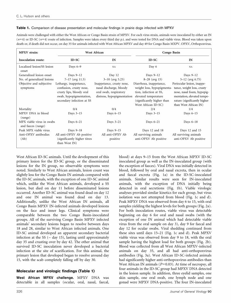
Table 1. Comparison of disease presentation and molecular findings in prairie dogs infected with MPXV
Animals were challenged with either the West African or Congo Basin strain of MPXV. For each virus strain, animals were inoculated by either an IN
(n54) or ID-SC (n54) route of infection. Samples were taken every third day p.i. and were tested for DNA and viable virus. Blood was taken upon
death or, if death did not occur, on day 35 for animals infected with West African MPXV and day 49 for Congo Basin MXPV. OPXV, Orthopoxvirus.
MPXV strain: West African Congo Basin
Inoculation route: ID-SC IN ID-SC IN
Localized lesion/SS lesion
onset
Days 6–9 NA Day 6 NA
Generalized lesion onset Days 9–12 Day 12 Days 9–12 Days 9–12
No. of generalized lesions 7–17 (avg 11.5) 3–10 (avg 5.25) 8–28 (avg 15) 5–12 (avg 6.75)
Objective and subjective
symptoms
Lethargy, inappetance,
confusion, crusty nose,
crusty lips, bloody oral
swab, hypopigmentation,
secondary infection at SS
Inappetance, crusty nose,
nasal discharge, bloody
oral swab, respiratory
distress, hypopigmentation
Diarrhoea, inappetance,
weight loss, hypopigmenta-
tion, infection at SS,
elevated temperature
(significantly higher than
West African ID-SC)
Periocular lesion, inappe-
tance, weight loss, crusty
nose, nasal foam, hypopig-
mentation, elevated tempe-
rature (significantly higher
than West African IN)
Mortality 0/4 0/4 2/4 1/4
MPXV DNA in blood
(range)
Days 3–15 Days 6–15 Days 3–15 Days 6–15
MPX viable virus in swabs
and faeces (range)
Days 6–21 Days 3–21 Days 6–21 Days 6–18
Peak MPX viable virus Days 9–18 Days 9–15 Days 12 and 18 Days 12 and 15
Anti-OPXV antibodies
(Ab)
All anti-OPXV Ab positive
(significantly higher titres
than West IN)
All anti-OPXV Ab
positive
All surviving animals
anti-OPXV Ab positive
All surviving animals
anti-OPXV Ab positive
C. L. Hutson and others
326 Journal of General Virology 90
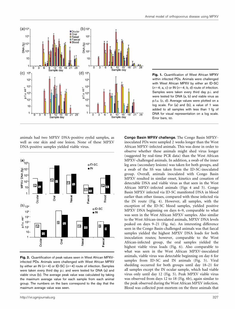
animals had two MPXV DNA-positive eyelid samples, aswell as one skin and one lesion. None of these MPXVDNA-positive samples yielded viable virus.
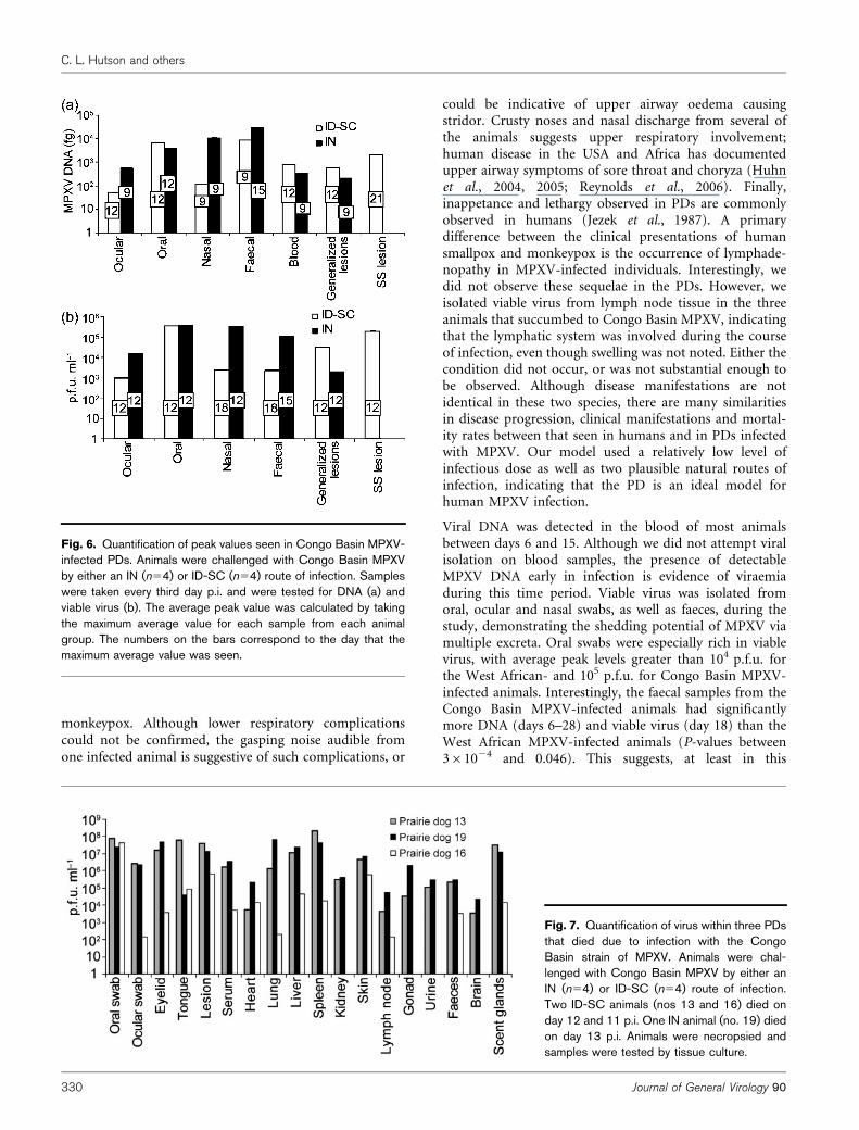
Congo Basin MPXV challenge. The Congo Basin MPXV-inoculated PDs were sampled 2 weeks longer than the WestAfrican MPXV-infected animals. This was done in order toobserve whether these animals might shed virus longer(suggested by real-time PCR data) than the West AfricanMPXV-challenged animals. In addition, a swab of the innerleg area (secondary lesions) was taken for both groups, anda swab of the SS was taken from the ID-SC-inoculatedgroup. Overall, animals inoculated with Congo BasinMPXV resulted in similar onset, kinetics and cessation ofdetectable DNA and viable virus as that seen in the WestAfrican MPXV-infected animals (Figs 4 and 5). CongoBasin MPXV infected via ID-SC manifested DNA in bloodearlier than other tissues, compared with those infected viathe IN route (Fig. 4). However, all samples, with theexception of the ID-SC blood samples, yielded positiveMPXV DNA beginning on days 6–9, comparable to whatwas seen in the West African MPXV samples. Also similarto the West African-inoculated animals, MPXV DNA levelspeaked on days 9–21 (Fig. 6a). An interesting differenceseen in the Congo Basin-challenged animals was that faecalsamples yielded the highest MPXV DNA loads for bothinoculation routes; however, comparable to the WestAfrican-infected group, the oral samples yielded thehighest viable virus loads (Fig. 6). Also comparable towhat was seen in the West African MPXV-inoculatedanimals, viable virus was detectable beginning on day 6 forsamples from ID-SC and IN animals (Fig. 5). Viralshedding occurred for both groups until day 18–21 forall samples except the IN ocular sample, which had viablevirus only until day 12 (Fig. 5). Peak MPXV viable viruswas observed from days 12 to 18 (Fig. 6b), again similar tothe peak observed during the West African MPXV infection.Blood was collected post-mortem on the three animals that
Fig. 1. Quantification of West African MPXVwithin infected PDs. Animals were challengedwith West African MPXV by either an ID-SC(n54; a, c) or IN (n54; b, d) route of infection.Samples were taken every third day p.i. andwere tested for DNA (a, b) and viable virus asp.f.u. (c, d). Average values were plotted on alog scale. For (a) and (b), a value of 1 wasadded to all samples with less than 1 fg ofDNA for visual representation on a log scale.Error bars, SD.
Fig. 2. Quantification of peak values seen in West African MPXV-infected PDs. Animals were challenged with West African MPXVby either an IN (n54) or ID-SC (n54) route of infection. Sampleswere taken every third day p.i. and were tested for DNA (a) andviable virus (b). The average peak value was calculated by takingthe maximum average value for each sample from each animalgroup. The numbers on the bars correspond to the day that themaximum average value was seen.
Animal model of orthopoxvirus disease using MPXV
http://vir.sgmjournals.org 327
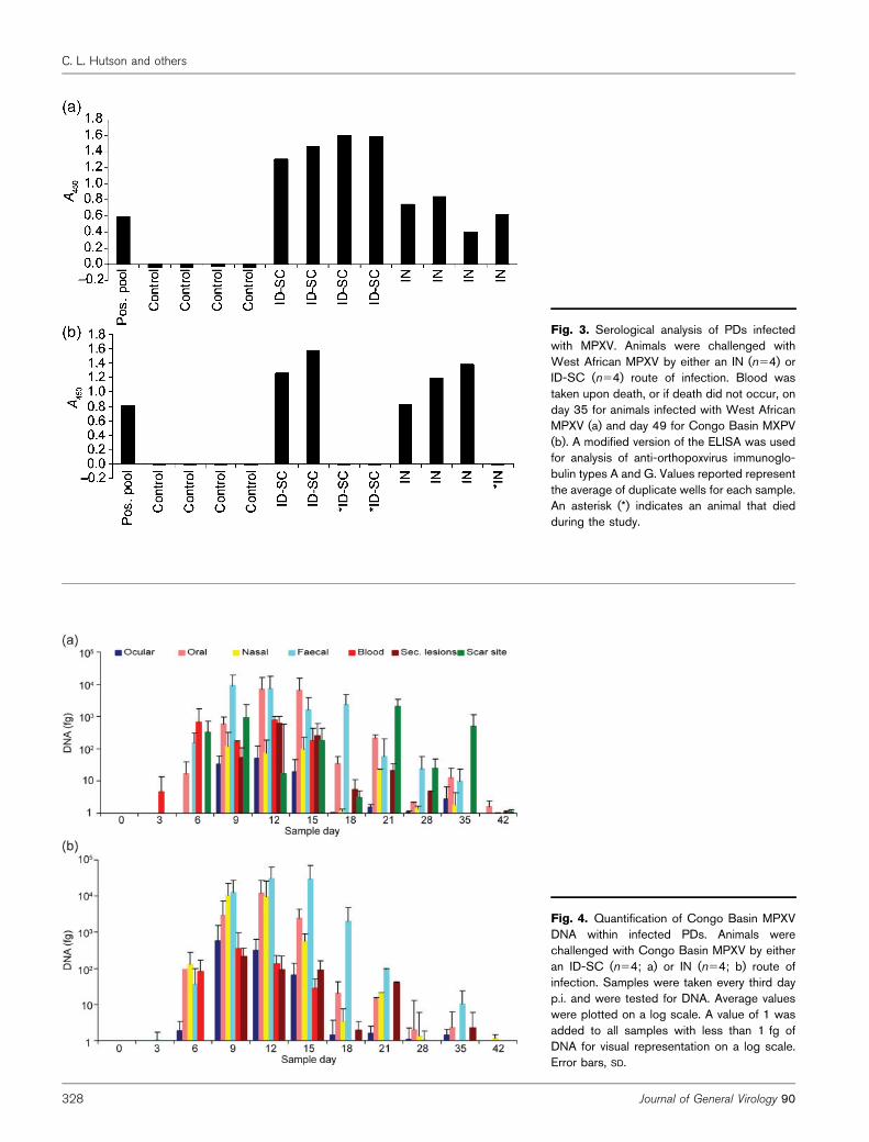
Fig. 3. Serological analysis of PDs infectedwith MPXV. Animals were challenged withWest African MPXV by either an IN (n54) orID-SC (n54) route of infection. Blood wastaken upon death, or if death did not occur, onday 35 for animals infected with West AfricanMPXV (a) and day 49 for Congo Basin MXPV(b). A modified version of the ELISA was usedfor analysis of anti-orthopoxvirus immunoglo-bulin types A and G. Values reported representthe average of duplicate wells for each sample.An asterisk (*) indicates an animal that diedduring the study.
Fig. 4. Quantification of Congo Basin MPXVDNA within infected PDs. Animals werechallenged with Congo Basin MPXV by eitheran ID-SC (n54; a) or IN (n54; b) route ofinfection. Samples were taken every third dayp.i. and were tested for DNA. Average valueswere plotted on a log scale. A value of 1 wasadded to all samples with less than 1 fg ofDNA for visual representation on a log scale.Error bars, SD.
C. L. Hutson and others
328 Journal of General Virology 90

died spontaneously, and on day 49 for the remaininganimals. The blood from the animals that died during thestudy was negative for orthopoxvirus (OPXV) antibodies.The other five convalescent Congo Basin-inoculated animalshad anti-OPXV antibodies detected at time of necropsy withsimilar levels as were observed in the serum collected fromthe West African-challenged animals (Fig. 3b). Unlike theWest African-challenged animals, there was no significantdifference seen between the antibody levels. At time ofeuthanasia, the five animals that survived had several MPXVDNA-positive samples which were negative for viable virus.This included two lesion samples and one skin sample fromthe ID-SC animals as well as one blood and one heart samplefrom the IN animals. The three animals that died during thecourse of the observations had high levels of both viral DNAand viable virus in the majority of samples tested (Fig. 7).
None of the uninfected animals, housed under the sameconditions, showed any evidence of MPXV infectionduring the duration of the study. Furthermore, all samplestaken from these control animals were negative for MPXVDNA when tested with real-time PCR (data not shown)and were negative for OPXV antibodies (Fig. 3).
DISCUSSION
Consistent with human MPXV infections (Huhn et al.,2005; Jezek et al., 1987; Reynolds et al., 2004), MPXVinfection in PDs was asymptomatic during a 6–12 day
incubation period, after which obvious skin lesionsdeveloped and subsequently resolved approximately28 days p.i. for surviving animals. Prior to developmentof generalized lesions, viral shedding could be detected inoropharyngeal secretions, consistent with descriptions ofthe oral enanthem of human systemic orthopoxvirusdisease. Generalized lesions developed and evolved at asimilar rate in a centrifugal pattern, similar to diseaseprogression for human monkeypox (Arita et al., 1985;Jezek et al., 1987).
In addition to disseminated vesiculo-pustular lesions,several other disease symptoms in the PD MPXV modelwere similar to those seen in human cases. One potentiallypainful symptom of human monkeypox cases is oral lesiondevelopment (Huhn et al., 2005; Jezek et al., 1987;Reynolds et al., 2006). Although we could not photographlesions in the oral cavities of animals since animals werenot anaesthetized during sampling, we were able to observelesions on/inside the mouths of several animals.Furthermore, bloody oral swabs collected from someanimals were probably caused by oral lesions. A complica-tion in human disease is development of secondarybacterial skin infections at lesion sites (Jezek et al., 1987).Although no disseminated lesions developed secondarybacterial infections, several of the SSs did. Diarrhoea is seenin a small percentage of human cases (Jezek et al., 1987),and similarly, was observed in one infected animal.Bronchopneumonia is another rare, late sequela of human
Fig. 5. Quantification of Congo Basin MPXVwithin infected PDs. Animals were challengedwith Congo Basin MPXV by either an ID-SC(n54; a) or IN (n54; b) route of infection.Samples were taken every third day p.i. andwere tested for viable virus. Average valueswere plotted on a log scale. Error bars, SD.
Animal model of orthopoxvirus disease using MPXV
http://vir.sgmjournals.org 329
monkeypox. Although lower respiratory complicationscould not be confirmed, the gasping noise audible fromone infected animal is suggestive of such complications, or
could be indicative of upper airway oedema causingstridor. Crusty noses and nasal discharge from several ofthe animals suggests upper respiratory involvement;human disease in the USA and Africa has documentedupper airway symptoms of sore throat and choryza (Huhnet al., 2004, 2005; Reynolds et al., 2006). Finally,inappetance and lethargy observed in PDs are commonlyobserved in humans (Jezek et al., 1987). A primarydifference between the clinical presentations of humansmallpox and monkeypox is the occurrence of lymphade-nopathy in MPXV-infected individuals. Interestingly, wedid not observe these sequelae in the PDs. However, weisolated viable virus from lymph node tissue in the threeanimals that succumbed to Congo Basin MPXV, indicatingthat the lymphatic system was involved during the courseof infection, even though swelling was not noted. Either thecondition did not occur, or was not substantial enough tobe observed. Although disease manifestations are notidentical in these two species, there are many similaritiesin disease progression, clinical manifestations and mortal-ity rates between that seen in humans and in PDs infectedwith MPXV. Our model used a relatively low level ofinfectious dose as well as two plausible natural routes ofinfection, indicating that the PD is an ideal model forhuman MPXV infection.
Viral DNA was detected in the blood of most animalsbetween days 6 and 15. Although we did not attempt viralisolation on blood samples, the presence of detectableMPXV DNA early in infection is evidence of viraemiaduring this time period. Viable virus was isolated fromoral, ocular and nasal swabs, as well as faeces, during thestudy, demonstrating the shedding potential of MPXV viamultiple excreta. Oral swabs were especially rich in viablevirus, with average peak levels greater than 104 p.f.u. forthe West African- and 105 p.f.u. for Congo Basin MPXV-infected animals. Interestingly, the faecal samples from theCongo Basin MPXV-infected animals had significantlymore DNA (days 6–28) and viable virus (day 18) than theWest African MPXV-infected animals (P-values between361024 and 0.046). This suggests, at least in this
Fig. 6. Quantification of peak values seen in Congo Basin MPXV-infected PDs. Animals were challenged with Congo Basin MPXVby either an IN (n54) or ID-SC (n54) route of infection. Sampleswere taken every third day p.i. and were tested for DNA (a) andviable virus (b). The average peak value was calculated by takingthe maximum average value for each sample from each animalgroup. The numbers on the bars correspond to the day that themaximum average value was seen.
Fig. 7. Quantification of virus within three PDsthat died due to infection with the CongoBasin strain of MPXV. Animals were chal-lenged with Congo Basin MPXV by either anIN (n54) or ID-SC (n54) route of infection.Two ID-SC animals (nos 13 and 16) died onday 12 and 11 p.i. One IN animal (no. 19) diedon day 13 p.i. Animals were necropsied andsamples were tested by tissue culture.
C. L. Hutson and others
330 Journal of General Virology 90
experimental animal model, there are potential strain-dependent differences in transmission. Viable virus levelspeaked at similar times for both MPXV strains, days 9–18for West African and 12–18 for Congo Basin. For bothstrains, infected animals continued shedding viable virusfrom certain samples until day 21 p.i., information lackingfor 2003 USA monkeypox outbreak animals and whichmight be potentially useful for formulating future outbreakcontrol measures. This period of viable MPXV sheddingfrom PDs was similar to human monkeypox outbreaks,with isolation of viable virus from samples taken up to18 days after the development of rash (Arita et al., 1985).The serology results confirmed that all animals, exceptthose that died during the study, had developed immuneresponses to orthopoxviruses. This assay was designed tocapture all Ig antibodies, but may not adequately captureIgM antibodies. The animals that perished most likely diedbefore developing IgG responses which would have beendetected with this ELISA. At time of necropsy, someanimals that survived the challenge had low levels of viralDNA in a few tissues, but no viable virus. However, thethree animals that died during the Congo Basin MPXVchallenge had high levels of viral DNA and viable virusthroughout the majority of tissues tested, suggesting thatMPXV has a wide range of tissue tropism. In one or moreof these three animals, samples including the oral swab,eyelid, tongue, lung, liver, spleen and scent glands hadlevels of viable virus at or above 107 p.f.u. ml21.
Similar to human infection (Breman et al., 1980; Fosteret al., 1972; Jezek et al., 1987; Likos et al., 2005; Reynoldset al., 2006), the Congo Basin MPXV strain was morepathogenic than the West African MPXV in the PD model.Morbidity was higher in Congo Basin MPXV-infectedanimals as measured by lesion count. A greater trend inweight loss and a significantly higher per cent increase intemperature in Congo Basin MPXV-challenged animalswere also noted. Furthermore, mortality rates of 50 % (ID-SC) and 25 % (IN) were seen in the Congo Basin MPXV-inoculated animals, compared with 0 % in West AfricanMPXV-inoculated animals. This is concordant withobservations of human disease, where Congo BasinMPXV causes 10–13 % mortality compared with 0 % withWest African MPXV. The ability to discern differences invirulence for MPXV from the two clades in this animalmodel should benefit future studies. Having such ananimal model will hopefully allow better understanding/identification of MPXV virulence factors.
It is noteworthy that the observed mortality rate was lowerthan that observed in a previously published PD challengestudy (Xiao et al., 2005) in which West African MPXVchallenge administered IN caused 60 % mortality andadministered intraperitoneally caused 100 % mortality.There are several possible explanations for this difference.Xiao et al. used a slightly higher dosage of virus (105 p.f.u.)and practices such as orbital bleeding and daily anaesthet-izing for sampling, compared with our current studyadministering 104.5 p.f.u., bleeding from the saphenous
vein and animal sampling every third day withoutanaesthesia. Also, throughout our study, animals wereprovided with highly palatable ‘monkey biscuits’, poten-tially a key dietary factor when anorexia occurred.Additionally, during the Xiao et al. study, the intraper-itoneal animals did not develop disseminated lesions,suggesting that this inoculation does not mimic naturaldisease progression, unlike animals infected in our study.
The incubation period before onset of secondary lesions(12 days versus 9–12 days), as well as the time it took forlesions to resolve, were similar for IN and ID-SC WestAfrican MPXV-infected animals in our study. Interestingly,this observation is somewhat different than that seen inhuman cases during the USA outbreak, in which humanswho received bites or scratches (the transmission route thatthe ID-SC model most closely mimics) had a shorterincubation period compared with infection via therespiratory route (Reynolds et al., 2006). Additionally, inthe USA human monkeypox study, a significant differencein rash burden between the two routes of exposure was notobserved. However, we did see significantly highernumbers of lesions in the ID-SC-infected animals com-pared with the IN-infected animals (P50.02). We con-sistently observed the development of a primary lesion atthe site of scarification (days 6–9) before the onset ofdisseminated lesions (days 9–12), which is similar to thedisease progression of inoculation smallpox versus respir-atory route-acquired smallpox (Fenner, 1948, 1990).Similarly, the incubation period of the probable respiratoryroute of human monkeypox was observed to be longerthan inoculation/complex exposure to MPXV (Reynolds etal., 2006). The differences seen in these previous humanstudies, compared with our PD model, might be attributedto differences between a retrospective (USA investigation)versus a prospective (our study) study design, or differenthost–pathogen interactions in our experimental animalmodel compared with human infection. Also, the infectiondose for our PD study may well have been higher than innaturally acquired human monkeypox cases.
There were several study limitations. Since we routinelycollected swabs instead of whole tissue, samples cannot bedirectly compared to evidence of virus per gram of tissue,but instead must be related to internally consistentcollection methods. For the faecal samples, not surpris-ingly, there were some bacterial and/or fungal contamina-tions that may have inhibited viral propagation in cells.Also, we had some sample preparation variation becausethe swabs from the Congo Basin MPXV challenge wereextracted in a larger amount of PBS compared with WestAfrican MPXV challenge samples. Although this wasaccounted for when calculating MPXV yield, the slightlyhigher dilution may have lowered the amount of detectablevirus in marginally positive samples. Also, because it ischallenging to do large scale studies with large rodents, ourrelatively small sample size limited statistical analysis.Finally, because PDs are wild-caught animals, not inbredlaboratory-acquired animals; there is individual animal
Animal model of orthopoxvirus disease using MPXV
http://vir.sgmjournals.org 331

variability which may affect the consistency of diseaseprogression.
Our results are concordant with previous data (Xiao et al.,2005; Hutson et al., 2007; Guarner et al., 2004) that showedPDs are highly susceptible to MPXV. We found bothinoculation routes gave similar infection results, and eitherroute would be a good mode of inoculating animals infuture experiments that may mimic natural transmission.Furthermore, because our model has a prolonged incuba-tion period, disseminated lesions, as well as cleardifferences in virulence of the two MPXV strains,reminiscent of the differences in human diseases andMPXV strains, the PD MPXV model has the potential to bea valuable model for disseminated human orthopoxvirusdisease, including both monkeypox as well as smallpox.The PD MPXV model will be valuable in the study ofvirulence factors within the two clades, orthopoxvirustherapeutics, transmission/shedding of MPXV, as well asthe study of other human orthopoxviruses.
REFERENCES
Arita, I. & Gromyko, A. (1982). Surveillance of orthopoxvirus
infections, and associated research, in the period after smallpox
eradication. Bull World Health Organ 60, 367–375.
Arita, I., Jezek, Z., Khodakevich, L. & Ruti, K. (1985). Human
monkeypox: a newly emerged orthopoxvirus zoonosis in the tropical
rain forests of Africa. Am J Trop Med Hyg 34, 781–789.
Boumandouki, P., Bileckot, R., Ibara, J. R., Satounkazi, C., Wassa,
W. D., Libama, E., Moudzeo, H., Bolanda, J. D. & Ngokaba, C. (2007).Simian smallpox (or monkey smallpox): study of 8 cases observed at
Impfondo Hospital in Republic of Congo. Bull Soc Pathol Exot 100,
17–21.
Breman, J. G. & Henderson, D. A. (2002). Diagnosis and management
of smallpox. N Engl J Med 346, 1300–1308.
Breman, J. G., Kalisa, R., Steniowski, M. V., Zanotto, E., Gromyko,A. I. & Arita, I. (1980). Human monkeypox, 1970–79. Bull World
Health Organ 58, 165–182.
Chen, N., Li, G., Liszewski, M. K., Atkinson, J. P., Jahrling, P. B.,Feng, Z., Schriewer, J., Buck, C., Wang, C. & other authors (2005).Virulence differences between monkeypox virus isolates from West
Africa and the Congo basin. Virology 340, 46–63.
Fenner, F. (1948). The pathogenesis of the acute exanthems. Lancet 2,
915–920.
Fenner, F. (1990). Poxviruses. In Fields Virology, 2nd edn, pp. 2113–
2133. Edited by B. N. Fields & D. M. Knipe. New York: Raven
Press.
Fine, P. E., Jezek, Z., Grab, B. & Dixon, H. (1988). The transmission
potential of monkeypox virus in human populations. Int J Epidemiol
17, 643–650.
Foster, S. O., Brink, E. W., Hutchins, D. L., Pifer, J. M., Lourie, B.,
Moser, C. R., Cummings, E. C., Kuteyi, O. E., Eke, R. E. & otherauthors (1972). Human monkeypox. Bull World Health Organ 46,
569–576.
Guarner, J., Johnson, B. J., Paddock, C. D., Shieh, W. J., Goldsmith,C. S., Reynolds, M. G., Damon, I. K., Regnery, R. L. & Zaki, S. R.(2004). Monkeypox transmission and pathogenesis in prairie dogs.
Emerg Infect Dis 10, 426–431.
Huhn, G. D., Chase, R. A. & Dworkin, M. S. (2004). Monkeypox in theWestern hemisphere. N Engl J Med 350, 1790–1791.
Huhn, G. D., Bauer, A. M., Yorita, K., Graham, M. B., Sejvar, J., Likos, A.,Damon, I. K., Reynolds, M. G. & Kuehnert, M. J. (2005). Clinicalcharacteristics of human monkeypox, and risk factors for severedisease. Clin Infect Dis 41, 1742–1751.
Hutin, Y. J., Williams, R. J., Malfait, P., Pebody, R., Loparev, V. N.,Ropp, S. L., Rodriguez, M., Knight, J. C., Tshioko, F. K. & otherauthors (2001). Outbreak of human monkeypox, DemocraticRepublic of Congo, 1996 to 1997. Emerg Infect Dis 7, 434–438.
Hutson, C. L., Lee, K. N., Abel, J., Carroll, D. S., Montgomery, J. M.,Olson, V. A., Li, Y., Davidson, W., Hughes, C. & other authors(2007). Monkeypox zoonotic associations: insights from laboratoryevaluation of animals associated with the multi-state US outbreak.Am J Trop Med Hyg 76, 757–768.
Jezek, Z., Szczeniowski, M., Paluku, K. M. & Mutombo, M. (1987).Human monkeypox: clinical features of 282 patients. J Infect Dis 156,293–298.
Learned, L. A., Reynolds, M. G., Wassa, D. W., Li, Y., Olson, V. A.,Karem, K., Stempora, L. L., Braden, Z. H., Kline, R. & other authors(2005). Extended interhuman transmission of monkeypox in ahospital community in the Republic of the Congo, 2003. Am J TropMed Hyg 73, 428–434.
Lehmann, E. L. (1975). Nonparametrics: Statistical Methods Based onRanks. San Francisco: Holden-Day.
Li, Y., Olson, V. A., Laue, T., Laker, M. T. & Damon, I. K. (2006).Detection of monkeypox virus with real-time PCR assays. J Clin Virol36, 194–203.
Likos, A. M., Sammons, S. A., Olson, V. A., Frace, A. M., Li, Y., Olsen-Rasmussen, M., Davidson, W., Galloway, R., Khristova, M. L. & otherauthors (2005). A tale of two clades: monkeypox viruses. J Gen Virol86, 2661–2672.
Marennikova, S. S. & Seluhina, E. M. (1976). Susceptibility of somerodent species to monkeypox virus, and course of the infection. BullWorld Health Organ 53, 13–20.
Meyer, H., Perrichot, M., Stemmler, M., Emmerich, P., Schmitz, H.,Varaine, F., Shungu, R., Tshioko, F. & Formenty, P. (2002). Outbreaksof disease suspected of being due to human monkeypox virusinfection in the Democratic Republic of Congo in 2001. J ClinMicrobiol 40, 2919–2921.
Mukinda, V. B., Mwema, G., Kilundu, M., Heymann, D. L., Khan, A. S. &Esposito, J. J. (1997). Re-emergence of human monkeypox in Zaire in1996. Monkeypox epidemiologic working group. Lancet 349, 1449–1450.
Mwanbal, P. T., Tshioko, K. F., Moudi, A., Mukinda, V., Mwema, G. N.,Messinger, D., Okito, L., Barakymfyte, D., Malfait, P. & other authors(1997). Human monkeypox in Kasai Oriental, Zaire (1996–1997).Euro Surveill 2, 33–35.
Reed, K. D., Melski, J. W., Graham, M. B., Regnery, R. L., Sotir, M. J.,Wegner, M. V., Kazmierczak, J. J., Stratman, E. J., Li, Y. & otherauthors (2004). The detection of monkeypox in humans in theWestern Hemisphere. N Engl J Med 350, 342–350.
Reynolds, M. G., Cono, J., Curns, A., Holman, R. C., Likos, A.,Regnery, R., Treadwell, T. & Damon, I. (2004). Human monkeypox.Lancet Infect Dis 4, 604–605.
Reynolds, M. G., Yorita, K. L., Kuehnert, M. J., Davidson, W. B., Huhn,G. D., Holman, R. C. & Damon, I. K. (2006). Clinical manifestations ofhuman monkeypox influenced by route of infection. J Infect Dis 194,773–780.
Rimoin, A. W., Kisalu, N., Kebela-Ilunga, B., Mukaba, T., Wright, L. L.,Formenty, P., Wolfe, N. D., Shongo, R. L., Tshioko, F. & other authors(2007). Endemic human monkeypox, Democratic Republic of Congo,2001–2004. Emerg Infect Dis 13, 934–937.
C. L. Hutson and others
332 Journal of General Virology 90
Shchelukhina, E. M. & Marennikova, S. S. (1975). Generalizedmonkeypox in orally infected rabbits and white mice. Vopr Virusol 6,703–705 (in Russian).
Tesh, R. B., Watts, D. M., Sbrana, E., Siirin, M., Popov, V. L. & Xiao,S. Y. (2004). Experimental infection of ground squirrels (Spermo-philus tridecemlineatus) with monkeypox virus. Emerg Infect Dis 10,1563–1567.
Xiao, S. Y., Sbrana, E., Watts, D. M., Siirin, M., da Rosa, A. P. & Tesh,R. B. (2005). Experimental infection of prairie dogs with monkeypoxvirus. Emerg Infect Dis 11, 539–545.
Zaucha, G. M., Jahrling, P. B., Geisbert, T. W., Swearengen, J. R. &Hensley, L. (2001). The pathology of experimental aerosolizedmonkeypox virus infection in cynomolgus monkeys (Macacafascicularis). Lab Invest 81, 1581–1600.
Animal model of orthopoxvirus disease using MPXV
http://vir.sgmjournals.org 333