A Novel Unstable Duplication Upstream of HAS2 Predisposes to a Breed-Defining Skin Phenotype and a...
11
A Novel Unstable Duplication Upstream of HAS2 Predisposes to a Breed-Defining Skin Phenotype and a Periodic Fever Syndrome in Chinese Shar-Pei Dogs Mia Olsson 1 *, Jennifer R. S. Meadows 1. , Katarina Truve ´ 2. , Gerli Rosengren Pielberg 1. , Francesca Puppo 3. , Evan Mauceli 4 , Javier Quilez 5 , Noriko Tonomura 4 , Giordana Zanna 6 , Maria Jose ´ Docampo 7 , Anna Bassols 7 , Anne C. Avery 8 , Elinor K. Karlsson 4,9 , Anne Thomas 10 , Daniel L. Kastner 3 , Erik Bongcam- Rudloff 11 , Matthew T. Webster 1 , Armand Sanchez 5 ,A ˚ ke Hedhammar 12 , Elaine F. Remmers 3 , Leif Andersson 1,2 , Lluis Ferrer 6 , Linda Tintle 13 *, Kerstin Lindblad-Toh 1,4 * 1 Science for Life Laboratory, Department of Medical Biochemistry and Microbiology, Uppsala University, Uppsala, Sweden, 2 Department of Animal Breeding and Genetics, Swedish University of Agricultural Sciences, Uppsala, Sweden, 3 National Human Genome Research Institute, Bethesda, Maryland, United States of America, 4 Broad Institute of Harvard and Massachusetts Institute of Technology (MIT), Cambridge, Massachusetts, United States of America, 5 Department of Animal and Food Science, Veterinary Molecular Genetics Service, Universitat Auto ` noma de Barcelona, Barcelona, Spain, 6 Department of Animal Medicine and Surgery, Universitat Auto ` noma de Barcelona, Barcelona, Spain, 7 Department of Biochemistry and Molecular Biology, Universitat Auto ` noma de Barcelona, Barcelona, Spain, 8 Department of Microbiology, Immunology, and Pathology, Colorado State University, Fort Collins, Colorado, United States of America, 9 FAS Center for Systems Biology, Harvard University, Cambridge, Massachusetts, United States of America, 10 ANTAGENE Laboratory, Limonest, France, 11 Linnaeus Centre for Bioinformatics, Uppsala University, Uppsala, Sweden, 12 Department of Clinical Sciences, Swedish University of Agricultural Sciences, Uppsala, Sweden, 13 Wurtsboro Veterinary Clinic, Wurtsboro, New York, United States of America Abstract Hereditary periodic fever syndromes are characterized by recurrent episodes of fever and inflammation with no known pathogenic or autoimmune cause. In humans, several genes have been implicated in this group of diseases, but the majority of cases remain unexplained. A similar periodic fever syndrome is relatively frequent in the Chinese Shar-Pei breed of dogs. In the western world, Shar-Pei have been strongly selected for a distinctive thick and heavily folded skin. In this study, a mutation affecting both these traits was identified. Using genome-wide SNP analysis of Shar-Pei and other breeds, the strongest signal of a breed-specific selective sweep was located on chromosome 13. The same region also harbored the strongest genome-wide association (GWA) signal for susceptibility to the periodic fever syndrome (p raw = 2.3 6 10 26 , p genome = 0.01). Dense targeted resequencing revealed two partially overlapping duplications, 14.3 Kb and 16.1 Kb in size, unique to Shar-Pei and upstream of the Hyaluronic Acid Synthase 2 (HAS2) gene. HAS2 encodes the rate-limiting enzyme synthesizing hyaluronan (HA), a major component of the skin. HA is up-regulated and accumulates in the thickened skin of Shar-Pei. A high copy number of the 16.1 Kb duplication was associated with an increased expression of HAS2 as well as the periodic fever syndrome (p,0.0001). When fragmented, HA can act as a trigger of the innate immune system and stimulate sterile fever and inflammation. The strong selection for the skin phenotype therefore appears to enrich for a pleiotropic mutation predisposing these dogs to a periodic fever syndrome. The identification of HA as a major risk factor for this canine disease raises the potential of this glycosaminoglycan as a risk factor for human periodic fevers and as an important driver of chronic inflammation. Citation: Olsson M, Meadows JRS, Truve ´ K, Rosengren Pielberg G, Puppo F, et al. (2011) A Novel Unstable Duplication Upstream of HAS2 Predisposes to a Breed- Defining Skin Phenotype and a Periodic Fever Syndrome in Chinese Shar-Pei Dogs. PLoS Genet 7(3): e1001332. doi:10.1371/journal.pgen.1001332 Editor: Michel Georges, University of Lie `ge, Belgium Received June 3, 2010; Accepted February 10, 2011; Published March 17, 2011 This is an open-access article distributed under the terms of the Creative Commons Public Domain declaration which stipulates that, once placed in the public domain, this work may be freely reproduced, distributed, transmitted, modified, built upon, or otherwise used by anyone for any lawful purpose. Funding: This work was supported by the Swedish Research Council; FORMAS; the Swedish Research Council for Environment, Agricultural Sciences, and Spatial Planning; the Swedish Foundation for Strategic Research; in part by the Intramural Research Program of the National Institute of Arthritis and Musculoskeletal and Skin Diseases and the National Human Genome Research Institute of the National Institutes of Health, the Chinese Shar-Pei Charitable Trust and the European Commission (FP7-LUPA, GA-201370). KL-T is the recipient of a EURYI award from the European Science Foundation. The funders had no role in study design, data collection and analysis, decision to publish, or preparation of the manuscript. Competing Interests: The authors KL-T, MO, and LT have filed a patent for development of a genetic test. * E-mail: [email protected] (MO); [email protected] (LT); [email protected] (KL-T) . These authors contributed equally to this work. Introduction Shar-Pei dogs have been companion animals for centuries within China where they were commissioned to guard and hunt, and to sometimes serve as fighting animals. At the beginning of the communist era dog ownership was highly taxed and the breed was brought close to extinction. A few Chinese Shar-Pei dogs were exported to the United States in the early 1970’s and Shar-Pei descending from this limited number of animals have undergone strong selection for a wrinkled skin phenotype and heavily padded muzzle and are called the ‘‘meatmouth’’ type (Figure 1A–1C) and have now found global popularity. The ancestral Shar-Pei, PLoS Genetics | www.plosgenetics.org 1 March 2011 | Volume 7 | Issue 3 | e1001332
-
Upload
independent -
Category
Documents
-
view
0 -
download
0
Transcript of A Novel Unstable Duplication Upstream of HAS2 Predisposes to a Breed-Defining Skin Phenotype and a...
A Novel Unstable Duplication Upstream of HAS2Predisposes to a Breed-Defining Skin Phenotype and aPeriodic Fever Syndrome in Chinese Shar-Pei DogsMia Olsson1*, Jennifer R. S. Meadows1., Katarina Truve2., Gerli Rosengren Pielberg1., Francesca
Puppo3., Evan Mauceli4, Javier Quilez5, Noriko Tonomura4, Giordana Zanna6, Maria Jose Docampo7,
Anna Bassols7, Anne C. Avery8, Elinor K. Karlsson4,9, Anne Thomas10, Daniel L. Kastner3, Erik Bongcam-
Rudloff11, Matthew T. Webster1, Armand Sanchez5, Ake Hedhammar12, Elaine F. Remmers3, Leif
Andersson1,2, Lluis Ferrer6, Linda Tintle13*, Kerstin Lindblad-Toh1,4*
1 Science for Life Laboratory, Department of Medical Biochemistry and Microbiology, Uppsala University, Uppsala, Sweden, 2 Department of Animal Breeding and
Genetics, Swedish University of Agricultural Sciences, Uppsala, Sweden, 3 National Human Genome Research Institute, Bethesda, Maryland, United States of America,
4 Broad Institute of Harvard and Massachusetts Institute of Technology (MIT), Cambridge, Massachusetts, United States of America, 5 Department of Animal and Food
Science, Veterinary Molecular Genetics Service, Universitat Autonoma de Barcelona, Barcelona, Spain, 6 Department of Animal Medicine and Surgery, Universitat
Autonoma de Barcelona, Barcelona, Spain, 7 Department of Biochemistry and Molecular Biology, Universitat Autonoma de Barcelona, Barcelona, Spain, 8 Department of
Microbiology, Immunology, and Pathology, Colorado State University, Fort Collins, Colorado, United States of America, 9 FAS Center for Systems Biology, Harvard
University, Cambridge, Massachusetts, United States of America, 10 ANTAGENE Laboratory, Limonest, France, 11 Linnaeus Centre for Bioinformatics, Uppsala University,
Uppsala, Sweden, 12 Department of Clinical Sciences, Swedish University of Agricultural Sciences, Uppsala, Sweden, 13 Wurtsboro Veterinary Clinic, Wurtsboro, New York,
United States of America
Abstract
Hereditary periodic fever syndromes are characterized by recurrent episodes of fever and inflammation with no knownpathogenic or autoimmune cause. In humans, several genes have been implicated in this group of diseases, but themajority of cases remain unexplained. A similar periodic fever syndrome is relatively frequent in the Chinese Shar-Pei breedof dogs. In the western world, Shar-Pei have been strongly selected for a distinctive thick and heavily folded skin. In thisstudy, a mutation affecting both these traits was identified. Using genome-wide SNP analysis of Shar-Pei and other breeds,the strongest signal of a breed-specific selective sweep was located on chromosome 13. The same region also harbored thestrongest genome-wide association (GWA) signal for susceptibility to the periodic fever syndrome (praw = 2.361026,pgenome = 0.01). Dense targeted resequencing revealed two partially overlapping duplications, 14.3 Kb and 16.1 Kb in size,unique to Shar-Pei and upstream of the Hyaluronic Acid Synthase 2 (HAS2) gene. HAS2 encodes the rate-limiting enzymesynthesizing hyaluronan (HA), a major component of the skin. HA is up-regulated and accumulates in the thickened skin ofShar-Pei. A high copy number of the 16.1 Kb duplication was associated with an increased expression of HAS2 as well as theperiodic fever syndrome (p,0.0001). When fragmented, HA can act as a trigger of the innate immune system and stimulatesterile fever and inflammation. The strong selection for the skin phenotype therefore appears to enrich for a pleiotropicmutation predisposing these dogs to a periodic fever syndrome. The identification of HA as a major risk factor for thiscanine disease raises the potential of this glycosaminoglycan as a risk factor for human periodic fevers and as an importantdriver of chronic inflammation.
Citation: Olsson M, Meadows JRS, Truve K, Rosengren Pielberg G, Puppo F, et al. (2011) A Novel Unstable Duplication Upstream of HAS2 Predisposes to a Breed-Defining Skin Phenotype and a Periodic Fever Syndrome in Chinese Shar-Pei Dogs. PLoS Genet 7(3): e1001332. doi:10.1371/journal.pgen.1001332
Editor: Michel Georges, University of Liege, Belgium
Received June 3, 2010; Accepted February 10, 2011; Published March 17, 2011
This is an open-access article distributed under the terms of the Creative Commons Public Domain declaration which stipulates that, once placed in the publicdomain, this work may be freely reproduced, distributed, transmitted, modified, built upon, or otherwise used by anyone for any lawful purpose.
Funding: This work was supported by the Swedish Research Council; FORMAS; the Swedish Research Council for Environment, Agricultural Sciences, and SpatialPlanning; the Swedish Foundation for Strategic Research; in part by the Intramural Research Program of the National Institute of Arthritis and Musculoskeletal andSkin Diseases and the National Human Genome Research Institute of the National Institutes of Health, the Chinese Shar-Pei Charitable Trust and the EuropeanCommission (FP7-LUPA, GA-201370). KL-T is the recipient of a EURYI award from the European Science Foundation. The funders had no role in study design, datacollection and analysis, decision to publish, or preparation of the manuscript.
Competing Interests: The authors KL-T, MO, and LT have filed a patent for development of a genetic test.
* E-mail: [email protected] (MO); [email protected] (LT); [email protected] (KL-T)
. These authors contributed equally to this work.
Introduction
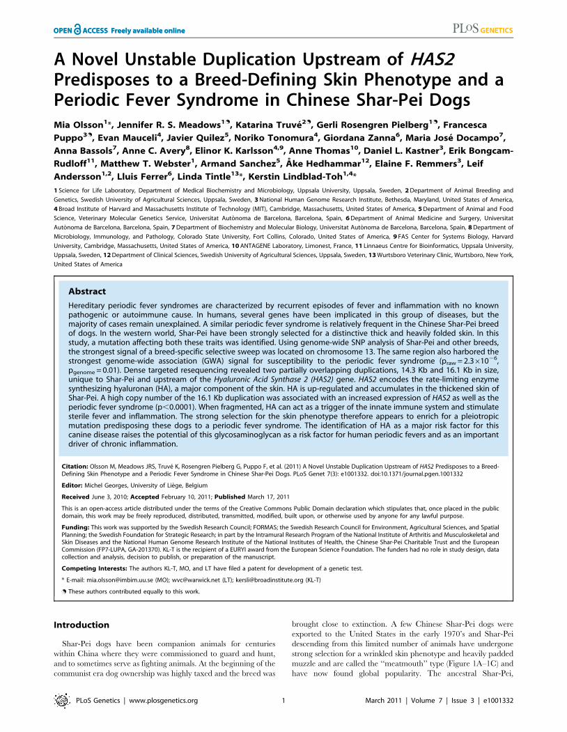
Shar-Pei dogs have been companion animals for centuries
within China where they were commissioned to guard and hunt,
and to sometimes serve as fighting animals. At the beginning of the
communist era dog ownership was highly taxed and the breed was
brought close to extinction. A few Chinese Shar-Pei dogs were
exported to the United States in the early 1970’s and Shar-Pei
descending from this limited number of animals have undergone
strong selection for a wrinkled skin phenotype and heavily padded
muzzle and are called the ‘‘meatmouth’’ type (Figure 1A–1C) and
have now found global popularity. The ancestral Shar-Pei,
PLoS Genetics | www.plosgenetics.org 1 March 2011 | Volume 7 | Issue 3 | e1001332
referred to as the ‘‘traditional’’ type Shar-Pei, still occurs and it
presents with a less accentuated skin condition (Figure 1D). The
major constituent of the deposit in the thickened skin is
hyaluronan or hyaluronic acid (HA). HA is a large, multifunc-
tional, linear, negatively charged, non-sulfated glycosaminoglycan
of the extracellular and pericellular matrices. It is composed of
repeating disaccharides and is widely spread throughout epithelial,
connective and neural tissues [1,2]. The biological role of HA
depends on its size, location and equilibrium between synthesis
and degradation [1–3]. Meatmouth Shar-Pei show two- to five-
fold higher serum levels of HA compared to other breeds [4],
allowing us to propose the term hyaluronanosis, a definition also used
for a comparable human condition [5]. HA is synthesized at the
plasma membrane by three HA synthases, HAS1, HAS2 and
HAS3, with HAS2 being the rate limiting-enzyme [6]. HAS2 is
overexpressed in dermal fibroblasts of Shar-Pei compared with
other canine breeds [7] suggesting a regulatory mutation as
causative for hyaluronanosis. HA is deposited throughout the skin
of Shar-Pei, often in microscopic lakes and grossly evident vesicles,
leading to the formation of thickened skin folds around the head
and tibiotarsal (hock) joints (Figure 1E). Almost all Shar-Pei seem
to be affected by hyaluronanosis, however the extent varies among
individuals and adults exhibit less skin folds and hyaluronanosis
than puppies. Strong selection by breeders for dogs who retained
their skin folds into adulthood has altered the phenotype of the
breed to the more commonly heavily wrinkled meatmouth type.
Meatmouth Shar-Pei also suffer a strong predisposition to an
autoinflammatory disease, Familial Shar-Pei Fever (FSF), which
clinically resembles some human hereditary periodic fever
syndromes, such as Familial Mediterranean Fever (FMF) [8].
Both diseases are characterized by seemingly unprovoked episodes
of fever and inflammation and both FMF and FSF present as short
(12–48 hour) recurrent bouts of high fever, accompanied by
localized inflammation usually involving major joints (especially
the tibiotarsal joints). Patients with FMF or Shar-Pei with FSF can
suffer episodes as often as every few weeks, but in the interim seem
symptom free. However, since acute phase reactants may endure
between episodes, a subclinical state and chronic autoinflamma-
tion may persist (Linda Tintle unpublished data). As a secondary
complication, the chronic state puts human patients, as well as
affected Shar-Pei dogs, at risk of developing reactive systemic AA
amyloidosis and subsequent kidney or liver failure [8,9]. In Shar-
Pei, the fever episodes are typically more frequent during the first
years of life and the percentage of affected dogs is very high,
estimated to be 23% in the US in 1992 [9].
Results
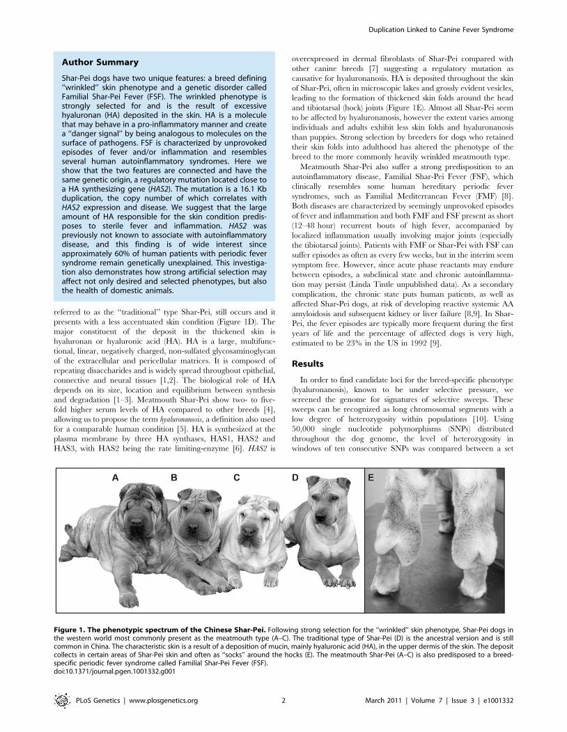
In order to find candidate loci for the breed-specific phenotype
(hyaluronanosis), known to be under selective pressure, we
screened the genome for signatures of selective sweeps. These
sweeps can be recognized as long chromosomal segments with a
low degree of heterozygosity within populations [10]. Using
50,000 single nucleotide polymorphisms (SNPs) distributed
throughout the dog genome, the level of heterozygosity in
windows of ten consecutive SNPs was compared between a set
Figure 1. The phenotypic spectrum of the Chinese Shar-Pei. Following strong selection for the ‘‘wrinkled’’ skin phenotype, Shar-Pei dogs inthe western world most commonly present as the meatmouth type (A–C). The traditional type of Shar-Pei (D) is the ancestral version and is stillcommon in China. The characteristic skin is a result of a deposition of mucin, mainly hyaluronic acid (HA), in the upper dermis of the skin. The depositcollects in certain areas of Shar-Pei skin and often as ‘‘socks’’ around the hocks (E). The meatmouth Shar-Pei (A–C) is also predisposed to a breed-specific periodic fever syndrome called Familial Shar-Pei Fever (FSF).doi:10.1371/journal.pgen.1001332.g001
Author Summary
Shar-Pei dogs have two unique features: a breed defining‘‘wrinkled’’ skin phenotype and a genetic disorder calledFamilial Shar-Pei Fever (FSF). The wrinkled phenotype isstrongly selected for and is the result of excessivehyaluronan (HA) deposited in the skin. HA is a moleculethat may behave in a pro-inflammatory manner and createa ‘‘danger signal’’ by being analogous to molecules on thesurface of pathogens. FSF is characterized by unprovokedepisodes of fever and/or inflammation and resemblesseveral human autoinflammatory syndromes. Here weshow that the two features are connected and have thesame genetic origin, a regulatory mutation located close toa HA synthesizing gene (HAS2). The mutation is a 16.1 Kbduplication, the copy number of which correlates withHAS2 expression and disease. We suggest that the largeamount of HA responsible for the skin condition predis-poses to sterile fever and inflammation. HAS2 waspreviously not known to associate with autoinflammatorydisease, and this finding is of wide interest sinceapproximately 60% of human patients with periodic feversyndrome remain genetically unexplained. This investiga-tion also demonstrates how strong artificial selection mayaffect not only desired and selected phenotypes, but alsothe health of domestic animals.
Duplication Linked to Canine Fever Syndrome
PLoS Genetics | www.plosgenetics.org 2 March 2011 | Volume 7 | Issue 3 | e1001332

of Shar-Pei (n = 50, all from the US, Table S1) and the average of
24 other canine breeds (n = 230). On four chromosomes (Cfa 5, 6,
13 and X) the reduction in heterozygosity in Shar-Pei was greater
than 4-fold the average of control breeds (Figure 2A). The
strongest signal of reduced heterozygosity appeared within a
3.7 Mb stretch on chromosome 13 (CanFam 2.0 Chr13:
23,487,992–27,227,623) (http://genome.ucsc.edu/) near the
HAS2 gene, where almost complete homozygosity was observed
in Shar Pei (Figure 2C). Here the reduction in heterozygosity was
greater than 10-fold in Shar-Pei and several smaller regions
showed complete homozygosity. The same region was confirmed
to show high levels of homozygosity when the analysis was
repeated in 37 additional Shar-Pei dogs sampled from Spain
(Table S1) and was overlapping a sweep region reported by others
for this breed [11]. The strong signal, together with the known
function of HAS2 and its aberrant expression pattern in Shar-Pei,
made this region an obvious candidate for the mutation causing
the wrinkled skin phenotype (hyaluronanosis).
In parallel, we performed a genome-wide association study to
map the susceptibility locus for FSF, using Shar-Pei strictly classified
as FSF affected (n = 24, classification code FSF+A and FSF+,
described in Materials and Methods) and unaffected (n = 17,
classification code H+, described in Materials and Methods). Five
SNPs were significantly associated (best SNP praw = 7.061027,
pgenome = 0.005 based on 100,000 permutations; software package
PLINK http://pngu.mgh.harvard.edu/˜purcell/plink [12]), all on
chromosome 13 (CanFam 2.0 Chr13: 22.4–30.7 Mb, Figure 2B).
After correcting for putative stratification, two outlier cases were
removed (Figure S1) and the same SNPs, forming the same signal of
association remained (best SNP praw = 2.361026, pgenome = 0.01;
Table S2) with a genomic inflation factor of 1.2. When the
association signal and the sweep signal were compared they
appeared interspersed, so that individual SNPs were either part of
homozygous regions or showed association with FSF (Figure 2C). It
was therefore difficult to determine exactly where the strongest
association fell, as variation is required to detect association.
Targeted sequence capture technology was used to further
investigate the sweep signal and to search for the hyaluronanosis
causative mutation. We resequenced 1.5 Mb around and
upstream of our candidate gene, HAS2 (CanFam 2.0 Chr13:
22,937,592–24,414,650) in four Shar-Pei (two meatmouth type
with high serum HA levels and two traditional type) and three
control dogs from other breeds. The obtained sequences were
mapped to the boxer reference sequence providing at least 5X
coverage for 96–98% of the resequenced region in each individual.
The targeted region also included the large intergenic noncoding
RNA, HAS2 antisense (HAS2as; Table S3) which has been proposed
as a negative post-transcriptional regulator of HAS2 mRNA [13].
After masking repetitive sequences we identified ,670 indels and
,1,500 SNP in each dog (Table S4) as well as two overlapping
duplications in the Shar-Pei (Figure 3A). Nine mutations (eight
SNPs and one indel) located in conserved elements as well as two
SNPs possibly regulating transcription, were selected for further
investigation due to their unique pattern in the sequenced Shar-
Pei dogs. Additional genotyping in Shar-Pei and dogs from other
breeds (Tables S1, S5) showed these mutations were not specific to
Shar-Pei and the variants were subsequently excluded as causative.
The two duplications were named after the Shar-Pei type in
which they were first identified. The ‘‘meatmouth’’ duplication
was the larger fragment, 16.1 Kb (CanFam 2.0 Chr13:
23,746,089–23,762,189) with breakpoints located in repeats (a
SINE at the centromeric end and a LINE at the telomeric end)
and individual copies separated by seven base pairs (Figure 3B).
The ‘‘traditional’’ duplication was 14.3 Kb (CanFam 2.0 Chr13:
23,743,906–23,758,214) and was identified in the two Shar-Pei
with a less accentuated skin phenotype (Figure 3B). We first
examined the duplications via Southern blot with control breeds
(n = 2), traditional (n = 2) and meatmouth Shar-Pei (n = 6)
(Figure 3C). As the digest cut outside and within both duplications,
we were able to observe the absence of the variants from control
breeds and separate restriction patterns in traditional and
meatmouth type Shar-Pei. Interestingly, one meatmouth dog
contained both duplication types (Figure 3C lane 6 and confirmed
by PCR across break points, data not shown). Two copy number
assays were developed to quantify these elements. The first (CNV-
E) measured only the meatmouth duplication whilst the second
(CNV-748), detected both the traditional and meatmouth
duplications. Copy number analysis was estimated as the relative
fold enrichment (DDCt) between an amplicon within the
duplication and one outside the duplication in a housekeeping
gene. Assay CNV-E was run on 90 Shar-Pei and 73 dogs from 24
other breeds (Table S1) and assay CNV-748 on a subset of 44
Shar-Pei and 14 dogs from other breeds. Assay CNV-748
demonstrated that both the traditional and meatmouth duplica-
tions are unique to the Shar-Pei breed (Figure 4 and Figure S2).
We used the results of both assays to search for a relationship
between Familial Shar-Pei Fever (FSF) and either meatmouth
copy number (Assay CNV-E), traditional copy number (the
normalized difference between CNV-748 and CNV-E) or total
traditional+meatmouth copy number (Assay CNV-748). Shar-Pei
dogs were strictly classified as affected by FSF (n = 28, FSF+A and
FSF+) or unaffected by FSF (n = 16, H+). The most significant
association was found when only the meatmouth copy number was
considered (p,0.0001, Figure 4) although a weaker association
with total copy number (p,0.01) was also seen. The observed
association between fever and meatmouth copy number, despite
the very high homozygosity in this region, strongly suggests that a
high copy number is not just a genetic marker for FSF but is
causally related to the development of disease.
Of the 153 dogs analyzed with the meatmouth copy number
assay, 31 Shar-Pei and 18 control animals also had serum
measures of HA available. No clear association was detected
between HA levels and copy number (Figure S3), however the
mean HA level in Shar-Pei with $ six copies was 9056403 ug/L
(n = 21), whilst Shar-Pei with fewer copies had a mean concentra-
tion of 7706494 ug/L (n = 12) and control breeds had HA serum
levels of 2066145 ug/L (n = 19). Interestingly, the three tradi-
tional Shar-Pei dogs had serum HA levels between 73 and
266 ug/L, which fell within the normal range [4].
The link between copy number and the expression of HAS2 and
HAS2as was examined on a smaller scale using dermal fibroblasts
cultured from six separate meatmouth Shar-Pei. The expression of
both genes was calibrated against the Shar-Pei with lowest copy
number (CNV estimate = 5) and both genes showed an increasing
trend of expression with copy number (Figure 5). These data
suggest that a regulatory element for HAS2 is located in the
duplicated region, however the interpretation of the HAS2as result
is less clear. A single study of a human osteosarcoma cell line
demonstrated that the expression of two isoforms of HAS2as were
able to reduce HAS2 expression, and so these mRNAs may act as
regulators of HA production [13]. Our data could indicate that
HAS2as expression is also influenced by a regulator element in the
duplication, or that HAS2as is up-regulated in response to HAS2
levels. If either of these scenarios were true, it is possible that if
RNA expression were measured at multiple time points we would
see temporal HAS2 repression. It could also be that the interaction
between canine fibroblast HAS2 and HAS2as does not mirror the
human system and that the canine antisense mRNA is non-
Duplication Linked to Canine Fever Syndrome
PLoS Genetics | www.plosgenetics.org 3 March 2011 | Volume 7 | Issue 3 | e1001332
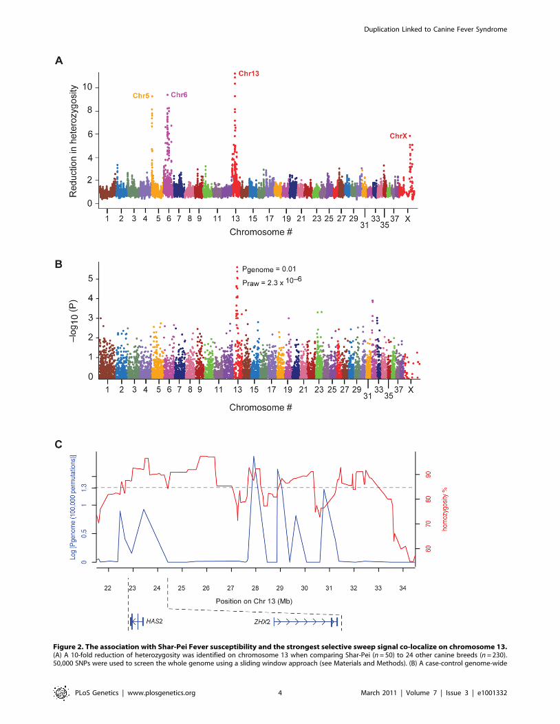
Figure 2. The association with Shar-Pei Fever susceptibility and the strongest selective sweep signal co-localize on chromosome 13.(A) A 10-fold reduction of heterozygosity was identified on chromosome 13 when comparing Shar-Pei (n = 50) to 24 other canine breeds (n = 230).50,000 SNPs were used to screen the whole genome using a sliding window approach (see Materials and Methods). (B) A case-control genome-wide
Duplication Linked to Canine Fever Syndrome
PLoS Genetics | www.plosgenetics.org 4 March 2011 | Volume 7 | Issue 3 | e1001332

functional. At present our results must be considered as
preliminary and it is clear that further exploration of the
interaction between canine HAS2 and HAS2as is required.
Discussion
Here we have identified a 16.1 Kb duplication located
approximately 350 Kb upstream of HAS2. This is clearly a
derived mutation since it occurs as a single copy sequence in other
dog breeds. We postulate that this is a causative mutation
associated with both hyaluronanosis and Shar-Pei fever, as the
observed correlation between copy number and susceptibility to
Shar-Pei fever was not expected if this was a linked, neutral
polymorphism. We suggest that the unique region of the
meatmouth type duplication identified in Shar-Pei contains one
or more regulatory elements that alter the expression of HAS2. It
appears possible that as the duplication copy number increases, so
does the copy number of potential enhancer elements within the
duplication, likely leading to a higher expression of HAS2 and
elevated HA levels, and resulting in the development of
hyaluronanosis in this breed. We propose a scenario whereby
the traditional duplication arose de novo in the traditional type of
Shar-Pei causing a milder skin phenotype. This event made the
region unstable and allowed the second meatmouth duplication to
occur. Breeders subsequently selected the meatmouth duplication
as a higher copy number enhanced the phenotypic effect in
appearance. However, it is not yet possible to say whether the
meatmouth duplication first occurred at low frequency in the
Chinese Shar-Pei population and quickly rose during breeding in
America, or if the mutation occurred spontaneously during breed
expansion in the West.
Tandem duplications are notoriously unstable and may show
copy number variation due to unequal crossing-over, as is clearly
illustrated by the copy number variation of a 450 Kb duplication
associated with dominant white colour in pigs [14]. The
meatmouth Shar-Pei duplication adds to the list of copy number
variants (CNVs), which affect phenotypic traits in domestic
animals (e.g. dominant white in pigs [14], gray color in horses
[15], the hair-ridge in Rhodesian ridgeback dogs [16], and pea-
comb in chicken [17]), several of which are linked not only to the
desirable trait but also to disease. Interestingly, all of these except
pea-comb, represent novel duplications derived from single copy
sequences. This is in contrast to most reported CNVs in humans,
which are mainly benign and represent expansions or contractions
of duplicated sequences [18].
Although we failed to find a significant correlation between
serum HA levels and copy number, this does not exclude our
proposed hyaluronanosis scenario. Difficulties in correlating
fluctuating serum levels of HA with other clinical and biomedical
parameters have also been reported in many human studies,
where no or only weak correlations were observed [19,20]. We
have shown that the 16.1 Kb duplication appears only in
meatmouth Shar-Pei, a breed type that has elevated levels of
HA compared to both traditional Shar-Pei and other breeds, and
that copy number correlates with a breed-specific syndrome
associated with excessive HA deposition and the over expression of
a HA synthesizing gene. Because HA is primarily a component of
the extracellular matrix, serum measurements may only broadly
reflect total body HA.
Hyaluronan can bind to several cellular receptors (e.g. CD44,
RHAMM and layilin), however it is the interaction between CD44
and HA which acts as a biological regulator, differentially
modulating the cellular microenvironment in response to homeo-
static versus inflammatory conditions [21]. Alterations in the
balance between native high molecular weight HA versus
fragmented HA may result in activation of innate immunity. HA
has been linked to sterile inflammation as an endogenous response
molecule to sterile tissue injury [21]. Shorter fragments of HA can
be generated by environmental insults such as sterile trauma [22],
reactive oxidative species (ROS) [23], or pathogenic hyaluroni-
dases, and it is these low molecular weight fractions which can
become pro-inflammatory danger associated molecular pattern
(DAMP) molecules [22,24] mimicking microbial surface mole-
cules.
Using a mouse model, Yamasaki and colleagues [25] showed
that HA can interact with the cell through two separate pathways
that culminate in the release of IL-1b, which together with IL-6, is
one of the main promoters of fever. In the first route, CD44 bound
HA is degraded at the plasma membrane by hyaluronidase-2
(HYAL2) prior to endocytosis and further cleavage by lysosomal
hyaluronidase-1 (HYAL1). The resultant small intracellular
oligosaccharides of HA activate the NLRP3 inflammasome, a
multiprotein complex consisting of the NLRP3 scaffold, the ASC
adaptor and caspase-1 [26]. In the second arm, the CD44-HA
complex activates toll like receptors 2 and 4 (TLR2 and 4), leading
to intracellular IL-1b mRNA transcription and the formation of
pro-IL-1b. Activation of the NLRP3 inflammasome by HA
oligosaccharides allows cleavage of this pro-IL-1b by caspase-1
and subsequent release of IL-1b. The NLRP3 inflammasome is
present in the cytosol of many cells including monocytes,
macrophages and mast cells, and has been implicated in the
pathogenesis of numerous autoinflammatory diseases in humans
including the cryopyrin-associated periodic syndromes which
result from mutations in NLRP3/CIAS1 [26].
The actual role of excessive HA in Shar-Pei needs to be
investigated further. Shar-Pei may experience exogenous frag-
mentation of their over-abundant HA from sterile or pathogenic
trauma. This, plus endogenous degradation of excessive native
HA, may contribute to induction of recurrent episodes of fever and
inflammation. Acute fever events in Shar-Pei respond rapidly to
dipyrone, a potent antipyretic and analgesic pyrazolone, which has
been demonstrated to inhibit IL-1b induced fever [27–29 and
Linda Tintle unpublished data]. It is therefore not surprising that
the strong selection on the hyaluronanosis phenotype, with
increased levels of cutaneous HA, may predispose Shar-Pei to
autoinflammation, potentially contributing to other pathologies
seen in this breed. One such example is renal medullary
amyloidosis. Histopathologically, kidneys of Shar-Pei in renal
failure have multifocal non-suppurative tubulointerstitial nephritis
with fibrosis. Medullary amyloidosis predominates and glomerular
deposition, although consistent, is highly variable in its extent
[8,30]. The renal medulla is naturally HA rich and enhanced renal
interstitial HA accumulation can be coupled to inflammatory
responses, such as ischemia-reperfusion injury, transplant-rejec-
association analysis identified a strong peak with several SNPs on chromosome 13 to be in association with Familial Shar-Pei Fever (FSF). Aftercorrecting for stratification and multiple testing (100,000 permutations), four SNPs retained significant association (p,0.05; strongest SNP association,CanFam 2.0 chr13: 27,913,803 Mb). Shar-Pei dogs used in the study were strictly classified into groups of affected (n = 22) and unaffected (n = 17) byFSF. (C) SNPs associated with FSF (blue line) are interspersed with the signals of selection (red line). The 39 Shar-Pei and 17,227 SNP common to bothanalyses were used to generate this graph.doi:10.1371/journal.pgen.1001332.g002
Duplication Linked to Canine Fever Syndrome
PLoS Genetics | www.plosgenetics.org 5 March 2011 | Volume 7 | Issue 3 | e1001332

tion, tubulointerstitial inflammation and diabetes [31]. In addition,
Shar-Pei are prone to mast cell disease including mast cell tumors
[32,33]. The binding of HA to CD44 has been shown to play a
critical role in regulation of murine cutaneous and connective
tissue mast cell proliferation [34]. As the CD44-HA interaction
may modulate local immune responses through regulation of mast
cell functions [35], excessive HA and its subsequent damage and
degradation may play a role also in the Shar-Pei breed’s
Figure 3. The identification of two breed-specific duplications in Shar-Pei. (A) Targeted resequencing of a 1.5 Mb region on chromosome13 identified a duplication with on average 3.5–4.5X higher read coverage in two meatmouth Shar-Pei (black and red), compared to three controlbreeds (green, Standard Poodle; orange, Neapolitan Mastiff and purple, Pug). A shorter duplication was detected in the traditional Shar-Pei (blue). (B)The meatmouth duplication was determined to be 16.1 Kb long (CanFam 2.0 Chr13: 23,746,089–23,762,189) with both breakpoints located in repeats(a SINE and a LINE) and with an insertion of 7 bp separating different copies. The duplication in the traditional Shar-Pei overlapped the meatmouthduplication and was slightly shorter, 14.3 Kb long (CanFam 2.0 Chr13: 23,743,906–23,758,214). In this case the copies were separated by 1 bp butwere still anchored in repeat motifs (c) Southern blot analysis using BsrGI digested gDNA from Shar-Pei and control breeds confirmed the existence oftwo duplication types in Shar-Pei. One meatmouth dog (lane 6) contained both duplication types. Individuals were classified as healthy (h) or asaffected by Familial Shar-Pei Fever (f).doi:10.1371/journal.pgen.1001332.g003
Duplication Linked to Canine Fever Syndrome
PLoS Genetics | www.plosgenetics.org 6 March 2011 | Volume 7 | Issue 3 | e1001332

predilection for allergic skin disease and other mast cell driven
inflammation.
This study suggests that HAS2 dysregulation can trigger a
periodic fever syndrome in dogs and therefore it will be relevant to
examine the approximately 60% of human fever patients who
currently have unexplained disease. Previously, the role of
hyaluronan in sterile inflammation has focused on HA signaling
and degradation; for example a deficiency of hyaluronidase causing
mucopolysaccharidosis type IX in humans has some autoinflam-
matory features [36]. However by directly implicating HAS2 in
inflammation, we suggest that a reexamination of genes further up
the biosynthetic pathway, such as those involved in HA synthesis
and polymerization is called for. In addition, the canine mutation
appears regulatory in nature and therefore regulators of HA should
be also be included in a broader scope pathway analysis of human
patients with unexplained autoinflammatory disease.
Finally, this study illustrates how copy number variations can
shape phenotypic traits and how strong artificial selection for
certain phenotypic traits may not only affect the desired trait but
also the health of the animal.
Materials and Methods
Samples and diagnostic procedureAll dog samples were collected from pet dogs after owner consent
following the ethical approval protocols (SLU, Dnr: C103/10, MIT
0910-074-13). DNA was extracted from blood samples using QIAamp
DNA Blood Midi Kit (QIAGEN) or PureLink Genomic DNA kit
(Invitrogen). All dogs, their breed type, geographic origin, health status
and experiment in which they were utilized are listed in Table S1.
Classification of Shar-Pei fever: Purebred Shar-Pei individuals
were divided into the following six groups based on their medical
records and evidence by owner and/or veterinarian:
1. FSF+A, the individual had experienced recurrent episodes of
high fever accompanied by inflammation of joints from an early
age (less than one year old). Additionally, post-mortem examina-
tion detected depositions of amyloid in kidneys and/or liver
(amyloidosis).
2. FSF+, the individual had experienced recurrent episodes of
high fever accompanied with inflammation of joints from an early
age (less than one year old).
3. Atypical FSF, the individual had experienced occasional
unexplained fever episodes or recurrent episodes with a late onset
(greater than three years old).
4. H+, the individual had never experienced unexplained fever
and/or inflammation, was older than five years old at the time of
sampling and also lacked first-degree relatives that could be
classified into the groups FSF+A, FSF+ or Atypical FSF.
5. H-, the individual had never experienced unexplained fever
and/or inflammation but was younger than 5 years at the time of
sampling and/or had first-degree relatives that could be classified
into the groups FSF+A, FSF+ or Atypical FSF.
Figure 4. The relationship between copy number estimate and susceptibility to Familial Shar-Pei Fever. A significant correlation(p = ,0.0001, Mann Whitney test) was seen when the meatmouth copy number in unaffected Shar-Pei (n = 16, H+) and individuals affected by FSF(n = 28, FSF+ and FSF+ A) were compared. Based on this limited sample size, most dogs with more than six copies had fever whereas most dogs withless than four copies did not.doi:10.1371/journal.pgen.1001332.g004
Duplication Linked to Canine Fever Syndrome
PLoS Genetics | www.plosgenetics.org 7 March 2011 | Volume 7 | Issue 3 | e1001332

6. Unknown, the individual’s medical record was not available.
Hyaluronanosis: Serum Hyaluronic Acid (HA) concentration
was used as a proxy for hyaluronanosis but no distinct cut-off value
was established. However, dogs with normal and abnormal
concentrations of serum HA were interpreted as before [4]. HA
measurements were performed using the Hyaluronan ELISA kit
(Echelon Biosciences INC) according to the manufacturer’s
instructions. The absorbance was read at 405 nm, and a semi-
log standard curve was used to calculate hyaluronic acid
concentrations.
Homozygosity and genome-wide association mappingA whole genome scan was performed with two array types, the
27K (v1) and 50K (v2) canine Affymetrix SNP chips. Results were
called using Affymetrix’s snp5-geno-qc software. The 50K array
was used when the rate of heterozygosity was calculated for US
Shar-Pei separately and for a reference group of 24 other breeds.
The ratio of heterozygosity in 10 SNP (<1 Mb) sliding windows
between the two groups was used as a measure of relative
heterozygosity. To look for regions of homozygosity within the
Shar-Pei genome only, the software package PLINK [12] was
used. This was performed both for the 50 K array with 50 US
Shar-Pei and replicated for 37 Spanish Shar-Pei using 22,362
SNPs genotyped with the Illumina CanineSNP20 BeadChip.
These data were collected with an Illumina BeadStation scanner
and genotypes were scored using GenomeStudio. Regions of
homozygosity were defined if shared across all Shar-Pei samples.
A case-control association analysis using 17,227 SNP common
to both the 27K and 50K arrays (MAF.0.05, call rate .75%) was
performed in Shar-Pei classified as affected (FSF+A and FSF+,
n = 39) or unaffected (H+, n = 17) by Shar-Pei fever. The software
package PLINK [12] was used for the analyses and to ensure
genome-wide significance, p-values were corrected for multiple
testing. Values used are the max (T) empirical p-values obtained
after 100,000 permutations. To assess whether signals from the
two genome scans overlapped, the 39 Shar-Pei with unambiguous
phenotypes were analyzed with the 17,227 SNPs common to both
SNP platforms.
Targeted resequencingTargeted capture of the 1.5 Mb candidate region (CanFam 2.0
Chr13: 22,937,592–24,414,650) was performed using a 385K
custom-designed sequence capture array from Roche NimbleGen.
Hybridization library preparation was performed as following:
Genomic DNA (15–20 mg) was fragmented using sonication;
blunting of DNA fragments using T4 DNA Polymerase, Klenow
Fragment and T4 Polynucleotide Kinase; adding A-overhangs
using Klenow Fragment exo2 and ligation of adaptors using T4
DNA Ligase with Single-read Genomic Adapter Oligo Mix
(Illumina). All enzymes were purchased from Fermentas and used
following manufacturers instructions. Purification steps were
performed using QIAquick PCR Purification Kit (QIAGEN).
Hybridization was performed following the manufacturer’s
instructions without amplification of the fragment library prior
to hybridization. Eluted captured DNA and uncaptured libraries
were amplified using Phusion High Fidelity PCR Master Mix
(Finnzymes) and the SYBR Green PCR Master Mix (Applied
Biosystems) was used to estimate the relative fold-enrichment.
Capture libraries with the estimated enrichment-factor of .200
were sequenced using Genome Analyzer (Illumina) and obtained
sequences were aligned to CanFam 2.0 [37] and to the targeted
region using Maq assembly (http://maq.sourceforge.net/) [38].
For each individual, sequence coverage was calibrated by dividing
the coverage in 100 bp windows by the average coverage for the
Figure 5. Expression analysis reveals a trend of increased HAS2 and HAS2as expression with copy number. Expression levels weremeasured in dermal fibroblasts that were cultured from individual Shar-Pei skin biopsies. The individual with the lowest copy number (CNV = 5) wasused to calibrate each assay.doi:10.1371/journal.pgen.1001332.g005
Duplication Linked to Canine Fever Syndrome
PLoS Genetics | www.plosgenetics.org 8 March 2011 | Volume 7 | Issue 3 | e1001332
total region. Three control breeds (Pug, Neapolitan Mastiff,
Standard Poodle) and two of each type of Shar-Pei (meatmouth
type and traditional type) were sequenced. The two traditional
type Shar-Pei were sequenced at different read lengths but were
aligned using the same strict criteria (allowing two mismatches per
read) and therefore vary in the percentage of mapped reads as well
as coverage when compared to the other individuals. Individual 7
(Table S4) was sequenced from whole genome amplified material
and this may have impacted the ability to map reads and detect
SNPs. This individual was not plotted in Figure 3A, but was used
in downstream analyses.
Polymerase Chain Reaction (PCR) and Sanger SequencingAll primers used were designed using Primer3 (http://frodo.wi.
mit.edu/primer3/) [39] and are listed in Table S6. PCR and
Sanger Sequencing was performed to investigate putative
mutations (ten SNPs and one indel) and were carried out with
20 ng genomic DNA using AmpliTaq Gold DNA Polymerase
(Applied Biosystems) following the manufacturer’s instructions.
The amplification of the copy number variant (CNV) breakpoints
was performed with 400 ng of DNA and a Long-range PCR with
Expand Long Template PCR System Mix 1 (Roche), cloned using
Zero Blunt TOPO Cloning Kit (Invitrogen) and plasmid DNA
prepared using QIAprep Spin Miniprep Kit (QIAGEN). PCR
products and plasmids were sequenced using capillary electropho-
resis 3730xl (Applied Biosystems), aligned and analyzed using
CodonCode Aligner version 2.0.6 (CodonCode).
Southern blot analysisFour micrograms of genomic DNA from each sample was
digested with BsrGI (New England BioLabs) and separated on a
0.7% agarose gel. A 910 bp probe (targeting CanFam 2.0 Chr13:
23,746,12–23,747,522) was used to detect the duplicated region.
Copy number assayEstimation of copy number was performed using the compar-
ative CT (DDCT) relative quantification method and a calibrator
animal (German Shepherd 95). The duplex reaction contained a
primer limited copy number assay (CNV-E: 300 nM each of
forward and reverse primers, 250 nM FAM labeled MGB probe;
CNV-748: 50 nM of forward and 300 nM reverse primers,
250 nM FAM labeled MGB probe, Applied Biosystems) and a
reference assay designed to C7orf28B (900 nM of forward and
reverse primers, 250 nM VIC and TAMRA labeled probe,
Applied Biosystems). Real Time PCR was performed in
quadruplet using 10 ng of gDNA, Genotyping Master Mix
(Applied Biosystems) and a 7900 HT Real Time PCR machine
(Applied Biosystems). The PCR primers used and dogs evaluated
can be found in Tables S4 and S1 respectively.
Fibroblast culturesCultures of dermal fibroblasts were established from skin
samples of Shar-Pei dogs as described previously [40]. Skin
samples were well shaved and cleaned with 70% EtOH/Betadine
before biopsy and cell isolation. Fat tissue and blood vessels were
removed from the skin and then samples were washed with PBS,
cut into small fragments (0.5 cm2) and digested with dispase II
solution (Boehringer Mannheim) for 16 h at 4uC. The next day,
after incubation for 30 min at 37uC in the same solution, the
dermis was separated from the epidermis. Washed dermal samples
were chopped into 1 mm3 fragments and incubated for 140 min in
15 ml of DMEM per gram of skin containing 30 mg bacterial
collagenase (Gibco), 18 mg hyaluronidase, 12 mg pronase, 1.5 mg
DNAse, supplemented with bovine albumin (all from Sigma) and
antibiotics. After digestion, cutaneous cells were washed with PBS
and grown in a humidified atmosphere at 37uC with 5% CO2 for
two days. Medium was changed twice a week and cells were used
at passages two-five.
Gene expression analysisRNA extraction from fibroblast cultures was performed as
described elsewhere [41]. 500 ng of RNA was reverse transcribed
using the High-Capacity cDNA Archive Kit (Applied Biosystems)
with random primers and following the manufacturer’s instruc-
tions. Two assays were designed to target HAS2 and HAS2as
cDNA, respectively. Real Time PCR in a volume of 20 ul was
performed in duplicate using SYBR Green PCR Master Mix
(Applied Biosystems) and primers at 300 nM in a 7900 HT Real-
Time PCR system (Applied Biosystems) with standard cycling.
PCR specificity assessment was performed by adding a dissociation
curve analysis at the end of the run. Each amplification run
contained negative controls. Relative fold-enrichment was per-
formed using the comparative DCT-method with Glucose-6-
phosphate dehydrogenase (G6PD) for normalization.
Web resourceshttp://pngu.mgh.harvard.edu/˜purcell/plink/
http://www.codoncode.com/
http://genome.ucsc.edu/
http://maq.sourceforge.net/
http://frodo.wi.mit.edu/primer3/
Supporting Information
Figure S1 Association study stratification plots prior to correc-
tion. The relationship between all individuals used in the
association study was examined by plotting components C1 and
C2 from the multidimensional scaling analysis. Shar-Pei individ-
uals were grouped into two classes, cases affected by Shar-Pei
Fever (FSF+A, FSF+; meatmouth type n = 24) and controls
unaffected by Shar-Pei Fever (H+; traditional type n = 4 and
meatmouth type n = 13). After removing two outlier cases (red
circle) the genomic inflation factor was 1.2.
Found at: doi:10.1371/journal.pgen.1001332.s001 (0.47 MB TIF)
Figure S2 Comparison of the two assays for duplication
detection. Two assays were developed to detect the duplications.
The results from each assay for a subset of dogs tested are plotted
side by side to enable comparison. The first assay, CNV-E, is
targeted to the meatmouth duplication (plotted on left hand side).
The second assay, CNV- 748, detects both the traditional and
meatmouth duplication variants (plotted right hand side). The
graph shows that meatmouth type Shar-Pei also harbour the
traditional duplication, however traditional type Shar-Pei (indi-
cated with an asterisk) do not carry the meatmouth duplication.
None of the control breeds sampled to date contain either
duplication.
Found at: doi:10.1371/journal.pgen.1001332.s002 (1.57 MB TIF)
Figure S3 No significant relationship detected between serum
hyaluronic acid (HA) concentration and copy number. Individuals
have been divided based on their breed and health status. For each
dog, meatmouth copy number is plotted using the left hand y-axis
and serum HA levels (mg/L) is on the right hand y-axis. All
individuals with HA levels higher than 600 mg L-1 carried the
duplication, although no significant correlation could be found
between HA concentration and copy numbers.
Found at: doi:10.1371/journal.pgen.1001332.s003 (1.51 MB TIF)
Duplication Linked to Canine Fever Syndrome
PLoS Genetics | www.plosgenetics.org 9 March 2011 | Volume 7 | Issue 3 | e1001332
Table S1 Individuals used in each experiment.
Found at: doi:10.1371/journal.pgen.1001332.s004 (0.07 MB PDF)
Table S2 SNPs highly associated with Familial Shar-Pei Fever on
Chromosome 13. A summary of 7 SNPs on chromosome 13 that
together formed a peak of association to the susceptibility of Familial
Shar-Pei Fever (FSF). 41 Shar-Pei classified as affected (n = 24, FSF+and FSF+A) and unaffected (n = 17, H+) by FSF were genotyped
with an 18 K SNPs and analyzed based on 100,000 permutations.
After correcting for stratification two outliers (affected by FSF) were
removed and the association remained (best SNP, p = 0.01).
Found at: doi:10.1371/journal.pgen.1001332.s005 (0.06 MB PDF)
Table S3 Comparative alignment of HAS2 and HAS2
antisense.
Found at: doi:10.1371/journal.pgen.1001332.s006 (0.06 MB PDF)
Table S4 Summary statistics of the targeted resequencing. The
dogs used for targeted sequencing were as follows: Control 1 =
Pug, Control 2 = Napolitan Mastiff, Control 3 = Standard
Poodle, Shar-Pei 1-2 are of meatmouth type and Shar-Pei 3-4
are of the traditional type. Two SNPs (Chr 13: 23,348,686 and
23,379,995) only appeared in the four sequenced Shar-Pei dogs
and not in control breeds but excluded as causative for the breed-
specific skin phenotype (hyaluronanosis) after further genotyping
in more control breeds.
Found at: doi:10.1371/journal.pgen.1001332.s007 (0.05 MB PDF)
Table S5 SNPs identified in conserved elements, HAS2 and
HAS2 antisense after targeted sequencing.
Found at: doi:10.1371/journal.pgen.1001332.s008 (0.10 MB PDF)
Table S6 Primers and probes used in the different experiments.
Found at: doi:10.1371/journal.pgen.1001332.s009 (0.06 MB PDF)
Acknowledgments
We thank all the dog owners, breeders, and breed clubs worldwide that
have supported this study and contributed samples. We thank veterinarians
and other colleagues for their help with samples including Jerome Abadie,
Laila Irene Baek, Kikka Posti, Hannes Lohi, Anne-lise Juncker, Barbara
LaVere, Patricia and Harry Roach, and Jeff Vidt. We also thank Kathleen
Long and Lazara Cuza for Shar-Pei photos. We thank Marie Lindersson
and Kristina Larsson at the SNP Technology Platform in Uppsala
(Sweden), the Broad Institute Genetic Analysis platform (US), as well as
Freyja Imsland and Snaevar Sigurdsson for technical assistance with
Illumina sequencing and bioinformatics.
Author Contributions
Conceived and designed the experiments: MO JRSM KT GRP FP AB
ACA DLK AS AH EFR LA LF LT KLT. Performed the experiments:
MO JRSM GRP FP JQ NT GZ MJD ACA LF. Analyzed the data: MO
JRSM KT GRP FP EM JQ NT GZ EKK EBR MTW KLT. Contributed
reagents/materials/analysis tools: GZ AB ACA AT AH LA LF LT KLT.
Wrote the paper: MO JRSM LT KLT.
References
1. Fraser JR, Laurent TC, Laurent UB (1997) Hyaluronan: its nature, distribution,
functions and turnover. Journal of Internal Medicine 242: 27–33.
2. Wheeler-Jones CP, Farrar CE, Pitsillides AA (2010) Targeting hyaluronan of theendothelial glycocalyx for therapeutic intervention. Current Opinion in
Investigational Drugs 11(9): 997–1006.
3. Laurent TC, Fraser JRE (1992) Hyaluronan. FASEB J 6: 2397–2404.
4. Zanna G, Fondevila D, Bardagi M, Docampo MJ, Bassols A, et al. (2008)
Cutaneous mucinosis in shar-pei dogs is due to hyaluronic acid deposition and is
associated with high levels of hyaluronic acid in serum. Vet Dermatol 19: 314–318.
5. Ramsden CA, Bankier A, Brown TJ, Cowen PSJ, Frost GI (2000) A new
disorder of hyaluronan metabolism associated with generalized folding andthickening of the skin. J of Ped 36: 62–68.
6. Weigel PH, Hascall VC, Tammi M (1997) Hyaluronan synthases. J Biol Chem
272: 13997–4000.
7. Zanna G, Docampo MJ, Fondevila D, Bardagi M, Bassols A, et al. (2009)
Hereditary cutaneous mucinosis in Shar-Pei dogs is associated with increases
hyaluronan synthase-2 mRNA transcription by cultured dermal fibroblasts. VetDermatol 20: 377–382.
8. Rivas AL, Tintle L, Kimball ES, Scarlett J, Quimby FW (1992) A canine febriledisorder associated with elevated interleukin-6. Clin Immunol Immunopathol
64: 36–45.
9. Stojanov S, Kastner DL (2005) Familial autoinflammatory diseases: genetics,pathogenesis and treatment. Curr Opin Rheumatol 17(5): 586–599.
10. Smith JM, Haigh J (1974) The hitchhiking effect of a favorable gene. Genetic
Research 23: 23–35.
11. Akey JM, Ruhe AL, Akey DT, Wong AK, Conelly CF, et al. (2010) Tracking
footprints of artificial selection in the dog genome. PNAS 19: 1160–1165.
12. Purcell S, Neale B, Todd-Brown K, Thomas L, Ferreira MA, et al. (2007)
PLINK: a tool set for whole-genome association and population-based linkage
analyses. Am J Hum Genet 81: 559–575.
13. Chao H, Spicer AP (2005) Natural Antisense mRNAs to Hyaluronan Synthase 2
Inhibit Hyaluronan Biosynthesis and Cell Proliferation. J Biol Chem 19:
27513–27522.
14. Giuffra E, Tornsten A, Marklund S, Bongcam-Rudloff E, Chardon P, et al.
(2002) A large duplication associated with dominant white color in pigs
originated by homologous recombination between LINE elements flanking KIT.Mamm Genome 13(10): 569–77.
15. Rosengren Pielberg G, Golovko A, Sundstrom E, Curik I, Lennartsson J (2008)A cis-acting regulatory mutation causes premature hair graying and suscepti-
bility to melanoma in the horse. Nat Genet 40(8): 1104–1009.
16. Salmon Hillbertz NHC, Isaksson M, Karlsson EK, Hellmen E, Pielberg GR,et al. (2007) A duplication of FGF3, FGF4, FGF9 and ORAOV1 causes the hair
ridge and predisposes to dermoid sinus in Ridgeback dogs. Nat Genet 38(11):
1318–1320.
17. Wright D, Boije H, Meadows JR, Bed’hom B, Vieaud A, et al. (2009) Copy
number variation in intron 1 of SOX5 causes the pea-comb in chickens. PLoS
Genet 5(6): 1000512. doi:10.1371/journal.pgen.1000512.
18. Sebat J, Lakshmi B, Malhotra D, Troge J, Lese-Martin C, et al. (2007) Strong
association of de novo copy number mutations with autism. Science 316:
445–449.
19. Goldberg RL, Huff JP, Lenz ME, Glickman P, Katz R, et al. (1991) Elevated
plasma levels of hyaluronan in patients with osteoarthritis and rheumatoid
arthritis. Arthritis Rheum 34: 799–807.
20. Hedin PJ, Weitoft T, Hedin H, Engstrom-Laurent A, Saxne T (1991) Serum
concentration of hyaluronan and proteoglycans in joint disease. Lack of
association. J Rheumatol 18: 1601–1605.
21. Pure E, Assoian RK (2009) Rheostatic signaling by CD44 and hyaluronan. Cell
Signal 21: 651–655.
22. Hascall VC, Majors AK, De La Motte CA, Evanko SP, Wang A, et al. (2004)
Intracellular hyaluronan: a new frontier for inflammation? Biochim Biophys
Acta 1673: 3–12.
23. Stern R, Asari AA, Sugahara KN (2006) Hyaluronan fragments: An
information-rich system. Eur J Cell Biol 85(8): 699–715.
24. Eberlein M, Schneiber KA, Black KE, Collins SL, Chan-Li Y, et al. (2008) Anti-oxidant inhibition of hyaluronan fragment-induced inflammatory gene expres-
sion. J Inflamm 5: 20.
25. Yamasaki K, Muto J, Taylor KR, Cogen AL, Audish D, et al. (2009) NLRP3/
Cryopyrin is necessary for Interleukin-1b (IL-b) release in response to
hyaluronan, an endogenous trigger in inflammation in response to injury.J Biol Chem 284(19): 12762–12771.
26. Kastner DL, Aksentijevich I, Goldbach-Mansky R (2010) Autoinflammatory
Disease reloaded: A clinical perspective. Cell 140: 784–790.
27. Shimada SG, Otterness IG, Stitt JT (1994) A study of the mechanism of action of
the mild analgesic dipyrone. Agents Actions 41: 188–192.
28. deSouza GE, Cardoso RA, Melo MC, Fabricio AS, Silva VM, et al. (2002) A
comparative study of the antipyretic effects of indomethacin and dipyrone in
rats. Inflamm Res 51(1): 24–32.
29. Prada J, Daza R, Chumbers O, Loayza I, Huicho L (2006) Antipyretic
efficacy and tolerability of oral ibuprofen, oral dipyrone and intramuscular
dipyrone in children: a randomized controlled trial. Sao Paolo Med J 124(3):135–140.
30. DiBartola SP, Tarr MJ, Webb DM, Giger U (1990) Familial renal amyloidosis in
Chinese Shar Pei dogs. J Am Vet Assos 15: 483–487.
31. Stridh S, Kerjaschki D, Chen Y, Rugenheimer L, Astrand ABM, et al. (2010)
Angiotensin converting enzyme inhibition blocks interstitial hyaluronandissipation in the neonatal rat kidney via hyaluronan synthase 2 and
hyaluronidase 1. Matrix Biol. In press.
32. Lopez A, Spracklin D, McConkey S, Hanna P (1999) Cutaneous mucinosis andmastocytosis in shar-pei. Can Vet J 40: 881–883.
33. Miller DM (1995) The occurrence of mast cell tumors in young shar-peis. J Vet
Diagn Invest 7: 360–363.
34. Takano H, Nakazawa S, Shirata N, Tamba S, Furuta K, et al. (2009)
Involvement of CD44 in mast cell proliferation during terminal differentiation.
Lab Invest 89(4): 446–55.
Duplication Linked to Canine Fever Syndrome
PLoS Genetics | www.plosgenetics.org 10 March 2011 | Volume 7 | Issue 3 | e1001332
35. Tanaka S (2010) Targeting CD44 in mast cell regulation. Expert Opin Ther
Targets 14(1): 31–43.36. Triggs-Raine B, Salo TJ, Zhang H, Wicklow BA, Natowicz MR (1999)
Mutations in HYAL1, a member of a tandemly distributed multigene family
encoding disparate hyaluronidase activities, cause a newly described lysosomaldisorder, mucopolysaccharidosis IX. Proc Natl Sci USA 11: 6296–6300.
37. Lindblad-Toh K, Wade CM, Mikkelsen TS, Karlsson EK, Jaffe DB, et al. (2005)Genome sequence, comparative analysis and haplotype structure of the domestic
dog. Nature 438: 803–819.
38. Heng L, Ruan J, Durbin R (2008) Mapping short DNA sequencing reads andcalling variants using mapping quality scores. Genome Res18: 1851–1858.
39. Rozen S, Skaletsky HO (2000) Primer3 on the WWW for general users and for
biologist programmers. Bioinformatics Methods and Protocols: Methods in
Molecular Biology. TotowaNew Jersey, , USA: Humana Press. pp 365–386.
40. Serra M, Brazıs P, Puigdemont A, Fondevilla D, Romano V, et al. (2007)
Development and characterization of a canine skin equivalent. Exp Dermatol
16: 135–142.
41. Chomczynski P, Mackey K (1995) Short technical report. Modification of the
TRIZOL reagent procedure for isolation of RNA from Polysaccharide-and
proteoglycan-rich sources. Biotechniques 19(6): 942–5.
Duplication Linked to Canine Fever Syndrome
PLoS Genetics | www.plosgenetics.org 11 March 2011 | Volume 7 | Issue 3 | e1001332