A new Upper Jurassic ophthalmosaurid ichthyosaur from the Slottsmøya Member, Agardhfjellet...
24
A New Upper Jurassic Ophthalmosaurid Ichthyosaur from the Slottsmøya Member, Agardhfjellet Formation of Central Spitsbergen Aubrey Jane Roberts 1 * ¤ , Patrick Scott Druckenmiller 2,3 , Glenn-Peter Sætre 4 , Jørn Harald Hurum 1 1 The Natural History Museum, University of Oslo, Oslo, Norway, 2 University of Alaska Museum, University of Alaska Fairbanks, Fairbanks, Alaska, United States of America, 3 Department of Geology and Geophysics, University of Alaska Fairbanks, Fairbanks, Alaska, United States of America, 4 Centre for Ecological and Evolutionary Synthesis, Department of Biosciences, University of Oslo, Oslo, Norway Abstract Abundant new ichthyosaur material has recently been documented in the Slottsmøya Member of the Agardhfjellet Formation from the Svalbard archipelago of Norway. Here we describe a partial skeleton of a new taxon, Janusaurus lundi, that includes much of the skull and representative portions of the postcranium. The new taxon is diagnosed by a suite of cranial character states including a very gracile stapedial shaft, the presence of a dorsal process on the prearticular and autapomorphic postcranial features such as the presence of an interclavicular trough and a conspicuous anterodorsal process of the ilium. The peculiar morphology of the ilia indicates a previously unrecognized degree of morphological variation in the pelvic girdle of ophthalmosaurids. We also present a large species level phylogenetic analysis of ophthalmosaurids including new and undescribed ichthyosaur material from the Upper Jurassic of Svalbard. Our results recover all Svalbard taxa in a single unresolved polytomy nested within Ophthalmosaurinae, which considerably increases the taxonomic composition of this clade. The paleobiogeographical implications of this result suggest the presence of a single clade of Boreal ophthalmosaurid ichthyosaurs that existed during the latest Jurassic, a pattern also reflected in the high degree of endemicity among some Boreal invertebrates, particularly ammonoids. Recent and ongoing descriptions of marine reptiles from the Slottsmøya Member Lagersta ¨ tte provide important new data to test hypotheses of marine amniote faunal turnover at the Jurassic-Cretaceous boundary. Citation: Roberts AJ, Druckenmiller PS, Sætre G-P, Hurum JH (2014) A New Upper Jurassic Ophthalmosaurid Ichthyosaur from the Slottsmøya Member, Agardhfjellet Formation of Central Spitsbergen. PLoS ONE 9(8): e103152. doi:10.1371/journal.pone.0103152 Editor: Andrew A. Farke, Raymond M. Alf Museum of Paleontology, United States of America Received February 21, 2014; Accepted June 23, 2014; Published August 1, 2014 This is an open-access article, free of all copyright, and may be freely reproduced, distributed, transmitted, modified, built upon, or otherwise used by anyone for any lawful purpose. The work is made available under the Creative Commons CC0 public domain dedication. Funding: Funding for fieldwork in 2010: Spitsbergen Tavel, Fugro, OMV and Exonmobil. In-kind donations from Powershop, Lividi, Telenor, Simula, Forskning.no, Directconnect, National Geographic, and Livestream.com. AJR’s collections visit was funded by CEES at the Department of Biosciences, University of Oslo. The funders had no role in study design, data collection and analysis, decision to publish, or preparation of the manuscript. Competing Interests: The authors have declared that no competing interests exist and that the funders (Spitsbergen Tavel; Fugro; OMV; Exonmobil; Powershop; Lividi; Telenor; Simula; Forskning.no; Directconnect; National Geographic and Livestream.com), had no role in study design, data collection and analysis, decision to publish, or preparation of the manuscript. This does not alter our adherence to PLOS ONE policies on sharing data and materials. * Email: [email protected] ¤ Current address: School of Ocean and Earth Science, The University of Southampton, Southampton, United Kingdom Introduction From 2004–2012, eight seasons of fieldwork in the Late Jurassic to earliest Cretaceous Slottsmøya Member of the Agardhfjellet Formation in the central Spitsbergen Sassenfjord area, have yielded numerous skeletal remains of marine amniotes (Figure 1) [1,2]. Through a number of papers, these Arctic localities have been documented [3–14], which built the framework for what is now known as the Slottsmøya Member Lagersta ¨ tte (SML), which resulted in the description of two new ophthalmosaurid ichthyo- saur taxa; Cryopterygius kristiansenae and Palvennia hoybergeti [15], as well as five new plesiosaurians [16–19]. Here, we present a description of a third new ophthalmosaurid taxon from the Slottsmøya Member, which includes most of the skull, girdle elements and fore- and hind limbs. The specimen, PMO 222.654, is significant in that it represents one of the stratigraphically oldest specimens excavated from the Slottsmøya Member and provides important new morphological data for comparisons with age pene-contemporaneous material from the Kimmeridge Clay Formation of the U.K. and elsewhere. An ever-expanding body of data on ichthyosaurs from the SML demonstrates previously unrecognized diversity among Late Jurassic taxa, similar to recent work recognizing high diversity among Early Cretaceous ichthyosaurs [20]. Collectively, the SML assemblage is also significant in being one of only two major marine amniote sites that span the Jurassic-Cretaceous boundary (the other being the Vaca Muerta Formation of Argentina [21,22]), thereby contributing to ongoing discussions regarding potential marine reptile turnover at the Jurassic-Cretaceous boundary [23]. Geological Setting The Slottsmøya Member is the uppermost of four members in the Agardhfjellet Formation [24]. It is overlain by the Mykle- gardfjellet Bed, which is the base of the Rurikfjellet Formation [24]. These two formations form the Janusfjellet Subgroup, which is part of the Adventdalen group first described by Parker [25]. The Janusfjellet Subgroup is Late Jurassic to Early Cretaceous in PLOS ONE | www.plosone.org 1 August 2014 | Volume 9 | Issue 8 | e103152
Transcript of A new Upper Jurassic ophthalmosaurid ichthyosaur from the Slottsmøya Member, Agardhfjellet...
A New Upper Jurassic Ophthalmosaurid Ichthyosaurfrom the Slottsmøya Member, Agardhfjellet Formation ofCentral SpitsbergenAubrey Jane Roberts1*¤, Patrick Scott Druckenmiller2,3, Glenn-Peter Sætre4, Jørn Harald Hurum1
1 The Natural History Museum, University of Oslo, Oslo, Norway, 2 University of Alaska Museum, University of Alaska Fairbanks, Fairbanks, Alaska, United States of America,
3 Department of Geology and Geophysics, University of Alaska Fairbanks, Fairbanks, Alaska, United States of America, 4 Centre for Ecological and Evolutionary Synthesis,
Department of Biosciences, University of Oslo, Oslo, Norway
Abstract
Abundant new ichthyosaur material has recently been documented in the Slottsmøya Member of the AgardhfjelletFormation from the Svalbard archipelago of Norway. Here we describe a partial skeleton of a new taxon, Janusaurus lundi,that includes much of the skull and representative portions of the postcranium. The new taxon is diagnosed by a suite ofcranial character states including a very gracile stapedial shaft, the presence of a dorsal process on the prearticular andautapomorphic postcranial features such as the presence of an interclavicular trough and a conspicuous anterodorsalprocess of the ilium. The peculiar morphology of the ilia indicates a previously unrecognized degree of morphologicalvariation in the pelvic girdle of ophthalmosaurids. We also present a large species level phylogenetic analysis ofophthalmosaurids including new and undescribed ichthyosaur material from the Upper Jurassic of Svalbard. Our resultsrecover all Svalbard taxa in a single unresolved polytomy nested within Ophthalmosaurinae, which considerably increasesthe taxonomic composition of this clade. The paleobiogeographical implications of this result suggest the presence of asingle clade of Boreal ophthalmosaurid ichthyosaurs that existed during the latest Jurassic, a pattern also reflected in thehigh degree of endemicity among some Boreal invertebrates, particularly ammonoids. Recent and ongoing descriptions ofmarine reptiles from the Slottsmøya Member Lagerstatte provide important new data to test hypotheses of marine amniotefaunal turnover at the Jurassic-Cretaceous boundary.
Citation: Roberts AJ, Druckenmiller PS, Sætre G-P, Hurum JH (2014) A New Upper Jurassic Ophthalmosaurid Ichthyosaur from the Slottsmøya Member,Agardhfjellet Formation of Central Spitsbergen. PLoS ONE 9(8): e103152. doi:10.1371/journal.pone.0103152
Editor: Andrew A. Farke, Raymond M. Alf Museum of Paleontology, United States of America
Received February 21, 2014; Accepted June 23, 2014; Published August 1, 2014
This is an open-access article, free of all copyright, and may be freely reproduced, distributed, transmitted, modified, built upon, or otherwise used by anyone forany lawful purpose. The work is made available under the Creative Commons CC0 public domain dedication.
Funding: Funding for fieldwork in 2010: Spitsbergen Tavel, Fugro, OMV and Exonmobil. In-kind donations from Powershop, Lividi, Telenor, Simula, Forskning.no,Directconnect, National Geographic, and Livestream.com. AJR’s collections visit was funded by CEES at the Department of Biosciences, University of Oslo. Thefunders had no role in study design, data collection and analysis, decision to publish, or preparation of the manuscript.
Competing Interests: The authors have declared that no competing interests exist and that the funders (Spitsbergen Tavel; Fugro; OMV; Exonmobil;Powershop; Lividi; Telenor; Simula; Forskning.no; Directconnect; National Geographic and Livestream.com), had no role in study design, data collection andanalysis, decision to publish, or preparation of the manuscript. This does not alter our adherence to PLOS ONE policies on sharing data and materials.
* Email: [email protected]
¤ Current address: School of Ocean and Earth Science, The University of Southampton, Southampton, United Kingdom
Introduction
From 2004–2012, eight seasons of fieldwork in the Late Jurassic
to earliest Cretaceous Slottsmøya Member of the Agardhfjellet
Formation in the central Spitsbergen Sassenfjord area, have
yielded numerous skeletal remains of marine amniotes (Figure 1)
[1,2]. Through a number of papers, these Arctic localities have
been documented [3–14], which built the framework for what is
now known as the Slottsmøya Member Lagerstatte (SML), which
resulted in the description of two new ophthalmosaurid ichthyo-
saur taxa; Cryopterygius kristiansenae and Palvennia hoybergeti[15], as well as five new plesiosaurians [16–19]. Here, we present a
description of a third new ophthalmosaurid taxon from the
Slottsmøya Member, which includes most of the skull, girdle
elements and fore- and hind limbs.
The specimen, PMO 222.654, is significant in that it represents
one of the stratigraphically oldest specimens excavated from the
Slottsmøya Member and provides important new morphological
data for comparisons with age pene-contemporaneous material
from the Kimmeridge Clay Formation of the U.K. and elsewhere.
An ever-expanding body of data on ichthyosaurs from the SML
demonstrates previously unrecognized diversity among Late
Jurassic taxa, similar to recent work recognizing high diversity
among Early Cretaceous ichthyosaurs [20]. Collectively, the SML
assemblage is also significant in being one of only two major
marine amniote sites that span the Jurassic-Cretaceous boundary
(the other being the Vaca Muerta Formation of Argentina
[21,22]), thereby contributing to ongoing discussions regarding
potential marine reptile turnover at the Jurassic-Cretaceous
boundary [23].
Geological SettingThe Slottsmøya Member is the uppermost of four members in
the Agardhfjellet Formation [24]. It is overlain by the Mykle-
gardfjellet Bed, which is the base of the Rurikfjellet Formation
[24]. These two formations form the Janusfjellet Subgroup, which
is part of the Adventdalen group first described by Parker [25].
The Janusfjellet Subgroup is Late Jurassic to Early Cretaceous in
PLOS ONE | www.plosone.org 1 August 2014 | Volume 9 | Issue 8 | e103152
age and is interpreted to be a marine shelf to prodeltaic succession
dominated by shale, with subordinate siltstone and sandstone
[24,25]. The Slottsmøya Member consists of 55-70 meters of dark-
grey to black silty mudstone, often weathered into paper shale.
There are discontinuous silty beds, with occurrences of siderite
and dolomite interbeds and yellow-to-red sideritic concretions
[24]. The Slottsmøya Member records a transgressive and
subsequent regressive period, with varying degrees of dysaerobic
sea bottom conditions [3,26]. There was a low sedimentation rate
and relatively high organic productivity in the upper water
column, leading to significant accumulations of organic matter in
the bottom sediments, reaching 5% in some layers [4,27].
The Slottsmøya Member has been dated biostratigraphically
from the Upper Volgian to Upper Ryazanian, which corresponds
to about 12 million years of deposition [4,6]. The member has
been divided into three units following Collignon & Hammer [3].
The lowest unit extends from the base of the member (222 m) to a
yellow echinoderm marker bed. The middle unit, for which a high
degree of stratigraphic resolution has been established, extends
from the yellow echinoderm bed (0) to the Dorsoplanites bed
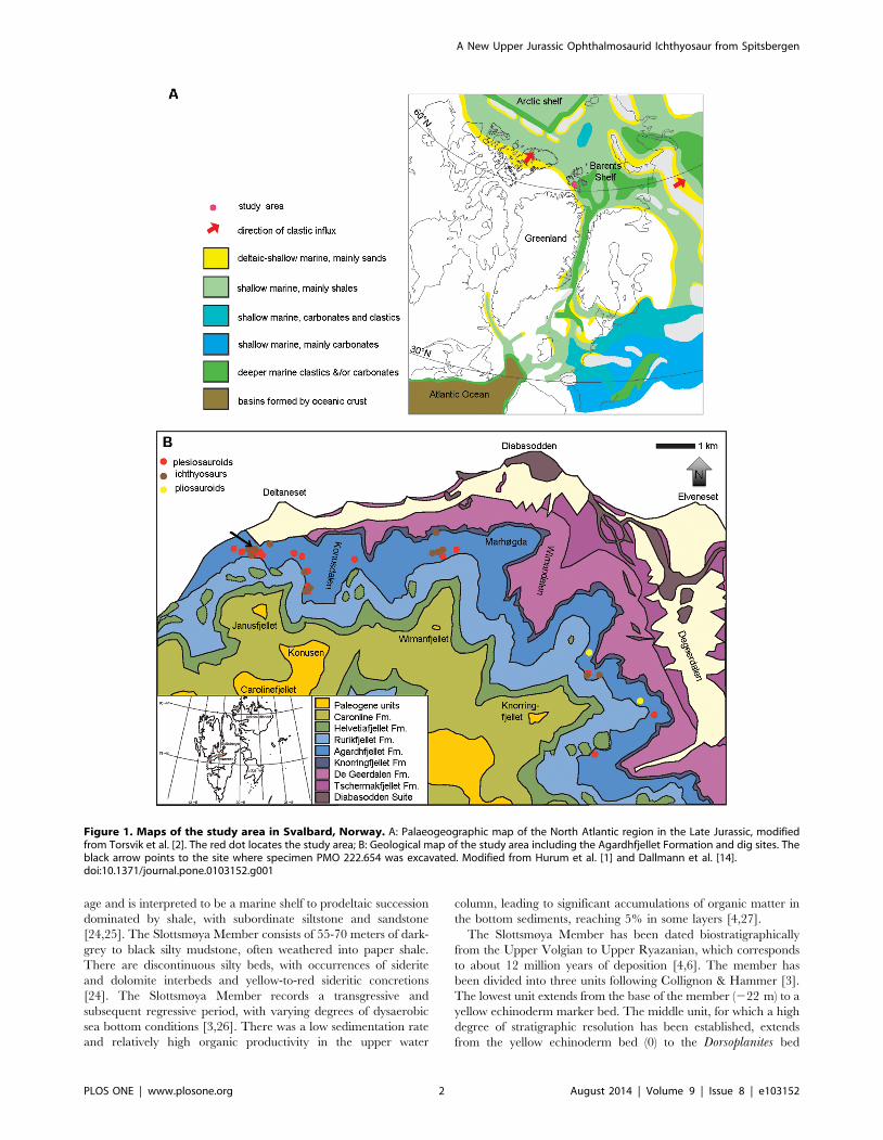
Figure 1. Maps of the study area in Svalbard, Norway. A: Palaeogeographic map of the North Atlantic region in the Late Jurassic, modifiedfrom Torsvik et al. [2]. The red dot locates the study area; B: Geological map of the study area including the Agardhfjellet Formation and dig sites. Theblack arrow points to the site where specimen PMO 222.654 was excavated. Modified from Hurum et al. [1] and Dallmann et al. [14].doi:10.1371/journal.pone.0103152.g001
A New Upper Jurassic Ophthalmosaurid Ichthyosaur from Spitsbergen
PLOS ONE | www.plosone.org 2 August 2014 | Volume 9 | Issue 8 | e103152
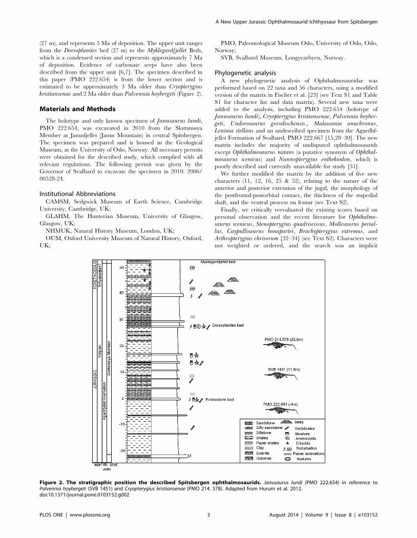
(27 m), and represents 5 Ma of deposition. The upper unit ranges
from the Dorsoplanites bed (27 m) to the Myklegardfjellet Beds,
which is a condensed section and represents approximately 7 Ma
of deposition. Evidence of carbonate seeps have also been
described from the upper unit [6,7]. The specimen described in
this paper (PMO 222.654) is from the lower section and is
estimated to be approximately 3 Ma older than Cryopterygiuskristiansenae and 2 Ma older than Palvennia hoybergeti (Figure 2).
Materials and Methods
The holotype and only known specimen of Janusaurus lundi,PMO 222.654, was excavated in 2010 from the Slottsmøya
Member at Janusfjellet (Janus Mountain) in central Spitsbergen.
The specimen was prepared and is housed in the Geological
Museum, at the University of Oslo, Norway. All necessary permits
were obtained for the described study, which complied with all
relevant regulations. The following permit was given by the
Governor of Svalbard to excavate the specimen in 2010: 2006/
00528-24.
Institutional AbbreviationsCAMSM, Sedgwick Museum of Earth Science, Cambridge
University, Cambridge, UK;
GLAHM, The Hunterian Museum, University of Glasgow,
Glasgow, UK;
NHMUK, Natural History Museum, London, UK;
OUM, Oxford University Museum of Natural History, Oxford,
UK;
PMO, Paleontological Museum Oslo, University of Oslo, Oslo,
Norway;
SVB, Svalbard Museum, Longyearbyen, Norway.
Phylogenetic analysisA new phylogenetic analysis of Ophthalmosauridae was
performed based on 22 taxa and 56 characters, using a modified
version of the matrix in Fischer et al. [23] (see Text S1 and Table
S1 for character list and data matrix). Several new taxa were
added to the analysis, including PMO 222.654 (holotype of
Janusaurus lundi), Cryopterygius kristiansenae, Palvennia hoyber-geti, Undorosaurus gorodischensis., Malawania anachronus,Leninia stellans and an undescribed specimen from the Agardhf-
jellet Formation of Svalbard, PMO 222.667 [15,28–30]. The new
matrix includes the majority of undisputed ophthalmosaurids
except Ophthalmosaurus natans (a putative synonym of Ophthal-mosaurus icenicus) and Nannopterygius enthekiodon, which is
poorly described and currently unavailable for study [31].
We further modified the matrix by the addition of five new
characters (11, 12, 16, 25 & 52), relating to the nature of the
anterior and posterior extension of the jugal, the morphology of
the postfrontal-postorbital contact, the thickness of the stapedial
shaft, and the ventral process on femur (see Text S2).
Finally, we critically reevaluated the existing scores based on
personal observation and the recent literature for Ophthalmo-saurus icenicus, Stenopterygius quadriscissus, Mollesaurus perial-lus, Caypullisaurus bonapartei, Brachypterygius extremus, and
Arthropterygius chrisorum [32–34] (see Text S2). Characters were
not weighted or ordered, and the search was an implicit
Figure 2. The stratigraphic position the described Spitsbergen ophthalmosaurids. Janusaurus lundi (PMO 222.654) in reference toPalvennia hoybergeti (SVB 1451) and Cryopterygius kristiansenae (PMO 214. 578). Adapted from Hurum et al. 2012.doi:10.1371/journal.pone.0103152.g002
A New Upper Jurassic Ophthalmosaurid Ichthyosaur from Spitsbergen
PLOS ONE | www.plosone.org 3 August 2014 | Volume 9 | Issue 8 | e103152

enumeration. The program TNT [35] was used to analyze the
character matrix and calculate Bremer Support and bootstrap
values A bootstrap analysis was run in TNT with 1000 replicates,
using the TBR algorithm.
Nomenclatural ActsThe electronic edition of this article conforms to the requirements
of the amended International Code of Zoological Nomenclature,
and hence the new names contained herein are available under that
Code from the electronic edition of this article. This published work
and the nomenclatural acts it contains have been registered in
ZooBank, the online registration system for the ICZN. The
ZooBank LSIDs (Life Science Identifiers) can be resolved and the
associated information viewed through any standard web browser
by appending the LSID to the prefix ‘‘http://zoobank.org/’’. The
LSID for this publication is: urn:lsid:zoobank.org:pub:FF4834F1-
AEED-4B08-8E74-7125801C1B3E
The electronic edition of this work was published in a journal
with an ISSN, and has been archived and is available from the
following digital repositories: PubMed Central, LOCKSS and
CRIStin (University of Oslo Library).
Results
Systematic PaleontologyICHTHYOSAURIA de Blainville 1835
Neoichthyosauria Sander 2000
Thunnosauria Motani 1999
Ophthalmosauridae Baur 1887
Janusaurus gen. nov.
urn:lsid:zoobank.org:act:4D77CFCF-22A0-4899-A619-
1E93D1ADF3C5
Janusaurus lundi sp. nov.
urn:lsid:zoobank.org:act:71E65B35-7215-44AA-BCE7-
E9A3B265E04F
(Figures. 3-14)
Holotype and only specimen: PMO 222.654, an incom-
plete skeleton consisting of a partial skull, representative cervical,
dorsal and caudal vertebrae, a nearly complete pectoral girdle and
left forefin, the right humerus, a partial pelvic girdle and both
femora.
Etymology: Genus name after the mountain Janusfjellet, on
which the specimen was found. Species name in honor of Bjørn
Lund, technician on the excavations in 2006-2012.
Holotype locality: North side of Janusfjellet, ,13 km
northeast of Longyearbyen, Spitsbergen, Svalbard, Norway.
UTM: N78 20.264 E15 50.044
Holotype horizon and stage: Slottsmøya Member, Agardhf-
jellet Formation, early Middle Volgian, Upper Jurassic; 31 m
below the Dorsoplanites Bed, 4 m below the echinoderm bed [1].
Differential diagnosisA moderately sized ophthalmosaurid (estimated body length of
3-4 meters) possessing the following autapomorphies (marked with
*) and unique character combinations: maxilla with extensive
lateral exposure (short in Ophthalmosaurus and Aegirosaurus);lacrimal contributes to the posterior margin of the external naris
(excluded in Cryopterygius and Athabascasaurus); posterodorsal
process of jugal forming half of the posterior margin of the orbit
(does not form any of the margin in Cryopterygius); narrow
postorbital bar (broad in Cryopterygius, Athabascasaurus and
Brachypterygius); absence of a squamosal (present in Athabasca-saurus and Aegirosaurus); extremely gracile and constricted
stapedial shaft*; reduced ophisthotic facet on the basioccipital
(large in Palvennia); presence of an angular-articular contact*;
extremely gracile dentition (more robust in Cryopterygius and
Brachypterygius); interclavicle with an interclavicular trough and
ventral foramen*; proximodistal length of scapula very reduced in
comparison to coracoid length*; humerus with three distal facets
(two in Nannopterygius and Cryopterygius); ulna is the largest
element of zeugopodium (radius is larger in Cryopterygius);anterodorsal process of the ilium*; ischiopubis completely fused
and lacking an obturator foramen (unfused distally in Cryopter-ygius and Undorosaurus and oburator foramen present in
Ophthalmosaurus); femur with two distal facets (three in
Platypterygius americanus and Platypterygius australis).
DescriptionThe estimated length of the animal is 3–4 m, based on
comparisons of skull and rib size in other more complete
ophthalmosaurids. The preserved portion of the skull is similar
in size and relative proportions to that of Palvennia hoybergeti,which is estimated to be a small to moderately sized (3–4 m)
species [15].
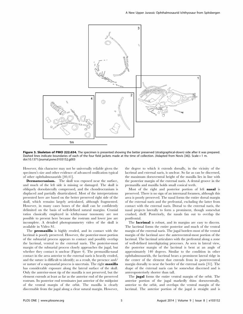
Taphonomy: The holotype specimen of Janusaurus lundiPMO 222.654, is an incomplete, partially articulated skeleton,
collected in four jackets (Figure 3) [36]. The skull, pectoral girdle,
left forelimb, and cervical vertebrae are closely associated. The
right humerus was disarticulated from the rest of the pectoral
girdle. Several ribs and gastralia were clustered together posterior
to the skull, as was the remaining material, including the presacral
and caudal vertebrae, left pelvic girdle and two femora. Several
vertebrae and the right ilium were collected as surface material in
the field. The individual appears to have come to rest on the sea
floor on its right ventrolateral side, which is also better preserved.
Because the specimen was collected in permafrost, the individual
bones are broken into millimeter-sized fragments due to congeli-
fraction, which is typical for all the marine reptile specimens from
the Slottsmøya Member [15].
An isolated tooth was associated with the skeleton of PMO
222.654 (Figure S2 K). The tooth was discovered during
preparation and was located in the vicinity of the disarticulated
right humerus. The tooth is incomplete and lacks the apex of the
crown and measures 3.5 mm in total length. It is very gracile and
needle-like, similar in overall morphology to teeth associated with
the plesiosaur Spitrasaurus larseni (SVB 1450) [16]. For this
reason, we attribute the tooth to a plesiosaurian. However, the
association between the tooth and the holotype specimen of
Janusaurus is unclear. It may be a random association, but we feel
this is unlikely given that this pattern has not previously been
observed in any other specimens of ichthyosaurs or plesiosaurians
in the Slottsmøya Member. The tooth could possibly be evidence
of scavenging on the carcass by a plesiosaurian, however this also
seems improbable given that the extremely gracile tooth
morphology is suggestive of a soft-bodied invertebrate feeder
[37]. Finally, there is a possibility that the tooth could represent
gut contents of the ichthyosaur, although the diminutive tooth
morphology of the ichthyosaur is seemingly inconsistent with this
interpretation, because no other elements of a plesiosaurian were
associated with the skeleton.
Ontogeny: The specimen is interpreted to be an adult, based
on the smoothness of the humeral shaft, the concavity of the distal
facets and the advanced ossification of the forefin elements
[38,39]. In addition, the cross-section shape of the dorsal ribs
exhibit a distinct figure-eight shape with thick cortical bone, which
Kear & Zammit [39] suggested as an ontogenetic adult trait.
Interestingly, the humeri of PMO 222.654 possess flat proximal
ends, which has been described as an indicator of immaturity [38].
A New Upper Jurassic Ophthalmosaurid Ichthyosaur from Spitsbergen
PLOS ONE | www.plosone.org 4 August 2014 | Volume 9 | Issue 8 | e103152
However, this character may not be universally reliable given the
specimen’s size and other evidence of advanced ossification typical
of other ophthalmosaurids [40,41].Dermatocranium. The skull was exposed near the surface,
and much of the left side is missing or damaged. The skull is
obliquely dorsolaterally compressed, and the chondrocranium is
displaced and partially disarticulated. Most of the interpretations
presented here are based on the better preserved right side of the
skull, which remains largely articulated, although fragmented.
However, in many cases bones of the skull can be confidently
delimited on the basis of well-defined sutural margins. Cranial
ratios classically employed in ichthyosaur taxonomy are not
possible to present here because the rostrum and lower jaw are
incomplete. A detailed photogrammetry video of the skull is
available in Video S1.
The premaxilla is highly eroded, and its contact with the
lacrimal is poorly preserved. However, the posterior-most portion
of the subnarial process appears to contact and possibly overlap
the lacrimal, ventral to the external naris. The posterior-most
margin of the subnarial process closely approaches the jugal, but
whether they contact is unclear (Figure 4). The premaxilla-nasal
contact in the area anterior to the external naris is heavily eroded,
and the suture is difficult to identify; as a result, the presence and/
or nature of a supranarial process is uncertain. The right maxillahas considerable exposure along the lateral surface of the skull.
Only the anterior-most tip of the maxilla is not preserved, but the
element extends at least as far as the anterior end of the preserved
rostrum. Its posterior end terminates just anterior of the midpoint
of the ventral margin of the orbit. The maxilla is clearly
discernable from the jugal along a clear sutural margin. However,
the degree to which it extends dorsally, in the vicinity of the
lacrimal and external naris, is unclear. So far as can be discerned,
the maximum dorsoventral height of the maxilla lies in line with
the posterior margin of the external naris. A dental groove in the
premaxilla and maxilla holds small conical teeth.
Most of the right and posterior portion of left nasal is
preserved. There is no sign of an internasal foramen, although this
area is poorly preserved. The nasal forms the entire dorsal margin
of the external naris and the prefrontal, excluding the latter from
contact with the external naris. Dorsal to the external naris, the
nasal projects laterally to form a prominent, though somewhat
crushed, shelf. Posteriorly, the nasals fan out to overlap the
postfrontal.
The lacrimal is robust, and its margins are easy to discern.
The lacrimal forms the entire posterior and much of the ventral
margin of the external naris. The jugal borders most of the ventral
margin of the lacrimal save the anteroventral-most portion of the
lacrimal. The lacrimal articulates with the prefrontal along a zone
of well-defined interdigitating processes. As seen in lateral view,
the posterior margin of the lacrimal is bent at an angle of
approximately 140 degrees. Similar to the condition in other
ophthalmosaurids, the lacrimal bears a prominent lateral ridge in
the center of the element that extends from its posteroventral
margin dorsally to near the border of the external naris [31]. The
shape of the external naris can be somewhat discerned and is
anteroposteriorly shorter than tall.
The jugal forms the entire ventral margin of the orbit. The
anterior portion of the jugal markedly thins dorsoventrally,
anterior to the orbit, and overlaps the ventral margin of the
lacrimal. The anterior portion of the jugal is straight and is
Figure 3. Skeleton of PMO 222.654. The specimen is presented showing the better preserved (stratigraphical-down) side after it was prepared.Dashed lines indicate boundaries of each of the four field jackets made at the time of collection. (Adapted from Novis [36]). Scale = 1 m.doi:10.1371/journal.pone.0103152.g003
A New Upper Jurassic Ophthalmosaurid Ichthyosaur from Spitsbergen
PLOS ONE | www.plosone.org 5 August 2014 | Volume 9 | Issue 8 | e103152
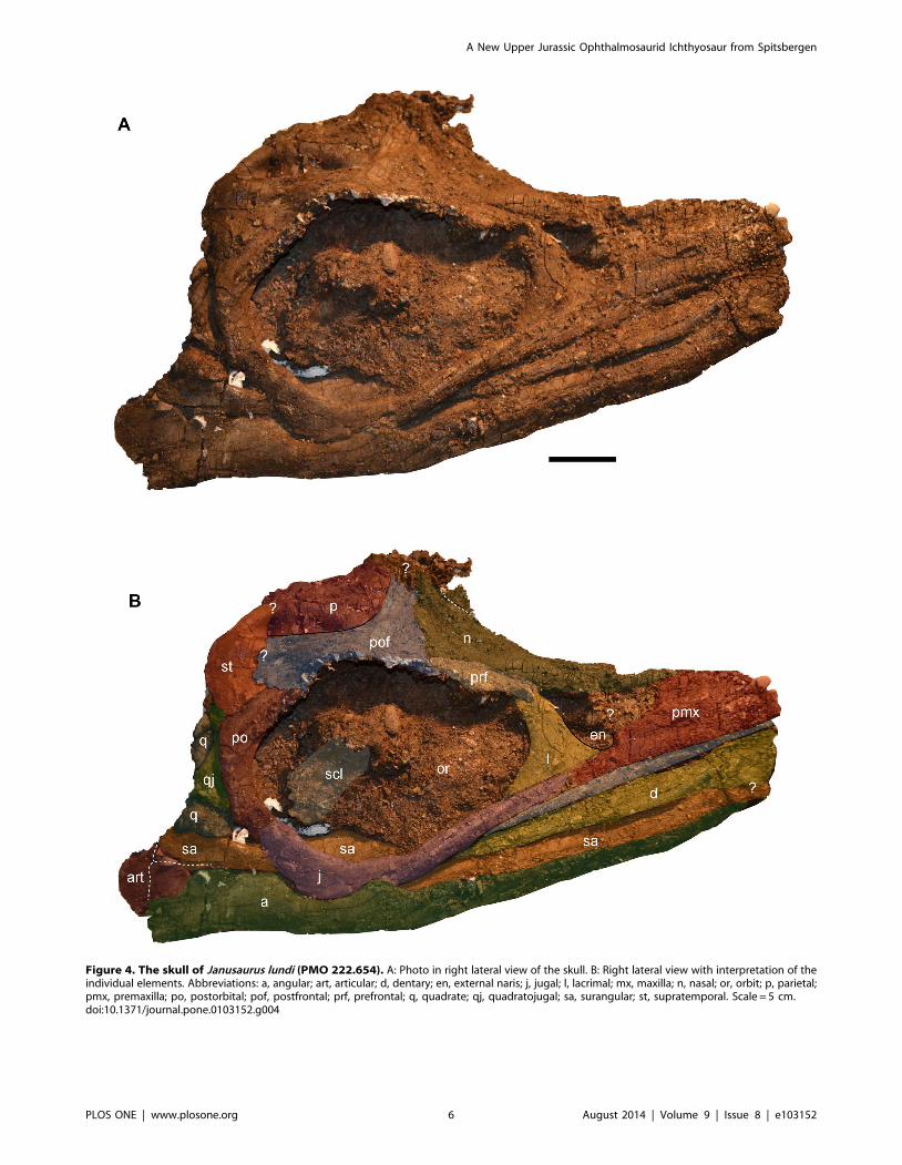
Figure 4. The skull of Janusaurus lundi (PMO 222.654). A: Photo in right lateral view of the skull. B: Right lateral view with interpretation of theindividual elements. Abbreviations: a, angular; art, articular; d, dentary; en, external naris; j, jugal; l, lacrimal; mx, maxilla; n, nasal; or, orbit; p, parietal;pmx, premaxilla; po, postorbital; pof, postfrontal; prf, prefrontal; q, quadrate; qj, quadratojugal; sa, surangular; st, supratemporal. Scale = 5 cm.doi:10.1371/journal.pone.0103152.g004
A New Upper Jurassic Ophthalmosaurid Ichthyosaur from Spitsbergen
PLOS ONE | www.plosone.org 6 August 2014 | Volume 9 | Issue 8 | e103152

bordered ventrally by the maxilla. The right jugal is broken and
distorted ventral to the mid-point of the orbit, but appears to
gently curve dorsally along the posterior third of its length before
contacting the postorbital (Figure 4).
The right prefrontal forms approximately one third of the
dorsal margin of the orbit. The anterior edge of the prefrontal does
not appear to contribute to the external naris. Together with the
postfrontal, the prefrontal forms the supraorbital flange. The nasal
and postfrontal overlap the prefrontal medially and posteriorly,
respectively. The right postfrontal is a large and prominent
element, whose size and extent are discerned on the basis of bone
fiber orientation, which radiates from the center of the bone. It
forms the posterior two-thirds of the dorsal border of the orbit.
The nasals appear to overlap the anterior margin of the
postfrontal. The postfrontal-postorbital contact is short externally,
but the two elements share a long overlapping contact ventrally,
on the medial side of the orbital rim (Figure 5). The postfrontal has
a broad contact posteriorly and medially with the supratemporal,
thereby excluding the postfrontal from participating in the lateral
margin of the temporal fenestra. However, the postfrontal forms
most, if not all, of the anterior border of the supratemporal
fenestra. Medially the relationship of the postfrontal to the frontal
is unclear. Anterior to the supratemporal fenestra, the postfrontal
has a long, straight overlapping contact with the parietal that
extends nearly to the midline of the skull.
The relationships of the frontals cannot be discerned due to
poor preservation of the skull roof. However, part of the right
frontal could be preserved at the dorsal-most area of the preserved
portion of the skull, associated with a structure that could
represent part of the pineal foramen. The right parietal is well
preserved and forms the entire medial margin of the supratem-
poral fenestra. The parietal-supratemporal contact is hard to
discern (Figure 5), although a supratemporal process is present.
There is no indication of ornamentation or a sagittal crest along
the dorsal surface of the skull. The supratemporal fenestra is
anteroposteriorly longer than wide, although crushing makes it
difficult to interpret its original shape.
The right supratemporal is exposed on both sides of the
specimen. Its anterior and medial processes form all of the lateral
and most of the posterior margins of the supratemporal fenestra,
respectively. A squamosal was not identified in PMO 222.654,
and is presumed to have been absent, as the region in which this
element is usually present is well preserved in the specimen. The
postorbital bar of Janusaurus is relatively narrow (Table 1) and
has a postorbital bar ratio (maximum anteroposterior width versus
anteroposterior length of the orbit) of 0.68. The postorbital has
Figure 5. Oblique dorsal view of the skull of Janusaurus lundi (PMO 222.654). The dotted lines mark eroded or equivocal sutures.Abbreviations: p, parietal; pfor, pineal foramen; prf, prefrontal; pof, postfrontal; po, postorbital; q, quadrate; qj, quadratojugal; st, supratemporal; stf,supratemporal fenestra. Scale = 1 cm.doi:10.1371/journal.pone.0103152.g005
A New Upper Jurassic Ophthalmosaurid Ichthyosaur from Spitsbergen
PLOS ONE | www.plosone.org 7 August 2014 | Volume 9 | Issue 8 | e103152

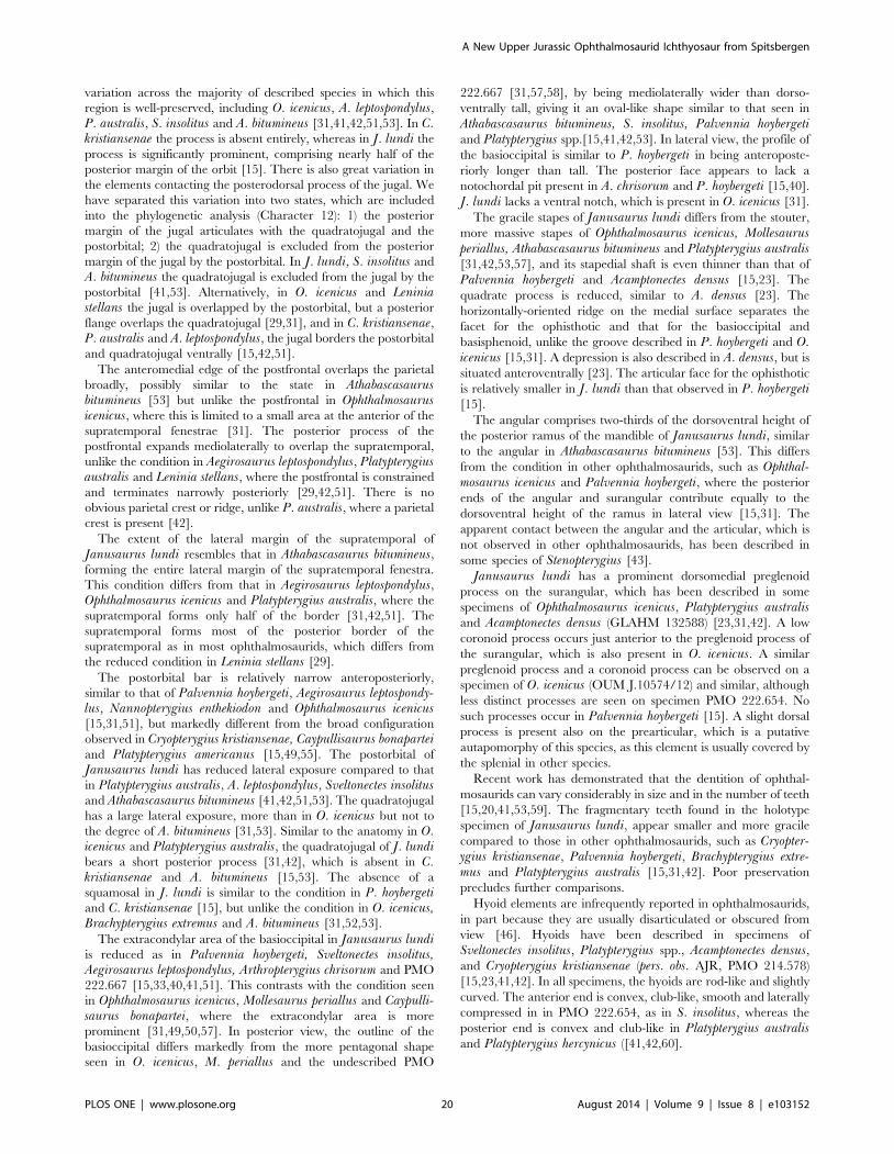
Figure 6. The basioccipital and right stapes from Janusaurus lundi (PMO 222.654). A: posterior view of the basioccipital; B: anterior view ofthe basioccipital; C: dorsal view of the basioccipital; D: ventral view of the basioccipital; E: lateral view of the right stapes, F: medial view of the medialhead of the right stapes; G: anterior view of the right stapes; H: posterior view of the right stapes. Abbreviations: baf/bsf, basioccipital andbasisphenoid facet;bsf, basiospenoid facet; hp, hyoid facet; oc, occipital condyle; opf, opisthotic facet; qf, quadrate facet; stf, stapes facet.Scale = 1 cm.doi:10.1371/journal.pone.0103152.g006
A New Upper Jurassic Ophthalmosaurid Ichthyosaur from Spitsbergen
PLOS ONE | www.plosone.org 8 August 2014 | Volume 9 | Issue 8 | e103152

clear sutural relationships with the postfrontal dorsally, the
supratemporal posterodorsally, the quadratojugal posteriorly,
and the jugal ventrally. The quadratojugal contacts the
postorbital along most of its posterior border and has a short
underlapping contact with the supratemporal dorsally. As seen in
lateral view, the ventral portion of the quadratojugal is antero-
posteriorly broader than the dorsal half and thus projects caudally
from the posterior margin of the postorbital bar.
Figure 7. Medial view of the preserved right mandible of Janusaurus lundi (PMO 222.654). Medial view of the posterior portion of the rightmandible and the posterior portion of the palate. Abbreviations: a, angular; art, articular; cor, coronid process; part, prearticular; partp, prearticulardorsal process; pgp, preglenoid process; pt, right pterygoid; sa, surangular. Scale = 1 cm.doi:10.1371/journal.pone.0103152.g007
Figure 8. The right hyoid from Janusaurus lundi (PMO 222.654). Posterior is to the left, anterior to the right. Ventral view is situated at the top,dorsal view below. Scale = 1 cm.doi:10.1371/journal.pone.0103152.g008
A New Upper Jurassic Ophthalmosaurid Ichthyosaur from Spitsbergen
PLOS ONE | www.plosone.org 9 August 2014 | Volume 9 | Issue 8 | e103152

Braincase. PMO 222.654 preserves the basioccipital, basi-
sphenoid and right stapes, but other elements are either not visible
or not preserved. Selected measurements can be found in Table 2.
The basioccipital of PMO 222.654 was disarticulated from the
braincase but is largely intact, though fragmented. The element
was not found in place and is poorly preserved. Our orientation is
presented in Figure 6 A-D. The occipital condyle is intact, but the
extracondylar area is eroded and part of it is missing. The occipital
condyle is convex and is mediolaterally wider than tall. In dorsal
and posterior view the condyle blocks the extracondylar area
almost entirely from view, although an indication of the lateral
facets for the ophisthotics are visible. It is only possible to describe
Figure 9. Pectoral girdle of Janusaurus lundi (PMO 222.654). A: posterior view of the left clavicle; B: posterior view of the right clavicle; C:anterior view of the left clavicle; D: anterior view of the right clavicle; E: ventral view of the interclavicle; F: dorsal view of the interclavicle; G: dorsal?view of the right scapula; H: dorsal view of the left coracoid; I: dorsal view of the right coracoid. Abbreviations: amp, anteromedial process; ict,interclavicular trough; ms, medial symphysis; sb, scapular blade; vf, ventral foramen. Scale = 5 cm.doi:10.1371/journal.pone.0103152.g009
A New Upper Jurassic Ophthalmosaurid Ichthyosaur from Spitsbergen
PLOS ONE | www.plosone.org 10 August 2014 | Volume 9 | Issue 8 | e103152
the facet for the left ophisthotic because the right has been
deformed. The length of this facet is approximately 3 cm. Most of
the dorsal surface is damaged, but an indication of the left
exoccipital facet is preserved. The anterior surface is fragmented,
with little surface available for description. However, the preserved
anterior surface is uneven and pitted and has a projection in the
center of the anterior surface; this process extends ventrally and
ends in an eroded ‘‘spine’’. There is no indication of a ventral
notch, but this area has been severely damaged, so this structure
may have been present.
Only the right stapes was recovered from the dorsal part of the
skull (Figure 6 E-H). The stapedial shaft is thin, rounded and
gracile along the entire shaft, expanding slightly at the quadrate
facet. The posterior side of the medial surface of the stapes is flat,
and expands posteriorly from the medial head in dorsal view. The
anterior margin is convex, especially at the medial stapedial head.
A ridge on the articular surface of the medial stapedial head
separates the surface for the ophisthotic from the ventrally-situated
surface for the basioccipital and basisphenoid. This surface
terminates in a ridge that could be homologous to the hyoid
process described for Ophthalmosaurus icenicus and Acamptonectesdensus [23,31]. The entire medial stapedial head is rugose, and a
tubercle on the ventral side is interpreted to be a surface for
attachment of a hyoid ligament as in O. icenicus and some
specimens of Acamptonectes [23]. The basisphenoid is lodged
inside the orbit and was not possible to remove for description.
Palatal complex. The palate is poorly preserved, and most
of the elements are lost. Parts of the posterior ramus of the right
pterygoid are visible in medial view (Figure S1). The right
pterygoid is displaced and damaged in several places. The
quadrate ramus is drawn out into three processes, extending
laterally, medially and dorsally. The ventral surface of the
quadrate ramus is smooth, but with a more irregular posterior
end. The pterygoid extends anteriorly up to the mid-point of the
orbit, where it is eroded. The right pterygoid is visible but situated
on top of the right quadrate, blocking it from medial view
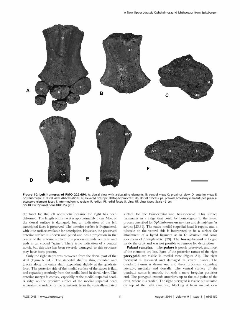
Figure 10. Left humerus of PMO 222.654. A: dorsal view with articulating elements; B: ventral view; C: proximal view; D: anterior view; E:posterior view; F: distal view. Abbreviations: er, elevated rim; dpc, deltopectoral crest; dp, dorsal process; pe, preaxial accessory element; pef, preaxialaccessory element facet; i, intermedium; r, radiale; R, radius; Rf, radial facet; U, ulna; Uf, ulnar facet. Scale = 5 cm.doi:10.1371/journal.pone.0103152.g010
A New Upper Jurassic Ophthalmosaurid Ichthyosaur from Spitsbergen
PLOS ONE | www.plosone.org 11 August 2014 | Volume 9 | Issue 8 | e103152
(Figure 7). The right quadrate is partially visible ventral to the
postorbital and quadratojugal, and in posteromedial view beneath
the right pterygoid (Figure S1). Unfortunately most of this element
is not visible enough to describe in detail.
Mandible. PMO 222.654 only preserves an incomplete right
lower jaw, visible in both lateral and medial views, but is missing
its anterior end. Measurements of the individual elements can be
found in Table 3. Only the posterior-most region of the mandible
is well-preserved, particularly in medial view (Figure 7). The
posterior margin of the dentary lies in line with the anterior third
of the orbit. A small portion of the dental groove is visible in which
diminutive conical teeth are held. The surangular extends at
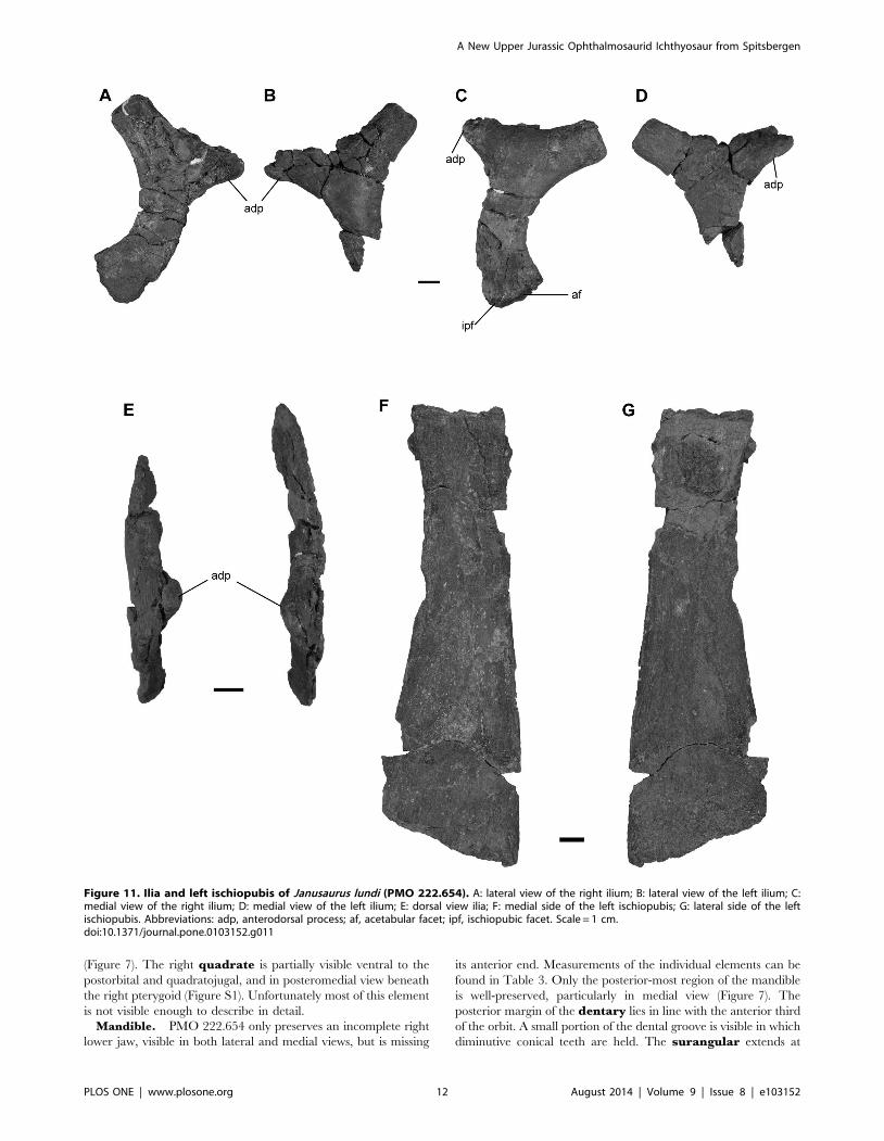
Figure 11. Ilia and left ischiopubis of Janusaurus lundi (PMO 222.654). A: lateral view of the right ilium; B: lateral view of the left ilium; C:medial view of the right ilium; D: medial view of the left ilium; E: dorsal view ilia; F: medial side of the left ischiopubis; G: lateral side of the leftischiopubis. Abbreviations: adp, anterodorsal process; af, acetabular facet; ipf, ischiopubic facet. Scale = 1 cm.doi:10.1371/journal.pone.0103152.g011
A New Upper Jurassic Ophthalmosaurid Ichthyosaur from Spitsbergen
PLOS ONE | www.plosone.org 12 August 2014 | Volume 9 | Issue 8 | e103152
least as far anteriorly as the preserved portion of the jaw. In medial
view, a small coronoid process is visible. Posterior to the coronoid
process is a second and more prominent, dorsally-projecting
process on the surangular, here termed the preglenoid process.
This structure, whose entire medial surface is marked with ridges,
has been interpreted to be the point of insertion for the M.
adductor mandibulae externus group (MAME) in Ophthalmo-saurus by Kirton [31] or the M. adductor mandibulae internus
pseudotemporalis (MAMIP) by Kear [42].
In lateral view, the anterior end of the angular lies near the
preserved portion of the skull. The angular contributes approx-
imately two-thirds of the dorsoventral height at the posterior end
of the mandible in lateral view. Although the posterodorsal portion
of the ramus was lost during preparation, its size and shape can be
determined by facets on the articular. In medial view, the angular
extends further posteriorly than the prearticular and forms the
ventral margin of the articular. The prearticular is dorsoven-
trally tall anteriorly as seen in medial view. The shape of the dorsal
margin of the prearticular somewhat mirrors that of the
surangular, in possessing a slight dorsal process before abruptly
tapering in height posteriorly to a narrow process, and terminating
along the anteromedial margin of the articular. Only a small
portion of the splenial is preserved medially, but little can be said
regarding its morphology.
The articular is articulated to the prearticular and angular,
and enclosed behind the surangular in lateral view (Figure S2 L). It
appears more anteroposteriorly elongated than round. The clear
sutures for the prearticular on the medial side as well as for the
surangular on the lateral side, suggest that the element is
articulated. In medial view the articular is convex, bulging
outwards posterodorsally, with a prominent ridge located along
the posterior edge. The anterodorsal edge articulating with the
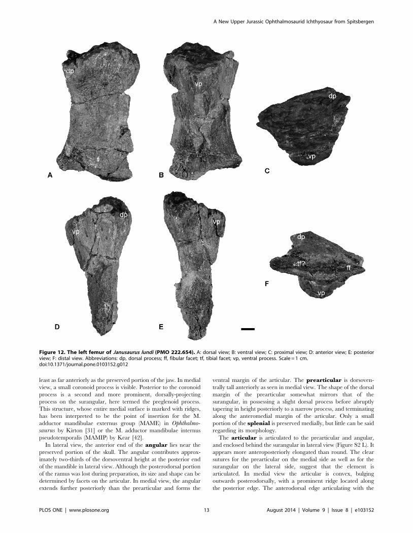
Figure 12. The left femur of Janusaurus lundi (PMO 222.654). A: dorsal view; B: ventral view; C: proximal view; D: anterior view; E: posteriorview; F: distal view. Abbreviations: dp, dorsal process; ff, fibular facet; tf, tibial facet; vp, ventral process. Scale = 1 cm.doi:10.1371/journal.pone.0103152.g012
A New Upper Jurassic Ophthalmosaurid Ichthyosaur from Spitsbergen
PLOS ONE | www.plosone.org 13 August 2014 | Volume 9 | Issue 8 | e103152

Figure 13. The axial skeleton of Janusaurus lundi (PMO 222.654). Including the partial atlas-axis complex, complete trunk vertebra, neuralarches, dorsal rib and gastralia from PMO 222.654. A: lateral? view of atlas-axis complex; B: anterior view of atlas-axis complex; C: lateral view of trunkvertebrae; D: anterior view of trunk vertebrae; E: posterior view of trunk vertebrae; F: dorsal view of trunk vertebrae; G: neural arch from the sacral
A New Upper Jurassic Ophthalmosaurid Ichthyosaur from Spitsbergen
PLOS ONE | www.plosone.org 14 August 2014 | Volume 9 | Issue 8 | e103152

quadrate is rounded and rugose, suggesting large amounts of
connective tissue. The ventromedial side is smooth and is slightly
concave, where it articulates with the prearticular and the angular.
An articulation between the articular and the angular has not been
previously identified in ophthalmosaurids, but occurs in some
specimens of Stenopterygius [43]. Anterolaterally the articular is
covered by the surangular and the angular. The medial surface of
the articular is smooth in the center, becoming more irregular
towards the articular surfaces.
Dentition. In medial view, disarticulated tooth fragments are
visible. The total number of fragments preserved is uncertain,
although they have remained in the jaw. The crown height based
on partial fragments is very short with an estimated height less
than 9 mm. Fine ridging is occurs on all sides of the teeth.
Scleral elements. A partially articulated but poorly pre-
served scleral ring is present in the right orbit. The minimum
length of a single plate, as measured from the inner to outer
margin of the ring, is approximately 6 cm.
Hyoid apparatus. A pair of hyoid rods was found on the
ventromedial side of the angular, but only the right hyoid was in
sufficient condition to be prepared (Figure 8). The rod is
completely three dimensional and gently curved in shape. The
anterior end is convex and club-like. The shaft of the hyoid rod is
rounded in cross section and its posterior end is semi-spatulate.
The appendicular skeletonThe pectoral girdle. The pectoral girdle of PMO 222.654
was articulated, although slightly displaced, and lacks only the left
scapula (Figure 9). The clavicles are well-preserved. The right
clavicle is 1.5 times as long as the anteroposterior length of the
right coracoid (Table 4; Figure 9 A-D). The medial ends of both
clavicles end in finger-like projections that neatly interdigitate with
their opposites [44]. The visceral surfaces of the medial portions of
the clavicles are dished, so as to envelope the lateral rami of the
interclavicle. The lateral ends of the clavicles curve dorsally and
posteriorly and bear facets for the scapula. The interclavicle is
complete and well preserved (Figure 9 E-F). The lateral rami are
dorsoventrally flattened and ventrally convex for reception with
the clavicles. The combined mediolateral width of both lateral
rami is greater than the anteroposterior length of the interclavicle.
Located on the anterior half of the dorsal surface of the posterior
ramus are two dorsally-projecting processes that form the lateral
margins of a narrow, anteroposteriorly-oriented excavation, which
is here termed the interclavicluar trough. Not all of the shale
imbedded in this posterior-most section of this trough was
removed during preparation. Thus, the true posterior extent of
the interclavicular trough is longer than shown on Figure 9 E-F.
The posterior end of the element is dorsoventrally flattened and is
somewhat expanded mediolaterally. A foramen is located on the
ventral surface of the interclavicle, where the lateral and posterior
rami meet.
The coracoids (Figure 9 H) are slightly longer anteroposteriorly
than wide mediolaterally. The medial symphysis forms the
anterior two-thirds of the coracoid; in medial view the outline of
the symphysis is lenticular and markedly short dorsoventrally. In
dorsal view, the anterior margin of the coracoid forms a prominent
anteromedial process, which marks the lateral margin of a well
formed anterior notch. The scapular and humeral facets are
approximately equal in size and are offset at an angle of 150
degrees from the sagittal plane. Only a poorly-preserved and
incomplete right scapula was recovered (Figure 9 G). The
suprascapular border is intact, although rather short compared
to the size of the rest of the pectoral girdle. The anterior portion of
the scapular blade is distorted and folded, which precludes further
description.
Forefin. Two partial forefins are preserved in PMO 222.654.
The left forefin includes a complete humerus disarticulated from
the pectoral girdle with an articulated zygopodium and a few
associated autopodial elements. The right humerus was entirely
disarticulated and was found in the vicinity of an isolated right
radius. The orientation and identity of the humeri was determined
by comparisons with articulated limbs of other ophthalmosaurid
remains from the Slottsmøya Member and from published
descriptions [15,31,45]. The anteroposterior axis was determined
region in anterior view; H: neural arch from the trunk region in lateral view; I: nearly complete dorsal rib; J: complete gastralium. Abbreviations: prz,prezygapophysis; dp, diapophysis; ns, neural spine; pp, parapophysis. A-H Scale = 1 cm, I Scale = 10 cm, J Scale = 1 cm.doi:10.1371/journal.pone.0103152.g013
Figure 14. Skull reconstruction of Janusaurus lundi in right lateral view. Abbreviations: a, angular; art, articular; bo, basioccipital; d, dentary;en, external naris; j, jugal; l, lacrimal; mx, maxilla; n, nasal; or, orbit; p, parietal; pmx, premaxilla; po, postorbital; pof, postfrontal; prf, prefrontal; q,quadrate; qj, quadratojugal; sa, surangular; st, supratemporal; stf, supratemporal fenestra.doi:10.1371/journal.pone.0103152.g014
A New Upper Jurassic Ophthalmosaurid Ichthyosaur from Spitsbergen
PLOS ONE | www.plosone.org 15 August 2014 | Volume 9 | Issue 8 | e103152
by the location of the preaxial accessory element and the
dorsoventral orientation via the shape and position of the dorsal
process and deltopectoral crest (Figure S2 A-D).
The humerus is proximodistally longer than anteroposteriorly
broad at its distal end. The distal end is anteroposteriorly broader
than the proximal end (Figure 10 A-B). The minimum anterior-
posterior width at midshaft is 16% smaller than the maximum
anteroposterior width of the proximal end, giving a length-to-
width ratio of 1.8. The proximal articular surface of the humerus is
relatively flat, with a rugose surface texture. An elevated rim circles
the periphery of the dorsal, anterior and ventral portions of the
articular facet. In dorsal view, the dorsal process originates near
the anteroposterior midpoint of the articular facet and extends to
midshaft. It is slightly angled towards the anterior margin. The
dorsal process is relatively tall and narrow compared to the
deltopectoral crest, which is more broadly rounded (Figure 10 E).
The deltopectoral crest begins near the anterior margin of the
humerus, extends to near the midpoint of the shaft, and slants
slightly posteriorly. In anterior view, the preaxial margin of the
humeral shaft is dorsoventrally shorter than tall and has a broadly
rounded postaxial margin.
Distally, the humerus bears three concave distal facets. The
anterior-most, for the preaxial accessory element, is approximately
half the anteroposterior length and dorsoventral height of the
radial facet (Table 5). The ulnar facet is dorsoventrally taller than
the radial facet, but slightly shorter anteroposteriorly (Table 5).
Relative to the long axis of the humerus, the facets for the preaxial
accessory element and the ulna are angled at approximately 17
degrees anteriorly and 30 degrees posteriorly, respectively.
The zygopodial row includes the radius, ulna and a preaxial
accessory element, where the radius and ulna are recognized as the
two largest elements [31,45]. The preaxial accessory element is
oval-shaped in dorsal view, but is dorsoventrally narrow anteriorly.
The proximal margin is three times the dorsoventral thickness of
the distal end. The preaxial accessory element bears two distinct
articular surfaces, one proximally for the humerus and the other
posteriorly for the radius. The ulna and radius are similar in
dorsoventral height, but the ulna is anteroposteriorly longer. In
dorsal view, the ulna is triangular in outline, whereas the radius is
more oval. Both elements are convex on their articular facets for
reception by the humerus (Figure 10 A). The surfaces in contact
with the humerus and other elements are pitted and rugose.
Very little of the autopodium of PMO 222.654 has been
preserved (Figure 10 A). The left intermedium and possibly the left
radiale are semi-articulated with the left zygopodium. The
intermedium articulates snugly between the radius and ulna and
is twice as thick dorsoventrally at its proximal end than at its distal
end. Several other small elements were found in the vicinity of the
left humerus but their identity is equivocal. It is not possible to
determine the number of digits in PMO 222.654.
The pelvic girdle. PMO 222.654 includes two disarticulated
ilia and an ischiopubis found in the vicinity of a series of
articulated caudal vertebrae, along with both disarticulated femora
and several limb elements (Figure 11).
One ilium is complete, whereas the other is missing its distal
portion (Figure 11 A-E). Our preferred orientation for the ilia
follows the description for Ophthalmosaurus icenicus provided by
Kirton [31], comparisons with a complete and articulated pelvic
girdle of Cryopterygius kristiansenae [15] and descriptions from
other articulated specimens [46]. Determination of the mediolat-
eral axis is based on curvature of the element along its
proximodistal axis, with the concave surface being medial. We
interpret the markedly concave margin of the ilial shaft to be
facing posteriorly. The proximal end is mediolaterally flattened,
and the proximal end twists medially, closer to the vertebral
column. The distal end is slightly larger than the proximal end and
has two facets for the ischiopubis and femur. Using these criteria
we interpret the one complete ilium to be the right. Using this
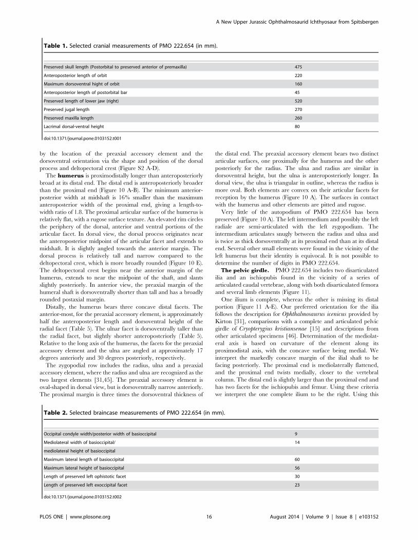
Table 1. Selected cranial measurements of PMO 222.654 (in mm).
Preserved skull length (Postorbital to preserved anterior of premaxilla) 475
Anteroposterior length of orbit 220
Maximum dorsoventral hight of orbit 160
Anteroposterior length of postorbital bar 45
Preserved length of lower jaw (right) 520
Preserved jugal length 270
Preserved maxilla length 260
Lacrimal dorsal-ventral height 80
doi:10.1371/journal.pone.0103152.t001
Table 2. Selected braincase measurements of PMO 222.654 (in mm).
Occipital condyle width/posterior width of basioccipital 9
Mediolateral width of basioccipital/ 14
mediolateral height of basioccipital
Maximum lateral length of basioccipital 60
Maximum lateral height of basioccipital 56
Length of preserved left ophistotic facet 30
Length of preserved left exoccipital facet 23
doi:10.1371/journal.pone.0103152.t002
A New Upper Jurassic Ophthalmosaurid Ichthyosaur from Spitsbergen
PLOS ONE | www.plosone.org 16 August 2014 | Volume 9 | Issue 8 | e103152

orientation, the prominent process visible on each ilium is located
in the proximal half of the element and projects anterodorsally
(Figure 11 A-D). This ilial process occurs on both elements and is
not a taphonomic artifact. The ilial process ends in a blunt tip and
curves slightly medially. The lateral surface of the process bears
small ridges and the medial surface is rugose. Both the proximal
and distal ends of the ilia are pitted.
The preserved ischiopubis is interpreted to be the right, based
solely on its association with the right ilium. It is nearly complete
but is slightly eroded at its distal end. The element is
mediolaterally thickened proximally, but is otherwise very flat
and anteroposteriorly broader distally (Figure 11 F-G). Both the
ischium and pubis are fused along their entire length, and there is
no indication of an obturator foramen. The maximum length is
3.3 times longer than the maximum width (Table 6). The
proximodorsal edge bears a ridge that flattens towards the distal
end [31].
Table 6. Selected pelvic girdle measurements of PMO 222.543
(in mm).
Hindfin. The hindfins are completely disarticulated and
include two femora and several partial zygopodial and/or
autopodial elements. These elements are too damaged to identify
and describe. Measurements of the individual femora can be found
in Table 7.
The identity of the femora was determined using the articulated
pelvic girdle and hind fin of Cryopterygius [15] and Maxwell et al.
[47]. The left femur (Figure 12) is better preserved than the right
(Figure S2 E-J), but the distal ends of both femora are compressed,
Table 3. Selected mandibular measurments from PMO 222.654 (in mm).
Preserved angular length 485
Preserved suranglar length 453
Maximum posterior angular height 50
Maximum posterior surangular height 25
Approximate articular anteroposterior length 65
Height of preglenoid process 12
Height of coronoid process 2
doi:10.1371/journal.pone.0103152.t003
Table 4. Selected pectoral girdle measurements of PMO 222.654 (in mm).
Coracoids
Coracoid Left
Maximum mediolateral width 201
Maximum anteroposterior length 206
Length of intercoracoid suture 142
Coracoid Right
Maximum mediolateral width 213
Maximum anteroposterior length 219
Length of intercoracoid suture 156
Length of scapular facet 45
Length of glenoid facet 49
Scapula (Right)
Maximum proximodistal length 173
Clavicles
Clavicle left
Preserved maximum length 306
Maximum width 75
Clavicle right
Maximum length 344
length of scapula facet 45
Maximum width 76
Interclavicle
Preserved anteroposterior length 160
Maximum length of lateral bar 219
doi:10.1371/journal.pone.0103152.t004
A New Upper Jurassic Ophthalmosaurid Ichthyosaur from Spitsbergen
PLOS ONE | www.plosone.org 17 August 2014 | Volume 9 | Issue 8 | e103152

so the facets for tibia and fibula are unclear. The proximal and
distal ends are nearly identical in anteroposterior width. The
proximal articular surface is well preserved in the left femur. This
surface is convex dorsally and concave ventrally at the location of
the ventral process. In proximal view, the femur is approximately
as dorsoventrally tall as anteroposteriorly wide. In ventral view, an
anteroposteriorly broad ventral process extends to near the
midpoint of the femur. The dorsal process originates near the
anterior margin and terminates midway along the shaft. There
appear to be only two facets located on the distal end, although
this is equivocal. On the right femur a slight ridge seems to
separate the two facets (Figure S2 H). The fibular facet is
approximately 20 percent longer than the tibia facet. The
anteroproximal side appears to have several foramina.
The axial skeleton. A total of 21 complete to partially
complete vertebrae were found, including the atlas-axis, four
articulated cervicals and several dorsal vertebrae, which were
associated with the skull. Seven articulated caudal vertebrae were
found near the pelvic girdle, along with 10 other disarticulated
sacral/caudal vertebrae. The atlas-axis complex is incomplete
and is laterally compressed, with only the right lateral side
(stratigraphically up) preserved. The complex is completely fused
and lacks any sign of a suture. The anterior face of the atlas
(Figure 13 A-B) is broad compared to the axis, and more deeply
cupped to articulate with the occipital condyle. Most of the dorsal
and cervical vertebrae were left in articulation with the dorsal ribs,
but a single cervical vertebra was removed, directly posterior to
the atlas-axis (Figure 13 C-F). It is laterally compressed and
deformed, but facets for the right rib and right side of the neural
arch are visible. The anterior surface of the vertebrae is very
concave and slightly irregular, suggesting connective tissue
investment. Two partial and disarticulated neural arches from
the sacral- and trunk region, were preserved but cannot be
measured or described further due to poor preservation (Figure 13
G-H).
Most of the preserved dorsal ribs are associated with the
dorsal vertebrae. The trunk ribs are estimated to be on average 90
cm in length. The most complete of these measures 84 cm in
length, although approximately 10 cm of the distal end was
missing (Figure 13 I). The ribs are figure-eight shaped in cross
section along the entire length of the shaft, apart from the circular
cross section at its ventral termination [46]. PMO 222.654
preserves a large number of gastralia, which are seldom
described in ophthalmosaurids. The gastralia have a circular cross
section. Their medial ends are softly rounded and the lateral ends
terminate in a thin point (Figure 13 J).
Discussion
Janusaurus lundi can be confidently placed in Ophthalmosaur-
idae on the basis of having a reduced extracondylar area of the
basioccipital, extensive lateral exposure of the angular, and a
preaxial accessory element in the forelimb [46]. The specimen has
an anterior twisting dorsal process on the humerus, which has also
been proposed as a synapomorphy of the clade [48]. A broad
contact between the premaxilla and lacrimal, although not present
in Platypterygius australis [42], has been identified as a general
synapomorphy, which the specimen could possibly share [40]. The
Table 5. Selected measurements from the left humerus of PMO 222.654 (in mm).
Maximum proximodistal length 152
Maximum anteroposterior width, proximal end 104
Maximum dorsoventral height, proximal end 86
Maximum anteroposterior width, distal end 136
Maximum dorsoventral height, proximal end 69
Mimimun anteroposterior width, midshaft 85
Length of radial facet 49
Length of ulnar facet 61
Length og preax. element facet 25
doi:10.1371/journal.pone.0103152.t005
Table 6. Selected pelvic girdle measurements of PMO 222.543 (in mm).
Ilium
Maximum anteroposterior length 97
Acetabulum process length 9
Posterior height 22
Length of anterodorsal process 24
Ischiopubis
Maximum proximodistal length 181
Anteroposterior length, proximal end 35
Anteroposterior length, distal end 55
Dorsovental thickness proximal end 11
doi:10.1371/journal.pone.0103152.t006
A New Upper Jurassic Ophthalmosaurid Ichthyosaur from Spitsbergen
PLOS ONE | www.plosone.org 18 August 2014 | Volume 9 | Issue 8 | e103152

following discussion compares Janusaurus lundi to all described
Middle Jurassic to Early Cretaceous ophthalmosaurids as well as
an undescribed ophthalmosaurid, PMO 222.667, from the
Agardhfjellet Formation and Malawania anachronus [28]. A skull
reconstruction can be found in Figure 14.
DermatocraniumThe subnarial process of the premaxilla appears to have
contacted and possibly even broadly overlapped the anterior
process of the lacrimal, similar to that of Caypullisaurus bonaparteiand Aegirosaurus leptospondylus [49–51], but unlike Platypter-ygius australis which lacks this contact [42]. Although a
premaxilla-jugal contact is unclear, this area lacks any evidence
for the broadly interdigitating contact seen in Brachypterygiusextremus [52]. The subnarial process of the premaxilla of
Janusaurus lundi participates in the anterior and anteroventral
boundary of the external naris similar to the condition in C.bonapartei and A. leptospondylus [49–51], but unlike the
morphology in P. australis and Athabascasaurus bitumineus which
lack any contact [42,53]. This also differs from the condition in
Ophthalmosaurus icenicus, where the premaxilla only borders the
anteroventral margin of the external naris [31].
The maxilla of Janusaurus lundi has considerable lateral
exposure, particularly posteriorly, similar to the condition in
Caypullisaurus bonapartei, Palvennia hoybergeti and Cryoptery-gius kristiansenae [15,49], but not to the degree of Leniniastellans, where the maxilla extends as far as the mid-point of the
orbit [29]. This trait is absent in Ophthalmosaurus icenicus and
Athabascasaurus, where the maxilla has almost no posterior
exposure [31,53]. The maxilla of J. lundi fails to contact the
lacrimal; this differs from O. icenicus, Maiaspondylus lindoi and P.hoybergeti, where the ventral border of the lacrimal clearly
contacts the maxilla in lateral view [15,31,54]. The maxilla does
not appear to clearly contact the external naris as in Platypterygiusaustralis and M. lindoi [42,54]. In the description for M. lindoi,Maxwell and Caldwell [54] described the extensive overlap
between jugal and maxilla as an autapomorphy for the species,
but this feature is now also described in J. lundi and C.kristiansenae [15]. Despite lacking its anterior-most margin, the
maxilla shows extensive anterior and lateral exposure compared to
most ophthalmosaurids [15,42,53].
The nasal of Janusaurus lundi participates in the external naris
and forms the dorsal border, possibly as an arched overhang
similar to the morphology in Platypterygius australis, and
Ophthalmosaurus icenicus [31,42]. The nasal forms the entire
dorsal boundary of the prefrontal similar to the morphology in O.icenicus, Sveltonectes insolitus and Palvennia hoybergeti, but
unlike the condition in Athabascasaurus bitumineus where the
posterodorsal portion of the prefrontal is covered by the
postfrontal [15,31,41,53].
A prominent anterior process of the lacrimal is present and
more pronounced than in Cryopterygius kristiansenae [15], but
less pronounced than in Aegirosaurus leptospondylus, Ophthal-
mosaurus icenicus, Platypterygius americanus and Platypterygius
bannovkensis [31,51,55,56]. The lacrimal of Janusaurus lundi
clearly forms the entire posterior border of the external naris,
unlike the anatomy in C. kristiansenae, Platypterygius australis
and Athabascasaurus bitumineus, where the lacrimal is excluded
from the external naris by an ascending process from the maxilla
[15,42,53]. The gently concave orbital margin of the lacrimal in J.
lundi contrasts with the 90 degree bend seen in C. kristiansenae
[15]. The external naris is dorsoventrally taller than anteropos-
teriorly long, similar to that in C. kristiansenae, but unlike the
condition in other ophthalmosaurids such as Palvennia hoybergeti,
O. icenicus and A. leptospondylus, where the naris is longer than
tall [15,31,51]. The prefrontal does not participate in the border of
the external naris, as in O. icenicus and Sveltonectes insolitus
[31,41].
There appears to be significant variation of the morphology and
relationships of the anterior process of the jugal in ophthalmo-
saurids. Thus, we have separated the variation into two different
states and incorporated them into the phylogenetic analysis
(Character 11): 1) the anterior process of the jugal terminates
posterior to the anteroventral margin of the lacrimal; 2) the
anterior process of the jugal reaches or extends anterior to the
anteroventral margin of the lacrimal. Most ophthalmosaurids
possess the first state (0), including Leninia stellans, Maiaspondyluslindoi, Cryopterygius kristiansenae, Caypullisaurus bonapartei,Aegirosaurus leptospondylus, Sveltonectes insolitus, Ophthalmo-saurus icenicus and Athabascasaurus bitumineus[15,29,31,41,51,53,54], while Platypterygius australis, Caypulli-saurus bonapartei and Brachypterygius extremus have the second
state (1) [31,42,50]. Janusaurus lundi is tentatively referred to the
first state, as the jugal terminates ventral to the lacrimal but does
not reach the anteroventral end. The shape of the jugal at the
ventral border of the orbit also shows great variation throughout
Ophthalmosauridae; the degree of ‘‘bowing’’ varies from being
entirely straight, to being gently bowed. The jugal of J. lundi has a
slightly bowed anterior process of the jugal which is neither as
straight as the jugal of B. extremus and C. kristiansenae, or as
gently bowed as the jugal observed in A. leptospondylus, O.icenicus, Palvennia hoybergeti and P. australis [15,31,42,51,52].
The variation in the degree of bowing needs to be further
investigated and constrained considerably before being included in
a phylogenetic analysis. The presence of a posterodorsal process of
the jugal articulating with the postorbital is also subject to
Table 7. Selected femora measurements of PMO 222.654 (in mm).
Left Femur
Maximum proximodistal length 103
Maximum anteroposterior width, proximal end 57
Maximum height, proximal end 49
Maximum anteroposterior width, distal end 58
Right Femur
Length of tibial facet 17
Length of fibular facet 21
doi:10.1371/journal.pone.0103152.t007
A New Upper Jurassic Ophthalmosaurid Ichthyosaur from Spitsbergen
PLOS ONE | www.plosone.org 19 August 2014 | Volume 9 | Issue 8 | e103152
variation across the majority of described species in which this
region is well-preserved, including O. icenicus, A. leptospondylus,P. australis, S. insolitus and A. bitumineus [31,41,42,51,53]. In C.kristiansenae the process is absent entirely, whereas in J. lundi the
process is significantly prominent, comprising nearly half of the
posterior margin of the orbit [15]. There is also great variation in
the elements contacting the posterodorsal process of the jugal. We
have separated this variation into two states, which are included
into the phylogenetic analysis (Character 12): 1) the posterior
margin of the jugal articulates with the quadratojugal and the
postorbital; 2) the quadratojugal is excluded from the posterior
margin of the jugal by the postorbital. In J. lundi, S. insolitus and
A. bitumineus the quadratojugal is excluded from the jugal by the
postorbital [41,53]. Alternatively, in O. icenicus and Leniniastellans the jugal is overlapped by the postorbital, but a posterior
flange overlaps the quadratojugal [29,31], and in C. kristiansenae,
P. australis and A. leptospondylus, the jugal borders the postorbital
and quadratojugal ventrally [15,42,51].
The anteromedial edge of the postfrontal overlaps the parietal
broadly, possibly similar to the state in Athabascasaurusbitumineus [53] but unlike the postfrontal in Ophthalmosaurusicenicus, where this is limited to a small area at the anterior of the
supratemporal fenestrae [31]. The posterior process of the
postfrontal expands mediolaterally to overlap the supratemporal,
unlike the condition in Aegirosaurus leptospondylus, Platypterygiusaustralis and Leninia stellans, where the postfrontal is constrained
and terminates narrowly posteriorly [29,42,51]. There is no
obvious parietal crest or ridge, unlike P. australis, where a parietal
crest is present [42].
The extent of the lateral margin of the supratemporal of
Janusaurus lundi resembles that in Athabascasaurus bitumineus,forming the entire lateral margin of the supratemporal fenestra.
This condition differs from that in Aegirosaurus leptospondylus,Ophthalmosaurus icenicus and Platypterygius australis, where the
supratemporal forms only half of the border [31,42,51]. The
supratemporal forms most of the posterior border of the
supratemporal as in most ophthalmosaurids, which differs from
the reduced condition in Leninia stellans [29].
The postorbital bar is relatively narrow anteroposteriorly,
similar to that of Palvennia hoybergeti, Aegirosaurus leptospondy-lus, Nannopterygius enthekiodon and Ophthalmosaurus icenicus[15,31,51], but markedly different from the broad configuration
observed in Cryopterygius kristiansenae, Caypullisaurus bonaparteiand Platypterygius americanus [15,49,55]. The postorbital of
Janusaurus lundi has reduced lateral exposure compared to that
in Platypterygius australis, A. leptospondylus, Sveltonectes insolitusand Athabascasaurus bitumineus [41,42,51,53]. The quadratojugal
has a large lateral exposure, more than in O. icenicus but not to
the degree of A. bitumineus [31,53]. Similar to the anatomy in O.icenicus and Platypterygius australis, the quadratojugal of J. lundibears a short posterior process [31,42], which is absent in C.kristiansenae and A. bitumineus [15,53]. The absence of a
squamosal in J. lundi is similar to the condition in P. hoybergetiand C. kristiansenae [15], but unlike the condition in O. icenicus,Brachypterygius extremus and A. bitumineus [31,52,53].
The extracondylar area of the basioccipital in Janusaurus lundiis reduced as in Palvennia hoybergeti, Sveltonectes insolitus,Aegirosaurus leptospondylus, Arthropterygius chrisorum and PMO
222.667 [15,33,40,41,51]. This contrasts with the condition seen
in Ophthalmosaurus icenicus, Mollesaurus periallus and Caypulli-saurus bonapartei, where the extracondylar area is more
prominent [31,49,50,57]. In posterior view, the outline of the
basioccipital differs markedly from the more pentagonal shape
seen in O. icenicus, M. periallus and the undescribed PMO
222.667 [31,57,58], by being mediolaterally wider than dorso-
ventrally tall, giving it an oval-like shape similar to that seen in
Athabascasaurus bitumineus, S. insolitus, Palvennia hoybergetiand Platypterygius spp.[15,41,42,53]. In lateral view, the profile of
the basioccipital is similar to P. hoybergeti in being anteroposte-
riorly longer than tall. The posterior face appears to lack a
notochordal pit present in A. chrisorum and P. hoybergeti [15,40].
J. lundi lacks a ventral notch, which is present in O. icenicus [31].
The gracile stapes of Janusaurus lundi differs from the stouter,
more massive stapes of Ophthalmosaurus icenicus, Mollesaurusperiallus, Athabascasaurus bitumineus and Platypterygius australis[31,42,53,57], and its stapedial shaft is even thinner than that of
Palvennia hoybergeti and Acamptonectes densus [15,23]. The
quadrate process is reduced, similar to A. densus [23]. The
horizontally-oriented ridge on the medial surface separates the
facet for the ophisthotic and that for the basioccipital and
basisphenoid, unlike the groove described in P. hoybergeti and O.icenicus [15,31]. A depression is also described in A. densus, but is
situated anteroventrally [23]. The articular face for the ophisthotic
is relatively smaller in J. lundi than that observed in P. hoybergeti[15].
The angular comprises two-thirds of the dorsoventral height of
the posterior ramus of the mandible of Janusaurus lundi, similar
to the angular in Athabascasaurus bitumineus [53]. This differs
from the condition in other ophthalmosaurids, such as Ophthal-mosaurus icenicus and Palvennia hoybergeti, where the posterior
ends of the angular and surangular contribute equally to the
dorsoventral height of the ramus in lateral view [15,31]. The
apparent contact between the angular and the articular, which is
not observed in other ophthalmosaurids, has been described in
some species of Stenopterygius [43].
Janusaurus lundi has a prominent dorsomedial preglenoid
process on the surangular, which has been described in some
specimens of Ophthalmosaurus icenicus, Platypterygius australisand Acamptonectes densus (GLAHM 132588) [23,31,42]. A low
coronoid process occurs just anterior to the preglenoid process of
the surangular, which is also present in O. icenicus. A similar
preglenoid process and a coronoid process can be observed on a
specimen of O. icenicus (OUM J.10574/12) and similar, although
less distinct processes are seen on specimen PMO 222.654. No
such processes occur in Palvennia hoybergeti [15]. A slight dorsal
process is present also on the prearticular, which is a putative
autapomorphy of this species, as this element is usually covered by
the splenial in other species.
Recent work has demonstrated that the dentition of ophthal-
mosaurids can vary considerably in size and in the number of teeth
[15,20,41,53,59]. The fragmentary teeth found in the holotype
specimen of Janusaurus lundi, appear smaller and more gracile
compared to those in other ophthalmosaurids, such as Cryopter-ygius kristiansenae, Palvennia hoybergeti, Brachypterygius extre-mus and Platypterygius australis [15,31,42]. Poor preservation
precludes further comparisons.
Hyoid elements are infrequently reported in ophthalmosaurids,
in part because they are usually disarticulated or obscured from
view [46]. Hyoids have been described in specimens of
Sveltonectes insolitus, Platypterygius spp., Acamptonectes densus,and Cryopterygius kristiansenae (pers. obs. AJR, PMO 214.578)
[15,23,41,42]. In all specimens, the hyoids are rod-like and slightly
curved. The anterior end is convex, club-like, smooth and laterally
compressed in in PMO 222.654, as in S. insolitus, whereas the
posterior end is convex and club-like in Platypterygius australisand Platypterygius hercynicus ([41,42,60].
A New Upper Jurassic Ophthalmosaurid Ichthyosaur from Spitsbergen
PLOS ONE | www.plosone.org 20 August 2014 | Volume 9 | Issue 8 | e103152
Appendicular and axial skeletonThe taxonomic utility of the pectoral girdle in many
ophthalmosaurids is questionable, due to the large amounts of
intraspecific variation [61]. However, the pectoral girdle of
Janusaurus lundi is well preserved and provides new data on
morphological variation in this region. The clavicles of J. lundi are
more robust and thickened in comparison to those in Cryopter-ygius kristiansenae and PMO 222.667 [15], and more closely
resemble those of Ophthalmosaurus icenicus and Aegirosaurusleptospondylus [31,51].
The scapula of Janusaurus lundi is notably shorter proximo-
distally than the same element in most ophthalmosaurids, when
compared against the anteroposterior length of the coracoid, a
feature that does not seem attributable to taphonomic distortion.
The interclavicles of Ophthalmosaurus icenicus (pers. obs. AJR:
NHMUK R.2180, R.2853, R.4753 CAMSM; J.68689eb, OUM;
J.48012) and PMO 222.667 are conspicuously thickened along the
anterior margin of the lateral ramii; this thickening is absent in J.lundi. Most notably the presence of an interclavicular trough is
unknown in any other ophthalmosaurid, and this appears to be an
autapomorphy of J. lundi. Likewise, a ventral foramen is absent in
O. icenicus, Aegirosaurus leptospondylus and Cryopterygiuskristiansenae [15,31,51].
The coracoids are similar in shape to that of Acamptonectesdensus, Ophthalmosaurus icenicus (pers. obs. AJR CAMSM
J29808, OUM J.48009), Cryopterygius kristiansenae and Aegir-osaurus leptospondylus, and are most similar to that of Arthropter-ygius chrisorum and Undorosaurus gorodischensis in general
morphology [15,23,30,40,44,51]. They differ from the strongly
dorsoventrally thickened coracoids in Platypterygius australis and
Sveltonectes insolitus [41,62]. The intercoracoid symphysis extends
along two-thirds of the medial length in Janusaurus lundi, in
contrast to C. kristiansenae, where the symphysis forms only half
of the medial length [15]. The coracoid symphysis of J. lundi is
dorsoventrally thinner than in S. insolitus, O. icenicus and P.australis and is of similar height to that of A. chrisorum[31,40,41,62]. The anterior notch in J. lundi is rounded and
shallow, in contrast to S. insolitus, where it is deep and narrow,
and in C. kristiansenae and A. densus where it is deep and wide
[15,23,41]. The scapular facet is approximately half the size of the
glenoid facet as in A. chrisorum and C. kristiansenae [15,40], in
contrast to the somewhat smaller facet in U. gorodischensis [30].
In overall morphology the humerus of Janusaurus lundi is most
similar to that in Arthropterygius chrisorum, Undorosaurusgorodischensis and Maiaspondylus lindoi [30,40,54], in that there
is little constriction of the midshaft and similar anteroposterior
lengths on the proximal and distal ends. This differs from the
humerus of Cryopterygius kristiansenae, Sveltonectes insolitus and
Platypterygius australis, where the proximal end is enlarged or of
the same length as the distal end [15,41,62]. Similar to the
humerus in the holotype specimen of A. chrisorum and to some
extent that in S. insolitus [40,41], the proximal surface of the
humerus in J. lundi is nearly flat, even though these specimens are
interpreted to be adults. The elevated rim found on the proximal
articular surface of J. lundi and PMO 222.667 is similar to the
condition described for U. gorodischensis and A. chrisorum.
However, pending first-hand examination of the specimens, it is
unclear if these structures are truly homologous [30,40]. The short
and narrow dorsal process of J. lundi differs from the tall dorsal
process found in S. insolitus, Acamptonectes densus and many
species of Platypterygius [23,41,60,62]. Unlike the humerus of
Ophthalmosaurus icenicus [31,46], the deltopectoral crest of J.lundi is more prominent in proximal and anterior views, although
it is still relatively shorter than the dorsal process.
There are three large and distinct distal facets on the humerus
in Janusaurus lundi, while only two distal facets are found in
Nannopterygius enthekiodon, Platypterygius americanus and
Sveltonectes insolitus [31,41,63], and a diminutive third facet is
sometimes found in Cryopterygius kristiansenae and Undorosaurusgorodischensis [15,30]. In some taxa which possess three facets,
such as Brachypterygius extremus, Aegirosaurus leptospondylus and
Maiaspondylus lindoi [51,52,54], the anterior-most is interpreted
as the radius, not the preaxial element, as found in J. lundi. The
facet for the radius is dorsoventrally wider, where the facet for the
ulna is anteroposteriorly longer, unlike Arthropterygius chrisorumand Ophthalmosaurus icenicus, where the radius facet is the larger
[31,40]. However, this trait seems to vary individually, particularly
in Acamptonectes densus [23].
The elements of the zeugopodium of the forefin in Janusauruslundi vary in dorsal outline; the radius is oval, the ulna slightly
triangular and the preaxial element is circular. This differs from
the elements in Sveltonectes insolitus, Cryopterygius kristiansenae,
Aegirosaurus leptospondylus and Maiaspondylus lindoi, where the
radius and ulna are pentagonal in form [15,41,51,54]. The
preaxial accessory is three times thicker proximally than distally as
in Arthropterygius chrisorum and Ophthalmosaurus icenicus[31,40]. The radius is slightly convex on the proximal surface,
similar to the radius in O. icenicus [31]. The ulna is the largest
element in PMO 222.654, as in specimens of O. icenicus and S.insolitus [31,41]. The pointed process observed on the proximal
edges of the ulna and radius in A. chrisorum are not present on the
described specimen [40].
The pelvic girdle of Janusaurus lundi is distinctive compared to
that in all other ophthalmosaurids, in that the ilium bears a
prominent, anterodorsally directed ilial process. A pelvic element
figured in the description of Caypullisaurus bonapartei, is
tentatively interpreted as a pubis [49,50]. Given the unusual ilial
morphology seen in J. lundi, there is the possibility that this
element may in fact be an ilium. The described specimen PMO
222.654 illustrates the possible morphological variation of this
element, so the pelvic element of C. bonapartei should be
reexamined to confirm this.
The ischiopubis of Janusaurus lundi lacks an obturator
foramen, similar to the condition in Aegirosaurus leptospondylus,Athabascasaurus bitumineus, Platypterygius australis and Svelto-nectes insolitus [41,51,53,64]. Many other ophthalmosaurids
possess an obturator foramen, such as Ophthalmosaurus icenicus[31], whereas Cryopterygius kristiansenae and Undorosaurusgorodischensis have an incompletely fused ischium and pubis at
their distal ends [15,30]. In J. lundi, the distal end of the
ischiopubis is 1.6 times wider than the proximal end, whereas it is
over twice as wide in A. leptospondylus [51].
The femur of Janusaurus lundi measures two-thirds of the
proximodistal length of the humerus, as in Arthropterygiuschrisorum and Ophthalmosaurus icenicus [31,40], but the femur
is only half the size of the humerus in Caypullisaurus bonapartei,Platypterygius hercynicus and Aegirosaurus leptospondylus [49-
51,60].The proximal and distal ends have the same maximum
anteroposterior width, unlike the femur in Cryopterygius kristian-senae and Undorosaurus gorodischensis where the distal end is
more expanded [15,30]. The proximal end of the femur in
Platypterygius australis differs from the condition in J. lundi, in
that the dorsal and ventral processes are both prominent, giving it
a completely different shape [64]. The dorsal process is small and
rounded in profile and is located anteriorly as in O. icenicus, A.chrisorum and C. kristiansenae [15,31,40]. In contrast, the dorsal
process in Sveltonectes insolitus and P. australis is much narrower
and is located closer to the proximal-distal midline of the femur in
A New Upper Jurassic Ophthalmosaurid Ichthyosaur from Spitsbergen
PLOS ONE | www.plosone.org 21 August 2014 | Volume 9 | Issue 8 | e103152

P. australis [41,64]. J. lundi has a prominent ventral process on
the femur, similar to A. chrisorum and C. kristiansenae [15,40].
The ventral process of J. lundi terminates just beyond the mid-
point of the femur, as in S. insolitus and Platypterygius americanus[41,55], but unlike O. icenicus, C. kristiansenae and A. chrisorumwhere it terminates at or proximal to the mid-point [15,31,40]. As
in most ophthalmosaurids, the distal end of the femur in J. lundibears two facets for the tibia and the fibula, in contrast to
Platypterygius spp., which have three [63].
PhylogenyOur analysis resulted in 3 equally most parsimonious trees, with a
length of 128 steps; the strict consensus tree is presented in
Figure 15. Congruent with Fischer et al. [23], Arthropterygius is
recovered as the basal member of Ophthalmosauridae, along with
Undorosaurus, which is included here for the first time in a
phylogenetic analysis. Our analysis also recovers two major
ophthalmosaurid clades, that are broadly congruent with Ophthal-
mosaurinae and Platypterygiinae of Fischer et al [23]. Within
Platyperygiinae, the tree topology is largely similar to that recovered
by Fischer et al. [23], with the major exception being the exclusion
of Athabascasaurus, which is here nested in Ophthalmosaurinae. In
our analysis, Ophthalmosaurinae is greatly expanded from that
recovered by Fischer et al. [23] in terms of taxonomic composition.
Within Ophthalmosaurinae, there are two subclades; 1) Acampto-nectes densus (Leninia stellans+Ophthalmosaurus icenicus), and 2)
Mollesaurus periallus (Athabascasaurus bitumineus (SML taxa)).
However, it is noted that bootstrap percentages and Bremer
Support values for most clades is poor.
The most significant result of this analysis is the recovery of all
three named species of SML ichthyosaurs (Janusaurus, Cryopter-ygius and Palvennia) and PMO 222.667 in a single polytomy
within Ophthalmosaurinae, which in turn is the sister group to
another high latitude taxon, Athabascasaurus. Although relation-
ships among the SML ichthyosaurs are not resolved, it does
suggest the presence of a single clade of Boreal ophthalmosaurids
that existed just prior to the Jurassic-Cretaceous boundary. From a
biogeographic standpoint, this result is intriguing in that it mirrors
interpretations regarding endemicity among Boreal ammonites
and onychites (cephalopod hooklets) at this same time [65,66].
Mutterlose et al. [67] proposed that the Greenland-Norwegian
segment of the NE Atlantic was the deepest section of the seaway
between the Boreal and Tethys Seas, bordered at both ends by
shallower waters and islands. As such, the effect of sea-level
fluctuations can significantly alter the palaeobiogeographic distri-
bution of marine faunas, with periods of regression enhancing
provincialism [66]. Low local sea-level at this time could explain
the varying regional degree of the provinciality of the faunal
Figure 15. Strict consensus tree of the family Ophthalmosauridae. The tree is formed of three MPTs with 128 steps, showing relationshipsamong Ophthalmosauridae and the outgroups. Boot strap value marked with number for values .50%, Bremer Support values shown for 2 andabove.doi:10.1371/journal.pone.0103152.g015
A New Upper Jurassic Ophthalmosaurid Ichthyosaur from Spitsbergen
PLOS ONE | www.plosone.org 22 August 2014 | Volume 9 | Issue 8 | e103152

realms [67]. The presence of endemic marine faunas in the Boreal
realm at this time is also important, in that it provides potential
insight into the timing and nature of connectivity between the
water masses of the Tethyan and Boreal realms during the
opening of the North Atlantic rift basin. Although these results
provide an intriguing hypothesis regarding the paleobiogeography
of ophthalmosaurids, more robust interpretations will ultimately
depend on the results of ongoing phylogenetic work on the clade,
particularly with respect to increasing taxon sampling and
expanding the numbers of taxonomically informative characters.
Conclusions
Janusaurus lundi gen. et sp. nov. is a new ophthalmosaurid
ichthyosaur from the early Volgian of Spitsbergen (Svalbard). It
possesses a unique combination of cranial and post-cranial features,
compared to other ophthalmosaurids, including autapomorphic
features of the pelvic and pectoral girdles. This new species adds to
our knowledge about the diversity of ophthalmosaurid ichthyosaurs
from the Jurassic-Cretaceous boundary. Including Janusauruslundi, nine genera of ichthyosaurs are now known from the
Tithonian/Volgian, four of which are found in high latitude
regions. Significantly, three of these are from the SML, and other
new taxa are currently under study. Phylogenetic analysis indicates
that all the Svalbard ophthalmosaurids nest within Ophthalmo-
saurinae, and they form an unresolved clade of Boreal ichthyosaurs.
All of the described ophthalmosaurid taxa from Spitsbergen to date
are endemic and are not referable to taxa from lower latitudes.
Based on the available information both plesiosaur and ichthyosaur
diversity may parallel ammonite diversity, in which there are unique
boreal taxa at this time [15]. This sparks the question whether there
was a complete marine connection towards the Tethys Sea the
entire NE Atlantic at this time interval, or simply that it was
inaccessible. Our knowledge of the Svalbard ophthalmosaurids is
far from complete, as many specimens have yet to be described. The
marine amniote material from the Slottsmøya Member will
continue to expand our knowledge of the diversity and distribution
of marine amniotes during the latest Jurassic.
Supporting Information
Figure S1 Medial view of the skull of Janusaurus lundi(PMO 222.654). The general interpretation is visible below.
Scale = 5 cm.
(TIF)
Figure S2 Other material from Janusaurus lundi (PMO222.654). A-D: Right humerus; A: dorsal view; B: ventral view;
C: anterior view; D: posterior view, scale = 5 cm. E-J: Right
femur; E: dorsal view; F: ventral view; G: proximal view; H: distal
view; I: anterior view; J: posterior view, scale = 1 cm. K: plesiosaur
tooth collected in the vicinity, scale = 1 cm. L: articular in medial
view, scale = 1 cm. Abbreviation: dp, dorsal process; dpc,
deltopectoral crest; ff, fibula facet; pef, preaxial accessory element
fact; tf, tibia facet; Uf, ulna facet; vp, ventral process.
(TIF)
Table S1 Datamatrix for the phylogenetic analysis. Abbrevia-
tions: A, (01); B, (12).
(DOCX)
Text S1 Description of characters used in the phyloge-netic analysis.
(DOCX)
Text S2 Methods for the phylogeny and critical reviewof characters.
(DOCX)
Video S1 A complied photogrammetry video of the skullof Janusaurus lundi (PMO 222.654).
(MP4)
Acknowledgments
Institutional support was provided by M. Riley (CAMSM), S. Chapman
(NHMUK) and E. Howlett (OUM). M.-L. Knutsen Funke, E. M. Knutsen
and E. Bandini are thanked for the assistance regarding the preparation of
the specimens PMO 222.654 and PMO 222.667. Sincere thanks are also
due to the volunteers in the Spitsbergen Jurassic Research Group, which
aided in the excavation of the specimens. Gratitude is also directed to
PreciTech A/S, for taking and editing the photogrammetry images. We
thank the reviewers B. Kear and M. Fernandez for their helpful comments
and for improving the manuscript. S. Pond is also thanked for the aid
regarding the interpreting of the photogrammetry data and compiling the
video. Finally D. Legg is thanked in earnest for support with the program
TNT, used for the phylogenetic analysis.
Author Contributions
Conceived and designed the experiments: AJR JHH GPS. Performed the
experiments: AJR. Analyzed the data: AJR. Contributed reagents/
materials/analysis tools: AJR JHH GPS. Wrote the paper: AJR PSD JHH.
References
1. Hurum JH, Nakrem HA, Hammer Ø, Knutsen EM, Druckenmiller PS, et al.
(2012) An Arctic Lagerstatte – the Slottsmøya Member of the Agardhfjellet
Formation (Upper Jurassic – Lower Cretaceous) of Spitsbergen. Norwegian
Journal of Geology 92: 55–64.
2. Torsvik TH, Carlos D, Mosar J, Cocks LRM, Malme TN (2002) Global
reconstructions and North Atlantic palaeogeography 400 Ma to Recent. In: Eide
EA, editor. BATLAS - Mid Norway plate reconstructions atlas with global and
Atlantic perspectives. Trondheim: Geological Survey of Norway. pp. 18–39.
3. Collignon M, Hammer Ø (2012) Petrography and sedimentology of the
Slottsmøya Member at Janusfjellet, central Spitsbergen. Norwegian Journal of
Geology 92: 89–101.
4. Hammer Ø, Collignon M, Nakrem HA (2012) Organic carbon isotope
chemostratigraphy and cyclostratigraphy in the Volgian of Svalbard. Norwegian
Journal of Geology 92: 55–112.
5. Hammer Ø, Nakrem HA, Little CTS, Hryniewicz K, Sandy MR, et al. (2011)
Hydrocarbon seeps from close to the Jurassic-Cretaceous boundary, Svalbard.
Palaeogeography, Palaeoclimatology, Palaeoecology 306.
6. Wierzbowski A, Hryniewicz K, Hammer Ø, Nakrem HA, Little CTS (2011)
Ammonites from hydrocarbon seep carbonate bodies from the uppermost
Jurassic - lowermost Cretaceous of Spitsbergen and their biostratigraphical
importance. Neues Jarbuch fur Geologie und Palaontologie Abhandlungen 262:
267–288.
7. Hryniewicz K, Hammer Ø, Nakrem HA, Little CTS (2012) Microfacies of the
Volgian-Ryazanian (Jurassic-Cretaceous) hydrocarbon seep carbonates from
Sassenfjorden, central Spitsbergen, Svalbard. Norwegian Journal of Geology 92:
113–131.
8. Rousseau J, Nakrem HA (2012) An Upper Jurassic Boreal echinoderm
Lagerstatte from Janusfjellet, central Spitsbergen. Norwegian Journal of Geology
92: 133–161.
9. Dypvik H, Zakharov V (2012) Late Jurassic-Early Cretaceous fine-grained
epicontinental Arctic sedimentation – mineralogy and geochemistry of shales
from the Late Jurassic-Early Cretaceous transition. Norwegian Journal of
Geology 92: 65–87.
10. Nakrem HA, Kiessling W (2012) Late Jurassic (Volgian) radiolarians from
central Spitsbergen – a preliminary study. Norwegian Journal of Geology 92:
149–155.
11. Hjalmarsdottir HR, Nakrem HA, Nagy J (2012) Foraminifera from Late Jurassic
– Early Cretaceous hydrocarbon seep carbonates, central Spitsbergen, Svalbard
– preliminary results. Norwegian Journal of Geology 92: 157–165.
12. Holmer L, Nakrem H (2012) The lingulid brachiopod Lingularia from
lowermost Cretaceous hydrocarbon seep bodies, Sassenfjorden area, central
Spitsbergen, Svalbard. Norwegian Journal of Geology 92: 167–174.
13. Druckenmiller PS, Knutsen EM (2012) Phylogenetic relationships of Upper
Jurassic (Middle Volgian) plesiosaurians (Reptilia: Sauropterygia) from the
A New Upper Jurassic Ophthalmosaurid Ichthyosaur from Spitsbergen
PLOS ONE | www.plosone.org 23 August 2014 | Volume 9 | Issue 8 | e103152

Agardhfjellet Formation of central Spitsbergen, Norway. Norwegian Journal of
Geology 92: 277–284.14. Dallmann WK, Major H, Haremo P, Andresen A, Kjærnet T, et al. (2001)
Geological map of Svalbard 1:1000,000, sheet C9G Adventdalen. Norsk
Polarinstitutt Temakart. pp. 4–55.15. Druckenmiller PS, Hurum JH, Knutsen EM, Nakrem HA (2012) Two new
ophthalmosaurids (Reptilia: Ichthyosauria) from the Agardhfjellet Formation(Late Jurassic: Volgian/Tithonian), Svalbard, Norway. Norwegian Journal of
Geology 92: 311–339.
16. Knutsen EM, Druckenmiller PS, Hurum JH (2012) Two new species of long-necked plesiosaurians (Reptilia:Sauropterygia) from the Upper Jurassic (Middle
Volgian) Agardhfjellet Formation of central Spitsbergen. Norwegian Journal ofGeology 92: 187–212.
17. Knutsen EM, Druckenmiller PS, Hurum JH (2012) Redescription andtaxonomic clarfication of ’Tricleidus’ svalbardensis based on new material from
the Agardhfjellet Formation (Middle Volgian). Norwegian Journal of Geology
92: 175–186.18. Knutsen EM, Druckenmiller PS, Hurum JH (2012) A new plesiosaurid
(Reptilia:Sauropterygia) from the Agardhfjellet Formation (Middle Volgian) ofcentral Spitsbergen, Norway. Norwegian Journal of Geology 92: 213–234.
19. Knutsen EM, Druckenmiller PS, Hurum JH (2012) A new species of Pliosaurus(Sauropterygia: Plesiosauria) from the Middle Volgian of central Spitsbergen,Norway. Norwegian Journal of Geology 92: 235–258.
20. Fischer V, Bardet N, Guiomar M, Godefroit P (2014) High diversity inCretaceous ichthyosaurs from Europe prior to their extinction. PLoS ONE 9:
e84709.21. Spalletti LA, Gasparini Z, Veiga G, Schwarz E, Fernandez M, et al. (1999)
Facies anoxicas, procesos deposicionales y herpetofauna de la rampa marina
titoniano-berriasiana en la Cuenca Neuquina (Yesera del Tromen), Neuquen,Argentina. Revista geologica de Chile 26: 109–123.
22. Herrera Y, Vennari VV (2014) Cranial anatomy and neuroantomical features ofa new specimen of Geosaurini (Crocodylomorpha: Metriorynchidae) from west-
central Argentina. Historical Biology: An International Journal of Paleobiology.
doi:10.1080/08912963.2013.861831.23. Fischer V, Maisch MW, Naish D, Kosma R, Liston J, et al. (2012) New
ophthalmosaurid ichthyosaurs from the European Lower Cretaceous demon-strate extensive ichthyosaur survival across the Jurassic–Cretaceous boundary.
PLoS ONE 7: e29234.24. Dypvik H, Nagy J, Eikeland TA, Backer-Owe K, Andresen A, et al. (1991) The
Janusfjellet Sugroup (Bathonian to Hauterivian) on central Spitsbergen: a revised
lithostratigraphy. Polar Research 9: 21–43.25. Parker KR (1967) The Jurassic and Cretaceous sequence in Spitsbergen.
Geological Magazine 104: 487–505.26. Nagy J, Reolid M, Rodriguez-Tovar FJ (2009) Foraminiferal morphogroups in
dysoxic shelf deposits from the Jurassic of Spitsbergen. Polar Research 28: 214–
221.27. Smelror M, Petrov OV, Larssen GB, Werner S (2009) Atlas: Geological History
of the Barents Sea. Trondheim, Norway: Geological Survey of Norway. 134 p.28. Fischer V, Appleby RM, Naish D, Riding JB, Brindley S, et al. (2013) A basal
thunnosaurian from Iraq reveals disparate phylogenetic origins for Cretaceousichthyosaurs. Biology Letters 9: 20130021.
29. Fischer V, Arkhangelsky MS, Uspensky GN, Stenshin IM, Godefroit P (2013) A
new Lower Cretaceous ichthyosaur from Russia reveals skull shape conservatismwithin Ophthalmosaurinae. Geological Magazine 151: 60–70.
30. Efimov VM (1999) A new family of ichthyosaurs, the Undorosauridae fam. nov.from the Volgian Stage of the European part of Russia. Paleontolgical Journal
33: 174–181.
31. Kirton AM (1983) A review of British Upper Jurassic ichthyosaurs. Ph.D. Thesis,University of Newcastle upon Tyne. 239 p.
32. Maxwell EE, Fernandez MS, Schoch RR (2012) First diagnostic marine reptileremains from the Aalenian (Middle Jurassic): A new ichthyosaur from
Southwestern Germany. PLoS ONE 7: e41692.
33. Fernandez MS, Maxwell EE (2012) The genus Arthropterygius Maxwell(Ichthyosauria:Ophthalmosauridae) in the Late Jurassic of the Neuquen Basin,
Argentina. Geobios 45: 535–540.34. Fernandez MS, Talevi M (2013) Ophthalmosaurian (Ichthyosauria) records
from the Aalenian–Bajocian of Patagonia (Argentina): an overview. GeologicalMagazine 151: 49–59.
35. Goloboff P, Farris J, Nixon K (2010) T.N.T. 1.1: Tree Analysis Using New
Techology Available: http://www.lillo.org.ar/phylogeny/tnt/. Accessed 2014Jul 13.
36. Novis LK (2012) A taphonomic study of marine reptiles from the Upper Jurassicof Svalbard. M. Sc. Thesis, University of Oslo. 54 p.
37. Massare J (1987) Tooth morphology and prey preference of Mesozoic marine
reptiles. Journal of Vertebrate Paleontology 7: 121–137.38. Johnson R (1977) Size independent criteria for estimating relative age and
relationships among growth parameters in a group of fossil reptiles (Reptilia-Ichthyosauria). Canadian Journal of Earth Sciences 14: 1916–1924.
39. Kear BP, Zammit M (2014) In utero foetal remains of the Cretacous
ichthyosaurian Platypterygius: ontogenetic implications for character state
efficacy. Geological Magazine 151: 71–86.
40. Maxwell EE (2010) Generic reassignment of an ichthyosaur from the Queen
Elizabeth Islands, Northwest Territories, Canada. Journal of Vertebrate
Paleontology 30: 403–415.
41. Fischer V, Masure E, Arkhangelsky MS, Godefroit P (2011) A new Barremian
(Early Cretaceous) ichthyosaur from Western Russia. Journal of Paleontology
31: 1010–1050.
42. Kear BP (2005) Cranial morphology of Platypterygius longmani Wade, 1990
(Reptilia: Ichthyosauria) from the Lower Cretaceous of Australia. Zoological
Journal of the Linnean Society 145: 583–622.
43. Godefroit P (1993) The skull of Stenopterygius longifrons (Owens 1881). Revue
de Paleobiologie Volume Speciale 7: 67–84.
44. Seeley HG (1874) On the pectoral arch and fore limb of Ophthalmosaurus, a
new ichthyosaurian genus from the Oxford Clay. Quarterly Journal of the
Geological Society 30: 635–646.
45. Motani R (1999) On the Evolution and Homologies of Ichthyopterygian
Forefins. Journal of Vertebrate Paleontology 19: 28–41.
46. McGowan C, Motani R (2003) Ichthyopterygia. In: Sues H-D, editor.
Handbook of Paleoherpetology. Munchen: Verlag Dr. Friedrich Pfeil. 175 p.
47. Maxwell EE, Zammit M, Druckenmiller PS (2012) Morphology and orientation
of the ichthyosaurian femur. Journal of Vertebrate Paleontology 32: 1207–1211.
48. Motani R (1999) Phylogeny of the Ichthyopterygia. Journal of Vertebrate
Paleontology 19: 473–496.
49. Fernandez MS (1997) A new ichthyosaur from the Tithonian (Late Jurassic) of
the Neuquen Basin, Northwestern Patagonia, Argentina. Journal of Paleontol-
ogy 71: 479–484.
50. Fernandez MS (2007) Redescription and phylogenetic position of Caypullisaurus(Ichthyosauria: Ophthalmosauridae). Journal of Paleontology 81: 677–681.
51. Bardet N, Fernandez MS (2000) A new Ichthyosaur from the Upper Jurassic
Lithographic Limestones of Bavaria. Journal of Paleontology 74: 503–511.
52. McGowan C (1976) The desciption and phenetic relationships of a new
ichthyosaur genus from the Upper Jurassic of England. Canadian Journal of
Earth Sciences 13: 668–683.
53. Druckenmiller PS, Maxwell EE (2010) A new lower Cretaceous (lower Albian)
ichthyosaur genus from the Clearwater Formation, Alberta, Canada. Canadian
Journal of Earth Sciences 47: 1037–1053.
54. Maxwell EE, Caldwell MW (2006) A new genus of ichthyosaur from the Lower
Cretaceeous of Western Canada. Palaeontology 49: 1043–1052.
55. Romer AS (1968) An ichthyosaur skull from the Cretaceous of Wyoming.
Contributions to Geology 7: 27–41.
56. Arkhangelsky MS (1998) On the ichthyosaurian genus Platypterygius. Paleon-
tolgical Journal 32: 611–615.
57. Fernandez MS (1999) A new ichthosaur from the Los Molles Formation (Early
Bajocian), Neuquen Basin, Argentina. Journal of Paleontology 73: 677–681.
58. Gilmore CW (1905) Osteology of Baptanodon (Marsh). Memoirs of the
Carnegie Museum 2: 77–129.
59. Angst D, Buffetaut E, Tabouelle J, Tong H (2010) An ichthyosaur skull from the
Late Jurassic of Svalbard. Bulletin de la Societe Geologique de France 181: 453–
458.
60. Kolb C, Sander MP (2009) Redescription of the ichthyosaur Platypterygiushercynicus (Kuhn 1946) from the Lower Cretaceous of Salzgitter (Lower Saxony,
Germany). Palaeontographica Abteilung A (Palazoologie-Stratigraphie) 288:
151–192.
61. Maxwell EE, Druckenmiller PS (2011) A small ichthyosaur from the Clearwater
Formation (Alberta, Canada) and a discussion of the taxonomic utility of the
pectroal girdle. Palaontologische Zeitschrift 85: 457–463.
62. Wade M (1984) Platpterygius australis, an Australian Cretaceous ichthyosaur.
Lethaia 17: 99–113.
63. Maxwell EE, Kear BP (2010) Postcranial anatomy of Platypterygius americanus(Reptilia: Ichthyosauria) from the Cretaceous of Wyoming. Journal of
Vertebrate Paleontology 30: 1059–1068.
64. Zammit M, Morris RM, Kear BP (2010) The Australian Cretaceous ichthyosaur
Platypterygius australis: a description and review of postcranial remains. Journal
of Vertebrate Paleontology 30: 1726–1735.
65. Hammer Ø, Hryniewicz K, Hurum JH, Høyberget M, Knutsen EM, et al.
(2013) Large onychites (cephalopod hooks) from the Upper Jurassic of the Boreal
Realm. Acta Palaeontologica Polonica 59: 827–835.
66. Cecca F, Vrielynck B, Lavoyer T, Gaget H (2005) Changes in the ammonite
taxonomical diversity gradient during the Late Jurssic-Early Cretaceous. Journal
of Biogeography 32: 535–547.
67. Mutterlose J, Brumsack H, Flogel S, Hay W, Klein C, et al. (2003) The
Greenland-Norwegian Seaway: A key area for understanding Late Jurassic to
Early Cretaceous paleoenvironments. Paleoceanography 18. doi:10.1029/
2001PA000625.
A New Upper Jurassic Ophthalmosaurid Ichthyosaur from Spitsbergen
PLOS ONE | www.plosone.org 24 August 2014 | Volume 9 | Issue 8 | e103152