A New Horizon in Modifications of Chitosan: Syntheses and Applications
91
Critical Reviews™ in Therapeutic Drug Carrier Systems, 30(2), 91–181 (2013) 0743-4863/13/$35.00 ©2013 Begell House, Inc. www.begellhouse.com 91 A New Horizon in Modifications of Chitosan: Syntheses and Applications Ankit Jain, Arvind Gulbake, Satish Shilpi, Ashish Jain, Pooja Hurkat, & Sanjay K. Jain* Pharmaceutics Research Projects Laboratory, Department of Pharmaceutical Sciences, Dr. Hari Singh Gour Vishwavidyalaya, Sagar, India *Address all correspondence to: Professor Sanjay K. Jain, Pharmaceutics Research Projects Laboratory, Department of Pharmaceutical Sciences, Dr. Hari Singh Gour Vishwavidyalaya, Sagar (M.P.) 470 003, India; Tel.: +91 7582 265457; Fax: +91 7582-264163; [email protected] ABSTRACT: Chitosan is a naturally occurring biopolymer having diversified applications not only in the pharmaceutical field, but also in the biomedical profession. The presence of func- tional groups, i.e., hydroxyl, acetamido, and amine in the chitosan parent backbone, makes it a suitable candidate for chemical modification, and introduces desired physicochemical and biochemical properties, without any changes in its fundamental skeleton. The various modifica- tions, i.e., alkylation, acylation, quaternization, hydroxyalkylation, carboxyalkylation, thiola- tion, sulfation, phosphorylation, enzymatic modifications, oligomerization, and graft copoly- merization with assorted modifications, and their pharmaceutical and biomedical applications, are discussed in this article. Additionally, it is also limelighted how the chemically engineered chitosan has established a better place with regard to the vista of applications in the arena of sciences such as pharmaceutical, biomedical, biotechnological, tissue engineering, the textile industry, chemistry, the food industry, and many more. This review, hopefully, could enrich knowledge and bring forth new thoughts in line with progress in chitosan polymer science. I. INTRODUCTION Chitosan is an aminoglucopyran composed of repeating units named as N-acetylglucos- amine (GlcNAc) and glucosamine (GlcN) residues. This renewable polysaccharide is currently being explored extensively for its applications in various fields such as phar- maceutical, cosmetics, biomedical, biotechnological, agriculture, and both food and nonfood industries. It has also found a good place in water treatment, and the paper and textile industries. 1,4 This gift of nature that is a boon polymer has emerged as a new class of biomaterial with highly sophisticated functions due to their versatile biological activity, excellent biocompatibility, and complete biodegradability, in addition to low toxicity. 2–4 To exploit such unique properties and to exploit the full potential of the ver- satility, attempts are being continuously made to engineer it in a way so as to get better architecture to fulfill the desired intent. II. GENERAL CHEMISTRY AND PHYSICOCHEMICAL CHARACTERISTICS Chitin, being the second most abundant natural biopolymer mainly derived from exo- skeletons of crustaceans and also from cell walls of fungi and insects, is a linear cationic
-
Upload
independent -
Category
Documents
-
view
0 -
download
0
Transcript of A New Horizon in Modifications of Chitosan: Syntheses and Applications
Critical Reviews™ in Therapeutic Drug Carrier Systems, 30(2), 91–181 (2013)
0743-4863/13/$35.00 ©2013 Begell House, Inc. www.begellhouse.com 91
A New Horizon in Modifications of Chitosan: Syntheses and ApplicationsAnkit Jain, Arvind Gulbake, Satish Shilpi, Ashish Jain, Pooja Hurkat, & Sanjay K. Jain*
Pharmaceutics Research Projects Laboratory, Department of Pharmaceutical Sciences, Dr. Hari Singh Gour Vishwavidyalaya, Sagar, India
*Address all correspondence to: Professor Sanjay K. Jain, Pharmaceutics Research Projects Laboratory, Department of Pharmaceutical Sciences, Dr. Hari Singh Gour Vishwavidyalaya, Sagar (M.P.) 470 003, India; Tel.: +91 7582 265457; Fax: +91 7582-264163; [email protected]
ABSTRACT: Chitosan is a naturally occurring biopolymer having diversified applications not only in the pharmaceutical field, but also in the biomedical profession. The presence of func-tional groups, i.e., hydroxyl, acetamido, and amine in the chitosan parent backbone, makes it a suitable candidate for chemical modification, and introduces desired physicochemical and biochemical properties, without any changes in its fundamental skeleton. The various modifica-tions, i.e., alkylation, acylation, quaternization, hydroxyalkylation, carboxyalkylation, thiola-tion, sulfation, phosphorylation, enzymatic modifications, oligomerization, and graft copoly-merization with assorted modifications, and their pharmaceutical and biomedical applications, are discussed in this article. Additionally, it is also limelighted how the chemically engineered chitosan has established a better place with regard to the vista of applications in the arena of sciences such as pharmaceutical, biomedical, biotechnological, tissue engineering, the textile industry, chemistry, the food industry, and many more. This review, hopefully, could enrich knowledge and bring forth new thoughts in line with progress in chitosan polymer science.
I. INTRODUCTION
Chitosan is an aminoglucopyran composed of repeating units named as N-acetylglucos-amine (GlcNAc) and glucosamine (GlcN) residues. This renewable polysaccharide is currently being explored extensively for its applications in various fields such as phar-maceutical, cosmetics, biomedical, biotechnological, agriculture, and both food and nonfood industries. It has also found a good place in water treatment, and the paper and textile industries.1,4 This gift of nature that is a boon polymer has emerged as a new class of biomaterial with highly sophisticated functions due to their versatile biological activity, excellent biocompatibility, and complete biodegradability, in addition to low toxicity.2–4 To exploit such unique properties and to exploit the full potential of the ver-satility, attempts are being continuously made to engineer it in a way so as to get better architecture to fulfill the desired intent.
II. GENERAL CHEMISTRY AND PHYSICOCHEMICAL CHARACTERISTICS
Chitin, being the second most abundant natural biopolymer mainly derived from exo-skeletons of crustaceans and also from cell walls of fungi and insects, is a linear cationic

Critical Reviews™ in Therapeutic Drug Carrier Systems
92 Ankit Jain et al.
heteropolymer of randomly distributed GlcNAc and GlcN residues with β-1, 4 link-age.5–6 Chitosan is a product derived from alkaline N-deacetylation of chitin. In general, the degree of deacetylation (DD) of chitosan ranges from 40 to 98% and the molecular weight ranges in between 50 kD and 2000 kD.7 The chemical structure of chitosan is shown in Fig. 1.
The degree of deacetylation and the degree of polymerization (DP) controls the molecular weight of a polymer and these two are important parameters deciding the use of chitosan and its modifications for various applications. Chitosan is also found to exhibit polymorphism like chitin.8 Chitosan possesses reactive hydroxyl and amino groups but is usually less crystalline than chitin, which presumably makes chitosan more accessible to reagents. After heating, it decomposes prior to melting; thus, it has no melting point. The solubility profile of chitosan well depicts that it is souble in dilute aqueous organic or mineral acids below pH 6.5, p-Toulenesulfonicacid, DMSO, and 10-Camphorsulfonic acid.9 Almost all aqueous acids dissolve chitosan, out of which the most commonly used are formic acid and acetic acid. The increase in its solubility by increasing both the polarity and the degree of electrostatic repulsion is due to gen-eration of an enormous number of cationic sites formed due to protonation of amino groups by acids along the chitosan backbone. Water-soluble salts of chitosan include formate, acetate, lactate, maleate, citrate, tartarate, glyoxylate, pyruvate, glycolate, malonate, and ascorbate.
III. IN VITRO TOXICITY PROFILE OF CHITOSAN DERIVATIVES
Chitosan is widely regarded as being a nontoxic, biologically compatible polymer.10 It has been approved for dietary applications in Japan, Italy, and Finland, and addi-tionally, it has been approved by the FDA for wound dressings.11,12 The modifications intended to chitosan could make it more or less toxic, and any residual reactant should be carefully removed. A summary of chitosan’s reported LD50s and IC50s is shown in Table 1.
FIGURE 1. Chemical structure of chitosan

Volume 30, Number 2, 2013
Modifications of Chitosan: Syntheses and Applications 93
TablE 1. Toxicity of Chitosan DerivativesChitosan (DD, MW) Modification assessment (IC50) Refs.95% DD, 18.7 kDa
Steric acid conjugation and entrap-ment in micelle
In vitro, A549 cells(234 ± 9 μg/ml)
13
97% DD, 65 kDa N-octyl-O-sulphate In vitro, primary rat hepato-cytes(>200 mg/ml)
14
97% DD, 65 kDa N-octyl-O-sulphate In vivo, IV, mice102.59 mg/kg
14
97% DD, 65 kDa N-octyl-O-sulphate In vivo, IP, mice130.53 mg/kg
14
87% DD, 20, 45, 200, 460 kDa
None, aspartic acid salt In vitro, Caco-2 cells, pH 6.2(0.67 ± 0.24,0.61 ± 0.10, 0.65 ± 0.20, 0.72 ± 0.16 mg/ml)
15
87% DD, 20, 45, 200, 460 kDa
None, glutamic acid salt 0.56 ± 0.10, 0.48 ± 0.07, 0.35 ± 0.06, 0.46 ± 0.06 mg/ml
15
87% DD, 20, 45, 200, 460 kDa
None, Lactic acid salt 0.38 ± 0.13, 0.31 ± 0.06, 0.34 ± 0.04, 0.37 ± 0.08 mg/ml
15
87% DD, 20, 45, 200, 460 kDa
None, hydrochloride salt 0.23 ± 0.13, 0.22 ± 0.06, 0.27 ± 0.08, 0.23 ± 0.08 mg/ml
15
78% DD, <50 kDa
None, lactic acid salt In vitro B16F10 cells2.50 mg/ml
16
82% DD, 150–170 kDa
None, lactic acid salt In vitro B16F10 cells2.00 ± 0.18 mg/ml
16
>80% DD, 60–90 kDa
None, glutamic acid salt In vitro B16F10 cells2.47 ± 0.14 mg/ml
16
77% DD, 180–230 kDa
None, lactic acid salt In vitro B16F10 cells1.73 ± 1.39 mg/ml
16
85% DD, 60–90 kDa
None, hydrochloric acid salt In vitro B16F10 cells2.24 ± 0.16 mg/ml
16
81% DD, 100–130 kDa
None, hydrochloric acid salt In vitro B16F10 cells0.21 ± 0.04 mg/ml
16
100% DD,152 kDa
Glycol chitosan In vitro B16F10 cells2.47 ± 0.15 mg/ml
16
100% DD,3–6 kDa
20, 44, 55% Trimethyl chitosan, chlo-ride salt
In vitro, MCF7 and COS7 cells, 6 h and 24 h >10 mg/ml
17
100% DD, 3–6 kDa
94% Trimethyl chitosan, chloride salt In vitro, MCF7, 6 h1.402 ± 0.210 mg/ml
17
100% DD, 3–6 kDa
94% Trimethyl chitosan, chloride salt In vitro, COS7, 6 h2.207 ± 0.381 mg/ml
17
100% DD,100 kDa
36% Trimethyl chitosan, chloride salt In vitro, MCF7, 6 h0.823 ± 0.324 mg/ml
17
100% DD,100 kDa
36% Trimethyl chitosan, chloride salt In vitro, COS7, 6 h>10 mg/ml
17

Critical Reviews™ in Therapeutic Drug Carrier Systems
94 Ankit Jain et al.
IV. MODIFIED CHITOSANS AT CYNOSURE
Chitosan is an acquiescent molecule with great hope in the fields of science. Without disturbing the DP of chitosan, this submissive polymer can be chemically engineered since it is bestowed with functional groups as primary amine and primary as well as a secondary hydroxyl groups in its backbone (Fig. 2). With the continual quest to bring its potential into the limelight, there have been perpetual publications (Fig. 3) and patents every year (Fig. 4).
TablE 1. Toxicity of Chitosan DerivativesChitosan (DD, MW) Modification assessment (IC50) Refs.84.7% DD, 400,100, 50, 25, 5 kDa
40% Trimethyl chitosan In vitro, L929 cells, 3 h30, 70, 90, 270, >1000 μg/ml
18
84.7% DD,1.89 MDa
12% PEG modified 40% trimethyl chitosan
In vitro, L929 cells, 3 h220 μg/ml
18
84.7% DD, 3.6 MDa
25.7% PEG modified 40% trimethyl chitosan
In vitro, L929 cells, 3 h370 μg/ml
18
84.7% DD,300 kDa
6.44% PEG modified 40% trimethyl chitosan (and all PEG modified TMC with lower Mw)
In vitro, L929 cells, 3 h>500 μg/ml
18
Continued
FIGURE 3. Statistics of publications based on chitosan derivatives
FIGURE 2. Amenable functionalities in chitosan

Volume 30, Number 2, 2013
Modifications of Chitosan: Syntheses and Applications 95
V. CHITO-OLIGOMERS
The high molecular weight leading to very high viscosity of chitosan poses its pitfalls in several biological applications. Subjecting chitosan to depolymerization (chitonolysis) leads to production of low–molecular weight chitosan oligosaccharides (named as chito-oligomers) and monomers (Fig. 5).
V.A. Methods of Depolymerization
Because of the excellent solubility of chitosan oligomers, their applications are numer-ous and varied.19–21 Several methods have been suggested for the preparation of chitosan oligomers, as listed in Table 2.
V.B. Applications of Chito-Oligomers
Low–molecular weight chitosans (LMWCs) and partially depolymerized chitosans (aver-age molecular weight of 10 kDa) seem to have enhanced biochemical significance as compared to native chitosan. Liu et al. explained their superior antibacterial activity in terms of inhibition of the transcription from DNA.56 LMWCs found to modulate plant re-sistance to diseases,57 stimulate murine peritoneal macrophages, and show antitumor ac-
FIGURE 4. Chronological increase in number of patents acquired on modified chitosan or chito-san derivatives

Critical Reviews™ in Therapeutic Drug Carrier Systems
96 Ankit Jain et al.
tivity,58 and were useful in functional food formulations as well.59 D-Glucosamine oligo-saccharides have also attracted much attention, since they possess physiological functions including induction of phytoalexins,60 hemostatic effects,61 and antitumor activities.62 It is found that the oligosaccharides with a chain length greater than pentasaccharide exhibit the greatest physiological activities. Hexa-N-acetylchitohexose [(GlcNAc)6] is found to have immunopotentiating and antitumor functions.63 They evoke defense mechanisms in plants64 and inhibit the growth of fungi and phytopathogens.65 In animal cells, they also affect the mitogenic response and chemotactic activities.66 Because of good lipid-binding67 properties, they are useful as ingredients for dietary and food preservation applications. LMWCs exhibited hypoglycaemic effects with the reduction of blood glucose and serum triglyceride levels in obese 124 K.V. diabetic KK-Ay mice.68 Oligochitosans were reported as antioxidants that (MW 1–3 and 3–5 kDa) prevented oxidative stress in mice.69 N-Ma-leoyl chitosan oligosaccharide (NMCOS) and N-succinyl chitosan oligosaccharide (NS-COS) were prepared by acylation with maleic anhydride and succinic anhydride, respec-tively.70 Low–molecular weight water-soluble chitosan, i.e., LMWSC, found application in efficient encapsulation of the water-insoluble anticancer drug paclitaxel in a core-shell–type system.71 A study of the biological activity of the derivatives of the chitosan oligomer with salicylic acid and its fragments showed that chitosan salicylate actively protected potato tubers against Phytophthora infestans but sharply inhibited reparation of potato tis-sues. N-(2-Hydroxybenzyl) chitosan exhibited good protective properties but did not in-fluence wound reparation, whereas N-(2-Hydroxy-3-methoxybenzyl)-N-pyridox chitosan was the most efficient, stimulating both defense against late blight and wound reparation
FIGURE 5. Synthesis of chitooligomers from chitosan

Volume 30, Number 2, 2013
Modifications of Chitosan: Syntheses and Applications 97
TablE 2. Methods of Depolymerization Suggested for Preparation of ChitooligomersS. N. Methods of Depolymerization Comments Refs.1. Chemical Limited to acidic hydrolysis with tradi-
tional heating methods22–24
Have disadvantages such as high cost, low yield, and residual acidity
Acid hydrolysis (acid degradative methods with strong acid such as HCl)
Not specific 19, 25–27Goes randomly generating a large amount of monomers, d-glucosamine as the reaction time increases. Therefore, it has usually been modified by working with 35% HCl at 80°C for a short time.LMWC have been prepared by salt-as-sisted acid hydrolysis under microwave irradiation (Xing et al., 2005). Mecha-nistically it is due to direct absorption of thermal energy by salt molecules caus-ing localized superheating of solution.
Deamination (with HNO2) The rate-limiting step is nitrosa-tion of the unprotonated amine by nitrous acid
Selective, rapid, and easily controlled with well-established stoichiometry and products.
19, 28–33
Being specific HNO2 attacks the amino group of D-units, with subsequent cleav-age of the adjacent glycosidic linkage.Oxidative degradation in concentrated HNO2 provided chitosan oligomers with a DP of 9–18 but it was difficult to pro-duce oligomers with DP below 10, and the final products contained 2, 5-anhy-dromannose residues by deamination with HNO2.
Free Radicals(H2O2, K2S2O8)
Nordtveit et al. (1994) demonstrated rapid decrease in the viscosity of chito-san solution in the presence of hydro-gen peroxide (H2O2) and FeCl3.
19, 52, 53
Tanioka et al. (1996) showed that Cu (II), ascorbate, and UV- H2O2 systems also gradually reduced the molecular weight of chitosan due to generation of OH– radicals in the experiment caused polymer degradation and that phenom-enon gave support to explain the disap-pearance of chitosan in vivo during biomedical applications.Reported yields are 10–20% for prod-ucts with DP 6–8, and neutralization and desalting steps are recommended.
With hot phosphoric acid Two types of chitosan oligomers with DP 7.3 and 16.8 were prepared also by homogeneous hydrolysis of chitosan in 85% phosphoric acid at room tempera-ture, but required long reaction times of more than four weeks.
34

Critical Reviews™ in Therapeutic Drug Carrier Systems
98 Ankit Jain et al.
in potato tissues.72 The growth of bifidobacterial strains (namely, B. adolescentis, B. bifi-dum, B. breve, B. catenulatum, B. infantis, and B. longum ssp. longum) in the presence of chitosan, its derivatives, and LMWCs was found to be decreased at 0.025% concentration; the bacterial growth was completely inhibited at a concentration of 0.5%. COS did not show any inhibitory effect, and an increased growth rate was even observed in the case of B. bifidum, B. catenulatum, and B. infantis.73 pH- and temperature-responsive polymeric drug carriers based on chitosan oligosaccharide (CSO)-g-Pluronic copolymers were suc-cessfully synthesized for doxorubicin (DOX)–controlled release.73 Low–molecular weight water-soluble chitosan LMWSC was modified with methoxy polyethylene glycol (LM-WSC-MPEG, ChitoPEG), and then it was conjugated with cholesterol (LMWSC-MPEG-Chol). Paclitaxel was encapsulated within polymer and core-shell–type nanoparticles were
TablE 2. Methods of Depolymerization Suggested for Preparation of ChitooligomersS. N. Methods of Depolymerization Comments Refs.
Fluorohydrolysis in anhydrous hydrogen
More convenient route than conven-tional chemical depolymerization and produced products of DP of 2–10 in good yield.
35–37
Practical limitations due to the necessity of defluorination as an additional step.
2. PhysicalRadiations (UV, γ)UltrasoundMicrowave Thermal treatments
Choi et al. (2002) investigated the irra-diation effect on chitosan in acetic acid solution with various dose rates and the yield of chitosan oligomers.
19, 38, 39, 54, 55
Using 85% phosphoric acid, LMWC were prepared by irradiation at differ-ent reaction temperatures and reaction time intervals, wherein the viscosity average molecular weights of chitosan decreased to 7.1 × 10⁴ from 21.4 × 10⁴ after 35 days of treatment (Jia and Shen, 2002).Achieved depolymerization efficiently with microwave technology assisted by the addition of salts under homoge-neous reaction conditions producing low–molecular weight chitosan in a shorter time.
3. EnzymaticChitinase/chitosanase
Preferred one since reaction and product formation can be controlled by means of pH, temperature, and reaction time.
19, 40–47
Nonspecific enzymes Nonspecific enzymes such as lyso-zyme, pectinase, cellulases, hemicel-lulases, lipases, and amylases, papain, pronase, chitin deacetylase have been tried well.
4. Miscellaneous Chemo-enzymatic means, recombinant approaches, physical means such as electromagnetic radiation and sonication.
48–51
Continued
Volume 30, Number 2, 2013
Modifications of Chitosan: Syntheses and Applications 99
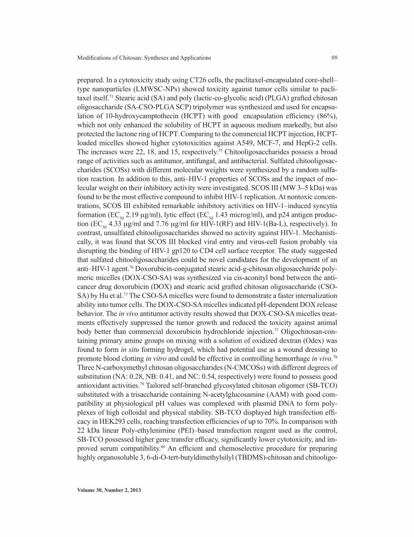
prepared. In a cytotoxicity study using CT26 cells, the paclitaxel-encapsulated core-shell–type nanoparticles (LMWSC-NPs) showed toxicity against tumor cells similar to pacli-taxel itself.71 Stearic acid (SA) and poly (lactic-co-glycolic acid) (PLGA) grafted chitosan oligosaccharide (SA-CSO-PLGA SCP) tripolymer was synthesized and used for encapsu-lation of 10-hydroxycamptothecin (HCPT) with good encapsulation efficiency (86%), which not only enhanced the solubility of HCPT in aqueous medium markedly, but also protected the lactone ring of HCPT. Comparing to the commercial HCPT injection, HCPT-loaded micelles showed higher cytotoxicities against A549, MCF-7, and HepG-2 cells. The increases were 22, 18, and 15, respectively.75 Chitooligosaccharides possess a broad range of activities such as antitumor, antifungal, and antibacterial. Sulfated chitooligosac-charides (SCOSs) with different molecular weights were synthesized by a random sulfa-tion reaction. In addition to this, anti–HIV-1 properties of SCOSs and the impact of mo-lecular weight on their inhibitory activity were investigated. SCOS III (MW 3–5 kDa) was found to be the most effective compound to inhibit HIV-1 replication. At nontoxic concen-trations, SCOS III exhibited remarkable inhibitory activities on HIV-1–induced syncytia formation (EC50 2.19 mg/ml), lytic effect (EC50 1.43 microg/ml), and p24 antigen produc-tion (EC50 4.33 mg/ml and 7.76 mg/ml for HIV-1(RF) and HIV-1(Ba-L), respectively). In contrast, unsulfated chitooligosaccharides showed no activity against HIV-1. Mechanisti-cally, it was found that SCOS III blocked viral entry and virus-cell fusion probably via disrupting the binding of HIV-1 gp120 to CD4 cell surface receptor. The study suggested that sulfated chitooligosaccharides could be novel candidates for the development of an anti–HIV-1 agent.76 Doxorubicin-conjugated stearic acid-g-chitosan oligosaccharide poly-meric micelles (DOX-CSO-SA) was synthesized via cis-aconityl bond between the anti-cancer drug doxorubicin (DOX) and stearic acid grafted chitosan oligosaccharide (CSO-SA) by Hu et al.73 The CSO-SA micelles were found to demonstrate a faster internalization ability into tumor cells. The DOX-CSO-SA micelles indicated pH-dependent DOX release behavior. The in vivo antitumor activity results showed that DOX-CSO-SA micelles treat-ments effectively suppressed the tumor growth and reduced the toxicity against animal body better than commercial doxorubicin hydrochloride injection.77 Oligochitosan-con-taining primary amine groups on mixing with a solution of oxidized dextran (Odex) was found to form in situ forming hydrogel, which had potential use as a wound dressing to promote blood clotting in vitro and could be effective in controlling hemorrhage in vivo.78 Three N-carboxymethyl chitosan oligosaccharides (N-CMCOSs) with different degrees of substitution (NA: 0.28, NB: 0.41, and NC: 0.54, respectively) were found to possess good antioxidant activities.79 Tailored self-branched glycosylated chitosan oligomer (SB-TCO) substituted with a trisaccharide containing N-acetylglucosamine (AAM) with good com-patibility at physiological pH values was complexed with plasmid DNA to form poly-plexes of high colloidal and physical stability. SB-TCO displayed high transfection effi-cacy in HEK293 cells, reaching transfection efficiencies of up to 70%. In comparison with 22 kDa linear Poly-ethylenimine (PEI)–based transfection reagent used as the control, SB-TCO possessed higher gene transfer efficacy, significantly lower cytotoxicity, and im-proved serum compatibility.80 An efficient and chemoselective procedure for preparing highly organosoluble 3, 6-di-O-tert-butyldimethylsilyl (TBDMS)-chitosan and chitooligo-

Critical Reviews™ in Therapeutic Drug Carrier Systems
100 Ankit Jain et al.
saccharides has been reported. Runarsson et al. prepared 3, 6-di-O-TBDMS-chitosan and chitooligosaccharides, which are useful precursors for selective N-modifications in com-mon organic solvents.81 Although many absorption enhancers have been investigated, very few are used clinically. A need exists therefore for more effective absorption enhancers. The drug-absorption–enhancing effects of combinations of N-trimethyl chitosan chloride (TMC) with various degrees of quaternization of 48 and 64%, dicarboxymethyl chitosan oligosaccharide, and chitosan lactate oligomer with monocaprin and melittin were com-pared with their individual performances using the in vitro Caco-2 cell model.82 The inter-actions of lipopolysaccharide (LPS) with the natural polycation chitosan and its deriva-tives were studied. The number of bonds stabilizing the complexes and the energy of LPS binding with chitosans decreased with increase in acetate group content in chitosans and resulted in changing of binding sites. The study reflected that binding sites of chitooligo-saccharides on R-LPS overlapped and chitooligosaccharide binding energies increased with increase in number of monosaccharide residues in chitosan molecules. The input of the hydrophobic fragment in complex formation energy is most prominent for complexes in water phase and is due to the hydrophobic interaction of chitooligosaccharide acyl frag-ment with fatty acid residues of LPS.83 Chitosan derivatives are potential candidates for gene delivery because they are biocompatible and less toxic. However, their use has been limited by their moderate transfection efficiency and rather large sizes of DNA complexes with high–molecular weight chitosans. To circumvent these limitations, low–molecular weight (approximately 5 kDa) chitosan grafted at 3 and 18 mol% with N-/2(3)-(dodec-2-enyl) succinoyl groups [hydrophobically modified low–mol. wt.-chitosan (LMW-ch)] that exhibit surfactant-like properties were used. HM(3%)-LMW-ch appeared to be a promising nonviral vector with low cytotoxicity and efficient transfection properties.84 The antioxidant potency of high–/low–molecular weight quaternary chitosan derivatives was investigated employing various established systems in vitro, such as superoxide (O2–*) and hydroxyl (*OH) radicals scavenging, reducing power. Low–molecular weight quaternary chitosan was found to have stronger scavenging effect on O2–* and *OH than high–molecu-lar weight quaternary chitosan. Also, the reducing power of low–molecular weight quater-nary chitosan was more pronounced than that of high–molecular weight quaternary chito-san.85 Selective targeting of drugs to kidneys may improve renal effectiveness and reduce extrarenal toxicity. Using fluorescence imaging, Huang et al.82 found that randomly 50% N-acetylated LMWC selectively accumulated in the kidneys, especially in the renal tu-bules after IV injection in mice. Strong electronic charge as an important factor for anti-cancer activity of chitooligosaccharides (COS) had been studied in a research. Differently charged COS derivatives were synthesized and their anticancer activities were studied using three cancer cell lines, HeLa, Hep3B, and SW480. Fluorescence microscopic obser-vations and DNA fragmentation studies confirmed that the anticancer effect of these high-ly charged COS derivatives were due to necrosis.86 The cytotoxicity of oligo-chitosan (OC) and N, O- carboxymethyl-chitosan (NO-CMC) derivatives (O-C 1%, O-C 5%, NO-CMC 1%, and NO-CMC 5%) was evaluated using primary normal human epidermal ke-ratinocyte (pNHEK) cultures as an in vitro toxicology model at standardized cell passages. In 3-[4, 5-dimethyl-2-thiazolyl]-2, 5-diphenyl tetrazolium bromide (MTT) as a cell viabil-

Volume 30, Number 2, 2013
Modifications of Chitosan: Syntheses and Applications 101
ity assay, the O-C 1% is one of the most compatible chitosan derivatives because it steadi-ly sustained >70% of viable cells until 72 h posttreatment. This was followed by O-C 5%, NO-CMC 5%, and NO-CMC 1%. Therefore, oligo-chitosan was found to have the ideal properties of a biocompatible material compared to N, O- carboxymethyl-chitosan.87 Chi-tosan nanoparticles, prepared from quaternized chitosan (DQ, 60%) and trimethylated chi-tosan oligomer, were used to encapsulate plasmid DNA (pDNA) encoding green fluores-cent protein (GFP) using the complex coacervation technique. TMCO-60%/pDNA nanoparticles had better in vitro and in vivo transfection activity than the other two, and with minimal toxicity, which made it a desirable nonviral vector for gene therapy via oral administration.88 Low–molecular weight chitosans grafted with N-2(3)-(dodec-2-enyl) succinoyl groups (HM-LMW-chitosans) with a mean molecular mass of 5 kDa, a degree of acetylation of 3%, and a degree of tetradecenoyl substitution (TDC) of 3–18 mol% had been synthesized. The HM-LMW-chitosans were found to form micelles through hydro-phobic interactions involving their tetradecenoyl chains and nonprotonated glucosamine monomers. Interaction with large unilamellar vesicles taken as model membranes indi-cated that HM-LMW-chitosans interact mainly with vesicles mimicking the inner leaflet of biomembranes both through electrostatic and hydrophobic interactions. This preferen-tial interaction was supposed to destabilize endosomal membranes and favor the DNA release into the cytoplasm in gene delivery applications. These properties suggested that the HM-LMW-chitosans may constitute a promising new class of nonviral vectors for gene therapy.89 N-trimethyl chitosan oligosaccharide (TMO; low molecular weight) with different degrees of quaternization was synthesized and evaluated for its absorption-en-hancing properties across mucosal epithelia. No acute toxicity was found with any of the synthesized chitosan derivative by means of the ciliary beat frequency (CBF) tests.90 Hu et al. studied BSA release from stearic acid modified chitosan oligosaccharide CSO nanopar-ticles and it was found to decrease when the pH values of the delivery media decreased, in the range from 7.2 to 5.8.91
VI. ENGINEERED CHITOSANS: SYNTHESES AND APPLICATIONS
Progress in chitosan modification has been perpetual with time, and with aquintance of its amenability for chemical modifications, and opportunities in various fields. Its ap-plication potential is shown in the form of tree in Fig. 6.
VI.A. Quaternized Chitosan and N-Alkyl Chitosan
Chitosan is widely studied for its pharmaceutical and nonpharmaceutical applications. However, uses of this polymer could not be always realized due to limited solubility. For example, it has been extensively evaluated for its mucoadhesive and absorption enhancement properties. A chitosan molecule acquires positive charge in acidic en-vironment and this positive charge seems to be crucial for absorption enhancement. However it is not soluble in a medium higher than pH 5.6, which limits permeation enhancement in body compartments of higher pH. In this view, with a need to widen
Critical Reviews™ in Therapeutic Drug Carrier Systems
102 Ankit Jain et al.
the solubility profile of chitosan, particularly at neutral and basic pH values, chitosan derivatives have to be made. Trimethylation of chitosan is an attempt to pave new directions in this regard. This quaternized derivative of chitosan possesses a posi-tive charge and is soluble over a wide range of pH. TMC, being a derivative of cat-ionic polymer possessing positive charge, shows better mucoadhesive, permeation enhancement, drug delivery, and DNA delivery properties. TMC can be further modi-fied or grafted for modulating properties such as solubility, cytotoxicity, or cell recog-nition ability. Quaternization of chitosan not only with a methyl group but a higher group such as ethyl or a long with spacer or quaternization of modified chitosan are also of interest. Different degrees of quaternization (methylation) of amino groups in chitosan can be obtained with methyl iodide in alkaline solution of N-methyl pyr-rolidinone.10,92 Chitosan and salts of chitosan (hydrochloride and glutamate) have been found to be absorption enhancers for protein and peptide drugs.93-95 Underlying mechanisms of action of chitosan for facilitating the paracellular transport of hydro-philic drugs has been proposed to be a combination of bioadhesion and a transient widening of the tight junctions in the membrane mediated by protonated chitosan in its uncoiled configuration. However, chitosan and its salts are found unable to increase the permeability due to solubility problems at pH 7.4. This property entails that its effectiveness as an absorption enhancer is limited to the area of the intestinal lumen where the pH values are close to its pKa. This is the reason that a chitosan and its salts may not be suitable carriers for targeted peptide drug delivery to specific sites of the intestine.10 Its quaternized derivative such as N, N, N-trimethyl chitosan chloride (TMC) with much higher aqueous solubility than chitosan in a much wider range of
FIGURE 6. Application potential of engineered chitosan

Volume 30, Number 2, 2013
Modifications of Chitosan: Syntheses and Applications 103
pH and concentration subdues this problem because it has been investigated well for its use as an absorption enhancer for test drugs, for example, buserelin, octreotide acetate, 9-desglyci namide-8-arginine vasopressin, fluorescein-isothiocyanate dextran (FD4, MW4400), mannitol, etc.96 Moreover, quaternization endows improvement of the mucoadhesivity with increase in degree of quaternization. Thanou et al.97–99 and others also reported this in their investigation wherein increased cationic character led to potentiate gene carrier efficiency in the epithelial cell line.17,92,97–99 Because of the strong basic property of the quaternary ammonium group, TMC is a better choice to deliver negatively charged species such as DNA/genes as compared to plain chitosan. Amphiphilic N-octyl-TMC derivatives have been investigated for polymericmicelles formation, solublization, and controlled release of 10-hydroxycamptothecin.100 TMC cross-linked with gultaraldehyde has been tried to fabricate hollow microspheres for drug loading.101 The primary amino group of chitosan undergoes Schiff reaction with aldehydes or ketones to get the corresponding aldimines and ketimines, which are then converted to N-alkyl derivatives on hydrogenation with borohydride.102,203 The alkyl chitosan can be subjected to quaternization. Jia et al.104 and Avadi et al.105 synthesized quaternary ammonium salts such as N, N, N-trimethyl chitosan, N-propyl-N, N-di-methyl chitosan and N-furfuryl-N, N-dimethyl chitosan and N-diethylmethylamino chitosan, and investigated their antibacterial activity. Quaternized chito-oligomers have also revealed antibacterial activity.104,106 Guo et al. synthesized alkylchitosans and subsequently quaternized them, which improved their antifungal activity due to improved polycationic nature.107 Additionally, such quaternized chitosans also showed better hydroxyl radical scavenging activity compared to other chitosans, although the role of positive charge is not well studied yet in manifestation of radical scavenging activity.108 Tommeraas et al. developed fluorescent chitosan by synthesizing Schiff base with 9-anthraldehyde and then subsequently reducing it with sodium cyanobo-rohydride.109 The Schiff bases of chitosan have been used to improve the properties of chitosan in concern to the chelation of metal ions, production of an analytical re-agent for determination of metal ions, preparation of modified electrodes, protection of amino group, etc.110 Guo et al. synthesized N-arylidene chitosans with derivatives of benzaldehyde and found that the antioxidant activity was equivalent to plain chi-tosan.111 Chitosan-Schiff bases with salicylaldehyde derivatives and N-(4-pyridilme-thylidene) chitosan have been synthesized.112,113 On reacting chitosan under normal as well as reducing conditions with the methoxyphenyl aldehydes such as vanillin, o-vanillin, syringaldehyde, and veratraldehyde, it imparted insolubility to chitosan so the films obtained from veratraldehyde were found to be insoluble, biodegradable, and mechanically resistant.114 Li et al. reported N, N-dialkyl chitosan prepared on re-peating the reductive alkylation with octyl, decyl, and dodecyl aldehydes, and studied their monolayers, corresponding vesicles by LB technique and drug release experi-ments.115,116 Alkylchitosan can also be obtained through an alkylation reaction using alkyl halides under alkaline condition with introduction of an alkyl group on the N and O atoms of chitosan backbone. Isobutylchitosan synthesized by this means displayed improved solubility in neutral aqueous solution due to reduction of crystallinity of
Critical Reviews™ in Therapeutic Drug Carrier Systems
104 Ankit Jain et al.
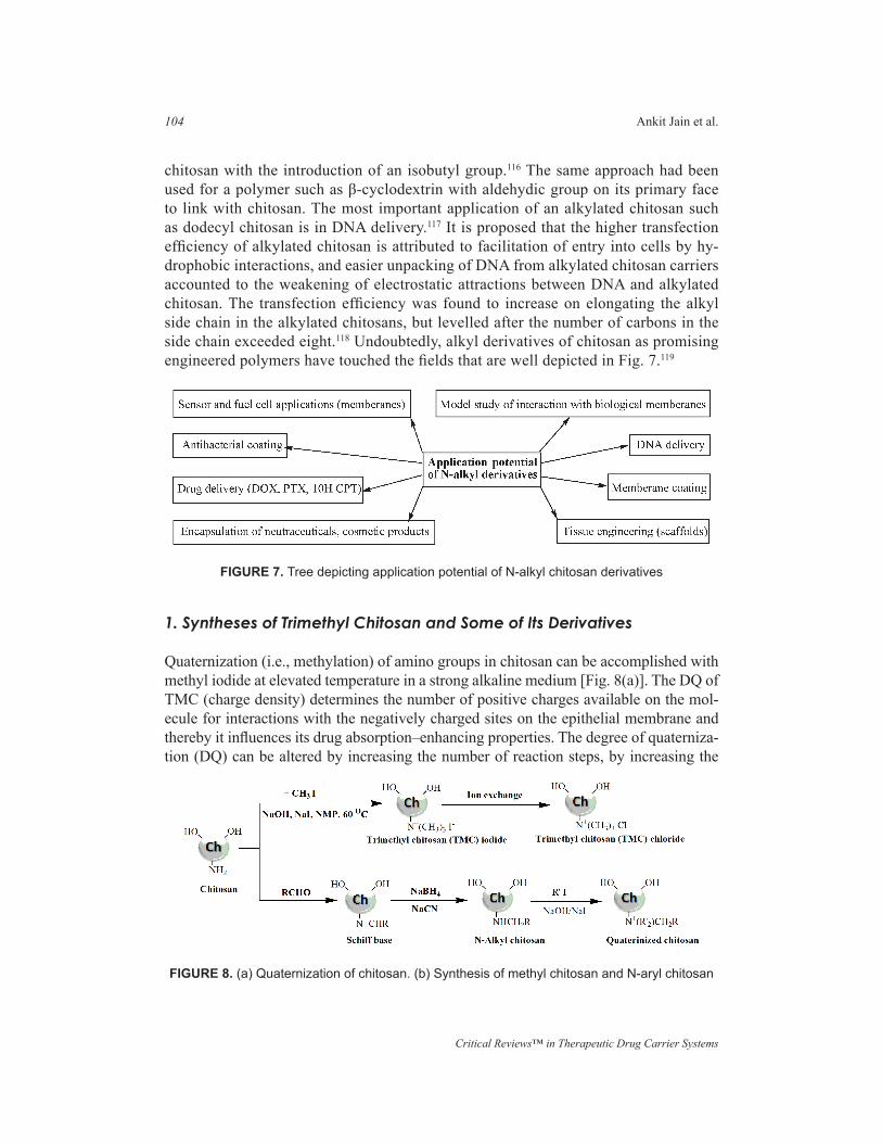
chitosan with the introduction of an isobutyl group.116 The same approach had been used for a polymer such as β-cyclodextrin with aldehydic group on its primary face to link with chitosan. The most important application of an alkylated chitosan such as dodecyl chitosan is in DNA delivery.117 It is proposed that the higher transfection efficiency of alkylated chitosan is attributed to facilitation of entry into cells by hy-drophobic interactions, and easier unpacking of DNA from alkylated chitosan carriers accounted to the weakening of electrostatic attractions between DNA and alkylated chitosan. The transfection efficiency was found to increase on elongating the alkyl side chain in the alkylated chitosans, but levelled after the number of carbons in the side chain exceeded eight.118 Undoubtedly, alkyl derivatives of chitosan as promising engineered polymers have touched the fields that are well depicted in Fig. 7.119
1. Syntheses of Trimethyl Chitosan and Some of Its Derivatives
Quaternization (i.e., methylation) of amino groups in chitosan can be accomplished with methyl iodide at elevated temperature in a strong alkaline medium [Fig. 8(a)]. The DQ of TMC (charge density) determines the number of positive charges available on the mol-ecule for interactions with the negatively charged sites on the epithelial membrane and thereby it influences its drug absorption–enhancing properties. The degree of quaterniza-tion (DQ) can be altered by increasing the number of reaction steps, by increasing the
FIGURE 7. Tree depicting application potential of N-alkyl chitosan derivatives
FIGURE 8. (a) Quaternization of chitosan. (b) Synthesis of methyl chitosan and N-aryl chitosan

Volume 30, Number 2, 2013
Modifications of Chitosan: Syntheses and Applications 105
reaction time, by controlling the reaction steps, or by using different deacetylation grades of chitosan. At higher degrees of quaternization, however, evidence of O-methylation on the 3- and 6- hydroxyl groups of chitosan is found. In general, O-methylation led to less soluble products. It is hence desirable to prepare trimethyl chitosan (TMC) polymers with a high DQ but with a low degree of O-methylation. The synthesis of N,N,N-trimethyl chitosan was reported by Domard et al. based on the dispersion of 5 g chitosan in 250 ml N-methyl-2 pyrrolidinone reacting with CH3I and NaOH (chitosan:CH3I:NaOH in molar ratio 1:15:2) for 3 h at 36°C.120 This method, however, caused extensive depoly-merization of chitosan. The process was modified by Le Dung with respect to the ratio of reactants (chitosan: CH3I: NaOH in molar ratio 1:15:3.45) to reduce polymer degradation and control the different parameters affecting quaternization. The 1H-NMR examination, however, suggests that such a procedure would mainly result in dimethylated polymer with only 10–15% of quaternization. Sieval et al. modified the process with respect to the solvent/reagent addition sequence and reported one-step and two-step syntheses. In one-step synthesis, chitosan was dispersed in NMP with CH3I and NaI, and then the mixture was made alkaline by adding aqueous NaOH solution.92 In two-step synthesis, chitosan was dispersed in aqueous NaOH with NaI, and then CH3I mixed with NMP was added. The resultant product was washed with ethanol and ether, and subjected to methylation again but with less quantity of CH3I. Dimethylation is significantly decreased by repeat-ing the basic reaction. Runarsson et al. changed the solvent system to a DMF/H2O mix-ture (50:50) and performed the reaction without the aid of a catalyst—sodium iodide.102 This significantly reduced O-methylation since DMF/water seems to lower the reactivity of the hydroxyl group enough to keep the O-methylation down. The DQ, however, was always low in the materials obtained. The DQ varied from 0 to 74% depending on the re-action conditions accompanied by monomethylation, dimethylation, and O-methylation (chitosan:CH3I:NaOH in molar ratio 1: 6 or 12:1.5–9, time 0.5–48 h, temperature 21, 50, 75°C). On the basis of this solvent system, they also recently claimed to get a high degree of substitution (81–88%) by a “one pot” synthesis procedure (chitosan: CH3I: NaOH in molar ratio 1:6:3, time 48 h, room temperature).121 They suggested protection group strategy for more selective N-quaternization (sequence of N-phtahloylation, O-tritylation, N-deprotection, N-methylation, and O-deprotection). The exchange of coun-ter ion iodide with chloride was done finally by dissolving the quaternized polymer in a small quantity of water followed by the addition of HCl in methanol or by dissolving in NaCl solution. The exchange can also be achieved by dialysis against NaCl solution and water. All these methods of methylation make use of methyl iodide, which despite being efficient, is a highly volatile, carcinogenic, and expensive reagent. In addition, it offers limited control over a perilous chemical reaction. In an attempt to overcome these dis-advantages, an alternative sequence for the synthesis of chitosan quaternized derivatives is proposed by De Britto and Assis using dimethylsulfate as the reactive agent wherein the polymer in solution of NaOH and NaCl is mixed and refluxed with methylating agent at room temperature or at 70°C.122 Here, the quaternization intensity was also time and temperature dependent. The undesirable O-methylation and polymeric degradation were also observed to take place for the reaction. Other synthetic strategies have been reported
Critical Reviews™ in Therapeutic Drug Carrier Systems
106 Ankit Jain et al.
to produce TMC derivatives but are not as widely used as the Domard reaction. One such method utilizes a sequence of formation of Schiff’s base and reduction reported by Muz-zarelli and Tanfani [Fig. 8(b)].123
The trimethylation of up to 60% of the amine groups could be accomplished by Schiff’s base formation with formaldehyde, followed by reduction with sodium bo-rohydride and quaternization in alkaline condition with methyl iodide. This two-step method likely prevents chain scission and deacetylation of remaining N-acetyl groups, and might result in TMC without O-methylation. With this, quaternization with differ-ent alkyl groups is also possible as in synthesis of N-diethylmethylchitosan,124 N-N-propyl-N, N dimethyl chitosan and N-furfuryl-N, N-dimethyl chitosan,125 N-butyl N, N dimethyl chitosan,123 and N-phenyl or N-(substituted phenyl) N,N-dimethyl chitosan.107 In an attempt to synthesize O-methyl–free TMC, Verheul et al. synthesized dimethylated chitosan first and quaternized it.127 The procedure was based on the method of Muzza-relli and Tanfani, with modifications in solvent and reducing agent system as use of a formic acid–formaldehyde methylation (Eschweiler-Clarke) and quaternization by CH3I in NMP without help of catalyst.
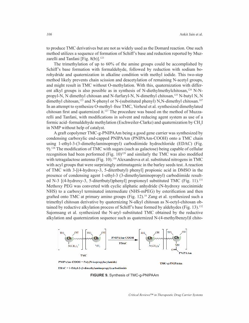
A graft copolymer TMC-g-PNIPAAm being a good gene carrier was synthesized by condensing carboxylic end-capped PNIPAAm (PNIPAAm-COOH) onto a TMC chain using 1-ethyl-3-(3-dimethylaminopropyl) carbodiimide hydrochloride (EDAC) (Fig. 9).128 The modification of TMC with sugars (such as galactose) being capable of cellular recognition had been performed (Fig. 10)129 and similarly the TMC was also modified with tetragalactose antenna (Fig. 10).130 Alexandrova et al. substituted nitrogens in TMC with acyl groups that were surprisingly antimutagenic in the barley seeds test. A reaction of TMC with 3-[(4-hydroxy-3, 5-ditertbutyl) phenyl] propionic acid in DMSO in the presence of condensing agent 1-ethyl-3 (3-dimethylaminopropyl) carbodiimide result-ed N-3 [(4-hydroxy-3, 5-ditertbutyl)phenyl] propionoyl substituted TMC (Fig. 11).131 Methoxy PEG was converted with cyclic aliphatic anhydride (N-hydroxy succinimide NHS) to a carboxyl terminated intermediate (NHS-mPEG) by esterification and then grafted onto TMC at primary amino groups (Fig. 12).18 Zang et al. synthesized such a trimethyl chitosan derivative by quaternizing N-alkyl chitosan as N-octyl-chitosan ob-tained by reductive alkylation process of Schiff’s base formed by aldehydes (Fig. 13).132 Sajomsang et al. synthesized the N-aryl–substituted TMC obtained by the reductive alkylation and quaternization sequence such as quaternized N-(4-methylbenzyl)l chito-
FIGURE 9. Synthesis of TMC-g-PNIPAAm

Volume 30, Number 2, 2013
Modifications of Chitosan: Syntheses and Applications 107
FIGURE 10. Synthesis of TMC-galactose conjugate and TMC-tetragalactose antenna conjugate
FIGURE 11. Synthesis of N-3-[(4-hydroxy-3, 5-ditertbutyl) phenyl] propionyl substituted TM
FIGURE 12. PEGylation of TMC

Critical Reviews™ in Therapeutic Drug Carrier Systems
108 Ankit Jain et al.
san, N-(4-N,N-dimethylaminob-enzyl) chitosan, and quaternized N-(4-pyridylmethyl)chitosan and tested for antibacterial activity.133 Li et al. synthesized a water-soluble am-phiphilic O-carboxymethyl-N-trimethyl chitosan chloride (CMTMC) as a flocculant to treat wastewater in a sugar refinery and found it more effective than chitosan (CS), N-trimethyl chitosan chloride (TMC), O-carboxymethyl chitosan (CMC) (Fig. 14).134
2. Physicochemical Properties of Trimethyl Chitosan versus Chitosan
TMC proved to be a derivative of chitosan with superior solubility and basicity, even at low degrees of quaternization, compared to chitosan and its salts. Chitosan and its salts are only soluble in acidic pH. TMC, even with a DQ as low as 10%, on the other hand, is soluble in an acidic, basic, or neutral medium (pH range 1–9 up to 10% w/v concen-tration). The highest solubility is reported with TMC of an intermediate DQ (40%) re-gardless of DD and molecular weight.135 The increase in solubility was attributed to the replacement of the primary amino group on the C-2 position of chitosan with quaternary amino groups. The absolute molecular weights, radius, and polydispersity of a range of TMC polymers with different degrees of quaternization (22.1, 36.3, 48.0, and 59.2%) were determined with size exclusion chromatography (SEC) and multi-angle laser light scattering (MALLS). The absolute molecular weight of the TMC polymers decreased with an increase in the DQ. The respective absolute molecular weights measured for each of the polymers were 2.02, 1.95, 1.66, and 1.43 g/mol. It should be noted that the
FIGURE 14. Synthesis of 6-O-Carboxymethyl-TMC
FIGURE 13. Synthesis of N-octyl derivative of TMC

Volume 30, Number 2, 2013
Modifications of Chitosan: Syntheses and Applications 109
molecular weight of the polymer chain increases during the reductive methylation pro-cess due to the addition of methyl groups to the amino group of the repeating monomers. However, a net decrease in the absolute molecular weight is observed due to degradation of the polymer chain caused by exposure to the reaction conditions such as the strong al-kaline environment and elevated experimental temperatures during the synthesis.136 The intrinsic viscosity, as an indication of molecular weight, also decreases with an increase in the DQ of the polymer. Like native chitosan, TMC has mucoadhesive properties.137 The intrinsic mucoadhesivity of TMC was found to be lower than the chitosan salts, chitosan hydrochloride and glutamate, but if compared to the reference polymer, pectin, TMC possesses superior mucoadhesive properties.138 The mucoadhesive properties of TMC with different DQs have been explored but the results are controversial. Sandri et al. reported the increase in mucoadhesive properties toward buccal mucosa with in-crease in DQ in the study of fluorescien isothiocyanate dextran (MW 4,400 Da) as a model drug.139 On the other hand, Synman et al. found that the mucoadhesive properties of TMC decreased with an increase in DQ between 22.1 and 48.8%.136,138,140 This may be due to the presence of fixed positive charges and their interaction with the negative sialic groups on the mucus protein structure. The decrease in mucoadhesion with an increase in the DQ may be explained by changes in the conformation of the respective TMC polymers due to interactions between the fixed positive charges on the C-2 position of each polymer. These interactions may force the polymer to change its conformation with a decrease in polymer-chain flexibility. Furthermore, steric effects caused by the attached methyl groups may also hide the positive charges on the amino groups. This decrease in flexibility and screening effect influences both the rate and amount of charge exchange between the negatively charged sialic groups of the mucus and the fixed posi-tive charge of the TMC polymers and the interpenetration into the mucus layer with a subsequent lower mucoadhesivity.
3. TMC Derivatives and their Applications
TMCs have been reported to possess antimicrobial (antifungal, antibacterial) activities of their own. Quaternized chitosan derivatives with high molecular weight demonstrated even stronger antifungal activities than those with low molecular weight. Antifungal activity of TMC was found to be higher than chitosan against Botrytis cinerea Pers. and Colletotrichum lagenarium (Pass) Ell.ethalsttosan.141 When investigated for antifungal activities, other quaternized derivatives such as N-(substituted phenyl)-N, N-dimethyl chitosans such as with 2-hydroxyl-phenyl; 5-chloro-2-hydroxyl-phenyl; 2-hydroxyl-5-nitro-phenyl; 5-bromo-2-hydroxyl-phenyl were found to posses better inhibitory ef-fects than chitosan. This improvement in antifungal activity is probably due to increased cationic charge in the quaternized macromolecules.142 The antibacterial activities of quaternized chitosan such as TMC, N-N-propyl-N, N-dimethyl chitosan and N-furfuryl-N, N-dimethyl chitosan, diethylmethylchitosan against Escherichia coli were explored and results showed stronger antibacterial activity of quaternized chitosan than chito-san, and it increased with acidic condition provided by acetic acid as well as molecular
Critical Reviews™ in Therapeutic Drug Carrier Systems
110 Ankit Jain et al.
weight.104,105 N-butyl-N, N-dimethyl chitosan, was used to prepare antibacterial fiber and mat by electrospining with polyvinylpyrrolidone followed by photo–cross-linking, exhibited high antibacterial activity against Escherichia coli and Staphylococcus au-reus.126 Quaternization (triethylation or trimethylation) of modified chitosan as 6-ami-no-6-deoxy-chitosan also offers antibacterial derivatives143 and trimethylation of O-car-boxymethyl chitosan gave compounds with hydroxyl radical scavenging activity.144 The free radical scavenging activity exhibited by TMC is assisted by its positive charges. To introduce positive charge, place a quaternary methyl group on N of glucosamine with a spacer of 2-hydroxy propyl group between two nitrogens. Such derivative N-(2-hydroxyl) propyl-3-trimethyl ammonium chitosan chloride (HTCC) can be synthesized by reaction of chitosan with glycidyl-trimethyl-ammonium chloride.108,145 HTCC, a wa-ter-soluble derivative, can be utilized to develop various formulations, such as hydro-gel,146 microsphere,147 microgels,148 coating of beads,149 and nanoparticles.149 It has also been evaluated as both antioxidant81 and antimicrobial.150–152 The other uses of HTCC comprise formation of nanofilrtation memberanes153,154 adsorbent for removal of dyes,155 catalyst,156 and support for catalysts.157 Novel quaternary chitosan derivatives such as N, N, O-[N, N-diethylaminomethyl(diethyldimethylene ammonium) methyl] chitosans are also reported with added applications.158Ambiguous outcomes are reported for the relationship between DQ and permeation enhancement.TMC DQ 60% has been proven to be a potent enhancer of both nasal and rectal insulin absorption in rats, especially at neutral pH values where TMC of DQ 12.3% and chitosan HCl were ineffective.159
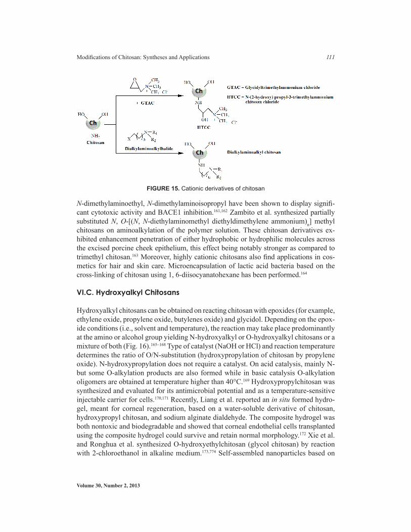
VI.B. Highly Cationic Chitosans
Highly cationic derivatives of chitosan have been reported in the literature because the cationic character of the chitosan makes it crucial for many of its applications (Table 3). These cationic polymers are generally prepared by reacting chitosan and dialkylamino-alkyl chloride in alkaline condition (Fig. 15).158
Xu et al. reported a water-soluble derivative of chitosan, N-(2-hydroxyl) propyl-3-tri-methylammonium chitosan chloride (HTCC), using glycidyl-trimethyl-ammonium chlo-ride (Fig. 15). The HTCC was used for protein delivery using HTCC nanoparticles.145 Chitopearl products (Fuji Spinning Co., Japan) belong to class of highly cationic deriva-tives of chitosan and are chitosan porous beads cross-linked by bifunctional reagents such a diisocyanate or diepoxy derivatives.159 Chitopearl spherical chitosan particles produced from diisocyanates are suitable for chromatographic purposes and as enzyme supports.160 Chitosan derivatives of dialkylaminoalkyl type with N-aminoethyl, N-diethylaminoethyl,
TablE 3. Potential applications of Highly Cationic Derivatives of ChitosanPotential applications
bioadhesion antitumor Anti-inflammatoryAbsorption enhance-ment
Antihypercholesterolemic effect Chromatographic purposes and as enzyme supports
Antimicrobial Hair and skin Cosmetics Transfection efficiency
Volume 30, Number 2, 2013
Modifications of Chitosan: Syntheses and Applications 111
N-dimethylaminoethyl, N-dimethylaminoisopropyl have been shown to display signifi-cant cytotoxic activity and BACE1 inhibition.161,162 Zambito et al. synthesized partially substituted N, O-[(N, N-diethylaminomethyl diethyldimethylene ammonium)n] methyl chitosans on aminoalkylation of the polymer solution. These chitosan derivatives ex-hibited enhancement penetration of either hydrophobic or hydrophilic molecules across the excised porcine cheek epithelium, this effect being notably stronger as compared to trimethyl chitosan.163 Moreover, highly cationic chitosans also find applications in cos-metics for hair and skin care. Microencapsulation of lactic acid bacteria based on the cross-linking of chitosan using 1, 6-diisocyanatohexane has been performed.164
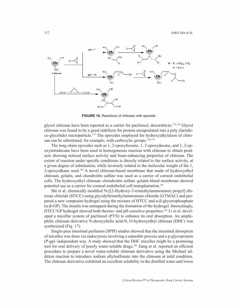
VI.C. Hydroxyalkyl Chitosans
Hydroxyalkyl chitosans can be obtained on reacting chitosan with epoxides (for example, ethylene oxide, propylene oxide, butylenes oxide) and glycidol. Depending on the epox-ide conditions (i.e., solvent and temperature), the reaction may take place predominantly at the amino or alcohol group yielding N-hydroxyalkyl or O-hydroxyalkyl chitosans or a mixture of both (Fig. 16).165–168 Type of catalyst (NaOH or HCl) and reaction temperature determines the ratio of O/N-substitution (hydroxypropylation of chitosan by propylene oxide). N-hydroxypropylation does not require a catalyst. On acid catalysis, mainly N- but some O-alkylation products are also formed while in basic catalysis O-alkylation oligomers are obtained at temperature higher than 40°C.169 Hydroxypropylchitosan was synthesized and evaluated for its antimicrobial potential and as a temperature-sensitive injectable carrier for cells.170,171 Recently, Liang et al. reported an in situ formed hydro-gel, meant for corneal regeneration, based on a water-soluble derivative of chitosan, hydroxypropyl chitosan, and sodium alginate dialdehyde. The composite hydrogel was both nontoxic and biodegradable and showed that corneal endothelial cells transplanted using the composite hydrogel could survive and retain normal morphology.172 Xie et al. and Ronghua et al. synthesized O-hydroxyethylchitosan (glycol chitosan) by reaction with 2-chloroethanol in alkaline medium.173,774 Self-assembled nanoparticles based on
FIGURE 15. Cationic derivatives of chitosan
Critical Reviews™ in Therapeutic Drug Carrier Systems
112 Ankit Jain et al.
glycol chitosan have been reported as a carrier for paclitaxel, doxorubicin.175,176 Glycol chitosan was found to be a good stabilizer for protein encapsulated into a poly (lactide-co-glycolide) microparticle.177 The epoxides employed for hydroxyalkylation of chito-san can be substituted, for example, with carboxylic groups.178,179
The long chain epoxides such as 1, 2-epoxyhexane, 1, 2-epoxydecane, and 1, 2-ep-oxytetradecane have been used in homogeneous reaction with chitosan to obtain prod-ucts showing noticed surface activity and foam-enhancing properties of chitosan. The extent of reaction under specific conditions is directly related to the surface activity, at a given degree of substitution, while inversely related to the molecular weight of the 1, 2-epoxyalkane used.180 A novel chitosan-based membrane that made of hydroxyethyl chitosan, gelatin, and chondroitin sulfate was used as a carrier of corneal endothelial cells. The hydroxyethyl chitosan–chondroitin sulfate–gelatin blend membrane showed potential use as a carrier for corneal endothelial cell transplantation.181
Shi et al. chemically modified N-[(2-Hydroxy-3-trimethylammonium) propyl] chi-tosan chloride (HTCC) using glycidyltrimethylammonium chloride (GTMAC) and pre-pared a new composite hydrogel using the mixture of HTCC and α-β-glycerophosphate (α-β-GP). The insulin was entrapped during the formation of the hydrogel. Interestingly, HTCC/GP hydrogel showed both thermo- and pH-sensitive properties.182 Li et al. devel-oped a micellar system of paclitaxel (PTX) to enhance its oral absorption. An amphi-philic chitosan derivative N-deoxycholic acid-N, O-hydroxyethyl chitosan (DHC) was synthesized (Fig. 17).
Single-pass intestinal perfusion (SPIP) studies showed that the intestinal absorption of micelles was done via endocytosis involving a saturable process and a p-glycoprotein (P-gp)–independent way. A study showed that the DHC micelles might be a promising tool for oral delivery of poorly water-soluble drugs.183 Jiang et al. reported an efficient procedure to prepare a novel water-soluble chitosan derivative using the Michael ad-dition reaction to introduce sodium allylsulfonate into the chitosan at mild condition. The chitosan derivative exhibited an excellent solubility in the distilled water and lower
FIGURE 16. Reactions of chitosan with epoxide

Volume 30, Number 2, 2013
Modifications of Chitosan: Syntheses and Applications 113
thermal stability than chitosan.184 In situ formed hydrogel based on a water-soluble de-rivative of chitosan, hydroxypropyl chitosan (HPCTS), and sodium alginate dialdehyde (SAD) was prepared for corneal endothelium reconstruction.185 Zhang et al. reported im-proved water resistance ability and mechanical properties of silk fibroin (SF)/hydroxy-butyl chitosan (HBC) nanofibrous scaffolds for tissue-engineering applications using genipin, glutaraldehyde (GTA), and ethanol as cross-linkers.186
Liu et al. synthesized an amphiphilic chitosan derivative, N-[(2-hydroxy-3-N, N-dimethylhexadecyl ammonium) propyl] chitosan chloride (N-CQCs). It showed higher accumulation in adipose tissue and gastrointestinal tract than in thymus, kidney, liver, and spleen at 48 h after administration. With the presumption of it possessing a hypo-choesterolemic effect, N-CQCs was found to play an important part in the metabolic process of body fat.187 Zhang et al. fabricated silk fibroin (SF)-hydroxybutyl chitosan (HBC) blend nanofibrous scaffolds using 1, 1, 1, 3, 3, 3-hexafluoro-2-propanol (HFIP) and trifluoroacetic acid (TFA) as solvents to biomimic the native ECM by electrospin-ning. Moreover, the use of genipin vapor not only induced conformation of SF to con-vert from random coil to β-sheet structure, but also acted as a cross-linking agent for SF and HBC. SF/HBC nanofibrous scaffolds presented good cellular compatibility.188 Togni et al. reported enhancement of antifungal potential of P-3051, which is an innovative 8% ciclopirox nail lacquer, using hydroxypropyl chitosan (HPCH) as a film-forming agent. In dilution, susceptibility tests (for Trichophyton rubrum and Candida parapsilosis) showed higher inhibition effects than those obtained by equal amounts of the ciclopirox reference nail lacquer.189 Zhao et al. prepared N-(2-hydroxyl) propyl-3-trimethyl am-monium chitosan chloride nanoparticle as a novel delivery system for parathyroid hor-mone-related protein 1-34. Chitosan (CS) and epoxy propyl trimethyl ammonium chlo-ride (EPTAC) were used to prepare the water-soluble N-(2-hydroxyl) propyl-3-trimethyl ammonium chitosan chloride (HTCC). HTCC/PTHrP1-34 nanopar-ticles were found to be suitable for the treatment of osteoporosis because of their slow-
FIGURE 17. Synthetic scheme of DHC
Critical Reviews™ in Therapeutic Drug Carrier Systems
114 Ankit Jain et al.
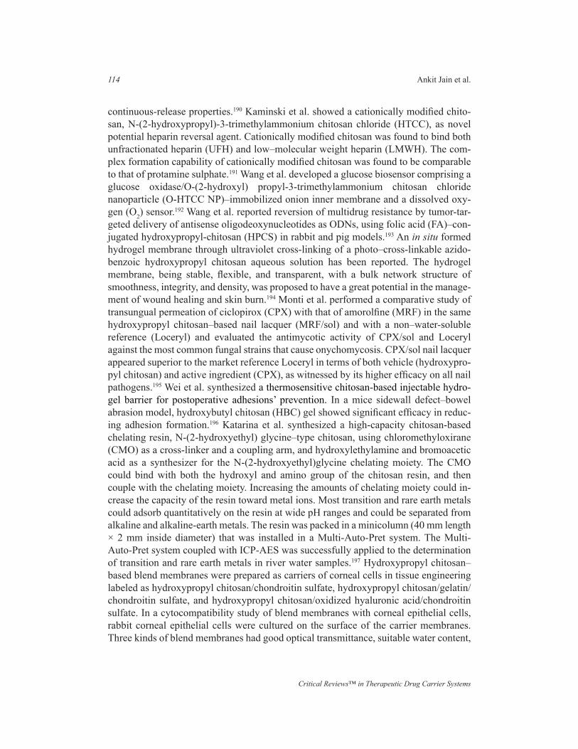
continuous-release properties.190 Kaminski et al. showed a cationically modified chito-san, N-(2-hydroxypropyl)-3-trimethylammonium chitosan chloride (HTCC), as novel potential heparin reversal agent. Cationically modified chitosan was found to bind both unfractionated heparin (UFH) and low–molecular weight heparin (LMWH). The com-plex formation capability of cationically modified chitosan was found to be comparable to that of protamine sulphate.191 Wang et al. developed a glucose biosensor comprising a glucose oxidase/O-(2-hydroxyl) propyl-3-trimethylammonium chitosan chloride nanoparticle (O-HTCC NP)–immobilized onion inner membrane and a dissolved oxy-gen (O2) sensor.192 Wang et al. reported reversion of multidrug resistance by tumor-tar-geted delivery of antisense oligodeoxynucleotides as ODNs, using folic acid (FA)–con-jugated hydroxypropyl-chitosan (HPCS) in rabbit and pig models.193 An in situ formed hydrogel membrane through ultraviolet cross-linking of a photo–cross-linkable azido-benzoic hydroxypropyl chitosan aqueous solution has been reported. The hydrogel membrane, being stable, flexible, and transparent, with a bulk network structure of smoothness, integrity, and density, was proposed to have a great potential in the manage-ment of wound healing and skin burn.194 Monti et al. performed a comparative study of transungual permeation of ciclopirox (CPX) with that of amorolfine (MRF) in the same hydroxypropyl chitosan–based nail lacquer (MRF/sol) and with a non–water-soluble reference (Loceryl) and evaluated the antimycotic activity of CPX/sol and Loceryl against the most common fungal strains that cause onychomycosis. CPX/sol nail lacquer appeared superior to the market reference Loceryl in terms of both vehicle (hydroxypro-pyl chitosan) and active ingredient (CPX), as witnessed by its higher efficacy on all nail pathogens.195 Wei et al. synthesized a thermosensitive chitosan-based injectable hydro-gel barrier for postoperative adhesions’ prevention. In a mice sidewall defect–bowel abrasion model, hydroxybutyl chitosan (HBC) gel showed significant efficacy in reduc-ing adhesion formation.196 Katarina et al. synthesized a high-capacity chitosan-based chelating resin, N-(2-hydroxyethyl) glycine–type chitosan, using chloromethyloxirane (CMO) as a cross-linker and a coupling arm, and hydroxylethylamine and bromoacetic acid as a synthesizer for the N-(2-hydroxyethyl)glycine chelating moiety. The CMO could bind with both the hydroxyl and amino group of the chitosan resin, and then couple with the chelating moiety. Increasing the amounts of chelating moiety could in-crease the capacity of the resin toward metal ions. Most transition and rare earth metals could adsorb quantitatively on the resin at wide pH ranges and could be separated from alkaline and alkaline-earth metals. The resin was packed in a minicolumn (40 mm length × 2 mm inside diameter) that was installed in a Multi-Auto-Pret system. The Multi-Auto-Pret system coupled with ICP-AES was successfully applied to the determination of transition and rare earth metals in river water samples.197 Hydroxypropyl chitosan–based blend membranes were prepared as carriers of corneal cells in tissue engineering labeled as hydroxypropyl chitosan/chondroitin sulfate, hydroxypropyl chitosan/gelatin/chondroitin sulfate, and hydroxypropyl chitosan/oxidized hyaluronic acid/chondroitin sulfate. In a cytocompatibility study of blend membranes with corneal epithelial cells, rabbit corneal epithelial cells were cultured on the surface of the carrier membranes. Three kinds of blend membranes had good optical transmittance, suitable water content,
Volume 30, Number 2, 2013
Modifications of Chitosan: Syntheses and Applications 115
and the ability of protein adsorption. The results showed that less injury was made to corneal epithelial cells by the hydroxypropyl chitosan/gelatin/chondroitin sulfate blend membrane than by the others. This kind of membrane was found to favor the growth and adhesion of corneal epithelial cells. The hydroxypropyl chitosan/gelatin/chondroitin sulfate blend membrane was thus proposed to be a promising carrier of corneal cells and can be used in reconstruction of tissue engineered cornea.198 Ling et al. modified hy-droxypropyl chitosan (HPCS), a water-soluble chitosan derivate, by introducing photo-reactive azide groups (4-azidobenzoic acid, Az-) to the amino groups of HPCS, resulting in a photo–cross-linkable Az-HPCS. Novel porous chitosan scaffolds thus were fabri-cated by ultraviolet (UV) light irradiation of Az-HPCS aqueous solutions. Preliminary data of cell culture on Az-HPCS scaffold suggested its potential applicability for tissue engineering.199 Wu et al. developed a thermosensitive hydrogel using N-[(2-hydroxy-3-trimethylammonium) propyl] chitosan chloride (HTCC) and poly (ethylene glycol) (PEG) with a small amount of alpha-beta-glycerophosphate (alpha-beta-GP). On nasal administration, the solution transformed into viscous hydrogel at body temperature, de-creasing nasal mucociliary clearance rate and releasing insulin slowly. An HTCC-PEG-GP formulation was found to improve the absorption of hydrophilic macromolecular drug via nasal route.200 Chen et al. studied the thermal-induced de-adhesion kinetics of smooth muscle cell (SMC) on thermoresponsive hydroxybutyl chitosan (HBC29) against different periods of preculture time at 37ºC using integrative biophysical tech-niques.201 An amphiphilic derivative of chitosan (2-hydroxyl-3-butoxyl)-propylcarboxy-methyl-chitosan (HBP-CMCHS) had been synthesized followed by development of puerarin-loaded micellar system of HBP-CMCHS and studied in vitro.202 Hydroxypro-pyl chitosan-graft-carboxymethyl beta-cyclodextrin (HPCH-g-CM beta-CD) micropar-ticles were synthesized for controlled and pH-responsive release of hydrophobic drug ketoprofen.203 Dubini et al. studied the in vitro antimycotic potential of a medical de-vice (Myfungar) containing 0.5% of piroctone olamine (CAS 68890-66-4, octopirox) in a hydroxypropyl chitosan hydroalcoholic solution using a nail permeation model and transungual water-soluble technology.204 Huang et al. reported free radical scavenging activity of hydroxyethyl chitosan sulfate (HCS) against 2, 2-diphenyl-1-picrylhydrazyl (DPPH), hydroxyl, and carbon-centered radical species to retard lipid peroxidation.205.Peng et al. synthesized water-soluble hydroxypropyl chitosan (HPCS) derivatives with different degrees of substitution (DS) and weight-average molecular weight (MW) from chitosan and propylene epoxide under basic conditions. In vitro antimicrobial activities of the HPCS derivatives were evaluated by the Kirby-Bauer disk diffusion method and the macrotube dilution broth method. The results suggested that a relatively lower DS and higher MW value enhanced the antifungal activity of HPCS derivatives.97
VI.D. Carboxyalkyl Chitosans
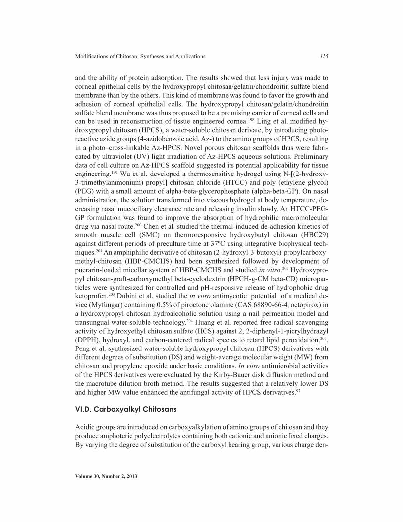
Acidic groups are introduced on carboxyalkylation of amino groups of chitosan and they produce amphoteric polyelectrolytes containing both cationic and anionic fixed charges. By varying the degree of substitution of the carboxyl bearing group, various charge den-
Critical Reviews™ in Therapeutic Drug Carrier Systems
116 Ankit Jain et al.
sities on the molecular chain can be obtained, making it flexible to control pH-dependent behavior. Both N-carboxyalkyl and O-carboxyalkyl chitosan derivatives have been pre-pared using different reaction conditions with monohalocarboxylic acid to attain the N versus O selectivity (Fig. 18).56,206 The other selective route for N-carboxyalkylation involves carboxyaldehydes in a reductive amination sequence.207 By using glyoxylic acid, water-soluble N-carboxymethyl chitosan is obtained: the product is a glucan car-rying pendant glycine groups.208 With the proper selection of the reactant ratio, i.e., with equimolar quantities of glyoxylic acid and amino groups, the product is in part N-monocarboxymethylated (0.3), N,N-dicarboxymethylated (0.3), and N-acetylated de-pending on the starting chitosan (0.08–0.15).209 N-Carboxymethyl chitosan is not only soluble in water, but has unique chemical, physical, and biological properties such as high viscosity, large hydrodynamic volume and film, and gel-forming capabilities, all of which make it an attractive option in connection with its use in food products and cosmetics.210 Carboxymethyl chitosan is used in development of different protein drug delivery systems as superporous hydrogels, pH-sensitive hydrogels, and cross-linked hydrogels.211-214 N,N-Dicarboxymethyl chitosan has shown to possess good chelating
FIGURE 18. Carboxylation of chitosan depending on reaction conditions O-carboxylated, N-car-boxylated, or N, O-carboxylated chitosan can be obtained

Volume 30, Number 2, 2013
Modifications of Chitosan: Syntheses and Applications 117
abilities and its chelate with calcium phosphate favored osteogenesis while promoting bone mineralization.215 O-Carboxymethyl chitosan exhibits antibacterial activity and modified adhesive properties, for instance, surface modification of tissue scaffolds of poly (lactide-co-glycolide acid) with O-carboxymethylchitosan enhances chondrocyte adhesion; surface modification of Dacron vascular grafts enhances the blood compat-ibility.216 Carboxymethyl chitosan and modified carboxymethyl chitosan at amino func-tion with hexanoic, linoleic acid have been employed as a carrier for delivering drugs as gatifloxacin, camptothecin, ibuprofen, and adriamycin.217–220 Ge and Luo reported preparation of carboxymethyl chitosan in aqueous solution under microwave irradia-tion.221 A higher homolog of carboxymethyl chitosan, i.e., N-(2-carboxyethyl) chitosan was obtained by reaction of chitosan and 3-halopropionic acids under mild alkaline condition and ambient temperature, where alkylation proceeds exclusively at the amino groups.222 This N-carboxyalkyl derivative was tested for antioxidant and antimutagenic activity.223nFull-size image (57K) Sashiwa et al. applied Michael reactions of various acryl reagents with chitosan.224
With application of water-soluble acryl reagents for this reaction, novel types of functional groups were introduced by a simple procedure. The reagents tried were hy-droxyethyl acrylate, hydroxypropyl acrylate, acrylamide, acrylonitrile, PEG-acrylate. Reaction of chitosan with acrylonitrile gives cyanoethyl chitosan whereas reaction of chitosan with ethyl acrylate in aqueous acidic medium gives an N-carboxyethyl ester intermediate, which can easily be hydrolyzed to free acid or used as an intermediate to substitute with various hydrophilic amines, without requiring protecting groups.225 The carboxyl bearing aromatic substitution can be done with aromatic aldehydes. Lin et al. synthesized N-carboxybenzyl chitosan by reductive amination sequence with 2-carboxy benzaldehyde and cross-linked with glutaraldehyde to develop a pH-sensitive hydrogel for colon-specific drug delivery of 5-flurouracil.226 α-Keto acids such as pyruvic acid (and its derivatives such as β-hydroxypyruvic acid, phenylpyruvic acid, 4-hydroxyphe-nylpyruvic acid), α-ketoglutaric acid, and levulinic acid are some of the other carboxy-aldehydes being employed for carboxyalkylation of chitosan. Stable and self-sustaining gels are obtained from 4-hydroxyphenylpyruvic acid modified chitosan, i.e., tyrosine glucan in the presence of tyrosinase. Similar gels are obtained from 3-hydroxybenzalde-hyde, 4-hydroxybenzaldehyde, and 3, 4-dihydroxybenzaldehyde: all of them are hydro-lyzed by lysozyme, lipase, and papain. No cross-linking is observed for chitosan deriva-tives of vanillin, syringaldehyde, and salicylaldehyde.227 Ding et al. effectively modified chitosan into chitosan α-ketoglutaric acid and hydroxamated chitosan α-ketoglutaric acid. The modified chitosan were employed in the formation of theophylline-loaded, iron (III)–cross-linked polymeric beads proven to be successful in prolonging drug re-lease as well as in augmenting adsorption properties.228,229
VI.E. Thiolated Chitosan
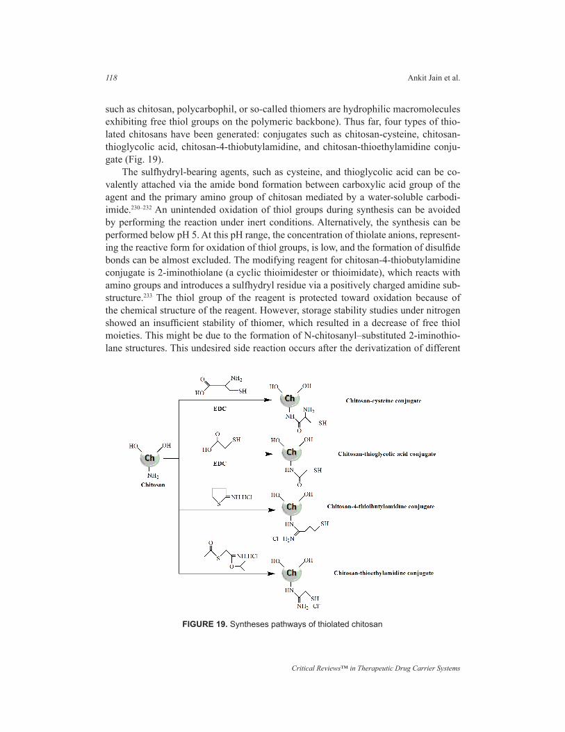
The derivatization of the primary amino groups of chitosan with coupling reagents bearing thiol functions leads to the formation of thiolated chitosans (thiolated polymers
Critical Reviews™ in Therapeutic Drug Carrier Systems
118 Ankit Jain et al.
such as chitosan, polycarbophil, or so-called thiomers are hydrophilic macromolecules exhibiting free thiol groups on the polymeric backbone). Thus far, four types of thio-lated chitosans have been generated: conjugates such as chitosan-cysteine, chitosan-thioglycolic acid, chitosan-4-thiobutylamidine, and chitosan-thioethylamidine conju-gate (Fig. 19).
The sulfhydryl-bearing agents, such as cysteine, and thioglycolic acid can be co-valently attached via the amide bond formation between carboxylic acid group of the agent and the primary amino group of chitosan mediated by a water-soluble carbodi-imide.230–232 An unintended oxidation of thiol groups during synthesis can be avoided by performing the reaction under inert conditions. Alternatively, the synthesis can be performed below pH 5. At this pH range, the concentration of thiolate anions, represent-ing the reactive form for oxidation of thiol groups, is low, and the formation of disulfide bonds can be almost excluded. The modifying reagent for chitosan-4-thiobutylamidine conjugate is 2-iminothiolane (a cyclic thioimidester or thioimidate), which reacts with amino groups and introduces a sulfhydryl residue via a positively charged amidine sub-structure.233 The thiol group of the reagent is protected toward oxidation because of the chemical structure of the reagent. However, storage stability studies under nitrogen showed an insufficient stability of thiomer, which resulted in a decrease of free thiol moieties. This might be due to the formation of N-chitosanyl–substituted 2-iminothio-lane structures. This undesired side reaction occurs after the derivatization of different
FIGURE 19. Syntheses pathways of thiolated chitosan
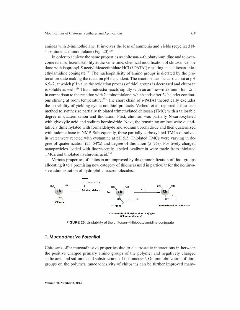
Volume 30, Number 2, 2013
Modifications of Chitosan: Syntheses and Applications 119
amines with 2-iminothiolane. It involves the loss of ammonia and yields recyclized N-substituted 2-iminothiolane (Fig. 20).234
In order to achieve the same properties as chitosan-4-thiobutyl-amidine and to over-come its insufficient stability at the same time, chemical modification of chitosan can be done with isopropyl-S-acetylthioacetimidate HCl (i-PATAI) resulting in a chitosan-thio-ethylamidine conjugate.235 The nucleophilicity of amino groups is dictated by the pro-tonation state making the reaction pH dependent. The reactions can be carried out at pH 6.5–7, at which pH value the oxidation process of thiol groups is decreased and chitosan is soluble as well.236 This imidoester reacts rapidly with an amine—maximum for 1.5 h in comparison to the reaction with 2-iminothiolane, which ends after 24 h under continu-ous stirring at room temperature.233 The short chain of i-PATAI theoretically excludes the possibility of yielding cyclic nonthiol products. Verheul et al. reported a four-step method to synthesize partially thiolated trimethylated chitosan (TMC) with a tailorable degree of quaternization and thiolation. First, chitosan was partially N-carboxylated with glyoxylic acid and sodium borohydride. Next, the remaining amines were quanti-tatively dimethylated with formaldehyde and sodium borohydride and then quaternized with iodomethane in NMP. Subsequently, these partially carboxylated TMCs dissolved in water were reacted with cystamine at pH 5.5. Thiolated TMCs were varying in de-gree of quaternization (25–54%) and degree of thiolation (5–7%). Positively charged nanoparticles loaded with fluorescently labeled ovalbumin were made from thiolated TMCs and thiolated hyaluronic acid.237
Various properties of chitosan are improved by this immobilization of thiol groups allocating it to a promising new category of thiomers used in particular for the noninva-sive administration of hydrophilic macromolecules.
1. Mucoadhesive Potential
Chitosans offer mucoadhesive properties due to electrostatic interactions in between the positive charged primary amino groups of the polymer and negatively charged sialic acid and sulfonic acid substructures of the mucus238. On immobilization of thiol groups on the polymer, mucoadhesivity of chitosans can be further improved many-
FIGURE 20. Unstability of the chitosan–4-thiobutylamidine conjugate
Critical Reviews™ in Therapeutic Drug Carrier Systems
120 Ankit Jain et al.
fold, and with a degree of modification of 25–250 mmol thiol groups per gram chito-san leads to the highest improvement in the mucoadhesive and permeation-enhancing properties. The enhancement of mucoadhesion can be explained by the formation of di-sulfide bonds with cysteine-rich subdomains of mucus glycoproteins, which are stron-ger than non-covalent bonds.238,239 This theory was supported by the results of tensile studies with tablets of thiolated chitosans, which demonstrated a positive correlation between the degree of modification with thiol-bearing moieties and the adhesive prop-erties of the polymer.232,240 The mucoadhesive properties of chitosan were even further improved with the new generation thiolated polymer-chitosan-TBA (4-thiobutylami-dine) conjugate. It has been shown that chitosan-TBA conjugates can lead to a 140- and 42-fold improvement in mucoadhesion compared to the unmodified polymer and the previously investigated chitosan-thioglycolic acid conjugates, respectively.232,233 These promising results can be explained by the fact that chitosan-TBA combines the formation of disulfide bonds with improved ionic interactions between the additional cationic amidine groups of modified chitosan and the anionic moieties provided by si-alic acid and sulfonic acid substructures within the mucus layer. Millotti et al. reported chitosan-6-mercaptonicotinic acid as a thiolated chitosan with strong mucoadhesive properties for oral peptide drug delivery.241 A thiomer derivative of glycol chitosan (GCS) has been synthesized by coupling with thioglycolic acid (TGA) and it was evaluated for the pulmonary delivery of peptides.242
2. Permeation-Enhancing Characteristics
The permeation of paracellular markers through intestinal mucosa can be enhanced 1.6-fold to threefold utilizing thiolated instead of unmodified chitosan. Chitosan pos-sess permeation-enhancing capabilities with increase in the paracellular route of ab-sorption, which is important for the transport of hydrophilic compounds such as thera-peutic peptides and antisense oligonucleotides across the membrane. The mechanism underlying this permeation-enhancing effect seems to be based on the positive charges of the polymer, which interact with the cell membrane resulting in a structural reor-ganization of tight junction-associated proteins.243 In the presence of the mucus layer, however, this permeation-enhancing effect is comparatively lower, since chitosan can-not reach the epithelium because of size limited diffusion and/or competitive charge interactions with mucin.244 The permeation-enhancing effect of chitosan can be strongly improved by the immobilization of thiol groups. The uptake of fluorescence labeled bacitracin, for instance, was improved 1.6-fold utilizing 0.5% of chitosan-cysteine conjugate instead of unmodified chitosan.245 The permeation-enhancing effect of thi-olated chitosan has been studied with permeation mediator glutathione, which indi-cates that chitosan-TBA/GSH is a potentially valuable tool for inhibiting the ATPase activity of P-gp (P-glycoprotein) in the intestine.246 The permeation-enhancing ef-fect also seems to be based on the inhibition of protein tyrosine phosphatase, result-ing in an opening of the tight junctions for hydrophilic macromolecules.247 This the-ory is supported by various in vitro and in vivo studies where significantly improved
Volume 30, Number 2, 2013
Modifications of Chitosan: Syntheses and Applications 121
pharmacological efficacy/bioavailability of model drugs such as calcitonin, insulin, and low–molecular weight heparin were achieved by utilizing chitosan-TBA/GSH systems.247,248
3. In Situ Gelling Characteristics and Cohesivity (being Important in Ma-trices for Controlled Release Drug Delivery Systems)
The reduced thiol functions on the chitosan backbone enable thiolated chitosans to form inter- as well as intramolecular disulfide bonds resulting in cross-linking of the polymeric chains. Hence, thiolated chitosans display, besides their strong mucoadhe-sive and permeation-enhancing properties, excellent cohesive property. This property provides a strong cohesion and stability of carrier matrices being based on thiolated chitosans and can guarantee a prolonged controlled release of embedded therapeutic ingredients. The usefulness of thiolated chitosans as carrier matrices for controlled drug release was demonstrated by means of model drugs, such as insulin,249 clotrima-zole,250 salmon calcitonin,251 and fluorescein-isothiocyanate labeled dextran.252 Mic-roparticles based on chitosan, on the other hand, disintegrate very rapidly unless they are combined with multivalent anionic compounds such as sodium sulfate or alginate leading to stabilization by an ionic cross-linking process.253,254 Due to the addition of such multivalent anionic compounds, however, the mucoadhesive properties of chi-tosan are strongly reduced. Thiolated chitosans demonstrate in situ gelling proper-ties due to the oxidation of thiol groups at physiological pH-values, which results in the formation of inter- and intramolecular disulfide bonds. This pH-dependent sol-gel transition property can be exploited to obtain liquid or semisolid drug formulations of favorable viscoelastic properties that will stabilize themselves once applied at the site of delivery and exhibit limited clearance with prolonged residence. The in situ gelling or cross-linking process can be observed within a pH range of 5–6, which makes the application of thiolated chitosans on vaginal, nasal, buccal, and ocular mu-cosa possible.177,232,255 Thiol-functionalized polymethacrylic acid-polyethylene glycol-chitosan (PCP)–based hydrogel microparticles were developed for oral delivery of insulin. Thiol modification was achieved by grafting cysteine to the activated surface carboxyl groups of PCP hydrogels (Cys-PCP). Thiolation was an effective strategy to improve insulin absorption across Caco-2 cell monolayers, however, the effect was reduced in the experiments using excised rat intestinal tissue. On the other hand, func-tionalized microparticles were more effective in eliciting a pharmacological response in diabetic animals as compared to unmodified PCP microparticles.256 Akhlaghi et al. prepared and characterized poly (methyl methacrylate) nanoparticles coated by chito-san-glutathione conjugate so as to encapsulate insoluble anticancer drugs. Nanoparti-cles were synthesized through radical polymerization of methyl methacrylate initiated by cerium (IV) ammonium nitrate. Paclitaxel (PTX), a model anticancer drug, was encapsulated in nanoparticles with a maximal encapsulation efficiency of 98.27%. Thiolated microparticles were shown to be stable and to have controlled drug release characteristics.257

Critical Reviews™ in Therapeutic Drug Carrier Systems
122 Ankit Jain et al.
4. Biodegradability
The biodegradability of thiolated chitosan has been paving the way for its use as a novel scaffold material.232 Further studies in this direction were performed with L-929 mouse fibroblasts seeded onto chitosan-thioglycolic acid sheets. Results of this study showed that thiolated chitosan can provide a porous scaffold structure guaranteeing cell anchor-age, proliferation, and tissue formation in three dimensions.258 Due to in situ gelling properties, it seems possible to provide a certain shape of the scaffold material by pour-ing a liquid thiolated chitosan cell suspension in a mold. Furthermore, liquid polymer cell suspensions may be applied by injection forming semisolid scaffolds at the site of tissue damage. Since low concentrated aqueous solutions of thiolated chitosan remain liquid when stored under inert conditions and get rapidly gelled on access of oxygen, they seem to be promising candidates for such applications.
5. Enzyme Inhibitory Potential
Zinc-dependent proteases such as aminopeptidases and carboxypeptidases are inhibited by thiomers. The underlying mechanism is based on the capability of thiomers to bind zinc ions. This inhibitory effect seems to be highly beneficial for the oral administration of peptide and protein drugs.231
VI.F. Acyl Chitosans
1. N-Acyl Chitosan
N-acyl derivatives of chitosan can be easily obtained from acyl chlorides and anhydrides (Fig. 21). Generally, acylation reactions are carried out in media such as aqueous ace-tic acid/methanol, pyridine, pyridine/chloroform, trichloroacetic acid/dichloroethane, ethanol/methanol mixture, methanol/formamide, or DMA-LiCl.259 Because of fairly dif-ferent reactivities of two hydroxyl groups and the amino group on chitosan backbone, acylation can be controlled at the expected sites, i.e., on amino group262 or on both hy-droxyls.261–266 The introduction of hydrophobic branches generally renders new physi-cochemical properties such as the formation of polymeric assemblies including gels,267 polymeric vesicles,268 liquid crystals,269,270 membranes,271 Langmuir-Blodgett films,272,273 and fibers.274,275 Hydrophobic-associating water-soluble polymers, being industrially im-portant macromolecules, have been found to mimic the endotoxins.276 Zong et al. synthe-sized acylchitosan with longer chains by reacting chitosan in pyridine/chloroform with hexanoyl, decanoyl, and lauroyl chlorides. These acylated chitosans with four degrees of substitution per monosaccharide ring (disubstitution at amino and monosubtitution each at hydroxyl groups) exhibited an excellent solubility in organic solvents such as chlo-roform, benzene, pyridine, and THF. The analyses indicated that these polymers form a layered structure in the solid state and the layer spacing increases linearly with increas-ing the length of side chains.277 The presence of such a layered structure was elucidated
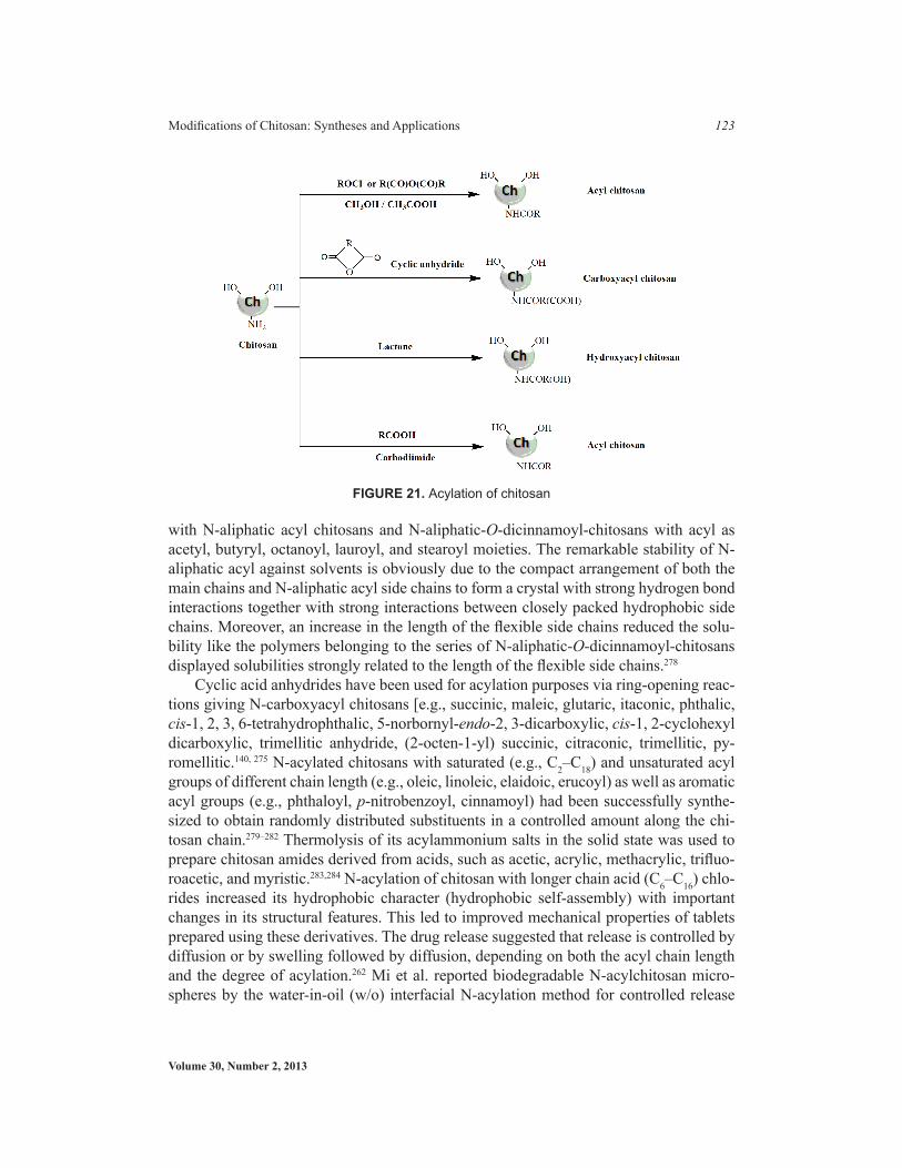
Volume 30, Number 2, 2013
Modifications of Chitosan: Syntheses and Applications 123
with N-aliphatic acyl chitosans and N-aliphatic-O-dicinnamoyl-chitosans with acyl as acetyl, butyryl, octanoyl, lauroyl, and stearoyl moieties. The remarkable stability of N-aliphatic acyl against solvents is obviously due to the compact arrangement of both the main chains and N-aliphatic acyl side chains to form a crystal with strong hydrogen bond interactions together with strong interactions between closely packed hydrophobic side chains. Moreover, an increase in the length of the flexible side chains reduced the solu-bility like the polymers belonging to the series of N-aliphatic-O-dicinnamoyl-chitosans displayed solubilities strongly related to the length of the flexible side chains.278
Cyclic acid anhydrides have been used for acylation purposes via ring-opening reac-tions giving N-carboxyacyl chitosans [e.g., succinic, maleic, glutaric, itaconic, phthalic, cis-1, 2, 3, 6-tetrahydrophthalic, 5-norbornyl-endo-2, 3-dicarboxylic, cis-1, 2-cyclohexyl dicarboxylic, trimellitic anhydride, (2-octen-1-yl) succinic, citraconic, trimellitic, py-romellitic.140, 275 N-acylated chitosans with saturated (e.g., C2–C18) and unsaturated acyl groups of different chain length (e.g., oleic, linoleic, elaidoic, erucoyl) as well as aromatic acyl groups (e.g., phthaloyl, p-nitrobenzoyl, cinnamoyl) had been successfully synthe-sized to obtain randomly distributed substituents in a controlled amount along the chi-tosan chain.279–282 Thermolysis of its acylammonium salts in the solid state was used to prepare chitosan amides derived from acids, such as acetic, acrylic, methacrylic, trifluo-roacetic, and myristic.283,284 N-acylation of chitosan with longer chain acid (C6–C16) chlo-rides increased its hydrophobic character (hydrophobic self-assembly) with important changes in its structural features. This led to improved mechanical properties of tablets prepared using these derivatives. The drug release suggested that release is controlled by diffusion or by swelling followed by diffusion, depending on both the acyl chain length and the degree of acylation.262 Mi et al. reported biodegradable N-acylchitosan micro-spheres by the water-in-oil (w/o) interfacial N-acylation method for controlled release
FIGURE 21. Acylation of chitosan

Critical Reviews™ in Therapeutic Drug Carrier Systems
124 Ankit Jain et al.
of 6-mercaptopurine using reagents for the interfacial N-acylation reaction such as ace-tic, propionic, and n-butyric anhydrides.285 Hexanoyl chitosan with carboxymethylation being developed into amphiphatic hydrogel with excellent water absorption and water retention abilities under neutral conditions was employed as a carrier for delivering am-phiphatic agents.219 The hexanoyl substitution enhances the water absorption ability of hydrogel by altering the number of water binding sites under low humidity, and the state of water in the fully swollen state retards water mobility during deswelling, and thus improves amphiphatic drug encapsulation efficiency compared to pristine chitosan. Acyl-ated chitosan has been applied for stabilization of nanoparticles such as iron oxide, and gold.286,287 Initially, N-succinyl-chitosan was developed as wound dressing materials288 but it is currently also used as a cosmetic material.289 New wound dressings composed of N-succinyl-chitosan and gelatin were also developed.290 N-succinyl-chitosan could be modified easily with respect to succinylation degree by varying reaction conditions and the molecular weight using hydrochloric acid.291,292 N-succinyl-chitosan has unique char-acteristics in vitro and in vivo due to the presence of a number of carboxyl groups. For example, ordinary chitosan can be dissolved in acidic water but not in alkaline, whereas N-succinyl-chitosan with a high degree of substitution (degree of succinylation >0.65) exhibits the opposite behavior.292 Due to the presence of -NH2 and -COOH groups in the structure, N-succinyl-chitosan can easily react with many kinds of agents and thus it is valuable as a drug carrier to readily prepare its conjugates with various drugs to avoid irritating complications. The water-insoluble and water-soluble drug conjugates could be prepared using a water-soluble carbodiimide and mitomycin C or using an activated ester of glutaric mitomycin.292–295 N-succinyl-chitosan shows great potential in controlled release drug delivery since it can form self-assembly of well-dispersed and stable nano-spheres in distilled water.296 It was used for preparation of oxymatrine nanoparticles.297 Succinyl moiety provides hydrophilic and alkyl moiety provides the hydrophobic proper-ties on modifying the succinyl chitosan for solubility by addition of a long alkyl moiety as hydrophobic function to the amino group. These adapted derivatives can form micelles in aqueous media, and were used as drug carriers for the anti-neoplastic agent doxorubi-cin.298 On introducing galactose or lactose to N-succinyl chitosan by reductive amination, liver-targeting ability is conferred in normal or tumor-bearing mice since the liver pa-renchymal cells have asialoglycoprotein receptors (ASGP), which specifically recognize the galactose.299,300 A series of hydrophobically modified chitosans N-2(3)-(dodec-2-enyl) succinoyl/chitosans have been prepared by reacting chitosan with 2-(dodecen-1-yl) suc-cinic anhydride as a new class of potential nonviral vectors for gene delivery.85 N-acyl-ation of chitosan was performed with butanoic, hexanoic, and benzoic anhydride under homogeneous conditions in the presence of methanol and the nanoparticles prepared from N-acyl chitosan were blood compatible.301 Hu et al. reported N-acylated chitosan as N-acetyl, N-propionyl and N-hexanoyl with different degrees of substitution and evalu-ated in vitro for antibacterial activity. The results showed that intermolecular aggregation characteristic of N-acetylated chitosans with low DD probably help in forming bridge to interact with bacterial cells.302 The regioselective acylation can be achieved at amino group by using protection as trityl group at the primary hydroxyl group (Fig. 22). This
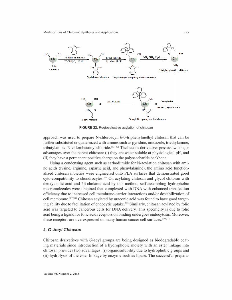
Volume 30, Number 2, 2013
Modifications of Chitosan: Syntheses and Applications 125
approach was used to prepare N-chloroacyl, 6-0-triphenylmethyl chitosan that can be further substituted or quaternized with amines such as pyridine, imidazole, triethylamine, tributylamine, N-chlorobetainyl chloride.303–305 The betaine derivatives possess two major advantages over the parent chitosan: (i) they are water soluble at physiological pH, and (ii) they have a permanent positive charge on the polysaccharide backbone.
Using a condensing agent such as carbodiimide for N-acylation chitosan with ami-no acids (lysine, arginine, aspartic acid, and phenylalanine), the amino acid function-alized chitosan moieties were engineered onto PLA surfaces that demonstrated good cyto-compatibility to chondrocytes.306 On acylating chitosan and glycol chitosan with deoxycholic acid and 5β-cholanic acid by this method, self-assembling hydrophobic macromolecules were obtained that complexed with DNA with enhanced transfection efficiency due to increased cell membrane-carrier interactions and/or destabilization of cell membrane.307,308 Chitosan acylated by uraconic acid was found to have good target-ing ability due to facilitation of endocytic uptake.309 Similarly, chitosan acylated by folic acid was targeted to cancerous cells for DNA delivery. This specificity is due to folic acid being a ligand for folic acid receptors on binding undergoes endocytosis. Moreover, these receptors are overexpressed on many human cancer cell surfaces.310,311
2. O-Acyl Chitosan
Chitosan derivatives with O-acyl groups are being designed as biodegradable coat-ing materials since introduction of a hydrophobic moiety with an ester linkage into chitosan provides two advantages: (i) organosolubility due to hydrophobic groups and (ii) hydrolysis of the ester linkage by enzyme such as lipase. The successful prepara-
FIGURE 22. Regioselective acylation of chitosan

Critical Reviews™ in Therapeutic Drug Carrier Systems
126 Ankit Jain et al.
tion of N, O-acyl chitosan in methanesulfonic acid MeSO3H as solvent was reported (Fig. 23).312,313 Although the selective O-acylation of chitosan in MeSO3H owing to the salt formation of primary amino group with MeSO3H was partly reported, the de-tailed chemical structure and the protecting effect of MeSO3H on amino group are not clear yet.262
The preparation of O, O-didecanoylchitosan, O-succinyl chitosan was also reported through protected N-phthaloylchitosan as an intermediate.314,315 This method, however, needs several steps such as the protection of the amino group by phthaloylation, O-acylation, and finally removal of protecting group by suitable method such as by us-ing hydrazine hydrate. Recently, a one-pot synthesis for the O-acylation of chitosan in MeSO3H has been reported.312 Conclusively, acyl chitosans have amazing application potential in various fields (Fig. 24).
VI.G. Cyclodextrin-Anchored Chitosan
Unique characteristics of chitosan possessing potential to form noncovalent inclu-sion complexes with CD and with a number of guest molecules such as chitosan-guest pendant have been developed altering their physicochemical properties for improved drug delivery system, cosmetics, and analytical chemistry.316,317 Sakairi and cowork-ers12,317,318 reported α-CD–linked chitosan using 2-O-formylmethyl-α-CD by reductive N-alkylation and confirmed the host-guest complex with p-nitrophenol. Auzely-Velty
FIGURE 24. Tree depicting application potential of Acyl-chitosan derivatives
FIGURE 23. O-Acylation of chitosan
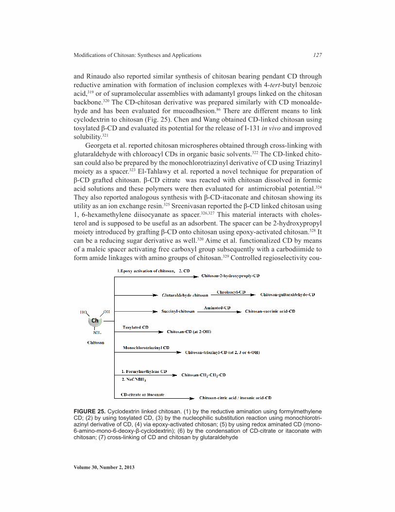
Volume 30, Number 2, 2013
Modifications of Chitosan: Syntheses and Applications 127
and Rinaudo also reported similar synthesis of chitosan bearing pendant CD through reductive amination with formation of inclusion complexes with 4-tert-butyl benzoic acid,319 or of supramolecular assemblies with adamantyl groups linked on the chitosan backbone.320 The CD-chitosan derivative was prepared similarly with CD monoalde-hyde and has been evaluated for mucoadhesion.86 There are different means to link cyclodextrin to chitosan (Fig. 25). Chen and Wang obtained CD-linked chitosan using tosylated β-CD and evaluated its potential for the release of I-131 in vivo and improved solubility.321
Georgeta et al. reported chitosan microspheres obtained through cross-linking with glutaraldehyde with chloroacyl CDs in organic basic solvents.322 The CD-linked chito-san could also be prepared by the monochlorotriazinyl derivative of CD using Triazinyl moiety as a spacer.323 El-Tahlawy et al. reported a novel technique for preparation of β-CD grafted chitosan. β-CD citrate was reacted with chitosan dissolved in formic acid solutions and these polymers were then evaluated for antimicrobial potential.324 They also reported analogous synthesis with β-CD-itaconate and chitosan showing its utility as an ion exchange resin.325 Sreenivasan reported the β-CD linked chitosan using 1, 6-hexamethylene diisocyanate as spacer.326,327 This material interacts with choles-terol and is supposed to be useful as an adsorbent. The spacer can be 2-hydroxypropyl moiety introduced by grafting β-CD onto chitosan using epoxy-activated chitosan.328 It can be a reducing sugar derivative as well.320 Aime et al. functionalized CD by means of a maleic spacer activating free carboxyl group subsequently with a carbodiimide to form amide linkages with amino groups of chitosan.329 Controlled regioselectivity cou-
FIGURE 25. Cyclodextrin linked chitosan. (1) by the reductive amination using formylmethylene CD; (2) by using tosylated CD, (3) by the nucleophilic substitution reaction using monochlorotri-azinyl derivative of CD, (4) via epoxy-activated chitosan; (5) by using redox aminated CD (mono-6-amino-mono-6-deoxy-β-cyclodextrin); (6) by the condensation of CD-citrate or itaconate with chitosan; (7) cross-linking of CD and chitosan by glutaraldehyde
Critical Reviews™ in Therapeutic Drug Carrier Systems
128 Ankit Jain et al.
pling could be achieved using 6-monotosyl-CD derivative as substrate for nucleophilic substitution with sodium maleate. Using N-succinyl chitosan and aminated-β-CD via amide bond formation, preparation of an insoluble cross-linked chitosan with β-CD pendant was reported in the presence of the water-soluble 1-ethyl-3-(3-dimethylami-nopropyl) carbodiimide (EDC) under homogeneous conditions.330 Chaudhury et al. re-viewed the potential of chitosan cyclodextrin complexes in the field of oral controlled release via chitosan nanoparticles and discussed its in vitro and in vivo implications.331 Generating new cochlear hair cells by forced Math1 expression may be a cure for hearing loss. Ren et al. prepared QCS/CM-beta-CD nanoparticle complexes combin-ing quaternized chitosan (QCS) with Na-carboxymethyl-beta-cyclodextrin (CM-beta-CD) as a novel nonviral vector, which adsorbs pRK5-Math1-EGFP perfectly at the mass ratio of 4:1. In vitro cell transfection studies’ transfect efficiency was found to be 40% and relatively lower cytotoxic than liposomes.332 An oral insulin delivery system based on methyl-β-cyclodextrin (MCD) complexed insulin encapsulated polymeth-acrylic acid (PMAA) hydrogel microparticles was evaluated by Sajeesh et al. Poly (methacrylic acid)-chitosan-polyethylene glycol (PCP) microparticles were prepared by the ionic gelation method. The insulin-MCD (IC) complex prepared was character-ized by fluorescence spectroscopic and isothermal titration micro-calorimeteric (ITC) methods. MCD complexed insulin was encapsulated onto PCP microparticles by dif-fusion filling method. Both insulin and MCD complexed insulin encapsulated PCP microparticles were effective in reducing blood glucose level in diabetic animal mod-els.333 Hydroxypropyl-beta-cyclodextrin (HP-beta-CD) with chitosans (CS) of differ-ent molecular weight as absorption enhancer was investigated and found to enhance the intranasal absorption of ISDN remarkably. In a nasal ciliotoxicity test, it showed a good safety profile.334 Li et al. reported layer-by-layer (LbL) assembly of phenyl chi-tosan-graft-cyclodextrin (Ph-CHI-g-CD)/PASP-g-OD and chitosan-graft-cyclodextrin (CHI-g-CD)/PASP-g-OD with electrostatic interaction and host-guest interaction. The experiment provided a new strategy to control the growth behavior of multilayered films via LbL assembly with multiple interactions.335 Mannila et al. studied the effect of alpha-cyclodextrin (α-CD) on sublingual absorption of a hydrophobic model peptide cyclosporin A (CsA), and the effect of temperature on the complexation of CsA with α-CD. The solubility of CsA in aqueous α-CD solution (14%) increased with decreas-ing temperature; the solubility of CsA at room temperature, 5°C, and 1°C was 1.2, 12, and 19 mg/ml, respectively. The study showed that decreased temperature could be effectively utilized to produce CsA/α-CD complexes. It was also shown that α-CD and chitosan (CH) may be advantageous in sublingual delivery of lipophilic peptides, although the absolute bioavailability remains low.336 To increase the aqueous solubil-ity of the tracer (laser dye 6-coumarin), the complexation with different cyclodextrins was adopted by Trapani et al. That led to the modulation of encapsulation efficiency in nanoparticles.337 Derivatives of beta-cyclodextrin (β-CD) and thiolated chitosan have been studied as inhibitors of intestinal P-glycoprotein.338 Liu et al. synthesized β-CD-modified chitosan via the Schiff base reaction between 6-O-(4-formylphenyl)-β-CD and chitosan (CHIT), and then fabricating as the supramolecular dyad assemblies using

Volume 30, Number 2, 2013
Modifications of Chitosan: Syntheses and Applications 129
the inclusion of adamantane-modified pyrene into the beta-cyclodextrin cavity and the wrapping of a CHIT chain on multiwalled carbon nanotubes (MWCNTs). It was found that the DNA-condensing capability of CHIT can be pronouncedly improved by either pyrene grafts or the MWCNT medium due to the cooperation between cationic and aro-matic groups as well as the dispersion of CHIT agglomerates by MWCNTs.339 Venter et al. peformed modification of chitosan by introducing β-CD and tested the mucoadhe-sive strength and inclusion properties of that synthesized cyclodextrin-polymer. β-CD was successfully grafted onto a chitosan chain polymer with a cyclodextrin grafting yield of 7% and a CD-chitosan yield of 85%. The chitosan-CD showed mucoadhesive strengths of 12% stronger than pectin, but 13.5% weaker than the parent chitosan. The synthesized chitosan-CD-polymer exhibited characteristics of a possible mucoadhesive drug delivery system with some inclusion properties from β-CD.86
VI.H. Chitosan Sulfates
Chitosan sulfates represent an array of biological activities. Various methods used so far for the sulfation of chitosan are shown in Fig. 26. For sulfation of chitosan, vari-ous reagents being used include concentrated sulfuric acid,340 oleum,341 sulfurtrioxide/pyridine,342 sulfurtrioxide/trimethylamine,343,344 sulfurtrioxide,345 sulfur trioxide/sulfur dioxide, chlorosulfonic acid-sulfuric acid,346 and the most commonly used is chlorosul-
FIGURE 26. Synthesis of sulfated chitosan
Critical Reviews™ in Therapeutic Drug Carrier Systems
130 Ankit Jain et al.
fonic acid315, 346,347 in homogeneous or heterogeneous conditions in media such as DMF, DMF-dichloroacetic acid, tetrahydrofuran, and formic acid348 at different temperature ranges or under microwave irradiation.349 Sulfuric acid and microwave irradiation pose the difficulty of depolymerization and polymer degradation. With all the reagents, chito-san sulfates obtained are not monosubstituted but often N,O-disubstituted and may also be partially N,O,O-tri-substituted. With a starting material such as N-acyl or N-alkyl chitosan, selective O-sulfonation occurs while O-substituted chitosan, selective N, or N, O-sulfonation occurs.344–349 The sulfonic acid function was also introduced into chitosan by reacting with 5-formyl-2-furansulfonic acid sodium salt, under the mild conditions of the Schiff reaction that on hydrogenation yielded N-sulfofurfuryl chitosan sodium salt, avoiding polymer degradation and O-substitution along with introduction of spacer in between chitosan backbone and sulfate group.350 The sulfonic acid function with small spacer was also introduced by using sulphating agent 2-chloroethane sulfonic acid so-dium salt.351 The sulfa group added to chitosan can be substituted further using sulfonat-ing agent 4-acetamidobenzene sulfonyl chloride, which reacts with -NH2 or -OH (C6 position) groups leading to sulfanilamide derivatives of chitosan.352
Chitosan sulfates have been shown to possess anticoagulant and hemagglutination inhibition activities due to the structural similarity to heparin.353–357 Other biological ac-tivities include antisclerotic, antiviral, anti-HIV, antibacterial, antioxidant, and enzyme inhibition activities.345–348,358,359 By sulfation of chitosan, some of the amino groups are converted to anionic centers and the polymer thus attains better polyelectrolyte proper-ties that can be utilized for developing potential drug carriers such as micelles or micro-capsules.360,361 Amphiphilicity of N-Alkyl-O-sulfated chitosan is due to the presence of long chain alkyl groups such as hydrophobic moiety and sulfated groups having hydro-philic moiety. Thus, it is found to form micelles and physically entrap water-insoluble drugs such as taxol in significant concentration.360,362 N-sulfonato-N, O-carboxymethyl-chitosan, polymer with anionic character has been evaluated positively in vitro and in vivo as an absorption enhancer for the oral delivery of macromolecules such as reviparin (low–molecular weight heparin), mannitol, FITC-dextran.363 Apart from these valuable biological properties, chitosan sulfates exhibit high sorption capacities so thus possess applications in metal ion recovery.350 The sulphur-containing derivatives were obtained by reacting chitosan with CS2, formaldehyde, and primaryamine. Recently, preparation of 1, 3, 5-thiadiazine-2-thione derivatives of chitosan and their potential antioxidant activity in vitro has been reported.364
1. Synthesis
The site specifies chemical modification of the amino and hydroxyl groups in chitosan with sulfate can generate products for a vista of applications because the structure of sulfated chitosans served as a key role player in anticoagulant activity, antisclerotic, antimicrobial, metal chelating property, antiviral activities, and drug delivery applica-tions.348,350,357,360,365–377 The common difficulty of the sulfation of polysaccharides en-countered is that the reaction cannot be performed in a heterogeneous medium, because
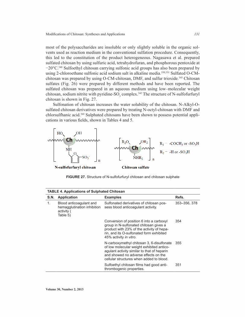
Volume 30, Number 2, 2013
Modifications of Chitosan: Syntheses and Applications 131
most of the polysaccharides are insoluble or only slightly soluble in the organic sol-vents used as reaction medium in the conventional sulfation procedure. Consequently, this led to the constitution of the product heterogeneous. Nagasawa et al. prepared sulfated chitosan by using sulfuric acid, tetrahydrofuran, and phosphorous pentoxide at −20°C.340 Sulfoethyl chitosan carrying sulfonic acid groups has also been prepared by using 2-chloroethane sulfonic acid sodium salt in alkaline media.350,351 Sulfated O-CM-chitosan was prepared by using O-CM-chitosan, DMF, and sulfur trioxide.345 Chitosan sulfates (Fig. 26) were prepared by different methods and have been reported. The sulfated chitosan was prepared in an aqueous medium using low–molecular weight chitosan, sodium nitrite with pyridine-SO3 complex.342 The structure of N-sulfofurfuryl chitosan is shown in Fig. 27.
Sulfonation of chitosan increases the water solubility of the chitosan. N-Alkyl-O-sulfated chitosan derivatives were prepared by treating N-octyl-chitosan with DMF and chlorsulfhanic acid.360 Sulphated chitosans have been shown to possess potential appli-cations in various fields, shown in Tables 4 and 5.
TablE 4. applications of Sulphated ChitosanS.N. application Examples Refs.1. Blood anticoagulant and
hemagglutination inhibition activity (Table 5)
Sulfonated derivatives of chitosan pos-sess blood anticoagulant activity.
353–356, 378
Conversion of position 6 into a carboxyl group in N-sulfonated chitosan gives a product with 23% of the activity of hepa-rin, and its O-sulfonated form exhibited 45% activity in vitro.
354
N-carboxymethyl chitosan 3, 6-disulfonate of low molecular weight exhibited antico-agulant activity similar to that of heparin and showed no adverse effects on the cellular structures when added to blood.
355
Sulfoethyl chitosan films had good anti-thrombogenic properties.
351
FIGURE 27. Structure of N-sulfofurfuryl chitosan and chitosan sulphate

Critical Reviews™ in Therapeutic Drug Carrier Systems
132 Ankit Jain et al.
TablE 4. applications of Sulphated ChitosanS.N. application Examples Refs.2. Adsorbing metal ions The grafting of sulfur compounds on
chitosan has been used for the design of chelating chitosan-based resins.
369–374, 379
Sulfonic groups have been also grafted on chitosan to improve sorption capacity for metal ions in acidic solutions.
375,376
The metal chelation power of N-sulfofur-furyl chitosan of following the order: Cu(II) > Pb(II) > Ni(II) > Cr(III) > Co(II). The N-sulfofurfuryl chitosan was found to be the most effective compound for removal of metal ions.
350
3. Antimicrobial agents The anionic soluble monomer, vinyl sulfonic acid sodium salt, was grafted onto chitosan to obtain a copolymer with zwitterionic property.
377
Antimicrobial activity of chitosan and graft copolymers has been observed against candida albicans, trichophyton rubrum, staphylococcus aeruginosa, pseudomonas aeruginisa and trichophyton violaceum. It depends largely on the amount and type of grafted chains and, ratio of sulfonation, as well as on the changes of pH.
368
4. Drug delivery applications N-Alkyl-O-sulfated chitosan having long chain alkyl groups as hydrophobic moi-eties and sulphated groups as hydrophilic moieties and it forms micelles in water. The result showed that the taxol concen-tration in the N-alkyl-O-sulfated chitosan micellar solution was found to be 2.01 mg/ml, much higher than that in water.
380
5. Anti HIV-1 activity Sulfated polysaccharides have reported to be effective to block the finding virus to CD-4 of lymph, but did not excrete to accumulate in animal body.
381, 382
Sosa et al. investigated that the N-carboxymethyl chitosan N, O-sulfate, a heparin-like polysaccharide derived from N-carboxymethyl chitosan by a random sulfation reaction, was also shown to inhibit HIV-1 replication and viral binding withCD4.
383
The selective sulfation atO-2 and/or O-3 offers potent anti-retroviral agents show-ing a much higher inhibitory effect on the infection of AIDS virus than that by the known 6-O-sulfated derivative (6-sulfate).
347
Continued
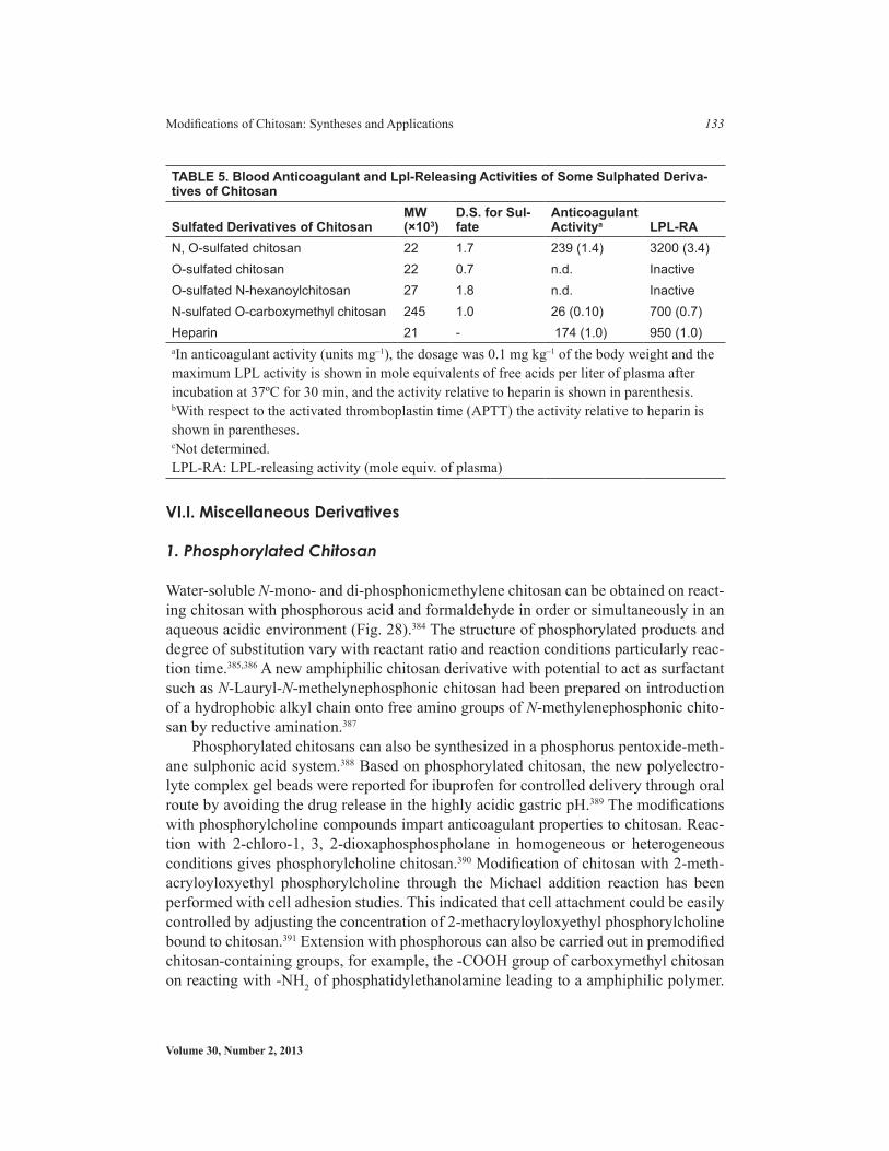
Volume 30, Number 2, 2013
Modifications of Chitosan: Syntheses and Applications 133
VI.I. Miscellaneous Derivatives
1. Phosphorylated Chitosan
Water-soluble N-mono- and di-phosphonicmethylene chitosan can be obtained on react-ing chitosan with phosphorous acid and formaldehyde in order or simultaneously in an aqueous acidic environment (Fig. 28).384 The structure of phosphorylated products and degree of substitution vary with reactant ratio and reaction conditions particularly reac-tion time.385,386 A new amphiphilic chitosan derivative with potential to act as surfactant such as N-Lauryl-N-methelynephosphonic chitosan had been prepared on introduction of a hydrophobic alkyl chain onto free amino groups of N-methylenephosphonic chito-san by reductive amination.387
Phosphorylated chitosans can also be synthesized in a phosphorus pentoxide-meth-ane sulphonic acid system.388 Based on phosphorylated chitosan, the new polyelectro-lyte complex gel beads were reported for ibuprofen for controlled delivery through oral route by avoiding the drug release in the highly acidic gastric pH.389 The modifications with phosphorylcholine compounds impart anticoagulant properties to chitosan. Reac-tion with 2-chloro-1, 3, 2-dioxaphosphospholane in homogeneous or heterogeneous conditions gives phosphorylcholine chitosan.390 Modification of chitosan with 2-meth-acryloyloxyethyl phosphorylcholine through the Michael addition reaction has been performed with cell adhesion studies. This indicated that cell attachment could be easily controlled by adjusting the concentration of 2-methacryloyloxyethyl phosphorylcholine bound to chitosan.391 Extension with phosphorous can also be carried out in premodified chitosan-containing groups, for example, the -COOH group of carboxymethyl chitosan on reacting with -NH2 of phosphatidylethanolamine leading to a amphiphilic polymer.
TablE 5. blood anticoagulant and lpl-Releasing activities of Some Sulphated Deriva-tives of Chitosan
Sulfated Derivatives of ChitosanMW (×103)
D.S. for Sul-fate
anticoagulant activitya lPl-Ra
N, O-sulfated chitosan 22 1.7 239 (1.4) 3200 (3.4)O-sulfated chitosan 22 0.7 n.d. InactiveO-sulfated N-hexanoylchitosan 27 1.8 n.d. InactiveN-sulfated O-carboxymethyl chitosan 245 1.0 26 (0.10) 700 (0.7)Heparin 21 - 174 (1.0) 950 (1.0)aIn anticoagulant activity (units mg–1), the dosage was 0.1 mg kg–1 of the body weight and the maximum LPL activity is shown in mole equivalents of free acids per liter of plasma after incubation at 37ºC for 30 min, and the activity relative to heparin is shown in parenthesis.bWith respect to the activated thromboplastin time (APTT) the activity relative to heparin is shown in parentheses.cNot determined.LPL-RA: LPL-releasing activity (mole equiv. of plasma)
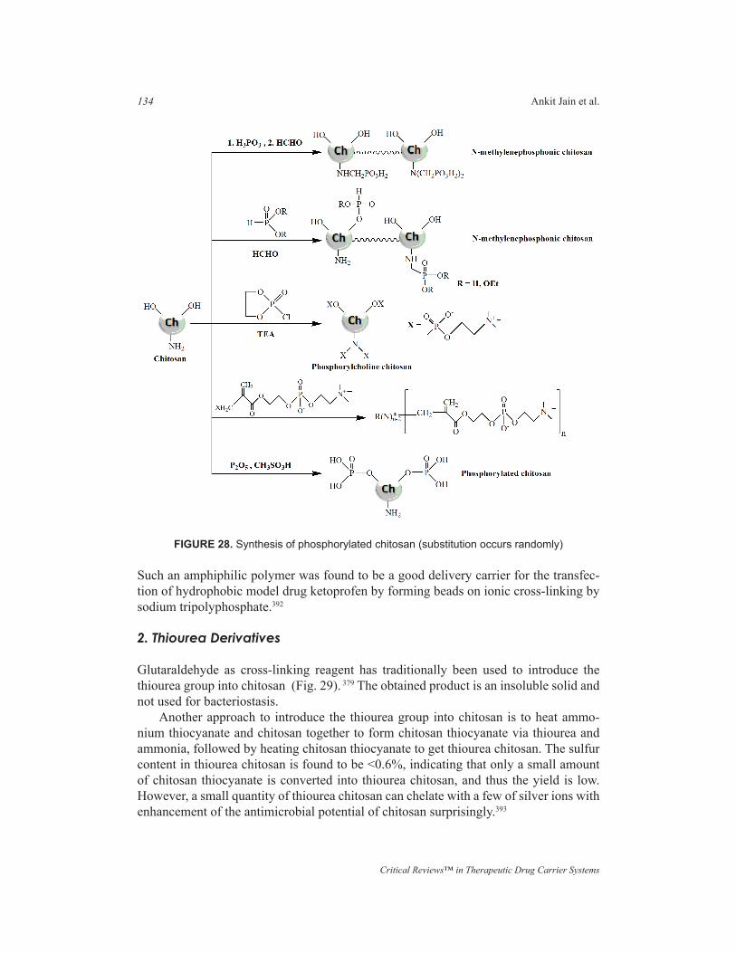
Critical Reviews™ in Therapeutic Drug Carrier Systems
134 Ankit Jain et al.
Such an amphiphilic polymer was found to be a good delivery carrier for the transfec-tion of hydrophobic model drug ketoprofen by forming beads on ionic cross-linking by sodium tripolyphosphate.392
2. Thiourea Derivatives
Glutaraldehyde as cross-linking reagent has traditionally been used to introduce the thiourea group into chitosan (Fig. 29). 379 The obtained product is an insoluble solid and not used for bacteriostasis.
Another approach to introduce the thiourea group into chitosan is to heat ammo-nium thiocyanate and chitosan together to form chitosan thiocyanate via thiourea and ammonia, followed by heating chitosan thiocyanate to get thiourea chitosan. The sulfur content in thiourea chitosan is found to be <0.6%, indicating that only a small amount of chitosan thiocyanate is converted into thiourea chitosan, and thus the yield is low. However, a small quantity of thiourea chitosan can chelate with a few of silver ions with enhancement of the antimicrobial potential of chitosan surprisingly.393
FIGURE 28. Synthesis of phosphorylated chitosan (substitution occurs randomly)
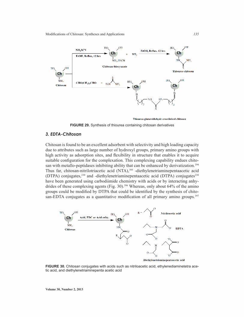
Volume 30, Number 2, 2013
Modifications of Chitosan: Syntheses and Applications 135
3. EDTA-Chitosan
Chitosan is found to be an excellent adsorbent with selectivity and high loading capacity due to attributes such as large number of hydroxyl groups, primary amino groups with high activity as adsorption sites, and flexibility in structure that enables it to acquire suitable configuration for the complexation. This complexing capability endues chito-san with metallo-peptidases inhibiting ability that can be enhanced by derivatization.394 Thus far, chitosan-nitrilotriacetic acid (NTA),395 -diethylenetriaminepentaacetic acid (DTPA) conjugates,230 and -diethylenetriaminepentaacetic acid (DTPA) conjugates230 have been generated using carbodiimide chemistry with acids or by interacting anhy-drides of these complexing agents (Fig. 30).396 Whereas, only about 64% of the amino groups could be modified by DTPA that could be identified by the synthesis of chito-san-EDTA conjugates as a quantitative modification of all primary amino groups.397
FIGURE 30. Chitosan conjugates with acids such as nitriloacetic acid, ethylenediaminetetra ace-tic acid, and diethylenetriaminepenta acetic acid
FIGURE 29. Synthesis of thiourea containing chitosan derivatives

Critical Reviews™ in Therapeutic Drug Carrier Systems
136 Ankit Jain et al.
This might be due to steric hindrance caused by already covalently bound DTPA that might restrict the linkage of another DTPA molecule to the area of the primary amino group of the polymer.
Of these derivatives, chitosan-EDTA conjugate has been tried for an array of pos-sibilities for modifications and applications. One such endeavor is to harbor the com-petitive inhibitors such as antipain, chymostatin, elastatinal, and Bowman-Birk inhibitor either to free the primary amino group of EDTA-chitosan or to the carboxyl group of EDTA on EDTA-chitosan by the formation of amide bonds between this and the primary amino groups of the inhibitor protein. In such an inhibitor linked EDTA-chitosan, the immobilized inhibitors provide a protective effect toward pancreatic serine proteases and the immobilized complexing agent guarantees an inhibition of various metallo-pep-tidases. These enzyme inhibitors can be used to protect the loaded drug from enzymatic degradation in dosage.398–400 Introduction of EDTA on the backbone of chitosan converts this cationogenic polymer to an anionogenic polymer that displays strong mucoadhesive properties that can be explained by the hydrogen bond formation of their carboxylic acid groups with the mucus gel layer. Chitosan EDTA can be used as a carrier matrix where the release of drugs can be controlled, for instance, by ionic cross-linking of the polymer provided by divalent cationic compounds such as lysine or 1, 8-diaminooctane.401 On the other hand, a covalent cross-linking can be achieved by employing a comparatively lower amount of EDTA during the coupling reaction since one EDTA molecule is there-by bound to more than only one amino group of chitosan.402 El-Sharif et al. performed a study to evaluate the antimicrobial activities of chitosan derivatives, EDTA, and the newly developed chitosan-EDTA combination against Gram-negative and Gram-positive bacteria as well as Candida albicans. Both minimal inhibitory concentrations (MIC) and minimal biocidal concentrations (MBC) were determined. Chitosan acetic acid recorded lower MIC values against Enterococcus faecalis, Escherichia coli, Staph-ylococcus aureus, Pseudomonas aeruginosa, and Candida albicans than those exhibited by EDTA. EDTA failed to have an inhibitory activity against Enterococcus faecalis, as well as MBC against any of the studied microorganisms. Chitosan acetic acid’s MBC were recorded to all examined species. Checkerboard assay results indicated a synergis-tic antimicrobial activity of the new combination against Staphylococcus aureus and an additive effect against other microorganisms. Moreover, a short microbial exposure to chitosan-EDTA (20–30 min) caused complete eradication.403
4. Azidated Chitosan
4-azidobenzoic acid is a photosensitive hetero-bifunctional cross-linking reagent that can be linked to chitosan by reacting an acid group of the cross-linking reagent and a free amino group of chitosan in the presence of condensing agents (Fig. 31). Ishi-hara and coworkers reported a photo–cross-linkable chitosan bearing p-azidobenzoic acid and lactobionic acid.404–407 On UV irradiation, its aqueous solution resulted in an insoluble, flexible soft rubberlike hydrogel having excellent properties such as strong tissue adherence, significant induction of wound contraction, and acceleration of wound

Volume 30, Number 2, 2013
Modifications of Chitosan: Syntheses and Applications 137
closure and healing, which could be a good biological adhesive in surgical applica-tions. Zu et al. bonded 4-azidobenzoic acid to O-butyryl chitosan and immobilized it on substrates displaying superior blood compatibility, significantly reduced fibrinogen adsorption, and deposition and spreading of platelets so its potential use in modification of biomedical devices.408 Similarly, it had also been used for micromolding of photo–cross-linkable chitosan hydrogel for spheroid microarray and coculture.409
5. Cyclic Host–Bound Chitosan
Crown ethers have particular molecular structures and good complexing selectivity for metal ions. These crown ether–bound chitosans will have a stronger complexing capac-ity and better selectivity for metal ions because of the synergistic effect of high molecu-lar weight. Tang et al. prepared crown ether–bound chitosan with Schiff’s-base type and its reduced form (Fig. 32). 410
Crown ether–bound chitosans had not only good adsorption capacities for metal ions Pd2+, Au3+, and Ag+, but also high selectivity for the adsorption of Pd2+ in the pres-ence of Cu2+ and Hg2+. Cross-linked types of crown ether–bound chitosans were also reported. These cross-linked derivatives have space net structures with embedded crown ethers, and each mesh has a certain space volume. When original chitosan reacted with 4, 4′-dibromobenzo-18-crown-6-crown ether, the cross-linked product between 6-OH and NH2 was obtained. However, this product would include heterogeneous cross-link-ing structure between 6-OH and 6-OH or NH2 and NH2. Benzylidene-protected chitosan (CTB) would produce a homogeneous cross-linking structure between 6-OH and 6-OH (Fig. 33). These crown ether–bound chitosans would be useful for separation and pre-concentration of heavy or precious metal ions in aqueous environments.411
On the other hand, calixarenes have demonstrated outstanding complex ability to-ward ions, organic molecules, etc., and are considered the third best host molecules, after cyclodextrins and crown ethers. Li et al. reported the first synthesis of calixarene-
FIGURE 31. Synthesis of azidated chitosan (substitution occurs randomly)
FIGURE 32. Crown ether–bound chitosans

Critical Reviews™ in Therapeutic Drug Carrier Systems
138 Ankit Jain et al.
modified chitosan (Fig. 34). The adsorption properties of calixarene-modified chitosans were greatly varied compared with that of original chitosan, especially with the adsorp-tion capacity toward Ag+ and Hg2+, because of the presence of the calixarene moiety. These derivatives did not dissolve in general organic solvent; however, they can easily be powdered and are thus better adsorbents than simple chitosan.412
In tissue engineering, one of the crucial roles of 3D scaffolds is to provide a temporary template with the biomechanical characteristics of the native extracellular matrix (ECM) until the regenerated tissue matures. Sawaguchi et al. performed a study to assess the effect of various cyclic mechanical stresses on cell proliferation and ECM production in a 3D scaffold made from chitosan and hyaluronan for ligament and tendon tissue engineering. Three-dimensional scaffolds seeded with rabbit patella tendon fibroblasts were attached to a bioreactor under various conditions: static group, no strain; stretch group, tensile strain; rotational group, rotational strain; combined group, rotational and tensile strain. DNA content of the combined group was significantly higher than that of the static and stretch groups, and that of the rotational group was significantly higher than that of the static and stretch groups at 21 days after cultivation. The mRNA level of types I and III collagen and fibromodulin in the combined group was significantly higher than that in the other three groups.413 In another study, N-heterocyclic chitosan aerogel derivatives were prepared by reacting 79% de-acetylated chitosan separately with 4-pyridinecarboxaldehyde and 2, 6-pyridinedicarboxaldehyde followed by subsequent solvent exchange into acetone, filtera-tion, and lyophilization.414 β-cyclodextrin was successfully grafted onto a chitosan chain polymer with a cyclodextrin grafting yield of 7% and a CD-chitosan yield of 85%. Al-though the complexation of (+)-catechin by the grafted beta-CD was found to be about five
FIGURE 33. Cross-linked type of crown ether–bound chitosan

Volume 30, Number 2, 2013
Modifications of Chitosan: Syntheses and Applications 139
times weaker than that by the beta-CD monoaldehyde and natural beta-CD, the inclusion properties of the chitosan-CD remain promising. The chitosan-CD showed mucoadhesive strengths 12% stronger than pectin, but 13.5% weaker than the parent chitosan.415
6. Chitosan-Dendrimer Hybrid
Dendrimer-like hyperbranched polymers, a new class of topological macromolecules, have recently been grafted onto chitosan. Tsubokawa and Takayama416 reported the surface modification of chitosan powder by grafting of hyperbranched dendritic poly-amidoamine. They found that the polyamidoamine was propagated from the surface of chitosan by repetition of two processes: (i) Micheal addition of methyl acrylate to the surface amino groups and (ii) amidation of the resulting esters with ethylenediamine to give polyamidoamine dendrimer grafted chitosan powder (Fig. 35). Sashiwa et al. established for the first time the synthesis of a variety of chitosan-dendrimer hybrids us-ing two procedures (Fig. 36). 417–422 In method I, the corresponding dendrimers bearing aldehyde and spacer are synthesized, and then followed by reaction with chitosan using reductive N-alkylation. The main advantage of this procedure is that no cross-linking takes place during the reaction. However, generation of reactive dendrimer is limited owing to its steric hindrance. Sashiwa et al. synthesized a dendronized chitosan-sialic acid hybrid using a convergent grafting method in which sialic acid dendrons bearing a focal aldehyde end group were synthesized by a reiterative amide bond strategy (Fig. 37). A polyamine ending trivalent (G1: first generation) and nona-valent (G2: second generation) dendrons having gallic acid as the branching unit and TEG being the spacer arm were prepared and initially attached to a sialic acid p-phenylisothiocyanate deriva-tive. Further, biological evaluation of these innovative hybrids is being investigated to-ward the inhibition of viral pathogens including the influenza virus.418
Similarly, using a convergent approach, first tetraethylene glycol was modified in five or seven steps to synthesize the dendrimer scaffold. PAMAM dendrimers of genera-tion (G) from 1 to 3 bearing tetraethylene glycol spacer were prepared, attached to sialic acid by reductive N-alkylation, and finally attached to chitosan (Fig. 38).419
FIGURE 34. Calixarene-bound chitosan
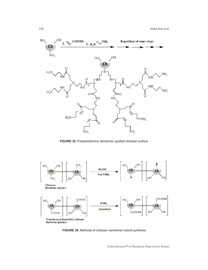
Critical Reviews™ in Therapeutic Drug Carrier Systems
140 Ankit Jain et al.
FIGURE 35. Polyamidiamine dendrimer–grafted chitosan surface
FIGURE 36. Methods of chitosan–dendrimer hybrid synthesis
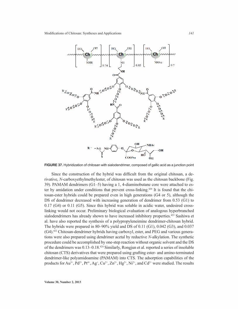
Volume 30, Number 2, 2013
Modifications of Chitosan: Syntheses and Applications 141
Since the construction of the hybrid was difficult from the original chitosan, a de-rivative, N-carboxyethylmethylester, of chitosan was used as the chitosan backbone (Fig. 39). PAMAM dendrimers (G1–5) having a 1, 4-diaminobutane core were attached to es-ter by amidation under conditions that prevent cross-linking.420 It is found that the chi-tosan-ester hybrids could be prepared even in high generations (G4 or 5), although the DS of dendrimer decreased with increasing generation of dendrimer from 0.53 (G1) to 0.17 (G4) or 0.11 (G5). Since this hybrid was soluble in acidic water, undesired cross-linking would not occur. Preliminary biological evaluation of analogous hyperbranched sialodendrimers has already shown to have increased inhibitory properties.423 Sashiwa et al. have also reported the synthesis of a polypropyleneimine dendrimer-chitosan hybrid. The hybrids were prepared in 80–90% yield and DS of 0.11 (G1), 0.042 (G3), and 0.037 (G4).422 Chitosan-dendrimer hybrids having carboxyl, ester, and PEG and various genera-tions were also prepared using dendrimer acetal by reductive N-alkylation. The synthetic procedure could be accomplished by one-step reaction without organic solvent and the DS of the dendrimers was 0.13–0.18.424 Similarly, Rongjun et al. reported a series of insoluble chitosan (CTS) derivatives that were prepared using grafting ester- and amino-terminated dendrimer-like polyamidoamine (PAMAM) into CTS. The adsorption capabilities of the products for Au3+, Pd2+, Pt4+, Ag+, Cu2+, Zn2+, Hg2+, Ni2+, and Cd2+ were studied. The results
FIGURE 37. Hybridization of chitosan with sialodendrimer, composed of gallic acid as a junction point

Critical Reviews™ in Therapeutic Drug Carrier Systems
142 Ankit Jain et al.
FIGURE 38. Chemical structure of chitosan-sialodendrimer hybrid
FIGURE 39. Reaction of N-methoxycarbonylethylchitosan with PAMAM dendrimer

Volume 30, Number 2, 2013
Modifications of Chitosan: Syntheses and Applications 143
showed that the products exhibited better adsorption capabilities for Au3+ and Hg2+ than for other metal ions, and the adsorption capabilities of amino-terminated products were higher than those of ester-terminated ones.425 Chanthateyanonth et al. converted low–molecular weight chitosan to water-soluble chitosan containing hyperbranched-vinylsulfonic acid sodium salt groups. The new chitosan derivatives showed better antimicrobial activity against Micrococcus luteus ATCC 10240 and Achromobacter xylosoxidans ATCC 2706. In addition, they displayed better chelating behavior with heavy metals, such as cadmium (II), copper (II), and nickel (II), than the starting chitosan.426
7. Sugar-Modified Chitosan
Initially, sugar-bound chitosans were investigated mainly for rheological studies, but since then the specific recognition of cells, viruses, and bacteria by sugars was discov-ered. This type of modification has usually been used to introduce cell-specific sugars into chitosan. Modification of chitosan with sugar is now primarily intended for en-hancement of cell recognition potential of the carrier so as to aquire safe and efficacious targeting. Various sugars such as monosaccharide and disaccharide have been anchored for different applications, as listed in the Table 6.
Reductive N-alkylation is a valuable process in chitosan chemistry (Figs. 40 and 41). Following this approach, Hall and Yalpani were the first to report sugar-modified chitosan derivatives.427,428 They synthesized sugar-modified chitosan by reductive N-alkylation using sodium cyanoborohydride and unmodified sugar or a sugar-aldehyde derivative. Galactosylated chitosan prepared from lactobionic acid and chitosan with 1-ethyl-3-(3-dimethylaminopropyl)-carbodiimide (EDC) and N-hydroxysuccinimide (NHS) showed promise as a synthetic extracellular matrix for hepatocyte attachment.429 Park et al. incorporated hydrophilic groups into galactosylated chitosan (GC) to prevent the GC-DNA complex from aggregation and interaction with plasma proteins. Three different kinds of hydrophilic groups such as dextran, poly(ethylene glycol) (PEG), and poly(vinyl pyrrolidone) (PVP) were grafted with GC.430,443
8. Steroid Derivatives
Steroids as natural compounds are also able to confer amphiphilic properties to chitosan. For hydrophobic modification of chitosan, the major steroids tried were 5β-cholanic acid,444,445 cholic acid,446 and cholesterol446,448 (Fig. 42). The strategy for hydrophobic modification basically comprises activation of the carboxylic acid by N-hydroxysuccin-imide (NHS) followed by its grafting onto primary amine of glycol chitosan, mediated by EDC. Hydrophobically modified glycol chitosan (HGC) with 5β- cholanic acid, as a new Cremophor EL-free alternative, has been developed for delivery of poorly wa-ter insoluble drugs such as paclitaxel and docetaxel.449 These paclitaxel loaded HGC nanoparticles found to have maximum drug content 10% (w/w) with more than 90% loading efficiency. In cytotoxicity studies on MCF7 breast cancer cells, they were found to less toxic than Cremophor EL, and enhanced therapeutic index.445

Critical Reviews™ in Therapeutic Drug Carrier Systems
144 Ankit Jain et al.
TablE 6. anchored Sugar Moieties with their application PotentialS. N. anchored Sugar Moiety,
Potential application Remarks Refs.
1. Miscellaneous monosac-charide and disaccharide
Sashiwa and Shigemasa reported N-alkylation of chitosan performed in aqueous methanol with various aldehydes, monosac-charides, and disaccharides (glycolaldehyde, Dl-glyceraldehyde, D-ribose, D-arabinose, D-xylose, 2-deoxy-D-ribose, D-gulcose, 2-deoxy-D-glucose, 3-O-Me-D-glucose, D-galactose, D-mannose, l-fucose, l-rham-nose, GlcNAc).
430
2. D- and l-fucose Morimoto et al. reported the synthesis of sugar bound chitosans, such as those with D- and l-fucose, and their specific interactions with lectin and cells.
431, 432, 433
3. LactoseArticular cartilage repair Stredanska and coworkers synthesized
lactose-modified chitosan for a potential ap-plication in the repair of the articular cartilage.
434, 435
Liver-specific drug delivery
Kato et al. also prepared lactosaminated N-succinyl-chitosan and its fluorescein thiocar-banyl derivative as a liver-specific drug carrier in mice through ASGP receptor.
299
Lactosaminated N-succinylchitosan was found to be a good drug carrier for mitomycin C in treatment of liver metastasis.
300
4. Galactose
DNA delivery Graft copolymers of galactosylated chitosan with poly(ethylene glycol) or poly(vinyl pyr-rolidone) and dextran were useful as hepato-cyte-targeting DNA carriers.
478, 436, 437
Gene delivery Quaternized galaoctosylaed chitosan also holds the cellular recognition ability and pos-sibility of gene delivery.
129
5. MannoseMacrophage targeting Akin specificity is observed for synthesized
mannosyl-chitosan for antigen presenting cells as macrophages and dendritic cells.
130, 438
Influenza and acute rejec-tion
A different type of spacer has been prepared on sialic acid or α-galactosyl epitope bound chitosans. These epitope bound chitosans may be useful as potent inhibitors of influenza viruses or blocking agents for acute rejection.
439, 440, 441
6. Amylose A larger carbohydrate as amylose can be introduced on chitosan by chemoenzymatic method as accomplished by Kaneko et al.
442

Volume 30, Number 2, 2013
Modifications of Chitosan: Syntheses and Applications 145
9. Fatty Acid Derivatives
Different fatty acids such as stearic acid,450–451 linoleic acid,452–453 and oleic acid454 have been successfully grafted to generate amphiphilic chitosan derivatives (Fig. 43). These form micellar structure whose stability can be controlled by balancing hydrophobic and hydrophilic groups. The desired critical micellar concentration of such modified chito-san could be achieved using a longer acyl chain length like stearoyl as compared to the smaller chain length like octanoyl.455
The most studied fatty acid grafted on chitosan is stearic acid, particularly as stearic acid-grafted chitosan oligosaccharides (CSO-SAs). CSO-SA has been investigated for its solubi-lization potential for several molecules, including 10-hydroxycamptothecin,454 mitomycin,456 lamivudine stearate,457 doxorubicin,454 and DNA.458 Because CSO-SA rapidly released the drug by dilution, stearic acid got solubilized into the core of CSO-SA micelles, and thus led to significant reduced release of doxorubicin. This might be due to the enhanced hydropho-bic interaction between stearic acid and stearic acid segments in CSO-SA forming a tightly packed hydrophobic core, and/or because of the ionic interaction between stearic acid and doxorubicin.450 To moderate the initial burst release of drug from CSO-SA micelles is to cross-link the CSO-SA micelles using glutaraldehyde.451 Additionally, PEGylation of CSO-SA has not been found to affect the cellular uptake of the micelles by cancer cells, but led to significant reduction in the internalization of the CSO-SA micelles into macrophages.456
FIGURE 40. Synthesis of sugar-linked chitosans

Critical Reviews™ in Therapeutic Drug Carrier Systems
146 Ankit Jain et al.
10. Polycaprolactone Derivatives
The grafting of hydrophobic biodegradable and biocompatible aliphatic polyester,459 and particularly poly (ɛ-caprolactone) (PCL), has been investigated for the preparation of biodegradable nanoparticles.460–462 Moreover, functionalization of chitosan with poly-caprolactone (PCL) has brought chitosan-PCL and a ternary derivative, chitosan-PCL-mPEG (CPP). 461–463 Through self-assembly of CPP in aqueous media, spherical micelles
FIGURE 41. Synthesis of sialic acid–chitosan and α-galactosyl chitosan followed by their N-succinylation
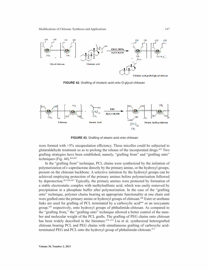
Volume 30, Number 2, 2013
Modifications of Chitosan: Syntheses and Applications 147
were formed with >5% encapsulation efficiency. These micelles could be subjected to glutaraldehyde treatment so as to prolong the release of the incorporated drugs.463 Two grafting strategies have been established, namely, “grafting from” and “grafting onto” techniques (Fig. 44).464,465
In the “grafting from” technique, PCL chains were synthesized by the initiation of polymerization of ɛ-caprolactone directly by the primary amine, or the hydroxyl groups, present on the chitosan backbone. A selective initiation by the hydroxyl groups can be achieved employing protection of the primary amines before polymerization followed by deprotection.462,466,467 Typically, the primary amines were protected by formation of a stable electrostatic complex with methylsulfonic acid, which was easily removed by precipitation in a phosphate buffer after polymerization. In the case of the “grafting onto” technique, polymer chains bearing an appropriate functionality at one chain end were grafted onto the primary amine or hydroxyl groups of chitosan.468 Ester or urethane links are used for grafting of PCL terminated by a carboxylic acid469 or an isocyanate group,459 respectively, onto hydroxyl groups of phthalimide-chitosan. As compared to the “grafting from,” the “grafting onto” technique allowed a better control of the num-ber and molecular weight of the PCL grafts. The grafting of PEG chains onto chitosan has been widely described in the literature.470–474 Liu et al. synthesized heterografted chitosan bearing PCL and PEG chains with simultaneous grafting of carboxylic acid-terminated PEG and PCL onto the hydroxyl group of phthalimide-chitosan.475
FIGURE 42. Grafting of cholanic acid onto O-glycol chitosan
FIGURE 43. Grafting of stearic acid onto chitosan

Critical Reviews™ in Therapeutic Drug Carrier Systems
148 Ankit Jain et al.
VII. ENZYMATIC MODIFICATION
Enzymatic modification of chistosan has been performed to render additional advan-tages. The enzymatic grafting of phenolic compounds onto chitosan to confer water solubility under basic conditions has been reported (Fig. 45).476
With the help of tyrosinase, a wide range of phenolic substrates are converted into electrophilic o-quinones that undergo two different subsequent nonenzymatic reactions with chitosan to yield either Shiff bases or Michael-type adducts. The feasibility of using tyrosinase as a catalyst for grafting hexyloxyphenol onto the chitosan has been investigat-ed successfully.477 In spectral studies, hexyloxyphenol-modified chitosans have dramati-cally altered the physicochemical behavior. On the basis of contact angle measurements, the heterogeneous modification rendered a hydrophobic surface, whereas homogeneous modification gave the rheological properties characteristic of associating water-soluble polymers. Using the enzymatic strategy with tyrosinase enzyme, a dipepetide Tyr-Ala and peptide from casein hydrolysate were grafted on chitosan to get potential value-added by-products from food processing waste.478 Another enzyme used for a functionalization pur-pose is horseradish peroxidise, which is used to graft the phenolic substrate dodecyl gallate onto the chitosan.479 A biochemically relevant quinone, menadione, a synthetic naphtho-quinone derivative having the physiological properties of vitamin K, is particularly prone to rapid reaction with chitosan, greatly modifying its spectral characteristics and increas-ing the surface hydrophobicity of treated chitosan films.480 Tyrosinase was observed to catalyze the oxidation of phenolic moieties of the synthetic polymer poly (4-hydroxy-styrene) (PHS) in water-methanol. Although oxidation was rapid, only a small number
FIGURE 44. Techniques of grafting employed for PCL. “grafting from” and “grafting onto”

Volume 30, Number 2, 2013
Modifications of Chitosan: Syntheses and Applications 149
of phenolic moieties of the PHS polymer (1–2%) underwent oxidation and subsequent nonenzymatic grafting reaction with chitosan.481 Tran et al. reported fast in situ forming supramolecular hydrogels consisted of the tyramine-conjugated supramolecular structures and chitosan derivative that were prepared via an enzymatic reaction with horseradish peroxidase (HRP) and hydrogen peroxide (H2O2). The gel formation was varied within a time period of 5 s to 10 min by controlling the concentrations of HRP, H2O2, and poly-mers. Tyramine conjugation at different sites of the supramolecular structure resulted in significant changes in physical properties and the degradation time of the hydrogels that were confirmed by water uptake, compressive strength, and degradation tests. In addition, the hydrogels showed a good cytocompatibility in vitro. These hydrogels could be promis-ing injectable biomaterials with adjustable degradation times to control both the cellular behaviors as a regenerative cell matrix and the drug release behavior as a drug delivery vehicle.482 Il’ina et al. described the optimal conditions for the enzymatic hydrolysis of chi-tosan and its chemically modified derivatives using a preparation extracted from king crab hepatopancrease possessing pronounced hydrolytic activity. The following preparations were used: chitosan with a molecular weight of 100 kDa and an acetylation level of 0.15, carboxymethyl chitosan 200 kDa witih an extent of replacement of 0.23, and N-succinyl chitosan 390 kDa with an extent of replacement of 0.8. Low–molecular weight samples of chitosan and of its modified derivatives were obtained with the yields of 85, 55, and 80%, respectively. The conditions of the hydrolysis were as follows: enzyme substrate ratio of 1:200, 37°C, and 20 h duration of hydrolysis.483 Chitosan derivatives such as N, N, N-trimethylated chitosan (TMC) are currently being investigated for the delivery of drugs, vaccines, and genes. However, the influence of the extent of N-acetylation of these
FIGURE 45. Enzymatic modification of chitosan

Critical Reviews™ in Therapeutic Drug Carrier Systems
150 Ankit Jain et al.
polymers on their enzymatic degradability and biological properties is unknown. Verheul et al. synthesized TMCs with a degree of acetylation (DA) ranging from 11 to 55% by us-ing a three-step method. First, chitosan was partially re-acetylated using acetic anhydride followed by quantitative dimethylation using formaldehyde and sodium borohydrate. Then, in the presence of an excess amount of iodomethane, TMC was synthesized. The TMCs obtained by this method showed neither detectable O-methylation nor loss in acetyl groups (1H NMR) and a slight increase in molecular weight (GPC) with increasing degree of substitution, implying that no chain scission occurred during synthesis. The extent of lysozyme-catalyzed degradation of TMC, and that of its precursor chitosan and dimethyl chitosan, was highly dependent on the DA and polymers with the highest DA showing the largest decrease in molecular weight. On Caco-2 cells, TMCs with a high DA (~50%), a DQ of ~44%, and with or without O-methylated groups, were not able to open tight junctions in the trans-epithelial electrical resistance (TEER) assay, in contrast with TMCs (both O-methylated and O-methyl free; concentration 2.5mg/ml) with a similar DQ but a lower DA that were able to reduce the TEER with 30% and 70%, respectively. Addition-ally, TMCs with a high DA (approx. 50%) demonstrated no cell toxicity (MTT, LDH re-lease) up to a concentration of 10 mg/ml.484 Kafedjiiski et al. performed a study to examine the biodegradability of thiomers and cross-linked thiomers in comparison with unmodified polymers. Disulfide–cross-linked conjugates were prepared by air oxidation at room tem-perature. Thiomers were investigated by viscosity measurements and spectrophotometric assays. The influence of different factors on the hydrolysis rate, such as the degree of mod-ification of thiomers, structure of the conjugates, pH value of the reaction medium, and the impact of the process of cross-linking, were evaluated. Due to the modification, thiolated chitosans degraded 12.9–24.7% less than unmodified chitosan in the framework of viscos-ity measurements. In addition, the hydrolysis degree of thiolated alginates and modified carboxymethylcelluloses were 25.6–32.4% and 18.4–27.0% lower, respectively, in com-parison to the corresponding unmodified polymers. Conjugates with higher coupling rate of thiol groups were degraded even more slowly. Moreover, the cross-linking process via disulfide bonds additionally reduced the rate of thiomer degradation. The range of degra-dation rates achieved in vitro could be modified by alterations of the contents of thiol and disulfide groups, as well as by suitable design of the polymer structure and ligands used. These results represented helpful basic information for the development of mucoadhesive drug delivery systems, implantable delivery systems, and tissue engineering constructs.485 Il’ina et al. reported an enzymatic hydrolysis of N-succinylchitosan using a complex of chitinolytic enzymes of Streptomyces kurssanovii and also lysozyme and Celloviridin, an industrial cellulase preparation. A biodegradation of N-succinylchitosan of various molec-ular masses was shown to proceed under the action of lysozyme, and the cleavage reaction was revealed to decelerate at a decrease in the polymer molecular mass.486
VIII. GRAFT COPOLYMERIZATION OF CHITOSAN
Graft copolymerization is found to be an attractive technique of modifying the chemical and physical properties of chitosan with enhanced applications. Controlled graft copo-

Volume 30, Number 2, 2013
Modifications of Chitosan: Syntheses and Applications 151
lymerization is achieved through consideration of the characteristics of the side chains, including molecular structure, length, and number. Until now, a number of researches have been carried out to study the effects of these variables on the grafting parameters and the properties of grafted chitosan polymers. Recently, Toh et al. grafted succinic acid onto chitosan to increase the water solubility of chitosan, and demonstrated a higher solubility in water at pH 7.3 when 20 mol% of primary amine were converted into car-boxylic acid. Moreover, the grafting of carboxylic acids onto chitosan chains improved the transfection efficiency as compared to pure chitosan.487
VIII.A. Methodologies
1. Graft Copolymerization by Radical Generation
The polyvinylic and polyacrylic synthetic materials are the most commonly grafted polymers on the polysaccharides. The process involves radical polymerization wherein free radicals are generated first on the biopolymer backbone by chemical or radiation initiation. These radicals serve as initiators for the vinyl or acrylic monomer. Chemical reagents reported as initiators are ceric ammonium nitrate, potassium or ammonium persulfate, and Fenton’s reagent. Grafting percentage and grafting efficiency are mainly influenced by type and concentration of initiator, monomer concentration, and reaction temperature and time. The different specific chitosans subjected to graft copolymeriza-tion are shown in Table 7.
2. Copolymerization via Polycondensation
Polycondensation has not been in practice for copolymerization of polysaccharides due to susceptibility of the saccharide backbone to high temperature and harsh conditions of reactions. However, in some articles, a pH-sensitive hydrogel was prepared using lactic acid (LA), which has been successfully graft copolymerized onto chitosan through con-densation polymerization of D, L-lactic acid in absence of a catalyst (Fig. 46). In another study, using catalyst 4-dimethylaminopyridine, polylactide was grafted through the hy-droxyl groups of phthaloylchitosan. To enhance the cell compatibility and adhesion, amino groups of chitosan were linked onto the polylactic acid surface or to a peptide using a carbodiimide process and such a graft was also found to support the proliferation of human endothelial cells.4
TablE 7. Different Specified Chitosans Subjected To Graft CopolymerizationSpecific Chitosans Subjected to Graft Copolymerization Refs.Carboxymethyl chitosan 488, 489N-carboxyethyl chitosan 488, 490, 491, 492Maleoyl chitosan 493, 494Hydroxypropyl chitosan 490, 495Trimethyl chitosan 18, 496
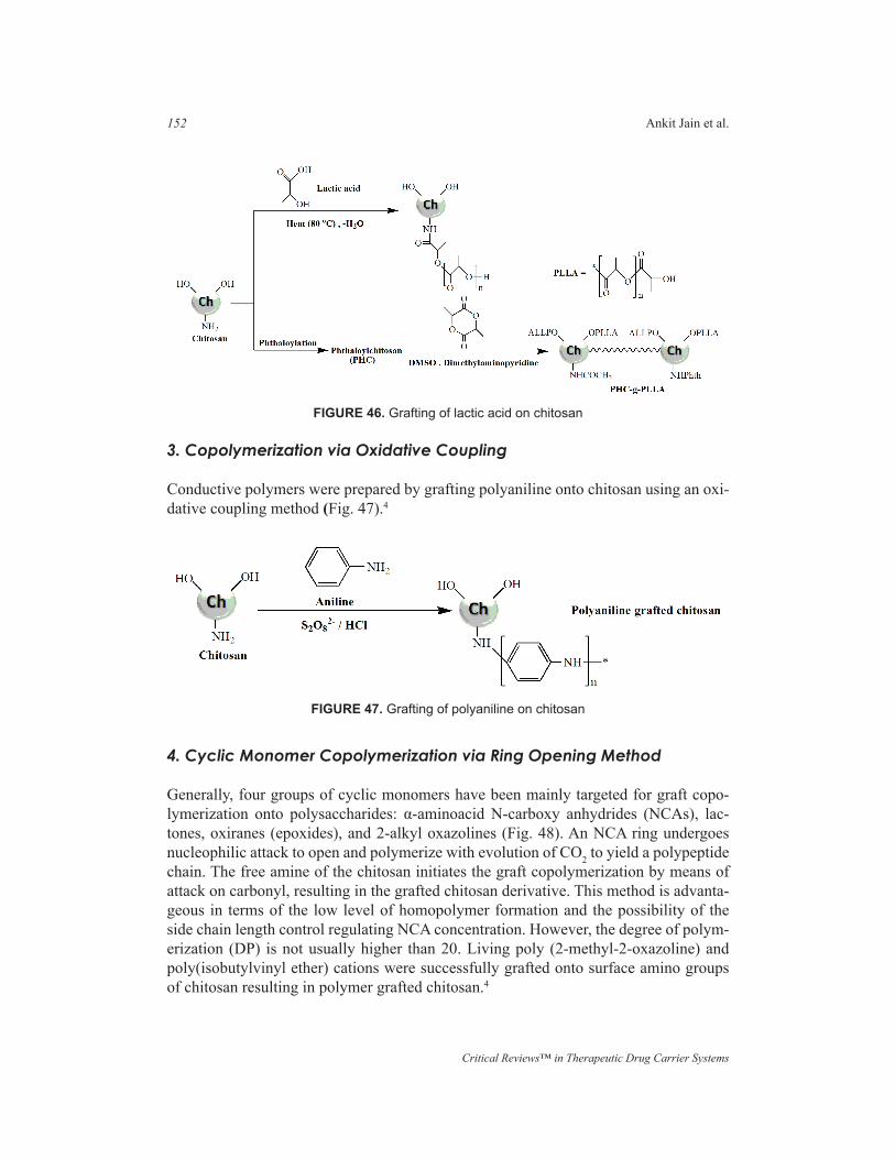
Critical Reviews™ in Therapeutic Drug Carrier Systems
152 Ankit Jain et al.
3. Copolymerization via Oxidative Coupling
Conductive polymers were prepared by grafting polyaniline onto chitosan using an oxi-dative coupling method (Fig. 47).4
4. Cyclic Monomer Copolymerization via Ring Opening Method
Generally, four groups of cyclic monomers have been mainly targeted for graft copo-lymerization onto polysaccharides: α-aminoacid N-carboxy anhydrides (NCAs), lac-tones, oxiranes (epoxides), and 2-alkyl oxazolines (Fig. 48). An NCA ring undergoes nucleophilic attack to open and polymerize with evolution of CO2 to yield a polypeptide chain. The free amine of the chitosan initiates the graft copolymerization by means of attack on carbonyl, resulting in the grafted chitosan derivative. This method is advanta-geous in terms of the low level of homopolymer formation and the possibility of the side chain length control regulating NCA concentration. However, the degree of polym-erization (DP) is not usually higher than 20. Living poly (2-methyl-2-oxazoline) and poly(isobutylvinyl ether) cations were successfully grafted onto surface amino groups of chitosan resulting in polymer grafted chitosan.4
FIGURE 46. Grafting of lactic acid on chitosan
FIGURE 47. Grafting of polyaniline on chitosan

Volume 30, Number 2, 2013
Modifications of Chitosan: Syntheses and Applications 153
5. Copolymerization by Grafting onto Method
Telechelic polymers have been defined as polymers containing one or more functional end groups possessing capacity for selective reaction to form bonds with another molecule. Dif-ferent polymers grafted on chitosan are poly (isobutylvinyl ether), poly (ethylene glycol) PEG, and derivatives, PEO-PPO-PEO (pluronic polyols or poloxamers), poly (ethyleneimine), polyurethane, poly (dimethylsiloxane), polylactide (Fig. 49). PEG is an important hydrophilic polymer commonly grafted (PEGylated) onto chitosan. Several methods have been reported for PEGylation of chitosan using PEGs with various terminal reactive groups (Fig. 50).
On regioselective grafting of PEG on chitosan, chitosan-o-poly(ethylene gly-col) graft copolymer are formed but accompanying prior protection of amino groups. Such etherification of chitosan has been done with poly(ethylene glycol) monomethyl ether (MPEG) using a triazine derivative as a coupler and with MPEG iodide without a coupler (Fig. 51). Regioselective modification of chitosan through the C-6 position of glucosamine units by poly (ethylene glycol) has been performed using 6-oxo-2-N-phthaloylchitosan, 6-O-dichlorotriazine-2-N-phthaloylchitosan and 3-O-acetyl-2-N-phthaloylchitosan intermediates with a high degree of substitution.498
VII.B. Other Applications
1. To Improve the Adhesion and Growth of Endothelial Cells on Chitosan
The cell adhesive peptide Gly-Arg-Gly-Asp (GRGD) was photochemically grafted to chitosan surface with prior activation of the peptide with N-succinimidyl-6-[4′-azido-2′-nitrophenylamino]-hexanoate to phenyl azido-derivatized peptides (Fig. 52).499
FIGURE 48. (a) Synthesis of chitosan–polypeptide bioconjugate via ring opening reaction of α-aminoacid N-carboxyanhydride (NCA), (b) grafting of living poly(2-alkyl-2-oxazoline) onto chitosan

Critical Reviews™ in Therapeutic Drug Carrier Systems
154 Ankit Jain et al.
2. To Enhance Hydrophilicity and Mucoadhesivity
Acrylic acid grafts of chitosan as possible means of creating hydrophilic and mucoad-hesive polymers have been reported.499,500 Siew et al. discussed how hydrophobic drug absorption is facilitated by the nanomedicine using quaternary palmitoyl glycol chitosan (Fig. 53), i.e., (i) increasing the dissolution rate of hydrophobic molecules, (ii) adhering
FIGURE 49. Different synthetic routes to chitosans conjugated with different macromolecular pen-dant groups through “grafting onto” method. PEI = polyethyleneimine, PHB = poly(3-hydroxybutyr-ate), PEO = poly(ethylene oxide), PPO = poly(propylene oxide), pNP = para-nitrophenyl, G-APG = gluadin APG (a partially hydrolyzed wheat gluten protein, MW = 5000), PPG = poly(propylene glycol), BPA = bisphenol A residue, PDMS = poly(dimethyl siloxane).
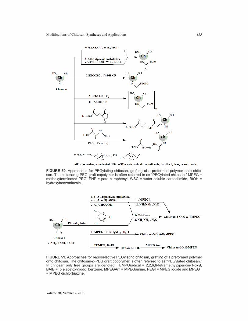
Volume 30, Number 2, 2013
Modifications of Chitosan: Syntheses and Applications 155
FIGURE 50. Approaches for PEGylating chitosan, grafting of a preformed polymer onto chito-san. The chitosan-g-PEG graft copolymer is often referred to as “PEGylated chitosan.” MPEG = methoxyterminated PEG, PNP = para-nitrophenyl, WSC = water-soluble carbodiimide, BtOH = hydroxybenzotriazole.
FIGURE 51. Approaches for regioselective PEGylating chitosan, grafting of a preformed polymer onto chitosan. The chitosan-g-PEG graft copolymer is often referred to as “PEGylated chitosan.” In chitosan only free groups are denoted, TEMPOradical = 2,2,6,6-tetramethylpiperidin-1-oxyl, BAIB = [bis(acetoxy)iodo] benzene, MPEGAm = MPEGamine, PEGI = MPEG iodide and MPEGT = MPEG dichlortriazine.

Critical Reviews™ in Therapeutic Drug Carrier Systems
156 Ankit Jain et al.
to and penetrating the mucus layer and thus enabling intimate contact between the drug and the gastrointestinal epithelium absorptive cells, and (iii) enhancing the transcellular transport of hydrophobic compounds.501
3. As Carrier for Drugs, Proteins, and DNA Plasmids and Oligonucleotides
Microspheres have been reported for entrapment of a number of drugs (Table 8). The ini-tial release of drug from polyacryl-chitosan microspheres depends on the polymer chain relaxation process, but after some time it becomes a molecular diffusion phenomenon. Drug release from such PEG or PVA grafted chitosan microspheres has been found to be pH dependent. Some copolymerized chitosan grafts have been reported for DNA delivery (Table 9). Grafted chitosan has also been used for hydrogel development. Many of these hydrogels exhibit environmentally sensitive behavior such as pH or temprature respon-sive or both. Such chitosan grafts have been reported with polyacrylnitrile,489 acrylamide
TablE 8. Copolymerized Chitosan Used for Microsphere Formulation bearing DrugDrug Copolymer/Method Refs.Sulphadiazine Polyacrylic acid grafted chitosan polymer
dispersion technique 498
Indomethacin, nifidipine Polyacrylic acid grafted chitosan glutaralde-hyde cross-linking method
506, 507
N-phenyl-1-napthylamine PEG grafted chitosan 474Prednisolone PVA grafted chitosan 508Paclitaxel N-mPEG-N-octyl-O-sulfate chitosan 509
FIGURE 53. Synthesis of quaternary palmitoyl glycol chitosan
FIGURE 52. Grafting of peptide on chitosan
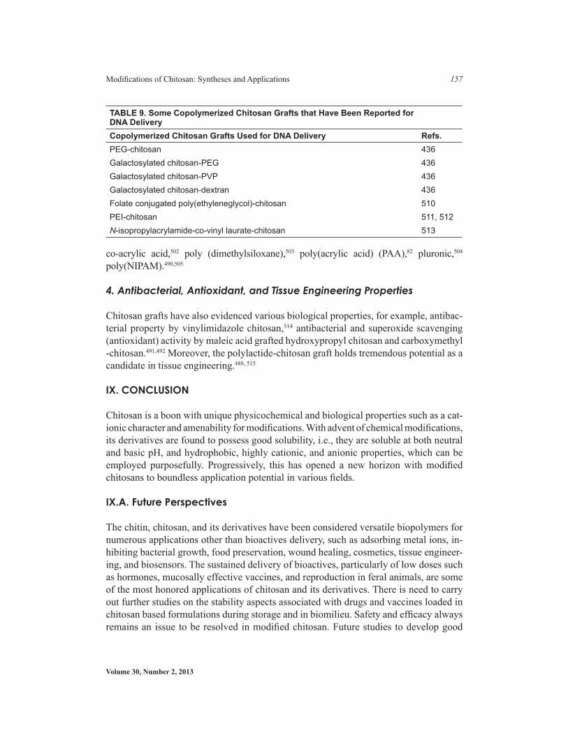
Volume 30, Number 2, 2013
Modifications of Chitosan: Syntheses and Applications 157
co-acrylic acid,502 poly (dimethylsiloxane),503 poly(acrylic acid) (PAA),82 pluronic,504 poly(NIPAM).490,505
4. Antibacterial, Antioxidant, and Tissue Engineering Properties
Chitosan grafts have also evidenced various biological properties, for example, antibac-terial property by vinylimidazole chitosan,514 antibacterial and superoxide scavenging (antioxidant) activity by maleic acid grafted hydroxypropyl chitosan and carboxymethyl -chitosan.491,492 Moreover, the polylactide-chitosan graft holds tremendous potential as a candidate in tissue engineering.488, 515
IX. CONCLUSION
Chitosan is a boon with unique physicochemical and biological properties such as a cat-ionic character and amenability for modifications. With advent of chemical modifications, its derivatives are found to possess good solubility, i.e., they are soluble at both neutral and basic pH, and hydrophobic, highly cationic, and anionic properties, which can be employed purposefully. Progressively, this has opened a new horizon with modified chitosans to boundless application potential in various fields.
IX.A. Future Perspectives
The chitin, chitosan, and its derivatives have been considered versatile biopolymers for numerous applications other than bioactives delivery, such as adsorbing metal ions, in-hibiting bacterial growth, food preservation, wound healing, cosmetics, tissue engineer-ing, and biosensors. The sustained delivery of bioactives, particularly of low doses such as hormones, mucosally effective vaccines, and reproduction in feral animals, are some of the most honored applications of chitosan and its derivatives. There is need to carry out further studies on the stability aspects associated with drugs and vaccines loaded in chitosan based formulations during storage and in biomilieu. Safety and efficacy always remains an issue to be resolved in modified chitosan. Future studies to develop good
TablE 9. Some Copolymerized Chitosan Grafts that Have been Reported for DNa DeliveryCopolymerized Chitosan Grafts Used for DNa Delivery Refs.PEG-chitosan 436Galactosylated chitosan-PEG 436Galactosylated chitosan-PVP 436Galactosylated chitosan-dextran 436Folate conjugated poly(ethyleneglycol)-chitosan 510PEI-chitosan 511, 512N-isopropylacrylamide-co-vinyl laurate-chitosan 513

Critical Reviews™ in Therapeutic Drug Carrier Systems
158 Ankit Jain et al.
correlation between chemical structures of the chitosan derivatives with their properties are now in progress and will be published when complete. Industrial scale-up and regu-latory requirements as per statutory bodies should also be ruminated while bringing en-gineered chitosans into existence. The future research arena of the chitosan world should be directed toward emphasizing in-depth molecular-level studies that could render a bet-ter insight into the unveiled molecular-level mechanism of chitosan and its derivatives that will help to explore their practical applications in various fields.
REFERENCES
1. Gupta KC, Ravikumar MNV. An overview on chitin and chitosan applications with an emphasis on controlled release formulations. J Mater Sci: Rev Macromol Chem Phys C. 2000;40:273–308.
2. Malette W, Quigley H, Adickes E. Chitosan effect in vascular surgery, tissue culture and tissue re-generation. In: Chitin in nature and technology. Muzzarelli RAA, Jeuniaux C, Gooday GW, editors, NewYork: Plenum Press;1986. p. 435–42.
3. Rinaudo M. Chitin and chitosan: properties and applications. Prog Polym Sci. 2006;31:603–32. 4. Mourya VK, Inamdar NN. Chitosa N-modifications and applications: opportunities galore. React
Funct Polym. 2008;68(6):1013–41.5. Peter MG. Chitin and Chitosan from Animal Sources. In: Biopolymers for medical and pharmaceuti-
cal applications. Steinbuchel A, Marchessault RH, editors, Weinheim: Wiley-VCH;2005. p. 419–512.6. Yui T, Kobayashi H, Kitamura S, Imada K. Conformational analysis of chitobiose and chitosan.
Biopolymers. 1994;34:203–8.7. Hejazi R, Amiji M. Chitosan-based gastrointestinal delivery systems. J Controlled Release.
2003;89:151–65. 8. Kozo O, Yui Y, Okuyama K. Three D structures of chitosan. Int J Biol Macromol. 2004;34:1–8.9. Hon DN. Chitin and chitosan: medical applications. In: Polysaccharides in medicinal applications. S
Dumitriu, editor. New York: Marcel Dekker;1996. p. 631–49.10. Thanou M, Verhoef JC, Junginger HE. Oral drug absorption enhancement by chitosan and its deri-
vatives. Adv Drug Del Rev. 2001;52:117–26.11. Illum L. Chitosan and its use as a pharmaceutical excipient. Pharm Res. 1998;15:1326–31.12. Wedmore I, McManus JG, Pusateri AE, Holcomb JB. A special report on the chitosan-based hemo-
static dressing: experience in current combat operations. J Trauma. 2006;60:655–8.13. Ye YQ, Chen FY, Wu QA, Hu FQ, Du YZ, Yuan H, Yu HY. Enhanced cytotoxicity of core modified
chitosan based polymeric micelles for doxorubicin delivery. J Pharm Sci. 2009;98:704–12. 14. Zhang C, Qu G, Sun Y, Yang T, Yao Z, Shen W, Shen Z, Ding Q, Zhou H, Ping Q. Biological eva-
luation of N-octyl-o-sulfate chitosan as a new nanocarrier of intravenous drugs. Eur J Pharm Sci. 2008;33:415–23.
15. Opanasopit P, Aumklad P, Kowapradit J, Ngawhiranpat T, Apirakaramwong A, Rojanarata T, Putti-pipatkhachorn S. Effect of salt forms and molecular weight of chitosans on in vitro permeability enhancement in intestinal epithelial cells (caco-2). Pharm Dev Technol. 2007;12:447–55.
16. Carreno-Gomez B, Duncan R. Evaluation of the biological properties of soluble chitosan and chito-san microspheres. Int J Pharm. 1997;148:231–40.
17. Kean T, Roth S, Thanou M. Trimethylated chitosans as non-viral gene delivery vectors: cytotoxicity and transfection efficiency. J Controlled Release. 2005;103:643–53.
18. Mao S, Shuai X, Unger F, Wittmar M, Xie X, Kissel T. Synthesis, characterization and cytotoxicity of poly (ethylene glycol)-graft-trimethyl chitosan block copolymers. Biomaterials. 2005;26:6343–56.
19. Prashanth KVH, Tharanathan RN. Chitin/chitosan: modifications and their unlimited application potential an overview. Trends Food Sci Technol. 2007;18:117–31.
20. Inui H, Tsujikubo M, Hirano S. Low molecular weight chitosan stimulation of mitogenic respon-
Volume 30, Number 2, 2013
Modifications of Chitosan: Syntheses and Applications 159
se to platelet-derived growth factor in vascular smooth muscle cells. Biosci Biotechnol Biochem. 1995;59:2111–14.
21. Sugano M, Watanabe S, Kishi A, Izume M, Ohtakara A. Hypocholesterolemic action of chitosans with different viscosity in rat. Lipids. 1988;23:187–91.
22. Kim S, Rajapakse N. Enzymatic production and biological activities of chitosan oligosaccharides (COS):a review. Carbohydr Polym. 2005;62:357–68.
23. Ilina AV, Varlamov VP. Hydrolysis of chitosan in lactic acid. Appl Biochem Microbiol. 2004;40:300–3.24. Einbu A, Grasdale H, Varum KM. Kinetics of hydrolysis of chitin/chitosan oligomers in concentra-
ted hydrochloric acid. Carbohydr Res. 2007;342:1055–62.25. Ikeda I, Sugano M, Yashida K, Sasaki E, Iwamoto Y, Hatono K. In: Dietary fibre and lipid absorp-
tion: interference of chitosan and its hydrolyzate with cholesterol and fatty acid absorption and metabolic consequences in rats. Dietary Fibre in Health and Disease. Kritchevsky D, Bonefield C, editors, St Paul MN: Eagan Press;1995. p. 95–106.
26. Sekiguchi S, Miura Y, Kaneko H, Nishimura SL, Nishi N, Iwase M, Tokura S. Molecular weight de-pendency of antimicrobial activity of chitosan oligomers. Food hydrocolloids: structure, properties and functions, Nishinari, K, Doi E, editors, New York: Plenum Press. 1994. p. 71–6.
27. Jeon YJ, Park PJ, Kim SK. Antimicrobial effect of chitooligosaccharides produced by bioreactor. Carbohydr Polym. 2001;44:71–6.
28. Defaye J, Gadelle A, Pedersen C. A convenient access to β-(1 → 4)-linked 2-amino-2-deoxy-d-glucopyranosyl fluoride oligosaccharides and β-(1 → 4)-linked 2-amino-2-deoxy-d-glucopyranosyl oligosaccharides by fluorolysis and fluorohydrolysis of chitosan. Carbohydr Res. 1994;261:267–77.
29. Peniston QP, Johnson EL. Process for depolymerization of chitosan. U.S. Patent No. 3922260, 1975. 30. Furusaki E, Ueno Y, Sakairi N, Nishi N, Tokura S. Facile preparation and inclusion ability of a chito-
san derivative bearing carboxymethyl-β-cyclodextrin. Carbohydr Polym. 1996;29:29–34.31. Allan GG, Peyron M. Molecular weight manipulation of chitosan I: kinetics of depolymerization by
nitrous acid. Carbohydr Res 1995;277:257–72. 32. Ogawa K, Oka K, Yui T. X-ray study of chitosan-transition metal complexes. Chem Mater
1993;5(5):727–8.33. Tian F, Liu Y, Hu K, Zhao B. The depolymerization mechanism of chitosan by hydrogen peroxide.
J Mater Sci. 2003;38:4709–12.34. Jia Z, Shen D. Effect of reaction temperature and reaction time on the preparation of low-molecular-
weight chitosan using phosphoric acid. Carbohydr Polym. 2002;49:393–6.35. Hasegawa M, Isogai A, Onabe F. Preparation of low-molecular-weight chitosan using phosphoric
acid. Carbohydr Polym. 1993;20:279–83.36. Defaye J, Gadelle A, Pedersen C. Chitin and chitosan oligosaccharides. In: Chitin and Chitosan,
Skjak-Braeak, G, Anthonsen T, Sandford P, editors. London: Elsevier Applied Science. 1989. p. 415–29.
37. Bosso C, Defaye J, Domard A, Gadelle A, Pedersen C. The behavior of chitin towards anhydrous hydrogen fluoride Preparation of β-(1→4)-linked 2-acetamido-2-deoxy-d-glucopyranosyl oligosac-charides. Carbohydr Res. 1986;156:57–68.
38. Sukwattanasinitt M, Zhu H, Sashiwa H, Aiba S. Utilization of commercial no N-chitinase enzy-mes from fungi for preparation of 2-acetamido-2-deoxy-d-glucose from β-chitin. Carbohydr Res. 2002;337:133–7.
39. Ren W, Yi H, Wang W, Ma X. The enzymatic degradation and swelling properties of chitosan matri-ces with different degrees of N-acetylation. Carbohydr Res. 2005;340:2403–10.
40. Muzzarelli RAA, Barontini G, Rocchetti R. Isolation of lysozyme on chitosan. Biotechnol Bioeng. 1978;20:87–94.
41. Muzzarelli RAA, Tanfani F, Scarpini GF. Chelating film-forming and coagulating ability of the chito-san–glucan complex from Aspergillus niger industrial wastes. Biotechnol Bioeng. 1980;22:885–96.
42. Roy I, Sardar M, Gupta MN. Evaluation of a smart bioconjugate of pectinase for chitin hydrolysis. Biochem Eng J. 2003;16:329–35.

Critical Reviews™ in Therapeutic Drug Carrier Systems
160 Ankit Jain et al.
43. Sashiwa H, Fujishima S, Yamano N, Kawasaki N, Nakayama A, Muraki E, Sukwattanasinitt M, Pichyangkura R, Aiba S. Enzymatic production of N-acetyl-d-glucosamine from chitin Degradation study of N-acetylchitooligosaccharide and the effect of mixing of crude enzymes. Carbohydr Polym. 2003;51:391–5.
44. Sashiwa H, Fujishima S, Yamano N, Kawasaki N, Nakayama A, Muraki E, Hiraga K, Oda K, Aiba S. Production of N-acetyl-d-glucosamine from α-chitin by crude enzymes from Aeromonas hydrophila H-2330. Carbohydr Res. 2002;337:761–3.
45. Fu YJ, Wu SM, Chang CT, Sung HY. Characterization of three chitosanase isozymes isolated from a commercial crude porcine pepsin preparation. J Agric Food Chem. 2003;51:1042–8.
46. Vishu Kumar AB, Varadaraj MC, Lalitha RG, Tharanathan RN. Low molecular weight chito-sans: preparation with the aid of papain and characterization. Biochim Biophys Acta. 2004;1670: 137–46.
47. Patil RS, Ghormade V, Deshpande MV. Chitinolytic enzymes: an exploration. Microbiol Technol. 2000;36:473–83.
48. Akiyama K, Kawazu K, Kobayashi A. A novel method for chemo-enzymatic synthesis of elicitor-active chitosan oligomers and partially N-deacetylated chitin oligomers using N-acylated chito-trioses as substrates in a lysozyme-catalyzed transglycosylation reaction system. Carbohydr Res. 1995;279:151–60.
49. Samain E, Drouillard S, Heyraud A, Driguez H, Geremia RA. Gram-scale synthesis of recombinant chitooligosaccharides in Escherichia coli. Carbohydr Res. 1997;302:35–42.
50. Liu H, Du YM, Kennedy JF. Hydration energy of the 1, 4-bonds of chitosan and their breakdown by ultrasonic treatment. Carbohydr Polym. 2007;68:598–600.
51. Hai L, Diep TB, Nagasawa N, Yoshii F, Kume T. Radiation depolymerization of chitosan to prepare oligomers. Nucl Instrum Methods Phys Res Sect B. 2003;208:466–70.
52. Nordtveit RJ, Varum KM, Smidsrod O. Degradation of fully water-soluble, partially N-acetylated chitosans with lysozyme. Carbohydr Polym. 1994;23:253–60.
53. Tanioka S, Matsui Y, Irie T, Tanigawa T, Tanaka Y, Shibata H, Sawa Y, Kono Y. Oxidative de-polymerization of chitosan by hydroxyl radical. Bioscience biotechnology and biochemistry. 1996;60(12):2001–4.
54. Choi WS, Ahn KJ, Lee DW, Byun MW, Park HJ. Preparation of chitosan oligomers by irradiation. Polymer Degradation and Stability. 2002;78(3):533–8.
55. Jia Z, Shen D. Effect of reaction temperature and reaction time on the preparation of low-molecular-weight chitosan using phosphoric acid. Carbohy polym. 2002, 49(4):393–6.
56. Liu XF, Guan YL, Yang DZ, Li Z, De Yao K. Antibacterial action of chitosan and carboxymethylated chitosan. J Appl Poly Sci. 2001;79:1324–35.
57. Vasyukova NI, Zinoveva SV, Ilinskaya LI, Perekhod EA, Chalenko GI, Gerasimova NG, Ilina AV, Varlamov VP, Ozeretskovskaya OL. Modulation of Plant Resistance to Diseases by Water-Soluble Chitosan. Applied Biochemistry and Microbiology. 2011;37(1):103–9.
58. Seo WG, Pae HO, Kim NY, Ot GS, Park IS, Kim YH. Synergistic cooperation between water-solu-ble chitosan oligomers and interferon gamma for induction of nitric oxide synthesis and tumoricidal activity in murine peritoneal macrophages. Cancer Letters. 2000;159:189–95.
59. Jeon YJ, Shahidi R, Kim SK. Preparation of chitin and chitosan oligomers and their applications in physiological functional foods. Food Reviews International. 2000;16:159–76.
60. Hadwiger LA, Chiang C, Victory S, Horovitz D. In: The molecular biology of chitosan in plant/pat-hogen interaction and its application in agriculture. Chitin and chitosan, Skjak-Braek G, Anthonsen T, Sandford P, editors, New York: Elsevier Applied Science. 1989. p. 119–38.
61. Malette WG, Quigley HJ Jr. Chitosan as hemostatics. U.S. Patent no. 4452785, 1984. 62. Toda M, Shimoji K, Sasaki J. Preparation of glucosamine derivatives as immunostimulants and
antitumor agents. EP226381, 1987.63. Suzuki K, Mikami T, Okawa Y, Tokora A, Suzuki S, Suzuki M. Antitumor effect of hexa-N-acetyl-
chitohexaose and chitohexaose. Carbohy Res. 1986;151:403–8.
Volume 30, Number 2, 2013
Modifications of Chitosan: Syntheses and Applications 161
64. Roby D, Gadelle A, Topp.an A. Chitin oligosaccharides as elicitors of chitinase activity in melon plants. Biochemical and Biophysical Research Communications. 1987;143:885–92.
65. Kendra DF, Hadwiger LA. Characterization of the smallest chitosan oligomer that is maximally that is anti fungal to Fusarium solani and elicits pisatin formation in Pisum sativum. Experimental Mycology. 1984;8:276–81.
66. Usami Y, Minami S, Okamoto Y, Matsubashi A, Shigemasa Y. Influence of chain length of N-acetyl-d-glucosamine and d-glucosamine residues on direct and complement mediated chemotactic activi-tes for canine polymorphonuclear cells. Carbohy Polym. 1997;32:115–22.
67. Ikeda I, Sugano M, Yoshida K, Sasaki E, Iwamoto Y, Hatano K. Effects of chitosan hydrolysates on lipid absorption and on serum and liver lipid concentration in rats. Journal of Agricultural and Food Chemistry. 1993;41:432–5.
68. Hayashi K Ito M. Antidiabetic action of low molecular weight chitosan in genetically obese diabetic KK-Ay mice. Biological and Pharmaceutical Bulletin. 2002;25:188–92.
69. Shon YH, Park IK, Moon IS, Chang HW, Nam KS. Effect of chitosan oligosaccharides on 2, 3, 7, 8-tetrachlorodibenzo-p-dioxin-induced oxidative stress in mice. Biological and Pharmaceutical Bul-letin. 2002;25:1161–4.
70. Sun T, Zhu Y, Xie J, Yin X. Antioxidant activity of N-acyl chitosan oligosaccharide with same sub-stituting degree. Bioorg Med Chem Lett. 2011;21:798–800.
71. Jang MK, Jeong YI, Nah JW. Characterization and preparation of core-shell type nanoparticle for encapsulation of anticancer drug. Colloids Surf B Biointerfaces. 2010;81:530–6.
72. Vasiukova NI, Ozeretskovskaia OL, Chalenko GI, Gerasimova NG, Lvova AA, Ilina AV, Levov AN, Varlamov VP, Tarchevskii IA. Immunomodulating activity of chitosan derivatives with salicylic acid and its fragments. Prikl Biokhim Mikrobiol. 2010;46:379–84.
73. Simunek J, Kopp.ova I, Filip L, Tishchenko G, Bełzecki G. The antimicrobial action of low-molar-mass chitosan, chitosan derivatives and chitooligosaccharides on bifidobacteria. Folia Microbiol (Praha). 2010;55:379–82.
74. Yang L, Guo C, Jia L, Liang X, Liu C, Liu H. Dual responsive copolymer micelles for drug control-led release. J Colloid Interface Sci. 2010;350:22–9.
75. Zhou YY, Du YZ, Wang L, Yuan H, Zhou JP, Hu FQ. Preparation and pharmacodynamics of stearic acid and poly (lactic-co-glycolic acid) grafted chitosan oligosaccharide micelles for 10-hydroxy-camptothecin. Int J Pharm. 2010;393:143–51.
76. Artan M, Karadeniz F, Karagozlu MZ, Kim MM, Kim SK. Anti-HIV-1 activity of low molecular weight sulfated chitooligosaccharides. Carbohydr Res. 2010;345:656–62.
77. Hu FQ, Liu LN, DuYZ, Yuan H. Synthesis and antitumor activity of doxorubicin conjugated stearic acid-g-chitosan oligosaccharide polymeric micelles. Biomaterials. 2009;30:6955–63.
78. Peng HT, Shek PN. Development of in situ-forming hydrogels for hemorrhage control. J Mater Sci Mater Med. 2009;20:1753–62.
79. Sun T, Yao Q, Zhou D, Mao F. Antioxidant activity of N-carboxymethyl chitosan oligosaccharides. Bioorg Med Chem Lett. 2008;18:5774–6.
80. Strand SP, Issa MM, Christensen BE, Varum KM, Artursson P. Tailoring of chitosans for gene delivery: novel self-branched glycosylated chitosan oligomers with improved functional properties. Biomacromolecules. 2008;9:3268–76.
81. Runarsson OV, Malainer C, Holappa J, Sigurdsson ST , Masson M. tert-Butyl-dimethylsilyl O-protected chitosan and chitooligosaccharides: useful precursors for N-modifications in common or-ganic solvents. Carbohydr Res. 2008;343:2576–82.
82. Enslin GM, Hamman JH, Kotze AF. Intestinal drug absorption enhancers: synergistic effects of combinations. Drug Dev Ind Pharm. 2008;34:1343–9.
83. Naberezhnykh GA, Gorbach VI, Likhatskaya GN, Davidova VN, Soloveva TF. Interaction of chito-sans and their N-acylated derivatives with lipopolysaccharide of gram-negative bacteria. Biochemis-try (Mosc). 2008;73:432–41.
84. Zhang X, Ercelen S, Duportail G, Schaub E, Tikhonov V, Slita A, Zarubaev V, Babak V, Mely Y.

Critical Reviews™ in Therapeutic Drug Carrier Systems
162 Ankit Jain et al.
Hydrophobically modified low molecular weight chitosans as efficient and nontoxic gene delivery vectors. J Gene Med. 2008;10:527–39.
85. Xing R, Liu S, Guo Z, Yu H, Zhong Z, Ji X, Li P. Relevance of molecular weight of chitosan-N-2-hydroxypropyl trimethyl ammonium chloride and their antioxidant activities. Eur J Med Chem. 2008;43:336–40.
86. Huang R, Mendis E, Rajapakse N, Kim SK. Strong electronic charge as an important factor for anticancer activity of chitooligosaccharides (COS). Life Sci. 2006;78:2399–408.
87. Lim CK, Halim AS, Lau HY , Ujang Z, Hazri A. In vitro cytotoxicology model of oligo-chitosan and n, o-carboxymethyl chitosan using primary normal human epidermal keratinocyte cultures. J Appl Biomater Biomech. 2007;5:82–7.
88. Zheng F, Shi XW, Yang GF, Gong LL, Yuan HY, Cui YJ, Wang Y, Du YM, Li Y. Chitosan nanopar-ticle as gene therapy vector via gastrointestinal mucosa administration: results of an in vitro and in vivo study. Life Sci. 2007;80:388–96.
89. Ercelen S, Zhang X, Duportail G, Grandfils C, Desbrieres J, Karaeva S, Tikhonov V, Mely Y, Babak V. Physicochemical properties of low molecular weight alkylated chitosans: a new class of potential nonviral vectors for gene delivery. Colloids Surf B Biointerfaces. 2006;51:140–8.
90. Jonker-Venter C, Snyman D, Janse van Rensburg C, Jordaan E, Schultz C, Steenekamp JH, Hamman JH, Kotze AF. Low molecular weight quaternised chitosan (11):in vitro assessment of absorption enhancing properties. Pharmazie. 2006;61:301–5.
91. Hu FQ, Li YH, Yuan H, Zeng S. Novel self-aggregates of chitosan oligosaccharide grafted stearic acid: preparation, characterization and protein association. Pharmazie. 2006;61:194–8.
92. Sieval AB, Thanou M, Kotze AF, Verhoef JC, Brussee J, Junginger HE. Preparation and NMR cha-racterization of highly substituted N-trimethyl chitosan chloride. Carbohydr Polym. 1998;36:157.
93. Illum L, Farraj NF, Davis SS. Chitosan as a novel nasal delivery system for peptide drugs. Pharm Res. 1994;11:1186–9.
94. Aspden TJ, Illum L, Skaugrud Q. Chitosan as a nasal delivery system: evaluation of insulin ab-sorption enhancement and effect on nasal membrane integrity using rat models. Eur J Pharm Sci. 1996;4:23–31.
95. Borchard G, Lueben HL, de Boer AG, Verhoef JC, Lehr CM, Junginger HE. The potential of mu-coadhesive polymers in enhancing intestinal peptide drug absorption III: Effects of chitosan-gluta-mate and carbomer on epithelial tight junctions in vitro. J Controllled Release. 1996;39:131–8.
96. Cano-Cebrian MJ, Zornoza T, Granero L. Intestinal absorption enhancement via the paracellu-lar route by fatty acids, chitosans and others: a target for drug delivery. Curr Drug Delivery. 2005;2:9–22.
97. Thanou M, Florea BI, Geldof M, Junginger HE. Quaternized chitosan oligomers as novel gene deli-very vectors in epithelial cell lines. Biomaterials. 2002;23:153–9.
98. Thanou MM, Kotze AF, Scharringhausen T, Luessen HL, de Boer AG, Verhoef JC, Junginger HE .Effect of degree of quaternization of N-trimethyl chitosan chloride for enhanced transport of hy-drophilic compounds across intestinal Caco-2 cell monolayers. J Control Rel. 2000;64:15–26.
99. Thanou M, Verhoef JC, Romeijin SG, Nagelkerke JF, Merkus FWHM, Junginger HE. Effects of N-trimethyl chitosan chloride, a novel absorption enhancer, on Caco-2 intestinal epithelia and the ciliary beat frequency of chicken embryo trachea. Int J Pharm. 1999;185:73–82.
100. Zhang, C, Ding Y, Yu L, Ping Q. Polymeric micelle systems of hydroxycamptothecin based on amphiphilic N-alkyl-N-trimethyl chitosan derivatives. Colloids Surf B: Biointerf. 2007;55: 192–9.
101. Peng, X, Zhang, L. Surface Fabrication of hollow microspheres from N-methylated chitosan cross-linked with glutaraldehyde. Langmuir. 2005;21:1091–5.
102. Hirano S, Nagamura K, Zhang M, Kim SK, Chung BG, Yoshikawa M. Chitosan staple fibers and their chemical modification with some aldehydes. Carbohydr Polym. 1999;38:293–8.
103. Moore GK, Roberts GAF. Reactions of chitosan: 3 Preparation and reactivity of Schiff’s base deri-vatives of chitosan. Int J Biol Macromol. 1981;3:337–40.
Volume 30, Number 2, 2013
Modifications of Chitosan: Syntheses and Applications 163
104. Jia Z, Shen D, Xu W. Synthesis and antibacterial activities of quaternary ammonium salt of chitosan. Carbohydr Res. 2001;333:1–6.
105. Avadi MR, Sadeghi AMM, Tahzibi A, Bayati K, Pouladzadeh M, Zohuriaan-Mehr MJ, Rafiee-Te-hrani M. Diethylmethyl chitosan as an antimicrobial agent: synthesis, characterization and antibac-terial effects. Eur Polym J. 2004;40:1355–61.
106. Runarsson OV, Holappa J, Nevalainen T, Hjalmarsdottir M, Jarvinen T, Loftsson T, Einarsson JM , Valdimarsdottir T, Jonsdottir S, Masson M. Antibacterial activity of methylated chitosan and chitoo-ligomer derivatives: synthesis and structure activity relationships. Eur Polym J. 2007;43:2660–71.
107. Guo Z, Xing R, Liu S, Zhong Z, Ji X, Wang L, Li P. Antifungal properties of Schiff bases of chitosan, N-substituted chitosan and quaternized chitosan. Carbohydr Res. 2007;342:1329–32.
108. Guo Z, Liu H, Chen X, Jia X, Li P. Hydroxyl radicals scavenging activity of N-substituted chitosan and quaternized chitosan. Bioorg Med Chem Lett. 2006;16:6348–50.
109. Tommeraas K, Strand SP, Tian W, Kenne L, Varum KM. Preparation and characterisation of fluo-rescent chitosans using 9-anthraldehyde as fluorophore. Carbohydr Res. 2001;336:291–6.
110. Guinesi LS, Cavalheiro ETG. Influence of some reactional parameters on the substitution de-gree of biopolymeric Schiff bases prepared from chitosan and salicylaldehyde. Carbohydr Polym 2006;65:557–61.
111. Guo Z, Xing R, Liu S, Yu H, Wang P, Li C, Li P. The synthesis and antioxidant activity of the Schiff bases of chitosan and carboxymethyl chitosan. Bioorg Med Chem Lett. 2005;15:4600–3.
112. Guinesi LS, Cavalheiro ETG. Influence of the degree of substitution in biopolymeric Schiff bases on the kinetic of thermal decomposition by non-isothermal procedure. Thermochim Acta. 2006;449:1–7.
113. Rodrigues CA, Laranjeira MCM, Stadler E, Dragod V. Preparation and characterization of the pentacyanoferrate(II) on the surface of N-(4-pyridilmethylidene)chitosan. Carbohydr Polym. 2000;42:311–4.
114. Muzzarelli RAA, Ilari P. Chitosans carrying the methoxyphenyl functions typical of lignin. Carbo-hydr Polym. 1994;23:155–60.
115. Li M, Su S, Xin M, Liao Y. Relationship between N, N-dialkyl chitosan monolayer and correspon-ding vesicle. J Colloid and Interface Science. 2007;311(1):286–8.
116. Li DH, Liu LM, Tian KL, Liu JC, Fan XQ. Synthesis, biodegradability and cytotoxicity of water-soluble Isobutylchitosan. Carbohydr Polym. 2007;67:40–5.
117. Liu WG, Yao KD, Liu QG. Formation of a DNA/N-dodecylated chitosan complex and salt-induced gene delivery. J Appl Polym Sci. 2001;82:3391–5.
118. Liu WG, Zhang X, Sun SJ, Sun GJ, Yao KD, Liang DC. N-Alkylated Chitosan as a Potential Nonvi-ral Vector for Gene Transfection. Bioconjugate Chem. 2003;14:782–9.
119. Inmaculada A, Ruth H, Angeles H. Chitosan amphiphilic derivatives: chemistry and applications. Current Organic Chemistry. 2010;14:308–30.
120. Domard A, Rinaudo M, Terrassin C. New method for the quatemization of chitosan, Int J Macromol. 1986;8:105–7.
121. Runarsson OV, Holappa J, Jonsdottir S, Steinsson H, Masson M. N-selective ‘one pot’ synthesis of highly N-substituted trimethyl chitosan (TMC). Carbohydr Polym. 2008;74:740–4.
122. De Britto D, Assis OBG. A novel method for obtaining a quaternary salt of chitosan. Carbohydr Polym. 2007;69:305–10.
123. Muzzarelli RAA, Tanfani F. The N-permethylation of chitosan and the preparation of N-trimethyl chitosan iodide. Carbohydr Polym. 1985;5:297–307.
124. Avadi MR, Jalali A, Sadeghi AMM, Shamimi K, Bayati KH, Nahid E, Dehpour AR, Rafiee-Tehrani M. Diethyl methyl chitosan as an intestinal paracellular enhancer: ex vivo and in vivo studies. Int J Pharm. 2005;293:83–9.
125. Jia Z, Shen D, Xu W. Synthesis and antibacterial activities of quaternary ammonium salt of chitosan. Carbohydr Res. 2001;333(1):1–6.
126. Ignatova M, Manolova N, Rashkov I. Novel antibacterial fibers of quaternized chitosan and poly (vinyl pyrrolidone) prepared by electrospinning. Eur Polym J. 2007;43:1112–22.
Critical Reviews™ in Therapeutic Drug Carrier Systems
164 Ankit Jain et al.
127. Verheul RJ, Amidi M, van der Wal S, van Riet E, Jiskoot W, Hennink WE. Synthesis, characteriza-tion and in vitro biological properties of O-methyl free N, N, N-trimethylated chitosan. Biomaterials. 2008;29:3642–49.
128. Mao Z, Ma L, Yan J, Yan M, Gao C, Shen J. The gene transfection efficiency of thermorespon-sive N, N, N-trimethyl chitosan chloride-g-poly (N-isopropylacrylamide) copolymer. Biomaterials. 2007;28:4488–500.
129. Murata J, Ohya Y, Ouchi T. Possibility of application of quaternary chitosan having pendant galac-tose residues as gene delivery tool. Carbohydr Polym. 1996;29:69–74.
130. Murata J, Ohya Y, Ouchi T. Design of quaternary chitosan conjugate having antennary galactose residues as a gene delivery tool. Carbohydr Polym. 1997;32:105–9.
131. Alexandrova VA, Obukhova GV, Topchiev DA. Synthesis and antimutagenic properties of no-vel systems based on poly (quaternized ammonium) salts. J Bioact Compat Polym. 2002;17: 321–41.
132. Zhang YD, Yu L Ping, Q. Polymeric micelle systems of hydroxycamptothecin based on amphiphilic N-alkyl-N-trimethyl chitosan derivatives. Colloid Surf B: Biointerfaces. 2007;55:192–9.
133. Sajomsang W, Tantayanon S, Tangpasuthadol V, Daly WH. Synthesis of methylated chitosan con-taining aromatic moieties: chemoselectivity and effect on molecular weight. Carbohydr Polym. 2008;72:740–50.
134. Li SQ, Zhou PJ, Yao PJ, Wei YA, Zhang YH, Yue W. Preparation of O-Carboxymethyl-N-Trimet-hyl Chitosan Chloride and Flocculation of the Wastewater in Sugar Refinery. J Appl Polym Sci. 2010;116:2742–8.
135. Jintapattanakit A, Mao S, Kissel T, Junyaprasert VB. Physicochemical properties and biocompati-bility of N-trimethyl chitosan: effect of quaternization and dimethylation. Eur J Pharm Biopharm. 2008;70(2):563–71.
136. Snyman D, Hamman JH, Kotze JS, Rollings JE, Kotze AF. The relationship between the absolute molecular weight and the degree of quaternisation of N-trimethyl chitosan chloride. Carbohydr Po-lym. 2002;50:145–50.
137. Cardile V, Frasca G, Rizza L, Bonina F, Puglia C, Barge A, Chiambretti N, Cravotto G. Impro-ved adhesion to mucosal cells of water-soluble chitosan tetraalkylammonium salts. Int J Pharm. 2008;362:88–92.
138. Snyman D, Hamman JH, Kotze AF. Evaluation of the mucoadhesive properties of N-trimethyl chito-san chloride. Drug Dev Ind Pharm. 2003;29:61–9.
139. Sandri G, Rossi S, Bonferoni MC, Ferrari F, Zambito Y, Di Colo G, Caramella C. Buccal penetration enhancement properties of N-trimethyl chitosan: influence of quaternization degree on absorption of a high molecular weight molecule. Int J Pharm. 2005;297:146–55.
140. Snyman D, Kotze AF, Walls TH, Govender T, Lachmann G. Conformational characterization of quaternized chitosan polymers. Proc Int Symp Control Release Bioact Mater. 2004;31:211.
141. Guo Z, Xing R, Liu S, Zhong Z, Ji X, Wang L, Li P. The influence of molecular weight of quaterni-zed chitosan on antifungal activity. Carbohydr Polym. 2008;71:694–7.
142. Guo Z, Xing R, Liu S, Zhong Z, Ji X, Wang L, Li P. The influence of the cationic of quaternized chitosan on antifungal activity. Int J Food Microbiol. 2007;118:214–7.
143. Sadeghi AMM, Amini M, Avadi MR, Siedi F, Rafiee-Tehrani M, Junginger HE. Synthesis, cha-racterization, and antibacterial effects of trimethylated and triethylated 6-NH2-6-deoxy chitosan. J Bioacti Compat Polym. 2008;23:262–75.
144. Guo Z, Xing R, Liu S, Zhong Z, Li P. Synthesis and hydroxyl radicals scavenging activity of quater-nized carboxymethyl chitosan. Carbohydr Polym. 2008;73:173–7.
145. Xu Y, Du Y, Huang R, Gao L. Preparation and modification of N-(2-hydroxyl) propyl-3-trimethyl ammonium chitosan chloride nanoparticle as a protein carrier. Biomaterials. 2003;24:5015–22.
146. Wu J, Su ZG, Ma GH. A thermo- and pH-sensitive hydrogel composed of quaternized chitosan/glycerophosphate. Int J Pharm. 2006;315:1–11.
147. Wu J, Wei W, Wang LY, Su ZG, Ma GH. Preparation of uniform-sized pH-sensitive quaternized

Volume 30, Number 2, 2013
Modifications of Chitosan: Syntheses and Applications 165
chitosan microsphere by combining membrane emulsification technique and thermal-gelation me-thod. Colloids Surf B. 2008;63:164–75.
148. Zhang H, Mardyani S, Chan WCW, Kumacheva E. Design of Biocompatible Chitosan Micro-gels for Targeted pH-Mediated Intracellular Release of Cancer Therapeutics Biomacromolecules. 2006;7:1568–72.
149. Shi XW, Du YM, Li J, Su XL, Yang JH. Release characteristics of brilliant blue from calcium-alginate beads coated with quaternized chitosan. J Microencapsul. 2006;23:405–15.
150. Qin C, Xiao Q, Li H, Fang M, Liua Y, Chen X, Li Q. Calorimetric studies of the action of chito-san-N-2-hydroxypropyl trimethyl ammonium chloride on the growth of microorganisms. Int J Biol Macromol. 2004;34:121–6.
151. Sun L, Du Y, Fan L, Chen X, Yang J. Preparation, characterization and antimicrobial activity of quaternized carboxymethyl chitosan and application as pulp-cap, Polymer (Guildf). 2006;47: 1796–804.
152. Lim SH, Hudson SM. Synthesis and antimicrobial activity of a water-soluble chitosan derivative with a fiber-reactive group. Carbohydr Res. 2004;339:313–9.
153. Huang R, Chen G, Sun M, Hu Y, Gao C. Preparation and characteristics of quaternized chitosan/poly(acrylonitrile) composite nanofiltration membrane from epichlorohydrin cross-linking. Carbo-hydr Polym. 2007;70:319–23.
154. Huang R, Chen G, Sun M, Hu Y, Gao C. Studies on nanofiltration membrane formed by diiso-cyanate cross-linking of quaternized chitosan on poly(acrylonitrile) (PAN) supp.ort. J Membr Sci. 2006;286:237–44.
155. Rosa S, Laranjeira MCM, Riela HG, Favere VT. Cross-linked quaternary chitosan as an adsorbent for the removal of the reactive dye from aqueous solutions. J Hazard Mater. 2008;155:253–60.
156. Zhao Y, Tian JS, Qi XH, Han ZN, Zhuang YY, He LN. Quaternary ammonium salt-functionalized chitosan: an easily recyclable catalyst for efficient synthesis of cyclic carbonates from epoxides and carbon dioxide. J Mol Catal A: Chem. 2007;271:284–89.
157. Qin C, Xiao L, Du Y, Shi X, Chen J. A new cross-linked quaternized-chitosan resin as the supp.ort of borohydride reducing agent. React Funct Polym. 2002;50:165–71.
158. Zambito Y, Zaino C, Uccello-Barretta G, Balzano F, Di Colo G. Improved synthesis of quaternary ammonium-chitosan conjugates (N+-Ch) for enhanced intestinal drug permeation. Eur J Pharm Sci. 2008;33:343–50.
159. Kotze AF, de Leeuw BJ, Lueben HL, de Boer AG, Verhoef JC, Junginger HE. Chitosans for enhan-ced delivery of therapeutic peptides across intestinal epithelia: in vitro evaluation in Caco-2 cell monolayers. Int J Pharm. 1997;159:243–53.
160. Je Y, Kim SK. Antioxidant activity of novel chitin derivative. Bioorg Med Chem. 2006;16(7):1884–7.161. Seo H, Kinemura Y. Chemistry, Biochemistry, Physical Properties and Application. In: Chitin and
chitosan sources, Skjak-braek G, Anthonsen T, Sandford P, editors, London: Elsevier Applied Scien-ces.1989. p. 585–8.
162. Yoshida H, Nishihara H, Kataoka T. Adsorption of BSA on strongly basic chitosan: equilibria. Bio-technol Bioeng. 1994;43:1087–93.
163. Lee JK, Lim HS, Kim JH. Cytotoxic activity of amino derivatized cationic chitosan derivatives. Bioorg Med Chem Lett. 2002;12:2949–51.
164. Je JY, Kim SK. Renin inhibition activity by chitooligosaccharides. Bioorg Med Chem 2008;18:2472–4.165. Zambito Y, Uccello-Barretta G, Zaino C, Balzano F, Di Colo G. Novel transmucosal absorption
enhancers obtained by aminoalkylation of chitosan. Eur J Pharm Sci. 2006;29:460–9.166. Groboillot AF, Champagne CP, Darling GD, Poncelet D, Neufeld RJ. Membrane formation by in-
terfacial cross-linking of chitosan for microencapsulation of Lactococcus lactis. Biotechnol Bioeng. 2004;42(10):1157–63.
167. Lang G, Konrad E, Wendel H, Maresch G , Hans-Rudi J, Titze J. Cosmetic compositions based upon N-hydroxypropyl-chitosans, new N-hyroxypropyl-chitosans, as well as processes for the production thereof. US4780310, 25 October, 1988.

Critical Reviews™ in Therapeutic Drug Carrier Systems
166 Ankit Jain et al.
168. Lang, G, Gerhard, M, Hans-Rudi, L, Konard, E, Breuer, L, Hoch, D. Cosmetic compositions on the basis of alkyl-hydroxypropyl-substituted chitosan derivatives, new chitosan derivatives and proces-ses for the production thereof. US4845204, 1989.
169. Lang G, Konrad E, Wendel H, Maresch G, Hans-Rudi L. Cosmetic compostions based upon N-hydroxybutyl-chitosans, N-hydroxybutyl-chitosans as well as processes for the production thereof. US4931271, 1990.
170. Donges R, Reichel D, Birgit K. Process for the preparation and work-up of N-hydroxyalkylchitosan soluble in aqueous medium.US6090928, 2000.
171. Maresh G, Clausen T, Lang G. Hydroxypropylation of chitosan. In: Chitin and Chitosan. Skjak-braek G, Anthonesen T, Sandford P, editors, Elsevier Applied Sciences: London. 1989. p. 389–95.
172. Peng Y, Han B, Liu W, Xu X. Preparation and antimicrobial activity of hydroxypropyl chitosan. Carbohydr Res. 2005;340(11):1846–51.
173. Dang JM, Sun DDN, Shin-Ya Y, Sieber AN, Kostuik JP, Leong KW. Temperature-responsive hy-droxybutyl chitosan for the culture of mesenchymal stem cells and intervertebral disk cells. Bioma-terials. 2006;27:406–18.
174. Liang Y, Liu W. An in situ formed biodegradable hydrogel for reconstruction of the corneal endothe-lium. Colloids Surf B Biointerfaces. 2011;82:1–7.
175. Ronghua H, Yumin D, Jianhong Y. Preparation and anticoagulant activity of carboxybutyrylated hydroxyethyl chitosan sulfates. Carbohydr Polym 2003;51:431–8.
176. Xie Y, Liu X, Chen Q. Synthesis and characterization of water-soluble chitosan derivate and its antibacterial activity. Carbohydr Polym. 2007;69:142–7.
177. Kim J H, Kim YS, Kim SW, Park JH, Kim K, Choi K, Chung H, Jeong SY, Park RW, Kim IS, Kwon, IC. Hydrophobically modified glycol chitosan nanoparticles as carriers for paclitaxel. J Controlled Release. 2006;111:228–34.
178. Park JH, Kwon S, Lee M, Chung H, Kim JH, Kim YS, Park RW, Kim IS, Seo SB, Kwon IC, Je-ong SY. Self-assembled nanoparticles based on glycol chitosan bearing hydrophobic moieties as carriers for doxorubicin: in vivo biodistribution and anti-tumor activity. Biomaterials. 2006;27: 119–26.
179. Lee ES, Park KH, Park IS, Na K. Glycol chitosan as a stabilizer for protein encapsulated into poly(lactide-co-glycolide) microparticle. Int J Pharm. 2007;338:310–6.
180. Gruber JV, Venceslav RP, Bandekar J, Konish, PN. Synthesis of N-[(3’-Hydroxy-2’, 3’-dicarboxy)- ethyl]chitosan: a new, water-soluble chitosan derivative. macromolecules. 1995;28:8865–7.
181. Gruber JV. Oxirane carboxylic acid derivatives of polyglucosamines. US5597811, 28 January, 1997.
182. Roberts GAF, Taylor KE. In: Chitin and Chitosan. Brine CJ, Sandford PA, Zikakis JP, editors, New York: Elsevier. 1992. p. 179–183.
183. Liang Y, Liu W, Han B, Yang C, Ma Q, Zhao W, Rong M, Li H. Fabrication and characters of a corneal endothelial cells scaffold based on chitosan. J Mater Sci Mater Med. 2011;22:175–83.
184. Shi W, Ji Y, Zhang X, Shu S, Wu Z. Characterization of pH- and thermosensitive hydrogel as a ve-hicle for controlled protein delivery. J Pharm Sci. 2011;100:886–95.
185. Li H, Huo M, Zhou J, Dai Y, Deng Y, Shi X, Masoud J. Enhanced oral absorption of paclitaxel in N-deoxycholic acid-N, O-hydroxyethyl chitosan micellar system. J Pharm Sci. 2010;99:4543–53.
186. Jiang M, Wang K, Kennedy JF, Nie J, Yu Q, Ma G. Preparation and characterization of water-soluble chitosan derivative by Michael addition reaction. Int J Biol Macromol. 2010;47:696–9.
187. Liang Y, Liu W, Han B, Yang C, Ma Q, Song F, Bi Q. An in situ formed biodegradable hydrogel for reconstruction of the corneal endothelium. Colloids Surf B. 2011;82:1–7.
188. Zhang K, Qian Y, Wang H, Fan L, Huang C, Yin A, Mo X. Genipin-crosslinked silk fibroin/hy-droxybutyl chitosan nanofibrous scaffolds for tissue-engineering application. J Biomed Mater Res A. 2010;95:870–81.
189. Liu X, Zeng A, Li L, Yang F, Wang Q, Sun Z, Shen J. Synthesis of a Novel Amphiphilic Quaternized Chitosan and Its Distribution in Rats. J Biomater Sci Polym Ed. 2011;22:1117–30.

Volume 30, Number 2, 2013
Modifications of Chitosan: Syntheses and Applications 167
190. Togni G, Mailland F. Antifungal activity, experimental infections and nail permeation of an inno-vative ciclopirox nail lacquer based on a water-soluble biopolymer. J Drugs Dermatol. 2010;9(5): 525–30.
191. Zhao SH, Wu XT, Guo WC, Du YM, Yu L, Tang J. N-(2-hydroxyl) propyl-3-trimethyl ammonium chitosan chloride nanoparticle as a novel delivery system for parathyroid hormone-related protein 1–34. Int J Pharm. 2010;393(1–2):268–72.
192. Kaminski K, Szczubialka K, Zazakowny K, Lach R, Nowakowska M. Chitosan derivatives as novel potential heparin reversal agents. J Med Chem. 2010;53(10):4141–7.
193. Wang F, Yao J, Russel M, Chen H, Chen K, Zhou Y, Ceccanti B, Zaray G, Choi MM. Development and analytical application of a glucose biosensor based on glucose oxidase/O-(2-hydroxyl)propyl-3-trimethylammonium chitosan chloride nanoparticle-immobilized onion inner epidermis. Biosens Bioelectron. 2010;25(10):2238–43.
194. Wang J, Tao X, Zhang Y, Wei D, Ren Y. Reversion of multidrug resistance by tumor targeted de-livery of antisense oligodeoxynucleotides in hydroxypropyl-chitosan nanoparticles. Biomaterials. 2010;31(15):4426–33.
195. Lu G, Ling K, Zhao P, Xu Z, Deng C, Zheng H, Huang J, Chen J. A novel in situ-formed hy-drogel wound dressing by the photocross-linking of a chitosan derivative. Wound Repair Regen. 2010;18(1):70–9.
196. Monti D, Saccomani L, Chetoni P, Burgalassi S, Senesi S, Ghelardi E, Mailland F. Hydrosoluble medicated nail lacquers: in vitro drug permeation and corresponding antimycotic activity. Br J Der-matol. 2010;162(2):311–7.
197. Wei CZ, Hou CL, Gu QS, Jiang LX, Zhu B, Sheng AL. A thermosensitive chitosan-based hydrogel barrier for post-operative adhesions’ prevention. Biomaterials. 2009;30(29):5534–40.
198. Katarina RK, Oshima M, Motomizu S. High-capacity chitosan-based chelating resin for on-line collection of transition and rare-earth metals prior to inductively coupled plasma-atomic emission spectrometry measurement. Talanta. 2009;79(5):1252–9.
199. Gao X, Liu W, Han B, Wei X. [Preparation and cytocompatibility of chitosan-based carriers of cor-neal cells]. Sheng Wu Gong Cheng Xue Bao. 2008;24(8):1381–6.
200. Ling K, Zheng F, Li J, Tang R, Huang J, Xu Y, Zheng H, Chen J. Effects of substitution degree of photoreactive groups on the properties of UV-fabricated chitosan scaffold. J Biomed Mater Res A. 2008;87(1):52–61.
201. Wu J, Wei W, Wang LY, Su ZG, Ma GH. A thermosensitive hydrogel based on quaternized chitosan and poly (ethylene glycol) for nasal drug delivery system. Biomaterials. 2007;28(13):2220–32.
202. Chen B, Dang J, Tan TL, Fang N, Chen WN, Leong KW, Chan V. Dynamics of smooth muscle cell deadhesion from thermosensitive hydroxybutyl chitosan. Biomaterials. 2007;28(8):1503–14.
203. Weiping S, Changqing Y, Yanjing C, Zhiguo Z, Xiangzheng K. Self-assembly of an amphiphi-lic derivative of chitosan and micellar solubilization of puerarin. Colloids Surf B Biointerfaces. 2006;48(1):13–16.
204. Prabaharan M, Mano JF. Hydroxypropyl chitosan bearing beta-cyclodextrin cavities: synthe-sis and slow release of its inclusion complex with a model hydrophobic drug. Macromol Biosci. 2005;5(10):965–73.
205. Dubini F, Bellotti MG, Frangi A, Monti D, Saccomani L. In vitro antimycotic activity and nail per-meation models of a piroctone olamine (octopirox) containing transungual water soluble technology. Arzneimittelforschung. 2005;55(8):478–83.
206. Huang R, Mendis E, Kim SK. Factors affecting the free radical scavenging behavior of chitosan sulfate. Int J Biol Macromol. 2005;36(1–2):120–7.
207. Kim CH, Choi KS. Synthesis and properties of carboxyalkyl chitosan derivatives. J. Ind. Eng. Chem. 1998;4(1):19–25.
208. Muzzarelli RA, Tanfani F, Emanuelli M, Mariotti S. N-(carboxymethylidene) chitosans and N-(carboxymethyl) chitosans: novel chelating polyampholytes obtained from chitosan glyoxylate. Carbohyd Res. 1982;107(2):199–214.
Critical Reviews™ in Therapeutic Drug Carrier Systems
168 Ankit Jain et al.
209. Muzzarelli RAA, Delben F. Ilari P. Tomasetti M. N-Carboxymethyl chitosan, a versatile chitin deri-vative. Agro-Food Industry High Tech. 1994;5(1–2):35–9.
210. Rinaudo M, Le Dung P, Gey C, Milas M. Substituent distribution on O, N- carboxymethylchitosans by 1H and 13C-NMR. Int J Biol Macromol. 1992;14:122–8.
211. Pavlov GM, Korneeva EV, Harding SE, Vichoreva GA. Dilute solution properties of carboxymethyl-chitins in high ionic-strength solvent, Polymer. 1998;39:6951–61.
212. Yin L, Fei L, Cui F, Tang C, Yin C. Superporous hydrogels containing poly(acrylic acid-co-acrylamide)/O-carboxymethyl chitosan interpenetrating polymer networks. Biomaterials. 2007;28:1258–66.
213. Chen L, Tian Z, Du Y. Synthesis and pH sensitivity of carboxymethyl chitosan-based polyampholyte hydrogels for protein carrier matrices. Biomaterials. 2004;(17):3725–32.
214. Lin YH, Liang HFG, Chung CH, Chen MC, Sung HW. Physically crosslinked alginate/N, O-carboxymethyl chitosan hydrogels with calcium for oral delivery of protein drugs. Biomaterials. 2005;26:2105–13.
215. Chen SC, Wu YH, Mi F, Lin YH, Yu LC, Sung HW. A novel pH-sensitive hydrogel composed of N, O-carboxymethyl chitosan and alginate cross-linked by genipin for protein drug delivery. J. Control-led Release. 2004;96:285–300.
216. Muzzarelli RAA, Ramos V, Stanic V, Dubini B, M. Mattioli-Belmonte, G. Tosi and R. Giardino. Osteogenesis promoted by calcium phosphate N, N-dicarboxymethyl chitosan. Carbohydr. Polym. 1998;36:267–76.
217. Zhu A, Fan N, Adhesion dynamics, morphology, and organization of 3T3 fibroblast on chitosan and its derivative: the effect of O-carboxymethylation. Biomacromolecules. 2005;6:2607–14.
218. Zhu A, Jin W, Yuan L, Yang G, Yu H, Wu H. O-Carboxymethylchitosan-based novel gatifloxacin delivery system. Carbohydr. Polym. 2007;68:693–700.
219. Zhu A, Liu J, Ye W. Effective loading and controlled release of camptothecin by O-carboxymethyl-chitosan aggregates. Carbohydr Polym. 2006;63:89–96.
220. Liu C, Fan W, Chen X, Liu C, Meng X, Park HJ. Self-assembled nanoparticles based on linoleic-acid modified carboxymethyl-chitosan as carrier of adriamycin (ADR). Curr Appl Phys. 2007;7S1:125–9.
221. Ge HC, Luo DK. Preparation of carboxymethyl chitosan in aqueous solution under microwave ir-radiation. Carbohydr Res. 2005;340:1351–6.
222. Skorik YA, Gomes CAR, Vasconcelos MTSD, Yatluk YG.N-(2-Carboxyethyl) chitosans: regioselec-tive synthesis, characterisation and protolytic equilibria. Carbohydr Res. 2003;338:271–6.
223. Kogan G, Skorik YA, Zjitnanova I, Krizkova L, Djrackova Z, Gomes CAR, Yatluk YG, Krajcovic J. Antioxidant and antimutagenic activity of N-(2-carboxyethyl)chitosan. Toxicol Appl Pharmacol. 2004;201:303–10.
224. Sashiwa H, Yamamori N, Ichinose Y, Sunamoto J, Aiba S. Michael Reaction of Chitosan with Vari-ous Acryl Reagents in Water. Biomacromolecules. 2003;4:1250–4.
225. Sashiwa H, Kawasaki N, Nakayama A, Muraki E, Yajima H, Yamamori N, Ichinose Y, Sunamoto J, Aiba S. Chemical modification of chitosan Part 15:Synthesis of novel chitosan derivatives by substi-tution of hydrophilic amine using N-carboxyethylchitosan ethyl ester as an intermediate. Carbohydr Res. 2003;338:557–61.
226. Lin Y, Chen Q, Luo H. Preparation and characterization of N-(2-carboxybenzyl) chitosan as a poten-tial pH-sensitive hydrogel for drug delivery. Carbohydr Res. 2007;342:87–95.
227. Ravi Kumar MNV, Muzzarelli RAA, Muzzarelli C, Sashiwa H, Domb AJ. Chitosan Chemistry and Pharmaceutical Perspectives. Chem Rev. 2004;104:6017–84.
228. Ding P, Huang KL, Li GY, Liu YF. Preparation and properties of modified chitosan as potential ma-trix materials for drug sustained-release beads. Int J Biol Macromol. 2007;41:125–31.
229. Ding P, Huang KL, Li GY, Zeng WW. Mechanisms and kinetics of chelating reaction between novel chitosan derivatives and Zn (II). J Hazard Mater. 2007;146:59–64.
230. Bernkop-Schnurch A, Brandt UM, Clausen AE. Synthesis and in vitro evaluation of chitosan-cystei-ne conjugates. Sci Pharm. 1999;67:196–208.
Volume 30, Number 2, 2013
Modifications of Chitosan: Syntheses and Applications 169
231. Bernkop-Schnurch A, Hopf TE. Synthesis and in vitro evaluation of chitosan-thioglycolic acid con-jugates. Sci Pharm. 2001;6:109–18.
232. Kast CE, Bernkop-Schnurch A. Thiolated polymers - thiomers: development and in vitro evaluation of chitosan–thioglycolic acid conjugates. Biomaterials. 2001;22:2345–52.
233. Bernkop-Schnurch A, Hornof M, Zoidl T. Thiolated polymers-thiomers: synthesis and in vitro eva-luation of chitosan–2-iminothiolane conjugates. Int J Pharm. 2003;260:229–37.
234. Singh R, Kats L, Blattler WA, Lambert JM. Formation of N-substituted 2-iminothiolanes when amino groups in proteins and peptides are modified by 2-iminothiolane, Anal Biochem. 1996;236:114–25.
235. Kafedjiiski K, Krauland AH, Hoffer MH, Bernkop-Schnurch A. Synthesis and in vitro evaluation of a novel thiolated chitosan. Biomaterials. 2005;26:819–26.
236. Delprino L, Giacomotti M, Dosio F, Brusa P, Ceruti M, Grosa G, Cattel L. Toxin-targeted design for anticancer therapy I: Synthesis and biological evaluation of new thioimidate heterobifunctional reagents. J Pharm Sci. 1993;82:699–704.
237. Verheul RJ, Van der Wal S, Hennink WE. Tailorable thiolated trimethyl chitosans for covalently stabilized nanoparticle. Biomacromolecules. 2010;11:1965–71.
238. Hassan EE, Gallo JM. A simple rheological method for the in vitro assessment of mucin-polymer bioadhesive bond strength, Pharm Res. 1990;7:491–5.
239. Leitner VM, Walker GF, Bernkop-Schnurch A. Thiolated polymers: evidence for the formation of disulphide bonds with mucus glycoproteins. Eur J Pharm Biopharm. 2003;56:207–14.
240. Roldo M, Hornof M, Caliceti P, Bernkop-Schnurch A. Mucoadhesive thiolated chitosans as plat-forms for oral controlled drug delivery: synthesis and in vitro evaluation. Eur J Pharm Biopharm. 2004;57:115–21.
241. Millotti G, Perera G, Vigl C, Pickl K, Sinner FM, Bernkop-Schnurch A. The use of chitosan-6-mer-captonicotinic acid nanoparticles for oral peptide drug delivery. Drug Deliv. 2011;18:190–7.
242. Makhlof A, Werle M, Tozuka Y, Takeuchi H. Nanoparticles of glycol chitosan and its thiolated de-rivative significantly improved the pulmonary delivery of calcitonin. Int J Pharm. 2010;397:92–5.
243. Schipper NG, Olsson S, Hoogstraate JA, de Boer AG, Varum KM, Artursson P. Chitosans as absorp-tion enhancers of poorly absorbable drugs: 2: Mechanism of absorption enhancement. Pharm Res. 1997;14(7):923–9.
244. Schipper NGM, Varum KM, Stenberg P, Ocklind G, Lennernas H, Artursson P. Chitosans as absorp-tion enhancers of poorly absorbable drugs: 3: Influence of mucus on absorption enhancement. Eur J Pharm Sci. 1999;8:335–43.
245. Bernkop-Schnurch A, Freudl J. Comparative in vitro study of different chitosan-complexing agent conjugates. Pharmazie. 1999;54:369–71.
246. Werle M, Hoffer M. Glutathione and thiolated chitosan inhibit multidrug resistance P-glycoprotein activity in excised small intestine. J Controlled Release. 2006;111:41–6.
247. Bernkop-Schnurch A, Kast CE, Guggi D. Permeation enhancing polymers in oral delivery of hy-drophilic macromolecules: thiomer/GSH systems. J Controlled Release. 2003;93:95–103.
248. Guggi D, Kast CE, Bernkop-Schnurch A. In vivo evaluation of an oral salmon calcitonin-delivery system based on a thiolated chitosan carrier matrix. Pharm Res. 2003;20:1989–94.
249. Kraulanda AH, Guggi D, Bernkop-Schnurch A. Oral insulin delivery: the potential of thiolated chitosan-insulin tablets on no N-diabetic rats. J Controlled Release. 2004;95:547–55.
250. Kast CE, Valenta C, Leopold M, Bernkop-Schnurch A. Design and in vitro evaluation of a novel bioadhesive vaginal drug delivery system for clotrimazole. J Controlled Release. 2002;81:347–54.
251. Guggi D, Krauland AH, Bernkop-Schnurch A. Systemic peptide delivery via the stomach: in vivo evaluation of an oral dosage form for salmon calcitonin. J Controlled Release. 2003;92:125–35.
252. Maculotti K, Genta I, Perugini P, Imam M, Bernkop-Schnurch A, Pavanetto F. Preparation and in vitro evaluation of thiolated chitosan microparticles. J Microencapsulation. 2005;225:459–70.
253. van der Lubben IM, Verhoef JC, van Aelst AC, Borchard G, Junginger HE. Chitosan microparticles for oral vaccination: preparation, characterization and preliminary in vivo uptake studies in murine Peyer’s patches. Biomaterials. 2001;22:687–94.
Critical Reviews™ in Therapeutic Drug Carrier Systems
170 Ankit Jain et al.
254. Coppi G, Iannuccelli V, Leo E, Bernabei MT, Cameroni R. Chitosan-alginate microparticles as a protein carrier. Drug Dev Ind Pharm. 2001;27:393–400.
255. Lee DW, Shirley SA, Lockey RF, Mohapatra SS. Colored Petri net modeling and simulation of sig-nal transduction pathways. Respiratory Res. 2006;7:112–22.
256. Sajeesh S, Vauthier C, Gueutin C, Ponchel G, Sharma CP. Thiol functionalized polymethacrylic acid-based hydrogel microparticles for oral insulin delivery. Acta Biomater. 2010;6:3072–80.
257. Hoyer H, Schlocker W, Krum K, Bernkop-Schnurch A. Preparation and evaluation of microparticles from thiolated polymers via air jet milling. Eur J Pharm Biopharm. 2008;69:476–85.
258. Kast CE, Frick W, Losert U, Bernkop-Schnurch A. Chitosan-thioglycolic acid conjugate: a new scaf-fold material for tissue engineering?. Int J Pharm. 2003;256:183–9.
259. Shigemasa Y, Usui H, Morimoto M, Saimoto H, Okamoto Y, Minami S, Sashiwa H. Chemical mo-dification of chitin and chitosan 1:preparation of partially deacetylated chitin derivatives via a ring-opening reaction with cyclic acid anhydrides in lithium chloride/ N, N-dimethylacetamide. Carbo-hydr Polym. 1999;39:237–43.
260. Hirano S, Yamaguchi Y, Kamiya M. Novel N-saturated-fatty-acyl derivatives of chitosan soluble in water and in aqueous acid and alkaline solutions. Carbohydr Polym. 2002;48:203–7.
261. Seo T, Hagura S, Kanbara T, Iijima T. Interaction of dyes with chitosan derivatives. J Appl Polym Sci. 1989;37:3011–27.
262. Tien C, Lacroix M, Ispas-Szabo P, Mateescu MA. N-acylated chitosan: hydrophobic matrices for controlled drug release. J Controlled Release. 2003;93:1–13.
263. Hao L, Li S, Han L, Huang J, Chang J. Preparation and Characterization of a Novel Magnetic Nano-Gene Vector. Acta Physico-Chimica Sinica. 2007;23(12):1860–3.
264. Grant S, Blair HS, Mckay G. Deacetylation effects on the dodecanoyl substitution of chitosan. Po-lym Commun. 1990;31:267–8.
265. Seo T, Ikeda Y, Torada K, Nakata Y, Shimomura Y. Synthesis of N, O-acylated chitosan 23 and its sorptivity. Chitin Chitosan Res. 2001;7:212–13.
266. Wu Y, Seo T, Maeda S, Sasaki T, Irie S, Sakurai K. Circular dichroism induced by the helical con-formations of acylated chitosan derivatives bearing cinnamate chromophores. J Polym Sci Polym Phys. 2005;43:1354–64.
267. Martin L, Wilson CG, Koosha F, Tetley L, Gray AI, Senel S, Uchegbu IF. The release of model macromolecules may be controlled by the hydrophobicity of palmitoyl glycol chitosan hydrogels. J Controlled Release. 2002;80:87–100.
268. Wang W, McConaghy AM, Tetley L, Uchegbu IF. Controls on polymer molecular weight may be used to control the size of palmitoyl glycol chitosan polymeric vesicles. Langmuir. 2001;17: 631–6.
269. Rout DK, Pulapura SK, Gross RA. Gel-sol transition and thermotropic behavior of a chitosan deri-vative in lyotropic solution. Macromolecules. 1993;26:6007–10.
270. Wu Y, Dong Y, Zhou F, Ruan Y, Wang H, Zhao Y. Studies on the critical phase-transition behavior of cholesteric N-phthaloyl chitosan/dimethyl sulfoxide solutions by five techniques. J Appl Polym Sci. 2003;90:583–6.
271. Seo Y, Ohtake H, Unishi T, Iijima T. Permeation of solutes through chemically modified chitosan membranes. J Appl Polym Sci. 1995;58:633–44.
272. Nishimura SI, Miura Y, Ren L, Sato M, Yamagishi A, Nishi N, Tokura S, Kurita K, Ishii S. An ef-ficient method for the syntheses of novel amphiphilic polysaccharides by regio- and thermoselective modifications of chitosan. Chem Lett. 1993;22(3):1623–6.
273. Xu J, McCarthy SP, Gross RA, Kaplan DL. Chitosan film acylation and effects on biodegradability. Macromolecules. 1996;29(3):3436–40.
274. Hirano S, Zhang M, Chung BG, Kim SK. The N-acylation of chitosan fibre and the N-deacetylation of chitin fibre and chitin-cellulose blended fibre at a solid state. Carbohydr Polym. 2000;41:175–9.
275. Hirano S, Moriyasu T. Some novel N-(carboxyacyl) chitosan filaments. Carbohydr Polym. 2004;55:245–8.

Volume 30, Number 2, 2013
Modifications of Chitosan: Syntheses and Applications 171
276. Desbrieres J, Martinez C, Rinaudo M. Hydrophobic derivatives of chitosan: characterization and rheological behaviour. Int J Biol Macromol.1996;19:21–8.
277. Zong Z, Kimura Y, Takahashi M, Yamane H. Characterization of chemical and solid state structures of acylated chitosans. Polymer. 2000;41:899–906.
278. Wu Y, Seo T, Sasaki T, Irie S, Sakurai K. Layered structures of hydrophobically modified chitosan derivatives. Carbohydr Polym. 2006;63:493–9.
279. Hirano S, Ohe Y, Ono H. Selective N-acylation of chitosan. Carbohydr Res. 1976;47:315–20.280. Hirano S, Yamaguchi R. N-acetylchitosan gel: a polyhydrate of chitin. Biopolymers. 1976;15:
1685–91. 281. Hirano S, Ohe YY. Chitosan gels: a novel molecular aggregation of chitosan in acidic solutions on a
facile acylation. Agric Biol Chem. 1975;39:1337–8.282. Hirano S, Moriyasu T. N-(Carboxyacyl) chitosans. Carbohydr Res. 1981;92:323–7.283. Toffey A, Glasser WG. Chitin derivatives III Formation of amidized homologs of chitosan. Cel-
lulose. 2001;8:35–47.284. Vasnev VA, Tarasov AI, Markova GD, Vinogradova SV, Garkusha OG. Synthesis and properties of
acylated chitin and chitosan derivatives. Carbohydr Polym. 2006;64:184–9.285. Mi FW, Peng CK, Huang MF, Lo SH, Yang CC. Preparation and characterization of N-acetylchito-
san, N-propionylchitosan and N-butyrylchitosan microspheres for controlled release of 6-mercap-tourine. Carbohydr Polym. 2005;60:219–27.
286. Bhattarai SR, Remantbahadur KC, Aryal S, Khil MS, Kim HY. Preparation and characterization of N-acetylchitosan, N-propionylchitosan and N-butyrylchitosan microspheres for controlled release of 6-mercaptourine. Carbohydr Polym. 2007;69(3):467–77.
287. Remant Bahadur KC, Aryal Bhattarai S, Bhattarai N, Kim CH, Kim HY. Stabilization of gold na-noparticles by hydrophobically-modified polycations. J Biomater Sci Polym Ed. 2006;17:579–89.
288. Kuroyanagi Y, Shiraishi A, Shirasaki Y, Nakakita N, Yasutomi Y, Takano Y, Shioya N. Development of a new wound dressing with antimicrobial delivery capability. Wound Repair Regen. 1994;2:122–9.
289. Izume M. The application of chitin and chitosan to cosmetics. Chitin Chitosan Res. 1998;4:12–17.290. Tajima M, Izume M, Fukuhara T, Kimura T, Kuroyanagi Y. Development of new wound dressing
composed of N-succinyl chitosan and gelatin. Seitai Zairyo. 2000;18:8–14.291. Yamaguchi R, Arai Y, Itoh T, Hirano S. Preparation of partially N-succinylated chitosans and their
cross-linked gels. Carbohydr Res. 1981;88:172–5.292. Kato Y, Onishi H, Machida Y. Depolymerization of N-succinyl-chitosan by hydrochloric acid. Car-
bohydr Res. 2002;337:561–4.293. Kato Y, Onishi H, Machida Y. N-succinyl-chitosan as a drug carrier: water-insoluble and water-
soluble conjugates. Biomaterials. 2004;25:907–15. 294. Song Y, Onishi H, Machida Y. Synthesis and drug-release characteristics of the conjugates of mito-
mycin C with N-succinyl-chitosan and carboxymethyl-chitin. Chem Pharm Bull. 1992;40:2822–5.295. Sato M, Onishi H, Kitano M, Machida Y, Nagai T. Preparation and drug release characteristics
of the conjugates of mitomycin C with glycol-chitosan and N-succinyl-chitosan. Biol Pharm Bull. 1996;19:241–5.
296. Zhu A, Tian C, Lanhua Y, Hao W, Ping L. Synthesis and characterization of N-succinyl-chitosan and its self-assembly of nanospheres. Carbohydr Polym. 2006;66:274–9.
297. Yan C, Chen D, Gu J, Lj L. Synthesis of N-Succinyl-chitosan (Suc-Chi) and Preparation of Oxy-matrine (OM)/N-Succinyl-chitosannanoparticles. Chem Res Chin Univ. 2006;225:589–92.
298. Xu X, Li L, Zhou J, Lu S, Yang J, Yin X, Ren J. Preparation and characterization of N-succinyl-N′-octyl chitosan micelles as doxorubicin carriers for effective anti-tumor activity. Colloids Surf B: Biointerf. 2007;55:222–8.
299. Kato Y, Onishi H, Machida Y. Biological characteristics of lactosaminated N-succinyl-chitosan as a liver-specific drug carrier in mice. J Controlled Release. 2001;70:295–307.
300. Kato Y, Onishi H, Machida Y. Lactosaminated and intact N-succinyl-chitosans as drug carriers in liver metastasis. Int J Pharm 2001;226:93–106.
Critical Reviews™ in Therapeutic Drug Carrier Systems
172 Ankit Jain et al.
301. Lee W, Powers K, Baney R. Physicochemical properties and blood compatibility of acylated chito-san nanoparticles. Carbohydr Polym. 2004;58:371–7.
302. Hu Y, Du Y, Yang J, Tang Y, Li J, Wang X. Self-aggregation and antibacterial activity of N-acylated chitosan. Polymer. 2007;48:3098–106.
303. Holappa J, Nevalainen T, Soininen P, Masson M, Jarvinen T. Synthesis of Novel Quaternary Chitosan Derivatives via N-Chloroacyl-6-O-triphenylmethylchitosans. Biomacromolecules. 2006;7:407–10.
304. Holappa J, Nevalainen T, Soininen P, Elomaa M, Safin R, Masson M, Jarvinen T. N-Chloroacyl-6-O-triphenylmethylchitosans: useful intermediates for synthetic modifications of chitosan. Biomacro-molecules. 2005;6:858–63.
305. Holappa J, Nevalainen T, Savolainen J, Soininen P, Elomaa M, Safin R, Suvanto S, Pakkanen T, Masson M, Loftsson T, Jarvinen T. Synthesis and Characterization of Chitosan N-betainates Having Various Degrees of Substitution. Macromolecules. 2004;37:2784–9.
306. Zhu HU, Ji J, Lin RG, Gao CG, Feng LX, Shen JC. Surface engineering of poly (D, L-lactic acid) by entrapment of chitosan-based derivatives for the promotion of chondrogenesis. J Biomed Mater Res. 2002;62:532–9.
307. Kim YH, Gihm SH, Park CR, Lee KY, Kim TW, Kwon IC, Chung H, Jeong SY. Structural characte-ristics of size-controlled self-aggregates of deoxycholic acid-modified chitosan and their application as a DNA delivery carrier. Bioconjugate Chem. 2001;12:932–8.
308. Yoo HS, Lee JE, Chung H, Kwon IC, Jeong SY. Self-assembled nanoparticles containing hydropho-bically modified glycol chitosan for gene delivery. J Controlled Release. 2005;103:235–43.
309. Kim TH, Ihm JE, Cho YJ, Nah JW, Cho CS. Efficient gene delivery by urocanic acid-modified chito-san. J Controlled Release. 2003;93:389–402.
310. Mansouri S, Cuie Y, Winnik F, Shi Q, Lavigne P, Benderdour M, Beaumont E, Fernandes JC. Characterization of folate-chitosan-DNA nanoparticles for gene therapy. Biomaterials. 2006;27: 2060–5.
311. Lee D, Lockey R, Mohapatra S. Folate receptor-mediated cancer cell specific gene delivery using folic acid-conjugated oligochitosans. J Nanosci Nanotechnol. 2006;6:9–10.
312. Sashiwa H, Norioki K, Atsuyoshi N, Einosuke M, Noboru Y, Sei-ichi A. Chemical Modification of Chitosan 141:Synthesis of Water-Soluble Chitosan Derivatives by Simple Acetylation. Biomacro-molecules. 2002;3(5):1126–8.
313. Sashiwa H, Kawasaki N, Nakayama A, Muraki E, Yamamoto N, Arvanitoyannis I, Zhu H, Aiba S. Chemical Modification of Chitosan 121:Synthesis of Organo-soluble Chitosan Derivatives toward Palladium Absorbent for Chemical Plating. Chem Lett. 2002;31(6):598–9.
314. Inoue K, Yoshizawa K, Ohto K, Nakagawa H. Solvent extraction of some metal ions with lipophilic chitosan chemically modified with functional groups of dithiocarbamate. Chem Lett. 2001;698–9.
315. Nishimura S, Kohgo O, Kurita K, Vittavatvong C, Kuwahara K. Syntheses of Novel Chitosan Derivatives Soluble in Organic Solvents by Regioselective Chemical Modifications. Chem Lett. 1990;243–6.
316. Zhang C, Ping Q, Zhang H, Shen J. Synthesis and characterization of water-soluble O-succinyl-chitosan. Eur Polym J. 2003, 39, 1629–34.
317. Prabaharan M, Mano JF. Chitosan derivatives bearing cyclodextrin cavitiesas novel adsorbent matri-ces. Carbohydr Polym. 2006;63:153–66.
318. Tanida F, Tojima T, Han SM, Nishi N, Tokura S, Sakairi N, Seino H, Hamada K. Novel synthesis of a water-soluble cyclodextrin-polymer having a chitosan skeleton. Polymer. 1998;39:5261–3.
319. Tojima T, Katsura H, Nishiki M, Nishi N, Tokura S, Sakairi N. Chitosan beads with pen-dant α-cyclodextrin: preparation and inclusion property to nitrophenolates. Carbohydr Polym. 1999;40:17–22.
320. Auzely-Velty R, Rinaudo M. Chitosan derivatives bearing pendant cyclodextrin cavities: synthesis and inclusion performance. Macromolecules. 2001;34:3574–80.
321. Auzely-Velty R, Rinaudo M. New supramolecular assemblies of a cyclodextrin-grafted chitosan through specific complexation. Macromolecules 2002;35:7955–62.
Volume 30, Number 2, 2013
Modifications of Chitosan: Syntheses and Applications 173
322. Chen S, Wang Y. Study on β-cyclodextrin grafting with chitosan and slow release of its inclusion complex with radioactive iodine. J Appl Polym Sci. 2001;82:2414–21.
323. Mocanu G, About-Jaudet E, LeCerf D, Picton L, Carpov A, Muller G. Synthesis of Chitosan Mi-crospheres Containing Pendant Cyclodextrin Moieties and their Interaction with Biological Active Molecule. Curr Drug Delivery. 2004;1:227–33.
324. Martel B, Devassine M, Crini G, Weltrowski M, Bourdonneau M, Morcellet, M. Preparation and sorption properties of a β-cyclodextrin-linked chitosan derivative. J Polym Sci Part A: Polym Chem. 2001;39:169–76.
325. El-Tahlawy K, Gaffar MA, El-Rafi S. Novel method for preparation of β-cyclodextrin/grafted chito-san and its application. Carbohydr Polym. 2006;63:385–92.
326. Gaffar MA, El-Rafie SM, El-Tahlawy KF. Preparation and utilization of ionic exchange resin via graft copolymerization of β-CD itaconate with chitosan. Carbohydr Polym. 2004;56:387–96.
327. Sreenivasan K. Synthesis and preliminary studies on a β-cyclodextrin-coupled chitosan as a novel adsorbent matrix. J Appl Polym Sci. 1998;69:1051–5.
328. Chen CY, Chen CC, Chung YC. Removal of phthalate esters by α-cyclodextrin-linked chitosan bead. Bioresour Technol. 2007;98:2578–83.
329. Zhang X, Wang Y, Yi Y. Synthesis and characterization of grafting β-cyclodextrin with chitosan. J Appl Polym Sci. 2004;94:860–4.
330. Aime S, Gianolio E, Uggerib F, Tagliapietra S, Barge A, Cravotto G. New paramagnetic supramole-cular adducts for MRI applications based on non-covalent interactions between Gd (III)-complexes and β- or γ-cyclodextrin units anchored to chitosan. J Inorg Biochem. 2006;100:931–8.
331. Aoki N, Nishikawa M, Hattori K. Synthesis of chitosan derivatives bearing cyclodextrin and adsorp-tion of p-nonylphenol and bisphenol A. Carbohydr Polym. 2003;52:219–23.
332. Chaudhury A, Das S. Recent advancement of chitosan-based nanoparticles for oral controlled deli-very of insulin and other therapeutic agents. AAPS PharmSciTech. 2011;12:10–20.
333. Ren LL, Wu Y, Han D, Zhao LD, Sun QM, Guo WW, Sun JH, Wu N, Li XQ, Zhai SQ, Han Y, Young WY, Yang SM. Math1 gene transfer based on the delivery system of quaternized chitosan/Na-carboxymethyl-beta-cyclodextrin nanoparticles. J Nanosci Nanotechnol. 2010;10:7262–5.
334. Sajeesh S, Bouchemal K, Marsaud V, Vauthier C, Sharma CP. Cyclodextrin complexed insu-lin encapsulated hydrogel microparticles: an oral delivery system for insulin. J Control Release. 2010;147:377–84.
335. Na L, Mao S, Wang J, Sun W. Comparison of different absorption enhancers on the intranasal ab-sorption of isosorbide dinitrate in rats. Int J Pharm. 2010;397:59–66.
336. Li C, Zhang J, Yang S, Li BL, Li YY, Zhang XZ, Zhuo RX. Controlling the growth behaviour of multilayered films via layer-by-layer assembly with multiple interactions. Phys Chem Chem Phys. 2009;11:8835–40.
337. Mannila J, Jarvinen K, Holappa J, Matilainen L, Auriola S, Jarho P. Cyclodextrins and chitosan derivatives in sublingual delivery of low solubility peptides: a study using cyclosporin A, alpha-cyclodextrin and quaternary chitosa N-betainate. Int J Pharm. 2009;381:19–24.
338. Trapani A, Sitterberg J, Bakowsky U, Kissel T. The potential of glycol chitosan nanoparticles as car-rier for low water soluble drugs. Int J Pharm. 2009;375:97–106.
339. Yan F, Si LQ, Huang JG, Li G. Advances in the study of excipient inhibitors of intestinal P-glyco-protein. Yao Xue Xue Bao. 2008;43:1071–6.
340. Liu Y, Yu ZL, Zhang YM, Guo DS, Liu YP. Supramolecular architectures of beta-cyclodextrin-mo-dified chitosan and pyrene derivatives mediated by carbon nanotubes and their DNA condensation. J Am Chem Soc. 2008;130:10431–9.
341. Nagasawa K, Tohira Y, Inoue Y, Tanoura N. Reaction between carbohydrates and sulfuric acid: Part I Depolymerization and sulfation of polysaccharides by sulfuric acid, Carbohydr Res.1971;18:95–102.
342. Vikhoreva G, Bannikova G, Stolbushkina P, Panov A, Drozd N, Makarov V, Varlamov V, Galbraikh L. Preparation and anticoagulant activity of a low-molecular-weight sulfated chitosan. Carbohydr Polym. 2005;62:327–32.
Critical Reviews™ in Therapeutic Drug Carrier Systems
174 Ankit Jain et al.
343. Gamzazade A, Sklyar A, Nasibov S, Sushkov I, Shashkov A, Knirel Y. Structural features of sulfated chitosans. Carbohydr Polym. 1997;34:113–16.
344. Je JY, Park PJ, Kim SK. Prolyl endopeptidase inhibitory activity of chitosan sulfates with different degree of deacetylation. Carbohydr Polym. 2005;60:553–6.
345. Holme KR, Perlin AS. Chitosan N-sulfate A water-soluble polyelectrolyte. Carbohydr Res. 1997;302:7–12.
346. Hirano S, Tanaka Y, Hasegawa M, Tobetto K, Nishioka A. Effect of sulfated derivatives of chitosan on some blood coagulant factors. Carbohydr Res. 1985;137:205–15.
347. Hagiwara K, Kuribayashi Y, Iwai H, Azuma I, Tokura S, Ikuta K, Ishihara C. A sulfated chitin inhi-bits hemagglutination by Theileria sergenti merozoites. Carbohydr Polym. 1999;39:245–8.
348. Nishimura SI, Hideaki K, Shinada K, Yoshida T, Tokura S, Kurita K, Nakashima H, Yamamoto N, Uryu T. Regioselective syntheses of sulfated polysaccharides: specific anti-HIV-1 activity of novel chitin sulfates. Carbohydr Res. 1998;306:427–33.
349. Vongchan P, Sajomsang W, Subyen D, Kongtawelert P. Anticoagulant activity of a sulfated chitosan, Carbohydr Res. 2002;337:1233–6.
350. Huang R, Du Y, Yang J. Preparation and anticoagulant activity of carboxybutyrylated hydroxyethyl chitosan sulfates. Carbohydr Polym. 2003;51:431–8.
351. Muzzarelli RAA. Modified chitosans carrying sulfonic acid groups, Carbohydr Polym. 1992; 19231–6.
352. Jayakumar R, Nwe N, Tokura S, Tamura H. Sulfated chitin and chitosan as novel biomaterials. In-ternational Journal of Biological Macromolecules. 2007;40(3):175–81.
353. Zhong Z, Ji X, Xing R, Liu S, Guo Z, Chen X, Li P. The preparation and antioxidant activity of the sulfanilamide derivatives of chitosan and chitosan sulfates. Bioorg Med Chem. 2007;15:3775–82.
354. Horton D, Just EK. Synthesis of unsaturated aldehydes by hydroboration of propargyl alcohols and from aldehydo sugar precursors. Carbohydr Res. 1973;28:173–9.
355. Whistler RJ, Kosik M. Anticoagulant activity of oxidized and N- and O-sulfated chitosan. Arch Biochem Biophys. 1971;142:106–10.
356. Muzzarelli RAA, Tanfani F, Emanuelli M, Pace DP, Chiurazzi E. In: Chitin in Nature and Techno-logy. Piani M, Muzzarelli RAA, Jeuniaux C, Goodday WG, editors, New York: Plenum Press. 1986. p. 469–76.
357. Hirano S. Chitin and chitosan as novel biotechnological materials. Polym Int. 1999;48:732–4.358. Drozd NN, Sher AI, Makarov VA, Vichoreva GA, Gorbachiova IN, Galbraich LS. Comparison of
Antithrombin Activity of the Polysulphate Chitosan Derivatives in In Vivo and In Vitro System. Thromb Res. 2001;102:445–55.
359. Huang R, Du Y, Zheng L, Liu H, Fan L. A new app.roach to chemically modified chitosan sulfates and study of their influences on the inhibition of Escherichia coli and Staphylococcus aureus growth. React Funct Polym. 2004;59(1):41–51.
360. Xing R, Yu H, Liu S, Zhang W, Zhang Q, Li Z, Li P. Antioxidant activity of differently regioselective chitosan sulfates in vitro. Bioorg Med Chem. 2005;13:1387–92.
361. Zhang C, Ping Q, Zhang H, Shen J. Preparation of N-alkyl-O-sulfate chitosan derivatives and micel-lar solubilization of taxol. Carbohydr Polym. 2003;54:137–41.
362. Berth G, Voigt A, Dautzenberg H, Donath E, Mohwald H. Polyelectrolyte Complexes and Layer-by-Layer Capsules from Chitosan/Chitosan Sulfate. Biomacromolecules. 2002;3:579–90.
363. Zhang C, Qineng P, Zhang H. Self-assembly and characterization of paclitaxel-loaded N-octyl-O-sulfate chitosan micellar system. Colloids Surf B: Biointerf. 2004;39:69–75.
364. Thanou M, Henderson S, Kydonieus A, Elson C. N-sulfonato-N, O-carboxymethylchitosan: a no-vel polymeric absorption enhancer for the oral delivery of macromolecules. J Controlled Release. 2007;117:171–8.
365. Ji X, Zhong Z, Chen X, Xing R, Liu S, Wang L, Li P. Preparation of 1,3,5-thiadiazine-2-thione derivatives of chitosan and their potential antioxidant activity in vitro. Bioorg Med Chem Lett. 2007;17:4275–9.
Volume 30, Number 2, 2013
Modifications of Chitosan: Syntheses and Applications 175
366. Desai UR. New Antithrombin-Based Anticoagulants. Med Res Rev. 2004;24:151–81.367. Drozd NN, Bashkov GV, Makarov VA, Kheilomski AB, Gorbachiova IN. Mechanisms of anticoagu-
lative action of chitozan sulfate ester. Voprosy Meditsinskot Khimii.1992;38:12–4.368. Nishimura S, Kai H, Shinada K, Yoshida T, Tokura S, Kurita K. Regioselective syntheses of sulfated
polysaccharides: specific anti-HIV-1 activity of novel chitin sulfates. Carbohydr Res. 1998;306: 427–33.
369. Youn RG, Ryual RS, Doung JH, Uk JB. Synthesis and characterization of β-poly (glucose-amine)-N-(2, 3- dihydroxypropyl) derivatives as medical care and biological joint material Family 2 Tri or tetra-sulfated β-chitosan. Macromol Symp. 2004;216:47–54.
370. Ruiz M, Sastre AM, Guibal E. Pd and Pt recovery using chitosan gel beads II Influence of chemical modifications on sorption properties. Sol Ext Ion Exch. 2002;37:2385–403.
371. Muzzarelli RAA, Tanfani F. N-(O-carboxybenzyl) chitosan N-Carboxymethyl chitosan and dithioc-arbamate chitosan: new chelating derivatives of chitosan. J Macromol Sci Part-A, Pure Appl Chem. 1982;54:2141–51.
372. Ni C, Xu Y. Studies on syntheses and properties of chelating resins based on chitosan, J Appl Polym Sci. 1996;59:499–504.
373. Cardenas G, Orlando P, Edelio T. Synthesis and applications of chitosan mercaptanes as heavy metal retention agent. Int J Biol Macromol. 2001;28:167–74.
374. Guibal E, Sweeney NVO, Vincent T, Tobin JM. Sulfur derivatives of chitosan for palladium sorp-tion. React Funct Polym. 2002;50:149–63.
375. Weltrowski M, Martel B, Morcellet M. Chitosan N-benzyl sulfonate derivatives as sorbents for removal of metal ions in an acidic medium. J Appl Polym Sci. 1996;59:647–54.
376. Kondo K, Nakagawa S, Matsumoto M, Yamashita T, Furukawa I. Selective adsorption of metal ions on novel chitosan-supported sulfonic acid resin. J Chem Eng. 1997;30:846–52.
377. Jung BO, Kim CH, Kim KS, Lee YM, Kim JJ. Preparation of amphiphilic chitosan and their antimi-crobial activities. J Appl Polym Sci. 1999;72:1713–9.
378. Park PJ, Je JY, Jung WK, Anh CB, Kim SK. Anticoagulant activity of heterochitosans and their oligosaccharide sulfates. Eur Food Res Tech. 2004;219:529–33.
379. Guibal E, Vincent T, Navarro Mendoza R. Synthesis and characterization of a thiourea derivative of chitosan for platinum recovery. J Appl Polym Sci. 2000;75:119–34.
380. Zhang J, Yuan Y, Shen J, Lin S. Synthesis and characterization of chitosan grafted poly(N, N-dimet-hyl- N-methacryloxyethyl- N-(3-sulfopropyl) ammonium) initiated by ceric (IV) ion. Eur Polym J. 2003;39:847–50.
381. Gallo RC, Sarin PS, Gelman EP, Robert-Guroff M, Ricjardson E, Kalyanaraman VS, Mann O, Sidha GD, Stahl RE, Zolla-Pazner S, Leibowitch J, Popovic M. Isolation of human T-cell leukemia virus in acquired immune deficiency syndrome (AIDS). Science. 1983;20:865–7.
382. Banie-Sinoussi F, Cherman JC, Rey F, Nugeyre MT, Chemaret S, Gruest J, Dauguest C, Axler-blin C, Vezinet-Brun F, Rouzioux C, Rozenbaum W, Montagnier L. Isolation of a T-lymphotro-pic retrovirus from a patient at risk for acquired immune deficiency syndrome (AIDS). Science. 1983;20:868–71.
383. Sosa M, Fazely F, Koch J, Vercellotti S, Ruprecht R. N-Carboxymethylchitosan-N, O-sulfate as an anti-HIV-1 agent. Biochem Biophys Res Commun. 1991;174:489–96.
384. Heras A, Rodriguez NM, Ramos VM, Agullo E. N-methylene phosphonic chitosan: a novel soluble derivative. Carbohydr Polym. 2001;44:1–8.
385. Ramos VM, Rodryguez NM, Dyaza MF, Rodryguez MS, Heras A, Agullo E. N-methylene phospho-nic chitosan Effect of preparation methods on its properties. Carbohydr Polym. 2003;52:39–46.
386. Matevosyan GL, Yukha YS, Zavlin PM. Phosphorylation of Chitosan, Russ J Gen Chem. 2003;73:1725–8.
387. Ramos VM, Rodryguez NM, Rodryguez MS, Heras A, Agullo E. Modified chitosan carrying phosphonic and alkyl groups. Carbohydr Polym. 2003;51:425–9.
388. Nishi N, Ebina A, Nishimura S, Tsutsumi A, Hasegawa O, Tokura S. Highly phosphorylated deri-
Critical Reviews™ in Therapeutic Drug Carrier Systems
176 Ankit Jain et al.
vatives of chitin, partially deacetylated chitin and chitosan as new functional polymers: preparation and characterization. Int J Biol Macromol. 1986;8:311–17.
389. Win PP, Shin-Ya Y, Hong J, Kajiuchi T. Formulation and characterization of pH sensitive drug car-rier based on phosphorylated chitosan (PCS). Carbohydr Polym. 2003;53:305–10.
390. Meng S, Liu Z, Zhong W, Wang Q, Du Q. Phosphorylcholine modified chitosan: appetent and safe material for cells. Carbohydr Polym. 2007;70:82–8.
391. Zhu A, Wang S, Yuan Y, Shen J. Cell adhesion behavior of chitosan surface modified by bonding 2-methacryloyloxyethyl phosphorylcholine. J Biomater Sci Polym Ed. 2002;13:501–10.
392. Prabaharan M, Reis RL, Mano JF. Carboxymethyl chitosan-graft-phosphatidylethanolamine: amphiphilic matrices for controlled drug delivery. React Funct Polym. 2007;67:43–52.
393. Chen S, Wu G, Zeng H. Preparation of high antimicrobial activity thiourea chitosan–Ag+ complex. Carbohydr Polym. 2005;60:33–8.
394. Bernkop-Schnurch A. Chitosan and its derivatives: potential excipients for peroral peptide delivery systems. Int J Pharm. 2000;194:1–3.
395. Tikhonov VE, Radigina LA, Yamskov YA. Metal-chelating chitin derivatives via reaction of chito-san with nitrilotriacetic acid. Carbohydr Res. 1996;290:33–41.
396. Inoue K, Yoshizuka K, Ohto K. Adsorptive separation of some metal ions by complexing agent types of chemically modified chitosan. Anal Chim Acta. 1999;388:209–18.
397. Bernkop-Schnurch A, Krajicek ME. Mucoadhesive polymers as platforms for peroral peptide deli-very and absorption: synthesis and evaluation of different chitosan–EDTA conjugates. J Controlled Release. 1998;50:215–23.
398. Bernkop-Schnurch A, Bratengeyer I, Valenta C. Development and in vitro evaluation of a drug deli-very system protecting from trypsinic degradation. Int J Pharm. 1997;157:17–25.
399. Bernkop-Schnurch A, Paikl C, Valenta C. Novel Bioadhesive Chitosan-EDTA Conjugate Protects Leucine Enkephalin from Degradation by Aminopeptidase N. Pharm Res. 1997;14:917–22.
400. Bernkop-Schnurch A. The use of inhibitory agents to overcome the enzymatic barrier to perorally administered therapeutic peptides and proteins. J Controlled Release. 1998;52:1–16.
401. Bernkop-Schnurch A, Humenberger C, Valenta C. Basic studies on bioadhesive delivery systems for peptide and protein drugs. Int J Pharm. 1998;165:217–25.
402. Loretz B, Bernkop-Schnurch A. In vitro evaluation of chitosan-EDTA conjugate polyplexes as a nanoparticulate gene delivery system. AAPS J. 2006;8:756–64.
403. El-Sharif AA, Hussain MH. Chitosan-EDTA new combination is a promising candidate for treat-ment of bacterial and fungal infections. Curr Microbiol. 2011;62:739–45.
404. Ishihara M. Photocrosslinkable chitosan hydrogel as a wound dressing and a biological adhesive. Trends Glycosci Glycotechnol. 2002;14:331–42.
405. Ono K, Saito Y, Yura H, Ishikawa K, Kurita A, Akaike T, Ishihara M. Photocrosslinkable chitosan as a biological adhesive. Biomed Mater Res. 2000;49:289–95.
406. Ishihara M, Nakanishi K, Ono K, Sato M, Kikuchi M, Saito Y, Yura H, Matsui T, Hattori H, Ue-noyama M, Kurita A. Photocrosslinkable chitosan as a dressing for wound occlusion and accelerator in healing process. Biomaterials. 2002;23:833–40.
407. Obara K, Ishihara M, Ishizuka T, Fujita M, Ozeki Y, Maehara T, Saito YH, Matsui T, Hattori H, Kikuchi M, Kurita A. Photocrosslinkable chitosan hydrogel containing fibroblast growth factor-2 stimulates wound healing in healing-impaired db/db mice. Biomaterials. 2003;24:3437–44.
408. Zhu A, Zhang M, Shen J. Cell adhesion behavior of chitosan surface modified by bonding 2-met-hacryloyloxyethyl phosphorylcholine. J Biomater Sci Polym Ed. 2002;13(5):501–10.
409. Fukuda J, Khademhosseini A, Yeo Y, Yang X, Yeh J, Eng G, Blumling J, Wang CF, Kohane DS, Langer R. Micromolding of photocrosslinkable chitosan hydrogel for spheroid microarray and co-cultures. Biomaterials. 2006;27:5259–67.
410. Tang XH, Tan SY, Wang YT. Study of the synthesis of chitosan derivatives containing ben-zo-21-crown-7 and their adsorption properties for metal ions. J Appl Polym Sci. 2002;83: 1886–91.

Volume 30, Number 2, 2013
Modifications of Chitosan: Syntheses and Applications 177
411. Wan L, Wang Y, Qian S. Study on the adsorption properties of novel crown ether crosslinked chito-san for metal ions. J Appl Polym Sci. 2002;84:29–34.
412. Li HB, Chen YY, Liu SL. Synthesis, characterization, and metal ions adsorption properties of chito-san–calixarenes (I). J Appl Polym Sci. 2003;89:1139–44.
413. Sawaguchi N, Majima T, Funakoshi T, Shimode K, Harada K, Minami A, Nishimura S. Effect of cyclic three-dimensional strain on cell proliferation and collagen synthesis of fibroblast-seeded chitosan-hyaluronan hybrid polymer fiber. J Orthop Sci. 2010;15:569–77.
414. Kumar S, Dutta J, Dutta PK. Preparation and characterization of N-heterocyclic chitosan derivative based gels for biomedical applications. Int J Biol Macromol. 2009;45(4):330–7.
415. Venter JP, Kotze AF, Auzely-Velty R, Rinaudo M. Synthesis and evaluation of the mucoadhesivity of a CD-chitosan derivative. Int J Pharm. 2006;313:36–42.
416. Tsubokawa N, Takayama T. Surface modification of chitosan powder by grafting of ‘dendrimer-like’ hyperbranched polymer onto the surface. React Funct Polym. 2000;43:341–50.
417. Sashiwa H, Shigemasa Y, Roy R. Chemical Modification of Chitosan 3 Hyperbranched Chito-san-Sialic Acid Dendrimer Hybrid with Tetraethylene Glycol Spacer. Macromolecules. 2000;33: 6913–5.
418. Sashiwa H, Shigemasa Y, Roy R. Chemical modification of chitosan 10 Synthesis of dendronized chitosan–sialic acid hybrid using convergent grafting of preassembled dendrons built on gallic acid and tri (ethylene glycol) backbone. Macromolecules. 2001;34:3905–6.
419. Sashiwa H, Shigemasa Y, Roy R. Highly convergent synthesis of dendrimerized chitosan–sialic acid hybrid. Macromolecules. 2001;34:3211–12.
420. Sashiwa H, Shigemasa Y, Roy R. Chemical modification of chitosan 8:preparation of chitosan–dendrimer hybrids via short spacer. Carbohydr Polym. 2002;47:191–9.
421. Sashiwa H, Shigemasa Y, Roy R. Chemical modification of chitosan 11:chitosan–dendrimer hybrid as a tree like molecule. Carbohydr Polym. 2002;49:195–205.
422. Sashiwa H, Yajima H, Aiba S. Synthesis of a chitosan-dendrimer hybrid and its biodegradation. Biomacromolecules. 2003;4:1244–9.
423. Reuter JD, Myc A, Hayes MM, Gan Z, Roy R, Qin D, Yin R, Piehler LT, Esfand R, Tomalia DA , Baker JR Jr. Inhibition of viral adhesion and infection by sialic-acid-conjugated dendritic polymers. Bioconjugate Chem. 1999;10:271–8.
424. Sashiwa H, Yajima H, Ichinose Y, Yamamori N, Sunamoto J, Aiba S. Chemical modification of chitosan 16:Synthesis of polypropyleneimine dendrimer-chitosan hybrid. Chitin Chitosan Res. 2003;9:45–51.
425. Rongjun Q, Sun C, Ji C, Wang C, Chen H, Niu Y, Liang C, Song Q. Preparation and metal-binding behaviour of chitosan functionalized by ester- and amino-terminated hyperbranched polyamidoa-mine polymers. Carbohy Res. 2008;343:267–73.
426. Chanthateyanonth R, Ruchirawat S, Srisitthiratkul C. Preparation of New Water-Soluble Chitosan Containing Hyperbranched-Vinylsulfonic Acid Sodium Salt and Their Antimicrobial Activities and Chelation with Metals. Wiley Periodicals, Inc, J Appl Polym Sci. 2010;116:2074–82.
427. Hall LD, Yalpani M. Formation of branched-chain, soluble polysaccharides from chitosan. J Chem Soc Chem Commun. 1980;23:1153–4.
428. Yalpani M, Hall LD. Some chemical and analytical aspects of polysaccharide modifications III For-mation of branched-chain, soluble chitosan derivatives. Macromolecules. 1984;17:272–81.
429. Park IK, Yang J, Jeong HJ, Bom HS, Harada I, Akaike T, Kim S, Cho CS. Galactosylated chitosan as a synthetic extracellular matrix for hepatocytes attachment. Biomaterials. 2003;24:2331–7.
430. Sashiwa H, Shigemasa Y. Chemical modification of chitin and chitosan 2:preparation and water soluble property of N-acylated or N-alkylated partially deacetylated chitins. Carbohydr Polym. 1999;39:127–38.
431. Morimoto M, Saimoto H, Usui H, Okamoto Y, Minami S, Shigemasa Y. Biological Activities of Carbohydrate-Branched Chitosan Derivatives, Biomacromolecules. 2001;2:1133–6.
432. Morimoto M, Saimoto H, Usui H, Okamoto Y, Minami S, Shigemasa Y. Control of Functions

Critical Reviews™ in Therapeutic Drug Carrier Systems
178 Ankit Jain et al.
of Chitin and Chitosan by Chemical Modification. Trends Glycosci Glycotechnol. 2002;14: 205–22.
433. Li X, Tsushima Y, Morimoto M, Saimoto H, Okamoto Y, Minami S, Shigemasa Y. Biological acti-vity of chitosan–sugar hybrids: specific interaction with lectin, Polym Adv Technol. 2000;11:176–9.
434. Li X, Morimoto M, Sashiwa H, Saimoto H, Okamoto Y, Minami S, Shigemasa Y. Synthesis of chito-san–sugar hybrid and evaluation of its bioactivity. Polym Adv Technol. 1999;10:455–8.
435. Donati I, Stredanska S, Silvestrini G, Vetere A, Marcon P, Marsich E, Mozetic P, Gamini A, Paoletti S, Vittur F. The aggregation of pig articular chondrocyte and synthesis of extracellular matrix by a lactose-modified chitosan. Biomaterials. 2005;26:987–8.
436. Park IK, Kim TH, Park YH, Shin BA, Choi ES, Chowdhury EH. Galactosylated chitosan-graft-poly(ethylene glycol) as hepatocyte-targeting DNA carrier. J Controlled Release. 2001;76:349–62.
437. Park YK, Park YH, Shin BA, Choi ES, Park YR, Akaike T, Cho CS. Galactosylated chitosan–graft–dextran as hepatocyte-targeting DNA carrier. J Controlled Release. 2000;69:97–108.
438. Kim TH, Nah JW, Cho MH, Park TG, Cho CS. Receptor-mediated gene delivery into antigen pre-senting cells using mannosylated chitosan/dna nanoparticles. J Nanosci Nanotechnol. 2006;6:9–10.
439. Sashiwa H, Thompson JM, Das SK, Shigemasa Y, Tripathy S, Roy R. Chemical modification of chitosan: preparation and lectin binding properties of α-galactosyl-chitosan conjugates potential inhibitors in acute rejection following xenotransplantation. Biomacromolecules. 2000;1:303–5.
440. Sashiwa H, Shigemasa Y, Roy R. Preparation and Lectin Binding Property of Chitosan–Carbohy-drate Conjugates. Bull Chem Soc Jpn. 2001;74:937–43.
441. Gamian A, Chomik M, Laferriere CA, Roy R. Inhibition of influenza A virus hemagglutinin and induction of interferon by synthetic sialylated glycoconjugates. Can J Microbiol. 1991;37:233–7.
442. Kaneko Y, Matsuda S, Kadokawa J. Chemoenzymatic Syntheses of Amylose-Grafted Chitin and Chitosan. Biomacromolecules. 2007;8:3959–64.
443. Park IK, Ihm JE, Park YH, Choi YJ, Kim SI, Kim WJ, Akaike T, Cho CS. Galactosylated chito-san (GC)-graft-poly(vinyl pyrrolidone) (PVP) as hepatocyte-targeting DNA carrier: prepara-tion and physicochemical characterization of GC-graft-PVP-DNA complex. J Control Release. 2003;86:349–59.
444. Park K, Kim JH. Effect of polymer molecular weight on the tumor targeting characteristics of self-assembled glycol chitosan nanoparticles. J Control Release. 2007;122:305–14.
445. Kim JH, Kim YS. Hydrophobically modified glycol chitosan nanoparticles as carriers for paclitaxel. J Control Release. 2006;111:228–34.
446. Ngawhirunpat T, Wonglertnirant N. Incorporation methods for cholic acid chitosan-g-mPEG self-assembly micellar system containing camptothecin. Colloids Surf B. 2009;74:253–9.
447. Yu JM, Li YH. Self-aggregated nanoparticles of cholesterol-modified glycol chitosan conjugate: preparation, characterization, and preliminary assessment as a new drug delivery carrier. Eur Polym J. 2008;44:555–5.
448. Wang Y, Tu S. Cholesterol succinyl chitosan anchored liposomes: preparation, characterization, phy-sical stability, and drug release behavior. Nanomed Nanotechnol Biol Med. 2010;6:471–7.
449. Hwang HY, Kim IS. Tumor targetability and antitumor effect of docetaxel-loaded hydrophobically modified glycol chitosan nanoparticles. J Control Release. 2008;128:23–31.
450. Ye YQ, Yang FL. Core-modified chitosan-based polymeric micelles for controlled release of doxoru-bicin. Int J Pharm. 2008;352:294–301.
451. Hu FQ, Wu X. Cellular uptake and cytotoxicity of shell crosslinked stearic acid-grafted chitosan oligosaccharide micelles encapsulating doxorubicin. Eur J Pharm Biopharm. 2008;69:117–25.
452. Du YZ, Wang L. Linoleic acid-grafted chitosan oligosaccharide micelles for intracellular drug deli-very and reverse drug resistance of tumor cells. Int J Biol Macromol. 2011;48:215–22.
453. Du YZ, Wang L. Preparation and characteristics of linoleic acid-grafted chitosan oligosaccharide micelles as a carrier for doxorubicin. Colloids Surf B. 2009;69:257–63.
454. Zhang J, Chen XG. Effect of molecular weight on the oleoyl-chitosan nanoparticles as carriers for doxorubicin. Colloids Surf B. 2010;77:125–30.
Volume 30, Number 2, 2013
Modifications of Chitosan: Syntheses and Applications 179
455. Jiang GB, Quan D. Preparation of polymeric micelles based on chitosan bearing a small amount of highly hydrophobic groups. Carbohydr Polym. 2006;66:514–20.
456. Hu FQ, Meng P. PEGylated chitosan-based polymer micelle as an intracellular delivery carrier for anti-tumor targeting therapy. Eur J Pharm Biopharm. 2008;70:749–57.
457. Li Q, Du YZ. Synthesis of Lamivudine stearate and antiviral activity of stearic acid-g-chitosan oli-gosaccharide polymeric micelles delivery system. Eur J Pharm Sci. 2010;41:498–507.
458. Du YZ, Lu P. Stearic acid grafted chitosan oligosaccharide micelle as a promising vector for gene delivery system: factors affecting the complexation. Int J Pharm. 2010;391:260–6.
459. Liu Y, Tian F. Synthesis and characterization of a brush-like copolymer of polylactide grafted onto chitosan. Carbohydr Res. 2004;339:845–51.
460. Liu L, Li Y. Synthesis and characterization of chitosan-graft-polycaprolactone copolymers. Eur Po-lym J. 2004;40:2739–44.
461. Wu H, Wang S. Chitosan-polycaprolactone copolymer microspheres for transforming growth factor-[beta]1 delivery. Colloids Surf B. 2011;82:602–8.
462. Duan K, Zhang X. Fabrication of cationic nanomicelle from chitosan-graft-polycaprolactone as the carrier of 7-ethyl-10-hydroxy-camptothecin. Colloids Surf B. 2010;76:475–82.
463. Chen C, Cai GQ. Chitosan-poly (epsilon-caprolactone)-poly (ethylene glycol) graft copolymers: synthesis, self-assembly, and drug release behavior. J Biomed Mater Res A. 2011;96A:116–24.
464. Gnanou Y. Design and synthesis of new model polymers. J Macromol Sci Rev Macromol Chem Phys C. 1996;36:77–117.
465. Zohuriaan-Mehr MJ. Advances in chitin and chitosan modification through graft copolymerization: a comprehensive review, Iran Polym J. 2005;14:235–65.
466. Liu L, Chen LX. Self-catalysis of phthaloylchitosan for graft copolymerization of epsilon-caprolac-tone with chitosan. Macromol Rapid Commun. 2006;27:1988–94.
467. Liu L, Wang YS. Preparation of chitosan-g-polycaprolactone copolymers through ring-opening polymerization of epsilon-caprolactone onto phthaloyl-protected chitosan. Biopolymers. 2005;78: 163–70.
468. Casettari L, Vllasaliu D. Effect of PEGylation on the toxicity and permeability enhancement of chitosan. Biomacromolecules. 2010;11:2854–65.
469. Cai G, Jiang H. A facile route for regioselective conjugation of organo-soluble polymers onto chito-san. Macromol Biosci. 2009;9:256–61.
470. Yoksan R, Akashi M. Controlled hydrophobic/hydrophilicity of chitosan for spheres without specific processing technique. Biopolymers. 2003;69:386–90.
471. Gorochovceva N, Makuska R. Synthesis and study of water-soluble chitosan-O-poly (ethylene gly-col) graft copolymers. Eur Polym J. 2004;40:685–91.
472. Zhou Y, Liedberg B. Chitosan-N-poly (ethylene oxide) brush polymers for reduced nonspecific pro-tein adsorption. J Colloid Interface Sci. 2007;305:62–71.
473. Saito H, Wu XD. Graft copolymers of poly (ethylene glycol) (PEG) and chitosan. Macromol Rapid Commun. 1997;18:547–50.
474. Ouchi T, Nishizawa H. Aggregation phenomenon of PEG-grafted chitosan in aqueous solution. Po-lymer. 1998;39:5171–5.
475. Liu L, Xu X. Synthesis and self-assembly of chitosan-based copolymer with a pair of hydropho-bic/hydrophilic grafts of polycaprolactone and poly (ethylene glycol). Carbohydr Polym. 2009;75: 401–7.
476. Kumar G, Smith PJ, Payne GF. Enzymatic grafting of a natural product onto chitosan to confer water solubility under basic conditions. Biotechnol Bioeng. 1999;63:154–65.
477. Chen T, Kumar G, Harris MT, Smith PJ, Payne GF. Enzymatic grafting of hexyloxyphenol onto chitosan to alter surface and rheological properties. Biotechnol Bioeng. 2000;70:564–73.
478. Aberg CM, Chen T, Olumide A, Raghavan SR, Payne GF. Enzymatic Grafting of Peptides from Ca-sein Hydrolysate to Chitosan Potential for Value-Added Byproducts from Food-Processing Wastes. J Agri Food Chem. 2004;52:788–93.

Critical Reviews™ in Therapeutic Drug Carrier Systems
180 Ankit Jain et al.
479. Vachoud L, Chen T, Payne GF, Duhalt RV. Peroxidase catalyzed grafting of gallate esters onto the polysaccharide chitosan. Enz Microbial Technol. 2001;29:380–5.
480. Muzzarelli C, Muzzarelli RAA. Reactivity of Quinones towards Chitosans. Trends Glycosci Glyco-technol. 2002;14:223–9.
481. Shao LH, Kumar G, Lenhart JL, Smith PJ, Payne GF. Enzymatic modification of the synthetic poly-mer polyhydroxystyrene. Enz Microbial Technol.1999;25:660–8.
482. Tran NQ, Joung YK, Lih E, Park KM, Park KD. Supramolecular hydrogels exhibiting fast in situ gel forming and adjustable degradation properties. Biomacromolecules. 2010;11:617–25.
483. Ilina AV, Zagorskaia DS, Levov AN, Albulov AI, Kovacheva NP, Varlamov VP. The use of enzyma-tic preparation for the production of low molecular-weight chitosan from the king crab hepatopan-crease. Prikl Biokhim Mikrobiol. 2009;45:415–21.
484. Verheul RJ, Amidi M, van Steenbergen MJ, van Riet E, Jiskoot W, Hennink WE. Influence of the degree of acetylation on the enzymatic degradation and in vitro biological properties of trimethyla-ted chitosans. Biomaterials. 2009;30:3129–35.
485. Kafedjiiski K, Foger F, Hoyer H, Bernkop-Schnurch A, Werle M. Evaluation of in vitro enzy-matic degradation of various thiomers and cross-linked thiomers. Drug Dev Ind Pharm. 2007;33: 199–208.
486. Ilina AV, Varlamov VP. Enzymatic depolymerization of N-succinylchitosan. Bioorg Khim. 2007;33:156–9.
487. Toh E, Chen H. Succinated chitosan as a gene carrier for improved chitosan solubility and gene transfection. Nanomedicine. 2011;7:174–83.
488. Joshi JM, Sinha VK. Ceric ammonium nitrate induced grafting of polyacrylamide onto carboxymet-hyl chitosan. Carbohydr Polym. 2007;67:427–35.
489. Najjar AMK, Zin WM, Yunus W, Ahmad MB, Rahman MZA. Preparation and characterization of poly (2-acrylamido-2-methylpropane-sulfonic acid) grafted chitosan using potassium persulfate as redox initiator. J Appl Polym Sci. 2000;77:2314–8.
490. Kang HM, Cai YL, Liu PS. Synthesis, characterization and thermal sensitivity of chitosan-based graft copolymers. Carbohydr Res. 2006;341:2851–7.
491. Sun T, Xie W, Xu P. Superoxide anion scavenging activity of graft chitosan derivatives. Carbohydr Polym. 2004;584:379–82.
492. Xie W, Xu P, Liu Q. Antioxidant activity of water-soluble chitosan derivatives. Bioorg Med Chem Lett. 2001;11:1699–1701.
493. Mun GA, Nurkeeva ZS, Dergunov SA, Nam IK, Maimakov TP, Shaikhutdinov EM, Lee SC, Park K. Studies on graft copolymerization of 2-hydroxyethyl acrylate onto chitosan. React Funct Polym. 2008;68:391–5.
494. Don TM, Chen HR. Synthesis and characterization of AB-crosslinked graft copolymers based on maleilated chitosan and N-isopropylacrylamide. Carbohydr Polym. 2005;61:334–47.
495. Xie W, Xu P, Wang W, Liu Q. Preparation and antibacterial activity of a water-soluble chitosan de-rivative. Carbohydr Polym. 2002;50:35–40.
496. Van der Merwe SM, Verhoef JC, Verheijden JHM, Kotze AF, Junginger HE. Trimethylated chitosan as polymeric absorption enhancer for improved peroral delivery of peptide drugs. Eur J Pharm Bio-pharm. 2004, 58:225–36.
497. Makuska R, Gorochovceva N. Regioselective grafting of poly (ethylene glycol) onto chitosan through C-6 position of glucosamine units. Carbohydr Polym. 2006;64:319–27.
498. Gorochovceva N, Kulbokaite R, Juokenas R, Makuska R. Synthesis and study of chitosan and poly (ethylene glycol) graft copolymers containing triazine moiety. Chemija. 2004;15:22–7.
499. Chung TW, Lu YF, Wang HY, Chen WP, Wang SS, Lin YS, Chu SH. Growth of Human Endothe-lial Cells on Different Concentrations of Gly-Arg-Gly-Asp Grafted Chitosan Surface. Artif Organs. 2003;272:155–61.
500. Shanthi C, Rao KP. Chitosan modified poly (glycidyl methacrylate–butyl acrylate) copolymer graf-ted bovine pericardial tissue-anticalcification properties. Carbohydr Polym. 2001;44:123–31.
Volume 30, Number 2, 2013
Modifications of Chitosan: Syntheses and Applications 181
501. Siew A, Le H, Thiovolet M, Gellert P, Schatzlein A, Uchegbu I. Enhanced Oral Absorption of Hy-drophobic and Hydrophilic Drugs using Quaternary Ammonium Palmitoyl Glycol Chitosan Nano-particles. Mol Pharmaceutics. 2012;9:14–28.
502. Mahdavinia GR, Pourjavadi A, Hosseinzadeh H, Zohuriaan MJ. Modified chitosan 4 Superabsorbent hydrogels from poly (acrylic acid-co-acrylamide) grafted chitosan with salt- and pH-responsiveness properties. Eur Polym J. 2004;40:1399–407.
503. Luckachan GE, Pillai CKS. Chitosan/oligo L-lactide graft copolymers: effect of hydrophobic side chains on the physico-chemical properties and biodegradability. Carbohydr Polym. 2006;64:254–66.
504. Chung HJ, Go DH, Bae JW, Jung IK, Lee JW, Park KD. Synthesis and characterization of Pluronic® grafted chitosan copolymer as a novel injectable biomaterial. Curr Appl Phys. 2005;5:485–8.
505. Cai H, Zhang ZP, Sun PC, He BL, Zhu XX. Synthesis and characterization of thermo- and pH-sensitive hydrogels based on Chitosan-grafted N-isopropylacrylamide via γ-radiation. Radiat Phys Chem. 2005;74:26–30.
506. Kumbar SG, Soppimath KS, Aminabhavi TM. Synthesis and characterization of polyacrylamide-grafted chitosan hydrogel microspheres for the controlled release of indomethacin. J Appl Polym Sci. 2003;87:1525–36.
507. Kumbar SG, Aminabhavi TM. Synthesis and characterization of modified chitosan microspheres: ef-fect of the grafting ratio on the controlled release of nifedipine through microspheres. J Appl Polym Sci. 2003;89:2940-9.
508. Kweon DK, Kang DW. Drug-release behavior of chitosan-g-poly (vinyl alcohol) copolymer matrix. J Appl Polym Sci. 1999;74:458–64.
509. Yao Z, Zhang C, Ping Q, Yu L. A series of novel chitosan derivatives: synthesis, characterization and micellar solubilization of paclitaxel. Carbohydr Polym. 2007;68:781–92.
510. Chan P, Kurisawa M, Chung JE, Yang YY. Synthesis and characterization of chitosan-g-poly (ethy-lene glycol)-folate as a non-viral carrier for tumor-targeted gene delivery. Biomaterials. 2007;28: 540–9.
511. Wong K, Sun G, Zhang X, Dai H, Liu Y, He C, Leong KW. Isochromosome 7q in Down syndrome. Bioconjugate Chem. 2006;17:152–4.
512. Jiang HL, Kim YK, Arote R, Nah JW, Cho JH, Choi YJ, Akaike T, Cho CS. Chitosan-graft-polyet-hylenimine as a gene carrier. J Controlled Release. 2007;117:273–80.
513. Sun S, Liu W, Cheng N, Zhang B, Cao Z, Yao K, Liang D, Zuo A, Guo G, Zhang J. A Thermore-sponsive Chitosan-NIPAAm/Vinyl Laurate Copolymer Vector for Gene Transfection. Bioconjugate Chem. 2005;16:972–80.
514. Kurita K, Iwawaki S, Ishi S, Nishimura SI. Introduction of poly (L-alanine) side chains into chitin as versatile spacer arms having a terminal free amino group and immobilization of nadh active sites. J Polym Sci Part A: Polym Chem. 1992;30:685–8.
515. Yao F, Chen W, Wang H, Liu H, Yao K, Sun P, Lin HA. A study on cytocompatible poly (chitosan-g-l-lactic acid). Polymer. 2003;44:6435–41.