A new archosauriform (Reptilia: Diapsida) from the Manda beds (Middle Triassic) of southwestern...
14
A New Archosauriform (Reptilia: Diapsida) from the Manda Beds (Middle Triassic) of Southwestern Tanzania Sterling J. Nesbitt 1 *, Richard J. Butler 2 , David J. Gower 3 1 Burke Museum and Department of Biology, University of Washington, Seattle, Washington, United States of America, 2 GeoBio-Center, Ludwig-Maximilians-Universita ¨t Mu ¨ nchen, Munich, Germany, 3 Department of Life Sciences, The Natural History Museum, Cromwell Road, London, United Kingdom Abstract Background: Archosauria and their closest relatives, the non-archosaurian archosauriforms, diversified in the Early and Middle Triassic, soon after the end-Permian extinction. This diversification is poorly documented in most Lower and Middle Triassic rock sequences because fossils of early groups of archosauriforms are relatively rare compared to those of other amniotes. The early Middle Triassic (? late Anisian) Manda beds of southwestern Tanzania form an exception, with early archosaur skeletons being relatively common and preserved as articulated or associated specimens. The Manda archosaur assemblage is exceptionally diverse for the Middle Triassic. However, to date, no non-archosaurian archosauriforms have been reported from these rocks. Methodology/Principal Findings: Here, we name a new taxon, Asperoris mnyama gen. et sp. nov., from the Manda beds and thoroughly describe the only known specimen. The specimen consists of a well-preserved partial skull including tooth- bearing elements (premaxilla, maxilla), the nasal, partial skull roof, and several incomplete elements. All skull elements are covered in an autapomorphic highly rugose sculpturing. A unique combination of character states indicates that A. mnyama lies just outside Archosauria as a stem archosaur within Archosauriformes, but more precise relationships of A. mnyama relative to other early archosauriform clades (e.g., Erythrosuchidae) cannot be determined currently. Conclusions/Significance: Asperoris mnyama is the first confirmed non-archosaurian archosauriform from the Manda beds and increases the morphological and taxonomic diversity of early archosauriforms known from the Middle Triassic. The direct association of A. mnyama with species referable to Archosauria demonstrates that non-archosaurian archosauriforms were present during the rise and early diversification of Archosauria. Non-archosaurian archosauriforms and archosaurs co- occur in fossil reptile assemblages across Pangaea from the late Early Triassic to the end of the Late Triassic. Citation: Nesbitt SJ, Butler RJ, Gower DJ (2013) A New Archosauriform (Reptilia: Diapsida) from the Manda Beds (Middle Triassic) of Southwestern Tanzania. PLoS ONE 8(9): e72753. doi:10.1371/journal.pone.0072753 Editor: Ulrich Joger, State Natural History Museum, Germany Received February 5, 2013; Accepted July 5, 2013; Published September 27, 2013 Copyright: ß 2013 Nesbitt et al. This is an open-access article distributed under the terms of the Creative Commons Attribution License, which permits unrestricted use, distribution, and reproduction in any medium, provided the original author and source are credited. Funding: This work was supported by National Science Foundation EAR (Division of Earth Sciences)-1024036 (to Christian Sidor) and a Natural History Museum (United Kingdom) collections improvement grant to curate Manda beds fossils (to P. Barrett). RJB was supported by a DFG Emmy Noether Programme award (BU 2587/3-1). The funders had no role in study design, data collection and analysis, decision to publish, or preparation of the manuscript. Competing Interests: Richard Butler is an Academic Editor for PLOS ONE. This does not alter the authors’ adherence to all the PLOS ONE policies on sharing data and materials. * E-mail: [email protected] Introduction Archosauria, the crown clade that includes living birds and crocodilians as well as extinct dinosaurs, pterosaurs and pseudosuchians (stem-crocodilians), is one of the most successful evolutionary radiations in the history of vertebrate life on land [1–3]. Archosauria is part of a wider evolutionary radiation, Archosauromorpha, that includes also extinct taxa more closely related to them than to lepidosauromorphs (lizards, snakes, rhynchocephalians and closely related extinct taxa) [4]. Within Archosauromorpha, Archosauria and its closest extinct relatives form the clade Archosauriformes ([5], = Archosauria of Benton in [6]), all of which possess anatomical features that were classically considered as key archosaur characters (e.g. presence of the antorbital fenestra in the skull). Whereas the origins of archosauromorphs extend into the latest Palaeozoic (e.g. [7]), the first non-archosaurian archosauriforms appear around the Permian–Triassic boundary (c. 252.6 Ma; [8]), and are more commonly recovered from Early and early Middle Triassic rock sequences (e.g. [9–13]). The first archosaur body fossils appear in the fossil record around the boundary between the Lower and Middle Triassic (c. 247 Ma; [14,15]), and the group subsequently radiated dramatically, becoming the ecologically dominant large- bodied vertebrate group in terrestrial ecosystems by the end of the Triassic [8,16,17]. By contrast, non-archosaurian archosauri- forms seemingly display reduced phylogenetic diversity but increased ecomorphological specialization (e.g., Vancleavea campi, proterochampsids) in terrestrial ecosystems from the late Middle Triassic onwards, and did not survive the end-Triassic extinction event. The late Early and Middle Triassic thus mark a period of major transition from assemblages dominated in abundance by non- archosaurian archosauriforms to assemblages dominated by archosaurs. In some parts of the world, this transition is marked by co-occurrences of non-archosaurian archosauriforms and PLOS ONE | www.plosone.org 1 September 2013 | Volume 8 | Issue 9 | e72753
Transcript of A new archosauriform (Reptilia: Diapsida) from the Manda beds (Middle Triassic) of southwestern...

A New Archosauriform (Reptilia: Diapsida) from theManda Beds (Middle Triassic) of Southwestern TanzaniaSterling J. Nesbitt1*, Richard J. Butler2, David J. Gower3
1 Burke Museum and Department of Biology, University of Washington, Seattle, Washington, United States of America, 2 GeoBio-Center, Ludwig-Maximilians-Universitat
Munchen, Munich, Germany, 3 Department of Life Sciences, The Natural History Museum, Cromwell Road, London, United Kingdom
Abstract
Background: Archosauria and their closest relatives, the non-archosaurian archosauriforms, diversified in the Early andMiddle Triassic, soon after the end-Permian extinction. This diversification is poorly documented in most Lower and MiddleTriassic rock sequences because fossils of early groups of archosauriforms are relatively rare compared to those of otheramniotes. The early Middle Triassic (? late Anisian) Manda beds of southwestern Tanzania form an exception, with earlyarchosaur skeletons being relatively common and preserved as articulated or associated specimens. The Manda archosaurassemblage is exceptionally diverse for the Middle Triassic. However, to date, no non-archosaurian archosauriforms havebeen reported from these rocks.
Methodology/Principal Findings: Here, we name a new taxon, Asperoris mnyama gen. et sp. nov., from the Manda bedsand thoroughly describe the only known specimen. The specimen consists of a well-preserved partial skull including tooth-bearing elements (premaxilla, maxilla), the nasal, partial skull roof, and several incomplete elements. All skull elements arecovered in an autapomorphic highly rugose sculpturing. A unique combination of character states indicates that A. mnyamalies just outside Archosauria as a stem archosaur within Archosauriformes, but more precise relationships of A. mnyamarelative to other early archosauriform clades (e.g., Erythrosuchidae) cannot be determined currently.
Conclusions/Significance: Asperoris mnyama is the first confirmed non-archosaurian archosauriform from the Manda bedsand increases the morphological and taxonomic diversity of early archosauriforms known from the Middle Triassic. Thedirect association of A. mnyama with species referable to Archosauria demonstrates that non-archosaurian archosauriformswere present during the rise and early diversification of Archosauria. Non-archosaurian archosauriforms and archosaurs co-occur in fossil reptile assemblages across Pangaea from the late Early Triassic to the end of the Late Triassic.
Citation: Nesbitt SJ, Butler RJ, Gower DJ (2013) A New Archosauriform (Reptilia: Diapsida) from the Manda Beds (Middle Triassic) of Southwestern Tanzania. PLoSONE 8(9): e72753. doi:10.1371/journal.pone.0072753
Editor: Ulrich Joger, State Natural History Museum, Germany
Received February 5, 2013; Accepted July 5, 2013; Published September 27, 2013
Copyright: � 2013 Nesbitt et al. This is an open-access article distributed under the terms of the Creative Commons Attribution License, which permitsunrestricted use, distribution, and reproduction in any medium, provided the original author and source are credited.
Funding: This work was supported by National Science Foundation EAR (Division of Earth Sciences)-1024036 (to Christian Sidor) and a Natural History Museum(United Kingdom) collections improvement grant to curate Manda beds fossils (to P. Barrett). RJB was supported by a DFG Emmy Noether Programme award (BU2587/3-1). The funders had no role in study design, data collection and analysis, decision to publish, or preparation of the manuscript.
Competing Interests: Richard Butler is an Academic Editor for PLOS ONE. This does not alter the authors’ adherence to all the PLOS ONE policies on sharingdata and materials.
* E-mail: [email protected]
Introduction
Archosauria, the crown clade that includes living birds and
crocodilians as well as extinct dinosaurs, pterosaurs and
pseudosuchians (stem-crocodilians), is one of the most successful
evolutionary radiations in the history of vertebrate life on land
[1–3]. Archosauria is part of a wider evolutionary radiation,
Archosauromorpha, that includes also extinct taxa more closely
related to them than to lepidosauromorphs (lizards, snakes,
rhynchocephalians and closely related extinct taxa) [4]. Within
Archosauromorpha, Archosauria and its closest extinct relatives
form the clade Archosauriformes ([5], = Archosauria of Benton
in [6]), all of which possess anatomical features that were
classically considered as key archosaur characters (e.g. presence
of the antorbital fenestra in the skull). Whereas the origins of
archosauromorphs extend into the latest Palaeozoic (e.g. [7]), the
first non-archosaurian archosauriforms appear around the
Permian–Triassic boundary (c. 252.6 Ma; [8]), and are more
commonly recovered from Early and early Middle Triassic rock
sequences (e.g. [9–13]). The first archosaur body fossils appear in
the fossil record around the boundary between the Lower and
Middle Triassic (c. 247 Ma; [14,15]), and the group subsequently
radiated dramatically, becoming the ecologically dominant large-
bodied vertebrate group in terrestrial ecosystems by the end of
the Triassic [8,16,17]. By contrast, non-archosaurian archosauri-
forms seemingly display reduced phylogenetic diversity but
increased ecomorphological specialization (e.g., Vancleavea campi,
proterochampsids) in terrestrial ecosystems from the late Middle
Triassic onwards, and did not survive the end-Triassic extinction
event.
The late Early and Middle Triassic thus mark a period of major
transition from assemblages dominated in abundance by non-
archosaurian archosauriforms to assemblages dominated by
archosaurs. In some parts of the world, this transition is marked
by co-occurrences of non-archosaurian archosauriforms and
PLOS ONE | www.plosone.org 1 September 2013 | Volume 8 | Issue 9 | e72753

archosaurs at a single site, or within a single rock package ([12];
see discussion).
The most important currently known fossil assemblage of early
archosaurs comes from the Lifua Member of the Manda beds of
southwestern Tanzania. Fieldwork conducted by British, German
and American-led teams in the Manda beds since the 1930s has
yielded a diverse series of early Middle Triassic (Anisian)
vertebrates [18–20]. This assemblage includes at least seven early
archosaur species, several of which remain unnamed, including
the earliest known dinosauromorph [20], a possible dinosaur [21],
and a minimum of five pseudosuchians [22–30]. However, to date
no non-archosaurian archosauriform has been described from the
Manda beds.
Here we describe the first non-archosaurian archosauriform
from the Manda beds, and erect a new genus and species for it on
the basis of its unusual cranial morphology. This new taxon
provides novel insights into the morphological and phylogenetic
diversity of early archosauriforms and the Manda assemblage
during the transition from early archosauriforms to early
archosaurs.
Specimen historyThe only known specimen of Asperoris mnyama was discovered
during the 1963 British Museum (Natural History) (now the
Natural History Museum, London) – University of London Joint
Palaeontological Expedition to northern Rhodesia (now the
eastern portion of Zambia) and Tanganyika (now the western
portion of Tanzania) (see [18]). The holotype was found and
collected on August 23rd, 1963, (Cox unpublished fieldnotes,
NHMUK) in the Lifua Member of the Manda beds in the
Ruhuhu Basin of southwestern Tanzania (Fig. 1). The specimen,
designated U9/1 under the locality recording system of the
expedition, was collected between the town of Litumba Ndyosi
and the mountains to the immediate west at locality U9. This
locality was described as being ‘‘south of the Njalila river near
the Njalila-Litumba road’’ (Cox unpublished fieldnotes,
NHMUK), and fragmentary postcranial remains of dicynodonts
(uncatalogued at NHMUK; SJN pers obs) and other amniote
remains were found in the local area (uncatalogued at NHMUK;
SJN pers obs). U9/1 was mapped more precisely by Cox [31] as
locality ‘‘9’’ lying between the Njalila and Hiasi rivers (Fig. 1 of
[31]) in the drainage of the Hita River (derived from local
knowledge, 2007, SJN). Unfortunately, no detailed geological
information was recorded for U9 but other localities in the
immediate area are characterized by fluvial sediments consisting
of sandstones and red mudstones [19].
No specimen preparation notes have been found, but we
deduced the following from close examination of the material and
additional preparation. As evidenced by slight residues in tiny
holes and the uneven dissolution of calcitic veins along the surface,
the specimen was prepared in an acid bath (acid type,
concentration, and immersion time unknown) to reveal fine details
of the bone surface. The bone surface is exquisitely preserved and
the matrix easily separated in most cases. After the acid bath, some
of the freed elements (e.g., premaxilla and maxilla) were
reassembled and rearticulated by the addition of an unidentified
compound composed of ground paper and an acetone-soluble
bonding agent. This bonding agent has now been removed by
preparation staff at the NHMUK as part of our re-examination of
the specimen. One of us (SJN) conducted additional preparation in
order to define morphological features by removing a medium
course sandstone composed of rounded quartz cemented by
calcium carbonate using an ARO Marxall pneumatic airscribe
modified with an HW-10 (HW [Hardy Winkler] Co.) microtip
adaptor. Butvar B-76 dissolved in acetone was applied with a small
brush to protect the surface following detailed surface preparation,
and fragments previously taken apart were reassembled using
Paraloid B-72 dissolved in ethanol. To enhance photographic
detail, each fragment of the specimen was covered in a thin coat of
water-based neutral gray paint (Winsor & Newton Designers
Gouache, Neutral Gray #3) and immediately washed away after
photography.
Institutional abbreviationsBP, Evolutionary Studies Institute of the University of the
Witswatersrand (formerly the Bernard Price Institute for Palaeon-
tological Research), Johannesburg, South Africa; GPIT, Palaon-
tologische Sammlung der Universitat Tubingen, Tubingen,
Germany; GR, Ruth Hall Museum of Paleontology, Ghost
Ranch, New Mexico, USA; IVPP, Institute of Vertebrate
Paleontology and Paleoanthropology, Beijing, China; NHMUK,
The Natural History Museum, London, United Kingdom; NMT,
National Museum of Tanzania, Dar es Salaam, Tanzania; PIN,
Paleontological Institute of the Russian Academy of Sciences,
Moscow, Russia; SAM, Iziko South African Museum, Cape
Town, South Africa; SMNS, Staatliches Museum fur Naturkunde,
Stuttgart, Germany; TTU-P, Museum of Texas Tech University,
Lubbock, TX, USA.
Methods
No permits were required for the described study, which
complied with all relevant regulations.
10 km
Lifua Mb.
Kingori Ss.Ma
nd
a
Usili Fm.
Ruhuhu Fm.
Tria
ssic
Pe
rmia
n
LU
MM
Kingori
Mt.
Ruhuhu
River
Tanzania
Ruhuhu basin
A19
B4
Lake
Malawi
Songea
Figure 1. Holotype locality of Asperoris mnyama (NHMUK PVR36615) in the Lifua Member of the Manda beds, RuhuhuBasin, southwestern Tanzania, Africa. Star indicates the approx-imate position of the holotype locality of Asperoris mnyama. Outcroparea modified from Stockley (1932). Abbreviations: Fm, formation; L,lower; M, middle; Mb, member; Mt., mountain; Ss., sandstone; U, upper.doi:10.1371/journal.pone.0072753.g001
Middle Triassic Archosauriform from Africa
PLOS ONE | www.plosone.org 2 September 2013 | Volume 8 | Issue 9 | e72753

Nomenclatorial actsThe electronic edition of this article conforms to the require-
ments of the amended International Code of Zoological Nomen-
clature, and hence the new names contained herein are available
under that Code from the electronic edition of this article. This
published work and the nomenclatural acts it contains have been
registered in ZooBank, the online registration system for the
ICZN. The ZooBank LSIDs (Life Science Identifiers) can be
resolved and the associated information viewed through any
standard web browser by appending the LSID to the prefix
‘‘http://zoobank.org/’’. The LSID for this publication is: urn: lsid:
zoobank.org: pub: E510913F-AE48-494B-80ED-
2D6CC5AAF74A. The electronic edition of this work was
published in a journal with an ISSN, and has been archived and
is available from the following digital repositories: PubMed
Central, LOCKSS.
Phylogenetic analysisWe added the new taxon to the archosauriform phylogenetic
dataset of Nesbitt [8] (see table 1). This dataset focuses primarily
on Triassic archosaurs with some sampling of stem archosaurs (i.e.
some non-archosaurian archosauriforms). Taxon sampling of non-
archosaurian archosauriforms in this analysis is limited, excluding
for example several possible members of Proterosuchidae and
Erythrosuchidae. Addition of these taxa will be needed in future
following ongoing systematic and anatomical revision of these taxa
[10]. Nevertheless, Nesbitt’s [8] dataset is considered sufficient to
determine whether the new taxon belongs to crown-group
Archosauria or is a close relative.
The new dataset comprised 79 terminal taxa and 412
characters, and analysis was similar to that of Nesbitt [8]. The
new taxon was scored for 47 of the 412 characters. Eighteen
characters (32, 52, 121, 137, 139, 156, 168, 188, 223, 247, 258,
269, 271, 291, 297, 328, 356, 399) were treated as ordered and the
non-archosauriform archosauromorph Mesosuchus browni was set as
the outgroup. We rescored character 4 for Erythrosuchus africanus
from 1 (in Nesbitt 2011) to 3 based on a reexamination of well
preserved material. A heuristic search was performed with 1000
random addition (RA) replicates using tree bisection and
reconnection (TBR) branch swapping as implemented in PAUP*
v4.0b10 [32]. Branches with a minimum length of zero were
collapsed. The nexus file is available in the supplementary data.
Results
Systematic paleontologyReptilia Laurenti, 1768 sensu [33].
Diapsida Osborn, 1903 sensu [34].
Archosauromorpha Huene, 1946 sensu [4].
Archosauriformes [5].
Asperoris mnyama gen. et sp. nov.
urn: urn: lsid: zoobank.org: act:621B690B-3D20-46F0-896B-
7B5237A86E81
Etymology. Asper- (L. asper, ‘‘rough’’), oris (L. oris, ‘‘face’’).
This combination refers to the aberrant sculpturing on the surface
of the skull bones; mnyama, Swahili for beast.
Holotype. NHMUK PV R36615, well-preserved incomplete
skull including much of the right maxilla, nearly complete right
premaxilla, much of the right nasal, ventral process of the
postorbital, right prefrontal, right frontal, right parietal, much of
right postfrontal, other unidentified skull fragments (Fig. 2). This is
the only known specimen of the species (Figs. 2–12).
Stratigraphic horizon and locality. Lifua Member of the
Manda beds, of early Middle Triassic (? late Anisian) age; locality
U9/1, drainage of the Hita River between the Njalila and Hiasi
rivers (exact locality not known) in the Ruhuhu basin, Songea
district, southwestern Tanzania (Fig. 1). The locality (along with
the taxon and associated taxonomic data) has been entered into
the Paleobiology Database (www.paleodb.org) and is collection
number 144368.
Diagnosis of the genus and species. Asperoris mnyama is a
medium-sized (estimated skull length of single known specimen
Table 1. Character scores for Asperoris mnyama added to the dataset of Nesbitt [8].
?0030 10000 000?? 00000 ?0000 010?? 0?0?0? ????0 00?0? ?????
????? ??00? 001?0 ????? ????? ????? ????? ????? ????? ?????
????? ????? ????? ????? ????? ????? ????? 11?00 ??001 00--?
????? ????? ????? ????? ???1? ????? ????? ????? ????? ?????
????? ????? ????? ????? ????? ????? ????? ????? ????? ?????
????? ????? ????? ????? ????? ????? ????? ????? ????? ?????
????? ????? ????? ????? ????? ????? ????? ????? ????? ?????
????? ????? ????? ????? ????? ????? ????? ????? ????? ?????
????? ????? ????? ????? ????? ????? ????? ????? ????? ?????
doi:10.1371/journal.pone.0072753.t001
na
en
pmx
afor
fr prfrpfrpa
po
mx
Figure 2. Reconstructed skull of the holotype of Asperorismnyama (NHMUK PV R36615) in right lateral view. Light graycolor indicates incomplete surfaces and dark gray colour hypothesizesthe shape of the external naris based on the preserved potions of thenasal and premaxilla. Scale = 5 cm. Abbreviations: af, antorbitalfenestra; en, external naris; fr, frontal; mx, maxilla; na, nasal; or, orbit; pa,parietal; pfr, postfrontal; pmx, premaxilla; po, postorbital; prfr,prefrontal.doi:10.1371/journal.pone.0072753.g002
Middle Triassic Archosauriform from Africa
PLOS ONE | www.plosone.org 3 September 2013 | Volume 8 | Issue 9 | e72753
50 cm) non-archosaurian archosauriform (see discussion). Asperoris
mnyama has the following unique combination of cranial character
states: posterodorsal process of the premaxilla fits into a distinct
slot into the ventral process of the nasal (shared with Erythrosuchus
africanus and other potential erythrosuchids); robust anteromedially
directed palatal process of the maxilla (possible archosaur
apomorphy); thecodont dentition (apomorphy of non-proterosu-
chid archosauriforms); the absence of an antorbital fossa on the
maxilla anterior and ventral to the antorbital fenestra (plesiomor-
phy of Archosauriformes); dorsoventrally shallow antorbital
fenestra; dorsoventrally thick skull roof; the absence of a parietal
foramen or fossa (synapomorphy of the clade uniting Vancleavea
campi + Archosauria within Archosauriformes in [8,35]); and the
possible presence of a postparietal element (plesiomorphy of
A
B
apmxppmx
apmxppmx
to
to
idp
a.mx
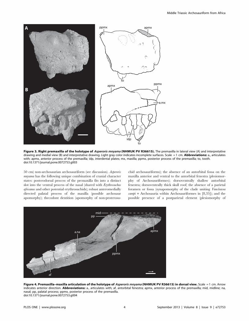
Figure 3. Right premaxilla of the holotype of Asperoris mnyama (NHMUK PV R36615). The premaxilla in lateral view (A) and interpretativedrawing and medial view (B) and interpretative drawing. Light gray color indicates incomplete surfaces. Scale = 1 cm. Abbreviations: a., articulateswith; apmx, anterior process of the premaxilla; idp, interdental plates; mx, maxilla; ppmx, posterior process of the premaxilla; to, tooth.doi:10.1371/journal.pone.0072753.g003
a.na
pp
apmx
ppmx
af
mid
Figure 4. Premaxilla–maxilla articulation of the holotype of Asperoris mnyama (NHMUK PV R36615) in dorsal view. Scale = 1 cm. Arrowindicates anterior direction. Abbreviations: a., articulates with; af, antorbital fenestra; apmx, anterior process of the premaxilla; mid, midline; na,nasal; pp, palatal process; ppmx, posterior process of the premaxilla.doi:10.1371/journal.pone.0072753.g004
Middle Triassic Archosauriform from Africa
PLOS ONE | www.plosone.org 4 September 2013 | Volume 8 | Issue 9 | e72753

Archosauriformes). Asperoris mnyama differs from all known
archosauriforms in having highly sculptured cranial elements
including the premaxilla, maxilla, nasal, prefrontal, frontal,
postfrontal, and parietal, and in having a highly sculptured,
dorsoventrally deep orbital margin of the frontal (Figs. 2–12).
In lacking an antorbital fossa at the base of the dorsal process of
the maxilla and on the dorsal margin of the posterior process of
the maxilla, the single known specimen of A. mnyama is
demonstrably distinct from other archosauriforms with compara-
ble elements from the Lifua Member of the Manda beds:
‘‘Mandasuchus tanyauchen’’ (NHMUK R6792), Parringtonia gracilis
(NHMUK R8646), ‘‘Pallisteria angustimentum’’ (NHMUK R36620),
and Asilisaurus kongwe (NMT RB159). Asperoris mnyama does not
share directly comparable elements with Stagonosuchus nyassicus
(GPIT/RE/3831), Hypselorhachis mirabilis (NHMUK R16586), an
unpublished archosaur skeleton (NMT RB48), ‘Teleocrater rhadinus’
(NHMUK R6795), and Nyasasaurus parringtoni (NHMUK R6856),
but the inferred phylogenetic position of Asperoris mnyama is not
consistent with it belonging to any of these taxa.
DescriptionPremaxilla. Most of the right premaxilla is preserved but it is
missing the anterodorsal process and much of the posterodorsal
process (Fig. 3). The lateral surface of the body is generally convex.
There is a weakly developed narial fossa positioned ventral to the
external naris, between the anterodorsal and posterior dorsal
processes. This shallow depression is located at the anteroventral
margin of the border of the naris and is demarcated by a weakly
developed ridge. The narial fossa is much shallower than that
described by Gower [36] for the premaxilla (SMNS 80260) of the
loricatan archosaur Batrachotomus kupferzellensis. A groove that is
present on the broken margin of the base of the anterodorsal
process stretches posteriorly around the base to reach the margin
of the premaxilla that borders the external naris. This groove
terminates in a foramen that opens posterodorsally. Two rounded
mounds positioned on the lateral surface just ventral to the border
of the external naris likely represent the external expression of the
most dorsal extent of the second and third tooth alveoli.
The lateral surface of the premaxilla is covered in a series of
large and tiny foramina and a system of short ridges. The larger
foramina are mostly restricted to the ventral margin and likely
represent nutrient foramina. Two foramina are present on the
lateral surface just ventral to the weak rim that defines the narial
fossa. Additionally, there are four anteriorly opening foramina
arranged in a vertical plane on the anterolateral surface, stretching
from the base of the anterodorsal process to the ventral margin of
the premaxilla. Tiny foramina occur on the anterior, ventral, and
posterior parts of the lateral surface of the premaxilla with the
highest concentrations nearest to the edges of the bone. The short
ridges occur across the premaxilla, but are primarily concentrated
Figure 5. Anterior fragment of the right maxilla of the holotype of Asperoris mnyama (NHMUK PV R36615). The maxilla in lateral view (A)and interpretative drawing and medial view (B) and interpretative drawing. Light gray color indicates incomplete surfaces. Scale = 1 cm. Arrowindicates anterior direction. Abbreviations: a., articulates with; af, antorbital fenestra; dmx, dorsal process of the maxilla; idp, interdental plates; na,nasal; pmx, premaxilla; pp, palatal process; to, tooth.doi:10.1371/journal.pone.0072753.g005
Middle Triassic Archosauriform from Africa
PLOS ONE | www.plosone.org 5 September 2013 | Volume 8 | Issue 9 | e72753

at the anterior edge of the element and on the lateral surface of the
posterodorsal process.
Only the ventral half of the posterior margin of the premaxilla is
preserved but the articulation with the maxilla is clearly observed.
The ventralmost portion of the posterior margin of the premaxilla
is slightly laterally deflected. When the maxilla and premaxilla are
in articulation, the anteroventral portion of the maxilla slightly
overlaps laterally this ventral portion of the premaxilla. A similarly
interlocking overlap does not appear to be common in archosauri-
forms, but is present in Batrachotomus kupferzellensis (SMNS 80260)
in which it is more strongly developed. When placed in
articulation, the premaxilla and the maxilla of A. mnyama fit
relatively tightly together (Fig. 4), although there is a narrow, slit-
like gap between the two elements. However, this gap between the
premaxilla and the maxilla is substantially less well developed in A.
mnyama than in B. kupferzellensis (SMNS 80260) and Postosuchus
kirkpatricki (TTU-P 9000; [37,38]) and there is no evidence on the
posterior border of the premaxilla for a distinct border of a
foramen (present in B. kupferzellensis). The dorsal half of the
posterior margin of the premaxilla of A. mnyama is mediolaterally
thinner than the ventral part, and partially laterally overlaps the
maxilla.
The broken anterodorsal ( = anterior dorsal) process is
mediolaterally compressed and its anteroposterior length at its
base is about half of the total length of the oral margin of the
premaxilla. This process very likely separated the external nares
along the midline. The mediolaterally compressed posterodorsal
process of the premaxilla articulates with a distinct groove on the
anterodorsal surface of the maxilla. The posterodorsal ( =
posterior dorsal) process projects posteriorly at about 45u to the
horizontal in lateral view. The posterodorsal process is broken
close to its base, and the main part of the body of the process is
missing, but the tip of the process is preserved in a slot in the nasal.
The tip of the posterodorsal process has a complex articulation
with the nasal that has been revealed by slight disarticulation
during preservation. The posterodorsal process of the premaxilla
splits into three small fingers that have precise articulations with
corresponding slots in the nasal. A tongue-in-groove articulation
between the premaxilla and nasal is also present in Erythrosuchus
africanus (BP/1/5207) and the archosaur Revueltosaurus callenderi
[39].
Medially, much of the medial surface of the premaxilla is flat,
marking the midline symphysis with its antimere (Figs. 3–4).
Posteriorly, the medial surface is divided into a rimmed depression
dorsally and a posteriorly directed palatal process. The depression
represents a very closely matching articular surface for the palatal
process of the maxilla. Ventral to this depression, the palatal
process projects posteromedially, but is broken at its tip. This
process, although broken, articulates with the maxilla ventral to
the maxillary palatal process, and fits into a distinct slot in this
element (see below). The interdental plates of the premaxilla are
separated from the medial surface of the rest of the premaxilla by a
distinct step that fades out posteriorly.
The premaxilla bears four alveoli separated from adjacent
alveoli by triangular interdental plates. A rounded foramen is
present between the dorsal margins of adjacent interdental plates,
and the first three of these foramina are open ventrally. The alveoli
are approximately equal in size.
Maxilla. The right maxilla is well-preserved in two sections
that do not connect (Figs. 5–6). The larger of the two sections
includes much of the anterior half of the maxilla with a partial
dorsal ( = ascending) process and complete palatal process, and
the smaller section is from the posterior portion of the maxilla and
preserves one complete alveolus and two partial alveoli. The
rather flat lateral surface of the maxilla bears sculpturing similar to
that of the frontal and nasal; however, this sculpturing is not evenly
distributed across the surface of the maxilla. The most distinctive
sculpturing is positioned just ventral and anterior to the antorbital
fenestra (Fig. 7). Here, deep grooves radiate from the antorbital
fenestra and surround small, rounded nubs of bone that extend
further laterally beyond the level of the majority of the lateral
surface of the maxilla. The ventral half of the lateral surface of the
maxilla has less distinct sculpturing. Here the bone surface is
uneven and consists of poorly differentiated knobs and seemingly
irregularly oriented grooves. Tiny foramina also cover this area
more abundantly near the ventral margin of the maxilla. This
particular surface sculpturing is unique to Aperoris mnyama, but does
have general similarities to the highly sculptured maxilla of the
possible erythrosuchid Guchengosuchus shiguaiensis (IVPP V8808). A
series of ventrally opening nutrient foramina lie close to the slightly
convex ventral edge of the maxilla. Two similarly sized foramina
are located 3 cm above the ventral edge of the maxilla near the
contact with the premaxilla (Fig. 5). These large foramina open
anteroventrally into anteroventrally trending grooves.
The surface of the maxilla for contact with the premaxilla slopes
anteroventrally and is slightly anterodorsally convex in lateral
view. The anterolateral termination of the maxilla tapers to a small
tab that slightly overlaps the premaxilla laterally when in
A B
C
af af
al
to
alal
Figure 6. Posterior fragment of the right maxilla and close upof the base of broken maxillary tooth of the holotype ofAsperoris mnyama (NHMUK PV R36615). Fragment in lateral (A) andmedial (B) views and a medial (C) view of the base of a maxillary tooth.Scales = 1 cm. Arrow indicates anterior direction. Abbreviations: af,antorbital fenestra; al, alveolus; to, tooth.doi:10.1371/journal.pone.0072753.g006
Middle Triassic Archosauriform from Africa
PLOS ONE | www.plosone.org 6 September 2013 | Volume 8 | Issue 9 | e72753

articulation (see above). In lateral view, the palatal process of the
maxilla (hidden by the premaxilla when the two are in articulation)
extends further anteriorly than the body of the maxilla. A clear
facet for articulation with the posterodorsal process of premaxilla
separates the palatal process from the rest of the maxilla; thus, the
maxilla is excluded from the external naris by the posterodorsal
process of the premaxilla. The anterodorsal surface of the dorsal
process of the maxilla bears a clear, mediolaterally deep slot for
articulation with the nasal. This slot is exposed in lateral view at its
ventral termination. The architecture of the articulation between
the nasal and the maxilla is precise; the grooves in the articular
facet of the maxilla correspond directly to ridges present on the
nasal (Fig. 7). Additionally, the articulation of the maxilla with the
nasal continues anterior to the slot on the anterodorsal edge of the
maxilla. Here a small splint of the nasal lies between the
posterodorsal process of the premaxilla and the main body of
the maxilla (Fig. 7–8).
The dorsal process of the maxilla is broken at its base; however,
a few details are discernable. The process is mediolaterally broad
anteriorly and tapers posteriorly toward the margin of the
antorbital fenestra. The articulation of the nasal with the maxilla
reveals that in lateral view the dorsal process of the maxilla
projects at an angle of 45u to the horizontal at its base, but also
that the process must be dorsoventrally short. This configuration
further indicates that the anterior portion of the antorbital fenestra
is not dorsoventrally deep. The posterior part of the base of the
dorsal process does not have an antorbital fossa and this is
consistent with the absence of an antorbital fossa along the ventral
margin of the antorbital fenestra. The presence of an antorbital
fenestra and the absence of an antorbital fossa alongside the
ventral portion of the antorbital fenestra in A. mnyama is similar to
that of non-archosaurian archosauriforms such as Proterosuchus
fergusi (BP/1/3993), Euparkeria capensis (SAM-PK-5867), and
Guchengosuchus shiguaiensis (IVPP V8808). However, the absence of
any antorbital fossa at the base of the dorsal process clearly
differentiates A. mnyama from Erythrosuchus africanus [11] and other
possible erythrosuchids (e.g., Chalishevia cotburnata, PIN 4356/1;
Garjainia prima, PIN 2394/5), and Youngosuchus sinensis (IVPP
V3239; [40,41]). The ventral margin of the antorbital fenestra is
nearly horizontal in A. mnyama, suggesting that the body of the
maxilla ventral to the antorbital fenestra maintained a broadly
consistent dorsoventral depth along its length, similar to the
condition in Proterosuchus fergusi (BP/1/3993), Euparkeria capensis
(SAM-PK-5867), Erythrosuchus africanus (BP/1/5207), and some
archosaurs such as Postosuchus kirkpatricki (TTU-P 9000). The height
of the section of the posterior portion of the maxilla further
supports the inference that the depth of the body of the maxilla
ventral to the antorbital fenestra remains uniform along its length,
and only tapered posteroventrally where the maxilla meets the
jugal.
The convex medial surface of the maxilla is divided by a
longitudinally oriented groove (referred to as the dental groove by
[42]) trending subparallel to the tooth-bearing edge (Fig. 5). The
surface of the body dorsal to the groove is smooth bone.
Anteriorly, a horizontally oriented and robust palatal process
extends anteriorly beyond the rest of the body of the maxilla, as in
Erythrosuchus africanus (SAM-PK-K1098; [11]) and most archosaurs
(e.g., Postosuchus kirkpatricki, TTU-P 9000). The palatal process of
Asperoris mnyama tapers to a point anteriorly and it fits precisely into
a broad and rimmed depression on the medial side of the
A B
mx pmx
na
enpmx
Figure 7. Unique sculpturing on the anterior portion of the skull of the holotype of Asperoris mnyama (NHMUK PV R36615) in rightlateral view. Close up of the sculpturing on the dorsal process of the maxilla (A) and close up of the sculpturing of the nasal (B). Scale = 1 cm.Abbreviations: en, external naris; mx, maxilla; na, nasal; pmx, premaxilla.doi:10.1371/journal.pone.0072753.g007
Middle Triassic Archosauriform from Africa
PLOS ONE | www.plosone.org 7 September 2013 | Volume 8 | Issue 9 | e72753

premaxilla (see above). Dorsally, a posteriorly extending groove
originates on the process, and defines the dorsal margin of the
maxilla in medial view. Ventral to the palatal process, there is a
clear facet on the medial surface of the maxilla for articulation
with the posteriorly projecting palatal process of the premaxilla. In
dorsal view, the palatal process of the maxilla arcs anteromedially
and converges upon the midline. When placed in articulation with
the premaxilla, it seems likely that the palatal process of the
maxilla would have met its antimere at the midline (Fig. 4), given
that the medial edge of the palatal process of the maxilla reaches a
point level with the medial extent of the premaxilla. However, the
medial surface of the maxillary palatal process lacks distinct facets
or grooves for contact with its antimere. The posteroventral
surface of the posterior portion of the palatal process may form the
articular surface for the vomer. The ventral edge of the antorbital
fenestra expands medially into a shelf and a shallow, longitudinally
orientated groove lies on the dorsal margin of this shelf.
Unfused interdental plates separate the alveoli from the
longitudinally oriented groove that bisects the medial surface of
the maxilla. Incompletely delimited foramina are positioned
ventral to and contacting the longitudinally oriented groove, and
open ventrally at the anteroposterior midpoint of each alveolus.
The lateral wall of each alveolus extends further ventrally than
does the medial wall. As a consequence of this alveolar
configuration, more of the apicobasal height of each tooth is
exposed in medial view than in lateral view. The broken medial
surfaces of the fifth and sixth alveoli indicate that the alveoli are
deep, as further evidenced by the presence of only 3 to 4 mm of
bone separating the antorbital fenestra from the dorsal end of the
tooth sockets.
There is a minimum of 10 alveoli in the maxilla, of which seven
are in the anterior section (Fig. 5) and three in the posterior section
(Fig. 6). The first alveolus is the smallest and the diameter of the
alveoli increase posteriorly. The alveoli decrease in diameter only
at the very posterior preserved extent of the maxilla.
Nasal. The incomplete right nasal preserves the articular
surface for the maxilla, the articular surface for the posterodorsal
process of the premaxilla with the tip of the posterodorsal process
preserved in articulation, and the posterior edge of the external
naris (Fig. 8). The lateral and dorsal surfaces bear a complex
sculpturing of knobs and grooves that is much more prominently
developed than that on the maxilla. Within the complex
sculpturing, a groove is present that originates at the posterior
margin of the external naris and bifurcates posteriorly, anterior to
the articular surface of the nasal with the maxilla (Fig. 8).
Sculpturing is absent only immediately adjacent to the posterior
margin of the external naris. A similar rugose sculpturing is also
present on the nasal of Guchengosuchus shiguaiensis (IVPP V8808), but
appears to be rare among most known non-archosaurian
archosauriforms.
The preserved portion of the nasal has two anterior processes:
an anterior process that would articulate with the anterodorsal
process of the premaxilla, and a more ventral ( = descending)
process that extends posterior and ventral to the external naris.
The more anterior process is broken at its base, slightly dorsal to
the external naris, but appears to have extended anteroventrally.
The more ventral process of the nasal bears a laterally open deep
groove, within which fits the tip of the posterodorsal process of the
premaxilla. As preserved, the posterior process of the premaxilla
has been displaced slightly anteriorly from its original articulation
with the nasal, revealing the complex contact between the two
en
en
ppmx
a.mx
ms
A
B
Figure 8. Right nasal of the holotype of Asperoris mnyama(NHMUK PV R36615). Nasal in lateral (A) and medial (B) views. Scale= 1 cm. Arrow indicates anterior direction. Abbreviations: a., articu-lates with; en, external naris; ms, midline suture; mx, maxilla; ppmx,posterior process of the premaxilla.doi:10.1371/journal.pone.0072753.g008
a.la
A B
C
a.fr
a.fr
or or
a.na?
a.fr
Figure 9. Right prefrontal of the holotype of Asperoris mnyama(NHMUK PV R36615). Prefrontal in lateral (A), medial (B), and dorsal(C) views. Scale = 1 cm. Arrow indicates anterior direction. Abbrevi-ations: a., articulates with; fr, frontal; la, lacrimal; na, nasal; or, orbit.doi:10.1371/journal.pone.0072753.g009
Middle Triassic Archosauriform from Africa
PLOS ONE | www.plosone.org 8 September 2013 | Volume 8 | Issue 9 | e72753

elements. Within the articular groove on the ventral process of the
nasal, three slots are present that correspond to three short prongs
on the tip of the posterodorsal process of the premaxilla. The cross
section of the broken surface of the ventral process of the nasal, in
anterior view, shows that a splint of the nasal separates the
posterior edge of the tip of the posterodorsal process of the
premaxilla from the maxilla, and that the nasal forms the complete
medial wall of the articular surface for the posterodorsal process of
the premaxilla. The insertion of the posterodorsal process of the
premaxilla into the nasal is rare among archosauriforms, but does
occur in the non-archosaurian archosauriforms Erythrosuchus
africanus (BP/1/5207; [11]), Guchengosuchus shiguaiensis (IVPP
V8808), Chalishevia cotburnata (PIN 4356/1), Garjainia prima (PIN
2394/5) and Shansisuchus shansisuchus (IVPP V2501; [43]), as well as
in at least two archosaurs (Turfanosuchus dabanensis, IVPP V3237;
Revueltosaurus callenderi; [39]). The relative size of the posterior
processes of the premaxilla that fit into the nasal is much larger in
E. africanus (BP/1/5207; [11]), G. shiguaiensis (IVPP V8808), and C.
cotburnata (PIN 4356/1) than in A. mnyama.
In Asperoris mnyama, the outline of the external naris is inferred to
have been anteroposteriorly elongated relative to its dorsoventral
height, on the basis of the portions of the rim preserved on the
nasal and premaxilla.
The ventral edge of the nasal preserves a complicated surface
for articulation with the dorsal process of the maxilla. The most
anterior portion of the nasal, posteroventral to the posterolateral
process of the premaxilla, extends medial to and is partially hidden
in lateral view by the maxilla when the two are in articulation.
More posteriorly, a flange of the nasal anterolaterally overlaps the
dorsal process of the maxilla, articulating with a groove on the
dorsal process. Posterior to this, the dorsal process of the maxilla
laterally overlaps the nasal, but the posterior extent of the
articulation is not well defined and it is not clear if some of the
nasal forms part of the antorbital fossa. This articular surface of
the nasal is smooth and tapers ventrally.
Medially, the nasal bears a distinct midline sutural surface for its
antimere that is about 1 cm deep. On the midline, a longitudinally
oriented groove trends parallel to two thin ridges for the length of
the preserved portion of the element. The midline surface also has
Figure 10. Right side of the skull roof of the holotype of Asperoris mnyama (NHMUK PV R36615) consisting of the frontal, postfrontaland parietal. Skull roof in dorsal view (A) and interpretative drawing, in ventral view (B) and interpretative drawing, in lateral view (C), andinterpretative drawing, and medial view (D) and interpretative drawing. Light gray color indicates incomplete surfaces. Scale = 1 cm. Arrow indicatesanterior direction. Abbreviations: a., articulates with; dep, depression; fr, frontal; kn, knob; ls, laterosphenoid; na, nasal; or, orbit; pa, parietal; pfr,postfrontal; pp, postparietal; prfr, prefrontal; soc, supraoccipital; su, suture; utf, upper temporal fenestra.doi:10.1371/journal.pone.0072753.g010
Middle Triassic Archosauriform from Africa
PLOS ONE | www.plosone.org 9 September 2013 | Volume 8 | Issue 9 | e72753

several smaller grooves and ridges. Ventral to the midline sutural
surface, the medial surface of the nasal is nearly smooth. A shallow
groove originating at the posterior end of the external naris trends
parallel to the midline suture for the entire preserved length of the
nasal.
Prefrontal. The right prefrontal is nearly complete, missing
only the tip of the ventral process (Fig. 9). The dorsal surface of the
prefrontal has sculpturing that is similar in form and development
to the dorsal surface of the nasal. The posterior portion of the
prefrontal forms the anterodorsal part of the margin of the orbit.
In this region, the sculpturing is composed of deep vertical grooves
that are similar to those seen along the portions of orbital rim
formed by the frontal and the postfrontal (see below). The
prefrontal has an interdigitating suture posteriorly with the part of
the frontal bordering the orbit. The prefrontal thins in dorsoven-
tral depth anteriorly and dorsally overlaps the dorsal surface of the
anterior portion of the frontal. A clear facet with short ridges and
grooves on the ventral surface of the prefrontal represents the
articular surface for the frontal.
The posteroventral portion of the prefrontal forms the anterior
part of the internal border of the orbit. Here the bone surface is
smooth and this surface is continuous with the fossa that borders
the orbit on the ventral surface of the frontal. The lateral surface of
the ventral process of the prefrontal is rugose with a few distinctive,
nearly horizontally orientated grooves. The anterior surface of the
ventral process forms a complex articular surface for the lacrimal.
A dorsoventrally oriented groove, located on the anterior edge,
likely matched a tongue of the lacrimal (not preserved). More
dorsally, an anteriorly projecting tongue of the prefrontal laterally
overlaps the lacrimal. The medial side of the ventral process is
smooth.
Frontal. The complete right frontal is preserved in articula-
tion with the postfrontal and the parietal (Fig. 10). The entire
dorsal surface of the frontal bears fine, evenly distributed grooves,
ridges, and foramina. A shallow, longitudinally oriented groove
crosses the entire dorsal surface of the body of the frontal and
continues onto the anterior half of the parietal. As a result, the
midline area and the orbital margin are dorsally raised above the
level of the middle of the frontal; the middle area is slightly
depressed. The main body of the frontal is dorsoventrally deep
(,1.5 cm) and thins anteriorly.
Anteriorly, the frontal bears two articular surfaces, one for the
prefrontal and one for the nasal. The prefrontal articular surface is
laterally deflected, nearly flat, and bears a few low anteroposte-
riorly oriented ridges. The articular surface of the frontal for the
nasal is located at the anterior margin of the element. The
anteriormost portion of the frontal is missing, but it is clear that a
finger of the nasal fitted into the frontal at the point where the
dorsal surface of the frontal intersects the articular facet for the
prefrontal. More medially, a finger of the frontal fitted into a
groove on the dorsal surface of the nasal. There does not appear to
have been long posteriorly directed processes of the nasals that
extended between the frontals at the midline, unlike the condition
in Erythrosuchus africanus (NHMUK R3592).
The frontal forms only a small section of the orbit margin in
lateral view. The lateral surface, like that of the postfrontal and the
prefrontal, is rugose and has dorsoventrally oriented ridges and
grooves. A similar sculpturing of the orbit margin is present in
A
a.ju
B
Figure 11. Incomplete postorbital of the holotype of Asperorismnyama (NHMUK PV R36615). Ventral process of the postorbital inlateral (A) and medial (B) views. Scale = 1 cm. Arrow indicates anteriordirection. Abbreviations: a., articulates with; j, jugal.doi:10.1371/journal.pone.0072753.g011
A B C
Figure 12. Unidentified skull elements of the holotype ofAsperoris mnyama (NHMUK PV R36615). Skull fragments in threeviews in (A) and (B) and two views in (C). Scales = 1 cm.doi:10.1371/journal.pone.0072753.g012
Euparkeria capensis*
Vancleavea campiErythrosuchus africanus*
Proterosuchus fergusi
Mesosuchus browniProlacerta broomi
ARCHOSAURIFORMES
PHYTOSAURIA
ARCHOSAURIA
PROTEROCHAMPSIDAE
Asperoris mnyama
Figure 13. The relationships of Asperoris mnyama (NHMUK PVR36615) among archosauromorphs. Strict consensus tree of 720MPTs (TL = 1288, CI = 0.374, RI = 0.775). Asterisks indicate the alternativesister-taxon relationships of Asperoris mnyama in the source MPTs.Species level taxa were collapsed into larger clades (clade name incapitals) and the interrelationships within these clades is identical tothat presented by Nesbitt (2011).doi:10.1371/journal.pone.0072753.g013
Middle Triassic Archosauriform from Africa
PLOS ONE | www.plosone.org 10 September 2013 | Volume 8 | Issue 9 | e72753

Batrachotomus kupferzellensis (SMNS 80260) and Nesbitt et al. [44]
argued that a palpebral element may have articulated with this
surface. However, in Asperoris mnyama there is not a clear facet for
articulation with any other element.
Posteriorly, the frontal forms interdigitating sutures with the
postfrontal laterally and the parietal posteriorly. In dorsal view, a
finger of the frontal articulates into the parietal laterally, and more
medially a finger of the parietal enters the body of the frontal
(Fig 10A). In medial view, a posterior process of the frontal extends
into the body of the parietal, such that the parietal both under-
and overlies the posterior portion of the frontal (Fig 10D). The
entire midline edge of the frontal forms a sutural surface for its
antimere. Here, grooves and ridges radiate from the center of the
element (Fig 10D).
Ventrally, most of the surface of the frontal is smooth. A
rounded ridge marks the anteromedial edge of the fossa bordering
the orbit, and a shallow groove parallels the orbit fossa rim on the
anteromedial side. The anterior portion of the frontal is ventrally
bowed transversely. The posterior portion of the ventral surface
forms an almost transverse, interdigitating suture with the parietal.
A small splint of the frontal extends between the postfrontal and
parietal (Fig. 10B). A deep pit that is defined by a clear rim is
located on the suture between the parietal and the frontal, and the
frontal contributes to the large depression formed by the
postfrontal, frontal, and parietal.
Postfrontal. Much of the right postfrontal is present with the
exception of the articular surface for the postorbital (Fig. 10). The
element is dorsoventrally thick, and has a similar depth and a
similar pattern of vertically oriented grooves and ridges to the orbit
margin of the frontal. The medial surface of the postfrontal forms
interdigitating sutures with both the parietal and the frontal.
Dorsally, the surface of the postfrontal is strongly rugose, similar to
that of the frontal and nasal. In ventral view, the surface of the
prefrontal is mostly smooth and the posterior portion of the
element contributes to the anterior border of a deep depression
that continues onto the parietal and frontal. The interdigitating
suture between the frontal and postfrontal is distinctly raised
relative to the surrounding bone.
Parietal. Most of the right parietal is preserved except for the
posterolateral end of the squamosal (occipital) process (Fig. 10).
Anteriorly, the parietal has a complex articulation with the frontal
(see above) and the lateral edge of the element forms the medial
portion of the upper temporal fenestra. It is not clear if and how
the parietal contacted the postorbital.
The rugose dorsal sculpturing on the frontal continues onto the
parietal. The dorsal surface of the parietal is essentially flat and
there is no parietal foramen on the midline or any indication of the
pineal fossa seen in, for example, Erythrosuchus africanus [11].
Additionally, the lateral and posterior edges of the dorsal surface of
the parietal slightly overhang the upper temporal fenestra and the
posterior surface of the parietal, respectively. The posterior edge of
dorsal surface of the parietal is raised relative to the surface of the
main body, as in Youngosuchus sinensis (IVPP V3239). In lateral view,
the medial wall of the upper temporal fenestra is dorsoventrally
deep (20 mm) and distinctly concave in lateral view.
Posteriorly, the parietal slopes posteroventrally to form the
occipital surface. A broken parietal tubercle is present near the
midline on the occipital surface, as in Erythrosuchus africanus [11].
An interdigitating sutural surface just lateral to the midline suture
of the parietal suggests that a postparietal might have been present
but was not preserved with the specimen. A pocket that marks the
articulation of the parietal with the dorsal surface of the
supraoccipital lies ventral to the possible articular surface for the
postparietal. The squamosal/occipital process of the parietal
Table 2. Non-archosaurian archosauriform (top) and archosaur (bottom) associations in the Early–Middle Triassic.
Temporal bin Location Taxon pairs
Lower Triassic
Russia (Yarenskian Gorizont)
Garjainia prima [56]
Vytshegdosuchus zheshartartensis [12,57]
Lower Triassic/Middle Triassic
China (lower Heshanggou Formation)
Proterosuchus [14]
Xilousuchus sapingensis [14,15]
Middle Triassic
China (upper Ermaying Formation)
Shansisuchus shansisuchus [43]
Wangisuchus tzeyii [14,43]
South America (Chanares Formation)
Chanaresuchus bonapartei [58]
Gracilisuchus stipanicicorum [59]
South America (Santa Maria sequence 1)
Archeopelta arborensis [60]
‘‘rauisuchian’’ [60]
North America (Moenkopi Formation)
non-archosaurian archosauriform [61]
Arizonasaurus babbitti [62–64]
doi:10.1371/journal.pone.0072753.t002
Middle Triassic Archosauriform from Africa
PLOS ONE | www.plosone.org 11 September 2013 | Volume 8 | Issue 9 | e72753

expands posterolaterally, but the surface for articulation with the
squamosal is not preserved. Ventrally, this process would have
articulated with the dorsal edge of the paroccipital process. It is not
clear if a posttemporal opening was present.
Ventrally, the parietal bears three distinct fossae, one that also
extends onto the frontal and postfrontal and which is positioned
near the lateral edges of all three elements, one positioned on the
interdigitating suture between the frontal and the parietal, and one
positioned entirely within the parietal and bisected by the midline
suture. This last fossa contained a well-developed knob of bone at
its center. An anteroposteriorly elongated facet on the lateral edge
of the ventral surface of the parietal possibly represents an area of
contact with the laterosphenoid.
Postorbital. Only the ventral process of the right? postorbital
is preserved (Fig. 11). The element tapers ventrally and bears a
depression with a rugose sculpturing on the anterolateral side of
the ventral end. A facet for the dorsal process of the jugal is present
on the posterovental side of the element, and this facet is divided
into two shallow and parallel basins. The more lateral basin is
visible in posterolateral view. The orbital margin is marked by
small grooves. Proximally, the cross section of the element is
triangular whereas more ventrally the cross section becomes more
oval.
Other skull fragments. Three skull fragments were col-
lected along with the holotype but cannot be identified (Fig. 12A–
C). All three likely belong to the skull given that they share a
similar preservation style and similarities of surface sculpturing
with the other skull elements. One fragment (Fig. 12A) might be
the pterygoid process of the basisphenoid, whereas the other two
(Fig. 12B–C) likely represent dermal elements given that both have
the distinct kind of sculpturing that is also present on the skull
table, premaxilla and maxilla.
Dentition. Only two broken teeth are preserved in the
holotype of Asperoris mnyama: a single premaxillary tooth in the
third alveolus (Fig. 3), and a single maxillary tooth in the fourth
alveolus (Fig. 4). Both teeth are fully erupted, with the base of the
crown as well as the part of the root preserved. The preserved
enamel of the premaxillary tooth is smooth, but there is not
enough of the crown preserved to determine if serrations were
present. The base of the premaxillary tooth is circular in cross
section whereas that of the maxillary tooth is oval, with the
anteroposterior axis longer than the mediolateral axis. The
dentition is seemingly thecodont given that each tooth sits in a
deep socket and is not directly attached to the surrounding bone.
Furthermore, there is no indication that the surrounding bone is
heavily remodelled as in the ankylothecondont non-archosaurian
archosauriforms Proterosuchus fergusi (BP/1/3993) and Guchengosuchus
shiguaiensis (IVPP V8808). However, the base of each tooth of A.
mnyama bears dorsoventrally oriented grooves and ridges that are
reminiscent of similar grooves and ridges present in these
ankylothecondont archosauriforms. In these taxa, the grooves
and ridges attach the base of the tooth directly to the premaxilla,
maxilla, or dentary whereas the grooves and ridges at the base of
the teeth of A. mnyama clearly do not attach to the premaxilla or
maxilla.
Phylogenetic analysisOur analysis found 720 most parsimonious trees (MPTs) of
length 1288 (consistency index [CI] = 0.374, retention index
= 0.775). In the strict consensus tree (Fig. 13), Asperoris mnyama was
recovered as a non-archosaurian archosauriform in a polytomy
with Erythrosuchus africanus, Vancleavea campi, Proterochampsidae,
Euparkeria capensis and Phytosauria + Archosauria. In all of the
MPTs, A. mnyama is found as either the sister taxon of Erythrosuchus
africanus or of Euparkeria capensis. The relationship between A.
mnyama and Erythrosuchus africanus is supported by the presence of
an articulation of the posterodorsal process of the premaxilla into
the nasal (character 4, state 3) whereas the relationship between A.
mnyama and Euparkeria capensis is supported only by archosauriform
plesiomorphies (character 146, state 0) or highly homoplastic
character states (character 27, state 1; CI = 0.154). Inclusion of A.
mnyama within Archosauria occurs only in trees at least four steps
longer than MPTs.
Discussion
Our phylogenetic analysis resolves Asperoris mnyama as a non-
archosaurian archosauriform. The presence of an antorbital
fenestra and the absence of a parietal foramen places A. mnyama
within archosauriforms given that these character states have been
repeatedly found as synapomorphies of Archosauriformes or less
inclusive clades in nearly all phylogenetic analyses of archosaur-
omorphs [4,8,35,45–49]. Fully thecodont dentition furthermore
suggests that A. mnyama lies within or as the sister taxon to the least
inclusive clade including Erythrosuchus africanus and Archosauria.
The preserved anatomy of A. mnyama appears to preclude an
assignment to Archosauria based on the absence of an antorbital
fossa on the main body of the maxilla and the likely presence of a
postparietal at the posterior portion of the skull table.
The lack of preservation of much of the skull and all of the
mandibular and postcranial skeleton of A. mnyama and the absence
of most stem archosaurs in our phylogenetic dataset precludes a
precise phylogenetic resolution of the new taxon among non-
archosaurian archosauriforms. However, our analysis did recover
a sister taxon relationship between A. mnyama and Erythrosuchus
africanus in some of the most parsimonious trees, and this
relationship is supported by the presence of a posterodorsal
process of the premaxilla that inserts into the ventral process of the
nasal. The presence of this character state is restricted to A. mnyama
and E. africanus among early archosauriforms in this analysis but
has a wider distribution among other early archosauriforms not
included in our phylogenetic analysis. For example, the postero-
dorsal process of the premaxilla inserts into the ventral process of
the nasal in Guchengosuchus shiguaiensis (IVPP V8808), Chalishevia
cotburnata (PIN 4356/1), Garjainia prima (PIN 2394/5) and
Shansisuchus shansisuchus (IVPP V2501). Currently, it is not clear if
all of these taxa are more closely related to E. africanus than to
other archosauriforms (i.e., Erythrosuchidae, see [50] for support
of monophyly) or to some extent form a grade, as recently
suggested by Ezcurra et al. [51] (see [10]). If these taxa represent a
grade, the presence of the posterodorsal process of the premaxilla
that inserts into the ventral process of the nasal would not support
A. mnyama as a particularly close relative of E. africanus.
Despite the nature of the premaxilla-nasal articulation, Asperoris
mnyama has some character states that can be used to argue against
an erythrosuchid affinity. For example, the parietals of A. mnyama
are flat like those of, for example, Vancleavea campi (GR 138) and
Euparkeria capensis (SAM-PK-5867) rather than having a depression
on their dorsal surface of the parietals at the midline ( = pineal
fossa of [50]) such as is found in Erythrosuchus africanus [11,50],
Guchengosuchus shiguaiensis (IVPP V8808), Garjainia prima (PIN 2394/
5), and Shansisuchus shansisuchus (IVPP V2504; [43]). Youngosuchus
sinensis (IVPP V3239; [50]) lacks a pineal fossa, but its status as a
possible erythrosuchid has been questioned [40]. Asperoris mnyama
also lacks the small antorbital fossa at the posterior portion of the
base of the dorsal process of the maxilla that is present in E.
africanus [11], Chalishevia cothurnata (PIN 4356/1), G. prima (PIN
Middle Triassic Archosauriform from Africa
PLOS ONE | www.plosone.org 12 September 2013 | Volume 8 | Issue 9 | e72753

2394/5) and Y. sinensis (IVPP V3239). Thus, the case that A.
mnyama is an erythrosuchid is far from compelling.
The enlarged palatal process of the maxilla of Asperoris mnyama
perhaps suggests a phylogenetic position closer to Archosauria.
Early archosauriforms such as Proterosuchus fergusi (NMQR 880)
completely lack palatal processes, and taxa such as Guchengosuchus
shiguaiensis (IVPP V8808) have a palatal process that is poorly
differentiated from the body of the maxilla. Erythrosuchus africanus
[11] and Euparkeria capensis (SAM-PK-5060) have a somewhat
developed palatal process of the maxilla, but this process might not
meet its antimere at the midline. In contrast, most members of
Archosauria have large palatal process that meet their antimeres at
the midline [8]. In these taxa (e.g. Postosuchus kirkpatricki, TTU-P
9000), the palatal process extends medially to reach the midline
and, in disarticulated maxillae, the palatal processes have ridges
and grooves that mark the midline articulation. The condition in
A. mnyama is somewhat intermediate between that of non-
archosaurian archosauriforms and archosaurs. The robust palatal
process expands medially to near the midline, but it is not clear
that it actually contacted its antimere and the medial surface of the
palatal process does not have any indication of being a contact
surface.
In sum, the available evidence suggests that Asperoris mnyama is
either a non-archosaurian archosauriform that is closer to
Erythrosuchus africanus than to other archosauriforms (i.e., possibly
an erythrosuchid) or is slightly phylogenetically closer to Arch-
osauria than is E. africanus. Regardless of the correct phylogenetic
position, A. mnyama is the first confirmed non-archosaurian
archosauriform among a diverse archosauriform fauna from the
Lifua Member of the Manda beds.
Archosaurs and non-archosaurian archosauriforms overlapped
temporally throughout the Early and Middle Triassic, as predicted
by ghost lineages determined from recent phylogenies (e.g.,
[8,15,52]), suggesting that associations of these taxa should be
commonplace. However, localities or a set of localities within a
single sedimentary package (e.g., member or formation) where
both stem archosaurs and archosaurs co-occur are relatively rare.
Associations do occur in the Lower Triassic in Russia, close to the
Lower to Middle Triassic boundary in China, and in the Middle
Triassic of Russia, China, South America, and possibly in North
America (see table 2). In the Late Triassic, proterochampsids and
early dinosaurs occur in the same area (Ischigualasto Formation) in
Argentina [53] and possibly in the Santa Maria sequence of
southern Brazil, whereas in North America Vancleavea campi occurs
with early dinosaurs throughout deposition within the Chinle basin
[35,54,55]. Moreover, if phytosaurs fall outside of Archosauria (see
[8]), then the co-occurrence of non-archosaurian archosauriforms
(i.e., phytosaurs) and archosaurs is common in North America,
Europe, northern Africa, and India throughout the Late Triassic
and persisted until the end-Triassic mass extinction event.
Acknowledgments
We thank S. Chapman and P. Barrett (NHMUK) for access to the
holotype, comparative material, and the notebooks of A. Charig and B.
Cox. Matthew Brown provided preparation and molding advice. We thank
M. Sander for helpful comments on the manuscript.
Author Contributions
Conceived and designed the experiments: SJN RJB DJG. Analyzed the
data: SJN RJB DJG. Wrote the paper: SJN RJB DJG.
References
1. Benton MJ (1990) Origin and interrelationships of dinosaurs. In: Weishampel
DB, Dobson P, Osmolska H, editors. The Dinosauria. Berkeley: University of
California Press. 11–30.
2. Fraser NC (2006) Dawn of the Dinosaurs: Life in the Triassic (Life of the Past).
Bloomington: Indiana University Press.
3. Sues H-D, Fraser NC (2010) Triassic Life on Land: The Great Transition. New
York: Columbia University Press. 236 p.
4. Dilkes DW (1998) The Early Triassic rhynchosaur Mesosuchus browni and the
interrelationships of basal archosauromorph reptiles. Philosophical Transactions
of the Royal Society of London Series B 353: 501–541.
5. Gauthier JA, Kluge AG, Rowe T (1988) Amniote phylogeny and the importance
of fossils. Cladistics 4: 105–209.
6. Benton MJ, Clark JM (1988) Archosaur phylogeny and the relationships of the
Crocodylia. In: Benton MJ, editor. The Phylogeny and Classification of the
Tetrapods Vol 1: Amphibians and Reptiles. Oxford: Clarendon Press. 295–338.
7. Gottmann-Quesada A, Sander PM (2009) A redescription of the early
archosauromorph Protorosaurus speneri Meyer, 1832 and its phylogenetic
relationships. Palaeontographica Abteilung B 287: 123–220.
8. Nesbitt SJ (2011) The early evolution of Archosauria: relationships and the
origin of major clades. Bulletin of the American Museum of Natural History
352: 1–292.
9. Ewer RF (1965) The anatomy of the thecodont reptile Euparkeria capensis Broom.
Philosophical Transactions of the Royal Society of London, Series B 248: 379–
435.
10. Ezcurra MD, Butler RJ, Gower DJ (2013) ‘Proterosuchia’: the origin and early
history of Archosauriformes. In: Nesbitt SJ, Desojo JB, Irmis RB, editors.
Anatomy, Phylogeny and Palaeobiology of Early Archosaurs and their Kin.
London: Geological Society, London, Special Publications. (doi:10.1144/
SP379.11).
11. Gower DJ (2003) Osteology of the early archosaurian reptile Erythrosuchus
africanus Broom. Annals of the South African Museum 110: 1–84.
12. Gower DJ, Sennikov AG (2000) Early archosaurs from Russia. In: Benton MJ,
Shishkin MA, Unwin DM, Kurochkin EN, editors. The Age of Dinosaurs in
Russia and Mongolia. New York: Cambridge University Press. 140–159.
13. Sookias RB, Butler RJ (2013) Euparkeriidae. In: Nesbitt SJ, Desojo JB, Irmis RB,
editors. Anatomy, Phylogeny and Palaeobiology of Early Archosaurs and their
Kin. London: Geological Society, London, Special Publications. (doi:10.1144/
SP379.6).
14. Nesbitt SJ, Liu J, Li C (2011) A sail-backed suchian from the Heshanggou
Formation (Early Triassic: Olenekian) of China. Earth and Environmental
Science Transactions of the Royal Society of Edinburgh 101: 271–284.
15. Butler RJ, Brusatte SL, Reich M, Nesbitt SJ, Schoch RR, et al. (2011) The sail-
backed reptile Ctenosauriscus from the latest Early Triassic of Germany and the
timing and biogeography of the early archosaur radiation. PLoS One 6: 1–28.
16. Sookias RB, Butler RJ, Benson RBJ (2012) Rise of dinosaurs reveals major body-
size transitions are driven by passive processes of trait evolution. Proceedings of
the Royal Society of London B 279: 2180–2187.
17. Brusatte SL, Benton MJ, Ruta M, Lloyd GT (2008) Superiority, competition,
and opportunism in the evolutionary radiation of dinosaurs. Science 321: 1485–
1488.
18. Attridge J, Ball HW, Charig A, Cox CB (1964) The British Museum (Natural
History)-University of London Joint Paleontological Expedition to Northern
Rhodesia and Tanganyika. Nature 201: 445–449.
19. Stockley GM (1932) The geology of the Ruhuhu coalfields, Tanganyika
Territory. Quarterly Journal of the Geological Society of London 88: 610–622.
20. Nesbitt SJ, Sidor CA, Irmis RB, Angielczyk KD, Smith RMH, et al. (2010)
Ecologically distinct dinosaurian sister-group shows early diversification of
Ornithodira. Nature 464: 95–98.
21. Nesbitt SJ, Barrett PM, Werning S, Sidor CA, Charig A (2012) The oldest
dinosaur? A Middle Triassic dinosauriform from Tanzania. Biology Letters 9:
20120949.
22. Butler RJ, Barrett PM, Abel RL, Gower DJ (2009) A possible ctenosauriscid
archosaur from the Middle Triassic Manda Beds of Tanzania. Journal of
Vertebrate Paleontology 29: 1022–1031.
23. Charig A (1967) Subclass Archosauria. In: Hartland WB, Holland CH, House
MR, Hughes NF, Reynolds AB, et al., editors. The Fossil Record. London:
Geological Society of London. 708–718.
24. Charig AJ (1956) New Triassic archosaurs from Tanganyika including
Mandasuchus and Teleocrater. Cambridge: University of Cambridge. 503 p.
25. Charig AJ (1957) New Triassic archosaurs from Tanganyika including
Mandasuchus and Teleocrater. Abstracts of Dissertations of the University of
Cambridge 1955–56: 28–29.
26. Gebauer EVI (2004) Neubeschreibung von Stagonosuchus nyassicus v. Huene, 1938
(Thecodontia, Rauisuchia) aus der Manda-Formation (Mittlere Trias) von
Sudwest-Tansania. Neues Jahrbuch fur Geologie und Palaeontologie, Abhan-
dlungen 231: 1–35.
Middle Triassic Archosauriform from Africa
PLOS ONE | www.plosone.org 13 September 2013 | Volume 8 | Issue 9 | e72753

27. Huene Fv (1938) Ein grosser Stagonolepide aus der jungeren Trias Ostafrikas.
Neues Jahrbuch fur Geologie und Palaeontologie, Beilage-Bande Abt B 80: 264–278.
28. Huene Fv (1939) Ein kleiner Pseudosuchier und ein Saurischier aus den
ostafrikanischen Mandaschichten. Neues Jahrbuch fur Geologie und Palaeonto-logie, Beilage-Bande Abt B 81: 61–69.
29. Lautenschlager S, Desojo JB (2011) Reassessment of the Middle Triassic‘‘rauisuchian’’ archosaurs Ticinosuchus ferox and Stagonosuchus nyassicus. Palaonto-
logische Zeitschrift 85: 357–381.
30. Nesbitt SJ, Butler RJ (2013) Redescription of the archosaur Parringtonia gracilis
from the Middle Triassic Manda Beds of Tanzania, and the antiquity of
Erpetosuchidae. Geological Magazine 150: 225–238.31. Cox CB (1991) The Pangaea dicynodont Rechnisaurus and the comparative
biostratigraphy of Triassic dicynodont faunas. Palaeontology 34: 767–784.32. Swofford DL (2002) PAUP* (Phylogenetic Analysis Using Parsimony and Other
Methods), Version 4.10b. Version 4.10b ed. Sunderland, MA: Sinauer
Association.33. Modesto SP, Anderson JS (2004) The phylogenetic definition of Reptilia.
Systematic Biology 53: 815–821.34. Gauthier JA, Kluge AG, Rowe T (1988) The early evolution of the Amniota. In:
Benton MJ, editor. The phylogeny and classification of the tetrapods, volume 1:
Amphibians, reptiles, birds. Oxford: Clarendon Press. 103–155.35. Nesbitt SJ, Stocker MR, Small B, Downs A (2009) The osteology and
relationships of Vancleavea campi (Reptilia: Archosauriformes). Zoological Journalof the Linnean Society 157: 814–864.
36. Gower DJ (1999) The cranial and mandibular osteology of a new rauisuchianarchosaur from the Middle Triassic of southern Germany. Stuttgarter Beitrage
zur Naturkunde Serie B (Geologie und Palaontologie) 280: 1–49.
37. Chatterjee S (1985) Postosuchus, a new thecodontian reptile from the Triassic ofTexas and the origin of tyrannosaurs. Philosophical Transactions of the Royal
Society of London B 309: 395–460.38. Weinbaum JC (2011) The skull of Postosuchus kirkpatricki (Archosauria:
Paracrocodyliformes) from the Upper Triassic of the United States. PaleoBios
30: 18–44.39. Parker WG, Irmis RB, Nesbitt SJ, Martz JM, Brown LS (2005) The Late
Triassic pseudosuchian Revueltosaurus callenderi and its implications for thediversity of early ornithischian dinosaurs. Proceedings of the Royal Society of
London B: 963–969.40. Kalandadze NN, Sennikov AG (1985) New reptiles from the Middle Triassic in
the southern forelands. Paleontological Journal 1995: 73–80.
41. Young CC (1973) On the occurrence of Vjushkovia in Sinkiang. Memoirs of theInstitute of Vertebrate Paleontology and Paleoanthropology 10: 38–53.
42. Galton PM (1985) The poposaurid thecodontian Teratosaurus suevicus v. Meyerplus referred specimens mostly based on prosauropod dinosaurs from the Middle
Stubensandstein (Upper Triassic) of Nordwurttemberg, West Germany.
Stuttgarter Beitrage zur Naturkunde Serie B (Geologie und Palaontologie)116: 1–29.
43. Young CC (1964) The pseudosuchians in China. Palaeontologia Sinica 151: 1–205.
44. Nesbitt SJ, Turner AH, Weinbaum JC (in press) A survey of skeletal elements inthe orbit of Pseudosuchia and the origin of the crocodylian palpebral. Earth and
Environmental Science Transactions of the Royal Society of Edinburgh.
45. Bennett SC (1996) The phylogenetic position of the Pterosauria withinArchosauromorpha. Zoological Journal of the Linnean Society 118: 261–308.
46. Benton MJ (2004) Origins and relationships of Dinosauria. In: Weishampel DB,
Dobson P, Osmolska H, editors. Dinosauria (2nd edition). Berkeley: University of
California Press. 7–24.
47. Gauthier JA (1984) A cladistic analysis of the higher systematic categories of
Diapsida. Berkeley: University of California Berkeley. 564 p.
48. Juul L (1994) The phylogeny of basal archosaurs. Palaeontologia Africana 31: 1–
38.
49. Parrish JM (1993) Phylogeny of the Crocodylotarsi, with reference to
archosaurian and crurotarsan monophyly. Journal of Vertebrate Paleontology
13: 287–308.
50. Parrish JM (1992) Phylogeny of the Erythrosuchidae. Journal of Vertebrate
Paleontology 12: 93–102.
51. Ezcurra MD, Lecuona A, Martinelli A (2010) A new basal archosauriform
diapsid from the Lower Triassic of Argentina. Journal of Vertebrate
Paleontology 30: 1433–1450.
52. Brusatte SL, Benton MJ, Desojo JB, Langer MC (2010) The higher-level
phylogeny of Archosauria (Tetrapoda: Diapsida). Journal of Systematic
Palaeontology 8: 3–47.
53. Martinez RN, Sereno PC, Alcober OA, Colombi CE, Renne PR, et al. (2011) A
basal dinosaur from the dawn of the dinosaur era in southwestern Pangaea.
Science 331: 206–210.
54. Hunt AP, Heckert AB, Lucas SG, Downs A (2002) The distribution of the
enigmatic reptile Vancleavea in the Upper Triassic Chinle Group of the western
United States. New Mexico Museum of Natural History and Science Bulletin 21:
269–273.
55. Hunt AP, Lucas SG, Spielmann JA (2005) The holotype specimen of Vancleavea
campi from Petrified Forest National Park, Arizona, with notes on the taxonomy
and distribution of the taxon. New Mexico Museum of Natural History and
Science Bulletin 29: 59–66.
56. Ochev VG (1958) [New data concerning the pseudosuchians of the USSR].
Doklay AN SSSR 123: 749–751.
57. Sennikov AG (1988) [New rauisuchids from the Triassic of the European
USSR]. Paleontologicheskii Zhurnal 1988: 124–128.
58. Romer AS (1971) The Chanares (Argentina) Triassic reptile fauna. XI. Two new
long-snouted thecodonts, Chanaresuchus and Gualosuchus. Breviora 379: 1–22.
59. Romer AS (1972) The Chanares (Argentina) Triassic reptile fauna. XIII. An
early ornithosuchid pseudosuchian, Gracilisuchus stipanicicorum, gen. et sp. nov.
Breviora 389: 1–24.
60. Desojo JB, Ezcurra MD, Schultz CL (2011) An unusual new archosauriform
from the Middle–Late Triassic of southern Brazil and the monophyly of
Doswelliidae. Zoological Journal of the Linnean Society 161: 839–871.
61. Nesbitt SJ (2005) A new archosaur from the upper Moenkopi Formation (Middle
Triassic) of Arizona and its implications for rauisuchian phylogeny and
diversification. Neues Jahrbuch fur Geologie und Palaeontologie Monatshefte
2005: 332–346.
62. Nesbitt SJ (2003) Arizonasaurus and its implications for archosaur divergence.
Proceedings of the Royal Society of London, B 270 (Supplement 2): S234–S237.
63. Nesbitt SJ (2005) The osteology of the Middle Triassic pseudosuchian archosaur
Arizonasaurus babbitti. Historical Biology 17: 19–47.
64. Welles SP (1947) Vertebrates from the Upper Moenkopi Formation of Northern
Arizona. University of California Publications in Geological Science 27: 241–
294.
Middle Triassic Archosauriform from Africa
PLOS ONE | www.plosone.org 14 September 2013 | Volume 8 | Issue 9 | e72753