A network model for the control of the differentiation process in Th cells
14
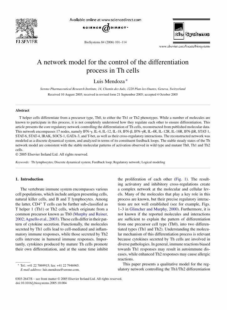
BioSystems 84 (2006) 101–114 A network model for the control of the differentiation process in Th cells Luis Mendoza ∗ Serono Pharmaceutical Research Institute, 14, Chemin des Aulx, 1228 Plan-les-Ouates, Geneva, Switzerland Received 10 August 2005; received in revised form 21 September 2005; accepted 4 October 2005 Abstract T helper cells differentiate from a precursor type, Th0, to either the Th1 or Th2 phenotypes. While a number of molecules are known to participate in this process, it is not completely understood how they regulate each other to ensure differentiation. This article presents the core regulatory network controlling the differentiation of Th cells, reconstructed from published molecular data. This network encompasses 17 nodes, namely IFN-, IL-4, IL-12, IL-18, IFN-, IFN-R, IL-4R, IL-12R, IL-18R, IFN-R, STAT-1, STAT-6, STAT-4, IRAK, SOCS-1, GATA-3, and T-bet, as well as their cross-regulatory interactions. The reconstructed network was modeled as a discrete dynamical system, and analyzed in terms of its constituent feedback loops. The stable steady states of the Th network model are consistent with the stable molecular patterns of activation observed in wild type and mutant Th0, Th1 and Th2 cells. © 2005 Elsevier Ireland Ltd. All rights reserved. Keywords: Th lymphocytes; Discrete dynamical system; Feedback loop; Regulatory network; Logical modeling 1. Introduction The vertebrate immune system encompasses various cell populations, which include antigen presenting cells, natural killer cells, and B and T lymphocytes. Among the latter, CD4 + T cells can be further sub-classified as T helper 1 (Th1) or Th2 cells, which originate from a common precursor known as Th0 (Murphy and Reiner, 2002; Agnello et al., 2003). These cells differ in their pat- tern of cytokine secretion. Functionally, the molecules secreted by Th1 cells lead to cell-mediated and inflam- matory immune responses, while those secreted by Th2 cells intervene in humoral immune responses. Impor- tantly, cytokines produced by mature Th cells promote their own differentiation, and at the same time inhibit ∗ Tel.: +41 22 7069915; fax: +41 22 7946965. E-mail address: [email protected]. the proliferation of each other (Fig. 1). The result- ing activatory and inhibitory cross-regulations create a complex network at the molecular and cellular lev- els. Many of the molecules that play a key role in this process are known, but their precise regulatory interac- tions are not well established (see for example, Figs. 1–3 in Glimcher and Murphy, 2000). Furthermore, it is not known if the reported molecules and interactions are sufficient to explain the pattern of differentiation from one precursor cell type (Th0), into two differen- tiated types (Th1 and Th2). Understanding the molecu- lar mechanism of this differentiation process is relevant because cytokines secreted by Th cells are involved in diverse pathologies. In general, immune reactions biased towards Th1 responses may result in autoimmune dis- eases, while enhanced Th2 responses may cause allergic reactions. This paper presents a qualitative model for the reg- ulatory network controlling the Th1/Th2 differentiation 0303-2647/$ – see front matter © 2005 Elsevier Ireland Ltd. All rights reserved. doi:10.1016/j.biosystems.2005.10.004
-
Upload
independent -
Category
Documents
-
view
0 -
download
0
Transcript of A network model for the control of the differentiation process in Th cells
BioSystems 84 (2006) 101–114
A network model for the control of the differentiationprocess in Th cells
Luis Mendoza ∗Serono Pharmaceutical Research Institute, 14, Chemin des Aulx, 1228 Plan-les-Ouates, Geneva, Switzerland
Received 10 August 2005; received in revised form 21 September 2005; accepted 4 October 2005
Abstract
T helper cells differentiate from a precursor type, Th0, to either the Th1 or Th2 phenotypes. While a number of molecules areknown to participate in this process, it is not completely understood how they regulate each other to ensure differentiation. Thisarticle presents the core regulatory network controlling the differentiation of Th cells, reconstructed from published molecular data.This network encompasses 17 nodes, namely IFN-�, IL-4, IL-12, IL-18, IFN-�, IFN-�R, IL-4R, IL-12R, IL-18R, IFN-�R, STAT-1,STAT-6, STAT-4, IRAK, SOCS-1, GATA-3, and T-bet, as well as their cross-regulatory interactions. The reconstructed network wasmodeled as a discrete dynamical system, and analyzed in terms of its constituent feedback loops. The stable steady states of the Thnetwork model are consistent with the stable molecular patterns of activation observed in wild type and mutant Th0, Th1 and Th2cells.© 2005 Elsevier Ireland Ltd. All rights reserved.
Keywords: Th lymphocytes; Discrete dynamical system; Feedback loop; Regulatory network; Logical modeling
1
cntTc2tsmctt
0
. Introduction
The vertebrate immune system encompasses variousell populations, which include antigen presenting cells,atural killer cells, and B and T lymphocytes. Amonghe latter, CD4+ T cells can be further sub-classified as
helper 1 (Th1) or Th2 cells, which originate from aommon precursor known as Th0 (Murphy and Reiner,002; Agnello et al., 2003). These cells differ in their pat-ern of cytokine secretion. Functionally, the moleculesecreted by Th1 cells lead to cell-mediated and inflam-atory immune responses, while those secreted by Th2
ells intervene in humoral immune responses. Impor-antly, cytokines produced by mature Th cells promoteheir own differentiation, and at the same time inhibit
∗ Tel.: +41 22 7069915; fax: +41 22 7946965.E-mail address: [email protected].
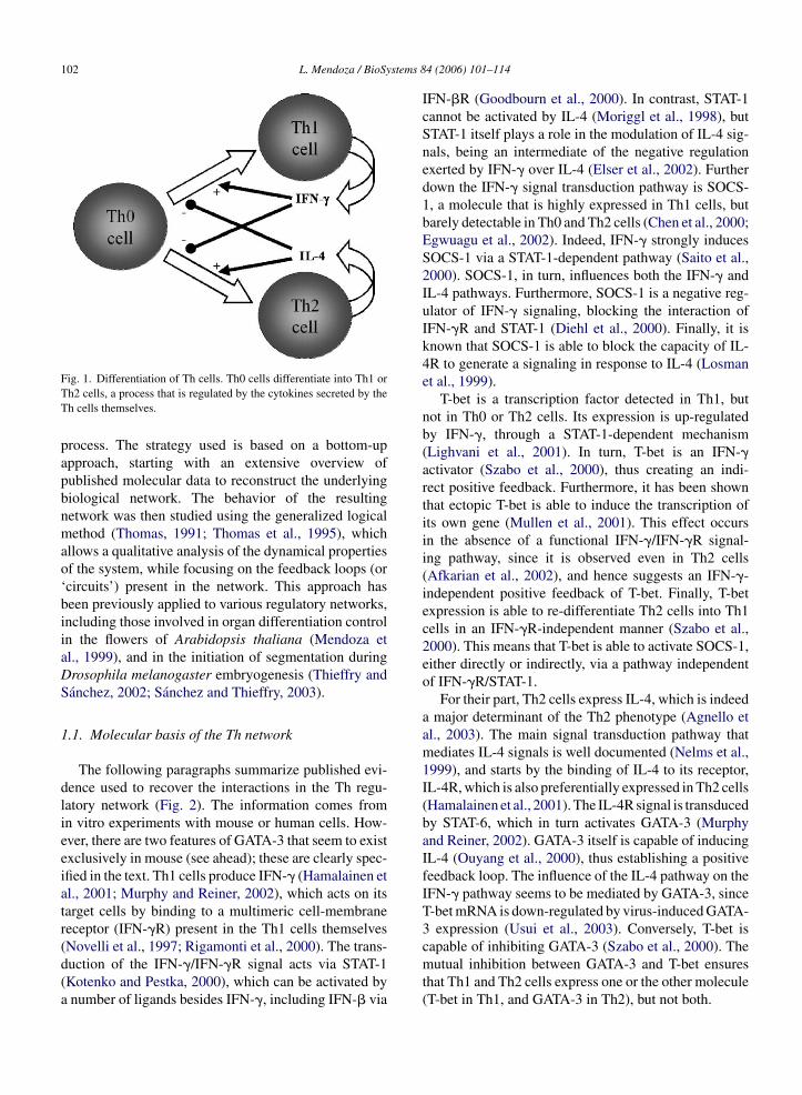
the proliferation of each other (Fig. 1). The result-ing activatory and inhibitory cross-regulations createa complex network at the molecular and cellular lev-els. Many of the molecules that play a key role in thisprocess are known, but their precise regulatory interac-tions are not well established (see for example, Figs.1–3 in Glimcher and Murphy, 2000). Furthermore, it isnot known if the reported molecules and interactionsare sufficient to explain the pattern of differentiationfrom one precursor cell type (Th0), into two differen-tiated types (Th1 and Th2). Understanding the molecu-lar mechanism of this differentiation process is relevantbecause cytokines secreted by Th cells are involved indiverse pathologies. In general, immune reactions biasedtowards Th1 responses may result in autoimmune dis-eases, while enhanced Th2 responses may cause allergicreactions.
This paper presents a qualitative model for the reg-ulatory network controlling the Th1/Th2 differentiation
303-2647/$ – see front matter © 2005 Elsevier Ireland Ltd. All rights reserved.doi:10.1016/j.biosystems.2005.10.004
102 L. Mendoza / BioSystems 84 (2006) 101–114
Fig. 1. Differentiation of Th cells. Th0 cells differentiate into Th1 orTh2 cells, a process that is regulated by the cytokines secreted by theTh cells themselves.
process. The strategy used is based on a bottom-upapproach, starting with an extensive overview ofpublished molecular data to reconstruct the underlyingbiological network. The behavior of the resultingnetwork was then studied using the generalized logicalmethod (Thomas, 1991; Thomas et al., 1995), whichallows a qualitative analysis of the dynamical propertiesof the system, while focusing on the feedback loops (or‘circuits’) present in the network. This approach hasbeen previously applied to various regulatory networks,including those involved in organ differentiation controlin the flowers of Arabidopsis thaliana (Mendoza etal., 1999), and in the initiation of segmentation duringDrosophila melanogaster embryogenesis (Thieffry andSanchez, 2002; Sanchez and Thieffry, 2003).
1.1. Molecular basis of the Th network
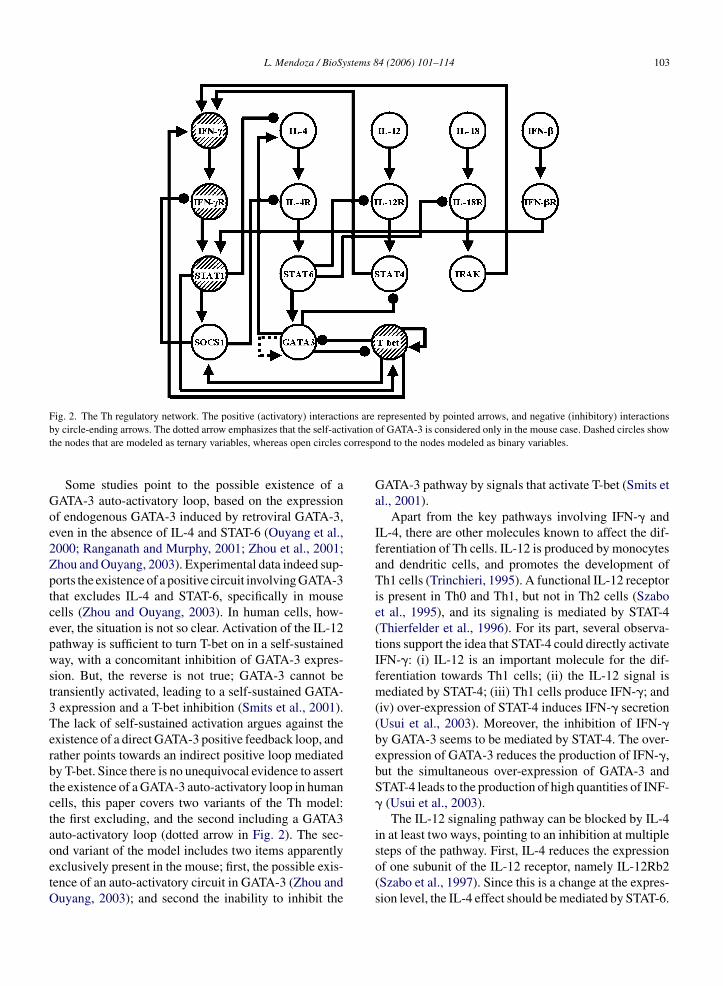
The following paragraphs summarize published evi-dence used to recover the interactions in the Th regu-latory network (Fig. 2). The information comes fromin vitro experiments with mouse or human cells. How-ever, there are two features of GATA-3 that seem to existexclusively in mouse (see ahead); these are clearly spec-ified in the text. Th1 cells produce IFN-� (Hamalainen etal., 2001; Murphy and Reiner, 2002), which acts on itstarget cells by binding to a multimeric cell-membranereceptor (IFN-�R) present in the Th1 cells themselves(Novelli et al., 1997; Rigamonti et al., 2000). The trans-
IFN-�R (Goodbourn et al., 2000). In contrast, STAT-1cannot be activated by IL-4 (Moriggl et al., 1998), butSTAT-1 itself plays a role in the modulation of IL-4 sig-nals, being an intermediate of the negative regulationexerted by IFN-� over IL-4 (Elser et al., 2002). Furtherdown the IFN-� signal transduction pathway is SOCS-1, a molecule that is highly expressed in Th1 cells, butbarely detectable in Th0 and Th2 cells (Chen et al., 2000;Egwuagu et al., 2002). Indeed, IFN-� strongly inducesSOCS-1 via a STAT-1-dependent pathway (Saito et al.,2000). SOCS-1, in turn, influences both the IFN-� andIL-4 pathways. Furthermore, SOCS-1 is a negative reg-ulator of IFN-� signaling, blocking the interaction ofIFN-�R and STAT-1 (Diehl et al., 2000). Finally, it isknown that SOCS-1 is able to block the capacity of IL-4R to generate a signaling in response to IL-4 (Losmanet al., 1999).
T-bet is a transcription factor detected in Th1, butnot in Th0 or Th2 cells. Its expression is up-regulatedby IFN-�, through a STAT-1-dependent mechanism(Lighvani et al., 2001). In turn, T-bet is an IFN-�activator (Szabo et al., 2000), thus creating an indi-rect positive feedback. Furthermore, it has been shownthat ectopic T-bet is able to induce the transcription ofits own gene (Mullen et al., 2001). This effect occursin the absence of a functional IFN-�/IFN-�R signal-ing pathway, since it is observed even in Th2 cells(Afkarian et al., 2002), and hence suggests an IFN-�-independent positive feedback of T-bet. Finally, T-betexpression is able to re-differentiate Th2 cells into Th1
duction of the IFN-�/IFN-�R signal acts via STAT-1(Kotenko and Pestka, 2000), which can be activated bya number of ligands besides IFN-�, including IFN-� via
cells in an IFN-�R-independent manner (Szabo et al.,2000). This means that T-bet is able to activate SOCS-1,either directly or indirectly, via a pathway independentof IFN-�R/STAT-1.
For their part, Th2 cells express IL-4, which is indeeda major determinant of the Th2 phenotype (Agnello etal., 2003). The main signal transduction pathway thatmediates IL-4 signals is well documented (Nelms et al.,1999), and starts by the binding of IL-4 to its receptor,IL-4R, which is also preferentially expressed in Th2 cells(Hamalainen et al., 2001). The IL-4R signal is transducedby STAT-6, which in turn activates GATA-3 (Murphyand Reiner, 2002). GATA-3 itself is capable of inducingIL-4 (Ouyang et al., 2000), thus establishing a positivefeedback loop. The influence of the IL-4 pathway on theIFN-� pathway seems to be mediated by GATA-3, sinceT-bet mRNA is down-regulated by virus-induced GATA-3 expression (Usui et al., 2003). Conversely, T-bet iscapable of inhibiting GATA-3 (Szabo et al., 2000). Themutual inhibition between GATA-3 and T-bet ensuresthat Th1 and Th2 cells express one or the other molecule(T-bet in Th1, and GATA-3 in Th2), but not both.
L. Mendoza / BioSystems 84 (2006) 101–114 103
Fig. 2. The Th regulatory network. The positive (activatory) interactions are represented by pointed arrows, and negative (inhibitory) interactionsby circle-ending arrows. The dotted arrow emphasizes that the self-activation of GATA-3 is considered only in the mouse case. Dashed circles showthe nodes that are modeled as ternary variables, whereas open circles correspond to the nodes modeled as binary variables.
Some studies point to the possible existence of aGATA-3 auto-activatory loop, based on the expressionof endogenous GATA-3 induced by retroviral GATA-3,even in the absence of IL-4 and STAT-6 (Ouyang et al.,2000; Ranganath and Murphy, 2001; Zhou et al., 2001;Zhou and Ouyang, 2003). Experimental data indeed sup-ports the existence of a positive circuit involving GATA-3that excludes IL-4 and STAT-6, specifically in mousecells (Zhou and Ouyang, 2003). In human cells, how-ever, the situation is not so clear. Activation of the IL-12pathway is sufficient to turn T-bet on in a self-sustainedway, with a concomitant inhibition of GATA-3 expres-sion. But, the reverse is not true; GATA-3 cannot betransiently activated, leading to a self-sustained GATA-3 expression and a T-bet inhibition (Smits et al., 2001).The lack of self-sustained activation argues against theexistence of a direct GATA-3 positive feedback loop, andrather points towards an indirect positive loop mediatedby T-bet. Since there is no unequivocal evidence to assertthe existence of a GATA-3 auto-activatory loop in humancells, this paper covers two variants of the Th model:the first excluding, and the second including a GATA3auto-activatory loop (dotted arrow in Fig. 2). The sec-ond variant of the model includes two items apparentlyexclusively present in the mouse; first, the possible exis-tence of an auto-activatory circuit in GATA-3 (Zhou andOuyang, 2003); and second the inability to inhibit the
GATA-3 pathway by signals that activate T-bet (Smits etal., 2001).
Apart from the key pathways involving IFN-� andIL-4, there are other molecules known to affect the dif-ferentiation of Th cells. IL-12 is produced by monocytesand dendritic cells, and promotes the development ofTh1 cells (Trinchieri, 1995). A functional IL-12 receptoris present in Th0 and Th1, but not in Th2 cells (Szaboet al., 1995), and its signaling is mediated by STAT-4(Thierfelder et al., 1996). For its part, several observa-tions support the idea that STAT-4 could directly activateIFN-�: (i) IL-12 is an important molecule for the dif-ferentiation towards Th1 cells; (ii) the IL-12 signal ismediated by STAT-4; (iii) Th1 cells produce IFN-�; and(iv) over-expression of STAT-4 induces IFN-� secretion(Usui et al., 2003). Moreover, the inhibition of IFN-�by GATA-3 seems to be mediated by STAT-4. The over-expression of GATA-3 reduces the production of IFN-�,but the simultaneous over-expression of GATA-3 andSTAT-4 leads to the production of high quantities of INF-� (Usui et al., 2003).
The IL-12 signaling pathway can be blocked by IL-4in at least two ways, pointing to an inhibition at multiplesteps of the pathway. First, IL-4 reduces the expressionof one subunit of the IL-12 receptor, namely IL-12Rb2(Szabo et al., 1997). Since this is a change at the expres-sion level, the IL-4 effect should be mediated by STAT-6.
104 L. Mendoza / BioSystems 84 (2006) 101–114
Hence, the inhibition of IL-4 to IL-12R is mediated bySTAT-6. Furthermore, GATA-3 is capable of reducingthe expression of STAT-4 (Usui et al., 2003), pointingto an indirect inhibition from IL-4, via GATA-3, to theIL-12 signaling pathway.
Finally, IL-18 is a cytokine produced by many celltypes, promoting IFN-� production by Th cells (Swain,2001). IL-18 binds its receptor, IL-18R, which actsthrough IRAK (Chang et al., 2000). Interestingly, IL-12and IL-18 act synergistically to increase IFN-� pro-duction, but using different pathways (Kanakaraj et al.,1999; Akira, 2000). As in the case of IL-12, IL-4 is ableto block the IL-18 signaling in a STAT-6-dependent man-ner, by down-regulating a subunit of the IL-18 receptor(Smeltz et al., 2001).
1.2. Logical modeling of the Th network
As a first approximation, the nodes of a regula-tory network can be considered as binary elements;that is, ‘ON/OFF switches’. This kind of modelinggreatly simplifies the dynamical analysis of the cor-responding network. Despite their apparent simplicity,Boolean networks share many qualitative features withsystems modeled using continuous functions. The gen-eralized logical formalism used in this study extends theBoolean formalism by allowing the variables to takemultiple discrete values (for an ample description ofthe method, see Thomas, 1991; Thomas et al., 1995;Thomas and Kaufman, 2001). Specifically, a logical vari-
meant to stress the qualitative nature of these levels ofactivation. Since the model does not explicitly incor-porate any specific mechanism of activation or inhibi-tion, these levels should be interpreted in the broadestsense possible, that is, either the molecule is presentand functioning at its full capacity (‘high’ state), at anintermediate value (‘medium’ state), or the molecule isabsent or not functional (‘low’ state). The molecularmechanisms that render a molecule functional or non-functional are not explicitly included in the model, andmight thus be multi-factorial. For example, a high levelof IL-12R node means that all the subunits of the recep-tor are present, correctly assembled, and that IL-12 isbound in sufficient quantities so as to activate IL-12R,thus starting a signaling cascade. On the other hand, alow IL-12R level means that either one or more of itssubunits are absent, or that the complex is not prop-erly assembled, or that it is inhibited, or even that IL-12is not functionally bound to the receptor. Any of thesepossibilities result in the absence of a signal originatingfrom IL-12R.
On the basis of published experimental information,one can assign three, rather than two, levels of activa-tion to the nodes representing IFN-�, IFN-�R, STAT1and T-bet. The evidence for these qualitative assign-ments can be summarized as follows. IL-12 or IL-18can induce the secretion of IFN-� in Th1 cells; however,a simultaneous treatment of IL-12 and IL-18 causes asignificantly larger secretion of IFN-� than any of thetwo cytokines alone (Yang et al., 2001). Hence, two
able is associated to each node of the network, withone specific value assigned to each distinct functionallevel.
Quantitative data on the expression of the moleculesrepresented in the Th regulatory network is currentlylacking. Hence, the model describes the functional lev-els of the nodes in terms of discrete variables with two orthree different levels (Fig. 2), depending on the qualita-tive experimental information available in the literature.Thirteen nodes are limited to two levels of functionality(‘low’ and ‘high’), whereas four others have three asso-ciated functional values (‘low’, ‘medium’, and ‘high’).This notation is equivalent to assigning values of ‘0’ and‘1’ in the first case, and ‘0’, ‘1’ and ‘2’ in the second. Thespecific activity level attainable by an element dependsof the values of the variables acting as input to the nodeunder consideration. The corresponding transition ruleswere derived from the experimental data mentioned inSection 1.1, completed by considerations discussed inthe following paragraphs.
The choice of naming the levels of activation aslow, medium or high, instead of giving numbers, was
levels of significant activation are introduced for theIFN-� node to reflect the cooperative effect of IL-12and IL-18 on it. For its part, IFN-�R is made up of twotypes of chains, IFN-�R1 and IFN-�R2. Flow cytometryexperiments have shown the ability of both chains to beexpressed at low or high levels (Bernabei et al., 2001),with the former promoting proliferation and the lattercausing apoptosis. Furthermore, Western blot analysisreveals that increasing doses of IFN-� result in cor-responding increased levels of STAT-1 in the nucleus(Ohmori and Hamilton, 1997). Therefore, the levels ofactivity of STAT-1 in the Th model should reflect thoseof IFN-� and IFN-�R, amounting to three different lev-els of activation. Finally, T-bet is clearly regulated bythe IFN-� signaling pathway, and its level is markedlyreduced, but not eliminated in either IFN-�− or STAT-1− loss-of-function mutant cells (Lighvani et al., 2001).This evidence can be easily captured in the Th modelby assigning three levels of functionality to the T-betnode.
The logical analysis focuses on the role of feedbackloops, also referred to as circuits. Circuits are defined as
L. Mendoza / BioSystems 84 (2006) 101–114 105
circular chains of interactions, such that each element ofa circuit influences its own future level of activation.Whenever specific signs can be associated with eachinteraction, a given circuit can be classified as either pos-itive or negative. Positive circuits contain zero or an evennumber of negative interactions, while negative circuitshave an odd number of negative interactions. In a posi-tive circuit, each element exerts a positive effect on itself,while in a negative circuit element has a negative effectupon itself. Consequently, positive and negative circuitshave different dynamical properties. A positive feedbackloop can give rise to multistationarity, whereas a nega-tive loop can generate damped or sustained oscillations.Depending on the dynamical rules of the system, themultistationary or oscillatory behavior may be presentin only a restricted region of the phase space, or eventotally absent. Because of this characteristic, if a posi-tive circuit does generate multiple stable steady states,even if in a restricted region of the state space, then thecircuit is said to be functional. Similarly, if a negative cir-cuit does generate an oscillatory behavior, even if onlyin a limited region of the state space, then the circuit issaid to be functional.
The functionality of a circuit is analyzed by assessingthe steadiness of its so-called characteristic state. Thecharacteristic state is defined as the state located at theactivity thresholds of all the elements that participate inthe circuit. For the elements with two states of activa-tion, the threshold ‘S’ lies between the low (‘l’) and high(‘h’) levels. Similarly, for elements with three states ofamstca
2
tncapartd(mi
signal transducers and transcription factors. Such molec-ular heterogeneity implies different types of regulatorymechanisms, which remain implicit in the model forthe sake of simplicity. The following sections considerthe results of two variants of the model, depending onthe exclusion or inclusion of an auto-activatory loop forGATA-3, corresponding to the human and mouse situa-tions, respectively.
2.1. Qualitative dynamical analysis of the Thnetwork
Excluding the GATA-3 auto-activatory loop, the reg-ulatory graph of Fig. 2 contains a total of 22 feedbackcircuits, 19 positive and 3 negative (see SupplementaryTable 1). The transition rules of Table 1 were used tostudy the functionality of these circuits as explained inSection 1.2, with the result that only eight of the 22 cir-cuits are functional in at least one region of the statespace. Importantly, only positive loops are functional.This characteristic of the model is relevant, because func-tional negative feedback loops would generate dampedor sustained oscillations (Thomas et al., 1995), and theirabsence in the dynamics of the model is consistentwith the lack of experimentally observed oscillationsin Th cells. Remember, however, that the Th modelcontains a relative low number of molecules, and adiscrete system is only a qualitative simplification ofthe biological system. It is entirely possible that fur-ther refinements of the model may unveil some func-
ctivation, a first threshold ‘S1’ separates the low andedium (‘m’) levels, whereas a second threshold ‘S2’
eparates the levels ‘m’ and ‘h’. Whenever the charac-eristic state of a circuit is steady, the circuit itself it isonsidered to be functional (Thomas, 1991; Thomas etl., 1995; Thomas and Kaufman, 2001).
. Results and discussion
The regulatory graph (Fig. 2) presented here consti-utes the most extensive attempt to model the regulatoryetwork controlling the differentiation of Th lympho-ytes to date. The topology of the network, and thessociated dynamical rules (Table 1), were derived fromublished experimental data, summarized in Section 1.1,fter a careful analysis to distinguish direct from indirectegulatory interactions. A preliminary Boolean model ofhe network has been published earlier, without a properiscussion of the supporting data and model predictionsRemy et al., in press). The present version of the Thodel is composed of 17 nodes, which represent var-
ous kinds of molecules: secreted cytokines, receptors,
tional domains for these, or other, negative circuits,thus unveiling their biological role in the differentiationprocess.
As mentioned before, a functional positive circuitgenerates multistationarity. In terms of the state space, apositive circuit creates a separatrix in the region of thestate space in which it is functional. Furthermore, theabsence of oscillations due to a lack of functional feed-back loops implies that the basins of attraction lead tofixed-point attractors. These attractors can be found byenumerating all the possible activation states of the net-work (a total of 663552), and evaluating which of thesestates remain unchanged when the rules of Table 1 areapplied to the system. The Th model has the followingfour attractors, respectively, characterized by:
1. Low levels of activation of all network nodes.2. High levels of IFN-�, SOCS-1 and T-bet, medium
levels of IFN-�R and STAT-1 (all other nodes at lowlevels).
3. Medium levels of IFN-�, IFN-�R, STAT-1 and T-bet;a high level of SOCS-1 (all other nodes at low levels).

106 L. Mendoza / BioSystems 84 (2006) 101–114
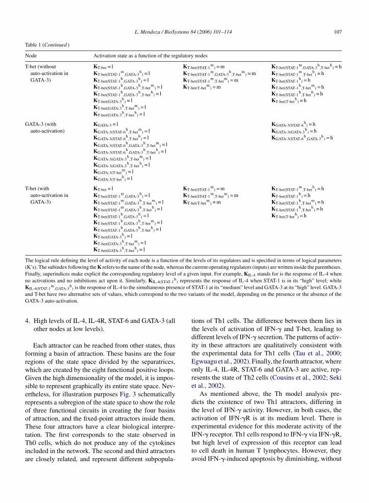
Table 1Dynamical rules for the Th model
Node Activation state as a function of the regulatory nodes
IFN-� KIFN-� = l KIFN-�(STAT-4h
) = m KIFN-�(STAT-4h
,IRAKh
) = hKIFN-�(IRAK
h) = l KIFN-�(STAT-4
h,T-bet
m) = m KIFN-�(STAT-4
h,IRAK
h,T-bet
m) = h
KIFN-�(IRAKh
,T-betm
) = m KIFN-�(STAT-4h
,IRAKh
,T-beth
) = hKIFN-�(T-bet
m) = m KIFN-�(STAT-4
h,T-bet
h) = h
KIFN-�(IRAKh
,T-beth
) = hKIFN-�(T-bet
h) = h
IL-4 KIL-4 = l KIL-4(GATA-3h
) = hKIL-4(STAT-1
m) = l
KIL-4(STAT-1m
,GATA-3h
) = lKIL-4(STAT-1
h) = l
KIL-4(STAT-1h
,GATA-3h
) = l
IL-12 KIL-12 = lIL-18 KIL-18 = lIFN-� KIFN-� = l
IFN-�R KIFN-�R = l KIFN-�R(IFN-�m
) = m KIFN-�R(IFN-�h
) = hKIFN-�R(SOCS-1
h) = l KIFN-�R(IFN-�
m,SOCS-1
h) = m
KIFN-�R(IFN-�h
,SOCS-1h
) = m
IL-4R KIL-4R = l KIL-4R(IL-4h
) = hKIL-4R(IL-4
h,SOCS-1
h) = l
KIL-4R(SOCS-1h
) = l
IL-12R KIL-12R = l KIL-12R(IL-12h
) = hKIL-12R(IL-12
h,STAT-6
h) = l
KIL-12R(STAT-6h
) = l
IL-18R KIL-18R = l KIL-18R(IL-18h
) = hKIL-18R(IL-18
h,STAT-6
h) = l
KIL-18R(STAT-6h
) = l
IFN-�R KIFN-�R = l KIFN-�R(IFN-�h
) = h
STAT-1 KSTAT-1 = l KSTAT-1(IFN-�Rh
) = m KSTAT-1(IFN-�Rh
) = hKSTAT-1(IFN-�R
m) = m KSTAT-1(IFN-�R
h,IFN-�R
h) = h
KSTAT-1(IFN-�Rm
,IFN-�Rh
) = m
STAT-6 KSTAT-6 = l KSTAT-6(IL-4Rh
) = h
STAT-4 KSTAT-4 = l KSTAT-4(IL-12Rh
) = hKSTAT-4(IL-12R
h,GATA-3
h) = l
KSTAT-4(GATA-3h
) = l
IRAK KIRAK = l KIRAK(IL-18Rh
) = hSOCS-1 KSOCS-1 = l KSOCS-1(STAT-1
m) = h
KSOCS-1(STAT-1m
,T-betm
) = hKSOCS-1(STAT-1
m,T-bet
h) = h
KSOCS-1(STAT-1h
) = hKSOCS-1(STAT-1
h,T-bet
m) = h
KSOCS-1(STAT-1h
,T-beth
) = hKSOCS-1(T-bet
m) = h
KSOCS-1(T-beth
) = h
GATA-3 (withoutauto-activation)
KGATA-3 = l KGATA-3(STAT-6h
) = hKGATA-3(STAT-6
h,T-bet
m) = l
KGATA-3(STAT-6h
,T-beth
) = lKGATA-3(T-bet
m) = l
KGATA-3(T-beth
) = l
L. Mendoza / BioSystems 84 (2006) 101–114 107
Table 1 (Continued )
Node Activation state as a function of the regulatory nodes
T-bet (withoutauto-activation inGATA-3)
KT-bet = l KT-bet(STAT-1m
) = m KT-bet(STAT-1m
,GATA-3h
,T-beth
) = hKT-bet(STAT-1
m,GATA-3
h) = l KT-bet(STAT-1
m,GATA-3
h,T-bet
m) = m KT-bet(STAT-1
m,T-bet
h) = h
KT-bet(STAT-1h
,GATA-3h
) = l KT-bet(STAT-1m
,T-betm
) = m KT-bet(STAT-1h
) = hKT-bet(STAT-1
h,GATA-3
h,T-bet
m) = l KT-bet(T-bet
m) = m KT-bet(STAT-1
h,T-bet
m) = h
KT-bet(STAT-1h
,GATA-3h
,T-beth
) = l KT-bet(STAT-1h
,T-beth
) = hKT-bet(GATA-3
h) = l KT-bet(T-bet
h) = h
KT-bet(GATA-3h
,T-betm
) = lKT-bet(GATA-3
h,T-bet
h) = l
GATA-3 (withauto-activation)
KGATA-3 = l KGATA-3(STAT-6h
) = hKGATA-3(STAT-6
h,T-bet
m) = l KGATA-3(GATA-3
h) = h
KGATA-3(STAT-6h
,T-beth
) = l KGATA-3(STAT-6h
,GATA-3h
) = hKGATA-3(STAT-6
h,GATA-3
h,T-bet
m) = l
KGATA-3(STAT-6h
,GATA-3h
,T-beth
) = lKGATA-3(GATA-3
h,T-bet
m) = l
KGATA-3(GATA-3h
,T-beth
) = lKGATA-3(T-bet
m) = l
KGATA-3(T-beth
) = l
T-bet (withauto-activation inGATA-3)
KT-bet = l KT-bet(STAT-1m
) = m KT-bet(STAT-1m
,T-beth
) = hKT-bet(STAT-1
m,GATA-3
h) = l KT-bet(STAT-1
m,T-bet
m) = m KT-bet(STAT-1
h) = h
KT-bet(STAT-1m
,GATA-3h
,T-betm
) = l KT-bet(T-betm
) = m KT-bet(STAT-1h
,T-betm
) = hKT-bet(STAT-1
m,GATA-3
h,T-bet
h) = l KT-bet(STAT-1
h,T-bet
h) = h
KT-bet(STAT-1h
,GATA-3h
) = l KT-bet(T-beth
) = hKT-bet(STAT-1
h,GATA-3
h,T-bet
m) = l
KT-bet(STAT-1h
,GATA-3h
,T-beth
) = lKT-bet(GATA-3
h) = l
KT-bet(GATA-3h
,T-betm
) = lKT-bet(GATA-3
h,T-bet
h) = l
The logical rule defining the level of activity of each node is a function of the levels of its regulators and is specified in terms of logical parameters(K’s). The subindex following the K refers to the name of the node, whereas the current operating regulators (inputs) are written inside the parentheses.Finally, superindices make explicit the corresponding regulatory level of a given input. For example, KIL-4 stands for is the response of IL-4 whenno activations and no inhibitions act upon it. Similarly, KIL-4(STAT-1
h) represents the response of IL-4 when STAT-1 is in its “high” level; while
KIL-4(STAT-1m
,GATA-3h
) is the response of IL-4 to the simultaneous presence of STAT-1 at its “medium” level and GATA-3 at its “high” level. GATA-3and T-bet have two alternative sets of values, which correspond to the two variants of the model, depending on the presence or the absence of theGATA-3 auto-activation.
4. High levels of IL-4, IL-4R, STAT-6 and GATA-3 (allother nodes at low levels).
Each attractor can be reached from other states, thusforming a basin of attraction. These basins are the fourregions of the state space divided by the separatrices,which are created by the eight functional positive loops.Given the high dimensionality of the model, it is impos-sible to represent graphically its entire state space. Nev-ertheless, for illustration purposes Fig. 3 schematicallyrepresents a subregion of the state space to show the roleof three functional circuits in creating the four basinsof attraction, and the fixed-point attractors inside them.These four attractors have a clear biological interpre-tation. The first corresponds to the state observed inTh0 cells, which do not produce any of the cytokinesincluded in the network. The second and third attractorsare closely related, and represent different subpopula-
tions of Th1 cells. The difference between them lies inthe levels of activation of IFN-� and T-bet, leading todifferent levels of IFN-� secretion. The patterns of activ-ity in these attractors are qualitatively consistent withthe experimental data for Th1 cells (Tau et al., 2000;Egwuagu et al., 2002). Finally, the fourth attractor, whereonly IL-4, IL-4R, STAT-6 and GATA-3 are active, rep-resents the state of Th2 cells (Cousins et al., 2002; Sekiet al., 2002).
As mentioned above, the Th model analysis pre-dicts the existence of two Th1 attractors, differing inthe level of IFN-� activity. However, in both cases, theactivation of IFN-�R is at its medium level. There isexperimental evidence for this moderate activity of theIFN-� receptor. Th1 cells respond to IFN-� via IFN-�R,but high level of expression of this receptor can leadto cell death in human T lymphocytes. However, theyavoid IFN-�-induced apoptosis by diminishing, without
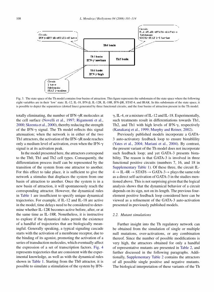
108 L. Mendoza / BioSystems 84 (2006) 101–114
Fig. 3. The state space of the Th model contains four basins of attraction. This figure represents the subdomain of the state space where the followingeight variables are in their ‘low’ state: IL-12, IL-18, IFN-�, IL-12R, IL-18R, IFN-�R, STAT-4, and IRAK. In this subdomain of the state space, itis possible to depict the separatrices (dotted lines) generated by three functional circuits, and the four basins of attraction present in the Th model.
totally eliminating, the number of IFN-�R molecules atthe cell surface (Novelli et al., 1997; Rigamonti et al.,2000; Skrenta et al., 2000), thereby reducing the strengthof the IFN-� signal. The Th model reflects this signalattenuation; when the network is in either of the twoTh1 attractors, the activation of the IFN-�R node reachesonly a medium level of activation, even when the IFN-�signal is at its activation peak.
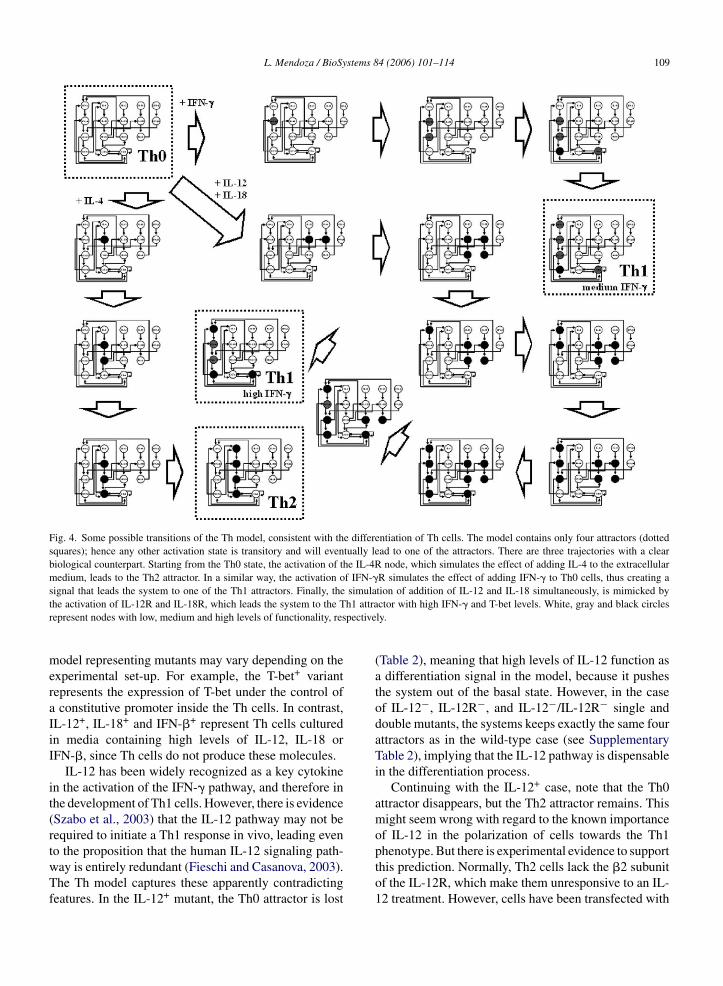
In the model presented here, the attractors correspondto the Th0, Th1 and Th2 cell types. Consequently, thedifferentiation process itself can be represented by thetransition of the system from one attractor to another.For this effect to take place, it is sufficient to give thenetwork a stimulus that displaces the system from onebasin of attraction to another. Once the system is in anew basin of attraction, it will spontaneously reach thecorresponding attractor. However, the dynamical rulesin Table 1 are insufficient to specify unique dynamicaltrajectories. For example, if IL-12 and IL-18 are activein the model, time delays need to be considered to deter-mine whether IL-12R becomes active before, after, or atthe same time as IL-18R. Nonetheless, it is instructiveto explore if the dynamical rules permit the existenceof a handful of trajectories that are biologically mean-ingful. Generally speaking, a typical signaling cascadestarts with the activation of a membrane receptor, due tothe binding of its agonist, promoting the activation of aseries of transduction molecules, which eventually affectthe expression of a set of transcription factors. Fig. 4represents trajectories that are consistent with the exper-
�, IL-4, or a mixture of IL-12 and IL-18. Experimentally,such treatments result in differentiations towards Th1,Th2, and Th1 with high levels of IFN-�, respectively(Kanakaraj et al., 1999; Murphy and Reiner, 2002).
Previously published models incorporate a GATA-3 auto-activatory feedback loop to ensure bistablility(Yates et al., 2004; Mariani et al., 2004). By contrast,the present variant of the Th model does not incorporatesuch feedback loop; and yet GATA-3 presents bista-bility. The reason is that GATA-3 is involved in threefunctional positive circuits (numbers 7, 16, and 18 inSupplementary Table 1). Of these three, the circuit IL-4 → IL-4R → STAT6 → GATA-3→ plays the same roleas a direct self-activation of GATA-3 in the studies men-tioned above. This is not surprising given that the logicalanalysis shows that the dynamical behavior of a circuitdepends on its sign, not on its length. The previous four-element positive feedback loop considered here can beviewed as a refinement of the GATA-3 auto-activationpresented in previously published models.
2.2. Mutant simulations
Further insight into the Th regulatory network canbe obtained from the simulation of single or multiplenull mutations, over-activations, or any combinationthereof. Since the number of possible modifications isvery high, the attractors obtained for only a handfulof representative mutants are presented in Table 2, andfurther discussed in the following paragraphs. Addi-
imental knowledge, as well as with the dynamical rulesshown in Table 1. Starting from the Th0 attractor, it ispossible to simulate a stimulation of the system by IFN-
tionally, Supplementary Table 2 contains the attractorsof all possible single positive and negative mutants.The biological interpretation of these variants of the Th
L. Mendoza / BioSystems 84 (2006) 101–114 109
Fig. 4. Some possible transitions of the Th model, consistent with the differentiation of Th cells. The model contains only four attractors (dottedsquares); hence any other activation state is transitory and will eventually lead to one of the attractors. There are three trajectories with a clearbiological counterpart. Starting from the Th0 state, the activation of the IL-4R node, which simulates the effect of adding IL-4 to the extracellularmedium, leads to the Th2 attractor. In a similar way, the activation of IFN-�R simulates the effect of adding IFN-� to Th0 cells, thus creating asignal that leads the system to one of the Th1 attractors. Finally, the simulation of addition of IL-12 and IL-18 simultaneously, is mimicked bythe activation of IL-12R and IL-18R, which leads the system to the Th1 attractor with high IFN-� and T-bet levels. White, gray and black circlesrepresent nodes with low, medium and high levels of functionality, respectively.
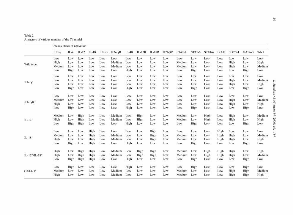
model representing mutants may vary depending on theexperimental set-up. For example, the T-bet+ variantrepresents the expression of T-bet under the control ofa constitutive promoter inside the Th cells. In contrast,IL-12+, IL-18+ and IFN-�+ represent Th cells culturedin media containing high levels of IL-12, IL-18 orIFN-�, since Th cells do not produce these molecules.
IL-12 has been widely recognized as a key cytokinein the activation of the IFN-� pathway, and therefore inthe development of Th1 cells. However, there is evidence(Szabo et al., 2003) that the IL-12 pathway may not berequired to initiate a Th1 response in vivo, leading evento the proposition that the human IL-12 signaling path-way is entirely redundant (Fieschi and Casanova, 2003).The Th model captures these apparently contradictingfeatures. In the IL-12+ mutant, the Th0 attractor is lost
(Table 2), meaning that high levels of IL-12 function asa differentiation signal in the model, because it pushesthe system out of the basal state. However, in the caseof IL-12−, IL-12R−, and IL-12−/IL-12R− single anddouble mutants, the systems keeps exactly the same fourattractors as in the wild-type case (see SupplementaryTable 2), implying that the IL-12 pathway is dispensablein the differentiation process.
Continuing with the IL-12+ case, note that the Th0attractor disappears, but the Th2 attractor remains. Thismight seem wrong with regard to the known importanceof IL-12 in the polarization of cells towards the Th1phenotype. But there is experimental evidence to supportthis prediction. Normally, Th2 cells lack the �2 subunitof the IL-12R, which make them unresponsive to an IL-12 treatment. However, cells have been transfected with
110L
.Mendoza
/BioSystem
s84
(2006)101–114
Table 2Attractors of various mutants of the Th model
Steady states of activation
IFN-� IL-4 IL-12 IL-18 IFN-� IFN-�R IL-4R IL-12R IL-18R IFN-�R STAT-1 STAT-6 STAT-4 IRAK SOCS-1 GATA-3 T-bet
Wild type
Low Low Low Low Low Low Low Low Low Low Low Low Low Low Low Low LowHigh Low Low Low Low Medium Low Low Low Low Medium Low Low Low High Low HighMedium Low Low Low Low Medium Low Low Low Low Medium Low Low Low High Low MediumLow High Low Low Low Low High Low Low Low Low High Low Low Low High Low
IFN-�-
Low Low Low Low Low Low Low Low Low Low Low Low Low Low Low Low LowLow Low Low Low Low Low Low Low Low Low Low Low Low Low High Low MediumLow Low Low Low Low Low Low Low Low Low Low Low Low Low High Low HighLow High Low Low Low Low High Low Low Low Low High Low Low Low High Low
IFN-�R−Low Low Low Low Low Low Low Low Low Low Low Low Low Low Low Low LowMedium Low Low Low Low Low Low Low Low Low Low Low Low Low High Low MediumHigh Low Low Low Low Low Low Low Low Low Low Low Low Low High Low HighLow High Low Low Low Low High Low Low Low Low High Low Low Low High Low
IL-12+Medium Low High Low Low Medium Low High Low Low Medium Low High Low High Low MediumHigh Low High Low Low Medium Low High Low Low Medium Low High Low High Low HighLow High High Low Low Low High Low Low Low Low High Low Low Low High Low
IL-18+
Low Low Low High Low Low Low Low High Low Low Low Low High Low Low LowMedium Low Low High Low Medium Low Low High Low Medium Low Low High High Low MediumHigh Low Low High Low Medium Low Low High Low Medium Low Low High High Low HighLow High Low High Low Low High Low Low Low Low High Low Low Low High Low
IL-12+/IL-18+High Low High High Low Medium Low High High Low Medium Low High High High Low HighHigh Low High High Low Medium Low High High Low Medium Low High High High Low MediumLow High High High Low Low High Low Low Low Low High Low Low Low High Low
GATA-3+Low High Low Low Low Low High Low Low Low Low High Low Low Low High LowMedium Low Low Low Low Medium Low Low Low Low Medium Low Low Low High High MediumHigh Low Low Low Low Medium Low Low Low Low Medium Low Low Low High High High
L. Mendoza / BioSystems 84 (2006) 101–114 111
the missing molecule, and then incubated with IL-12(Heat et al., 2000), thus rendering IL-12R continuouslyactive. Interestingly, under such treatment, committedTh2 cells do not produce IFN-� or reduce the productionof IL-4, thus matching the Th2 attractor. Hence, once thesystem is in the Th2 basin of attraction, even high levelsof IL-12, IL-12R and STAT-4 are not sufficient to takethe system into another basin of attraction.
The Th model also brings some insight into the originof the unexpected phenotypes of IFN-�− and IFN-�R−loss-of-function mutants. IFN-� and IFN-�R belong tothe same positive feedback loop, so one might expectthat eliminating one or the other would result in thesame phenotype. But, as shown in Table 2, the attrac-tors of these mutants differ in the level of activation ofIFN-�. This difference has also been observed experi-mentally; IFN-�− mutants do not produce IFN-� (Tanget al., 1998) as expected. But IFN-�R− mutants do pro-duce IFN-� (Diehl et al., 2000); and at first sight it isnot obvious why. Just by looking at the topology of thenetwork, it is clear that the elimination of either IFN-�or IFN-�R breaks the IFN-� → IFN-�R → STAT1 → T-bet → circuit. However, as the T-bet→ circuit remainsfunctional (see Supplementary Table 1), any transientsignal capable of activating T-bet (via STAT-1, for exam-ple) will result in a self-sustained stable activation ofT-bet, which in turn will activate IFN-� if it is intact,even in the absence of IFN-�R.
Besides mutations, the inclusion of some nodes withmore than two states of activation allows to accountfipeiITtSKrpacsvossIaI
the Th model, the simultaneous action of IL-12 and IL-18forces IFN-� to reach its activation peak.
The previous paragraphs provide some examples forthe interpretation of the number and nature of the attrac-tors for different perturbations. The Th model can beused to simulate many other situations, by fixing thelevel of activation of one node, or of a combination ofnodes, at some specific level(s). Supplementary Table 2includes the attractors found for 35 variants of the Thmodel, namely: the wild-type case, 17 single-node inac-tivations, and 17 single-node constitutive activations. Intotal, all these variants encompass 110 attractors. Manyof these attractors represent predictions of the model,as the corresponding situations have not yet been thor-oughly characterized.
2.3. Special case: the mouse
As mentioned previously, this study is based on exper-imental data originating from both human and mouse.However, it is instructive to explicitly consider the situ-ation in the mouse, which presents two unique features:first, the possible existence of a GATA-3 auto-activatorycircuit (Zhou and Ouyang, 2003); and second, the inabil-ity of GATA-3 to sustain its expression when T-bet ispresent (Smits et al., 2001). These two characteristicscan be included in the Th model by using an alterna-tive set of logical rules for GATA-3 and T-bet (last tworows of Table 1). Specifically, KGATA-3(GATA-3
h) = h indi-
cates that GATA-3 is able to activate itself, implying the
or the phenomenon of potentiation. Specifically, IL-18s a very poor inductor of IFN-� on its own, but is aotent inductor in combination with IL-12 (Kanakarajt al., 1999). In Table 1, KIFN-�(IRAKh) = l means that
f IRAK is at its high level (due to the presence ofL-18), the IFN-� node will remain in its low level.hen, KIFN-�(STAT-4
h) = m indicates that IFN-� will turn
o its medium level in response to an activation fromTAT-4, which is part of the IL-12 pathway. Finally,IFN-�(IRAK
h,STAT-4
h) = h means that the IFN-� node
eaches its high level when both STAT-4 and IRAK areresent. These logical rules result in the generation oflternative attractors for the different mutants. In thease of IL-18+, the system can reach four attractors,imilar to those of the wild-type, but with the extra acti-ation of IL-18, IL-18R, and IRAK (Table 2). Thus, thever-expression of the IL-18 pathway does not force theystem to behave differently from the wild-type. Thisituation contrasts with the loss of the Th0 attractor inL-12+ cells. Finally, a combined IL-12+/IL-18+ over-ctivation also leads to the loss the Th0 attractor, butFN-� is then highly expressed. Therefore, according to
existence of a feedback loop. And since STAT-6 actspositively on GATA-3, then a combination of GATA-3and STAT-6 should also activate GATA-3; represented byKGATA-3(STAT-6
h,GATA-3
h) = h. For its part, T-bet exerts a
strong inhibition GATA-3 thus making all other parame-ters of KGATA-3 equal to ‘low’. Finally, since GATA-3 isnot strong enough to inhibit T-bet, any KT-bet parametercontaining GATA-3 is set to ‘low’.
The resulting attractors are identical to those obtainedin the human case; that is, four attractors representing theTh0, Th2 and two Th1 patterns of activation. The dif-ferences just appear in the domains of functionality forsome of the circuits in the model. Specifically, the alter-native rules generate one new functional circuit (number23 in Supplementary Table 3), cause the loss of func-tionality of one circuit (number 7), render four circuitsfunctional (numbers 8, 12, 15, and 17), and affect thefunctionality domains of four other circuits (numbers 9,11, 18, and 19). Despite these changes, it is important tostress that, as in the case of the human variant, the logicalanalysis reveals that none of the negative feedback loopsare functional. Hence, the asymptotic dynamical behav-
112 L. Mendoza / BioSystems 84 (2006) 101–114
ior of the Th model is not significantly modified by thealternative rules used to best represent the informationobserved in mouse cells.
3. Conclusions
The differentiation process of T lymphocytes offersmultiple advantages from the modeling point of view:it is a system that has been amply studied experimen-tally, many of the key molecular players are known, andtheir responses to many signals have been evaluated. Asa result, models for different aspects of the physiology ofT lymphocytes have been published, which address pro-cesses involved in the activation (Kaufman et al., 1999;Sarkar and Franza, 2004), or the determination of cellu-lar fate (Bergmann and van Hemmen, 2001; Bergmannet al., 2002). Such studies, however, do not incorporatethe intracellular molecular network enabling the cellsto acquire different physiological states or fates, exceptfor the inclusion of a small number of key transcriptionfactors. Despite the lack of comprehensive moleculardata, previous models provide information on the kindof minimal model that might still capture the essenceof Th cell differentiation. Specifically, a model with twomutually inhibitory nodes, each with a positive feedbackloop, suffices to create a system with three steady stablestates representing the main differentiated T helper celltypes (Kaufman et al., 1985; Yates et al., 2004; Marianiet al., 2004). Since T-bet and GATA-3 inhibit each otherand are involved in direct and indirect positive feedback
spond to the patterns of activation observed in wild-typeTh cells. Moreover, the model can be modified so asto describe the asymptotic patterns of expression of nullmutants, as well as constitutive-expression variants. Thiscapacity of the model to simulate mutants helps to inter-pret some apparently contradictory phenotypes. Specifi-cally, an explanation has been provided for the apparentredundancy of the IL-12 pathway, despite its recognizedimportance in the differentiation of Th0 into Th1 cells.It also helps in understanding why the phenotypes ofnull mutations in IFN-� and IFN-�R are different. Thiscapacity of describing mutants is an important feature ofthe model. Although many genetically modified Th cellshave been studied experimentally, the molecular charac-terization of these cells is usually restricted to only a fewmolecules of interest, typically IFN-�, IL-4, GATA-3 andT-bet. Now, the Th model makes it possible to explorethe effects of multiple null mutations, or combinationsof mutations and over-expressions.
The present version of the Th network contains 17nodes. Clearly, many more molecules are involved inthe differentiation of T helper cells. For example, the Tcell receptor (TCR) and IL-10 pathways are known tobias the system towards the Th1 or Th2 cellular fate.The incorporation of these molecules should result ina more accurate description of the differentiation pro-cess. Despite its limited number of nodes, the present Thmodel is able to reproduce the basic wild type cellularstates (Th0, Th1, and Th2), and a large number of mutantphenotypes.
loops, these two molecules constitute the central fea-ture of models of T helper differentiation. The presentstudy is aimed at complementing such minimal models,by providing more comprehensive data on the nature ofthe intracellular regulatory network of Th cells. In thisaspect, the present regulatory network is an extension,or a refinement of the minimal models, as explained forthe case of GATA-3 positive feedback. However, thereis valuable information that can be gained by increasingthe molecular detail incorporated to the network. Par-ticularly, a more biologically complete network permitsthe delineation of the effect of single and multiple null-and/or constitutive-expression mutants on the differen-tiation of Th cells, as shown in Supplementary Table 2.
This study introduced a network model for the controlof the differentiation process of T helper cells, whosetopology was deduced from published molecular data.The network was modeled as a discrete system, and itsasymptotic behavior was studied with the aid of the gen-eralized logical formalism. The analysis permitted theidentification of all the stable states of the system. TheTh model has only four attractors, which clearly corre-
The logical approach that was used for this studymade it possible to find all the attractors in the differentvariants of the Th model. However, to represent the selec-tion a specific trajectory, it is necessary to incorporatetime delays into the model, so as to determine the preciseorder of response for each node in the network. As infor-mation about such delays is not yet available, they wereomitted so as to keep the number of suppositions to a min-imum. In any case, the resulting focus on stable states hasthe advantage to provide an easy way for experimentalverification. Indeed, experimental studies rely heavily oncell cultures and measurements of secreted or internallyexpressed molecules after long periods of incubation.Such experimental conditions (see, for example, Fig. 7in Cousins et al., 2002) can be directly associated withthe stable patterns of expression as represented by anattractor.
The prospects for the growth of the Th model involveaddressing three main areas of simplification used for itselaboration. First, the model does not explicitly incorpo-rate molecular mechanisms. Cells carry out activationsand inhibitions in many different ways, e.g. through
L. Mendoza / BioSystems 84 (2006) 101–114 113
chromatin remodeling, protein degradation, phospho-rylation, transcriptional regulation, etc. The eventualinclusion of such mechanisms into the model may helpto understand the relative importance, temporality, andreversibility of the different regulatory interactions. Sec-ondly, the logical model presented here could be trans-posed into a continuous formalism (using ODEs) in orderto generate quantitative predictions, which could be usedmake a more accurate comparison with experimentaldata. Finally, the model does not deal with cell popula-tions. In vivo and in vitro experiments involve multiplecells and usually also multiple cell types, and cellularcommunication is definitely a key to maintain pools ofdifferentiated and undifferentiated cells, as well as mem-ory cells. Hence, it is clearly necessary to expand the Thmodel, or to integrate it with other models, so as to beable to describe cell populations to make a more realisticdescription of the differentiation of T lymphocytes.
Acknowledgements
The author would like to thank Ioannis Xenarios,Massimo de Francesco, Yolande Chvatchko, Anne Cor-baz, Ram Selvaraju, Georg Feger, Denis Thieffry, andtwo anonymous referees for their invaluable help andinsights.
Appendix A. Supplementary data
bb
R
A
A
A
B
B
B
Chang, J.T., Segal, B.M., Nakanishi, K., Okamura, H., Shevach, E.M.,2000. The costimulatory effect of IL-18 on the induction of antigen-specific IFN-gamma production by resting T cells is IL-12 depen-dent and is mediated by up-regulation of the IL-12 receptor beta2subunit. Eur. J. Immunol. 30, 1113–1119.
Chen, X.P., Losman, J.A., Rothman, P., 2000. SOCS proteins, regula-tors of intracellular signaling. Immunity 13, 287–290.
Cousins, D.J., Lee, T.H., Staynov, D.Z., 2002. Cytokine coexpres-sion during human Th1/Th2 cell differentiation: direct evidencefor coordinated expression of Th2 cytokines. J. Immunol. 169,2498–2506.
Diehl, S., Anguita, J., Hoffmeyer, A., Zapton, T., Ihle, J.N., Fikrig,E., Rincon, M., 2000. Inhibition of Th1 differentiation by IL-6 ismediated by SOCS1. Immunity 13, 805–815.
Egwuagu, C.E., Yu, C.R., Zhang, M., Mahdi, R.M., Kim, S.J., Gery, I.,2002. Suppressors of cytokine signaling proteins are differentiallyexpressed in Th1 and Th2 cells: implications for the Th cell lineagecommitment and maintenance. J. Immunol. 168, 3181–3187.
Elser, B., Lohoff, M., Kock, S., Giaisi, M., Kirchhoff, S., Krammer,P.H., Li-Weber, M., 2002. IFN-� represses IL-4 expression viaIRF-1 and IRF-2. Immunity 17, 703–712.
Fieschi, C., Casanova, J.L., 2003. The role of interleukin-12 in humaninfectious diseases: only a faint signature. Eur. J. Immunol. 33,1461–1464.
Glimcher, L.H., Murphy, K.M., 2000. Lineage commitment in theimmune system: the T helper lymphocyte grows up. Genes Dev.14, 1693–1711.
Goodbourn, S., Didcock, L., Randal, R.E., 2000. Interferons: cellsignalling, immune modulation, antiviral responses and virus coun-termeasures. J. Gen. Virol. 81, 2341–2364.
Hamalainen, H., Zhou, H., Chou, W., Hashizume, H., Heller, R.,Lahesmaa, R., 2001. Distinct gene expression profiles of humantype 1 and type 2 T helper cells. Genome Biol. 2, research0022.1–0022.11.
Heat, V.L., Showe, L., Crain, C., Barrat, F.J., Trinchieri, G., O’Garra,
Supplementary data associated with this article cane found, in the online version, at doi:10.1016/j.iosystems.2005.10.004.
eferences
fkarian, M., Sedy, J.R., Yang, J., Jacobson, N.G., Cereb, N., Yang,S.Y., Murphy, T.L., Murphy, K.M., 2002. T-bet is a STAT1-induced regulator of IL-12R expression in naive CD4+ T cells.Nat. Immunol. 3, 549–557.
gnello, D., Lankford, C.S.R., Bream, J., Morinobu, A., Gadina, M.,O’Shea, J., Frucht, D.M., 2003. Cytokines and transcription factorsthat regulate T helper cell differentiation: new players and newinsights. J. Clin. Immunol. 23, 147–161.
kira, S., 2000. The role of IL-18 in innate immunity. Curr. Opin.Immunol. 12, 59–63.
ergmann, C., van Hemmen, J.L., 2001. Th1 or Th1: how an appropri-ate T helper response can be made. Bull. Math. Biol. 63, 405–430.
ergmann, C., van Hemmen, J.L., Segel, L.A., 2002. How instructionand feedback can select the appropriate T helper response. Bull.Math. Biol. 64, 425–446.
ernabei, P., Coccia, E.M., Rigamonti, L., Bosticardo, M., Forni,G., Pestka, S., Krause, C.D., Battistini, A., Novelli, F., 2001.Interferon-� receptor 2 expression as the deciding factor in humanT, B, and myeloid cell proliferation or death. J. Leukoc. Biol. 70,950–960.
A.O., 2000. Cutting edge: ectopic expression of the IL-12 receptor-�2 in developing and committed Th2 cells does not affect theproduction of IL-4 or induce the production of IFN-�. J. Immunol.164, 2861–2865.
Kanakaraj, P., Ngo, K., Wu, Y., Angulo, A., Ghazal, P., Harris, C.A.,Siekierka, J.J., Peterson, P.A., Fung-Leung, W.P., 1999. Defectiveinterleukin (IL)-18-mediated natural killer and T helper cell type 1response in IL-1 receptor-associated kinase (IRAK)-deficient mice.J. Exp. Med. 189, 1129–1138.
Kaufman, M., Urbain, J., Thomas, R., 1985. Towards a logical analysisof the immune response. J. Theor. Biol. 114, 527–561.
Kaufman, M., Andris, F., Leo, O., 1999. A logical analysis of T cellactivation and anergy. Proc. Natl. Acad. Sci. U.S.A. 96, 3894–3899.
Kotenko, S.V., Pestka, S., 2000. Jak-Stat signal transduction pathwaythrough the eyes of cytokine class II receptor complexes. Oncogene19, 2557–2565.
Lighvani, A.A., Frucht, D.M., Jankovic, D., Yamane, H., Aliberti, J.,Hissong, B.D., Nguyen, B.V., Gadina, M., Sher, A., Paul, W.E.,O’Shea, J.J., 2001. T-bet is rapidly induced by interferon-� inlymphoid and myeloid cells. Proc. Natl. Acad. Sci. U.S.A. 98,15137–15142.
Losman, J.A., Chen, X.P., Hilton, D., Rothman, P., 1999. Cuttingedge: SOCS-1 is a potent inhibitor of IL-4 signal transduction.J. Immunol. 162, 3770–3774.
Mariani, L., Lohning, M., Radbruch, A., Hofer, T., 2004. Transcrip-tional control networks of cell differentiation: insights from helperT lymphocytes. Prog. Biophys. Mol. Biol. 86, 45–76.
114 L. Mendoza / BioSystems 84 (2006) 101–114
Mendoza, L., Thieffry, D., Alvarez-Buylla, E.R., 1999. Genetic con-trol of flower morphogenesis in Arabidopsis thaliana: a logicalanalysis. Bioinformatics 15, 593–606.
Moriggl, R., Kristofic, C., Kinzel, B., Volarevic, S., Groner, B.,Brinkmann, V., 1998. Activation of STAT proteins and cytokinegenes in human Th1 and Th2 cells generated in the absence ofIL-12 and IL-4. J. Immunol. 160, 3385–3392.
Mullen, A.C., High, F.A., Hutchins, A.S., Lee, H.W., Villarino, A.V.,Livingston, D.M., Kung, A.L., Cereb, N., Yao, T.P., Yang, S.Y.,Reiner, S.L., 2001. Role of T-bet in commitment of TH1 cellsbefore IL-12-dependent selection. Science 292, 1907–1910.
Murphy, K.M., Reiner, S.L., 2002. The lineage decisions on helper Tcells. Nat. Rev. Immunol. 2, 933–944.
Nelms, K., Keegan, A.D., Zamorano, J., Ryan, J.J., Paul, W.E., 1999.The IL-4 receptor: signaling mechanisms and biologic functions.Annu. Rev. Immunol. 17, 701–738.
Novelli, F., D’Elios, M.M., Bernabei, P., Ozmen, L., Rigamonti, L.,Almerigogna, F., Forni, G., Del Prete, G., 1997. Expression androle in apoptosis of the �- and �-chains of the IFN-� receptor inhuman Th1 and Th2 clones. J. Immunol. 159, 206–213.
Ohmori, Y., Hamilton, T.A., 1997. IL-4-induced STAT6 suppressesIFN-gamma-stimulated STAT1-dependent transcription in mousemacrophages. J. Immunol. 159, 5474–5482.
Ouyang, W., Lohning, M., Gao, Z., Assenmacher, M., Ranganath,S., Radbruch, A., Murphy, K.M., 2000. Stat6-independent GATA-3 autoactivation directs IL-4-independent Th2 development andcommitment. Immunity 12, 27–37.
Ranganath, S., Murphy, K.M., 2001. Structure and specificity of GATAproteins in Th2 development. Mol. Cell. Biol. 21, 2716–2725.
Remy, E., Ruet, P., Mendoza, L., Thieffry, D., Chaouiya, C., in press.From logical regulatory graphs to standard Petri nets: dynamicalroles and functionality of feedback circuits. Trans. Comput. Sys-tems Biol.
Rigamonti, L., Ariotti, S., Losana, G., Gradini, R., Russo, M.A.,Jouanguy, E., Casanova, J.L., Forni, G., Novelli, F., 2000. Sur-
tion of IL-12 responsiveness and loss of GATA-3 expression. Eur.J. Immunol. 31, 1055–1065.
Swain, S.L., 2001. Interleukin 18: tipping the balance towards a Thelper cell 1 response. J. Exp. Med. 194, F11–F14.
Szabo, S.J., Jacobson, N.G., Dighe, A.S., Gubler, U., Murphy, K.M.,1995. Developmental commitment to the Th2 lineage by extinctionof IL-12 signaling. Immunity 2, 665–675.
Szabo, S.J., Dighe, A.S., Gubler, U., Murphy, K.M., 1997. Regulationof the interleukin (IL)-12R �2 subunit expression in developing Thelper 1 (Th1) and Th2 cells. J. Exp. Med. 185, 817–824.
Szabo, S.J., Kim, S.T., Costa, G.L., Zhang, X., Fathman, C.G., Glim-cher, L.H., 2000. A novel transcription factor, T-bet, directs Th1lineage commitment. Cell 100, 655–669.
Szabo, S.J., Sullivan, B.M., Peng, S.L., Glimcher, L.H., 2003. Molec-ular mechanisms regulating Th1 immune responses. Annu. Rev.Immunol. 21, 713–758.
Tang, H., Sharp, G.C., Peterson, K.P., Braley-Mullen, H., 1998.IFN-�-deficient mice develop severe granulomatous experimen-tal autoimmune thyroiditis with eosinophil infiltration in thyroids.J. Immunol. 160, 5105–5112.
Tau, G.Z., von der Weid, T., Lu, B., Cowan, S., Kvatyuk, M., Pernis,A., Cattoretti, G., Braunstein, N.S., Coffman, R.L., Rothman, P.B.,2000. Interferon � signaling alters the function of T helper type 1cells. J. Exp. Med. 192, 977–986.
Thieffry, D., Sanchez, L., 2002. Alternative epigenetic states under-stood in terms of specific regulatory structures. Ann. N.Y. Acad.Sci. 981, 135–153.
Thierfelder, W.E., van Deursen, J.M., Yamamoto, K., Tripp, R.A.,Sarawar, S.R., Carson, R.T., Sangster, M.Y., Vignali, D.A.,Doherty, P.C., Grosveld, G.C., Ihle, J.N., 1996. Requirement forStat4 in interleukin-12-mediated responses of natural killer and Tcells. Nature 382, 171–174.
Thomas, R., 1991. Regulatory networks seen as asynchronousautomata: a logical description. J. Theor. Biol. 153, 1–23.
Thomas, R., Thieffry, D., Kaufman, M., 1995. Dynamical behaviour
face expression of the IFN-�R2 chain is regulated by intracellulartrafficking in human T lymphocytes. J. Immunol. 164, 201–207.Saito, H., Morita, Y., Fujimoto, M., Narazaki, M., Naka, T., Kishimoto,T., 2000. IFN regulatory factor-1-mediated transcriptional activa-tion of mouse STAT-induced STAT inhibitor-1 gene promoter byIFN-�. J. Immunol. 164, 5833–5843.
Sanchez, L., Thieffry, D., 2003. Segmenting the fly embryo: a logicalanalysis of the pair-rule cross-regulatory module. J. Theor. Biol.224, 517–537.
Sarkar, A., Franza, B.R., 2004. A logical analysis of the process of Tcell activation: different consequences depending on the state ofCD28 engagement. J. Theor. Biol. 226, 455–466.
Seki, Y., Hayashi, K., Matsumoto, A., Seki, N., Tsukada, J., Ransom, J.,Naka, T., Kishimoto, T., Yoshimura, A., Kubo, M., 2002. Expres-sion of the suppressor of cytokine signaling-5 (SOCS5) negativelyregulates IL-4-dependent STAT6 activation and Th2 differentia-tion. Proc. Natl. Acad. Sci. U.S.A. 99, 13003–13008.
Skrenta, H., Yang, Y., Pestka, S., Fathman, C.G., 2000. Ligand-independent down-regulation of IFN-� receptor 1 following TCRengagement. J. Immunol. 164, 3506–3511.
Smeltz, R.B., Chen, J., Hu-Li, J., Shevach, E.M., 2001. Regulation ofinterleukin (IL)-18 receptor � chain expression on CD4+ T cellsduring T helper (Th)1/Th2 differentiation: critical downregulatoryrole of IL-4. J. Exp. Med. 194, 143–153.
Smits, H.H., van Rietschoten, J.G., Hilkens, C.M., Sayilir, R.,Stiekema, F., Kapsenberg, M.L., Wierenga, E.A., 2001. IL-12-induced reversal of human Th2 cells is accompanied by full restora-
of biological regulatory networks–I. Biological role of feedbackloops and practical use of the concept of the loop-characteristicstate. Bull. Math. Biol. 57, 247–276.
Thomas, R., Kaufman, M., 2001. Multistationarity, the basis of celldifferentiation and memory. I. Structural conditions of multista-tionarity and other nontrivial behavior. Chaos 11, 170–179.
Trinchieri, G., 1995. Interleukin-12: a proinflammatory cytokine withimmunoregulatory functions that bridge innate resistance andantigen-specific adaptive immunity. Annu. Rev. Immunol. 13,251–276.
Usui, T., Nishikomori, R., Kitani, A., Strober, W., 2003. GATA-3 suppresses Th1 development by downregulation of Stat4 andnot through effects on IL-12R�2 chain or T-bet. Immunity 18,415–428.
Yang, J., Zhu, H., Murphy, T.L., Ouyang, W., Murphy, K.M., 2001.IL-18-stimulated GADD45� required in cytokine-induced, but notTCR-induced, IFN-� production. Nat. Immunol. 2, 157–164.
Yates, A., Callard, R., Stark, J., 2004. Combining cytokine signallingwith T-bet and GATA-3 regulation in Th1 and Th2 differentiation:a model for cellular decision-making. J. Theor. Biol. 231, 181–196.
Zhou, M., Ouyang, W., Gong, Q., Katz, S.G., White, J.M., Orkin,S.H., Murphy, K.M., 2001. Friend of GATA-1 represses GATA-3-dependent activity in CD4+ cells. J. Exp. Med. 194, 1461–1471.
Zhou, M., Ouyang, W., 2003. The function role of GATA-3 in Th1 andTh2 differentiation. Immunol. Res. 28, 25–37.