A multivalent combination of experimental antituberculosis DNA vaccines based on Ag85B and regions...
10
Original article A multivalent combination of experimental antituberculosis DNA vaccines based on Ag85B and regions of difference antigens Ajay Grover a,1 , Mir Fayaz Ahmed b , Balwan Singh b , Indu Verma a , Pawan Sharma b , G.K. Khuller a, * a Department of Biochemistry, Postgraduate Institute of Medical Education and Research, Chandigarh 160 012, India b Immunology Group, International Centre for Genetic Engineering and Biotechnology, New Delhi 110 064, India Received 13 October 2005; accepted 17 April 2006 Available online 18 July 2006 Abstract Two candidate DNA vaccines based on the proteins CFP10 and CFP21 encoded by regions of difference (RDs) of Mycobacterium tubercu- losis were evaluated individually and in multivalent combination with the immunodominant protein Ag85B for induction of protective immune responses against experimental tuberculosis. Experimental DNA vaccines induced substantial levels of cell-mediated immune responses as indicated by marked lymphocyte proliferation, significant release of the Th1 cytokines IFN-g and IL-12 (p40), and predominant cytotoxic T cell activity. High levels of antigen-specific IgG1 and IgG2a antibodies observed in the sera of immunized mice depicted strong humoral responses generated by DNA vaccine constructs. The multivalent combination of three DNA vaccine constructs induced maximal T cell and humoral immune responses. All the experimental vaccines imparted significant protection against challenge with M. tuberculosis H 37 Rv (in terms of colony-forming unit reduction in lungs and spleen) as compared to vector controls. The level of protection exhibited by multivalent DNA vaccine formulation was found to be equivalent to that of Mycobacterium bovis BCG observed both at 4 and 8 weeks post-challenge. These results show the protective potential of the multivalent DNA vaccine formulation used in this study. Ó 2006 Elsevier SAS. All rights reserved. Keywords: DNA vaccines; Regions of difference; Tuberculosis 1. Introduction Bacillus Calmette-Guerin (BCG), the only tuberculosis (TB) vaccine available at present, is unable to protect against pulmonary TB, and its efficacy varies tremendously in differ- ent human populations. As a result there is now a concerted effort to use other promising vaccine candidates in more effective strategies to combat TB. Currently, DNA vaccines are under intensive investigation, because both long-lived humoral and cellular immune responses can be engendered by a DNA vaccine [1]. Among secretory proteins of Mycobacterium tuberculosis, members of the Ag85 complex (Ag85A, B, and C) have been evaluated most extensively as vaccine candidates. A level of protection superior to that given by BCG has been demon- strated to be induced by vaccination with recombinant BCG overexpressing Ag85B [2]. These findings argue strongly in favor of Ag85B as an important candidate for a tuberculosis subunit vaccine. BCG vaccination of humans, however, in- duces T cell responses to the Ag85 complex, but protection re- mains incomplete. It is therefore envisaged that in order to Abbreviations: RD, regions of difference; Ag85B, antigen 85B; CTL, cytotoxic T lymphocyte. * Corresponding author. Tel.: þ91 1722747585-92x282; fax: þ91 172 2744401. E-mail address: [email protected] (G.K. Khuller). 1 Present address: Mycobacterial Research Laboratories, Department of Microbiology, Immunology & Pathology, Colorado State University, Fort Collins, CO 80523, USA. 1286-4579/$ - see front matter Ó 2006 Elsevier SAS. All rights reserved. doi:10.1016/j.micinf.2006.04.025 Microbes and Infection 8 (2006) 2390e2399 www.elsevier.com/locate/micinf
-
Upload
independent -
Category
Documents
-
view
0 -
download
0
Transcript of A multivalent combination of experimental antituberculosis DNA vaccines based on Ag85B and regions...

Original article
Microbes and Infection 8 (2006) 2390e2399www.elsevier.com/locate/micinf
A multivalent combination of experimental antituberculosis DNAvaccines based on Ag85B and regions of difference antigens
Ajay Grover a,1, Mir Fayaz Ahmed b, Balwan Singh b, Indu Verma a,Pawan Sharma b, G.K. Khuller a,*
a Department of Biochemistry, Postgraduate Institute of Medical Education and Research, Chandigarh 160 012, Indiab Immunology Group, International Centre for Genetic Engineering and Biotechnology, New Delhi 110 064, India
Received 13 October 2005; accepted 17 April 2006
Available online 18 July 2006
Abstract
Two candidate DNA vaccines based on the proteins CFP10 and CFP21 encoded by regions of difference (RDs) of Mycobacterium tubercu-losis were evaluated individually and in multivalent combination with the immunodominant protein Ag85B for induction of protective immuneresponses against experimental tuberculosis. Experimental DNA vaccines induced substantial levels of cell-mediated immune responses asindicated by marked lymphocyte proliferation, significant release of the Th1 cytokines IFN-g and IL-12 (p40), and predominant cytotoxic Tcell activity. High levels of antigen-specific IgG1 and IgG2a antibodies observed in the sera of immunized mice depicted strong humoralresponses generated by DNA vaccine constructs. The multivalent combination of three DNA vaccine constructs induced maximal T cell andhumoral immune responses. All the experimental vaccines imparted significant protection against challenge with M. tuberculosis H37Rv (interms of colony-forming unit reduction in lungs and spleen) as compared to vector controls. The level of protection exhibited by multivalentDNA vaccine formulation was found to be equivalent to that of Mycobacterium bovis BCG observed both at 4 and 8 weeks post-challenge. Theseresults show the protective potential of the multivalent DNA vaccine formulation used in this study.� 2006 Elsevier SAS. All rights reserved.
Keywords: DNA vaccines; Regions of difference; Tuberculosis
1. Introduction
Bacillus Calmette-Guerin (BCG), the only tuberculosis(TB) vaccine available at present, is unable to protect againstpulmonary TB, and its efficacy varies tremendously in differ-ent human populations. As a result there is now a concerted
Abbreviations: RD, regions of difference; Ag85B, antigen 85B; CTL,
cytotoxic T lymphocyte.
* Corresponding author. Tel.: þ91 1722747585-92x282; fax: þ91 172
2744401.
E-mail address: [email protected] (G.K. Khuller).1 Present address: Mycobacterial Research Laboratories, Department
of Microbiology, Immunology & Pathology, Colorado State University,
Fort Collins, CO 80523, USA.
1286-4579/$ - see front matter � 2006 Elsevier SAS. All rights reserved.
doi:10.1016/j.micinf.2006.04.025
effort to use other promising vaccine candidates in moreeffective strategies to combat TB. Currently, DNA vaccinesare under intensive investigation, because both long-livedhumoral and cellular immune responses can be engenderedby a DNA vaccine [1].
Among secretory proteins of Mycobacterium tuberculosis,members of the Ag85 complex (Ag85A, B, and C) havebeen evaluated most extensively as vaccine candidates. A levelof protection superior to that given by BCG has been demon-strated to be induced by vaccination with recombinant BCGoverexpressing Ag85B [2]. These findings argue strongly infavor of Ag85B as an important candidate for a tuberculosissubunit vaccine. BCG vaccination of humans, however, in-duces T cell responses to the Ag85 complex, but protection re-mains incomplete. It is therefore envisaged that in order to

2391A. Grover et al. / Microbes and Infection 8 (2006) 2390e2399
achieve more effective induction of cell-mediated immunity,including memory immunity, additional antigens may be re-quired. Some of these important T cell antigens could be lo-cated within the missing genomic portions of BCG [3,4].Numerous studies have demonstrated the antituberculosis vac-cine potential of ESAT-6 and MPT-64, two proteins absent inBCG [5,6]. Besides ESAT-6 and MPT-64, other proteins of RDregions, such as CFP21 and CFP10, are known to inducestrong IFN-g production, proliferation of T cells and moderatecytotoxic T cell activity in M. tuberculosis infected mice [7].These results indicate that it is worthwhile to evaluate the im-munoprophylactic potential of these RD encoded proteins asDNA vaccines against experimental tuberculosis.
According to available information, co-immunization usingthe plasmids encoding protective antigens induces a greaterdegree of protection over a vaccine consisting of a single im-munodominant antigen [1]. Careful selection of antigens usedto construct a multivalent combination ensures that heteroge-neous populations representing a broad spectrum of MHCmolecules respond to vaccine [5]. In this regard, a combinationof Ag85 protein and immunodominant proteins encoded byRD antigens can represent a broad epitopic repertoire thatleads to activation of helper T cell and cytotoxic T cellresponses. To date, various multivalent combinations ofDNA vaccines encoding Ag85 complex proteins along withseveral immunodominant secretory proteins have been evalu-ated [8e10]. However, no reports are available on the use ofa multivalent combination of DNA vaccines based on RD-encoded CFP10 and CFP21 along with the Ag85 complexproteins against tuberculosis. Thus in this study, we have eval-uated candidate DNA vaccines based on Ag85B, CFP10 andCFP21 for their immunoprophylactic potential against experi-mental tuberculosis.
2. Materials and methods
2.1. Materials
All chemicals were purchased from Sigma (Sigma-Aldrich)except when otherwise noted. M. tuberculosis H37Rv origi-nally obtained from the National Collection of Type Culture(NCTC), London and maintained on Lowenstein Jensen’s(LJ) medium in the laboratory was used in the study.
2.2. Cloning of genes in DNA vaccine vectors
The open reading frames (ORFs) of genes Rv3874(CFP10), Rv1984c (CFP21) and Rv1886c (Ag85B) of M.tuberculosis H37Rv were amplified by PCR. The PCR productswere ligated to pGEMT-Easy vector (Promega) and thensequenced. The CFP10 gene was subcloned in VR1020 vector(Vical Inc.), whereas the Ag85B and CFP21 genes were sub-cloned in pVAX1 vector (Invitrogen). All three genes men-tioned above were also cloned in Escherichia coli expressionvectors, and recombinant proteins from E. coli host strainswere purified using Ni-NTA chromatography. The concentra-tion of endotoxin in protein preparations was determined
using E-toxate kit (Sigma) according to the manufacturer’sinstructions.
2.3. In vitro expression of candidate DNA vaccines
The ability of the recombinant VR1020 to express CFP10antigen was studied in vitro in a mammalian cell culturesystem. 3T3 murine fibroblast cells (2 � 105 cells/ml) weretransfected with 10 mg DNA complexed with 10 mg of Lipo-fectin. Transfected 3T3 cells were then washed in PBScontaining 0.5% BSA and cells were incubated in a 1:100dilution of anti-CFP10 rabbit polyclonal sera for 40 min onice. This was followed by washing and incubation withFITC-labeled anti-rabbit IgG (1:2000 dilution). Finally, aftera final washing, the pellets were suspended in 0.1% parafor-maldehyde before flow cytometric analysis. Expression ofantigens 85B and CFP21 by DNA vaccine constructs(pVAX85 and pVAX21) was studied using TNT� quick-cou-pled transcription/translation systems (Promega) according tothe manufacturer’s instructions.
2.4. Immunization of mice
Approximately 6-week-old female C57BL/6J mice wereused to verify the immunogenicity of the constructs. All theanimal experiments were carried out in accordance withanimal ethical regulations. Endotoxin-free plasmids were pre-pared using Endofree plasmid isolation giga kit (Qiagen);100 mg DNA/animal was given in the individual vaccinegroups, and 150 mg DNA/animal (50 mg of each vaccine con-struct) was given in the multivalent combination group, threetimes at three-week intervals. The DNA was administeredintramuscularly (i.m.) in the anterior tibialis muscle witha 30-G needle. The immune responses were studied at 4, 8and 16 weeks post-immunization (p.im.). For this purpose,spleens of mice from a single group were pooled, and the dif-ference between immune responses of different groups wasanalyzed as ‘‘fold-change’’.
2.5. Splenocyte proliferation assay
Spleen cells from immunized mice were suspended inRPMI-1640 medium supplemented with 10% heat inactivatedfetal bovine serum. The concentration of splenocytes wasadjusted to 2 � 105 cells/well in a 96-well culture plate. Puri-fied recombinant protein(s) was added to each well at a finalconcentration of 2 mg/ml. The plates were incubated for4 days at 37 �C in 5% CO2 followed by incubation with[3H]-thymidine (1 mCi/well) for 18 h. The cells were har-vested using a cell harvester, and the radioactivity was countedin a betaplate scintillation counter. Results were expressed as
Stimulation index � Counts per min in stimulated culture
Counts per min in unstimulated culture

2392 A. Grover et al. / Microbes and Infection 8 (2006) 2390e2399
2.6. IFN-g assay
Levels of IFN-g induced in culture supernatants of spleno-cytes in response to proteins were estimated after 72 h usingan anti-mouse IFN-g ELISA kit (R&D Systems) accordingto the manufacturer’s instructions.
2.7. IL-12 (p40) assay
Peritoneal exudate cells (PEC) of immunized mice were ob-tained by washing the peritoneal cavity, and 1 � 105 cells/100 mlwere cultured in RPMI-1640 medium in 96-well culture plates.Recombinant proteins were added at a concentration of 2 mg/ml.IL-12 (p40) levels were estimated in culture supernatants after24 h using an antimouse IL-12 (p40) ELISA kit (R&D Systems).
2.8. Cytotoxic T cell assay
The cytotoxic T lymphocyte (CTL) response was measuredby neutral red uptake assay [11]. Briefly, effector cells (stimu-lated splenocytes cultured in the presence of 15 mg/mlrecombinant proteins and 10 U/ml IL-2 for seven days) and tar-get cells (peritoneal exudate cells cultured with 5 mg/ml proteinfor 12 h) were incubated in appropriate ratio for 16 h at 37 �C.The microplate was gently washed with RPMI medium toremove effector cells, and 0.036% neutral red prepared inPBS was added. After 30 min of incubation, wells were washedthree times with PBS, and neutral red taken by intact targetcells was released by addition of 10 mM acetic acid andethanol. Absorbance was measured at 540 nm, and the resultswere expressed as percentage cytotoxicity at each effector/tar-get ratio calculated as [(CeB)�(EeB)/(CeB)] � 100:
WhereC ¼ mean absorbance of macrophages without effector cells.B ¼ mean absorbance of wells without cells.E ¼ mean absorbance of macrophages with effector cells.
2.9. IgG subtyping
Antigen-specific IgG1 and IgG2a subtype levels weredetermined by ELISA in the serum of individual animals(five/group). Recombinant protein (1 mg/well and 0.33 mgeach protein/well for combination) in 100 ml of 0.1 M sodiumcarbonate-bicarbonate buffer was coated for 2 h at 37 �C. Plateswere then blocked with 5% BSA in PBS for 2 h at 37 �Cfollowed by three washings with PBS-Tween 20 (0.05%).This was followed by incubation of serum samples (1:100 dilu-tion) and washing with PBS-Tween 20. Secondary antibodies(anti-mouse IgG1 and IgG2a conjugated with horseradish per-oxidase) at 1:1000 were incubated for 2 h at 37 �C. Color wasdeveloped by adding O-phenyldiamine and H2O2 in citratebuffer (pH 5.0), and the absorbance was measured at 490 nm.
2.10. Immunophenotyping of cell surface markers
Splenocytes (1 � 106/ml) of mice immunized with a combi-nation of DNA vaccine constructs were incubated with
recombinant antigens (5 mg/ml) in 24-well plates at 37 �Cfor 72 h. After incubation, cells were harvested, washed andstained with a 1:200 dilution of anti-mouse CD80-FITC/CD86-FITC/CD45RB-PE antibody for 40 min in the dark at4 �C. Finally, the cells were resuspended in 0.1% paraformal-dehyde after washing with PBS þ0.5% BSA, and flow cyto-metric analysis was done.
2.11. Protection studies
C57BL/6J mice received 100 mg of DNA intramuscularly(i.m.) in individual vaccine groups and 150 mg (50 mg ofeach vaccine) in multivalent combination vaccine groups 3times at 3-week intervals. BCG was inoculated subcutaneously(105 CFU/animal) at the time of the first dose of candidateDNA vaccine(s). Mice were challenged with 0.1 ml (105 -bacilli) suspension of M. tuberculosis H37Rv per animal intra-venously at 4 weeks p.im. Immunized/control animals weresacrificed on day 30 and day 60 post-challenge. Serialdilutions of homogenates of spleen and lungs of individualanimals were plated on Middlebrook 7H10 agar plates. Col-ony-forming units (CFUs) were counted after incubation at37 �C for 4e6 weeks. Differences between vaccinated andnon-vaccinated groups were evaluated by using an unpaired,two-tailed Student’s t-test.
3. Results
3.1. In vitro expression of candidate DNA vaccines
The surface of 3T3 cells transfected with VRCF10 showedan increase in fluorescence as compared to controls, indicatingthe presence of CFP10 on the surface of 3T3 cells (Fig. 1A).The presence of CFP10 in cell culture supernatant was alsoconfirmed by immunoblotting with anti-CFP10 antibody thatshowed the presence of a 10-kDa band (data not shown). Theexpression of in vitro translated Ag85B (30 kDa) and CFP21(21 kDa) protein was confirmed by autoradiography (Figs.1B,C). Some non-specific products that are usually present inreaction mixture of in vitro translation were also seen in theautoradiogram. All three proteins were also over-expressedin E. coli and purified using Ni-NTA chromatography. Themolecular weights of recombinant CFP10, CFP21 andAg85B protein were 10, 21 and 30 kDa, respectively asrevealed by SDS-PAGE and Western blotting with anti-Hismonoclonal antibody (data not shown). Recombinant proteinswere used for in vitro stimulation of cells in immunologicalassays. The concentration of lipopolysaccharide in proteinswas found to be less than 0.1 ng/ml, much below toxicity levelfor the cells cultured in vitro.
3.2. DNA vaccines induced splenocyte proliferation
All DNA vaccines were found to induce significant lympho-proliferative responses (Fig. 2), when taking a stimulation

2393A. Grover et al. / Microbes and Infection 8 (2006) 2390e2399
B C
A
No Vector
VR1020
VRCF10
30kDa
6kDa
21kDa
28kDa
35kDa
52kDa
92kDa
118kDa
321
50
40
30
20
10
0100 101
FL1-H
Cou
nts
102 103 104
321
15kDa
25kDa
35kDa
10kDa
30kDa
21kDa
4
Fig. 1. Expression of candidate DNA vaccines in vitro. (A) murine fibroblast cell line 3T3 was lipofected with VRCF10 and control vector VR1020 in two separate
sets along with one set of non-transfected cells. Cells were screened for expression of CFP10 on the surface of 3T3 cells by flow cytometry using anti-rabbit CFP10
antibody and anti-rabbit IgG-FITC as primary and secondary antibodies, respectively. Expression of Ag85B protein (B) and CFP21 protein (C) from candidate
DNA vaccine pVAX85 and pVAX21 was ascertained by an in vitro translation assay using a rabbit reticulocyte system (TNT� Expression kit, Promega). Trans-
lation of proteins was followed by incorporation of [S-35]-labeled methionine þ cysteine (‘‘Express’’, PE Life Technologies). Reaction products were run on 15%
SDS-PAGE and visualized by autoradiography. (B) Lanes 1e2, pVAX85 showing the expression of 30-kDa (Ag85B) protein. Lane 3, protein molecular weight
markers. (C) Lane 1, control pVAX1 plasmid; lanes 2e3, pVAX21 showing the expression of 21-kDa (CFP21) protein; lane 4, protein molecular weight markers.
index of 3.0 as a positive cutoff value. Among individualvaccines, the lymphoproliferative response of the pVAX85vaccine was two-fold higher than the other two DNA vaccines(VRCF10 and pVAX21), whereas the stimulation index ofmultivalent combination was 1.2-fold higher than that ofpVAX85. The value of stimulation indices observed at alltime points was in order of multivalent combination >pVAX85 > VRCF10 > pVAX21.
3.3. Measurement of cytokine levels
As observed in lymphocyte proliferation, IFN-g responseswere also found to be highest for the multivalent combinationof DNA vaccines (Fig. 3A). Splenocytes from pVAX85 immu-nized mice induced more than two-fold higher synthesis of
IFN-g in comparison to VRCF10 and pVAX21 immunizedmice at 4 and 8 weeks p.im. However, there was no differencebetween the levels of IFN-g obtained in mice immunized withmultivalent combination and pVAX85. IFN-g levels observedfor all DNA vaccines decreased at 16 weeks p.im., thoughthese were higher than those of control vectors. Fig. 3B showsIL-12 (p40) levels released in PEC culture supernatants of im-munized mice. The highest levels of IL-12 (p40) were ob-served for the multivalent combination of genes at all timepoints and were w1.7-fold higher than those of pVAX85 atboth 4 and 8 weeks p.im. IL-12 (p40) levels decreased sub-stantially at 16 weeks p.im; however, levels were higher thanthe values obtained in control vectors. Concentrations of IL-12 (p40) observed for DNA vaccines were also in order ofmultivalent combination > pVAX85 > VRCF10 > pVAX21.

2394 A. Grover et al. / Microbes and Infection 8 (2006) 2390e2399
3.4. Cytotoxic T cell response generatedby DNA vaccines
The mean cytotoxic T cell responses were found to behighest at a 10:1 effector: target cell ratio in all groups ofDNA vaccines (data not shown). The cytotoxic T cellresponses induced in response to in vitro stimulation withpurified recombinant mycobacterial proteins were very highcompared to those of no-antigen control, indicating mycobac-terial antigen-specific cytotoxic T-cell responses. The meancytotoxic T-cell responses (% cytotoxicity) observed at 10:1effector:target cell ratio of different vaccine groups are pre-sented in Fig. 4. The combination of 3 DNA constructswas observed to exhibit more than 90% cytoxicity, followedby pVAX85 (80e85%) at 4 and 8 weeks p.im. The cytotoxicT-cell response decreased at 16 weeks p.im., but these valueswere still higher than those of controls. The multivalent com-bination showed prominent cytotoxic T cell response even at16 weeks p.im., followed by pVAX21. However, percent cy-totoxicity of pVAX85 decreased substantially at 16 weeksp.im.
3.5. Humoral response induced by DNA vaccines
IgG1 antibody levels are presented in Fig. 5A. The multiva-lent combination induced the highest IgG1 antibody levels,followed by pVAX85, pVAX21 and VRCF10 at all timepoints. Compared to pVAX85 vaccine, the level of IgG1antibodies was w1.8-fold higher in multivalent vaccinatedanimals at both 4 and 8 weeks p.im. A gradual decrease inIgG1 levels was observed until 16 weeks p.im. Serum IgG2aantibody levels observed in different groups of DNAvaccine-immunized animals are presented in Fig. 5B. Themultivalent combination also induced the highest IgG2aantibody levels, followed by pVAX85, pVAX21 andVRCF10 at all time points.
Week 4 Week 8 Week 16
Stim
ulatio
n In
dex
0
5
10
15
20
25
30
35 VR1020pVAX1VR1020+pVAX1VRCF10pVAX21pVAX85Vaccines Combination
Fig. 2. Splenocyte proliferative responses of candidate DNA vaccines in immu-
nized mice at 4, 8 and 16 weeks p.im. Highest mean stimulation index of Con-
A was 75 � 8.3. The results are mean � SD of stimulation indices of triplicate
wells of pooled splenocytes from 5 animals.
3.6. Immunophenotyping studies of splenocytes
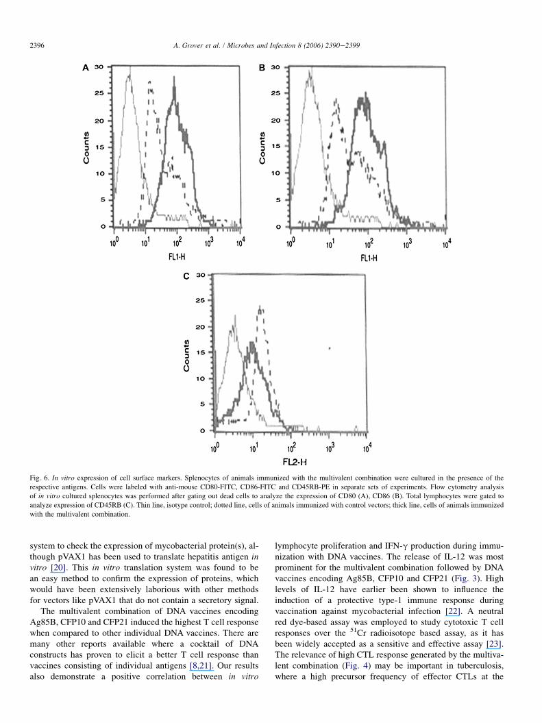
The in vitro stimulated splenocytes of multivalent vaccineimmunized animals were analyzed for the expression ofB7.1 (CD80), B7.2 (CD86) and CD45RB surface markers.An increase in the mean fluorescence intensity of the multiva-lent combination indicated an upregulation of the expressionof B7.1 and B7.2 surface markers (Figs. 6A,B). However, a de-crease in the mean fluorescence intensity indicated a downre-gulation in the expression of CD45RB molecules (Fig. 6C).
3.7. The multivalent combination inducedsignificant protection against TB
The log10 CFUs recovered from target organs (lungs andspleen) were significantly lower in all experimental groupscompared to unvaccinated controls at 4 and 8 weeks post-challenge (Tables 1 and 2). The log10 CFUs observed in thelungs of animals immunized with the multivalent combination(4.71 � 0.137) were comparable to those immunized withBCG (4.76 � 0.14). Similarly, log10 CFUs in the spleen ofanimals immunized with the multivalent combination
Week 4 Week 8 Week 16
Week 4 Week 8 Week 16
IF
N-g
am
ma C
on
c. (p
g/m
l)
0
200
400
600
800
1000
1200
1400
1600 VR1020pVAX1VR1020+pVAX1VRCF10pVAX21pVAX85Vaccines Combination
VR1020pVAX1VR1020+pVAX1VRCF10pVAX21pVAX85Vaccines Combination
B
IL
-12 (p
40) C
on
c. (p
g/m
l)
0
200
400
600
800
1000
1200
1400
1600
1800
A
Fig. 3. IFN-g (A) and IL-12 (p40) (B) responses of candidate DNA vaccines in
immunized mice at 4, 8 and 16 weeks p.im. The results are responses of pooled
culture supernatant from triplicate wells stimulated with respective antigens.

2395A. Grover et al. / Microbes and Infection 8 (2006) 2390e2399
(4.85 � 0.275) were comparable to those immunized withBCG (4.94 � 0.173). The multivalent combination impartedmaximum resistance in terms of reduction in CFUs amongall candidate vaccines. The log10 CFUs observed in the lungsof animals immunized with the multivalent combination were
Week 4 Week 8 Week 16
Percen
tag
e C
yto
to
xicity
0
20
40
60
80
100 VR1020pVAX1VR1020+pVAX1VRCF10pVAX21pVAX85Vaccines Combination
Fig. 4. Cytotoxic T cell responses of candidate DNA vaccines in immunized mice.
The results are expressed as percentage cytotoxicity of pooled cells from each group
at effector:target cell ratio of 10/1 standardized in ratio optimization studies. Non-
specific lysis calculated in the absence of antigen stimulus was below 10%.
A
Week 4 Week 8 Week 16
Week 4 Week 8 Week 16
(Ig
G1) A
bso
rb
an
ce at 490n
m
0.0
0.2
0.4
0.6
0.8
1.0
1.2
VR1020pVAX1VR1020+pVAX1VRCF10pVAX21pVAX85Vaccines Combination
VR1020pVAX1VR1020+pVAX1VRCF10pVAX21pVAX85Vaccines Combination
B
(Ig
G2a) A
bso
rb
an
ce at 490n
m
0.0
0.5
1.0
1.5
2.0
Fig. 5. Serum antibody (IgG1 and IgG2a) responses from sera of candidate
DNA vaccine immunized mice at 4, 8 and 16 weeks p.im. The results of
IgG1 (A) and IgG2a (B) are expressed as mean OD of antigen coated wells
at 490 nm.
significantly low compared to individual DNA constructs bothat 4 and 8 weeks post-challenge ( p < 0.05 for pVAX85,p < 0.05 for pVAX21 and p < 0.001 for VRCF10). Thelog10 CFUs observed in the spleen of animals immunizedwith the multivalent combination were also significantly lowerthan any individual DNA construct both at 4 and 8 weeks post-challenge ( p < 0.05 for pVAX85, p < 0.01 for pVAX21 andp < 0.001 for VRCF10). When compared with BCG immu-nized animals, the multivalent combination imparted protec-tion equivalent to BCG at 4 and 8 weeks post-challenge.These results demonstrated the utility of the combination ofDNA vaccines as a future multicomponent DNA vaccine,and further evaluation in a murine low-dose aerosol model isrequired.
4. Discussion
Studies based on human immune recognition have shownthat proteins encoded by RDs are recognized by healthytuberculosis contacts but not by patients in endemic areas oftuberculosis [12e14]. These findings stress the importanceof RD antigens for vaccine design in contrast to the existingparadigm of their use in immunodiagnosis. The hypothesisthat important T cell antigens are missing from BCG hasrecently been supported by numerous studies of vaccinationwith ESAT-6 and MPT-64, two RD antigens absent in BCG[5,15]. In addition to RD antigens, members of the Ag85 com-plex have been shown to be immunodominant antigens thatimpart high levels of protection against M. tuberculosisinfection [8,16,17]. Work done on TB DNA vaccines in exper-imental animals has clearly demonstrated that the Ag85complex and RD antigens within the mycobacterial genomeshould be evaluated in detail owing to their contributiontowards protective immunity against tuberculosis. Accordingto available information, co-immunization using the plasmidsencoding protective antigens induces a greater degree of pro-tection over vaccines consisting of a single immunodominantantigen [8,18]. Hence in the present study, candidate DNAvaccine constructs having genes for Ag85B, RD1-encodedCFP10 and RD2-encoded CFP21 were evaluated individuallyand in combination for their immunoprophylactic potentialagainst experimental tuberculosis.
In the present study, expression of CFP10 on the surface ofin vitro cultured cells was studied using flow cytometry(Fig. 1A). To date, no studies are available on the use offlow cytometry to confirm expression of mycobacterial pro-teins in transfected mammalian cells. Earlier, Delogu et al.[18] studied the expression of a DNA vaccine construct encod-ing MPT64 in lysates of rhabdomyosarcoma cells using West-ern blotting. The expression of plasmodium protein(s) byVR1020 in mammalian cells has been studied using immuno-fluorescence [19]. However, flow cytometric analysis appearsto be more appropriate to study the expression of proteins byhigh copy number plasmids such as VR1020. Expression ofAg85B and CFP21 by pVAX1 was studied using a rabbit retic-ulocyte-based translation system in vitro (Fig. 1B,C). There isno report available on the use of such an in vitro translation
2396 A. Grover et al. / Microbes and Infection 8 (2006) 2390e2399
Fig. 6. In vitro expression of cell surface markers. Splenocytes of animals immunized with the multivalent combination were cultured in the presence of the
respective antigens. Cells were labeled with anti-mouse CD80-FITC, CD86-FITC and CD45RB-PE in separate sets of experiments. Flow cytometry analysis
of in vitro cultured splenocytes was performed after gating out dead cells to analyze the expression of CD80 (A), CD86 (B). Total lymphocytes were gated to
analyze expression of CD45RB (C). Thin line, isotype control; dotted line, cells of animals immunized with control vectors; thick line, cells of animals immunized
with the multivalent combination.
system to check the expression of mycobacterial protein(s), al-though pVAX1 has been used to translate hepatitis antigen invitro [20]. This in vitro translation system was found to bean easy method to confirm the expression of proteins, whichwould have been extensively laborious with other methodsfor vectors like pVAX1 that do not contain a secretory signal.
The multivalent combination of DNA vaccines encodingAg85B, CFP10 and CFP21 induced the highest T cell responsewhen compared to other individual DNA vaccines. There aremany other reports available where a cocktail of DNAconstructs has proven to elicit a better T cell response thanvaccines consisting of individual antigens [8,21]. Our resultsalso demonstrate a positive correlation between in vitro
lymphocyte proliferation and IFN-g production during immu-nization with DNA vaccines. The release of IL-12 was mostprominent for the multivalent combination followed by DNAvaccines encoding Ag85B, CFP10 and CFP21 (Fig. 3). Highlevels of IL-12 have earlier been shown to influence theinduction of a protective type-1 immune response duringvaccination against mycobacterial infection [22]. A neutralred dye-based assay was employed to study cytotoxic T cellresponses over the 51Cr radioisotope based assay, as it hasbeen widely accepted as a sensitive and effective assay [23].The relevance of high CTL response generated by the multiva-lent combination (Fig. 4) may be important in tuberculosis,where a high precursor frequency of effector CTLs at the
2397A. Grover et al. / Microbes and Infection 8 (2006) 2390e2399
time of infection is required to limit dissemination of infec-tion. As IL-12 and IFN-g cytokines induced by candidateDNA vaccines in the present study can direct IgG2a produc-tion, it follows that the subclass of antibodies generated byplasmid DNA immunization is biased towards IgG2a (Fig. 5).
This work is probably the first report where an antitubercu-losis DNA vaccine-based immunophenotyping study involvingB7.1/B7.2 expression was performed (Fig. 6A, B). There areno reports available on the activation of co-stimulatory mole-cules by DNA vaccines during tuberculosis. The observationsof the present study suggest the potential of experimental com-binations of vaccines to activate antigen-presenting cells, lead-ing to higher B7.1/B7.2 expression, consequently aiding in theefficient generation of T cell (CD4 and CD8) responses. Inmice, the level of CD45RB expression is used to distinguishna€ıve and memory lymphocytes [24]. Flow cytometric analy-sis of lymphocytes from mice immunized with the multivalentcombination showed downregulation of surface expression ofCD45RB (Fig. 6C). This indicated the induction of a memory
Table 2
The log10 CFUs of M. tuberculosis at 4 and 8 weeks post-infection in spleens
of C57BL/6J mice vaccinated with different candidate DNA vaccines or BCG
Log10 CFUs in spleen
4 weeks 8 weeks
Control 5.74 � 0.119 5.76 � 0.122
pVAX1 5.74 � 0.155 5.64 � 0.148
VR1020 5.65 � 0.153 5.65 � 0.141
Vector-comb 5.65 � 0.138 5.62 � 0.127
pVAX85 5.35 � 0.135*** 5.21 � 0.123***
pVAX21 5.46 � 0.121* 5.32 � 0.204*
VRCF10 5.55 � 0.126* 5.45 � 0.141*
Vaccine-comb 5.07 � 0.16***
CCCþþ::4.85 � 0.275***
CCþþ:
BCG 4.86 � 0.135*** 4.94 � 0.173þ***
The results are mean � SD log10 CFUs obtained in the spleen of individual
animals (5e8 animals/group at each time point). ***p < 0.001, *p < 0.05
w.r.t. controls. þþp < 0.01, þp < 0.05 w.r.t. pVAX21. ::p < 0.01,
:p < 0.05 w.r.t. pVAX85. CCCp < 0.001, CCp < 0.01, w.r.t. VRCF10.
Table 1
The log10 CFUs of M. tuberculosis at 4 and 8 weeks post-infection in lungs of
C57BL/6 mice vaccinated with different candidate DNA vaccines or BCG
Log10 CFUs in lungs
Candidate DNA vaccine 4 weeks 8 weeks
Control 5.65 � 0.139 5.63 � 0.17
pVAX1 5.64 � 0.144 5.53 � 0.171
VR1020 5.55 � 0.12 5.58 � 0.13
Vector-comb 5.57 � 0.119 5.51 � 0.187
pVAX85 5.12 � 0.175*** 4.97 � 0.166***
pVAX21 5.33 � 0.116* 5.21 � 0.337*
VRCF10 5.42 � 0.174* 5.30 � 0.213*
Vaccine-comb 4.85 � 0.149***
þþ::CCC
4.71 � 0.137***
þ:CCC
BCG 4.74 � 0.124*** 4.76 � 0.14***
The results are mean � SD log10 CFUs obtained in the lungs of individual
animals (5e8 animals/group at each time point). ***p < 0.001, *p < 0.05
w.r.t. controls. þþp < 0.01, þp < 0.05 w.r.t. pVAX21. ::p < 0.01,
:p < 0.05 w.r.t. pVAX85. CCCp < 0.001 w.r.t. VRCF10.
response following immunization with the experimental com-bination vaccine.
When different DNA vaccine constructs were compared,promising protection data was obtained, as all DNA vaccine(s)imparted better protection than that of vector controls (Tables1 and 2). A significantly higher level of protection wasobtained by the multivalent combination than that of any indi-vidual DNA vaccine. Of special note, the protection obtainedby the multivalent DNA vaccine was equivalent to that ofBCG in the lungs and spleen at 4 and 8 weeks post-challenge.The high protective effect of the multivalent combination andother single DNA constructs can be associated with a systemicexpansion of antigen-specific IFN-g secreting T cells early inthe course of infection [8]. It has also been suggested that inorder to generate effective protection, stimulation of a precisecascade of multiple components of the immune response isnecessary [25]. A combination of antigens can activate rele-vant immune components such as cytokine production andanti-mycobacterial CTLs, in addition to evoking an IFN-gresponse and therefore prove more efficacious than singleantigen-based vaccines. In this regard, choosing appropriateantigens for the constitution of the next generation of multi-component antituberculous vaccine is an important task.
Ag85B-DNA induced the highest levels of protection, inagreement with other studies where high levels of protectionimparted by members of the Ag85 complex have been demon-strated [16]. These findings strengthen the basis of the presentstudy, indicating that at least one member of the Ag85 com-plex [8,17] should be included in future multicomponentDNA vaccines. Although, many reports are available onnucleic acid vaccination using members of the Ag85 complex,the levels of immune response induced by Ag85 proteins indifferent studies remains variable [17,26,27]. As neitherAg85A nor Ag85B contains H-2Kb-or H-2Db-restricted CD8T cell epitopes in their sequence [26], the totality of theimmune response induced by Ag85B DNA vaccines in H-2b
mice is assumed to be mediated by CD4 T cells. However,the high cytotoxic T cell responses observed with Ag85B-DNA could be attributed to high levels of IFN-g elicited bythe Ag85B protein during 16 h of incubation in neutral redassay, leading to activation of natural killer cells (Denis, per-sonal communication). Lozes et al. [26] have also observedan early cytotoxic T cell response in Ag85A-DNA immunizedH-2b mice in the neutral red assay. The findings regarding thecytotoxic T cell response can be correlated with the protectionimparted by Ag85B-DNA in mice, where a high reduction inlog10 CFUs was not obtained until 8 weeks post-challenge.The protective efficacy of Ag85B-DNA was not studiedbeyond 8 weeks post-challenge in the present work. However,Tanghe et al. [28] have shown that protection with Ag85-DNAgradually disappears at later time points. It has been suggestedthat a combination of Ag85-DNA vaccines that stimulatestrong CD4 T cell responses, with CTL epitopes such asa H-2Db-restricted epitope from any other immunodominantprotein, may help to overcome the problem [29]. The multiva-lent combination induced a high CTL response until 16 weeksp.im. in this study (Fig. 4). Of special note, bioinformatic

2398 A. Grover et al. / Microbes and Infection 8 (2006) 2390e2399
analysis using the Propred1 program [30] revealed the pres-ence of an MHC class I H-2Db-restricted epitope in the se-quence of the CFP21 protein. The presence of CFP21 alongwith Ag85B in a multivalent combination might have stimu-lated antigen-specific CD8 and CD4 T cells responsible forprotection equivalent to that of BCG observed in the presentstudy (Tables 1 and 2). These observations support the inclu-sion of both CD4 and CD8 T cell epitopes as an answer tothe question of how DNA vaccines can be tailored to generatethe desired immune response.
Thus, the results of the present study highlight the impor-tance of RD antigens as potential candidates for future tuber-culosis vaccines. Our observations indicate that a multivalentcombination consisting of CFP10, CFP21 and Ag85B DNAconstructs induced protective immunity equivalent to that ofBCG in a mouse model of pulmonary tuberculosis. It can bestated that further optimization of these DNA constructs maylead to nucleic acid preparations that are more potent thanthe current vaccine M. bovis BCG.
Acknowledgments
This work was supported by a grant to Prof. G.K. Khullerand Dr. Indu Verma from DST, Govt. of India, New Delhiand UNDP/World Bank/WHO grant # V25/181/203 to Dr.Pawan Sharma.
References
[1] K. Huygen, On the use of DNA vaccines for the prophylaxis of mycobac-
terial diseases, Infect. Immun. 71 (2003) 1613e1621.
[2] M.A. Horwitz, G. Harth, B.J. Dillon, S. Maslesa-Galic, Recombinant
bacillus calmette-guerin (BCG) vaccines expressing the Mycobacterium
tuberculosis 30-kDa major secretory protein induce greater protective
immunity against tuberculosis than conventional BCG vaccines in
a highly susceptible animal model, Proc. Natl. Acad. Sci. U.S.A. 97
(2000) 13853e13858.
[3] M.A. Behr, M.A. Wilson, W.P. Gill, H. Salamon, G.K. Schoolnik,
S. Rane, P.M. Small, Comparative genomics of BCG vaccines by
whole-genome DNA microarray, Science 284 (1999) 1520e1523.
[4] P. Andersen, TB vaccines: progress and problems, Trends Immunol. 22
(2001) 160e168.
[5] L. Brandt, M. Elhay, I. Rosenkrands, E.B. Lindblad, P. Andersen, ESAT-
6 subunit vaccination against Mycobacterium tuberculosis, Infect.
Immun. 68 (2000) 791e795.
[6] A. Weinrich Olsen, L.A. van Pinxteren, L. Meng Okkels, P. Birk Rasmussen,
P. Andersen, Protection of mice with a tuberculosis subunit vaccine based
on a fusion protein of antigen 85b and esat-6, Infect. Immun. 69 (2001)
2773e2778.
[7] K. Weldingh, I. Rosenkrands, S. Jacobsen, P.B. Rasmussen, M.J. Elhay,
P. Andersen, Two-dimensional electrophoresis for analysis of Mycobac-
terium tuberculosis culture filtrate and purification and characterization
of six novel Mycobacterium tuberculosis, Infect. Immun. 66 (1998)
3492e3500.
[8] A.T. Kamath, C.G. Feng, M. Macdonald, H. Briscoe, W.J. Britton,
Differential protective efficacy of DNA vaccines expressing secreted
proteins of combinations expressing either tissue plasminogen activator
signal sequence fusion proteins or ubiquitin-conjugated antigens induce
sustained protective immunity in a mouse model of pulmonary tubercu-
losis, Infect. Immun. 67 (1999) 1702e1707.
[9] G. Delogu, A. Li, C. Repique, F. Collins, S.L. Morris, DNA vaccine
combinations expressing either tissue plasminogen activator signal
sequence fusion proteins or ubiquitin-conjugated antigens induce sus-
tained protective immunity in a mouse model of pulmonary tuberculosis,
Infect. Immun. 70 (2002) 292e302.
[10] H. Cai, X. Tian, X.D. Hu, Y.H. Zhuang, Y.X. Zhu, Combined DNA
vaccines formulated in DDA enhance protective immunity against tuber-
culosis, DNA Cell Biol. 23 (2004) 450e456.
[11] O. Denis, E. Lozes, K. Huygen, Induction of cytotoxic T-cell responses
against culture filtrate antigens in Mycobacterium bovis bacillus Calmette-
Guerin-infected mice, Infect. Immun. 65 (1997) 676e684.
[12] A. Demissie, P. Ravn, J. Olobo, T.M. Doherty, T. Eguale, M. Geletu,
W. Hailu, P. Andersen, S. Britton, T-cell recognition of Mycobacterium
tuberculosis culture filtrate fractions in tuberculosis patients and their
household contacts, Infect. Immun. 67 (1999) 5967e5971.
[13] P. Ravn, A. Demissie, T. Eguale, H. Wondwosson, D. Lein,
H.A. Amoudy, A.S. Mustafa, A.K. Jensen, A. Holm, I. Rosenkrands,
F. Oftung, J. Olobo, F. von Reyn, P. Andersen, Human T cell responses
to the ESAT-6 antigen from Mycobacterium tuberculosis, J. Infect. Dis.
179 (1999) 637e645.
[14] J. Vekemans, C. Lienhardt, J.S. Sillah, J.G. Wheeler, G.P. Lahai,
M.T. Doherty, T. Corrah, P. Andersen, K.P. McAdam, A. Marchant,
Tuberculosis contacts but not patients have higher gamma interferon
responses to ESAT-6 than do community controls in The Gambia, Infect.
Immun. 69 (2001) 6554e6557.
[15] A.W. Olsen, P.R. Hansen, A. Holm, P. Andersen, Efficient protection
against Mycobacterium tuberculosis by vaccination with a single subdom-
inant epitope from the ESAT-6 antigen, Eur. J. Immunol. 30 (2000) 1724e
1732.
[16] M.A. Horwitz, B.W. Lee, B.J. Dillon, G. Harth, Protective immunity
against tuberculosis induced by vaccination with major extracellular
proteins of Mycobacterium tuberculosis, Proc. Natl. Acad. Sci. U.S.A.
92 (1995) 1530e1534.
[17] K. Huygen, J. Content, O. Denis, D.L. Montgomery, A.M. Yawman,
R.R. Deck, C.M. DeWitt, I.M. Orme, S. Baldwin, C. D’Souza,
A. Drowart, E. Lozes, P. Vandenbussche, J.P. Van Vooren, M.A. Liu,
J.B. Ulmer, Immunogenicity and protective efficacy of a tuberculosis
DNA vaccine, Nat. Med. 2 (1996) 893e898.
[18] G. Delogu, A. Howard, F.M. Collins, S.L. Morris, DNA vaccination
against tuberculosis: expression of a ubiquitin-conjugated tuberculosis
protein enhances antimycobacterial immunity, Infect. Immun. 68 (2000)
3097e3102.
[19] D. Bhardwaj, B. Hora, N. Singh, S.K. Puri, P. Lalitha, P. Rupa,
V.S. Chauhan, Immunogenicity and protective efficacy of three DNA
vaccines encoding pre-erythrocytic- and erythrocytic-stage antigens of
Plasmodium cynomolgi in rhesus monkeys, FEMS Immunol. Med.
Microbiol. 34 (2002) 33e43.
[20] L. Frelin, M. Alheim, A. Chen, J. Soderholm, B. Rozell, C. Barnfield,
P. Liljestrom, M. Sallberg, Low dose and gene gun immunization with
a hepatitis C virus non-structural (NS) 3 DNA-based vaccine containing
NS4A inhibit NS3/4A-expressing tumors in vivo, Gene Ther. 10 (2003)
686e699.
[21] Z. Li,A. Howard, C. Kelley, G. Delogu, F. Collins, S. Morris, Immunogenicity
of DNAvaccines expressing tuberculosis proteins fused to tissue plasminogen
activator signal sequences, Infect. Immun. 67 (1999) 4780e4786.
[22] I.S. Leal, B. Smedegard, P. Andersen, R. Appelberg, Interleukin-6 and
interleukin-12 participate in induction of a type 1 protective T-cell
response during vaccination with a tuberculosis subunit vaccine, Infect.
Immun. 67 (1999) 5747e5754.
[23] Y. He, R. Vemulapalli, A. Zeytun, G.G. Schurig, Induction of specific
cytotoxic lymphocytes in mice vaccinated with Brucella abortus RB51,
Infect. Immun. 69 (2001) 5502e5508.
[24] C. Bunce, E.B. Bell, CD45RC isoforms define two types of CD4 memory
T cells, one of which depends on persisting antigen, J. Exp. Med. 185
(1997) 767e776.
[25] M.J. Colston, The cellular and molecular basis of immunity against myco-
bacterial diseases, Soc. Appl. Bacteriol. Symp. Ser. 25 (1996) 33Se39S.
[26] E. Lozes, K. Huygen, J. Content, O. Denis, D.L. Montgomery,
A.M. Yawman, P. Vandenbussche, J.P. Van Vooren, A. Drowart,
J.B. Ulmer, M.A. Liu, Immunogenicity and efficacy of a tuberculosis

2399A. Grover et al. / Microbes and Infection 8 (2006) 2390e2399
DNA vaccine encoding the components of the secreted antigen 85 com-
plex, Vaccine 15 (1997) 830e833.
[27] S. D’Souza, O. Denis, T. Scorza, F. Nzabintwali, H. Verschueren,
K. Huygen, CD4 þ T cells contain Mycobacterium tuberculosis infection
in the absence of CD8 þ T cells in mice vaccinated with DNA encoding
Ag85A, Eur. J. Immunol. 30 (2000) 2455e2459.
[28] A. Tanghe, P. Lefevre, O. Denis, S. D’Souza, M. Braibant, E. Lozes,
M. Singh, D. Montgomery, J. Content, K. Huygen, Immunogenicity
and protective efficacy of tuberculosis DNA vaccines encoding putative
phosphate transport receptors, J. Immunol. 162 (1999) 1113e1119.
[29] S. D’Souza, V. Rosseels, O. Denis, A. Tanghe, N. De Smet, F. Jurion,
K. Palfliet, N. Castiglioni, A. Vanonckelen, C. Wheeler, K. Huygen,
Improved tuberculosis DNA vaccines by formulation in cationic lipids,
Infect. Immun. 70 (2002) 3681e3688.
[30] H. Singh, G.P. Raghava, ProPred1: prediction of promiscuous MHC
Class-I binding sites, Bioinformatics 19 (2003) 1009e1014.