A method for generating virus-free cassava plants to ... - CORE
11
Contents lists available at ScienceDirect Physiological and Molecular Plant Pathology journal homepage: www.elsevier.com/locate/pmpp A method for generating virus-free cassava plants to combat viral disease epidemics in Africa M.N. Maruthi a,∗ , E. Charles Whitfield a , Gerald Otti a , Silver Tumwegamire b , Edward Kanju b , James P. Legg b , Geoffrey Mkamilo c , Robert Kawuki d , Ibrahim Benesi e , Anabela Zacarias f , Therezia Munga g , Francis Mwatuni h , Edward Mbugua i a Natural Resources Institute (NRI), University of Greenwich, Chatham Maritime, Kent, United Kingdom b International Institute of Agriculture (IITA), P.O Box 34441, Dar es Salaam, Tanzania c Department of Agricultural Research and Development (DRD), P.O Box 2066, Dar es Salaam, Tanzania d National Agricultural Research Organization (NARO), P.O Box 295, Entebbe, Uganda e Department for Agricultural Research Services (DARS), Chitedze Research Station, P. O. Box 158, Lilongwe, Malawi f National Institute of Agricultural Research (IIAM), P.O Box 3658, Maputo, Mozambique g Kenya Agricultural and Livestock Research Organization (KALRO), Kenya h Kenya Plant Inspectorate Services (KEPHIS), Plant Quarantine and Biosecurity Station, Muguga, P.O. Box 49592 00100, Nairobi, Kenya i Genetic Technologies International Limited (GTIL), P.O. Box 47430-00100, Nairobi, Kenya ARTICLE INFO Keywords: Manihot esculenta Cassava brown streak disease Cassava mosaic disease Virus indexing Virus diagnosis ABSTRACT Here, we report a method to clean cassava plants from viral infections that cause cassava mosaic and brown streak diseases in Africa. Infected plants of resistant or tolerant varieties from Malawi, Mozambique, Kenya, Tanzania and Uganda were cleaned in the UK using a combination of tissue culture, chemotherapy and ther- motherapy. In the first cycle of our virus-indexing procedure, we successfully cleaned 27 of the 31 varieties (87%), and after an additional three cleaning cycles, all plants were virus-free. Virus-free tissue-cultured plants were shipped back to Africa for distribution to farmers. This first cross-boundary effort provides important lessons for mitigating the two-major cassava viral diseases. 1. Introduction Cassava (Manihot esculenta Crantz; Euphorbiaceae) is a staple crop for more than 800 million people in the tropics [1]. The crop is affected by more than 100 insect and mite species and about 30 cassava diseases induced by viruses, phytoplasmas, bacteria or fungi [2]. Among the diseases, cassava mosaic disease (CMD) and cassava brown streak dis- ease (CBSD) are the most important viral diseases in Africa [3–6]. Epidemics of both diseases have occurred during the last two decades (sometimes simultaneously), severely affecting cassava production and threatening the livelihoods of farmers and food security in eastern African countries [7]. CMD is caused by 11 species of single-stranded DNA viruses be- longing to the family Geminiviridae, genus Begomovirus, which are collectively called cassava mosaic begomoviruses or CMBs [4,5]. These viruses are transmitted by whitefly, Bemisia tabaci Gennadius (family Aleyrodidae, order Hemiptera), as well as disseminated through the propagation of infected cuttings [6]. The most obvious symptoms of CMD include a characteristic mosaic of pale green to yellow chlorotic areas on leaves, usually accompanied by distortion (Fig. 1). Growth of susceptible plants infected at the cutting stage is severely stunted and there is poor development of the tuberous roots, which ultimately re- sults in low yields [8]. The spread of an unusually severe form of CMD, the so-called “CMD pandemic”, was first recorded in Uganda in the late 1980s and subsequently spread to affect an area greater than 4 million km 2 across 11 countries of East and Central Africa [5,6,8,9]. Since then, considerable success has been achieved in mitigating the effects of the CMD pandemic through the multiplication and dissemination of CMD- https://doi.org/10.1016/j.pmpp.2018.09.002 Received 18 January 2018; Received in revised form 25 July 2018; Accepted 13 September 2018 Abbreviations: ACMV, African cassava mosaic virus; CBSD, Cassava brown streak disease; CBSV, Cassava brown streak virus; CMD, Cassava mosaic disease; CTAB, Cetyl trimethylammonium bromide; EACMV, East African cassava mosaic virus; EACMV-Ug, East African cassava mosaic virus-Uganda; LFC, Laminar flow cabinet; MS, Murashige and Skoog; PPM, Plant Preservation Mixture; RH, Relative humidity; SDW, Sterilised deionised water; UCBSV, Ugandan cassava brown streak virus; UIC, Unique identifying code ∗ Corresponding author. Natural Resources Institute (NRI), University of Greenwich, Chatham Maritime, Kent, ME4 4TB, United Kingdom. E-mail address: [email protected] (M.N. Maruthi). Physiological and Molecular Plant Pathology xxx (xxxx) xxx–xxx 0885-5765/ © 2018 The Authors. Published by Elsevier Ltd. This is an open access article under the CC BY license (http://creativecommons.org/licenses/BY/4.0/). Please cite this article as: Maruthi, M.N., Physiological and Molecular Plant Pathology, https://doi.org/10.1016/j.pmpp.2018.09.002 brought to you by CORE View metadata, citation and similar papers at core.ac.uk provided by Greenwich Academic Literature Archive
-
Upload
khangminh22 -
Category
Documents
-
view
2 -
download
0
Transcript of A method for generating virus-free cassava plants to ... - CORE

Contents lists available at ScienceDirect
Physiological and Molecular Plant Pathology
journal homepage: www.elsevier.com/locate/pmpp
A method for generating virus-free cassava plants to combat viral diseaseepidemics in Africa
M.N. Maruthia,∗, E. Charles Whitfielda, Gerald Ottia, Silver Tumwegamireb, Edward Kanjub,James P. Leggb, Geoffrey Mkamiloc, Robert Kawukid, Ibrahim Benesie, Anabela Zacariasf,Therezia Mungag, Francis Mwatunih, Edward Mbuguai
aNatural Resources Institute (NRI), University of Greenwich, Chatham Maritime, Kent, United Kingdomb International Institute of Agriculture (IITA), P.O Box 34441, Dar es Salaam, Tanzaniac Department of Agricultural Research and Development (DRD), P.O Box 2066, Dar es Salaam, TanzaniadNational Agricultural Research Organization (NARO), P.O Box 295, Entebbe, Ugandae Department for Agricultural Research Services (DARS), Chitedze Research Station, P. O. Box 158, Lilongwe, MalawifNational Institute of Agricultural Research (IIAM), P.O Box 3658, Maputo, Mozambiqueg Kenya Agricultural and Livestock Research Organization (KALRO), Kenyah Kenya Plant Inspectorate Services (KEPHIS), Plant Quarantine and Biosecurity Station, Muguga, P.O. Box 49592 00100, Nairobi, KenyaiGenetic Technologies International Limited (GTIL), P.O. Box 47430-00100, Nairobi, Kenya
A R T I C L E I N F O
Keywords:Manihot esculentaCassava brown streak diseaseCassava mosaic diseaseVirus indexingVirus diagnosis
A B S T R A C T
Here, we report a method to clean cassava plants from viral infections that cause cassava mosaic and brownstreak diseases in Africa. Infected plants of resistant or tolerant varieties from Malawi, Mozambique, Kenya,Tanzania and Uganda were cleaned in the UK using a combination of tissue culture, chemotherapy and ther-motherapy. In the first cycle of our virus-indexing procedure, we successfully cleaned 27 of the 31 varieties(87%), and after an additional three cleaning cycles, all plants were virus-free. Virus-free tissue-cultured plantswere shipped back to Africa for distribution to farmers. This first cross-boundary effort provides importantlessons for mitigating the two-major cassava viral diseases.
1. Introduction
Cassava (Manihot esculenta Crantz; Euphorbiaceae) is a staple cropfor more than 800 million people in the tropics [1]. The crop is affectedby more than 100 insect and mite species and about 30 cassava diseasesinduced by viruses, phytoplasmas, bacteria or fungi [2]. Among thediseases, cassava mosaic disease (CMD) and cassava brown streak dis-ease (CBSD) are the most important viral diseases in Africa [3–6].Epidemics of both diseases have occurred during the last two decades(sometimes simultaneously), severely affecting cassava production andthreatening the livelihoods of farmers and food security in easternAfrican countries [7].
CMD is caused by 11 species of single-stranded DNA viruses be-longing to the family Geminiviridae, genus Begomovirus, which are
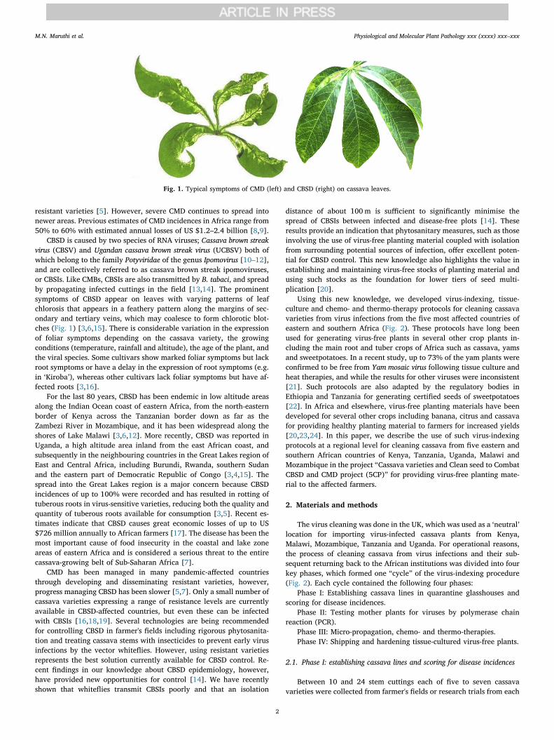
collectively called cassava mosaic begomoviruses or CMBs [4,5]. Theseviruses are transmitted by whitefly, Bemisia tabaci Gennadius (familyAleyrodidae, order Hemiptera), as well as disseminated through thepropagation of infected cuttings [6]. The most obvious symptoms ofCMD include a characteristic mosaic of pale green to yellow chloroticareas on leaves, usually accompanied by distortion (Fig. 1). Growth ofsusceptible plants infected at the cutting stage is severely stunted andthere is poor development of the tuberous roots, which ultimately re-sults in low yields [8]. The spread of an unusually severe form of CMD,the so-called “CMD pandemic”, was first recorded in Uganda in the late1980s and subsequently spread to affect an area greater than 4 millionkm2 across 11 countries of East and Central Africa [5,6,8,9]. Since then,considerable success has been achieved in mitigating the effects of theCMD pandemic through the multiplication and dissemination of CMD-
https://doi.org/10.1016/j.pmpp.2018.09.002Received 18 January 2018; Received in revised form 25 July 2018; Accepted 13 September 2018
Abbreviations: ACMV, African cassava mosaic virus; CBSD, Cassava brown streak disease; CBSV, Cassava brown streak virus; CMD, Cassava mosaic disease; CTAB,Cetyl trimethylammonium bromide; EACMV, East African cassava mosaic virus; EACMV-Ug, East African cassava mosaic virus-Uganda; LFC, Laminar flow cabinet; MS,Murashige and Skoog; PPM, Plant Preservation Mixture; RH, Relative humidity; SDW, Sterilised deionised water; UCBSV, Ugandan cassava brown streak virus; UIC,Unique identifying code∗ Corresponding author. Natural Resources Institute (NRI), University of Greenwich, Chatham Maritime, Kent, ME4 4TB, United Kingdom.E-mail address: [email protected] (M.N. Maruthi).
Physiological and Molecular Plant Pathology xxx (xxxx) xxx–xxx
0885-5765/ © 2018 The Authors. Published by Elsevier Ltd. This is an open access article under the CC BY license (http://creativecommons.org/licenses/BY/4.0/).
Please cite this article as: Maruthi, M.N., Physiological and Molecular Plant Pathology, https://doi.org/10.1016/j.pmpp.2018.09.002
brought to you by COREView metadata, citation and similar papers at core.ac.uk
provided by Greenwich Academic Literature Archive
resistant varieties [5]. However, severe CMD continues to spread intonewer areas. Previous estimates of CMD incidences in Africa range from50% to 60% with estimated annual losses of US $1.2–2.4 billion [8,9].
CBSD is caused by two species of RNA viruses; Cassava brown streakvirus (CBSV) and Ugandan cassava brown streak virus (UCBSV) both ofwhich belong to the family Potyviridae of the genus Ipomovirus [10–12],and are collectively referred to as cassava brown streak ipomoviruses,or CBSIs. Like CMBs, CBSIs are also transmitted by B. tabaci, and spreadby propagating infected cuttings in the field [13,14]. The prominentsymptoms of CBSD appear on leaves with varying patterns of leafchlorosis that appears in a feathery pattern along the margins of sec-ondary and tertiary veins, which may coalesce to form chlorotic blot-ches (Fig. 1) [3,6,15]. There is considerable variation in the expressionof foliar symptoms depending on the cassava variety, the growingconditions (temperature, rainfall and altitude), the age of the plant, andthe viral species. Some cultivars show marked foliar symptoms but lackroot symptoms or have a delay in the expression of root symptoms (e.g.in ‘Kiroba’), whereas other cultivars lack foliar symptoms but have af-fected roots [3,16].
For the last 80 years, CBSD has been endemic in low altitude areasalong the Indian Ocean coast of eastern Africa, from the north-easternborder of Kenya across the Tanzanian border down as far as theZambezi River in Mozambique, and it has been widespread along theshores of Lake Malawi [3,6,12]. More recently, CBSD was reported inUganda, a high altitude area inland from the east African coast, andsubsequently in the neighbouring countries in the Great Lakes region ofEast and Central Africa, including Burundi, Rwanda, southern Sudanand the eastern part of Democratic Republic of Congo [3,4,15]. Thespread into the Great Lakes region is a major concern because CBSDincidences of up to 100% were recorded and has resulted in rotting oftuberous roots in virus-sensitive varieties, reducing both the quality andquantity of tuberous roots available for consumption [3,5]. Recent es-timates indicate that CBSD causes great economic losses of up to US$726 million annually to African farmers [17]. The disease has been themost important cause of food insecurity in the coastal and lake zoneareas of eastern Africa and is considered a serious threat to the entirecassava-growing belt of Sub-Saharan Africa [7].
CMD has been managed in many pandemic-affected countriesthrough developing and disseminating resistant varieties, however,progress managing CBSD has been slower [5,7]. Only a small number ofcassava varieties expressing a range of resistance levels are currentlyavailable in CBSD-affected countries, but even these can be infectedwith CBSIs [16,18,19]. Several technologies are being recommendedfor controlling CBSD in farmer's fields including rigorous phytosanita-tion and treating cassava stems with insecticides to prevent early virusinfections by the vector whiteflies. However, using resistant varietiesrepresents the best solution currently available for CBSD control. Re-cent findings in our knowledge about CBSD epidemiology, however,have provided new opportunities for control [14]. We have recentlyshown that whiteflies transmit CBSIs poorly and that an isolation
distance of about 100m is sufficient to significantly minimise thespread of CBSIs between infected and disease-free plots [14]. Theseresults provide an indication that phytosanitary measures, such as thoseinvolving the use of virus-free planting material coupled with isolationfrom surrounding potential sources of infection, offer excellent poten-tial for CBSD control. This new knowledge also highlights the value inestablishing and maintaining virus-free stocks of planting material andusing such stocks as the foundation for lower tiers of seed multi-plication [20].
Using this new knowledge, we developed virus-indexing, tissue-culture and chemo- and thermo-therapy protocols for cleaning cassavavarieties from virus infections from the five most affected countries ofeastern and southern Africa (Fig. 2). These protocols have long beenused for generating virus-free plants in several other crop plants in-cluding the main root and tuber crops of Africa such as cassava, yamsand sweetpotatoes. In a recent study, up to 73% of the yam plants wereconfirmed to be free from Yam mosaic virus following tissue culture andheat therapies, and while the results for other viruses were inconsistent[21]. Such protocols are also adapted by the regulatory bodies inEthiopia and Tanzania for generating certified seeds of sweetpotatoes[22]. In Africa and elsewhere, virus-free planting materials have beendeveloped for several other crops including banana, citrus and cassavafor providing healthy planting material to farmers for increased yields[20,23,24]. In this paper, we describe the use of such virus-indexingprotocols at a regional level for cleaning cassava from five eastern andsouthern African countries of Kenya, Tanzania, Uganda, Malawi andMozambique in the project “Cassava varieties and Clean seed to CombatCBSD and CMD project (5CP)” for providing virus-free planting mate-rial to the affected farmers.
2. Materials and methods
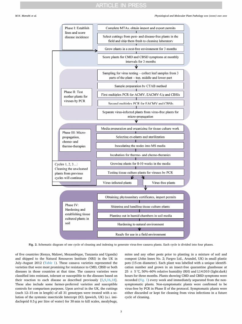
The virus cleaning was done in the UK, which was used as a ‘neutral’location for importing virus-infected cassava plants from Kenya,Malawi, Mozambique, Tanzania and Uganda. For operational reasons,the process of cleaning cassava from virus infections and their sub-sequent returning back to the African institutions was divided into fourkey phases, which formed one “cycle” of the virus-indexing procedure(Fig. 2). Each cycle contained the following four phases:
Phase I: Establishing cassava lines in quarantine glasshouses andscoring for disease incidences.
Phase II: Testing mother plants for viruses by polymerase chainreaction (PCR).
Phase III: Micro-propagation, chemo- and thermo-therapies.Phase IV: Shipping and hardening tissue-cultured virus-free plants.
2.1. Phase I: establishing cassava lines and scoring for disease incidences
Between 10 and 24 stem cuttings each of five to seven cassavavarieties were collected from farmer's fields or research trials from each
Fig. 1. Typical symptoms of CMD (left) and CBSD (right) on cassava leaves.
M.N. Maruthi et al. Physiological and Molecular Plant Pathology xxx (xxxx) xxx–xxx
2
of five countries (Kenya, Malawi, Mozambique, Tanzania and Uganda)and shipped to the Natural Resources Institute (NRI) in the UK inJuly–August 2012 (Table 1). These cassava varieties represented thevarieties that were most promising for resistance to CMD, CBSD or bothdiseases in those countries at that time. The cassava varieties wereclassified into resistant, tolerant or susceptible to the diseases based ontheir reaction to each disease as described previously [5,9,16,18].These also include some farmer-preferred varieties and susceptiblecontrols for comparison purposes. Upon arrival in the UK, the cuttings(each 12–15 cm in length) of all 31 genotypes were treated with a so-lution of the systemic insecticide Intercept (ICL Ipswich, UK) (a.i. imi-dacloprid 0.5 g per litre of water) for 30min to kill scales, mealybugs,
mites and any other pests prior to planting in a mixture of soil andcompost (John Innes No. 2; Fargro Ltd., Arundel, UK) in small plasticpots (15 cm diameter). Each plant was labelled with a unique identifi-cation number and grown in an insect-free quarantine glasshouse at25 ± 5 °C, 50%–60% relative humidity (RH) and L14:D10 (light:dark)hours for three months. Plants showing CMD and CBSD symptoms wererecorded (Fig. 1) every week and immediately separated from the non-symptomatic plants. Non-symptomatic plants were confirmed to bevirus-free by PCR in Phase II of the protocol. Symptomatic plants wereeither discarded or kept for cleaning from virus infections in a futurecycle of cleaning.
Fig. 2. Schematic diagram of one cycle of cleaning and indexing to generate virus-free cassava plants. Each cycle is divided into four phases.
M.N. Maruthi et al. Physiological and Molecular Plant Pathology xxx (xxxx) xxx–xxx
3
2.2. Phase II: testing mother plants for viruses by PCR
2.2.1. Total nucleic acid extraction for virus-indexingLeaf samples were collected from the top, middle and bottom part of
the plant and total nucleic acid was extracted from all non-symptomaticplants using a modified cetyl trimethylammonium bromide (CTAB)method [25–28]. The optimised protocol was as follows: First, theCTAB extraction buffer (2% (w/v) CTAB, 1.4M NaCl, 0.2% (v/v) 2-mercaptoethanol, 20mM EDTA, 100mM Tris-HCl, pH 8.0) was pre-heated to 60 °C for 10min. Mercaptoethanol was added fresh to thebuffer. Approximately 100mg of fresh plant leaf tissue was placed intoa thick-gauge plastic bag (10× 15 cm) and the tissue was ground finelyusing a ball-bearing grinder (Qiagen Ltd. Dorset UK). Each 100mg ofground plant tissue was then mixed with 1mL of CTAB extractionbuffer. About 750 μL of the sample was poured into a 1.5 mL centrifugetube and heated at 60 °C for 30min. Next, the samples were mixed withan equal volume (750 μL) of phenol:chloroform:isoamylalcohol(25:24:1) in a fume hood and centrifuged at 12,281 relative centrifugalforce (g) for 10min. Only the aqueous phase was transferred into a new1.5 mL centrifuge tube. To precipitate the DNA, we added 300 μL ofcold (−20 °C) isopropanol and incubated at −20 °C for at least 1 h.Samples were then centrifuged at 12,281 g at 4 °C for 10min. The su-pernatant was discarded, and the pellet was washed with 0.5ml of 70%ethanol, vortexed and centrifuged for 5min at 12,281 g. Then theethanol was removed and the pellet was vacuum-dried for 5min. Thedried pellet was suspended in 100 μL 1x TE buffer and stored at−20 °C.The extracts were diluted 1:10 fold in sterilised deionised water (SDW)before using them in PCR and reverse-transcriptase PCR (RT-PCR) forvirus detection (Fig. 3).
2.2.2. cDNA synthesisSynthesis of cDNA was done using ImProm-II™ Reverse
Transcriptase kit (Promega, Southampton, UK) following the manu-facturer's instructions. Briefly, we prepared 5 μL Master Mix I persample containing 1 μL each of SDW, Oligo-dT primer and 3 μL of RNAtemplate. We incubated the mix at 70 °C for 5min and immediatelychilled on ice for 2min. Next, we prepared 15 μL Master Mix II persample containing 7.5 μL SDW, 4.0 μL ImProm-IITM 5X reaction buffer,2.0 μL 25mM MgCl2, 1.0 μL 2.5mM dNTPs and 0.5 μL ImProm-IIReverse Transcriptase, and gently mixed in a vortexer for 3–5 s. Next,we added 15 μL Master Mix II into 5 μL Master Mix I making up a totalvolume of 20 μL per reaction. The reaction mixture was incubated at25 °C for 5min for primer annealing, at 40 °C for 60min for cDNAsynthesis, and finally at 70 °C for 15min for inactivation of the ImPro-II™ Reverse Transcriptase. The resulting cDNA samples were stored at−20 °C until further analysis.
2.2.3. Choice of PCR testsA range of tests are available for diagnosing CMBs and CBSIs, in-
cluding endpoint PCR and RT-PCR for detecting one virus at a time, aswell as duplex and multiplex reactions that can detect two or moreviruses in a single reaction [25,26, 29, 35]. Several real-time PCR assaysare also available for CMBs and CBSIs [28,30; 34]. Tests were chosenbased on the sensitivity required and the range of viruses to be tested,as well as personal preference, expertise and the facilities available. Inthis study, to detect CMBs (ACMV, EACMV and EACMV-Ug) and CBSIswe initially used the protocols published in Ref. [25] (Fig. 3) andsubsequently [28]. A summary of the viruses detected, primers usedand PCR conditions is in Tables 2–3.
2.3. Phase III: micro-propagation, thermo- and chemotherapies
2.3.1. Preparing MS medium for cassava nodal-bud cultureThe protocol used to make 1 L of tissue-culture media (∼125 tubes
of 8mL each) was as follows: first, we added 2.2 g Murashige and Skoog(MS) medium to a 2 L capacity beaker, then we added 20 g sucrose and∼950mL of deionised water. A magnetic stirrer was used to mix the
Table 1The 31 cassava varieties obtained from five eastern and southern Africancountries and cleaned of viral infections in the UK.
Country of origin Variety name Reaction to diseases No. of stemsimported andplanted in theUK
CMD CBSD
Kenya F10-30-R2 Tolerant Tolerant 20F19-NL Tolerant Tolerant 23LMI/2008/363 Tolerant Tolerant 22Kibandameno Susceptible Susceptible 20Shibe Tolerant Tolerant 21Tajirika Tolerant Tolerant 20
Malawi CH05/203 Tolerant Tolerant 24Kalawe Tolerant Tolerant 20Mbundumali Susceptible Susceptible 21Sangoja Tolerant Tolerant 20Sauti Tolerant Tolerant 22Yizaso Tolerant Tolerant 20
Mozambique Coliacanana Susceptible Tolerant 21Eyope Tolerant Tolerant 22Nziva Susceptible Tolerant 22Okhumelela Tolerant Tolerant 21Orera Susceptible Tolerant 22
Tanzania KBH2002/066 Tolerant Tolerant 21KBH2006/026 Tolerant Tolerant 21Albert Resistant Susceptible Previous
collectionKiroba Susceptible Tolerant Previous
collectionKizimbani Tolerant Tolerant 21Mkombozi Resistant Susceptible 17Mkumba Susceptible Tolerant 21Pwani Tolerant Tolerant 21
Uganda TZ130 Resistant Tolerant 14NASE1 Resistant Tolerant 14NASE3 Tolerant Tolerant 12NASE14 Resistant Tolerant 13NASE18 Resistant Tolerant 10TME204 Resistant Susceptible 13
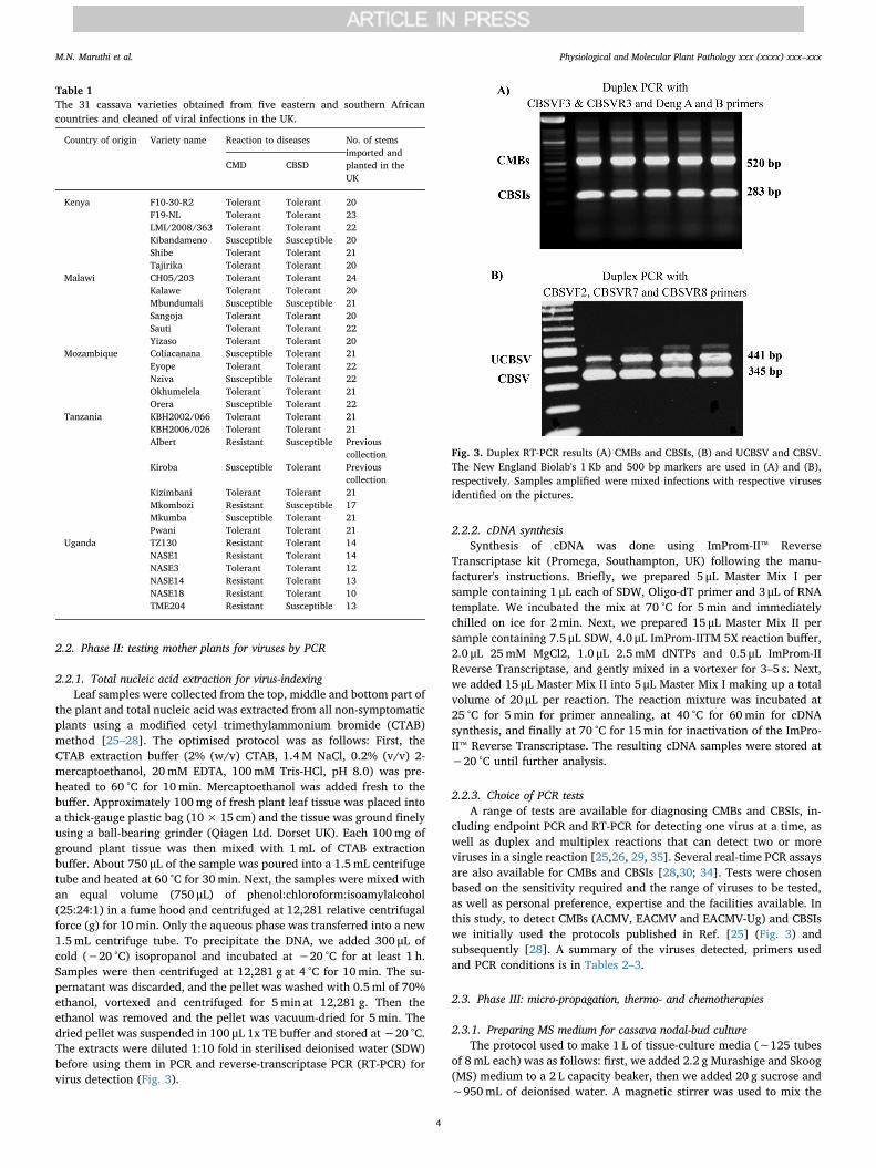
Fig. 3. Duplex RT-PCR results (A) CMBs and CBSIs, (B) and UCBSV and CBSV.The New England Biolab's 1 Kb and 500 bp markers are used in (A) and (B),respectively. Samples amplified were mixed infections with respective virusesidentified on the pictures.
M.N. Maruthi et al. Physiological and Molecular Plant Pathology xxx (xxxx) xxx–xxx
4
solution. Next, we added growth regulators (8 mL of NAA/L at conc.27 μM) and Plant Preservation Mixture (PPM, Sigma-Aldrich, Dorset,UK) at 0.2–0.25 v/v % of PPM. The pH was adjusted to 5.7–5.8 usingNaOH and/or HCl buffer solutions (buffer solutions should be 10% and1% for fine adjustments). We then added deionised water to make so-lutions up to 1 L. Next, we added 2 g phytagel (Sigma-Aldrich, Dorset,UK) and 25mg Ribavrin (final concentration 0.1 mM). Addition ofRibavrin was only required for media used for chemotherapy of virus-infected plant material. Each solution was mixed using a magneticstirrer to ensure an even distribution of undissolved gelling agent whilethe media was pipetted into culture tubes. We added 8mL media intoclean glass tubes (e.g. Timstar, borosilicate glass, 100×25mm) andsealed them with lids. The tubes were then wrapped in greaseproofpaper and/or aluminium foil (minimum of two layers) and autoclavedat 121 °C for 15min. Tubes were then dried in an oven for 4 h at 60 °Cand finally cooled to ambient temperature before use.
2.3.2. Surface sterilisation of explantsAt least 20 cuttings from each of 31 genotypes were collected from
the five African countries. At the time of collection, the top green partsof each plant's stem were cleaned separately, all leaves were removedand the stems were placed in plastic, labelled bags. Upon arrival in theUK, the stems (explants) were trimmed to a suitable size (usually up to5 cm) that contained at least one nodal bud. The explants were thentransferred into separate glass jars and washed 3 times in running tapwater. The explants were then immersed in 70% ethanol for 3–5 s be-fore the ethanol was poured out of the jars and replaced with a ster-ilisation solution (5% v/v sodium hypochlorite and 0.1mL/L of Tween20) that almost covered the explants. The jars were then placed on ashaker and mixed vigorously on an orbital shaker at 100 g for20–30min. After shaking, the explants were rinsed with SDW underaseptic conditions in a laminar flow cabinet (LFC). Explants were rinsedthree to four times until no foam was left in the bottles. These sterileexplants were used for seeding into tissue-culture media.
2.3.3. Seeding nodal buds into MS mediaThe protocol for seeding nodal buds into MS media was done inside
the LFC. Prior to beginning work with the explants, the UV lamp was
turned on for 10min, the laminar flow was turned on, and the workingsurfaces were decontaminated with 100% ethanol. Explants were ex-cised in sterile petri dishes using sterile knives and sterile forceps. Weretained only 2–3mm of each node bud by removing excess tissuearound the bud (Fig. 4A). The node bud was then transferred into MSmedia using forceps ensuring that the node bud was facing upwards(Fig. 4B). The cap of the tube was closed immediately and all tubes froma single plant were labelled before seeding the next plant (Fig. 4C).Work was done near a Bunsen flame to further reduce the risk of con-taminating organisms. Seeded tubes were transferred into a plantgrowth chamber (SLS Ltd., Wilford, UK) that was maintained at 35 °C,50%–60% RH, and 14L:10D for heat treatment. Plants were maintainedat these conditions for two weeks. Note: this step was only performedfor virus-infected material receiving thermotherapy. Plants were thentransferred to 25 °C growth rooms for the next 10 weeks before eitherbeing shipped back to the project's African partners or being hardenedby planting into soil media for further growth in the UK.
2.4. Phase IV: hardening and establishing tissue-culture cassava plants inthe soil
2.4.1. Transplanting and hardening tissue-culture plantsPlantlets were removed from the glass tubes, the media was gently
washed off using tap water or SDW. The oldest and middle leaves wereremoved using sharp scissors, retaining only three or four developingleaves at the top of the plant. Further, to prevent fungal growth on anytraces of leftover media, the plantlets were rinsed in the fungicide so-lution Dithane, 6 g/L (Mancozeb 80% a.i.) (ICL Ipswich, UK) for 10min.Plantlets were placed into small pots (10 cm diameter) with compostand soil in 1:1 proportion and the pots were placed onto a propagatortray and watered just enough to soak the soil mixture (Fig. 5). Afterwatering, the tray was immediately covered with a transparent lid thathad been sprayed with water to raise the humidity. Lids were keptclosed for a minimum of two weeks to allow the plants to develop newroots and leaves. Conditions were maintained at 30 ± 5 °C, 50–60%RH, 14L:10D. After two weeks, the vents in the lids were opened for oneweek and then we started lifting the lids incrementally using spacersbetween the lid and the tray. At the end of four weeks, the lids were
Table 2Primers used for detecting CMBs and CBSIs in uniplex, duplex and multiplex RT-PCR assays.
Primer name Primer sequence (5′–3′) Targeta Reference
Primers used for amplifying CMBsDeng A TAATATTACCKGWKGVCCSC Begomovirus [31]Deng B TGGACYTTRCAWGGBCCTTCACA Begomovirus [31]CMBRep/F CRTCAATGACGTTGTACCA ACMV & EACMV [29]CMBCP/F GKCGAAGCGACCAGGAGAT ACMV & EACMV-Ug [29]ACMVCP/R CCCTGYCTCCTGATGATTATA ACMV [29]EACMV-UG/R CGCCTAAGCAAGGAATGGCGT EACMV-Ug [25]EACMVRep/R GGTTTGCAGAGAACTACATC EACMV [29]Primers used for amplifying CBSIsRT-PCRCBSVF3 GGARCCRATGTAYAAATTTGC CBSIs [25]CBSVR3 AGGAGCWGCTARWGCAAA CBSIs [25]RT-qPCRCBSVR4
+ CBSVF3GCWGCTTTTATYACAAAMGC CBSIs [28]
Primers used for amplifying CBSV and UCBSV individuallyCBSVF2 GGRCCATACATYAARTGGTT CBSIs [25]CBSVR7 CCCTTTGCAAARCTRAAATARC CBSV [25]CBSVR8 CCATTRTCTYTCCAMADCTTC UCBSV [25]Primers used for amplifying cassava housekeeping genesRubiscoLF CTTTCCAAGGCCCGCCTCA RubiscoL [32]RubiscoLR CATCATCTTTGGTAAAATCAAGTCCA RubiscoL [29]L2F TGGTGTTGCCATGAACCCTGTAGA Ribosomal protein (L2) [33]L2R CGACCAGTCCTCCTTGCAGC Ribosomal protein (L2) [33]
a ACMV – African cassava mosaic virus; EACMV – East African cassava mosaic virus; EACMV-Ug – East African cassava mosaic virus-Uganda; CBSIs – cassavabrown streak ipomoviruses; CBSV – Cassava brown streak virus; UCBSV – Ugandan Cassava brown streak virus.
M.N. Maruthi et al. Physiological and Molecular Plant Pathology xxx (xxxx) xxx–xxx
5
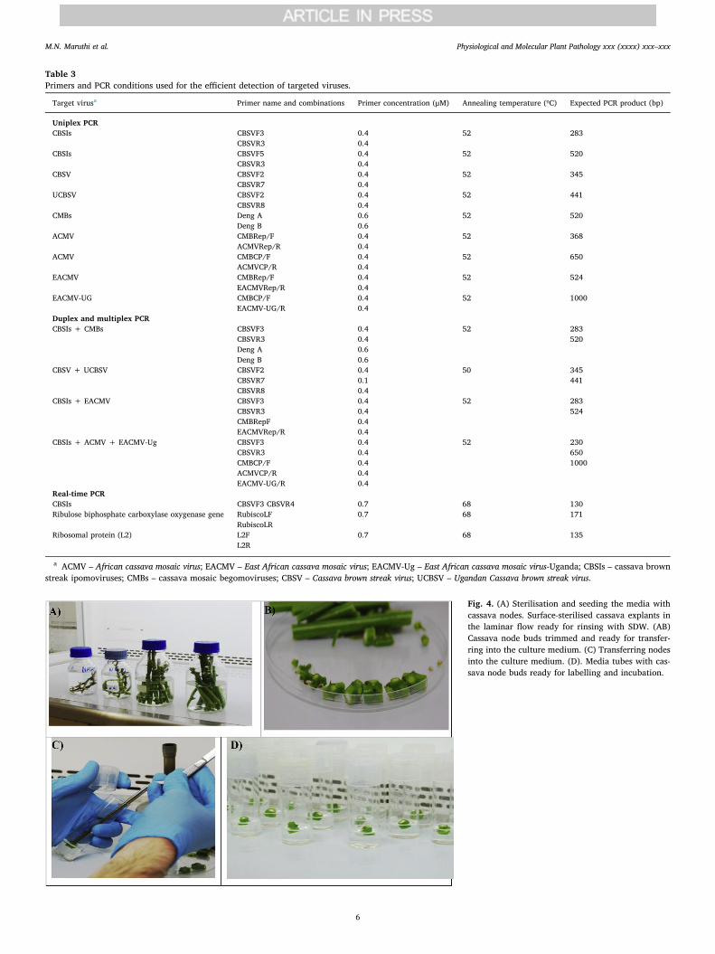
Table 3Primers and PCR conditions used for the efficient detection of targeted viruses.
Target virusa Primer name and combinations Primer concentration (μM) Annealing temperature (ºC) Expected PCR product (bp)
Uniplex PCRCBSIs CBSVF3
CBSVR30.40.4
52 283
CBSIs CBSVF5CBSVR3
0.40.4
52 520
CBSV CBSVF2CBSVR7
0.40.4
52 345
UCBSV CBSVF2CBSVR8
0.40.4
52 441
CMBs Deng ADeng B
0.60.6
52 520
ACMV CMBRep/FACMVRep/R
0.40.4
52 368
ACMV CMBCP/FACMVCP/R
0.40.4
52 650
EACMV CMBRep/FEACMVRep/R
0.40.4
52 524
EACMV-UG CMBCP/FEACMV-UG/R
0.40.4
52 1000
Duplex and multiplex PCRCBSIs + CMBs CBSVF3
CBSVR3Deng ADeng B
0.40.40.60.6
52 283520
CBSV + UCBSV CBSVF2CBSVR7CBSVR8
0.40.10.4
50 345441
CBSIs + EACMV CBSVF3CBSVR3CMBRepFEACMVRep/R
0.40.40.40.4
52 283524
CBSIs + ACMV + EACMV-Ug CBSVF3CBSVR3CMBCP/FACMVCP/REACMV-UG/R
0.40.40.40.40.4
52 2306501000
Real-time PCRCBSIs CBSVF3 CBSVR4 0.7 68 130Ribulose biphosphate carboxylase oxygenase gene RubiscoLF
RubiscoLR0.7 68 171
Ribosomal protein (L2) L2FL2R
0.7 68 135
a ACMV – African cassava mosaic virus; EACMV – East African cassava mosaic virus; EACMV-Ug – East African cassava mosaic virus-Uganda; CBSIs – cassava brownstreak ipomoviruses; CMBs – cassava mosaic begomoviruses; CBSV – Cassava brown streak virus; UCBSV – Ugandan Cassava brown streak virus.
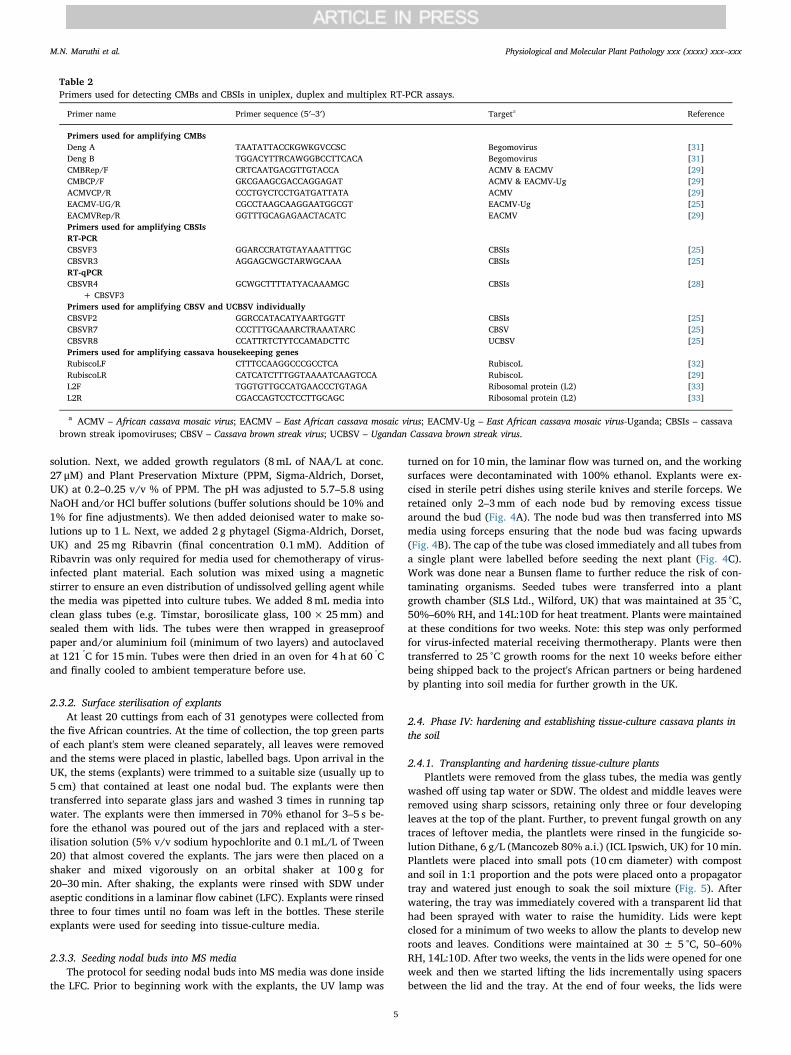
Fig. 4. (A) Sterilisation and seeding the media withcassava nodes. Surface-sterilised cassava explants inthe laminar flow ready for rinsing with SDW. (AB)Cassava node buds trimmed and ready for transfer-ring into the culture medium. (C) Transferring nodesinto the culture medium. (D). Media tubes with cas-sava node buds ready for labelling and incubation.
M.N. Maruthi et al. Physiological and Molecular Plant Pathology xxx (xxxx) xxx–xxx
6
removed completely, and we applied NPK fertiliser containing Mg(ratio of 30:10:10:2) and other trace elements (10 g dissolved in 10 Lwater) (Fargro Ltd., Arundel, UK). Plants were grown for an additionalfour weeks and then hardened by reducing the amount water providedwithout allowing plants to dry out completely or shed leaves. Suchhardened plants can be ready to transplant into the soil media.
2.4.2. Accountability and traceability of virus-cleaned plantsWe designed a full traceability system that was set up to track each
stem cutting from its arrival at the quarantine facility at NRI, throughthe micro-propagation and virus-indexing procedures, to the shipmentof clean material back the partner organisations in East Africa (i.e.Phases I–IV). A well-designed traceability system that can track plantmaterial through the processing phases to the partner organisations inAfrica is essential. Such a system provides a control mechanism. If anyplant material was later found to contain a virus, the traceability systemcould identify other potentially infected stock, thus limiting viral spread
(Fig. 6).There were two parts to the traceability system; the first was a re-
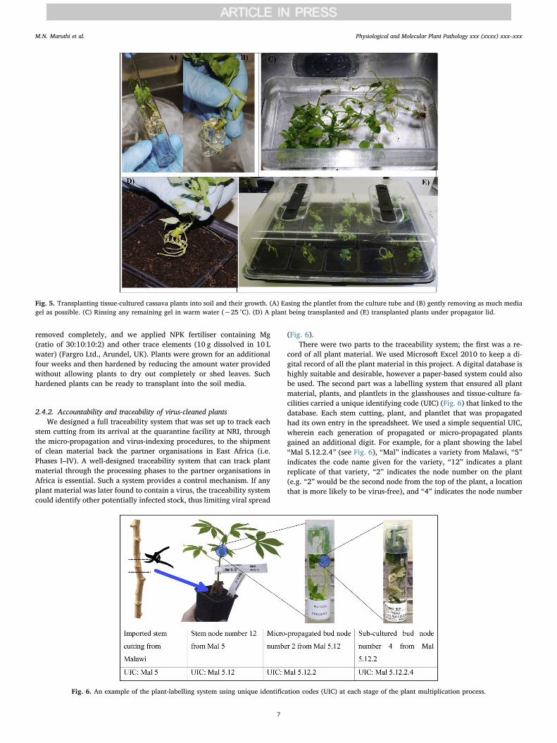
cord of all plant material. We used Microsoft Excel 2010 to keep a di-gital record of all the plant material in this project. A digital database ishighly suitable and desirable, however a paper-based system could alsobe used. The second part was a labelling system that ensured all plantmaterial, plants, and plantlets in the glasshouses and tissue-culture fa-cilities carried a unique identifying code (UIC) (Fig. 6) that linked to thedatabase. Each stem cutting, plant, and plantlet that was propagatedhad its own entry in the spreadsheet. We used a simple sequential UIC,wherein each generation of propagated or micro-propagated plantsgained an additional digit. For example, for a plant showing the label“Mal 5.12.2.4” (see Fig. 6), “Mal” indicates a variety from Malawi, “5”indicates the code name given for the variety, “12” indicates a plantreplicate of that variety, “2” indicates the node number on the plant(e.g. “2” would be the second node from the top of the plant, a locationthat is more likely to be virus-free), and “4” indicates the node number
Fig. 5. Transplanting tissue-cultured cassava plants into soil and their growth. (A) Easing the plantlet from the culture tube and (B) gently removing as much mediagel as possible. (C) Rinsing any remaining gel in warm water (∼25 °C). (D) A plant being transplanted and (E) transplanted plants under propagator lid.
Fig. 6. An example of the plant-labelling system using unique identification codes (UIC) at each stage of the plant multiplication process.
M.N. Maruthi et al. Physiological and Molecular Plant Pathology xxx (xxxx) xxx–xxx
7
after the first sub-culturing from the previous plant. The spreadsheetwas setup such that each row (entry) represented a single bud nodefrom a plant that had been propagated, while the columns containeddata on various aspects of that bud node and its history including theUIC, country of origin, cutting number, varietal common name, virusassessments (visual and PCR based), infection status, location (withinglasshouse or incubator), date of tissue culture, number of nodes micro-propagated, shipping date, and destination. For each entry, additionalcolumns were added to record details of thermo- and chemotherapiesand their outcomes, number of nodes that were tissue-cultured, resultsof repeated PCR tests (used to confirm previous results prior to ship-ping), dates plantlets were hardened, dates plants were transferred tothe glasshouses, or anything else pertinent to the work. The “PivotTable” function of Excel was used to quickly generate summaries of thedata and track overall progress.
We used ‘mail merge’ function to link between an Excel spreadsheetand a Word document. A template for labels was set up in a Worddocument, wherein each label included all the relevant fields (e.g. UIC,date, location). The ‘mail merge’ allows a linked spreadsheet to be fil-tered by specific fields, and then automatically generates the data intothe relevant fields for each label within the Word document. We used‘mail merge’ to automatically generate sticky labels for the micro-pro-pagation tubes, storage and shipping containers, thus minimising thepotential for human error in labelling. A label printer (LabelStation Pro200, Advanced Labelling Ltd., Dorset, UK) and the LabelStation soft-ware were used to generate the tags and labels for plants in the glass-house. Similar to Microsoft ‘Mail Merge’, the LabelStation software waslinked to the Excel spreadsheet. Again, a label template was set up tocontain the relevant fields. The link between the Excel spreadsheet andthe LabelStation software allowed for data filtering, enabling us togenerate plant tags for specific batches of plants with the correct UICand other relevant information.
3. Results
3.1. Phase I: establishing cassava lines and scoring disease incidences
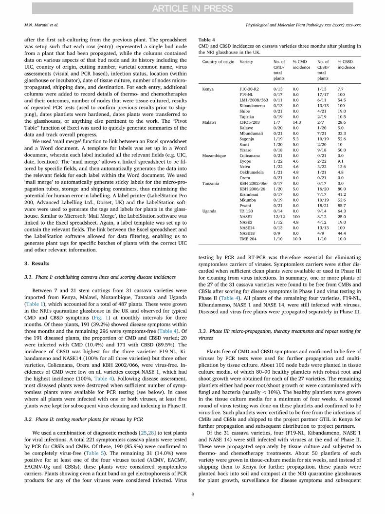
Between 7 and 21 stem cuttings from 31 cassava varieties wereimported from Kenya, Malawi, Mozambique, Tanzania and Uganda(Table 1), which accounted for a total of 487 plants. These were grownin the NRI's quarantine glasshouse in the UK and observed for typicalCMD and CBSD symptoms (Fig. 1) at monthly intervals for threemonths. Of these plants, 191 (39.2%) showed disease symptoms withinthree months and the remaining 296 were symptoms-free (Table 4). Ofthe 191 diseased plants, the proportion of CMD and CBSD varied; 20were infected with CMD (10.4%) and 171 with CBSD (89.5%). Theincidence of CBSD was highest for the three varieties F19-NL, Ki-bandameno and NASE14 (100% for all three varieties) but three othervarieties, Colicanana, Orera and KBH 2002/066, were virus-free. In-cidences of CMD were low on all varieties except NASE 1, which hadthe highest incidence (100%, Table 4). Following disease assessment,most diseased plants were destroyed when sufficient number of symp-tomless plants were available for PCR testing (see below). In caseswhere all plants were infected with one or both viruses, at least fiveplants were kept for subsequent virus cleaning and indexing in Phase II.
3.2. Phase II: testing mother plants for viruses by PCR
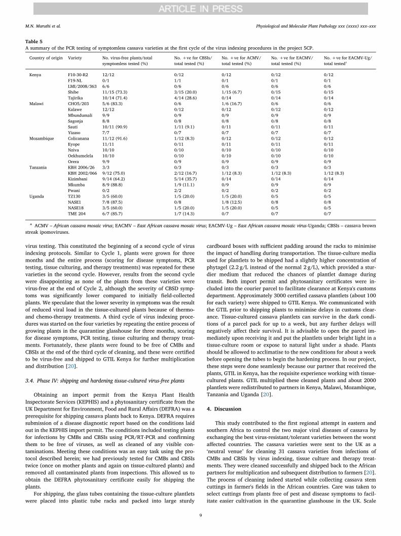
We used a combination of diagnostic methods [25,28] to test plantsfor viral infections. A total 221 symptomless cassava plants were testedby PCR for CBSIs and CMBs. Of these, 190 (85.9%) were confirmed tobe completely virus-free (Table 5). The remaining 31 (14.0%) werepositive for at least one of the four viruses tested (ACMV, EACMV,EACMV-Ug and CBSIs); these plants were considered symptomlesscarriers. Plants showing even a faint band on gel electrophoresis of PCRproducts for any of the four viruses were considered infected. Virus
testing by PCR and RT-PCR was therefore essential for eliminatingsymptomless carriers of viruses. Symptomless carriers were either dis-carded when sufficient clean plants were available or used in Phase IIIfor cleaning from virus infections. In summary, one or more plants ofthe 27 of the 31 cassava varieties were found to be free from CMBs andCBSIs after scoring for disease symptoms in Phase I and virus testing inPhase II (Table 4). All plants of the remaining four varieties, F19-NL,Kibandameno, NASE 1 and NASE 14, were still infected with viruses.Diseased and virus-free plants were propagated separately in Phase III.
3.3. Phase III: micro-propagation, therapy treatments and repeat testing forviruses
Plants free of CMD and CBSD symptoms and confirmed to be free ofviruses by PCR tests were used for further propagation and multi-plication by tissue culture. About 100 node buds were planted in tissueculture media, of which 80–90 healthy plantlets with robust root andshoot growth were obtained for each of the 27 varieties. The remainingplantlets either had poor root/shoot growth or were contaminated withfungi and bacteria (usually< 10%). The healthy plantlets were grownin the tissue culture media for a minimum of four weeks. A secondround of virus testing was done on these plantlets and confirmed to bevirus-free. Such plantlets were certified to be free from the infections ofCMBs and CBSIs and shipped to the project partner GTIL in Kenya forfurther propagation and subsequent distribution to project partners.
Of the 31 cassava varieties, four (F19-NL, Kibandameno, NASE 1and NASE 14) were still infected with viruses at the end of Phase II.These were propagated separately by tissue culture and subjected tothermo- and chemotherapy treatments. About 50 plantlets of eachvariety were grown in tissue-culture media for six weeks, and instead ofshipping them to Kenya for further propagation, these plants wereplanted back into soil and compost at the NRI quarantine glasshousesfor plant growth, surveillance for disease symptoms and subsequent
Table 4CMD and CBSD incidences on cassava varieties three months after planting inthe NRI glasshouse in the UK.
Country of origin Variety No. ofCMD/totalplants
% CMDincidence
No. ofCBSD/totalplants
% CBSDincidence
Kenya F10-30-R2 0/13 0.0 1/13 7.7F19-NL 0/17 0.0 17/17 100LM1/2008/363 0/11 0.0 6/11 54.5Kibandameno 0/13 0.0 13/13 100Shibe 0/21 0.0 4/21 19.0Tajirika 0/19 0.0 2/19 10.5
Malawi CHO5/203 1/7 14.3 2/7 28.6Kalawe 0/20 0.0 1/20 5.0Mbundumali 0/21 0.0 7/21 33.3Sagonja 1/19 5.3 10/19 52.6Sauti 1/20 5.0 2/20 10Yizaso 0/18 0.0 9/18 50.0
Mozambique Colicanana 0/21 0.0 0/21 0.0Eyope 1/22 4.6 2/22 9.1Nziva 1/22 4.6 3/22 13.6Oekhumelela 1/21 4.8 1/21 4.8Orera 0/21 0.0 0/21 0.0
Tanzania KBH 2002/066 0/17 0.0 0/17 0.0KBH 2006/26 1/20 5.0 16/20 80.0Kizimbani 0/17 0.0 7/17 41.2Mkumba 0/19 0.0 10/19 52.6Pwani 0/21 0.0 18/21 85.7
Uganda TZ 130 0/14 0.0 9/14 64.3NASE1 12/12 100 3/12 25.0NASE3 1/12 4.8 4/12 19.0NASE14 0/13 0.0 13/13 100NASE18 0/9 0.0 4/9 44.4TME 204 1/10 10.0 1/10 10.0
M.N. Maruthi et al. Physiological and Molecular Plant Pathology xxx (xxxx) xxx–xxx
8
virus testing. This constituted the beginning of a second cycle of virusindexing protocols. Similar to Cycle 1, plants were grown for threemonths and the entire process (scoring for disease symptoms, PCRtesting, tissue culturing, and therapy treatments) was repeated for thesevarieties in the second cycle. However, results from the second cyclewere disappointing as none of the plants from these varieties werevirus-free at the end of Cycle 2, although the severity of CBSD symp-toms was significantly lower compared to initially field-collectedplants. We speculate that the lower severity in symptoms was the resultof reduced viral load in the tissue-cultured plants because of thermo-and chemo-therapy treatments. A third cycle of virus indexing proce-dures was started on the four varieties by repeating the entire process ofgrowing plants in the quarantine glasshouse for three months, scoringfor disease symptoms, PCR testing, tissue culturing and therapy treat-ments. Fortunately, these plants were found to be free of CMBs andCBSIs at the end of the third cycle of cleaning, and these were certifiedto be virus-free and shipped to GTIL Kenya for further multiplicationand distribution [20].
3.4. Phase IV: shipping and hardening tissue-cultured virus-free plants
Obtaining an import permit from the Kenya Plant HealthInspectorate Services (KEPHIS) and a phytosanitary certificate from theUK Department for Environment, Food and Rural Affairs (DEFRA) was aprerequisite for shipping cassava plants back to Kenya. DEFRA requiressubmission of a disease diagnostic report based on the conditions laidout in the KEPHIS import permit. The conditions included testing plantsfor infections by CMBs and CBSIs using PCR/RT-PCR and confirmingthem to be free of viruses, as well as cleaned of any visible con-taminations. Meeting these conditions was an easy task using the pro-tocol described herein; we had previously tested for CMBs and CBSIstwice (once on mother plants and again on tissue-cultured plants) andremoved all contaminated plants from inspections. This allowed us toobtain the DEFRA phytosanitary certificate easily for shipping theplants.
For shipping, the glass tubes containing the tissue-culture plantletswere placed into plastic tube racks and packed into large sturdy
cardboard boxes with sufficient padding around the racks to minimisethe impact of handling during transportation. The tissue-culture mediaused for plantlets to be shipped had a slightly higher concentration ofphytagel (2.2 g/L instead of the normal 2 g/L), which provided a stur-dier medium that reduced the chances of plantlet damage duringtransit. Both import permit and phytosanitary certificates were in-cluded into the courier parcel to facilitate clearance at Kenya's customsdepartment. Approximately 3000 certified cassava plantlets (about 100for each variety) were shipped to GTIL Kenya. We communicated withthe GTIL prior to shipping plants to minimise delays in customs clear-ance. Tissue-cultured cassava plantlets can survive in the dark condi-tions of a parcel pack for up to a week, but any further delays willnegatively affect their survival. It is advisable to open the parcel im-mediately upon receiving it and put the plantlets under bright light in atissue-culture room or expose to natural light under a shade. Plantsshould be allowed to acclimatise to the new conditions for about a weekbefore opening the tubes to begin the hardening process. In our project,these steps were done seamlessly because our partner that received theplants, GTIL in Kenya, has the requisite experience working with tissue-cultured plants. GTIL multiplied these cleaned plants and about 2000plantlets were redistributed to partners in Kenya, Malawi, Mozambique,Tanzania and Uganda [20].
4. Discussion
This study contributed to the first regional attempt in eastern andsouthern Africa to control the two major viral diseases of cassava byexchanging the best virus-resistant/tolerant varieties between the worstaffected countries. The cassava varieties were sent to the UK as a‘neutral venue’ for cleaning 31 cassava varieties from infections ofCMBs and CBSIs by virus indexing, tissue culture and therapy treat-ments. They were cleaned successfully and shipped back to the Africanpartners for multiplication and subsequent distribution to farmers [20].The process of cleaning indeed started while collecting cassava stemcuttings in farmer's fields in the African countries. Care was taken toselect cuttings from plants free of pest and disease symptoms to facil-itate easier cultivation in the quarantine glasshouse in the UK. Scale
Table 5A summary of the PCR testing of symptomless cassava varieties at the first cycle of the virus indexing procedures in the project 5CP.
Country of origin Variety No. virus-free plants/totalsymptomless tested (%)
No. +ve for CBSIs/total tested (%)
No. +ve for ACMV/total tested (%)
No. +ve for EACMV/total tested (%)
No. +ve for EACMV-Ug/total testeda
Kenya F10-30-R2 12/12 0/12 0/12 0/12 0/12F19-NL 0/1 1/1 0/1 0/1 0/1LMI/2008/363 6/6 0/6 0/6 0/6 0/6Shibe 11/15 (73.3) 3/15 (20.0) 1/15 (6.7) 0/15 0/15Tajirika 10/14 (71.4) 4/14 (28.6) 0/14 0/14 0/14
Malawi CHO5/203 5/6 (83.3) 0/6 1/6 (16.7) 0/6 0/6Kalawe 12/12 0/12 0/12 0/12 0/12Mbundumali 9/9 0/9 0/9 0/9 0/9Sagonja 8/8 0/8 0/8 0/8 0/8Sauti 10/11 (90.9) 1/11 (9.1) 0/11 0/11 0/11Yizaso 7/7 0/7 0/7 0/7 0/7
Mozambique Colicanana 11/12 (91.6) 1/12 (8.3) 0/12 0/12 0/12Eyope 11/11 0/11 0/11 0/11 0/11Nziva 10/10 0/10 0/10 0/10 0/10Oekhumelela 10/10 0/10 0/10 0/10 0/10Orera 9/9 0/9 0/9 0/9 0/9
Tanzania KBH 2006/26 3/3 0/3 0/3 0/3 0/3KBH 2002/066 9/12 (75.0) 2/12 (16.7) 1/12 (8.3) 1/12 (8.3) 1/12 (8.3)Kizimbani 9/14 (64.2) 5/14 (35.7) 0/14 0/14 0/14Mkumba 8/9 (88.8) 1/9 (11.1) 0/9 0/9 0/9Pwani 0/2 2/2 0/2 0/2 0/2
Uganda TZ130 3/5 (60.0) 1/5 (20.0) 1/5 (20.0) 0/5 0/5NASE1 7/8 (87.5) 0/8 1/8 (12.5) 0/8 0/8NASE18 3/5 (60.0) 1/5 (20.0) 1/5 (20.0) 0/5 0/5TME 204 6/7 (85.7) 1/7 (14.3) 0/7 0/7 0/7
a ACMV – African cassava mosaic virus; EACMV – East African cassava mosaic virus; EACMV-Ug – East African cassava mosaic virus-Uganda; CBSIs – cassava brownstreak ipomoviruses.
M.N. Maruthi et al. Physiological and Molecular Plant Pathology xxx (xxxx) xxx–xxx
9
insects, mealybugs and mites would add additional challenges duringviral cleaning because these pests are difficult to control in the con-tained environment of a glasshouse. Whenever possible, we also se-lected plants free of viral symptoms because starting with plants thathave low or no viral load greatly reduces the time and effort required toeliminate viruses. In our experience, susceptible plants of the fourcassava varieties with severe viral infections required 2–3 cycles oftherapy and tissue culturing and this took up to two years (Fig. 1).Tolerant and resistant plants were cleaned in the first cycle, which tookonly about 8–10 months. Cleaning of vegetatively-propagated crops likecassava in this study and bananas, potatoes, sweetpotatoes, sugarcaneand other crops by others has been done before [21,22,24]. However,cleaning and germplasm exchange at regional level is the first of its kindto our knowledge for any vegetatively-propagated crop and sets anexample to follow for other crops for better disease control, improvedyields and food securities. Use of insecticides have been increasinglyrecommended for controlling the whitefly vector of CMBs and CBSIs[5,7]. Our virus-free cassava varieties that have high levels of toleranceto CMD and CBSD can therefore be part of an integrated program forcontrolling both whiteflies and viral diseases.
A critical part of our method for virus indexing and cleaning wascareful monitoring and surveillance of each plant. Plants were grownfor three months and scored for disease symptoms every month.Surveillance of each plant throughout the three-month growth periodwas critical because some tolerant varieties have longer incubationperiods before the expression of viral symptoms. A three-month growthperiod thus minimises false positives (plants incorrectly identified asvirus-free) and reduces the number of plants that need to be confirmedvirus-free by PCR and subsequent tissue culturing in Phase II [23]. Acritical examination of symptoms therefore saves both time and re-sources while minimising the multiplication of virus-infected plants. Itis important to note, however, that although CMD symptoms are easilyrecognisable, scoring for CBSD was done by an experienced researcherbased on detailed description of symptoms and how they are affected byenvironments [3,16].
The PCR tests were chosen by the sensitivity required based on thesource of the sampled material. For example, the recent real-time PCRTaqman assays [28] which were up to 300% more sensitive were pre-ferred over the end-point PCR methods [25,26] when they becameavailable in our laboratories. We found that collecting leaf samplesfrom three parts of the plant (top, middle and bottom) and poolingthem together during sample preparation greatly increased the chanceof virus detection because viruses that are restricted to one part of theplant are more likely to be detected using this sampling method. Weadopted this sampling method for testing all plants in this study [28].
Another critical aspect of this work was adapting a ‘zero tolerance’policy for virus testing to prevent inadvertent virus introduction andmovement between the affected countries. To prevent this, all plantswere tested twice by PCR for CMBs and CBSIs during the cleaningprocess. The first testing was done before tissue culturing of the motherplants and thus prevented multiplication of infected material at the verybeginning of the process. The second testing was done after the tissueculturing was done and before the plants were shipped out to Africa tomake sure plants were indeed virus-free. Unlike leaf collection from themother plants in the glasshouse for virus testing, collecting re-presentative leaf samples from the tissue-culture plants that were grownin small glass tubes (2.5 cm diameter) was difficult because of theirsmall size. We used sharp forceps to collect 2–3 leaves from eachplantlet in aseptic conditions in a laminar flow hood but without dis-turbing plant's root system. Leaf samples of all plantlets from a singlemother plant were pooled for virus testing by real-time PCR [28]. Thetraceability procedures ensured that any plantlets found to containvirus at this stage could be linked back to the original mother plant andall other associated cultured plantlets derived from that same motherplant. In our project, these tests were done using real-time PCR (whichhas higher sensitivity) than the end-point PCR protocols [25,26] but did
not detect CMBs or CBSIs from any tissue-cultured plants. This con-firmed that the approach that we have used for cleaning cassava fromCMBs and CBSIs has worked. Such plantlets were certified to be virus-free and shipped to the project partner Genetic Technologies Interna-tional Limited (GTIL) in Kenya. The GTIL further multiplied the plantsin their tissue culture facilities and distributed to project partners inKenya, Tanzania, Malawi, Mozambique and Uganda [20].
Hardening is an important process in establishing tissue-culturedplants. Complete loss of plants can occur if proper care is not takenduring the hardening process. At this stage, plantlets are highly sus-ceptible to infections by fungi, attack by root insects, low humidity andhigh temperatures. Consequently, the plantlets should be kept in aclean, pest-free environment and provided with enough water tomaintain humidity but not so much water that plants rot. Fertilisersshould only be applied after plants are well established. The processfrom initial transplanting to complete hardening takes about 8–10weeks depending on the cassava variety and specific growth conditions.Plants develop quicker in warmer temperatures (30–35 °C) and brightlong days. However, temperatures exceeding 40 °C such as in poly-houses can severely hinder plant growth or result in plant death. Goodcare of the plants was therefore taken which resulted on average >90% plant survival after planting into the soil.
An important but often neglected aspect of a virus cleaning andindexing system is traceability of the plants. As each plant is propa-gated, PCR tested, cleaned of viruses, and multiplied to hundreds ofplantlets, it is essential to check for viruses, if any, manifested in sub-sequent steps. If an infection is discovered, any plants linked to theinfected plant could then easily be re-tested, cleaned, or removedwithout having to discard a larger batch of plants. During this project,we employed a relatively simple but encompassing traceability systemusing commonly available software such as the Microsoft's Excelworkbooks. This system allowed us to manage more than 5000 plantsthrough phases I to IV and ensured that we generated and shipped onlycertified clean material of known origins to our project partners ineastern and southern Africa. It is important to remember that a trace-ability system is only as good as the data that is put into it, so it is vitalthat all data input into the system are accurate. Correct labelling andtagging of all material can minimise mistakes. We used the automatedlabelling systems to reduce human error that can occur when writinghundreds of labels by hand. Finally, we suggest that if the data in atraceability database is discovered to be incorrectly entered, all thematerial in that run should be destroyed, and the data should be se-parated from the main documents to avoid the risk of miscategorisingmaterial. In summary, we have demonstrated that it is possible to cleanthe vegetatively-propagated crops such as cassava from two major virusinfections for germplasm exchange at the regional level. This can serveas an example for cleaning and exchange of germplasm of other vege-tatively-propagated crops such as bananas, potatoes, sweetpotatoes,sugarcane and others.
5. Conclusions
In this multi-country collaborative project, we developed a methodfor successfully cleaning 31 resistant/tolerant cassava varieties frominfections of CMBs and CBSIs. Plants were taken from the areas ofeastern and southern Africa that are most affected by CMD and CBSD:Kenya, Malawi, Mozambique, Tanzania and Uganda, and representedthe most promising virus-resistant varieties of cassava in each country.This work has allowed the exchange of best cassava germplasm be-tween the worst affected countries and this is contributing to controlthe two most important diseases of cassava at the regional level. Theclean cassava lines are now deposited into a permanent collection at theInternational Institute of Tropical Agriculture-Nigeria germplasm col-lection centre, thus making them available for many generations tocome as sources of disease-resistant stock.
M.N. Maruthi et al. Physiological and Molecular Plant Pathology xxx (xxxx) xxx–xxx
10
Conflicts of interest
The authors declare no competing interests.
Author contributions
MNM led the NRI part of the project coordinating collection anddelivery of cassava germplasm from the five African countries. CWcarried out virus-cleaning work and GO contributed to virus indexingby PCR testing at NRI.
ST, EK and JPL were the lead project team from IITA, developed theconcept, secured the grant and contributed to collection of the germ-plasm.
GM, RK, IB, and AZ were country partners from Tanzania, Uganda,Malawi and Mozambique, respectively. TM, FM and EM, all fromKenya, provided leadership, quarantine advice and tissue-culture pro-pagation, respectively.
Acknowledgements
This work was supported by the project grant “New cassava vari-eties and clean seed to combat CBSD and CMD project (5CP)” awardedto the International Institute of Tropical Agriculture by the Bill andMelinda Gates Foundation, Seattle, WA [grant number 0PP1022738].
Appendix A. Supplementary data
Supplementary data related to this article can be found at https://doi.org/10.1016/j.pmpp.2018.09.002.
References
[1] FAO, Save and Grow Cassava: a Guide to Sustainable Production Intensification,(2013), p. 129 http://www.fao.org/3/a-i3278e.pdf , Accessed date: 12 March 2018.
[2] J.C. Lozano, R.H. Booth, Cassava diseases and pests, PANS (Pest. Artic. NewsSumm.) 20 (1974) 40–45.
[3] R.J. Hillocks, D.L. Jennings, Cassava brown streak disease: a review of presentknowledge and research needs, Int. J. Pest Manag. 49 (2003) 225–234.
[4] J.P. Legg, S.C. Jeremiah, H.M. Obiero, M.N. Maruthi, I. Ndyetabula, G. Okao-Okuja,H. Bouwmeester, S. Bigirimana, W. Tata-Hangy, G. Gashaka, G. Mkamilo, T. Alicai,L.P. Kumar, Comparing the regional epidemiology of the cassava mosaic and cas-sava brown streak virus pandemics in Africa, Virus Res. 159 (2011) 161–170.
[5] J.P. Legg, P.L. Kumar, T. Makeshkumar, L. Tripathi, M. Ferguson, E. Kanju,P. Ntawuruhunga, W. Cuellar, Cassava virus diseases: biology, epidemiology, andmanagement, Adv. Virus Res. 91 (2015) 85–142.
[6] J.M. Thresh, D. Fargette, G.W. Otim-Nape, The viruses and virus diseases of cassavain Africa, Afr. Crop Sci. J. 2 (1994) 459–478.
[7] J.P. Legg, E.A. Somado, I. Barker, L. Beach, H. Ceballos, W. Cuellar, W. Elkhoury,D. Gerling, J. Helsen, C. Hershey, A. Jarvis, P. Kulakow, L. Kumar, J. Lorenzen,J. Lynam, M. McMahon, G. Maruthi, D. Miano, K. Mtunda, P. Ntawuruhunga,E. Okogbenin, P. Pezo, E. Terry, G. Thiele, M. Thresh, J. Wadsworth, S. Walsh,S. Winter, J. Tohme, C. Fauquet, A global alliance declaring war on cassava virusesin Africa, Food Secur. 6 (2014) 231–248.
[8] J.M. Thresh, G.W. Otim-Nape, J.P. Legg, D. Fargette, African cassava mosaic virusdisease: the magnitude of the problem, Afr. J. Root Tuber Crop 2 (1997) 13–19.
[9] J.P. Legg, B. Owor, P. Sseruwagi, J. Ndunguru, Cassava mosaic virus disease in Eastand Central Africa: epidemiology and management of a regional pandemic, Adv.Virus Res. 67 (2006) 355–418.
[10] D.R. Mbanzibwa, Y.P. Tian, S.B. Mukasa, J.P.T. Valkonen, Cassava brown streak virus(Potyviridae) encodes a putative Maf/HAM1 pyrophosphatase implicated in re-duction of mutations and a P1 proteinase that suppresses RNA silencing but con-tains no HC-Pro, J. Virol. 83 (2009) 6934–6940.
[11] W.A. Monger, T. Alicai, J. Ndunguru, Z.M. Kinyua, M. Potts, R.H. Reeder,D.W. Miano, I.P. Adams, N. Boonham, R.H. Glover, J. Smith, The complete genomesequence of the Tanzanian strain of Cassava brown streak virus and comparison withthe Ugandan strain sequence, Arch. Virol. 155 (2010) 429–433.
[12] S. Winter, M. Koebler, B. Stein, A. Pietruszka, M. Paape, A. Butgereitt, The analysisof cassava brown streak viruses reveals the presence of a distinct virus speciescausing cassava brown streak disease in East Africa, J. Gen. Virol. 91 (2010)365–376.
[13] M.N. Maruthi, R.J. Hillocks, K. Mtunda, M.D. Raya, M. Muhanna, H. Kiozia,A.R. Rekha, J. Colvin, J.M. Thresh, Transmission of Cassava brown streak virus by
Bemisia tabaci, J. Phytopathol. 153 (2005) 307–312.[14] M.N. Maruthi, S.C. Jeremiah, I.U. Mohammed, J.P. Legg, The role of the whitefly,
Bemisia tabaci (Gennadius), and farmer practices in the spread of cassava brownstreak ipomoviruses, J. Phytopathol. 165 (2017) 707–717.
[15] T. Alicai, C.A. Omongo, M.N. Maruthi, R.J. Hillocks, Y. Baguma, R. Kawuki, A. Bua,G.W. Otim-Nape, J. Colvin, Re-emergence of cassava brown streak disease inUganda, Plant Dis. 91 (2007) 24–29.
[16] E.A. Masinde, G. Mkamillo, J.O. Ogendo, R. Hillocks, R.M.S. Mulwa, B. Kimata,M.N. Maruthi, Genotype by environment interactions in identifying cassava(Manihot esculenta Crantz) resistant to cassava brown streak disease, Field Crop.Res. 215 (2018) 39–48.
[17] M.N. Maruthi, Limiting the Impact of Cassava Brown Streak Disease onSmallholders, Women and the Cassava Value Chain, Interim project narrative reportsubmitted to the African Union Commission, Addis Ababa, Ethiopia (2015), pp.1–16.
[18] T. Kaweesi, R. Kawuki, V. Kyaligonza, Y. Baguma, G. Tusiime, M.E. Ferguson, Fieldevaluation of selected cassava genotypes for cassava brown streak disease based onsymptom expression and virus load, Virol. J. 11 (2014) 216.
[19] M.N. Maruthi, S. Bouvaine, H.A. Tufan, I.U. Mohammed, R.J. Hillocks,Transcriptional response of virus-infected cassava and identification of putativesources of resistance for cassava brown streak disease, PLoS One 9 (5) (2014)e96642.
[20] S. Tumwegamire, E. Kanju, J.P. Legg, R. Shirima, S. Kombo, G. Mkamilo,K. Mtunda, K. Sichalwe, H. Kulembeka, I. Ndyetabula, H. Saleh, R. Kawuki,T. Alicai, G. Adiga, I. Benesi, A. Mhone, A. Zacarias, N. Nicosa, S.F. Matsimbe,T. Munga, E. Ateka, L. Navangi, M.N. Maruthi, F. Mwatuni, G. Ngudo,M. Mwangangi, E. Mbugua, J. Ndunguru, C. Rajabu, D. Mark, The Process andLessons of Exchanging and Managing In-vitro Elite Germplasm to Combat CBSD andCMD in Eastern and Southern Africa, Food Security, 2018 (Accepted).
[21] M. Balogun, N. Maroya, J. Augusto, A. Ajayi, L. Kumar, B. Aighewi, R. Asiedu,Relative efficiency of positive selection and tissue culture for generating pathogen-free planting materials of yam (Dioscorea spp.), Czech J. Genet. Plant Breed.(CJGPB) 53 (2017) 9–16.
[22] S. Rajendran, L.N. Kimenye, M. McEwan, Strategies for the development of thesweetpotato early generation seed sector in eastern and southern Africa, OpenAgric. 2 (2017) 236–243.
[23] I.U. Mohammed, S. Ghosh, M.N. Maruthi, Generating virus-free cassava plants by invitro propagation with chemical and heat treatment, Afr. J. Biotechnol. 16 (2017)1551–1560.
[24] R. Selvarajan, V. Balasubramanian, M.M. Sheeba, R.R. Mohan, M.M. Mustaffa,Virus-indexing technology for production of quality banana planting material: aboon to the tissue-culture industry and banana growers in India, Acta Hortic.(Wagening.) 897 (2011) 463–470.
[25] M.M. Abarshi, I.U. Mohammed, S.C. Jeremiah, J.P. Legg, P.L. Kumar, R.J. Hillocks,M.N. Maruthi, Multiplex RT-PCR assays for the simultaneous detection of both RNAand DNA viruses infecting cassava and the common occurrence of mixed infectionsby two cassava brown streak viruses in East Africa, J. Virol Meth. 179 (2012)176–184.
[26] M.M. Abarshi, I.U. Mohammed, P. Wasswa, R.J. Hillocks, J. Holt, J.P. Legg,S.E. Seal, M.N. Maruthi, Optimization of diagnostic RT-PCR protocols and samplingprocedures for the reliable and cost-effective detection of Cassava brown streak virus,J. Virol Meth. 163 (2010) 353–359.
[27] M.N. Maruthi, J. Colvin, S. Seal, G. Gibson, J. Cooper, Co-adaptation betweencassava mosaic geminiviruses and their local vector populations, Virus Res. 86(2002) 71–85.
[28] G. Otti, S. Bouvaine, B. Kimata, G. Mkamillo, P.L. Kumar, K. Tomlins, M.N. Maruthi,High throughput multiplex real time PCR assay for the simultaneous quantificationof DNA and RNA viruses infecting cassava plants, J. Appl. Microbiol. 120 (2016)1346–1356.
[29] O.J. Alabi, P.L. Kumar, R.A. Naidu, Multiplex PCR for the detection of Africancassava mosaic virus and East African cassava mosaic Cameroon virus in cassava, J.Virol Meth. 154 (2008) 111–120.
[30] I.P. Adams, P. Abidrabo, D.W. Miano, T. Alicai, Z.M. Kinyua, et al., High throughputreal-time RT-PCR assays for specific detection of cassava brown streak diseasecausal viruses, and their application to testing of planting material, Plant Pathol. 62(2013) 233–242.
[31] D. Deng, P.F. McGrath, D.J. Robinson, B.D. Harrison, Detection and differentiationof whitefly-transmitted geminiviruses in plants and vector insects by PCR withdegenerate primers, Ann. Appl. Biol. 125 (1994) 327–336.
[32] A. Nassuth, E. Pollari, K. Helmeczy, S. Stewart, S.A. Kofalv, Improved RNA ex-traction and one-tube RT-PCR assay for simultaneous detection of control plantRNA plus several viruses in plant extracts, J. Virol Meth. 90 (2000) 37–49.
[33] N. Nicot, J.F. Hausman, L. Hoffmann, D. Evers, Housekeeping gene selection forreal-time RT-PCR normalization in potato during biotic and abiotic stress, J. Exp.Bot. 56 (2005) 2907–2914.
[34] I. Moreno, W. Gruissem, H. Vanderschuren , Reference genes for reliable potyvirusquantitation in cassava and analysis of Cassava brown streak virus load in hostvarieties, J. Virol. Methods 177 (2011) 49–54.
[35] D. Mbanzibwa, Y. Tian, A. Tugume, S. Mukasa, F. Tairo, S. Kyamanywa, A. Kullaya,J. Valkonen, J. Virol. Methods 171 (2011) 394–400.
M.N. Maruthi et al. Physiological and Molecular Plant Pathology xxx (xxxx) xxx–xxx
11