A Fully Human Monoclonal Antibody to the Insulin-Like Growth Factor I Receptor Blocks...
11
2003;63:8912-8921. Cancer Res Douglas Burtrum, Zhenping Zhu, Dan Lu, et al. in Vivo Signaling and Inhibits Human Tumor Growth Growth Factor I Receptor Blocks Ligand-Dependent A Fully Human Monoclonal Antibody to the Insulin-Like Updated version http://cancerres.aacrjournals.org/content/63/24/8912 Access the most recent version of this article at: Cited Articles http://cancerres.aacrjournals.org/content/63/24/8912.full.html#ref-list-1 This article cites by 47 articles, 31 of which you can access for free at: Citing articles http://cancerres.aacrjournals.org/content/63/24/8912.full.html#related-urls This article has been cited by 60 HighWire-hosted articles. Access the articles at: E-mail alerts related to this article or journal. Sign up to receive free email-alerts Subscriptions Reprints and . [email protected] Department at To order reprints of this article or to subscribe to the journal, contact the AACR Publications Permissions . [email protected] Department at To request permission to re-use all or part of this article, contact the AACR Publications Cancer Research. on September 28, 2014. © 2003 American Association for cancerres.aacrjournals.org Downloaded from Cancer Research. on September 28, 2014. © 2003 American Association for cancerres.aacrjournals.org Downloaded from
-
Upload
independent -
Category
Documents
-
view
0 -
download
0
Transcript of A Fully Human Monoclonal Antibody to the Insulin-Like Growth Factor I Receptor Blocks...
2003;63:8912-8921. Cancer Res Douglas Burtrum, Zhenping Zhu, Dan Lu, et al.
in VivoSignaling and Inhibits Human Tumor Growth Growth Factor I Receptor Blocks Ligand-Dependent A Fully Human Monoclonal Antibody to the Insulin-Like
Updated version
http://cancerres.aacrjournals.org/content/63/24/8912
Access the most recent version of this article at:
Cited Articles
http://cancerres.aacrjournals.org/content/63/24/8912.full.html#ref-list-1
This article cites by 47 articles, 31 of which you can access for free at:
Citing articles
http://cancerres.aacrjournals.org/content/63/24/8912.full.html#related-urls
This article has been cited by 60 HighWire-hosted articles. Access the articles at:
E-mail alerts related to this article or journal.Sign up to receive free email-alerts
SubscriptionsReprints and
To order reprints of this article or to subscribe to the journal, contact the AACR Publications
Permissions
To request permission to re-use all or part of this article, contact the AACR Publications
Cancer Research. on September 28, 2014. © 2003 American Association forcancerres.aacrjournals.org Downloaded from
Cancer Research. on September 28, 2014. © 2003 American Association forcancerres.aacrjournals.org Downloaded from
[CANCER RESEARCH 63, 8912–8921, December 15, 2003]
A Fully Human Monoclonal Antibody to the Insulin-Like Growth Factor I ReceptorBlocks Ligand-Dependent Signaling and Inhibits Human Tumor Growth in Vivo
Douglas Burtrum, Zhenping Zhu, Dan Lu, Donna Marie Anderson, Marie Prewett, Daniel S. Pereira, Rajiv Bassi,Rashed Abdullah, Andrea T. Hooper, Henry Koo, Xenia Jimenez, Danielle Johnson, Robin Apblett, Paul Kussie,Peter Bohlen, Larry Witte, Daniel J. Hicklin, and Dale L. LudwigImClone Systems Incorporated, New York, New York
ABSTRACT
The insulin-like growth factor I receptor (IGF-IR) is overexpressed inmany diverse tumor types and is a critical signaling molecule for tumorcell proliferation and survival. Therapeutic strategies targeting theIGF-IR may therefore be effective broad-spectrum anticancer agents.Through screening of a Fab phage display library, we have generated afully human antibody (A12) that binds to the IGF-IR with high affinity(4.11 � 10�11 M) and inhibits ligand binding with an IC50 of 0.6–1 nM.Antibody-mediated blockade of ligand binding to the IGF-IR inhibiteddownstream signaling of the two major insulin-like growth factor (IGF)pathways, mitogen-activated protein kinase and phosphatidylinositol 3�-kinase/Akt, in MCF7 human breast cancer cells. As a result, the mitogenicand proliferative potential of IGF-I and IGF-II were significantly reduced.A12 did not block insulin binding to the insulin receptor but could blockbinding to atypical IGF-IR in MCF7 cells. In addition, A12 was shown toinduce IGF-IR internalization and degradation on specific binding totumor cells, resulting in a significant reduction in cell surface receptordensity. In xenograft tumor models in vivo, IGF-IR blockade by A12 wasshown to occur rapidly, resulting in significant growth inhibition ofbreast, renal, and pancreatic tumors. Histological analysis of tumor sec-tions demonstrated a marked increase in apoptotic tumor cells in anti-body-treated animals. These results demonstrate that A12 possessesstrong antitumor activity in vitro and in vivo and may therefore be aneffective therapeutic candidate for the treatment of cancers that aredependent on IGF-IR signaling for growth and survival.
INTRODUCTION
Research and clinical studies have implicated the insulin-likegrowth factor I receptor (IGF-IR) and its ligands, insulin-like growthfactor (IGF)-I and IGF-II, in the development, maintenance, andprogression of cancer (1, 2). IGF-IR can stimulate cell proliferation,cell differentiation, changes in cell size, and protect cells from apo-ptosis. In tumor cells, overexpression of the receptor, often in concertwith overexpression of IGF ligands, leads to potentiation of thesesignals and, as a result, enhanced cell proliferation and survival. IGF-Iand IGF-II have been shown to be strong mitogens for a wide varietyof cancer cell lines including prostate (3, 4), breast (5–8), colon(9–11), myeloma (12), melanoma (13), ovary (14), and lung (15), andthis effect is mediated through the IGF-IR. High circulating levels ofIGF-I in serum have been associated with an increased risk of breast,prostate, and colon cancer (16). Overexpression of IGF-II in cell linesand tumors occurs with high frequency and may result from loss ofgenomic imprinting of the IGF-II gene (17). In addition, highlymetastatic cancer cells have been shown to possess higher expressionof IGF-II and IGF-IR than tumor cells that are less prone to metas-tasize (18).
The principle pathways for transduction of the IGF signal are
mitogen-activated protein kinase (MAPK) and phosphatidylinositol3�-kinase/Akt (1, 19). After ligand-dependent receptor autophospho-rylation, the IGF-IR phosphorylates a series of adaptor proteins,including insulin receptor substrate-1 (IRS-1), to activate intracellularsignaling cascades. The MAPK pathway is primarily responsible forthe mitogenic signal elicited after IGF stimulation but may also playa role in cell survival in cells overexpressing the IGF-IR (20). IGF-dependent signaling through phosphatidylinositol 3�-kinase elicitssurvival processes including the phosphorylation and activation of theantiapoptotic protein Akt and, as a result, has been shown to protectcells from damage-induced apoptosis (21). IGF signaling through theIGF-IR has also been shown to protect tumor cells from the cytotoxiceffects of chemotherapy and radiation and may be an important factorin tumor cell drug resistance (5, 22). Furthermore, recent evidencesuggests that resistance to the anti-HER2 antibody trastuzumab (Her-ceptin) in some forms of breast cancer may be due to activation ofIGF-IR signaling in those cancers (23). Consequently, inhibition ofIGF signaling has been shown to increase the susceptibility of tumorcells to chemotherapeutic agents, making it a logical target for ther-apeutic intervention (10, 24).
A variety of strategies have been developed to inhibit the IGF-IRsignaling pathway in tumor cells. Approaches using antisense oligo-nucleotides, inhibitory peptide, soluble receptor, and dominant nega-tive receptor mutants that target the IGF-IR have been effective atinhibiting the proliferation of tumor cell lines in vitro and in experi-mental mouse models (25–29). Murine antibodies directed against thehuman IGF-IR, in particular anti-IR-3, have also been shown toinhibit the proliferation in vitro and tumorigenesis in vivo of a varietyof human tumor cell types (30–35). These studies have establishedtargeting the IGF-IR as an attractive anticancer therapeutic strategyand validated an antibody approach as an effective mechanism toinhibit IGF-IR signaling. However, murine antibodies as such are notideal human therapeutics, due to the high probability of developingspecific immunity or allergic reactions to the therapeutic. Fully humanantibodies offer the greatest potential for success as human therapeu-tics because they would be less likely to elicit an immune responseand should possess a longer half-life in vivo (36). To this end, we havedeveloped a fully human monoclonal antibody that specifically targetsthe human IGF-IR. We demonstrate that this antibody possesses highaffinity for the receptor and acts as an antagonist of ligand binding andsignaling. In addition, we show that this antibody is capable offacilitating the degradation of the IGF-IR after uptake and internal-ization, leading to a reduction in surface receptor density on treatedtumor cells. As a result, this antibody exhibits strong antitumor cellactivity in vitro and in vivo, demonstrating its potential as a candidatefor antitumor therapy.
MATERIALS AND METHODS
Reagents and Cell Lines. Human recombinant extracellular IGF-IR andinsulin receptor (IR) and ligands IGF-I, IGF-II, and insulin were purchasedfrom R&D Systems Inc. (Minneapolis, MN). 125I-IGF-I and 125I-insulin werepurchased from Amersham Biosciences (Piscataway, NJ). Reagent chemicals
Received 6/10/03; revised 10/1/03; accepted 10/9/03.The costs of publication of this article were defrayed in part by the payment of page
charges. This article must therefore be hereby marked advertisement in accordance with18 U.S.C. Section 1734 solely to indicate this fact.
Requests for reprints: Dale L. Ludwig, Department of Molecular Biology, ImCloneSystems Incorporated, 180 Varick Street, New York, New York 10014. Phone: (646) 638-5187; Fax: (212) 645-2054; E-mail: [email protected].
8912
Cancer Research. on September 28, 2014. © 2003 American Association forcancerres.aacrjournals.org Downloaded from
were purchased from Sigma (St. Louis, MO). DNA modifying or restrictionenzymes were from Fermentas (Hanover, MD). Cell culture media and re-agents were obtained from Invitrogen/Life Technologies, Inc. (Rockville,MD), except FCS was obtained from HyClone (Logan, UT). Cell lines used inthis study were obtained from American Type Culture Collection (Manassas,VA). MCF7 and T47D were cultured in DMEM containing 10% FCS and 2mM glutamine. ZR-75-1, BxPC-3, RPMI-8226, and Colo205 were cultured inRPMI 1640 supplemented with FCS and glutamine. All cell lines were culturedin humidified incubators at 37°C in 5% CO2.
Phage Display Screening. Recombinant extracellular human IGF-IR wasused to screen a human naı̈ve (nonimmunized) bacteriophage Fab librarycontaining 3.7 � 1010 unique clones (37, 38). Soluble IGF-IR (50 �g/ml) wascoated onto tubes and blocked with 3% milk/PBS. Phage were prepared bygrowing library stock to log phase, rescuing with M13K07 helper phage, andamplifying overnight at 30°C in 2YT culture medium containing ampicillinand kanamycin selection. The resulting phage preparation was precipitated in4% polyethylene glycol/0.5 M NaCl and resuspended in 3% milk/PBS. Immo-bilized IGF-IR was incubated with the phage preparation for 1 h at roomtemperature, and tubes were washed 10 times with PBST (PBS containing0.1% Tween 20) and with PBS. The bound phage was eluted with 1 ml of 100mM triethylamine and allowed to infect log phase TG1 Escherichia coli.Infected TG1 cells were pelleted, spread onto selection plates, and incubatedovernight at 30°C. Resulting colonies were pooled and stored in 2YT (10%glycerol) at –70°C. For second-round selection, this phage stock was used forenrichment of IGF-IR selective Fab following the above procedure, but with areduced concentration of immobilized IGF-IR (5 �g/ml) and increased num-bers of washes. Individual Fab candidates were rescued from selected TG1clones as described above. Amplified phage preparations were blocked with3% milk/PBS at room temperature for 1 h and added to Maxi-sorb 96-wellmicrotiter plates (Nunc) coated with IGF-IR (1 �g/ml � 100 �l). Afterincubation at room temperature for 1 h, the plates were washed three timeswith PBST and incubated with a mouse anti-M13 phage horseradish peroxi-dase-conjugated antibody (Amersham Pharmacia Biotech, Piscataway, NJ).The plates were washed five times, tetramethylbenzidine (TMB) peroxidasesubstrate (KPL, Gaithersburg, MD) was added, and the absorbance at 450 nmwas read using a microplate reader (Molecular Devices, Sunnyvale, CA). Thediversity of anti-IGF-IR Fab clones was assessed by restriction analysis. Fabpurification was performed as described previously (38).
Generation of Light Chain-Shuffled Phage Display Library. The humanphage display library, from which anti-IGF-IR Fab 2F8 was identified, wasused as the source of the antibody light chain repertoire in the shuffled library.A phagemid preparation from the Fab library was first digested with SfiI andNotI, followed by electrophoresis on an agarose gel to delete the VH genefragments from the antibody light chain-containing backbone vector. The geneencoding the VH domain of clone 2F8 was generated by digestion of 2F8-coding phagemid with the same restriction enzymes and gel purified. The 2F8VH-coding gene was then ligated into the purified backbone vector to createthe antibody light chain-shuffled Fab repertoire. TG1 cells were transformedwith the ligation mixtures via electroporation with a BTX600 (BTX, Holliston,MA). The transformed TG1 cells were grown in 2YT media at 37°C for 60 minand plated onto 2YT agar plates containing 100 �g/ml ampicillin and 2%glucose (2YTAG). Resulting colonies were scraped into 5 ml of 2YTAGmedia, mixed with 50% glycerol to a final concentration of 10%, aliquoted,and stored at �70°C. Screening of the shuffled Fab library for IGF-IR bindingwas performed as described above, except that IGF-IR was coated at aconcentration of 2 �g/ml.
BIAcore Analysis. The binding kinetics of soluble Fab and antibodyproteins to IGF-IR was determined by using a BIACORE 3000 (BIAcore,Piscataway, NJ). Recombinant IGF-IR was immobilized onto a sensor chip,and Fab or antibody was injected at various concentrations. Sensorgrams wereobtained and evaluated using BIA Evaluation 2.0 software to determine rateconstants. The affinity constant, KD, was calculated from the ratio of the rateconstants Koff/Kon.
Solid Phase Receptor Binding Assays. Recombinant human IGF-IR or IR(100 ng/well) was coated onto 96 strip-well plates and blocked with 5%milk/PBS. Antibody preparations were diluted onto IGF-IR plates and incu-bated for 0.5–1 h at room temperature. Bound antibody was detected with agoat antihuman Fc-horseradish peroxidase antibody (Sigma) and visualizedwith TMB reagents and microplate reader at A450 nm. For competitive blocking
assay, 40 pM 125I-IGF-I or 125I-insulin was added after antibody pretreatment,and the plates were incubated for an additional 90 min. Wells were thenwashed three times with ice-cold PBS/0.1% BSA, dried, and counted in agamma scintillation counter. Anti-insulin blocking antibody 47-9 was obtainedfrom NeoMarkers/LabVision (Fremont, CA).
Cell-Based Blocking Assay. Assays were performed as described previ-ously (31), with minor modifications. MCF7 or ZR-75-1 human breast cancercells were seeded into 24-well dishes and cultured overnight. Subconfluentmonolayers were washed two to three times in binding buffer (Iscove’sMedium/0.1% BSA), and antibody was added in binding buffer. After a shortincubation with antibody or cold ligand on ice, 40 pM 125I-IGF-I or 125I-insulin(approximately 40,000 cpm) was added to each well and incubated for anadditional 3 h with gentle agitation. The wells were then washed three timeswith ice-cold PBS/0.1%BSA. Monolayers were then lysed with 200 �l of 0.5NNaOH and counted in a gamma counter.
Antibody Engineering and Expression. The DNA sequences encodingthe heavy and light chain genes from Fab candidates were amplified by PCRfor cloning into glutamine synthetase system expression vectors (Lonza Bio-logics plc, Slough, Berkshire, United Kingdom). Engineered immunoglobulinexpression vectors were stably transfected in NS0 cells using glutaminesynthetase selection, and clones were screened for antibody expression byanti-Fc ELISA. Full-length IgG1 antibody was purified by protein A affinitychromatography (Poros A; PerSeptive Biosystems Inc., Foster City, CA).
Cell Proliferation and Mitogenesis Assays. For proliferation inhibition,10,000 MCF7 cells were seeded into 24-well plates in complete medium. After24 h, 50 nM antibody was added to plates in triplicate and allowed to culturefor an additional 3 days. The mouse monoclonal antibody anti-IR-3 wasobtained from Oncogene Science (Cambridge, MA). The total number of cells(bound and suspension) for each well was determined using a Coulter counter.Mitogenic assay was performed as described previously (39), with somemodification. Human tumor cells were plated into 96-well tissue culture platesat 5,000–10,000 cells/well and allowed to adhere. RPMI-8226 cells werecultured in round-bottomed dishes and sedimented by gentle centrifugationbefore medium changes. The medium was replaced with serum-free mediumand incubated overnight at 37°C. Cells were incubated with IGF-I with orwithout antibody and incubated overnight at 37°C. [3H]Thymidine (0.25 �Ci;ICN, Irvine, CA) was subsequently added to each well and incubated for 5 hat 37°C. The supernatant was aspirated, and the cells were suspended bytrypsinization for 5 min. The cells were collected onto a filter and washed threetimes with water, using a cell harvester. After drying, the filter was processedfor reading in a scintillation counter.
Western Blotting and Immunoprecipitation. Cells were plated into10-cm or 6-well culture dishes and grown to 70–80% confluence. Monolayerswere washed twice in PBS and cultured overnight in serum-free medium.Antibody was then added in fresh serum-free media and incubated at 37°C for30–60 min. Cells were incubated with ligand for 10 min and then placed onice and washed with ice-cold PBS. The cells were lysed in 50 mM Tris-HCl(pH 7.4), 150 mM NaCl, 1% Triton X-100, 1 mM EDTA, 1 mM phenylmeth-ylsulfonyl fluoride, 0.5 mM Na3VO4, 1 �g/ml leupeptin, 1 �g/ml pepstatin, and1 �g/ml aprotinin on ice for 10 min. The lysate was clarified by centrifugationat 4°C. Solubilized IGF-IR was then immunoprecipitated from the lysate.Antibody IGF-IR�, clone C-20 (Santa Cruz Biotechnology, Santa Cruz, CA)or A12 at 4 �g/ml were incubated with 400 �l of lysate overnight at 4°C.Immune complexes were precipitated by the addition of protein A-agarosebeads for 2 h at 4°C, pelleted, and washed three times with lysis buffer. Forimmunoprecipitation of pure IRs, IGF-IR-depleted supernatant from an IGF-IRimmunoprecipitate was immunoprecipitated with anti-insulin antibody 18-44(NeoMarkers/LabVision). Immunoprecipitates bound to the protein A-agarosebeads were stripped into denaturing gel sample buffer. Lysates or immuno-precipitates were processed for denaturing gel electrophoresis and run on a4–12% acrylamide gel and blotted to nitrocellulose membrane by Westernblot. Tyrosine-phosphorylated protein was detected on the blot using ananti-phospho-tyrosine antibody (NeoMarkers/LabVision) and an antimouse-horseradish peroxidase secondary antibody. IGF-IR-� was detected withmonoclonal antibody C-20 (Santa Cruz Biotechnology), and IR-� was detectedusing antibody CT-3 (Cell Signaling Technology, Beverly, MA). Phospho-Aktand total Akt antibodies were obtained from PharMingen (BD Biosciences,San Diego, CA). For MAPK phosphorylation, phospho-p44/42 and totalp44/42 antibodies were purchased from Cell Signaling Technology). Phospho-
8913
HUMAN ANTIBODY ANTAGONIST TO THE IGF-IR
Cancer Research. on September 28, 2014. © 2003 American Association forcancerres.aacrjournals.org Downloaded from
IRS-1 and total IRS-1 were detected with 2381 and 2382, respectively, fromCell Signaling. Bands were visualized with the enhanced chemiluminescencereagent (Amersham Pharmacia Biotech) on X-ray film (Eastman Kodak,Rochester, NY).
Antibody Internalization Assay. Antibody A12 was radioiodinated with125I [Amersham Pharmacia Biotech using IODO beads (Pierce, Rockford, IL)]according to manufacturer’s instructions. MCF7 human breast cancer cellswere plated into 6-well plates and cultured overnight to 70% confluence. One�g of 125I-A12 was added to each well and incubated at 37°C or kept on iceat 4°C. Plates were incubated for 30, 90, or 180 min, and each time point wasperformed in triplicate. The control was held at 4°C for 180 min. At each timepoint, wells were washed three times with PBS/0.2% BSA and then strippedfor 5 min with 100 mM glycine-HCl, 2 M urea (pH 2.5). Cells were washedthree times with PBS/0.2% BSA and solubilized with 1N NaOH/1% TritonX-100. Stripped and solubilized fractions were then read on a gamma counter.
Receptor Degradation Analysis. MCF7 cells were plated in regular cul-ture medium overnight followed by overnight incubation in serum-free me-dium. IGF-I (50 nM) or A12 was then added, and cells were incubated at 37°Cfor up to 24 h. Cells were washed in ice-cold PBS, lysed in radioimmunopre-cipitation assay buffer, and quantitated by BCA kit (Pierce) for equivalentloading onto 4–12% Tris-glycine gels (Invitrogen). Proteins were electro-phoresed and transferred to nitrocellulose membrane, and IGF-IR was detectedusing antibody C-20 against IGF-IR� (Santa Cruz Biotechnology), antirabbithorseradish peroxidase-conjugated secondary antibody, and enhanced chemi-luminescence reagent (Amersham Biosciences). Experiments including inhib-itors were performed as described above, except that cells were pretreated witheither 40 mM methylamine (Sigma) for 4 h or 30 �M MG115 (Sigma) orDMSO (Sigma) vehicle control for 2 h before the addition of ligand or A12.
Fluorescence-Activated Cell-Sorting Scanning of IGF-I Surface Recep-tor Density. Adherent MCF 7 cells were treated for 4 h with 50 nM of eitherIGF-I or A12 at 37°C. Cells were washed in ice-cold PBS/5% BSA twice, and1 � 106 cells were aliquoted to staining tubes and placed on ice. Anti-IGF-IRantibody, Ab-1 (NeoMarkers, Fremont, CA), was then incubated with cells at4°C for 2 h. After PBS/BSA washes, cells were incubated with antimouse IgGphycoerythrin-conjugated secondary antibody (PharMingen, BD Biosciences)for 1 h on ice. After PBS/BSA wash, cells were analyzed by fluorescence-activated cell-sorting assay using a FACSvantage SE flow cytometer (BDBioscience).
Human Tumor Xenograft Models. Tumor xenografts were established bys.c. injection of 5 � 106 MCF7, 2 � 106 BxPC-3, or 2 � 106 Colo205 cellsmixed in Matrigel (Collaborative Research Biochemicals, Bedford, MA) intothe left flank of 5–6-week-old female athymic (nu/nu) mice (Charles RiverLaboratories, Wilmington, MA). In the MCF7 xenograft model, a pelletcontaining 0.72 mg of 17�-estradiol (Innovative Research of America) wasimplanted s.c. into the shoulder area of mice 3 days before tumor cell injection.Tumors were allowed to reach 150–300 mm3 in size, and then mice wererandomized into groups of 10 animals each. Mice were treated by i.p. injectionevery 3 days with vehicle control (saline) or monoclonal antibody A12 at adose of 1 mg. Treatment of animals was continued for the duration of thestudy. Tumors were measured twice each week with calipers, and tumorvolumes were calculated by the following formula: (�/6 (w1 � w2 � w2)),where w1 represents the largest tumor diameter, and w2 represents the smallesttumor diameter. Tumor volumes were analyzed using the Mann-Whitney Utest and computed using the statistical package in SigmaStat (version 2.03;Jandel Scientific, San Rafeal, CA).
Immunohistochemistry of Tumor Sections. MCF7 tumor samples fromrepresentative animals were fixed in 10% neutral buffered formalin, embeddedin paraffin, and sectioned at 4 �m onto slides. After deparaffinization andrehydration, sections were processed for H&E, Ki-67 or terminal deoxynucle-otidyl transferase-mediated nick end labeling (TUNEL) staining. H&E-stainedsections were stained with Gill’s hematoxylin (VWR) and eosin (RichardAllen, Kalamazoo, MI). For Ki-67 staining, epitopes were unmasked at95°C�99°C for 20 min in Target Retrieval Solution (DAKO, Carpinteria,CA). Endogenous peroxidases were blocked using Peroxidase Block (En-Vision� Rabbit System; DAKO) for 5 min at room temperature followed bya 1-h incubation at room temperature in nonspecific protein block (5% BSA,10% normal goat serum, and 0.02% Tween 20). Ki67 was detected withantibody Ab-4 (LabVision, Fremont, CA) and 3,3�-diaminobenzidine� perEnVision� Rabbit System instructions. For detection of TUNEL-positive
cells, the In Situ Cell Death Detection Kit (Roche Applied Science, Indianap-olis, IN) was used per kit instructions with minor modifications. Briefly,sections were digested for 15 min at room temperature with proteinase K (20�g/ml) and permeabilized with 0.1% sodium citrate buffer containing 0.1%Triton X-100 for 2 min at 4°C, followed by incubation with TUNEL labelingmix at 37°C for 1 h. After multiple washes, sections were counterstained withHoechst 33342 (Molecular Probes, Eugene, OR) and coverslipped using ananti-fade reagent. Light and fluorescent images of immunostained tissue wereviewed on a Zeiss Axioskop and digitized using a SONY camera and ScionCG-7 framegrabber. Proliferation index was determined by quantitation ofKi-67 immunostaining by calculating the number of 3,3�-diaminobenzidine-positive pixels/total number of nuclear pixels (hematoxylin-positive pixels plus3,3�-diaminobenzidine-positive pixels) � 100 in 10 fields at �200. Theapoptosis index, determined by TUNEL staining, is calculated from the num-ber of TUNEL-positive pixels/total number of Hoechst-positive pixels � 100in 10 fields at �200. The apoptosis/proliferation ratio equals the apoptosisindex/proliferation index � 1000. For single-dose immunohistochemistryanalysis, athymic nude mice received injection with 2 � 106 tumor cells mixedin an equal volume of Matrigel. When tumors reached approximately 300–400mm3, they received i.p. injection with a single dose of A12 (1 mg) or saline.Tumor samples from four animals were removed at time intervals aftertreatment, fixed in 10% neutral buffered formalin, embedded in paraffin, andsectioned at 4 �m onto slides for immunohistological analyses. Sections werestained with primary antibodies [polyclonal anti-IGF-IR� (1:100; Santa CruzBiotechnology), polyclonal anti-phosphorylated IGF-IR (Tyr1131)/IR (Tyr1146;1:50; Cell Signaling Technology), or polyclonal anti-phosphorylated p44/42MAPK (Thr202/Tyr204; 1:150; Cell Signaling Technology)] and visualized asdescribed above.
RESULTS
Selection and Engineering of Human Anti-IGF-IR BlockingMonoclonal Antibodies. To develop a fully human antibody antag-onist to the human IGF-IR, we used a human naı̈ve Fab phage displaylibrary containing �1010 independent clones. Fab candidates thatbound to immobilized recombinant soluble extracellular humanIGF-IR were cross-screened for blocking activity against radiolabeledIGF-I. After two rounds of selection, 80% of independent clones werepositive for binding to IGF-IR. Candidates that exhibited �30%inhibition of radiolabeled ligand binding to recombinant receptor wereselected, and in vitro blocking titers were determined. Four cloneswere identified. Of these, only Fab clone 2F8 was shown to inhibitligand binding by �50%, with an IC50 of approximately 200 nM (datanot shown), and was selected for conversion to full-length IgG1format.
Light and heavy chain variable regions from 2F8 Fab were ampli-fied by PCR and cloned in-frame to human immunoglobulin constantregion sequences for expression as full-length IgG1 in mammaliancells. Binding kinetic analysis (BIAcore) was performed on 2F8 IgG,and 2F8 was shown to bind to the IGF-IR with an affinity of 0.6–1 nM
(KD � 0.6–1 � 10�9M; Kon � 2.8 � 105
M�1s�1; Koff � 1.8 � 10�4
s�1). Because the binding affinity of native IGF-I for the IGF-IR hasbeen determined to be as high as 0.1 nM (40, 41), we sought toimprove on the binding affinity of 2F8 to make it more competitivewith ligand. A second-generation Fab phage library was generated inwhich the 2F8 heavy chain was conserved, and the light chain wasvaried to a diversity of �108 unique species. This method, termedlight chain shuffling, has been used successfully to improve theaffinity of selected antibodies for a given target antigen (42, 43). Thislibrary was screened for binding to the human IGF-IR, and thepanning process was performed for a total of three rounds. To selectfor higher affinity Fab candidates, the concentration of immobilizedreceptor was reduced, and the length of washes before elution wereincreased substantially. Seven clones were analyzed after isolation,and all seven contained the same DNA sequence and restriction digest
8914
HUMAN ANTIBODY ANTAGONIST TO THE IGF-IR
Cancer Research. on September 28, 2014. © 2003 American Association forcancerres.aacrjournals.org Downloaded from
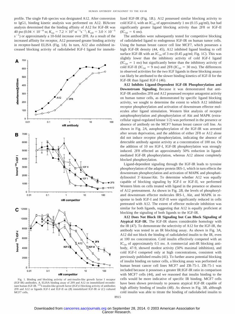
profile. The single Fab species was designated A12. After conversionto IgG1, binding kinetic analysis was performed on A12. BIAcoreanalysis determined that the binding affinity of A12 for IGF-IR was40 pM (0.04 � 10�9
M; Kon � 7.2 � 105M
�1s�1; Koff � 3.0 � 10�5
s�1) or approximately a 10-fold increase over 2F8. As a result of theincreased affinity for receptor, A12 possessed greater binding activityin receptor-based ELISA (Fig. 1A). In turn, A12 also exhibited in-creased blocking activity of radiolabeled IGF-I ligand for immobi-
lized IGF-IR (Fig. 1B.). A12 possessed similar blocking activity tocold IGF-I, with an IC50 of approximately 1 nM (0.15 �g/ml), but hadsignificantly greater ligand blocking activity than 2F8 or IGF-II(IC50 � 6 nM).
The antibodies were subsequently tested for competitive blockingof radiolabeled ligand to endogenous IGF-IR on human tumor cells.Using the human breast cancer cell line MCF7, which possesses ahigh IGF-IR density (44, 45), A12 inhibited ligand binding to cellsurface IGF-IR with an IC50 of 3 nM (0.45 �g/ml; Fig. 1C). This wasslightly lower than the inhibitory activity of cold IGF-I ligand(IC50 � 1 nM) but significantly better than the inhibitory activity ofcold IGF-II (IC50 � 9 nM) and 2F8 (IC50 � 30 nM). The differencesin observed activities for the two IGF ligands in these blocking assayscan likely be attributed to the slower binding kinetics of IGF-II for theIGF-IR than ligand IGF-I (46).
A12 Inhibits Ligand-Dependent IGF-IR Phosphorylation andDownstream Signaling. Because it was demonstrated that anti-IGF-IR antibodies 2F8 and A12 possessed receptor antagonist activityon human tumor cells, as demonstrated by specific ligand blockingactivity, we sought to determine the extent to which A12 inhibitedreceptor phosphorylation and activation of downstream effector mol-ecules after ligand stimulation. Western blot analysis of receptorautophosphorylation and phosphorylation of Akt and MAPK (extra-cellular signal-regulated kinase 1/2) was performed in the presence orabsence of antibody on the MCF7 human breast cancer cell line. Asshown in Fig. 2A, autophosphorylation of the IGF-IR was arrestedafter serum deprivation, and the addition of either 2F8 or A12 alonedid not induce receptor phosphorylation, indicating the absence ofdetectable antibody agonist activity at a concentration of 100 nM. Onthe addition of 10 nM IGF-I, IGF-IR phosphorylation was stronglyinduced. 2F8 effected an approximately 50% reduction in ligand-mediated IGF-IR phosphorylation, whereas A12 almost completelyblocked phosphorylation.
Ligand-dependent signaling through the IGF-IR leads to tyrosinephosphorylation of the adaptor protein IRS-1, which in turn effects thedownstream phosphorylation and activation of MAPK and phosphati-dylinositol 3�-kinase/Akt. To determine whether A12 was equallycapable of blocking signaling by IGF-I or IGF-II, we performedWestern blots on cells treated with ligand in the presence or absenceof A12 pretreatment. As shown in Fig. 2B, the levels of phosphoryl-ated downstream effector molecules IRS-1, Akt, and MAPK in re-sponse to both IGF-I and IGF-II were significantly reduced in cellspretreated with A12. The extent of effector molecule inhibition wassimilar for both ligands, suggesting that A12 is equally proficient atblocking the signaling of both ligands to the IGF-IR.
A12 Does Not Block IR Signaling but Can Block Signaling ofAtypical IGF-IR. The IGF-IR shares considerable homology withthe IR (47). To demonstrate the selectivity of A12 for the IGF-IR, theantibody was tested in an IR blocking assay. As shown in Fig. 3A,A12 did not block the binding of radiolabeled insulin to the IR, evenat 100 nM concentration. Cold insulin effectively competed with anIC50 of approximately 0.5 nM. A commercial anti-IR blocking anti-body, 47-9, showed modest activity (50% maximal inhibition), andcold IGF-I competed only at high concentrations, consistent withpreviously published results (41). To further assess potential blockingof insulin binding on tumor cells, a blocking assay was performed onhuman breast cancer cell lines MCF7 and ZR-75-1. ZR-75-1 wasincluded because it possesses a greater IR:IGF-IR ratio in comparisonwith MCF7 cells (44), and we reasoned that insulin binding to thecells would be more indicative of specific IR binding. MCF7 cellshave been shown previously to possess atypical IGF-IR capable ofhigh affinity binding of insulin (48). As shown in Fig. 3B, althoughcold insulin was able to titrate the binding of radiolabeled insulin to
Fig. 1. Binding and blocking activity of anti-insulin-like growth factor I receptor(IGF-IR) antibodies. A, ELISA binding assay of 2F8 and A12 on immobilized recombi-nant human IGF-IR. 125I-insulin-like growth factor (IGF)-I blocking activity of antibodies2F8 and A12 or ligands IGF-I and IGF-II on (B) immobilized IGF-IR or (C) culturedMCF7 cells.
8915
HUMAN ANTIBODY ANTAGONIST TO THE IGF-IR
Cancer Research. on September 28, 2014. © 2003 American Association forcancerres.aacrjournals.org Downloaded from
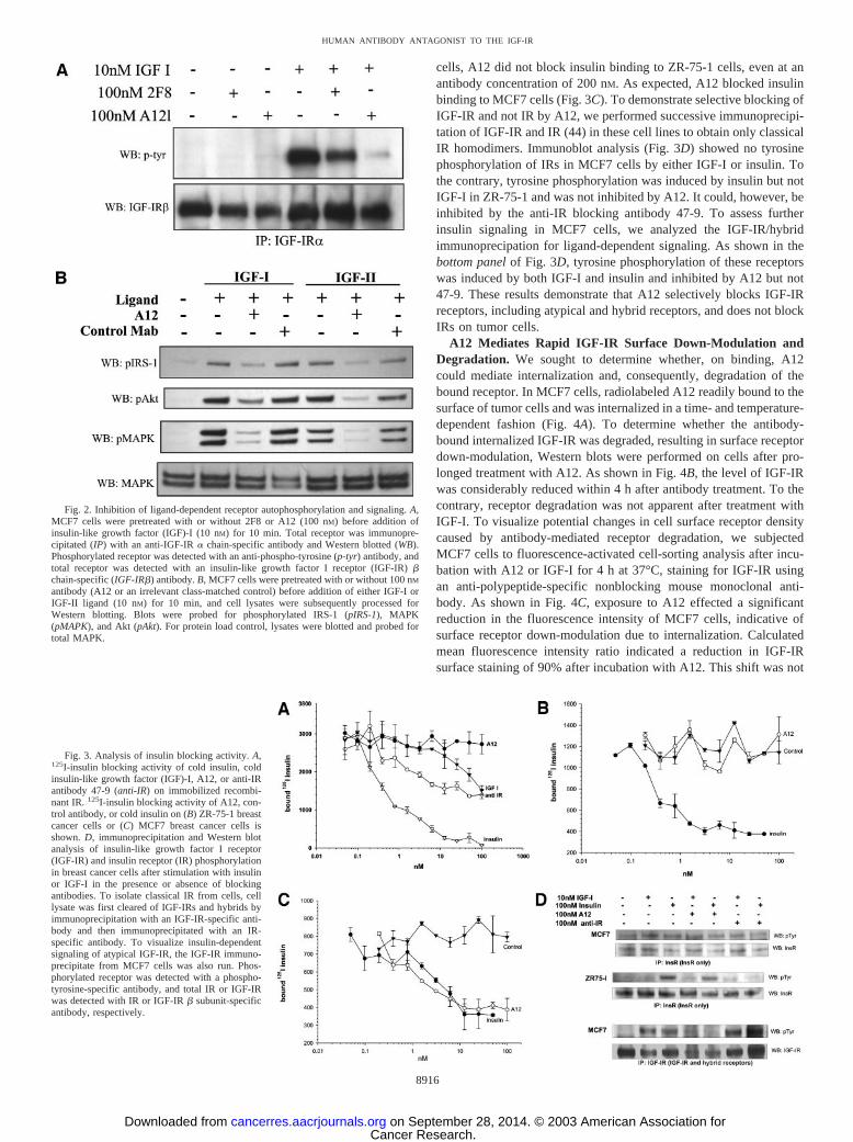
cells, A12 did not block insulin binding to ZR-75-1 cells, even at anantibody concentration of 200 nM. As expected, A12 blocked insulinbinding to MCF7 cells (Fig. 3C). To demonstrate selective blocking ofIGF-IR and not IR by A12, we performed successive immunoprecipi-tation of IGF-IR and IR (44) in these cell lines to obtain only classicalIR homodimers. Immunoblot analysis (Fig. 3D) showed no tyrosinephosphorylation of IRs in MCF7 cells by either IGF-I or insulin. Tothe contrary, tyrosine phosphorylation was induced by insulin but notIGF-I in ZR-75-1 and was not inhibited by A12. It could, however, beinhibited by the anti-IR blocking antibody 47-9. To assess furtherinsulin signaling in MCF7 cells, we analyzed the IGF-IR/hybridimmunoprecipation for ligand-dependent signaling. As shown in thebottom panel of Fig. 3D, tyrosine phosphorylation of these receptorswas induced by both IGF-I and insulin and inhibited by A12 but not47-9. These results demonstrate that A12 selectively blocks IGF-IRreceptors, including atypical and hybrid receptors, and does not blockIRs on tumor cells.
A12 Mediates Rapid IGF-IR Surface Down-Modulation andDegradation. We sought to determine whether, on binding, A12could mediate internalization and, consequently, degradation of thebound receptor. In MCF7 cells, radiolabeled A12 readily bound to thesurface of tumor cells and was internalized in a time- and temperature-dependent fashion (Fig. 4A). To determine whether the antibody-bound internalized IGF-IR was degraded, resulting in surface receptordown-modulation, Western blots were performed on cells after pro-longed treatment with A12. As shown in Fig. 4B, the level of IGF-IRwas considerably reduced within 4 h after antibody treatment. To thecontrary, receptor degradation was not apparent after treatment withIGF-I. To visualize potential changes in cell surface receptor densitycaused by antibody-mediated receptor degradation, we subjectedMCF7 cells to fluorescence-activated cell-sorting analysis after incu-bation with A12 or IGF-I for 4 h at 37°C, staining for IGF-IR usingan anti-polypeptide-specific nonblocking mouse monoclonal anti-body. As shown in Fig. 4C, exposure to A12 effected a significantreduction in the fluorescence intensity of MCF7 cells, indicative ofsurface receptor down-modulation due to internalization. Calculatedmean fluorescence intensity ratio indicated a reduction in IGF-IRsurface staining of 90% after incubation with A12. This shift was not
Fig. 2. Inhibition of ligand-dependent receptor autophosphorylation and signaling. A,MCF7 cells were pretreated with or without 2F8 or A12 (100 nM) before addition ofinsulin-like growth factor (IGF)-I (10 nM) for 10 min. Total receptor was immunopre-cipitated (IP) with an anti-IGF-IR � chain-specific antibody and Western blotted (WB).Phosphorylated receptor was detected with an anti-phospho-tyrosine (p-tyr) antibody, andtotal receptor was detected with an insulin-like growth factor I receptor (IGF-IR) �chain-specific (IGF-IR�) antibody. B, MCF7 cells were pretreated with or without 100 nM
antibody (A12 or an irrelevant class-matched control) before addition of either IGF-I orIGF-II ligand (10 nM) for 10 min, and cell lysates were subsequently processed forWestern blotting. Blots were probed for phosphorylated IRS-1 (pIRS-1), MAPK(pMAPK), and Akt (pAkt). For protein load control, lysates were blotted and probed fortotal MAPK.
Fig. 3. Analysis of insulin blocking activity. A,125I-insulin blocking activity of cold insulin, coldinsulin-like growth factor (IGF)-I, A12, or anti-IRantibody 47-9 (anti-IR) on immobilized recombi-nant IR. 125I-insulin blocking activity of A12, con-trol antibody, or cold insulin on (B) ZR-75-1 breastcancer cells or (C) MCF7 breast cancer cells isshown. D, immunoprecipitation and Western blotanalysis of insulin-like growth factor I receptor(IGF-IR) and insulin receptor (IR) phosphorylationin breast cancer cells after stimulation with insulinor IGF-I in the presence or absence of blockingantibodies. To isolate classical IR from cells, celllysate was first cleared of IGF-IRs and hybrids byimmunoprecipitation with an IGF-IR-specific anti-body and then immunoprecipitated with an IR-specific antibody. To visualize insulin-dependentsignaling of atypical IGF-IR, the IGF-IR immuno-precipitate from MCF7 cells was also run. Phos-phorylated receptor was detected with a phospho-tyrosine-specific antibody, and total IR or IGF-IRwas detected with IR or IGF-IR � subunit-specificantibody, respectively.
8916
HUMAN ANTIBODY ANTAGONIST TO THE IGF-IR
Cancer Research. on September 28, 2014. © 2003 American Association forcancerres.aacrjournals.org Downloaded from
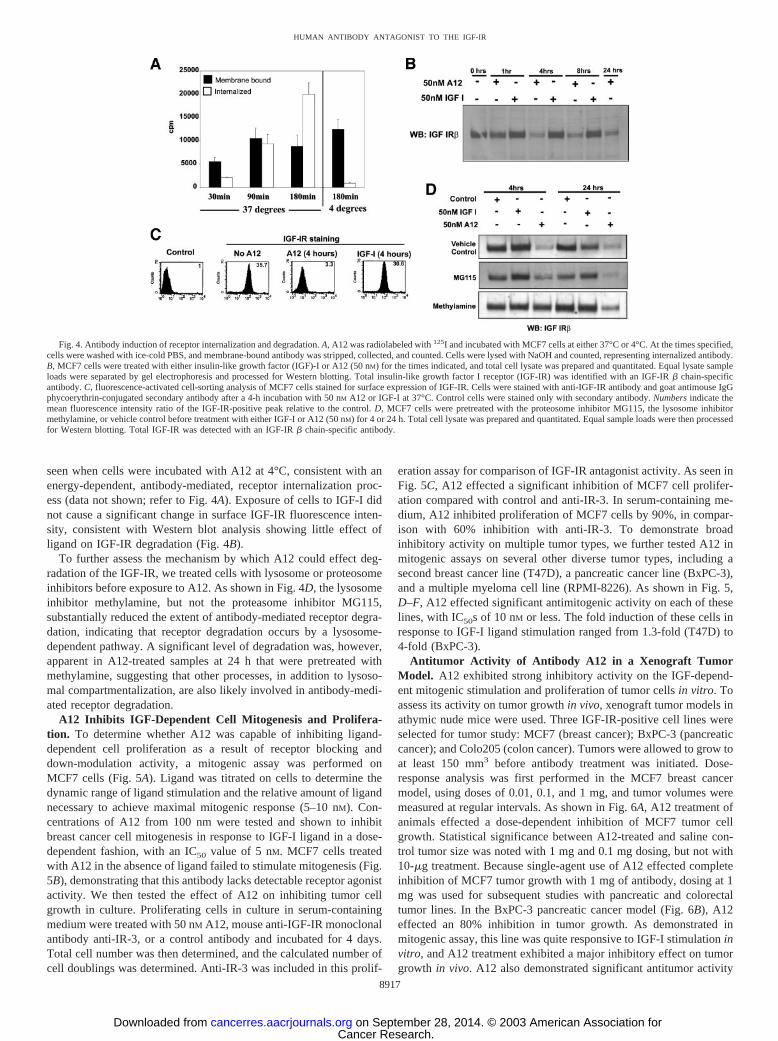
seen when cells were incubated with A12 at 4°C, consistent with anenergy-dependent, antibody-mediated, receptor internalization proc-ess (data not shown; refer to Fig. 4A). Exposure of cells to IGF-I didnot cause a significant change in surface IGF-IR fluorescence inten-sity, consistent with Western blot analysis showing little effect ofligand on IGF-IR degradation (Fig. 4B).
To further assess the mechanism by which A12 could effect deg-radation of the IGF-IR, we treated cells with lysosome or proteosomeinhibitors before exposure to A12. As shown in Fig. 4D, the lysosomeinhibitor methylamine, but not the proteasome inhibitor MG115,substantially reduced the extent of antibody-mediated receptor degra-dation, indicating that receptor degradation occurs by a lysosome-dependent pathway. A significant level of degradation was, however,apparent in A12-treated samples at 24 h that were pretreated withmethylamine, suggesting that other processes, in addition to lysoso-mal compartmentalization, are also likely involved in antibody-medi-ated receptor degradation.
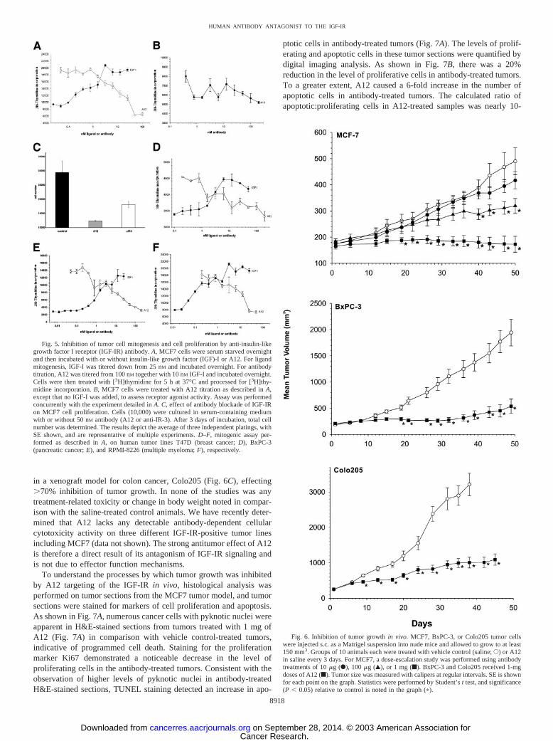
A12 Inhibits IGF-Dependent Cell Mitogenesis and Prolifera-tion. To determine whether A12 was capable of inhibiting ligand-dependent cell proliferation as a result of receptor blocking anddown-modulation activity, a mitogenic assay was performed onMCF7 cells (Fig. 5A). Ligand was titrated on cells to determine thedynamic range of ligand stimulation and the relative amount of ligandnecessary to achieve maximal mitogenic response (5–10 nM). Con-centrations of A12 from 100 nm were tested and shown to inhibitbreast cancer cell mitogenesis in response to IGF-I ligand in a dose-dependent fashion, with an IC50 value of 5 nM. MCF7 cells treatedwith A12 in the absence of ligand failed to stimulate mitogenesis (Fig.5B), demonstrating that this antibody lacks detectable receptor agonistactivity. We then tested the effect of A12 on inhibiting tumor cellgrowth in culture. Proliferating cells in culture in serum-containingmedium were treated with 50 nM A12, mouse anti-IGF-IR monoclonalantibody anti-IR-3, or a control antibody and incubated for 4 days.Total cell number was then determined, and the calculated number ofcell doublings was determined. Anti-IR-3 was included in this prolif-
eration assay for comparison of IGF-IR antagonist activity. As seen inFig. 5C, A12 effected a significant inhibition of MCF7 cell prolifer-ation compared with control and anti-IR-3. In serum-containing me-dium, A12 inhibited proliferation of MCF7 cells by 90%, in compar-ison with 60% inhibition with anti-IR-3. To demonstrate broadinhibitory activity on multiple tumor types, we further tested A12 inmitogenic assays on several other diverse tumor types, including asecond breast cancer line (T47D), a pancreatic cancer line (BxPC-3),and a multiple myeloma cell line (RPMI-8226). As shown in Fig. 5,D–F, A12 effected significant antimitogenic activity on each of theselines, with IC50s of 10 nM or less. The fold induction of these cells inresponse to IGF-I ligand stimulation ranged from 1.3-fold (T47D) to4-fold (BxPC-3).
Antitumor Activity of Antibody A12 in a Xenograft TumorModel. A12 exhibited strong inhibitory activity on the IGF-depend-ent mitogenic stimulation and proliferation of tumor cells in vitro. Toassess its activity on tumor growth in vivo, xenograft tumor models inathymic nude mice were used. Three IGF-IR-positive cell lines wereselected for tumor study: MCF7 (breast cancer); BxPC-3 (pancreaticcancer); and Colo205 (colon cancer). Tumors were allowed to grow toat least 150 mm3 before antibody treatment was initiated. Dose-response analysis was first performed in the MCF7 breast cancermodel, using doses of 0.01, 0.1, and 1 mg, and tumor volumes weremeasured at regular intervals. As shown in Fig. 6A, A12 treatment ofanimals effected a dose-dependent inhibition of MCF7 tumor cellgrowth. Statistical significance between A12-treated and saline con-trol tumor size was noted with 1 mg and 0.1 mg dosing, but not with10-�g treatment. Because single-agent use of A12 effected completeinhibition of MCF7 tumor growth with 1 mg of antibody, dosing at 1mg was used for subsequent studies with pancreatic and colorectaltumor lines. In the BxPC-3 pancreatic cancer model (Fig. 6B), A12effected an 80% inhibition in tumor growth. As demonstrated inmitogenic assay, this line was quite responsive to IGF-I stimulation invitro, and A12 treatment exhibited a major inhibitory effect on tumorgrowth in vivo. A12 also demonstrated significant antitumor activity
Fig. 4. Antibody induction of receptor internalization and degradation. A, A12 was radiolabeled with 125I and incubated with MCF7 cells at either 37°C or 4°C. At the times specified,cells were washed with ice-cold PBS, and membrane-bound antibody was stripped, collected, and counted. Cells were lysed with NaOH and counted, representing internalized antibody.B, MCF7 cells were treated with either insulin-like growth factor (IGF)-I or A12 (50 nM) for the times indicated, and total cell lysate was prepared and quantitated. Equal lysate sampleloads were separated by gel electrophoresis and processed for Western blotting. Total insulin-like growth factor I receptor (IGF-IR) was identified with an IGF-IR � chain-specificantibody. C, fluorescence-activated cell-sorting analysis of MCF7 cells stained for surface expression of IGF-IR. Cells were stained with anti-IGF-IR antibody and goat antimouse IgGphycoerythrin-conjugated secondary antibody after a 4-h incubation with 50 nM A12 or IGF-I at 37°C. Control cells were stained only with secondary antibody. Numbers indicate themean fluorescence intensity ratio of the IGF-IR-positive peak relative to the control. D, MCF7 cells were pretreated with the proteosome inhibitor MG115, the lysosome inhibitormethylamine, or vehicle control before treatment with either IGF-I or A12 (50 nM) for 4 or 24 h. Total cell lysate was prepared and quantitated. Equal sample loads were then processedfor Western blotting. Total IGF-IR was detected with an IGF-IR � chain-specific antibody.
8917
HUMAN ANTIBODY ANTAGONIST TO THE IGF-IR
Cancer Research. on September 28, 2014. © 2003 American Association forcancerres.aacrjournals.org Downloaded from
in a xenograft model for colon cancer, Colo205 (Fig. 6C), effecting�70% inhibition of tumor growth. In none of the studies was anytreatment-related toxicity or change in body weight noted in compar-ison with the saline-treated control animals. We have recently deter-mined that A12 lacks any detectable antibody-dependent cellularcytotoxicity activity on three different IGF-IR-positive tumor linesincluding MCF7 (data not shown). The strong antitumor effect of A12is therefore a direct result of its antagonism of IGF-IR signaling andis not due to effector function mechanisms.
To understand the processes by which tumor growth was inhibitedby A12 targeting of the IGF-IR in vivo, histological analysis wasperformed on tumor sections from the MCF7 tumor model, and tumorsections were stained for markers of cell proliferation and apoptosis.As shown in Fig. 7A, numerous cancer cells with pyknotic nuclei wereapparent in H&E-stained sections from tumors treated with 1 mg ofA12 (Fig. 7A) in comparison with vehicle control-treated tumors,indicative of programmed cell death. Staining for the proliferationmarker Ki67 demonstrated a noticeable decrease in the level ofproliferating cells in the antibody-treated tumors. Consistent with theobservation of higher levels of pyknotic nuclei in antibody-treatedH&E-stained sections, TUNEL staining detected an increase in apo-
ptotic cells in antibody-treated tumors (Fig. 7A). The levels of prolif-erating and apoptotic cells in these tumor sections were quantified bydigital imaging analysis. As shown in Fig. 7B, there was a 20%reduction in the level of proliferative cells in antibody-treated tumors.To a greater extent, A12 caused a 6-fold increase in the number ofapoptotic cells in antibody-treated tumors. The calculated ratio ofapoptotic:proliferating cells in A12-treated samples was nearly 10-
Fig. 5. Inhibition of tumor cell mitogenesis and cell proliferation by anti-insulin-likegrowth factor I receptor (IGF-IR) antibody. A, MCF7 cells were serum starved overnightand then incubated with or without insulin-like growth factor (IGF)-I or A12. For ligandmitogenesis, IGF-I was titered down from 25 nM and incubated overnight. For antibodytitration, A12 was titered from 100 nM together with 10 nM IGF-I and incubated overnight.Cells were then treated with [3H]thymidine for 5 h at 37°C and processed for [3H]thy-midine incorporation. B, MCF7 cells were treated with A12 titration as described in A,except that no IGF-I was added, to assess receptor agonist activity. Assay was performedconcurrently with the experiment detailed in A. C, effect of antibody blockade of IGF-IRon MCF7 cell proliferation. Cells (10,000) were cultured in serum-containing mediumwith or without 50 nM antibody (A12 or anti-IR-3). After 3 days of incubation, total cellnumber was determined. The results depict the average of three independent platings, withSE shown, and are representative of multiple experiments. D–F, mitogenic assay per-formed as described in A, on human tumor lines T47D (breast cancer; D), BxPC-3(pancreatic cancer; E), and RPMI-8226 (multiple myeloma; F), respectively.
Fig. 6. Inhibition of tumor growth in vivo. MCF7, BxPC-3, or Colo205 tumor cellswere injected s.c. as a Matrigel suspension into nude mice and allowed to grow to at least150 mm3. Groups of 10 animals each were treated with vehicle control (saline; E) or A12in saline every 3 days. For MCF7, a dose-escalation study was performed using antibodytreatments of 10 �g (F), 100 �g (Œ), or 1 mg (f). BxPC-3 and Colo205 received 1-mgdoses of A12 (f). Tumor size was measured with calipers at regular intervals. SE is shownfor each point on the graph. Statistics were performed by Student’s t test, and significance(P � 0.05) relative to control is noted in the graph (�).
8918
HUMAN ANTIBODY ANTAGONIST TO THE IGF-IR
Cancer Research. on September 28, 2014. © 2003 American Association forcancerres.aacrjournals.org Downloaded from
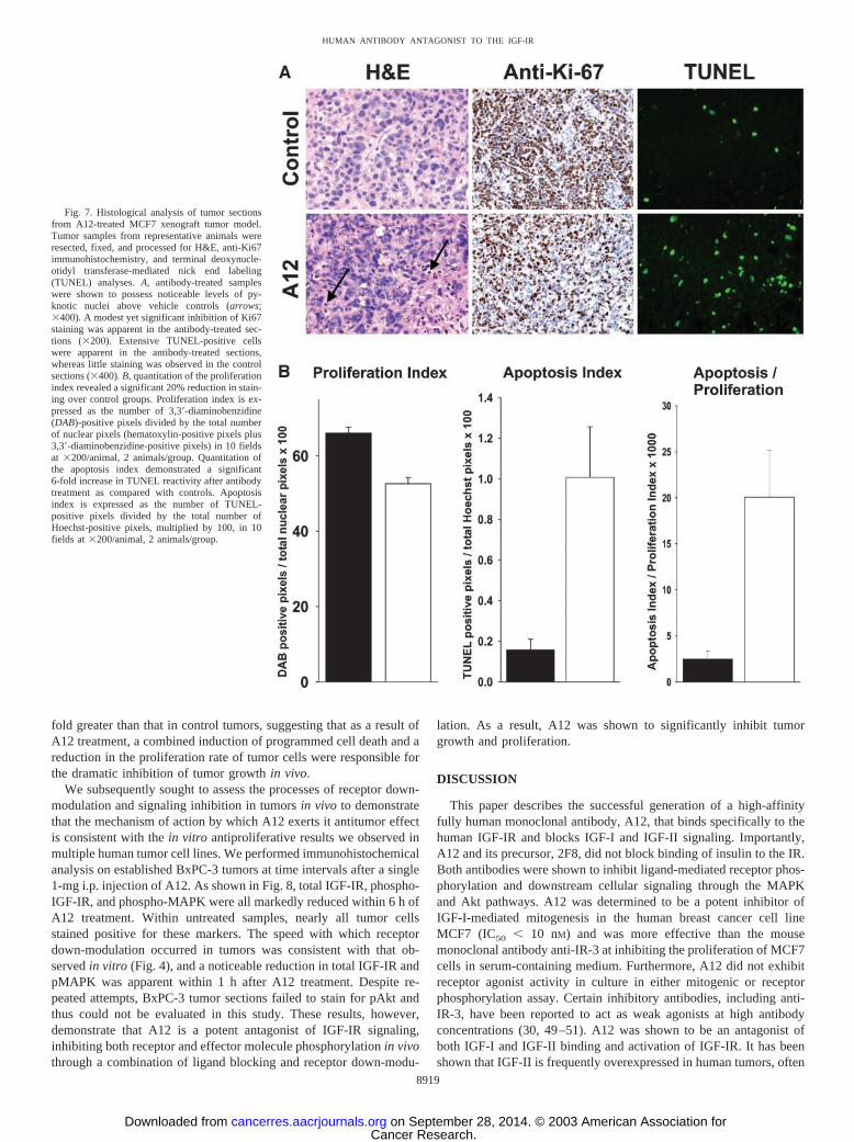
fold greater than that in control tumors, suggesting that as a result ofA12 treatment, a combined induction of programmed cell death and areduction in the proliferation rate of tumor cells were responsible forthe dramatic inhibition of tumor growth in vivo.
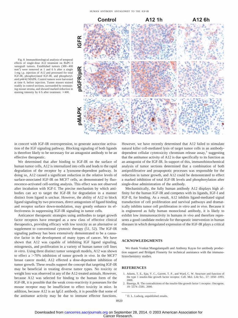
We subsequently sought to assess the processes of receptor down-modulation and signaling inhibition in tumors in vivo to demonstratethat the mechanism of action by which A12 exerts it antitumor effectis consistent with the in vitro antiproliferative results we observed inmultiple human tumor cell lines. We performed immunohistochemicalanalysis on established BxPC-3 tumors at time intervals after a single1-mg i.p. injection of A12. As shown in Fig. 8, total IGF-IR, phospho-IGF-IR, and phospho-MAPK were all markedly reduced within 6 h ofA12 treatment. Within untreated samples, nearly all tumor cellsstained positive for these markers. The speed with which receptordown-modulation occurred in tumors was consistent with that ob-served in vitro (Fig. 4), and a noticeable reduction in total IGF-IR andpMAPK was apparent within 1 h after A12 treatment. Despite re-peated attempts, BxPC-3 tumor sections failed to stain for pAkt andthus could not be evaluated in this study. These results, however,demonstrate that A12 is a potent antagonist of IGF-IR signaling,inhibiting both receptor and effector molecule phosphorylation in vivothrough a combination of ligand blocking and receptor down-modu-
lation. As a result, A12 was shown to significantly inhibit tumorgrowth and proliferation.
DISCUSSION
This paper describes the successful generation of a high-affinityfully human monoclonal antibody, A12, that binds specifically to thehuman IGF-IR and blocks IGF-I and IGF-II signaling. Importantly,A12 and its precursor, 2F8, did not block binding of insulin to the IR.Both antibodies were shown to inhibit ligand-mediated receptor phos-phorylation and downstream cellular signaling through the MAPKand Akt pathways. A12 was determined to be a potent inhibitor ofIGF-I-mediated mitogenesis in the human breast cancer cell lineMCF7 (IC50 � 10 nM) and was more effective than the mousemonoclonal antibody anti-IR-3 at inhibiting the proliferation of MCF7cells in serum-containing medium. Furthermore, A12 did not exhibitreceptor agonist activity in culture in either mitogenic or receptorphosphorylation assay. Certain inhibitory antibodies, including anti-IR-3, have been reported to act as weak agonists at high antibodyconcentrations (30, 49–51). A12 was shown to be an antagonist ofboth IGF-I and IGF-II binding and activation of IGF-IR. It has beenshown that IGF-II is frequently overexpressed in human tumors, often
Fig. 7. Histological analysis of tumor sectionsfrom A12-treated MCF7 xenograft tumor model.Tumor samples from representative animals wereresected, fixed, and processed for H&E, anti-Ki67immunohistochemistry, and terminal deoxynucle-otidyl transferase-mediated nick end labeling(TUNEL) analyses. A, antibody-treated sampleswere shown to possess noticeable levels of py-knotic nuclei above vehicle controls (arrows;�400). A modest yet significant inhibition of Ki67staining was apparent in the antibody-treated sec-tions (�200). Extensive TUNEL-positive cellswere apparent in the antibody-treated sections,whereas little staining was observed in the controlsections (�400). B, quantitation of the proliferationindex revealed a significant 20% reduction in stain-ing over control groups. Proliferation index is ex-pressed as the number of 3,3�-diaminobenzidine(DAB)-positive pixels divided by the total numberof nuclear pixels (hematoxylin-positive pixels plus3,3�-diaminobenzidine-positive pixels) in 10 fieldsat �200/animal, 2 animals/group. Quantitation ofthe apoptosis index demonstrated a significant6-fold increase in TUNEL reactivity after antibodytreatment as compared with controls. Apoptosisindex is expressed as the number of TUNEL-positive pixels divided by the total number ofHoechst-positive pixels, multiplied by 100, in 10fields at �200/animal, 2 animals/group.
8919
HUMAN ANTIBODY ANTAGONIST TO THE IGF-IR
Cancer Research. on September 28, 2014. © 2003 American Association forcancerres.aacrjournals.org Downloaded from
in concert with IGF-IR overexpression, to generate autocrine activa-tion of the IGF signaling pathway. Blocking signaling of both ligandsis therefore likely to be necessary for an antagonist antibody to be aneffective therapeutic.
We determined that after binding to IGF-IR on the surface ofhuman tumor cells, A12 is internalized into cells and leads to the rapiddegradation of the receptor by a lysosome-dependent pathway. Indoing so, A12 caused a significant reduction in the relative levels ofsurface-associated IGF-IR on MCF7 cells, as demonstrated by fluo-rescence-activated cell-sorting analysis. This effect was not observedafter incubation with IGF-I. The precise mechanism by which anti-bodies can act to target the IGF-IR for degradation in a mannerdistinct from ligand is unclear. However, the ability of A12 to blockligand signaling by two processes, direct antagonism of ligand bindingand receptor surface down-modulation, may greatly enhance its ef-fectiveness in suppressing IGF-IR signaling in tumor cells.
Anticancer therapeutic strategies using antibodies to target growthfactor receptors have emerged as a new class of effective clinicaltherapeutics, providing efficacy with low toxicity as an alternative orsupplement to conventional cytotoxic therapy (51, 52). The IGF-IRsignaling pathway has been extensively demonstrated to be a causa-tive factor in the development of many types of cancer. We haveshown that A12 was capable of inhibiting IGF ligand signaling,mitogenesis, and proliferation in a variety of human tumor cell linesin vitro. Using three distinct tumor xenograft models, A12 was shownto effect a �70% inhibition of tumor growth in vivo. In the MCF7breast cancer model, A12 effected a dose-dependent inhibition oftumor growth. These results support the concept that targeting IGF-IRmay be beneficial in treating diverse tumor types. No toxicity orweight loss was observed in any of the A12-treated animals. However,because A12 was selected for binding to the human form of theIGF-IR, it is possible that the weak cross-reactivity it possesses for themouse receptor may be insufficient to effect toxicity in mice. Inaddition, because A12 is an IgG1 antibody, it is possible that some ofthe antitumor activity may be due to immune effector functions.
However, we have recently determined that A12 failed to stimulatenatural killer cell-mediated lysis of target tumor cells in an antibody-dependent cellular cytotoxicity chromium release assay,1 suggestingthat the antitumor activity of A12 is due specifically to its function asan antagonist of the IGF-IR. In support of this, immunohistochemicalanalysis of tumor sections determined that a combination of bothantiproliferative and proapoptotic processes was responsible for thereduction in tumor growth, and A12 could be demonstrated to effecta marked inhibition of total IGF-IR levels and phosphorylation aftersingle-dose administration of the antibody.
Mechanistically, the fully human antibody A12 displays high af-finity for the human IGF-IR and competes with its ligands, IGF-I andIGF-II, for binding. As a result, A12 inhibits ligand-mediated signaltransduction of cell proliferation and survival pathways and dramat-ically inhibits tumor cell proliferation in vitro and in vivo. Because itis engineered as fully human monoclonal antibody, it is likely toexhibit low immunoreactivity in humans in vivo and therefore repre-sents a good candidate molecule for therapeutic intervention in humandiseases in which deregulated expression of the IGF-IR plays a criticalrole.
ACKNOWLEDGMENTS
We thank Venkat Mangalampalli and Anthony Kayas for antibody produc-tion support and Bridgett Finnerty for technical assistance with the immuno-histochemistry studies.
REFERENCES
1. Adams, T. E., Epa, V. C., Garrett, T. P., and Ward, C. W. Structure and function ofthe type 1 insulin-like growth factor receptor. Cell. Mol. Life Sci., 57: 1050–1093,2000.
2. Baserga, R. The contradictions of the insulin-like growth factor 1 receptor. Oncogene,19: 5574–5581, 2000.
1 D. L. Ludwig, unpublished results.
Fig. 8. Immunohistological analysis of temporaleffects of single-dose A12 treatment on BxPC-3xenograft tumors. Established tumors (300–400mm3) were removed at 1 and 6 h after a single1-mg i.p. injection of A12 and processed for totalIGF-IR, phosphorylated IGF-IR, and phosphoryl-ated p44/42 MAPK. Control tumors were harvestedat time 0, before injection. Tumor masses stainedreadily in control sections, surrounded by nonstain-ing mouse stroma, and showed marked reduction instaining intensity by 6 h after treatment. �400.
8920
HUMAN ANTIBODY ANTAGONIST TO THE IGF-IR
Cancer Research. on September 28, 2014. © 2003 American Association forcancerres.aacrjournals.org Downloaded from
3. Nickerson, T., Chang, F., Lorimer, D., Smeekens, S. P., Sawyers, C. L., and Pollak,M. In vivo progression of LAPC-9 and LNCaP prostate cancer models to androgenindependence is associated with increased expression of insulin-like growth factor I(IGF-I) and IGF-I receptor (IGF-IR). Cancer Res., 61: 6276–6280, 2001.
4. Hellawell, G. O., Turner, G. D., Davies, D. R., Poulsom, R., Brewster, S. F., andMacaulay, V. M. Expression of the type 1 insulin-like growth factor receptor isup-regulated in primary prostate cancer and commonly persists in metastatic disease.Cancer Res., 62: 2942–2950, 2002.
5. Gooch, J. L., Van Den Berg, C. L., and Yee, D. Insulin-like growth factor (IGF)-Irescues breast cancer cells from chemotherapy-induced cell death: proliferative andanti-apoptotic effects. Breast Cancer Res. Treat., 56: 1–10, 1999.
6. Cullen, K. J., Yee, D., Sly, W. S., Perdue, J., Hampton, B., Lippman, M. E., andRosen, N. Insulin-like growth factor receptor expression and function in human breastcancer. Cancer Res., 50: 48–53, 1990.
7. Peyrat, J. P., and Bonneterre, J. Type 1 IGF receptor in human breast diseases. BreastCancer Res. Treat., 22: 59–67, 1992.
8. Lee, A. V., and Yee, D. Insulin-like growth factors and breast cancer. Biomed.Pharmacother., 49: 415–421, 1995.
9. Hassan, A. B., and Macaulay, V. M. The insulin-like growth factor system as atherapeutic target in colorectal cancer. Ann. Oncol., 13: 349–356, 2002.
10. Perer, E. S., Madan, A. K., Shurin, A., Zakris, E., Romeguera, K., Pang, Y., andBeech, D. J. Insulin-like growth factor I receptor antagonism augments response tochemoradiation therapy in colon cancer cells. J. Surg. Res., 94: 1–5, 2000.
11. Wu, Y., Yakar, S., Zhao, L., Hennighausen, L., and LeRoith, D. Circulating insulin-like growth factor-I levels regulate colon cancer growth and metastasis. Cancer Res.,62: 1030–1035, 2002.
12. Ge, N-L., and Rudikoff, S. Insulin-like growth factor I is a dual effector of multiplemyeloma cell growth. Blood, 96: 2856–2861, 2000.
13. All-Ericsson, C., Girnita, L., Seregard, S., Bartolazzi, A., Jager, M. J., and Larsson,O. Insulin-like growth factor-1 receptor in uveal melanoma: a predictor for metastaticdisease and a potential therapeutic target. Investig. Ophthalmol. Vis. Sci., 43: 1–8,2002.
14. Yee, D., Morales, F. R., Hamilton, T. C., and Von Hoff, D. D. Expression ofinsulin-like growth factor I, its binding proteins, and its receptor in ovarian cancer.Cancer Res., 51: 5107–5112, 1991.
15. Quinn, K. A., Treston, A. M., Unsworth, E. J., Miller, M. J., Vos, M., Grimley, C.,Battey, J., Mulshine, J. L., and Cuttitta, F. Insulin-like growth factor expression inhuman cancer cell lines. J. Biol. Chem., 271: 11477–11483, 1996.
16. Pollak, M. Insulin-like growth factor physiology and cancer risk. Eur. J. Cancer, 36:1224–1228, 2000.
17. Yaginuma, Y., Nishiwaki, K., Kitamura, S., Hayashi, H., Sengoku, K., and Ishikawa,M. Relaxation of insulin-like growth factor-II gene imprinting in human gynecologictumors. Oncology (Basel), 54: 502–507, 1997.
18. Guerra, F. K., Eijan, A. M., Puricelli, L., Alonso, D. F., Bal de Kier Joffe, E.,Kornblihgtt, A. R., Charreau, E. H., and Elizalde, P. V. Varying patterns of expressionof insulin-like growth factors I and II and their receptors in murine mammaryadenocarcinomas of different metastasizing ability. Int. J. Cancer, 65: 812–820, 1996.
19. Blakesley, V. A., Butler, A. A., Koval, A. P., Okubo, Y., and LeRoith, D. IGF-Ireceptor function. In: R. G. Rosenfeld and C. T. Roberts, Jr. (eds.), The IGF System:Molecular Biology. Physiology, and Clinical Applications, pp. 143–163. Totowa, NJ:Humana Press, 1999.
20. Heron-Milhavet, L., Karas, M., Goldsmith, C. M., Baum, B. J., and LeRoith, D.Insulin-like growth factor-I (IGF-I) receptor activation rescues UV-damaged cellsthrough a p38 signaling pathway. Potential role of the IGF-I receptor in DNA repair.J. Biol. Chem., 276: 18185–18192, 2001.
21. Kulik, G., Klippel, A., and Weber, M. J. Antiapoptotic signalling by the insulin-likegrowth factor I receptor, phosphatidylinositol 3-kinase, and Akt. Mol. Cell. Biol., 17:1595–1606, 1997.
22. Turner, B. C., Haffty, B. G., Narayanan, L., Yuan, J., Havre, P. A., Gumbs, A. A.,Kaplan, L., Burgaud, J. L., Carter, D., Baserga, R., and Glazer, P. M. Insulin-likegrowth factor-I receptor overexpression mediates cellular radioresistance and localbreast cancer recurrence after lumpectomy and radiation. Cancer Res., 57: 3079–3083, 1997.
23. Lu, Y., Zi, X., Zhao, Y., Mascarenhas, D., and Pollak, M. Insulin-like growth factor-Ireceptor signaling and resistance to trastuzumab (Herceptin). J. Natl. Cancer Inst.(Bethesda), 93: 1852–1857, 2001.
24. Benini, S., Manara, M. C., Baldini, N., Cerisano, V., Serra, M., Mercuri, M., Lollini,P. L., Nanni, P., Picci, P., and Scotlandi, K. Inhibition of insulin-like growth factor Ireceptor increases the antitumor activity of doxorubicin and vincristine againstEwing’s sarcoma cells. Clin. Cancer Res., 7: 1790–1797, 2001.
25. Muller, M., Dietel, M., Turzynski, A., and Wiechen, K. Antisense phosphorothioateoligodeoxynucleotide down-regulation of the insulin-like growth factor I receptor inovarian cancer cells. Int. J. Cancer, 77: 567–571, 1998.
26. Pietrzkowski, Z., Wernicke, D., Porcu, P., Jameson, B. A., and Baserga, R. Inhibitionof cellular proliferation by peptide analogues of insulin-like growth factor 1. CancerRes., 52: 6447–6451, 1992.
27. D’Ambrosio, C., Ferber, A., Resnicoff, M., and Baserga, R. A soluble insulin-likegrowth factor I receptor that induces apoptosis of tumor cells in vivo and inhibitstumorigenesis. Cancer Res., 56: 4013–4020, 1996.
28. Haylor, J., Hickling, H., El Eter, E., Moir, A., Oldroyd, S., Hardisty, C., and El Nahas,A. M. JB3, an IGF-I receptor antagonist, inhibits early renal growth in diabetic anduninephrectomized rats. J. Am. Soc. Nephrol., 11: 2027–2035, 2000.
29. Scotlandi, K., Avnet, S., Benini, S., Manara, M. C., Serra, M., Cerisano, V., Perdi-chizzi, S., Lollini, P. L., De Giovanni, C., Landuzzi, L., and Picci, P. Expression of
an IGF-I receptor dominant negative mutant induces apoptosis, inhibits tumorigenesisand enhances chemosensitivity in Ewing’s sarcoma cells. Int. J. Cancer, 101: 11–16,2002.
30. Arteaga, C. L., and Osborne, C. K. Growth inhibition of human breast cancer cells invitro with an antibody against the type I somatomedin receptor. Cancer Res., 49:6237–6241, 1989.
31. Arteaga, C. L., Kitten, L. J., Coronado, E. B., Jacobs, S., Kull, F. C., Jr., Allred, D. C.,and Osborne, C. K. Blockade of the type I somatomedin receptor inhibits growth ofhuman breast cancer cells in athymic mice. J. Clin. Investig., 84: 1418–1423, 1989.
32. Scotlandi, K., Benini, S., Nanni, P., Lollini, P. L., Nicoletti, G., Landuzzi, L., Serra,M., Manara, M. C., Picci, P., and Baldini, N. Blockage of insulin-like growth factor-Ireceptor inhibits the growth of Ewing’s sarcoma in athymic mice. Cancer Res., 58:4127–4131, 1998.
33. Furlanetto, R. W., Harwell, S. E., and Baggs, R. B. Effects of insulin-like growthfactor receptor inhibition on human melanomas in culture and in athymic mice.Cancer Res., 53: 2522–2526, 1993.
34. Kalebic, T., Tsokos, M., and Helman, L. J. In vivo treatment with antibody againstIGF-1 receptor suppresses growth of human rhabdomyosarcoma and down-regulatesp34cdc2. Cancer Res., 54: 5531–5534, 1994.
35. Li, S. L., Liang, S. J., Guo, N., Wu, A. M., and Fujita-Yamaguchi, Y. Single-chainantibodies against human insulin-like growth factor I receptor: expression, purifica-tion, and effect on tumor growth. Cancer Immunol. Immunother., 49: 243–252, 2000.
36. Park, J. W., and Smolen, J. S. Monoclonal antibody therapy. In: E. M. Scolnick (ed.),Advances in Protein Chemistry, Vol. 56. Drug Discovery and Design, pp. 360–421.San Diego, CA: Academic Press, 2001.
37. de Haard, H. J., van Neer, N., Reurs, A., Hufton, S. E., Roovers, R. C., Henderikx,P., de Bruine, A. P., Arends, J. W., and Hoogenboom, H. R. A large non-immunizedhuman Fab fragment phage library that permits rapid isolation and kinetic analysis ofhigh affinity antibodies. J. Biol. Chem., 274: 18218–18230, 1999.
38. Lu, D., Jimenez, X., Zhang, H., Bohlen, P., Witte, L., and Zhu, Z. Selection of highaffinity human neutralizing antibodies to VEGFR2 from a large antibody phagedisplay library for antiangiogenesis therapy. Int. J. Cancer, 97: 393–399, 2002.
39. Prager, D., Li, H. L., Asa, S., and Melmed, S. Dominant negative inhibition oftumorigenesis in vivo by human insulin-like growth factor I receptor mutant. Proc.Natl. Acad. Sci. USA, 91: 2181–2185, 1994.
40. Casella, S. J ., Han, V. K., D’Ercole, A. J., Svoboda, M. E., and Van Wyk, J. J.Insulin-like growth factor II binding to the type I somatomedin receptor. Evidence fortwo high affinity binding sites. J. Biol. Chem., 261: 9268–9273, 1986.
41. Siddle, K., Urso, B., Niesler, C. A., Cope, D. L., Molina, L., Surinya, K. H., and Soos,M. A. Specificity in ligand binding and intracellular signalling by insulin andinsulin-like growth factor receptors. Biochem. Soc. Trans., 29: 513–525, 2001.
42. Chames, P., Willemsen, R. A., Rojas, G., Dieckmann, D., Rem, L., Schuler, G.,Bolhuis, R. L., and Hoogenboom, H. R. TCR-like human antibodies expressed onhuman CTLs mediate antibody affinity-dependent cytolytic activity. J. Immunol.,169: 1110–1118, 2002.
43. Zhu, Z., Hattori, K., Zhang, H., Jimenez, X., Ludwig, D. L., Dias, S., Kussie, P., Koo,H., Kim, H. J., Lu, D., Liu, M., Tejada, R., Friedrich, M., Bohlen, P., Witte, L., andRafii, S. Inhibition of human leukemia in an animal model with human antibodiesdirected against vascular endothelial growth factor receptor 2. Correlation betweenantibody affinity and biological activity. Leukemia (Baltimore), 17: 604–611, 2003.
44. Pandini, G., Vigneri, R., Costantino, A., Frasca, F., Ippolito, A., Fujita-Yamaguchi,Y., Siddle, K., Goldfine, I. D., and Belfiore, A. Insulin and insulin-like growthfactor-I (IGF-I) receptor overexpression in breast cancers leads to insulin/IGF-Ihybrid receptor overexpression: evidence for a second mechanism of IGF-I signaling.Clin. Cancer Res., 5: 1935–1944, 1999.
45. Hailey, J., Maxwell, E., Koukouras, K., Bishop, W. R., Pachter, J. A., and Wang, Y.Neutralizing anti-insulin-like growth factor receptor 1 antibodies inhibit receptorfunction and induce receptor degradation in tumor cells. Mol. Cancer Ther., 1:1349–1353, 2002.
46. Jansson, M., Hallen, D., Koho, H., Andersson, G., Berghard, L., Heidrich, J., Nyberg,E., Uhlen, M., Kordel, J., and Nilsson, B. Characterization of ligand binding of asoluble human insulin-like growth factor I receptor variant suggests a ligand-inducedconformational change. J. Biol. Chem., 272: 8189–8197, 1997.
47. Werner, H. Molecular biology of the type I IGF receptor. In: R. G. Rosenfeld andC. T. Roberts, Jr. (eds.), The IGF System: Molecular Biology, Physiology, andClinical Applications, pp. 63–88. Totowa, NJ: Humana Press, 1999.
48. Milazzo, G., Yip, C. C., Maddux, B. A., Vigneri, R., and Goldfine, I. D. High-affinityinsulin binding to an atypical insulin-like growth factor-I receptor in human breastcancer cells. J. Clin. Investig., 89: 899–908, 1992.
49. Kato, H., Faria, T. N., Stannard, B., Roberts, C. T., Jr., and LeRoith, D. Role oftyrosine kinase activity in signal transduction by the insulin-like growth factor-I(IGF-I) receptor. Characterization of kinase-deficient IGF-I receptors and the actionof an IGF-I-mimetic antibody (� IR-3). J. Biol. Chem., 268: 2655–2661, 1993.
50. Sachdev, D., Li, S. L., Hartell, J. S., Fujita-Yamaguchi, Y., Miller, J. S., and Yee, D.A chimeric humanized single-chain antibody against the type I insulin-like growthfactor (IGF) receptor renders breast cancer cells refractory to the mitogenic effects ofIGF-I. Cancer Res., 63: 627–635, 2003.
51. Nahta, R., Hortobagyi, G. N., and Esteva, F. J. Growth factor receptors in breastcancer: potential for therapeutic intervention. Oncologist, 8: 5–17, 2003.
52. Cohen, R. B. Epidermal growth factor receptor as a therapeutic target in colorectalcancer. Clin. Colorectal Cancer, 2: 246–251, 2003.
53. Vaughan, T. J., Osbourn, J. K., and Tempest, P. R. Human antibodies by design. Nat.Biotechnol., 16: 535–539, 1998.
8921
HUMAN ANTIBODY ANTAGONIST TO THE IGF-IR
Cancer Research. on September 28, 2014. © 2003 American Association forcancerres.aacrjournals.org Downloaded from