The Genetic Dissection of Complex Traits in a Founder Population
Upload
independentCategory
view
0download
0
OR I G INA L ART I C L E
A founder CEP120 mutation in Jeune asphyxiatingthoracic dystrophy expands the role of centriolarproteins in skeletal ciliopathiesRanad Shaheen1,†, Miriam Schmidts2,8,†, Eissa Faqeih3, Amal Hashem4,Ekkehart Lausch5, Isabel Holder2, Andrea Superti-Furga6, UK10K Consortium,Hannah M. Mitchison2, Agaadir Almoisheer1, Rana Alamro1, Tarfa Alshiddi1,Fatma Alzahrani1, Philip L. Beales2,*, and Fowzan S. Alkuraya1,7,*1Department of Genetics, King Faisal Specialist Hospital and Research Center, Riyadh 11211, Saudi Arabia,2Genetics and Genomic Medicine, UCL Institute of Child Health, 30 Guilford Street, London WC1N 1EH, UK,3Department of Pediatrics, King FahadMedical City, Riyadh 59046, Saudi Arabia, 4Department of Pediatrics, PrinceSultan Military Medical City, Riyadh 11159, Saudi Arabia, 5Pediatric Genetics Division, Center for Adolescent andPediatric Medicine, University Hospital Freiburg, Freiburg 79108, Germany, 6Department of Pediatrics, LausanneUniversity Hospital, University of Lausanne, Lausanne 1011, Switzerland, 7Department of Anatomy and CellBiology, College of Medicine, Alfaisal University, Riyadh 11533, Saudi Arabia, and 8Department of HumanGenetics, Radboud University Medical Center and Radboud Institute for Molecular Life Sciences, RadboudUniversity Nijmegen, 6500 HB Nijmegen, the Netherlands
*To whom correspondence should be addressed at: Tel: +966 11 442 7875; Fax: +966 11 442 4585; Email: [email protected] (F.S.A.) andTel: +44 2072429789; Fax: +44 2074076191; Email: [email protected] (P.B.)
AbstractJeune asphyxiating thoracic dystrophy (JATD) is a skeletal dysplasia characterized by a small thoracic cage and a range ofskeletal and extra-skeletal anomalies. JATD is genetically heterogeneouswith at least nine genes identified, all encoding ciliaryproteins, hence the classification of JATD as a skeletal ciliopathy. Consistent with the observation that the heterogeneousmolecular basis of JATD has not been fully determined yet, we have identified two consanguineous Saudi families segregatingJATD who share a single identical ancestral homozygous haplotype among the affected members. Whole-exome sequencingrevealed a single novel variant within the disease haplotype in CEP120, which encodes a core centriolar protein. Subsequenttargeted sequencing of CEP120 in Saudi and European JATD cohorts identified two additional families with the samemissensemutation. Combining the four families in linkage analysis confirmed a significant genome-wide linkage signal at the CEP120locus. This missense change alters a highly conserved amino acid within CEP120 (p.Ala199Pro). In addition, we show markedreduction of cilia and abnormal number of centrioles in fibroblasts from one affected individual. Inhibition of the CEP120ortholog in zebrafish produced pleiotropic phenotypes characteristic of cilia defects including abnormal body curvature,hydrocephalus, otolith defects and abnormal renal, head and craniofacial development. We also demonstrate that in CEP120
† These two authors have contributed equally and should be considered as co-first authors.Received: June 13, 2014. Revised: October 21, 2014. Accepted: October 27, 2014.
© The Author 2014. Published by Oxford University Press.This is an Open Access article distributed under the terms of the Creative Commons Attribution License (http://creativecommons.org/licenses/by/4.0/),which permits unrestricted reuse, distribution, and reproduction in any medium, provided the original work is properly cited.
Human Molecular Genetics, 2014, 1–10
doi: 10.1093/hmg/ddu555
Original Article
1
HMG Advance Access published November 17, 2014 at U
CL
Library Services on D
ecember 1, 2014
http://hmg.oxfordjournals.org/
Dow
nloaded from
morphants, cilia are shortened in the neural tube and disorganized in the pronephros. These results are consistent withaberrant CEP120 being implicated in the pathogenesis of JATD and expand the role of centriolar proteins in skeletal ciliopathies.
IntroductionJeune asphyxiating thoracic dystrophy (JATD [MIM208500]) is avery rare skeletal dysplasia affecting 1:100 000 live births (1).The core phenotype comprises a small or narrow thoracic cage,often leading to early lethality by virtue of cardiorespiratory in-sufficiency, and characteristic spurs (trident) of the acetabularroof. Other skeletal findings include short long bones as well aspolydactyly (2). This skeletal profile is shared by other skeletaldysplasias that are collectively known as short rib-polydactyly(SRP [MIM 208500, 611263, 613819, 615630, 266920, 615633,613091, 269860, 614091, 615503]) syndromes and are differen-tiated clinically by the degree of severity of skeletal changes aswell as the presence of extra-skeletal anomalies (2). However,the value of this distinction is questionable in view of the signifi-cant overlap in their associated clinical spectrums and, moreimportantly, in their molecular pathogenesis (see below) (3).
Both the skeletal as well as the extra-skeletal features of JATDare known to exist in a number of malformation syndromes re-ferred to as ciliopathies because the underlying pathogenesis re-volves around impaired physiology of a microtubule-basedcellular antenna known as the primary cilium (4). For example,JATD individuals can have a cerebellar hypoplasia pattern typicalof Joubert syndrome (5), abnormal retina typical of cilia-relatedretinal dystrophy (6) and cystic renal changes typical of nephro-nophthisis (7,8). These clinical observationshavebeencorroboratedover the past few years by the demonstration that JATD can becaused by mutations in genes that encode various ciliary proteins,which justifies the classification of JATD as a skeletal ciliopathy (2).
To date, nine genes have been found to be mutated in indivi-duals with JATD (Supplementary Material, Fig. S1). The most fre-quently mutated gene appears to be DYNC2H1 (MIM 603297),which encodes a component of the dynein motor involved inthe retrograde transport of cargo from the tip of the cilium toits base. Two studies have shown that DYNC2H1 is mutated in41–59% of JATD individuals (3,9). Several components of the cil-iary cargo transport system known as intraflagellar transport(IFT) were also implicated in the pathogenesis of JATD includingIFT80 (MIM 611177) (10), TTC21B/IFT139 (MIM 612014) (11), IFT140(MIM 614620) (12), WDR19/IFT144 (MIM 608151) (13), WDR34 (MIM613363) (14), WDR60 (MIM 615462) (15), and IFT172 (MIM 607386)(8). Further, mutations in CSPP1 (MIM 611654) have been recentlyidentified in individuals with Jeune Syndrome and Joubertphenotype (16,17). Importantly, several of these genes havebeen found to be mutated across the phenotypic spectrum ofSRP syndromes, lending further support to the notion that theclinical distinction is arbitrary (18,19). Despite the recent expan-sion of locus heterogeneity of JATD, it is clear that not all affectedindividuals can be explained by mutations in these genes andthat additional loci likely exist. In this report, we establish anovel locus for JATD on 5q23.2 by linkage analysis and demon-strate that a mutation in CEP120 (MIM 613446) within this locusis the most likely cause of the disease.
ResultsJATD is mapped to CEP120
The first individual (Family 1_II:2) is a female stillbirth who wasborn vaginally at 35 weeks of gestation to healthy first-cousin
Saudi parents. In addition to a severely narrow chest, apparentphysical anomalies included relative macrocephaly, hypertelor-ism, scant eyebrows, amild cleft lip (notch), short limbs and syn-polydactyly (Figs 1A–D and 2A). Radiological examinationrevealed a narrow thorax with very short and horizontal ribs, arelatively normal spine, dysplastic and small pelvic bones witha narrow sciatic notch bilaterally, round ended femur bones,small tibia and fibula, pre-axial polydactyly and extremely hypo-plastic middle and distal phalanges. She died at 5 h of age due tosevere cardiopulmonary insufficiency and autopsy was declined.The second individual (Family 2_II:4) is a male baby born tohealthy first-cousin Saudi parents who have a healthy child buthad an intrauterine fetal death with skeletal dysplasia and pre-axial polydactyly. The similarity with the index case in Family 1was striking in that there was a very narrow chest, short extrem-ities and synpolydactyly. However, he lacked cleft lip but insteadhad tongue hamartoma (lobulated tongue) and omphalocele. Hisskeletal radiological findings included small facial bones, narrowthorax with small and horizontal ribs, flaring of the iliac boneswith trident sciatic notch, short long bones of the upper andlower limbs and pre-axial polydactyly. Brainmagnetic resonanceimaging (MRI) showed cerebellar hypoplasia with Dandy-Walkermalformation and extra-axial fluid collection. Abdominal ultra-sound was normal as was electroretinography (ERG; Figs 1E–Hand 2A). He later succumbed to cardiopulmonary insufficiency.Again, the family declined autopsy.
In order to identify the underlying genetic cause, DNA fromlymphocytes was extracted from the two affected individualsas well as their parents and available healthy siblings after enrol-ling them in an institutional review board (IRB)-approved proto-col with informed consent. The two families were apparentlyunrelated sowe set out to perform single-nucleotide polymorph-ism (SNP)-based autozygome analysis in each index as describedbefore (20). However, it was later apparent that the autozygomeofthe two individuals overlapped on six chromosomal locations(Fig. 2B, Supplementary Material, Table S1), none of which isknown to harbor genes previously reported to be mutated inJATD. More importantly, only one of these overlaps representedan identical shared haplotype (Fig. 2C and SupplementaryMater-ial, Table S1) so we hypothesized that JATD in these two familiesis caused by a recessive mutation contained within thisshared founder haplotype. The critical locus (chr5:121,129,268–123,737,922) harbors 17 RefSeq genes (Fig. 2B) so we proceededwith whole-exome sequencing (WES) followed by filtering thevariants as described before (21). Briefly, we only consideredhomozygous coding/splicing variants within the 2.6 Mb criticallocus. Only one novel variant fitting these criteria was identified(NM_153223.3: c.595G>C). This variant which was confirmed bySanger sequencing and is predicted to replace a highly conservedamino acid residue in CEP120, p.Ala199Pro (Fig. 2D), is predictedto be pathogenic in silico [Polyphen = probably damaging (1),SIFT = deleterious (0.01) and GERP = 5.75] and is absent in the1000Genomes database. This allelewas observed once in thehet-erozygous state among 1294 Saudi alleles tested and twiceamong the 12 000 alleles tested in the exome variant server.The very low frequency (MAF 0.00023) is compatible with thismutation being causal of a rare phenotype like JATD with a dis-ease prevalence of less than 1:100 000. Reassuringly, no patho-genic variants were identified in any of the genes previously
2 | Human Molecular Genetics
at UC
L L
ibrary Services on Decem
ber 1, 2014http://hm
g.oxfordjournals.org/D
ownloaded from
reported to bemutated in JATD andwe achieved 100% coverage ofthe coding exons of all the genes within the critical locus byWESwith an average depth of 45× and a range of 3–131.
In order to test whether the variant we identified in CEP120is disease-causing, we sought to search for likely pathogenicvariants in this gene in independent individuals. A total of 109affected individuals of JATD/SRPS (representing Saudi and Euro-pean cohorts) were analyzed by exome sequencing, autozygositymapping and/or Sanger sequencing of CEP120. This ‘replication’cohort proved complementary in that two additional families(Family 3 and Family 4) were found to carry the sameCEP120 vari-ant described above. Reassuringly, genome-wide linkage ana-lysis combining the four families revealed a single linkage peakon 5q23.2 with logarithm of odds score of 3.6 (SupplementaryMaterial, Fig. S2).
The index individual in Family 3 (Family 3_II:1; Supplemen-tary Material, Fig. S3) is the first child of healthy unrelatedSwiss parents; however, both parents come from the same vil-lage. He was born at term after an unremarkable pregnancywith a birth weight of 2.8 kg (10th centile), body length of 45 cm(<3rd centile) and occipitofrontal head circumference of 32 cm(25th centile). At birth, thoracic dystrophy and symmetric
pre-axial hexadactyly of both feet was evident; the hands werenormal. Except for microretrognathia, no facial dysmorphismor cleft lip was noted. Respiratory insufficiency necessitatedintubation and high frequency mechanical ventilation frombirth. Because of hypoplastic lungs, oxygenation was insufficientand the clinical course was unfavorable with fatal pulmonaryfailure in the first week of life. Ultrasound scans showed de-creased size of both kidneys with increased echogenicity; theliver was unremarkable. No postmortem examination was per-formed. Haplotype analysis using intragenic SNPs between Fam-ily 1_II:2, Family 2_II:4 and Family 3_II:1 indicated that themutation resulted from a common founder individual withshared runs of homozygosity (ROH) of 714 139 bps, most likelyvery ancient consistent with the different ethnic and geographicorigin of these families (Supplementary Material, Fig. S2).
The index in Family 4 (Family 4_II:1; Supplementary Material,Fig. S3) was born via C/S section at 37 weeks of gestation due tocomplications of uncontrolled gestational diabetes mellitus.Family history is notable for first cousin parents who had fourconsecutive miscarriages and a boy with history of renal tumor.Short stature was evident with length of only 35 cm, as was thevery narrow chest and associated respiratory distress requiring
Figure 1.Clinical images of the study families. (A–D) Postnatal clinical photograph and radiological imaging of the index case fromFamily 1 showing severely narrow chest
and thorax, mild cleft lip (notch), short limbs and synpolydactyly. (E–H) Postnatal clinical photograph and radiological imaging of the index case from Family 2 showing
very narrowchest, short extremities, synpolydactyly, tonguehamartoma (lobulated tongue) and omphalocele. (I–L) Postnatal clinical photographand radiological imaging
of the individual Family 3_II:1 showing long and narrow thorax with short horizontal ribs, dysplastic pelvis with acetabular spurs and hexadactyly of the feet. (M–R)Postnatal clinical photograph, radiological imaging, 3D skull computed tomography and MRI of the index case from Family 4 showing coarse facies, midface
hypoplasia, partially bifid tongue, polydactyly, short tubular bones, bell-shaped thorax with short ribs, unilateral coronal craniosynostosis with prominent and
widened anterior and posterior fontanels, and brain vermian hypoplasia with molar-tooth appearance.
Human Molecular Genetics | 3
at UC
L L
ibrary Services on Decem
ber 1, 2014http://hm
g.oxfordjournals.org/D
ownloaded from
neonatal intensive care unit admission and ventilator support.Echocardiography revealed large patent ductus arteriosus. Hehad coarse facies, midface hypoplasia, large nose with promin-ent nostrils, partially bifid tongue, natal teeth and large earswith prominent lobules. The limbs were micromelic with pre-axial polydactyly in all extremities and hallux deformity bilat-erally. The genitalia was ambiguous with phallus-like structureand prominent prepuce. Routine karyotype demonstrated nor-mal 46;XY and abdominal and pelvic sonography showed undes-cended testicles. Endocrine workup was normal. The skeletalsurvey showed short tubular bones, bell-shaped thorax withshort ribs and acetabular bony spurs with resulting narrow sa-crosciatic notch. The iliac wings are small and squared and theacetabular roof is horizontal. A three-dimensional (3D) skullcomputed tomography revealed unilateral coronal craniosynos-tosis with prominent and widened anterior and posterior fonta-nels. Brain MRI revealed vermian hypoplasia with molar-toothappearance. The patient died on the seventh day because ofpoor lung function despite high ventilator support (Fig. 1). TheDNA of the index was not available for testing, but his parentswere found to harbor the same CEP120 mutation on the samehaplotype background as families 1–3 in a heterozygous state(Supplementary Material, Fig. S2).
CEP120 mutation causes abnormal cilium number andaberrant centrosome number
CEP120 encodes a protein that was previously identified by twoindependent functional screens. The first involved global proteo-mics analysis of purified centrosomes in which CEP120 wasfound to encode a generic coiled-coil domain containing proteindubbed CCDC100 (22). The second was a yeast-two-hybrid screenfor interactors with focal adhesion kinase (FAK), which is knownto play a role in organizing centrosome-associatedmicrotubules.This screen uncovered a 120 kD protein shown to localize to thecentrosome in the same study, so it was named centrosomal pro-tein of 120 kD (CEP120) (23). The centrosomal localization ofCEP120 was found to be resistant to microtubule destabilization,which is consistentwith it representing a ‘core’ centriolar proteinas later shown by demonstrating that it localizes to the entire‘barrel’ of the centriole but not its lumen (24). Centrosomes areformed of a pair of microtubule-basedmother and daughter cen-trioles and a surrounding matrix (pericentriolar matrix or PCM).These cellular organelles serve numerous roles that vary depend-ing on the cell cycle. In dividing cells, centrosomes assume therole of the microtubule-organizing center (MOC) in animal cellsand serve the critical function of organizing the mitotic spindle
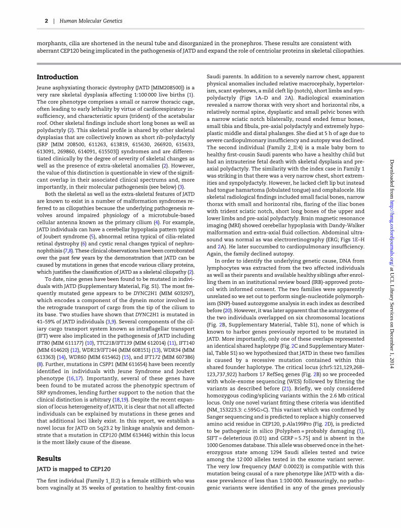
Figure 2. Identification of homozygous missense variant in CEP120 in two consanguineous Saudi families with JATD. (A) Pedigrees of the two consanguineous Saudi
families. The index is indicated in each pedigree by an arrow. (B) AgileMultiIdeogram showing the shared ROH regions of homozygosity shared between the index
cases from each of the two families (dark blue). (C) AutoSNPa showing the identical haplotype between individual II:2 in Family 1 and individual II:4 in Family 2
denoted by black lines (boxed in red lines). (D) Upper panel: sequence chromatogram of the missense mutation (control tracing is shown for comparison, and the
location of the mutation is denoted by an asterisk) and multisequence alignment orthologs of the mutation (p.Ala199) residue showing that the mutation is
conserved across species down to Danio rerio (boxed in red). Lower panel: schematic representation of CEP120 and the location of the homozygous missense
substitution identified in this study.
4 | Human Molecular Genetics
at UC
L L
ibrary Services on Decem
ber 1, 2014http://hm
g.oxfordjournals.org/D
ownloaded from

to ensure proper alignment and disjunction of chromosomes (inmitosis) and chromatids (in meiosis). In non-dividing cells, how-ever, the mother centriole assumes the role of basal bodies thatnucleate the axoneme of the budding cilium (25).
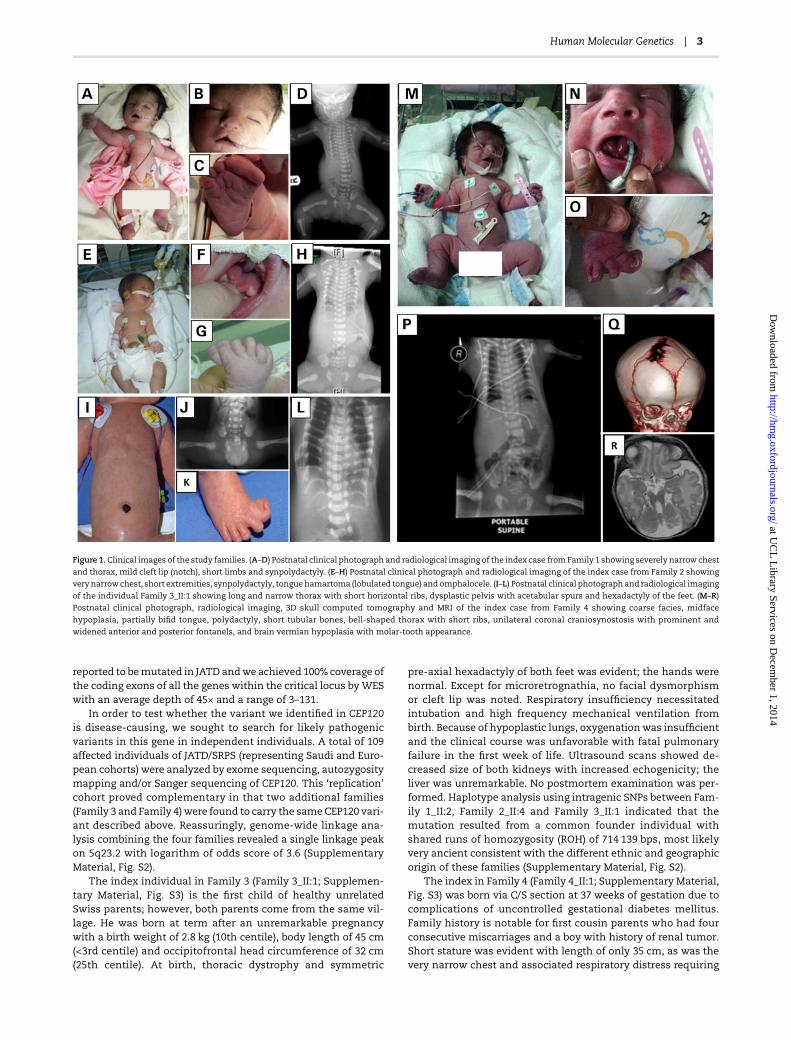
Although deficiency of CEP120 has been found to be compat-ible with centrosome formation, it has been suggested thatnormal levels of CEP120 are required for the proper elongationand division of the centrioles. Depletion of CEP120 resulted inshorter centrioles whereas overexpression was associated withcentrioles that are longer than average (26). Similarly, depletionof CEP120 has been associated with failure of centrioles to dupli-cate resulting in loss of centrioles with progressive cell divisions,and although overexpression was shown by some to result in anamplification phenotype, others failed to replicate this (24). Be-cause all JATD-related genes to date have been linked to ciliarydefects, we sought to investigate the potential effect of theJATD-associated CEP120 mutation on centrosomes and de-creased cilium number. We carried out a standard ciliogenesisassay on skin-derived fibroblasts from individual Family 1-II:2and observed a dramatic reduction in the frequency of ciliatedcells compared with control (three biological replicates, 87% ofthe cells without cilia in patient cells versus 7% in controls; P <1.977 × 10⁻⁸). The assay was based on using two axonemal mar-kers (acetylated α-tubulin and IFT88) and cilia formation wascounted based on observing protrusion of the cilia axoneme.No multiciliated cells were observed. Furthermore, in contrastto control fibroblast cells after 24 h of serum starvation to induceciliogenesis serum starvation that consistently showed a singlecentrosome, about half the cells derived from individual Family
1_II:2 showed a variable number of centrosomes ranging from 1to >4 (Fig. 3A, B, D and Supplementary Material, Figs S4 and S5).Although this result suggests that CEP120 is required for main-taining the normal number of centrioles and centrosomes, futureanalysis will be required to determine the mechanism by whichthe CEP120mutation in this study causes this cellular phenotype.Interestingly, this is very similar to the centrosomal phenotypewe previously described in another skeletal ciliopathy causedby deficiency of POC1A, another core centriolar protein (27).
Knockdown of CEP120 in zebrafish causes ciliopathyphenotypes
To confirm the role of CEP120 in vivo, we next performed knock-down of the zebrafish (ZF) homolog (CABZ01084007.1–201,ENSDART00000122378) using an antisense morpholino oligo-nucleotide targeting the inton1–exon2 splice site resulted in atypical ciliopathy phenotype with ventrally curved body axis,hydrocephalus, otolith defects, heart edema and smaller eyesat 3 days post-fertilization compared with control morpholino-injected embryos (Fig. 4A–G; Supplementary Material, Fig. S6A).Embryos that survived to post-fertilization day 4 developed pro-nephric duct dilatation and glomerular cysts and severe generaledema (Supplementary Material, Fig. S6B and C), indicating thatthe expanded pronephros observed at 24 h post-fertilization islikely a result of cilia dysfunction and/or cilia disorganization inthe pronephros.
Alcian-Blue staining revealed striking craniofacial defectswith most of the craniofacial cartilage missing (Fig. 4I and J).
Figure 3. CEP120-related ciliopathy is associated with decreased cilium number and abnormal number of centrosomes. Immunofluorescence images of serum-starved
fibroblasts from individual II:2 in Family 1 (A and B) and control fibroblasts (C) stained for pericentrin (Abcam) (red), the ciliary marker acetylated a-tubulin (Sigma-
Aldrich) (green) and DNA (blue). Compared with control, fibroblasts from II:2 in Family 1 showed a marked decrease in cilium number and variable number of
centrosomes (some cells have 3, 4 and fragmentation of more than 5). Scale bars in the main figure: 50 µm. (D) Graphs show significant reduction in the number of
fibroblast cells derived from individual II.2 from Family 1 with one centrosome (P < 0.005) and an increase in number of cells with more than one centrosome (P < 0.0005).
Human Molecular Genetics | 5
at UC
L L
ibrary Services on Decem
ber 1, 2014http://hm
g.oxfordjournals.org/D
ownloaded from

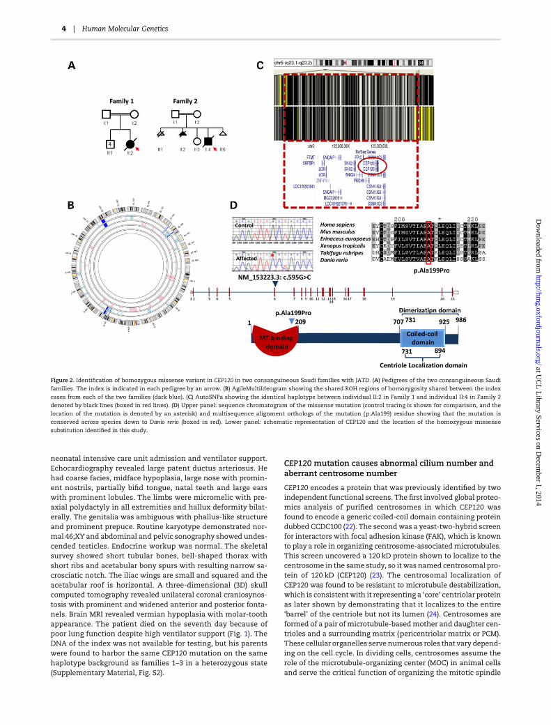
Figure 4.Knockdown CEP120 ortholog in ZF results in typical ciliopathies associated phenotypes in ZF. Injection of CEP120 antisensemorpholino oligonucleotide resulting
in centrally curved tail (D), hydrocephalus (E), otolith defects (F and G) and craniofacial defects (I and J) compared with control morpholino-injected embryos (A, B, C and
H). Visualization of cilia using anti-acetylated tubulin in CEP120-injected embryos showing disorganized cilia in the pronephros (L) and shorter cilia in the neural tube (N)
compared with controls (K and M).
6 | Human Molecular Genetics
at UC
L L
ibrary Services on Decem
ber 1, 2014http://hm
g.oxfordjournals.org/D
ownloaded from

Co-injection of p53 morpholino did not change this phenotype(data not shown). Co-injection of human CEP120 RNA resultedin partial but significant rescue of the curvature of the tail andhydrocephalus aspects of the morpholino-induced ciliopathyphenotype, however only the tail curvature rescue was still sig-nificant after Bonferroni correction. Co-injection of the mutantCEP120 RNA was weaker in rescue than WT RNA (significantlyless rescue of the tail curvature, also after Bonferroni correction)suggesting a hypomorphic nature of the mutation (Supplemen-tary Material, Figs S7 and S8).
We next performed immunofluorescence analysis of cilia inthe pronephros and neural tube at 24 h post-fertilization.Imaging using confocal microscopy revealed disorganized ciliain the pronephric duct and shorter cilia in the neural tube(Fig. 4L and N and Supplementary Material, Fig. S9). However,we observed no significant reduction in the number of cilia (Sup-plementary Material, Fig. S9).
Zebrafish embryos at 24 hpf were investigated for abnormalnumber of centrosomes by immunofluorescence and whole-mount confocal microscopy using anti-gamma tubulin antibody(GTU88, Sigma) andnuclear counterstain (28).We did not observemultiple centrosomes in the ZF morphants or cells withincreased numbers of centrioles; however, the number of cellswhich can be confidently assessed for the number of centrosomein the tail area is low and cells in other areas of the embryo suchas the brain and pronephros could not be investigated. Therefore,the presence of cells with abnormal number of centrosomescould not be ruled out. It is possible that CEP120 loss has differentconsequences in vivo versus in vitro, which may explain our fail-ure to identify cells with abnormal number of centrosomes.One can also invoke the presence of residual CEP120 protein inthe embryos due to incomplete morpholino-mediated knock-down or the maternal mRNA as another explanation.
DiscussionMost of the genes implicated in the pathogenesis of JATD areinvolved in IFT, but there are precedents for genes encoding resi-dent centriolar proteins being mutated in skeletal ciliopathiesand in JATD specifically. For example, a formof primordial dwarf-ism was found to be caused by mutations in the gene encodingthe major centriolar protein POC1A (27). Very recently, we andothers have shown that mutations in CSPP1, a gene encoding acore centriolar protein, can underlie a range of ciliopathy pheno-type including JATD (16,17).
CEP120 is conserved in the flagellated unicellular organismChlamydomonas reinhardtii and depletion of its ortholog Uni2 wasfound to eliminate formation of the flagellum (29). Subsequentanalysis has demonstrated that CEP120 is also required for ciliaformation in mouse tracheal epithelial cells (24). Consistent withthese reports, here we show that CEP120 knockdown in ZFembryos resulted in effects strongly resembling the phenotypeobserved for knockdown of other ciliary components becausethe cilia appeared disorganized in the pronephros and shorterin the neural tube confirming a role of CEP120 in ciliogenesis.However, in contrast to fibroblasts from affected individuals, thetotal number of cilia appeared normal which could be attributedto several factors as discussed above. Asmousemodels exhibitingstrong ciliogenesis defects in vivo seemtobe lethal aroundmidges-tation, it seems doubtful that the subjects described here whosurvived until term would exhibit such defects in vivo.
The primary cilium has an established role in skeletal devel-opment and abnormal cilia function has been associated withpreviously reported JATD-related genes (8,14,15,19). It seems
likely that CEP120 mutation identified in this study can cause aJATDphenotype by suppressing normal ciliary function. One pro-posed mechanism for how cilia mediate normal chondrogenesisand skeletal specification is through their role as signaling cen-ters that harbor key receptors and facilitate their cascade effectin the cell, particularly, Indian hedgehog (IHH) and Sonic Hedge-hog. IHH deficiency inmice results in a phenotype highly consist-ent with JATD, and SHH is known to act as a morphogen thatdetermines the patterning of the limbs (30,31). Hedgehog path-way receptors localize to the cilium so it is conceivable that thepathogenesis of skeletal ciliopathies, including JATD, resultsfrom impaired hedgehog signaling as a result of lack of ciliogen-esis or cilia dysfunction.
Some authors have attempted to draw clinical significancefrom genotype–phenotype correlation in JATD. For example,IFT140 and IFT172 mutations are associated with a mild thoraxphenotype but a very high frequency of cystic and nephro-nophthisis-like renal changes and childhood-onset retinaldegeneration, whereas DYNC2H1 mutations appear to be fre-quently devoid of associated extra-skeletal findings except per-haps for retinal impairment detected by ERG with no reportedclinically relevant visual impairment (3,8,9). Unfortunately, thevery small number of individuals reported here carrying causa-tive CEP120 mutations does not allow for any definitive correl-ation although it is worth highlighting that polydactyly, whichis only seen in a minority of JATD individuals, in general was ob-served in all four individuals in this study (Fig. 1). Furthermore, allaffected individuals died from respiratory failure and a severeshort rib phenotype which is not observed in individuals withIFT140, IFT172 or IFT80 mutations. Because of their short lifespan we cannot tell from the individuals in our study if loss ofCEP120 function can be associated with clinically relevantextra-skeletal disease such as renal, hepatic or retinal involve-ment; however, we observed increased renal echogenicity onpostnatal ultrasound in individual II:1 in Family 3. Finally, thefinding of hypoplastic cerebellum in the index in Family 3 andmolar tooth sign in the index in Family 4 suggests a significantoverlap with oral-facial-digital syndrome type VI and Joubertsyndrome. It will be interesting, therefore, to examine the contri-bution of CEP120, if any, to the mutation burden in these andother related ciliopathies.
ConclusionIn summary, we describe a new JATD locus which we show islinked to a single mutation in a gene encoding the core centroso-mal protein CEP120. This is the second time a core centrosomalprotein has been implicated in the pathogenesis of JATD andwe suggest that future search for candidate genes for this diseaseshould expand beyond IFT-related genes to include others thatencode proteins with other cilia-related roles.
Materials and MethodsHuman subjects
Diagnosis of JATD was made clinically and radiologically. Af-fected individuals and their available relatives were recruitedwith informed consent according to an IRB-approved protocol.
Autozygosity mapping
DNA samples from both affected and unaffected members weregenotyped on an Axiom Chip platform as per themanufacturer’s
Human Molecular Genetics | 7
at UC
L L
ibrary Services on Decem
ber 1, 2014http://hm
g.oxfordjournals.org/D
ownloaded from

protocol (Affymetrix, Santa Clara, CA, USA). ROH >2 Mb and thatspan 107 SNPs were used as surrogates of autozygosity given theconsanguineous nature of the families using AutoSNPa andAgileMultiIdeogram (20). The entire set of autozygosity blocks(autozygome) was determined for each patient and potentialoverlap between the autozygome of affected members was pur-sued. Shared ancestral haplotype was confirmed manuallyfrom the SNP files.
WES
Exome capture was performed using TruSeq Exome Enrichmentkit (Illumina) following the manufacturer’s protocol. Sampleswere prepared as an Illumina sequencing library, and in the se-cond step, the sequencing libraries were enriched for the desiredtarget using the Illumina Exome Enrichment protocol. The cap-tured libraries were sequenced using Illumina HiSeq 2000Sequencer. The reads are mapped against UCSC hg19 (http://genome.ucsc.edu/) by Burrows-Wheeler aligner (http://bio-bwa.sourceforge.net/). The SNPs and Indels are detected by SAM-TOOLS (http://samtools.sourceforge.net/).
Tissue culture, ciliogenesis and immunofluorescence
For analysis of centrioles and ciliogenesis patient (individual II:2in Family 1) and control skin fibroblasts were plated onto cover-slip, cultured in Dulbecco’s modified Eagle’s medium (DMEM)supplemented with 10% fetal bovine serum, glutamine and peni-cillin and streptomycin for 48 h. The medium was replaced withOptiMem (GIBCO)without serumand cultured for 72 h. Cellswerefixed in 3.6% paraformaldehyde (PFA) for 10 min, then permeabi-lized with 0.1% Triton X-100 (Sigma) followed by blocking in goatserum and then stained with antibodies [anti-acetylated alphatubulin (SigmaT7451), and/or anti-pericentrin (Abcam, ab28144and ab4448), CEP164 (Sdix cat 4533.00.02) and IFT88 (Proteintech13967-1-AP)]. The unbound antibody was washed with phos-phate buffer saline (PBS) and incubated with fluorescent-dye-conjugated secondary antibodies (ImmunoPure, Thermo Scien-tific). The coverslips were mounted in VectaShield containing4′,6-diamidino-2-phenylindole (DAPI) (Vector Labs) and cellswere observed under a fluorescent microscope (Nikon Eclipse90i).
Three coverslips for each of cell lines were imaged with afluorescent microscope. For frequency analysis, more than 100tubulin-stained cilia were counted from each cell line.
Zebrafish husbandry, knockdown, phenotype analysisand rescue experiments
Zebrafish husbandry was performed under standard conditionsin accordance with UK Home Office and European regulationsas previously described (32,33). Knockdown of the ZF homolog(CABZ01084007.1–201, ENSDART00000122378) was performedusing an antisense morpholino oligonucleotide targeting theinton1–exon2 splice site (cep120 MO: 5′-CTGACGACCTGAAGAAATACAACAA-3′). Morpholinos were dissolved in double dis-tillation water and injections performed at the 1–4 cell stage,using standard techniques previously described (32,34). Silencingcontrolmorpholino not targeting any known ZF genewas used tocontrol for non-specific effects due to the injection (control MO:5′-TAGTGCAAAGCTTA-3′). p53 activation-dependent effectswere excluded using 5 ng of anti-p53 morpholino (p53 MO:5′-GCGCCATTGCTTTGC-3′) (33). Morpholino effects on the tran-script level was assessed by reverse transcription. The aberrant
transcript created by the morpholino is likely degraded by non-sense medicated decay as we did not observe any band fromthe reverse transcription polymerase reaction experiment (Sup-plementary Material, Fig. S10).
A dose of 1 nl of the 0.5 m CEP120 splice site targeting mor-pholino solutionwas used for injection. Alcian-Blue staining wasperformed as previously described at 4.5 days post-fertilization(33). Rescue experiments were performed using co-injection ofhuman CEP120 RNA synthesized from CEP120 cDNA derivedfrom control fibroblasts individual II:2 in Family 1 using mMes-sagemMachine kit (T7 Polymerase, Ambion). Scoring the ZF phe-notypes was unblinded. Statistical analysis was performed usingordinary one-way ANOVA test, GraphPad Prism software.
For immunofluorescence analysis of cilia in the pronephrosand neural tube at 24 h post-fertilization, ZF embryos werefixed in 4% PFA, treated with 0.05% Triton-X100 for 30 min,blocked 1 h using 4% BSA in PBS and then incubated withmouse monoclonal anti-acetylated tubulin IgG2b, clone 6-11-b1form Sigma followed by goat anti-mouse IgG2b Alexa Fluor 488and DAPI (Molecular Probes, Invitrogen). Imaging was carriedout using a Zeiss LSM710 confocal microscope and statisticalanalysis was performed using Student’s t-test, GraphPad Prismsoftware.
Supplementary MaterialSupplementary Material is available at HMG online.
AcknowledgementsWe thank the families for their participation in the study. Wethank the Genotyping and Sequencing Core Facilities at King Fai-sal Specialist Hospital and Research Center for their technicalhelp. Special thanks toMais Hashem,Niema Ibrahimand FirdousMohammed for their help as clinical research coordinators. Weare grateful to the UK10K consortium (www.uk10k.org), in par-ticular, the Rare Diseases Group for making this study possible;a full list of the UK10K investigators is available at http://www.uk10k.org/publications_and_posters.html.
Conflict of Interest statement. The authors declare no conflict ofinterest.
FundingFunding for KFSHRC was provided by DHFMR Collaborative Re-search Grant (FSA). Funding for UK10K was provided by theWell-come Trust under award WT091310. M.S. is supported by anAction Medical Research UK Clinical Training Fellowship (RTF-1411), Radboud Excellence Fellowship and Radboud UMCHypatiaFellowship, and M.S. and P.L.B. acknowledge funding from theDutch Kidney Foundation (CP11.18). P.L.B. is aWellcomeTrust Se-nior Research Fellow and acknowledges funding from the Euro-pean Community’s Seventh Framework Programme FP7/2009under grant agreement no: 241955, SYSCILIA. P.L.B. and H.M.M.are supported by the Great Ormond Street Hospital Children’sCharity. E.L. is supported by a grant from the German Ministryfor Education and Research (BMBF, FACE consortium, agreementno 01GM1109A, TP1) and by the European Commission (SYBILconsortium, FP7 grant agreement no. 602300 and InterregIV, pro-ject A27). H.M.M. is supported by grants from the Milena CarvajalPro-Kartagener Foundation, Action Medical Research (GN2101)and Newlife Foundation for Disabled Children UK (10-11/15).G.J. is supported by a stipend from Association Française Contreles Myopathies. Funding to pay the Open Access publication
8 | Human Molecular Genetics
at UC
L L
ibrary Services on Decem
ber 1, 2014http://hm
g.oxfordjournals.org/D
ownloaded from
charges for this article was provided by the Wellcome Trust,Member of the Charity Open Access Fund (CAOF).
References1. Oberklaid, F., Danks, D., Mayne, V. and Campbell, P. (1977) As-
phyxiating thoracic dysplasia. Clinical, radiological, andpathological information on 10 patients. Arch. Dis. Child., 52,758–765.
2. Huber, C. and Cormier‐Daire, V. (2012) Ciliary disorder of theskeleton. Am. J. Med. Genet. C Semin. Med. Genet. 160, 165–174.
3. Baujat, G., Huber, C., El Hokayem, J., Caumes, R., Thanh, C.D.N., David, A., Delezoide, A.-L., Dieux-Coeslier, A., Estournet, B.and Francannet, C. (2013) Asphyxiating thoracic dysplasia:clinical and molecular review of 39 families. J. Med. Gen., 50,91–98.
4. Tobin, J.L. and Beales, P.L. (2009) The nonmotile ciliopathies.Genet. Med., 11, 386–402.
5. Lehman, A., Eydoux, P., Doherty, D., Glass, I., Chitayat, D.,Chung, B., Langlois, S., Yong, S., Lowry, R. and Hildebrandt,F. (2010) Co‐occurrence of Joubert syndrome and Jeune as-phyxiating thoracic dystrophy. Am. J. Med. Genet. A, 152,1411–1419.
6. Wilson, D.J., Weleber, R.G. and Beals, R.K. (1987) Retinal dys-trophy in Jeune’s syndrome. Arch. Ophthalmol., 105, 651–657.
7. Herdman, R.C. and Langer, L.O. (1968) The thoracic asphyxi-ant dystrophy and renal disease. Am. J. Dis. Child., 116, 192–201.
8. Halbritter, J., Bizet, A.A., Schmidts, M., Porath, J.D., Braun, D.A., Gee, H.Y., McInerney-Leo, A.M., Krug, P., Filhol, E. andDavis, E.E. (2013) Defects in the IFT-B component IFT172cause Jeune and Mainzer-Saldino Syndromes in humans.Am. J. Hum. Genet., 93, 915–925.
9. Schmidts, M., Arts, H.H., Bongers, E.M., Yap, Z., Oud, M.M.,Antony, D., Duijkers, L., Emes, R.D., Stalker, J. and Yntema,J.-B.L. (2013) Exome sequencing identifies DYNC2H1 muta-tions as a common cause of asphyxiating thoracic dystrophy(Jeune syndrome) without major polydactyly, renal or retinalinvolvement. J. Med. Gen., 50, 309–323.
10. Beales, P.L., Bland, E., Tobin, J.L., Bacchelli, C., Tuysuz, B., Hill,J., Rix, S., Pearson, C.G., Kai, M. and Hartley, J. (2007) IFT80,which encodes a conserved intraflagellar transport protein,is mutated in Jeune asphyxiating thoracic dystrophy. Nat.Genet., 39, 727–729.
11. Davis, E.E., Zhang, Q., Liu, Q., Diplas, B.H., Davey, L.M., Hart-ley, J., Stoetzel, C., Szymanska, K., Ramaswami, G. andLogan, C.V. (2011) TTC21B contributes both causal and modi-fying alleles across the ciliopathy spectrum. Nat. Genet., 43,189–196.
12. Schmidts, M., Frank, V., Eisenberger, T., Al Turki, S., Bizet, A.A., Antony, D., Rix, S., Decker, C., Bachmann, N. and Bald, M.(2013) Combined NGS approaches identify mutations in theintraflagellar transport gene IFT140 in skeletal ciliopathieswith early progressive kidney disease. Hum. Mutat., 34,714–724.
13. Bredrup, C., Saunier, S., Oud,M.M., Fiskerstrand, T., Hoischen,A., Brackman, D., Leh, S.M., Midtbø, M., Filhol, E. and Bole-Feysot, C. (2011) Ciliopathies with skeletal anomalies andrenal insufficiency due to mutations in the IFT-A geneWDR19. Am. J. Hum. Genet., 89, 634–643.
14. Schmidts, M., Vodopiutz, J., Christou-Savina, S., Cortés, C.R.,McInerney-Leo, A.M., Emes, R.D., Arts, H.H., Tüysüz, B., D’Sil-va, J. and Leo, P.J. (2013) Mutations in the gene encoding IFT
Dynein Complex Component WDR34 cause Jeune asphyxiat-ing thoracic dystrophy. Am. J. Hum. Genet., 93, 932–944.
15. McInerney-Leo, A.M., Schmidts, M., Cortés, C.R., Leo, P.J.,Gener, B., Courtney, A.D., Gardiner, B., Harris, J.A., Lu, Y.and Marshall, M. (2013) Short-rib polydactyly and Jeune syn-dromes are caused by mutations in WDR60. Am. J. Hum.Genet., 93, 515–523.
16. Shaheen, R., Shamseldin, H.E., Loucks, C.M., Seidahmed, M.Z., Ansari, S., Ibrahim Khalil, M., Al-Yacoub, N., Davis, E.E.,Mola, N.A. and Szymanska, K. (2014) Mutations in CSPP1, en-coding a core centrosomal protein, cause a range of ciliopathyphenotypes in humans. Am. J. Hum. Genet., 94, 73–79.
17. Tuz, K., Bachmann-Gagescu,R., O’Day,D.R., Hua,K., Isabella, C.R., Phelps, I.G., Stolarski, A.E., O’Roak, B.J., Dempsey, J.C. andLourenco, C. (2014) Mutations in CSPP1 cause primary cilia ab-normalities and Joubert syndrome with or without Jeune as-phyxiating thoracic dystrophy. Am. J. Hum. Genet., 94, 62–72.
18. Cavalcanti, D.P., Huber, C., Sang, K.-H.L.Q., Baujat, G., Collins,F., Delezoide, A.-L., Dagoneau, N., Le Merrer, M., Martinovic, J.and Mello, M.F.S. (2011) Mutation in IFT80 in a fetus with thephenotype of Verma-Naumoff provides molecular evidencefor Jeune-Verma-Naumoff dysplasia spectrum. J. Med. Gen.,48, 88–92.
19. Dagoneau,N., Goulet,M., Geneviève, D., Sznajer, Y.,Martinovic,J., Smithson, S.,Huber, C., Baujat,G., Flori, E. andTecco, L. (2009)DYNC2H1 mutations cause asphyxiating thoracic dystrophyand short rib-polydactyly syndrome, type III. Am. J. Hum.Genet., 84, 706–711.
20. Alkuraya, F.S. (2012) Discovery of rare homozygousmutationsfrom studies of consanguineous pedigrees. Curr. Protoc. Hum.Genet., doi: 10.1002/0471142905.hg0612s75.
21. Alkuraya, F.S. (2013) The application of next-generationsequencing in the autozygosity mapping of human recessivediseases. Hum. Genet., 132, 1197–1211.
22. Andersen, J.S., Wilkinson, C.J., Mayor, T., Mortensen, P., Nigg,E.A. and Mann, M. (2003) Proteomic characterization of thehuman centrosome by protein correlation profiling. Nature,426, 570–574.
23. Xie, Z., Moy, L.Y., Sanada, K., Zhou, Y., Buchman, J.J. and Tsai,L.-H. (2007) Cep120 and TACCs control interkinetic nuclearmigration and the neural progenitor pool. Neuron, 56, 79–93.
24. Mahjoub, M.R., Xie, Z. and Stearns, T. (2010) Cep120 is asym-metrically localized to the daughter centriole and is essentialfor centriole assembly. J. Cell. Biol., 191, 331–346.
25. Bettencourt-Dias, M. and Glover, D.M. (2007) Centrosomebiogenesis and function: centrosomics brings new under-standing. Nat. Rev. Mol. Biol., 8, 451–463.
26. Lin, Y.-N., Wu, C.-T., Lin, Y.-C., Hsu, W.-B., Tang, C.-J.C., Chang,C.-W.andTang,T.K. (2013)CEP120 interactswithCPAPandposi-tively regulates centriole elongation. J. Cell. Biol., 202, 211–219.
27. Shaheen, R., Faqeih, E., Shamseldin, H.E., Noche, R.R., Sunker,A., Alshammari, M.J., Al-Sheddi, T., Adly, N., Al-Dosari, M.S.and Megason, S.G. (2012) POC1A truncation mutation causesa ciliopathy in humans characterized by primordial dwarf-ism. Am. J. Hum. Genet., 91, 330–336.
28. Jeong,K., Jeong, J.-Y., Lee,H.-O., Choi, E. andLee,H. (2010) Inhib-ition of Plk1 induces mitotic infidelity and embryonic growthdefects in developing zebrafish embryos. Dev. Biol., 345, 34–48.
29. Piasecki, B.P., LaVoie, M., Tam, L.-W., Lefebvre, P.A. andSilflow, C.D. (2008) The Uni2 phosphoprotein is a cell cycle-regulated component of the basal body maturation pathwayin Chlamydomonas reinhardtii. Mol. Biol. Cell., 19, 262–273.
30. St-Jacques, B., Hammerschmidt, M. andMcMahon, A.P. (1999)Indian hedgehog signaling regulates proliferation and
Human Molecular Genetics | 9
at UC
L L
ibrary Services on Decem
ber 1, 2014http://hm
g.oxfordjournals.org/D
ownloaded from
differentiation of chondrocytes and is essential for bone for-mation. Genes Dev., 13, 2072–2086.
31. Anderson, E., Peluso, S., Lettice, L.A. and Hill, R.E. (2012)Human limb abnormalities caused by disruption of hedge-hog signaling. Trends Genet., 28, 364–373.
32. Mitchison, H.M., Schmidts, M., Loges, N.T., Freshour, J., Drit-soula, A., Hirst, R.A., O’Callaghan, C., Blau, H., Al Dabbagh,M. and Olbrich, H. (2012) Mutations in axonemal dynein as-sembly factor DNAAF3 cause primary ciliary dyskinesia.Nat. Genet., 44, 381–389.
33. Rooryck, C., Diaz-Font, A., Osborn, D.P., Chabchoub, E., Her-nandez-Hernandez, V., Shamseldin, H., Kenny, J., Waters,A., Jenkins, D. and Al Kaissi, A. (2011) Mutations in lectincomplement pathway genes COLEC11 and MASP1 cause3MC syndrome. Nat. Genet., 43, 197–203.
34. Krock, B.L., Mills-Henry, I. and Perkins, B.D. (2009) Retrogradeintraflagellar transport by cytoplasmic dynein-2 is requiredfor outer segment extension in vertebrate photoreceptorsbut not arrestin translocation. Invest. Ophthalmol. Vis. Sci.,50, 5463–5471.
10 | Human Molecular Genetics
at UC
L L
ibrary Services on Decem
ber 1, 2014http://hm
g.oxfordjournals.org/D
ownloaded from
Copyright © 2022 FDOKUMEN