signifying the local: media productions rendered in - CiteSeerX
Upload
independentCategory
view
2download
0
Neuron, Vol. 35, 1057–1066, September 12, 2002, Copyright 2002 by Cell Press
A Divergent Pattern of Sensory Axonal ProjectionsIs Rendered Convergent by Second-Order Neuronsin the Accessory Olfactory Bulb
bulb (MOB), where they innervate glomeruli, globosecondensations of neuropil. Within glomeruli, OSN axonssynapse with the dendrites of second-order neurons,the mitral/tufted cells, which in turn project their axonsto the primary olfactory cortex and other regions of the
Karina Del Punta,1 Adam Puche,2 Niels C. Adams,1,4
Ivan Rodriguez,1,5 and Peter Mombaerts1,3
1The Rockefeller University1230 York AvenueNew York, New York 10021
brain. Vomeronasal sensory neurons (VSNs) reside2University of Marylandwithin the epithelium of a blind capsule, the vomeronasalDepartment of Anatomy and Neurobiologyorgan (VNO), and project their axons to a specialized685 West Baltimore Streetregion of the dorsocaudal olfactory bulb, the accessoryBaltimore, Maryland 21201olfactory bulb (AOB). VSN axons in the AOB also inner-vate glomeruli, but these structures are smaller and lessdefined than MOB glomeruli. VSN axons also synapseSummarywith the apical dendrites of mitral cells, which projectto the medial amygdala and hypothalamic nuclei, thusThe mammalian vomeronasal system is specializedbypassing the primary olfactory cortex (Halpern, 1987).in pheromone detection. The neural circuitry of the
Recent molecular and genetic studies have shed lightaccessory olfactory bulb (AOB) provides an anatomi-on the logic of information coding in the olfactory sys-cal substrate for the coding of pheromone information.tem. In mammals, each OSN expresses one of �1000Here, we describe the axonal projection pattern ofodorant receptor (OR) genes (Buck and Axel, 1991; Mal-vomeronasal sensory neurons to the AOB and the den-nic et al., 1999). In contrast to their scattered distributiondritic connectivity pattern of second-order neurons.in the sensory epithelium, OSNs expressing a given ORGenetically traced sensory neurons expressing a givensend convergent axonal projections to a few spatiallygene of the V2R class of vomeronasal receptors proj-conserved glomeruli in the MOB (Ressler et al., 1994;ect their axons to six to ten glomeruli distributed inVassar et al., 1994; Mombaerts et al., 1996; Mombaerts,globally conserved areas of the AOB, a theme similar2001) (Figure 1A). A mitral cell in the MOB projects itsto V1R-expressing neurons. Surprisingly, second-ordersingle apical dendrite to a single glomerulus. This orga-neurons tend to project their dendrites to glomerulinization supports a model of olfactory coding in whichinnervated by axons of sensory neurons expressingodor quality is encoded by a specific combination ofthe same V1R or the same V2R gene. Convergence ofactivated mitral cells, each of which receives inputs fromreceptor type information in the olfactory bulb mayOSNs that express the same OR.
represent a common design in olfactory systems.Much less is known about the circuit organization of
the vomeronasal system. This sensory system exhibitsIntroduction a distinct anatomical and molecular dichotomy (Figure
1A) whose biological significance remains enigmatic.The sense of smell endows animals with the ability to Two subdivisions of VSNs are distinguished by the loca-deduce information about the chemical composition of tion of the cell body within the vomeronasal epithelium,the external world. Many mammals have two distinct the class of vomeronasal receptor expressed, the typeolfactory systems to recognize and process this infor- of G protein subunit expressed (Halpern et al., 1995;mation: the main olfactory and the vomeronasal sys- Berghard and Buck, 1996), and the site of terminationtems. The main olfactory system is responsible for de- of axons in the AOB (Jia and Halpern, 1996; Yoshihara ettecting an enormous variety of volatile airborne odorants al., 1997). VSNs whose cell bodies are situated apicallyand translating this information into cognitive and emo- coexpress the V1R class of vomeronasal receptors (VRs)tional responses (Firestein, 2001). In contrast, the vom- (Dulac and Axel, 1995; Del Punta et al., 2000; Rodriguezeronasal system is thought to be involved mainly, al- et al., 2002) with G�i2 and project their axons to the
rostral half of the AOB. In contrast, VSNs with morethough not exclusively (Sam et al., 2001), in the detectionbasally located cell bodies coexpress the V2R class ofof pheromones (Halpern, 1987; Keverne, 1999). It re-receptors (Herrada and Dulac, 1997; Matsunami andsponds to a more specific set of stimuli (Holy et al., 2000;Buck, 1997; Ryba and Tirindelli, 1997) with G�o and proj-Leinders-Zufall et al., 2000), which elicit neuroendrocineect their axons to the caudal half of the AOB. The V1Reffects and instinctive behaviors related to reproductionand V2R gene families encode seven-transmembraneand social dominance.domain receptors, which have no significant sequenceThe olfactory and vomeronasal systems have segre-homology with each other or with ORs. In the AOB, thegated anatomical organizations. Olfactory sensory neu-innervation of a single mitral cell is restricted to eitherrons (OSNs) of the main olfactory epithelium (MOE) inthe rostral or the caudal AOB (Jia and Halpern, 1997;the nasal cavity project their axons to the main olfactoryvon Campenhausen et al., 1997). The two segregatedpathways are thought to respond to different stimuli
3Correspondence: [email protected] mediate differential behaviors (Brennan et al., 1999;4Present address: Regeneron Pharmaceuticals, Inc., 777 Old SawDudley and Moss, 1999; Kumar et al., 1999; Halem etMill River Road, Tarrytown, New York 10591.al., 2001).5Present address: Department of Zoology and Animal Biology, Sci-
ences III, University of Geneva, 1211 Geneva 4, Switzerland. How is the initial event of chemoreception translated

Neuron1058
Figure 1. Genetic Approach to Visualizing V2R-Expressing Neurons
(A) Anatomical organization of axonal projections of sensory neurons of the main and vomeronasal sytems. Schematic representation of OSNsand VSNs projecting their axons to the MOB and AOB, respectively. OSNs expressing the same OR, shown as pink and green for two differentneuronal populations, project their axons to a single glomerulus in each half of a MOB. Each mitral cell in the MOB has a single apical dendrite,innervating a single glomerulus. In contrast, VSNs expressing a given V1R (orange) project their axons to multiple, smaller glomeruli in theAOB. The pattern of projections of VSNs expressing a given V2R (blue) is unknown. Mitral cells in the AOB have apical dendrites innervatingmultiple glomeruli.(Ba–Bd) Targeted mutagenesis of the V2r1b locus. (Ba) Wild-type V2r1b locus. The yellow boxes represent the coding exons. Restriction sitesare indicated for EcoRI (R) and KpnI (K). ATG is the start codon; TGA is the stop codon. (Bb) V2r1b-IRES-tauGFP-LNL targeting vector. Thewhite box labeled “i” represents the IRES sequence, the green box represents the coding sequence of tauGFP, and the gray box labeled“neo” represents the neo-selectable marker LNL flanked by loxP sites (indicated by red triangles). (Bc) V2r1b locus after homologousrecombination with the V2r1b-IRES-tauGFP-LNL targeting vector. The probe used to detect homologous recombination in Southern blots isrepresented as a small horizontal bar in the middle. (Bd) V2r1b locus after Cre-mediated excision of the neo cassette, which leaves a singleloxP site behind. This is the targeted mutation studied here.(Ca and Cb) V2r1b-expressing neurons in the VNO. (Ca) Whole-mount lateral view of the VNO. Anterior is to the left, dorsal is to the top.Neurons expressing V2r1b also express GFP and are fluorescent green. Scale bar, 500 �m. (Cb) Cross-section through the VNO. V2r1b-expressing neurons are situated in the basal layer of the epithelium. Cell bodies, dendrites, and axons can be discerned. Scale bar, 100 �m.
to create a neural representation of the stimuli in the raises the question as to how mitral cell connectivityrelates to glomerular receptor identity. Whether a mitralbrain? Analogous to OSNs, VSNs appear to express a
single V1R or V2R gene, but exceptions exist (Martini cell innervates glomeruli with the same or different re-ceptor specificities has implications for our understandinget al., 2001). VSNs expressing a particular VR have no
obvious distribution throughout the VNE. However, in of the coding of chemosensory information in the AOB.To determine the wiring logic of the AOB, we herecontrast to the convergence onto one or a few glomeruli
by OSN axons in the MOB, axons from VSNs expressing trace with a genetic technique the axonal projections ofneurons expressing a given V2R, and we describe thea given V1R innervate multiple (15–30) smaller glomeruli
in the rostral half of the AOB (Belluscio et al. 1999; dendritic connectivity pattern between mitral cells andglomeruli with the same V1R or V2R receptor specificityRodriguez et al., 1999) (Figure 1A). Glomeruli are distrib-
uted in globally conserved areas in the AOB of different (Figure 1A). First, we show that VSNs expressing theV2r1b gene project their axons to six to ten glomeruliindividuals, although their precise individual location is
variable. The pattern of axonal projections of neurons that reside in globally conserved areas within the caudalAOB. These glomeruli appear to be exclusive targets ofexpressing V2R genes to the caudal AOB has not yet
been described. axons from V2r1b-expressing neurons. Thus, based onthis example, neurons expressing V1R or V2R genesHow is this complex glomerular pattern in the AOB
processed by the mitral cells? It has been known for rely on similar strategies to organize the input of informa-tion at the glomerular level, although they constitute twonearly a century that second-order neurons in the AOB
have a qualitatively different organization than those of anatomically segregated compartments using receptorswithout significant sequence homology. Second, injec-the MOB: apical dendrites of AOB mitral cells elaborate
an extensive branching pattern such that each cell inner- tion of lipophilic tracers into individual genetically la-beled glomeruli reveals that mitral cells connect to multi-vates multiple glomeruli (Ramon y Cajal, 1911; Takami
and Graziadei, 1990, 1991). The peculiar anatomical rela- ple glomeruli innervated by neurons expressing thesame V1R or V2R. Thus, the divergent pattern of projec-tionship between glomeruli and mitral cells in the AOB

Circuitry of the Vomeronasal System1059
tions of VSNs neurons in the AOB is resolved by dendritic Pattern of V2r1b-Axonal Projections in the AOBIn the absence of any obvious distribution of V2r1b-convergence of the second-order neurons.expressing neurons at the level of the epithelium, weasked if their axons converge to glomeruli in the AOBand whether a spatial distribution of these glomeruli canResultsbe discerned.
In whole-mount specimens of V2R-GFP mice ana-Visualizing V2r1b-Expressing Sensory NeuronsThe genomic structure of the V2r1b gene, a member lyzed by confocal microscopy (n � 15 females and 15
males), V2r1b-axons converge onto six to ten glomeruliof the V2R class of vomeronasal receptors (Ryba andTirindelli, 1997), is shown in Figure 1B. The coding se- per AOB (Figures 2Aa–2Ac), a situation intermediate be-
tween that of OSN axons (two to three glomeruli) (Mom-quence spans six exons: five exons (E1 to E5) encodethe long extracellular domain, and a single exon (E6) baerts, 2001) and that of axons of VSN expressing any
of three examined V1R receptors (15–30 glomeruli) (Bel-codes for the transmembrane domain. The predictedamino acid sequence of V2r1b shares 98.4% identity luscio et al. 1999; Rodriguez et al., 1999). The V2r1b
glomeruli are invariably located in the rostral half of thewith its closest homolog, V2R1.To visualize V2r1b-expressing VSNs and their axonal caudal AOB. Figure 2Ad shows a similar analysis of the
AOB of a homozygous mouse of the VRi2-IRES-tauGFPprojections to the AOB, we generated a targeted muta-tion in the V2r1b locus that results in cotranslation of strain (Rodriguez et al., 1999), here abbreviated as V1R-
GFP. The general glomerular pattern of V2R-GFP micetauGFP along with V2r1b from a bicistronic message.An IRES-tauGFP-LNL cassette was introduced immedi- is established by postnatal day 8 and does not obviously
vary with age, gender, or sexual experience. No consis-ately after the stop codon of the V2r1b coding sequence(Figure 1B). In this well-established design (Rodriguez tent changes were observed in the number and position
of V2r1b glomeruli in sexually naıve versus experiencedet al., 1999; Zheng et al., 2000; Strotmann et al., 2000;Potter et al., 2001; Bozza et al., 2002; Treloar et al., mice (males and females) (data not shown).
To evaluate better the spatial distribution of the V2r1b2002), the fluorescent label is present in axons and axonterminals of V2r1b-expressing VSNs. V2r1b-IRES- glomeruli, we performed three-dimensional reconstruc-
tions of sagittal sections through the AOB. This type oftauGFP mice are abbreviated as V2R-GFP mice.To investigate the distribution of V2r1b-expressing analysis (Figures 2Ba–2Bc) revealed common features
among the relative depth of the V2r1b glomeruli withincells throughout the VNO, we analyzed VNO wholemounts of homozygous V2R-GFP mice by confocal mi- the glomerular layer (Figures 2Bd–2Bg) that could not
be appreciated in the whole-mount analysis. The onecroscopy. The cell bodies of V2r1b-expressing neuronsare distributed throughout the sensory epithelium of the or two most lateral glomeruli are consistently superficial
(red) and the remainder are positioned deeper, at theVNO. There is no obvious preferential distribution ofthese neurons along the anterior-posterior and dorsal- boundaries of the glomerular layer with the external
plexiform layer.ventral axes (Figure 1Ca). In coronal sections throughthe VNO, cell bodies of V2r1b-expressing neurons arefound in the basal layer of the epithelium, as expected Circuit Organization of Glomerulifor a member of the V2R family (Figure 1Cb). Labeled and Second-Order Neuronsneurons are usually found in clusters of two to five cells. Given that the major output neurons in the AOB, theNo significant difference was found between males and mitral cells, receive input from multiple glomeruli (Ra-females (data not shown). mon y Cajal, 1911; Takami and Graziadei, 1991) and
Monoallelic expression has been observed both for that VSNs expressing a given VR project to multipleOR genes (Chess et al., 1994; Strotmann et al., 2000; glomeruli, the question is raised as to how the mitral cellIshii et al., 2001) and V1R genes (Belluscio et al., 1999; connectivity in the AOB relates to glomerular specificity.Rodriguez et al., 1999). To determine if the V2R genes Three general models can be proposed, each with dif-maintain this olfaction-associated trait, we counted the ferent functional implications (Figure 3). In the firsttotal number of GFP-positive cell bodies in coronal sec- model, glomeruli are homogeneously innervated by ax-tions through the entire VNO of homozygous and hetero- ons of neurons expressing the same receptor, and azygous V2R-GFP mice. Homozygous mice have approx- given mitral cell connects to those multiple glomeruliimately twice the number of cells seen in heterozygous that have the same receptor identity (homotypic connec-mice (237 � 18 versus 432 � 49, respectively), sug- tivity). In the second model, glomeruli are also homoge-gesting that the V2r1b gene is also monoallelicaly ex- neous at the input level, but a given mitral cell projects itspressed. This genetic feature will prove useful for com- dendrites to glomeruli with different receptor identitiesparison of projection patterns in heterozygous and (heterotypic connectivity). The third model proposeshomozygous mice (see below). glomeruli heterogeneously innervated by axons of neu-
The V2R2 gene and closely related genes are coex- rons expressing different receptor types and mitral cellspressed with other V2R genes in most VSNs of the basal exhibiting a complex pattern of glomerular connectivitylayer (Martini et al., 2001). In sections of the VNO from (mixed connectivity).V2R-GFP mice, we observe colocalization of the V2R2 To evaluate these models, we first examined the ho-antigen with GFP (data not shown). Thus, neurons ex- mogeneity of input to glomeruli in the AOB. It has beenpressing V2r1b also express member(s) of the V2R2 shown that V1R glomeruli in the anterior AOB are gener-subfamily, suggesting that V2r1b is representative gene ally homogeneous at the input level (Belluscio et al.,
1999; Rodriguez et al., 1999), with a few exceptions inof the V2R repertoire.

Neuron1060
Figure 2. Patterns of Axonal Projections inthe Accessory Olfactory Bulb
(A) Whole-mount views of the left and rightAOBs.(Aa–Ac) Five-week-old homozygous V2R-GFP mice. Each AOB has six to ten glomeruli,with a globally conserved spatial distribution(Aa–Ac). (Ad) Five-week-old homozygousV1R-GFP mouse. Each AOB has 15–30 greenfluorescent glomeruli in the rostral region.Scale bar, 500 �m.(Ba–Bg) Three-dimensional reconstruction ofsagittal serial sections through the AOB. Car-toon showing the method employed to createthree-dimensional reconstructions of serialsections through the AOB (Ba–Bc). (Ba) Dor-sal view of the left and right AOBs (same viewas in Figure 2A). The six sagittal sectionsthrough the AOB are color-coded from lateralto medial: red, orange, yellow, green, blue,purple. The orientation is: r, rostral; c, caudal;l, lateral. (Bb) The six individual serial sagittalsections of the left AOB with the same colorcoding as in (Ba). Only the nerve and glomeru-lar layers are drawn. Each section is flipped90� in the horizontal plane such that to theleft is the boundary of the glomerular layerand to the right is the boundary of the nervelayer. Top is rostral, and bottom is caudal.(Bc) Illustration of how the sections are recon-structed on top of each other for the left andright AOBs. The orientation of the section isindicated as follows: r, rostral; c, caudal; n,boundary of nerve layer; gl, boundary of glo-merular layer. (Bd–Bg) Three-dimensional re-constructions showing the conservation ofglomerular distributions in the AOB. Recon-structions of the left and right AOBs of fourmice are shown.
which some glomeruli appear to be compartmentalized lus (red fluorescence) fully correspond with the greenfluorescent axons, or whether nongreen fluorescent(Belluscio et al., 1999). With a similar objective, we
stained serial sagittal sections of the AOB of homozy- axons intermingle in the glomerulus. In homozygousV2R-GFP mice, there is a high degree of concordancegous and heterozygous V2R-GFP mice with antibodies
against the presynaptic marker synapsin I (Figure 4). We between synapsin I immunofluorescence and GFP ex-pression, suggesting that only GFP-positive axons in-asked whether synaptic markers in each V2r1b glomeru-
Figure 3. Models of Connectivity in the MainOlfactory and Vomeronasal Systems
(A) In the main olfactory system, axons fromOSNs expressing the same OR converge toone (or a few) glomeruli (GL) in the MOB. Mi-tral cells (MC) send their single apical den-drite to a single glomerulus.(B) In the vomeronasal system, axons fromVSNs expressing the same VR converge tomultiple small glomeruli (GL) in the AOB. Mi-tral cells (MC) have several apical dendritesthat innervate multiple glomeruli. Three pos-sible models of connectivity between VSNsand mitral cells are proposed.
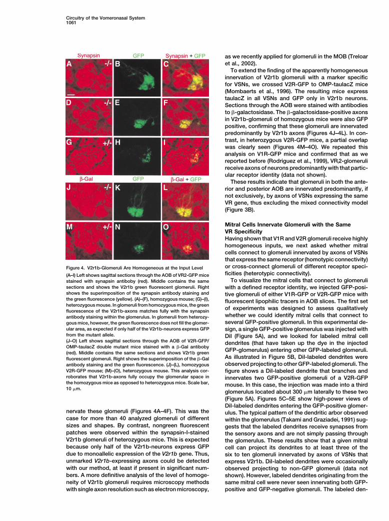
Circuitry of the Vomeronasal System1061
as we recently applied for glomeruli in the MOB (Treloaret al., 2002).
To extend the finding of the apparently homogeneousinnervation of V2r1b glomeruli with a marker specificfor VSNs, we crossed V2R-GFP to OMP-taulacZ mice(Mombaerts et al., 1996). The resulting mice expresstaulacZ in all VSNs and GFP only in V2r1b neurons.Sections through the AOB were stained with antibodiesto �-galactosidase. The �-galactosidase-positive axonsin V2r1b-glomeruli of homozygous mice were also GFPpositive, confirming that these glomeruli are innervatedpredominantly by V2r1b axons (Figures 4J–4L). In con-trast, in heterozygous V2R-GFP mice, a partial overlapwas clearly seen (Figures 4M–4O). We repeated thisanalysis on V1R-GFP mice and confirmed that as wereported before (Rodriguez et al., 1999), VRi2-glomerulireceive axons of neurons predominantly with that partic-ular receptor identity (data not shown).
These results indicate that glomeruli in both the ante-rior and posterior AOB are innervated predominantly, ifnot exclusively, by axons of VSNs expressing the sameVR gene, thus excluding the mixed connectivity model(Figure 3B).
Mitral Cells Innervate Glomeruli with the SameVR SpecificityHaving shown that V1R and V2R glomeruli receive highlyhomogeneous inputs, we next asked whether mitralcells connect to glomeruli innervated by axons of VSNsthat express the same receptor (homotypic connectivity)or cross-connect glomeruli of different receptor speci-Figure 4. V2r1b-Glomeruli Are Homogeneous at the Input Levelficities (heterotypic connectivity).(A–I) Left shows sagittal sections through the AOB of VR2-GFP mice
To visualize the mitral cells that connect to glomerulistained with synapsin antiboby (red). Middle contains the samesections and shows the V2r1b green fluorescent glomeruli. Right with a defined receptor identity, we injected GFP-posi-shows the superimposition of the synapsin antibody staining and tive glomeruli of either V1R-GFP or V2R-GFP mice withthe green fluorescence (yellow). (A)–(F), homozygous mouse; (G)–(I), fluorescent lipophilic tracers in AOB slices. The first setheterozygous mouse. In glomeruli from homozygous mice, the green
of experiments was designed to assess qualitativelyfluorescence of the V2r1b-axons matches fully with the synapsinwhether we could identify mitral cells that connect toantibody staining within the glomerulus. In glomeruli from heterozy-several GFP-positive glomeruli. In this experimental de-gous mice, however, the green fluorescence does not fill the glomer-
ular area, as expected if only half of the V2r1b-neurons express GFP sign, a single GFP-positive glomerulus was injected withfrom the mutant allele. DiI (Figure 5A), and we looked for labeled mitral cell(J–O) Left shows sagittal sections through the AOB of V2R-GFP/ dendrites (that have taken up the dye in the injectedOMP-taulacZ double mutant mice stained with a �-Gal antiboby
GFP-glomerulus) entering other GFP-labeled glomeruli.(red). Middle contains the same sections and shows V2r1b greenAs illustrated in Figure 5B, DiI-labeled dendrites werefluorescent glomeruli. Right shows the superimposition of the �-Galobserved projecting to other GFP-labeled glomeruli. Theantibody staining and the green fluorescence. (J)–(L), homozygous
V2R-GFP mouse; (M)–(O), heterozygous mouse. This analysis cor- figure shows a DiI-labeled dendrite that branches androborates that V2r1b-axons fully occupy the glomerular space in innervates two GFP-positive glomeruli of a V2R-GFPthe homozygous mice as opposed to heterozygous mice. Scale bar, mouse. In this case, the injection was made into a third10 �m.
glomerulus located about 300 �m laterally to these two(Figure 5A). Figures 5C–5E show high-power views ofDiI-labeled dendrites entering the GFP-positive glomer-
nervate these glomeruli (Figures 4A–4F). This was the ulus. The typical pattern of the dendritic arbor observedcase for more than 40 analyzed glomeruli of different within the glomerulus (Takami and Graziadei, 1991) sug-sizes and shapes. By contrast, nongreen fluorescent gests that the labeled dendrites receive synapses frompatches were observed within the synapsin-I-stained the sensory axons and are not simply passing throughV2r1b glomeruli of heterozygous mice. This is expected the glomerulus. These results show that a given mitralbecause only half of the V2r1b-neurons express GFP cell can project its dendrites to at least three of thedue to monoallelic expression of the V2r1b gene. Thus, six to ten glomeruli innervated by axons of VSNs thatunmarked V2r1b-expressing axons could be detected express V2r1b. DiI-labeled dendrites were occasionallywith our method, at least if present in significant num- observed projecting to non-GFP glomeruli (data notbers. A more definitive analysis of the level of homoge- shown). However, labeled dendrites originating from theneity of V2r1b glomeruli requires microscopy methods same mitral cell were never seen innervating both GFP-
positive and GFP-negative glomeruli. The labeled den-with single axon resolution such as electron microscopy,

Neuron1062
Figure 5. Mitral Cells Can Innervate MultipleGlomeruli with the Same VR Specificity
(A) Diagram illustrating the experimental de-sign and results. A GFP-positive glomerulusat the surface (left) of a thick slice is injectedwith DiI (red). The dye is taken up by the den-drites of mitral cells innervating that glomeru-lus and is transported to the dendrites andsoma. Labeled dendrites of a mitral cell in-nervating the injected glomeruli also projectto two other GFP-glomeruli that are approxi-mately 300 �m away from the injection site.The thick slice is subsequently cut into thin-ner sections for confocal analysis.(B) Dendritic projection from mitral cells la-beled with DiI (red) in a GFP-positive glomer-ulus approximately 300 �m away (and notvisible here). The two GFP-positive glomeruliare innervated by labeled dendrites, and thusappear yellow. This means that at least threeGFP-positive glomeruli receive dendritesfrom the same mitral cell. Scale bar, 100 �m.(C–E) High-power view of the glomerulus in(B), showing a branching labeled apical den-drite penetrating the GFP-positive glomeru-lus. (C) shows the GFP-glomerulus, (D) showsthe labeled apical dendrite, and (E) showssuperimposition of both images. Scale bar,20 �m.
drites entering a GFP-negative glomerulus may result that reside outside the slice. Second, some mitral cellsmay project their dendrite(s) to only a single glomerulus.from dye uptake by dendrites that pass through the
injected glomerulus without synapsing. Alternatively, Figures 6C and 6E show V2R-GFP and V1R-GFP glo-meruli that were injected with DiI and DiD, respectively:they may represent a population of mitral cells with some
degree of heterotypic connectivity. injections were controlled such that each was strictlyconfined to the GFP-positive glomeruli. Figures 6D andIn a second, quantitative set of experiments, pairs of
GFP-positive glomeruli from either V1R-GFP or V2R- 6F depict resulting double- and single-labeled mitralcells from an injection in an AOB slice of V2R-GFP andGFP mice were injected with lipophilic dyes with differ-
ent wavelength emissions, in tangential slices through V1R-GFP mice, respectively.Taken together, these findings suggest that mitralthe AOB. One slice per AOB was used and each slice
contained an average of three to four GFP-positive glo- cells in both rostral and caudal AOB tend to projecttheir dendrites to glomeruli that receive input from VSNsmeruli. In slices prepared from both strains of mice,
injections were made with DiI into a GFP-positive glo- expressing the same receptor, thereby reorganizing thedispersed glomerular inputs into a more convergentmerulus and DiA or DiD into a second GFP-positive
glomerulus (Figure 6A). Control experiments were per- pattern.formed in which the second injection was made intoa random, GFP-negative glomerulus at a comparable Discussiondistance. If the same mitral cell innervates both injectedglomeruli, its cell body would be labeled with both dyes The Functional Anatomy of the Olfactory System
In most mammals, two sensory systems have evolvedsimultaneously. If mitral cells preferentially project den-drites to multiple glomeruli with the same receptor iden- to mediate distinct biological responses to chemical
compounds: the main olfactory and vomeronasal sys-tity, we would expect to observe double-labeled mitralcells with dendrites projecting simultaneously to both tems. While significant progress has been made in our
knowledge of the main olfactory system, progress inGFP-positive injected glomeruli at a frequency higherthan when a GFP-positive and a random GFP-negative our understanding of the vomeronasal system has been
slow. The functional organization of the sensory neuronsglomerulus are injected. Indeed, the percentage of dou-ble-labeled mitral cells observed when two GFP-labeled and their first synaptic relay station in the vomeronasal
pathway differ from the main olfactory system in twoglomeruli were injected was significantly higher thanwhen a GFP and a non-GFP glomerulus were injected aspects: first, axons of VSNs expressing the same V1R
project to multiple glomeruli (Belluscio et al., 1999; Rod-(Figure 6B). The absolute percentage of double- versussingle-labeled mitral cells within each double-injection riguez et al., 1999), and second, dendrites of second-
order neurons innervate multiple glomeruli (Ramon yexperiment is not informative in this experimental designbecause a high number of single-labeled mitral cells is Cajal, 1911; Takami and Graziadei, 1991).
To reveal the wiring logic of the vomeronasal system,expected for at least two reasons. First, some mitralcells will innervate some of the other GFP-positive glo- we have described the pattern of axonal projections of
VSNs expressing a given V2R gene to glomeruli andmeruli that reside in the slice but were not injected or

Circuitry of the Vomeronasal System1063
analyzed the pattern of connectivity between definedsets of glomeruli and second-order output neurons inthe AOB. We show that the organization of the twopathways in the vomeronasal system follows similarprinciples, although the repertoires of putative chemo-sensory receptors employed by these pathways shareno sequence similarity. Axons of neurons expressing agiven V1R or V2R converge onto multiple glomeruli inthe AOB that receive homogeneous input from neuronsexpressing the same receptor, with some exceptions ofapparently compartmentalized glomeruli (Belluscio etal., 1999). Within each subdivision of the AOB, a givenmitral cell sends dendrites that terminate in multipleglomeruli innervated by neurons expressing the samereceptor. Thus, the functional organization of the vom-eronasal system is more convergent than originallythought. The apparent divergent axonal connectivity ofVSNs to the AOB is reorganized by the dendrites of thesecond-order neurons into a more convergent outputpattern, similar to that observed in the MOB.
Glomerular Patterns in the AOBThe patterns of glomeruli innervated by neurons ex-pressing a given V1R or V2R have both conserved andvariable features. While V1R or V2R glomeruli are distrib-uted in broadly conserved areas of the AOB, their pre-cise locations vary across individuals and between bothbulbs of the same individual.
Although we have described the expression and pro-jection patterns for only one gene of a repertoire of �140V2R genes (Matsunami and Buck, 1997), experience withboth OR genes and V1R genes has shown that the basictheme of these patterns appears to be generalizablebased on one or a few randomly chosen gene members.Nonetheless, certain characteristics vary across theneuronal populations studied, such as the dependency
bars represents experiments in which a first dye is injected into aGFP-labeled glomerulus and the second dye in a randomly chosenglomerulus, at distances to the first injected glomeruli comparableto the distances of other GFP-labeled glomeruli in the slice (n � 3slices for V1R-GFP and n � 4 slices for V2R-GFP). The total numberof labeled cells for the GFP-GFP glomerular injections was 73 forV1R-GFP and 65 for V2R-GFP; of these, 20 cells were double labeledin V2R-GFP mice (27.4%) and 16 cells in V2R-GFP mice (24.6%).
Figure 6. Mitral Cell Dendrites Tend to Project to Glomeruli With For the GFP-random glomerular injections, the total number of la-the Same VR Identity beled cells was 11 for V1R-GFP and 44 for V2R-GFP; 0 cells were(A) Diagram showing slice section plane through the AOB and the double labeled in V2R-GFP (0%) and 2 cells in V2R-GFP (4.5%).resulting slice, with (Gl) and mitral cell (M) layers. The experimental For both V1R-GFP and V2R-GFP, the percentage of double-labeleddesign is illustrated for an injection with DiD (blue) and DiI (red). mitral cells in the GFP-GFP injections is significantly higher thanEach dye is injected into one of two GFP-positive glomeruli in the that in the GFP-random injections, with p � 0.044 and p � 0.004,slice. Dendrites of mitral cells innervating this glomerulus take up respectively, as evidenced with the Fisher’s Exact test.the dye, which is transported to the soma. Mitral cells that send (C) DiI (red) injection site in a slice of the AOB of a V2R-GFP mouse.dendrites to both glomeruli show colocalization of both dyes in their The core of the injection site is situated in the center of the GFP-cell bodies (pink); mitral cells that send dendrites to only one of two positive glomerulus (green). The overlay results in a yellow color.GFP-positive glomeruli have a single dye in their soma (either blue Scale bar, 20 �m for (C) and (D).or red). (D) Mitral cell labeled by both DiA (green) and DiI (red) (left cell) and(B) Double-injection experiments. The vertical axis depicts the per- a mitral cell labeled by DiI alone (right cell) in V2R-GFP AOB. Scalecentage of the total number of double-labeled mitral cells over the bar, 20 �m.total number of labeled mitral cells for all the experiments performed (E) DiD (blue) injection site in a V1R-GFP AOB. The core of thein each group. The total number of labeled mitral cells per experi- injection is restricted to the GFP-positive glomerulus. The overlayment was taken as the average number of cells labeled with DiI and results in a light-blue color.DiA/DiD. Gray bars represent experiments in which each dye (DiI (F) A DiD/DiI double-labeled mitral cell (left, pink) and a DiI-onlyand DIA or DiD) is injected into one of two GFP-labeled glomeruli labeled mitral cell (right cell, red) in the AOB of a V1R-GFP mouse.(n � 6 slices for V1R-GFP and n � 7 slices for V2R-GFP). Black Scale bar, 20 �m.

Neuron1064
of the glomerular MOB pattern on an active cyclic nucle- and mitral cell dendrites based on receptor specificity.We cannot exclude the possibility that some degree ofotide-gated channel (Zheng et al., 2000) and the hetero-
geneity of V1R-axonal projections in a subset of larger cross-connection to glomeruli with different VR specific-ities occur. However, it must be a rare event, as evi-AOB glomeruli (Belluscio et al., 1999). Genetic tagging
of additional V2R genes will be required to test the gen- denced by the low percentage of mitral cells that projectto both GFP-positive and GFP-negative glomeruli. Weerality of our findings.
The evidence for rigid locations of glomeruli in the cannot exclude either a highly organized pattern of con-nectivity in which mitral cells preferentially connect glo-MOB is biased by the small number of glomeruli that
are associated with a given OR. Recently, it has become meruli with the same VR specificity but also project toother glomeruli with a different but particular VR speci-clear that the position of glomeruli for a given OR is not
as fixed and invariable as originally thought. Instead ficity. Because random glomeruli were injected in thecontrol experiments, this possibility could not be evalu-local permutations can be discerned within an area of
�30 glomeruli (Strotmann et al., 2000; Schaefer et al., ated. Injection of fluorescent tracers into individual mi-tral cells would be required to evaluate these possibili-2001). If the same extent of variability for individual glo-
meruli is extended to 6–30 glomeruli instead of a few, tites. However, this faces the technical difficulty ofidentifying a population of mitral cells projecting to GFP-the variability of the glomerular patterns would become
more pronounced. Thus, it remains to be determined to labeled glomeruli.what extent spatial uncertainty differs between the MOBand AOB at the level of the individual glomerulus. Axonal Divergence and Dendritic Convergence
A circuit organization in which mitral cells receive inputsfrom the same type of VSNs resembles that of the mainHomotypic Connectivity
It has been known for nearly a century (Ramon y Cajal, olfactory system in which mitral cells receive inputs froma single glomerulus. The two systems thus may use a1911) and was shown later in greater detail (Takami and
Graziadei, 1990, 1991) that AOB mitral cells project their similar general computational scheme, with a conver-gence of receptor-specific sensory inputs onto a smallapical dendrites to multiple glomeruli. The number of
apical dendrites per mitral cell is variable, with some population of output neurons. This convergence isachieved at the level of glomeruli in the MOB and at thecells having only one apical dendrite while others up to
six (Takami and Graziadei, 1990, 1991). This particular level of the mitral cells in the AOB.Why would the vomeronasal system be structured indendritic projection pattern of mitral cells, combined
with the multiple projections sites of neurons with the such a way that there is first divergence of informationinto multiple glomeruli followed by convergence at thesame V1R specificity, has led to the proposal of different
models for the connectivity of these two neuronal popu- level of the mitral cells? One possibility is that there areno significant functional advantages to the distributedlations in the AOB (Keverne, 1999) (Figure 5).
The first model (homotypic connectivity) proposes arrangement of glomeruli but that this arrangement re-sults as a consequence of a set of developmental restric-that each glomerulus receives input only from neurons
expressing the same VR and that a given mitral cell tions different from those operating on the MOB. If thehigh convergence of axons from neurons expressingprojects its dendrites to glomeruli with the same recep-
tor identity. In this model, a convergence of receptor the same receptor in the MOB is mainly a consequenceof developmental wiring efficiency and if this constraintinformation is achieved by the second-order neurons,
the mitral cells. The second model (heterotypic connec- is less stringent in the AOB, then a more divergent pat-tern of sensory axonal projections could be expected.tivity) also considers that glomeruli are innervated ho-
mogeneously by neurons with the same VR specificity, If the convergence of receptor type information is criticalfor the coding of olfactory information, it can as well bebut a given mitral cell projects its dendrites to glomeruli
with different receptor identities. This model assigns a achieved at the level of the output neurons instead ofat the level of the glomeruli. Alternatively, the distributedkey role to mitral cells in integrating information from
different receptor types. In the third model (mixed con- arrangement of glomeruli may reflect a unique functionof the vomeronasal system, such as extracting informa-nectivity), the glomeruli are innervated by intermingling
axons of neurons expressing different receptor types tion about concentration features of the different com-pounds in a pheromone blend. Another possibility isand mitral cells receive inputs from multiple mixed glo-
meruli. that the formation of multiple glomeruli of the samereceptor type may be important for enhancing signal toWe observe that V1R- or V2R-specific glomeruli are
innervated predominantly, if not exclusively, by axons noise via glomerular level lateral inhibition. Multiplesmall glomeruli of the same type are adjacent to aof sensory neurons expressing the same receptor type
and that mitral cells tend to project their dendrites to greater number of other receptor type glomeruli than asingle large glomerulus, as the surface area is greatlyglomeruli with the same VR specificity. These results
support the first model, that of homotypic connectivity. increased. Thus, the ensemble of the multiple glomeruliof the same receptor type has the potential for moreThe observation that some V1R glomeruli may exhibit
compartmentalization (Belluscio et al., 1999) does not extensive and more diverse lateral inhibition comparedto a single glomerulus of larger size.preclude such organization: the spatial segregation of
axons from neurons expressing the same V1R within a Distinct functional implications result from a scenarioin which mitral cells preferentially connect to glomerulicompartmentalized glomerulus could match the apical
dendrites of a specific mitral cell. innervated by neurons expressing the same receptorbut also connect to some other glomeruli with differentOur results suggest an organization of sensory axons

Circuitry of the Vomeronasal System1065
Tissue Preparation and Whole-Mount Analysisbut, perhaps, particular receptor specificities. In thisDeeply anesthetized V2R-GFP mice were transcardially perfusedcase, each mitral cell collects information resulting fromwith 4% paraformaldehyde in PBS (pH 7.4). The VNOs and brainsthe activation of the same receptor type, but this wouldwere postfixed in the same fixative for 1 hr and then transferred to
be read in the context of the concurrent activation of 30% sucrose in PBS for 48 hr at 4�C. Whole mounts were imagedthe glomerulus (or glomeruli) with a different receptor using a Zeiss confocal microscope and 8–15 Z stacks were acquired
every 8–9 �m. The resulting stacks were reconstructed manually inidentity. This arrangement could be advantageous forAdobe Photoshop.decoding pheromonal information in an odor mixture of
components that need to be present in a critical ratioThree-Dimensional Reconstructionsof concentrations (Hildebrand, 1995). Mitral cells mayAOBs from more than 20 mice were serially sectioned in a sagittalsense the relative concentration of two compounds inplane at 50 �m using a freezing sliding microtome (Leica). Sections
a blend by computing the relative activation of the glo- were stained with DAPI to allow identification of the glomerular layer.meruli of the same receptor identity vis-a-vis those with Images were taken for each section with a Digital camera (SPOT-
RT, Diagnostic Instruments) attached to a fluorescence microscopeother receptor identities. However, such a model would(Zeiss Axioskop II). Boundaries of the AOB and glomerular layer andrequire mitral cell dendrite connectivity between differ-glomeruli in each section were drawn with CorelDraw. Each serialent glomeruli to precisely match biologically relevantsection was given a different color, from lateral to medial, and thepheromone blends, an enormous problem of specificity.sections were reconstructed on top of each other, aligning them
How do dendrites of the same mitral cell specifically with the outer border of the nerve layer.innervate glomeruli with the same VR identity? This isanother formidable wiring problem. It is possible that Immunohistochemistry
VNOs were sectioned coronally, using a freezing sliding microtomedendritic connections may be first widespread over(Leica) and AOBs sagittally at 20–40 �m thickness. VNO sectionsmany glomeruli with different receptor specificities andwere mounted onto slides for immunostaining while AOBs werebe later refined by activity-dependent pruning of inap-stained as free-floating sections. Sections were treated accordingpropriate connections. Only those dendrites that areto standard free-floating immunohistochemistry protocols. For syn-
activated concurrently could survive. Conversely, a apsin I staining, an antibody generated against the C terminus ofmatching set of guidance molecules expressed by mitral synapsin 1a (G304) was provided by Paul Greengard (The Rockefel-
ler University) and used at 1:1000. This antibody recognizes mousecells could specifically recognize each population ofsynapsin 1a, 2a, and 3a. A rabbit anti-V2R2 antibody (Martini et al.,axons carrying a different vomeronasal receptor.2001) was provided by Roberto Tirindelli (University of Parma, Italy)and used at 1:500. For �-galactosidase immunohistochemistry, aExperimental Proceduresrabbit IgG fraction (Cappel) was used in a 1:100 dilution. An anti-rabbit secondary antibody conjugated to Cy3 was used to visualizeTargeted Mutagenesis of the V2r1b Genewhere the primary antibodies bound (Jackson ImmunoResearch).A 9 kb EcoRI fragment containing the transmembrane domain exonImmunostained sections were imaged using a Zeiss confocal micro-and 3� nontranslated region of the V2r1b gene was subcloned inscope and Z stacks were acquired every 1–2 �m.pBluescript from a Stratagene 129/Sv genomic library in FIX by
Nicholas Ryba (NIH) and kindly provided to us. A portion of theGlomerular Injections with Fluorescent Dyesintron region 5� to the transmembrane domain exon was isolatedVibratome sections (350 �m) were cut in a horizontal plane throughby PCR from the 9 kb fragment subclone and used as a probe tothe AOB (approximately 35� from coronal plane). These slices passscreen a 129/Sv genomic BAC library (Genome Systems). Determin-through the center of the AOB perpendicular to the glomerular layering the sequence of the six exons spanning the V2r1b coding regionand contain all layers of the AOB in cross-section. Each slice com-and of the intron-exon boundaries revealed a complete open readingprises approximately 25%–35% of the AOB and contains three toframe and conserved splice sites. The sequence corresponded tofour GFP-positive glomeruli per AOB. A single slice per AOB wasthat of the V2r1b cDNA cloned by Ryba, excluding that it is a pseu-used. Each slice was maintained in constant flow oxygenated artifi-dogene. This analysis was of particular importance because of thecial cerebrospinal fluid (120 mM NaCl, 3 mM KCl, 1.3 mM CaCl2, 1.3high proportion of pseudogenes reported for the V2R family (HerradamM MgSO4, 25 mM NaH2CO3, 10 mM glucose, and 5 mM BES) atand Dulac, 1997; Matsunami and Buck, 1997).30�C–32�C on an upright microscope fitted with a long workingA 7.5 kb Kpn-EcoR1 fragment containing exon 6 of the V2r1bdistance 40 water immersion objective and fluorescence. Micropi-gene was used to engineer the targeting vector. A PacI site waspettes (1.5 �m tip) containing either DiI, DiA, or DiD were guidedgenerated by recombinant PCR immediately after the stop codonvisually into GFP glomeruli using an hydraulic micromanipulatorof V2r1b. The IRES-tauGFP-LNL cassette (Rodriguez et al., 1999)and a small (5–10 �m) iontophoretic injection made. For controlwas inserted in this PacI site to assemble the V2r1b-IRES-tauGFPexperiments, the double injections were made at similar distancestargeting vector.as for the two GFP-positive glomeruli. In general, injected glomeruliTargeting vectors were linearized with AscI. Electroporation andwere 100–150 �m apart. Slices were fixed with 4% paraformalde-cell culture of embryonic day (E)14 cells were carried out as de-hyde in 0.1 M phosphate-buffered saline (pH 7.4) for 1 hr and thenscribed before (Mombaerts et al., 1996). Genomic DNA from G418-transferred to 1% paraformaldehyde in 0.1 M phosphate-bufferedresistant ES clones was digested with EcoR1 and homologous re-saline (pH 7.4) for 24–48 hr. These 350 �m sections were resectionedcombinant clones were identified by Southern blot hybridizationat 50 �m on a vibratome, mounted in a DABCO (Sigma) basedwith a 5� probe external to the targeting vector. One of these clonesanti-fade mounting media, and optically sectioned on a FluoViewwas introduced into blastocysts to generate chimeric mice fromconfocal microscope (Olympus Instruments). The proportions of mi-which germline transmission was obtained.tral cells were counted in slices that contained at least two labeledThe neo-selectable marker was removed from the targeted muta-mitral cells in both injections.tion by crossing mice heterozygous for the LNL allele to Ella-Cre
transgenic mice (Lakso et al., 1996). Transgenic mice used werebackcrossed by us at least four times to the C57BL/6 background. AcknowledgmentsIntercrossing of the loxP-positive mice resulted in loxP heterozygousand loxP homozygous mice that were devoid of the Cre transgene. We are grateful to Ruben Peraza and Annemarie Walsh from the-
Transgenic Service at The Rockefeller University for generating chi-All analyses were performed with mice that did not carry the Cretransgene. Mice are in a mixed (129 C57BL/6) background. Mice meric mice. We thank Tom Bozza, Stuart Firestein, Charlie Greer,
and Leslie Vosshall for useful comments on the manuscript. A.C.P.carrying the V2r1b-IRES-tauGFP targeted mutation (strain V1G180)are termed V2R-GFP. was supported by National Institutes of Health grant DC000347. I.R.
Neuron1066
was supported by the Swiss National Foundation for Research. Lakso, M., Pichel, J.G., Gorman, J.R., Sauer, B., Okamoto, Y., Lee,E., Alt, F.W., and Westphal, H. (1996). Efficient in vivo manipulationP.M. received grant support from the March of Dimes Birth Defects
Organization and the National Institutes of Health. of mouse genomic sequences at the zygote stage. Proc. Natl. Acad.Sci. USA 93, 5860–5865.
Received: April 23, 2002 Leinders-Zufall, T., Lane, A.P., Puche, A.C., Ma, W., Novotny, M.V.,Revised: August 14, 2002 Shipley, M.T., and Zufall, F. (2000). Ultrasensitive pheromone detec-
tion by mammalian vomeronasal neurons. Nature 405, 792–796.References Malnic, B., Hirono, J., Sato, T., and Buck, L.B. (1999). Combinatorial
receptor codes for odors. Cell 96, 713–723.Belluscio, L., Koentges, G., Axel, R., and Dulac, C. (1999). A map of
Martini, S., Silvotti, L., Shirazi, A., Ryba, N.J.P., and Tirindelli, R.pheromone receptor activation in the mammalian brain. Cell 97,(2001). Coexpression of putative pheromone receptors in the sen-209–220.sory neurons of the vomeronasal organ. J. Neurosci. 17, 843–848.
Berghard, A., and Buck, L.B. (1996). Sensory transduction in vom-Matsunami, H., and Buck, L.B. (1997). A multigene family encodingeronasal neurons: evidence for G�o, G�i2, G, and adenlylyl cylcasea diverse array of putative pheromone receptors in mammals. CellII as major components of a pheromone signaling cascade. J. Neu-90, 775–784.rosci. 16, 909–918.Mombaerts, P. (2001). How smell develops. Nat. Neurosci. 4, 1192–Brennan, P.A., Schellinck, H.M., and Keverne, E.B. (1999). Patterns1998.of expression of the immediate-early gene egr-1 in the accessoryMombaerts, P., Wang, F., Dulac, C., Chao, S.K., Nemes, A., Mendel-olfactory bulb of female mice exposed to pheromonal constituentssohn, M., Edmondson, J., and Axel, R. (1996). Visualizing an olfactoryof male urine. Neuroscience 90, 1463–1470.sensory map. Cell 87, 675–686.Bozza, T., Feinstein, P., Zheng, C., and Mombaerts, P. (2002). Odor-Potter, S.M., Zheng, C., Koos, D.S., Feinstein, P., Fraser, S.E., andant receptor expression defines functional units in the mouse olfac-Mombaerts, P. (2001). Structure and emergence of specific olfactorytory system. J. Neurosci. 22, 3033–3043.glomeruli in the mouse. J. Neurosci. 21, 9713–9723.Buck, L., and Axel, R. (1991). A novel multigene family may encodeRamon y Cajal, S. (1911). Histologie du systeme nerveux de l’hommeodorant receptors: a molecular basis for odor recognition. Cell 65,et des vertebres. Reprinted by Consejo Superior de Investigaciones175–187.Cientificas, Madrid, 1955 edition (Paris: Maloine).Chess, A., Simon, I., Cedar, H., and Axel, R. (1994). Allelic inactivationRessler, K.J., Sullivan, S.L., and Buck, L.B. (1994). Information cod-regulates olfactory receptor gene expression. Cell 78, 823–834.ing in the olfactory system: evidence for a stereotyped and highlyDel Punta, K., Rothman, A., Rodriguez, I., and Mombaerts, P. (2000).organized epitope map in the olfactory bulb. Cell 79, 1245–1255.Sequence diversity and genomic organization of vomeronasal re-Rodriguez, I., Feinstein, P., and Mombaerts, P. (1999). Variable pat-ceptor genes in the mouse. Genome Res. 10, 1958–1967.terns of axonal projections of sensory neurons in the mouse vomero-Dudley, C.A., and Moss, R.L. (1999). Activation of an anatomicallynasal system. Cell 97, 199–208.distinct subpopulation of accessory olfactory bulb neurons by che-Rodriguez, I., Del Punta, K., Rothman, A., Ishii, T., and Mombaerts,mosensory stimulation. Neuroscience 91, 1549–1556.P. (2002). Multiple new and isolated families within the mouse super-Dulac, C., and Axel, R. (1995). A novel family of genes encodingfamily of V1R vomeronasal receptors. Nat. Neurosci. 5, 134–140.putative pheromone receptors in mammals. Cell 83, 195–206.Ryba, N.J.P., and Tirindelli, R. (1997). A new multigene family of putativeFirestein, S. (2001). How the olfactory system makes sense ofpheromone receptors. Neuron 19, 371–379.scents. Nature 413, 211–218.Sam, M., Vora, S., Malnic, B., Ma, W., Novotny, M.V., and Buck, L.B.Halem, H.A., Baum, M.J., and Cherry, J.A. (2001). Sex difference and(2001). Odorants may arouse instinctive behaviours. Nature 412, 142.steroid modulation of pheromone-induced immediate early genes inSchaefer, M.L., Finger, T.E., and Restrepo, D. (2001). Variability ofthe two zones of the mouse accessory olfactory system. J. Neurosci.position of the P2 glomerulus within a map of the mouse olfactory21, 2474–2480.bulb. J. Comp. Neurol. 30, 351–362.Halpern, M. (1987). The organization and function of the vomerona-Strotmann, J., Conzelmann, S., Beck, A., Feinstein, P., Breer, H.,sal system. Annu. Rev. Neurosci. 10, 325–362.and Mombaerts, P. (2000). Local permutations in the glomerular
Halpern, M., Shapiro, R.S., and Jia, C. (1995). Differential localizationarray of the mouse olfactory bulb. J. Neurosci. 20, 6927–6938.
of G proteins in the opossum vomeronasal system. Brain Res. 677,Takami, S., and Graziadei, P.P. (1990). Morphological complexity of157–161.the glomerulus in the rat accessory olfactory bulb–a Golgi study. Brain
Herrada, G., and Dulac, C. (1997). A novel family of putative phero-Res. 510, 339–342.
mone receptors in mammals with a topographically organized andTakami, S., and Graziadei, P.P. (1991). Light microscopic Golgi studysexually dimorphic distribution. Cell 90, 763–773.of mitral/tufted cells in the accessory olfactory bulb of the adult rat.
Hildebrand, J.G. (1995). Analysis of chemical signals by nervousJ. Comp. Neurol. 311, 65–83.
systems. Proc. Natl. Acad. Sci. USA 92, 67–74.Treloar, H.B., Feinstein, P., Mombaerts, P., and Greer, C.A. (2002).
Holy, T.E., Dulac, C., and Meister, M. (2000). Responses of vomero- Specificity of glomerular targeting by olfactory sensory axons. J. Neu-nasal neurons to natural stimuli. Science 28, 1569–1572. rosci. 22, 2469–2477.Ishii, T., Serizawa, S., Kohda, A., Nakatani, H., Shiroishi, T., Okumura, Vassar, R., Chao, S.K., Sitcheran, R., Nunez, J.M., Vosshall, L.B., andK., Iwakura, Y., Nagawa, F., Tsuboi, A., and Sakano, H. (2001). Mono- Axel, R. (1994). Topographic organization of sensory projections to theallelic expression of the odourant receptor gene and axonal projec- olfactory bulb. Cell 79, 981–991.tion of olfactory sensory neurones. Genes Cells 6, 71–78.
von Campenhausen, H., Yoshihara, Y., and Mori, K. (1997). OCAMJia, C., and Halpern, M. (1996). Subclasses of vomeronasal receptor reveals segregated mitral/tufted cell pathways in developing accessoryneurons: differential expression of G proteins (Gi�2 and Go�) and olfactory bulb. Neuroreport 8, 2607–2612.segregated projections to the accessory olfactory bulb. Brain Res.
Yoshihara, Y., Kawasaki, M., Tamada, A., Fujita, H., Hayashi, H., Ka-719, 117–128.gamiyama, H., and Mori, K. (1997). OCAM: A new member of the neural
Jia, C., and Halpern, M. (1997). Segregated populations of mitral/tufted cell adhesion molecule family related to zone-to-zone projection ofcells in the accessory olfactory bulb. Neuroreport 8, 1887–1890. olfactory and vomeronasal axons. J. Neurosci. 17, 5830–5842.Keverne, E.B. (1999). The vomeronasal organ. Science 286, 716–720. Zheng, C., Feinstein, P., Bozza, T., Rodriguez, I., and Mombaerts, P.Kumar, A., Dudley, C.A., and Moss, R.L. (1999). Functional dichot- (2000). Peripheral olfactory projections are differentially affected inomy within the vomeronasal system: distinct zones of neuronal ac- mice deficient in a cyclic nucleotide-gated channel subunit. Neurontivity in the accessory olfactory bulb correlate with sex-specific 26, 81–91.behaviors. J. Neurosci. 19, RC32.
Copyright © 2022 FDOKUMEN