A diagnostic algorithm for detection of antibodies to influenza A viruses in dogs in Italy...
8
http://vdi.sagepub.com/ Investigation Journal of Veterinary Diagnostic http://vdi.sagepub.com/content/22/6/914 The online version of this article can be found at: DOI: 10.1177/104063871002200610 2010 22: 914 J VET Diagn Invest Cynda Crawford and Ilaria Capua Paola De Benedictis, Tara C. Anderson, Andrés Perez, Elisabetta Viale, Crispina Veggiato, Silvia Tiozzo Caenazzo, P. 2008) - A Diagnostic Algorithm for Detection of Antibodies to Influenza A Viruses in Dogs in Italy (2006 Published by: http://www.sagepublications.com On behalf of: Official Publication of the American Association of Veterinary Laboratory Diagnosticians, Inc. can be found at: Journal of Veterinary Diagnostic Investigation Additional services and information for http://vdi.sagepub.com/cgi/alerts Email Alerts: http://vdi.sagepub.com/subscriptions Subscriptions: http://www.sagepub.com/journalsReprints.nav Reprints: http://www.sagepub.com/journalsPermissions.nav Permissions: What is This? - Nov 1, 2010 Version of Record >> at UNIV HAWAII on February 1, 2012 vdi.sagepub.com Downloaded from
-
Upload
independent -
Category
Documents
-
view
2 -
download
0
Transcript of A diagnostic algorithm for detection of antibodies to influenza A viruses in dogs in Italy...
http://vdi.sagepub.com/Investigation
Journal of Veterinary Diagnostic
http://vdi.sagepub.com/content/22/6/914The online version of this article can be found at:
DOI: 10.1177/104063871002200610
2010 22: 914J VET Diagn InvestCynda Crawford and Ilaria Capua
Paola De Benedictis, Tara C. Anderson, Andrés Perez, Elisabetta Viale, Crispina Veggiato, Silvia Tiozzo Caenazzo, P.2008)−A Diagnostic Algorithm for Detection of Antibodies to Influenza A Viruses in Dogs in Italy (2006
Published by:
http://www.sagepublications.com
On behalf of:
Official Publication of the American Association of Veterinary Laboratory Diagnosticians, Inc.
can be found at:Journal of Veterinary Diagnostic InvestigationAdditional services and information for
http://vdi.sagepub.com/cgi/alertsEmail Alerts:
http://vdi.sagepub.com/subscriptionsSubscriptions:
http://www.sagepub.com/journalsReprints.navReprints:
http://www.sagepub.com/journalsPermissions.navPermissions:
What is This?
- Nov 1, 2010Version of Record >>
at UNIV HAWAII on February 1, 2012vdi.sagepub.comDownloaded from
A diagnostic algorithm for detection of antibodies to influenza Aviruses in dogs in Italy (2006–2008)
Paola De Benedictis, Tara C. Anderson, Andres Perez, Elisabetta Viale, Crispina Veggiato,Silvia Tiozzo Caenazzo, P. Cynda Crawford, Ilaria Capua1
Abstract. Since 2004, there have been several reports of Influenza A virus (FLUAV) infection in dogs.Dogs have been infected with equine influenza H3N8, avian influenza H3N2 and H5N1, and the pandemicH1N1 virus. Because of recent avian and equine influenza outbreaks in Italy, the objectives of the presentstudy were to estimate the level of exposure of Italian dogs to influenza A viruses and to assess a diagnosticalgorithm for detection of FLUAV exposure in dogs. Sera collected from 6,858 dogs from 2006 to 2008 werescreened in a competitive enzyme-linked immunosorbent assay (cELISA) for antibodies to the highlyconserved influenza A nucleoprotein. Samples positive in the cELISA were confirmed by testing inhemagglutination inhibition (HI) and fluorescent antibody test (FAT). Two seropositive dogs had antibodiesto H3 hemagglutinin proteins, consistent with exposure to recent canine and equine subtype H3N8 viruses.Using a Bayesian model, the sensitivity and specificity of the cELISA were estimated as 93.98% (probabilityintervals [PI]: 81.67–99.08%) and 98.71% (PI: 98.43–98.96%), respectively. After accounting for the imperfectsensitivity and specificity of the cELISA, the Bayesian posterior prevalence of FLUAV exposure among testedItalian dogs was 0.5% (PI: 0.1–1.4%). The study results indicate that screening with a cELISA for influenza Anucleoprotein antibody, followed by confirmatory testing with HI and/or FAT, is a highly sensitive and highlyspecific approach for diagnosing FLUAV exposure in dogs.
Key words: Dogs; enzyme-linked immunosorbent assay; hemagglutination inhibition; fluorescentantibody test; Influenza A virus; Italy; serology.
Introduction
Since the recognition of canine Influenza A virus(FLUAV) subtype H3N8 (CIV H3N8) in the UnitedStates, there has been renewed interest in the potentialrole of dogs in the ecology of influenza A viruses. Inother countries, serological surveys14,15 of dogs forevidence of exposure to CIV H3N8 have beenconducted with largely negative results; however,additional transmissions of influenza A viruses from
multiple species to dogs have been reported. Retro-spective studies implicated equine FLUAV subtypeH3N8 as the cause of isolated respiratory outbreaksin dogs in the United Kingdom in 2002 and 200312,20
and in Australia,13 where dozens of dogs in contactwith infected horses developed influenza-like illnessduring the 2007 equine influenza outbreak.
During the spread of highly pathogenic avianFLUAV subtype H5N1 throughout Asia, a dog inThailand died after ingesting an H5N1-infectedduck.1,25 A subsequent serosurvey5 revealed that25% of 629 asymptomatic dogs had been exposed tothe virus in Thailand. The transmission of an entireavian FLUAV subtype H3N2 to dogs in South Koreawas identified in 2007,23 and additional studies16,24
support the sustained transmission of this avian-origin H3N2 virus between South Korean dogs. Sinceits appearance in April 2009, pandemic H1N1 hasalso been reported in dogs in both the United Statesand China [ProMED-mail: 2009, PRO/AH/EDR.In-fluenza pandemic (H1N1) 2009, animal (40): USA(NY) canine. Available at http://www.promedmail.org/pls/apex/f?p52400:1000, archive number 20091222.4305. Accessed July 17, 2010; ProMED-mail: 2009,PRO/AH/EDR.Influenza pandemic (H1N1) 2009,animal (30): China, canine. Available at http://www.
From the Istituto Zooprofilattico Sperimentale delle Venezie,World Animal Health Organization/Food and Agriculture Orga-nization (OIE/FAO) and the National Reference Laboratory forAvian Influenza and Newcastle Disease, OIE CollaboratingCentre for Diseases at the Human Animal Interface, Legnaro,Padova, Italy (De Benedictis, Viale, Veggiato, Tiozzo Caenazzo,Capua); the University of Florida, College of VeterinaryMedicine, Gainesville, FL (Anderson, Crawford); the Center forAnimal Diseases Modeling and Surveillance, School of VeterinaryMedicine, University of California, Davis, CA (Perez); and theConsejo Nacional de Investigaciones Cientıficas y Tecnicas,Facultad de Ciencias Veterinarias, Universidad Nacional deRosario, Argentina (Perez).
1Corresponding Author: Ilaria Capua, Istituto ZooprofilatticoSperimentale delle Venezie, OIE/FAO and National ReferenceLaboratory for Avian Influenza and Newcastle Disease, OIECollaborating Centre for Diseases at the Human AnimalInterface, Viale dell’Universita 10, 35020 Legnaro, Padova, [email protected]
J Vet Diagn Invest 22:914–920 (2010)
914
at UNIV HAWAII on February 1, 2012vdi.sagepub.comDownloaded from
promedmail.org/pls/apex/f?p52400:1000, archive num-ber 20091128.4079. Accessed July 17, 2010]. Clearly,these examples confirm the susceptibility of dogs toinfluenza virus infection.
Serological studies11,15–17,21,23 of influenza A expo-sure in dogs have been conducted by means ofenzyme-linked immunosorbent assay (ELISA) and/orhemagglutination inhibition (HI) assay as screeningand confirmatory tests, respectively. The sensitivity(Se) and specificity (Sp) of these assays underunknown field conditions have not been reportedfor canine sera. Several ELISA kits for influenza Anucleoprotein have recently been developed. Mostcommercially available kits are indirect ELISAs,which have been validated for select species. Othersare based on a competitive ELISA (cELISA) formatand could theoretically be used for detection of serumantibodies to avian or mammalian influenza A virusesin any species. No ELISA kits, however, have beendeveloped, validated, or authorized for use withcanine sera. The HI assay is universally consideredthe gold standard for serological diagnosis ofinfluenza A viruses in animals, and it generatesindirect evidence of the viral hemagglutinin subtypeto which an animal has been exposed. However, itsreliability is dependent on a variety of factors,including the receptor affinity of the virus (a-2,3 ora-2,6); the quality, concentration, and origin of theerythrocyte suspension used in the assay; and thehomology of antigen used with the circulatingvirus.6,19 In addition, the HI assay is a time-con-suming and labor-intensive test that has the majordiagnostic characteristic of being subtype specificand, thus, is not suitable for a serosurvey aimed atdetection of multiple virus subtypes.
Since 2000, several FLUAV outbreaks have oc-curred in poultry (H7N1, H7N3, H5N2) and horses(H3N8) in Italy,7,8,18 providing ample opportunities fordogs to be exposed to these viruses by direct or indirectcontact. A recent study,22 which investigated thepotential exposure of 637 dogs and cats to influenzaA viruses in northeastern Italy using an in-houseELISA, did not identify any seropositive animals. Todate, however, there have not been any studies thathave comprehensively investigated the potential expo-sure of dogs to influenza A viruses in Italy. Theobjectives of the present study were to estimate thelevel of exposure of Italian dogs to influenza A virusesand to assess a diagnostic algorithm for detection ofFLUAV exposure in dogs using a combination ofserological tests, including cELISA, HI, and fluores-cent antibody test (FAT). A Bayesian analysis wasused to determine the Se and Sp of the cELISA foridentifying FLUAV exposure in dogs with unknownpotential for contact with infected animals.
Materials and methods
Estimating the level of exposure of Italian dogs toinfluenza A viruses
Samples. Serum samples collected from pet dogs inItaly from 2006 to 2008 were originally submitted to theIstituto Zooprofilattico Sperimentale delle Venezie (IZSVe;Legnaro, Padova, Italy) for assessment of rabies vaccineefficacy. Serum samples from 6,858 dogs that were at least1 year of age and represented all 20 regions of Italy wereselected for testing for antibodies to influenza A viralproteins (Fig. 1). Serum samples had been stored at 220uCprior to testing. The selected samples included 2,041 dogsfrom 2006; 2,038 dogs from 2007; and 2,779 dogs from2008. The total sample size is equivalent to accepting anerror in the point estimates of 0.15%, assuming an expectedprevalence of 0.4%, a 95% confidence level, and apopulation size that approximates to infinity. For example,if the true prevalence was 0.4% and the diagnostic test wasperfect, this sample size would provide detection of aprevalence value between 0.25% and 0.55% with a 95%level of confidence.
A panel of 97 convalescent serum samples collected fromracing greyhounds naturally infected with A/canine/FL/04(H3N8) served as reference sera in the study. These sampleswere previously confirmed11 as seropositive for antibodiesto the H3 protein in HI and virus neutralization assays. Thereference sera served as positive controls in all assays. Seracollected from specific-pathogen-free dogs housed in abarrier research facility served as negative controls.
Diagnostic tests. Testing for antibody to influenza Anucleoprotein was performed using a commercially avail-able cELISA.a In addition to its high throughput format,this assay was selected as the initial screening test because itdetects exposure to any FLUAV strain, regardless of thesubtype. The test was performed according to themanufacturer’s instructions. Samples were diluted at a1:10 ratio in the provided dilution buffer. Positive andnegative control sera provided in the kit as well as areference positive and negative canine serum sample wereincluded in each test plate.
All Italian dog sera that had positive or doubtful resultsin the cELISA (competition percentage greater than 45%)were tested in a FAT, as an influenza type A–specificconfirmatory test. An in-house FAT was developed todetect antibodies to influenza A viral proteins in infectedcells. Briefly, Madin–Darby canine kidney cells werecultured in Eagle minimum essential medium with 10%fetal calf serum and 1% L-glutamine.b A 50-ml volume ofcell culture suspension (1.5 3 104 cells/well) was dispensedinto 96-well platesc and incubated for 24 hr at 37uC (62uC)in the presence of 5% CO2. Plates were washed with Earlebalanced salt solutiond to remove residual calf serum. Thewells were inoculated with 1 multiplicity of infection of A/canine/FL/04 (H3N8) diluted in Eagle minimum essentialmedium containing 1% L-glutamine and 0.01% trypsin.d
When the beginning of cytopathic effects appeared at 24 hrpostinfection, the cells were fixed with 80% acetone (v/v insterile distilled water). As a result of the small amount ofavailable sera, samples were tested in singlet. A reference
Serological detection of Influenza A virus exposure in dogs 915
at UNIV HAWAII on February 1, 2012vdi.sagepub.comDownloaded from

Figure 1. Map showing the distribution and number of serum samples collected from dogs in Italy from 2006 to 2008. Purple dots:.100 samples; light blue dots: 10–100 samples; green dots: ,10 samples. The actual number of samples is indicated for each dot.
916 De Benedictis et al.
at UNIV HAWAII on February 1, 2012vdi.sagepub.comDownloaded from
positive and negative canine serum sample was tested oneach plate. Positive and negative reference sera were alsotested in wells containing uninfected cells to control fornonspecific reactions. Starting at a dilution of 1:8, 10 seriallog2 dilutions of each canine serum sample were prepared inphosphate buffered saline (PBS). Briefly, 50 ml of eachserum dilution was added to the wells and the plates wereincubated for 30 min at 37uC and then washed 3 times withPBS. Specific anti–influenza A antibodies in tested serawere detected with fluorescein isothiocyanate (FITC)–conjugated rabbit anti-dog immunoglobulin Gb diluted1:5,000 in Evan Blue solution as the contrast dye. A 50-mlvolume of the FITC-conjugated detection antibody wasadded to each well and incubated for 30 min at 37uC,followed by 3 washes with PBS. Using a fluorescentmicroscope, serum samples were considered positive forinfluenza A antibodies based on the presence of green fociin the cellular membrane and cytoplasm on a brown–redbackground. The endpoint FAT titer was defined as thereciprocal of the last dilution of serum that producedfluorescent foci.
All Italian dog sera that had positive or doubtful resultsin the cELISA (competition percentage greater than 45%)were also tested in subtype-specific HI assays. The HIassays were performed using 9 different influenza A virusesas antigens: H1N1 (A/swine/Italy/711/06); H2N3 (A/duck/Germany/1215/73); H3N8 (A/canine/Florida/04, A/equine/NM/99, and A/passerine/Italy/6000/00); H5N3 (A/duck/Italy/775/04); H7N3 (A/turkey/Italy/2962/V03); H9N2 (A/mallard/Italy/3817-34/05); and H10N1 (A/ostrich/SouthAfrica/01). These subtypes were selected as prototypes ofviruses likely circulating in poultry and mammals duringthe period of canine serum collection. Each virus waspropagated in 10-day-old embryonated chicken eggs. Theviruses in the harvested allantoic fluids were inactivatedwith 0.0005% b-propiolactonee and stored at 280uC orwere lyophilized and stored at 220uC.
Prior to testing, the Italian canine serum samples andcontrol sera were incubated with receptor destroyingenzyme (RDEb; 1 part serum to 3 parts RDE) for 16 hrat 37uC, followed by heat-inactivation at 56uC for 30 min.The HI assays were performed using 4 hemagglutinatingunits/25 ml of virus, 8 log2 serial dilutions of serum samplesin PBS starting at 1:8 ratio, and 0.5% (mammalian-originvirus) or 1% (avian-origin virus) suspensions of chickenerythrocytes in PBS. The 2 different erythrocyte suspen-sions were used based on World Organization for AnimalHealth28–30 standard procedures established for use ofavian- and mammalian-origin viruses in HI assays. The HItiters were estimated visually after incubating at roomtemperature for 45 min with 0.5% erythrocytes or for30 min with 1% erythrocytes. The endpoint HI titer wasdefined as the reciprocal of the last dilution of serum thatcompletely inhibited hemagglutination. The cutoff titer forseropositivity was 32, based on previous testing of serafrom specific-pathogen-free dogs and dogs experimentallyinfected with canine FLUAV subtype H3N8.11
The agar gel immunodiffusion (AGID) assay is astandard serological test for diagnosis of avian FLUAVinfection based on detection of antibodies to the highly
conserved viral matrix protein and nucleoprotein in thenucleocapsid.2 An AGID assay using A/canine/FL/04 asthe antigen was performed on the known positive referencesera using standard procedures.2,28–30 However, as a resultof poor performance (43/97 sera detected as positive),further AGID testing was not pursued with the Italian dogsera.
Sensitivity and specificity of the cELISA
A Bayesian approach was used to estimate the Se and Spof the cELISA. The Se and Sp of the HI and FAT could notbe estimated because of the low number of positive resultsin the cELISA and the negligible discordant results betweenthe HI and FAT. The methodology for application ofBayesian theory to inference of diagnostic test Se and Sphas been described elsewhere.3,4 Briefly, uncertainty re-garding the true value of the Se and Sp of the test and thedisease prevalence (p) was modeled using probabilitydistributions referred to as prior distributions. Prior valuesfor the cELISA, based on the uncertainty for the Se and Spvalues, were modeled using data reported in peer-reviewedliterature9–11,15,27 and at professional conferences (Rigoni M,Viale E, Mancin M, et al.: 2009, Development of acompetitive ELISA system for the detection of influenza Avirus nucleoprotein antibodies and validation in five speciesof poultry. In: Proceedings of the World Association ofVeterinary Laboratory Diagnosticians 14th InternationalSymposium, p. 174. Madrid, Spain). The prior distributionfor the value of p was estimated based on an author’s(Capua) expert opinion. Alternatively, the prior distributionof p was parameterized using a beta (1,1) distribution toassess the robustness of the results to the prior uncertaintyon the true value of p. The prior beta distributions of thesetest parameters were fitted using the betabuster software,available at the Bayesian Epidemiologic Screening Tech-niques webpage (http://www.epi.ucdavis.edu/diagnostictests/betabuster.html). Data collected from the field were thenused to modify the prior distributions to produce newdistributions, referred to as posterior distributions. Posteriordistributions represent the uncertainty for the Se, Sp, andp values for the cELISA given the researchers’ prioruncertainty for these values and the test results. The Se andSp of the cELISA were estimated using the 6,858 canine sera,assuming that samples were obtained from 1 single popula-tion (i.e., dogs in Italy; 1 test, 1 population).3,4 Allcalculations were performed using the WinBUGS software,26
and results were computed for 100,000 iterations of the modelafter the initial 1,000 were discarded. The results of thecELISA, HI, and FAT were securely stored in the FMDBioPortal database (https://fmdbioportal.ucdavis.edu/),which is a Web-based system for visualization and analysisof infectious animal diseases.
Results
Estimating the level of exposure of Italian dogs toinfluenza A viruses
A video displaying results from the 6,858 Italiandogs tested is available at the FMD BioPortal Website (http://fmd.ucdavis.edu/ma/AI_Italy.avi). Of the
Serological detection of Influenza A virus exposure in dogs 917
at UNIV HAWAII on February 1, 2012vdi.sagepub.comDownloaded from
2,041 Italian canine serum samples from 2006, 18(0.88%) were positive for nucleoprotein antibody bycELISA, and 2 (0.09%) samples were doubtful. Forthe 2,038 samples from 2007, 23 (1.12%) were positiveby cELISA, and 14 (0.69%) were doubtful. For the2,779 samples from 2008, 16 (0.58%) were positive bycELISA, and 9 (0.32%) were doubtful. All 82cELISA-positive and doubtful samples were retestedin the HI and FAT. Only 1 cELISA-positive serumsample from 2006 and 1 cELISA-positive samplefrom 2007 were also positive in both the HI and FAT.The 2 samples were positive for antibody to the H3proteins of A/canine/FL/04 (H3N8) and A/equine/NM/99 (H3N8) viruses. The HI titer was 256 andthe FAT titer was 1,024 for the 2006 serum sample,while the HI titer was 64 and the FAT titer was 256for the 2007 serum sample. The remaining cELISA-positive or doubtful samples had HI titers of less than8. The apparent prevalence of FLUAV exposure forthe tested Italian dogs was, therefore, 0.03% (2/6,858dogs).
For the reference serum panel from dogs naturallyinfected with A/canine/FL/04 (H3N8) virus, 92 (95%)tested positive in the cELISA, and 95 (98%) testedpositive in the FAT. None of the serum samples fromuninfected specific-pathogen-free dogs tested positivein any of the assays.
Sensitivity and specificity of the cELISA
Eighty-two samples were considered positive ordoubtful in the cELISA and were therefore classifiedas cELISA-positive samples for purposes of theanalysis. Two of these 82 samples were also positivein both the HI and FAT, 79 were negative in both theHI and FAT, 1 was HI negative and FAT positive,and none were HI positive and FAT negative.Sensitivity and Sp of the HI and FAT could not becomputed using Bayesian methods because of the lowproportion of cELISA-positive samples, along withthe negligible proportion of discordant results for theHI and FAT. The Bayesian posterior Se and Spestimates for the cELISA, the Bayesian posteriordistribution of p after accounting for the imperfect
Se and Sp of the test, and their respective 95%probability intervals are listed in Table 1.
Discussion
The present study identified 2 Italian dogs that hadserum antibodies to highly conserved influenza Aproteins and to H3 proteins from contemporarycanine and equine H3N8 viruses using cELISA, HI,and FAT. Although evaluation of disease transmis-sion was beyond the scope of this study, the potentialsource of FLUAV exposure for the 2 seropositivedogs was investigated. The dog that tested positivefrom 2006 was a 5-year-old mixed-breed canine thathad lived in Miami, Florida, for 3 years beforemoving to Sardinia, Italy, in 2006. The dog was bled3 weeks after arrival in preparation for travel to theUnited Kingdom and was likely infected by canineFLUAV subtype H3N8 while in Florida. The dogthat tested positive from 2007 was a 4-year-old mixed-breed canine from Lombardy, Italy. The dog hadnever traveled outside of continental Europe; how-ever, the owner confirmed that the dog was raised inclose contact with horses, including during 2003,when there was an equine influenza outbreak inItaly.18 The owner did not report whether the doghad been exposed to sick horses during the outbreak,and the owners of both dogs never observed anyinfluenza-like illness in their dogs.
The current study evaluated a large-scale diagnosticapproach for detection of FLUAV antibodies incanine sera. The proposed diagnostic algorithmconsists of an initial screening with a cELISA forinfluenza A nucleoprotein antibody, followed byconfirmatory testing of positive samples with a FATfor influenza A nucleocapsid antibodies and/or HIassays for hemagglutinin subtype–specific antibodies.This algorithm was supported by the Bayesiananalysis results for the cELISA and the generallyrecognized advantages of the confirmatory HI andFAT. The commercially available cELISA demon-strated good Se (93.98%) and Sp (98.71%). Inaddition, the high-throughput microtiter plate formatof the cELISA is ideal for screening large numbers of
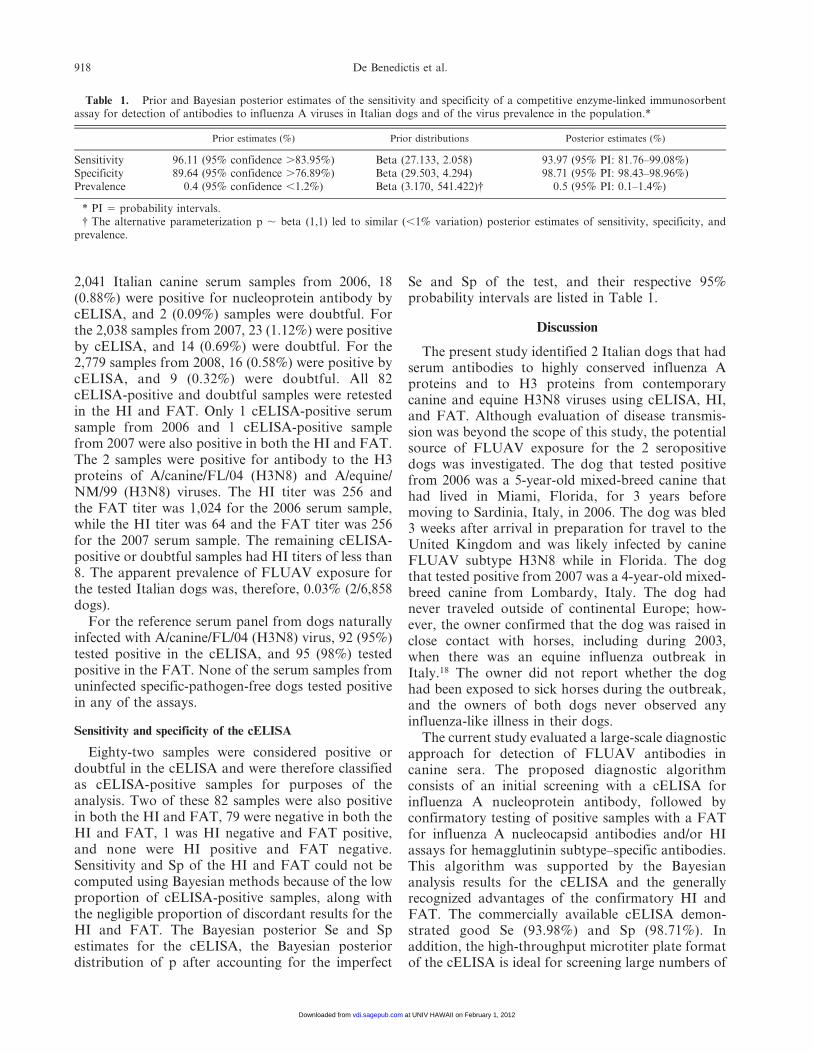
Table 1. Prior and Bayesian posterior estimates of the sensitivity and specificity of a competitive enzyme-linked immunosorbentassay for detection of antibodies to influenza A viruses in Italian dogs and of the virus prevalence in the population.*
Prior estimates (%) Prior distributions Posterior estimates (%)
Sensitivity 96.11 (95% confidence .83.95%) Beta (27.133, 2.058) 93.97 (95% PI: 81.76–99.08%)Specificity 89.64 (95% confidence .76.89%) Beta (29.503, 4.294) 98.71 (95% PI: 98.43–98.96%)Prevalence 0.4 (95% confidence ,1.2%) Beta (3.170, 541.422){ 0.5 (95% PI: 0.1–1.4%)
* PI 5 probability intervals.{ The alternative parameterization p , beta (1,1) led to similar (,1% variation) posterior estimates of sensitivity, specificity, and
prevalence.
918 De Benedictis et al.
at UNIV HAWAII on February 1, 2012vdi.sagepub.comDownloaded from

canine sera. A limitation, however, is that theBayesian model used herein is not identifiable becausethere are more parameters than degrees of freedom.For that reason, prior information about the modelparameters is required (Table 1). Because no priorinformation was available regarding the diseaseprevalence in the assessed population, best-guess(based on expert opinion) and noninformative pa-rameterizations of p were alternatively assumed.Results were robust for alternative parameterizationof p and were consistent with the presented results.
Although the Se and Sp of the HI and FAT couldnot be estimated using Bayesian analysis in the currentstudy, cELISA-positive and doubtful samples shouldbe confirmed in a highly sensitive and specific assay.The Se and Sp of the HI assay are generally high whenthe test antigen is homologous to the circulating virus.In the current study the HI assay was conducted with 7hemagglutinin subtypes, including 3 H3N8 virusesrepresenting canine, equine, and avian isolates. Theseviruses were selected as prototypes of those likelycirculating in poultry and mammals during the periodof canine serum collection. The 80 HI-negative sampleshad titers of ,8, indicating that the Se of the assay wasnot negatively affected by the defined cutoff titer forseropositivity of 32. However, the authors acknowl-edge that exposure to the remaining virus subtypes wasnot definitively ruled out since sera were not testedagainst all 16 influenza A hemagglutinin subtypes. TheFAT allows type-specific detection of influenza virusexposure, and the assay is highly sensitive and specific;however, major disadvantages of the FAT include thenecessary in-house development of the test and itsadmittedly subjective interpretation.
Although the low seroprevalence (0.5% afteraccounting for the imperfect Se and Sp of thecELISA) in the present study indicates that dogs donot currently play a significant role in FLUAVecology in Italy, the identified seropositive dogshighlight 2 potential sources of FLUAV exposurefor dogs in Italy and abroad: the potential spread ofcanine-specific influenza A viruses by internationalmovement of infected dogs and exposure of dogs toother species infected by FLUAV. Based on interna-tional observations, monitoring dogs during futureinfluenza A outbreaks in other species is recom-mended. A diagnostic algorithm utilizing cELISA aswell as HI and/or FAT is a highly sensitive andspecific approach to assessing exposure to anyinfluenza A subtype in dogs, especially in those withunknown exposure histories at the time of testing.
Acknowledgements
Dr. William G. Dundon and Giovanni Cattoli, IZSVe-Italy, and Dr. Jeanet Van der Goot, CIDC–The Nether-
lands, are acknowledged for technical advice on thisproject. In part, the European Commission, projectFLUTRAIN (Training and Technology Transfer of AvianInfluenza Diagnostics and Disease Management Skills) andEPIZONE (Network of Excellence for Epizootic DiseaseDiagnosis and Control), and the U.S. National Center forMedical Intelligence funded the current project. Paola DeBenedictis and Tara C. Anderson contributed equally tothis article.
Sources and manufacturers
a. ID ScreenH Influenza A Antibody Competition Assay, IDVet, Montpellier, France.
b. Sigma-Aldrich, St. Louis, MO.
c. BD Bioscience, San Jose, CA.
d. GibcoH, Invitrogen Corp., San Diego, CA.
e. Ferak Berlin GmbH, Berlin, Germany.
References
1. Amonsin A, Songserm T, Chutinimitkul S, et al.: 2007,Genetic analysis of influenza A virus (H5N1) derived fromdomestic cat and dog in Thailand. Arch Virol 152:1925–1933.
2. Beard CV: 1970, Demonstration of type-specific antibody inmammalian and avian sera by immunodiffusion. Bull W H O42:779–785.
3. Branscum AJ, Gardner IA, Johnson WO: 2004, Bayesianmodeling of animal- and herd-level prevalences. Prev Vet Med66:101–112.
4. Branscum AJ, Gardner IA, Johnson WO: 2005, Estimation ofdiagnostic-test sensitivity and specificity through Bayesianmodeling. Prev Vet Med 68:145–163.
5. Butler D: 2006, Thai dogs carry bird-flu virus, but will theyspread it? Nature 439:773.
6. Capua I, Kajaste-Rudnitski A, Bertoli E, Vicenzi E: 2009,Pandemic vaccine preparedness—have we left somethingbehind? PLoS Pathog 5:e1000482.
7. Capua I, Marangon S, Cordioli P, et al.: 2002, H7N3 avianinfluenza in Italy. Vet Rec 151:743–744.
8. Capua I, Mutinelli F, Pozza MD, et al.: 2002, The 1999–2000avian influenza (H7N1) epidemic in Italy: veterinary andhuman health implications. Acta Trop 83:7–11.
9. Capua I, Terregino C, Cattoli G, et al.: 2003, Development ofa DIVA (Differentiating Infected from Vaccinated Animals)strategy using a vaccine containing a heterologous neuramin-idase for the control of avian influenza. Avian Pathol32:47–55.
10. Chan PK, Ng KC, Chan RC, et al.: 2004, Immunofluores-cence assay for serologic diagnosis of SARS. Emerg Infect Dis10:530–532.
11. Crawford PC, Dubovi EJ, Castleman WL, et al.: 2005,Transmission of equine influenza virus to dogs. Science310:482–485.
12. Daly JM, Blunden AS, Macrae S, et al.: 2008, Transmission ofequine influenza virus to English foxhounds. Emerg Infect Dis14:461–464.
13. Kirkland PD, Finlaison DS, Crispe E, Hurt AC: 2010,Influenza virus infection in dogs—transmission during theAustralian equine influenza outbreak. Emerg Infect Dis16:699–702.
14. Knesl O, Allan FJ, Shields S: 2009, The seroprevalence ofcanine respiratory coronavirus and canine influenza virus indogs in New Zealand. N Z Vet J 57:295–298.
Serological detection of Influenza A virus exposure in dogs 919
at UNIV HAWAII on February 1, 2012vdi.sagepub.comDownloaded from
15. Kruth SA, Carman S, Weese JS: 2008, Seroprevalence ofantibodies to canine influenza virus in dogs in Ontario. CanVet J 49:800–802.
16. Lee C, Song D, Kang B, et al.: 2009, A serological survey ofavian origin canine H3N2 influenza virus in dogs in Korea.Vet Microbiol 137:359–362.
17. Maas R, Tacken M, Ruuls L, et al.: 2007, Avian influenza(H5N1) susceptibility and receptors in dogs. Emerg Infect Dis13:1219–1221.
18. Martella V, Elia G, Decaro N, et al.: 2007, An outbreak ofequine influenza virus in vaccinated horses in Italy is due to anH3N8 strain closely related to recent North American repre-sentatives of the Florida sub-lineage. Vet Microbiol 121:56–63.
19. Meijer A, Bosman A, van de Kamp EE, et al.: 2006,Measurement of antibodies to avian influenza virus A(H7N7)in humans by hemagglutination inhibition test. J VirolMethods 132:113–120.
20. Newton R, Cooke A, Elton D, et al.: 2007, Canine influenza virus:cross-species transmission from horses. Vet Rec 161:142–143.
21. Payungporn S, Crawford PC, Kouo TS, et al.: 2008, InfluenzaA virus (H3N8) in dogs with respiratory disease, Florida.Emerg Infect Dis 14:902–908.
22. Piccirillo A, Pasotto D, Moreno Martin A, Cordioli P: 2009,Serological survey for influenza type A viruses in domesticdogs (Canis lupus familiaris) and cats (Felis catus) in north-eastern Italy. Zoonoses Public Health 57:239–243.
23. Song D, Kang B, Lee C, et al.: 2008, Transmission of avianinfluenza virus (H3N2) to dogs. Emerg Infect Dis 14:741–746.
24. Song D, Lee C, Kang B, et al.: 2009, Experimental infection ofdogs with avian-origin canine influenza A virus (H3N2).Emerg Infect Dis 15:56–58.
25. Songserm T, Amonsin A, Jam-on R, et al.: 2006, Fatal avianinfluenza A H5N1 in a dog. Emerg Infect Dis 12:1744–1747.
26. Spiegelhalter D, Thomas A, Best N, Gilks W: 1996, BUGS0.05: Bayesian Inference Using Gibbs Sampling. MRCBiostatistics Unit, Cambridge, UK.
27. Toffan A, Olivier A, Mancin M, et al.: 2010, Evaluation ofdifferent serological tests for the detection of antibodiesagainst highly pathogenic avian influenza in experimentallyinfected ostriches (Struthio camelus). Avian Pathol 39:11–15.
28. World Organization for Animal Health (OIE): 2008, Chapter2.3.4, Avian influenza. In: Manual of diagnostic tests andvaccines for terrestrial animals, 6th ed., vol. 1, pp. 1–20. OIE,Paris, France.
29. World Organization for Animal Health (OIE): 2008, Chapter2.5.7, Equine influenza. In: Manual of diagnostic tests andvaccines for terrestrial animals, 6th ed., vol. 2, pp. 871–883.OIE, Paris, France.
30. World Organization for Animal Health (OIE): 2008, Chapter2.8.8, Swine influenza. In: Manual of diagnostic tests andvaccines for terrestrial animals, 6th ed., vol. 2, pp. 1128–1138.OIE, Paris, France.
920 De Benedictis et al.
at UNIV HAWAII on February 1, 2012vdi.sagepub.comDownloaded from