a clinical study on kumba vatham (periarthritis) with ...
146
A CLINICAL STUDY ON KUMBA VATHAM (PERIARTHRITIS) WITH MUKKIRATTAI CHOORANAM Dissertation submitted to THE TAMILNADU DR. M.G.R. MEDICAL UNIVERSITY, CHENNAI–32 For the partial fulfilment of the requirement for the degree of DOCTOR OF MEDICINE (SIDDHA) (Branch-I Pothu Maruthuvam) DEPARTMENT OF POTHU MARUTHUVAM GOVERNMENT SIDDHA MEDICAL COLLEGE PALAYAMKOTTAI-627 002 OCTOBER 2016
-
Upload
khangminh22 -
Category
Documents
-
view
0 -
download
0
Transcript of a clinical study on kumba vatham (periarthritis) with ...

A CLINICAL STUDY ON KUMBA VATHAM (PERIARTHRITIS)
WITH MUKKIRATTAI CHOORANAM
Dissertation submitted to
THE TAMILNADU DR. M.G.R. MEDICAL UNIVERSITY, CHENNAI–32
For the partial fulfilment of the
requirement for the degree of
DOCTOR OF MEDICINE (SIDDHA)
(Branch-I Pothu Maruthuvam)
DEPARTMENT OF POTHU MARUTHUVAM
GOVERNMENT SIDDHA MEDICAL COLLEGE
PALAYAMKOTTAI-627 002
OCTOBER 2016


"F'
GOVERNMENT SIDDHA MEDICAL COLLEGE
PALAYAMKOTTAI
Certificate of Botanical Authe"'ticity I
Certified the following plant drug used in Siddha formulation Mukkirattai Chooranam
(Internal) for the management of Kumba vatham (Periarthritis in shoulder) taken up for Post
Graduation Dissertation Studies by Dr.A.Roshini (Reg No.321311006) PG Dept, of Pothu
Maruthuvam are correctly identified and authenticated through Visual inspection /
Organoleptic Characters / Experience, Education & Training Morphology Microscopical and
Taxonomical methods.
S.N Name
1. Mukkirattai
Station: Palayamkottai
Date: 2-2- \ \ I ) \ ';-
Botanical Name
Boerhavia difJusa
Family Parts Used
N yctaginaceae Dried Leaves
. . · ~'h't~~V.f't'<fi':'!\'''''' c "
Dr. S. SUTHA, M.Sc.,M.Ed.,Ph.D.; Associate Professor
Dept. of Medicinal Botany . Govt. Siddha Medical Colle(;ji '. Palayamkottai, Tirunelvell - 2.
~.~~. ,;./;.!..' •• ~. ... " ",'


ACKNOWLEDGEMENTS
First of all I am very thankful, grateful to the Almighty God for his showered
blessing upon me in performing my dissertation works. Many individuals have helped
in brining out this dissertation work. I take it as a pleasure to acknowledge those
concerned.
I express my gratitude to the Vice-Chancellor, the Tamil Nadu Dr. M.G.R.
Medical University, Chennai and Special Commissioner, Commissionerate of Indian
Medicine and Homeopathy, Chennai for granted me to under take this dissertation
work.
I sincerely thank Prof. Dr. S.Victoria, M.D.(S)., Principal, Govt. Siddha
Medical College and Hospital, Palayamkottai.
I express my whole hearted and gratitude to Prof. Dr. A. Manoharan, M.D.
(S)., Head of the Department, Pothu Maruthuvam for his valuable guidance in each
and every step and encouragement in my dissertation work.
I would take this moment to signify my sincere gratitude to Dr. T. Komala
Valli, M.D.(S)., Associate Professor, Department of Pothu Maruthuvam for her
valuable guidance.
I express my thanks to Dr. S. Justus Antony, M.D.(S)., Dr. G. Subash
Chandran, M.D.(S)., Dr. S. Chithra, M.D.(S)., Dr. S. Uma Kalyani, M.D.(S)., and
Dr. P. Sathish Kumar, M.D.(S)., Lecturers of Department of Pothu Maruthuvam.
I express my thanks to Mrs. Dr. Sutha, M.Sc., Ph.D., Associate Professor in
Department of Medicinal Botany, Govt. Siddha Medical College and Hospital,
Palayamkottai for her valuable suggestions in the botanical aspect.
I thank Prof. Mrs. N. Naga Prema, M.Sc., M.Phil., and other technical staff
Department of Bio Chemistry for helping me in doing biochemical analysis of the
trial drug.

I express my thank to Mrs. T. Poonkodi, M.A., M.L.I.S., Librarian of Govt.
Siddha Medical College and Hospital, Palayamkottai for permitting me to utilize the
college library for my dissertation work.
I express my thanks to Periyar College of Pharmaceutical Sciences, Trichy
who helped in eliciting pharmacological analysis of the trial drug.
I express my deep sense of gratitude to my parents and my brother who gave
me marvellous support and moral courage.
And my whole hearted thanks to my dear friends Dr.T.Jenefa Rose Priya,
Dr. B. Manikandan, Dr. N. Rajkumar who helped me for my dissertation work.
I express my sincere thanks to my colleagues and other staff members who
helped me during this whole study period.

CONTENTS
CHAPTER
No. TITLE
Page
No.
ABBREVIATIONS
I INTRODUCTION 1
II AIM AND OBJECTIVES 3
III REVIEW OF LITERATURES 4
a). SIDDHA ASPECTS 4
b). MODERN ASPECTS 23
IV MATERIALS AND METHODS 39
V RESULTS AND OBSERVATIONS 42
VI DISCUSSION 81
VII SUMMARY 88
VIII CONCLUSION 90
ANNEXURES
ANNEXURES-I i
ANNEXURES-II iii
ANNEXURES-III v
ANNEXURES-IV x
ANNEXURES-V xiii
BIBLIOGRAPHY

LIST OF TABLES
TABLE
No. TITLE
Page
No.
1. SEX OF DISTRIBUTION 44
2. AGE OF DISTRIBUTION 45
3. KAALAM 46
4. THEGI 47
5. GUNAM 48
6. RELIGION 49
7. PARUVA KAALAM 50
8. THINAI 51
9. OCCUPATIONAL STATUS 52
10. DIET 53
11. SOCIO ECONOMICAL STATUS 54
12. AETIOLOGY FACTOR 55
13. MODE OF ONSET 56
14. DURATION OF ILLNESS 57
15. CLINICAL MANIFESTATION 58
16. GNANENDRIUM 59
17. KANMENDRIUM 60
18. CONDITION OF MUKKUTRAM
(a). CONDITION IN VATHAM 61
(b). CONDITION IN PITHAM 63
(c). CONDITION IN KAPHAM 64

TABLE
No. TITLE
Page
No.
19. INVOLVEMENT UDAL THATHUKKAL 65
20. CONDITION OF ENVAGAI THERVUGAL 67
21. NAADI 69
22. NEER KURI 70
23. NEI KURI 71
24. ASSESSMENT OF OUTCOME 72
25. GRADATION OF RESULTS 74
26. LABORATORY INVESTIGATIONS
a). OUT PATIENTS 75
b). IN PATIENTS 76
27. CASE SUMMARY
(a).OUT PATIENTS 77
(a). IP PATIENTS 79

LIST OF FIGURES
FIGURE
No. TITLE
Page
No.
1. SEX OF DISTRIBUTION 44
2. AGE OF DISTRIBUTION 45
3. KAALAM 46
4. THEGI 47
5. GUNAM 48
6. RELIGION 49
7. PARUVA KAALAM 50
8. THINAI 51
9. OCCUPATIONAL STATUS 52
10. DIET 53
11. SOCIO ECONOMICAL STATUS 54
12. AETIOLOGY FACTOR 55
13. MODE OF ONSET 56
14. DURATION OF ILLNESS 57
15. CLINICAL MANIFESTATION 58
16. GNANENDRIUM 59
17. KANMENDRIUM 60
18. CONDITION OF MUKKUTRAM
(a). CONDITION IN VATHAM 62
(b). CONDITION IN PITHAM 63
(c). CONDITION IN KAPHAM 64

FIGURE
No. TITLE
Page
No.
19. INVOLVEMENT UDAL THATHUKKAL 66
20. CONDITION OF ENVAGAI THERVUGAL 68
21. NAADI 69
22. NEER KURI 70
23. NEI KURI 71
24. ASSESSMENT OF OUTCOME 73
25. GRADATION OF RESULTS 74

ABSTRACT
Kumba Vatham is one of the Vatha disease described in Yugi Vaithiya
Chindamani 800.
This disease is correlated with periarthritis which is mentioned in medicine.
The disease is diagnosed by using Siddha parameters like “Envagai Thervugal,
Kaalam, Thinai, Mukkutra Verupadugal and modern parameters like laboratory and
radiological investigations.
20 patients of either sex has been selected, both as In patients and Out patients
and they were administrated with the clinical trial drug.
Mukkirattai Chooranam – 2gm twice a day with hot water for 30 days.
The trial drug is subjected to biochemical and pharamacological analysis. At
the end of the trial study majority of cases showed good clinical improvement.

LIST OF ABBREVIATIONS
% - Percentage
i.e., - That is
RA - Rheumatoid Arthritis
ESR - Erythrocyte Sedimentation Rate
ECG - Electro cardiogram
gms - Grams
kg - Kilogram
mg - Milligram
dl - Decilitre
ml - Milli litre
cm - Centimeter
S E M - Structural Equation Modelling
ANOVA - Analysis Of Variance
Hb - Haemoglobin
TC - Total Count
MCV - Mean Corpuscular Volume
DC - Differential Count
MCHC - Mean Corpuscular Haemolgobin Concentration
P - Polymorphs
L - Lymphocytes
E - Eosinophils
WBC - White Blood Corpuscles
RBC - Red Blood Corpuscles
Ref - Reference
CT - Computerized Tomography
MRI - Magnetic Resonance and Imaging
JVP - Jugular Venous Pulsation
Bid - Twice a day

1
CHAPTER-I
INTRODUCTION
‘fw;w FUthf;Fq; fhjypj;j thflKk;
gw;Wf; Nfhnyhd;Nw ghpfhuk; - Kw;w
mtd; nghWg;gy;yh nyhd;Wkhtjpy;iy
nad;Nw ,tDzuf; fPHj;jp mq;F”
Siddha system of medicine is the oldest system of medicine in India. It is of
Dravidian origin and has it entire literatures in Tamil language. Its origin is also
traced to mythological sources belonging to the shaivam. According to the tradition,
Lord Shiva conveyed the knowledge of medicine to his wife Parvathi.
The knowledge was passed from her to Nandhi and finally it was given to the
Siddhars.
This world and universe around it are made up of five basic elements
(Panchaboothams) namely earth, water, fire, air and space. More over,
panchabootham constitutes the arusuvaigal. So changes in the arusuvaigal may
influences the diseases of the human body. These five elements combine to form three
humours (Vatham, Pitham, Kapham). According to Siddha, the human body
composes of 96 thathuvam whose balance is very essential for the healthy human life.
‘mz;lj;jpYs;sNj gpz;lk;
gpz;lj;jpYs;sNj mz;lk;
mz;lKk;> gpz;lKk; xd;Nw
mwpe;J jhd; ghHf;Fk; NghNj”
In the normal human body all these three are in the homeostasis and exist in
the ratio 1:1/2:1/4. Disharmony of this homeostasis may produce disease and called
astrithudam as tridhosam. The three thathus are formed by the following way:
Vatham Abanan + Idakalai
Pitham Pranan + Pinkalai
Kapham Samanan + Suzhumunai

2
So, panchabootham, arusuvaigal, trithathus and human body are interrelated
with each other so dishormany in any of the above may produce the disease.
More over mind influences the formation of many disease. Now-a-days there
is to more stress and strain. So Siddhars has already advised to treat the body as well
as the mind.
Vatha disease were classified into 10 types.
Kumba Vatham is one among the Vatha disease. It is correlated with
periarthritis in modern medicine. The advantage and unique features of Siddha
medicine is the removal of the root causes of the disease and effective, perfect remedy
for mind and soul. They have enumerated many ways that are to be followed to
maintain the body without being affected by any disease, to maintain sound mind and
to have disease free life are called “MARUNTHU”.
Medicine can be defined as that which removes distress and leads an
individual to perfect happiness (heavenly bliss). Siddhars have utilized herbs, metals,
minerals, mercury, red oxide, alkalis, acid and poisons.
It states that,
‘NtH ghU jioghU kpQ;rpdf;fhy; nky;y nky;y
gw;g nre;J}uk; ghNu”
Ref: mfj;jpaH itj;jpak; vz;gJ gjpndz; rpj;jH ghba rpy;yiuf;
Nfhit.
First initiate the medication with roots and leafs of the herbs then later on use
parpam and chendoorams. They have developed their particular way of diagnosing the
disease by Envagai Thervugal.
So this study of ancient Siddha’s concept of Kumba Vatham and it’s treatment
with herbal preparation is taken up for the detailed study in preparing this dissertation.
I have tried to formulate the methodology to treat this disease very carefully,
to help the sufferers of Kumba Vatham in giving new modality of treatment.

3
CHAPTER-II
AIM AND OBJECTIVES
Vatha diseases cripples many people from attending their daily routine work.
As we are people belonging to medical world, we are very much worried about their
health conditions.
So far a perfect and complete remedy for this diseases has not been arrived at.
The purpose of the author is to elucidate a good medicine from ancient Siddha
literatures and to create hope and faith in their treatment. The drug which is prepared
for the diseases ‘Kumba Vatham’ is economically beneficial for all levels of people.
It is so effective when compared with other system of medicines.
The open labelled randomized clinical trial drug is,
MUKKIRATTAI CHOORANAM (internal medicine)
Ref: Gunapadam Mooligai Mudhal Vagupu (Page No.780)
OBJECTIVE
i. To study the clinical course of the disease Kumba Vatham with keen
observation on the aetiology, pathology, diagnosis, prognosis,
complications by making use of siddha concept.
ii. To expose the unique diagnostic methods mentioned by Siddha text.
iii. To know the disease Kumba Vatham alters the normal conditions under
the topic of Mukkutram, Poripulangal, 7 Udal kattukal and Envagai
Thervugal.
iv. To know the extent of correlation of aetiology, classification, signs and
symptoms of Kumba Vatham in Siddha aspect with ‘Periarthritis’ in
modern medical science.
v. To have an idea about the incidence of the disease with age, sex, socio-
economic status, habit, family history and climatic conditions.
vi. To study the effects of trial drugs for various stages of Kumba Vatham.
vii. To evaluate the bio-chemical and pharmacological effects of the trial
clinical medicine.
viii. To have a plan for further extensive studies on this disease.

4
CHAPTER-III
REVIEW OF LITERATURES
a) SIDDHA ASPECTS
There are 4448 diseases mentioned in Siddha system of medicine. Special
attention have been given to these thiridhosam and there is a general assumption, that
there are 80 Vatha diseases, 40 Pitha diseases and 20 Kapham diseases.
‘vd;dNt thjkJ vz;gjhFk;”
- a+fp itj;jpa rpe;jhkzp-800
and Kumba Vatham is one of the variety of Vatha disease.
Among three vital humours, Vatham is placed as chief factor (as king) to be
affected and leads derangement of other two factors named as Pitham and Kapham
and they initiate the diseases.
‘thjky;yhJ Nkdp nflhJ”
This verse denotes Vatha is the root cause to bodly affliction.
‘xOq;Fld; jNjo; %r;Nrhq;fp ,aq;f
vOr;rp ngw vg;gzpAkhw;w vOe;jphpa
Ntfk; Gyd;fSf;F Nktr; RWRWg;G
thfspf;Fk; khe;jHf;F thA”
- kUj;Jt jdpg;ghly;
Function of Vayu / Vatham is mentioned in the above poem, as it provides
functional energy and briskness to physical, mental and special senses for human.
Though the three basic vital humours are the structural component of whole
body they have their own seats in the body as it is dominant in such places. The poem
below says the particular regions mostly influenced by Vatha.
‘cz;b rikj;Jlw; $l;Lq;Flw;gFjp
jpz;bw nyd;G nrtpFwq;F - tpz;l
njhLTzHT Njhw;Wtpf;Fk; - NjhypLg;gpy; thWk;
5
tLtpyplkhk; tspf;F”
- kUj;Jt jdpg;ghly;
Accordingly Vatham is originally placed in intestine, bone, thigh, skin,
nervous system, joints and muscles etc., so derangement of Vatha produces
pathological conditions such as,
Body pain & various type of pain – cly; Nehjy;> Fj;jy;> gpsj;jy;
Weakness of nervous function – euk;G Fd;wy;
Tremor – eLf;fk;
Rigidity - ,Wf;fkhjy;
Dryness of fluids – ePHg;girapd;ik (Including Synovial fluid)
Subluxation – iffhy; ,lk; tpl;Lg; ngauy;
Weakness of organs or functional units – cWg;Gj; jsHr;rp
Stiffness or numbness – cWg;Gf;fs; njhopyw;W kuk;Nghy; fplj;jy;
Constipation, oliguria – kyk;> rpWePH jPa;jy;
Bony pain – vYk;Gf;Fs; Jisg;gJ Nghd;w czHr;rp
Loss of function of extermities – iffhy; klf;fTk;> ePl;lTk; Kbahjgb
nra;jy;.
Perverted taste and excessive salivation – vr;RitAk; JtHg;gha; ,Uj;jy;
my;yJ JtHg;gha; tha; eP&Wjy;
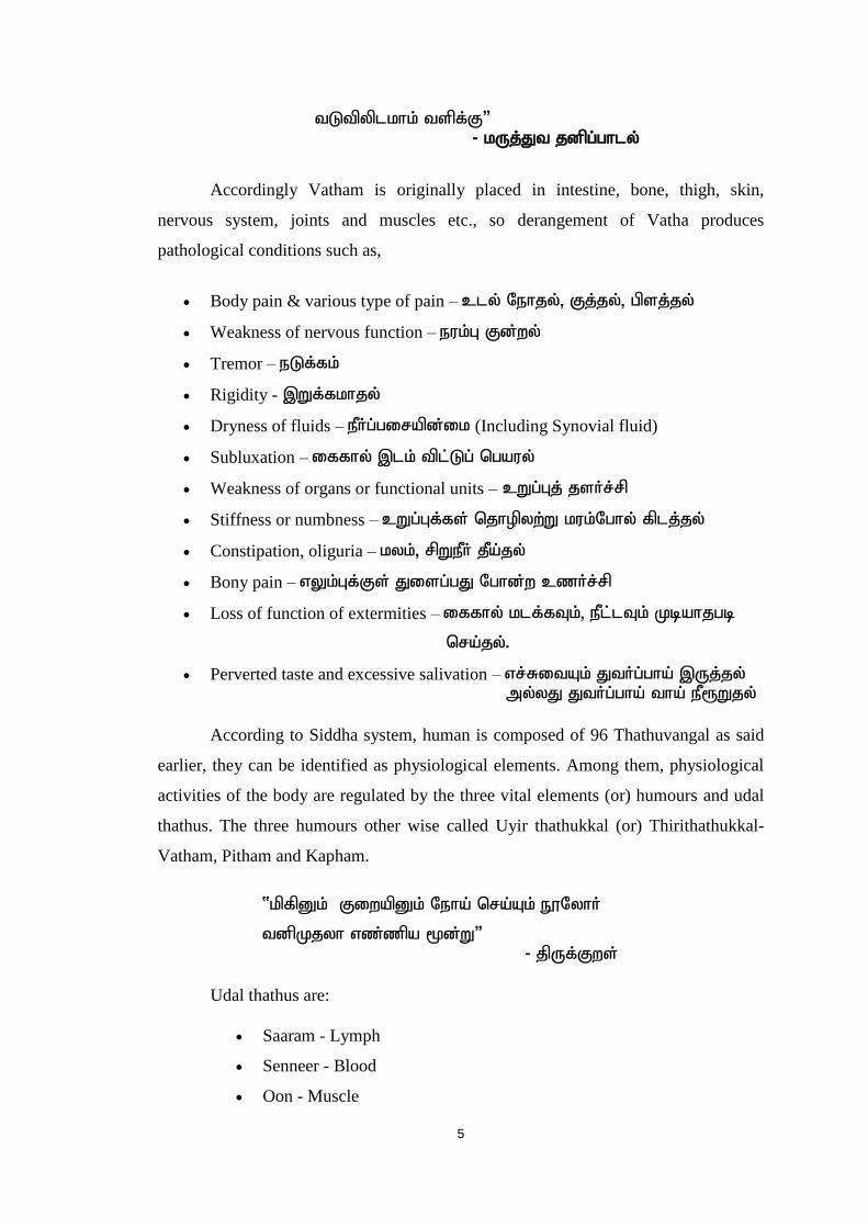
According to Siddha system, human is composed of 96 Thathuvangal as said
earlier, they can be identified as physiological elements. Among them, physiological
activities of the body are regulated by the three vital elements (or) humours and udal
thathus. The three humours other wise called Uyir thathukkal (or) Thirithathukkal-
Vatham, Pitham and Kapham.
‘kpfpDk; FiwapDk; Neha; nra;Ak; E}NyhH
tdpKjyh vz;zpa %d;W”
- jpUf;Fws;
Udal thathus are:
Saaram - Lymph
Senneer - Blood
Oon - Muscle

6
Kozhupu - Fat
Enbu - Bone
Moolai - Bone marrow
Sukkilam / Suronitham - Sperm and Ova.
VATHAM:
Synonym: Vayu, Arasan, Air
Definition:
Vatham (or) Vayu is not mere a wind, but it is that which causes motion,
energy and sensation of every cell in the body. So, Vatham is a humour which is
responsible for constructive nature of works in the human body.
Genesis of Vatha:
Vatham = vali + aagayam
Vali bootham and aagaya bootham are combined to form Vatham.
Genesis of vatha naadi:
During respiration abana vayu is combined with idakalai naadi. Thus the
Vatha humour is generated physiologically.
Vatha naadi = idakalai + abana vayu
Aetiology of Vatha diseases:
The common etiological factor for all types of Vatha diseases including
“Kumba Vatham” have been described generally in Yugi Vaidhya Chinthamani-800,
Agasthiyar Kanma Kaandam-300 and Agasthiyar Gunavagadam.
It is essential to know the disease, cause for the disease, also the nature of the
patient, the degree of the illness, season and also the time of occurrence of the disease
before treating the patient. This must to be considered.

7
In Yugi Vaidhya Chinthamani – 800:
‘vd;dNt thje;jhndz;gjhFk;
,fj;jpNy kdpjHfSf; nfa;JkhW
gpd;dNt nghd;jidNa NrhuQ;nra;J
nghpNahHfs; gpuhkiziuj; J}\dpj;Jk;
td;d Nttr; nrhj;jpw; NrhuQ; nra;J
khjh gpjh FUit kwe;j NgHf;Fk;
fd;dNt Ntjj;ij epe;ij nra;j NgHf;Fq;
fhaj;jpw; fye;jpLNk thje;jhNd”
Breach of trust.
Abusing the holy man.
Exploiting of the properties of charities.
Ingratitude towards mother, father and teachers and abusing holy.
Intake of food with bitter, astringent and pungent taste foods.
Intake of spoiled food.
Drinking of polluted rain water.
Day sleep.
Lifting over weight.
Excessive lust.
Disregarding the divinity.
Refusing food for clestitudes and sanyasingal.
Disregarding the advice of preceptors.
Involving in murdering, stealing, speaking lie.
Indulging in sexual act during vititation of Vatha.
Walking for a long distance.
Exposure to dampness, cold-eating.
Taking excessive curd immediately after eating vegetables and fruits
causes toxic factors which affect bones and muscles.
In Agasthiyar Kanma Kaandam – 300
‘E}nyd;w thjk; te;j tifjhNdJ
Jz;ikaha;f; fd;kj;jpd; tifiaf; NfS
fhypNy Njhd;wpaJ fLg;gNjJ

8
iffhypy; Kof;fpaJ tPf;fkJ
NfhypNy gLfpd;w tpUl;rkhd
Foe;ij kue;jid ntl;ly; Nky;Njhy;rPty;
E}ypNy rPt[e;J fhy; Kwpj;jy;
ey;ynfhk;G jioKwpj;jy; eypj;jy; jhNd”
- mfj;jpaH fd;kfhz;lk;
(ghly; vz;.56> gf;fk; vz;.23)
Removing the bark of living trees.
Breaking the legs of living animals.
Cutting the branches removing leaves.
In Agasthiar Gunavagadam:
‘njhy;iy nra;a ,d;Wk; ntF thj Neha;fs;
njhy;Yyfpy; khe;jpUf;Fr; fhz;gJz;L
vy;iyapy;iy thjNeha; Neh;ik jd;ik
,ay;ghf mwpe;jplNt tpguq;NfNs”
‘tpguklh mrjp rd;dp %is NehT
tphpthd %isaJ kpUJthfp
mtdpjdpy; %j;jpuf; Fz;bf;fha; tpahjpahYk;
jtKdpth; jPh;fhf;if NkfNuhfk;
jd;ikAs;s Kj;jz;Lf; nfhb tpahjp
mtkpyhg; ghpr euk;gOj;jq; fz;lha;”
‘mZFklh khkprj;jpd; tpahjpahYk;
mg;gNd #jfj;jpd; ngUf;fhYk;
Fzkpy;yh ,urk; tq;fk; jpd;dyhYk;
FbnfLj;j thjkJ cz;ghkg;gh”
- mfj;jpah; Fzthflk;
Brain diseases.
Renal disorders.
Sexually transmitted diseases.
Diseases of the vertebral and spinal cord.
Menorrhagia.
Taking improperly prepared medicine of mercury and lead.

9
Causes according to Astrology:
‘$Wnkhd;W %d;Wld; FyT ehiye;NjhopDk;
Fw;wkhk; eyj;jpDk; nfh^uk; gz;zpuz;bYk;
NruNt Gjd; jhDNkh rPhpak;kid epd;wpoy;
nrg;nghdhj jPiknahL nra;Agr;re;jhDk;
neLe;Jf;f kpf;fthk; elf;Fe;jhJ njhopy; jhk;
epe;ijahFq; fPy;gpbg;G ePLnka;apy; Njhd;Wkhk;
fhhpaq;fs; Nrkjhq; fy;taJ FiwAkhk;
fz;LzHe;J fzpjty;Nyhd; fUj;Jld; nrg;gpdhNuh”
- kzp ke;jpu itj;jpa Nrfuk;
Ref: Heritage of the Tamils Siddha medicine
Certain position of planets of certain period of human life will produce
keelpidippu and causes sequlare of Vatha vitiation.
All the above causes described from various texts are the major causes for the
derangement of Vatham.
CLINICAL FEATURE FOR “KUMBA VATHAM:”
in Yugi Vaithiya Chinthamani,
‘etpypNt Njhz;kPJq; fuj;jpd; kPJk;
eype;jnkj;j thfpNa erTz; lhFk;
ftpyhNt fd;dnkhL eade; jhDq;
fLj;JNk tpWtpWg;G nkhpTq; fhZk;
JtpypNt Jbg;ghFq; rpuR jd;dpw;
Row;wpNa ehgpf;fPo; typA Kz;lhk;
mtpyNt mb ehf;fpy; mod;W fhZk;
kyUNk tUFk;g thje; jhNd”
- a+fp itj;jpa rpe;jhkzp-800
Pain in the shoulder joints and upper limbs.
Burning and tingling sensation over the cheek and eyes.
Twitching over the scalp.
Pain in the hypogastrium.
Glossitis.

10
‘jhndd;w frg;NghJ JtHg;Giug;G
rhjfkha; kpQ;RfpDQ; rikj;j tz;zk;
Mndd;w thwpdJ nghrpj;jyhYk;
Mfhaj; NjwyJ Fbj;jyhYk;
ghndd;w gfYwf;f kpuhtpopg;G
gl;bdpna kpfTWjy; ghunka;jy;
Njndd;w nkhopahH Nkw; rpe;ijahfpy;
rPf;fpukha; thjkJ nrdpf;Fk; jhNd”
- a+fp itj;jpa rpe;jhkzp-800
Also stated in Yugi Vaidhiya Chinthamani, that imbalance diet like increased
taste of bitter, sour, hot and rotten meals starvation, inappropriate habits and disturbed
mind readily affect Vatha and causes disease.
‘tspjU fha; fpoq;F tiutpyh japyy; Nfhio
Kdp japH Nghd; kpLf;F Kiwapyh Tz;b Nfhly;
FspHjU tspapw;NwfLq;Fdpg;Gw Tyty; ngz;bH
fspjU Kaf;fk; ngw;NwhH fb nray; fUtpahkhy;”
- rghgjp ifNaL
In Sabapathy manuscript, Excess intake of carbohydrate diet, curd,
inappropriate diet, exposure to cold, increased sexual action will disturb Vatha & thus
cause Vatha diseases.
‘ghhpdpw; gag;gl;lhYk; gyUld; Nfhgpj;jhYk;
fhnudf; fUfpNahbf; fOkuj;Juj;jpdhYk;
VH ngW jdJ neQ;rpd; kpfj;Jf;fkile;jpl;lhYk;
ghhpa fhw;wpdhYk; glhpDk; thjq;fhZk;”
- guuhr Nrfuk;
The book of Pararaja Sekarama denotes, mental afflictions such as fear, anger,
sorrow and excess manual work like running, climatical changes also causes
disturbances of Vatham.

11
Qualities of Vatham:
Own qualities-6
Vatha is dry - twl;rp
Vatha is cold - FspHr;rp
Vatha is subtl - mZj;Jtk;
Vatha is rough - fbdk;
Vatha is Unstabl - mirjy;
Vatha is light - ,yF
Opposite qualities - 6
Unctuous - gRik
Hot - mf;fpdp
Solid - nfl;b
Soft - kpUJ
Stabl - ];jpuk;
Heavy - gST
Vatham Types:
Piranan
Abanan
Viyanan
Udhanan
Samanan
Nagan
Koorman
Kirukaran
Devathathan
Dhananjeyan

12
Functions of Vatham:
Sl.
No. VATHAM GENERAL FEATURES
CHANGES IN
KUMBA
VATHAM
1. Piranan
(Uyir Kaal)
Responsible for respiration and
digestion. Normal
2. Abanan
(Kizhnokkumkaal)
Responsible for voiding of urine,
stools, semen, menstrual flow. Normal
3. Udhanan It is responsible for the reflex actions
like vomiting, hiccough, cough etc. Normal
4. Viyanan
(Paravukaal)
Found in extension and flexion of the
parts of the body and helps in
distribution of the nutrients to various
parts of the body.
Affected
5. Samanan
(Nadukkaal)
Transfers nutrients to each and every
organ. Affected
6. Nagan Helps in opening & closing of eyelids. Normal
7. Koorman Responsible for vision, lacrimation
and yawning. Normal
8. Kirugaran
Induces appetite,and all secretions in
the body including nasal secretion and
sneezing.
Normal
9. Thevathathan
Induces and stimulates a person to
become alert, get anger, to quarrel, to
sleep etc.
Affected
10. Dhananjeyan
Resides in the cranium and produces
bloating of the body after death. This
leaves from the body after 3 days of
death, forming a way through the
skull.
----
In Kumba Vatham, Viyanan, Samanan, Thevathathan will be mainly
affected.
Exaggeration of Vatham:
Body weakness and darkness.
Shivering.
Abdominal distension.
Constipation.
Insomnia.
Liking to eat hot foods.

13
Immunity.
Giddiness.
Diminition of Vatham:
Body ache.
Hoarseness of voice.
Loss of memory.
Altered consciousness.
Sleep disturbances.
Anorexia.
Paleness & coolness of body.
Excessive salivation.
Heaviness of body.
The functions of other two humours are discussed below:
FUNTIONS OF PITHAM:
Pitham is functionally divided into five types. They are:
Sl.
No. PITHAM NORMAL FEATURES
CHANGES IN
KUMBA VATHAM
1. Anala Pitham Helps in appetite and
digestion. Normal
2. Ranjaga Pitham Maintains colour and
contents of blood. Normal
3. Sathaga Pitham
Controls the whole body and
responsible for fulfiling a
purpose.
Affected
4. Prasaga Pitham
Dwells in the skin and
concerned with the shine,
glow, texture and its
complexion.
Normal
5. Alosaga Pitham Vision. Normal
In Kumba Vatham, Sathaga Pitham is mainly affected.

14
FUNCTIONS OF KAPHAM:
It is of five types. They are:
Sl.
No. KAPHAM GENERAL FEARTURES
CHANGES IN
KUMBA
VATHAM
1. Avalambagam Controls the heart and
circulatory system. Normal
2. Kilethagam Moistens, softens the food
and helps to be digested. Normal
3. Pothagam Sensory perception of taste. Normal
4. Tharpagam
Responsible for the coolness
of the eyes, sometimes may
be referred to as
cerebrospinal fluid.
Normal
5. Santhigam
Necessary for the lubrication
and the free movements of
joints.
Affected
In Kumba Vatham, Santhigam is affected.
UDAL THATHUKKAL (SEVEN PHYSICAL CONSTITUENTS):
Sl.
No.
UDAL
THATHUKKAL
GENERAL
FEARTURES
CHANGES IN
KUMBA VATHAM
1. Saaram
(Digestive essence)
Responsible for the
growth & development. Normal
2. Senneer (Blood)
Responsible for the
colour of blood and
nourishment of the body.
Normal
3. Oon (Muscle)
Gives contour to the body
which is needed for the
physical activity.
Affected
4. Kozhuppu (Fat)
Lubricates the organs to
facilitate frictionless
functions.
Normal
5. Enbu (Bones) Supports & protects the
vital organs. Affected
6. Moolai
(Bone marrow)
Nourishes the bone
marrow. Normal
7. Sukkilam /
Suronitham
Responsible for
reproduction -
Derangement of Vatham leads to vitiation of Pitham and Kapham leading to
disturbances in Udal Thathukkal.

15
The Other factors responsible for derangement of three vital humours and
Udal Thathukkal are discussed below:
THINAI (LAND):
The geographical distribution of the land is classified into five regions:
1. Kurinchi - Mountain and its surroundings, causes Kapha noigal
2. Mullai - Forest and its adjacent area, causes Pitha, Vatha noigal
3. Marudham - Fertile fields and its surroundings, safest place
4. Neidhal - Sea and its surroundings, Vatha diseases are common
here.
5. Paalai - Desert and its surroundings, Vatha, Pitha, Kapha
diseases are common
The above features are described clearly in Thanvanthiri Naadi as,
‘FwpQ;rp epyNk thjkhq; $Wk; ghiy gpj;jkhQ;
nrwpe;j kUje; rpNyj;kkjhQ; rpNyj;k thj Ky;iyajhk;
epiwe;j nea;jy; thj gpj;jk; epyq;f sjid kaf;fhap
Yiwj;j tpahjp fe;jpUf;f Kgha kwpe;J nra;tPNu”
- jd;te;jphp ehb
KAALA MARUBADUGAL: PARUVAKALAM (SEASONS)
Sl.
No. KAALAM KUTTRAM
STATE OF
KUTTRAM SUVAI
1.
Kaar Kaalam Aavani &
Purattasi 16th
August To
15th
October
VATHAM-
PITHAM-
Vettrunilai
Vazharchi, Thanilai
Vazharchi
Enippu,
Pulippu,
Uppu
2.
Koothir Kaalam Iypasi &
Karthigai
16th
October To
15th
December
VATHAM-
PITHAM-
Thanilai Vazharchi,
Vettrunilai
Vazharchi
Enippu,
Kaippu,
Thuvarppu
3.
Munpani Kaalam
Margazhi & Thai
16th
December To
15th
February
PITHAM- Thannilai
Adaithal
Enippu,
Pulippu,
Uppu
4.
Pinpani Kaalam Masi &
Panguni
16th
February To
15th
June
KAPHAM Thannilai
Vazharchi
Enippu,
Pulippu,
Thuvarppu

16
5.
Elavenir Kaalam Chithirai
& Vaikaasi 16th
April To
15th
June
KAPHAM Vettrunilai
Vazharchi
Kaippu,
kaarppu
Thuvarppu
6.
Mudhuvenir Kaalam
Aani & Aadi
16th
June To 15th
August
VATHAM
KAPHAM
Thannilai
Vazharchi
Thannilai Adaithal
Enippu
UDAL VANMAI:
It means strength and vitality of the body and classified into 3 types.
1. Eyarkai vanmai - Inherited immunity
2. Kala vanmai - Immunity occurred by Age, season & time
3. Cheyarkai vankai - Improvement of 3 vitality obtained by diet
(jpdrhp) day-to-day habits and physical
exercise.
Any modifications in Udal Vanmai leads to diseases.
NOI KANIPPU VIVADHAM (Differential Diagnosis):
Resembling the symptoms of Kumba Vatham are mentioned here:
1. Sagana vatham:
‘NfSNk fOj;jpd; fPoiuf;F NkYk;
Nfbahd fukpuz;L kpfNt nehe;J
thSNk rhPunky;yhq; fdj;jpUf;Fk;
thypgHf;F kdq;fz;Z kaf;fkhFk;
VSNk ,uz;L fz;Zk; vhpr;rYz;lhk;
Vw;wkha; kye;jhDk; ,Wfpf; fhZk;
NjSNk nfhl;bdJ Nghw; fLf;Fk;
Nrfd thjj;jpdple; jPHfe;jhNd”
- a+fp itj;jpa rpe;jhkzp-800
Neck pain.
Radiating pain to the shoulders and upper limb.
Heaviness of the body.
Mental depression.

17
Giddiness.
Burning sensation in the eyes.
Constipation.
2. Paanikambavatham:
‘khHf;fkha; tha;Tkha;........................
eLf;fkha; ifapuz;Le; jpkpU Kz;lhk;
............................ JzHr;rpaw;W
ghzp fk;gthjj;jpd; ghq;FjhNd”
- a+fp itj;jpa rpe;jhkzp-800
Tingling sensation numbness of upper limbs.
Tremor of upper limbs.
Noi Nadal Noi Muthal Nadal
Diagnostic methods adopted in Siddha system of medicine are given below.
a). Piniyari Muraimai it based on following principles:
1. Poriyal arithal
2. Pulanal arithal
3. Vinathal
“Pori and Pulan are the five organs of perceptions and their senses
respectively. Nose – smell, Tongue – taste, Eyes – vision, Ears & Skin – auditory &
touch. Porigal of patient and Doctors‟ are used by the physician as instruments.
Vinathal is a method of enquiring about the details of patient‟s problem from
his own words or from their attendant.
The above mentioned principles can be compared to that of Interrogation
(Vinathal) and Inspection (Poriyal arithal), Auscultation, Percussion & Palpation
(Pulanal arithal).
The important method adopted to diagnose the disease is by means of
“Envagai Theruvagal”.

18
‘ehb ];ghprk; eh epwk; nkhop tpop
kyk; %j;jpukpit kUj;JtuhAjk;”
It is considered to be physician‟s instrument and this can be understand by
following stanza:
‘njhLf;fYw;W ml;l tpjg; ghPl;ir jd;id
Jyf;fKWk; gz;bjNu njspthfg;
gFf;fhpa ehbia eP gpbj;Jg; ghU
gfHfpd;w thHj;ij ghH ehitg; ghU
tFf;fhpa Njfnkd;wj; njhl;Lg; ghU
tskhd rhPuj;jpd; epwj;ijg; ghU
rfpf;fhpa kyj;ijg; ghH ryj;ijg; ghU
rhHe;j tpop jidg; ghHj;J njsptha; fhNz”
- mfj;jpaH itj;jpa ty;yhjp-600
Envagai Thervugal Constitutes:
Nadi, Sparisam, Naa, Niram, Mozhi, Vizhi, Malam, Mootharam.
The fact regarding Envagai Thervugal suggests that it is mostly used as a
diagnostic standard in Siddha system and more concentration should be given to get
proficient knowledge.
Naadi (Pulse):
According to Agathiyar Naadi,
‘fhpKf dbia tho;jpf;
ifjdpy; ehb ghHf;fpy;
ngUtpu yq;F yj;jpy;
gpbj;jb eLNt njhl;lhy;
xUtpu Nyhby; thjk;
capHeL tpuypw; gpj;jk;
jpUtpuy; %d;wp Nyhby;
rpNyj;Jk ehb jhNd”
- mfj;jpaH ehb

19
ngUtpuy; gf;fkhf kzpf;fl;Lf;F xU mq;Fyj;jpw;F Nky; Miu
vd;gpd; NkNyhLk; ehb euk;G ,uj;jf;Fohapd; Nky; %d;W tpuy;fis itj;J
rw;W mOj;jpAk;> jsHj;jpAk; ghHf;f Ms;fhl;b tpuyhfpa Kjy; tpuypy;
czHj;JtJ thjk; vdTk;> eLtpuypy; czHj;JtJ gpj;jk; vdTk; ngsj;jpu
tpuypy; czHj;JtJ fgk; vdTk; mwpaTk;.
‘mwpe;JghH thjNk jdpj;j jhdhy;
md;dk; Nghy; elf;Fkg;gh ehb ghU
rhpe;jplNt fhy; Klf;Fk; NghJ fhl;Lk;”
- mfj;jpaH uj;jpd RUf;fk;
This poem says, Vitiated Vatha causes difficultly in walking (or) impaired
function of lower extremities. The examination of Naadi has been recognised as one
of the principle means of Diagnosis and prognosis of disease from times immemorial.
Sparisam (Skin):
Skin examination can be made out by inspection and touch. It reveals
warmness / chillness / dry / weeping skin / rough / smooth / soft / hard, tenderness or
presence of ulcers, swelling, wrinkles, hair, pigmentation etc.,
‘miwe;Njhk; thj NuhfpAly;
Mow;fz; KfKk; gy; kyKk;
epiwe;j tpopapy; ePH tbAk;
ePz;l ehT fWj;jplTk;
epiwe;j Ks;sha; jhdpUf;Fk;
ciwe;j ePuhy; fUfUj;J
Kiwaha; Nuhfk; cz;lhNk”
Naa (Tongue):
The colour, character and condition of the tongue changes according to the
changes in Mukkutram.

20
Niram (Colour complexion):
As Vatham is root cause for Vatham the colour of the patient‟s skin, tooth,
etc., would be dark or black in colour.
Mozhi (Speech):
Speech in Vatha patients may vary according to deranged dhosam and grade
of the disease.
Vizhi (Eye):
In this disease condition, both motor and sensory disturbances of the eye can
be expected.
Burning of the eyes, lacrimation, irritation, colour changes are also noticed
under this group.
Mostly in Kumba Vatham the eyes become dark and smoky in colour.
Malam (Stools):
In Vatha diseases stools would be black in colour with constipation.
Moothiram (Urine):
According to Agasthiyar Naadi,
‘ciwe;j ePUq; fUfUj;J
Kiwaha; NuhfK Kz;lhNk”
- mfj;jpaH ehb
In Vatha disease, the colour of urine would be black leading to various
diseases.
Neer kuri:
Niram - Denote the colour of the urine voided
Manam - Denote the smell of the urine
Edai - Denote the specific gravity of the urine
Nurai - Denote the frothy nature of the urine voided
Enjal - Indicates the quantity of the urine voided

21
Nei kuri:
The method of Nei Kuri is described in Theriyar Venba as,
‘mUe;JkhwpujKk; mtpNuhjkha;
mf;fy; myHjy; mfhyt+z; jtpHj;jow;
Fw;wstUe;jp cwq;fp itfiw
Mbf; fyrj; jhtp NafhJ nga;
njhU K$Hj;jf; fiyf;Fl;gL ePhpd;
epwf;Fwp nea;f;Fwp ep&gpj;jy; flNd”
- NjiuaH ntz;gh
Method:
Prior to the day of urine examination, the patient is advised to take balanced
diet and the quantity of food must be proportionate to his or her appetite and he or she
should have had a good sleep. After waking up in the morning, urine voided first, is
collected in a glass container and is subjected to analysis within 1 ½ hours. A drop of
gingelly oil is dropped without shake. The nature of the Nei kuri should be noticed in
direct sunlight.
Character of different Neer:
‘muntd ePz;bbd ‡Nj thjk;”
When drop of oil spreads like a snake, it indicates Vatha neer,
‘MopNghw; gutpd; m‡Nj gpj;jk;”
If the drop of oil remains as that of ring it indicates Pitha neer.
‘Kj;njhj;J epw;fpd; nkhoptjd; fgNk”
If the drop of oil remains as that of pearl it indicates Kapha neer.
Along with above mentioned eight types of examination another few
principles in Siddha medicine are there. Man is composed of 5 elements like universe.
‘mz;lj;jpYs;sNj gpz;lk;
gpz;lj;jpYs;sNj mz;lk;

22
mz;lKk;> gpz;lKk; xd;Nw
mwpe;Jjhd; ghHf;Fk; NghNj”
- rl;lKdp epfz;L
Time, place, nature of the body (Pirakiruthi) and environmental changes have
inter relations among them.
So, besides Envagai Thervugal, Parva Kalangaal and Thinnai also should be
taken in consideration to arrive at perfect and correct diagnosis.
23
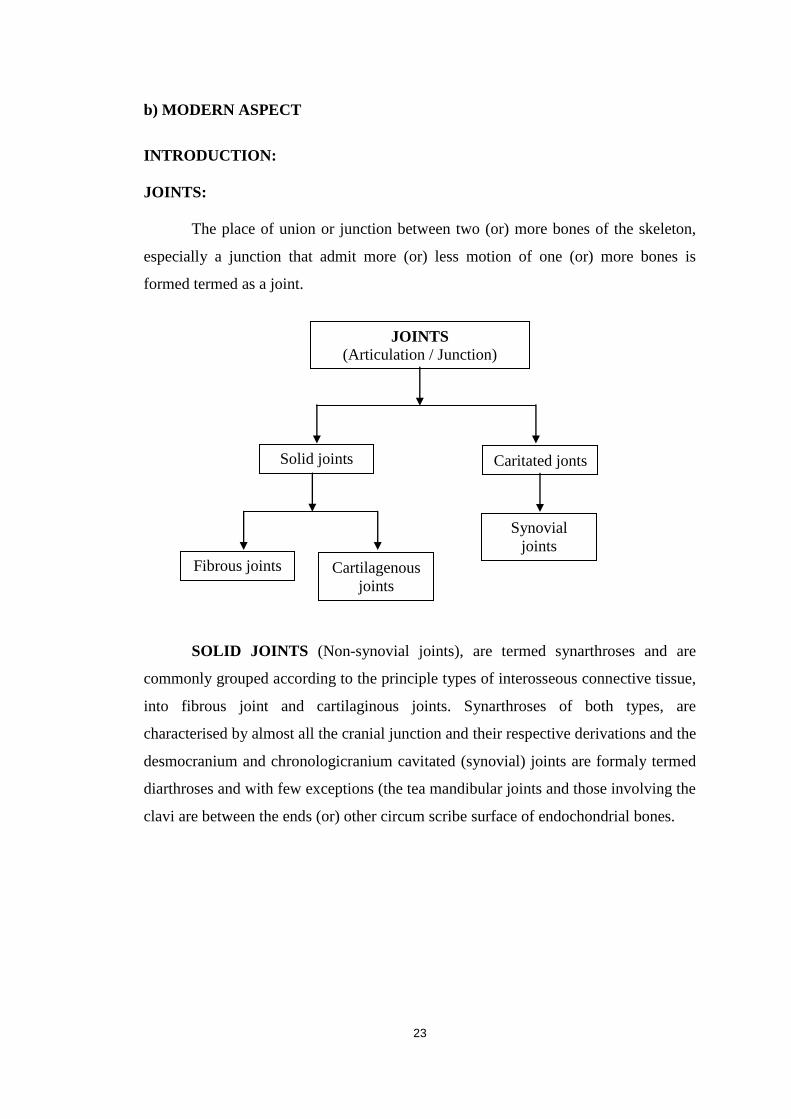
b) MODERN ASPECT
INTRODUCTION:
JOINTS:
The place of union or junction between two (or) more bones of the skeleton,
especially a junction that admit more (or) less motion of one (or) more bones is
formed termed as a joint.
SOLID JOINTS (Non-synovial joints), are termed synarthroses and are
commonly grouped according to the principle types of interosseous connective tissue,
into fibrous joint and cartilaginous joints. Synarthroses of both types, are
characterised by almost all the cranial junction and their respective derivations and the
desmocranium and chronologicranium cavitated (synovial) joints are formaly termed
diarthroses and with few exceptions (the tea mandibular joints and those involving the
clavi are between the ends (or) other circum scribe surface of endochondrial bones.
JOINTS
(Articulation / Junction)
Solid joints Caritated jonts
Cartilagenous
joints
Fibrous joints
Synovial
joints

24
GENERAL CLASSIFICATION OF SYNOVIAL JOINTS:
1. Plane joints.
2. Ginglymi (hinge) joints.
3. Trochoid (pivot) joints.
4. Spheroidal joints (Ball and socket).
5. Bicondylar joints.
6. Ellipsoid joints.
7. Sellar (saddle) joints.
PERIARTICULAR DISORDERS OF THE EXTREMITIES:
A number of periarticular disorders have become increasingly common, due in
part to greater participation in recreational sports by individuals of a wide range of
ages. Periarticular disorders most commonly affect the knee (or) shoulder.
SHOULDER JOINT:
Made up of three bones, they are:
i) Clavicle (Collar bone).
ii) Hamerus (Upper arm bone).
iii) Scapula (Shoulder blade),
as well as associated muscles, ligaments and tendons. The articulation between
the bones of the shoulder make up the shoulder joints. “Shoulder joint”, typically
which is the major joint of the “shoulder”. But can more broadly include the
acromioclavicular joint. In human anatomy, the shoulder joint comprises the part of
the body where the humerus attaches to the scapula, the head sitting in the glenoid.
The shoulder must be mobile enough dor the wide range of the arms hands,
but also stable enough to allow for action such as lifting and pulling.
i) Clavicle (Collar bone):
The clavicle (or) collar bone is a flat bones that serves as a structure between
the scapula and the sternum. It is the only long bone in the body that lies horizontally.
It makes up part of the shoulder and the pectoral girdle and is palpable in all people.

25
ii) Humerus (Upper arm bone):
It‟s nearly hemispherical in form. It is directed upward, medial and a little
backward and articulates with the glenoid cavity of the scapula to form the
glenohumeral joint (Shoulder joint).
iii) Scapula (Shoulder blade):
Also known as shoulder blade. It is a triangular, flat bone of the shoulder
girdle that articulates with the head of humerus at the glenohumeral joint, which is
popularly called as shoulder joint.
JOINTS OF SHOULDER:
i) Sternoclavicular Joint.
ii) Glenohumeral Joint.
iii) Acromioclavicular Joint.
i) Sternoclavicular joint:
This joint is formed by medial end of the clavicle with the manubrium (or) top
most portion of the sternum. The costo clavicular ligament is the main stabilizer of the
of the joint. The sternoclavicular joint is the sole connection between the axial
skeleton and the upper extremity.
ii) Acromio clavicular joint:
The acromio clavicular joint is the sole joint of acromion, clavicle and scapula.
The joint is diarthrodial joint itself has a little movement. It is held together by it‟s
joint capsule and the coracoacromial ligaments.
iii) Glenohumeral joint:
The glenohumeral joint is ball and socket synovial joint formed by the
articular surface of the glenoid cavity and the head of the humerus. It is the main joint
of the shoulder and allows the arm to rotate in a circular fashion (or) to hinge out and
up away from the body. Shoulder joint is very mobile because glenoid fossa is
shallow but this also adds to instability of the shoulder.

26
LIGAMENTS OF SHOULDER JOINT:
i) Coracoclavicular ligament.
ii) Glenohumeral ligament.
a). Superior
b). Middle
c). Inferior
iii) Coracohumeral ligament.
i) Coracoclaviacular ligament:
Two coracoclavicular ligaments are,
a). Conoid
b). Trapezoid
which maintain the articulation of the clavicle with the coracoid process of the
scapula. It is primary resistant to superior and posterior acromioclavicular dislocation.
ii) Glenohumeral ligament:
Superior glenohumeral ligament has a variable origin on lateral aspect of
scapula and inserts on the humerus near the lesser tubercle. Middle glenohumeral
ligament originates from the laburm and inserts on the humerus, medial to the lesser
tubercle.
Inferior glenohumeral ligament originates from the labrum and the adjacent
glenoid neck, inserts on the anatomical neck of the humerus.
iii) Coracohumeral ligament:
The coracohumeral ligament originates on the base and lateral border of the
coracoid process of the scapula and inserts on the greater tubercle.
NEUROVASCULAR SUPPLY:
Arterial supply to the glenohumeral joint is via the anterior and posterior
circumflex humeralarteries and the suprascapular arteries. Branches from these
arteries form an anastamotic network around the joint.

27
MOVEMENTS:
As a ball and socket synovial joint, wide range of movement or
permitted.
Extension (upper limb backwards in sagittal plane).
Flexion (upper limb forwards in sagittal plane).
Abduction (upper limb away from midline in coronal plane).
Adduction (upper limb towards midline in coronal plane).
Medical rotation (Rotation towards the midline, so that the thumb is
pointing medially).
Lateral rotation (Rotation away from the midline, so that the thumb is
pointing laterally).
MOBILITY AND STABILITY:
The shoulder joint is one of the most mobile in the body, at the expense of
stability. Here, we shall consider the factors that permit movement and those that
contribute towards joint structure.
PERIARTHRITIS
Introduction:
„Periarthritis‟ also called frozen shoulder (or) adhesive capsulitis of shoulder,
is a chronic inflammatory disorder of the shoulder and surrounding of the soft tissues.
This condition is frequently caused by injury, leading to pain and lack of use. As the
joint becomes progressively lighter and stiffer, simple movements, such as raising the
arm, becomes difficult. If inflammation occurs with the capsule itself, the shoulder
bones are unable to move with in the joint. In some instances, the patients may be
unable to move the shoulder at all.
The definition, also defined by Lundberg, is:
- Shoulder joint elevation of ≤135°
- Restriction of motion is localised to the glenohumeral joint
- History, clinical and radiological examination show no other explanation

28
EPIDEMIOLOGY:
The incidence of periarthritis is approximately 3 per cent in the general
population. Occurrence is rare in children and people under 40 but peaks between 40
and 70 years of age. At least in its idiopathic form, the condition is much more
common in women than in men (70% of patients are women aged 40–60). Frozen
shoulder is more frequent in diabetic patients and is more severe and more protracted
than in the non-diabetic population. Vaccine-related shoulder injuries (SIRVA) are
increasing.
CLASSIFICATION:
As in all situations where not all facts are known, many different ways of
classifying may exist.
Perhaps it is wise to use the initial classification of Lundberg (1969):
- Primary periarthritis
- Secondary periarthritis
But not even this is clear – these groups need to be subdivided:
- Primary periarthritis - Diabetic
- No other explanation
- Secondary periarthritis - Post-traumatic
- Iatrogenic
- Other
There exists, of course, other types of classification. In some, the diabetic
periarthritis is a secondary type. The secondary may be divided into extrinsic (causes
outside the joint) and intrinsic (causes inside the joint).
RISK FACTORS AND CAUSES:
A risk factor is something that elevates the risk of developing a disease or
condition. For example, smoking is a risk factor for cancer - it elevates the risk of
developing lung cancer.

29
Common risk factors are:
Age - being over 40 years of age.
Gender – common in women.
Recent surgery or arm fracture- immobility of recovery may cause the
shoulder capsule to stiffen.
Chondral lesions, avascular necrosis or tendon injuries.
Scarring following traumatic tissue injury.
Diabetes - two to four times more likely to develop periarthritis for unknown
reasons; symptoms may be more severe.
Having suffered a stroke.
Hyperthyroidism (overactive thyroid).
Hypothyroidism (underactive thyroid).
Cardiovascular disease (heart disease).
Parkinson's disease.
Tonic seizures
Accidents
Lung disease
Connective tissue diseases
Rheumatoid arthritis
Highly active antiretroviral therapy (HAART).
After breast and lung surgery
May have an autoimmune component.
Hypertriglyceridemia,
CVA with upper-extremity paresis,
Brachial plexus injuries.
Idiopathic frozen shoulder
Cervical spinal cord injury, and
Parkinson disease.

30
SIGNS AND SYMPTOMS:
The most pervasive sign or symptom is;
A persistently painful and stiff shoulder joint.
Severe pain and sleep deprivation for prolonged periods due to pain that gets
worse when lying still and restricted movement/positions.
The condition can lead to depression, problems in the neck and back, and
severe weight loss due to long-term lack of deep sleep.
People who suffer from periarthritis may have extreme difficulty
concentrating, working, or performing daily life activities for extended periods
of time.
Signs and symptoms of periarthritis develop gradually; usually in three stages
in which signs and symptoms worsen gradually and resolve within a two - year
period.
STAGES OF PERIARTHRITIS:
There are three stages of periarthritis,
Painful stage or freezing stage - The freezing stage shows an insidious onset
where pain is dominating the clinical picture. Quite often, subacromial
impingement is initially suspected because of the involvement of the
subacromial bursa. At the end of this period range of motion becomes limited
in the typical way and diagnosis is usually no longer a problem. The shoulder
becomes stiff and then very painful with movement. Movement becomes
limited. Pain typically worsens at night.
Frozen/adhesive stage - the shoulder becomes increasingly stiff, severely
limiting range of motion. Pain may not diminish, but it does not usually
worsen.
Thawing stage - The thawing includes successive reestablishment of normal
or near normal range of motion. Movement in the shoulder begins to improve.
Pain may fade, but occasionally recur.
However, this time plane is an approximation of the most common time
course of this disease. It may vary greatly between different patients.

31
PATHOGENESIS:
There are three different modalities of outcome following primary FS:
- 40% restore the range of motion (ROM) and are pain free
- 45% regain functional ROM but show residual symptoms
- 15% show persisting stiffness with marked handicap (Noel et al 2000,
Shaffer et al 1992).
In frozen shoulder, there is a lack of synovial fluid, which normally helps the
shoulder joint, a ball and socket joint, move by lubricating the gap between the
humerus (upper arm bone) and the socket in the shoulder blade. The shoulder capsule
thickens, swells, and tightens due to bands of scar tissue (adhesions) that have formed
inside the capsule. As a result, there is less room in the joint for the humerus, making
movement of the shoulder stiff and painful. This restricted space between the capsule
and ball of the humerus distinguishes periarthritis from a less complicated, painful,
stiff shoulder.
Dupuytren‟s disease is again shown to be related to PA. Dupuytren‟s disease
is significantly more common than usual among male relatives to FS patients and the
microscopic changes in the anterior capsule and coracohumeral ligament are very
similar to those in Dupuytren‟s disease of the hand (Bunker & Anthony 1995, Smith
et al 2001). They show that half of the patients with FS also show signs of
Dupuytren‟s! Similarities with Dupuytren‟s are also shown when analysing the
fibrotic capsule for cytokines and proteinases (Bunker et al 2000).
Pathoanatomically there is an involvement of the capsule in the glenohumeral
joint. The capsule volume is reduced and this is the cause for the restricted range of
motion (Itoi & Tabata 1992).
Diabetes and its relationship to periarthritis:
• The incidence of periarthritis is two to four times higher in diabetics than
in the general population.
• The prevalence of diabetes in patients with periarthritis was 38.6%,
whereby the total prevalence of a diabetic condition in patients with
periarthritis was 71.5%

32
• Shoulder arthritis is common in type I and type II diabetic patients.
However, it is associated with age in type I and II diabetic patients and with
the duration of diabetes in type I patients.
Possible aetiology:
Diabetics have a higher incidence of periarthritis, probably because poor
circulation leads to abnormal collagen repair and degenerative changes. The theory is
that platelet derived growth factor is released from abnormal or ischemic blood
vessels, which will then act as a stimulus to local myofibroblast proliferation. What
follows has been proposed that microvascular disease, abnormalities of collagen
repair and predisposition to infection may link diabetes with frozen shoulder.
NATURAL COURSE:
Primary periarthritis is usually considered to be a self limiting disease which
usually lasts for 18-24 months but will usually heal with minor residual handicap.
Examination:
Currently the diagnosis of primary periarthritis is based on the findings of the
patient history and physical examination.
The following outcome measures have been used in studies researching
periarthritis.
Shoulder Pain and Disability Index (SPADI)
Disability of the Arm, Shoulder and Hand scale
American Shoulder and Elbow Surgeons Standardized Shoulder Assessment
Form (ASES)
Simple Shoulder Test (SST)
Penn Shoulder Scale (PSS)
NPRS
VAS
SF-36
33
In a recent systematic review, the psychometric properties of the SPADI,
DASH, ASES and SST were examined. Reliability, construct validity and
responsiveness were all found to be favourable for various shoulder pathologies but
the review did not address their strength relative to adhesive capsulitis specifically.
Observation of Posture and Positioning:
Scapular winging of the involved shoulder may be viewable from the
posterior and/or lateral views.
Screen: Upper Quarter Exam (UQE) & Neuro Screen (dermatomes, myotomes,
reflexes)
A full UQE should be performed to rule out cervical spine involvement or any
neurological pathologies.
ROM Screen: Active/Passive/Overpressure:
Cervical, Thoracic, Shoulder ROMs with OP as well as rib mobility should be
performed.
Scapular substitution frequently accompanies active shoulder motion.
Resisted muscle tests:
Shoulder External Rotation (ER)/ Internal Rotation (IR) / Abduction
(ABd) (seated) should be performed.
Patients with adhesive capsulitis present with weakness in shoulder ER, IR
and ABd relative to the uninvolved side.
Formal ROM: Active/Passive/Overpressure
Shoulder Flex/ABd/ER/IR:
The method of measuring ER and IR ROM in patients with suspected
periarthritis varies in the literature.
Patients with periarthritis commonly present with ROM restrictions in
a capsular pattern. A capsular pattern is a proportional motion
restriction unique to every joint that indicates irritation of the entire

34
joint. The shoulder joint has a capsular pattern where external rotation
is more limited than abduction which is more limited than internal
rotation (ER limitations > ABD limitations > IR limitations). In the
case of periarthritis, ER is significantly limited when compared to IR
and ABD, while ABD and IR were not seen to be different.
Joint Accessory Mobility:
Glenohumeral joint:
Anterior
Inferior
Posterior
Posterior Capsule Stretch
In patients with periarthritis, the anterior and inferior capsule will be the most
limited but joint mobility will be restricted in all directions.
Special tests:
Yang et al. investigated the reliability of three function-related tests in patients
with shoulder pathologies via a non-experimental study (See Resources for scoring
guide):

35
Hand-to-neck:
Shoulder flexion + abduction + ER
Similar to ADLs like combing hair, putting on a necklace
Hand-to-scapula:
Shoulder extension + adduction + IR
Similar to ADLs like getting into back pocket
Hand-to-opposite scapula:
Shoulder flexion + horizontal Adduction
Reliability of the three tests was excellent, ranging from 0.83-0.9. Correlation
between the three was moderate (r=0.64 to 0.66).
These functional measures appear to be helpful for their objectivity in
measuring shoulder dysfunction. However, even though the test battery is believed to
be comprised of movements fundamental to activities of daily living, the direct
relationship between these tests and activities of daily living cannot be assumed.
Other tests:
No specific clinical test for periarthritis has been reported in the literature and
there remains no gold standard to diagnose periarthritis. While there are no confirmed
diagnostic criteria, a recent study determined a set of clinical identifiers that achieved
consensus among 70 experts in the field for the first or early stage of primary
(idiopathic) periarthritis. The following are tools that can be used to help determine
the stage of periarthritis and/or irritability status.
Consensus was achieved on eight clinical identifiers clustered into two
discrete domains (pain and movement) as well as an age component.
1) Pain:
Strong component of night pain
Pain with rapid or unguarded movement
Discomfort lying on the affected shoulder

36
Pain easily aggravated by movement
2) Movement:
Global loss of active and passive ROM
Pain at end-range in all directions
3) ONSET > 35 years of age
DIAGNOSIS:
One sign of a periarthritis is that the joint becomes so tight and stiff that it is
nearly impossible to carry out simple movements, such as raising the arm. The
movement that is most severely inhibited is external rotation of the shoulder.
People complain that the stiffness and pain worsen at night. Pain due to
periarthritis is usually dull or aching. It can be worsened with attempted motion, or if
bumped. Periarthritis can be diagnosed if limits to the active range of motion (range
of motion from active use of muscles) are the same or almost the same as the limits to
the passive range of motion (range of motion from a person manipulating the arm and
shoulder). An arthrogram or an MRI scan may confirm the diagnosis, though in
practice this is rarely required.
MRI and ultrasound:
Imaging features of adhesive capsulitis are seen on non-contrast MRI, though
MR arthrography and invasive arthroscopy are more accurate in diagnosis. Ultrasound
and MRI can help in diagnosis by assessing the coracohumeral ligament, with a width
of greater than 3 mm being 60% sensitive and 95% specific for the diagnosis. The
condition can also be associated with oedema or fluid at the rotator interval, a space in
the shoulder joint normally containing fat between the supraspinatus and
subscapularis tendons, medial to the rotator cuff. Shoulders with adhesive capsulitis
also characteristically fibrose and thicken at the axillary pouch and rotator interval,
best seen as dark signal on T1 sequences with oedema and inflammation on T2
sequences. A finding on ultrasound associated with adhesive capsulitis is hypoechoic
material surrounding the long head of the biceps tendon at the rotator interval,

37
reflecting fibrosis. In the painful stage, such hypoechoic material may demonstrate
increased vascularity with Doppler ultrasound.
DIFFERENTIAL DIAGNOSIS:
Some conditions can present with similar impairments and should be included
in the clinician‟s differential diagnosis. These include, but are not limited to,
osteoarthritis, acute calcific bursitis/tendinitis, rotator cuff pathologies, parsonage-
Turner syndrome, a locked posterior dislocation, or a proximal humeral fracture.
Osteoarthritis (OA):
Both may have limited abduction and external rotation AROM but with OA,
PROM will not be limited. Also, OA will have the most limitations with flexion while
this is the motion that is least affected in adhesive capsulitis. Radiography have been
used to rule out pathology of osseous structures.
Bursitis:
Bursitis presents very similarly to adhesive capsulitis, especially compared to
the early phases of frozen shoulder. Patients with bursitis will present with a non-
traumatic onset of severe pain with most motions being painful. A main difference
will be the amount of PROM achieved with adhesive capsulitis being extremely
limited and painful while bursitis will, while still painful, have larger ranges.
Parsonage -Turner Syndrome (PTS):
PTS occurs due to inflammation of the brachial plexus. Patients will present
without a history of trauma and with painful restrictions of all motions. The pain with
PTS usually subsides much quicker than with adhesive capsulitis, and patients
eventually display neurological problems (atrophy of muscles or weakness) that are
seen several weeks after initial onset of pain.
Rotator Cuff (RC) Pathologies:
The primary way to distinguish RC pathologies from adhesive capsulitis is to
examine the specific ROM restrictions. Adhesive capsulitis presents with restrictions
in the capsular pattern while RC involvement typically does not. RC tendinopathy

38
may present similarly to the first stage of adhesive capsulitis because there is limited
loss of external rotation and strength tests may be normal. MRI and ultrasonography
can be used to identify soft tissue abnormalities of the soft tissue and labrum.
Posterior Dislocation:
A posteriorly dislocated shoulder can present with shoulder pain and limited
ROM but, unlike adhesive capsulitis, started with a specific traumatic event. If the
patient is unable to fully supinate the arm while flexing the shoulder, the clinician
should suspect a posterior dislocation.
PREVENTION:
To prevent the problem, a common recommendation is to keep the shoulder
joint fully moving to prevent a frozen shoulder. Often a shoulder will hurt when it
begins to freeze. Because pain discourages movement, further development of
adhesions that restrict movement will occur unless the joint continues to move full
range in all directions (adduction, abduction, flexion, rotation, and extension).

39
CHAPTER-IV
MATERIALS AND METHODS
INTRODUCTION
An open labelled randomized clinical trial to evaluate the therapeutic efficacy
of the Siddha formulation MUKKIRATTAI CHOORANAM (internal) for the
treatment of KUMBA VATHAM (Periarthritis) in shoulder joint.
DATA COLLECTION
Siddha texts & literatures
Medical journals
Internet
Modern medicine text books
TRIAL SPOT
The entire study was conducted on Out patients and In patients department of
Government Siddha Medical College and Hospital, Palayamkottai.
STUDY POPULATION
Periarthritis develop above the age of 30 years. Found to be approximately
3.4% adults in the general population.
Inclusion and exclusion criteria were mentioned below.
Inclusion criteria:
Age: 30 – 60 years.
Sex: both gender
Patient having main symptoms of shoulder joint pain, radiating towards
upper arm and forearm, numbness, restricted movement of upper limb,
loss of abduction and forwarded flexion followed by stiffness of the
shoulder joints.
Patient willing to sign the informed consent stating that he / she will
consciously stick to the treatment during 30 days. But can opt out of the
trial of his / her own.

40
Willing for doing laboratory investigations and X-ray imaging.
Diabetes mellitus.
Exclusion criteria:
Rheumatoid Arthritis.
Cervical spondylosis.
Ischemic heart diseases.
Systemic hypertension.
Pregnancy and lactation.
Recent shoulder dislocation.
Duration of treatment:
Mukkirattai Chooranam was given 2gm b.i.d. with hot water for 30 days.
Patients were followed under the guidance and supervision of the Head of the
Department, Professor, Reader, Lecturer and Assistant Lecturer of Department of
Pothu Maruthuvam, Government Siddha Medical College, Palayamkottai.
Totally 40 patients were selected and studied, the medical history, clinical
examinations and every investigation, pain assessment score will be noted for each
and every patient.
Formation of clinical parameters:
History including, past, personal, family, dietary, occupation and seasonal
variation.
Lab Investigations:
Blood: TC, DC, ESR, HB, Blood sugar, Blood urea, Creatinine, Serum
cholesterol, Uric acid, RA factor.
Urine:
Albumin
Sugar
Deposit

41
Specific Investigation:
X-Ray of affected shoulder joint (AP and lateral view).
ECG for some selected cases (R/O Ischemic heart disease).
Assessment of out come:
Pain assessment by oxford shoulder score.
Investigation based on Siddha system:
Envagai Thervugal:
Naadi, Sparisam, Naa, Niram, Mozhi, Vizhi, Malam, Moothiram.
Neer kuri:
Niram, Manam, Eadai, Nurai, Enjal.
Nei kuri:
A case sheet proforma is prepared on the basis of the Siddha Methodology i.e.,
Envagai Thervugal, Mukkutram, Nilam, Kaalam, Udal Thathugal. Individual case
sheet is maintained for each patient.

42
CHAPTER-V
RESULTS AND OBSERVATIONS
The randomized clinical trial Phase-II open labelled study was done among 20
In patients and 20 Out patients were treated in Department of Pothu Maruthuvam,
Government Siddha Medical College, Palayamkottai. The patients were treated with
clinical trial medicine Mukkiratti Chooranam 2 gm twice a day with hot water for
30 days. Result were observed with respect to following criteria.
1. Sex Distribution
2. Age Distribution
3. Kaalam
4. Thegi
5. Gunam
6. Religion
7. Paruvakaalam
8. Thinai
9. Occupational status
10. Diet
11. Socio-Economical status
12. Aetiological Factor
13. Mode of Inset
14. Duration of illness
15. Clinical manifestation
16. Grananendrium
17. Kanmendrium
18. Conditions of Mukkutram (Vatha, Pitha, Kapha)
19. Udal Kattukal
20. Envagai Thervugal
21. Naadi
22. Neer kuri
23. Nei kuri
24. Assessment of outcome
24. Gradation of results

43
25. Laboratory Findings
a). Out patients
b). In patients
26. Case summary
a). Out patients
b). In patients

44
0%
5%
10%
15%
20%
25%
30%
35%
40%
45%
50%
Male Female
50% 50% 50% 50%
Pe
rce
nta
ge
OP
IP
1. SEX DISTRIBUTION:
Table-1 Illustrates the Sex Distribution and its percentage.
TABLE-1
SEX DISTRIBUTION
Sl. No. Sex
Out Patients (OP) In Patients (IP)
No. of Cases
Percentage (%)
No. of Cases
Percentage (%)
1. Male 10 50% 10 50%
2. Female 10 50% 10 50%
Total 20 100% 20 100%
Among 20 Out patients, 50% were Male and 50% were Female.
Among 20 In patients, 50% were Male and 50% were Female.
FIGURE-1
SEX DISTRIBUTION

45
0%
10%
20%
30%
40%
50%
60%
70%
31 – 40 41 – 50 51 – 60
10%
35%
55%
5%
25%
70%
Pe
rce
nta
ge
Age Groups (in Years)
OP
IP
2. AGE DISTRIBUTION:
Table-3 Illustrates the Age Distribution and its percentage.
TABLE-2
AGE DISTRIBUTION
Sl. No.
Age groups (in Years)
Out Patients (OP) In Patients (IP)
No. of Cases
Percentage (%)
No. of Cases
Percentage (%)
1. 31 – 40 2 10% 1 5%
2. 41 – 50 7 35% 5 25%
3. 51 – 60 11 55% 14 70%
Total 20 100% 20 100%
Among 20 Out patients, 10% were in the age group of 31 – 40 years, 35%
were in the age group of 41 – 50 years, 55% were in the age group of 51 – 60 years.
Among 20 In patients, 5% were in the age group of 31 – 40 years, 25% were
in the age group of 41 – 50 years, 70% were in the age group of 51 – 60 years.
FIGURE-2
AGE DISTRIBUTION

46
0%
10%
20%
30%
40%
50%
60%
70%
80%
90%
100%
Kapha Kaalam Pitha Kaalam Vatha Kaalam
0%
100%
0% 0%
100%
0%
Pe
rce
nta
ge
Kaalam
OP
IP
3. KAALAM:
Table-3 Illustrates the Kaalam and its percentage.
TABLE-3 KAALAM
Sl. No. Kaalam
Out Patients (OP) In Patients (IP)
No. of Cases
Percentage (%)
No. of Cases
Percentage (%)
1. Kapha Kaalam - - - -
2. Pitha Kaalam 20 100% 20 100%
3. Vatha Kaalam - - - -
Total 20 100% 20 100%
In Siddha literature age of individual is fixed as 100 is to 3 Kaalam as,
Kapha kaalam - First 33 years and 4 months.
Pitha kaalam - Second 33 years and 4 months.
Vatha kaalam - Third 33 years and 4 months.
Among 20 Out patients and In patients, 100% of the cases belongs to Pitha
Kaalam.
FIGURE-3 KAALAM

47
0%
10%
20%
30%
40%
50%
60%
70%
80%
90%
100%
Vatha Thegi Pitha Thegi Kapha Thegi Thontha Thegi
0% 0% 0%
100%
0% 0% 0%
100%
Pe
rce
nta
ge
Constitution of the body
OP
IP
4. CONSTUTION OF THE BODY:
Table-4 Illustrates the Constitution of The Body and its percentage.
TABLE-4
CONSTITUTION OF THE BODY
Sl. No.
Constitution of the body
Out Patients (OP) In Patients (IP)
No. of Cases
Percentage (%)
No. of Cases
Percentage (%)
1. Vatha Thegi - - - -
2. Pitha Thegi - - - -
3. Kapha Thegi - - - -
4. Thontha Thegi 20 100% 20 100%
Total 20 100% 20 100%
Among 20 Out patients, 100% were Thontha Thegi.
Among 20 In patients, 100% were Thontha Thegi.
FIGURE-4
CONSTITUTION OF THE BODY

48
0%
20%
40%
60%
80%
100%
Sathuva gunamRajo gunam
Thamasa gunam
0%
100%
0%
0%
100%
0%
Pe
rce
nta
ge
Gunam
OP
IP
5. GUNAM:
Table-5 Illustrates the Gunam and its percentage.
TABLE-5
GUNAM
Sl. No. Gunam
Out Patients (OP) In Patients (IP)
No. of Cases
Percentage (%)
No. of Cases
Percentage (%)
1. Sathuva gunam - - - -
2. Rajo gunam 20 100% 20 100%
3. Thamasa gunam - - - -
Total 20 100% 20 100%
Among 20 Out patients, 100% had Rajo Gunam.
Among 20 In patients, 100% had Rajo Gunam.
FIGURE-5
GUNAM

49
0%
20%
40%
60%
80%
100%
Hindu
Muslim
Christian
75%
10% 15%
100%
0% 0%
Pe
rce
nta
ge
Religion Distribution
OP
IP
6. RELIGION DISTRIBUTION:
Table-6 Illustrates the Religion Distribution and its percentage.
TABLE-6
RELIGION DISTRIBTUTION
Sl. No.
Religion Distribution
Out Patients (OP) In Patients (IP)
No. of Cases
Percentage (%)
No. of Cases
Percentage (%)
1. Hindu 15 75% 20 100%
2. Muslim 2 10% - -
3. Christian 3 15% - -
Total 20 100% 20 100%
Among 20 Out patients, 75% were Hindu, 10% were Muslim and 15% were
Christian.
Among 20 In patients, 100% were Hindu.
FIGURE-6
RELIGION DISTRIBTUTION

50
0%
20%
40%
60%
80%
100%
Kaar kaalamKoothirkaalam
Munpanikaalam
Pinpanikaalam Elavenil
kaalam Mudhu venilkaalam
0% 0%
90%
10%
0% 0%
0% 0% 10%
45% 45%
0%
Pe
rce
nta
ge
Paruva Kaalam
OP
IP
7. PARUVA KAALAM:
Table-7 Illustrates the Paruva Kaalam and its percentage.
TABLE-7
PARUVA KAALAM
Sl. No. Paruva Kaalam
Out Patients (OP) In Patients (IP)
No. of Cases
Percentage (%)
No. of Cases
Percentage (%)
1. Kaar kaalam - - - -
2. Koothir kaalam - - - -
3. Munpani kaalam 18 90% 2 10%
4. Pinpani kaalam 2 10% 9 45%
5. Elavenil kaalam - - 9 45%
6. Mudhu venil kaalam - - - -
Total 20 100% 20 100%
Among 20 Out patients, 90% cases were affected in Munpani kaalam, 10%
cases were affected in Pinpani kaalam. Among 20 IP patients, 10% cases were
affected in Munpani kaalam, 45% cases were affected in Pinpani kaalam and 45%
cases were affected in Elavenil kaalam.
FIGURE-7 PARUVA KAALAM

51
0%
10%
20%
30%
40%
50%
60%
70%
80%
90%
100%
Kurinji Mullai Marutham Neithal Paalai
0% 0%
100%
0% 0% 0% 0%
90%
10%
0%
Pe
rce
nta
ge
Thinai
OP
IP
8. THINAI:
Table-8 Illustrates the Thinai and its percentage.,
TABLE-8
THINAI
Sl. No. Thinai
Out Patients (OP) In Patients (IP)
No. of Cases
Percentage (%)
No. of Cases
Percentage (%)
1. Kurinji - - - -
2. Mullai - - - -
3. Marutham 20 100% 18 90%
4. Neithal - - 2 10%
5. Paalai - - - -
Total 20 100% 20 100%
Among 20 Out patients, 100% cases were in Marutham.
Among 20 In patients, 90% cases were in Marutham and 10% cases were in
Neithal.
FIGURE-8
THINAI

52
0%
10%
20%
30%
40%
50%
60%
LabourHouse wife
ClerkDriver
35%
20%
15%
30%
55%
35%
5% 5%
Pe
rce
nta
ge
Occupation
OP
IP
9. OCCUPATION:
Table-9 Illustrates the Occupation and its percentage.
TABLE-9
OCCUPATION
Sl. No. Occupation
Out Patients (OP) In Patients (IP)
No. of Cases
Percentage (%)
No. of Cases
Percentage (%)
1. Labour 7 35% 11 55%
2. House wife 4 20% 7 35%
3. Clerk 3 15% 1 5%
4. Driver 6 30% 1 5%
Total 20 100% 20 100%
Among 20 Out patients, 35% cases were Labours, 20% were House Wife,
15% were Clerk and 30% were Driver.
Among 20 In patients, 55% cases were Labours, 35% cases were House Wife,
5% cases were Clerk and 5% cases were Driver.
FIGURE-9
OCCUPATION

53
0%
10%
20%
30%
40%
50%
60%
70%
80%
90%
Vegetarian Non – Vegetarian
10%
90%
15%
85%
Pe
rce
nta
ge
Food Habits
OP
IP
10. FOOD HABITS:
Table-10 Illustrates the Food Habits and its percentage.
TABLE-10
FOOD HABITS
Sl. No. Food Habits
Out Patients (OP) In Patients (IP)
No. of Cases
Percentage (%)
No. of Cases
Percentage (%)
1. Vegetarian 2 10% 3 15%
2. Non – Vegetarian 18 90% 17 85%
Total 20 100% 20 100%
Among 20 Out patients, 10% cases were Vegetarian and 90% cases were Non
– Vegetarian.
Among 20 In patients, 15% cases were Vegetarian and 85% cases were Non –
Vegetarian.
FIGURE-10
FOOD HABITS

54
11. SOCIO-ECONOMICAL STATUS:
Table-11 Illustrates the Socio-Economical Status and its percentage.
TABLE-11
SOCIO – ECONOMICAL STATUS
Sl. No.
Socio-Economical Status
Out Patients (OP) In Patients (IP)
No. of Cases
Percentage (%)
No. of Cases
Percentage (%)
1. High income class - - - -
2. Middle income class 6 30% 5 25%
3. Low income class 14 70% 15 75%
Total 20 100% 20 100%
Among 20 Out patients, 30% cases were Middle income class and 70% cases
were Low income class.
Among 20 In patients, 25% cases were Middle income class and 75% cases
were Low income class.
FIGURE-11
SOCIO – ECONOMICAL STATUS
0%
10%
20%
30%
40%
50%
60%
70%
80%
High income class
Middle income classLow income class
0%
30%
70%
0%
25%
75%
Pe
rce
nta
ge
Socia - Economical Status
OP
IP
55
0%
10%
20%
30%
40%
50%
60%
70%
80%
90%
Age Occupation Miscellaneous
80%
20%
0%
85%
15%
0%
Pe
rce
nta
ge
Aetiological factors
OP
IP
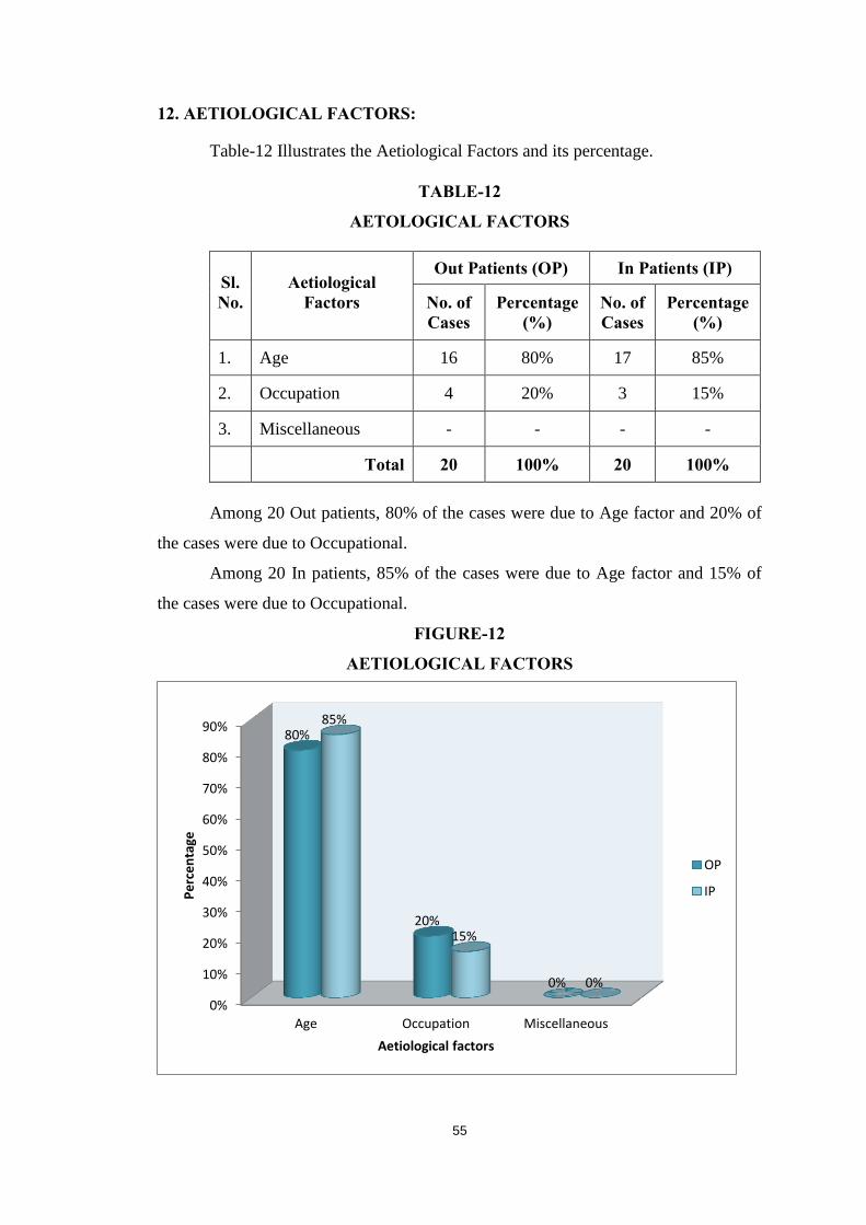
12. AETIOLOGICAL FACTORS:
Table-12 Illustrates the Aetiological Factors and its percentage.
TABLE-12
AETOLOGICAL FACTORS
Sl. No.
Aetiological Factors
Out Patients (OP) In Patients (IP)
No. of Cases
Percentage (%)
No. of Cases
Percentage (%)
1. Age 16 80% 17 85%
2. Occupation 4 20% 3 15%
3. Miscellaneous - - - -
Total 20 100% 20 100%
Among 20 Out patients, 80% of the cases were due to Age factor and 20% of
the cases were due to Occupational.
Among 20 In patients, 85% of the cases were due to Age factor and 15% of
the cases were due to Occupational.
FIGURE-12
AETIOLOGICAL FACTORS

56
0%
10%
20%
30%
40%
50%
60%
70%
80%
90%
100%
Acute Chronic
0%
100%
0%
100%
Pe
rece
nta
ge
Mode of onset
OP
IP
13. MODE OF ONSET:
Table-13 Illustrates the Mode of Onset and its percentage.
TABLE-13
MODE OF ONSET
Sl. No. Mode of Onset
Out Patients (OP) In Patients (IP)
No. of Cases
Percentage (%)
No. of Cases
Percentage (%)
1. Acute - - - -
2. Chronic 20 100% 20 100%
Total 20 100% 20 100%
Among 20 Out patients, 100% of the cases were Chronic onset.
Among 20 In patients, 100% of the cases were Chronic onset.
FIGURE-13
MODE OF ONSET

57
14. DURATION OF ILLNESS:
Table-14 Illustrates the Duration of Illness and its percentage.
TABLE-14
DURATION OF ILLNESS
Sl. No. Duration of Illness
Out Patients (OP) In Patients (IP)
No. of Cases
Percentage (%)
No. of Cases
Percentage (%)
1. 1 – 2 months - - 3 15%
2. 2 - 3 months 11 55% - -
3. 3 – 4 months 4 20% 2 10%
4. 4 – 5 months - - 3 15%
5. 5 – 6 months - - 1 5%
6. 6 – 7 months 5 25% 8 40%
7. 7 – 8 months - - - -
8. Above 8 months - - 3 15%
Total 20 100% 20 100%
Among 20 Out patients, Duration of Illness is 55% in 2 – 3 months, 20% in 3
– 4 months and 25% in 6 – 7 months.
Among 20 In patients, Duration of Illness is 15% in 1-2 months, 10% in 3 – 4
months, 15% in 4 – 5 months, 5 % in 5 – 6 months, 40% in 6 – 7 months and 15% in
above 8 months.
FIGURE-14
DURATION OF ILLNESS
0%
10%
20%
30%
40%
50%
60%
1 – 2 months
2 - 3months
3 – 4 months
4 – 5 months
5 – 6 months
6 – 7 months
7 – 8 months
Above 8months
0%
55%
20%
0% 0%
25%
0% 0%
15%
0%
10%
15%
5%
40%
0%
15%
Pe
rce
nta
ge
Duration of illness
OP
IP

58
0%
20%
40%
60%
80%
100%
Painfulshoulder Stiffness
TendernessDifficulty inmoving the
shoulder joint
100%
80%
40%
75%
100%
90%
55%
50%
Pe
rce
nta
ge
Clinical manifestation
OP
IP
15. CLINICAL MANIFESTATION:
Table-15 Illustrates the Clinical Manifestation and it percentage.
TABLE-15
CLINICAL MANIFESTATION
Sl. No. Clinical manifestation
Out Patients (OP) In Patients (IP)
No. of Cases
Percentage (%)
No. of Cases
Percentage (%)
1. Painful shoulder 20 100% 20 100%
2. Stiffness 16 80% 18 90%
3. Tenderness 8 40% 11 55%
4. Difficulty in moving
the shoulder joint 15 75% 10 50%
Among 20 Out patients, 100% of the cases had Painful shoulder, 80% of the
cases had Stiffness, 40% of the cases had Tenderness, and 75% of the cases had
Difficulty in moving the shoulder joint.
Among 20 In patients, 100% of the cases had Pain in their shoulder, 90% of
the cases had Stiffness, 55% of the cases had Tenderness, and 50% of the cases had
Difficulty in moving the shoulder joint.
FIGURE-15
CLINICAL MANIFESTATION

59
0%
20%
40%
60%
80%
100%
MeiVai
KanMookku
Sevi
100%
0% 0%
0% 0%
100%
0% 0%
0% 0%
Pe
rce
nta
ge
Gnanendrium
OP
IP
16. GNANENDRIUM:
Table-16 Illustrates the Gnanendrium and its percentage.
TABLE-16
GNANENDRIUM
Sl. No. Gnanendrium
Out Patients (OP) In Patients (IP)
No. of Cases
Percentage (%)
No. of Cases
Percentage (%)
1. Mei 20 100% 20 100
2. Vai - - - -
3. Kan - - - -
4. Mookku - - - -
5. Sevi - - - -
Total 20 100% 20 100%
In both Out patients and In patients, 100% Mei was affected.
FIGURE-16
GNANENDRIUM

60
0%
10%
20%
30%
40%
50%
60%
70%
80%
90%
100%
Vaai Kaal Kai Eruvai Karuvai
0% 0%
100%
0% 0% 0% 0%
100%
0% 0%
Pe
rce
nta
ge
Kanmendrium
OP
IP
17. KANMENDRIUM:
Table-17 Illustrates the Kanmendrium and its percentage.
TABLE-17
KANMENDRIUM
Sl. No. Kanmendrium
Out Patients (OP) In Patients (IP)
No. of Cases
Percentage (%)
No. of Cases
Percentage (%)
1. Vaai - - - -
2. Kaal - - - -
3. Kai 20 100% 20 100%
4. Eruvai - - - -
5. Karuvai - - - -
Total 20 100% 20 100%
In both Out patients and In patients, 100% Kai was affected.
FIGURE-17
KANMENDRIUM

61
18. CONDITIONS OF MUKKUTRAM:
Table-18 (a). Illustrates the Condition in Vatham and its percentage.
TABLE-18 (a)
CONDITION IN VATHAM
Sl. No.
Condition in Vatham
Out Patients (OP) In Patients (IP)
No. of Cases
Percentage (%)
No. of Cases
Percentage (%)
1. Piranan 2 10% 2 10%
2. Abanan - - - -
3. Viyanan 20 100% 20 100%
4. Udhanan - - - -
5. Samanan 20 100% 20 100%
6. Naagan - - - -
7. Koorman - - - -
8. Kirukaran - - - -
9. Dhevathathan 2 10% 2 10%
10. Dhananjeyan - - - -
Among 20 Out patients, 10% of the cases were affected in Piranan,
100% of the cases affected in Viyanan, 100% of the cases were affected in
Samanan and 10% of the cases were affected in Dhevathathan.
Among 20 In patients, 10% of the cases were affected in Piranan,
100% of the cases affected in Viyanan, 100% of the cases were affected in
Samanan and 10% of the cases were affected in Dhevathathan.

62
0%
20%
40%
60%
80%
100%
10%
0%
100%
0%
100%
0% 0% 0%
10%
0%
10%
0%
100%
0%
100%
0% 0% 0%
10%
0%
Pe
rce
nta
ge
Condition in Vatham
OP
IP
FIGURE-18 (a)
CONDITION IN VATHAM

63
0%
10%
20%
30%
40%
50%
60%
70%
80%
90%
100%
Anala pitham Ranjagam Prasagam Alosagam Sathagam
0% 0% 0% 0%
100%
0% 0% 0% 0%
100%
Pe
rce
nta
ge
Condition in Pitham
OP
IP
18 (b). CONDITIONS OF PITHAM:
Table-18 (b) Illustrates the Condition in Pitham and its percentage.
TABLE-18 (b)
CONDITION IN PITHAM
Sl. No.
Condition in Pitham
Out Patients (OP) In Patients (IP)
No. of Cases
Percentage (%)
No. of Cases
Percentage (%)
1. Anala pitham - - - -
2. Ranjagam - - - -
3. Prasagam - - - -
4. Alosagam - - - -
5. Sathagam 20 100% 20 100%
Among 20 Out patients, 100% of the cases were affected in Sathagam
and Among 20 In Patients 100% of the cases were affected in Sathagam.
FIGURE-18 (b)
CONDITION IN PITHAM

64
0%
20%
40%
60%
80%
100%
AvalambagamKilethagam
PothagamTharpagam
Santhigam
0% 0%
0% 0%
100%
0% 0%
0% 0%
100%
Pe
rce
nta
ge
Condition in Kapham
OP
IP
18 (c). CONDITIONS IN KAPHAM:
Table-18 (c) Illustrates the Condition in Kapham.
TABLE-18 (c)
CONDITION IN KAPHAM
Sl. No.
Condition in Kapham
Out Patients (OP) In Patients (IP)
No. of Cases
Percentage (%)
No. of Cases
Percentage (%)
1. Avalambagam - - - -
2. Kilethagam - - - -
3. Pothagam - - - -
4. Tharpagam - - - -
5. Santhigam 20 100% 20 100%
Among 20 Out patients, 100% of the cases were affected in Santhigam
and Among 20 In Patients 100% of the cases were affected in Santhigam.
FIGURE-18 (c)
CONDITION IN KAPHAM

65
19. INVOLVEMENT OF UDAL THATHUKKAL:
Table-19 Illustrates the involvement of Udal thathukkal and its percentage.
TABLE-19
INVOLVEMENT OF UDAL THATHUKKAL
Sl. No. Udal Thathukkal
Out Patients (OP) In Patients (IP)
No. of Cases
Percentage (%)
No. of Cases
Percentage (%)
1. Saaram 20 100% 20 100%
2. Senneer - - - -
3. Oon - - - -
4. Kozhuppu 10 50% 10 50%
5. Enbu 20 100% 20 100%
6. Moolai - - - -
7. Sukkilam /
suronitham - - - -
Among 20 Out patients, 100% of the cases were affected in Saaram,
50% of the cases were affected in Kozhuppu and 100% of the cases were
affected in Enbu.
Among 20 In patients, 100% of the cases were affected in Saaram,
50% of the cases were affected in Kozhuppu and 100% of the cases were
affected in Enbu.

66
0%
10%
20%
30%
40%
50%
60%
70%
80%
90%
100%
Saaram Senneer Oon Kozhuppu Enbu Moolai Sukkilam /suronitham
100%
0% 0%
50%
100%
0% 0%
100%
0% 0%
50%
100%
0% 0%
Pe
rce
nta
ge
Udal Thathukkal
OP
IP
FIGURE-19
INVOLVEMENT OF UDAL THATHUKKAL

67
20. CONDITIONS OF ENVAGAI THERVUGAL:
Table-20 Illustrates the conditions of Envagai Thervugal and its percentage.
TABLE-20
CONDITIONS OF ENVAGAI THERVUGAL
Sl. No. Envagai Thervugal
Out Patients (OP) In Patients (IP)
No. of Cases
Percentage (%)
No. of Cases
Percentage (%)
1. Naa - - - -
2. Niram - - - -
3. Mozhi - - - -
4. Vizhi - - - -
5. Malam - - - -
6. Moothiram - - - -
7. Sparisam 15 75% 10 50%
8. Naadi (Thontha
Naadi) 20 100% 20 100%
Among 20 Out patients, 75% of the cases were affected in Sparisam,
and 100% of the cases had Thontha Naadi.
Among 20 In patients, 75% of the cases were affected in Sparisam, and
100% of the cases had Thontha Naadi.

68
0%10%20%30%40%
50%
60%
70%
80%
90%
100%
NaaNiram
MozhiVizhi
MalamMoothiram
SparisamNaadi
(ThonthaNaadi)
0% 0% 0% 0% 0% 0%
75%
100%
0% 0% 0% 0% 0% 0%
50%
100%
Pe
rce
nta
ge
Conditions of Envagai Thervugal
OP
IP
FIGURE-20
CONDITIONS OF ENVAGAI THERVUGAL

69
0%
20%
40%
60%
80%
100%
VathaPitham
VathaKapham
PithaVatham
PithaKapham
KaphaVatham
KaphaPitham
Total
80%
0%
20%
0% 0%
0%
100%
100%
0% 0% 0% 0%
0%
100%
Pe
rce
nta
ge
Naadi
OP
IP
21. NAADI:
Table-21 Illustrates the Naadi and its percentage.
TABLE-21
NAADI
Sl. No. Naadi
Out Patients (OP) In Patients (IP)
No. of Cases
Percentage (%)
No. of Cases
Percentage (%)
1. Vatha Pitham 16 80% 20 100%
2. Vatha Kapham - - - -
3. Pitha Vatham 4 20% - -
4. Pitha Kapham - - - -
5. Kapha Vatham - - - -
6. Kapha Pitham - - - -
Total 20 100% 20 100%
Among 20 Out patients, 80% of the cases were Vatha Pitham and 20% of the
cases were Pitha Vatham. Among 20 In patients, 100% of the cases were Vatha
Pitham.
FIGURE-21
NAADI

70
0%
5%
10%
15%
20%
25%
30%
35%
Niram Manam Edai Nurai Enjal
30%
0% 0%
20%
0%
35%
0% 0%
10%
0%
Pe
rce
nta
ge
Neer kuri
OP
Ip
22. NEER KURI
Table-22 Illustrates the Neer Kuri and its percentage.
TABLE-22
NEER KURI
Sl. No. Neer kuri
Out Patients (OP) In Patients (IP)
No. of Cases
Percentage (%)
No. of Cases
Percentage (%)
1. Niram 6 30% 7 35%
2. Manam - - - -
3. Edai - - - -
4. Nurai 4 20% 2 10%
5. Enjal - - - -
Among 20 Out patients 30% of the cases were affected in Niram and 20%
cases of the cases were affected in Nurai. Among 20 In patients 35% of the cases
were affected in Niram and 10% of the cases were affected in Nurai. FIGURE-22
NEER KURI

71
0%
20%
40%
60%
80%
100%
Vatha Neer(Spreading like
snake)
Pitha NeerSpreading like
ring)
Kapha Neer(Spreading
pearl)
Thontha Neer(Asathiya Nei
Kurigal)
100%
0% 0%
0%
100%
0% 0%
0%
Pe
rce
nta
ge
Nei Kuri
OP
IP
23. NEI KURI
Table-22 Illustrates the Neikuri and its percentage.
TABLE-23
NEI KURI
Sl. No. Nei kuri
Out Patients (OP) In Patients (IP)
No. of Cases
Percentage (%)
No. of Cases
Percentage (%)
1. Vatha Neer
(Spreading like snake) 20 100% 20 100%
2. Pitha Neer Spreading
like ring) - - - -
3. Kapha Neer
(Spreading pearl) - - - -
4. Thontha Neer
(Asathiya Nei Kurigal) - - - -
Total 20 100% 20 100%
Among 20 Out patients and 20 In patients 100% of the cases were vatham.
FIGURE-23
NEI KURI

72
24. ASSESSMENT OF OUTCOME;
Table-24 Illustrates the pain assessment scale and its percentage.
TABLE-24
ASSESSMENT OF OUTCOME
Sl. No.
Pain scale
Before Treatment After Treatment
Out Patients
(OP)
In Patients (IP)
Out Patients
(OP)
In Patients
(IP) N
o. o
f C
ases
Perc
enta
ge
(%)
No.
of
Cas
es
Perc
enta
ge
(%)
No.
of
Cas
es
Perc
enta
ge
(%)
No.
of
Cas
es
Perc
enta
ge
(%)
1. 0 – 19 12 60% 2 10% 11 55% 2 10%
2. 20 – 29 5 25% 3 15% 5 25% 2 10%
3. 30 – 39 3 15% 2 10% 4 20% 2 10%
4. 40 – 48 0 0% 13 65% 0 0 0 0%
Assessment of out come:
Oxford shoulder score:
0-19 indicate severe shoulder arthritis
20-29 indicate moderate to severe
30-39 indicate middle to moderate
40-48 indicate satisfactory joint function
Ref: F. Younis, J.Sultan, S.Dire, P.J. Hughes the range of the oxford shoulder
score in the asymptomatic population , Vol.8, Page No.93, Edition Nov.2011.

73
0%
10%
20%
30%
40%
50%
60%
70%
Percentage (%) Percentage (%) Percentage (%) Percentage (%)
OP IP OP IP
Before treatment After treatment
60%
10%
55%
10%
25%
15%
25%
10%
15%
10%
20%
10%
Pe
rce
nta
ge
0 – 19
20 – 29
30 – 39
40 – 48
Before Treatment:
Pain score OP IP
0-19 60% 55%
20-29 25% 25%
30-39 15% 20%
40-48 0% 0%
High incidence of outcome is reported in the pain score range between 0-19.
After Treatment:
Pain score OP IP
0-19 10% 10%
20-29 15% 10%
30-39 10% 10%
40-48 65% 70%
High incidence of outcome is reported in the pain score range between 40-48.
FIGURE-24
ASSESSMENT OF OUTCOME

74
0%
20%
40%
60%
80%
ImproveModeratelyimproved Not improved
65%
20%
15%
70%
20%
10%
Pe
rce
nta
ge
Results
OP
IP
25. GRADATION OF RESULTS:
Table-25 Illustrates the pain Gradation Results and its percentage.
TABLE-25
GRADATION OF RESULTS
Sl. No. Results
Out Patients (OP) In Patients (IP)
No. of Cases
Percentage (%)
No. of Cases
Percentage (%)
1. Improve 13 65% 14 70%
2. Moderately
improved 4 20% 4 20%
3. Not improved 3 15% 2 10%
Total 20 100% 20 100%
Among In patients, 65% of cases were Improved, 20% of cases were
Moderately improved and 15% of cases were Not improved.
Among Out patients, 70% of cases were Improved, 20% of cases were
Moderately improved and 10% of cases were Not improved.
FIGURE-25
GRADATION OF RESULTS

75
TABLE-26 LABORATORY INVESTIGATION a). Out Patients
Sl. N
o.
Out
Pat
ient
No.
HAEMOTOLOGICAL REPORT BIO-CHEMICAL ANALYSIS URINE ANALYSIS
BEFORE TREATMENT AFTER TREATMENT BEFORE TREATMENT
AFTER TREATMENT
BEFORE TREATMENT
AFTER TREATMENT
TC
DC ESR (mm)
Hb% (gms) TC
DC ESR (mm)
Hb% (gms) Su
gar
Ure
a
Cho
lest
erol
Suga
r
Ure
a
Cho
lest
erol
Alb
umin
Suga
r
Dep
- Epi
. cel
ls /
Pus c
ells
Alb
umin
Suga
r
Dep
- Epi
cel
ls /
Pus c
ells
P L
E
½ H
r.
1 H
r.
P L
E
½ H
r.
1 H
r.
1. 8729 8600 64 34 5 10 12 11.40 8400 62 36 4 8 10 11.40 212 17 144 210 19 146 NIL NIL NAD NIL NIL NAD 2. 9171 8300 61 37 3 14 18 10.40 8200 63 34 3 7 18 10.40 173 14 149 170 12 146 NIL NIL NAD NIL NIL NAD 3. 9236 8400 63 35 2 12 20 12.60 8300 60 32 2 12 12 12.60 179 20 188 176 18 182 NIL NIL NAD NIL NIL NAD 4. 9998 8800 66 30 4 10 16 11.80 8600 68 36 4 10 18 11.80 192 18 183 190 16 180 NIL NIL NAD NIL NIL NAD 5. 10048 8600 70 26 4 12 16 10.80 8800 72 24 4 14 12 10.80 182 16 167 180 18 164 NIL Trace 1-2 pus
cells NIL NIL NAD
6. 10970 8500 68 28 5 15 20 12.20 8600 66 30 5 6 18 12.20 180 22 130 176 20 128 NIL NIL NAD NIL NIL NAD 7. 11199 7800 60 36 4 10 14 11.60 7600 64 34 4 14 13 11.60 112 13 168 116 14 162 NIL NIL NAD NIL NIL NAD 8. 11248 7600 64 32 3 16 10 12.80 7800 66 36 3 10 18 12.80 234 15 154 230 16 152 NIL NIL NAD NIL NIL NAD 9. 11248 8200 56 38 2 9 18 11.20 8400 58 32 2 15 14 11.20 232 17 180 228 15 176 NIL NIL NAD NIL NIL NAD 10. 11353 8600 58 40 2 16 20 12.40 8800 56 44 2 7 18 12.40 225 19 182 221 18 180 NIL NIL NAD NIL NIL NAD 11. 13655 7500 64 32 6 10 18 12.60 7300 66 28 6 11 6 12.60 144 21 160 142 20 158 NIL NIL Few pus
cells NIL NIL NAD
12. 14130 7200 66 32 4 8 10 12.80 7400 68 34 4 3 8 12.80 282 23 205 280 22 200 NIL NIL NAD NIL NIL NAD 13. 14357 7400 70 28 3 14 18 11.80 7200 64 32 3 4 16 11.80 230 18 174 228 19 172 NIL NIL NAD NIL NIL NAD 14. 14763 8000 68 30 2 10 16 11.20 8200 65 34 2 13 18 11.20 216 17 182 214 18 180 NIL NIL NAD NIL NIL NAD 15. 14837 8200 66 36 3 16 20 11.80 8400 68 34 3 12 18 11.80 228 20 186 224 19 184 NIL NIL NAD NIL NIL NAD 16. 15316 8400 64 32 4 12 14 12.80 8300 62 30 4 9 20 12.80 214 19 172 212 20 170 NIL NIL NAD NIL NIL NAD 17. 15387 8700 68 26 5 9 16 10.80 8600 70 28 5 10 16 10.80 220 22 166 218 21 164 NIL NIL NAD NIL NIL NAD 18. 15388 8500 64 30 6 13 20 11.60 8400 66 32 6 8 20 11.60 230 24 184 228 23 180 NIL NIL NAD NIL NIL NAD 19. 15700 8200 60 34 4 7 14 12.80 8300 64 30 4 11 12 12.80 216 15 168 214 16 170 NIL NIL NAD NIL NIL NAD 20. 17605 8800 56 38 2 13 20 11.00 8600 58 36 2 6 16 11.00 222 23 178 220 21 174 NIL NIL NAD NIL NIL NAD

76
TABLE-26 LABORATORY INVESTIGATION b). In Patients
Sl. N
o.
In P
atie
nt N
o.
HAEMOTOLOGICAL REPORT BIO-CHEMICAL ANALYSIS URINE ANALYSIS
BEFORE TREATMENT AFTER TREATMENT BEFORE TREATMENT
AFTER TREATMENT
BEFORE TREATMENT
AFTER TREATMENT
TC
DC ESR (mm)
Hb% (gms) TC
DC ESR (mm)
Hb% (gms) Suga
r
Ure
a
Cho
lest
erol
Suga
r
Ure
a
Cho
lest
erol
Alb
umin
Suga
r
Dep
- Epi
. cel
ls /
Pus c
ells
Alb
umin
Suga
r
Dep
- Epi
. cel
ls /
Pus c
ells
P L
E
½ H
r.
1 H
r.
P L
E
½ H
r.
1 H
r.
1. 218 8200 62 34 4 16 18 10.60 8000 64 34 6 10 14 10.80 358 17 192 354 15 190 NIL NIL NAD NIL NIL NAD 2. 388 7000 58 40 2 14 16 11.20 7200 58 38 4 8 16 11.20 145 27 149 142 24 146 NIL NIL NAD NIL NIL NAD 3. 420 8400 60 38 2 18 30 9.40 9400 58 38 4 10 10 9.90 177 29 262 174 25 260 NIL NIL NAD NIL NIL NAD 4. 483 8400 60 36 4 12 14 11.00 8600 62 36 4 8 12 11.40 105 40 196 108 38 194 NIL NIL 1-2 pus
cells NIL NIL NAD
5. 535 8300 60 37 3 14 18 10.20 8400 58 38 4 10 12 10.60 367 26 165 364 24 162 NIL +++ 1-2 pus
cells NIL NIL NAD
6. 783 8600 64 33 3 13 18 10.40 8800 60 36 4 6 8 10.60 285 22 163 282 20 160 NIL NIL NAD NIL NIL NAD 7. 859 7800 55 40 5 14 18 11.90 7600 55 40 5 8 10 12.20 318 27 140 314 25 138 NIL +++ Few pus
cells NIL NIL NAD
8. 881 7600 56 38 6 15 20 10.40 7400 57 32 6 8 12 10.60 220 22 152 216 20 150 NIL NIL NAD NIL NIL NAD 9. 897 7200 62 34 4 14 18 11.20 7000 60 36 4 7 10 11.60 232 21 156 230 18 154 NIL NIL NAD NIL NIL NAD 10. 910 8800 60 38 2 18 20 11.80 8600 60 38 2 6 8 12.20 210 18 160 208 16 156 NIL NIL NAD NIL NIL NAD 11. 969 7400 62 34 4 17 20 10.80 7500 60 38 4 10 12 11.00 228 22 172 224 20 170 NIL NIL NAD NIL NIL NAD 12. 1100 7200 63 32 5 16 18 11.40 7400 62 32 6 8 10 11.60 205 20 158 203 18 160 NIL NIL NAD NIL NIL NAD 13. 1116 8600 60 36 4 13 16 10.40 8400 60 36 4 10 12 10.60 216 18 160 214 16 162 NIL NIL NAD NIL NIL NAD 14. 1127 8800 62 35 3 15 19 10.60 8600 60 35 5 7 9 10.80 225 17 164 223 15 163 NIL NIL NAD NIL NIL NAD 15. 1140 8600 63 35 2 16 15 11.20 8800 62 34 4 10 12 11.40 230 15 162 228 13 160 NIL NIL NAD NIL NIL NAD 16. 1149 8400 60 37 2 14 16 11.40 8200 60 42 2 10 12 11.60 218 18 168 216 16 166 NIL NIL NAD NIL NIL NAD 17. 1160 8200 63 34 3 18 20 12.20 8400 63 34 3 11 13 12.40 180 21 170 178 20 168 NIL NIL NAD NIL NIL NAD 18. 1180 8600 58 38 4 13 16 12.40 8800 58 36 6 6 8 12.60 172 25 172 170 23 170 NIL NIL NAD NIL NIL NAD 19. 1221 8300 54 40 3 16 20 10.80 8600 58 30 2 10 12 11.00 186 24 174 184 22 172 NIL NIL NAD NIL NIL NAD 20. 1231 8600 54 40 6 16 18 10.80 8400 54 40 6 8 10 11.00 157 19 117 153 17 115 NIL NIL NAD NIL NIL NAD

77
TABLE-27 CASE SUMMARY (a) OUT PATIENTS
Sl. No.
OP No. Name Age /
Sex Treatment
starting date
End of the treatment
date
Duration of Illness
Treatment with Dose
Total No. of days
treated
Radiological findings Result
1. 8729 Indira 48 / F 27.01.2016 25.02.2016 4 Months
All
th
e ca
ses
wer
e tr
eate
d w
ith M
ukkir
atta
i C
hoora
nam
2 g
m t
wic
e a
day
wit
h h
ot
wat
er
30 Normal Good
2. 9171 Boominathan 60 / M 28.01.2016 26.02.2016 6 Months 30 Osteophytic change + Fair
3. 9236 Ramanujam 60 / M 28.01.2016 26.02.2016 4 Months 30 Narrowing of joint space Fair
4. 9998 Valliammal 52 / F 30.01.2016 28.02.2016 3 Months 30 Normal Good
5. 10048 Mari 35 / F 30.01.2016 28.02.2016 4 Months 30 Narrowing of joint space Fair
6. 10910 Madasamy 60 / M 02.02.2016 02.03.2016 6 Months 30 Osteophytic change + Fair
7. 11199 Sankarammal 55 / F 03.02.2016 03.03.2016 6 Months 30 Normal Good
8. 11248 Mohammed Raja 42 / M 03.02.2016 03.03.2016 3 Months 30 Normal Good
9. 11285 Kannan 42 / M 03.02.2016 03.03.2016 3 Months 30 Osteophytic change + Fair
10. 11353 Sumathi 45 / F 03.02.2016 03.03.2016 7 Months 30 Normal Good
11. 13655 Srinivasan 57 / M 10.02.2016 10.03.2016 4 Months 30 Normal Good
12. 14130 Ponpandi 58 / M 11.02.2016 11.03.2016 6 Months 30 Normal Good
13. 14357 Saradha 58 / F 12.02.2016 12.03.2016 3 Months 30 Normal Good
14. 14763 Murugan 60 / M 13.02.2016 13.03.2016 3 Months 30 Normal Good
15. 14837 Sri Ram 40 / M 13.02.2016 13.03.2016 2 Months 30 Normal Good
16. 15316 Jeyamary 60 / F 15.02.2016 15.03.2016 2 Months 30 Normal Good
17. 15387 Mala Antony 47 / F 15.02.2016 15.03.2016 2 Months 30 Normal Good
18. 15388 Akila Nepolien 43 / F 15.02.2016 15.03.2016 3 Months 30 Normal Good
19. 15700 Thangamani 60 / F 16.02.2016 16.03.2016 2 Months 30 Osteophytic change + Fair
20. 17605 Abdul Kadhar 43 / M 22.02.2016 24.03.2016 3 Months 30 Normal Good

78
TABLE-27 CASE SUMMARY OUT PATIENTS
Sl. No. OP No. Name Age /
Sex
RA Factors (IU / ml)
ECG
1. 8729 Indira 48 / F 16.5 Normal
2. 9171 Boominathan 60 / M 12.2 Normal
3. 9236 Ramanujam 60 / M 17.3 Normal
4. 9998 Valliammal 52 / F 11.6 Normal
5. 10048 Mari 35 / F 10.1 Normal
6. 10910 Madasamy 60 / M 12.2 Normal
7. 11199 Sankarammal 55 / F 13.1 Normal
8. 11248 Mohammed Raja 42 / M 14.6 Normal
9. 11285 Kannan 42 / M 11.8 Normal
10. 11353 Sumathi 45 / F 18.2 Normal
11. 13655 Srinivasan 57 / M 13.8 Normal
12. 14130 Ponpandi 58 / M 12.8 Normal
13. 14357 Saradha 58 / F 16.4 Normal
14. 14763 Murugan 60 / M 17.5 Normal
15. 14837 Sri Ram 40 / M 17.8 Normal
16. 15316 Jeyamary 60 / F 12.3 Normal
17. 15387 Mala Antony 47 / F 14.2 Normal
18. 15388 Akila Nepolien 43 / F 16.2 Normal
19. 15700 Thangamani 60 / F 18.7 Normal
20. 17605 Abdul Kadhar 43 / M 19.2 Normal
Reference range:
Rheumatoid factor : Upto 20 IU/mL

79
TABLE-27 CASE SUMMARY (b) IN PATIENTS
Sl. No.
IP No. Name Age /
Sex Treatment
starting date
End of the treatment
date
Duration of Illness
Treatment with Dose
No. of days treated
Total No. of days
treated
Radiological findings
Res
ult
IP Follow 1. 218 Mani 60 / M 29.01.2016 27.02.2016 1 Month
All
th
e ca
ses
wer
e tr
eate
d w
ith M
ukkir
atta
i C
hoora
nam
2 g
m t
wic
e a
day
wit
h h
ot
wat
er
30 - 30 Normal Good
2. 288 Parvathi 60 / F 15.02.2016 16.03.2016 1 Year 31 - 31 Osteophytic changes + Fair
3. 420 Sundari 50 / F 17.02.2016 18.03.2016 2 Months 31 - 31 Normal Good
4. 483 Aavudaiyammal 49 / F 23.02.2016 04.03.2016 6 Months 11 19 30 Normal Good
5. 535 Manikandan 59 / M 28.02.2016 28.03.2016 4 Months 30 - 30 Normal Good
6. 783 Krishnamoorthy 60 / M 24.03.2016 23.04.2016 3 Months 31 - 31 Narrowing of joint space Fair
7. 859 Mani 60 / M 29.03.2016 27.04.2016 6 Months 30 - 30 Normal Good
8. 881 Velammal 60 / F 31.03.2016 22.04.2016 3 Months 23 7 30 Osteophytic changes + Fair
9. 897 Subbammal 58 / F 02.04.2016 02.05.2016 6 Months 31 - 31 Normal Good
10. 910 Mookammal 50 / F 03.04.2016 25.04.2016 5 Months 23 7 30 Normal Good
11. 969 Latha 45 / F 08.04..2016 14.04.2016 7 Months 7 23 30 Normal Good
12. 1100 Moorthi 60 / M 24.04.2016 23.05.2016 4 Months 30 - 30 Narrowing of joint space Fair
13. 1116 Paramashivam 58 / M 26.04.2016 11.05.2016 1 Month 16 14 30 Normal Good
14. 1127 Aarumugam 39 / F 27.04.2016 27.05.2016 6 Months 31 - 31 Normal Good
15. 1140 Chettah 55 / M 28.04.2016 21.05.2016 8 Months 24 6 30 Normal Good
16. 1149 Gomathi 50 / F 29.04.2016 21.05.2016 6 Months 23 7 30 Normal Good
17. 1160 Parvathy 55 / F 02.05.2016 14.05.2016 4 Months 13 17 30 Normal Good
18. 1180 Palpandi 56 / M 03.05.2016 01.06.2016 7 Months 30 - 30 Normal Good
19. 1221 Ravindran 52 / M 07.05.2016 05.06.2016 7 Months 30 - 30 Normal Good
20. 1231 Ramasamy 60 / M 09.05.2016 09.06.2016 1 Year 32 - 32 Osteophytic changes + Fair

80
TABLE-27 CASE SUMMARY IN PATIENTS
Sl. No.
IP No. Name Age /
Sex
RA Factors (IU / ml)
ECG
1. 218 Mani 60 / M 16.4 Normal
2. 288 Parvathi 60 / F 17.5 Normal
3. 420 Sundari 50 / F 17.8 Normal
4. 483 Aavudaiyammal 49 / F 13.8 Normal
5. 535 Manikandan 59 / M 11.8 Normal
6. 783 Krishnamoorthy 60 / M 14.2 Normal
7. 859 Mani 60 / M 17.2 Normal
8. 881 Velammal 60 / F 13.1 Normal
9. 897 Subbammal 58 / F 10.8 Normal
10. 910 Mookammal 50 / F 11.2 Normal
11. 969 Latha 45 / F 18.6 Normal
12. 1100 Moorthi 60 / M 9.6 Normal
13. 1116 Paramashivam 58 / M 8.2 Normal
14. 1127 Aarumugam 39 / F 17.2 Normal
15. 1140 Chettah 55 / M 9.6 Normal
16. 1149 Gomathi 50 / F 18.2 Normal
17. 1160 Parvathy 55 / F 8.1 Normal
18. 1180 Palpandi 56 / M 12.4 Normal
19. 1221 Ravindran 52 / M 12.2 Normal
20. 1231 Ramasamy 60 / M 14.3 Normal
Reference range:
Rheumatoid factor : Upto 20 IU/mL

81
CHAPTER-VI
DISCUSSION
Among 80 types of Vatha disease, Kumba Vatham is one of them,
characterized by inflammation of joint, stiffness and paining shoulder joint.
As the joint becomes progressively painful and stiffer, simple movement such
as raising the arm becomes difficult. The inflammation occurs within the
capsule itself.
So Vatha disease has received more attention and good prognosis is the field
of alternative medicine, specially in Siddha medicine. But they need scientific
approach to study and prove it.
All patients were subjected to preliminary investigations which include
radiological, haematological and urine examination.
The trial medicine ‘Mukkirattai Chooranam’ was administered from the
next day on wards. The course of treatment is 30 days twice a day with hot
water.
20 Out and In patients were subjected on this, results can be discussed as
follows,
1. Sex Distribution:
Among 20 Out patients, 50% were Male and 50% were Female.
Among 20 In patients, 50% were Male and 50% were Female.
2. Age Distribution:
Among 20 Out patients, 10% were in the age group of 31 – 40
years, 35% were in the age group of 41 – 50 years, 55% were in the
age group of 51 – 60 years.
Among 20 In patients, 5% were in the age group of 31 – 40
years, 25% were in the age group of 41 – 50 years, 70% were in the
age group of 51 – 60 years.
3. Kaalam:
In Siddha literature age of individual is fixed as 100 is to 3 Kaalam as,

82
Kapha kaalam - First 33 years and 4 months.
Pitha kaalam - Second 33 years and 4 months.
Vadha kaalam - Third 33 years and 4 months.
Among 20 Out patients and In patients, 100% of the cases belongs to
Pitha Kaalam.
4. Constitution of the body:
Among 20 Out patients, 100% were Thontha Thegi.
Among 20 In patients, 100% were Thontha Thegi.
5. Gunam:
Among 20 Out patients, 100% had Rajo Gunam.
Among 20 In patients, 100% had Rajo Gunam.
6. Religion Distribution:
Among 20 Out patients, 75% were Hindu, 10% were Muslim and
15% were Christian.
Among 20 In patients, 100% were Hindu.
7. Paruva Kaalam:
Most of cases were affected in Munpani Kaalam, Pinpani Kaalam and
Elavenil Kaalam.
8. Thinai:
Among 20 Out patients, 100% cases were in Marutham.
Among 20 In patients, 90% cases were in Marutham and 10% cases
were in Neithal.
9. Occupation:
Among 20 Out patients, 35% cases were Labours, 20% were House
Wife, 15% were Clerk and 30% were Driver.
Among 20 In patients, 55% cases were Labours, 35% cases were
House Wife, 5% cases were Clerk and 5% cases were Driver.

83
10. Food Habits:
Among 20 Out patients, 10% cases were Vegetarian and 90% cases
were Non – Vegetarian.
Among 20 In patients, 15% cases were Vegetarian and 85% cases were
Non – Vegetarian.
11. Socio – Economical status:
Among 20 Out patients, 30% cases were Middle income class and 70%
cases were Low income class.
Among 20 In patients, 25% cases were Middle income class and 75%
cases were Low income class.
12. Aetiological Factors:
Among 20 Out patients, 80% of the cases were due to Age factor and
20% of the cases were due to Occupational.
Among 20 In patients, 85% of the cases were due to Age factor and
15% of the cases were due to Occupational.
13. Mode of onset:
Among 20 Out patients, 100% of the cases were Chronic onset.
Among 20 In patients, 100% of the cases were Chronic onset.
14. Duration of illness:
Among 20 Out patients, Duration of Illness is 55% in 2 – 3 months,
20% in 3 – 4 months and Duration of Illness 25% in 6 – 7 months.
Among 20 In patients, Duration of Illness is 15% in 1-2 months, 10%
in 3 – 4 months, 15% in 4 – 5 months, 5 % in 5 – 6 months, 40% in 6 – 7
months and 15% in above 8 months.

84
15. Clinical manifestation:
Among 20 Out patients, 100% of the cases had Painful shoulder, 80%
of the cases had Stiffness, 40% of the cases had Tenderness, and 75% of
the cases had difficulty in moving the shoulder joint.
Among 20 In patients, 100% of the cases had Painful shoulder, 90% of
the cases had Stiffness, 55% of the cases had Tenderness, and 50% of the
cases had difficulty in moving the shoulder joint.
16. Gnanendrium:
In both Out patients and In patients, 100% Mei was affected.
17. Kanmendrium:
In both Out patients and In patients, 100% Kai was affected.
18. Conditions of Mukkutram:
18 (a). Condition of Vatham:
Among 20 Out patients, 10% of the cases were affected in Piranan,
100% of the cases affected in Viyanan, 100% of the cases were affected in
Samanan and 10% of the cases were affected in Dhevathathan.
Among 20 In patients, 10% of the cases were affected in Piranan,
100% of the cases affected in Viyanan, 100% of the cases were affected in
Samanan and 10% of the cases were affected in Dhevathathan.
18 (b). Condition of Pitahm:
Among 20 Out patients, 100% of the cases were affected in sathagam
and Among 20 In Patients, 100% of the cases were affected in sathagam.
18 (c). Condition of Kapham:
Among 20 Out patients, 100% of the cases were affected in santhigam
and Among 20 In Patients, 100% of the cases were affected in santhigam.

85
19. Involvement of Udal Thathukkal:
Among 20 Out patients, 100% of the cases were affected in Saaram,
50% of the cases were affected in Kozhuppu and 100% of the cases were
affected in Enbu.
Among 20 In patients, 100% of the cases were affected in Saaram,
50% of the cases were affected in Kozhuppu and 100% of the cases were
affected in Enbu.
20. Conditions of Envagai Thervugal:
Among 20 Out patients, 75% of the cases were affected sparisam, and
100% of the cases had thontha naadi.
Among 20 In patients, 75% of the cases were affected sparisam, and
100% of the cases had thontha naadi.
21. Naadi:
Among 20 Out patients, 80% of the cases were Vatha Pitham and 20%
of the cases were Pitha Vatham.
Among 20 In patients, 100% of the cases were Vatha Pitham.
22. Neer kuri:
Among 20 Out patients, 30% of the cases were affected in Niram and
20% cases of the cases were affected in Nurai.
Among 20 In patients, 35% of the cases were affected in Niram and
10% of the cases were affected in Nurai.
23. Nei kuri:
Among 20 Out patients and 20 In patients, 100% of the cases were
vatham.

86
24. Assessment of outcome:
Before Treatment:
Pain score OP IP
0-19 60% 55%
20-29 25% 25%
30-39 15% 20%
40-48 0% 0%
High incidence of outcome is reported in the pain score range
between 0-19.
After Treatment:
Pain score OP IP
0-19 10% 10%
20-29 15% 10%
30-39 10% 10%
40-48 65% 70%
High incidence of outcome is reported in the pain score range between
40-48.
Trial medicine:
All the 40 patients treated with the clinical trial medicine Mukkirattai
Chooranam with hot water for 30 days the disease and treatment are based primarily
on the derangement of Mukkutram, which again is based on the Pancha Bootham
theory. Incidence of Kumbavatham and treatment are also based on these primary
principles of Siddha medicine.
The bootham araises vatha kuttram in the body and so leads to degenerate and
weakness of shoulder joints. Increased vatha kutram is brought to normal mainly by
kaippu suvai and thuvarppu suvai.

87
a) Space + Air = Kaippu
b) Earth + Air = Thuvarppu
Thus, they decreases the vatha kutram. So, I conclude the trial drugs control
the disease Kumbavatham and it comes under Ethirurai Maruthuvam.
25. Gradation of results:
Among In patients, 65% of cases were Improved, 20% of cases were
Moderately improved and 15% of cases were Not improved.
Among Out patients, 70% of cases were Improved, 20% of cases were
Moderately improved and 10% of cases were Not improved.

88
CHAPTER-VII
SUMMARY
The purpose of the study is to reduce the symptoms of Kumba Vatham
(Periarthritis). This premilinary study of anti-vatha along with rejuventation therapy
has been tried with trial drugs and regiments.
The clinical trial drug MUKKIRATTI CHOORANAM is given prepared as
per Siddha literature. The duration of period is 30 days.
The clinical trial dose is MUKKIRATTAI CHOORANAM 2gms twice a
day with hot water.
Total 40 patients of both sex 30 - 60 years with age group were selected based
on inclusion and exclusion criteria.
Maximum incidence in age 30-60 years. Before treatment, routine blood and
urine analysis were taken in all patients.
Siddha diagnostic methods like Udal thathukkal, Envagai Thervugal, Neer
kuri and Nei kuri were documented.
The entire details of the patients were noted in the case sheet proforma.
FROM THE CLINICAL STUDY THE FOLLOWING DATA WERE
OBSERVED:
Age:
Most of the patients were affected in age of 30-60 years.
Sex:
Commonly affected in both sex.
Occupation:
Mostly affected in Labours, House wife and Drivers

89
Mukkutram:
Vatham – Piranan, Samanan, Dheva thathan was affected.
Pitham – Sathagam was affected.
Kapham – Santhigam was affected.
Udal thathukkal:
Saaram, Kozhuppu, Enbu was affected.
Naadi:
Vathapitha Naadi was commonly affected.
Neer kuri:
In most of the patients Vatham was affected.
RESULTS AFTER TREATMENT:
65% of Out patients and 70% of In patients showed good improvement, 20%
of Out & In patients showed moderate improvement, 15% of Out patient and10% In
patients showed poor improvement was observed.
In Bio-Chemical analysis, the trial medicine contains amino acid, ferrous
iron, starch, un saturated compounds.
Pharmacological studies of Mukkirattai Chooranam showed potent analgesic
and anti inflammatory effects in rats.
Clinically the drug was free from side effect.

90
CHAPTER-VIII
CONCLUSION
Kumba Vatham (Periarthritis) is primarily, due to derangement of
Vatham.
Mukkirattai Chooranam predominating with Kaippu Suvai
respectively neutralize the Vatham.
Clinical pharmacological studies which showed very good analgesic
and anti – inflammatory activity.
The trial medicine do not produce any toxicity in clinical studies. So
safe drug for prolong use to Kumba Vatham.
In Bio-chemical analysis the trial medicine contains amino acid,
ferrous iron, starch, un saturated compounds.
No toxic effects were observed during the treatment and after the
completion of treatment.
The trial medicines was reduce the symptoms of Kumba Vatham. It is
observed only by clinical and assessment outcome.
The preparation of trial medicine was easy and very low cost.
Therefore, this clinical trial medicine Mukkiratti Chooranam should
be a positive remedy for Kumba Vatham (Periarthritis).

i
ANNEXURE-I
PREPARATION OF TRIAL DRUG
LITERATURE VIEW OF TRIAL DRUG
Drug : Mukkirattai Chooranam
Ref : Mooligai Gunapadam Mudhal Vagappu (Page No.780)
Ingredient : Mukkirattai leaf
Procedure : Purified dry drug is taken and powdered and then it is filtered
using pure white cloth and kept in air tight container.
Dosage : 2 gm b.i.d. after food
Adjuvant : Hot water
Duration : 30 days
Indication : Vatha disease
TRIAL DRUG
MUKKIRATTAI:
Botanical name : Boerhavia diffusa
Family : NYCTAGINACEAE
NtW ngaH : Gl;gfk;> %f;Ful;il> ,uj;jGl;gpfh
Rit : ifg;G
jd;ik : ntg;gk;
gphpT : fhHg;G
nra;if : Nfhioafw;wp (Expectorant)
rpWePHg; ngUf;fp (Diuretic)
kykpsf;fp (Larative)
FspHr;rpAz;lhf;fp (Refrigerant)
GOf;nfhy;yp (Anthelmintic)
the;jpAz;lhf;fp (Emetic)
nghJf;Fzk; : rPj kfw;We; jpdtlf;Fq; fhe;jpjUk;
thj tpidia kbf;Fq;fhz; - Ngjp
nfhLf;Fkij cz;lhf;fhw; NfhksNk! gpj;jk;
mLf;FNk %f;Ful;ilaha;
- mfj;jpaH Fzthflk;
ii
nghUs; : Iak;> eikr;ry;> tspg;gpzp ,itfis ePf;Fk;.
moifAk;> fopr;riyAk;> moy; gpzpiaAk;
cz;lhf;Fk;.
‘%f;fpul;ilapdpiy KiwAz thj Neh
ahf;ifapw; ngl;b auntd; tlq;FNk”
- Njud; ntz;gh
nghUs; : %f;fpul;il ,iyia czT KiwahfNtDk;>
kUj;Jt KiwahfNtDk; Grpj;J tu clypYs;s
tsp Neha;fs; xd;We; jiyfhl;lhJ. mit
ngl;bapw; ghk;G Nghy mlq;fptpLk;.

iii
ANNEXURE-II
BIO-CHEMICAL ANALYSIS OF
MUKKIRATTAI CHOORANAM
Preparation of the extract:
5 gms of the drug was weighed accurately and placed in a 250ml clean beaker
then 50ml of distilled water is added and dissolved well. Then it is boiled well for
about 10 minutes. It is cooled and filtered in a 100ml volumetric flask and then it is
made to 100ml with distilled water. This extract is taken for analysis.
QUALITATIVE ANALYSIS
Sl.
No. EXPERIMENT OBSERVATION INFERENCE
1.
TEST FOR CALCIUM
2ml of the above prepared extract is taken in a
clean test tube. To this add 2ml of 4%
Ammonium oxalate solution
No white
precipitate is
formed.
Absence of
calcium
2. TEST FOR SULPHATE
2ml of the extract is added to 5% Barium
chloride solution.
No white
precipitate is
formed.
Absence of
sulphate
3. TEST FOR CHLORIDE
The extract is treated with silver nitrate solution
No white
precipitate is
formed.
Absence of
chloride
4. TEST FOR CARBONATE
The extract is treated with concentrated Hcl.
No brisk
effervescence is
formed.
Absence of
carbonate
5. TEST FOR STARCH
The extract is added with weak iodine solution
Blue colour is
formed
Indicates the
Presence of
starch
6. TEST FOR FERRIC IRON
The extract is acidified with Glacial acetic acid
and potassium ferro cyanide.
No blue colour is
formed
Absence of
ferric iron
7. TEST FOR FERROUS IRON The extract is treated with concentrated Nitric
acid and Ammonium thiocyanate solution
Blood red colour
is formed
Indicates the
presence of
ferrous iron
8. TEST FOR PHOSPHATE
The extract is treated with Ammonium
Molybdate and concentrated nitric acid
No yellow
precipitate is
formed
Absence of
phosphate
9. TEST FOR ALBUMIN
The extract is treated with Esbach’s reagent
No yellow
precipitate is
formed
Absence of
albumin

iv
10. TEST FOR TANNIC ACID
The extract is treated with ferric chloride.
No blue black
precipitate is
formed
Absence of
Tannic acid
11. TEST FOR UNSATURATION
Potassium permanganate solution is added to the
extract
It gets
decolourised
Indicates the
presence of
unsaturated
compound
12.
TEST FOR THE REDUCING SUGAR
5ml of Benedict’s qualitative solution is taken in
a test tube and allowed to boil for 2 minutes and
add 8-10 drops of the extract and again boil it
for 2 minutes.
No colour change
occurs
Absence of
reducing
sugar
13.
TEST FOR AMINO ACID
One or two drops of the extract is placed on a
filter paper and dried well. After drying, 1%
Ninhydrin is sprayed over the same and dried it
well.
Violet colour is
formed
Indicates the
presence
amino acid
14. TEST FOR ZINC
The extract is treated with Potassium
Ferrocyanide.
No white
precipitate is
formed
Absence of
zinc
Inference:
Indicates the Presence of amino acid, ferrous iron, starch and un starched
compound.

v
ANNEXURE-III
ANALGESIC ACTIVITY OF MUKKIRATTAI CHOORANAM AGAINST
ACETIC ACID INDUCED WRITHING REFLUX IN MICE
Analgesic activity of Mukkirattai Chooranam at a dose of 100 mg/kg and
200 mg/kg was evaluated by acetic acid induced writhing reflux in mice. Painful
reactions in animals may be produced by the chemicals such as phenylquionolone,
bradykinin etc. Like that, acetic acid pain reaction which is characterized as a
writhing response. Construction of abdomen, turning of trunk (twist) and extension of
hind legs are taken as reaction to chemically induced pain. Analgesics (both narcotic
and non-narcotic) inhibit writhing response.
Requirements
Animal: Swiss albino mice (20 – 25g) either sex
Drugs and chemicals: Diclofenac sodium (standard),
Acetic acid (1%), Mukkirattai Chooranam
Method:
Treatment protocol:
Group-1: Treated as normal control received 10ml/kg of normal saline through
orally
Group-2: Treated as standard control received 10mg/kg of diclofenac sodium
through orally
Group-3: Treated as Test-I received 100mg/kg of Mukkirattai Chooranam with 2
ml of sterile water through orally
Group- 4: Treated as Test- II received 200mg/kg of Mukkirattai Chooranam with 2
ml of sterile water through orally
Both dose of Mukkirattai Chooranam were administered one hour prior to the
acetic acid administration. Note the onset on writhing. Record the numbers of
abdominal contractions, trunk twist and extension of hind limbs as well as the number
of animals showing such response during a period of 10 minutes were noted.

vi
Statistics:
Data are expressed as mean ± S E M; data analyzed by one way ANOVA
followed by Dunnet’s multiple range tests to determine the significance of the
difference between the control group and rats treated with extracts.
* Values were considered significant at P < 0.01.
ANALGESIC ACTIVITY OF MUKKIRATTAI CHOORANAM
AGAINST ACETIC ACID INDUCED WRITHING REFLUX IN MICE
Treatment Dose (mg/kg) No. of
writhing
%
reduction
in reaction
time
Group-I
Normal saline
Inject 1%v/v acetic acid
1ml/100g of body weight
35.2±2.6 -
Group-II
Standard
10mg/kg Diclofenac
sodium through orally
7.5±0.7 68.70***
Group-III
Mukkirattai Chooranam
100mg/kg administered
through orally
10.96 ± 0.2 58.19***
Group-IV
Mukkirattai Chooranam
200mg/kg administered
through orally
10.4 ±0.5 67.35***
Values are expressed as mean ± S E M
Values are analysed by one way ANOVA followed by Dunnet’s multiple range tests
*** Values were considered significant at P < 0.001.
Results:
The table values show that analgesic activity of Mukkirattai Chooranam at a
dose of 100mg/kg and 200mg/kg by acetic acid induced writhing reflex. The result
reveals that both doses of Mukkirattai Chooranam possess significant analgesic
activity at P < 0.001.

vii
ANTI-INFLAMMATORY ACTIVITY
ANTI-INFLAMMATORY ACTIVITY OF MUKKIRATTAI CHOORANAM
AGAINST CARRAGEENAN INDUCED PAW EDEMA IN RATS
The anti-inflammatory activities of Mukkirattai Chooranam at a dose of
100mg/kg and 200mg/kg body weight were evaluated using Carrageenan induced paw
edema method. The inflammation was readily produced in the form of edema with the
help of the irritant such as carrageenan. Carrageenan is a sulphated polysaccharide
obtained from sea weed (Rhodophyceae) and when injected cause the release of
prostaglandins by the way it produces inflammation and edema.
Requirements:
Animal : Albino rat (180 – 200g)
Drugs and chemicals : Diclofenac sodium (standard),
carrageenan (1%), Mukkirattai Chooranam
Digital plethysmometer UGO Basile (Italy)
Method:
The animals were divided into 4 groups each having six animals
Treatment protocol:
Group-1: Treated as normal control received 10ml/kg of normal saline through
orally
Group-2: Treated as standard control received 10mg/kg of diclofenac sodium
through orally
Group-3: Treated as treatment control received 100mg/kg of Mukkirattai
Chooranam with 2 ml of sterile water through orally
Group- 4: Treated as treatment control received 200mg/kg of Mukkirattai
Chooranam with 2 ml of sterile water administered through orally
A freshly prepared suspension of carrageenan (1% w/v, 0.1ml) was injected to
the plantar region of left hind paw of each rat. One group was kept as control and the
animals of the other groups were pretreated with Mukkirattai Chooranam given
through orally 60 min before the carrageenan treatment. The paw volumes of the test
compounds, standard and control groups were measured at 60, 120, 180 minutes of

viii
carrageenan treatment with the help of Digital plethysmometer UGO Basile (Italy).
Mean increase in paw volume was measured and the percentage of inhibition was
calculated.
% anti-inflammatory activity = (Vc – Vt/Vc) x 100
Where Vt is mean increase in paw volume in rats treated with test compounds
Vc is mean increase in paw volume in control group of rats
Statistics:
Data are expressed as mean ± S E M; data analysed by one way ANOVA
followed by Dunnet’s multiple range tests to determine the significance of the
difference between the control group and rats treated with test compounds.
* Values were considered significant at P < 0.01.
ANTI-INFLAMMATORY ACTIVITY OF MUKKIRATTAI CHOORANAM
AGAINST CARRAGEENAN INDUCED PAW EDEMA IN RATS
Treatment Dose (mg/kg)
Paw volume(ml)
as measured at 3
hour
Percentage
inhibition of
paw edema
Group-I
Normal saline 5ml/kg orally 4.95 ± 0.76
Group-II
Standard
10mg/kg Diclofenac
sodium through orally 1.48 ± 0.26 70.02**
Group-III
Mukkirattai
Chooranam
100mg/kg administered
through orally 3.54 ± 0.02 50.25**
Group-IV
Mukkirattai
Chooranam
200mg/kg administered
through orally 1.98 ± 0.95 59.91**
Values are expressed as mean ± S E M
Values are analyzed by one way ANOVA followed by Dunnet’s multiple range tests,
to determine the significance of the difference between the control group and rats
treated with the test compounds.
** Values were considered significant at P < 0.01.

ix
Results:
Mukkirattai Chooranam at a dose of 100 and 200mg/kg were tested for their
anti-inflammatory activity by using carrageenan induced rat paw edema method and
the results are tabulated in table. The results reveals that both doses of Mukkirattai
Chooranam 100 and 200mg/kg possess significant anti-inflammatory activity when
compared to control group at p<0.01.

x
ANNEXURE-IV
ACUTE TOXICITY STUDY
Acute oral toxicity refers to those adverse effects occurring following oral
administration of a single dose of a substance or multiple doses given within 24 hours.
Acute toxic class method (OECD guidelines 423, (2000) was followed to arrive at the
maximum safety dose of the drug extracts. Three Wistar strain female albino rats (8-
12 weeks old, 180-200g body weight) were used in each group. Single dose (2g/kg) of
the Mukkirattai Chooranam was orally administered to overnight fasted (food but not
water withheld) animals while control animals received the vehicle (0.3%w/v CMC).
Animals were observed individually after dosing at least once during the first 4 hrs
and daily thereafter, for a total of 14days. Body weights of the animals were recorded.
The other observations include changes for skin, fur, eyes and mucous membranes,
respiratory, circulatory and autonomic and central nervous system and somatomotor
activity and behavior pattern. At the end of 14 days, all animals were subjected to
gross necropsy.
Statistics:
Data are expressed as mean ± S E M; data analysed by one way ANOVA
followed by Dunnet’s multiple range tests to determine the significance of the
difference between the control group and rats treated with test compounds.
* Values were considered significant at P < 0.5.
Results:
Acute toxicity study:
All of the rats fed with the food sample showed normal general behavior,
respiratory pattern, cardiovascular signs, motor activities, reflexes and normal change
in skin and fur.

xi
TABLE-1
HEMATOLOGICAL VALUES OF MUKKIRATTAI CHOORANAM
IN THE ACUTE TOXICITY STUDY
S.
No Parameter Control
Sample
2g/kg
1. White blood cells (x103/µl) 9.36±0.54 8.91 ±1.26
2. Hemoglobin (g/dl) 11.50±0.26 12.95 ± 0.98
3. Mean corpuscular volume 60.45±2.3 58.90 ± 0.51
4. Mea corpuscular hemoglobin conc. (g/dl) 34.56±0.86 34.51 ± 1.47
5. Platelet (x105/µl ) 5.60±0.52 4.95 ± 1.03
6. Red blood cell (x106/µl ) 3.87±0.24 2.98 ± 0.29
Values are expressed as Mean ± S E M
All groups were treated with oral dose of 2g/kg body weight
No significant different from normal control
TABLE-2
BLOOD CHEMICAL VALUES OF FOOD SAMPLE
IN THE ACUTE TOXICITY STUDY
S.
No. Parameter Control Sample 2g/kg
1 Glucose (mg/dl) 148.75±3.96 142.85 ± 2.19
2 BUN (mg/dl) 34.26±1.23 31.95 ± 0.21
3 Creatinine (mg/dl) 0.46±0.06 0.69 ± 0.14
4 Total protein (g/dl) 5.48±0.23 4.98 ±0.04
5 Albumin (g/dl) 3.49±0.62 2.98 ± 0.29
6 Total bilirubin (mg/dl) 0.26±0.02 0.38 ± 0.15
7 AST (u/l) 141.5±3.76 139.26 ± 0.29
8 ALT (u/l) 86.36±1.75 90.51 ± 0.12
9 ALP (u/l) 75.57±2.16 74.19 ± 0.38
Values are expressed as Mean ± S E M
All groups were treated with oral dose of 2g/kg body weight
No significant different from normal control

xii
DISCUSSION AND CONCLUSION:
In acute toxicity study for 14 days, at a dose of 2g/kg of Mukkirattai
Chooranam sample were chosen for the experiment. In the aspect of general
behaviours, the rats treated with food sample at a single dose had no signs of behavior
changes and toxic signs. The treated groups revealed no significant differences in
body weight gain. The increase in body weight may have resulted from physiological
changes in rats such as metabolism, food and water intake. However, the result from
animal health monitoring in the entire period of 14days showed no sign of morbidity
and diseases.
The albino Wistar rats were healthy as shown by the normal appearance of
general behavior, respiratory pattern, cardiovascular signs, motor activities, reflexes
and normal change in skin fur.
With regards to hematological values, most of values in treated groups were
normal in comparison with the control group. Significantly, some values were
different from those of the control group such as RBC, MCV, MCHC, and platelet.
However, such values are within the normal ranges. These variations may have
resulted from variation among animal groups (Feldman et al., 2000) (Inala et al.,
2002). Therefore, these results suggest that the test drug did not cause hematological
or immunological defects in rats.
Furthermore, blood chemical examination was performed in order to evaluate
any toxic effects on liver. In this study, the levels of these blood chemical values were
minor changes and remained within the normal range (Casley and King, 1980)
(Levine, 1995) (Angkhasirisap et al., 2002).
In conclusion, Mukkirattai Chooranam sample given orally to Wistar rats
did not produce toxicities.

xiii
ANNEXURE-V
GOVERNMENT SIDDHA MEDICAL COLLEGE & HOSPITAL,
PALAYAMKOTTAI, TIRUNELVELI DISTRICT
DEPARTMENT OF POTHU MARUTHUVAM
PRECLINICAL AND PHASE-II RANDOMIZED CLINICAL TRIAL ON
KUMBA VATHAM (PERIARTHRITIS) WITH
MUKKIRATTAI CHOORANAM
FORM-I
SCREENING AND SELECTION PROFORMA
1. Name___________ 2.Age_____ 3.Gender________ 4.Phone no _________
5. OP No. 6. IP No. 7. Sl. No. .
INCLUSION CRITERIA:
Age: 30 – 60 years.
Sex: both male and female
Patient having main symptoms of shoulder joint pain, radiating towards
upper arm and forearm numbness, restricted movement of upper limb, loss
of abduction and forwarded flexion followed by stiffness of the shoulder
joints.
Patient willing to sign the informed consent stating that he / she will
consciously stick to the treatment during 30 days. But can opt out of the
trial of his / her own.
Willing for doing laboratory investigations and X-ray, imaging.
Diabetes mellitus.

xiv
EXCLUSION CRITERIA
Rheumatoid Arthritis.
Cervical spondylosis.
Ischemic heart diseases.
Systemic hypertension.
Pregnancy and lactation.
Recent shoulders dislocation.
Date :
Station :
Signature of the Investigator:
Signature of the Lecturer: Signature of the HOD:

xv
GOVERNMENT SIDDHA MEDICAL COLLEGE & HOSPITAL,
PALAYAMKOTTAI, TIRUNELVELI DISTRICT
DEPARTMENT OF POTHU MARUTHUVAM
PRECLINICAL AND PHASE-II RANDOMIZED CLINICAL TRIAL ON
KUMBA VATHAM (PERIARTHRITIS) WITH
MUKKIRATTAI CHOORANAM
FORM-IA
CASE SHEET PROFORMA
I.P. No. : NATIONALITY :
BED No. : RELIGION :
WARD No. : OCCUPATION :
NAME : INCOME :
AGE : D.O.A. :
SEX : D.O.D. :
PERMANENT ADDRESS: DIAGNOSIS :
MEDICAL OFFICER
COMPLAINTS AND DURATION:
HISTORY OF PRESENT ILLNESS:
HISTORY OF PAST ILLNESS:
PERSONAL HISTORY & HABITS:

xvi
1. Diet : Veg. / Non-Veg.
2. Marital status : Single / Married
3. Emotional stress : Yes / No
4. Addiction : Yes / No
If yes specify :
5. Bowel habit : Regular / Constipation
6. Sleep : Good / Disturbed / Insomnia
7. Presence of anxiety : Yes / No
FAMILY HISTORY:
1. Cardiovascular disease : Yes / No
2. Tuberculosis : Yes / No
3. Others : Yes / No
If yes specify:

xvii
GOVERNMENT SIDDHA MEDICAL COLLEGE & HOSPITAL,
PALAYAMKOTTAI, TIRUNELVELI DISTRICT
DEPARTMENT OF POTHU MARUTHUVAM
PRECLINICAL AND PHASE-II RANDOMIZED CLINICAL TRIAL ON
KUMBA VATHAM (PERIARTHRITIS) WITH
MUKKIRATTAI CHOORANAM
FORM-II & II-A
CLINICAL ASSESSMENT ON ENROLLMENT AND ON VISITS
1. S.No.: _______ 2. OP/IP No.: __________
3. Name: _______________________ 4. Gender: __________
5. Date of assessment: _____________
GENERAL EXAMINATION:
1. Physical build : Normal / Obese
2. Height (cm) :
3. Weight (kg) :
4. Pulse rate :
5. Heart rate :
6. Respiratory rate :
7. Blood pressure :
8. Pallor :
9. Cyanosis :
10. Jaundice :
11. Clubbing :
12. Pedal oedema :
13. JVP :
SYSTEMIC EXAMINATION:
1. CVS : Normal / Abnormal
If abnormal,
2. CNS : Normal / Abnormal

xviii
If abnormal,
3. Respiratory system : Normal / Abnormal
If abnormal,
4. Digestive system : Normal / Abnormal
If abnormal,
5. Urogenital system : Normal / Abnormal
If abnormal,
CLINICAL EXAMINATION - SIDDHA ASPECTS:
UDAL NILAI:
1. Vatham
2. Pitham
3. Kapham
4. Kalappu
PARUVA KAALAM (SEASONS):
1. Kaar Kaalam (Aavani-Puratasi) Aug-Sept
2. Koothir Kaalam (Iypasi - Karthigai) Oct - Nov
3. Munpani Kaalam (Maargazhi-Thai) Dec-Jan
4. Pinpani Kaalam (Masi-Panguni) Feb-Mar
5. Elavenil Kaalam (Chithirai-Vaikasi)Apr-May
6. Mudhuvenil Kaalam (Aani-Aadi) Jun-Jul
NILAM (PLACES):
1. Kurinchi (Hills Areas)
2. Mullai (Forest)
3. Marudham (Fertile Areas)
4. Neithal (Sea Areas)
5. Paalai (Desert Areas)

xix
IYAMPORIGAL/PULANGAL:
1. Mei (Sensation) :
2. Vaai (Taste) :
3. Kann (Vision) :
4. Mooku (Smell) :
5. Sevi (Hearing) :
KANMENTHIRIYAM / KANMAVIDAYAM:
1. Kai (Koduthal) :
2. Kaal (Nadathal) :
3. Vaai (Pesal) :
4. Eruvai (Malam kazhithal) :
5. Karuvai (Aananthithal) :
UYIR THATHUKKAL:
VATHAM: PITHAM:
1. Pranan : 1. Anala pitham :
2. Abanan : 2. Ranjaga pitham :
3. Viyanan : 3. Saadhaga pitham :
4. Udhanan : 4. Aalosaga Pitham :
5. Samanan : 5. Prasaga Pitham :
6. Naagan :
7. Koorman : KAPHAM:
8. Kirukaran : 1. Avalambagam :
9. Devadathan : 2. Kledagam :
10. Dhananjeyan : 3. Podhagam :
4. Tharpagam :
5. Santhigam :

xx
UDAL THATHUKKAL:
1. Saaram :
2. Senneer :
3. Oon :
4. Kozhuppu :
5. Enbu :
6. Moolai :
7. Sukkilam / Suronitham :
ENVAGAI THERVUGAL:
1. Naadi :
2. Sparisam :
3. Naa :
4. Niram :
5. Mozhi :
6. Vizhi
7. Malam :
a) Niram :
b) Nurai :
c) Erugal :
d) Elagal :
8. Moothiram :
a. Neer kuri :
1) Niram :
2) Edai :
3) Manam :
4) Nurai :
5) Enjal :
b. Nei kuri :

xxi
NEI KURI EXAMINATION:
Before treatment: After treatment:
SIGNS AND SYMPTOMS:
PRESENT ABSENT
1. Pain in shoulder joints
2. Tenderness
3. Stiffness
4. Difficulty to move the joint
5. Radiating pain
CLINICAL EXAMINATION OF AFFECTED JOINT
INSPECTION:
Present Absent
Symmetry
Swelling
Joint deformity
If present, _____________________
Muscle wasting
Range of motion: Affected / Not affected
PALPATION:
Present Absent
Symmetry
If present, _____________________
Warmth
Tenderness
Crepitation

xxii
MOVEMENTS:
1. Restriction of movements : Full Partial NO
2. Movements:
Movements
Pain Muscular
spasm
Range of
movement
Yes NO Yes NO
Norm
al
Red
uce
d
i). Flexion
ii). Extension
iii). Abduction
iv). Adduction
v). Circumduction
vi). Rotation
vii). Lateral rotation (External)
viii). Medical rotation (Internal)
ix). Others

xxiii
PAIN ASSESMENT
OXFORD SHOULDER SCORE
Please answer the following 12 multiple choice questions.
o None
o Mild
o Moderate
o Severe
o Unbearable
o Yes, easily
o With little difficulty
o With moderate difficulty
o With extreme difficulty
o No, impossible
o No trouble at all
o Little trouble
o Moderate trouble
o Extreme difficulty
o Impossible to do
o None
o Very mild
o Mild
o Moderate
o Severe
o No trouble at all
o Very Little trouble
o Moderate trouble
o Extreme difficulty
o Impossible to do
o Yes, easily
o With little difficulty
o With moderate difficulty
o With great difficulty
o No, impossible
o Yes, easily
o With little difficulty
o With moderate difficulty
o With extreme difficulty
o No, impossible
o Yes, easily
o With little difficulty
o With moderate difficulty
o With extreme difficulty
o No, impossible
1. How would you describe the worst
pain you had from your shoulder?
7. Could you brush/comb your hair with
the affected arm?
2. Have you had any trouble dressing
yourself because of your shoulder?
8. How would you describe the pain you
usually had from your shoulder?
3. Have you had any trouble getting in
and out of a car or using public
transport because of your shoulder?
9. Could you hand your clothes up in a
wardrobe, using the affected arm?
(Whichever you tend to use)
4. Have you been able to do your daily
activities all at the same time?
10. Have you been able to wash and dry
yourself under both arms?

xxiv
o Yes, easily
o With little difficulty
o With moderate difficulty
o With extreme difficulty
o No, impossible
o Not at all
o A little bit
o Moderately
o Greatly
o Totally
o Yes, easily
o With little difficulty
o With moderate difficulty
o With great difficulty
o No, impossible
o No nights
o Only 1 or 2 nights
o Some nights
o Most nights
o Every night
The oxford Shoulder Score is: 48
Interpreting the Oxford Shoulder Score
Score 0 to 19 Indicate sever shoulder arthritis
Score 20 to 29 Indicate moderate to severe
Score 30 to 39 Indicate mild to moderate
Score 40 to 48 Indicate satisfactory joint function
Before treatment After treatment
Date :
Station :
Signature of the Investigator:
Signature of the Lecturer :
Signature of the HOD
5. Could you do the household
shopping on your own?
11. How much has pain from your
shoulder interfered with your usual
work (including housework)?
6. Could you carry a tray containing a
plate of food across a room?
12. Have you been troubled by pain
from your shoulder in bed at night?

xxv
GOVERNMENT SIDDHA MEDICAL COLLEGE & HOSPITAL,
PALAYAMKOTTAI, TIRUNELVELI DISTRICT
DEPARTMENT OF POTHU MARUTHUVAM
PRECLINICAL AND PHASE-II RANDOMIZED CLINICAL TRIAL ON
KUMBA VATHAM (PERIARTHRITIS) WITH
MUKKIRATTAI CHOORANAM
FORM-III
LABORTORY INVESTIGATIONS
1. S.No.: _______ 2. OP/IP No.: __________
3. Name: _______________________ 4. Gender: __________
5. Date of assessment: _____________
INVESTIGATION BEFORE
TREATMENT
AFTER
TREATMENT
TC
DC
Hb gms%
ESR:
30 mints
60 mints
Blood Sugar :
Fasting
Postprandial
S.Cholesterol
B. Urea
B.Creatinine
Uric acid
RA factor
Urine:
Albumin
Sugar
Deposit

xxvi
SPECIFIC INVESTIGATION:
X-Ray of affected shoulder joint (AP and lateral view)
ECG for some selected cases (R/O Ischemic heart disease)
Medical Officer Signature:
Prof. / H.O.D

xxvii
GOVERNMENT SIDDHA MEDICAL COLLEGE & HOSPITAL,
PALAYAMKOTTAI, TIRUNELVELI DISTRICT
DEPARTMENT OF POTHU MARUTHUVAM
PRECLINICAL AND PHASE-II RANDOMIZED CLINICAL TRIAL ON
KUMBA VATHAM (PERIARTHRITIS) WITH
MUKKIRATTAI CHOORANAM
FORM-IV
CONSENT FORM
I certify that I have disclosed all the details about the study in the terms readily
understood by the patient.
Date : Signature:
Name:
CONSENT BY THE PATIENT
I have been informed to my satisfaction by the attending physician for the
purpose of the clinical trial and the nature of the drug treatment and follow up
including the lab investigation to be performed to monitor and safeguard my body
functions.
I am aware of my right to opt out of the trial at any time during the course of
the trial without having to give reasons for doing so.
I, exercising my free power of choice, here by give my consent to be included
as a subject in the clinical trial of MUKKIRATTAI CHOORANAM for the
treatment of KUMBAVATHAM.
Date: Signature: …………………………………
Name: …………………………………….
Signature of Witness: …………………….
Name………….. …………………………
Relationship: …………………………….

xxviii
muR rpj;j kUj;Jtf; fy;Y}hp kw;Wk; kUj;Jtkid>
ghisaq;Nfhl;il> jpUney;Ntyp khtl;lk;.
gl;lNkw;gbg;G nghJ kUj;Jtj;Jiw
Fk;gthj Neha;f;F kUe;jhf
%f;fpul;ilr; #uzk;
ghpfhpg;Gj; jpwidf; fz;lwpAk; kUj;Jt Ma;T xg;Gjy; gbtk;
Ma;thshpd; rhd;wpjo;
ehd; ,e;j Ma;T Fwpj;j midj;J tpguq;fisAk; Nehahspf;Fg; GhpAk;
tifapy; vLj;Jiuj;Njd; vd cWjpaspf;fpNwd;.
ifnahg;gk;
ngaH:
Njjp :
,lk; :
Nehahspapd; xg;Gjy;
vd;dplk; ,e;j kUj;Jt Ma;tpd; fhuzj;ijAk;> kUe;jpd; jd;ik
kw;Wk; kUj;Jt topKiwiag; gw;wpAk; njhlHe;J vdJ cly; ,af;fj;ij
fz;fhzpf;fTk;> mjidg; ghJfhf;fTk; gad;gLk; kUj;Jt Ma;Tf; $l
ghpNrhjidfs; gw;wp jpUg;jp mspf;Fk; tifapy; Ma;T kUj;Jtuhy; tpsf;fpf;
$wg;gl;lJ.
ehd; ,e;j kUj;Jt Ma;tpd;NghJ fhuzk; vJTk; $whky; vg;nghOJ
Ntz;LkhdhYk; ,e;j Ma;tpypUe;J vd;id tpLtpj;Jf; nfhs;Sk;
chpikia njhpe;jpUf;fpNwd;.
ehd; vd;Dila Rje;jpukhf NjHT nra;Ak; chpikiaf; nfhz;L
Fk;gthj Neha;f;F kUe;jhf %f;fpul;ilr; #uzk; ghpfhpg;Gj; jpwidf;
fz;lwpAk; kUj;Jt Ma;tpw;F vd;id cl;gLj;j xg;Gjy; mspf;fpNwd;.
ifnahg;gk;
ngaH:
Njjp :
,lk; :
rhl;rpapd; ifnahg;gk;
ngaH:

xxix
GOVERNMENT SIDDHA MEDICAL COLLEGE & HOSPITAL,
PALAYAMKOTTAI, TIRUNELVELI DISTRICT
DEPARTMENT OF POTHU MARUTHUVAM
PRECLINICAL AND PHASE-II RANDOMIZED CLINICAL TRIAL ON
KUMBA VATHAM (PERIARTHRITIS) WITH
MUKKIRATTAI CHOORANAM
FORM-IV A
WITHDRAWAL FORM
Name: ________________________ OPD/ IPD number: __________
Age : ________
Date of trial commencement: ___________
Date of withdrawal from trial: ___________
Reasons for withdrawal:
Long absence at reporting : Yes No
Irregular treatment : Yes No
Shift of locality : Yes No
Increase in severity of symptoms : Yes No
Development of severe adverse drug reactions: Yes No
Date :
Station :
Signature of Investigator Signature of HOD
Signature of Lecturer

xxx
GOVERNMENT SIDDHA MEDICAL COLLEGE & HOSPITAL,
PALAYAMKOTTAI, TIRUNELVELI DISTRICT
DEPARTMENT OF POTHU MARUTHUVAM
PRECLINICAL AND PHASE-II RANDOMIZED CLINICAL TRIAL ON
KUMBA VATHAM (PERIARTHRITIS) WITH
MUKKIRATTAI CHOORANAM
FORM-IV B
PATIENT INFORMATION SHEET
It is a inflammatory disorder.
This disease is not contagious.
It cause painful shoulder, stiffness, tenderness, difficulty in moving the
shoulder joint.
Many herbal and mineral siddha medicines are currently practiced by the
siddha practitioners for this disease.
The trial drug is prescribed only with evidence of Siddha literature.
The trial drug is prepared at the Gunapadam Lab of Government Siddha
Medical College & Hospital, Palayamkottai under the direct supervision of
teaching faculties of Maruthuvam and Gunapadam Department.
Details of the trial drug:
Drug : Mukkirattai Chooranam
Dose : 2 gms; twice a day.
Adjuvant : Hot water
Duration : 30 days.

xxxi
murpdH rpj;j kUj;Jtf; fy;Y}hp kw;Wk; kUj;Jtkid>
ghisaq;Nfhl;il> jpUney;Ntyp khtl;lk;.
gl;lNkw;gbg;G nghJ kUj;Jtj;Jiw
%f;fpul;ilr; #uzj;jpd; ghpfhpg;Gj; jpwidf;
fz;lwpAk; kUj;Jt Ma;T jfty; gbtk;
Fk;gthj NehahdJ %l;Lf;fspy; cs;s jir ehHfspy; tPf;fj;ij
Vw;gLj;jf; $baJ.
,e;NehahdJ Njhs;gl;il %l;Lf;fspy; typ> tpiug;Gj;jd;ik>
%l;Lf;fis nray;gLj;Jtjpy; rpukk; Mfpa FwpFzq;fis
Vw;gLj;Jk;.
rpj;j kUj;Jtj;jpy; mjpf msT %ypiffs; kw;Wk; jhJg; nghUl;fs;
,e;Neha;f;F kUe;jhfg; gad;gLfpwJ.
Vw;fdNt cgNahfj;jpy; cs;s ,J Nghd;w kUe;J ,Jtiu
Nehahspfsplk; ve;jtpj gf;f tpisTfis Vw;gLj;jtpy;iy.
,e;j kUe;J rpwg;ghf Fk;gthj Neha;f;fhf mq;fPfhpf;fg;gl;l rpj;j
kUj;Jt E}ypy; ngwg;gl;Ls;sJ.
Nkw;fz;l kUe;jhdJ murpdH rpj;j kUj;Jtf; fy;Y}hp kw;Wk;
kUj;Jtkid> ghisaq;Nfhl;ilapy; cs;s gl;lNkw;gbg;G Fzg;ghlk;
kUe;J nra; Ma;tfj;jpy; nra;J Kbf;fg;gl;lJ.
,e;j Muha;r;rp rk;ge;jkhf rpy Nfs;tpfisf; Nfl;fTk;> Njitahd
Ma;tf ghpNrhjidf;Fj; jq;fis cl;gLj;jTk; cs;Nsd;.
,J rk;ge;jkhf jq;fsJ midj;J tpguq;fSk; ufrpakhf
itf;fg;gLk; vd;W cWjpaspf;fpNwd;.
,e;j Muha;r;rpapd; NghJ clYf;F NtW ghjpg;G Vw;gLk;gl;rj;jpy;
murpdH rpj;j kUj;Jtf; fy;Y}hp kw;Wk; kUj;Jtkid>
ghisaq;Nfhl;ilapy; jf;f rpfpr;ir mspf;fg;gLk;.
,e;j Muha;r;rpapy; NrHe;j gpwF cq;fSf;F tpUg;gk; ,y;iynadpy;
vg;nghOJ Ntz;LkhdhYk; tpyfp nfhs;syhk;.
NkYk; ,e;j Muha;r;rpf;F IEC mDkjpr; rhd;W ngwg;gl;Ls;sJ.

xxxii
GOVERNMENT SIDDHA MEDICAL COLLEGE & HOSPITAL,
PALAYAMKOTTAI, TIRUNELVELI DISTRICT
DEPARTMENT OF POTHU MARUTHUVAM
PRECLINICAL AND PHASE-II RANDOMIZED CLINICAL TRIAL ON
KUMBA VATHAM (PERIARTHRITIS) WITH
MUKKIRATTAI CHOORANAM
FORM–IV C
ADVERSE DRUG REACTION FORM
Name: ________________________ OPD/ IPD No: __________
Age: ________
Date of trial commencement: ___________
Date of withdrawal from trial: ___________
Description of adverse reaction:
____________________________________________________________________
____________________________________________________________________
Date :
Station :
Signature of Investigator Signature of HOD
Signature of Lecturer

xxxiii
GOVERNMENT SIDDHA MEDICAL COLLEGE & HOSPITAL,
PALAYAMKOTTAI, TIRUNELVELI DISTRICT
DEPARTMENT OF POTHU MARUTHUVAM
PRECLINICAL AND PHASE-II RANDOMIZED CLINICAL TRIAL ON
KUMBA VATHAM (PERIARTHRITIS) WITH
MUKKIRATTAI CHOORANAM
FORM-IV D
DISCHARGE CASE SHEET PROFORMA
I.P. No. : Occupation :
Bed No. : Income :
Ward No. : Nationality :
Name : Religion :
Age / Sex : Date of Admission :
Address : Date of Discharge :
Diagnosis :
No. of Days treated :
CLINICAL PROGNOSIS:
AT THE TIME OF
ADMISSION
AT THE TIME OF
DISCHARGE
Medical Officer Signature:
PROF / H.O.D

xxxiv
GOVERNMENT SIDDHA MEDICAL COLLEGE & HOSPITAL,
PALAYAMKOTTAI, TIRUNELVELI DISTRICT
DEPARTMENT OF POTHU MARUTHUVAM
PRECLINICAL AND PHASE-II RANDOMIZED CLINICAL TRIAL ON
KUMBA VATHAM (PERIARTHRITIS) WITH
MUKKIRATTAI CHOORANAM
FORM-IV E
DRUG COMPLIANCE FORM
Name of The Drug : MUKKIRATTAI CHOORANAM
Drugs issued date :
Drugs returned date :
Days DATE DRUG TAKEN TIME
MORNING TIME EVENING TIME
Day 1
Day 2
Day 3
Day 4
Day 5
Day 6
Day 7
Day 8
Day 9
Day 10
Day 11
Day 12
Day 13
Day 14
Day 15
Name : _____________________ Age/ Sex : ___________ S. No : _________
OPD/ IPD No : ____________ Date : ___________ Bed No : _________

xxxv
Day 16
Day 17
Day 18
Day 19
Day 20
Day 21
Day 22
Day 23
Day 24
Day 25
Day 26
Day 27
Day 28
Day 29
Day 30
Date :
Station :
Signature of Investigator Signature of HOD
Signature of Lecturer

BIBLIOGRAPHY
1. f.r. KUNfr KjypahH / Fzghlk; %ypif Kjy; tFg;G> 2008> Kjy;
gjpg;G> gf;.780.
2. f.md;guR, B.S., M.S., / a+fp itj;jpa rpe;jhkzp 800> ,uz;lhk; gjpg;G>
[_iy 2013.
3. Njt MrPHthjk; rhKNty;> M.D.(S)., / kUe;J nra; ,aYk; fiyAk;>
kWg;gjpg;G> 2014.
4. mfj;jpaH ,uj;jpdr; RUf;fk; 360> Kjy; gjpg;G> njhFj;jtH:
S. tPungUkhs; gps;is> =uq;fk; rpj;j itj;jparhiy.
5. mfj;jpaH FUehb rh];jpuk;-235> 1992.
6. mfj;jpaH itj;jpa ty;yhjp-600> njhFj;jtH: R.C. Nkhfd;> 2001.
7. mfj;jpaH itj;jpak; vd;gJ gjpndz; rpj;jH ghba rpy;yiwf; Nfhit.
8. S.P. ,uhkr;re;jpud;> mfj;jpaH fd;kfhz;lk;-300> Kjyhk; gjpg;G> 1995.
9. mfj;jpaH Fzthflk;.
10. rghgjp ifNaL.
11. kU. I. nghd;idah gps;is> guuhr Nrfuk;.
12. jd;te;jphp ehb.
13. rl;lKdp epfz;L.
14. Dr. jpahfuh[d;> Njud; ntz;gh.
15. kUj;Jt jdpg;ghly;.
16. kU. rz;KfNtY> M, H.P.I.M.> Neha; ehly; Neha; Kjy; ehly;> ghfk;:1>2.
kWg;gjpg;G:2003.
17. kU. cj;jkuhad;> K.S., H.P.I.M., rpj;j kUj;Jthq;f RUf;fk;> Kjy;
gjpg;G.
18. T.V.rhk;grpt gps;is> Tamil & English Dictionary.
19. Dr. Narkarni, Materia Medica.
20. Fauci, Braunwald Kasper Hauser. Longo, Jameson, Loscalzo, Harrison
principles of internal medicine.
21. Text book of physiology Dr. Guyton and Hall.
22. Dagmar Riley, Literature review of traditional chinen medicine.
23. A Clinical study of blood – letting puncture combined with electro acupuncture
on scapula humeral paper, Feb 15, 2015.
24. V.Wright and A.M. Hag / www.ncbi.n/m.nih.gov.

25. F.Younis, J.Sultan, S.Dire, PJ Hughes / The range of the oxford shoulder
score in the asymptomatic population / Volume-8, edition, Nov.2011.
26. The Siddha pharmacopeia committee / The Siddha pharmacopeia of India.
Part-I, Vol-I (First Edition June 2008).
27. www.nebrastammed.com
28. www.wikipedia.com