A blueprint for a brain map of syntax1 - CiteSeerX
29
In Y. Grodzinsky & K. Amunts, eds., Broca’s Region. New York: Oxford University Press, in press. A blueprint for a brain map of syntax 1 Yosef Grodzinsky, McGill and Tel Aviv ([email protected]) I review the current state of the evidence regarding the representation of syntax in the brain. Broca’s region turns out to be one of several areas that govern syntactic operations. Citing a wide range of cross-linguistic and cross-task evidence from aphasia and fMRI, I show that syntactic movement (and not much else) is consistently related to Broca’s region, albeit in intriguingly varied ways. I analyze this variation and show that it follows from the Trace-Deletion Hypothesis, if this hypothesis is construed modulo the syntactic variation that the world’s languages exhibit, as well as the different demands made by the various tasks. Next, I review recent fMRI results from healthy speakers of 3 languages, regarding the neural computation of intra-sentential dependencies that are distinct from movement. Operations that govern Dative Shift seem to map onto the right hemisphere (anterior Insula and vPCS), located on the same side of, and just posterior to, operations that are related to reflexive binding (which are on the right Superior Frontal Gyrus). I use the emerging map to explore the possibility that this neurological distribution of syntactic operations is not accidental, and that the brain map for syntax is both neurologically and linguistically meaningful. 1. The Multi-functionality of Broca’s Region Broca’s region on the left in humans can do many things. While this multi-functionality has long been recognized, distinctions are becoming finer with time. Broca viewed it as the locus (siége) of the faculté du langage articulé (which he aptly distinguished from other aspects of linguistic capacity, see Broca, 1861 [this volume]). The distinctness of the language faculty did not gain universal acceptance (cf. Hughlings-Jackson, 1878, this volume), but Broca nonetheless had influential successors who placed language production in the area they named after him, and proceeded to localize other linguistic activities (i.e., comprehension, repetition, reading, writing and naming) elsewhere in additional “language” regions (see Wernicke, 1874, Lichtheim, 1885, Geschwind, 1979 [this volume]. See Basso, 2003 for a recent historical review). When this clinical scene was later invaded by psychologists and linguists, the focus of investigation into brain/language relations shifted: the borders of Broca’s region – now known as Left Inferior Frontal Gyrus (LIFG), or areas 44, 45, in Brodmann’s nomenclature – were now aligned not only with activities/modalities, but also with linguistic concepts such as phonology, lexicon, and syntax (e.g., Blumstein, 1973; Goodglas and Hunt, 1958; Zurif, 1980). Some of these functions, gleaned almost exclusively through analyses of aberrant linguistic behavior in Broca’s aphasia, were at times imputed neither to deficiencies in activities, nor to loss of linguistic knowledge but rather, to impaired “psychological mechanisms”, such as fluency (Goodglass, Fodor and Schulhoff, 1967), general sensory-motor failures (e.g., Schuell & Sefer, 1973), or memory (Paulesu, Frith & Frackowiak, 1993). The bag of descriptive tools used in accounts of brain/language relations
-
Upload
khangminh22 -
Category
Documents
-
view
4 -
download
0
Transcript of A blueprint for a brain map of syntax1 - CiteSeerX
In Y. Grodzinsky & K. Amunts, eds., Broca’s Region. New York: Oxford University Press, in press.
A blueprint for a brain map of syntax1 Yosef Grodzinsky, McGill and Tel Aviv
I review the current state of the evidence regarding the representation of syntax in the brain. Broca’s region turns out to be one of several areas that govern syntactic operations. Citing a wide range of cross-linguistic and cross-task evidence from aphasia and fMRI, I show that syntactic movement (and not much else) is consistently related to Broca’s region, albeit in intriguingly varied ways. I analyze this variation and show that it follows from the Trace-Deletion Hypothesis, if this hypothesis is construed modulo the syntactic variation that the world’s languages exhibit, as well as the different demands made by the various tasks. Next, I review recent fMRI results from healthy speakers of 3 languages, regarding the neural computation of intra-sentential dependencies that are distinct from movement. Operations that govern Dative Shift seem to map onto the right hemisphere (anterior Insula and vPCS), located on the same side of, and just posterior to, operations that are related to reflexive binding (which are on the right Superior Frontal Gyrus).
I use the emerging map to explore the possibility that this neurological distribution of syntactic operations is not accidental, and that the brain map for syntax is both neurologically and linguistically meaningful.
1. The Multi-functionality of Broca’s Region Broca’s region on the left in humans can do many things. While this multi-functionality has long been recognized, distinctions are becoming finer with time. Broca viewed it as the locus (siége) of the faculté du langage articulé (which he aptly distinguished from other aspects of linguistic capacity, see Broca, 1861 [this volume]). The distinctness of the language faculty did not gain universal acceptance (cf. Hughlings-Jackson, 1878, this volume), but Broca nonetheless had influential successors who placed language production in the area they named after him, and proceeded to localize other linguistic activities (i.e., comprehension, repetition, reading, writing and naming) elsewhere in additional “language” regions (see Wernicke, 1874, Lichtheim, 1885, Geschwind, 1979 [this volume]. See Basso, 2003 for a recent historical review). When this clinical scene was later invaded by psychologists and linguists, the focus of investigation into brain/language relations shifted: the borders of Broca’s region – now known as Left Inferior Frontal Gyrus (LIFG), or areas 44, 45, in Brodmann’s nomenclature – were now aligned not only with activities/modalities, but also with linguistic concepts such as phonology, lexicon, and syntax (e.g., Blumstein, 1973; Goodglas and Hunt, 1958; Zurif, 1980). Some of these functions, gleaned almost exclusively through analyses of aberrant linguistic behavior in Broca’s aphasia, were at times imputed neither to deficiencies in activities, nor to loss of linguistic knowledge but rather, to impaired “psychological mechanisms”, such as fluency (Goodglass, Fodor and Schulhoff, 1967), general sensory-motor failures (e.g., Schuell & Sefer, 1973), or memory (Paulesu, Frith & Frackowiak, 1993). The bag of descriptive tools used in accounts of brain/language relations
A BLUEPRINT FOR A BRAIN MAP OF SYNTAX
2
was growing, containing now a mixed vocabulary of activities/modalities, sensory/motor and cognitive concepts, and finally, linguistic terminology. Matters were getting complicated.
With time, the amount of relevant data and analyses grew. More experimentation and enhanced methods led to refined perspectives on the role of Broca’s region in linguistic behavior. The advent of functional neuroimaging technologies made the picture even richer. That is where we now stand. This chapter is about some of these intriguing complexities, and the way they might bear on our understanding of the nature of language and its relation to neural tissue.
Current literature underscores the multi-functionality of Broca’s region: It is implicated in phonology (Blumstein, 1998), and in the way words are handled (see Cappa & Perani, this volume); it is also said to contain resources that are recruited in working memory tasks (Smith & Jonides, 1999); there is even some evidence linking it to mental imagery (Binkofski et al., 2002). Many things seem to happen, then, in this relatively small portion of the left cerebral hemisphere. Finally, Broca’s region seems to be crucial for syntactic analysis (see chapters by Avrutin, Friederici, Friedmann, and Shapiro, this volume). This chapter is about the role this brain area plays in receptive syntax, and its place in the broader context – within a brain map for syntax.
More specifically, as it is becoming increasingly clear that Broca’s region plays a limited role in receptive syntax (see Grodzinsky, 2000a for a recent review), an attempt to draw a full-blown syntax brain map must go beyond this area. Based on new findings that seem to localize pieces of syntax in other parts of the brain (particularly in the right hemisphere), I try to provide a rough sketch (based on the sparse available evidence), and consider its potential significance to neuroscience and linguistics.
A syntax map locates syntactic operations in brain space. However, a map merely points to the anatomical addresses of distinct operations, remaining a chapter in Phrenology (albeit new and refined). I will aim for more intricate properties that a syntax map might have, from which clues can be obtained regarding the character of principles of syntax. The idea is to try to harness the spatial geometry of the cerebral representation of syntactic operations for theoretical purposes. Drawing on results obtained in vision and in somato-sensory physiology, I will consider the theoretical significance that a syntax map might have. I will entertain the following idea:
(1) Syntacto-Topic Conjecture (STC)
a. Major syntactic operations are neurologically individuated. b. The organization of these operations in brain space is linguistically significant.
Part (1a) of the STC conjectures that formal properties of the linguistic signal are neurologically significant, that is, they reside in distinct brain loci and align with anatomically defined borders; the study of the functional neuroanatomy of syntax thus must make use of linguistic tools. Part (1b) supposes that the spatial properties of this organization in neural tissue are linguistically significant. If supported, this conjecture would add an anatomical, perhaps even a quantitative dimension to the theory of syntax.
The STC is a very general framework, and is formulated against the background of current approaches to the visual, auditory and sensory-motor systems. To give it life (i.e., empirical content) requires a long journey. Currently, there seems to be more questions than answers, yet a first step is to examine the current experimental record, and see whether
YOSEF GRODZINSKY
3
relevant information can be gleaned for a syntactic brain map. I will begin with a short review of two current methods for the study of brain language relations (section 2), and move on to syntactic deficits in Broca’s aphasia, which I will argue are restricted to syntactic movement (a k a grammatical transformations, section 3). Section 4 reviews the current experimental record in neuroimaging of the healthy brain in Broca’s region, and seeks convergence with the aphasia results.
Section 5 looks beyond this region. It reviews two rather surprising recent findings that have located certain intra-sentential dependency relations in different portions of the right hemisphere. These results drive the conclusion that a rough brain map for syntax may be within reach. Finally, section 6 proposes dimensions along which the STC may be explored by examining how visual maps are currently investigated. 2. New Phrenological Tools: Errors in Aphasia, fMRI in Health Of the plethora of experimental techniques currently in use, two seem to have contributed the most towards an understanding of brain/language relations: The study of linguistic behavior in aphasic patients who suffer focal lesions in Broca’s region, and functional imaging investigations of language in neurologically intact adults.2 In aphasia, various types of linguistic stimuli and tasks are used, the typical dependent measure being error level. Erroneous performances are then correlated with lesion location. In neuroimaging of healthy language users, normal behavior is correlated with both anatomical locus and relative intensity of activation per a stimulus contrast (in a given task). As results obtained with these methods constitute the empirical backbone of this chapter, I now review some of their properties, in an attempt to understand the nature of the inference from data to theory that can later be made. A. Componential analysis of behavior: In health, functional imaging measures brain correlates of normal behavior. Units of behavior can be identified on the basis of loci and relative intensity of Blood Oxygen Level Dependent (BOLD) response. In aphasiology, behavioral abnormalities (errors) help discover neurologically natural classes of behavior – those affected and those spared by focal brain damage. This is done through the construction of deductive accounts that map the theory of the normal onto the pathological. B. The nature of the inference: We map health onto pathology by removing components that seem to underlie the observed aberrant behavior subsequent to focal brain lesion. A success in deducing the absolute level of errors points to the crucial role of the removed component in the processing of the relevant stimulus in health. Such deficit analyses identify the role that the missing neural tissue plays in health. Such accounts are difficult to construct on the basis of fMRI data, because these data typically come in the form of contrasts in relative, rather than absolute, activation level. Activation of a brain region by some stimulus contrast is thus at best indicative of that region’s participation in processing, but not necessarily of a critical role it plays (see Jezzard, Matthews & Smith, 2001). C. Analysis of brain activity: remote and poorly understood as the index of brain activation that imaging currently provide may be, it does provide a measure of neural activity. No such measure is made in lesion studies discussed below. D. Anatomical accuracy: Neuroimaging technologies are hailed as a technological breakthrough that enables unprecedented anatomical accuracy. This may be true, yet in the context of language it should be considered against three facts: (i) Inter-individual variation in the language regions is great, a finding that repeats with every known anatomical mapping
A BLUEPRINT FOR A BRAIN MAP OF SYNTAX
4
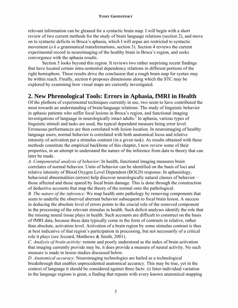
method (see Amunts, Petrides, this volume). (ii) It appears that, as Brodmann (1909 [this volume]) proposed, the anatomical method that produces borders which align best with functional distinctions is the cytoarchitectonic mapping method (Mattelli, Luppino & Rizzolatti, 1991). Yet, cytoarchitectonic borders (not visible in fMRI) do not align well with topographic borders (visible in fMRI). As a result, our ability to localize linguistic processes precisely is constrained by the biology (see Amunts et al., 1999). (iii) Lesion size and lesion variation in aphasic patients are thought to be on average larger than the corresponding measures in fMRI in health. This may be true, yet it is important to note that no study that compares lesion volume to volume of activations in health has ever been conducted. E. Anatomical constraints: Neuroimaging methods are not limited to a specific brain area, as unlike aphasia, they are not lesion dependent. As a consequence, a broader view of the brain is possible. Below, we shall see how significant this feature is. In at least one case, aphasia results exclude the involvement of Broca’s area, yet only fMRI investigations localize them elsewhere. This table below summarizes the main points of comparison:
method dimension
LESION STUDIES (Aphasia)
fMRI IN HEALTH
A. Type of measured behavior
errors normal performance
B. Possibility for a deductive account
yes no
C. Measured brain activity none blood flow D. Degree of anatomical precision
up to lesion size and inter-individual lesion overlap
up to resolution of functional image, and individual variation
E. Possibility of a broad view of the brain
no yes
3. Focal insult to Broca’s Area results in a Syntactic Movement Failure – TDH Common wisdom is that Broca’s area on the left hemisphere is entrusted with syntactic responsibilities. However, it is becoming increasingly clear that these are limited, and do not encompass all of syntax. Take out Broca’s area from a person, and s/he will be left with quite a lot of syntax; create a functional image of this area during syntactic analysis, and you will find that it remains silent on many syntactic tasks. Thus important parts of syntax must be elsewhere in the brain, if they are to have neurological existence.
Focusing on receptive abilities in Broca’s aphasia, I will first present a view of the role Broca’s region plays in supporting syntactic computations. I assume no prior knowledge in neuroimaging or in linguistics, although occasional [bracketed] hints and comments for imagers and linguists are included.
Focal insult to the vicinity of LIFG (i.e., the area that "encompass[es] most of the operculum, insula, and subjacent white matter." Mohr, 1978, p. 202) impairs linguistic ability in highly specific ways. The etiology of this condition may be stroke, hemorrhage, protrusion wound, tumor or excision of tissue. As we look into syntax, only studies that use minimal pairs can be of use. That is, while many studies incorporate varieties of syntactic considerations into their design. Below, I only discuss studies that contrast syntactic types with other syntactic types. This restricted domain has been a focus of intense study in recent
YOSEF GRODZINSKY
5
years, and a rich body of data is currently available. Work carried out in many laboratories, through varied experimental methods and on several languages, has indicated that the receptive abilities of Broca’s aphasics at the sentence level are selectively compromised. When tested in comprehension, grammaticality judgment as well as receptive timed tasks, they yield mixed results, success or failure (or aberrant performance) depending on sentence type. The goal of this section is to uncover a pattern in their performance.
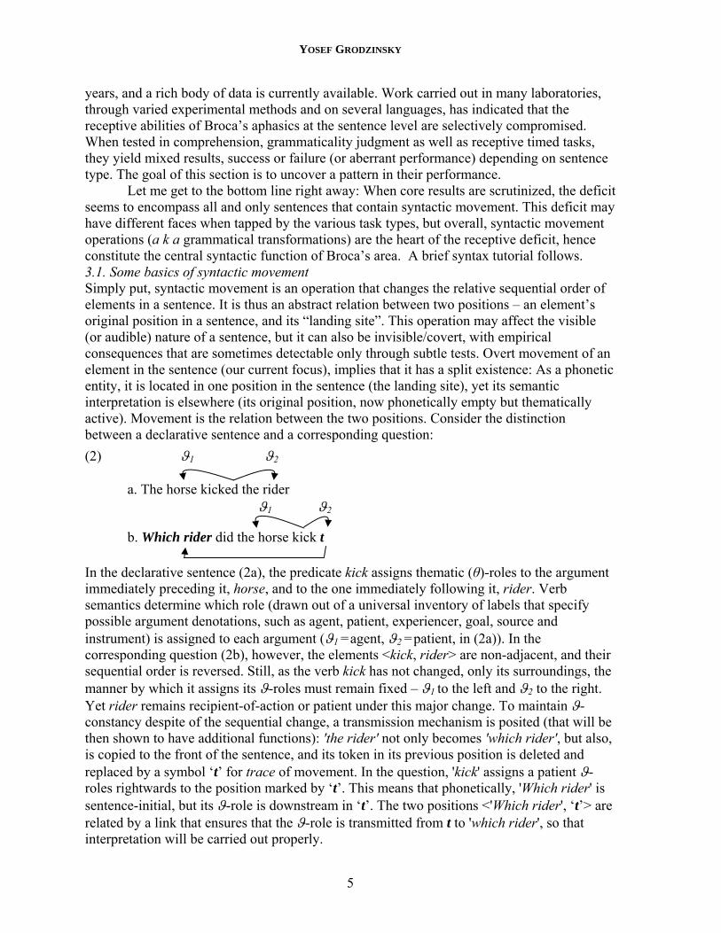
Let me get to the bottom line right away: When core results are scrutinized, the deficit seems to encompass all and only sentences that contain syntactic movement. This deficit may have different faces when tapped by the various task types, but overall, syntactic movement operations (a k a grammatical transformations) are the heart of the receptive deficit, hence constitute the central syntactic function of Broca’s area. A brief syntax tutorial follows. 3.1. Some basics of syntactic movement Simply put, syntactic movement is an operation that changes the relative sequential order of elements in a sentence. It is thus an abstract relation between two positions – an element’s original position in a sentence, and its “landing site”. This operation may affect the visible (or audible) nature of a sentence, but it can also be invisible/covert, with empirical consequences that are sometimes detectable only through subtle tests. Overt movement of an element in the sentence (our current focus), implies that it has a split existence: As a phonetic entity, it is located in one position in the sentence (the landing site), yet its semantic interpretation is elsewhere (its original position, now phonetically empty but thematically active). Movement is the relation between the two positions. Consider the distinction between a declarative sentence and a corresponding question:
(2) ϑ1 ϑ2
a. The horse kicked the rider ϑ1 ϑ2
b. Which rider did the horse kick t
In the declarative sentence (2a), the predicate kick assigns thematic (θ)-roles to the argument immediately preceding it, horse, and to the one immediately following it, rider. Verb semantics determine which role (drawn out of a universal inventory of labels that specify possible argument denotations, such as agent, patient, experiencer, goal, source and instrument) is assigned to each argument (ϑ1 =agent, ϑ2 =patient, in (2a)). In the corresponding question (2b), however, the elements <kick, rider> are non-adjacent, and their sequential order is reversed. Still, as the verb kick has not changed, only its surroundings, the manner by which it assigns its ϑ-roles must remain fixed – ϑ1 to the left and ϑ2 to the right. Yet rider remains recipient-of-action or patient under this major change. To maintain ϑ-constancy despite of the sequential change, a transmission mechanism is posited (that will be then shown to have additional functions): 'the rider' not only becomes 'which rider', but also, is copied to the front of the sentence, and its token in its previous position is deleted and replaced by a symbol ‘t’ for trace of movement. In the question, 'kick' assigns a patient ϑ-roles rightwards to the position marked by ‘t’. This means that phonetically, 'Which rider' is sentence-initial, but its ϑ-role is downstream in ‘t’. The two positions <'Which rider', ‘t’> are related by a link that ensures that the ϑ-role is transmitted from t to 'which rider', so that interpretation will be carried out properly.
A BLUEPRINT FOR A BRAIN MAP OF SYNTAX
6
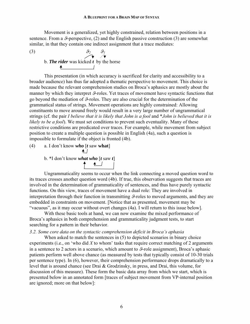
Movement is a generalized, yet highly constrained, relation between positions in a sentence. From a ϑ-perspective, (2) and the English passive construction (3) are somewhat similar, in that they contain one indirect assignment that a trace mediates:
(3) ϑ2 ϑ1
b. The rider was kicked t by the horse
This presentation (in which accuracy is sacrificed for clarity and accessibility to a broader audience) has thus far adopted a thematic perspective to movement. This choice is made because the relevant comprehension studies on Broca’s aphasics are mostly about the manner by which they interpret ϑ-roles. Yet traces of movement have syntactic functions that go beyond the mediation of ϑ-roles. They are also crucial for the determination of the grammatical status of strings. Movement operations are highly constrained: Allowing constituents to move around freely would result in a very large number of ungrammatical strings (cf. the pair I believe that it is likely that John is a fool and *John is believed that it is likely to be a fool). We must set conditions to prevent such eventuality. Many of these restrictive conditions are predicated over traces. For example, while movement from subject position to create a multiple question is possible in English (4a), such a question is impossible to formulate if the object is fronted (4b).
(4) a. I don’t know who [t saw what]
b. *I don’t know what who [t saw t]
Ungrammaticality seems to occur when the link connecting a moved question word to its traces crosses another question word (4b). If true, this observation suggests that traces are involved in the determination of grammaticality of sentences, and thus have purely syntactic functions. On this view, traces of movement have a dual role: They are involved in interpretation through their function in transmitting ϑ-roles to moved arguments, and they are embedded in constraints on movement. [Notice that as presented, movement may be “vacuous”, as it may occur without overt changes (4a). I will return to this issue below].
With these basic tools at hand, we can now examine the mixed performance of Broca’s aphasics in both comprehension and grammaticality judgment tests, to start searching for a pattern in their behavior.
3.2. Some core data on the syntactic comprehension deficit in Broca’s aphasia When asked to match the sentences in (5) to depicted scenarios in binary choice
experiments (i.e., on ‘who did X to whom’ tasks that require correct matching of 2 arguments in a sentence to 2 actors in a scenario, which amount to ϑ-role assignment), Broca’s aphasic patients perform well above chance (as measured by tests that typically consist of 10-30 trials per sentence type). In (6), however, their comprehension performance drops dramatically to a level that is around chance (see Drai & Grodzinsky, in press, and Drai, this volume, for discussion of this measure). These form the basic data array from which we start, which is presented below in an annotated form [traces of subject movement from VP-internal position are ignored; more on that below]:
YOSEF GRODZINSKY
7
(5) Above-chance comprehension a. The woman is chasing the man b. The woman who t is chasing the man is tall c. Show me the woman who t is chasing the man d. It is the woman that t is chasing the man e. Which man t touched Mary?
(6) Chance comprehension a. The man that the woman is chasing t is tall b. Show me the man who the woman is chasing t c. It is the man that the woman is chasing t d. The man is chased t by the woman e. Which man did Mary touch t?
This pattern of performance is intricate, and its connection to syntactic movement as described above is not immediately apparent. That is, traces feature in many (if not all) sentence representations in (5)-(6), and thus their presence or absence does not place the cases in the correct performance groupings. Still, syntactic movement and traces do function as critical building blocks in various incarnations of a deficit analysis known as the Trace-Deletion Hypothesis (TDH, Grodzinsky, 1984; 1986; 1995; 2000a), which attempts to account for these data. The idea behind the TDH is that the core receptive deficit in Broca’s aphasia inheres in an inability to represent traces of movement in syntactic representations. If true, this theory would mean that the central role of Broca’s area in sentence perception is to support syntactic movement. Below I present the logic behind the TDH, and show it at work.
3.3. Mapping Deficient Representations onto Performance Suppose that traces of movement are deleted from syntactic representations in Broca’s aphasia, as the TDH would have it. On minimal expectations, it is not clear how any behavior can be derived from such a supposition. On the one hand, if every trace deletion is to affect performance, more comprehension failures than observed are expected, because most cases not only in (6), but also in (5), contain traces, and their deletion is supposed to cause comprehension problems. On the other hand, it is not clear why the deletion of traces would impact patients’ success rate in comprehension tasks in the first place. This account, then, seems to be both too strong and too weak
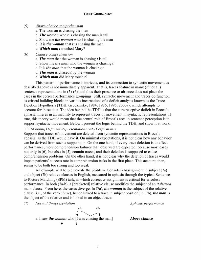
An example will help elucidate the problem. Consider ϑ-assignment in subject (7a) and object (7b) relative clauses in English, measured in aphasia through the typical Sentence-to-Picture Matching (SPM) task, in which correct ϑ-assignment is critical for errorless performance. In both (7a-b), a [bracketed] relative clause modifies the subject of an italicized main clause. From here, the cases diverge. In (7a), the woman is the subject of the relative clause (i.e., of the verb chase), hence linked to a trace in subject position; in (7b), the man is the object of the relative and is linked to an object trace:
(7) Normal θ-representation Aphasic performance ϑ1 ϑ2
a. I saw the woman who [t was chasing the man] Above chance
A BLUEPRINT FOR A BRAIN MAP OF SYNTAX
8
ϑ1 ϑ2
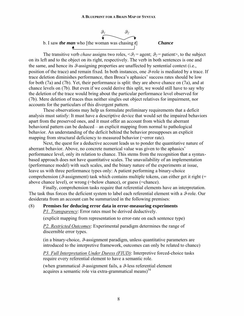
b. I saw the man who [the woman was chasing t] Chance
The transitive verb chase assigns two roles, <ϑ1 = agent; ϑ2 = patient>, to the subject on its left and to the object on its right, respectively. The verb in both sentences is one and the same, and hence its ϑ-assigning properties are unaffected by sentential context (i.e., position of the trace) and remain fixed. In both instances, one ϑ-role is mediated by a trace. If trace deletion diminishes performance, then Broca’s aphasics’ success rates should be low for both (7a) and (7b). Yet, their performance is split: they are above chance on (7a), and at chance levels on (7b). But even if we could derive this split, we would still have to say why the deletion of the trace would bring about the particular performance level observed for (7b). Mere deletion of traces thus neither singles out object relatives for impairment, nor accounts for the particulars of this divergent pattern.
These observations may help us formulate preliminary requirements that a deficit analysis must satisfy: It must have a descriptive device that would set the impaired behaviors apart from the preserved ones, and it must offer an account from which the aberrant behavioral pattern can be deduced – an explicit mapping from normal to pathological behavior. An understanding of the deficit behind the behavior presupposes an explicit mapping from structural deficiency to measured behavior (=error rate).
Next, the quest for a deductive account leads us to ponder the quantitative nature of aberrant behavior. Above, no concrete numerical value was given to the aphasics’ performance level, only its relation to chance. This stems from the recognition that a syntax-based approach does not have quantitative scales. The unavailability of an implementation (performance model) with such scales, and the binary nature of the experiments at issue, leave us with three performance types only: A patient performing a binary-choice comprehension (ϑ-assignment) task which contains multiple tokens, can either get it right (= above chance level), or wrong (=below chance), or guess (=chance).
Finally, comprehension tasks require that referential elements have an interpretation. The task thus forces the deficient system to label each referential element with a ϑ-role. Our desiderata from an account can be summarized in the following premises:
(8) Premises for deducing error data in error-measuring experiments P1. Transparency: Error rates must be derived deductively.
(explicit mapping from representation to error-rate on each sentence type)
P2. Restricted Outcomes: Experimental paradigm determines the range of discernible error types.
(in a binary-choice, ϑ-assignment paradigm, unless quantitative parameters are introduced to the interpretive framework, outcomes can only be related to chance)
P3. Full Interpretation Under Duress (FIUD): Interpretive forced-choice tasks require every referential element to have a semantic role.
(when grammatical ϑ-assignment fails, a ϑ-less referential element acquires a semantic role via extra-grammatical means)34
YOSEF GRODZINSKY
9
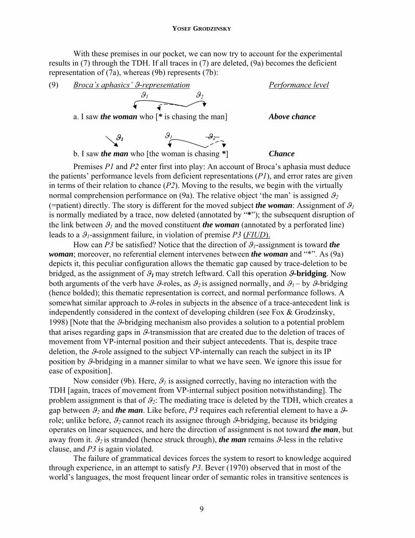
With these premises in our pocket, we can now try to account for the experimental results in (7) through the TDH. If all traces in (7) are deleted, (9a) becomes the deficient representation of (7a), whereas (9b) represents (7b):
(9) Broca’s aphasics’ ϑ-representation Performance level ϑ1 ϑ2
a. I saw the woman who [* is chasing the man] Above chance
b. I saw the man who [the woman is chasing *] Chance
Premises P1 and P2 enter first into play: An account of Broca’s aphasia must deduce the patients’ performance levels from deficient representations (P1), and error rates are given in terms of their relation to chance (P2). Moving to the results, we begin with the virtually normal comprehension performance on (9a). The relative object ‘the man’ is assigned ϑ2 (=patient) directly. The story is different for the moved subject the woman: Assignment of ϑ1 is normally mediated by a trace, now deleted (annotated by “*”); the subsequent disruption of the link between ϑ1 and the moved constituent the woman (annotated by a perforated line) leads to a ϑ1-assignment failure, in violation of premise P3 (FIUD).
How can P3 be satisfied? Notice that the direction of ϑ1-assignment is toward the woman; moreover, no referential element intervenes between the woman and “*”. As (9a) depicts it, this peculiar configuration allows the thematic gap caused by trace-deletion to be bridged, as the assignment of ϑ1 may stretch leftward. Call this operation ϑ-bridging. Now both arguments of the verb have ϑ-roles, as ϑ2 is assigned normally, and ϑ1 – by ϑ-bridging (hence bolded); this thematic representation is correct, and normal performance follows. A somewhat similar approach to ϑ-roles in subjects in the absence of a trace-antecedent link is independently considered in the context of developing children (see Fox & Grodzinsky, 1998) [Note that the ϑ-bridging mechanism also provides a solution to a potential problem that arises regarding gaps in ϑ-transmission that are created due to the deletion of traces of movement from VP-internal position and their subject antecedents. That is, despite trace deletion, the ϑ-role assigned to the subject VP-internally can reach the subject in its IP position by ϑ-bridging in a manner similar to what we have seen. We ignore this issue for ease of exposition].
Now consider (9b). Here, ϑ1 is assigned correctly, having no interaction with the TDH [again, traces of movement from VP-internal subject position notwithstanding]. The problem assignment is that of ϑ2: The mediating trace is deleted by the TDH, which creates a gap between ϑ2 and the man. Like before, P3 requires each referential element to have a ϑ-role; unlike before, ϑ2 cannot reach its assignee through ϑ-bridging, because its bridging operates on linear sequences, and here the direction of assignment is not toward the man, but away from it. ϑ2 is stranded (hence struck through), the man remains ϑ-less in the relative clause, and P3 is again violated.
The failure of grammatical devices forces the system to resort to knowledge acquired through experience, in an attempt to satisfy P3. Bever (1970) observed that in most of the world’s languages, the most frequent linear order of semantic roles in transitive sentences is
ϑ1 ϑ2 ϑ1
A BLUEPRINT FOR A BRAIN MAP OF SYNTAX
10
<ϑ1=agent, ϑ2=patient>. Suppose that in order to satisfy P3, a link – driven by an extra-grammatical Default Strategy – is established between the ϑ-less NP and the role that is most frequently associated with its linear position in the string. A clause-initial NP that has not obtained its ϑ-role grammatically would thus become agent.
Consider now the thematic representation that a Broca’s aphasic patient has for a sentence with movement under the TDH. It is based on two knowledge sources: an incomplete grammar, and the strategy. In (9b), the resulting representation contains 2 arguments, both associated with ϑ1= agent. In a task that requires ϑ-assignment, a ϑ-conflict arises, and chance performance is forced. The grammar based strategic ϑ-assignment pull in opposite directions (e.g., given two arguments and an <agent, Patient> ϑ-representation of the predicate, grammar dictates that argument1=agent, and strategy dictates that argument2=agent). Note that chance performance follows only if the thematic output of the deficient grammar and the thematic dictum of the strategy have equal weights. Whether this is true remains to be empirically investigated (see Drai, this volume).
Many of these ideas have been around for a while. What is new here is the explicit formulation of interpretive principles (8) which link syntactic representations to numerical results of experiments. P1 posits a general requirement, that an account derive aberrant performance deductively. P2 sets up a range of possible experimental outcomes for the specific task under consideration, and P3 connects deficient representations to compensatory mechanisms that may be involved when deficient comprehenders perform this task. Hopefully, these premises will help shed new light not only on the structure of neurolinguistic explanation, but also, on the interpretation of similar experiments with language-deficient populations in general (e.g., developing children).
Next, it is important to distinguish the TDH from an apparently simpler, canonicity-based account of deficient performance (e.g., Frazier and Friederici, 1992; Zurif, 1995). This approach suggest that canonical sentences, in which the linear order of overt arguments corresponds to their order in the lexicon, yield normal comprehension in Broca’s aphasia, whereas deviation from canonicity leads to comprehension difficulties. As the TDH relies on directionality of ϑ-role assignment – itself an expression of the canonical arrangement of arguments around a predicate (agent to its left, patient to its right, etc.) – it is important to compare the two accounts.
To begin with, the canonicity account is incommensurate with interpretive premise P1 – Transparency. While it may partition the cases correctly into those that induce error (6) and those that do not (5), it says nothing about why errors are observed, and why performance is at these levels. The TDH, by contrast, is transparent.
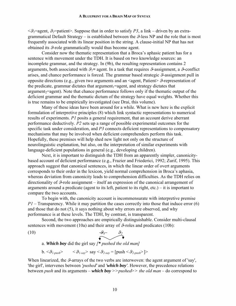
Second, the two approaches are empirically distinguishable. Consider multi-clausal sentences with movement (10a) and their array of ϑ-roles and predicates (10b):
(10) ϑ1 ϑ2
a. Which boy did the girl say [* pushed the old man]
b. <ϑ1-push> <ϑ1-say> say <ϑ2-say = [push <ϑ2-push>]>
When linearized, the ϑ-arrays of the two verbs are interwoven: the agent argument of 'say', 'the girl', intervenes between 'pushed' and 'which boy'. However, the precedence relations between push and its arguments – which boy >>pushed>> the old man – do correspond to
YOSEF GRODZINSKY
11
the order of ϑ-roles in the lexicon. This correspondence seems to be the rationale behind a canonicity-based account of the comprehension deficit in Broca’s aphasia, which contends that patients who succeed use lexical knowledge to compensate for a syntactic deficit (of an unspecified nature). In (10a), however, similarity in precedence relations does not mean congruence with order in the lexicon. Reliance on lexical information – as a canonicity-based account would have it – may not be sufficient, and a comprehension failure would follow.
By contrast, a straightforward construal of the TDH predicts normal comprehension in Broca’s aphasia: which boy is dissociated from its agent ϑ-role due to trace-deletion, but since it is clause-initial, this deficiency is expected to be correctly compensated for by the Default Strategy.
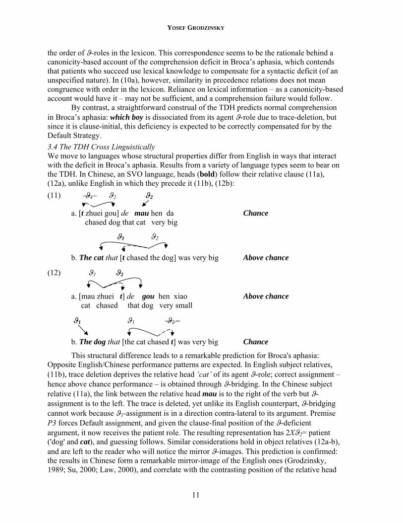
3.4 The TDH Cross Linguistically We move to languages whose structural properties differ from English in ways that interact with the deficit in Broca’s aphasia. Results from a variety of language types seem to bear on the TDH. In Chinese, an SVO language, heads (bold) follow their relative clause (11a), (12a), unlike English in which they precede it (11b), (12b):
(11) ϑ1 ϑ2 ϑ2
a. [t zhuei gou] de mau hen da Chance chased dog that cat very big
ϑ1 ϑ2
b. The cat that [t chased the dog] was very big Above chance
(12) ϑ1 ϑ2
a. [mau zhuei t] de gou hen xiao Above chance cat chased that dog very small
ϑ1 ϑ1 ϑ2 –
b. The dog that [the cat chased t] was very big Chance
This structural difference leads to a remarkable prediction for Broca's aphasia: Opposite English/Chinese performance patterns are expected. In English subject relatives, (11b), trace deletion deprives the relative head ‘cat’ of its agent ϑ-role; correct assignment – hence above chance performance – is obtained through ϑ-bridging. In the Chinese subject relative (11a), the link between the relative head mau is to the right of the verb but ϑ-assignment is to the left. The trace is deleted, yet unlike its English counterpart, ϑ-bridging cannot work because ϑ1-assignment is in a direction contra-lateral to its argument. Premise P3 forces Default assignment, and given the clause-final position of the ϑ-deficient argument, it now receives the patient role. The resulting representation has 2Xϑ2= patient ('dog' and cat), and guessing follows. Similar considerations hold in object relatives (12a-b), and are left to the reader who will notice the mirror ϑ-images. This prediction is confirmed: the results in Chinese form a remarkable mirror-image of the English ones (Grodzinsky, 1989; Su, 2000; Law, 2000), and correlate with the contrasting position of the relative head
A BLUEPRINT FOR A BRAIN MAP OF SYNTAX
12
in the two languages. The phenomenon of ϑ-conflict is thus generalized, manifesting as agent vs. agent in English, and as patient vs. patient in Chinese.
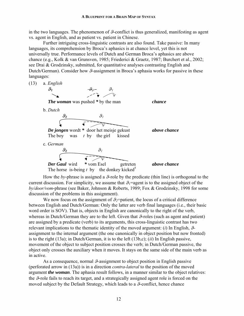
Further intriguing cross-linguistic contrasts are also found. Take passive: In many languages, its comprehension by Broca’s aphasics is at chance level, yet this is not universally true. Performance levels of Dutch and German Broca’s aphasics are above chance (e.g., Kolk & van Grunsven, 1985; Friederici & Graetz, 1987; Burchert et al., 2002; see Drai & Grodzinsky, submitted, for quantitative analyses contrasting English and Dutch/German). Consider how ϑ-assignment in Broca’s aphasia works for passive in these languages:
(13) a. English ϑ1 ϑ2 – ϑ1
The woman was pushed * by the man chance
b. Dutch ϑ2 ϑ1
De jongen wordt * door het meisje gekust above chance The boy was t by the girl kissed
c. German ϑ2 ϑ1
Der Gaul wird * vom Esel getreten above chance The horse is-being t by the donkey kicked5
How the by-phrase is assigned a ϑ-role by the predicate (thin line) is orthogonal to the current discussion. For simplicity, we assume that ϑ1=agent is to the assigned object of the by/door/vom-phrase (see Baker, Johnson & Roberts, 1989; Fox & Grodzinsky, 1998 for some discussion of the problems in this assignment).
We now focus on the assignment of ϑ2=patient, the locus of a critical difference between English and Dutch/German: Only the latter are verb final languages (i.e., their basic word order is SOV). That is, objects in English are canonically to the right of the verb, whereas in Dutch/German they are to the left. Given that ϑ-roles (such as agent and patient) are assigned by a predicate (verb) to its arguments, this cross-linguistic contrast has two relevant implications to the thematic identity of the moved argument: (i) In English, ϑ-assignment to the internal argument (the one canonically in object position but now fronted) is to the right (13a); in Dutch/German, it is to the left (13b,c); (ii) In English passive, movement of the object to subject position crosses the verb; in Dutch/German passive, the object only crosses the auxiliary when it moves. It stays on the same side of the main verb as in active.
As a consequence, normal ϑ-assignment to object position in English passive (perforated arrow in (13a)) is in a direction contra-lateral to the position of the moved argument the woman. The aphasia result follows, in a manner similar to the object relatives: the ϑ-role fails to reach its target, and a strategically assigned agent role is forced on the moved subject by the Default Strategy, which leads to a ϑ-conflict, hence chance
YOSEF GRODZINSKY
13
performance; in Dutch/German, by contrast, ϑ-bridging is possible: the moved constituent De jongen or Der Gaul is ipsi-lateral to the ϑ-role is should receive, and no intervener stands between the ϑ-role and its target. A uniform cross-linguistic view of the deficit in Broca’s aphasia follows, which may actually provide critical hints regarding the correct analysis of passive in Dutch and German.
ϑ-bridging has been repeatedly invoked here, and it needs some elaboration. A ϑ-role fails to reach its target argument only if the direction of assignment is contra-lateral to the direction of movement. Otherwise, ϑ-bridging works both in the subject versus object trace contrast in English, and the cross-linguistic contrast in passive we have just seen. While this might make intuitive sense, there are still puzzles that remain. Specifically, there are cases in which movement leaves an NP ipsi-lateral to its ϑ-role, and still, aphasic performance is at chance. This is the case in scrambling constructions in SOV languages. They might shed light on ϑ-bridging.
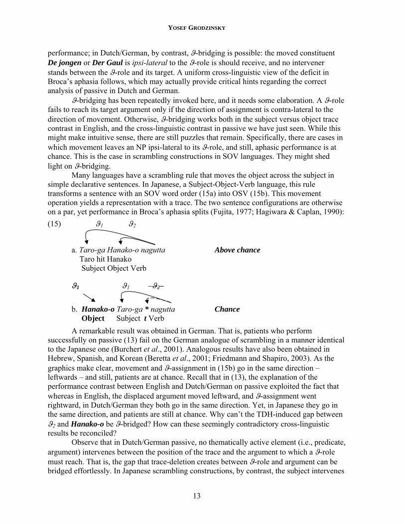
Many languages have a scrambling rule that moves the object across the subject in simple declarative sentences. In Japanese, a Subject-Object-Verb language, this rule transforms a sentence with an SOV word order (15a) into OSV (15b). This movement operation yields a representation with a trace. The two sentence configurations are otherwise on a par, yet performance in Broca’s aphasia splits (Fujita, 1977; Hagiwara & Caplan, 1990):
(15) ϑ1 ϑ2
a. Taro-ga Hanako-o nagutta Above chance Taro hit Hanako Subject Object Verb
ϑ1 ϑ1 ϑ2–
b. Hanako-o Taro-ga * nagutta Chance Object Subject t Verb
A remarkable result was obtained in German. That is, patients who perform successfully on passive (13) fail on the German analogue of scrambling in a manner identical to the Japanese one (Burchert et al., 2001). Analogous results have also been obtained in Hebrew, Spanish, and Korean (Beretta et al., 2001; Friedmann and Shapiro, 2003). As the graphics make clear, movement and ϑ-assignment in (15b) go in the same direction – leftwards – and still, patients are at chance. Recall that in (13), the explanation of the performance contrast between English and Dutch/German on passive exploited the fact that whereas in English, the displaced argument moved leftward, and ϑ-assignment went rightward, in Dutch/German they both go in the same direction. Yet, in Japanese they go in the same direction, and patients are still at chance. Why can’t the TDH-induced gap between ϑ2 and Hanako-o be ϑ-bridged? How can these seemingly contradictory cross-linguistic results be reconciled?
Observe that in Dutch/German passive, no thematically active element (i.e., predicate, argument) intervenes between the position of the trace and the argument to which a ϑ-role must reach. That is, the gap that trace-deletion creates between ϑ-role and argument can be bridged effortlessly. In Japanese scrambling constructions, by contrast, the subject intervenes
A BLUEPRINT FOR A BRAIN MAP OF SYNTAX
14
between the trace and the moved object, and blocks the ϑ-role from bridging the gap that trace deletion created.6
Informally speaking, the consequence is that in Broca’s aphasia, ϑ- assignment to a moved element may succeed despite trace-deletion if two conditions are met: (i) movement is in the same direction as ϑ-assignment and (ii) there is no thematically relevant intervener between the trace and the antecedent. These conclusions are important: they indicate that scrambling and cases of XP-movement form a neurological natural class.
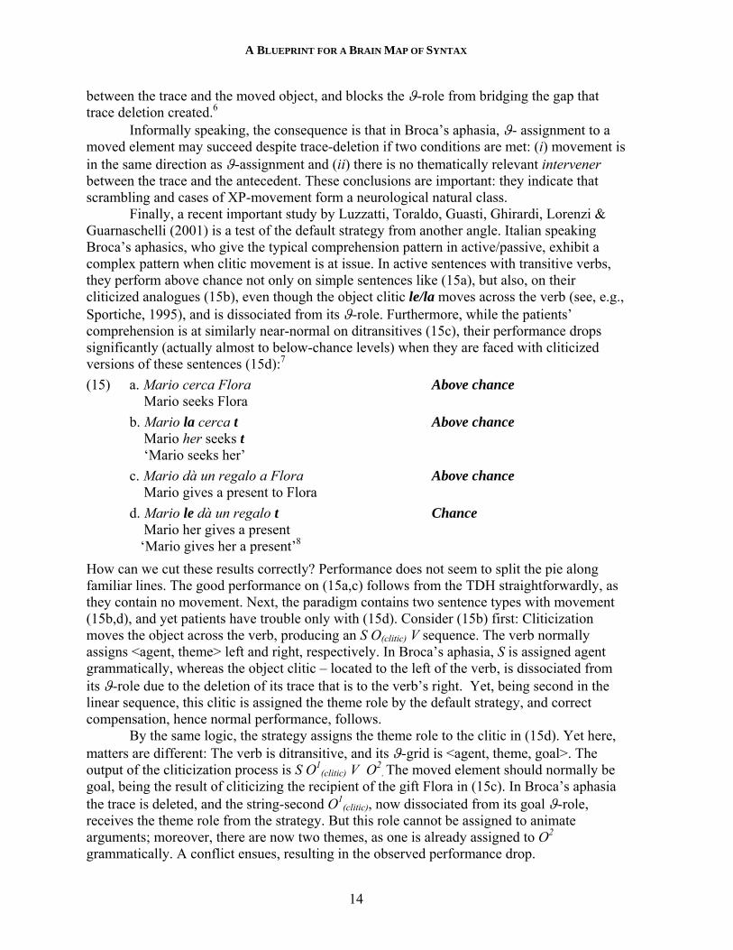
Finally, a recent important study by Luzzatti, Toraldo, Guasti, Ghirardi, Lorenzi & Guarnaschelli (2001) is a test of the default strategy from another angle. Italian speaking Broca’s aphasics, who give the typical comprehension pattern in active/passive, exhibit a complex pattern when clitic movement is at issue. In active sentences with transitive verbs, they perform above chance not only on simple sentences like (15a), but also, on their cliticized analogues (15b), even though the object clitic le/la moves across the verb (see, e.g., Sportiche, 1995), and is dissociated from its ϑ-role. Furthermore, while the patients’ comprehension is at similarly near-normal on ditransitives (15c), their performance drops significantly (actually almost to below-chance levels) when they are faced with cliticized versions of these sentences (15d):7
(15) a. Mario cerca Flora Above chance Mario seeks Flora
b. Mario la cerca t Above chance Mario her seeks t ‘Mario seeks her’
c. Mario dà un regalo a Flora Above chance Mario gives a present to Flora
d. Mario le dà un regalo t Chance Mario her gives a present ‘Mario gives her a present’8
How can we cut these results correctly? Performance does not seem to split the pie along familiar lines. The good performance on (15a,c) follows from the TDH straightforwardly, as they contain no movement. Next, the paradigm contains two sentence types with movement (15b,d), and yet patients have trouble only with (15d). Consider (15b) first: Cliticization moves the object across the verb, producing an S O(clitic) V sequence. The verb normally assigns <agent, theme> left and right, respectively. In Broca’s aphasia, S is assigned agent grammatically, whereas the object clitic – located to the left of the verb, is dissociated from its ϑ-role due to the deletion of its trace that is to the verb’s right. Yet, being second in the linear sequence, this clitic is assigned the theme role by the default strategy, and correct compensation, hence normal performance, follows.
By the same logic, the strategy assigns the theme role to the clitic in (15d). Yet here, matters are different: The verb is ditransitive, and its ϑ-grid is <agent, theme, goal>. The output of the cliticization process is S O1
(clitic) V O2. The moved element should normally be
goal, being the result of cliticizing the recipient of the gift Flora in (15c). In Broca’s aphasia the trace is deleted, and the string-second O1
(clitic), now dissociated from its goal ϑ-role, receives the theme role from the strategy. But this role cannot be assigned to animate arguments; moreover, there are now two themes, as one is already assigned to O2
grammatically. A conflict ensues, resulting in the observed performance drop.
YOSEF GRODZINSKY
15
The logic underlying the TDH, as well as its implementation in a range of fairly complex cases is thus demonstrated. It can be summarized thus:
(16) TDH
a. Trace-deletion: Delete all traces of phrasal movement b. Interpret Referential Elements: A ϑ-argument satisfies P3 either by ϑ-bridging, or by a linear Default Strategy that assigns it a role
Part a specifies the syntactic deficit; part b proposes a mechanism to satisfy P3 (FIUD). Limiting myself to an informal account, I offer no precise definitions, and only note that the Linear Default Strategy is defined relative to the sequence of ϑ-roles around the predicate, and that ϑ-bridging enables an argument which is adjacent to a deleted trace to acquire the latter’s ϑ-role in the absence of an intervener (whose precise nature awaits definition). These principles are obviously related, perhaps reducible to a single statement. As they are predicated over the linear order of elements, one can perhaps imagine a reformulation of (at least parts of) the TDH, as a pathological modification of conditions on the linearization of syntactic representations in normal speakers (cf. Fox & Pesetsky, in press, for one framework which may be a candidate platform for this idea).
If this type of account is on the right track, we can conclude that the core syntactic deficit in Broca’s aphasia – hence the central role of Broca’s region in sentence reception – is the computation of syntactic movement. Next, I present additional converging evidence from aphasic performance in experimental tasks that do not require ϑ-assignment, and show that this conclusion is not task dependent.
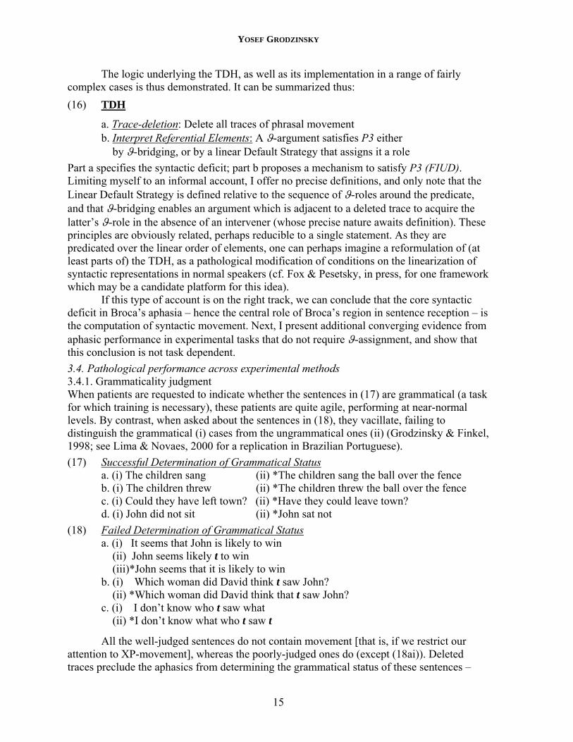
3.4. Pathological performance across experimental methods 3.4.1. Grammaticality judgment When patients are requested to indicate whether the sentences in (17) are grammatical (a task for which training is necessary), these patients are quite agile, performing at near-normal levels. By contrast, when asked about the sentences in (18), they vacillate, failing to distinguish the grammatical (i) cases from the ungrammatical ones (ii) (Grodzinsky & Finkel, 1998; see Lima & Novaes, 2000 for a replication in Brazilian Portuguese).
(17) Successful Determination of Grammatical Status a. (i) The children sang (ii) *The children sang the ball over the fence
b. (i) The children threw (ii) *The children threw the ball over the fence c. (i) Could they have left town? (ii) *Have they could leave town? d. (i) John did not sit (ii) *John sat not
(18) Failed Determination of Grammatical Status a. (i) It seems that John is likely to win
(ii) John seems likely t to win (iii)*John seems that it is likely to win b. (i) Which woman did David think t saw John? (ii) *Which woman did David think that t saw John? c. (i) I don’t know who t saw what (ii) *I don’t know what who t saw t
All the well-judged sentences do not contain movement [that is, if we restrict our attention to XP-movement], whereas the poorly-judged ones do (except (18ai)). Deleted traces preclude the aphasics from determining the grammatical status of these sentences –
A BLUEPRINT FOR A BRAIN MAP OF SYNTAX
16
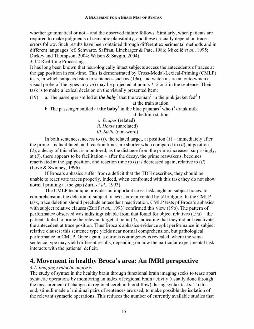
whether grammatical or not – and the observed failure follows. Similarly, when patients are required to make judgments of semantic plausibility, and these crucially depend on traces, errors follow. Such results have been obtained through different experimental methods and in different languages (cf. Schwartz, Saffran, Linebarger & Pate, 1986; Mikelič et al., 1995; Dickey and Thompson, 2004; Wilson & Saygın, 2004). 3.4.2 Real-time Processing It has long been known that neurologically intact subjects access the antecedents of traces at the gap position in real-time. This is demonstrated by Cross-Modal-Lexical-Priming (CMLP) tests, in which subjects listen to sentences such as (19a), and watch a screen, onto which a visual probe of the types in (i-iii) may be projected at points 1, 2 or 3 in the sentence. Their task is to make a lexical decision on the visually presented item:
(19) a. The passenger smiled at the baby1 that the woman2 in the pink jacket fed3 t at the train station b. The passenger smiled at the baby1 in the blue pajamas2 who t3 drank milk at the train station i. Diaper (related) ii. Horse (unrelated) iii. Strile (non-word)
In both sentences, access to (i), the related target, at position (1) – immediately after the prime – is facilitated, and reaction times are shorter when compared to (ii); at position (2), a decay of this effect is monitored, as the distance from the prime increases; surprisingly, at (3), there appears to be facilitation – after the decay, the prime reawakens, becomes reactivated at the gap position, and reaction time to (i) is decreased again, relative to (ii) (Love & Swinney, 1996).
If Broca’s aphasics suffer from a deficit that the TDH describes, they should be unable to reactivate traces properly. Indeed, when confronted with this task they do not show normal priming at the gap (Zurif et al., 1993).
The CMLP technique provides an important cross-task angle on subject traces. In comprehension, the deletion of subject traces is circumvented by ϑ-bridging. In the CMLP task, trace deletion should preclude antecedent reactivation. CMLP tests pf Broca’s aphasics with subject relative clauses (Zurif et al., 1993) confirmed this view (19b). The pattern of performance observed was indistinguishable from that found for object relatives (19a) – the patients failed to prime the relevant target at point (3), indicating that they did not reactivate the antecedent at trace position. Thus Broca’s aphasics evidence split performance in subject relative clauses: this sentence type yields near normal comprehension, but pathological performance in CMLP. Once again, a curious contingency is revealed, where the same sentence type may yield different results, depending on how the particular experimental task interacts with the patients’ deficit. 4. Movement in healthy Broca’s area: An fMRI perspective 4.1. Imaging syntactic analysis The study of syntax in the healthy brain through functional brain imaging seeks to tease apart syntactic operations by monitoring an index of regional brain activity (usually done through the measurement of changes in regional cerebral blood flow) during syntax tasks. To this end, stimuli made of minimal pairs of sentences are used, to make possible the isolation of the relevant syntactic operations. This reduces the number of currently available studies that
YOSEF GRODZINSKY
17
can be discussed, because many, if not most, imaging investigations at the sentence level are designed with more general goals in mind (e.g., whether syntactic and semantic processes are distinct, Dapretto and Bookheimer, 1999; Vandenbergh et al., 2002; see Grodzinsky, 2002, for a critical review). Still, the few relevant results that are available provide preliminary hints regarding the relevance of this endeavor to the STC. Before these are presented, a short digression to experimental issues is necessary.
Effects in fMRI experiments are always difficult to obtain, due to a relatively poor signal-to-noise ratio (see Jezzard et al., 2001 for tutorials). In experiments that feature syntactic contrasts, matters are considerably harder: Sentence stimuli are typically of exceedingly long duration, ~4 seconds per stimulus, compared to ~300-400msec in vision experiments. To isolate a syntactic operation, a difference in brain response between members of a minimal syntactic pair needs to be detected. Like in vision experiments, we compare the difference in the strength of the Blood Oxygen Level Dependent (BOLD) response between experimental conditions. Yet unlike vision, the effect that such contrasts produce is weak relative to the overall BOLD response generated by the cognitively taxing analysis of long sentence stimuli.
The most compelling evidence for regional activity in the brain that correlates with a stimulus contrast is provided through statistical maps (e.g., Worsley et al., 2001). These maps are constructed through an exhaustive search across the whole brain for a significant difference in BOLD response between experimental conditions. Signal intensity that each contrast induces is compared on every brain volume (voxel) that the technology defines. This method thus involves multiple statistical comparisons. The nature of statistical comparisons opens way to spurious effects in such cases, for which we must control (i.e., to correct for accidental effects obtained simply by the multiplicity of statistical tests). As a consequence, the threshold for each comparison is elevated, requiring more power to be significant. Syntactic effects, whose size is typically small, rarely pass the required threshold, making the direct construction of syntax maps difficult.
An alternative approach is the Regions of Interest (ROI) approach, which seeks to increase signal intensity, and at the same time reduce the number of statistical comparisons. It focuses on those parts of the brain that interest us and ignores the rest; at the same time, it pulls together all voxels within this region and treats them as one unit. The result is higher signal intensity, with less correction. Weaker effects may surface to the fore. These gains do not come for free: anatomical resolution is sacrificed for statistical power. Moreover, as an a priori anatomical choice is made, ROI analyses need motivation, as well as a method for the delineation of the chosen brain areas. Most (though not all) the data I will present comes from ROI analyses.
Currently, three findings can be reported: (i) A series of studies in English, German and Hebrew in multiple contrasts and tasks correlate a movement effect with increased BOLD response in Broca’s region of the left cerebral hemisphere. These results converge on the findings for Broca’s aphasia; most, but not all, of these contrasts also activate left (and perhaps right) Wernicke’s region; (ii) Reflexive binding uniquely activates the Superior Frontal Gyrus of the right hemisphere; (iii) Dative Shift uniquely activates the anterior Insula (aINS) and the ventral portion of the Pre-Central Sulcus (vPCS), both in the right hemisphere, and does not activate Broca’s region. The remainder of this section reviews the movement studies; the rest are described in the subsequent one.
A BLUEPRINT FOR A BRAIN MAP OF SYNTAX
18
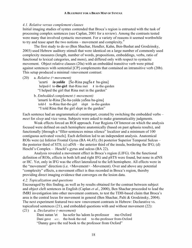
4.1. Relative versus complement clauses Initial imaging studies of syntax contended that Broca’s region is entrusted with the task of processing complex sentences (see Caplan, 2001 for a review). Among the contrasts tested were many that involved syntactic movement. For a variety of reasons it seemed worthwhile to try and tease apart the two notions – movement and complexity.9
The first study to do so (Ben Shachar, Hendler, Kahn, Ben-Bashat and Grodzinsky, 2003) used Hebrew auditory stimuli that were identical on a large number of commonly used complexity measures (length, number of words, propositions, embeddings, verbs, ratio of functional to lexical categories, and more), and differed only with respect to syntactic movement. Object relative clauses (20a) with an embedded transitive verb were pitted against sentences with sentential [CP] complements that contained an intransitive verb (20b). This setup produced a minimal ±movement contrast:
(20) a. Relative (+movement) 'azarti la-yalda [Še-Rina pagŠa t ba-gina] helped-I to-the-girl that-Rina met t in-the-garden “I helped the girl that Rina met in the garden”
b. Embedded complement (–movement) 'amarti le-Rina [Še-ha-yalda yaŠna ba-gina] told-I to-Rina that-the-girl slept in-the-garden “I told Rina that the girl slept in the garden”
Each sentence had an ungrammatical counterpart, created by switching the embedded verbs – meet for sleep and vice versa. Subjects were asked to make grammaticality judgments.
Weak effects forced an ROI approach. Four Regions Of Interest on which the analysis focused were defined on each hemisphere anatomically (based on past aphasia results), and functionally [through a “filler-sentences minus silence” localizer and a minimum of 100 contiguous activated voxels]. Each definition led to an independent analysis. Anatomical ROIs were (a) Inferior Frontal Gyrus (BA 44,45); (b) posterior Superior Temporal Sulcus – the posterior third of STS; (c) aINS – the anterior third of the insula, bordering the IFG; (d) Heschl’s Complex – Heschl’s gyrus and sulcus (BA 22).
Analysis revealed a movement effect in Broca’s region (LIFG). On the functional definition of ROIs, effects in both left and right IFG and pSTS were found, but none in aINS or HC. Yet, only in IFG was the effect lateralized to the left hemisphere. All effects were in the “movement” direction (i.e. +Movement> –Movement). Over and above any potential “complexity” effects, a movement effect is thus recorded in Broca’s region, thereby providing direct imaging evidence that converges on the lesion data.
4.2. Topicalization and questions Encouraged by this finding, as well as by results obtained for the contrast between subject and object cleft sentences in English (Caplan et al., 2000), Ben Shachar proceeded to lead the fMRI investigation into other movement contrasts, to test the TDH-based claim that Broca’s area is the central locus for movement in general (Ben Shachar, Palti & Grodzinsky, 2004). The next experiment featured two new ±movement contrasts in Hebrew: Declarative vs. topicalized sentences (21), and embedded questions with and without movement (22): (21) a. Declarative (–movement)
Dani natan 'et ha-sefer ha-'adom la-professor me-Oxford Dani gave ACC. the-book the-red to-the-professor from-Oxford “Danny gave the red book to the professor from Oxford”
YOSEF GRODZINSKY
19
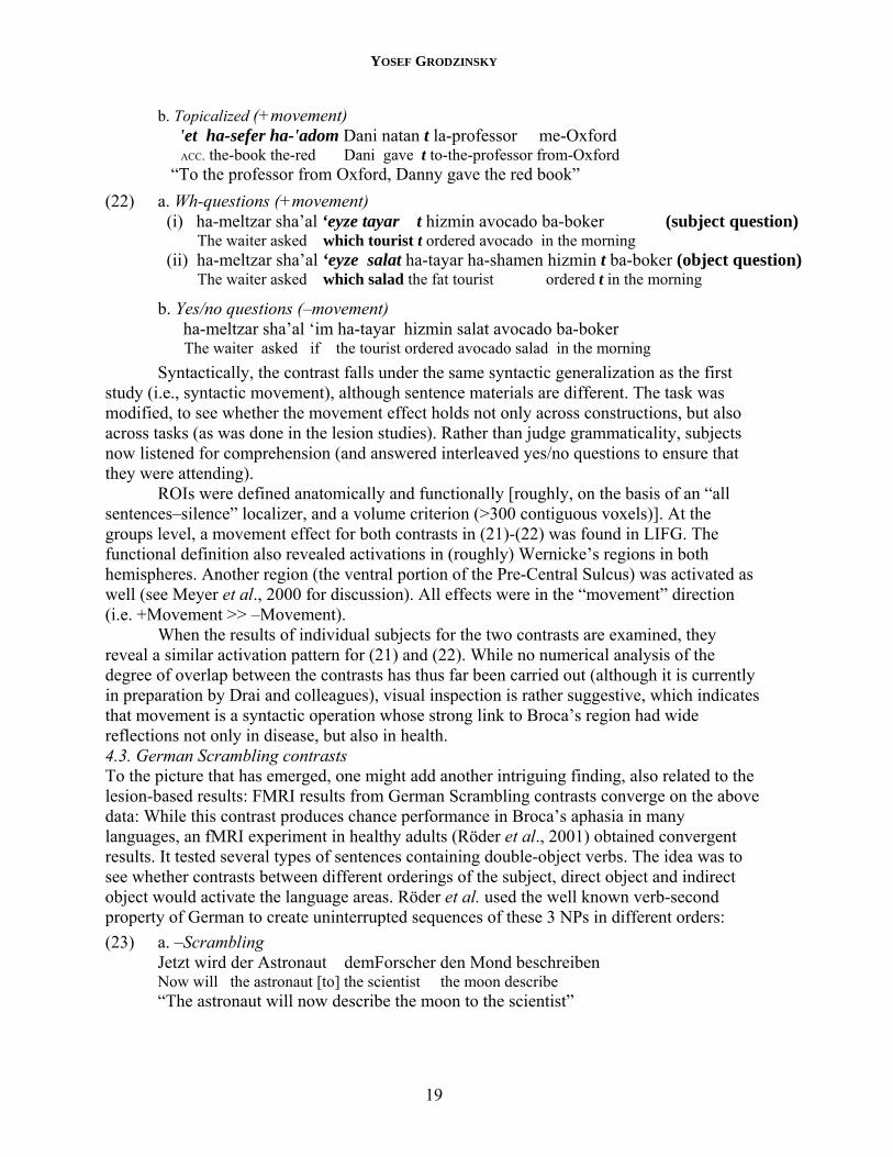
b. Topicalized (+movement) 'et ha-sefer ha-'adom Dani natan t la-professor me-Oxford ACC. the-book the-red Dani gave t to-the-professor from-Oxford “To the professor from Oxford, Danny gave the red book”
(22) a. Wh-questions (+movement) (i) ha-meltzar sha’al ‘eyze tayar t hizmin avocado ba-boker (subject question) The waiter asked which tourist t ordered avocado in the morning (ii) ha-meltzar sha’al ‘eyze salat ha-tayar ha-shamen hizmin t ba-boker (object question) The waiter asked which salad the fat tourist ordered t in the morning
b. Yes/no questions (–movement) ha-meltzar sha’al ‘im ha-tayar hizmin salat avocado ba-boker The waiter asked if the tourist ordered avocado salad in the morning
Syntactically, the contrast falls under the same syntactic generalization as the first study (i.e., syntactic movement), although sentence materials are different. The task was modified, to see whether the movement effect holds not only across constructions, but also across tasks (as was done in the lesion studies). Rather than judge grammaticality, subjects now listened for comprehension (and answered interleaved yes/no questions to ensure that they were attending).
ROIs were defined anatomically and functionally [roughly, on the basis of an “all sentences–silence” localizer, and a volume criterion (>300 contiguous voxels)]. At the groups level, a movement effect for both contrasts in (21)-(22) was found in LIFG. The functional definition also revealed activations in (roughly) Wernicke’s regions in both hemispheres. Another region (the ventral portion of the Pre-Central Sulcus) was activated as well (see Meyer et al., 2000 for discussion). All effects were in the “movement” direction (i.e. +Movement >> –Movement).
When the results of individual subjects for the two contrasts are examined, they reveal a similar activation pattern for (21) and (22). While no numerical analysis of the degree of overlap between the contrasts has thus far been carried out (although it is currently in preparation by Drai and colleagues), visual inspection is rather suggestive, which indicates that movement is a syntactic operation whose strong link to Broca’s region had wide reflections not only in disease, but also in health. 4.3. German Scrambling contrasts To the picture that has emerged, one might add another intriguing finding, also related to the lesion-based results: FMRI results from German Scrambling contrasts converge on the above data: While this contrast produces chance performance in Broca’s aphasia in many languages, an fMRI experiment in healthy adults (Röder et al., 2001) obtained convergent results. It tested several types of sentences containing double-object verbs. The idea was to see whether contrasts between different orderings of the subject, direct object and indirect object would activate the language areas. Röder et al. used the well known verb-second property of German to create uninterrupted sequences of these 3 NPs in different orders:
(23) a. –Scrambling Jetzt wird der Astronaut demForscher den Mond beschreiben Now will the astronaut [to] the scientist the moon describe “The astronaut will now describe the moon to the scientist”
A BLUEPRINT FOR A BRAIN MAP OF SYNTAX
20
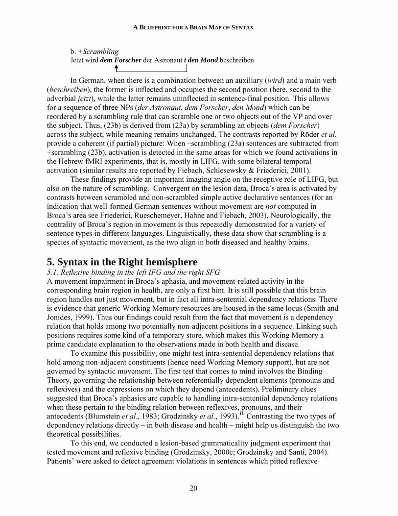
b. +Scrambling Jetzt wird dem Forscher der Astronaut t den Mond beschreiben
In German, when there is a combination between an auxiliary (wird) and a main verb (beschreiben), the former is inflected and occupies the second position (here, second to the adverbial jetzt), while the latter remains uninflected in sentence-final position. This allows for a sequence of three NPs (der Astronaut, dem Forscher, den Mond) which can be reordered by a scrambling rule that can scramble one or two objects out of the VP and over the subject. Thus, (23b) is derived from (23a) by scrambling an objects (dem Forscher) across the subject, while meaning remains unchanged. The contrasts reported by Röder et al. provide a coherent (if partial) picture: When –scrambling (23a) sentences are subtracted from +scrambling (23b), activation is detected in the same areas for which we found activations in the Hebrew fMRI experiments, that is, mostly in LIFG, with some bilateral temporal activation (similar results are reported by Fiebach, Schlesewsky & Friederici, 2001).
These findings provide an important imaging angle on the receptive role of LIFG, but also on the nature of scrambling. Convergent on the lesion data, Broca’s area is activated by contrasts between scrambled and non-scrambled simple active declarative sentences (for an indication that well-formed German sentences without movement are not computed in Broca’s area see Friederici, Rueschemeyer, Hahne and Fiebach, 2003). Neurologically, the centrality of Broca’s region in movement is thus repeatedly demonstrated for a variety of sentence types in different languages. Linguistically, these data show that scrambling is a species of syntactic movement, as the two align in both diseased and healthy brains. 5. Syntax in the Right hemisphere 5.1. Reflexive binding in the left IFG and the right SFG A movement impairment in Broca’s aphasia, and movement-related activity in the corresponding brain region in health, are only a first hint. It is still possible that this brain region handles not just movement, but in fact all intra-sentential dependency relations. There is evidence that generic Working Memory resources are housed in the same locus (Smith and Jonides, 1999). Thus our findings could result from the fact that movement is a dependency relation that holds among two potentially non-adjacent positions in a sequence. Linking such positions requires some kind of a temporary store, which makes this Working Memory a prime candidate explanation to the observations made in both health and disease.
To examine this possibility, one might test intra-sentential dependency relations that hold among non-adjacent constituents (hence need Working Memory support), but are not governed by syntactic movement. The first test that comes to mind involves the Binding Theory, governing the relationship between referentially dependent elements (pronouns and reflexives) and the expressions on which they depend (antecedents). Preliminary clues suggested that Broca’s aphasics are capable to handling intra-sentential dependency relations when these pertain to the binding relation between reflexives, pronouns, and their antecedents (Blumstein et al., 1983; Grodzinsky et al., 1993).10 Contrasting the two types of dependency relations directly – in both disease and health – might help us distinguish the two theoretical possibilities.
To this end, we conducted a lesion-based grammaticality judgment experiment that tested movement and reflexive binding (Grodzinsky, 2000c; Grodzinsky and Santi, 2004). Patients’ were asked to detect agreement violations in sentences which pitted reflexive
YOSEF GRODZINSKY
21
binding against movement (24). Every case involved a dependency relation between a reflexive and an antecedent in sentences with two NPs as potential antecedents that differed in gender. Each sentence contained a sequence of the form <NP1,NP2, reflexive>, where genderNP1 ≠ genderNP2. In the grammatical sentences, the antecedent was either local (24-25a), or moved (24-25c); in the ungrammatical sentences, the gender of the reflexive was changed, so that it did not match the proper antecedent, and matched an NP that could not function as its antecedent (24-25b,d). The necessary knowledge is (a) Condition A of the Binding Theory that essentially says that a reflexive must have a local antecedent, and (b) that movement leaves a trace that may serve as a local antecedent. After some training with the task, subjects were asked to decide whether the reflexive matched NP1 or NP2. The sentences divide along the ±Movement, ±Grammatical dimensions.
(24) a. The woman believes the man likes himself –M,+G b. *The woman believes the man likes herself –M, –G c. Which man does The woman believe t likes himself +M,+G d. *Which man does The woman believe t likes herself +M, –G (25) a. It seems to Sally that the father rewards himself –M,+G b. *It seems to Sally that the father rewards herself +M, –G c. The father seems to Sally t to reward himself +M,+G d. *The father seems to Sally t to reward herself +M, –G
We tested Broca’s, Wernicke’s and right hemisphere lesioned patients, and carried out between and within group comparisons. The performance of the right hemisphere patients was near ceiling on all conditions. The performances of Broca’s and Wernicke’s patients were not distinguishable on the basis of this test. For both groups, the performance on movement conditions was lower than on the reflexive conditions (which were all above chance level).While these results are suggestive, they are somewhat weak.
The quest for converging evidence from the intact brain led us to adapt this test for fMRI in healthy adults who were asked to judge the grammaticality of sequences as in (26), in order to tease apart neural responses to movement and to binding (Grodzinsky and Santi, 2004). The basic logic was the same as above:
(26) a. [NP1Which man] does [NP2The woman] believe t likes himself/*herself b. [NP1The woman] believes [NP2the man] likes himself/*herself c. [NP1Which man] does [NP2The woman] believe t likes/*slept Mary d. [NP1The woman] believes [NP2the man] likes/*slept Mary
As before, special care was taken to ensure that the subjects who participated in the experiment understood the fine nature of the task (all subjects were right handed monolingual English speaking linguists or students of linguistics). Only data from the grammatical sentences were analyzed. The effects we got were stronger than most: We were able to construct a GLM based statistical map (that is, one that makes no a priori selection of brain regions of interest), which revealed a significant reflexive effect (p<.005, uncorrected for multiple comparisons). That is, when conditions containing reflexives were compared to the rest, activations were detected in the right Superior Frontal Gyrus (a frontal region above Broca’s, and in front of vPCS). No movement effect was detected in this region. In the left hemisphere, three effects were detected (also at p<.005, uncorrected): In LIFG we detected
A BLUEPRINT FOR A BRAIN MAP OF SYNTAX
22
both a movement and a reflexive effect; a movement effect was also detected in the left Inferior Posterior Central Sulcus.
An ROI approach delineated an ROI functionally, through the use of a fairly standard “all sentences–silence” localizer, captured a contiguous area that spanned over the posterior part of Broca’s region (BA 44), as well as the vPCS. An ANOVA revealed a significant difference between the movement and reflexive conditions (movement>reflexive).
We thus have two results: First, reflexives, but not movement, activated the right SFG. Second, an ROI analysis revealed a mail effect of movement.
The results obtained thus far are not clear cut, and are open to a variety of criticisms. An improved version of this test, designed by Andrea Santi, is currently underway. It parameterizes the distance between the reflexive, the trace and their antecedents, to control for potential distance effects. The overall picture suggests a limited role for Broca’s region in the processing of dependencies other than movement.
5.2 Dative Shift in the left anterior Insula and the right vPCS Preliminary evidence is now available about an additional syntactic dependency relation – Dative Shift. This is a regular relation (studied extensively, e.g., Larson, 1988; Aoun & Li, 1989; Beck & Johnson, 2004) that holds among two orderings of objects in sentences containing triadic predicates, hence 2 objects (O1, O2), such as give, send, mail:
(27) a. Dative Danny gave/sent/mailed [O1 a book] to [O2 Donna] b. Double Object Danny gave/sent/mailed [O2 Donna] [O1 a book]
The change in the relative order suggests a movement relation [note that the annotation of objects as O1, O2 and the exclusion of the preposition to are just for expository purposes]. Indeed, two recurrent questions have concerned linguists:
(I) What type of movement (if any) is involved? (II) If there is movement, which complement order is base generated and which is derived? Is it O1 O2 or vice versa? A neuroimaging perspective on these questions was recently attempted in Ben-
Shachar & Grodzinsky (2002). We tried to see whether it is possible to use the location and intensity of the fMRI signal as a tool for the examination of these questions. To this end, we conducted a comprehension test of Hebrew double objects, aimed to get an imaging perspective on the linguistic analysis of this construction. While Hebrew is somewhat different from English, linguistic tests suggest that its dative (28a) is like its English counterpart (27a); likewise, Hebrew (28b) is comparable to (27b).
We embedded these materials in the above topicalization experiment: Sentences like (27a,b) were mixed with their topicalized and untopicalized counterparts (28a,b):
(28) Dative Shift a. Dative: Dani natan 'et [O1 ha-sefer ha-'adom] la-[O2 professor me-Oxford] Dani gave ACC. the-book the-red to-the-professor from-Oxford b. Double Object: Dani natan [O2 la-professor me-Oxford] 'et [O1 ha-sefer ha-'adom] Dani gave to-the-prof. from-Ox. ACC the-book the-red
(29) Topicalization a. 'et ha-sefer ha-'adom Dani natan la-professor me-Oxford ACC. the-book the-red Dani gave to-the-professor from-Oxford b. la-professor me-Oxford Dani natan 'et ha-sefer ha-'adom To-the-prof. from-Ox. Dani gave ACC. the-book the-red
YOSEF GRODZINSKY
23
Question (I) can be resolved when the activation pattern of the dative contrast is compared to that of the topicalization contrast. That is, an activation-by-region interaction between the dative-shift contrast (28a,b) and the topicalization contrast (29a,b) would imply that the two relations are computed in different regions, and are thus neurologically distinct. Question (II) can be resolved when the relative intensity of the signal in (28a,b) is compared. That is, the derived sentence should produce a stronger signal than the base configuration. Our study utilized 2 types of empirical argument: the anatomical locus of the fMRI signal as reflecting uniformity or distinctness of operations (topicalization vs. dative shift), and the relative intensity of the fMRI signal within an anatomical region as reflecting more mental computation (double object vs. dative).
We obtained two results: First, regarding question (I), our results indicated a spatial pattern quite different for Dative Shift from when compared to the topicalization contrast. Specifically, the comparison between (28a) and (28b) activated two frontal regions in the right cerebral hemisphere, and not any of the topicalization-related regions (29a) vs. (29b), that like other movement contrasts activated Broca’s area on the left and Wernicke’s area bilaterally. This difference suggests that a different type of operation is involved.
Second, regarding question (II) the intensity of the BOLD signal was measured in the two right frontal regions that are sensitive to the dative-shift contrast. The result for double objects (28b) was significantly higher than for datives (28a), suggesting that Hebrew double objects are more demanding than datives, and providing an indication of their derived nature. Dative Shift, then, is a distinct dependency relation that appears to be computed outside Broca’s area. Finally, it is surprising, perhaps that the Dative Shift contrast activates anterior regions of the right hemisphere.
Dative shift and reflexive experiments activated different regions in the right hemisphere – the former in Superior Frontal Gyrus, the latter in vPCS and anterior Insula. This provides preliminary hints that syntax is not exclusively on the left side of the brain.
5.3. Wernicke’s Area in Brief It has been recurrently observed that Wernicke’s area is engaged in similar activity – similar, but not identical. In aphasia studies of syntax, initial striking differences (e.g., Zurif, 1980) in functional role gave way to findings that indicated overlap. Some studies conducted over the years have found that the performance of Wernicke’s aphasics to hold great individual variation, and present a blurry picture that is not quantitatively discernible from that of the Broca’s aphasics (e.g., Grodzinsky & Finkel, 1998, Grodzinsky & Santi, 2004). At the same time, important syntactic differences between Broca’s and Wernicke’s aphasics have been demonstrated (Zurif et al., 1993; Shapiro et al., 1993; Zurif, 2003). While it is fairly clear that the role of these areas in syntax is not identical, providing a precise account that would tease apart their functions has turned out to be quite difficult.
In this respect, the empirical record obtained by fMRI is not that clear either: Some, but not all, experiments have detected activation not only in the left Wernicke’s area, but also on the right (Ben Shachar et al., 2003, 2004; Friederici et al., 2003; Röder et al. 2001). The paucity of data and analyses at this point – from both lesion and imaging studies – precludes a more thorough assessment of the role of Wernicke’s area, yet this clearly is a question that warrants careful attention and should be investigated.
A BLUEPRINT FOR A BRAIN MAP OF SYNTAX
24
6. A Blueprint for a Syntax Map I hope the reader has become convinced that the beginnings of a brain map for syntax are starting to surface, as three seemingly distinct syntactic operations are supported by mechanisms in distinct brain areas – Movement in Broca’s (perhaps to an extent in Wernicke’s) are; Dative Shift in posterior portions of the right frontal lobe (vPCS, aINS); reflexive-antecedent binding in an anterior part of the right frontal lobe (SFG). It is time now to come full circle, and return to the STC:
(1) Syntacto-Topic Conjecture (STC)
a. Major syntactic operations are neurologically individuated. b. The organization of these operations in brain space is linguistically
significant.
We have seen that the use of descriptive tools from linguistics is beneficial for brain mapping; we have also seen some evidence that suggests the brain honors fine syntactic distinctions in both aphasia and fMRI. In aphasia research this is not new: correspondence between neurological and syntactic natural classes has been found several times in the past 60 years, since Jakobson (1941) first hinted at the possibility (Avrutin, 2001; this volume; Goodglass and Hunt, 1958; Grodzinsky, Pierce and Marakovitz, 1991; Grodzinsky et al., 1993; Friedmann & Grodzinsky, 1997).
The STC seeks to go beyond these results, and see whether the manner by which syntax is organized in the brain is theoretically meaningful. There is a large set of related questions: Is the arrangement of syntactic components in the brain haphazard or orderly? Is the representation of language similar for all humans, whose experiences vis-à-vis language are so diverse? Why would the cerebral arrangement of grammatical abilities be in different places in the brain? Is there, in other words, any significance to the spatial arrangement of the neural patches that support certain central syntactic computations? And if so, what are the relevant parameters, and how can they be studied?
Looking at adjacent domains might actually help. Since the sensory-motor homunculi discovered by Penfield & Rasmussen (1950), much neurophysiological work – in vision, audition and in the sensory-motor domain – has charted the spatial arrangement of functional systems in the brain. Thus somatotopy in sensory and motor cortex has long been investigated (e.g. Beisteiner et al., 2004), as has retinotopy in visual cortex (e.g., Tootel et al., 1997), and tonotopy in auditory cortex (e.g., Pantev et al., 1989; Zhang et al., 2001). Diesch et al. (1996) have even attempted (not very successfully) to detect for the spatial organization of vowels.
Moreover, some have gone beyond mere functional localization, and tried to harness the anatomy to the quest of constitutive principles. To this end, efforts have been made to see whether the topological arrangement of neural systems that support behavior is theoretically meaningful or arbitrary. One salient example comes from vision, where there have been recent attempts to identify overarching principles that govern the organization of cortical maps for vision. Take visual maps, for example. For a while it has been known that these are composed of receptive fields, each having separate maps for orientation, spatial frequency, direction of motion, color, ocular dominance, and perhaps more (see Swindale, 1996 for a review). Yet is the topographic organization of these maps within a receptive field accidental, or does it have some meaning? Moreover, is there any significance to the manner by which the various functions are laid on the receptive fields, and superimposed on one another?
YOSEF GRODZINSKY
25
Until very recently, no definitive answer could be given to this type of question, although it has long occupied the minds of vision scientists. The advent of optical and MR imaging methods has led to several discoveries regarding the spatial geometry of visual functions. Thus it was found that orientation columns, that mark orientation preference of cells, are organized in cycles around orientation centers, known as singularities or “pinwheels” (Bonhoeffer & Grinvald, 1991); it was also found that some of these systems are superimposed on one another in geometrically regular ways (Huebener et al., 1997). While the reason for this type of arrangement is not immediately clear, its regularity is quite striking, and calls for an explanation. Indeed, there have been attempts to discover general organizing principles for the visual system through the study of the neural geometry of its subsystems (see Swindale et al., 2000 for a formalization and an empirical test of general principles proposed by Hubel & Wiesel, 1977).
Given the findings presented above, it seems reasonable to explore whether such an arrangement exists in the language domain. Why would syntactic abilities be localized in the first place, and if so, why would certain parts be in the left hemisphere and others in the right? We are coming to a point when such questions can actually be formulated. In particular, I think that the following questions can now begin to be asked: 1. Can specific rule systems be characterized by the volume of tissue involved in their implementation? More concretely, when we compare the volume of tissue in Broca’s region activated during the computation of movement to that activated on the right SFG during binding, can the result have implications to the relation between movement and binding? 2. Does spatial arrangement matter? Does relative proximity and hemisphericity of specific grammatical components have theoretical significance? What are we to make of the fact that Dative Shift and Binding are anatomically adjacent? 3. Does signal intensity matter? Why do certain syntactic contrasts produce stronger signals than others? 4. How much individual variation is there for each principle, and is there a cerebral reflection of parametric variation observed in grammatical systems? 5. Are there more abstract spatial relations couched within the anatomy that are linguistically relevant?
It is of course possible that the emerging topological arrangement of syntactic principles is arbitrary, and that nothing can be learned from a syntax map, which would be meaningless, its geometry being of no consequence.
Yet if the preliminary clues I presented above are a lead, then the biology is actually telling us something important: that an attempt to make sense of those vague patches we see in those images is a key to a future understanding of the language faculty. And while it is difficult to predict how far we can get, to my mind this is an opportunity that should not be missed.
REFERENCES Amunts, K., Axel Schleicher, U. Bürgel, Hartmut Mohlberg, Harry Uylings & Karl Zilles (1999). Broca's region revisited:
Cytoarchitecture and intersubject variability. Journal of Comparative Neuroogy,. 412, 319-341. Aoun, Joseph & Audrey Li (1989). Scope and constituency. Linguistic Inquiry 20, 141-172. Hickok, Greg & Sergey Avrutin (1995). Representation, referentiality, and processing in agrammatic comprehension: Two
case studies.Brain and Language, 50,10-26. Avrutin, Sergey (2000) Comprehension of Wh-questions by children and Broca’s aphasics. In Yosef Grodzinsky, Lewis P.
Shapiro, & David A. Swinney (Eds.), Language and the Brain: Representation and Processing. Academic Press, San Diego, 295-312.
A BLUEPRINT FOR A BRAIN MAP OF SYNTAX
26
Avrutin, Sergey (2001). Linguistics and agrammatism. Glot International, 5.3, 1-5. Baker, Mark, Kyle Johnson & Ian Roberts (1989). Passive Arguments Raised. Linguistic Inquiry 20, 219-251. Basso, Anna (2003). Aphasia and its Therapy. New York: Oxford University Press. Beck, Sigrid & Kyle Johnson (2004). Double objects again. Linguistic Inquiry, 35, 97-123. Beisteiner, Roland, Andreas Gartus, Marcus Erdler, Dagmar Mayer, Rupert Lanzenberger & Lüder Deecke (2004).
Magnetoencephalography indicates finger motor somatotopy. European Journal of Neuroscience 19, 465-472. Ben-Shachar, Michal, Talma Hendler, Itamar Kahn, Dafna Ben-Bashat & Yosef Grodzinsky (2003). The Neural Reality of
Syntactic Transformations: Evidence from fMRI. Psychological Science, 14.5, pp. 433-440. Ben Shachar, Michal & Yosef Grodzinsky (2002). On the derivation of Hebrew double objects – a functional imaging
investigation. Paper presented at NELS 33, MIT. Ben-Shachar, Michal, Dafna Palti & Yosef Grodzinsky. 2004. Neural correlates of syntactic movement: Converging
evidence from two fMRI experiments. NeuroImage, 21, 1320-1336. Beretta, Alan, Cristina Schmitt, John Halliwell, Alan Munn, Fernando Cuetos and Sujung Kim (2001). The effects of
scrambling on Spanish and Korean agrammatic interpretation: why linear models fail and structural models survive. Brain and Language, 79, 407–425.
Bever, Tom (1970). The cognitive basis of linguistic structures. In J. R. Hayes, ed. Cognition and the Development of Language. New York: Wiley.
Binkofski, Ferdinand, Katrin Amunts, K.M. Stephan, S. Posse, Torsten Schormann, H.-J. Freund, Karl Zilles & R. Seitz (2000). Broca's region subserves imagery of motion: A combined cytoarchitectonic and fMRI study. Human Brain Mapping, 11, 273-285.
Blumstein, Sheila (1973). Phonological Investigations of Aphasia. The Hague: Mouton. Blumstein, Sheila (1998). Phonological aspects of aphasia. In M. Sarno (Ed.), Acquired aphasia, 157–185. New York:
Academic Press. Blumstein, Sheila, Harold Goodglass, Susan Statlender & Carol Biber 1983. Comprehension strategies determining
reference in aphasia: A study of reflexivization. Brain & Language, 18, 115-127. Bobaljik. Jonathan (2002). A-chains at the PF-interface: Copies and 'Covert' Movement. Natural Language and Linguistic
Theory, 20, 197-267. Bonhoeffer, Tobias & Amiram Grinvald (1991) Iso-orientation domains in cat visual cortex are arranged in
pinwheel-like patterns. Nature 353, 429–431. Burchert, Frank, Ria de Bleser & Karen Sonntag (2001). Does case make the difference? Cortex 37, 700-703. Caplan, David, (2001). Functional neuroimaging studies of syntactic processing. Journal of Psycholinguistic Research 30,
297-320. Caplan, David, Vijayan, S., Gina Kuperberg, West, C., Waters, Gloria, Greve, D., Dale, Anders (2002). Vascular responses
to syntactic processing: Event-related fMRI study of relative clauses. Human Brain Mapping 15, 26-38. Caplan, David & Gloria Waters. (1999). Verbal working memory and sentence comprehension. Behavioral and Brain
Sciences, 22, 77–126. Chomsky, Noam (1995). The Minimalist Program. Cambridge, MA: MIT Press. Dapretto, Mirella, & Susan Bookheimer (1999). Form and Content: Dissociating Syntax and Semantics in Sentence
Comprehension. Neuron, 24, 427-432. Dickey, Mike W. & Cynthia Thompson. (2004) The resolution and recovery of filler-gap dependencies in aphasia:
Evidence from on-line anomaly detection. Brain and Language, 88, 108–127. Eugen Diesch, Carsten Eulitz, Scott Hampson & Bernhard Ross (1996). The Neurotopography of Vowels as Mirrored by Evoked Magnetic Field Measurements. Brain & Language, 53, 143–168. Drai, Dan & Yosef Grodzinsky (submitted). Stability of functional role in Broca’s region: Quantitative neurosyntactic
analysis of a large data set from Aphasia. Fiebach, Christian, Matthias Schlesewsky & Angela Friederici. (2001). An ERP investigation of syntactic working memory
during the processing of German wh-questions. Journal of Memory and Language. Fox, Danny (2002). Antecedent contained deletion and the copy theory of movement. Linguistic Inquiry, 33, 63-96. Fox, Danny and David Pesetsky. In press. Cyclic Linearization of Syntactic Structure. Theoretical Linguistics, special issue
on Object Shift in Scandinavian; Katalin É. Kiss, ed. Friederici, Anegla, & Lyn Frazier (1992). Thematic analysis in agrammatic comprehension: Syntactic structures and task
demands. Brain and Language, 42, 1–29. Friederici, Angela & Patty Graetz (1987). Processing passive sentences in aphasia: Deficits and strategies. Brain and
Language, 30, 93-105.
YOSEF GRODZINSKY
27
Friederici, Angela, Anja Hahne & A. Mecklinger (1996). The temporal structure of syntactic parsing: Early and late event-related brain potential effects elicited by syntactic anomalies. Journal of Experimental Psychology: Learning, Memory, and Cognition, 22, 1219-1248.
Friederici, Angela, Shirley-Ann Rueschemeyer, Anja Hahne & Christian Fiebach (2003). The Role of Left Inferior Frontal and Superior Temporal Cortex in Sentence Comprehension: Localizing Syntactic and Semantic Processes. Cerebral Cortex,13, 170–177.
Friedmann N. & Y.Grodzinsky. (1997). Tense and Agreement in Agrammatic Production: Pruning the Syntactic Tree. Brain & language, 56, 397-425.
Friedmann, Na’ama & Aviah Gvion (2003). Sentence comprehension and working memory limitation in aphasia: A dissociation between semantic-syntactic and phonological reactivation. Brain & language, 86, 23-39.
Friedmann, Na’ama & Lewis Shapiro (2003). Agrammatic comprehension of simple active sentences with moved constituents: Hebrew OSV and OVS structures. Journal of Speech and Hearing Research, 46, 288-297.
Fujita, I., T. Miyake, Y. Takahashi, K. Sakai & M. Akitake (1977). Shi tsugoshyoosha no koobunn no rikai [syntactic recognition in aphasics]. Onse Gengo Igaku [The Japan Journal of Logopedics and Phoniatrics], 18, 6-13.
Gabrieli, John, Russel Poldrack & J. Desmond (1998). The role of left prefrontal cortex in langauge and memory. Proceedings of the National Academy of Sciences USA, 95, 906-913.
Geschwind. Norman (1979). Specializations of the human brain. Scientific American, September. Goodglass, Harold, Iris Fodor & C. Schulhoff (1967). Prosodic factors in grammar – evidence from aphasia. Journal of
speech and hearing research, 10, 5-20. Goodglass, Harold and J. Hunt (1958). Grammatical complexity and aphasic speech. Word, 14, 197-207. Grodzinsky, Yosef (1984). Language Deficits and Linguistic Theory. Doctoral dissertation, Brandeis University. Grodzinsky, Yosef (1986). Language deficits and the theory of syntax. Brain & language, 27, 135-159. Grodzinsky, Yosef (1990). Theoretical Perspectives on Language Deficits. Cambridge, MA: MIT Press. Grodzinsky, Yosef (2000a). The neurology of syntax: language use without Broca’s area. Behavioral and Brain Sciences,
23.1, 1-71. Grodzinsky, Yosef (2000b). Syntactic dependencies as memorized sequences in the brain. A paper presented at the
TENNET XI annual conference on theoretical and experimental neuropsychology, Montreal, Canada. Available at http://freud.tau.ac.il/~yosef1
Grodzinsky, Yosef (2002). Neurolinguistics and Neuroimaging: Forward to the Future, or is it Back? Psychological Science, 13, pp. 189-193.
Grodzinsky, Yosef (2004). Variation in Broca’s region: preliminary cross-methodological comparisons. In Lyle Jenkins, ed., Variation and Universals in Biolinguistics. Oxford: Elsevier.
Grodzinsky, Yosef & Lisa Finkel. (1998). The neurology of empty categories. Journal of Cognitive Neuroscience, 10.2, pp. 281-292.
Grodzinsky, Yosef, Amy Pierce & Susan Marakovitz (1991). Neuropsychological reasons for a transformational analysis of verbal passive. Natural Language & Linguistic Theory, 9, 431-453.
Grodzinsky, Yosef & Andrea Santi (2004). An fMRI investigation into reflexive binding vs. movement. Cognitive Neuroscience Society, April, San Francisco.
Grodzinsky, Yosef, Ken Wexler, Yu-Chin Chien, Susan Marakovitz & Julie Solomon. (1993). The breakdown of binding relations. Brain & Language, 45, 396-422.
Hagiwara, Hiroko and David Caplan (1990). Syntactic Comprehension in Japanese aphasics: Effects of Category and Thematic role order. Brain & language, 38, 159-170.
Hahne, Anja & Angela Friederici (1999). Electrophysiological evidence for two steps in syntactic analysis: Early automatic and late controlled processes. Journal of Cognitive Neuroscience, 11, 194-205.
Hickok, Gregory and Sergey Avrutin (1995). Comprehension of Wh-questions by two agrammatic Broca’s aphasics. Brain & Language, 51, 10-26.
Hubel, David & Torsten Wiesel (1977). Functional architecture of macaque monkeyvisual cortex. Proceedings of the Royal Society, London, B 198, 1–59. Hughlings-Jackson, John (1878/1932). Selected Writings of John Hughlings Jackson (2 Vols.). Edited by J. Taylor.
London: Hodder and Stoughton. Jezzard, Peter, P.M. Matthews and S.M. Smith, eds. (2001) Functional MRI: An Introduction to Method. Oxford: Oxford
University Press. Kluender, Robert and Marta Kutas (1993). Bridging the Gap: Evidence from ERPs on the Processing of Unbounded
Dependencies. Journal of Cognitive Neuroscience, 5, 196-214. Larson, Richard (1988). On the double object construction. Linguistic Inquiry, 19, 335-391.
A BLUEPRINT FOR A BRAIN MAP OF SYNTAX
28
Law, Sam-Po (2000). Structural prominence hypothesis and chinese aphasic sentence comprehension. Brain and Language, 74, 260–268.
Lichtheim, Ludwig (1885). Über Aphasie. Deutsches Archiv für klinische Medicin, Leipzig, 36, 204-268. [Eng.: On Aphasia, Brain, 7, 433-484].
Lima, Ricardo & Celso Novaes (2000). Grammaticality judgments by agrammatic aphasics: Data from Brazilian-Portuguese. Brain and Language, 74, 515–551.
Love, Tracy & David Swinney (1996). Coreference Processing and Levels of analysis in Object Relative Constructions; Demonstration of Antecedent Reactivation with the Cross Modal Paradigm. Journal of Psycholinguistic Research, 25, 5-24.
Luzzatti, Claudio , Alessio Toraldo, Maria Teresa Guasti, Graziella Ghirardi, Lorena Lorenzi & Caterina Guarnaschelli (2001). Comprehension of reversible active and passive sentences in agrammatism. Aphasiology, 15, 419–441.
Matelli, Massimo, G. Luppino & Giacomo Rizzolatti (1991) Architecture of superior and mesial area 6 and the adjacent cingulate cortex in the macaque monkey. J.Comp.Neurol. 311:445-462.
Mikelič, S., Zeljko Boškovič, Stephen Crain, & Donald Shankweiler. (1995). Comprehension of nonlexical categories in agrammatism. Journal of Psycholinguistic Research, 24, 299-311.
Mohr. J. P. 1978. Broca’s area and Broca’s aphasia. In H. Whitaker. Ed., Studies in Neurolinguistics, Vol II. San Diego: Academic Press, 201-235.
Neville, Helen, Janet Nicol, Andrew Barss, Ken Forster & Merrill Garrett (1991). Syntactically based sentence processing classes: Evidence from event-related potentials. Journal of Cognitive Neuroscience, 3, 151-165.
Pantev, Christo, Hoke M, Lütkenhöner B, Lehnertz K. (1989). Tonotopic organization of the auditory cortex: Pitch versus frequency representation. Science, 246, 486-488
Paulesu, Eraldo, Chris Frith & Richard Frackowiak (1993). The neural correlates of the verbal component of working memory. Nature, 362, 342–345.
Penfield, Wilder & Theodore Rasmussen (1950). The Cerebral Cortex of Man. A Clinical Study of Localization of Function. New York: Macmillan.
Röder, Brigitte, Oliver Stock, Helen Neville, S. Bien & Frank Rösler. (2001) Brain Activation Modulated by the Comprehension of Normal and Pseudo-word Sentences of Different Processing Demands: A Functional Magnetic Resonance Imaging Study. NeuroImage, 15, 1003-1014.
Schuell, Hildred & Joyce W. Sefer (1973) Differential Diagnosis of Aphasia With the Minnesota Test. University of Minnesota Press.
Schwartz, Myrna, Marcia Linebarger, Eleanor Saffran & Debra Pate (1987). Syntactic transparency and sentence interpretation in aphasia. Language and Cognitive Processes, 2, 85–113.
Shapiro, L.P., Gordon, B. Hack, N. and Killackey, J. (1993). Verb-argument Structure processing in complex sentences in Broca’s and Wernicke’s aphasia. Brain & Language 45.3, 423-447.
Su, Yi-Ching (2000). Asyntactic Thematic Role Assignment: Implications from Chinese Aphasics. Paper presented at the LSA Meeting, Chicago.
Smith, Edward & John Jonides (1999). Storage and executive processes in the frontal lobes. Science, 283,1657–1661. Sportiche, Dominique (1995). Clitic constructions. In R. Johan & L. Zaring (Eds.), Phrase structure and the lexicon.
Bloomington, IN: IUCL Press. Swindale, Nicholas (1998). Modules, polymaps and mosaics. Current Biology, 8, R270-273. Swindale, Nicholas, Doron Shoham, Amiram Grinvald, Tobias Bonhoeffer & Mark Hübener (2000).Visual cortex maps
are optimized for uniform coverage. Nature Neuroscience 3, 822-826. Thompson, Cynthia K., Mary E. Tait, Kirrie J. Ballard & Stephen C. Fix (1999). Agrammatic aphasic subjects’
comprehension of subject and object extracted Wh-questions. Brain and Language, 67, 169–187. Tomaiuolo, Francesco, J.D. MacDonald, Z. Caramanos, G. Posner, M. Chiavaras, Alan C. Evans & Michael Petrides
(1999). Morphology, morphometry and probability mapping of the pars opercularis of the inferior frontal gyrus: an in vivo MRI analysis. European Journal of Neuroscience, 11, 3033-3046.
Tootel, Roger, Janine D. Mendola, Nouchine K. Hadjikhani, Patrick J. Ledden, Arthur. K. Liu, John B. Reppas, Martin I. Sereno, and Anders M. Dale (1997). Functional Analysis of V3A and Related Areas in Human Visual Cortex. The Journal of Neuroscience, 17, 7060–7078.
Vandenberghe, R. A. C. Nobre & Cathy. J. Price (2002) The Response of Left Temporal Cortex to Sentences. Journal of Cognitive Neuroscience 14, 550–560.
Wernicke, Carl (1874). Der Aphasische Symptomenkompleks. Breslau: M. Crohn und Weigert. Wilson, Stephen & Ayše Pınar Saygın (2004) Grammaticality Judgment in Aphasia: Deficits Are Not Specific to Syntactic
Structures, Aphasic Syndromes, or Lesion Sites. Journal of Cognitive Neuroscience, 16, 238–252.
YOSEF GRODZINSKY
29
Worsley, Keith., Liao, C., Aston, J., Petre, V., Duncan, G.H., Morales, F., Evans, A.C. (2002). A general statistical analysis for fMRI data. NeuroImage, 15, 1-15.
Zhang, Li, Shaowen Bao and Michael M. Merzenich (2001).Persistent and specific influences of early acoustic environments on primary auditory cortex. Nature Neuroscience, 4, 1123-1130.
Zurif, Edgar B. (1980). Language mechanisms: a neuropsychological perspective. American Scientist, May. Zurif, Edgar B.,(1995). Brain regions of relevance to syntactic processing. in L. Gleitman and M. Liberman (eds.), An
invitation to Cognitive Science, Vol. I,. Cambridge, MA: MIT Press [2nd edition]. Zurif, Edgar B. (2003). The neuroanatomical organization of some features of sentence comprehension: Studies of real-
time syntactic and semantic composition. Psychologica, 32, 13-24. Zurif, Edgar B. & Alfonso Caramazza. (1976). Linguistic structures in aphasia: Studies in syntax and semantics. In H.
Whitaker and H. H. Whitaker, (eds), Studies in neurolinguistics, Vol. 2. New York: Academic Press. Zurif, Edgar B., David Swinney, Penny Prather, Julie Solomon & Camille Bushell (1993). An On-line Analysis of
Syntactic Processing in Broca’s and Wernicke’s Aphasia. Brain and Language, 45, 448-464.
NOTES
1 Supported in part by Canada Research Chairs, Canada Foundation for Innovation, and a McGill VP-research grant. Thanks to Dan Drai, Lew Shapiro and especially Na’ama Friedmann and Andrea Santi for their helpful comments.
2 Space limitations led me to focus on core data from aphasia and fMRI. Many important results were unfortunately omitted (e.g., Hickok and Avrutin, 1995, Thompson et al., 1999); also absent is discussion of other experimental techniques (e.g., ERP – Friederici et al., 1996; Hahne and Friederici, 1999; Neville et al., 1991). Future work will hopefully incorporate these into the discussion.
3 P3 refers to a semantic role, rather than a ϑ-role because this requirement may be met outside the language faculty. That is, it may be that the forced assignment (unconscious as it is) takes place once the thematically deficient representation is matched against the task. As ϑ-role is a designated term for a syntactically sensitive semantic role, I worded P3 thus. Yet, the reader will see below that I label the semantic roles assigned by extra-grammar as ϑi. This is done for simplicity.
4 Premise (8c) is formulated over referential elements because it is formulated in the context of forced-choice interpretive experiments, where every NP corresponds to character, and where the subject’s task is to either establish or to check the match between characters and NPs. In the context of experiments that do not necessarily require interpretation, i.e., certain judgment tests (see below), P3 does not apply.
5 The reader may have noticed that the trace is assumed to be located to the left of the by-phrase in Dutch/German. This is done for simplicity of exposition. Even if it turns out to be to the right of the trace, it may be possible to block its intervention in ϑ-bridging by assuming that only arguments count for such interventions (see Bobaljik, 2002, for a treatment of phonetic effects in Scandinavian Object shift along similar lines).
6 One important piece of data that remains a mystery is the performance of Japanese and Korean speaking Broca’s aphasics on passive. These are SOV languages, and unless a reason is given, their passive construction is analyzed as the German and Dutch ones. And yet, contrary to Dutch/German, Korean and Japanese patients perform at chance. While properties of passive may differ cross-linguistically, I am currently unaware of a way to capture this fact. One possible direction to explore may capitalize on the fact that in Korean/Japanese, passive sentences contain no auxiliary verb, only a passive morpheme, while Dutch/German require an auxiliary verb, just like English.
7 The characterization of these results here differs from Luzzatti et al.’s. The reason is a somewhat different approach I took to their data. Three of their 11 Broca’s aphasics were at ceiling on all conditions, as can be seen from the raw individual subject results that the paper provides. As we are looking for pathological patterns, the inclusion of these normally performing patients in the sample mitigates the results. The results as described in the text reflect the statistical analysis on Luzatti et al.’s data, carried out through an ANOVA and a series of t-tests after these 3 patients were excluded from the sample.
8 Luzzatti et al.’s experiment had another level, using subject pronouns instead of name. Our ANOVA detected no subject pronoun effect, hence this aspect of the study – that is orthogonal to present purposes – is ignored here.
9 This might be true to an extent, yet as complexity – even if made precise – is at best a property of constructions, it is not clear how to formulate a mechanism that handles it. More importantly, it is not clear how this putative role of Broca’s region is related to its role in syntactic movement, as evidenced in aphasia.
10 I am ignoring for the moment certain deficiencies in the way the patients treat pronouns, as these may not follow from sentence-level. See Grodzinsky et al., 1993; Avrutin, this volume.