662fdd0d-İMJ Haziran 2018.pdf
111
ISSN 2619-9793 • EISSN 2148-094X VOLUME 19 • ISSUE 2 • JUNE 2018 İ S T A N B U L T I P D E R G İ S İ istanbulmedicaljournal.org Original Articles Fatty Acids in Differentiating hiPSCs Nasim Parsafam et al; Tabriz-İran Volar Plating in Distal Radius Fracture Ahmet Şenel et al; Elazığ, İstanbul, Sakarya-Türkiye In Situ Pinning of SCFE Kayahan Karaytuğ et al; Kars, İstanbul, İzmir-Türkiye Evaluation of Transobturator Tape Operation on Sexual Function Serpil Polat; İstanbul-Türkiye Differential Diagnosis in Lymphadenopathies Zeynep Canan Özdemir et al; Eskişehir-Türkiye Methods for identification of Helicobacter pylori Salih Maçin et al ; Konya, Ankara-Türkiye Quality of Life after CRS HIPEC Özgul Düzgün et al; Adana, İstanbul-Türkiye Patients with Primary and Recurrent Endometriomas Hale Göksever Çelik et al; İstanbul-Türkiye The NOS3 Gene Variants in Psoriasis Sacide Pehlivan et al; İstanbul, Gaziantep, Çorum-Türkiye Comparison of Nidek-AL Scan and Sirius Sadık Etka Bayramoğlu et al; İstanbul-Türkiye NLR and MPV in COM Enes Yiğit et al; Kırklareli, İstanbul-Türkiye
-
Upload
khangminh22 -
Category
Documents
-
view
0 -
download
0
Transcript of 662fdd0d-İMJ Haziran 2018.pdf

ISSN 2619-9793 • EISSN 2148-094X
V O L U M E 1 9 • I S S U E 2 • J U N E 2 0 1 8
İ S T A N B U L T I P D E R G İ S İ
i s t a n b u l m e d i c a l j o u r n a l . o r g
Original ArticlesFatty Acids in Differentiating hiPSCsNasim Parsafam et al; Tabriz-İran
Volar Plating in Distal Radius FractureAhmet Şenel et al; Elazığ, İstanbul, Sakarya-Türkiye
In Situ Pinning of SCFEKayahan Karaytuğ et al; Kars, İstanbul, İzmir-Türkiye
Evaluation of Transobturator Tape Operation on Sexual FunctionSerpil Polat; İstanbul-Türkiye
Differential Diagnosis in LymphadenopathiesZeynep Canan Özdemir et al; Eskişehir-Türkiye
Methods for identification of Helicobacter pyloriSalih Maçin et al ; Konya, Ankara-Türkiye
Quality of Life after CRS HIPECÖzgul Düzgün et al; Adana, İstanbul-Türkiye
Patients with Primary and Recurrent EndometriomasHale Göksever Çelik et al; İstanbul-Türkiye
The NOS3 Gene Variants in PsoriasisSacide Pehlivan et al; İstanbul, Gaziantep, Çorum-Türkiye
Comparison of Nidek-AL Scan and SiriusSadık Etka Bayramoğlu et al; İstanbul-Türkiye
NLR and MPV in COMEnes Yiğit et al; Kırklareli, İstanbul-Türkiye
İ S T A N B U L T I P D E R G İ S İ
Publisherİbrahim KARA
Publication DirectorAli ŞAHİN
Finance and AdministrationZeynep YAKIŞIRER
Deputy Publication DirectorGökhan ÇİMEN
Editorial DevelopmentGizem KAYAN
Publication CoordinatorsBetül ÇİMENÖzlem ÇAKMAKOkan AYDOĞANMerve SAĞLAMERİrem DELİÇAY
Project Assistants Büşra PARMAKSIZEcenur ASLIMNeslihan KÖKSAL
Graphics DepartmentÜnal ÖZERNeslihan YAMANDeniz DURAN
Contact: Address: Büyükdere Cad. 105/9 34394 Mecidiyeköy, Şişli, İstanbulPhone: +90 212 217 17 00Fax: +90 212 217 22 92E-mail: [email protected]
Editor in Chief
Tevfik Fikret ÇERMİKClinic of Nuclear Medicine, Health Sciences University, İstanbul Training and Research Hospital, İstanbul, Türkiye
Associate Editors
Turgut KARABAĞDepartment of Cardiology, Bülent Ecevit University School of Medicine, Zonguldak, Türkiye
Serkan SARIClinic of General Surgery, Health Sciences University, İstanbul Training and Research Hospital, İstanbul, Türkiye
Behiye Pınar GÖKSEDEFClinic of Obstetrics and Gynecology, Health Sciences University, Haseki Education and Research Hospital, İstanbul, Türkiye
Feray AKBAŞClinic of Internal Diseases, Health Science Universty, İstanbul Training and Research Hospital, İstanbul, Türkiye
Owner
Özgür YİĞİTClinic of Otorhinolaryngology, Health Sciences University, İstanbul Training and Research Hospital, İstanbul, Türkiye
Publishing Manager
Tevfik Fikret ÇERMİKClinic of Nuclear Medicine, Health Sciences University, İstanbul Training and Research Hospital, İstanbul, Türkiye
A-I
Sağlık Bilimleri Üniversitesi İstanbul Eğitim ve Araştırma Hastanesi adına sahibi / Owned by on behalf of the Health Sciences University İstanbul Training and Research Hospital: Özgür Yiğit • Sorumlu Yazı İşleri Müdürü/ Executive Editor: Tevfik Fikret Çermik • Yayın türü / Publication Type: Yerel süreli / Bimonthly periodical • Basım yeri/ Printed at: Matsis Matbaa hizmetleri Tic. Ltd. Şti., Tevfikbey Mah. Dr. Ali Demir Cad. No: 51 Sefaköy, İstanbul, Turkey (+90-212-624 21 11) • Basım tarihi / Printing Date: HAZİRAN 2018/ JUNE 2018

İ S T A N B U L T I P D E R G İ S İ
A-II
N. Volkan ADSAY Department of Pathology, Emory University Hospital, Atlanta GA, USA
Sedat ALTIN Clinic of Chest Diseases, Health Sciences University, Yedikule Chest Diseases and Chest Surgery Training and Research Hospital, İstanbul, Türkiye
Ferihan ARALDepartment of Endocrine Diseases, İstanbul School of Medicine, İstanbul University, İstanbul, Türkiye
Baki ARPACIDepartment of Neurology, Bakırköy Psychiatric Hospital, İstanbul, Türkiye
Talip ASİL Department of Neurology, School of Medicine, Bezmialem University, İstanbul, Türkiye
Ali ATAŞ Department of Child Health and Diseases, School of Medicine, Harran University, Şanlıurfa, Türkiye
Yağmur AYDIN Department of Plastic and Reconstructive Surgery, Cerrahpaşa School of Medicine, İstanbul University, İstanbul, Türkiye
Mustafa BAŞBUĞ Department of Gynecology and Obstetrics, School of Medicine, Erciyes University, Kayseri, Türkiye
Nil ÇAĞLAR Clinic of Physical Therapy and Rehabilitation, Health Sciences University İstanbul Training and Research Hospital, İstanbul, Türkiye
Oğuz ÇETİNKALE Department of Plastic and Reconstructive Surgery, Cerrahpaşa School of Medicine, İstanbul University, İstanbul, Türkiye
Oktay DEMİRKESEN Department of Urology, Cerrahpaşa School of Medicine, Istanbul University, İstanbul, Türkiye
Fuat DEMİRKIRAN Department of Gynecology and Obstetrics, Cerrahpaşa School of Medicine, Istanbul University, İstanbul, Türkiye
Feza EKİZ Department of General Surgery, Hepatobiliary Surgery and Gastrointestinal Surgery, İstanbul School of Medicine, İstanbul University, İstanbul, Türkiye
Murat ELEVLİ Clinic of Child Health and Diseases, Health Sciences University Haseki Training and Research Hospital, İstanbul, Türkiye
Kadir ELTUTARClinic of Eye Diseases, Health Sciences University İstanbul Training and Research Hospital, İstanbul, Türkiye
Haluk EMİR Department of Pediatric Surgery, Cerrahpaşa School of Medicine, Istanbul University, İstanbul, Türkiye
Veysel ERDENClinic of Anesthesiology and Reanimation, İstanbul Training and Research Hospital, İstanbul, Turkiye
Füsun ERDENENClinic of Internal Medicine, Health Sciences University İstanbul Training and Research Hospital, İstanbul, Türkiye
Acar ARENClinic of General Surgery, İstanbul Training and Research Hospital, İstanbul, Turkiye
Elvan ERHANDepartment of Algology, School of Medicine, Ege University, İzmir, Türkiye
Muzaffer FİNCANCI Clinic of Clinical Microbiology and Infectious Diseases, Health Sciences University İstanbul Training and Research Hospital, İstanbul, Türkiye
Selim GÖKÇE Department of Pediatric Gastroenterology, School of Medicine, Biruni University, İstanbul, Türkiye
Gonca GÖKDEMİR Clinic of Dermatology, Health Science University Şişli Hamidiye Etfal Training and Research Hospital, İstanbul, Türkiye
Mehmet Salih GÜREL Department of Dermatology, İstanbul Medeniyet University, School of Medicine, İstanbul, Türkiye
Abdil Cem İBİŞDepartment of General Surgery, Hepatobiliary Surgery, İstanbul School of Medicine, İstanbul University, İstanbul, Türkiye
Gökhan İPEKDepartment of Cardiovascular Surgery, Cerrahpaşa School of Medicine, İstanbul University, İstanbul, Türkiye
Sibel KALAÇA Department of Public Health, School of Medicine, Marmara University, İstanbul, Türkiye
i s tanbulmedical journal .org
Advisory Board

İ S T A N B U L T I P D E R G İ S İ
A-III
Kamil KAYNAK Department of Thoracic Surgery, Cerrahpaşa School of Medicine, İstanbul University, İstanbul, Türkiye
Mehmet Yaşar KAYNARDepartment of Neurosurgery, Cerrahpaşa School of Medicine, İstanbul University, İstanbul, Türkiye
Esra SAĞLAM KAYTAN Department of Radiation Oncology, İstanbul School of Medicine, İstanbul University, İstanbul, Türkiye
Hayrettin KESMEZACAR Department of Orthopedics and Traumatology, School of Medicine, İstanbul Bilim University, İstanbul, Türkiye
Özgür KILIÇKESMEZ Clinic of Radiology, Health Sciences University İstanbul Training and Research Hospital, İstanbul, Türkiye
Altan KIRClinic of Thoracic Surgery, Department of Thoracic and Cardiovascular Health, Anadolu Health Centre, Kocaeli, Türkiye
Zafer KOÇAK Department of Radiation Oncology, School of Medicine, Trakya University, Edirne, Türkiye
Uğur KORMAN Department of Radiodiagnostics, Cerrahpaşa School of Medicine, İstanbul University, İstanbul, Türkiye
Kadir KOTİLAcademy of Medical Science, İstanbul Arel University School of Health Sciences, İstanbul, Türkiye
Güniz MEYANCI KÖKSAL Department of Anesthesiology and Reanimation, Cerrahpaşa School of Medicine, İstanbul University, İstanbul, Türkiye
Cüneyt MÜDERRİSOĞLUDepartment of Internal Medicine, Health Sciences University İstanbul Training and Research Hospital, İstanbul, Türkiye
İsmail MİHMALLIDepartment of Radiodiagnostic, Cerrahpaşa School of Medicine, İstanbul University, İstanbul, Türkiye
Hamza MÜSLÜMANOĞLU İstanbul Fatih Public Hospitals Association, İstanbul, Türkiye
Yusuf ÖZTÜRKMENClinic of Orthopedics, İstanbul Training and Research Hospital, Health Science University, İstanbul, Türkiye
Zuhal PARILDARDepartment of Biochemistry, School of Medicine, Ege University, İzmir, Türkiye
Mehmet Emin PİŞKİNPAŞAClinic of Internal Medicine, İstanbul Training and Research Hospital, İstanbul, Türkiye
Erdinç SERİN Health Sciences University Haydarpaşa Numune Training and Research Hospital, İstanbul, Türkiye
Ziya SALİHOĞLU Department of Anesthesiology and Reanimation, Cerrahpaşa School of Medicine, İstanbul University, İstanbul, Türkiye
Kaya SARIBEYOĞLU Department of General Surgery, Cerrahpaşa School of Medicine, İstanbul University, İstanbul, Türkiye
Atakan SEZER Department of General Surgery, School of Medicine, Trakya University, Edirne, Türkiye
Yunus SÖYLET Department of Pediatric Surgery, Cerrahpaşa School of Medicine, Istanbul University, İstanbul, Türkiye
Hakan TOPAÇOĞLU Clinic of Emergency Medicine, Health Sciences University İstanbul Training and Research Hospital, İstanbul, Türkiye
Emine Nur TOZANDepartment of Algology, İstanbul School of Medicine, İstanbul University, İstanbul, Türkiye
Yalçın TÜZÜN Department of Dermatology, Cerrahpaşa School of Medicine, İstanbul University, İstanbul, Türkiye
Ayşe YALIMAN Department of Physical Medicine and Rehabilitation, İstanbul School of Medicine, İstanbul University, İstanbul, Türkiye
Nurhayat YILDIRIMDepartment of Chest Diseases, Cerrahpaşa School of Medicine, İstanbul University, İstanbul, Türkiye
Orhan YILMAZ Clinic of Otolaryngology, Health Sciences University Dışkapı Yıldırım Beyazıt Training and Research Hospital, Ankara, Türkiye
Özgür YİĞİTClinic of Otorhinolaryngology, İstanbul Training and Research Hospital, Health Science University, İstanbul, Türkiye
i s tanbulmedical journal .org

İ S T A N B U L T I P D E R G İ S İ
AIMS AND SCOPE
A-IV
İstanbul Medical Journal is the scientific open access publication organ of İstanbul Training and Research Hospital. Four issues are released every year in March, June, September and December. Publication language is Turkish and English.
The aim of the journal is to publish high level clinical and experimental studies conducted in all medical branches, reviews comprising the latest research findings, reports on rare and educative cases and letters to the editor.
The journal follows double-blinded peer-review process by external and independent reviewers in evaluation and approval of the manu-scripts for publication.
The target population of the journal includes specialists in all medical branches, academicians and relevant health care professionals.
Publication policy and editorial processes follow the guidelines of International Committee of Medical Journal Editors (ICMJE), World Associa-tion of Medical Editors (WAME), Council of Science Editors (CSE), European Association of Science Editors (EASE) and Committee on Publication Ethics (COPE).
The İstanbul Medical Journal is indexed in Web of Science-Emerging Sources Citation Index, TUBITAK ULAKBIM TR Index, EBSCO, CINAHL, Index Copernicus and GALE.
The journal is financially supported by İstanbul Training and Research Hospital.
The journal is an open access publication and the content may be accessed free of charge through the web site (istanbulmedicaljournal.org - istanbultipdergisi.org). Printed copies are released in limited numbers and those willing to subscribe should refer to the Editorial Office.
İstanbul Training and Research Hospital is the sole copyright holder of the name and brand of the journal and all materials contained in the content. Any part of the content may be quoted only by providing reference to the journal; for all other utilizations, permission should be obtained from Editorial Office.
Editor : Tevfik Fikret ÇERMİKAddress : Clinic of Nuclear Medicine, Health Sciences University İstanbul Training and Research Hospital, İstanbul, Türkiye Phone : +90 212 459 64 53Fax : +90 212 530 80 55E-mail : [email protected]
Publisher : AVESAddress : Büyükdere Cad. 105/9 34394 Mecidiyeköy, Şişli, İstanbulPhone : +90 (212) 217 17 00Fax : +90 (212) 217 22 92E-mail : [email protected]
İ S T A N B U L T I P D E R G İ S İ
INSTRUCTIONS TO AUTHORS
A-V
İstanbul Medical Journal publishes all qualified clinical and experi-mental studies conducted in all scientific branches relevant to human health. Reviews on contemporary topics that would be useful for the education of physicians and other health care professionals working in the medical field and to help improve their clinical practice, case reports on rare clinical pictures, editorial comments and letters to the editor are also within the scope of the journal.
Evaluation of the manuscripts is based on double-blind peer-review by external and independent reviewers. The most important conditions for approval include attaining high scientific value and having high citation potential.
It is mandatory that submitted manuscripts have not been published or accepted for publication in elsewhere. Referring reviewer evaluation reports from previous submissions that were concluded with rejection will accelerate the evaluation process.
In the first phase of the evaluation by İstanbul Medical Journal, manuscripts are checked for plagiarism, replication and duplicate publication. Detected violations are treated in accordance with the guidelines of the Committee on Publication Ethics (COPE) and necessary sanctions are imposed.
The manuscripts are prepared in accordance with the standards of IC-MJE-Recommendations for the Conduct, Reporting, Editing and Publica-tion of Scholarly Work in Medical Journals (updated in December 2017 - http://www.icmje.org/icmje-recommendations.pdf) issued by Interna-tional Committee of Medical Journal Editors (ICMJE). Authors should follow CONSORT for the reporting of randomized trials, STROBE for ob-servational studies, STARD for diagnostic studies, PRISMA for systematic reviews and meta-analyses, ARRIVE for animal studies, and TREND for non-randomized behavioral and public health intervention studies.
Protection of authorship rights and prevention of ghost and honorary authorship are important elements of the editorial policy of the journal. For this purpose, Author Contribution Form stating individual contribu-tions of each author should be filled and submitted to the journal by the corresponding author. During the evaluation process, Editors or Review-ers may request removal of certain names or inclusion of these names in the Acknowledgements section due to their insufficient contribution on the manuscript. Upon approval of the manuscript for publication, addition to or removal from the author list or any changes in the author order may not be requested.
Financial supports received for the preparation of the manuscript and conflict of interests should be declared. ICMJE Potential Conflict of In-terests Disclosure Form should be signed by all authors at the time of submission of the manuscript and delivered to the Editorial Office.
Ethical principles in line with the international standards should be followed while conducting the research and preparing the manuscript. Ethics committee approval prepared in accordance with “WMA Decla-ration of Helsinki-Ethical Principles for Medical Research Involving Hu-
man Subjects” and “Guide for the Care and Use of Laboratory Animals” is required for experimental and clinical studies. During the evaluation process, the authors may be asked to submit this report or a substitute official report, if required.
Approval of local or Ministry of Health Clinical Research Ethics Commit-tee should be obtained for studies conducted on human subjects and for experimental animal studies. The authors are to send Ethics Commit-tee report indicating the approval with issue date and number before approval of the manuscript, when requested. For experimental animal studies, at least one of the authors is required to have Experimental Animals Utilization Certificate. The authors are required to send the cer-tification before approval of the manuscript, when requested.
The copyright of all submitted manuscripts are transferred to İstanbul Medical Journal. All authors should sign Copyright Transfer Form during the submission. Copyright will be automatically returned to the authors if the manuscript is not accepted for publication. Authors are respon-sible for the content of the text and all contained materials. In case of obtaining any table, figure, or all other image from different sources all financial liability and legal responsibility associated with the copyright of these materials which is protected by national and international laws, the responsibility belong to the authors. Authors will be responsible for all losses that the journal would suffer.
The journal only accepts papers written in Turkish or in English. Title, abstract and keywords of the manuscripts received from countries other than Turkey will be translated into Turkish by the journal.
The manuscripts are initially reviewed by the Editors. Manuscripts deemed not to be appropriate to the publication policy and general in-structions of the journal are returned to the author. Manuscripts are evaluated by at least 2 reviewers after passing the editorial review. Re-viewers are selected among independent experts having publications in the international literature on the topic of the manuscript.
Research articles, systematic reviews and meta-analyses are also evalu-ated by statistical consultants.
The authors are deemed to have accepted that required revisions are to be made by the journal provided that this will not make a substantial change in the main text and in the objectives of the manuscript.
If the manuscript is withdrawn by the authors during the evaluation process, the submission will be concluded as “rejection”. The submis-sion will also be rejected if the author does not respond timely for the manuscripts returned for revision.
Abbreviations should be completely spelled out in first use and the ab-breviation should be given in parentheses after the definition. Abbrevia-tions should be avoided in the title.
Pharmaceuticals should be specified with their generic names, and medical products and devices should be identified with brand name and company name, including city and country.
İ S T A N B U L T I P D E R G İ S İ
A-VI
Anatomic terms and the names of the microorganisms should be used in their original forms in italic characters. Name of the microorganism should be written out in full in the first mention, then abbreviated by capitalizing the first name followed by a full stop and the name of the species should be written is lower case letters (Example; Streptococcus pneumoniae, S. pneumoniae) (With submissions in Turkish, if only a spe-cies name is being mentioned, the name of the microorganism can be written in (Example: Lejyonella). Text documents should be prepared in Microsoft Word using 12 point Times New Roman font and with single line spacing.
For double-blind peer-review, names, affiliations should not be includ-ed in any part of the text document, tables or images. Technical specifi-cations of different types of manuscripts are given below.
The following forms should be uploaded during submission. Any proce-dure regarding the submitted manuscript will not be carried out until the delivery of these forms.
• Copyright Transfer FormShould be signed by all authors and uploaded to the system. If the au-thors are in different institutions and addresses, each author can sign his/her own seperate form.
• Author Contribution FormCorresponding author should include the names of the authors who contributed to the preparation of the study and the manuscript and up-load into the system after signing the form.
• Title PageTitle page should be uploaded to the online submission system as a seperate document and should include the title of the study in full, short title, open names of the authors with the current academic degrees, af-filiations, and city and country names. Name, mail and e-mail address-es, and phone and fax numbers of the corresponding author should also be included. If the study had been presented at a meeting prior to the submission, the name, date and the place of the meeting should be stated in the page. Also, if there are any individuals or institutions to acknowledge, it should be stated in this page.
Manuscript documents should be prepared in the following format.
Original Research• Abstracts should be submitted through the online submission system and they should be structured with “Objective, Methods, Results and Conclusion” headings without exceeding 250 words. Minimum 3, maxi-mum 6 keywords should be provided with each submission. Keywords in English should conform to Medical Subject Headings (MeSH) terms prepared by National Library of Medicine (NLM) (http://www.nlm.nih.gov/mesh/MBrowser. html) while keywords in Turkish should conform to Turkish Science Terms (http://www.bilimterimleri.com).
• Main Text should be submitted in a single document and should in-clude Introduction, Methods, Results, Discussion, Conclusion, and Refer-
ences sections. Author and Institution information should not be includ-ed in the main document. Tables, figures, images, statement of conflict of interest and statement of financial support, if available, are placed at the end of the manuscript. Main text should not exceed 5000 words and the number of references should be limited to 50.
• Statistical analyses should be conducted in accordance with the inter-national standards of statistical reporting (Altman DG, Gore SM, Gardner MJ, Pocock SJ. Statistical guidelines for contributors to medical journals. Br Med J 1983; 7: 1489-93). Statistical software should certainly be speci-fied. Data should be expressed as mean ± standard deviation when parametric tests are used to compare continuous variables. Data should be expressed as median (minimum-maximum) or percentile (25th and 75th percentiles) when non-parametric tests are used. In advanced and complicated statistical analyses, relative risk (RR), odds ratio (OR), and hazard ratio (HR) should be supported by confidence intervals (CI) and p values.
Review• Upon invitation from the journal, reviews are prepared by authors who are experienced and knowledgeable on a particular field and who have higher number of publications and higher citation potential in the inter-national literature. The review should be prepared to explain, discuss, and evaluate the latest position attained in a particular topic for use in the clinical practice and should guide future studies.
• The manuscript file should contain the title in full, short title, unstruc-tured abstract not exceeding 250 words, minimum 3 and maximum 6 key-words (keywords in English should conform to Medical Subject Headings (MeSH) terms prepared by National Library of Medicine (NLM) while key-words in Turkish should conform to Turkish Science Terms (http://www.bilimterimleri.com)), main text divided into subheadings by the authors according to the subject discussed (suggested subheadings include Intro-duction, Clinical and Research Outcomes and Conclusion), references, tables, figures and images. Author and Institution information should not be included in the main document. The text should not exceed 5000 words and the number of references should be maximum 50.
Case Report• Due to limited place spared for the case reports in the journal, only re-ports on rare cases that constitute challenges in the diagnosis and treat-ment, those offering new treatment methods or revealing knowledge not included in the books, and interesting and educative case reports are accepted for publication.
• The manuscript file should contain the title in full, short title, unstruc-tured abstract not exceeding 250 words, minimum 3 and maximum 6 keywords (keywords in English should conform to Medical Subject Head-ings (MeSH) terms prepared by National Library of Medicine (NLM) while keywords in Turkish should conform to Turkish Science Terms (http://www.bilimterimleri.com)), main text divided into subheadings of In-troduction, Case Report, Discussion, Conclusion, References, tables and
İ S T A N B U L T I P D E R G İ S İ
A-VII
images. Author and Institution information should not be included in the main document. The text should not exceed 1000 words and the number of references should be limited to 10.
Letter to the Editor• Manuscripts discussing the importance, overlooked features and defi-cient parts of a previously published study, comments on the subjects that might attract the readers’ attention and particularly those on edu-cative cases are submitted in the form of Letter to the Editor. Apart from the experts in a particular field, other readers can also submit their com-ments in the form of Letter to the Editor.
• The manuscript file should contain title, unstructured main text not exceeding 500 words, and maximum 5 references. If the letter is con-cerning a previously published study, this study should be included as the first reference and cited in the document. This type of manuscript does not contain abstract and keywords
All images (i.e. tables, figures, graphs) should be numbered in order of citation within the text. Abbreviations should be explained in alpha-betical order at the footnote. Roman numerals should be avoided while numbering the Tables and Figures, or while identifying the tables in the text. Decimal fractions in the text, tables and figures should be separat-ed by decimals points in sections in English and commas in sections in Turkish. Graphs, pictures and photographs should be in high resolution with minimum 300 dpi.
The references should be given using Arabic numerals after “et al.” with-in the sentence or in parentheses (i.e. “(35).”) at the end of the sentence and should be numbered at the end of the text in the order cited. Only published data or manuscripts accepted for publication and particularly the latest publications should be included. Authors are responsible for the accuracy of the references. Inaccessible data sources and those not indexed in any database should be omitted. Titles of the journals should be abbreviated according to Index Medicus-NLM Style (Patrias K. Citing medicine: the NLM style guide for authors, editors, and publishers [In-ternet]. 2nd ed. Wendling DL, technical editor. Bethesda (MD): National Library of Medicine (US); 2007 - [updated 2011 Sep 15; cited Year Month Day] (http://www.nlm.nih.gov/citingmedicine). All authors should be listed if an article has six or less authors; if an article has more than six authors, first six authors are listed and the rest is represented by “ve ark.” in Turkish articles and by “et al.” in English articles. Reference for-mat and punctuation should be as in the following examples.
Journal Article: You CH, Lee KY, Chey WY, Menguy R. Electrogastrograph-ic study of patients with unexplained nausea, bloating, and vomiting. Gastroenterology 1980; 79: 311-4.
Book with single author: Colson JH, Armour WJ. Sports injuries and their treatment. 2nd ed. London: S. Paul; 1986.
Section in a Book: Weinstein L, Swartz MN. Pathogenic properties of invading microorganisms. In: Sodeman WA Jr, Sodeman WA, editors.
Pathologic physiology: mechanisms of disease. Philadelphia: W.B. Saun-ders; 1974.p.457-72.
Editor(s) as author: Norman IJ, Redfern SJ, editors. Mental health care for elderly people. New York: Churchill Livingstone; 1996.
Conference Proceedings: Bengisson S. Sothemin BG. Enforcement of data protection, privacy and security in medical informatics. In: Lun KC, Degoulet P, Piemme TE, Rienhoff O, editors. MEDINFO 92.Proceedings of the 7th World Congress on Medical Informatics; 1992 Sept 6-10; Geneva, Switzerland. Amsterdam: North-Holland; 1992.p.1561-5.
Scientific or Technical Report: Smith P. Golladay K. Payment for durable medical equipment billed during skilled nursing facility stays. Final report. Dallas (TX) Dept. of Health and Human Services (US). Office of Evaluation and Inspections: 1994 Oct. Report No: HHSIGOE 169200860.
Thesis: Kaplan SI. Post-hospital home health care: the elderly access and utilization (dissertation). St. Louis (MO): Washington Univ. 1995.
Manuscripts accepted for publication, not published yet: Leshner AI. Molecular mechanisms of cocaine addiction. N Engl J Med In press 1997.
Epub ahead of print Articles: Aksu HU, Ertürk M, Gül M, Uslu N. Success-ful treatment of a patient with pulmonary embolism and biatrial throm-bus. Anadolu Kardiyol Derg 2012 Dec 26. doi: 10.5152/akd.2013.062. [Epub ahead of print].
Manuscripts published in electronic format: Morse SS. Factors in the emergence of infectious diseases. Emerg Infect Dis (serial online) l995 Jan-Mar (cited 1996 June 5): 1(1): (24 screens). Available from: URL: http:/ www.cdc.gov/ncidodlElD/cid.htm.
İstanbul Medical Journal accepts submission only over the web page at istanbulmedicaljournal.org - istanbultipdergisi.org. Information about the current status of the submitted manuscripts can be accessed at is-tanbulmedicaljournal.org - istanbultipdergisi.org. Contact details of the Editorial Office and the Publisher are given below for correspondence in every respect.
Editor : Tevfik Fikret ÇERMİKAddress : Clinic of Nuclear Medicine, Health Sciences University
İstanbul Training and Research Hospital, İstanbul, Türkiye
Phone : +90 212 459 64 53Fax : +90 212 530 80 55E-mail : [email protected]
Publisher : AVESAddress : Büyükdere Cad. 105/9 34394 Mecidiyeköy, Şişli, İstanbul, TürkiyePhone : +90 (212) 217 17 00Fax : +90 (212) 217 22 92 E-mail : [email protected]
İ S T A N B U L T I P D E R G İ S İ
CONTENTS
ReviewsEffects of Vitamin E Supplementation on Exercise-induced Oxidative Stress: Friend or Foe?Aslı Devrim, Aylin Ayaz; Ankara-Türkiye
Role and Use of Probiotics in Allergic Diseases: Review of the LiteratureÖner Özdemir; Sakarya-Türkiye
Role of Computed Tomography in Intestinal ObstructionOnur Taydaş, Emre Ünal, Mehmet Ruhi Onur, Erhan Akpınar; Ankara-Türkiye
Original ArticlesEffect of Hepatic Differentiation on Fatty Acid Composition of Induced Pluripotent Stem Cells Derived from Human Dermal FibroblastsNasim Parsafam, Yagoub Rahimi, Amir Mehdizadeh, Hojjatollah Nozad Charoudeh, Mohammad Nouri, Maghsod Shaaker, Masoud Darabi; Tabriz-İran
Clinical and Radiologic Outcomes of Volar Plate Fixation in AO Type C Distal Radius FracturesAhmet Şenel, Yusuf Öztürkmen, Yunus Emre Akman, Erhan Şükür, Ethem Ayhan Ünkar; Elazığ, İstanbul, Sakarya-Türkiye
Long-term Results of In Situ Pinning Treatment of Femoral Head Slippage PatientsKayahan Karaytuğ, Gökhan Polat, Turgut Akgül, Ali Asma, Cengiz Şen, Mehmet Aşık; Kars, İstanbul, İzmir-Türkiye
Evaluation of the Effect of Transobturator Tape Operation Used in Urinary Incontinence Therapy on Sexual FunctionSerpil Polat, Derya Sivri Aydın, Zeynep Soyman, Ahmet Birtan Boran; İstanbul-Türkiye
Can Platelet Indices and Serum Lactate Dehydrogenase Levels be Used for the Differential Diagnosis of Malignancy in Children with Lymphadenopathies?Zeynep Canan Özdemir, Aslı Deniz, Yeter Düzenli Kar, Hülya Özen, Özcan Bör; Eskişehir-Türkiye
Comparison of culture, Real-time-PCR, ELISA, and histopathological examination methods for identification of Helicobacter pyloriSalih Maçin, Alpaslan Alp, Burçin Şener, Cenk Sökmensüer, Diclehan Orhan, Hasan Özen, Taylan Kav, Yakut Akyön; Konya, Ankara-Türkiye
Short-term Quality of Life after Cytoreductive Surgery and Hyperthermic Intraperitoneal ChemotherapyÖzgul Düzgün, İnanç Şamil Sarıcı, Serkan Gökçay; Adana, İstanbul, Adana-Türkiye
Is there Any Difference between the Patients with Primary Endometriomas and those with Recurrent Endometriomas?Hale Göksever Çelik, Engin Çelik, Gökçe Turan, İbrahim Polat; İstanbul-Türkiye
Is there any Association between the Functional Variants of the NOS3 Gene and Psoriasis?Sacide Pehlivan, Hüseyin Serhat İnalöz, Ayşe Feyda Nursal, Aslıhan Gülel, Mustafa Pehlivan; Gaziantep, Çorum-Türkiye
Comparison of Keratometry, Central Corneal Thickness, and Anterior Chamber Depth Results Measured With Nidek-AL Scan Biometry and Sirius Topography DevicesSadık Etka Bayramoğlu, Nihat Sayın, Dilbade Yıldız Ekinci, Mehmet Erdoğan; İstanbul-Türkiye
Neutrophil-to-Lymphocyte Ratio and Mean Platelet Volume in Chronic Otitis Media with or Without CholesteatomaEnes Yiğit, Özlem Önerci Çelebi, Ela Araz Server, Ecem Sevim Longur; Kırklareli, İstanbul-Türkiye
Case ReportsChylothorax: A Rare Complication of Endoscopic Thoracic SympathectomyMustafa Çalık, Hıdır Esme, Taha Tahir Bekçi, Saniye Göknil Çalık; Konya-Türkiye
Thrombotic Thrombocytopenic Purpura in a Patient with Klinefelter SyndromeSinan Demircioğlu, Seda Yılmaz, Özlen Bektaş, Özcan Çeneli; Konya-Türkiye
Development of Hypersensitivity Reactions after Using Different Oral Iron PreparationsÖner Özdemir, Mustafa Büyükavcı; Sakarya-Türkiye
Cause of Paraparesia in Childhood: Spinal ChondrosarcomaÇağrı Damar, Ali Murat Koç, Ayşe Gül Alımlı, Betül Emine Derinkuyu, Alp Özgün Börcek, Nil Tokgöz; Gaziantep,İzmir, Ankara-Türkiye
Air Guns: Would you Buy these “Toys” for your Children?Saniye Göknil Çalık, Mustafa Çalık, Hıdır Esme; Konya-Türkiye
Transverse Testicular Ectopia: Two Case ReportsTugay Tartar, Mehmet Saraç, Ünal Bakal, Şenay Canpolat, Ahmet Kazez; Elazığ-Türkiye
Notalgia paresthetica: A Rare Cause of Neuropathic PainEmrah Kovalak, Çiğdem Aydoğan; Isparta, Artvin-Türkiye
A-VIII
89
95
124
134
138
147
158
167
162
173
105
129
143
152
170
177
181
184
187
113
119
Giriş
Hücreler yaşam döngüsü boyunca vücuttaki metabolik olaylar kapsamında devamlı ola-rak reaktif oksijen türleri (ROS) ve serbest radikaller üretirler. Enzimatik (katalaz, süperoksit dismutaz,glutatyon peroksidaz vb.) veya nonenzimatik (A, C ve E vitamini, glutatyon, ubiquinon, flavonoid vb.) antioksidan öğeler aktive olarak serbest radikallerin nötralize olmalarını sağlar-lar. ROS ve antioksidan sistem arasındaki dengeyi bozan etmenlerden biri de yapılan egzersizdir. Egzersiz, ROS birikimine ve buna bağlı olarak da oksidatif strese neden olmaktadır (1). Egzersize yanıt olarak oksidatif fosforilasyonun artışıyla birlikte serbest radikallerin de arttığı saptanmıştır. Egzersiz sırasında katekolaminlerin salınımı da serbest radikallerin oluşumunu hızlandıran bir faktördür (2).
Sporcularda egzersizle artan oksidatif strese karşı savunma mekanizması geliştirmek için antioksi-dan supleman kullanımı oldukça yaygındır. Bireyler ağır egzersizler de dahil olmak üzere egzersiz yaptıklarında, diyetlerinde antioksidan alımını arttırmalarına yönelik bir ihtiyaç olduğu netlik kazanmamıştır. ROS’un artmış üretimi, kas membran E vitamini (α- tokoferol) gibi doğal hücre an-tioksidan koruyucuları bastırarak engelleyebilmekte, bu da lipit peroksidasyonunun oluşmasına ve kas hasarının meydana gelmesini tetiklemektedir (3-5). E vitamini, peroksit radikallerine karşı koruyucu etki gösteren besin ögesidir. Valko ve ark. (6) tarafından yapılan çalışmada, yoğun egzer-siz sonrasında α-tokoferol kullanımının arttığı belirtilmiştir. Bu derleme yazıda, ROS oluşum me-kanizmaları, egzersizle oluşan oksidatif strese E vitaminin etkisinin incelenmesi amaçlanmıştır.
Egzersizin Fizyolojik Etkileri ve Egzersizle İndüklenen Serbest Reaktif Oksijen Türlerinin (Ros) Oluşum Mekanizmaları
Egzersizle İndüklenen Oksidatif StresOksidatif stres, reaktif oksijen türlerinin (ROS) üretimi ile bu reaktif metabolitlerin detoksifikas-
Egzersizle İndüklenen Oksidatif Strese E Vitamini Suplemanının Etkileri: Dost mu, Düşman mı?Effects of Vitamin E Supplementation on Exercise-induced Oxidative Stress: Friend or Foe?
Reaktif oksijen türleri (ROS) aerobik metabolizma reaksiyonları sonucu vücut hücreleri tarafından üretilmektedir. Oksidatif stres, ROS ile vücu-dun antioksidan sistemi arasındaki dengenin bozulması olarak tanım-lanmaktadır. Egzersiz, ROS ile antioksidan sistem arasındaki dengeyi bo-zan faktörlerden biridir. ROS üretim seviyesi, yapılan egzersizin şiddeti ve yoğunluğuna bağlıdır. Sporcularda egzersizle indüklenen oksidatif strese karşı koruyucu mekanizma geliştirmek için antioksidan suple-manların kullanımı oldukça yaygındır. Ancak, ağır egzersizler de dahil olmak üzere egzersiz sonrası antioksidan alımının arttırılmasının gerekli olup olmadığı netlik kazanmamıştır. Araştırmacılar, vücuttaki ROS üreti-minin vücutta hücre sinyal iletim yolunun aktivasyonu gibi hücre meta-bolizması üzerinde yararlı etkilerinin olduğunu ve hormesis teoremine göre, endojen antioksidan savunmanın gelişimi için düşük seviyede ROS üretiminin gerekli olduğunu vurgulamaktadır. Yapılan çalışmalar, ant-renman sonrasında antioksidan alımının (antioksidan bakımından zen-gin besinler veya antioksidan suplemanları ile) gerekli olup olmadığı ve vücut mekanizması için yeterli miktarda antioksidan alımını sağlayacak yolun hangisi olması gerektiği konusunda çelişkilidir.
Anahtar Kelimeler: E vitamini, egzersiz, oksidatif stres, hormesis teoremi
Reactive oxygen species are produced by body cells, and this is a con-sequence of aerobic metabolic reactions. Oxidative stress is defined as an imbalance between the body antioxidant defense and production of reactive oxygen species. Exercise is one of the factors that disrupts the balance between ROS and the antioxidant system. The ROS pro-duction level depends on the exercise strength and intensity. To imp-rove the mechanism against exercise-induced oxidative stress, use of antioxidant supplements is quite common in athletes. But it is not clear whether an increased antioxidant consumption is needed during periods of training, including strenuous exercise. Researchers have de-monstrated that ROS have a beneficial role in the cell metabolism, such as the activation of cell signal transduction pathway, and according to the hormesis theory, oxidative stress at low level is required to regulate endogenous oxidant defenses. Studies are conflicted about whether an increased antioxidant intake (with antioxidant-rich foods or antioxidant supplements) is necessary after exercise, and which way provides the most adequate amount of antioxidant intake for the human body mec-hanism.
Keywords: Vitamin E, exercise, oxidative stress, hormesis theory
Öz
/ Abs
trac
t
ORCID IDs of the authors: A.D. 0000-0002-4267-9950, A.A. 0000-0002-3543-7881
Hacettepe Üniversitesi Tıp Fakültesi, Beslenme ve Diyetetik Bölümü, Ankara, Türkiye
Yazışma AdresiAddress for Correspondence:Aslı DevrimE-mail: [email protected]
Geliş Tarihi/Received: 03.03.2017
Kabul Tarihi/Accepted: 28.07.2017
© Telif Hakkı 2018 Makale metnine istanbultipdergisi.org web sayfasından ulaşılabilir.
© Copyright 2018 by Available online at istanbulmedicaljournal.org
Derleme / Review İstanbul Med J 2018; 19: 89-94DOI: 10.5152/imj.2018.37167
Aslı Devrim , Aylin Ayaz
yonu veya ROS ile oluşan hasarın antioksidan öğeler tarafından yeterli düzeyde onarılması arasındaki dengenin sağlanamaması sonucunda oluşmaktadır. Bu dengenin bozulması, protein, kar-bonhidrat, lipit ve nükleik asitleri içeren tüm hücre bileşenlerin-de oksidatif hasara neden olmaktadır (6,7). Oksidatif strese karşı oluşan redoks dengesinde egzersizin, yaş, cinsiyet ve antrenman düzeyinin etkisi oldukça karmaşıktır. Yapılan egzersizin şiddeti ve süresi, oluşacak oksidatif stresi etkileyen önemli unsurlardır (7).
Hücrelerin sürekli olarak prooksidan bir çevreye maruz kalmala-rı redoks tepkimelerine duyarlı hedeflerini değiştirebildiği bilin-mektedir. Egzersizle indüklenen oksidatif stresi değerlendirmede en yaygın kullanılan yaklaşım, oksidatif hasarla birlikte artan bir veya birkaç moleküler belirtecin ölçümüdür (8). Egzersizle indük-lenen oksidatif stres düzeyi, hücre bileşenlerinin (lipit, protein ve/veya DNA) oksidatif hasarı ölçülerek değerlendirilmektedir. Morales-Alamo ve ark. (9) tarafından yapılan çalışmada, insanlar-da egzersizle indüklenen oksidatif stresin düzeyi değerlendiril-miştir. Bu çalışmada, %65 VO2 max şiddetinde 60 dk dayanıklılık egzersizi yapan bireylerde ekspire edilen pentan (lipit peroksi-dasyonu belirteci) seviyelerinin arttığı ve bu bireylere yapılan E vitamini takviyesi ile dinlenme veya egzersiz durumunda indük-lenen pentan üretiminin azaldığı rapor edilmiştir (9). Yapılan ça-lışmalarda, farklı şiddette (VO2 max; %55-75) ve sürede yapılan farklı spor dallarında (bisiklet, koşu, dayanıklılık, kuvvet sporları vb.) oksidatif stres belirteçlerinin kan ve iskelet kasında arttığı bildirilmiştir (10, 11).
Egzersiz sırasında birçok dokuda ROS üretimi gerçekleşebilmekte-dir. Ancak dokularda değerlendirme yapmada kesit almak müm-kün olmadığı için, başlıca hangi organların ROS üretiminden sorumlu olduğunu ortaya koyan çok az sayıda çalışma bulunmak-tadır (12). Son dönemlerde yapılan çalışmalarda, iskelet kasında egzersizle artan kasılmalar ile ROS üretiminin arttığı, egzersizle birlikte hücrelerdeki serbest radikal ve ROS oluşumundaki artışın ana kaynağının iskelet kası olduğu bildirilmiştir. Ancak kalp, ak-ciğer ve kandaki dokuların da total vücut ROS üretimi artışından sorumlu olabilecekleri belirtilmiştir (12, 13).
Mitokondrinin kas hücrelerinde ROS üretiminin başlıca kaynağı ol-duğu, mitokondri tarafından tüketilen toplam oksijenin %2-5’inde süperoksit üretimiyle bir elektron azalma oluşacağı belirtilmiştir. Jackson ve ark. (14) yaptığı çalışmada bu görüşten farklı olarak, mitokondri tarafından tüketilen toplam oksijenin %2-5’den daha küçük bir bölümünün süperokside dönüştürüleceği, %0-15’inin ise ROS üretiminde kullanıldığı saptanmıştır. Yapılan çalışmalarda mitokondriyal süperoksit üretimindeki başlıca etkin basamakların elektron transport zincirinin (ETS) 1. ve 3. kompleksinde olduğu saptanmıştır (15, 16). Kontraktil aktivite sırasında kas liflerinde ROS üretim artışının, mitokondriyal solunumun artmasını takiben artan oksijen tüketimiyle direk olarak ilişkili olduğu bilinmektedir. Bu sonuç, aerobik kasılmalar sırasında iskelet kasındaki süperoksit üretiminin 50-100 kat artmasını açıklamaktadır (17).
Nikotinamid adenin dinükleotid fosfat (NADPH) oksidaz, NADPH’tan moleküler oksijene elektron taşıyarak süperoksit üre-timinden sorumludur. NADPH oksidaz, sarkoplazmik retikulum (SR), sarkolemma ve transvers tübüllerde olmak üzere kas lifleri-nin önemli hücresel bölümlerde bulunmaktadır (18). Kaslardaki NADPH oksidaz aracılığıyla oluşan ROS üretiminin olumlu birçok fizyolojik amacının olabileceği düşünülmektedir. NADPH oksidaz
enzimleri hem kalp hem iskelet kas lifleri SR’de bulunmakta, bu enzimler ryanodin reseptörlerini okside ederek SR’den kalsiyum salınımını sağlamaktadır. Hücre içindeki NADPH’ın kontraktil ak-tivite sırasında hücre mebranında süperoksit üretiminin bir subs-tratı olarak da görev alabildiği, bunun da hücre membranındaki elektron transferini arttırmasıyla sonuçlanabileceği belirtilmiştir. İskelet kaslarındaki bu etkilerinin olumlu veya olumsuz yönleri henüz net olarak saptanamamıştır (19).
Fosfolipaz A2 (PLA2), membran fosfolipidlerini parçalayarak lipok-
sijenaz gibi ROS üreten enzim sistemlerinin bir substratı olan ara-şidonik asidin salgılanmasını sağlayan enzimdir. Ayrıca PLA2 akti-vasyonu, NADPH oksidazları aktive ederek kas mitokondrisinde ve sitozolde süperoksit üretimini uyarmaktadır (20, 21).
Ksantin oksidaz, ksantin üretimi ve süperoksit radikallerinin olu-şumu için kullanılan hipoksantini okside eden enzimdir. Judge ve Dodd (22) tarafından yapılan çalışmada, kaslardaki ksantin oksi-daz aktivasyonunun, egzersizle indüklenen ROS üretiminin artışın-da önemli rolü olduğu vurgulanmış, ancak rat kaslarında yüksek miktarda bulunan bu enzimin insan iskelet kasında düşük miktar-larda olduğu belirtilmiştir.
İskelet Kasında Ros Oluşumunun Sonuçları Yapılan ağır egzersizlere bağlı olarak plazmada kreatin kinaz (CK) ve laktat dehidrogenaz enzimleri gibi kas enzimlerinin düzeyleri arttığı için bu enzimler kas hasarını belirlemede indikatör olarak kullanılmaktadır. CK’nın kas liflerinden salınımını takiben dola-şımdan bir miktarının temizlendiği bilinmektedir. Bu nedenle CK bir parametre olarak yorumlanırken dikkatli olunmalıdır. Apple ve Rhodes (23), maratoncularda yarıştan 24-48 saat sonrasında CK se-viyelerinin önemli miktarda yükseldiğini bildirmişlerdir.
Ekzantrik egzersizin süresinin uzamasıyla, mitokondriyal respiras-yon artmakta, bu da suya dönüşecek oksijenin yetersiz indirgen-mesine ve reaktif oksijen türlerinin (ROS) üretiminin artmasına neden olmaktadır. Ekzantrik kasılmayla indüklenen kas hasarına yanıt olarak, nötrofiller ve makrofajlar bu bölgeye ulaşarak kas dokusuna girmekte ve sitokinleri aktive ederek daha fazla ROS üretimine neden olmaktadırlar (24). ROS’un artmış üretimi, kas membran E vitamini (α- tokoferol) gibi doğal hücre antioksidan koruyucuları bastırarak engelleyebilmekte, bu da lipit peroksidas-yonunun oluşmasına ve kas hasarının meydana gelmesini tetikle-mektedir. E vitamini, peroksit radikallerine karşı koruyucu olarak etki etmektedir (25).
Yapılan deneysel çalışmalarda, egzersiz sonrası lipit peroksidasyo-nun arttığı saptanmıştır (25, 26). Vincent ve ark. (27) yaptığı çalış-mada ise böyle bir değişikliğin olmadığı vurgulanmıştır. Sonuçlar arasında görülen bu farklılık, çalışmalarda uygulanan egzersizle-rin kas hasarı oluşturma potansiyeli, şiddeti veya sürelerinin farklı olmasından kaynaklanabilmektedir. Lipit peroksidasyonunu de-ğerlendirmede belirleyici bir marker olan malondialdehit- tiobar-bütürik asit reaktif maddeleri (MDA-TBARS) ölçülmektedir. Ancak, MDA sadece lipit peroksidasyonunun spesifik bir ürünü olmadığı için MDA- TBARS’ın yorumlanması oldukça güçtür (28).
Düzenli yapılan orta şiddetteki egzersizin oksidatif stres ve sağlık için yararlı olduğu bilinirken, aerobik veya anaerobik egzersizlerin akut veya ağır periyotlarının ROS üretimini arttırdığı bildirilmiştir (24).
İstanbul Med J 2018; 19: 89-94
90
Devrim ve Ayaz. Egzersiz, Oksidatif Stres ve E Vitamini
91
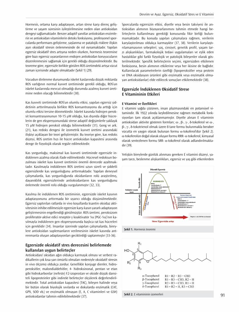
Hormesis, ortama karşı adaptasyon, artan strese karşı direnç geliş-tirme ve yaşam süresinin iyileştirilmesine neden olan antioksidan dengeyi sağlamaktadır. Benzer adaptif yanıtlar antioksidan enzimle-rin ve antioksidan vitaminlerin detoks fonksiyonu, profesyonel spor-cularda performans geliştirme, yaşlanma ve patalojik risklere ilişkin aşırı oksidatif stresin önlenmesinde de rol oynamaktadır. Yapılan egzersiz oksidatif stres artışına neden olurken, hormesis teoremine göre bazı egzersiz uyaranlarının endojen antioksidan koruyucuların düzenlenmesini sağlamak için gerekli olduğu düşünülmektedir. Bu teoreme göre, egzersizle birlikte görülen ROS üretimdeki artışa vücut zaman içerisinde adapte olmaktadır (Şekil 1) (29).
Vücudun dinlenme durumunda iskelet kaslarında düşük miktarda ROS varlığının normal kuvvet üretiminde gerekli olduğu, ROS’un iskelet kaslarında mevcut olmadığı durumda azalmış kuvvet üreti-mine neden olacağı bilinmektedir (30).
Kas kuvveti üretiminde ROS’un olumlu etkisi, yapılan egzersiz şid-detinin arttırılmasıyla birlikte ROS konsantrasyonu da arttığı için olumlu etkisi tersine dönmektedir. İskelet kasında hidrojen perok-sit konsantrasyonunun 10-15 μM olduğu, kas dışında diğer hücre-lerin de gen ekspresyonundaki strese adaptif değişimlerle yaklaşık 15 μM hidrojen peroksit olduğu bilinmektedir (31). Gong ve ark. (21), kas redoks dengesi ile izometrik kuvvet üretimi arasındaki ilişkiyi açıklayan bir teori geliştirmiştir. Bu teoriye göre, kas redoks düzeyi, ROS üretim hızı ile hücre antioksidan kapasitesi arasında denge ile fizyolojik olarak regüle edilmektedir.
Kas yorgunluğu, maksimal kas kuvveti üretiminde egzersizle in-düklenen azalma olarak ifade edilmektedir. Hücresel redoksun bo-zulması iskelet kası kuvvet üretimini önemli derecede azaltmak-tadır. Kasılmayla indüklenen ROS üretimi uzun süreli ve şiddetli egzersizlerde kas yorgunluğunu arttırmaktadır. Yapılan deneysel çalışmalarda, kas yorgunluğunda oksidanların rolü araştırılmış, dayanıklılık egzersizlerinde antioksidanların kas yorgunluğunu önlemede önemli rolü olduğu vurgulanmıştır (32, 33).
Kasılma ile indüklenen ROS üretiminin, egzersizde iskelet kasının adaptasyonunu arttırmada bir uyarıcı olduğu düşünülmektedir. Egzersiz yaptırılan ratlarda in vivo koşullarda ksantin oksidaz akti-vitesinin inhibe edilmesiyle egzersize karşı kasın yararlı adaptasyon geliştirmesinin engellendiği görülmüştür. ROS üretimi, peroksizom proliferatör-aktive edici reseptör-γ koaktivatör 1α (PGC-1α)’nın ka-sılmayla indüklenen gen ekspresyonunda başlıca rat kas hücreleri için gereklidir (34). İnsanlar üzerinde yapılan çalışmalarda, birey-lere antioksidan suplemanların verilmesinin iskelet kasında ant-renmanla oluşan adaptasyonları geciktirdiği saptanmıştır (33-36).
Egzersizde oksidatif stres derecesini belirlemede kullanılan uygun belirteçlerAntioksidan/ oksidan ağın oldukça karmaşık olması ve serbest ra-dikallerin çok kısa yarı ömürlü olmaları nedeniyle oksidatif stresin in vivo ölçümü oldukça zordur. Genellikle konjuge dienler, hidro-peroksitler, malondialdehitler, 4- hidroksinonal, pentan ve etan gibi hidrokarbonlar (nefeste) F2-izoprostan ve okside düşük dansi-teli lipoproteinler gibi indirekt belirteçler ölçülerek değerlendiril-mektedir. Total antioksidan kapasitesi (TAC), bileşen halinde veya bir bütün olarak biyolojik sıvılarda ve dokularda enzimatik (CAT, GPX, SOD vb.) ve enzimatik olmayan (E, A, C vitaminleri ve GSH) antioksidanlar tahmin edilebilmektedir (37).
Sporcularda egzersizin etkisi, diyetle veya besin takviyesi ile an-tioksidan alımının biyoyararlanımını tahmin etmede hangi be-lirteçlerin kullanılması gerektiği konusunda fikir birliği bulun-mamaktadır. Bu konuda yapılan çalışmalara rağmen, verilerin karşılaştırılması oldukça karmaşıktır (37, 38). Verilerin karşılaştı-rılamamasının sebepleri; yaş, cinsiyet, genetik profil, yaşam tar-zı alışkanlıkları, farmakolojik tedavi uygulamaları ve eşlik eden hastalıklar gibi farklı fizyolojik ve patolojik bileşenler olarak gös-terilmektedir. Spesifik belirteçlerin seçimi, egzersizden etkilenen fonksiyona, besin alımının etkilerine veya her ikisine de bağlıdır. Kullanılacak parametrelerin özelliği (lipoperoksitler veya protein ve DNA oksidasyon ürünleri gibi enzimatik veya enzimatik olma-yan antioksidanlar) elde edilecek sonuçları etkilemektedir (38).
Egzersizle İndüklenen Oksidatif Strese E Vitamininin Etkileri
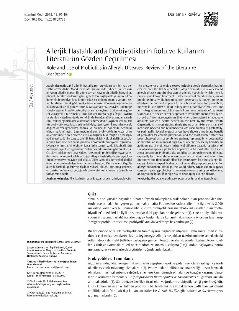
E Vitamini ve ÖzellikleriE vitamini yağda çözünen, insan plazmasındaki en potansiyel vi-tamindir. İlk 1922 yılında keşfedilmesine rağmen metabolik fonk-siyonları tam olarak açıklanmamıştır. Diyetle alınan E vitaminin antioksidan aktivite gösteren formları; α-, β-, γ-, δ-tokoferol ve α-, β-, γ-, δ-tokotrienol olmak üzere 8 tane formu bulunmakla beraber vücutta en yaygın olarak bulunan formu α-tokoferol’dür (Şekil 2). α-tokoferolün doğal olarak oluşan formu RRR- α-tokoferol, kimyasal olarak sentezlenen formu SRR- α-tokoferol olarak adlandırılmakta-dır (39).
Yetişkin bireylerde günlük alınması gereken E vitamini düzeyi, ya-şam tarzı, beslenme alışkanlıkları, egzersiz ve yaş gibi etkenlerden
Şekil 1. Hormesis teoremi
Şekil 2. E vitamininin izomerleri

etkilenmekle beraber yetişkinlerde tahmini ortalama günlük ge-reksinme (EAR- Estimated Average Requirement) 12 mg, önerilen günlük alımı ise (RDA-Recomended Dietary Allowances) 15 mg’dır. Bitkisel yağlar E vitaminin zengin kaynakları olarak bilinmektedir. Buğday tohumu yağı, ayçiçeği yağı, aspir yağı, zeytinyağı ve kanola yağı genellikle α-tokoferol, soya ve mısır yağı ise γ-tokoferol içer-mektedir (39, 40).
Egzersizi takiben oksidatif stresin artışıyla beraber antioksidan en-zimlerin düzeyinin de arttığı gösterilmiştir. Bu antioksidan savun-madaki artış, prooksidan olayların artışına karşı oluşan ihtiyaçları fizyolojik olarak orantılı şekilde karşılayamamakta, bu da E vitami-ni gibi diyetle alınan antioksidanlara olan ihtiyacı etkileyebilmek-tedir. Rokitzki ve ark. (41) tarafından, antrenmanlı bisikletçilere E vitamini suplemanı 5 ay süresince günlük 330 mg verilmiş, laktat eşiğinde bir değişim saptanmazken, serum CK miktarında önemli derecede azalma olduğu görülmüştür.
Bu çalışmalardan farklı olarak E vitaminin CK ve lipit peroksidas-yonu üzerinde egzersizle indüklenen değişimler üzerinde etkili olmadığını savunan araştırmalar da vardır. Jakemann ve Maxwell (38), eksantrik egzersiz yapan bireylerde 7 gün boyunca yapılan E vitamini takviyesinin serum CK seviyeleri üzerine hiçbir etkisinin olmadığını saptamışlardır.
E vitamini suplemantasyonunun egzersiz sonrası oluşan sitokin ya-nıtını değerlendiren çalışmalar da oldukça çelişkilidir. Cannon ve ark. (42), Downhill koşusu öncesi 48 gün boyunca günde 800IU E vitamini suplemanı ve plasebo verdiği bireylerde, koşudan 24 saat sonra plasebo grubunda endotoksinle indüklenen IL-1β salgısının arttığı, E vitamini suplemanı alan grupta ise artış belirlenmemiştir. Normalde egzersizle indüklenen bir sitokin olan IL-6 seviyeleri, E vitamini verilenlerde azalmıştır. TNFα seviyeleri, E vitamininden etkilenmeyerek iki grupta da artış göstermiştir. Singh ve ark. (43) tarafından yapılan çalışmada, %65-70 VO
2 max şiddetinde tükene-ne kadar devam edecek kadın koşucularda akut E vitamini suple-mantasyonunun etkisine bakılmış, plazma IL-6 seviyelerinde artış saptanırken, E vitamini suplemanının etkili olmadığı bulunmuştur.
E vitamini eksikliği, egzersiz sonrası oluşan serbest radikallerle in-düklenen doku hasarını arttırabilmektedir. E vitamini serum sevi-yelerinin yeterli olması, egzersiz sırasında membran bütünlüğünü korumada oldukça önemlidir. Ancak E vitamini suplementasyonu ile yapılan çalışmalarda, E vitaminin lipit peroksidasyonu üzerinde hiçbir etkisinin olmadığı savunan çalışmalarla birlikte (1, 6), egzer-sizden önce veya sonra oluşan lipit peroksidasyonunda küçük ama önemli etkilerinin olduğunu savunan çalışmalar da bulunmakta-dır (10, 24). Meydani ve ark. (44), genç ve yaşlı sağlıklı, eksantrik egzersiz yapan bireylere 48 gün süresince günde 800 IU E vitami-ni takviyesinin iskelet kası α-tokoferol seviyelerini arttırdığını ve eksantrik egzersiz sonucu kaslarda oluşan konjuge dien üretimini azaltarak oksidatif hasarı azalttığını saptamışlardır.
Kelly ve ark. (45), submaksimal egzersizlerde N-asetilsistein (NAC)’ın tiol donörünü azaltıp, glutatyon peroksidaz sentezini arttırarak kas yorgunluğunu geciktirdiği, ancak maksimal veya maksimale yakın şiddetteki egzersizlerde kas yorgunluğunu geciktirmede etkili ol-madığı gözlenmiştir.
Yorgunluğu geciktirmede antioksidan tedavinin etkisi, antioksida-nın türüne bağlı olarak değişmektedir. C ve E vitamini suplemanı-
nın, dayanıklılık egzersizlerindeki yorgunluğu geciktirmede etkili olmadığı bildirilmiştir (46).
Diyetle ve Supleman olarak E Vitamini Alımının ROS Üzerine Etki MekanizmalarıGeçmiş dönemlerde, antioksidan suplemantasyonunun iskelet kası ve kalpte ağır egzersizle indüklenen serbest radikal oluşu-munun kümülatif etkisine karşı etkili olabileceği düşünülmüştür. Antioksidan ögelerin besinlerle alımı yerine supleman olarak alı-mı sonucunda fizyolojik olarak önerilen düzeyin üstüne çıkılması nedeniyle zararlı olabileceği ve oksidatif stres seviyelerini arttıra-bileceği bildirilmiştir. Ayrıca egzersiz sırasında ROS üretimi egzer-sizle indüklenen hormetik yanıttaki proteinlerin ekspresyonu için gereklidir. Antioksidan E vitamini suplemantasyonunun, ROS’un olması istenen konsantrasyonunun oldukça alt seviyelerine düş-mesini sağlayacağı için yan etkilere neden olabilmekte, antioksi-dan suplemantasyonunun uzun dönemde oluşturacağı sonuçlar net olarak bilinmemektedir (10, 44, 46).
Sporcularda dengeli bir diyetle antioksidan alımının oldukça ya-rarlı olduğu düşünülmektedir. Günlük diyetlerinde alınması öneri-len antioksidan miktarı, yaptıkları sporun tipine (aerobik/anaero-bik) göre değişkenlik göstermektedir. Besinlerle antioksidan alımı, antrenman periyotlarında (yarış öncesi/sonrası, aşırı antrenman sendromu vb.) olumlu etki yaratabilmektedir. Sezon değişikliğine de bağlı olarak tüketilmesi gereken antioksidan miktarları deği-şebilmektedir. Kişiye özel bir beslenme programının antrenman durumları ve diğer koşullar göz önüne alınarak yapılması en etkili müdahaledir (46).
Besin takviyesi olarak alınan antioksidanlar, sporcularda kas hasa-rını azaltma, egzersiz performansını geliştirme, ağır egzersizlerin patolojik sonuçlarını azaltmada yararlı noninvazif bir yardımcı olduğu düşünülmektedir. Bu konuda yürütülen çalışmalar ince-lendiğinde tutarlı bir veri olmadığı, Gaeini ve ark. (46) tarafından yapılan çalışmada besin takviyesi olarak antioksidan alımının fiz-yolojik parametreler üzerine hiç etkisinin olmadığı, Pingitore ve ark. (29) tarafından yapılan çalışmada ise negatif etkisinin olabile-ceği bildirilmiştir. Yüksek dozda antioksidan alımı ile antioksidan kapasitenin prooksidan etki gösterebileceği ve ROS aracılığı ile dü-zenlenen fizyolojik yanıtlar üzerinde önemli negatif etki yaratabi-leceği bildirilmiştir (8, 29, 46).
Besinlerle alınan antioksidanların etkilerinin değerlendirildiği çalışmalarda, antioksidan bakımından zengin doğal besinlerin tü-ketimi ve lifli besinler, meyveler, sebzelerden zengin, dengeli bir beslenme tarzı ile optimal antioksidan seviyesinin sağlanabileceği ve antioksidan etkinin optimize edilmesinde etkili olacağı bildiril-miştir. Akdeniz diyetinin uygun olabileceği düşünülmekle beraber bu konuda yeterli veri bulunmamaktadır. (29, 47).
Yaşlanma ile birlikte mitokondriyal respiratuvar fonksiyonun azalması nedeniyle, özellikle yaşlı bireyler egzersize bağlı lipit peroksidasyona karşı duyarlı olabilmekte, bu durum da ROS üretiminde büyük artışa neden olmaktadır. Aynı zamanda, yaşlı bireylerde artmış ROS üretimine yanıt olarak antioksidan seviye-lerinin de arttığı belirtilmiştir (47). Bununla birlikte bireylerdeki artmış antioksidan yanıtın yoğun egzersiz sonucunda oluşan yüksek miktardaki oksidatif stresle mücadelede yetersiz kalacağı saptanmıştır. Yaşla birlikte fagositik hücre aktivitesi azalmakta,
İstanbul Med J 2018; 19: 89-94
92

bu da oksidatif olarak modifiye olan DNA ve doku metabolit-lerinin birikmesine neden olmaktadır. Elde edilen bu bilgiler doğrultusunda, yoğun egzersiz yapan yaşlı bireylerde diyetle E vitamini gibi antioksidanların takviyesine gereksinim olduğu belirtilmiştir (47-49).
Sonuç ve Öneriler Oksidatif stres, kanser, inflamasyon, kardiyovasküler ve nörodejene-ratif hastalıklarda, yaşlanma ve egzersiz gibi fizyolik durumlarda ta-nımlanmıştır. Kontraktil iskelet kaslarında, mitokondri, NADPH ok-sidaz, PLA2- bağımlı süreçler, ksantin oksidaz gibi ROS üretiminden sorumlu potansiyel mekanizmalar bulunmakta, bu mekanizmala-rın oksidasyon üzerine etkilerinin belirlenebilmesi için daha fazla çalışma yapılmasına gereksinim duyulmaktadır (18-22). E vitamini tokoferol ve tokotrienolleri içeren, lipit radikallerini temizleyen ve oksidatif zincir reaksiyonlarını sonlandırma yeteneğine sahip yağda çözünen bir moleküldür. Sporcularda besin takviyesi olarak antiok-sidan alımının faydalarına ilişkin net veri olmamasına rağmen son yıllarda tüketiminde artış görülmüştür. Oksidan ve antioksidanlar arasındaki hassas denge günlük diyetle antioksidan alımıyla (eks-trinsik faktör) sağlanabilmektedir. Akdeniz diyetiyle beslenen spor-cuların, orta yüksek yoğunlukta egzersizde veya yüksek şiddette dayanıklılık egzersizi yapan bireylerde, yaşam kalitesini arttırdığı, insülin duyarlılığı, kan basıncını azalttığı, endotelyal disfonksiyonu düzelttiği şeklinde olumlu etkilerinin olduğu vurgulanmıştır (8, 29). Egzersiz yapan bireylerde hormetik dengenin sağlanmasında ROS’un önemli etkisinin olması nedeniyle diyetsel yaklaşımlarda E vitamini alımının değerlendirilmesinde öneriler geliştirmek için uzun vadeli yapılacak randomize klinik çalışmalara ihtiyaç vardır.
Peer-review: Externally peer-reviewed.
Author contributions: Concept - A.D., A.A.; Design - A.D., A.A..; Supervision - A.A.; Literature Search - A.D., A.A.; Writing - A.D.; Critical Reviews - A.D., A.A.
Conflict of Interest: Authors have no conflicts of interest to declare.
Financial Disclosure: The authors declared that this study has received no financial support.
Hakem Değerlendirmesi: Dış Bağımsız.
Yazar Katkıları: Fikir - A.D., A.A..; Tasarım - A.D., A.A.; Denetleme - A.A.; Li-teratür taraması - A.D., A.A. Yazıyı Yazan - A.D.; Eleştirel İnceleme - A.D., A.A.
Çıkar Çatışması: Yazarların beyan edecek çıkar çatışması yoktur.
Finansal Destek: Yazarlar bu çalışma için finansal destek almadıklarını beyan etmişlerdir.
Kaynaklar
1. Guzel NA, Hazar S, Erbas D. Effects of different resistance exercise pro-tocols on nitric oxide, lipit peroxidation and creatine kinase activity in sedentary males. J Sports Sci Med 2007; 6, 417-422.
2. Jackson MJ. Handbook of Oxidants and Antioxidants in Exercise. Ams-terdam: Elsevier, 2000: 34-36.
3. Peternelj TT,Coombes JS. Antioxidant Supplementation during Exercise Training. Sports Med 2011; 41, 1043-69. [CrossRef]
4. Ji LL. Antioxidants and oxidative stress in exercise. Proc Soc Exp Biol Med 1999; 222, 283-292. [CrossRef]
5. Brisswalter J, Louis J. Vitamin supplementation benefits in master ath-letes. Sports Med 2014; 44: 311-8. [CrossRef]
6. Valko M, Leibfritz D, Moncol J, Cronin MTD, Mazur M,Telser J. Free ra-dicals and antioxidants in normal physiological functions and human disease. Int J Biochem Cell Biol 2007; 39: 44-84. [CrossRef]
7. Falone S, Mirabilio A, Pennelli A, Cacchio M, Di Baldassarre A, Gallina S, et al. Differential impact of acute bout of exercise on redox- and oxida-tive damage-related profiles between untrained subjects and amateur runners. Physiol Res 2010; 59, 953-61.
8. Greilberger JF, Greilberger M, Djukic R. Biomarkers Part I: Biomarkers to Estimate Bioefficacy of Dietary/Supplemental Antioxidants in Sport. (M Lamprecht, Ed.). Antioxidants in Sport Nutrition. Boca Raton (FL): CRC Press/Taylor & Francis, 2015: 54-72.
9. Morales-Alamo D,Calbet JAL. Free radicals and sprint exercise in hu-mans. Free Radic Res 2014; 48: 30-42. [CrossRef]
10. Stepanyan V, Crowe M, Haleagrahara N,Bowden B. Effects of vitamin E supplementation on exercise-induced oxidative stress: a meta-analysis. Appl Physiol Nutr Metab 2014; 39, 1029-37. [CrossRef]
11. Gaeini AA, Rahnama N, Hamedinia MR. Effects of vitamin E supple-mentation on oxidative stress at rest and after exercise to exhaustion in athletic students. J Sports Med Phys Fitness 2006; 46: 458-61.
12. Ortenblad N, Madsen K,Djurhuus MS. Antioxidant status and lipid pe-roxidation after short-term maximal exercise in trained and untrained humans. Am J Physiol 1997; 272: 1258-63. [CrossRef]
13. Laura M, Massimo F, Monica B, Laura M, Ilaria T, Andrea M ve diğ. Di-etary Flavonoids: Molecular Mechanisms of Action as Anti- Inflamma-tory Agents. Recent Pat Inflamm Allergy Drug Discov 2011; 5: 200-220 [CrossRef]
14. Jackson MJ, Pye D, Palomero J. The production of reactive oxygen and nitrogen species by skeletal muscle. J Appl Physiol(1985) 2007; 102: 1664-70. [CrossRef]
15. St-Pierre J, Buckingham JA, Roebuck SJ, Brand MD. Topology of supe-roxide production from different sites in the mitochondrial electron transport chain. J Biol Chem 2002; 277, 44784-90. [CrossRef]
16. Muller FL, Liu Y,Van Remmen H. Complex III releases superoxide to both sides of the inner mitochondrial membrane. J Biol Chem 2004; 279: 49064-73. [CrossRef]
17. Urso ML,Clarkson PM. Oxidative stress, exercise, and antioxidant supp-lementation. Toxicology 2003; 189: 41-54. [CrossRef]
18. Jackson MJ. Free radicals generated by contracting muscle: By-products of metabolism or key regulators of muscle function? Free Radic Biol Med 2008; 44: 132-41. [CrossRef]
19. Javeshghani D, Magder SA, Barreiro E, Quinn MT,Hussain SN. Molecu-lar characterization of a superoxide-generating NAD(P)H oxidase in the ventilatory muscles. Am J Respir Crit Care Med 2002; 165: 412-8. [CrossRef]
20. Nethery D, Callahan LA, Stofan D, Mattera R, DiMarco A, Supinski G. PLA(2) dependence of diaphragm mitochondrial formation of reactive oxygen species. J Appl Physiol (1985) 2000; 89: 72-80. [CrossRef]
21. Gong MC, Arbogast S, Guo Z, Mathenia J, Su W,Reid MB. Calcium-in-dependent phospholipase A2 modulates cytosolic oxidant activity and contractile function in murine skeletal muscle cells. J Appl Physiol (1985) 2006; 100: 399-405. [CrossRef]
22. Judge AR, Dodd SL. Xanthine oxidase and activated neutrophils cause oxidative damage to skeletal muscle after contractile claudication. Am J Physiol Heart Circ Physiol 2004; 286: 252-6. [CrossRef]
23. Apple FS, Rhodes M. Enzymatic estimation of skeletal muscle damage by analysis of changes in serum creatine kinase. J Appl Physiol (1985) 1988; 65, 2598-600.
24. Sacheck JM, Blumberg JB. Role of vitamin E and oxidative stress in exer-cise. Nutrition 2001; 17: 809-14. [CrossRef]
25. Mastaloudis A, Leonard SW,Traber MG. Oxidative stress in athletes du-ring extreme endurance exercise. Free Radic Biol Med 2001; 31: 911-22. [CrossRef]
26. Janero DR. Malondialdehyde and thiobarbituric acid-reactivity as diag-nostic indices of lipit peroxidation and peroxidative tissue injury. Free Radic Biol Med 1990; 9: 515-40. [CrossRef]
Devrim ve Ayaz. Egzersiz, Oksidatif Stres ve E Vitamini
93

27. Vincent HK, Powers SK, Demirel HA, Coombes JS, Naito H. Exercise trai-ning protects against cont raction- induced lipid peroxidation in the di-aphragm. Eur J Appl Physiol Occup Physiol 1999; 79: 268-73. [CrossRef]
28. Pattwell D, Ashton T, McArdle A, Griffiths RD, Jackson MJ. Ischaemia and reperfusion of skeletal muscle leads to appearance of a stable lipid free radical in the circulation. Am J Physiol Heart Circ Physiol 2003; 284, 2400-2404. [CrossRef]
29. Pingitore A, Lima GP, Mastorci F, Quinones A, Iervasi G, Vassalle C. Exercise and oxidative stress: Potential effects of antioxidant dietary strategies in sports. Nutrition 2015; 31, 916-22. [CrossRef]
30. Supinski GS, Callahan LA. Free radical-mediated skeletal muscle dysfunction in inflammatory conditions. J Appl Physiol (1985) 2007; 102: 2056-63. [CrossRef]
31. Powers SK, Jackson MJ. Exercise-Induced Oxidative Stress: Cellular Mechanisms and Impact on Muscle Force Production. Phsiol Rev 2008; 88: 1243-76. [CrossRef]
32. Coombes JS, Rowell B, Dodd SL, Demirel HA, Naito H, Shanely RA, Powers SK. Effects of vitamin E deficiency on fatique and contractile properties. Eur J Appl Physiol 2002; 87; 272-7. [CrossRef]
33. Gandevia SC. Spinal and supraspinal factors in human muscle fati-gue. Physiol Rev 2001; 81, 1725-89. [CrossRef]
34. Silveira LR, Pilegaard H, Kusuhara K, Curi R, Hellsten Y. The contrac-tion induced increase in gene expression of peroxisome proliferator-activated receptor (PPAR)-gamma coactivator 1alpha (PGC-1alpha), mitochondrial uncoupling protein 3 (UCP3) and hexokinase II (HKII) in primary rat skeletal muscle cells is dependent on reactive oxygen species. Biochim Biophys Acta 2006; 1763: 969-76. [CrossRef]
35. Gomez-Cabrera MC, Domenech E, Romagnoli M, Arduini A, Borras C, Pallardo F, et al. Oral administration of vitamin C decreases muscle mitochondrial biogenesis and hampers training-induced adaptations in endurance performance. Am J Clin Nutr 2006; 87, 142-9. [CrossRef]
36. Avery NG, Kaiser JL, Sharman MJ, Scheett TP, Barnes DM, Gomez AL, et al. Effects of Vitamin E Supplementation on 11 Recovery From Repeated Bo-uts of Resistance Exercise. J Strength Cond Res 2003; 17, 801-9. [CrossRef]
37. Vassalle CPA, De Giuseppe R, Vigna L, Bamonti F. Biomarkers to esti-mate bioefficacy of dietary/supplemental antioxidants in sports. (LM, Ed.). Antioxidants in sport nutrition. Boca Raton: FL: Taylor& Francis Group, 2015: 48-72.
38. Jakeman P, Maxwell S. Effect of antioxidant vitamin supplementation on muscle function after eccentric exercise. Eur J Appl Physiol Occup Physiol 1993; 67, 426-30. [CrossRef]
39. Morrissey PA, Kiely M. Vitamin E: Physiology and Health Effects. In: Caballero B, editor. Encyclopedia of Human Nutrition 3rd edition Waltham: Academic Press 2013; 124-46. [CrossRef]
40. Galli F, Azzi A, Birringer M, Cook-Mills JM, Eggersdorfer M, Frank J, et al. Vitamin E: Emerging aspects and new directions. Free Radic Biol Med 2017; 102, 16-36. [CrossRef]
41. Rokitzki L, Logemann E, Huber G, Keck E, Keul J. Alpha-Tocopherol supplementation in racing cyclists during extreme endurance trai-ning. Int J Sport Nutr 1994; 4, 253-64. [CrossRef]
42. Cannon JG, Meydani SN, Fielding RA, Fiatarone MA, Meydani M, Far-hangmehr M, et al. Acute phase response in exercise. II. Associations between vitamin E, cytokines, and muscle proteolysis. Am J Physiol 1991; 260, 1235-1240. [CrossRef]
43. Singh A, Papanicolaou DA, Lawrence LL, Howell EA, Chrousos GP,Deuster, PA. Neuroendocrine responses to running in women after zinc and vitamin E supplementation. Med Sci Sports Exerc 1999; 31, 536-42. [CrossRef]
44. Meydani M, Evans WJ, Handelman G, Biddle L, Fielding RA, Meydani SN, et al. Prosective effect of vitamin E on exercise-induced oxidative damage in young and older adults. Am J Physiol 1993; 264, 992-8.
45. Kelly MK, Wicker RJ, Barstow TJ, Harms CA. Effects of N-acetylcysteine on respiratory muscle fatigue during heavy exercise. Respir Physiol Neurobiol 2009; 165, 67-72. [CrossRef]
46. Gaeini AA, Rahnama N, Hamedinia MR. Effects of vitamin E supple-mentation on oxidative stress at rest and after exercise to exhaustion in athletic students. J Sports Med Phys Fitness 2006; 46, 458-61.
47. Dato S, Crocco P, D'Aquila P, De Rango F, Bellizzi D, Rose G, et al. Exploring the Role of Genetic Variability and Lifestyle in Oxidative Stress Response for Healthy Aging and Longevity. Int J Mol Sci 2013; 14: 16443-72. [CrossRef]
48. Morales-Alamo D, Calbet JA. Free radicals and sprint exercise in hu-mans. Free Radic Res 2014; 48, 30-42. [CrossRef]
49. Jessup JV, Horne C, Yarandi H,Quindry J. The effects of endurance exercise and vitamin E on oxidative stress in the elderly. Biol Res Nurs 2003; 5: 47-55. [CrossRef]
Cite this article as: Devrim A, Ayaz A. Effects of Vitamin E Supplementation on Exercise-induced Oxidative Stress: Friend or Foe? İstanbul Med J 2018; 19: 89-94.
İstanbul Med J 2018; 19: 89-94
94
Giriş Yirmi birinci yüzyılın başından itibaren faydalı mikroplar olarak adlandırılan probiyotikler üze-rinde araştırmalar her geçen gün artmakta hatta Pubmed’de sadece allerji ile ilgili yıllık 2.500 makaleye kadar veri bulunmaktadır. Vücutta probiyotiklerin metabolik art ürünleri olan ‘meta-biyotikler’ in etkileri ile ilgili araştırmalar dahi yayınlanır hale gelmiştir (1). Yine probiyotikler vü-cudun ihtiyacına/hastalığına göre değişik hastalıklarda kullanılmak amacıyle önceden tasarlanıp ‘designer probiotic: tasarımcı probiyotik’ vücuda verilmeye başlanılmıştır (2).
Bu derlemede öncelikle probiyotikleri tanımlayarak başlamak istiyoruz. Daha sonra insan vücu-dunda etki mekanizmalarına kısaca değineceğiz. Allerjik hastalıklar üzerine önleme ve tedavideki rolleri atopik dermatit (AD)‘den başlayarak güncel literatür verileri üzerinden bahsedilecektir. Al-lerjik rinit ve astımdaki rolleri önce randomize kontrollü çalışma (RKÇ)’ lardan başlayarak, sonra metaanalizler ve rehberlerdeki görüşler ışığında anlatılacaktır.
Probiyotikler: TanımlamaAğızdan alındığında, konağın mikroflorasını değiştirebilecek ve potansiyel olarak sağlığına yararlı olabilecek canlı mikroorganizmalardır (3). Probiyotiklerin bilinen üç ana özelliği; insan kaynaklı olmaları, intestinal sistemde değişik etkenlere karşı dirençli olmaları ve konağın yararına olma-larıdır. Asırlardır fermente sütle (Streptococcus thermophilus ve Lactobacillus bulgaricus) vücuda alınmaktadırlar (4). Günümüzde özellikle ticari olan yoğurtların probiyotik içeriği yeterli değildir. En sık kullanılan ve en iyi bilinen probiyotik bakteriler laktik asit bakterileri (LAB) olan Laktobasil ve Bifidobakteri’dir. LAB dışı kullanılan türler ise E. coli, Bacillus gibi bakteri ve Saccharomyces gibi mantarlardır (5).
Allerjik Hastalıklarda Probiyotiklerin Rolü ve Kullanımı: Literatürün Gözden GeçirilmesiRole and Use of Probiotics in Allergic Diseases: Review of the Literature
Atopik dermatit dâhil allerjik hastalıkların prevalansı son bir kaç de-kattır artmaktadır. Atopik dermatit günümüzde bilinen bir tedavisi olmayan allerjik marşın ilk adımı sayılan yaygın bir allerjik hastalıktır. Güncel literatür verilerine göre, gebelikten başlayarak yaşamın erken döneminde probiyotik kullanımı etkin bir önleme metotu ve ümit ve-ren bir strateji olarak görünmekle beraber uzun dönem önleyici etkileri hakkında çok az bilgi mevcuttur. Burada amacımız, tedavi ve önlemeye yönelik yapılan literatürdeki çalışmaların sonuçlarını özetlemek ve gün-cel yaklaşımları tartışmaktır. Probiyotikler Dünya Sağlık Örgütü (WHO) tarafından ‘yeterli miktarda verildiğinde konağa sağlık açısından yararlı canlı mikroorganizmalar’ olarak tarif edilmektedir. Çoğu çalışmada, tek tür probiyotik veya laktik asit ve bifidobakteri içeren karışımlar olarak doğum öncesi (gebelikte), sonrası ya da her iki dönemde perinatal olarak kullanılmıştır. Bazı metaanalizler, probiyotiklerin egzamanın önlenmesinde orta derecede etkili olduğunu bildirmiştir. En belirgin etki ailesel yatkınlık sonucu allerjik hastalık için yüksek riskli süt çocuk-larında kombine perinatal (prenatal+postnatal) probiyotik uygulama-sıyla gösterilmiştir. Yine birden fazla farklı bakteri ya da laktobasil türü içeren probiyotikler, egzamanın önlenmesinde en etkin görünmektedir. Çocuk ve erişkinlerde orta- şiddetli egzamada probiyotikler egzama te-davisinde bir seçenek olabilir. Diğer allerjik hastalıklarda probiyotikle-rin önlemede ve tedavide yeri yoktur. Diğer uzmanlık dernekleri alerjiyi önlemede probiyotikleri önermemekle beraber, Dünya Allerji Örgütü allerjik hastalık geliştirme riskinin yüksek olduğu durumda gebede, emzirirken ve/veya süt çocuğunda probiyotik kullanımının düşünülme-sini önermektedir.
Anahtar Kelimeler: Allerji, allerjik hastalık, egzama, astım, rinit, probiyotik
The prevalence of allergic diseases including atopic dermatitis has in-creased over the last few decades. Atopic dermatitis is a widespread allergic disease and the first step of allergic march, for which there is presently no known treatment. Under the recent literature data, use of probiotics in early life beginning from pregnancy is thought to be an effective method and appears to be a hopeful tactic for prevention, but very little is known about its long-term preventive effect. Here, our aim is to give an outline of the results from these prevention/treatment studies and to discuss current approaches. Probiotics are universally de-scribed as “live microorganisms that, when administered in adequate amounts, confer a health benefit on the host” by the World Health Organization. In most studies, single strains or a mixture of strains of lactic acid bacteria and bifidobacteria was used prenatally, postnatally, or perinatally. Several meta-analyses have shown a moderate benefit of probiotics for eczema prevention, and the most reliable effect has been observed with a combined perinatal (prenatally + postnatally) administration in infants at high risk of allergic disease by heredity. In addition, use of multi-strain (mixture of different bacterial species or of Lactobacillus species) probiotics appeared to be most effective for ec-zema prevention. Probiotics also could be an option in eczema therapy, especially for moderate to severe eczema in children and adults. No preventive and therapeutic effect has been shown for other allergic dis-orders. To date, expert bodies do not generally propose probiotics for allergy prevention, although the World Allergy Organization suggests considering using probiotics in pregnant women, during breastfeeding, and/or to the infant if at high risk of developing allergic disease.
Keywords: Allergy, allergic disease, eczema, asthma, rhinitis, probiotic
Öz
/ Abs
trac
t
ORCID IDs of the authors: Ö.Ö. 0000-0002-5338-9561
Sakarya Üniversitesi Tıp Fakültesi, Çocuk immünolojisi ve Alerjik Hastalıkları Bölümü, Sakarya Üniversitesi Eğitim ve Araştırma Hastanesi, Sakarya, Türkiye
Yazışma Adresi/Address for Correspondence:Öner ÖzdemirE-mail: [email protected]
Geliş Tarihi/Received: 09.06.2017Kabul Tarihi/Accepted: 03.08.2017
© Telif Hakkı 2018 Makale metnine istanbultipdergisi.org web sayfasından ulaşılabilir.
© Copyright 2018 by Available online at istanbulmedicaljournal.org
Derleme / Review İstanbul Med J 2018; 19: 95-104DOI: 10.5152/imj.2018.09735
Öner Özdemir
Probiyotiklerin Allerjide Kullanılma NedenleriSon yıllarda gelişmiş ülkelerdeki hijyen hipotezi ve Batılı yaşam tarzına bağlanan alerjik hastalıklarda artış çevremizdeki ‘bioçeşit-liliğin azalması’ ve perinatal kolonizasyondaki kişisel farklılıklar ile de karakterizedir. Çiftlikte yaşayan çocukların daha az allerjik has-talığa yakalanmasının görülmesi de bunu desteklemektedir (4, 5).
Yine laboratuar çalışmalarında germ hücrelerinin olmadığı fare-lerde, oral toleransın gelişmemesi, ancak patojensiz farelere Bi-fidobakterilerin verilmesiyle oral toleransın gelişmesi (6); öncül çalışmalarda probiyotiklerin alerjik hastalıklar üzerine etkinliğinin gösterilmesi bu hastalıklarda etkili olabileceklerini ve bu amaçla kullanılabileceklerini düşündürmüştür (7)
Probiyotiklerin Allerjide Sistemik Etki MekanizmalarıLokal olarak barsak bariyerinin olgunlaştırması yanında anti-mik-robiyal aktivite, anti-inflamatuar etki ve immünomodülasyona yol açarlar. İmmunomodülasyon sonucu da sistemik kronik (low-grade) enflamasyon oluşur. Bu etkiyi Th1 yolak ve T regülatör (Treg) hücrelerinin üretimi artırarak, Th2 ve Th17 yolaklarını inhibe ede-rek sağlamaktadır. Yine tolerojenik dendritik hücrelerin gelişimi ve Toll-like reseptör (TLR) -2/-9’lerin uyarılması önemli etkilerin-dendir (7, 8).
Probiyotiklerin sistemik etki mekanizmalarına her geçen gün yeni bulgular eklenmektedir. Histamin sinyali üretiminin dahi LAB ile suprese edilebildiği bir çalışmada gösterilmiştir (9). Çocukluk eg-zamasında genetik duyarlılığın modifikasyonunun bile 2 değişik probiyotik türünden biriyle başarıldığı gösterilmiştir (10).
Probiyotiklerin Egzamaya Etki MekanizmalarıDerideki immün sistem hücreleri üzerinde immünomodulatör etkisi mevcuttur. Zararlı deri mikroflorası (Staf. aureus, Herpes ve mantar vb.) ile yarışabilmesi ve zararlı çevresel etkenlere karşı ko-ruyucu kılıf (bariyer fonksiyonu) işlevini görebilmesi; deri pH’sını azaltması ve faydalı metabolitlerin salınması ile oluşur (11). Yine anti-oksidan olarak etki gösterebilme gibi kapasiteleri vardır. Tüm laktobasiller, Lactobacillus delbrueckii hariç, deri patojenlerine kar-şı organik asit üretimiyle beraber antimikrobiyal aktivite gösterir-ler. Aynı zamanda çoğu biofilm oluşumunu engeller, fakat sadece Propioniferax innocua adlı probiyotik matür biofilmi parçalayabilir (12). Bu özellikler, egzama herediter bir cilt hastalığı olduğundan, önleme ve tedavide probiyotiklerin etkinliği açısından önemlidir
Egzama’nın Allerjik Hastalık Gelişiminde ÖnemiDünyada genel allerjik hastalık prevalansı ortalama %20’dir. Yine gelişmiş Kuzey Avrupa ülkeleri ve ABD’de AD prevalansı da %20 ci-varındadır (13). Atopik dermatit, çocukluk çağı egzaması, allerjik hastalığa yatkınlığın ilk belirtisi (atopik/allerjik yürüyüşün (marş) ilk basamağı) olduğundan eğer probiyotiklerin allerjik hastalıkları ön-leme ve tedavisinde etkisi değerlendirilecekse egzama üzerinde ça-lışılması en doğru yoldur. Bu yüzden literatürde ilk çalışmalar araş-tırmacılar tarafından bu hastalık üzerinde gerçekleştirilmiştir (14).
Egzama’nın Önlenmesinde Probiyotiklerin RolüLiteratürdeki egzama üzerine yapılan çalışmalar 2000’li yılların he-men öncesinde Majamaa ve Isolauri (14) tarafından başlatılmıştır. Son 20 yıldır yapılan çalışmalarda yöntemler standart olmadığın-dan probiyotikler bazen prenatal dönemde (gebelikte) anneye, bazen de postnatal dönemde emziriyorsa anneye emzirmiyorsa
bebeğe ya da hem prenatal hem de postnatal dönemde anne ve/veya bebeğe verilmiştir (15-17).
İlk önemli çalışmalardan biri Kalliomaki ve ark. (18) tarafından 2001 yılında bildirilen RKÇ’dır. Lactobacillus rhamnosus GG (LGG) 145 Fin’li gebe ve emziren anneye veya emzirmiyorsa çocuğa veri-lerek 2 yaşına kadar olan atopik egzama insidansı izlenmiş, riskli bebeklerde egzama gelişiminin yarı yarıya (%46’dan %23’e) azal-dığı gösterilmiştir. Aynı grup hastaları izlemeye devam etmiş ve probiyotik etkisinin 4 yaşa kadar devam ettiğini göstermiştir. LGG almaya devam eden atopik egzamalıların 14/53’ünde (RR: 0.57), plasebo alan Fin çocuklarının 25/54’ünde 4 yaşında egzama ge-lişmiştir (19). Bununla beraber aynı metot ve aynı probiyotik LGG ile Kopp ve ark. (20) tarafından 2008 yılında Alman çocuklarında yapılan çalışmada ise, 2 yaşında AD sıklığında bir fark bulunma-mıştır. Kontrole göre ne klinik ne de immünolojik etkiler açısından fark saptanmamıştır. Huurre ve ark. (21) Finlilerde yaptığı diğer bir çalışmada aynı metot (- pre- ve post-natal -) ile farklı bir probiyotik karışımı (LGG+Bifidobacterium) kullanılmış ve 1 yaşında yapılan değerlendirmede çalışmaya katılanlarda herhangi bir etki görül-memiştir. Bu çalışmalar ayrı ırkta probiyotiklerin farklı etkiler mey-dana getirdiği ya da etkisiz kaldığını ayrıca farklı probiyotiklerin aynı ırkta benzer sonuca yol açmadığı göstermektedir.
Probiyotik Verilme Zamanının Önemi: Prenatal? Postnatal?Prenatal ve postnatal uygulama ile ilgili veriler ve bunların sonuç-ları aşağıda anlatılacağı gibi tablo 1’de özetlenmiştir.
Sadece Prenatal Probiyotik TakviyesiPrenatal/antenatal (gebelikte) probiyotik kullanımı çok sık uygula-nılmış olmakla beraber, postnatal verilmeyen çalışmalarda etkin-liği yetersiz bulunduğundan terk edilmiştir. Pubmed taramasında bulduğumuz tek RKÇ, Boyle ve ark. (22) olandır. Bu çalışmada 250 riskli bebek doğuracak gebeye LGG 1,8×10 (10) cfu/gün dozunda 36. gestasyon haftasından itibaren verilmiştir. Prenatal probiyotik verilimi egzama veya IgE-ilişkili egzama gelişim riskini düşürme-miştir.
Prenatal ve Postnatal (Prenatal Anne ve Postnatal Emziren Anne ve/veya Süt Çocuğuna) TakviyePrenatal anne ve postnatal emziren anne veya süt çocuğuna direkt olarak daha uzun bir süre verilmesi atopik (IgE-ilişkili egzama) ve nonatopik egzama önlemede saptadığımız en az 6/9 RKÇ’da etkili bulunmuştur. Wickens’ın 2 ve Kalliomaki’nin 3 RKÇ’ında prenatal anneye ve postnatal hem emziren anne hem de süt çocuğuna probiyotik verilmiştir. Rautava ve ark. 2002 ve 2012’de bildirilen 2 ayrı çalışmalarında prenatal ve postnatal dönemde sadece anneye (gebe ve emzirirken) probiyotik verilmiştir. Prenatal dönemde an-neye ve postnatal dönemde süt çocuğuna probiyotik verilen çalış-malardan ikisi Allen ark., birisi Kuitunen ve ark. ve ikisi Kukkonen ve ark. çalışmalarıdır. Bu her iki dönemde de probiyotik verilme metodunun sadece prenatal veya postnatal dönemde probiyotik verilmeye göre başarılı olmasının nedenlerinden bazıları probiyo-tiklerin bir şekilde fetüs immün sistemini etkileyebilmesi, mater-nal vaginal floranın ve bu vaginal yoldan doğan süt çocuğunun intestinal florasının probiyotiklerin etkisiyle modifikasyonuna bağlanmıştır. Yine postnatal dönemde verilmenin yanında prena-tal dönemde anneye probiyotik takviyesinin süt çocuğu barsağının probiyotik bakterilerle hızlı kolonizasyonu ve anne sütü içeriğinde
İstanbul Med J 2018; 19: 95-104
96

Özdemir Ö. Allerjik Hastalıklarda Probiyotik Kullanımı
97
değişimlere (immünoregülatör IgA ve TGF-β’ yi artırması) yol aça-rak allerji gelişimini önlemede etkili olabileceğini göstermektedir (15-17).
Sadece Postnatal (Anne veya Süt Çocuğuna) Probiyotik takviyesiÇoğu çalışmada sadece postnatal probiyotik takviyesi yapılmış ve etkisiz bulunmuştur. Bunlardan Taylor ve ark. (23) 2007’de bildir-diği RKÇ’da yüksek riskli bebeğe ilk 6 ayda probiyotik verilmesinin AD riskini azaltmadığı aksine alerjen sensitizasyonunu artırdığı saptanmıştır. Postnatal emziren anneye, Ortiz-Andrellucchi ve ark. (24) yaptığı çalışma mevcuttur. Hem postnatal emziren anne hem de postnatal süt çocuğuna aynı anda probiyotik verilen çalışma şu ana kadar saptayabildiğimiz yoktur. Postnatal süt çocuğuna probi-yotik verilen çalışmalar Hascoet ve ark. (25), Lodinova ve ark. (26), Morisset ve ark. (27), West ve ark. (28) tarafından gerçekleştirilmiştir. Bunlardan sadece Lodinova ve ark.’nın (26) probiyotik olarak E.coli kullandıkları çalışmada uzun dönemde dahi etki görülmüştür.
Bu yolla başarılı olunamamasında vaginal flora üzerinden bebeğin barsak kolonizasyonunun etkilenememesi ve ayrıca belki de yeni-doğanın ve ilk 3-6 ay arasında bebeğin immün sisteminin modifiye edilebilecek kadar olgunlaşmaması sayılabilir (29).
Probiyotik Türünün Rolü: Bifidobakteri? Laktobasil?Bifidobakteri ağırlıklı probiyotik karışımı içeren 2 çalışmada yük-sek riskli grupta beklenen etki görülmüştür (30, 31). Bir çalışma-da Lactobacillus rhamnosus’un egzamayı azaltmada daha başarılı olduğu, Bifidobacterium animalis subsp. lactis gibi Bifidobakteri türlerinin tek başına kullanıldıklarında etkisinin olmadığı bildiril-
miştir (32). Bifidobakteriler genelde diğer probiyotiklerle karışım halinde kullanılmışlardır.
Yine Lactobacillus rhamnosus GG (LGG) uzun dönemde AD gelişimini önlemede etkili bulunmuştur. Benzer şekilde, yüksek riskli çocuk-larda AD gelişimini 6 yaşa kadar önlemede Lactobacillus rhamnosus HN001’nin etkinliği gösterilmiştir. Aynı etkinin Bifidobacterium lactis HN019 ile görülmemiş olması etkinin türe bağlı olduğunu düşün-dürür (33). Yine metaanaliz çalışmalarında, Laktobasil içeren pro-biyotiklerin Bifidobakterilere üstünlüğü gösterilmiştir (34). Özellikle LGG’nin prenatal ve postnatal kullanımının yalnız prenatal kullanı-mına karşı plasebo ile karşılaştırıldığında (Risk Ratio: 0.88 vs. 0.68 ), 12.-24. ay arasında etkisi çok belirgindir (35).
Probiyotik Karışımlarının RolüNorveç’te yapılan bir çalışmada probiyotik karışımı (LGG, Laktoba-cillus acidophilus La-5 ve Bifidobacterium animalis) 1 ay prenatal ve 3 ay postnatal dönemde emziren anneye verilmiş ve 2 yaşında değerlendirildiğinde atopik egzama ve egzama [OR:0.51] gelişimi-ni azaltmıştır (36).
Probiyotik karışımın (2 Laktobasil, Bifidobakteri ve Propionibakte-ri) yüksek riskli grupta kullanıldığı çalışmada 5 yaşına ulaşanlarda yapılan değerlendirmede yalnızca sezaryan ile doğanların bulun-duğu altgrupta IgE’ye bağlı allerjik hastalık insidansının düşük ol-duğu bulunmuştur (37).
Huurre ve ark.’nın (21) Finli çocuklarda yaptığı çalışmada pre-/post-natal LGG ve Bifidobacterium probiyotik karışımı kullanılmış ve 1 yaşında değerlendirilmiş fakat etkili bulunmamıştır.
Tablo 1. Atopik dermatiti önlemeye yönelik probiyotik örnek çalışmalarında verilme zamanına göre hastalık üzerine etkileri görülmektedir
Verilme Zamanı Probiyotik türleri Sonuç Doz Kaynak
Prenatal (anneye) LGG ↔ ≥109-10 22
Prenatal+Postnatal (anneye) LGG, Bifidobacterium lactis Bb12 ↔ ≥109-10 21
LGG, Bifidobacterium Bb-12, Lactobacillus acidophilus La-5 ↓ ≥109-10 36
Lactobacillus rhamnosus LP + Bifidobacterium longum veya ↓, ↓ ≥109-10 15-17, 73
Lactobacillum paracasei + Bifidobacterium longum
Prenatal+Postnatal (anne+bebeğe) LGG ↓ ≥109-10 18, 19, 45
Lactobacillus rhamnosus HN001,Bifidobacterium lactis HN019 ↓ ≥109-10 32, 33, 46
Lactobacillus reuteri ↔ ≥109-10 38, 47
LGG, Lactobacillus rhamnosus LC705, Bifidobacterium breve ↓ ≥109-10 41 Bb99, Propionibacterium shermanii
LGG ↔ ≥109-10 20
Bifidobacterium bifidum, Bifidobacterium lactis, Lactobacillus ↓ ≥109-10 31 acidophilus
Prenatal+Postnatal (bebeğe) Bifidobacterium bifidum, Bifidobacterium lactis, Lactococcus lactis ↓ ≥109-10 30
Postnatal (bebeğe) Lactobacillus acidophilus ↔, * ≥109-10 23
Lactobacillus F19 ↓, ↔** ≥109-10 28, 48
Lactobacillus acidophilus ↔, * ≥109-10 49
Escherichia coli ↓, *** ≥109-10 26
Postnatal (anneye) Lactobacillus casei ↔ ≥109-10 24
LGG: Lactobacillus rhamnosus GG; ↔: etkisizlik; ↑: etkiyle artma; ↓: etkiyle azalma; *: alerjik sensitizasyonda artma; **: uzun dönemde etki kaybı; ***: uzun dönemde etki kaybolmaz
Yine metaanaliz çalışmalarında, özellikle Laktobasil içeren probi-yotik karışımlarının diğerlerine üstünlüğü gösterilmiştir (16).
Egzama Türünün Rolü (IgE-ilişkili (atopik) Egzama/Nonatopik Egzama)Beşten fazla çalışmada özellikle atopik (IgE-ilişkili, özellikle besin alerjisi olan) egzamada ya da atopik egzama/dermatitte etkin bu-lunmuştur (38-42). Yapılan metaanalizlerde, bu etki çok net olma-sa da kısmen gösterilmiştir (43, 44).
Uzun Süreli Takibe Dayalı Atopik Dermatiti Önlemeye Yönelik ÇalışmalarıKalliomaki ve ark. (45) LGG alan grupta 4 yıllık süreden 7 yıllık süreye kadar egzamaya karşı koruma etkisinin (OR: 0.58) devam ettiğini göstermiştir.
Wickens ve ark. (46) yüksek riskli çocuklarda AD gelişimini 6 yaşa kadar önlemede Laktobacillus rhamnosus HN001’nin etkinliği gös-termiştir. Aynı etki Bifidobacterium lactis HN019 ile görülmemiş ve bu da etkinin probiyotik türüne bağlı olduğunu düşündürmüştür.
Abrahamsson ve ark. (47) çalışmasında gebelik son ayda ve ilk yıl içinde İsveçli 232 alerjik hastalığı olan ailenin çocuklarında Lacto-bacillus reuteri kullanılmış ve 7 yıllık takibi 184 çocuk tamamla-yabilmiştir. AD prevalansı kontrol grubuna göre farklı bulunma-mıştır. Benzer şekilde, West ve ark. (48) yaptığı çalışmada, 8-9 yaşa kadar izlenen İsveçli çocuklarda Lactobacillus paracasei (LF19)’nin allerjik hastalık üzerine etkisi bulunmamıştır.
Probiyotiklerin uzun süreli etkisini araştıran çalışmalar vardır. Tay-lor ve ark.’nın (23) 1 yıl ve Prescott ve ark. (49) 2,5 yılık takip sü-resinin sonunda yapılan değerlendirmelerde probiyotik etkisinin kalmadığı görülmüştür. Kukkonen ve ark. (41) çalışmasında 2 yıl boyunca probiyotik etkili bulunmuştur. Kuitunen ve ark. (37) ça-lışmasında 5 yıl boyunca takip sonrasında, sadece sezaryanlılarda etkili bulunmuştur. Lin ve ark. (50) ve Loo ve ark. (51) çalışmasında postnatal ilk 6 ayda verilen probiyotik Asya’lı riskli süt çocuğunda 5. yılda etkili bulunmamıştır. Simpson ve ark. (52) perinatal (ma-ternal) verilen probiyotiğin 6 yıl boyunca etkin olduğunu bildir-miştir. Postnatal verilen probiyotik E. coli ise 10 ve 20 yıl sonra incelendiğinde bile etkin bulunmuştur (53).
Zuccotti ve ark.’nın (16) metaanalizine göre de ilk 2 yaşa kadar, probiyotiklerin etkilerinin daha belirgin olduğu, ileri yaşlarda bu etkinin zamanla azaldığı görülmektedir.
Egzama Tedavisinde Probiyotiklerin RolüLiteratür verilerine göre, probiyotik takviyesinin genelde tedavi-deki etkisinin egzama skorlaması olan SCORAD’da ve hastaların semptomlarında azalmayla beraber yaşam kalitesinde artmaya yol açtığı bildirilmiştir. Yine çoğu çalışmada SCORAD’da azalma ve yaşam kalitesindeki artmanın probiyotik kesilmesinden 4 hafta sonrasına kadar devam ettiği gösterilmiştir (54). Bazı çalışmalar-da, yüksek konsantrasyonda Lactobacillus salivarius LS01 içeren jel kompleks olarak topikal probiyotik uygulamasının da AD’li hasta-larda faydası da gösterilmiştir (55).
Anlamlı düzelmeye yol açan probiyotiklerLactobacillus acidophilus L-92'yi de içeren ve tedavideki rolünü araştıran çok sayıda çalışmada etkin saptanmıştır (56, 57).
Kısmi düzelme yapanlarLactobacillus plantarum CJLP133 ve Lactobacillus salivarius gibi probiyotiklerin kullanıldığı çalışmalarda, SCORAD’da ve yaşam ka-litesindeki kısmi düzelme bildirilmiştir (58).
Etkisiz olanlarGore ve ark. (59) ile diğer bazı yazarların Lactobacillus paracasei veya Bifidobacterium lactis içeren probiyotiklerle yapılan çalışma-larında, ve Torley ve ark. (60) bildirdiği derlemelerde etkisiz bu-lunmuştur.
Tedavi ile ilgili bir metaanaliz Kim ve ark. (61) tarafından yayınlan-mıştır. Çocuk ve erişkinlerdeki orta-şiddetli egzamanın tedavisinde faydalı olabileceği vurgulanmıştır.
Egzama Önleme/Tedavisinde Probiyotiklerin Rolünü İrdeleyen Derleme/MetaanalizlerVan der Aa ve ark. (62) yaptığı metaanalizlerde çocuklarda AD önlenmesi/tedavisinde probiyotiklerin yeri olmadığı kanaatine varmıştır. Eigenmann’ın (63) vardığı sonuçta olduğu gibi, etkinin genel olarak gebelikte başlayan kombine probiyotik kullanımının erken süt çocuklukta devam edilmesiyle ortaya çıktığı bulunmuş-tur. Kuitunen ve ark. (64) ve diğerlerinin belirttiği gibi, en bariz etki, prenatal dönemde anneye ve postnatal direkt bebeğe Lacto-bacillus rhamnosus suplementasyonuna özgün bulunmuştur (65). Lee ve ark. (66) metaanalizinde tedaviden ziyade önlemede etkili olduğu yine Baquerizo ve ark. (67) derlemesinde egzama insidan-sını azaltabileceği belirtilmiştir. Sonuç olarak, AD önlenme ve te-davisinde probiyotiklerin rolünü araştıran derleme ve metaanaliz-lerde; probiyotiklerin tedaviden daha çok önlemede etkili olduğu ve egzama insidansını düşürebildiği görülmüştür.
2007 yılında bildirilen Cochrane metaanalizinde, 5/12 RKÇ’da 1477 süt çocuğu meta-analizle değerlendirilmiştir. Egzamanın azaldığı (RR: 0.82) ve Lactobacillus rhamnosus ’un yüksek riskli grupta fay-dalı olduğu bulunmuştur (68). 2008 yılında bildirilen Cochrane metaanalizinde, probiyotiklerin egzama tedavisinde etkili olduğu ispatlanamamıştır (69). 2009 yılında bildirilen Cochrane metaa-nalizinde, egzama için RR: 0.82; atopik egzama için RR: 0.80 bu-lunmuştur (70). Cochrane metaanalizlerinde, probiyotiğin olumlu etkisinin olduğundan bahsedilse de bu konuda kesin bir sonuca ulaşılamadığı vurgulanmıştır.
Bazı uluslararası rehberlerde (WAO ve EAACI), 2012 yılında bildiri-len probiyotiklerin çocuk allerjisinde klinik kullanımını araştıran Dünya Alerji Organizasyonu rehberinde olduğu gibi, önleme ve tedavide yeri yok denilmiştir (71). 2014 yılı EAACI (Avrupa Alerji ve Klinik İmmünoloji Derneği) rehberinde de önlemede yeri ol-madığından bahsedilmektedir (72). 2015 yılında Dünya Alerji Organizasyonu’nun allerjik hastalık önlenmesine yönelik rehbe-rinde (WAO-GLAD-P), hastalık gelişim riski yüksekse; gebe, anne ve çocukta probiyotiklerin kullanılabileceği vurgulanmıştır (73).
Diğer Allerjik Hastalıklarda Probiyotiklerin KullanımıBurada önce besin alerjisi, allerjik rinit, astım ve refrakter kronik ürtikerdeki etkilerinden bahsedilecektir (Tablo 2).
Besin AllerjisiBesin alerjisinin önlenmesi ve tedavisi ile ilgili çalışmalar, AD üze-rine olduğu kadar yaygın ve çok sayıda değildir. İlk yayınlarda inek
İstanbul Med J 2018; 19: 95-104
98

sütü alerjisi olan egzamalı hastalarda probiyotik etkisiyle SCORAD düşmüştür (14, 39, 42). Yine inek sütü alerjisi olan hastalarda probiyotik (LGG) takviyesinin, formüla mamaların içine konularak yapıldığı gibi, bu hastalarda tolerans indüksiyonuna yol açtığı gös-terilmiştir (14, 74). Tang ve ark. (75) tarafından yapılan çalışmada yer fıstığı alerjisi olanlarda yapılan oral immunoterapiye eklenen probiyotiğin (PPOIT çalışması) tolerans gelişiminde faydalı olduğu gösterilmiştir.
Besin alerjisi ile ilgili çalışmaların bazı metaanalizlerinde; besine tolerans, sensitizasyon ve SCORAD’ın etkilenmediği gösterilmiştir (76). Besin alerjisi ile ilgili çalışmaların Zhang (77) tarafından yapı-lan metaanalizinde ise, prenatal (anne) ve postnatal (süt çocuğu-na) uygulanan probiyotiğin atopi ve besin hipersensitivite riskini azalttığı (relatif risk: 0.78 ve 0.77) gösterilmiştir.
2007 yılındaki Cochrane metaanalizinde besin allerjisi üzerine ko-ruyucu etkisinin olmadığı belirtilmiştir (68). Yine Kong ve ark. (78) değerlendirmelerinde prenatal ve postnatal probiyotik takviyesi-nin besin allerjisi üzerine koruyucu etkisinin (RR:0.88) olmadığı saptanmıştır. 2014 yılı EAACI (Avrupa Alerji ve Klinik İmmünoloji Derneği) rehberinde de besin alerjisinde probiyotiklerin yeri olma-dığından bahsedilmektedir (72).
Allerjik Rinitte Probiyotiklerin Rolü
Mevsimsel ve perennial allerjik rinitMevsimsel ve persistan allerjik rinit tedavisinde etkisiz olduğunu söyleyenler yanında, etkili olduğunu söyleyen araştırmacılar da mevcuttur. Lactobacillus türleri (L. casei, L. acidophilus strain L-92, L. paracasei-33) ile yapılan çalışmalarda mevsim öncesi ve sonra-sında faydalı bulunmuştur (79, 80). Probiyotik emdirilmiş yatak-yorgan- yastık kılıfları ile yapılan bir çalışmada da allerjik rinitli hastaların semptomlarında düzelme ve yaşam kalitesinde artma saptanmıştır (81).
Allerjik rinit: meta-analizAllerjik rinit konusunda çok az olan meta-analizlerden birini içe-ren Zajac ve ark. (82) 2015’de yaptığı derlemede; 23 çalışma ve 1919 hasta üzerinde değerlendirme yapılmıştır. Allerjik rinitli has-
taların semptomlarında düzelme ve yaşam kalitesinde artma sap-tanmasına rağmen, mevcut verilerin kısıtlı olduğu ve daha nitelikli çalışmalara ihtiyaç duyulduğunu göstermektedir.
Astımda Probiyotiklerin Rolü
Astım gelişimini önlemeBazı çalışmalarda probiyotiklerin etkisinin olmadığı gösterilmiştir (20, 23, 38). Benzer şekilde, Kuitunen ve ark. (37, 64) ve Kalliomäki ve ark. (45) tarafından 5-8 yaş üzerinde etkisiz bulunmuştur. Aksi-ne Johansson ve van der Aa ark. (62, 70) 5 yaşta ve etkili olduğunu göstermişlerdir.
Astım tedavisiAstım tedavisinde etkisiz olduğunu bildirenler yanında, etkili oldu-ğunu söyleyenler de mevcuttur (83, 84).
Allerjik Havayolu (Allerjik Rinit ve Astım) HastalığıAstım ve alerjik rinitle ilgili çalışmalar literatürde oldukça az olup, bunların bir kısmı her ikisini de içerecek şekilde allerjik havayolu hastalığı (allerjik rinit+astım) üzerine yapılıp sonuçlar elde edil-meye çalışılmıştır. Aşağıda bunlarla ilgili önce RKÇ, derleme, meta-analizler sırasıyle sunulacaktır.
Önlemeye yönelik çalışmalarYedi yıllık takipte probiyotiklerin respiratuar alerji, allerjik havayo-lu hastalığı (AR ve astım), üzerinde etkisi saptanamamıştır (85). LGG içeren probiyotiklerin, 4. yaşta yapılan değerlendirmede hava yolu semptomları üzerine etkisi bulunmamıştır (86).
Allerjik havayolu hastalığı tedavisi üzerine olan çalışmaların derlemeleriTedaviye yönelik derlemelerde yapılan değerlendirmelerde yaşam kalitesinde artış bildirilmekle beraber, laboratuvar parametreleri-nin etkilenmediği görülmüştür (87).
Astım ve allerjik rinit ile ilgili bu çalışmaları özetlersek; a) Farklı suş, doz ve sürede probiyotikler kullanılmışlardır. Minimal doz: >5x10⁹ cfu/gün ve minimal kulla nım süresi 1 ay olmuştur. b) Astımlı hastalarda daha çok Laktoba sil (acidophilus, rhamnosus,
Özdemir Ö. Allerjik Hastalıklarda Probiyotik Kullanımı
99
Tablo 2. Allerjik hastalıklarda kullanılan probiyotikler ve kullanım şekilleri
Hastalık Probiyotik Zamanlaması Doz Sonuç Kaynak
Atopik Dermatit
Önleme LGG Prenatal+Postnatal ≥109-10 ↓ 18-20, 45
Tedavi LGG Çocukluk çağı ≥109-10 ↓ 61
Besin Allerjisi
Önleme Çeşitli Prenatal+Postnatal ≥109-10 ↔ 68, 72, 78
Tedavi LGG Süt çocukluğu ≥109-10 ↓ 14, 74, 75
Allerjik Rinit
Önleme Çeşitli Prenatal+Postnatal ≥109-10 ↔ 18-20, 45
Tedavi Bifidobakteri Erken çocukluk ≥109-10 ↓ 79, 80
Astım
Önleme Çeşitli Prenatal+Postnatal ≥109-10 ↔ 18-20, 45
Tedavi Lactobacillus Oyun çocuğu ≥109-10 ↔ 83, 84
LGG: Lactobacillus rhamnosus GG; ↔: etkisizlik; ↑: etkiyle artma; ↓: etkiyle azalma
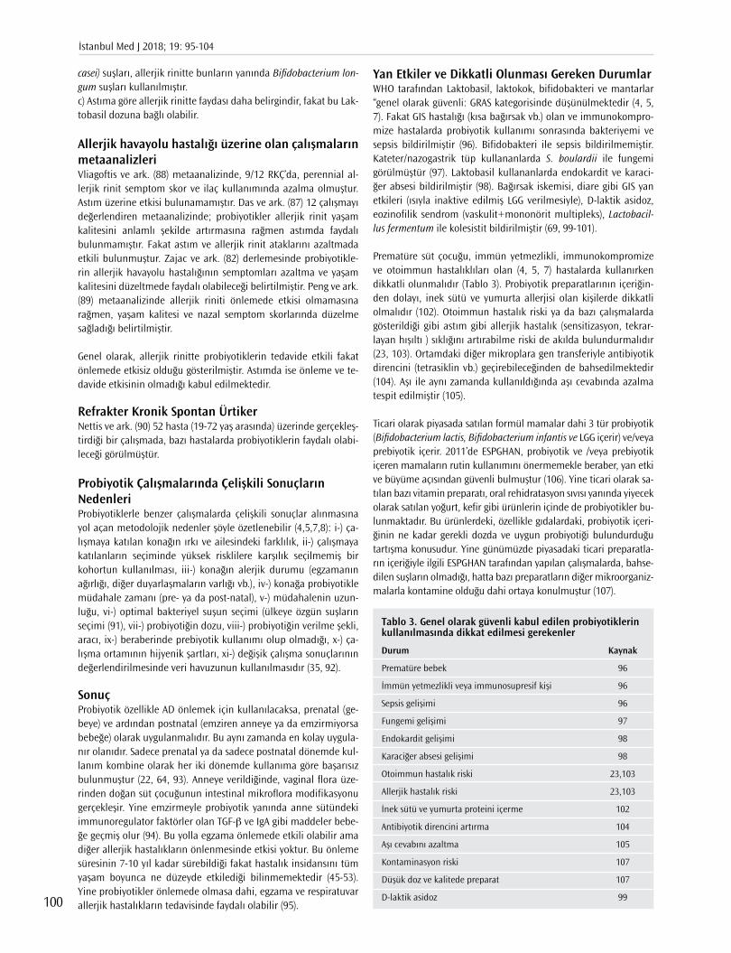
casei) suşları, allerjik rinitte bunların yanında Bifidobacterium lon-gum suşları kullanılmıştır. c) Astıma göre allerjik rinitte faydası daha belirgindir, fakat bu Lak-tobasil dozuna bağlı olabilir.
Allerjik havayolu hastalığı üzerine olan çalışmaların metaanalizleriVliagoftis ve ark. (88) metaanalizinde, 9/12 RKÇ’da, perennial al-lerjik rinit semptom skor ve ilaç kullanımında azalma olmuştur. Astım üzerine etkisi bulunamamıştır. Das ve ark. (87) 12 çalışmayı değerlendiren metaanalizinde; probiyotikler allerjik rinit yaşam kalitesini anlamlı şekilde artırmasına rağmen astımda faydalı bulunmamıştır. Fakat astım ve allerjik rinit ataklarını azaltmada etkili bulunmuştur. Zajac ve ark. (82) derlemesinde probiyotikle-rin allerjik havayolu hastalığının semptomları azaltma ve yaşam kalitesini düzeltmede faydalı olabileceği belirtilmiştir. Peng ve ark. (89) metaanalizinde allerjik riniti önlemede etkisi olmamasına rağmen, yaşam kalitesi ve nazal semptom skorlarında düzelme sağladığı belirtilmiştir.
Genel olarak, allerjik rinitte probiyotiklerin tedavide etkili fakat önlemede etkisiz olduğu gösterilmiştir. Astımda ise önleme ve te-davide etkisinin olmadığı kabul edilmektedir.
Refrakter Kronik Spontan ÜrtikerNettis ve ark. (90) 52 hasta (19-72 yaş arasında) üzerinde gerçekleş-tirdiği bir çalışmada, bazı hastalarda probiyotiklerin faydalı olabi-leceği görülmüştür.
Probiyotik Çalışmalarında Çelişkili Sonuçların NedenleriProbiyotiklerle benzer çalışmalarda çelişkili sonuçlar alınmasına yol açan metodolojik nedenler şöyle özetlenebilir (4,5,7,8): i-) ça-lışmaya katılan konağın ırkı ve ailesindeki farklılık, ii-) çalışmaya katılanların seçiminde yüksek risklilere karşılık seçilmemiş bir kohortun kullanılması, iii-) konağın alerjik durumu (egzamanın ağırlığı, diğer duyarlaşmaların varlığı vb.), iv-) konağa probiyotikle müdahale zamanı (pre- ya da post-natal), v-) müdahalenin uzun-luğu, vi-) optimal bakteriyel suşun seçimi (ülkeye özgün suşların seçimi (91), vii-) probiyotiğin dozu, viii-) probiyotiğin verilme şekli, aracı, ix-) beraberinde prebiyotik kullanımı olup olmadığı, x-) ça-lışma ortamının hijyenik şartları, xi-) değişik çalışma sonuçlarının değerlendirilmesinde veri havuzunun kullanılmasıdır (35, 92).
SonuçProbiyotik özellikle AD önlemek için kullanılacaksa, prenatal (ge-beye) ve ardından postnatal (emziren anneye ya da emzirmiyorsa bebeğe) olarak uygulanmalıdır. Bu aynı zamanda en kolay uygula-nır olanıdır. Sadece prenatal ya da sadece postnatal dönemde kul-lanım kombine olarak her iki dönemde kullanıma göre başarısız bulunmuştur (22, 64, 93). Anneye verildiğinde, vaginal flora üze-rinden doğan süt çocuğunun intestinal mikroflora modifikasyonu gerçekleşir. Yine emzirmeyle probiyotik yanında anne sütündeki immunoregulator faktörler olan TGF-β ve IgA gibi maddeler bebe-ğe geçmiş olur (94). Bu yolla egzama önlemede etkili olabilir ama diğer allerjik hastalıkların önlenmesinde etkisi yoktur. Bu önleme süresinin 7-10 yıl kadar sürebildiği fakat hastalık insidansını tüm yaşam boyunca ne düzeyde etkilediği bilinmemektedir (45-53). Yine probiyotikler önlemede olmasa dahi, egzama ve respiratuvar allerjik hastalıkların tedavisinde faydalı olabilir (95).
Yan Etkiler ve Dikkatli Olunması Gereken DurumlarWHO tarafından Laktobasil, laktokok, bifidobakteri ve mantarlar “genel olarak güvenli: GRAS kategorisinde düşünülmektedir (4, 5, 7). Fakat GIS hastalığı (kısa bağırsak vb.) olan ve immunokompro-mize hastalarda probiyotik kullanımı sonrasında bakteriyemi ve sepsis bildirilmiştir (96). Bifidobakteri ile sepsis bildirilmemiştir. Kateter/nazogastrik tüp kullananlarda S. boulardii ile fungemi görülmüştür (97). Laktobasil kullananlarda endokardit ve karaci-ğer absesi bildirilmiştir (98). Bağırsak iskemisi, diare gibi GIS yan etkileri (ısıyla inaktive edilmiş LGG verilmesiyle), D-laktik asidoz, eozinofilik sendrom (vaskulit+mononörit multipleks), Lactobacil-lus fermentum ile kolesistit bildirilmiştir (69, 99-101).
Prematüre süt çocuğu, immün yetmezlikli, immunokompromize ve otoimmun hastalıklıları olan (4, 5, 7) hastalarda kullanırken dikkatli olunmalıdır (Tablo 3). Probiyotik preparatlarının içeriğin-den dolayı, inek sütü ve yumurta allerjisi olan kişilerde dikkatli olmalıdır (102). Otoimmun hastalık riski ya da bazı çalışmalarda gösterildiği gibi astım gibi allerjik hastalık (sensitizasyon, tekrar-layan hışıltı ) sıklığını artırabilme riski de akılda bulundurmalıdır (23, 103). Ortamdaki diğer mikroplara gen transferiyle antibiyotik direncini (tetrasiklin vb.) geçirebileceğinden de bahsedilmektedir (104). Aşı ile aynı zamanda kullanıldığında aşı cevabında azalma tespit edilmiştir (105).
Ticari olarak piyasada satılan formül mamalar dahi 3 tür probiyotik (Bifidobacterium lactis, Bifidobacterium infantis ve LGG içerir) ve/veya prebiyotik içerir. 2011’de ESPGHAN, probiyotik ve /veya prebiyotik içeren mamaların rutin kullanımını önermemekle beraber, yan etki ve büyüme açısından güvenli bulmuştur (106). Yine ticari olarak sa-tılan bazı vitamin preparatı, oral rehidratasyon sıvısı yanında yiyecek olarak satılan yoğurt, kefir gibi ürünlerin içinde de probiyotikler bu-lunmaktadır. Bu ürünlerdeki, özellikle gıdalardaki, probiyotik içeri-ğinin ne kadar gerekli dozda ve uygun probiyotiği bulundurduğu tartışma konusudur. Yine günümüzde piyasadaki ticari preparatla-rın içeriğiyle ilgili ESPGHAN tarafından yapılan çalışmalarda, bahse-dilen suşların olmadığı, hatta bazı preparatların diğer mikroorganiz-malarla kontamine olduğu dahi ortaya konulmuştur (107).
İstanbul Med J 2018; 19: 95-104
100
Tablo 3. Genel olarak güvenli kabul edilen probiyotiklerin kullanılmasında dikkat edilmesi gerekenler
Durum Kaynak
Prematüre bebek 96
İmmün yetmezlikli veya immunosupresif kişi 96
Sepsis gelişimi 96
Fungemi gelişimi 97
Endokardit gelişimi 98
Karaciğer absesi gelişimi 98
Otoimmun hastalık riski 23,103
Allerjik hastalık riski 23,103
İnek sütü ve yumurta proteini içerme 102
Antibiyotik direncini artırma 104
Aşı cevabını azaltma 105
Kontaminasyon riski 107
Düşük doz ve kalitede preparat 107
D-laktik asidoz 99

Yakın Gelecekten BeklentilerAllerjik hastalıkları engelleyici/önleyici etkisinin kesin bir gerçek olup olmadığı, allerjik hastalığın gerçek sıklığını azaltıp azaltma-dığı net olarak bilinmemektedir. Allerjik hastalıkların gelişiminin engellenmesinin uzun dönemde ne kadar sürdüğü yine ortaya konulamamıştır. Kanaatimizce, allerjik hastalığın ortaya çıkmasını daha çok geciktiriyor gibi gözükmektedir (4, 5, 7).
Günümüzde vücuda verilmeden önce probiyotikler yapacağı işleve göre tasarlanır (designer probiotics). Vücuda verildiğinde terapö-tik protein ve DNA aşılarının mukozal ulaşımını sağlayan vektörler olarak görev yapabilirler. Örneğin, rekombinant Laktokok DNA’sını ökaryotik hücrelere ulaştırır. Lactobacillus lactis, plasmidi epitel membran hücrelerine verir (108). Adjuvan olarak kullanılan LAB ya da Clostridium butyricum, major birch (huş ağacı) polen alerjeni (Bet v 1) ile kombinasyonu sonucu polen immunoterapisinde ya da akarlara karşı immunoterapide başarılı olarak kullanılmıştır (109-111). Laktokok ve laktobasilleri içeren rekombinant laktik asit bakterileri (RecLAB), Th1 sitokini (IL-10 /-12 ) - eksprese eden pro-biyotik bakteri (IL-10 salan Lactobacillus lactis) suşu olarak üretile-bilmiştir (112). Yine benzer şekilde, gıda antijeni (β-laktoglobulin (süt) ve ovalbumin (yumurta)) - eksprese eden üretilmiş probiyotik bakteriler besinlere karşı immunoterapide denenmeye başlanmış-tır (113, 114).
Hakem Değerlendirmesi: Dış Bağımsız.
Çıkar Çatışması: Yazarların beyan edecek çıkar çatışması yoktur.
Finansal Destek: Yazar bu çalışma için finansal destek almadıklarını beyan etmiştir.
Peer-review: Externally peer-reviewed.
Conflict of Interest: Authors have no conflicts of interest to declare.
Financial Disclosure: The author declared that this study has received no financial support.
Kaynaklar
1. Sharma M, Shukla G. Metabiotics: One step ahead of probiotics; an in-sight into mechanisms involved in anticancerous effect in colorectal cancer. Front Microbiol 2016; 7: 1940. [CrossRef]
2. Chua KJ, Kwok WC, Aggarwal N, Sun T, Chang MW. Designer probiotics for the prevention and treatment of human diseases. Curr Opin Chem Biol 2017; 40: 8-16. [CrossRef]
3. Food and Agriculture Organization, World Health Organization (FAO/WHO) Report of Joint FAO/WHO expert consultation on evaluation of health and nutritional properties of probiotics in food including pow-der milk with live lactic acid bacteria. FAO/WHO Report no. 10-1-2001. WHO INT; Córdoba, Argentina.
4. Özdemir Ö. Prebiyotikler, probiyotikler ve alerji (Bölüm 76). Şekerel BE, editor. Çocukluk Çağında Allerji, Astım ve İmmünoloji. Balıkesir: Ada Basın-Yayın; 2015; 819-43.
5. Ozdemir O. Various effects of different probiotic strains in allergic di-sorders: an update from laboratory and clinical data. Clin Exp Immu-nol 2010; 160: 295-304. [CrossRef]
6. Sudo N, Sawamura S, Tanaka K, Aiba Y, Kubo C, Koga Y. The requ-irement of intestinal bacterial flora for the development of an IgE production system fully susceptible to oral tolerance induction. J Im-munol 1997; 159: 1739-45.
7. Ozdemir O. Prebiotics and probiotics in allergy: Potential mecha-nisms of prebiotics' and probiotics' actions in allergy - (Part 1). MOJ Immunol 2016; 3: 00069. [CrossRef]
8. Özdemir Ö. Mechanisms of preventative and therapeutic role of pro-biotics in different allergic and autoimmune disorders. Open Journal of Immunology 2013; 3: 1-16. [CrossRef]
9. Dev S, Mizuguchi H, Das AK, Matsushita C, Maeyama K, Umehara H, et al. Suppression of histamine signaling by probiotic Lac-B: a possible mechanism of its anti-allergic effect. J Pharmacol Sci 2008; 107: 159-66. [CrossRef]
10. Morgan AR, Han DY, Wickens K, Barthow C, Mitchell EA, Stanley TV, et al. Differential modification of genetic susceptibility to childhood ec-zema by two probiotics. Clin Exp Allergy 2014; 44: 1255-65. [CrossRef]
11. Lew LC, Liong MT. Bioactives from probiotics for dermal health: func-tions and benefits. J Appl Microbiol 2013; 114: 1241-53. [CrossRef]
12. Lopes EG, Moreira DA, Gullón P, Gullón B, Cardelle-Cobas A, Tavaria FK. Topical application of probiotics in skin: adhesion, antimicrobial and antibiofilm in vitro assays. J Appl Microbiol 2013; 114: 1241-53.
13. Nutten S. Atopic Dermatitis: Global Epidemiology and Risk Factors. Ann Nutr Metab 2015; 66: 8-16. [CrossRef]
14. Majamaa H, Isolauri E. Probiotics: a novel approach in the mana-gement of food allergy. J Allergy Clin Immunol 1997; 99: 179-85. [CrossRef]
15. Cuello-Garcia CA, Brożek JL, Fiocchi A, Pawankar R, Yepes-Nu-ez JJ, Ter-racciano L, et al. Probiotics for the prevention of allergy: A systematic review and meta-analysis of randomized controlled trials. J Allergy Clin Immunol 2015; 136: 952-61. [CrossRef]
16. Zuccotti G, Meneghin F, Aceti A, Barone G, Callegari ML, Di Mauro A, et al. Probiotics for prevention of atopic diseases in infants: systematic review and meta-analysis. Allergy 2015; 70: 1356-71. [CrossRef]
17. Ricci G, Cipriani F, Cuello-Garcia CA, Brożek JL, Fiocchi A, Pawankar R, et al. A clinical reading on "World Allergy Organization-McMaster University Guidelines for Allergic Disease Prevention (GLAD-P): Probi-otics". World Allergy Organ J 2016; 9: 9. [CrossRef]
18. Kalliomäki M, Salminen S, Arvilommi H, Kero P, Koskinen P, Isolauri E. Probiotics in primary prevention of atopic disease: a randomised placebo-controlled trial. Lancet 2001; 357: 1076-9. [CrossRef]
19. Kalliomäki M, Salminen S, Poussa T, Arvilommi H, Isolauri E. Probio-tics and prevention of atopic disease: 4-year follow-up of a randomi-sed placebo-controlled trial. Lancet 2003; 361: 1869-71. [CrossRef]
20. Kopp MV, Hennemuth I, Heinzmann A, Urbanek R. Randomized, double-blind, placebo-controlled trial of probiotics for primary pre-vention: no clinical effects of Lactobacillus GG supplementation. Pe-diatrics 2008; 121: 850-6. [CrossRef]
21. Huurre A, Laitinen K, Rautava S, Korkeamäki M, Isolauri E. Impact of maternal atopy and probiotic supplementation during pregnancy on infant sensitization: a double-blind placebo-controlled study. Clin Exp Allergy 2008; 38: 1342-8. [CrossRef]
22. Boyle RJ, Ismail IH, Kivivuori S, Licciardi PV, Robins-Browne RM, Mah LJ, et al. Lactobacillus GG treatment during pregnancy for the pre-vention of eczema: a randomized controlled trial. Allergy 2011; 66: 509-16. [CrossRef]
23. Taylor AL, Dunstan JA, Prescott SL. Probiotic supplementation for the first 6 months of life fails to reduce the risk of atopic dermatitis and increases the risk of allergen sensitization in high-risk children: a ran-domized controlled trial. J Allergy Clin Immunol 2007; 119: 184-91. [CrossRef]
24. Ortiz-Andrellucchi A, Sanchez-Villegas A, Rodriguez-Gallego C, Lemes A, Molero T, Soria A, et al. Immunomodulatory effects of the intake of fermented milk with Lactobacillus casei DN114001 in lactating mot-hers and their children. Br J Nutr 2008; 100: 834-45. [CrossRef]
25. Hascoet JM, Hubert C, Rochat F, Legagneur H, Gaga S, Emady-Azar S, et al. Effect of formula composition on the development of infant gut microbiota. J Pediatr Gastroenterol Nutr 2011; 52: 756-62. [CrossRef]
26. Lodinova-Zadnikova R, Prokesova L, Kocourkova I, Hrdy J, Zizka J. Pre-vention of allergy in infants of allergic mothers by probiotic Escheric-hia coli. Int Arch Allergy Immunol 2010; 153: 201-6. [CrossRef]
Özdemir Ö. Allerjik Hastalıklarda Probiyotik Kullanımı
101
27. Morisset M, Aubert-Jacquin C, Soulaines P, Moneret-Vautrin DA, Du-pont C. A non-hydrolyzed, fermented milk formula reduces digestive and respiratory events in infants at high risk of allergy. Eur J Clin Nutr 2011; 65: 175-83. [CrossRef]
28. West CE, Hammarstr€om ML, Hernell O. Probiotics during weaning reduce the incidence of eczema. Pediatr Allergy Immunol 2009; 20: 430-7. [CrossRef]
29. Damm JA, Smith B, Greisen G, Krogfelt KA, Clausen ML, Agner T. The influence of probiotics for preterm neonates on the incidence of ato-pic dermatitis-results from a historically controlled cohort study. Arch Dermatol Res 2017; 309: 259-64. [CrossRef]
30. Niers L, Martín R, Rijkers G, Sengers F, Timmerman H, van Uden N, et al. The effects of selected probiotic strains on the development of eczema (the PandA study). Allergy 2009; 64: 1349-58. [CrossRef]
31. Kim JY, Kwon JH, Ahn SH, Lee SI, Han YS, Choi YO, et al. Effect of pro-biotic mix (Bifidobacterium bifidum, Bifidobacterium lactis, Lactoba-cillus acidophilus) in the primary prevention of eczema: a double-blind, randomized, placebo-controlled trial. Pediatr Allergy Immunol 2010; 21: 386-93. [CrossRef]
32. Wickens K, Black PN, Stanley TV, Mitchell E, Fitzharris P, Tannock GW, et al. A differential effect of 2 probiotics in the prevention of eczema and atopy: a double-blind, randomized, placebo controlled trial. J Allergy Clin Immunol 2008; 122: 788-94. [CrossRef]
33. Wickens K, Black P, Stanley TV, Mitchell E, Barthow C, Fitzharris P, et al. A protective effect of Lactobacillus rhamnosus HN001 against eczema in the first 2 years of life persists to age 4 years. Clin Exp Allergy 2012; 42: 1071-9. [CrossRef]
34. Yao TC, Chang CJ, Hsu YH, Huang JL. Probiotics for allergic diseases: rea-lities and myths. Pediatr Allergy Immunol 2010; 21: 900-19. [CrossRef]
35. Szajewska H, Shamir R, Turck D, van Goudoever JB, Mihatsch WA, Fewtrell M. Recommendations on probiotics in allergy prevention should not be based on pooling data from different strains. J Allergy Clin Immunol 2015; 136: 1422. [CrossRef]
36. Dotterud CK, Storrø O, Johnsen R, Oien T. Probiotics in pregnant wo-men to prevent allergic disease: a randomized, double-blind trial. Br J Dermatol 2010; 163: 616-23. [CrossRef]
37. Kuitunen M, Kukkonen K, Juntunen-Backman K, Korpela R, Poussa T, Tuure T, et al. Probiotics prevent IgE-associated allergy until age 5 years in cesarean-delivered children but not in the total cohort. J Allergy Clin Immunol 2009; 123: 335-41. [CrossRef]
38. Abrahamsson TR, Jakobsson T, Böttcher MF, Fredrikson M, Jenmalm MC, Björkstén B, et al. Probiotics in prevention of IgE-associated ec-zema: a double-blind, randomized, placebo-controlled trial. J Allergy Clin Immunol 2007; 119: 1174-80. [CrossRef]
39. Viljanen M, Savilahti E, Haahtela T, Juntunen-Backman K, Korpela R, Poussa T, et al. Probiotics in treatment of atopic eczema/dermatitis syndrome in infants: a double-blind placebo-controlled trial. Allergy 2005; 60: 494-500. [CrossRef]
40. Sistek D, Kelly R, Wickens K, Stanley T, Fitzharris P, Crane J. Is the effect of probiotics on atopic dermatitis confined to food sensitized child-ren? Clin Exp Allergy 2006; 36: 629-33. [CrossRef]
41. Kukkonen K, Savilahti E, Haahtela T, Juntunen-Backman K, Korpela R, Poussa T, et al. Probiotics and prebiotic galacto-oligosaccharides in the prevention of allergic diseases: a randomized, double blind, placebo-controlled trial. J Allergy Clin Immunol 2007; 119: 192-8. [CrossRef]
42. Rosenfeldt V, Benfeldt E, Nielsen SD, Michaelsen KF, Jeppesen DL, Va-lerius NH, et al. Effect of probiotic Lactobacillus strains in children with atopic dermatitis. J Allergy Clin Immunol 2003; 111: 389-95. [CrossRef]
43. Pelucchi C, Chatenoud L, Turati F, Galeone C, Moja L, Bach JF, et al. Probiotics supplementation during pregnancy or infancy for the pre-vention of atopic dermatitis: a meta-analysis. Epidemiology 2012; 23: 402-14. [CrossRef]
44. Kim HJ, Kim HY, Lee SY, Seo JH, Lee E, Hong SJ. Clinical efficacy and mechanism of probiotics in allergic diseases. Korean J Pediatr 2013; 56: 369-76. [CrossRef]
45. Kalliomäki M, Salminen S, Poussa T, Isolauri E. Probiotics during the first 7 years of life: a cumulative risk reduction of eczema in a ran-domized, placebo-controlled trial. J Allergy Clin Immunol 2007; 119: 1019-21. [CrossRef]
46. Wickens K, Stanley TV, Mitchell EA, Barthow C, Fitzharris P, Purdie G, et al. Early supplementation with Lactobacillus rhamnosus HN001 reduces eczema prevalence to 6 years: does it also reduce atopic sen-sitization? Clin Exp Allergy 2013; 43: 1048-57. [CrossRef]
47. Abrahamsson TR, Jakobsson T, Björkstén B, Oldaeus G, Jenmalm MC. No effect of probiotics on respiratory allergies: a seven-year follow-up of a randomized controlled trial in infancy. Pediatr Allergy Immunol 2013; 24: 556-61. [CrossRef]
48. West CE, Hammarström ML, Hernell O. Probiotics in primary preventi-on of allergic disease--follow-up at 8-9 years of age. Allergy 2013; 68: 1015-20. [CrossRef]
49. Prescott SL, Wiltschut J, Taylor A, Westcott L, Jung W, Currie H, et al. Early markers of allergic disease in a primary prevention study using probiotics: 2.5-year follow-up phase. Allergy 2008; 63: 1481-90. [CrossRef]
50. Lin JH. Supplementation with probiotics in the first 6 months of life did not protect against eczema and allergy in at-risk Asian infants: A 5-year follow-up. Pediatrics 2014; 134: 142. [CrossRef]
51. Loo EX, Llanora GV, Lu Q, Aw MM, Lee BW, Shek LP. Supplementation with probiotics in the first 6 months of life did not protect against eczema and allergy in at-risk Asian infants: a 5-year follow-up. Int Arch Allergy Immunol 2014; 163: 25-8. [CrossRef]
52. Simpson MR, Dotterud CK, Storrø O, Johnsen R, Øien T. Perinatal pro-biotic supplementation in the prevention of allergy related disease: 6 year follow up of a randomised controlled trial. BMC Dermatol 2015; 15: 13. [CrossRef]
53. Lodinová-Zádníková R, Cukrowska B, Tlaskalova-Hogenova H. Oral administration of probiotic Escherichia coli after birth reduces frequency of allergies and repeated infections later in life (after 10 and 20 years). Int Arch Allergy Immunol 2003; 131: 209-11. [CrossRef]
54. Michail SK, Stolfi A, Johnson T, Onady GM. Efficacy of probiotics in the treatment of pediatric atopic dermatitis: a metaanalysis of randomi-zed controlled trials. Ann Allergy Asthma Immunol 2008; 101: 508-16. [CrossRef]
55. Drago L, De Vecchi E, Toscano M, Vassena C, Altomare G, Pigatto P. Treatment of atopic dermatitis eczema with a high concentration of Lactobacillus salivarius LS01 associated with an innovative gelling complex: a pilot study on adults. J Clin Gastroenterol 2014; 48: 47-51. [CrossRef]
56. Yamamoto K, Yokoyama K, Matsukawa T, Kato S, Kato S, Yamada K, et al. Efficacy of prolonged ingestion of Lactobacillus acidophilus L-92 in adult patients with atopic dermatitis. J Dairy Sci 2016; 99: 5039-46. [CrossRef]
57. Torii S, Torii A, Itoh K, Urisu A, Terada A, Fujisawa T, et al. Effects of oral administration of Lactobacillus acidophilus L-92 on the symptoms and serum markers of atopic dermatitis in children. Int Arch Allergy Immunol 2011; 154: 236-45. [CrossRef]
58. Han Y, Kim B, Ban J, Lee J, Kim BJ, Choi BS, et al. A randomized trial of Lactobacillus plantarum CJLP133 for the treatment of atopic dermati-tis. Pediatr Allergy Immunol 2012; 23: 667-73. [CrossRef]
59. Gore C, Custovic A, Tannock GW, Munro K, Kerry G, Johnson K, et al. Treatment and secondary prevention effects of the probiotics Lacto-bacillus paracasei or Bifidobacterium lactis on early infant eczema: randomized controlled trial with follow-up until age 3 years. Clin Exp Allergy 2012; 42: 112-22. [CrossRef]
60. Torley D, Futamura M, Williams HC, Thomas KS. What's new in atopic eczema? An analysis of systematic reviews published in 2010-11. Clin Exp Dermatol 2013; 38: 449-56. [CrossRef]
61. Kim SO, Ah YM, Yu YM, Choi KH, Shin WG, Lee JY. Effects of probiotics for the treatment of atopic dermatitis: a meta-analysis of randomi-zed controlled trials. Ann Allergy Asthma Immunol 2014; 113: 217-26. [CrossRef]
İstanbul Med J 2018; 19: 95-104
102
62. Van der Aa LB, Heymans HS, van Aalderen WM, Sprikkelman AB. Pro-biotics and prebiotics in atopic dermatitis: review of the theoretical background and clinical evidence. Pediatr Allergy Immunol 2010; 21: 355-67. [CrossRef]
63. Eigenmann PA. Evidence of preventive effect of probiotics and prebi-otics for infantile eczema. Curr Opin Allergy Clin Immunol 2013; 13: 426-31. [CrossRef]
64. Kuitunen M. Probiotics and prebiotics in preventing food allergy and eczema. Curr Opin Allergy Clin Immunol 2013; 13: 280-6. [CrossRef]
65. Mansfield JA, Bergin SW, Cooper JR, Olsen CH. Comparative probiotic strain efficacy in the prevention of eczema in infants and children: a systematic review and meta-analysis. Mil Med 2014; 179: 580-92. [CrossRef]
66. Lee J, Seto D, Bielory L. Meta-analysis of clinical trials of probiotics for prevention and treatment of pediatric atopic dermatitis. J Allergy Clin Immunol. 2008; 121: 116-121. [CrossRef]
67. Baquerizo Nole KL, Yim E, Keri JE. Probiotics and prebiotics in derma-tology. J Am Acad Dermatol 2014; 71: 814-21. [CrossRef]
68. Osborn DA, Sinn JK. Probiotics in infants for prevention of allergic disease and food hypersensitivity. Cochrane Database Syst Rev 2007; 4: CD006475. [CrossRef]
69. Boyle RJ, Bath-Hextall FJ, Leonardi-Bee J, Murrell DF, Tang ML. Pro-biotics for treating eczema. Cochrane Database Syst Rev 2008; 4: CD006135. [CrossRef]
70. Johansson SG, Bieber T, Dahl R, Friedmann PS, Lanier BQ, Lockey RF, et al. J Allergy Clin Immunol 2004; 113: 832-6. [CrossRef]
71. WAO Special Committee on Food Allergy and Nutrition. Clinical Use of Probiotics in Pediatric Allergy (CUPPA): A World Allergy Organization Position Paper. World Allergy Organ J 2012; 5: 148-67. [CrossRef]
72. Muraro A, Halken S, Arshad SH, Beyer K, Dubois AE, Du Toit G, et al. EAACI food allergy and anaphylaxis guidelines. Primary prevention of food allergy. Allergy 2014; 69: 590-601. [CrossRef]
73. Fiocchi A, Pawankar R, Cuello-Garcia C. World Allergy Organizati-on-McMaster University Guidelines for Allergic Disease Prevention (GLAD-P): Probiotics. World Allergy Organ J 2015; 8: 4. [CrossRef]
74. Berni Canani R, Nocerino R, Terrin G, Frediani T, Lucarelli S, Cosenza L, et al. Formula selection for management of children with cow's milk allergy influences the rate of acquisition of tolerance: a prospective multicenter study. J Pediatr 2013; 163: 771-7. [CrossRef]
75. Tang ML, Ponsonby AL, Orsini F, Tey D, Robinson M, Su EL, et al. Admi-nistration of a probiotic with peanut oral immunotherapy: A rando-mized trial. J Allergy Clin Immunol 2015; 135: 737-44. [CrossRef]
76. Toh ZQ, Anzela A, Tang ML, Licciardi PV. Probiotic therapy as a novel approach for allergic disease. Front Pharmacol 2012; 3: 171. [CrossRef]
77. Zhang GQ, Hu HJ, Liu CY, Zhang Q, Shakya S, Li ZY. Probiotics for Pre-vention of Atopy and Food Hypersensitivity in Early Childhood: A PRIS-MA-Compliant Systematic Review and Meta-Analysis of Randomized Controlled Trials. Medicine (Baltimore). 2016; 95: e2562. [CrossRef]
78. Kong XY, Yang Y, Guan J, Wang RZ. Probiotics' preventive effect on pe-diatric food allergy: a meta-analysis of randomized controlled trials. Chin Med Sci J 2014; 29: 144-7. [CrossRef]
79. Wang MF, Lin HC, Wang YY, Hsu CH. Treatment of perennial allergic rhinitis with lactic acid bacteria. Pediatr Allergy Immunol 2004; 15: 152-8.[CrossRef]
80. Ivory K, Chambers SJ, Pin C, Prieto E, Arqués JL, Nicoletti C. Oral delivery of Lactobacillus casei Shirota modifies allergen-induced immune res-ponses in allergic rhinitis. Clin Exp Allergy 2008; 38: 1282-9. [CrossRef]
81. Berings M, Jult A, Vermeulen H, De Ruyck N, Derycke L, Ucar H, et al. Probiotics-impregnated bedding covers for house dust mite allergic rhinitis: A pilot randomized clinical trial. Clin Exp Allergy 2017; 47: 1092-6. [CrossRef]
82. Zajac AE, Adams AS, Turner JH. A systematic review and meta-analysis of probiotics for the treatment of allergic rhinitis. Int Forum Allergy Rhinol 2015; 5: 524-32.[CrossRef]
83. Miraglia Del Giudice M, Maiello N, Decimo F, Fusco N, D' Agostino B, Sullo N, et al. Airways allergic inflammation and L. reuterii treatment in asthmatic children. J Biol Regul Homeost Agents 2012; 26: 35-40.
84. van de Pol MA, Lutter R, Smids BS, Weersink EJ, van der Zee JS. Synbio-tics reduce allergen-induced T-helper 2 response and improve peak ex-piratory flow in allergic asthmatics. Allergy 2011; 66: 39-47. [CrossRef]
85. Matsui E. No effect of probiotics on respiratory allergies: A seven-year follow-up of a randomized controlled trial in ınfancy. Pediatrics 2014; 134: 141-2. [CrossRef]
86. Rose MA, Schubert R, Schulze J, Zielen S. Follow-up of probiotic Lac-tobacillus GG effects on allergic sensitization and asthma in infants at risk. Clin Exp Allergy 2011; 41: 1819-21. [CrossRef]
87. Das RR, Naik SS, Singh M. Probiotics as additives on therapy in allergic airway diseases: a systematic review of benefits and risks. Biomed Res Int 2013; 2013: 231979.[CrossRef]
88. Vliagoftis H, Kouranos VD, Betsi GI, Falagas ME. Probiotics for the tre-atment of allergic rhinitis and asthma: systematic review of randomi-zed controlled trials. Ann Allergy Asthma Immunol 2008; 101: 570-9. [CrossRef]
89. Peng Y, Li A, Yu L, Qin G. The role of probiotics in prevention and treatment for patients with allergic rhinitis: A systematic review. Am J Rhinol Allergy 2015; 29: 292-8. [CrossRef]
90. Nettis E, Di Leo E, Pastore A, Distaso M, Zaza I, Vacca M, et al. Probio-tics and refractory chronic spontaneous urticaria. Eur Ann Allergy Clin Immunol 2016; 48: 182-7.
91. Vernocchi P, Del Chierico F, Fiocchi AG, El Hachem M, Dallapiccola B, Rossi P, et al. Understanding probiotics' role in allergic children: the clue of gut microbiota profiling. Curr Opin Allergy Clin Immunol 2015; 15: 495-503. [CrossRef]
92. Elazab N, Mendy A, Gasana J, Vieira ER, Quizon A, Forno E. Probiotic administration in early life, atopy, and asthma: a meta-analysis of clinical trials. Pediatrics 2013; 132: 666-76. [CrossRef]
93. Özdemir Ö. The role of probiotics in atopic dermatitis prevention and therapy. In: Esparza-Gordillo J, Dekio I, Eds. Atopic Dermatitis - Disea-se Etiology and Clinical Management. InTech, Open Access Company, Croatia, 2012: 353-86. [CrossRef]
94. Munblit D, Boyle RJ. Modulating breast milk composition - the key to allergy prevention? Int Arch Allergy Immunol 2012; 159: 107-8. [CrossRef]
95. Özdemir Ö. Is there any effect of probiotics on allergic airway disease? J Chin Med Assoc 2015; 78: 627-8. [CrossRef]
96. McClave SA, Heyland DK, Wischmeyer PE. Comment on: probiotic prophylaxis in predicted severe acute pancreatitis: a randomized, double-blind, placebo-controlled trial. JPEN J Parenter Enteral Nutr 2009; 33: 444-6. [CrossRef]
97. Roy U, Jessani LG, Rudramurthy SM, Gopalakrishnan R, Dutta S, Chak-ravarty C, et al. Seven cases of Saccharomyces fungaemia related to use of probiotics. Mycoses 2017; 60: 375-80. [CrossRef]
98. Kato K, Funabashi N, Takaoka H, Kohno H, Kishimoto T, Nakatani Y, et al. Lactobacillus paracasei endocarditis in a consumer of probiotics with advanced and severe bicuspid aortic valve stenosis complicated with diffuse left ventricular mid-layer fibrosis. Int J Cardiol 2016; 224: 157-61. [CrossRef]
99. Munakata S, Arakawa C, Kohira R, Fujita Y, Fuchigami T, Mugishima H. A case of D-lactic acid encephalopathy associated with use of probio-tics. Brain Dev 2010; 32: 691-4. [CrossRef]
100. Mendoza FA, Purohit S, Kenyon L, Jimenez SA. Severe eosinophilic syndrome associated with the use of probiotic supplements: a new entity? Case Rep Rheumatol 2012; 2012: 934324.
101. Chery J, Dvoskin D, Morato FP, Fahoum B. Lactobacillus fermentum, a pathogen in documented cholecystitis. Int J Surg Case Rep 2013; 4: 662-4. [CrossRef]
102. Martín-Munoz MF, Fortuni M, Caminoa M, Belver T, Quirce S, Cabal-lero T. Anaphylactic reaction to probiotics. Cow's milk and hen's egg allergens in probiotic compounds. Pediatr Allergy Immunol 2012; 23: 778-84. [CrossRef]
103. Peldan P, Kukkonen AK, Savilahti E, Kuitunen M. Perinatal probiotics decreased eczema up to 10 years of age, but at 5-10 years, allergic rhino-conjunctivitis was increased. Clin Exp Allergy 2017; 47: 975-9. [CrossRef]
Özdemir Ö. Allerjik Hastalıklarda Probiyotik Kullanımı
103

104. Imperial IC, Ibana JA. Addressing the antibiotic resistance problem with probiotics: reducing the risk of ıts double-edged sword effect. Front Microbiol 2016; 7: 1983. [CrossRef]
105. Praharaj I, John SM, Bandyopadhyay R, Kang G. Probiotics, antibiotics and the immune responses to vaccines. Philos Trans R Soc Lond B Biol Sci 2015; 370. [CrossRef]
106. Braegger C, Chmielewska A, Decsi T, Kolacek S, Mihatsch W, Moreno L, et al. Supplementation of infant formula with probiotics and/or prebiotics: a systematic review and comment by the ESPGHAN com-mittee on nutrition. J Pediatr Gastroenterol Nutr 2011; 52: 238-50. [CrossRef]
107. Kolaček S, Hojsak I, Canani RB, Guarino A, Indrio F, Orel R, et al. Commercial probiotic products: A call for improved quality control. A Position Paper by the ESPGHAN Working Group for Probiotics and Prebiotics. J Pediatr Gastroenterol Nutr 2017; 65: 117-24. [CrossRef]
108. del Carmen S, Martín Rosique R, Saraiva T, Zurita-Turk M, Miyoshi A, Azevedo V, et al. Protective effects of lactococci strains delivering either IL-10 protein or cDNA in a TNBS-induced chronic colitis model. J Clin Gastroenterol 2014; 48: 12-7. [CrossRef]
109. Xu LZ, Yang LT, Qiu SQ, Yang G, Luo XQ, Miao BP, et al. Combination of specific allergen and probiotics induces specific regulatory B cells and enhances specific immunotherapy effect on allergic rhinitis. Oncotar-get 2016; 7: 54360-9. [CrossRef]
110. Moussu H, Van Overtvelt L, Horiot S, Tourdot S, Airouche S, Zuercher A, et al. Bifidobacterium bifidum NCC 453 promotes tolerance induc-tion in murine models of sublingual immunotherapy. Int Arch Allergy Immunol 2012; 158: 35-42. [CrossRef]
111. Van Overtvelt L, Moussu H, Horiot S, Samson S, Lombardi V, Mascarell L, et al. Lactic acid bacteria as adjuvants for sublingual allergy vacci-nes. Vaccine 2010; 28: 2986-92. [CrossRef]
112. de Moreno de LeBlanc A, Del Carmen S, Chatel JM, Azevedo V, Lan-gella P, Bermudez-Humaran L, et al. Evaluation of the biosafety of recombinant lactic acid bacteria designed to prevent and treat colitis. J Med Microbiol 2016; 65: 1038-46. [CrossRef]
113. Adel-Patient K, Ah-Leung S, Creminon C, Nouaille S, Chatel JM, Lan-gella P, et al. Oral administration of recombinant Lactococcus lactis expressing bovine beta-lactoglobulin partially prevents mice from sensitization. Clin Exp Allergy 2005; 35: 539-46. [CrossRef]
114. Bermúdez-Humarán LG, Aubry C, Motta JP, Deraison C, Steidler L, Vergnolle N, et al. Engineering lactococci and lactobacilli for human health. Curr Opin Microbiol. 2013; 16: 278-83. [CrossRef]
Cite this article as: Özdemir Ö. Role and Use of Probiotics in Allergic Diseases: Review of the Literature. İstanbul Med J 2018; 19: 95-104.
İstanbul Med J 2018; 19: 95-104
104
GirişMekanik barsak obstrüksiyonları, karın ağrısı nedeniyle acil servise yapılan başvuruların %15’ini oluşturmaktadır (1). Ayrıca akut abdominal cerrahilerin yaklaşık %20’si barsak obstrüksiyonları nedeniyle gerçekleştirilmektedir (2). Barsak obstrüksiyonu şüphesinde radyoloğun görevi obstrük-siyon varlığını saptamak, yerini, geçiş noktasını ve sebebini belirlemek, komplikasyon varlığını sorgulamak ve klinik öneme sahip olan bulguların varlığını sorgulamaktır:
• Tam veya komplet obstrüksiyon: obstrüksiyon seviyesinin ilerisine sıvı ve gaz geçişinin izlen-mediğini ifade eder.
• İnkomplet veya parsiyel obstrüksiyon: obstrüksiyon seviyesinin ilerisine sıvı ve gaz geçişinin bir miktar mevcut olduğunu ifade eder.
• Kapalı lup obstrüksiyon: bir barsak segmentinin hem distal hem de proksimal ucunun obs-trüksiyonunu ifade eder. Komplikasyon riski fazladır (3, 4).
Barsak obstrüksiyonu, barsak içerisinde gaz ve sıvı birikimi sonucu olarak abdominal distansiyona yol açar. Distansiyon ve intraluminal basınç artışı ilk aşamada peristaltizm artışına neden olurken ilerleyen dönemde tam tersi etkiye yol açar. Peristaltizmin bozulmasına bağlı olarak bakteriyel aşırı çoğalma olur ve bu bakterilerin ve ürettikleri toksinlerin kana ve lenfatik dolaşıma geçişi artar. Bu nedenle zamanında tedavi edilmeyen obstrüksiyonlarda sepsis riski vardır. Distansiyon ilerlerse basınç artışına bağlı önce venöz ve sonra da arteriyel dolaşım bozulur. Bu da perforasyon ve nekroza yol açar. Kapalı lup obstrüksiyonlarda bu risk daha fazladır (2, 3).
Klinik olarak en önemli bulgular karın ağrısı, kusma, karında şişlik, gaz ve gayta çıkışında azalma-dır. Karın ağrısı başlangıçta kolik tarzda iken ilerleyen dönemde peristaltizmin azalmasına bağlı olarak sürekli bir hal alır.
Klasik olarak sıklıkla barsak obstrüksiyonda ilk tercih edilen görüntüleme yöntemi ayakta direkt batın grafisi (ADBG) olsa da; yorumlayan kişiler arası farklılıklar ve obstrüksiyonun nedeni ile ilgili sınırlı bilgi vermesi nedeniyle tanıdaki yeri oldukça sınırlıdır (5, 6). Buna karşılık bilgisayarlı to-mografi (BT), obstrüksiyon tanısı, geçiş noktasının yeri, kapalı lup obstrüksiyon ve komplikasyon varlığının belirlenmesinde güvenle kullanılabilir. BT’nin obstrüksiyon tanısındaki duyarlılığı %82-100 olup, oldukça yüksektir (7). Her ne kadar hastaların büyük çoğunluğu cerrahi gereksinimi
Barsak Obstrüksiyonlarında Bilgisayarlı Tomografinin RolüRole of Computed Tomography in Intestinal Obstruction
Barsak obstrüksiyonları, karın ağrısı nedeniyle yapılan acil servis başvu-rularının önemli bir kısmını oluşturmakta olup, doğru tanı konulama-dığı ve tedavi edilemediği durumda ciddi mortalite ve morbiditeye yol açabilmektedir. İnce ve kalın barsak obstrüksiyonları nedenleri ve klinik bulguları açısından farklılık gösterse de her ikisinde de tercih edilmesi gereken görüntüleme yöntemi bilgisayarlı tomografidir (BT). Özellikle cerrahi tedavi kararı verilirken BT bulguları kritik öneme sahip olduğu için, görüntülerin uygun şekilde değerlendirilmesi gerekmektedir. BT intravenöz kontrast madde kullanılarak çekilmeli, aksiyal görüntüler sagital ve koronal reformat görüntülerle birlikte değerlendirilmelidir. BT bulguları obstrüksiyon varlığı, geçiş noktasının yeri, obstrüksiyona yol açan neden ve obstrüksiyona bağlı gelişen komplikasyonlar açısın-dan değerlendirilmelidir.
Anahtar Kelimeler: İntestinal obstrüksiyon, bilgisayarlı tomografi, radyoloji
Intestinal obstructions constitute a significant proportion of all emer-gency department admissions for abdominal pain and may lead to sig-nificant mortality and morbidity rates when left untreated. Small and large bowel obstructions may possess different etiologies and different clinical manifestations; yet, computed tomography (CT) is the imaging method of choice for both. Images need to be meticulously interpre-ted; in that, CT findings may be critical in deciding about a surgical intervention. CT examinations should be conducted using an intrave-nous contrast material, and axial images and reformatted sagittal and coronal plane images should be evaluated simultaneously. Presence of the obstruction, location of the transition point, etiology, and possible secondary complications should be looked for.
Keywords: Intestinal obstruction, computed tomography, radiology
Öz
/ Abs
trac
t
ORCID ID of the authors: O.T. 0000-0002-9881-7240; E.Ü. 0000-0002-1520-2487; M.R.O. 0000-0003-1732-7862; E.A. 0000-0001-8245-4804.
Hacettepe Üniversitesi Tıp Fakültesi, Radyoloji Anabilim Dalı, Ankara, Türkiye
Yazışma Adresi/ Address for Correspondence:Onur TaydaşE-mail: [email protected]
Geliş Tarihi/Received: 27.03.2017Kabul Tarihi/Accepted: 07.08.2017
© Telif Hakkı 2018 Makale metnine istanbultipdergisi.org web sayfasından ulaşılabilir.
© Copyright 2018 by Available online at istanbulmedicaljournal.org
Derleme / Review İstanbul Med J 2018; 19: 105-12DOI: 10.5152/imj.2018.81084
Onur Taydaş , Emre Ünal , Mehmet Ruhi Onur , Erhan Akpınar
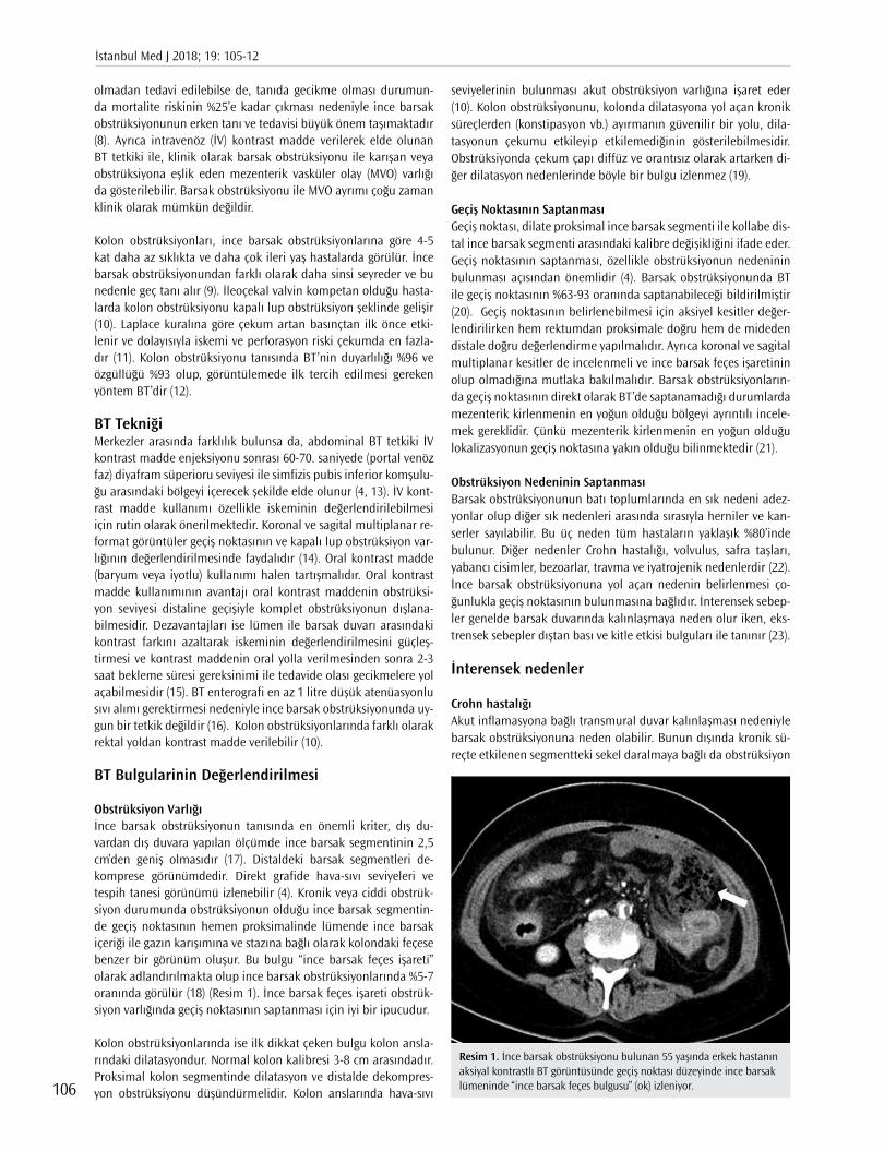
olmadan tedavi edilebilse de, tanıda gecikme olması durumun-da mortalite riskinin %25’e kadar çıkması nedeniyle ince barsak obstrüksiyonunun erken tanı ve tedavisi büyük önem taşımaktadır (8). Ayrıca intravenöz (İV) kontrast madde verilerek elde olunan BT tetkiki ile, klinik olarak barsak obstrüksiyonu ile karışan veya obstrüksiyona eşlik eden mezenterik vasküler olay (MVO) varlığı da gösterilebilir. Barsak obstrüksiyonu ile MVO ayrımı çoğu zaman klinik olarak mümkün değildir.
Kolon obstrüksiyonları, ince barsak obstrüksiyonlarına göre 4-5 kat daha az sıklıkta ve daha çok ileri yaş hastalarda görülür. İnce barsak obstrüksiyonundan farklı olarak daha sinsi seyreder ve bu nedenle geç tanı alır (9). İleoçekal valvin kompetan olduğu hasta-larda kolon obstrüksiyonu kapalı lup obstrüksiyon şeklinde gelişir (10). Laplace kuralına göre çekum artan basınçtan ilk önce etki-lenir ve dolayısıyla iskemi ve perforasyon riski çekumda en fazla-dır (11). Kolon obstrüksiyonu tanısında BT’nin duyarlılığı %96 ve özgüllüğü %93 olup, görüntülemede ilk tercih edilmesi gereken yöntem BT’dir (12).
BT TekniğiMerkezler arasında farklılık bulunsa da, abdominal BT tetkiki İV kontrast madde enjeksiyonu sonrası 60-70. saniyede (portal venöz faz) diyafram süperioru seviyesi ile simfizis pubis inferior komşulu-ğu arasındaki bölgeyi içerecek şekilde elde olunur (4, 13). İV kont-rast madde kullanımı özellikle iskeminin değerlendirilebilmesi için rutin olarak önerilmektedir. Koronal ve sagital multiplanar re-format görüntüler geçiş noktasının ve kapalı lup obstrüksiyon var-lığının değerlendirilmesinde faydalıdır (14). Oral kontrast madde (baryum veya iyotlu) kullanımı halen tartışmalıdır. Oral kontrast madde kullanımının avantajı oral kontrast maddenin obstrüksi-yon seviyesi distaline geçişiyle komplet obstrüksiyonun dışlana-bilmesidir. Dezavantajları ise lümen ile barsak duvarı arasındaki kontrast farkını azaltarak iskeminin değerlendirilmesini güçleş-tirmesi ve kontrast maddenin oral yolla verilmesinden sonra 2-3 saat bekleme süresi gereksinimi ile tedavide olası gecikmelere yol açabilmesidir (15). BT enterografi en az 1 litre düşük atenüasyonlu sıvı alımı gerektirmesi nedeniyle ince barsak obstrüksiyonunda uy-gun bir tetkik değildir (16). Kolon obstrüksiyonlarında farklı olarak rektal yoldan kontrast madde verilebilir (10).
BT Bulgularinin Değerlendirilmesi
Obstrüksiyon Varlığıİnce barsak obstrüksiyonun tanısında en önemli kriter, dış du-vardan dış duvara yapılan ölçümde ince barsak segmentinin 2,5 cm’den geniş olmasıdır (17). Distaldeki barsak segmentleri de-komprese görünümdedir. Direkt grafide hava-sıvı seviyeleri ve tespih tanesi görünümü izlenebilir (4). Kronik veya ciddi obstrük-siyon durumunda obstrüksiyonun olduğu ince barsak segmentin-de geçiş noktasının hemen proksimalinde lümende ince barsak içeriği ile gazın karışımına ve stazına bağlı olarak kolondaki feçese benzer bir görünüm oluşur. Bu bulgu “ince barsak feçes işareti” olarak adlandırılmakta olup ince barsak obstrüksiyonlarında %5-7 oranında görülür (18) (Resim 1). İnce barsak feçes işareti obstrük-siyon varlığında geçiş noktasının saptanması için iyi bir ipucudur.
Kolon obstrüksiyonlarında ise ilk dikkat çeken bulgu kolon ansla-rındaki dilatasyondur. Normal kolon kalibresi 3-8 cm arasındadır. Proksimal kolon segmentinde dilatasyon ve distalde dekompres-yon obstrüksiyonu düşündürmelidir. Kolon anslarında hava-sıvı
seviyelerinin bulunması akut obstrüksiyon varlığına işaret eder (10). Kolon obstrüksiyonunu, kolonda dilatasyona yol açan kronik süreçlerden (konstipasyon vb.) ayırmanın güvenilir bir yolu, dila-tasyonun çekumu etkileyip etkilemediğinin gösterilebilmesidir. Obstrüksiyonda çekum çapı diffüz ve orantısız olarak artarken di-ğer dilatasyon nedenlerinde böyle bir bulgu izlenmez (19).
Geçiş Noktasının SaptanmasıGeçiş noktası, dilate proksimal ince barsak segmenti ile kollabe dis-tal ince barsak segmenti arasındaki kalibre değişikliğini ifade eder. Geçiş noktasının saptanması, özellikle obstrüksiyonun nedeninin bulunması açısından önemlidir (4). Barsak obstrüksiyonunda BT ile geçiş noktasının %63-93 oranında saptanabileceği bildirilmiştir (20). Geçiş noktasının belirlenebilmesi için aksiyel kesitler değer-lendirilirken hem rektumdan proksimale doğru hem de mideden distale doğru değerlendirme yapılmalıdır. Ayrıca koronal ve sagital multiplanar kesitler de incelenmeli ve ince barsak feçes işaretinin olup olmadığına mutlaka bakılmalıdır. Barsak obstrüksiyonların-da geçiş noktasının direkt olarak BT’de saptanamadığı durumlarda mezenterik kirlenmenin en yoğun olduğu bölgeyi ayrıntılı incele-mek gereklidir. Çünkü mezenterik kirlenmenin en yoğun olduğu lokalizasyonun geçiş noktasına yakın olduğu bilinmektedir (21).
Obstrüksiyon Nedeninin SaptanmasıBarsak obstrüksiyonunun batı toplumlarında en sık nedeni adez-yonlar olup diğer sık nedenleri arasında sırasıyla herniler ve kan-serler sayılabilir. Bu üç neden tüm hastaların yaklaşık %80’inde bulunur. Diğer nedenler Crohn hastalığı, volvulus, safra taşları, yabancı cisimler, bezoarlar, travma ve iyatrojenik nedenlerdir (22). İnce barsak obstrüksiyonuna yol açan nedenin belirlenmesi ço-ğunlukla geçiş noktasının bulunmasına bağlıdır. İnterensek sebep-ler genelde barsak duvarında kalınlaşmaya neden olur iken, eks-trensek sebepler dıştan bası ve kitle etkisi bulguları ile tanınır (23).
İnterensek nedenler
Crohn hastalığıAkut inflamasyona bağlı transmural duvar kalınlaşması nedeniyle barsak obstrüksiyonuna neden olabilir. Bunun dışında kronik sü-reçte etkilenen segmentteki sekel daralmaya bağlı da obstrüksiyon
Resim 1. İnce barsak obstrüksiyonu bulunan 55 yaşında erkek hastanın aksiyal kontrastlı BT görüntüsünde geçiş noktası düzeyinde ince barsak lümeninde “ince barsak feçes bulgusu” (ok) izleniyor.
İstanbul Med J 2018; 19: 105-12
106
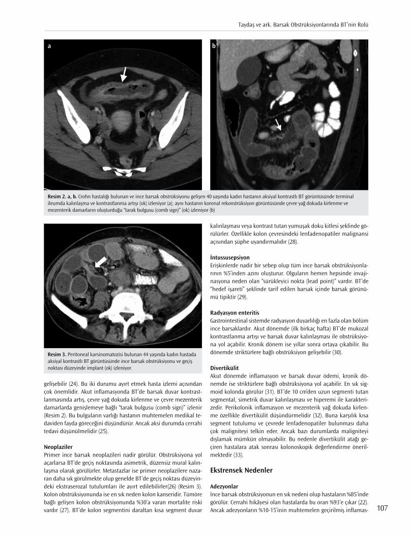
gelişebilir (24). Bu iki durumu ayırt etmek hasta izlemi açısından çok önemlidir. Akut inflamasyonda BT’de barsak duvar kontrast-lanmasında artış, çevre yağ dokuda kirlenme ve çevre mezenterik damarlarda genişlemeye bağlı “tarak bulgusu (comb sign)” izlenir (Resim 2). Bu bulguların varlığı hastanın muhtemelen medikal te-daviden fayda göreceğini düşündürür. Ancak aksi durumda cerrahi tedavi düşünülmelidir (25).
Neoplaziler Primer ince barsak neoplazileri nadir görülür. Obstrüksiyona yol açarlarsa BT’de geçiş noktasında asimetrik, düzensiz mural kalın-laşma olarak görülürler. Metastazlar ise primer neoplazilere naza-ran daha sık görülmekte olup genelde BT’de geçiş noktası düzeyin-deki ekstraserozal tutulumları ile ayırt edilebilirler(26) (Resim 3). Kolon obstrüksiyonunda ise en sık neden kolon kanseridir. Tümöre bağlı gelişen kolon obstrüksiyonunda %30’a varan mortalite riski vardır (27). BT’de kolon segmentini daraltan kısa segment duvar
kalınlaşması veya kontrast tutan yumuşak doku kitlesi şeklinde gö-rülürler. Özellikle kolon çevresindeki lenfadenopatiler malignansi açısından şüphe uyandırmalıdır (28).
İntussusepsiyonErişkinlerde nadir bir sebep olup tüm ince barsak obstrüksiyonla-rının %5’inden azını oluşturur. Olguların hemen hepsinde invaji-nasyona neden olan “sürükleyici nokta (lead point)” vardır. BT’de “hedef işareti” şeklinde tarif edilen barsak içinde barsak görünü-mü tipiktir (29).
Radyasyon enteritisGastrointestinal sistemde radyasyon duyarlılığı en fazla olan bölüm ince barsaklardır. Akut dönemde (ilk birkaç hafta) BT’de mukozal kontrastlanma artışı ve barsak duvar kalınlaşması ile obstrüksiyo-na yol açabilir. Kronik dönem ise yıllar sonra ortaya çıkabilir. Bu dönemde striktürlere bağlı obstrüksiyon gelişebilir (30).
DivertikülitAkut dönemde inflamasyon ve barsak duvar ödemi, kronik dö-nemde ise striktürlere bağlı obstrüksiyona yol açabilir. En sık sig-moid kolonda görülür (31). BT’de 10 cm’den uzun segmenti tutan segmental, simetrik duvar kalınlaşması ve hiperemi ile karakteri-zedir. Perikolonik inflamasyon ve mezenterik yağ dokuda kirlen-me özellikle divertikülit düşündürmelidir (32). Buna karşılık kısa segment tutulumu ve çevrede lenfadenopatiler bulunması daha çok maligniteyi telkin eder. Ancak bazı durumlarda maligniteyi dışlamak mümkün olmayabilir. Bu nedenle divertikülit atağı ge-çiren hastalara atak sonrası kolonoskopik değerlendirme öneril-mektedir (33).
Ekstrensek Nedenler
Adezyonlar İnce barsak obstrüksiyonun en sık nedeni olup hastaların %85’inde görülür. Cerrahi hikâyesi olan hastalarda bu oran %93’e çıkar (22). Ancak adezyonların %10-15’inin muhtemelen geçirilmiş inflamas-
Resim 2. a, b. Crohn hastalığı bulunan ve ince barsak obstrüksiyonu gelişen 40 yaşında kadın hastanın aksiyal kontrastlı BT görüntüsünde terminal ileumda kalınlaşma ve kontrastlanma artışı (ok) izleniyor (a); aynı hastanın koronal rekonstrüksiyon görüntüsünde çevre yağ dokuda kirlenme ve mezenterik damarların oluşturduğu “tarak bulgusu (comb sign)” (ok) izleniyor (b)
a b
Resim 3. Peritoneal karsinomatozisi bulunan 44 yaşında kadın hastada aksiyal kontrastlı BT görüntüsünde ince barsak obstrüksiyonu ve geçiş noktası düzeyinde implant (ok) izleniyor.
Taydaş ve ark. Barsak Obstrüksiyonlarında BT’nin Rolü
107
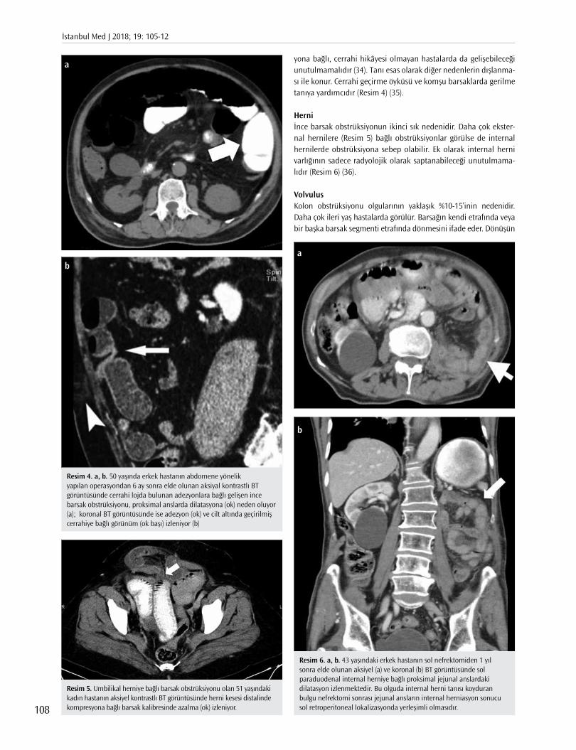
yona bağlı, cerrahi hikâyesi olmayan hastalarda da gelişebileceği unutulmamalıdır (34). Tanı esas olarak diğer nedenlerin dışlanma-sı ile konur. Cerrahi geçirme öyküsü ve komşu barsaklarda gerilme tanıya yardımcıdır (Resim 4) (35).
Herniİnce barsak obstrüksiyonun ikinci sık nedenidir. Daha çok ekster-nal hernilere (Resim 5) bağlı obstrüksiyonlar görülse de internal hernilerde obstrüksiyona sebep olabilir. Ek olarak internal herni varlığının sadece radyolojik olarak saptanabileceği unutulmama-lıdır (Resim 6) (36).
VolvulusKolon obstrüksiyonu olgularının yaklaşık %10-15’inin nedenidir. Daha çok ileri yaş hastalarda görülür. Barsağın kendi etrafında veya bir başka barsak segmenti etrafında dönmesini ifade eder. Dönüşün
Resim 4. a, b. 50 yaşında erkek hastanın abdomene yönelik yapılan operasyondan 6 ay sonra elde olunan aksiyal kontrastlı BT görüntüsünde cerrahi lojda bulunan adezyonlara bağlı gelişen ince barsak obstrüksiyonu, proksimal anslarda dilatasyona (ok) neden oluyor (a); koronal BT görüntüsünde ise adezyon (ok) ve cilt altında geçirilmiş cerrahiye bağlı görünüm (ok başı) izleniyor (b)
a
b
Resim 6. a, b. 43 yaşındaki erkek hastanın sol nefrektomiden 1 yıl sonra elde olunan aksiyel (a) ve koronal (b) BT görüntüsünde sol paraduodenal internal herniye bağlı proksimal jejunal anslardaki dilatasyon izlenmektedir. Bu olguda internal herni tanısı koyduran bulgu nefrektomi sonrası jejunal ansların internal herniasyon sonucu sol retroperitoneal lokalizasyonda yerleşimli olmasıdır.
a
b
Resim 5. Umbilikal herniye bağlı barsak obstrüksiyonu olan 51 yaşındaki kadın hastanın aksiyel kontrastlı BT görüntüsünde herni kesesi distalinde kompresyona bağlı barsak kalibresinde azalma (ok) izleniyor.
İstanbul Med J 2018; 19: 105-12
108
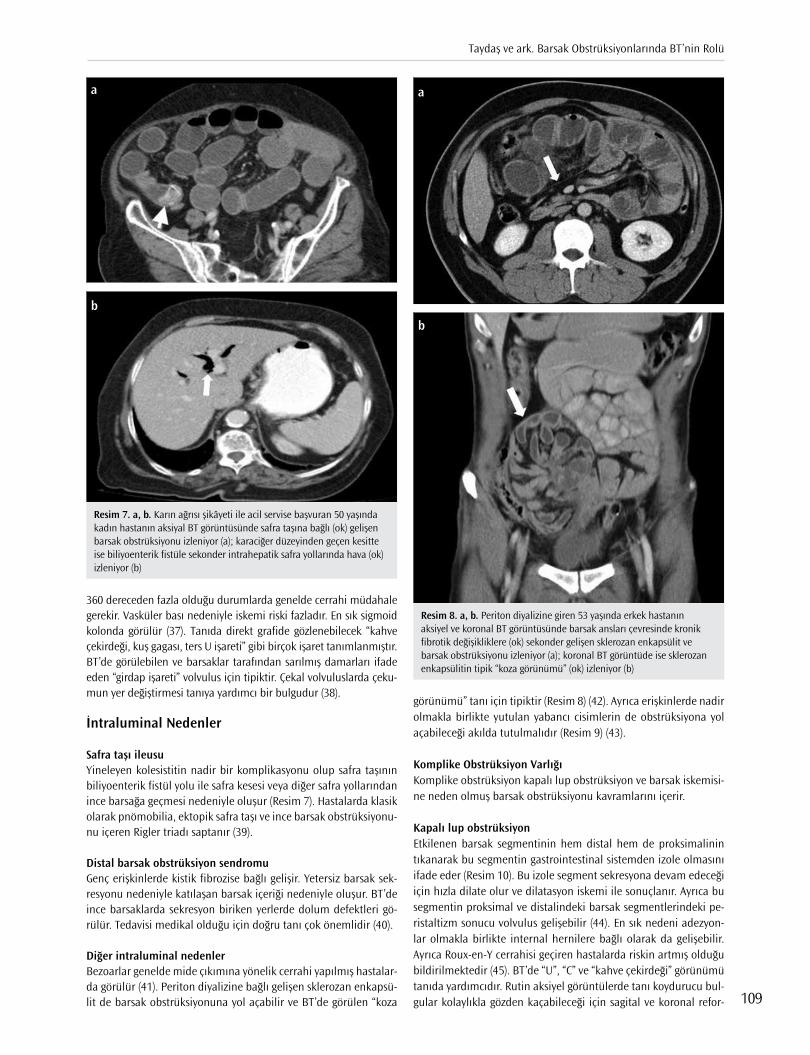
360 dereceden fazla olduğu durumlarda genelde cerrahi müdahale gerekir. Vasküler bası nedeniyle iskemi riski fazladır. En sık sigmoid kolonda görülür (37). Tanıda direkt grafide gözlenebilecek “kahve çekirdeği, kuş gagası, ters U işareti” gibi birçok işaret tanımlanmıştır. BT’de görülebilen ve barsaklar tarafından sarılmış damarları ifade eden “girdap işareti” volvulus için tipiktir. Çekal volvuluslarda çeku-mun yer değiştirmesi tanıya yardımcı bir bulgudur (38).
İntraluminal Nedenler
Safra taşı ileusuYineleyen kolesistitin nadir bir komplikasyonu olup safra taşının biliyoenterik fistül yolu ile safra kesesi veya diğer safra yollarından ince barsağa geçmesi nedeniyle oluşur (Resim 7). Hastalarda klasik olarak pnömobilia, ektopik safra taşı ve ince barsak obstrüksiyonu-nu içeren Rigler triadı saptanır (39).
Distal barsak obstrüksiyon sendromuGenç erişkinlerde kistik fibrozise bağlı gelişir. Yetersiz barsak sek-resyonu nedeniyle katılaşan barsak içeriği nedeniyle oluşur. BT’de ince barsaklarda sekresyon biriken yerlerde dolum defektleri gö-rülür. Tedavisi medikal olduğu için doğru tanı çok önemlidir (40).
Diğer intraluminal nedenlerBezoarlar genelde mide çıkımına yönelik cerrahi yapılmış hastalar-da görülür (41). Periton diyalizine bağlı gelişen sklerozan enkapsü-lit de barsak obstrüksiyonuna yol açabilir ve BT’de görülen “koza
görünümü” tanı için tipiktir (Resim 8) (42). Ayrıca erişkinlerde nadir olmakla birlikte yutulan yabancı cisimlerin de obstrüksiyona yol açabileceği akılda tutulmalıdır (Resim 9) (43).
Komplike Obstrüksiyon VarlığıKomplike obstrüksiyon kapalı lup obstrüksiyon ve barsak iskemisi-ne neden olmuş barsak obstrüksiyonu kavramlarını içerir.
Kapalı lup obstrüksiyonEtkilenen barsak segmentinin hem distal hem de proksimalinin tıkanarak bu segmentin gastrointestinal sistemden izole olmasını ifade eder (Resim 10). Bu izole segment sekresyona devam edeceği için hızla dilate olur ve dilatasyon iskemi ile sonuçlanır. Ayrıca bu segmentin proksimal ve distalindeki barsak segmentlerindeki pe-ristaltizm sonucu volvulus gelişebilir (44). En sık nedeni adezyon-lar olmakla birlikte internal hernilere bağlı olarak da gelişebilir. Ayrıca Roux-en-Y cerrahisi geçiren hastalarda riskin artmış olduğu bildirilmektedir (45). BT’de “U”, “C” ve “kahve çekirdeği” görünümü tanıda yardımcıdır. Rutin aksiyel görüntülerde tanı koydurucu bul-gular kolaylıkla gözden kaçabileceği için sagital ve koronal refor-
Resim 7. a, b. Karın ağrısı şikâyeti ile acil servise başvuran 50 yaşında kadın hastanın aksiyal BT görüntüsünde safra taşına bağlı (ok) gelişen barsak obstrüksiyonu izleniyor (a); karaciğer düzeyinden geçen kesitte ise biliyoenterik fistüle sekonder intrahepatik safra yollarında hava (ok) izleniyor (b)
a
b
Resim 8. a, b. Periton diyalizine giren 53 yaşında erkek hastanın aksiyel ve koronal BT görüntüsünde barsak ansları çevresinde kronik fibrotik değişikliklere (ok) sekonder gelişen sklerozan enkapsülit ve barsak obstrüksiyonu izleniyor (a); koronal BT görüntüde ise sklerozan enkapsülitin tipik “koza görünümü” (ok) izleniyor (b)
a
b
Taydaş ve ark. Barsak Obstrüksiyonlarında BT’nin Rolü
109

mat görüntüler de değerlendirilmelidir (46). Volvulus ile komplike olan durumlarda ayrıca girdap işareti görülebilir (47). Kolon obs-trüksiyonları kapalı lup olmaya eğimli olduğu için komplikasyon riski fazladır. Kapalı lup obstrüksiyonları sıklıkla iskemi ile sonuç-landığı için iskemi varlığı dikkatle araştırılmalıdır (3).
Barsak iskemisiİnce barsak obstrüksiyonu olan hastaların yaklaşık %10’unda görülür. Yüksek mortaliteye sahiptir. BT’nin iskemiyi saptamadaki duyarlılığı %83, özgüllüğü ise %92’dir (48). BT’de; etkilenen barsak segmentle-rinde duvar kalınlaşması, duvar kontrastlanmasında anormal azal-ma, pnömotozis, mezenterik ödem ve mezenterik damarlar ile portal vende gaz görülebilir (49). Özellikle barsak duvar kontrastlanmasında anormal azalma iskemi tanısında güvenilir bir bulgudur (8, 50).
TedaviTedavide özellikle sıvı-elektrolit dengesinin korunması ve nazo-gastrik kateter aracılığı ile gastrointestinal sistemdeki dekompres-yonun sağlanması amaçlanır. Hastanın klinik olarak kötüleşmesi ve distansiyonun artması durumunda cerrahi girişim düşünülme-lidir. Ayrıca tam obstrüksiyon varlığında cerrahi girişim öncelikli olarak düşünülebilir (13).
SonuçBarsak obstrüksiyonları, tanısında ve takibinde görüntülemenin ve özellikle de BT’nin önemli yer tutması nedeniyle radyoloji pratiğin-de sıklıkla karşılaşılabilecek bir tablodur. BT; barsak obstrüksiyonu tanısı, obstrüksiyonun nedeni ve geçiş noktası lokalizasyonunun belirlenmesi için en uygun görüntüleme yöntemidir.
Hakem Değerlendirmesi: Dış Bağımsız.
Yazar Katkıları: Fikir - O.T., E.Ü., M.R.O.; Tasarım - O.T., M.R.O., E.A.; Denet-leme - E.Ü., M.R.O., E.A.; Kaynaklar - O.T., E.Ü., M.R.O.; Malzemeler - O.T., M.R.O., E.A.; Veri Toplanması ve/veya işlemesi - E.Ü., M.R.O., E.A.; Analiz ve/veya Yorum - O.T., E.Ü., M.R.O.; Literatür taraması - O.T., M.R.O., E.A.; Yazıyı Yazan - O.T., E.Ü., M.R.O.; Eleştirel İnceleme - M.R.O., E.A.
Resim 9. a, b. Barsak obstrüksiyonu olan 55 yaşında kadın hastanın aksiyel BT görüntüsünde obstrüksiyonun geçiş noktası düzeyinde intraluminal yabancı cisim izleniyor (ok) (a); aynı hastada proksimal ince barsakların içerisinde de benzer yabancı cisimler görülüyor (oklar) (b)
a
b
Resim 10. a, b. Kapalı lup ince barsak obstrüksiyonu olan 57 yaşındaki erkek hastada aksiyel kontrastlı BT görüntüsünde barsak segmentinde izole dilatasyon (ok) ve bu segmente komşu mezenterik yağ dokuda belirgin kirlenme izleniyor (a); koronal BT görüntüsünde bu segmentin hem proksimal hem de distalinde geçiş noktasının izlenmesi (oklar) ve abdomenin diğer kısımlarında barsak dilatasyon ile mezenterik yağ dokuda kirlenme olmaması kapalı lup obstrüksiyonu düşündürüyor (b)
a
b
İstanbul Med J 2018; 19: 105-12
110
Çıkar Çatışması: Yazarların beyan edecek çıkar çatışması yoktur.
Finansal Destek: Yazarlar bu çalışma için finansal destek almadıklarını beyan etmişlerdir.
Peer-review: Externally peer-reviewed.
Author Contributions: Concept - O.T., E.Ü., M.R.O.; Design - O.T., M.R.O., E.A.; Supervision - E.Ü., M.R.O., E.A.; Resource - O.T., E.Ü., M.R.O.; Materials - O.T., M.R.O., E.A.; Data Collection and/or Processing - E.Ü., M.R.O., E.A.; Analysis and/or Interpretation - O.T., E.Ü., M.R.O.; Literature Search - O.T., M.R.O., E.A.; Writing - O.T., E.Ü., M.R.O.; Critical Reviews - M.R.O, E.A.
Conflict of Interest: Authors have no conflicts of interest to declare.
Financial Disclosure: The authors declared that this study has received no financial support.
Kaynaklar
1. Taylor MR, Lalani N. Adult small bowel obstruction. Acad Emerg Med 2013; 200: 528-44. [CrossRef]
2. Cappell MS, Batke M. Mechanical obstruction of the small bowel and colon. Med Clin North Am 2008; 92: 575-97, viii. [CrossRef]
3. Gore RM, Silvers RI, Thakrar KH, Wenzke DR, Mehta UK, Newmark GM, et al. Bowel Obstruction. Radiol Clin North Am 2015; 53: 1225-40. [CrossRef]
4. Paulson EK, Thompson WM. Review of small-bowel obstruction: the diagnosis and when to worry. Radiology 2015; 275: 332-42. [CrossRef]
5. Thompson WM, Kilani RK, Smith BB, Thomas J, Jaffe TA, Delong DM, et al. Accuracy of abdominal radiography in acute small-bowel obstruc-tion: does reviewer experience matter? AJR Am J Roentgenol 2007; 188: 233-8. [CrossRef]
6. Maglinte DD, Reyes BL, Harmon BH, Kelvin FM, Turner WW, Jr., Hage JE, et al. Reliability and role of plain film radiography and CT in the diagnosis of small-bowel obstruction. AJR Am J Roentgenol 1996; 167: 1451-5. [CrossRef]
7. Santillan CS. Computed tomography of small bowel obstruction. Ra-diol Clin North Am 2013; 51: 17-27. [CrossRef]
8. Zalcman M, Sy M, Donckier V, Closset J, Gansbeke DV. Helical CT signs in the diagnosis of intestinal ischemia in small-bowel obstruction. AJR Am J Roentgenol 2000; 175: 1601-7. [CrossRef]
9. Biondo S, Pares D, Frago R, Marti-Rague J, Kreisler E, De Oca J, et al. Large bowel obstruction: predictive factors for postoperative morta-lity. Dis Colon Rectum 2004; 47: 1889-97. [CrossRef]
10. Jaffe T, Thompson WM. Large-Bowel Obstruction in the Adult: Classic Radiographic and CT Findings, Etiology, and Mimics. Radiology 2015; 275:651-63. [CrossRef]
11. Krajewski K, Siewert B, Eisenberg RL. Colonic dilation. AJR Am J Roent-genol 2009 Nov; 193: 363-72. [CrossRef]
12. Frager D, Rovno HD, Baer JW, Bashist B, Friedman M. Prospective eva-luation of colonic obstruction with computed tomography. Abdom Imaging 1998; 23: 141-6. [CrossRef]
13. Desser TS, Gross M. Multidetector row computed tomography of small bowel obstruction. Semin Ultrasound CT MR 2008; 29: 308-21. [CrossRef]
14. Horton KM, Fishman EK. The current status of multidetector row CT and three-dimensional imaging of the small bowel. Radiol Clin North Am 2003; 41: 199-212. [CrossRef]
15. O'Malley RG, Al-Hawary MM, Kaza RK, Wasnik AP, Platt JF, Francis IR. MDCT findings in small bowel obstruction: implications of the cause and presence of complications on treatment decisions. Abdom Ima-ging 2015; 40: 2248-62. [CrossRef]
16. Paulsen SR, Huprich JE, Fletcher JG, Booya F, Young BM, Fidler JL, et al. CT enterography as a diagnostic tool in evaluating small bowel disor-ders: review of clinical experience with over 700 cases. Radiographics 2006; 26: 641-57. [CrossRef]
17. Fukuya T, Hawes DR, Lu CC, Chang PJ, Barloon TJ. CT diagnosis of small-bowel obstruction: efficacy in 60 patients. AJR Am J Roentgenol 1992; 158: 765-9; discussion 71-2. [CrossRef]
18. Mayo-Smith WW, Wittenberg J, Bennett GL, Gervais DA, Gazelle GS, Mueller PR. The CT small bowel faeces sign: description and clinical significance. Clin Radiol 1995; 50: 765-7. [CrossRef]
19. Wittenberg J. The diagnosis of colonic obstruction on plain abdomi-nal radiographs: start with the cecum, leave the rectum to last. AJR Am J Roentgenol 1993; 161: 443-4. [CrossRef]
20. Colon MJ, Telem DA, Wong D, Divino CM. The relevance of transition zones on computed tomography in the management of small bowel obstruction. Surgery 2010; 147: 373-7. [CrossRef]
21. Khurana B, Ledbetter S, McTavish J, Wiesner W, Ros PR. Bowel obstruc-tion revealed by multidetector CT. AJR Am J Roentgenol 2002; 178: 1139-44. [CrossRef]
22. Miller G, Boman J, Shrier I, Gordon PH. Etiology of small bowel obs-truction. Am J Surg 2000; 180: 33-6. [CrossRef]
23. Silva AC, Pimenta M, Guimaraes LS. Small bowel obstruction: what to look for. Radiographics 2009; 29: 423-39. [CrossRef]
24. Akcalar S, Turkbey B, Karcaaltincaba M, Akpinar E, Akhan O. Small bo-wel wall thickening: MDCT evaluation in the emergency room. Emerg Radiol 2011; 18: 409-15. [CrossRef]
25. Al-Hawary MM, Kaza RK, Platt JF. CT enterography: concepts and ad-vances in Crohn's disease imaging. Radiol Clin North Am 2013; 51: 1-16. [CrossRef]
26. Idelevich E, Kashtan H, Mavor E, Brenner B. Small bowel obstruction caused by secondary tumors. Surg Oncol 2006; 15: 29-32. [CrossRef]
27. Irvin GL 3rd, Horsley JS 3rd, Caruana JA Jr. The morbidity and mor-tality of emergent operations for colorectal disease. Ann Surg 1984; 199: 598-603. [CrossRef]
28. Horton KM, Abrams RA, Fishman EK. Spiral CT of colon cancer: ima-ging features and role in management. Radiographics 2000; 20: 419-30. [CrossRef]
29. Rea JD, Lockhart ME, Yarbrough DE, Leeth RR, Bledsoe SE, Clements RH. Approach to management of intussusception in adults: a new para-digm in the computed tomography era. Am Surg 2007; 73: 1098-105.
30. Maturen KE, Feng MU, Wasnik AP, Azar SF, Appelman HD, Francis IR, et al. Imaging effects of radiation therapy in the abdomen and pel-vis: evaluating "innocent bystander" tissues. Radiographics 2013; 33: 599-619. [CrossRef]
31. Taourel P, Kessler N, Lesnik A, Pujol J, Morcos L, Bruel JM. Helical CT of large bowel obstruction. Abdom Imaging 2003; 28: 267-75. [CrossRef]
32. Horton KM, Corl FM, Fishman EK. CT evaluation of the colon: inflam-matory disease. Radiographics 2000; 20: 399-418. [CrossRef]
33. Sai VF, Velayos F, Neuhaus J, Westphalen AC. Colonoscopy after CT diagnosis of diverticulitis to exclude colon cancer: a systematic litera-ture review. Radiology 2012; 263: 383-90. [CrossRef]
34. Caoili EM, Paulson EK. CT of small-bowel obstruction: another pers-pective using multiplanar reformations. AJR Am J Roentgenol. 2000: 174: 993-8. [CrossRef]
35. Attard JA, MacLean AR. Adhesive small bowel obstruction: epidemio-logy, biology and prevention. Can J Surg 2007; 50: 291-300.
36. Aguirre DA, Casola G, Sirlin C. Abdominal wall hernias: MDCT findings. AJR Am J Roentgenol 2004; 183: 681-90. [CrossRef]
37. Ballantyne GH, Brandner MD, Beart RW Jr., Ilstrup DM. Volvulus of the colon. Incidence and mortality. Ann Surg 1985; 202: 83-92. [CrossRef]
38. Peterson CM, Anderson JS, Hara AK, Carenza JW, Menias CO. Volvulus of the gastrointestinal tract: appearances at multimodality imaging. Radiographics 2009; 29:1281-93. [CrossRef]
39. Delabrousse E, Bartholomot B, Sohm O, Wallerand H, Kastler B. Gall-stone ileus: CT findings. Eur Radiol 2000; 10: 938-40. [CrossRef]
40. Colombo C, Ellemunter H, Houwen R, Munck A, Taylor C, Wilschanski M. Guidelines for the diagnosis and management of distal intestinal obstruction syndrome in cystic fibrosis patients. J Cyst Fibros 2011; 10: 24-8. [CrossRef]
41. Delabrousse E, Brunelle S, Saguet O, Destrumelle N, Landecy G, Kast-ler B. Small bowel obstruction secondary to phytobezoar CT findings. Clin Imaging 2001; 25: 44-6. [CrossRef]
Taydaş ve ark. Barsak Obstrüksiyonlarında BT’nin Rolü
111
42. George C, Al-Zwae K, Nair S, Cast JE. Computed tomography appea-rances of sclerosing encapsulating peritonitis. Clin Radiol 2007; 62: 732-7. [CrossRef]
43. Agrawal GA, Johnson PT, Fishman EK. Intermittent small bowel obs-truction with retained endoscopic capsule: MDCT and plain radiog-raphic appearance. Emerg Radiol 2007; 13: 319-21. [CrossRef]
44. Sandhu PS, Joe BN, Coakley FV, Qayyum A, Webb EM, Yeh BM. Bo-wel transition points: multiplicity and posterior location at CT are associated with small-bowel volvulus. Radiology 2007; 245: 160-7. [CrossRef]
45. Capella RF, Iannace VA, Capella JF. Bowel obstruction after open and laparoscopic gastric bypass surgery for morbid obesity. J Am Coll Surg 2006; 203: 328-35. [CrossRef]
46. Frager DH, Baer JW, Rothpearl A, Bossart PA. Distinction between pos-toperative ileus and mechanical small-bowel obstruction: value of CT compared with clinical and other radiographic findings. AJR Am J Roentgenol 1995; 164: 891-4. [CrossRef]
47. Duda JB, Bhatt S, Dogra VS. Utility of CT whirl sign in guiding mana-gement of small-bowel obstruction. AJR Am J Roentgenol 2008; 191: 743-7. [CrossRef]
48. Mallo RD, Salem L, Lalani T, Flum DR. Computed tomography diagno-sis of ischemia and complete obstruction in small bowel obstruction: a systematic review. J Gastrointest Surg 2005; 9: 690-4. [CrossRef]
49. Jancelewicz T, Vu LT, Shawo AE, Yeh B, Gasper WJ, Harris HW. Predic-ting strangulated small bowel obstruction: an old problem revisited. J Gastrointest Surg 2009; 13: 93-9. [CrossRef]
50. Sheedy SP, Earnest Ft, Fletcher JG, Fidler JL, Hoskin TL. CT of small-bowel ischemia associated with obstruction in emergency department patients: diagnostic performance evaluation. Radiology 2006; 241: 729-36. [CrossRef]
Cite this article as: Taydaş O, Ünal E, Onur MR, Akpınar E. Role of Computed Tomography in Intestinal Obtruction. İstanbul Med J 2018; 19: 105-12.
İstanbul Med J 2018; 19: 105-12
112

Introduction
Human induced pluripotent stem cells (hiPSCs), mostly derived from fibroblasts, have been recog-nized as key progenitor cells for liver regeneration, which could potentially be used for the repair of liver injury. The process of hepatic differentiation involves the sequential expression of hepatic markers such as α-fetoprotein, albumin, urea, and liver-specific metabolic enzymes, including aminotransferases (1, 2). In addition, hepatic differentiation is marked by major metabolic chang-es related to lipid metabolism (3-5). Such specific changes in lipid metabolism are important for the formation of competent hepatic cells. The unique metabolomics pattern of hiPSCs has been described in detail in another study; as identified some lipid species were associated with main-taining pluripotency(6). However, the association of hepatic differentiation of hiPSCs with the ex-pression of hepatic enzyme markers and change in cellular lipids has not been well characterized.
Cellular lipid composition is tightly regulated by de novo synthesis, metabolic conversion, and degradation processes. However, the level of multiple lipid species, including fatty acids, may be influenced by the process of hepatic differentiation. Owing to this complexity, an assay for these changes is not readily available. Alteration of the cellular fatty acid pool affects cell membrane functions and signaling pathways that control cell fate determination. Furthermore, fatty acid metabolism is key to guaranteeing adequate energy supply during specific lineage differentiation (7). This indicates that such perturbations strongly impact adipogenic and cardiac and neuronal
Effect of Hepatic Differentiation on Fatty Acid Composition of Induced Pluripotent Stem Cells Derived from Human Dermal Fibroblastsİnsan Dermal Fibroblastlarından Türeyen Uyuşan İndüklenmiş Pluripotent Kök Hücrelerin Yağ Asidi Kompozisyonu Üzerine Hepatik Farklılaşmanın Etkisi
Introduction: Human induced pluripotent stem cells (hiPSCs) have been recognized as key progenitor cells for liver regeneration, which could potentially be used in the repair of liver injury. Differentiation of stem cells involves specific changes in lipid metabolism to form competent hepatic cells. However, the relationship between the hepa-tic differentiation of hiPSCs and change in cellular lipids has not been well characterized. The aim of the present study was to determine the fatty acid pattern during hepatic differentiation of hiPSCs derived from dermal fibroblasts.
Methods: Hepatocyte differentiation was induced in three stages using Wnt family member 3A, hepatocyte growth factor, and oncostatin M and was impaired by a chemical extracellular signal-regulated kinase signaling inhibitor. Hepatocyte-specific metabolic markers including gamma-glutamyltransferase and aminotransferases were evaluated using kinetic chromogenic assay kits. During the hepatic differentiation of hiPSCs, changes in individual fatty acids were determined at multiple time points using gas-liquid chromatography.
Results: Significant increases were observed in hepatic enzyme mar-kers during hepatic differentiation. Endodermal induction produced a transient increase in saturated fatty acids (33%, p<0.01) and decrease in monounsaturated fatty acids (-15%, p<0.01). Total n-6 polyunsaturated fatty acid was elevated in undifferentiated hiPSCs, and a gradual down-ward trend was observed after endodermal induction (-10%, p=0.08) and hepatic lineage commitment (-19%, p<0.01). All of these changes in metabolic differentiation markers and individual fatty acids were suppressed by impaired induced hepatic differentiation.
Conclusion: Our findings indicate that the pattern of cellular fatty acids is dynamic and changes with the progress of hepatic differentiation, which includes both transient fluctuations and linear trends.
Keywords: Adult stem cells, cell lineage, endoderm, lipids
Amaç: İnsan indüklenmiş pluripotent kök (iPK) hücreleri potansiyel olarak karaciğer hasarının onarımında kullanılabilecek karaciğer rejenerasyonu için önemli progenitör hücreler olarak bilinmektedir. Kök hücrelerin farklılaşması, yetkin hepatik hücreleri oluşturmak için lipit metabolizmasında spesifik değişiklikler içerir. Bununla birlikte, insan iPK hücrelerinin hepatik farklılaşması ve hücresel lipitlerdeki değişim arasındaki ilişki iyi tanımlanmamıştır. Bu araştırmanın ama-cı, dermal fibroblastlarından türetilen insan iPK hücrelerinin hepatik farklılaşması sırasında yağ asidi paternini belirlemektir.
Yöntemler: Hepatosit farklılaşması, Wnt-3a, HGF ve onkostatin M kul-lanılarak üç aşamada indüklendi ve bir kimyasal Hücre dışı sinyalle-düzenlenen kinaz (ERK) sinyal verme inhibitörü PD98059 tarafından bozuldu. Gama-glutamil transferaz ve aminotransferazlar da dahil ol-mak üzere hepatosit spesifik metabolik belirteçler kinetik kromojenik test kitleri kullanılarak tahmin edildi. Insan iPK hücrelerinin hepatik farklılaşması sırasında, gaz-sıvı kromatografısi kullanılarak çoklu za-man noktalarında bireysel yağ asitlerinin değişimleri belirlendi.
Bulgular: Hepatik enzim belirteçlerinde hepatik farklılaşması sırasında anlamlı artışlar gözlendi. Endodermal indüksiyon sonrasında doymuş yağ asitlerinde geçici artış (%33, p<0,01) ve tekli doymamış yağ asitlerin-de (%-15, p<0,01) azalma meydana geldi. Total n-6 çoklu doymamış yağ asitleri, farklılaşmamış insan iPK hücrelerinde yüksekti ve endodermal indüksiyon (%-10, p=0,08) ve hepatik lineage commitment sonrasi kade-meli bir düşüş eğilimi gözlendi (%-19, p<0,01). Metabolik farklılaşma be-lirteçlerinde ve bireysel yağ asitlerinde meydana gelen bu değişikliklerin tümü, indüklenmiş hepatik farklılaşmanın bozulmasıyla bastırılmıştır.
Sonuç: Bulgularımız hücresel yağ asitlerin paternini dinamik oldu-ğunu ve hem geçici dalgalanmalar hem de doğrusal eğilimleri içeren hepatik farklılaşmanın ilerlemesiyle değiştiğini göstermektedir.
Anahtar Kelimeler: Erişkin kök hücreler, hücre soyları, endoderm, lipitler
Abst
ract
/Öz
ORCID ID of the authors: N.P. 0000-0001-6873-4922; Y.R. 0000-0003-2320-6303; A.M. 0000-0002-3029-4172; H.N.C., 0000-0003-4883-9924; M.N. 0000-0002-5367-9956; M.S. 0000-0001-9250-7033; M.D. 0000-0001-6380-272X.
This study was presented in the 2nd National Festival International Congress on Stem Cell Sciences and Technologies and Regenerative Medicine, July 13-15, 2017, Tehran, Iran1Stem Cell Research Center, Tabriz University of Medical Sciences, Tabriz, İran2Department of Biochemistry and Clinical Laboratories, Tabriz University of Medical Sciences, School of Medicine, Tabriz, İran3Endocrine Research Center, Tabriz University of Medical Sciences, Tabriz, İran4Stem Cell and Regenerative Medicine Institute, Tabriz University of Medical Sciences, Tabriz, İran
Address for Correspondence/Yazışma Adresi:Masoud DarabiE-mail: [email protected]
Received/Geliş Tarihi: 04.04.2017Accepted/Kabul Tarihi: 04.11.2017
© Telif Hakkı 2018 Makale metnine istanbultipdergisi.org web sayfasından ulaşılabilir.
© Copyright 2018 by Available online at istanbulmedicaljournal.org
Original Investigation/Orijinal Araştırmaİstanbul Med J 2018; 19: 113-8DOI: 10.5152/imj.2018.60465
Nasim Parsafam1 , Yagoub Rahimi2 , Amir Mehdizadeh3 , Hojjatollah Nozad Charoudeh1 , Mohammad Nouri4
, Maghsod Shaaker2 , Masoud Darabi1
differentiation (8, 9). Therefore, profiling cellular fatty acids during hepatic differentiation may be a valuable addition to the study of hepatic cells. In a previous study, we have reported a coordinate gene expression and activity index pattern of a fatty acid-metab-olizing enzyme during hepatic differentiation of hiPSCs (10). The aim of the present study was to characterize patterns of individual fatty acids during hepatic differentiation of hiPSCs. In the present study, we describe hepatic enzyme markers and fatty acid compo-sition at different stages of in vitro differentiation of hiPSCs that are generated from fibroblasts. Different fatty acid patterns occur in cells undergoing hepatic differentiation. Furthermore, these specific changes in fatty acid pattern were suppressed by impaired induced hepatic maturation.
Methods
The present study was approved by the local ethics committee. Skin fibroblasts used for hiPS cell generation were obtained from a single donor who provided written informed consent (11).
Materials
All culture materials were purchased from Invitrogen/GIBCO (Carls-bad, CA, USA). All other materials were from Sigma, unless stated otherwise. Conditioned media and cell extracts from a human hepatoma cell line (HepG2, National Cell Bank, Pasteur Institute of Iran) served as positive controls (12).
Cell culture and induced differentiationExpansion medium comprised mouse embryonic fibroblast feeder cells inactivated with mitomycin C, Iscove’s Modified Dulbecco’s Medium, and 20% knockout serum replacement, supplemented with 100 ng/mL basic fibroblast growth factor, 1 mmol/L l-gluta-mine, 100 μM nonessential amino acids, 50 U/mL penicillin, and 50 mg/mL streptomycin. The hiPS cell line, R1-hiPSC9, was gen-erated from harvested human skin fibroblasts by means of viral transduction of reprogramming factors (Oct4, Sox2, Klf4, and c-Myc) as described in another study (11). This cell line reportedly expressed the pluripotency-associated marker Oct-4 and was able to spontaneously differentiate into the three embryonic germ lay-ers in vitro (11, 13). The hiPSCs were passaged every 5-7 days.
The method of hepatic differentiation was similar to that used in a previous study (12) and according to that in the study by Chen et al. (14). Briefly, the differentiation protocol included endodermal induction, hepatic lineage commitment, and hepatic maturation stages.
Impaired induction of hepatocyte differentiationThe inhibition of the mitogen-activated protein kinase/extracel-lular signal-regulated kinase (ERK) signaling pathway is known to negatively affect hepatic differentiation of stem cells (15). In an alternate approach to control the effect of differentiation, the ERK inhibitor, PD98059, at a concentration of 5 μM was added from days 2-10 during the hepatic differentiation of hiPSCs.
Determination of hepatic enzymesFor the quantification of extracellular and intracellular hepatic markers, the culture medium was collected from each well and centrifuged at 800g at 4°C for 10 min, and the supernatant was filtered to remove cell bodies. Conditioned medium was then
concentrated approximately 25× using ultrafiltration units with a molecular weight cut-off value of 10 kDa (Amicon Ultra-2, Milli-pore, MA, USA). Finally, the collected medium, containing secreted hepatic markers, was stored at −80°C until analyzed (16). After the removal of culture medium, the cells in each well were washed with PBS and harvested in a lysis buffer containing 1% SDS, 10 mM EDTA, 50 mM Tris-HCl, and pH 8.1. The lysates were centrifuged, and the supernatant was stored as described above.
Protein concentration of the lysate was determined by the Lowry protein assay using bovine serum albumin as the standard (17). En-zyme activities were measured using commercially available kinet-ic chromogenic assay kits on a BT 3000 autoanalyzer (Biotecnica Institute, Italy). Determination of alanine aminotransferase (ALT) and aspartate aminotransferase (AST) was based on the oxidation of 2-oxoglutarate, leading to the indirect oxidation of nicotin-amide adenine dinucleotide (NADH). Gamma-glutamyltransferase (GGT) assay was based on the monitoring of the enzyme-catalyzed transfer of γ-glutamyl group from the substrate l-γ-glutamyl-p-nitroanilide, liberating the chromogen p-nitroanilide. Measure-ment of lactate dehydrogenase (LDH) in both the fractions was based on the consumption of NADH in the reduction of pyruvate to l-lactate.
Gas-liquid chromatography of cellular fatty acidsTotal lipids were extracted from the whole cell lysate using the conventional Bligh-Dyer method (18). Fatty acid methyl esters were prepared by transesterification of the extracted lipids using methanol with acetyl chloride, acting as an acid catalyst, at 100°C (19). Fatty acid methyl esters formed were extracted using 1.5 mL of n-hexane, dried under N2, and reconstituted into 0.5 mL n-hex-ane for analysis.
Data from gas-liquid chromatography (GLC) were obtained using a Buck Scientific model 610 gas chromatograph (SRI Instruments, Torrance, USA) fitted with a highly polar biscyanopropyl poly-siloxane capillary column (TR-CN100 60 m×0.25 mm id, 0.2 μm film thickness; Teknokroma, Spain). The gas chromatograph was equipped with a split injector, a flame ionization detector, a hy-drogen generator (H2 NM Plus; LNI Schmidlin, Switzerland), a com-puter interface, and PeakSimple software version 3.59. The operat-ing conditions for GLC were: injection, 1 μL, split mode (1:90) with 60 s valve time; carrier gas, helium at 20.0 cm/s; and injector and linear temperature, 250°C. The oven temperature of GLC was pro-grammed as follows: 5 min at 190°C then ramped to 210°C at 1°C/min and then isothermal for 15 min.
Statistical AnalysisData were presented as mean ± standard deviation of three sepa-rate experiments performed in duplicate. Statistical differences between the groups were assessed using analysis of variance with post-hoc Tukey’s test for multiple comparisons. A p<0.05 was considered to be statistically significant. Data were analyzed us-ing statistical functions in Microsoft Excel (Microsoft Corporation, Redmond, WA, USA).
Results
In a previous study, the differentiation protocol used was efficient at directing hiPSCs toward a hepatic-like lineage in vitro, which was confirmed by a robust increase in specific markers, including
İstanbul Med J 2018; 19: 113-8
114
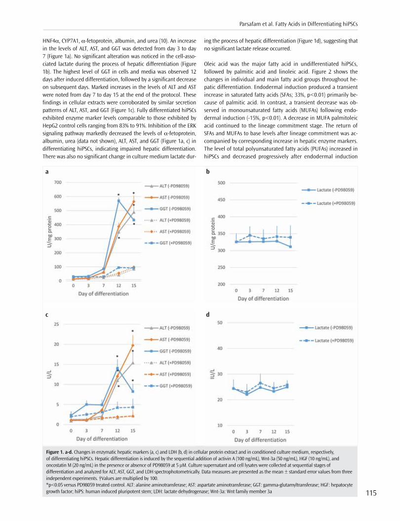
HNF4α, CYP7A1, α-fetoprotein, albumin, and urea (10). An increase in the levels of ALT, AST, and GGT was detected from day 3 to day 7 (Figure 1a). No significant alteration was noticed in the cell-asso-ciated lactate during the process of hepatic differentiation (Figure 1b). The highest level of GGT in cells and media was observed 12 days after induced differentiation, followed by a significant decrease on subsequent days. Marked increases in the levels of ALT and AST were noted from day 7 to day 15 at the end of the protocol. These findings in cellular extracts were corroborated by similar secretion patterns of ALT, AST, and GGT (Figure 1c). Fully differentiated hiPSCs exhibited enzyme marker levels comparable to those exhibited by HepG2 control cells ranging from 83% to 91%. Inhibition of the ERK signaling pathway markedly decreased the levels of α-fetoprotein, albumin, urea (data not shown), ALT, AST, and GGT (Figure 1a, c) in differentiating hiPSCs, indicating impaired hepatic differentiation. There was also no significant change in culture medium lactate dur-
ing the process of hepatic differentiation (Figure 1d), suggesting that no significant lactate release occurred.
Oleic acid was the major fatty acid in undifferentiated hiPSCs, followed by palmitic acid and linoleic acid. Figure 2 shows the changes in individual and main fatty acid groups throughout he-patic differentiation. Endodermal induction produced a transient increase in saturated fatty acids (SFAs; 33%, p<0.01) primarily be-cause of palmitic acid. In contrast, a transient decrease was ob-served in monounsaturated fatty acids (MUFAs) following endo-dermal induction (-15%, p<0.01). A decrease in MUFA palmitoleic acid continued to the lineage commitment stage. The return of SFAs and MUFAs to base levels after lineage commitment was ac-companied by corresponding increase in hepatic enzyme markers. The level of total polyunsaturated fatty acids (PUFAs) increased in hiPSCs and decreased progressively after endodermal induction
Figure 1. a-d. Changes in enzymatic hepatic markers (a, c) and LDH (b, d) in cellular protein extract and in conditioned culture medium, respectively, of differentiating hiPSCs. Hepatic differentiation is induced by the sequential addition of activin A (100 ng/mL), Wnt-3a (50 ng/mL), HGF (10 ng/mL), and oncostatin M (20 ng/mL) in the presence or absence of PD98059 at 5 μM. Culture supernatant and cell lysates were collected at sequential stages of differentiation and analyzed for ALT, AST, GGT, and LDH spectrophotometrically. Data measures are presented as the mean ± standard error values from three independent experiments. †Values are multiplied by 100. *p<0.05 versus PD98059 treated control. ALT: alanine aminotransferase; AST: aspartate aminotransferase; GGT: gamma-glutamyltransferase; HGF: hepatocyte growth factor; hiPS: human induced pluripotent stem; LDH: lactate dehydrogenase; Wnt-3a: Wnt family member 3a
c
a
d
b
Parsafam et al. Fatty Acids in Differentiating hiPSCs
115
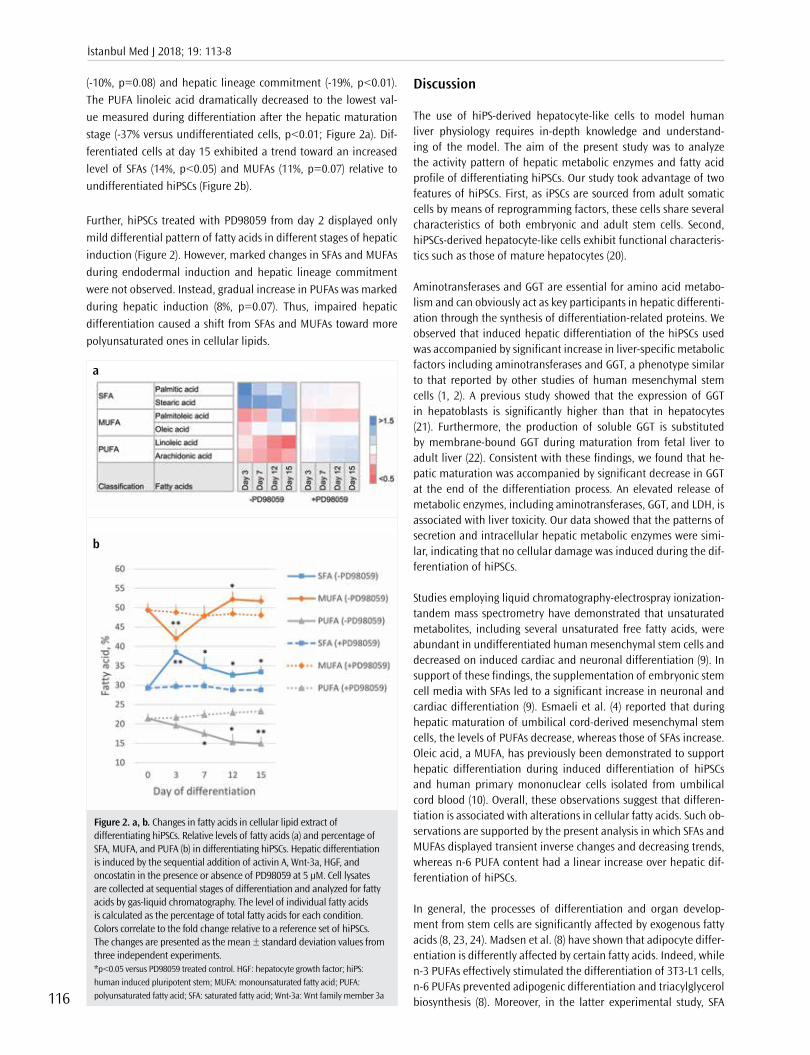
(-10%, p=0.08) and hepatic lineage commitment (-19%, p<0.01). The PUFA linoleic acid dramatically decreased to the lowest val-ue measured during differentiation after the hepatic maturation stage (-37% versus undifferentiated cells, p<0.01; Figure 2a). Dif-ferentiated cells at day 15 exhibited a trend toward an increased level of SFAs (14%, p<0.05) and MUFAs (11%, p=0.07) relative to undifferentiated hiPSCs (Figure 2b).
Further, hiPSCs treated with PD98059 from day 2 displayed only mild differential pattern of fatty acids in different stages of hepatic induction (Figure 2). However, marked changes in SFAs and MUFAs during endodermal induction and hepatic lineage commitment were not observed. Instead, gradual increase in PUFAs was marked during hepatic induction (8%, p=0.07). Thus, impaired hepatic differentiation caused a shift from SFAs and MUFAs toward more polyunsaturated ones in cellular lipids.
Discussion
The use of hiPS-derived hepatocyte-like cells to model human liver physiology requires in-depth knowledge and understand-ing of the model. The aim of the present study was to analyze the activity pattern of hepatic metabolic enzymes and fatty acid profile of differentiating hiPSCs. Our study took advantage of two features of hiPSCs. First, as iPSCs are sourced from adult somatic cells by means of reprogramming factors, these cells share several characteristics of both embryonic and adult stem cells. Second, hiPSCs-derived hepatocyte-like cells exhibit functional characteris-tics such as those of mature hepatocytes (20).
Aminotransferases and GGT are essential for amino acid metabo-lism and can obviously act as key participants in hepatic differenti-ation through the synthesis of differentiation-related proteins. We observed that induced hepatic differentiation of the hiPSCs used was accompanied by significant increase in liver-specific metabolic factors including aminotransferases and GGT, a phenotype similar to that reported by other studies of human mesenchymal stem cells (1, 2). A previous study showed that the expression of GGT in hepatoblasts is significantly higher than that in hepatocytes (21). Furthermore, the production of soluble GGT is substituted by membrane-bound GGT during maturation from fetal liver to adult liver (22). Consistent with these findings, we found that he-patic maturation was accompanied by significant decrease in GGT at the end of the differentiation process. An elevated release of metabolic enzymes, including aminotransferases, GGT, and LDH, is associated with liver toxicity. Our data showed that the patterns of secretion and intracellular hepatic metabolic enzymes were simi-lar, indicating that no cellular damage was induced during the dif-ferentiation of hiPSCs.
Studies employing liquid chromatography-electrospray ionization-tandem mass spectrometry have demonstrated that unsaturated metabolites, including several unsaturated free fatty acids, were abundant in undifferentiated human mesenchymal stem cells and decreased on induced cardiac and neuronal differentiation (9). In support of these findings, the supplementation of embryonic stem cell media with SFAs led to a significant increase in neuronal and cardiac differentiation (9). Esmaeli et al. (4) reported that during hepatic maturation of umbilical cord-derived mesenchymal stem cells, the levels of PUFAs decrease, whereas those of SFAs increase. Oleic acid, a MUFA, has previously been demonstrated to support hepatic differentiation during induced differentiation of hiPSCs and human primary mononuclear cells isolated from umbilical cord blood (10). Overall, these observations suggest that differen-tiation is associated with alterations in cellular fatty acids. Such ob-servations are supported by the present analysis in which SFAs and MUFAs displayed transient inverse changes and decreasing trends, whereas n-6 PUFA content had a linear increase over hepatic dif-ferentiation of hiPSCs.
In general, the processes of differentiation and organ develop-ment from stem cells are significantly affected by exogenous fatty acids (8, 23, 24). Madsen et al. (8) have shown that adipocyte differ-entiation is differently affected by certain fatty acids. Indeed, while n-3 PUFAs effectively stimulated the differentiation of 3T3-L1 cells, n-6 PUFAs prevented adipogenic differentiation and triacylglycerol biosynthesis (8). Moreover, in the latter experimental study, SFA
Figure 2. a, b. Changes in fatty acids in cellular lipid extract of differentiating hiPSCs. Relative levels of fatty acids (a) and percentage of SFA, MUFA, and PUFA (b) in differentiating hiPSCs. Hepatic differentiation is induced by the sequential addition of activin A, Wnt-3a, HGF, and oncostatin in the presence or absence of PD98059 at 5 μM. Cell lysates are collected at sequential stages of differentiation and analyzed for fatty acids by gas-liquid chromatography. The level of individual fatty acids is calculated as the percentage of total fatty acids for each condition. Colors correlate to the fold change relative to a reference set of hiPSCs. The changes are presented as the mean ± standard deviation values from three independent experiments. *p<0.05 versus PD98059 treated control. HGF: hepatocyte growth factor; hiPS: human induced pluripotent stem; MUFA: monounsaturated fatty acid; PUFA: polyunsaturated fatty acid; SFA: saturated fatty acid; Wnt-3a: Wnt family member 3a
a
b
İstanbul Med J 2018; 19: 113-8
116
and MUFA did not affect differentiation. It is likely that such effects are related to agonistic and antagonistic effects on transcription factors that control cell fate specification and differentiation, in-cluding sterol regulatory element-binding proteins, liver X recep-tors, and peroxisome proliferator-activated receptors (8).
Finally, we found that the effect of induced hepatic differentia-tion on the fatty acid profile of hiPSCs changes with ERK inhibi-tor treatment. ERK signaling pathway participates in hepatocyte differentiation and proliferation through the relay of hepatocyte growth factor signaling toward cell cycle machinery and differenti-ation pathways (15). Consistent with this finding, we observed that ERK inhibitor treatment during induced differentiation markedly reduced the production of functional hepatic markers. Thus, the lack of marked changes in fatty acids during endodermal induc-tion and hepatic lineage commitment state implies that differenti-ation-inducing factors in the absence of functional differentiation had no significant effect on cellular fatty acids. Future biochemical and molecular studies of hepatic differentiation should focus on the role of selected enzymatic reactions in the synthesis or modi-fication of fatty acids.
Conclusion
In conclusion, our findings indicate that the pattern of cellular fatty acids is dynamic and changes with the progress of hepatic differen-tiation, which includes both transient fluctuations and linear trends. Our result is of interest because hiPSCs derived from human dermal fibroblasts are promising candidates for regenerative medicine and drug discovery in addition to being applied to addressing the basis of cellular fatty acid metabolism. This finding can aid efficient dif-ferentiation of hiPSCs, particularly for hepatic regeneration.
Ethics Committee Approval: Ethics committee approval was received for this study from the ethics committee of Tabriz University School of Medi-cine (Approval Date: 05.12.2014; Approval Number: 5/104/1207).
Informed Consent: Written informed consent had been obtained from the patient who donated skin fibroblasts for this study.
Peer-review: Externally peer-reviewed.
Author contributions: Concept - M.D., H.N.C.; Design - M.D., H.N.C., M.N.; Supervision - M.D., H.N.C., M.N.; Resource - M.D., H.N.C., M.N.; Materials - H.N.C., M.N.; Data Collection and/or Processing - N.P., Y.R., A.M.; Analysis and/or Interpretation - N.P., Y.R., M.S.; Literature Search - N.P., Y.R., A.M.; Writing - N.P., Y.R., A.M., M.S.; Critical Reviews - M.D., H.N.C., M.N.
Acknowledgements: The authors would like to thank the Stem Cell Re-search Center at Tabriz University of Medical Sciences and Iranian Council for Development of Stem Cell Sciences and Technologies for their financial and technical supports.
Conflict of Interest: Authors have no conflicts of interest to declare.
Financial Disclosure: This study has received financial supports from Stem Cell Research Center at Tabriz University of Medical Sciences and Iranian Council for Development of Stem Cell Sciences and Technologies.
Etik Komite Onayı: Bu çalışma için etik komite onayı Tabriz Üniversitesi Tıp Fakültesi Etik Kurul Komitesi’nden alınmıştır (Onay Tarihi: 05.12.2014; Onay Numarası: 5/104/1207).
Hasta Onamı: Hasta Onamı bu çalışma için deri fibroblastlarını bağışlayan hastadan alınmıştır.
Hakem Değerlendirmesi: Dış Bağımsız.
Yazar Katkıları: Fikir - M.D., H.N.C.; Tasarım - M.D., H.N.C., M.N.; Denet-leme - M.D., H.N.C., M.N.; Kaynaklar - M.D., H.N.C., M.N.; Malzemeler - H.N.C., M.N.; Veri Toplanması ve/veya işlemesi - N.P., Y.R., A.M.; Analiz ve/veya Yorum - N.P., Y.R., M.S.; Literatür taraması - N.P., Y.R., A.M.; Yazıyı Ya-zan - N.P., Y.R., A.M., M.S.; Eleştirel İnceleme - M.D., H.N.C., M.N.
Teşekkür: Yazarlar Tebriz Tıp Bilimleri Üniversitesi Kök Hücre Araştırma Merkezi’ne ve İran Kök Hücre Bilim ve Teknoloji Konseyi’ne finansal ve tek-nik destekleri için teşekkür etmek isterler.
Çıkar Çatışması: Yazarların beyan edecek çıkar çatışması yoktur.
Finansal Destek: Bu çalışma Tebriz Tıp Üniversitesi Kök Hücre Araştırma Merkezi ve İran Kök Hücre Bilim ve Teknoloji Konseyi’nden finansal destek almıştır.
References
1. Allameh A, Ahmadi-Ashtiani H, Emami Aleagha MS, Rastegar H. The metabolic function of hepatocytes differentiated from human mes-enchymal stem cells is inversely related to cellular glutathione levels. Cell Biochem Funct 2014; 32: 194-200. [CrossRef]
2. Kwon MJ, Kang SJ, Park YI, Yang YH, Bang S I, Park Y H, et al. Hepatic differentiation of human adipose tissue-derived mesenchymal stem cells and adverse effects of arsanilic acid and acetaminophen during in vitro hepatic developmental stage. Cell Biol Toxicol 2015; 31: 149-59. [CrossRef]
3. Hayhurst GP, Lee YH, Lambert G, Ward JM, Gonzalez FJ. Hepatocyte nuclear factor 4alpha (nuclear receptor 2A1) is essential for mainte-nance of hepatic gene expression and lipid homeostasis. Mol Cell Biol 2001; 21: 1393-403. [CrossRef]
4. Esmaeli S, Allameh A, Soleimani M, Rahbarizadeh F, Frouzandeh-Moghadam M. The role of albumin and PPAR-α in differentiation-dependent change of fatty acid profile during differentiation of mesenchymal stem cells to hepatocyte-like cells. Cell Biochem Funct 2014; 32: 410-9. [CrossRef]
5. Vacanti NM, Metallo CM. Exploring metabolic pathways that contrib-ute to the stem cell phenotype. Biochim Biophys Acta 2013; 1830: 2361-9. [CrossRef]
6. Panopoulos AD, Yanes O, Ruiz S, Kida YS, Diep D, Tautenhahn R, et al. The metabolome of induced pluripotent stem cells reveals metabolic changes occurring in somatic cell reprogramming. Cell Res 2012; 22: 168-77. [CrossRef]
7. Hu C, Fan L, Cen P, Chen E, Jiang Z, Li L. Energy Metabolism Plays a Critical Role in Stem Cell Maintenance and Differentiation. Int J Mol Sci 2016; 17: 253. [CrossRef]
8. Madsen L, Petersen RK, Kristiansen K. Regulation of adipocyte dif-ferentiation and function by polyunsaturated fatty acids. Biochim Biophys Acta 2005; 1740: 266-86. [CrossRef]
9. Yanes O, Clark J, Wong DM, Patti GJ, Sánchez Ruiz A, Benton HP, et al. Metabolic oxidation regulates embryonic stem cell differentiation. Nat Chem Biol 2010; 6: 411-7. [CrossRef]
10. Rahimi Y, Mehdizadeh A, Nozad Charoudeh H, Nouri M, Valaei K, Fayezi S, et al. Hepatocyte differentiation of human induced pluripo-tent stem cells is modulated by stearoyl-CoA desaturase 1 activity. Dev Growth Differ 2015; 57: 667-74. [CrossRef]
11. Totonchi M, Taei A, Seifinejad A, Tabebordbar M, Rassouli H, Farrokhi A, et al. Feeder- and serum-free establishment and expansion of hu-man induced pluripotent stem cells. Int J Dev Biol 2010; 54: 877-86. [CrossRef]
12. Knowles B, Howe C, Aden D. Human hepatocellular carcinoma cell lines secrete the major plasma proteins and hepatitis B surface anti-gen. Science 1980; 209: 497-9. [CrossRef]
Parsafam et al. Fatty Acids in Differentiating hiPSCs
117
13. Pakzad M, Ashtiani MK, Mousavi-Gargari SL, Baharvand H. Develop-ment of a simple, repeatable, and cost-effective extracellular matrix for long-term xeno-free and feeder-free self-renewal of human plu-ripotent stem cells. Histochem Cell Biol 2013; 140: 635-48. [CrossRef]
14. Chen YF, Tseng CY, Wang HW, Kuo HC, Yang VW, Lee OK. Rapid genera-tion of mature hepatocyte-like cells from human induced pluripotent stem cells by an efficient three-step protocol. Hepatology 2012; 55: 1193-203. [CrossRef]
15. Gao W, Zhou P, Ma X, Tschudy-Seney B, Chen J, Magner NL,et al. Etha-nol negatively regulates hepatic differentiation of hESC by inhibi-tion of the MAPK/ERK signaling pathway in vitro. PLoS One 2014; 9: 112698. [CrossRef]
16. Kale VP, Patel SG, Gunjal PS, Wakchaure SU, Sundar RS, Ranvir RK, et al. Effect of repeated freezing and thawing on 18 clinical chemistry analytes in rat serum. J Am Assoc Lab Anim Sci 2012; 51: 475-8.
17. Waterborg JH. The Lowry Method for Protein Quantitation. In: Walker JM, editor. The Protein Protocols Handbook. Totowa, NJ: Humana Press 2002; 7-9. [CrossRef]
18. Bligh EG, Dyer WJ. A rapid method of total lipid extraction and purifi-cation. Can J Biochem Physiol 1959; 37: 911-7. [CrossRef]
19. Lepage G, Roy CC. Direct transesterification of all classes of lipids in a one-step reaction. J Lipid Res 1986; 27: 114-20.
20. Espejel S, Roll GR, McLaughlin KJ, Lee AY, Zhang JY, Laird DJ, et al. In-duced pluripotent stem cell-derived hepatocytes have the functional
and proliferative capabilities needed for liver regeneration in mice. J Clin Invest 2010; 120: 3120-6. [CrossRef]
21. Holic N, Suzuki T, Corlu A, Couchie D, Chobert MN, Guguen-Guil-louzo C, et al. Differential expression of the rat gamma-glutamyl transpeptidase gene promoters along with differentiation of hepa-toblasts into biliary or hepatocytic lineage. Am J Pathol 2000; 157: 537-48. [CrossRef]
22. Igarashi T, Satoh T, Ueno K, Kitagawa H. Changes of gamma-glutam-yltranspeptidase activity in the rat during development and compari-son of the fetal liver, placental and adult liver enzymes. Life sciences 1981; 29: 483-91. [CrossRef]
23. Teboul L, Febbraio M, Gaillard D, Amri EZ, Silverstein R, Grimaldi PA. Structural and functional characterization of the mouse fatty acid translocase promoter: activation during adipose differentiation. Bio-chem J 2001; 360: 305-12. [CrossRef]
24. Kang JX, Wan JB, He C. Concise Review: Regulation of stem cell prolif-eration and differentiation by essential fatty acids and their metabo-lites. Stem Cells 2014; 32: 1092-8. [CrossRef]
Cite this article as: Parsafam N, Rahimi Y, Mehdizadeh A, Nozad Charoudeh H, Nouri M, Shaaker M, et al. Effect of Hepatic Differentiation on Fatty Acid Composition of Induced Pluripotent Stem Cells Derived from Human Dermal Fibroblasts. İstanbul Med J 2018; 19: 113-8.
İstanbul Med J 2018; 19: 113-8
118
Giriş
Distal radius kırıkları en sık karşılaşılan kırık tiplerinden biri olup; pediatrik yaş grubundaki kırık-ların %25’ini, erişkinlerde görülen kırıkların ise %18’ini oluşturmaktadır (1). Yaşa göre incelendi-ğinde biomodal bir dağılımı gösterdiği görülmüştür (2). Özellikle genç hastalarda yüksek enerjili travmalar ile oluşurken, ileri yaş hastalarda (>65 yaş) eşlik eden osteoporoz nedeniyle düşük ener-jili travmalarla karşımıza çıkmaktadır (3). Yüksek enerjili travmalarla oluşan distal radius kırıkları erkeklerde kadınlara göre daha sık görülmesine rağmen genel yaşa göre düzeltilmiş insidansı kadınlarda erkeklere oranla 4-5 kat daha fazladır (4).
Pratik hayatta distal radius kırıkları için sık kullanılan Arbeitsgemeinschaft für Osteosynthesefra-gen (AO) sınıflamasında “C” alt grubu eklemi tamamen içeren parçalı kırıklar için tanımlanmıştır (5). Bu tip kırıklarda cerrahi dışı tedavilerle uzun dönemde yanlış kaynama, eklem uyumsuzluğu ve osteoartrit gelişme riskinin cerrahi yöntemlere göre daha sık olduğu bildirilmiştir (6). Günü-müzde distal radius parçalı kırıkları için dorsal veya volar plaklama, external fiksasyon, köprü plaklama, ve fragman spesifik fiksasyon gibi çeşitli cerrahi tedaviler tanımlanmıştır (4). Bütün bu yöntemlerin birbirine üstünlükleri, avantaj ve dezavantajları literatürde birçok çalışmaya konu olmuştur (6-10).
Biz bu çalışmada kliniğimizde distal radius AO tip C kırık nedeniyle volar plaklama ile açık redük-siyon internal fiksasyon yapılan hastaların klinik sonuçlarını değerlendirmeyi amaçladık.
AO Tip C Distal Radius Kırıklarında Volar Plak İle Osteosentez Uygulamasının Klinik ve Radyolojik SonuçlarıClinical and Radiologic Outcomes of Volar Plate Fixation in AO Type C Distal Radius Fractures
Amaç: Erişkinlerde AO (Arbeitsgemeinschaft für Osteosynthesefragen) tip C distal radius kırıklarının volar anatomik plak ile tedavisi ve so-nuçlarının değerlendirilmesi amaçlanmıştır.
Yöntemler: Hastane dosya ve radyoloji arşivinden Ocak 2015 ile Nisan 2016 yılları arasında AO tip C distal radius kırığı nedeniyle opere edilen hastalar retrospektif olarak tarandı. Operasyon son-rası poliklinik kontrollerine gelen hastaların dosyalarından kol, omuz ve el sorunları anketi (DASH) ile skorlaması alınarak klinik sonuçları kaydedildi. Radyoloji arşivinden elde edilen görüntüler ile ölçümler yapılıp Stewart radyolojik skorları tespit edildi. Gelişen komplikasyonlar kaydedildi.
Bulgular: Çalışmaya 9 kadın ve 7 erkek olmak üzere 16 hasta dahil edildi. Ortalama yaş 52; ortalama takip süreleri 11,9 ay olarak tes-pit edildi. Ortalama DASH skoru 33,9 ve ortalama Stewart radyolojik skoru 0,9 olarak ölçüldü. Tüm hastalarda kaynama sağlandı. Komp-likasyonlar, 1 hastada yüzeyel enfeksiyon, 1 hastada eklem sertliği, 1 hastada karpal tünel sendromu ve 2 hastada tendinitiydi.
Sonuçlar: AO tip C distal radius kırıklarında volar plak uygulaması, gerek fonksiyonel gerekse radyolojik açıdan tatminkar sonuçlar sağ-lamaktadır. Buna karşın gelişebilecek komplikasyonlar azımsanma-yacak kadar yüksektir.
Anahtar Kelimeler: Volar plak, distal radius, AO tip C
Introduction: To evaluate the clinical and radiological outcomes of anatomic volar plate application in AO type C distal radius fractures.
Methods: Patients who were operated on between January 2015 and April 2016 due to AO type C distal radius fracture were screened ret-rospectively from the hospital file and radiology archive. Clinical out-comes were determined by the disabilities of the arm, shoulder, and hand (DASH) score from outpatient records. The Stewart radiological scores were assessed with the measurements obtained from radiology archives. The complications were noted.
Results: Overall, 16 patients (mean age, 52 years) were included in the study. In total, 9 of the patients were female, and 7 of the patients were male. The mean follow-up period was 11.9 months. The mean DASH score was 33.9, and the mean Stewart radiological score was 0.9. The bone union was accomplished in all patients. One infection, one joint stiffness, one carpal tunnel syndrome, and 2 tendinitis were observed as complication.
Conclusion: Volar plate fixation in the AO type C distal radius fractu-res provides satisfactory results in terms of either clinical or radiologi-cal outcomes. Despite that, the complications regarding surgery and the implants are not rare.
Keywords: Volar plate, distal radius, AO type C
Öz
/ Abs
trac
t
ORCID ID of the authors: A.Ş. 0000-0002-7648-1504; Y.Ö. 0000-0002-2199-2411; Y.E.A. 0000-0003-2939-0519; E.Ş. 0000-0002-4697-7904; E.A.Ü. 0000-0002-0093-7886
1Elazığ Kovancılar Devlet Hastanesi, Ortopedi ve Travmatoloji Kliniği, Elazığ, Türkiye2İstanbul Eğitim ve Araştırma Hastanesi, Ortopedi Ve Travmatoloji Kliniği, İstanbul, Türkiye3Metin Sabancı Baltalimanı Kemik Hastalıkları Eğitim ve Araştırma Hastanesi, Ortopedi ve Travmatoloji Kliniği, İstanbul, Türkiye4Sakarya Üniversitesi Eğitim ve Araştırma Hastanesi, Ortopedi ve Travmatoloji Kliniği, Sakarya, Türkiye
Yazışma Adresi / Address for Correspondence:Ahmet ŞenelE-mail: [email protected]
Geliş Tarihi/Received: 22.12.2016
Kabul Tarihi/Accepted: 06.11.2017
© Telif Hakkı 2018 Makale metnine istanbultipdergisi.org web sayfasından ulaşılabilir.
© Copyright 2018 by Available online at istanbulmedicaljournal.org
Orijinal Araştırma/Original Investigationİstanbul Med J 2018; 19: 119-23DOI: 10.5152/imj.2018.77044
Ahmet Şenel1 , Yusuf Öztürkmen2 , Yunus Emre Akman3 , Erhan Şükür4 , Ethem Ayhan Ünkar3

Yöntemler
Kliniğimizde Ocak 2015 ile Nisan 2016 yılları arasında distal radi-us eklem içi parçalı kırığı tanısıyla volar plaklama yapılan 16 hasta, hastane dosya ve radyoloji arşivinden retrospektif taranarak tespit edilip çalışmaya dahil edildi. Çalışmaya dahil edilme kriterimiz; has-tanın 20 yaşından büyük olması, operasyon sonrası poliklinik kont-rollerine gelerek klinik fonksiyonları değerlendirilmiş, radyolojik görüntülemeleri yapılmış, yazılı ve sözlü onamları mevcut olan, AO sınıflamasına göre tip C (eklemin tamamını ilgilendiren çok parçalı kırık), açık olmayan ve nöro vasküler hasarın eşlik etmediği kırıklar olarak belirlendi (Resim 1). Çalışma için etik kurul izni alındı.
Tüm hastalara aynı cerrahi prosedür uygulanmıştır. Genel anestezi sonrası opere edilecek kola hastanın sistolik kan basıncı göz önünde bulundurularak uygun basınçta pnömotik turnike sarıldı. El dahil turnike alanına kadar üst ekstremite %10’luk polivinilprolidon iyot kompleksi ile temizlenip cerrahi olarak antisepsi sağlandıktan sonra hasta, örtü seti ile steril bir şekilde örtüldü. Cilt flexor carpiradialis (FCR) tendonu üzerinden longitudinal olarak açıldı. FCR tendonu ra-dial tarafa ekarte edilip pronator quadratus kası radial origo bölge-sinden sıyrılıp ular tarafa deviye edildi. Kırık bölgesine ulaşıldı. Skopi kontrolü altında geçici k-telleri ile redüksiyon sağlandıktan sonra 2,7 mm’lik anatomik distal radius kilitli volar plak (TST, İstanbul, Türki-ye), 3,5 mm kilitli vidalarla (TST, İstanbul, Türkiye) kırık bölgesine tespit edildi (Resim 2). Plak tespiti sonrası k-telleri çıkarıldı. Pronator quadratus kası tekrar radial origosuna sütüre edildi. Turnike açılarak kanama kontrolü sağlandı. Cilt kapatıldı. Hastalara pansuman son-rası kısa kol atelle ekstremite immobilizasyonu sağlandı.
Hastalar ameliyat sonrası 2. gün insizyon yeri kontrol edilip pansu-man yapılarak taburcu edildi. Hastalara taburculuk sonrası 3 gün süreyle profilaksi amaçlı günde 2 gr 1. kuşak sefalosporin (sefa-leksin) profilaksisi reçete edildi. Evde parmak hareket egzersizleri başlaması önerildi.
Hastaların ameliyat sonrası kırık redüksiyonu kontrolü ve sütürle-rin alınması için 12. gün, atel çıkarılması için 21. Gün kontrolleri yapıldı. Atel çıkarılması sonrası aktif eklem hareket egzersizleri başlandı. Ardından 1. ay, 3. ay ve 6. Ay kontrollerinde klinik de-ğerlendirmede fizik muayene ile kol, omuz ve el sorunları anketi (DASH) skorları dökümante edildi. Radyolojik değerlendirilmesi ise nizami ön-arka ve lateral el bileği grafilerinden faydalanılarak Ste-wart skorlaması ile sağlandı (Resim 3, 4).
İstatistiksel Analizİstatistiksel analiz için Statistical Package for Socien Sciences 15.0 (SPSS Inc.; Chicago, IL, USA) programı kullanıldı. Tanımlayı-cı istatistikler; sayısal değişkenler için ortalama, standart sapma, minumum, maksimum olarak verildi. Bağımsız iki grupta sayısal değişken normal dağılım koşulu sağlandığından Student t testi ile ordinal değişken Mann Whitney U testi ile karşılaştırıldı. Sayısal değişkenler arası ilişkiler parametrik test koşulu sağlandığında Pa-erson Korelasyon analizi ile, test koşulları sağlanmadığında Spe-arman Korelasyon Analizi ile incelendi. İstatistiksel alfa anlamlılık seviyesi p<0,05 olarak kabul edildi.
Bulgular
Çalışmaya dahil edilen hastaların 9’u kadın, 7’si erkek; ortalama yaş 52 (dağılım 24-81) olarak tespit edildi. Sekiz hastada sağ, se-kiz hastada sol taraf opere edildi. Hastalar ortalama 11,8 (dağılım
6-21) ay takip edildi (Tablo 1). Etyolojinin hastaların 3’ünde spor yaralanması, birinde künt yaralanma, diğer 1 hastada araç dışı tra-fik kazası ve 11 hastada ise basit düşme olduğu öğrenildi. Takip süreleri sonunda ortalama DASH skoru 33,9 olarak ölçüldü. Takip-ler sonunda tüm kırıkların kaynadığı tespit edildi. Bir hastada in-
Resim 1. a, b. Distal radius AO tip C kırığın (a) ön arka grafi, (b) lateral grafi görüntüsü
a b
Resim 2. a, b. Volar plak uygulaması sonrası 3. ay (a) ön arka, (b) lateral grafi görüntüsü
a b
Resim 3. Kırk dokuz yaşında erkek hastanın ameliyat sonrası 6. ay klinik görünümü
İstanbul Med J 2018; 19: 119-23
120
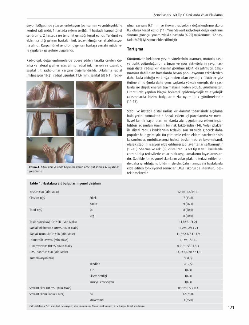
sizyon bölgesinde yüzeyel enfeksiyon (pansuman ve antibiyotik ile kontrol sağlandı), 1 hastada eklem sertliği, 1 hastada karpal tünel sendromu, 2 hastada ise tendinit geliştiği tespit edildi. Tendinit ve eklem sertliği gelişen hastalar fizik tedavi kliniğince rehabilitasyo-na alındı. Karpal tünel sendromu gelişen hastaya cerrahi müdahe-le yapılarak gevşetme uygulandı.
Radyolojik değerlendirmelerde opere edilen tarafta çekilen ön-arka ve lateral grafiler esas alınıp radial inklinasyon ve uzunluk, sagital tilt, radio-ulnar varyans değerlendirildi. Ortalama radial inklinasyon 16,2˚, radial uzunluk 11,6 mm, sagital tilt 6,1˚; radio-
ulnar varyans 0,7 mm ve Stewart radyolojik değerlendirme skoru 0,9 olarak tespit edildi (11). Yine Stewart radyolojik değerlendirme skoruna göre çalışmamızdaki 4 hastada (% 25) mükemmel, 12 has-tada (%75) iyi sonuç elde edilmiştir
Tartışma
Günümüzde beklenen yaşam sürelerinin uzaması, motorlu taşıt ve trafik yoğunluğunun artması ve spor aktivitelerin yaygınlaş-ması distal radius kırıklarının görülme sıklığı da artmıştır. Çalış-mamıza dahil olan hastalarda bayan popülasyonun erkeklerden daha fazla olduğu ve kırığa neden olan etyolojik faktörler göz önüne alındığında daha genç yaşlarda yüksek enerjili, ileri yaş-larda ise düşük enerjili travmaların neden olduğu görülmüştür. Literatürde yapılan birçok bölgesel epidemiyolojik ve etyolojik çalışmalarda bizim bulgularımızla uyumluluk görülmektedir (11-13).
Stabil ve instabil distal radius kırıklarının tedavisinde alçılama hala yerini tutmaktadır. Ancak eklem içi parçalanma ve meta-fizyel kemik kaybı olan kırıklarda alçı uygulaması eklem insta-bilitesi açısından önemli bir risk faktörüdür (14). Volar plaklar ile distal radius kırıklarının tedavisi son 10 yılda giderek daha popüler hale gelmiştir. Bu yöntemle erken eklem hareketlerinin kazanılması, mobilizasyona hızlıca başlanması ve biyomekanik olarak stabil fiksasyon elde edilmesi gibi avantajlar sağlanmıştır (15-16). Sharma ve ark. (6), distal radius AO tip B ve C kırıklarda cerrahi dışı tedavilerle volar plak uygulamalarını kıyaslamışlar-dır. Özelikle fonksiyonel skorların volar plak ile tedavi edilenler-de daha iyi olduğunu bildirmişlerdir. Çalışmamızdaki hastalarda elde edilen fonksiyonel sonuçlar (DASH skoru) da literatürü des-teklemektedir.
Resim 4. Altmış bir yaşında bayan hastanın ameliyat sonrası 6. ay klinik görünümü
Tablo 1. Hastalara ait bulguların genel dağılımı
Yaş Ort±SD (Min-Maks) 52,1±16,5/24-81
Cinsiyet n(%) Erkek 7 (43,8)
Kadın 9 (56,3)
Taraf n(%) Sol 8 (50,0)
Sağ 8 (50,0)
Takip süresi (ay) Ort±SD (Min-Maks) 11,8±5,1/4-21
Radial inklinasyon Ort±SD (Min-Maks) 16,2±3,2/13-24
Radiak uzunluk Ort±SD (Min-Maks) 11,6±2,1/7,4-14,9
Palmar tilt Ort±SD (Min-Maks) 6,1±4,1/0-13
Ulnar varyans Ort±SD (Min-Maks) 0,71±1,53/-1,8-3
DASH skor Ort±SD (Min-Maks) 33,9±7,1/20,7-44,8
Komplikasyon n(%) 5(31,3)
Tendinit 2(12,5)
KTS 1(6,3)
Eklem sertliği 1(6,3)
Yüzeyel enfeksiyon 1(6,3)
Stewart Skor Ort.±SD (Min-Maks) 0,94±0,77 / 0-3
Stewart Skoru Sonucu n (%) İyi 12 (75,0)
Mükemmel 4 (25,0)
Ort: ortalama; SD: standart deviasyon; Min: minimum; Maks: maksimum; KTS: karpal tünel sendromu
Şenel ve ark. AO Tip C Kırıklarda Volar Plaklama
121
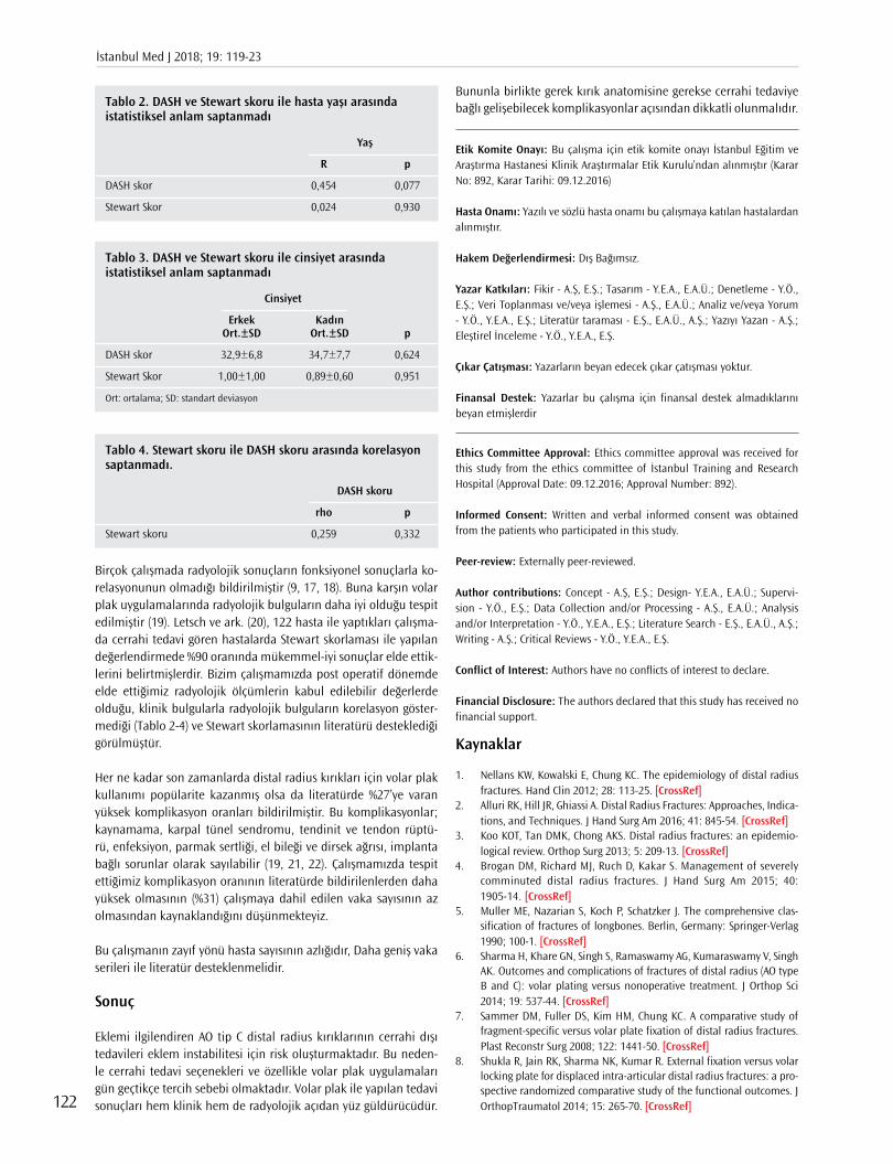
Birçok çalışmada radyolojik sonuçların fonksiyonel sonuçlarla ko-relasyonunun olmadığı bildirilmiştir (9, 17, 18). Buna karşın volar plak uygulamalarında radyolojik bulguların daha iyi olduğu tespit edilmiştir (19). Letsch ve ark. (20), 122 hasta ile yaptıkları çalışma-da cerrahi tedavi gören hastalarda Stewart skorlaması ile yapılan değerlendirmede %90 oranında mükemmel-iyi sonuçlar elde ettik-lerini belirtmişlerdir. Bizim çalışmamızda post operatif dönemde elde ettiğimiz radyolojik ölçümlerin kabul edilebilir değerlerde olduğu, klinik bulgularla radyolojik bulguların korelasyon göster-mediği (Tablo 2-4) ve Stewart skorlamasının literatürü desteklediği görülmüştür.
Her ne kadar son zamanlarda distal radius kırıkları için volar plak kullanımı popülarite kazanmış olsa da literatürde %27’ye varan yüksek komplikasyon oranları bildirilmiştir. Bu komplikasyonlar; kaynamama, karpal tünel sendromu, tendinit ve tendon rüptü-rü, enfeksiyon, parmak sertliği, el bileği ve dirsek ağrısı, implanta bağlı sorunlar olarak sayılabilir (19, 21, 22). Çalışmamızda tespit ettiğimiz komplikasyon oranının literatürde bildirilenlerden daha yüksek olmasının (%31) çalışmaya dahil edilen vaka sayısının az olmasından kaynaklandığını düşünmekteyiz.
Bu çalışmanın zayıf yönü hasta sayısının azlığıdır, Daha geniş vaka serileri ile literatür desteklenmelidir.
Sonuç
Eklemi ilgilendiren AO tip C distal radius kırıklarının cerrahi dışı tedavileri eklem instabilitesi için risk oluşturmaktadır. Bu neden-le cerrahi tedavi seçenekleri ve özellikle volar plak uygulamaları gün geçtikçe tercih sebebi olmaktadır. Volar plak ile yapılan tedavi sonuçları hem klinik hem de radyolojik açıdan yüz güldürücüdür.
Bununla birlikte gerek kırık anatomisine gerekse cerrahi tedaviye bağlı gelişebilecek komplikasyonlar açısından dikkatli olunmalıdır.
Etik Komite Onayı: Bu çalışma için etik komite onayı İstanbul Eğitim ve Araştırma Hastanesi Klinik Araştırmalar Etik Kurulu’ndan alınmıştır (Karar No: 892, Karar Tarihi: 09.12.2016)
Hasta Onamı: Yazılı ve sözlü hasta onamı bu çalışmaya katılan hastalardan alınmıştır.
Hakem Değerlendirmesi: Dış Bağımsız.
Yazar Katkıları: Fikir - A.Ş, E.Ş.; Tasarım - Y.E.A., E.A.Ü.; Denetleme - Y.Ö., E.Ş.; Veri Toplanması ve/veya işlemesi - A.Ş., E.A.Ü.; Analiz ve/veya Yorum - Y.Ö., Y.E.A., E.Ş.; Literatür taraması - E.Ş., E.A.Ü., A.Ş.; Yazıyı Yazan - A.Ş.; Eleştirel İnceleme - Y.Ö., Y.E.A., E.Ş.
Çıkar Çatışması: Yazarların beyan edecek çıkar çatışması yoktur.
Finansal Destek: Yazarlar bu çalışma için finansal destek almadıklarını beyan etmişlerdir
Ethics Committee Approval: Ethics committee approval was received for this study from the ethics committee of İstanbul Training and Research Hospital (Approval Date: 09.12.2016; Approval Number: 892).
Informed Consent: Written and verbal informed consent was obtained from the patients who participated in this study.
Peer-review: Externally peer-reviewed.
Author contributions: Concept - A.Ş, E.Ş.; Design- Y.E.A., E.A.Ü.; Supervi-sion - Y.Ö., E.Ş.; Data Collection and/or Processing - A.Ş., E.A.Ü.; Analysis and/or Interpretation - Y.Ö., Y.E.A., E.Ş.; Literature Search - E.Ş., E.A.Ü., A.Ş.; Writing - A.Ş.; Critical Reviews - Y.Ö., Y.E.A., E.Ş.
Conflict of Interest: Authors have no conflicts of interest to declare.
Financial Disclosure: The authors declared that this study has received no financial support.
Kaynaklar
1. Nellans KW, Kowalski E, Chung KC. The epidemiology of distal radius fractures. Hand Clin 2012; 28: 113-25. [CrossRef]
2. Alluri RK, Hill JR, Ghiassi A. Distal Radius Fractures: Approaches, Indica-tions, and Techniques. J Hand Surg Am 2016; 41: 845-54. [CrossRef]
3. Koo KOT, Tan DMK, Chong AKS. Distal radius fractures: an epidemio-logical review. Orthop Surg 2013; 5: 209-13. [CrossRef]
4. Brogan DM, Richard MJ, Ruch D, Kakar S. Management of severely comminuted distal radius fractures. J Hand Surg Am 2015; 40: 1905-14. [CrossRef]
5. Muller ME, Nazarian S, Koch P, Schatzker J. The comprehensive clas-sification of fractures of longbones. Berlin, Germany: Springer-Verlag 1990; 100-1. [CrossRef]
6. Sharma H, Khare GN, Singh S, Ramaswamy AG, Kumaraswamy V, Singh AK. Outcomes and complications of fractures of distal radius (AO type B and C): volar plating versus nonoperative treatment. J Orthop Sci 2014; 19: 537-44. [CrossRef]
7. Sammer DM, Fuller DS, Kim HM, Chung KC. A comparative study of fragment-specific versus volar plate fixation of distal radius fractures. Plast Reconstr Surg 2008; 122: 1441-50. [CrossRef]
8. Shukla R, Jain RK, Sharma NK, Kumar R. External fixation versus volar locking plate for displaced intra-articular distal radius fractures: a pro-spective randomized comparative study of the functional outcomes. J OrthopTraumatol 2014; 15: 265-70. [CrossRef]
Tablo 2. DASH ve Stewart skoru ile hasta yaşı arasında istatistiksel anlam saptanmadı
Yaş
R p
DASH skor 0,454 0,077
Stewart Skor 0,024 0,930
Tablo 4. Stewart skoru ile DASH skoru arasında korelasyon saptanmadı.
DASH skoru
rho p
Stewart skoru 0,259 0,332
Tablo 3. DASH ve Stewart skoru ile cinsiyet arasında istatistiksel anlam saptanmadı
Cinsiyet
Erkek Kadın Ort.±SD Ort.±SD p
DASH skor 32,9±6,8 34,7±7,7 0,624
Stewart Skor 1,00±1,00 0,89±0,60 0,951
Ort: ortalama; SD: standart deviasyon
İstanbul Med J 2018; 19: 119-23
122
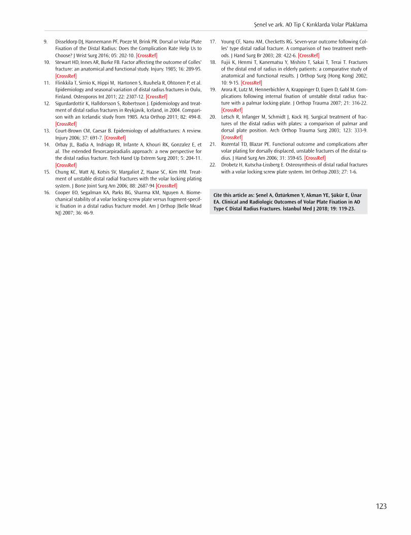
9. Disseldorp DJ, Hannemann PF, Poeze M, Brink PR. Dorsal or Volar Plate Fixation of the Distal Radius: Does the Complication Rate Help Us to Choose? J Wrist Surg 2016; 05: 202-10. [CrossRef]
10. Stewart HD, Innes AR, Burke FB. Factor affecting the outcome of Colles' fracture: an anatomical and functional study. Injury. 1985; 16: 289-95. [CrossRef]
11. Flinkkila T, Sirnio K, Hippi M, Hartonen S, Ruuhela R, Ohtonen P, et al. Epidemiology and seasonal variation of distal radius fractures in Oulu, Finland. Osteoporos Int 2011; 22: 2307-12. [CrossRef]
12. Sigurdardottir K, Halldorsson S, Robertsson J. Epidemiology and treat-ment of distal radius fractures in Reykjavik, Iceland, in 2004. Compari-son with an Icelandic study from 1985. Acta Orthop 2011; 82: 494-8. [CrossRef]
13. Court-Brown CM, Caesar B. Epidemiology of adultfractures: A review. Injury 2006; 37: 691-7. [CrossRef]
14. Orbay JL, Badia A, Indriago IR, Infante A, Khouri RK, Gonzalez E, et al. The extended flexorcarpiradialis approach: a new perspective for the distal radius fracture. Tech Hand Up Extrem Surg 2001; 5: 204-11. [CrossRef]
15. Chung KC, Watt AJ, Kotsis SV, Margaliot Z, Haase SC, Kim HM. Treat-ment of unstable distal radial fractures with the volar locking plating system. J Bone Joint Surg Am 2006; 88: 2687-94 [CrossRef]
16. Cooper EO, Segalman KA, Parks BG, Sharma KM, Nguyen A. Biome-chanical stability of a volar locking-screw plate versus fragment-specif-ic fixation in a distal radius fracture model. Am J Orthop (Belle Mead NJ) 2007; 36: 46-9.
17. Young CF, Nanu AM, Checketts RG. Seven-year outcome following Col-les' type distal radial fracture. A comparison of two treatment meth-ods. J Hand Surg Br 2003; 28: 422-6. [CrossRef]
18. Fujii K, Henmi T, Kanematsu Y, Mishiro T, Sakai T, Terai T. Fractures of the distal end of radius in elderly patients: a comparative study of anatomical and functional results. J Orthop Surg (Hong Kong) 2002; 10: 9-15. [CrossRef]
19. Arora R, Lutz M, Hennerbichler A, Krappinger D, Espen D, Gabl M. Com-plications following internal fixation of unstable distal radius frac-ture with a palmar locking-plate. J Orthop Trauma 2007; 21: 316-22. [CrossRef]
20. Letsch R, Infanger M, Schmidt J, Kock HJ. Surgical treatment of frac-tures of the distal radius with plates: a comparison of palmar and dorsal plate position. Arch Orthop Trauma Surg 2003; 123: 333-9. [CrossRef]
21. Rozental TD, Blazar PE. Functional outcome and complications after volar plating for dorsally displaced, unstable fractures of the distal ra-dius. J Hand Surg Am 2006; 31: 359-65. [CrossRef]
22. Drobetz H, Kutscha-Lissberg E. Osteosynthesis of distal radial fractures with a volar locking screw plate system. Int Orthop 2003; 27: 1-6.
Cite this article as: Şenel A, Öztürkmen Y, Akman YE, Şükür E, Ünar EA. Clinical and Radiologic Outcomes of Volar Plate Fixation in AO Type C Distal Radius Fractures. İstanbul Med J 2018; 19: 119-23.
Şenel ve ark. AO Tip C Kırıklarda Volar Plaklama
123
Giriş
Adolesan çağda en sık karşılaşılan kalça sorunu FBEK dir. FBEK genellikle 10-16 yaş arası erkekler-de daha fazla gözlenmektedir (1, 2). Etiyoloji tam olarak aydınlatılamamış olsa da obezite, erkek cinsiyet ve endokrin anomaliler risk faktör olarak kabul edilmektedir (3).
FBEK Patoanatomisinde kayan femur başının, femur boynuna göre posterior ve inferior’a yer de-ğiştirmesi söz konusudur. Femur başının bu yerleşimi nedeni ile femur proksimalinde varus, dış rotasyon ve ekstansiyon deformiteleri gözlenir (4).
Günümüzde FBEK’nin sınıflandırılmasında birçok sistem kullanılmakla birlikte güncel olarak en çok tercih edilen sınıflama sistemi fizin stabilite durumuna dayanmaktadır. Bu sınıflamaya göre yaralanan ekstremite üzerine yük verilebiliyorsa stabil FBEK, yük verilemiyorsa instabil FBEK ola-rak kabul edilmektedir (5).
Femur Başı Epifiz Kayması Nedeniyle İn-situ Pinleme ile Tedavi Edilen Hastaların Uzun Dönem SonuçlarıLong-term Results of in Situ Pinning Treatment of Femoral Head Slippage Patients
Amaç: Femur başı epifiz kayması(FBEK), femur başının posterıor ve ınferıora yer değiştirmesi sonucunda ortaya çıkan bir hastalıktır. Ado-lesan çağda en sık karşılaşılan kalça patolojisidir. FBEK yük verebilme durumuna göre stabil ve instabil FBEK olarak ayrılmaktadır. Stabil FBEK tedavisinde yaygın şekilde kabul gören yöntem İn situ pinleme’dir. İns-tabil FBEK de ise minimal redüksiyon ve in situ pinleme önerilen te-davi yöntemidir. İn situ pinleme erken dönemde ağrının giderilmesi ve kaymanın ilerlemesini engellemede etkili bir yöntemdir. Diğer yandan fizisin anatomik olmayan redüksiyonu nedeni ile orta ve uzun dönemde osteoartit gelişimi beklenen bir durumdur. Bizim bu rapordaki amacı-mız 3. basamak bir travma merkezinde in situ fiksasyon ile tedavi edil-miş FBEK hastalarının uzun dönem fonksiyonel sonuçlarını bildirmektir.
Yöntemler: Bu çalışma 3.basamak bir travma merkezinde retrospek-tif bilgi toplama ve prospektif değerlendirme ve gözleme dayalı ta-nımlayıcı araştırma olarak tasarlanmıştır. FBEK tanısı ile kapalı olarak insitu pinleme ile opere edilmiş hastalar zamanlama açısından akut, subakut ve kronik,kayma şiddeti açısından Southwick sınıflamasına göre hafif,orta,şiddetli ve yük verip verememe açısından stabil/ ins-tabil olarak retrospektif değerlendirilmiştir. Postoperatif klinik ve fonksiyonel sonuçları değerlendirmek amacıyla hasta memnuniyet anketleri (SF-12, WOMAC, HHS, HOS) kullanılmıştır.
Bulgular: Çalışmaya 33 hasta dahil edilmiş olup, bu 33 hastanın 38 kalçası (28 hasta unilateral, 5 hasta bilateral) cerrahi tedavi yapılmış-tır. 11 hastanın (%33,3) hiç şikayeti yoktu.11 hastada(%33,3) ise ana şikayet topallama olarak tespit edildi. 7 hastanın(%21,2) temel şikaye-ti hareket kısıtlılığı ve 4 hastanın(%12,1) temel yakınması ağrı olarak tespit edildi.Hastaların ilk cerrahi tedavilerinden sonra fonksiyonel sonuçlarının ortalama değerleri HHS 82,6; HOS 87,2; WOMAC 82,8; SF-12 PCS 43,8; SF-12 MCS 48,2 olarak bulundu.
Sonuç: Bu çalışmada in situ pinleme yöntemi ile erken dönemde tatmin edici sonuçlar sağlanırken uzun dönemde osteoartit ve diğer morbid komplikasyonları engellemede yeterli olmadığı ve sekonder cerrahilere ihtiyaç duyulduğu sonucuna ulaşılmıştır.
Anahtar Kelimeler: SCFE, adolesan, in situ pinleme, osteoatrit, sekonder cerrahi
Introduction: Slipped capital femoral epiphysis (SCFE) is a disease that occurs as a result of the posterior and inferior displacement of the femur head. SCFE is the most common hip pathology in adoles-cence. It is classified as stable and unstable SCFE according to the loadability of the affected leg. The widely accepted method for stable SCFE treatment is in situ pinning. In situ pinning is an effective met-hod for relieving pain in the early period and preventing the prog-ression of slippage. On the other hand, because of the non-anatomic reduction of physis, it is expected in middle and long-term osteo-arthritis. Our aim in this study is to report the long-term functional outcomes of patients with SCFE who were treated with in situ fixation at a trauma center.
Methods: This study was designed as a retrospective collection of data at a tertiary trauma center, and descriptive research based on prospective assessment and observation. Patients who were treated with closed-loop in situ pinning were evaluated retrospectively for acute, subacute, and chronic slip intensity according to the Southwick classification as mild, moderate, severe, and load-failing. Patient sa-tisfaction surveys (SF-12, WOMAC, HHS, and HOS) were used to evalu-ate postoperative clinical and functional outcomes.
Results: Overall, 33 patients were included in the study, and 38 hips (28 unilateral, 5 bilateral) were treated surgically. In total, 11 patients (33.3%) had no complaint. In 11 patients (33.3%), the main complaint was limping. The main complaint of 7 patients (21.2%) was restric-ted movement, and 4 patients (12.1%) were found to have underlying pain. The mean functional results of the patients after the first sur-gical treatment were HHS, 82.6; HOS, 87.2; WOMAC, 82.8; SF-12 PCS, 43.8. The SF-12 MCS was found to be 48.2.
Conclusion: In this study, satisfactory results were obtained early in the in situ pinning method, and it was not enough to prevent osteo-arthritis but other morbid complications in the long term.
Keywords: SCFE, adolescence, in situ pinning, osteoarthritis, secondary surgery
Öz
/ Abs
trac
t
Bu çalışma 25. Ulusal Türk Ortopedi ve Travmatoloji Kongresi’nde (27 Ekim -1 Kasım 2015, Antalya, Türkiye) sunulmuştur.
1Sarıkamış Devlet Hastanesi, Ortopedi ve Travmatoloji Kliniği, Kars, Türkiye2İstanbul Üniversitesi İstanbul Tıp Fakültesi Ortopedi ve Travmatoloji Anabilim Dalı, İstanbul Türkiye3Dokuz Eylül Üniversitesi, Ortopedi ve Travmatoloji Anabilim Dalı, İzmir, Türkiye
Yazışma Adresi / Address for Correspondence:Kayahan KaraytuğE-mail: [email protected]
Geliş Tarihi/Received: 22.04.2017Kabul Tarihi/Accepted: 15.11.2017
© Telif Hakkı 2018 Makale metnine istanbultipdergisi.org web sayfasından ulaşılabilir.
© Copyright 2018 by Available online at istanbulmedicaljournal.org
Orijinal Araştırma/Original Investigationİstanbul Med J 2018; 19: 124-8DOI: 10.5152/imj.2018.09815
Kayahan Karaytuğ1, Gökhan Polat2, Turgut Akgül2, Ali Asma3, Cengiz Şen2, Mehmet Aşık2
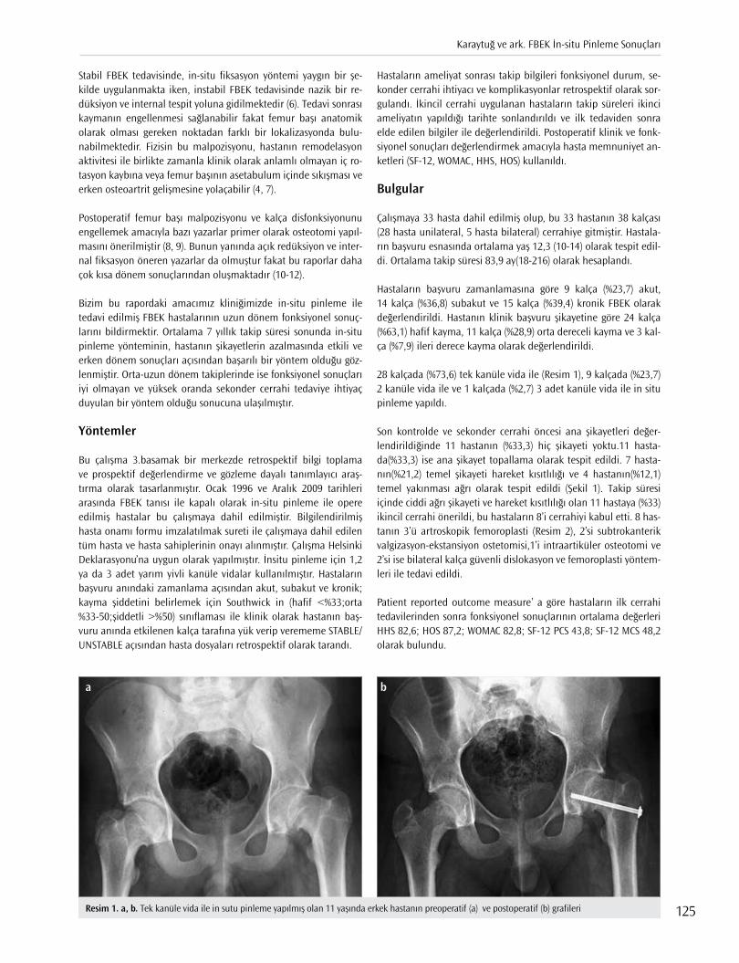
Stabil FBEK tedavisinde, in-situ fiksasyon yöntemi yaygın bir şe-kilde uygulanmakta iken, instabil FBEK tedavisinde nazik bir re-düksiyon ve internal tespit yoluna gidilmektedir (6). Tedavi sonrası kaymanın engellenmesi sağlanabilir fakat femur başı anatomik olarak olması gereken noktadan farklı bir lokalizasyonda bulu-nabilmektedir. Fizisin bu malpozisyonu, hastanın remodelasyon aktivitesi ile birlikte zamanla klinik olarak anlamlı olmayan iç ro-tasyon kaybına veya femur başının asetabulum içinde sıkışması ve erken osteoartrit gelişmesine yolaçabilir (4, 7).
Postoperatif femur başı malpozisyonu ve kalça disfonksiyonunu engellemek amacıyla bazı yazarlar primer olarak osteotomi yapıl-masını önerilmiştir (8, 9). Bunun yanında açık redüksiyon ve inter-nal fiksasyon öneren yazarlar da olmuştur fakat bu raporlar daha çok kısa dönem sonuçlarından oluşmaktadır (10-12).
Bizim bu rapordaki amacımız kliniğimizde in-situ pinleme ile tedavi edilmiş FBEK hastalarının uzun dönem fonksiyonel sonuç-larını bildirmektir. Ortalama 7 yıllık takip süresi sonunda in-situ pinleme yönteminin, hastanın şikayetlerin azalmasında etkili ve erken dönem sonuçları açısından başarılı bir yöntem olduğu göz-lenmiştir. Orta-uzun dönem takiplerinde ise fonksiyonel sonuçları iyi olmayan ve yüksek oranda sekonder cerrahi tedaviye ihtiyaç duyulan bir yöntem olduğu sonucuna ulaşılmıştır.
Yöntemler
Bu çalışma 3.basamak bir merkezde retrospektif bilgi toplama ve prospektif değerlendirme ve gözleme dayalı tanımlayıcı araş-tırma olarak tasarlanmıştır. Ocak 1996 ve Aralık 2009 tarihleri arasında FBEK tanısı ile kapalı olarak in-situ pinleme ile opere edilmiş hastalar bu çalışmaya dahil edilmiştir. Bilgilendirilmiş hasta onamı formu imzalatılmak sureti ile çalışmaya dahil edilen tüm hasta ve hasta sahiplerinin onayı alınmıştır. Çalışma Helsinki Deklarasyonu’na uygun olarak yapılmıştır. İnsitu pinleme için 1,2 ya da 3 adet yarım yivli kanüle vidalar kullanılmıştır. Hastaların başvuru anındaki zamanlama açısından akut, subakut ve kronik; kayma şiddetini belirlemek için Southwick in (hafif <%33;orta %33-50;şiddetli >%50) sınıflaması ile klinik olarak hastanın baş-vuru anında etkilenen kalça tarafına yük verip verememe STABLE/UNSTABLE açısından hasta dosyaları retrospektif olarak tarandı.
Hastaların ameliyat sonrası takip bilgileri fonksiyonel durum, se-konder cerrahi ihtiyacı ve komplikasyonlar retrospektif olarak sor-gulandı. İkincil cerrahi uygulanan hastaların takip süreleri ikinci ameliyatın yapıldığı tarihte sonlandırıldı ve ilk tedaviden sonra elde edilen bilgiler ile değerlendirildi. Postoperatif klinik ve fonk-siyonel sonuçları değerlendirmek amacıyla hasta memnuniyet an-ketleri (SF-12, WOMAC, HHS, HOS) kullanıldı.
Bulgular
Çalışmaya 33 hasta dahil edilmiş olup, bu 33 hastanın 38 kalçası (28 hasta unilateral, 5 hasta bilateral) cerrahiye gitmiştir. Hastala-rın başvuru esnasında ortalama yaş 12,3 (10-14) olarak tespit edil-di. Ortalama takip süresi 83,9 ay(18-216) olarak hesaplandı.
Hastaların başvuru zamanlamasına göre 9 kalça (%23,7) akut, 14 kalça (%36,8) subakut ve 15 kalça (%39,4) kronik FBEK olarak değerlendirildi. Hastanın klinik başvuru şikayetine göre 24 kalça (%63,1) hafif kayma, 11 kalça (%28,9) orta dereceli kayma ve 3 kal-ça (%7,9) ileri derece kayma olarak değerlendirildi.
28 kalçada (%73,6) tek kanüle vida ile (Resim 1), 9 kalçada (%23,7) 2 kanüle vida ile ve 1 kalçada (%2,7) 3 adet kanüle vida ile in situ pinleme yapıldı.
Son kontrolde ve sekonder cerrahi öncesi ana şikayetleri değer-lendirildiğinde 11 hastanın (%33,3) hiç şikayeti yoktu.11 hasta-da(%33,3) ise ana şikayet topallama olarak tespit edildi. 7 hasta-nın(%21,2) temel şikayeti hareket kısıtlılığı ve 4 hastanın(%12,1) temel yakınması ağrı olarak tespit edildi (Şekil 1). Takip süresi içinde ciddi ağrı şikayeti ve hareket kısıtlılığı olan 11 hastaya (%33) ikincil cerrahi önerildi, bu hastaların 8’i cerrahiyi kabul etti. 8 has-tanın 3’ü artroskopik femoroplasti (Resim 2), 2’si subtrokanterik valgizasyon-ekstansiyon ostetomisi,1’i intraartiküler osteotomi ve 2’si ise bilateral kalça güvenli dislokasyon ve femoroplasti yöntem-leri ile tedavi edildi.
Patient reported outcome measure’ a göre hastaların ilk cerrahi tedavilerinden sonra fonksiyonel sonuçlarının ortalama değerleri HHS 82,6; HOS 87,2; WOMAC 82,8; SF-12 PCS 43,8; SF-12 MCS 48,2 olarak bulundu.
Resim 1. a, b. Tek kanüle vida ile in sutu pinleme yapılmış olan 11 yaşında erkek hastanın preoperatif (a) ve postoperatif (b) grafileri
a b
Karaytuğ ve ark. FBEK İn-situ Pinleme Sonuçları
125
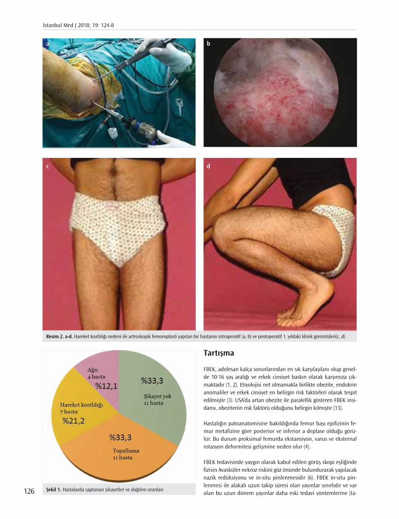
Tartışma
FBEK, adolesan kalça sorunlarından en sık karşılaşılanı olup genel-de 10-16 yaş aralığı ve erkek cinsiyet baskın olarak karşımıza çık-maktadır (1, 2). Etiyolojisi net olmamakla birlikte obezite, endokrin anomaliler ve erkek cinsiyet en belirgin risk faktörleri olarak tespit edilmiştir (3). USA’da artan obezite ile paralellik gösteren FBEK insi-dansı, obezitenin risk faktörü olduğunu belirgin kılmıştır (13).
Hastalığın patoanatomisine bakıldığında femur başı epifizinin fe-mur metafizine göre posterior ve inferior a deplase olduğu görü-lür. Bu durum proksimal femurda ekstansiyon, varus ve eksternal rotasyon deformitesi gelişmine neden olur (4).
FBEK tedavisinde yaygın olarak kabul edilen görüş skopi eşliğinde fizisin Avasküler nekroz riskini göz önünde bulundurarak yapılacak nazik redüksiyonu ve in-situ pinlenmesidir (6). FBEK in-situ pin-lenmesi ile alakalı uzun takip süresi olan yayınlar sınırlıdır ve var olan bu uzun dönem yayınlar daha eski tedavi yöntemlerine (ta-
Resim 2. a-d. Hareket kısıtlılığı nedeni ile artroskopik femoroplasti yapılan bir hastanın intraperatif (a, b) ve postoperatif 1. yıldaki klinik görüntüleri(c, d)
c
a
d
b
Şekil 1. Hastalarda saptanan şikayetler ve dağılım oranları
İstanbul Med J 2018; 19: 124-8
126
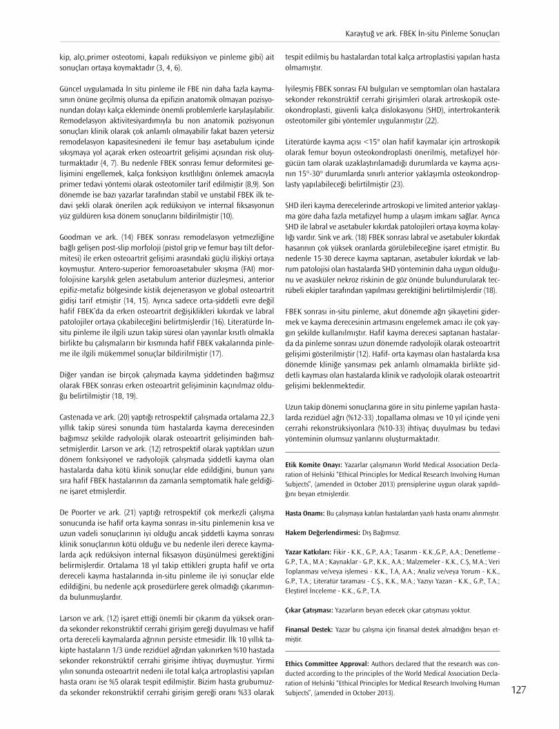
kip, alçı,primer osteotomi, kapalı redüksiyon ve pinleme gibi) ait sonuçları ortaya koymaktadır (3, 4, 6).
Güncel uygulamada İn situ pinleme ile FBE nin daha fazla kayma-sının önüne geçilmiş olunsa da epifizin anatomik olmayan pozisyo-nundan dolayı kalça ekleminde önemli problemlerle karşılaşılabilir. Remodelasyon aktivitesiyardımıyla bu non anatomik pozisyonun sonuçları klinik olarak çok anlamlı olmayabilir fakat bazen yetersiz remodelasyon kapasitesinedeni ile femur başı asetabulum içinde sıkışmaya yol açarak erken osteoartrit gelişimi açısından risk oluş-turmaktadır (4, 7). Bu nedenle FBEK sonrası femur deformitesi ge-lişimini engellemek, kalça fonksiyon kısıtlılığını önlemek amacıyla primer tedavi yöntemi olarak osteotomiler tarif edilmiştir (8,9). Son dönemde ise bazı yazarlar tarafından stabil ve unstabil FBEK ilk te-davi şekli olarak önerilen açık redüksiyon ve internal fiksasyonun yüz güldüren kısa dönem sonuçlarını bildirilmiştir (10).
Goodman ve ark. (14) FBEK sonrası remodelasyon yetmezliğine bağlı gelişen post-slip morfoloji (pistol grip ve femur başı tilt defor-mitesi) ile erken osteoartrit gelişimi arasındaki güçlü ilişkiyi ortaya koymuştur. Antero-superior femoroasetabuler sıkışma (FAI) mor-folojisine karşılık gelen asetabulum anterior düzleşmesi, anterior epifiz-metafiz bölgesinde kistik dejenerasyon ve global osteoartrit gidişi tarif etmiştir (14, 15). Ayrıca sadece orta-şiddetli evre değil hafif FBEK’da da erken osteoartrit değişiklikleri kıkırdak ve labral patolojiler ortaya çıkabileceğini belirtmişlerdir (16). Literatürde İn-situ pinleme ile ilgili uzun takip süresi olan yayınlar kısıtlı olmakla birlikte bu çalışmaların bir kısmında hafif FBEK vakalarında pinle-me ile ilgili mükemmel sonuçlar bildirilmiştir (17).
Diğer yandan ise birçok çalışmada kayma şiddetinden bağımsız olarak FBEK sonrası erken osteoartrit gelişiminin kaçınılmaz oldu-ğu belirtilmiştir (18, 19).
Castenada ve ark. (20) yaptığı retrospektif çalışmada ortalama 22,3 yıllık takip süresi sonunda tüm hastalarda kayma derecesinden bağımsız şekilde radyolojik olarak osteoartrit gelişiminden bah-setmişlerdir. Larson ve ark. (12) retrospektif olarak yaptıkları uzun dönem fonksiyonel ve radyolojik çalışmada şiddetli kayma olan hastalarda daha kötü klinik sonuçlar elde edildiğini, bunun yanı sıra hafif FBEK hastalarının da zamanla semptomatik hale geldiği-ne işaret etmişlerdir.
De Poorter ve ark. (21) yaptığı retrospektif çok merkezli çalışma sonucunda ise hafif orta kayma sonrası in-situ pinlemenin kısa ve uzun vadeli sonuçlarının iyi olduğu ancak şiddetli kayma sonrası klinik sonuçlarının kötü olduğu ve bu nedenle ileri derece kayma-larda açık redüksiyon internal fiksasyon düşünülmesi gerektiğini belirmişlerdir. Ortalama 18 yıl takip ettikleri grupta hafif ve orta dereceli kayma hastalarında in-situ pinleme ile iyi sonuçlar elde edildiğini, bu nedenle açık prosedürlere gerek olmadığı çıkarımın-da bulunmuşlardır.
Larson ve ark. (12) işaret ettiği önemli bir çıkarım da yüksek oran-da sekonder rekonstrüktif cerrahi girişim gereği duyulması ve hafif orta dereceli kaymalarda ağrının persiste etmesidir. İlk 10 yıllık ta-kipte hastaların 1/3 ünde rezidüel ağrıdan yakınırken %10 hastada sekonder rekonstrüktif cerrahi girişime ihtiyaç duymuştur. Yirmi yılın sonunda osteoartrit nedeni ile total kalça artroplastisi yapılan hasta oranı ise %5 olarak tespit edilmiştir. Bizim hasta grubumuz-da sekonder rekonstrüktif cerrahi girişim gereği oranı %33 olarak
tespit edilmiş bu hastalardan total kalça artroplastisi yapılan hasta olmamıştır.
İyileşmiş FBEK sonrası FAI bulguları ve semptomları olan hastalara sekonder rekonstrüktif cerrahi girişimleri olarak artroskopik oste-okondroplasti, güvenli kalça dislokasyonu (SHD), intertrokanterik osteotomiler gibi yöntemler uygulanmıştır (22).
Literatürde kayma açısı <15° olan hafif kaymalar için artroskopik olarak femur boyun osteokondroplasti önerilmiş, metafizyel hör-gücün tam olarak uzaklaştırılamadığı durumlarda ve kayma açısı-nın 15°-30° durumlarda sınırlı anterior yaklaşımla osteokondrop-lasty yapılabileceği belirtilmiştir (23).
SHD ileri kayma derecelerinde artroskopi ve limited anterior yaklaşı-ma göre daha fazla metafizyel hump a ulaşım imkanı sağlar. Ayrıca SHD ile labral ve asetabuler kıkırdak patolojileri ortaya koyma kolay-lığı vardır. Sink ve ark. (18) FBEK sonrası labral ve asetabuler kıkırdak hasarının çok yüksek oranlarda görülebileceğine işaret etmiştir. Bu nedenle 15-30 derece kayma saptanan, asetabuler kıkırdak ve lab-rum patolojisi olan hastalarda SHD yönteminin daha uygun olduğu-nu ve avasküler nekroz riskinin de göz önünde bulundurularak tec-rübeli ekipler tarafından yapılması gerektiğini belirtilmişlerdir (18).
FBEK sonrası in-situ pinleme, akut dönemde ağrı şikayetini gider-mek ve kayma derecesinin artmasını engelemek amacı ile çok yay-gın şekilde kullanılmıştır. Hafif kayma derecesi saptanan hastalar-da da pinleme sonrası uzun dönemde radyolojik olarak osteoartrit gelişimi gösterilmiştir (12). Hafif- orta kayması olan hastalarda kısa dönemde kliniğe yansıması pek anlamlı olmamakla birlikte şid-detli kayması olan hastalarda klinik ve radyolojik olarak osteoartrit gelişimi beklenmektedir.
Uzun takip dönemi sonuçlarına göre in situ pinleme yapılan hasta-larda rezidüel ağrı (%12-33) ,topallama olması ve 10 yıl içinde yeni cerrahi rekonstrüksiyonlara (%10-33) ihtiyaç duyulması bu tedavi yönteminin olumsuz yanlarını oluşturmaktadır.
Etik Komite Onayı: Yazarlar çalışmanın World Medical Association Decla-ration of Helsinki “Ethical Principles for Medical Research Involving Human Subjects”, (amended in October 2013) prensiplerine uygun olarak yapıldı-ğını beyan etmişlerdir.
Hasta Onamı: Bu çalışmaya katılan hastalardan yazılı hasta onamı alınmıştır.
Hakem Değerlendirmesi: Dış Bağımsız.
Yazar Katkıları: Fikir - K.K., G.P., A.A.; Tasarım - K.K.,G.P., A.A.; Denetleme - G.P., T.A., M.A ; Kaynaklar - G.P., K.K., A.A.; Malzemeler - K.K., C.Ş, M.A.; Veri Toplanması ve/veya işlemesi - K.K., T.A, A.A.; Analiz ve/veya Yorum - K.K., G.P., T.A.; Literatür taraması - C.Ş., K.K., M.A.; Yazıyı Yazan - K.K., G.P., T.A.; Eleştirel İnceleme - K.K., G.P., T.A.
Çıkar Çatışması: Yazarların beyan edecek çıkar çatışması yoktur.
Finansal Destek: Yazar bu çalışma için finansal destek almadığını beyan et-miştir.
Ethics Committee Approval: Authors declared that the research was con-ducted according to the principles of the World Medical Association Decla-ration of Helsinki “Ethical Principles for Medical Research Involving Human Subjects”, (amended in October 2013).
Karaytuğ ve ark. FBEK İn-situ Pinleme Sonuçları
127
Informed Consent: Written informed consent was obtained from patients who participated in this study.
Peer-review: Externally peer-reviewed.
Author contributions: Concept - K.K., G.P., A.A.; Design - K.K., G.P., A.A.; Supervision - G.P., T.A., M.A; Resource - G.P., K.K., A.A.; Materials - K.K., C.Ş, M.A.; Data Collection and/or Processing - K.K., T.A, A.A.; Analysis and/or Interpretation - K.K., G.P., T.A.; Literature Search - C.Ş., K.K., M.A.; Writing - K.K., G.P., T.A.; Critical Reviews - K.K., G.P., T.A.
Conflict of Interest: Authors have no conflicts of interest to declare.
Financial Disclosure: The author declared that this study has received no financial support.
Kaynaklar
1. Bhatia NN, Pirpiris M, Otsuka NY Body mass index in paitents with Slipped capital femoral epiphysis. J Pediatr Orthop 2006; 26: 197-9. [CrossRef]
2. Lehmann CL, Arons RR, Loder RT, Vitale MG. The epidemiology of slipped capital femoral epiphysis: an update. J Pediatr Orthop 2006; 26: 286-90. [CrossRef]
3. Jingushi S, Suenega E. Slipped capital femoral epiphysis: etiology and treatment. J Orthop Sci 2004; 9: 214-9. [CrossRef]
4. Aronsson DD, Loder RT, Breur GJ, Weinsteinn SL. Slipped capital femo-ral epiphysis: current concepts. J Am Acad Orthop Surg 2006; 14: 666-79. [CrossRef]
5. Loder RT, Richards BS, Shapiro PS, Reznick LR, Aronsson DD. Acute slipped capital femoral epiphysis: The importance of physeal stabil-ity. J Bone Joint Surg Am 1993; 75: 1134-40. [CrossRef]
6. Loder RT, Dietz FR. What is the best evidence for the treatment of slipped capital femoral epiphysis? J Pediatr Orthop 2012; 32: 158-65. [CrossRef]
7. Leunig M, Casillas MM, Hamlet M, Hersche O, Nötzli H, Slongo T. Slipped capital femoral epiphysis:early mechanical damage to the acetabular cartilage by a prominent femoral metaphysis. Acta Orthop Scand 2000; 71: 370-5. [CrossRef]
8. Southwick WO (1967) Osteotomy through the lesser trochanter for slipped capital femoral epiphysis.J Bone Joint Surg Am 1967; 49: 807-35. [CrossRef]
9. Witbreuk MM, Bolkenbaas M, Mullender MG, Sierevelt IN, Besselaar PP. The results of downgrading moderate and severe slipped capital femoral epiphysis by an early Imhauser femur osteotomy. J Child Or-thop 2009; 3: 405-10. [CrossRef]
10. Alves C, Steele M, Narayanan U, Howard A, Alman B, Wright JG. Open reduction and internal fixation of unstable slipped capital femoral epiphysis by means of surgical dislocation does not decrease the rate
of avascular necrosis: a preliminary study.J Child Orthop 2012; 6: 277-83. [CrossRef]
11. Tibor LM, Sink EL. Risks and benefits of the modified Dunn ap-proach for treatment of moderate or severe slipped capital femoral epiphysis.J Pediatr Orthop 2013; 33: 99-102. [CrossRef]
12. Larson AN, Sierra RJ, Yu EM, Trousdale RT, Stans AA. Outcomes of slipped capital femoral epiphysis treated with in situ pinning. J Pedi-atr Orthop 2012; 32: 125-30. [CrossRef]
13. Murray AW, Wilson NI. Changing incidence of slipped capital femoral epiphysis: A relationship with obesity? J Bone Joint Surg Br 2008; 90: 92-4. [CrossRef]
14. Goodman DA, Feighan JE, Smith AD, Latimer B, Buly RL, Cooperman DR. Subclinical slipped capital femoral epiphysis.Relations to osteoar-throsis of the hip. J Bone Joint Surg Am 1997; 79: 1489-97. [CrossRef]
15. Fraitzl CR, Kafer W, Nelitz M, Reichel H. Radiological evidence of fem-oracetabular impingement in mild slipped capital femoral epiphysis: a mean follow-up of 14.4 years after pinning in situ. J Bone Joint Surg Br 2007; 89: 1592-6. [CrossRef]
16. Tscholl PM, Zingg PO, Dora C, Frey E, Dierauer S, Ramseier LE. Ar-throscopic osteochondroplasty in patients with mild slipped capital femoral epiphysis after in situ fixation. J Child Orthop 2016; 10: 25-30. [CrossRef]
17. Boero S,Brunenghi GM,Carbone M, Stella G, Calevo MG. Pinning in slipped capital femoral epiphysis:long-term follow-up study.J Pediatr Orthop B 2003;12: 372-9. [CrossRef]
18. Sink EL, Zaltz I, Heare T, Dayton M. Acetabular cartilage and labral damage observed during surgical hip dislocation for stable slipped capital femoral epiphysis. J Pediatr Orthop 2010; 30: 26-30. [CrossRef]
19. Dodds MK, McCormack D, Mulhall KJ. Femoracetabular impigement after slipped capital femoral epiphysis:does slip severity predict clini-cal symptoms? J Pediatr Orthop 2009; 29: 535-9. [CrossRef]
20. Castañeda P1, Ponce C, Villareal G, Vidal C. The natural history of osteoarthritis after a slipped capital femoral epiphysis/the pistol grip deformity. J Pediatr Orthop 2013; 33: 76-82. [CrossRef]
21. de Poorter JJ, Beunder TJ,Gareb B, Oostenbroek HJ, Bessems GH, van der Lugt JC, et al. Long-term outcomes of slipped capital femoral epiphysis treated with in situ pinning. J Child Orthop 2016; 10: 371-9. [CrossRef]
22. Kuzyk PR, Kim YJ, Millis MB. Surgical Management of Healed Slipped Capital Femoral Epiphysis. J Am Acad Orthop Surg 2011; 19: 667-77. [CrossRef]
23. Sekiya JK, Wojtys EM, Loder RT, Hensinger RN. Hip artrhoscopy using a limited anterior exposure: An alternativeapproach for arthroscopic access. Arthroscopy 2000; 16: 16-20. [CrossRef]
Cite this article as: Karaytuğ K, Polat G, Akgül T, Asma A, Şen C, Aşık M. Long-term Results of In Situ Pinning Treatment of Femoral Head Slippage Patients. İstanbul Med J 2018; 19: 124-8.
İstanbul Med J 2018; 19: 124-8
128
Giriş
Üriner inkontinans, sosyal ya da hiljenik problem haline gelen istem dışı idrar kaçırma olarak tanımlanmaktadır (1). Sorunun sıklığı yaş ile artmakta birlikte yaşlanmanın bir parçası olarak kabul edilebilecek önemsiz, tedavisi gereksiz veya tedavinin mümkün olmadığı bir durum olarak değerlendirilebilmektedir. Ancak aslında ciddi sosyal, hijyenik, emosyonel ve cinsel sorunlara yol açabildiği bilinen bir sağlık problemidir (2, 3).
Stress üriner inkontinans (SUİ) öksürme, hapşırma veya fiziksel egzersiz sırasında idrar kaçırma olarak tanımlanır. Prevalansı dünya çapındaki kadınlarda %25, Avrupa ülkelerinde %35 olarak bildirilmiştir (1, 4). SUİ tedavisinde pelvik taban ekzersizleri, davranış terapisi, biofeedback ve elektrik stimülasyonu gibi tedavilerin yanında yanıt alınamayan olgularda cerrahi tedavi uygu-lanır. SUİ tedavisi için birçok cerrahi prosedür tanımlanmıştır. Transobturator Tape (TOT) tekniği Delorme tarafından geliştirilmiş subüretral sling operasyonu olup (5), günümüzde SUİ’nin cerrahi tedavisinde standart cerrahi tedavi olarak kullanılmaktadır.
Stress üriner inkontinans ya da üriner system disfonksiyonu olan kadınların %50 kadarında seksüel disfonksiyon görülmektedir (6). Pelvik taban yetersizliğinin düzeltilmesi ile cinsel fonk-siyonlar düzelse de cerrahi ve operasyon materyalleri bazı hastalarda cinsel fonksiyon bozuk-luğuna sebep olabilir. Cinsel fonksiyonun tedavi öncesinde değerlendirilmesi gerekmektedir (7). Female Sexual Function Index (FSFI), bu amaçla kullanılan, son 4 haftadaki, uyarılma, cinsel istek, orgazm, lubrikasyon, cinsel doyum ve ağrıyı sorgulayan ve 19 sorudan oluşan bir formdur (8). Bu çalışmada FSFI kullanarak stres üriner inkontinansı olup tek başına TOT operasyonu planlanan hastalarda operasyon öncesi ve sonrası dönemde operasyonunun cinsel fonksiyona etkisini araştırmayı amaçladık.
Üriner İnkontinans Tedavisinde Uygulanan Transobturator Tape Operasyonunun Cinsel Fonksiyon Üzerine EtkisiEvaluation of the Effect of Transobturator Tape Operation Used in Urinary Incontinence Therapy on Sexual Function
Amaç: Stres üriner inkontinansı olup tek başına Transobturator Tape (TOT) operasyonu planlanan hastalarda operasyon öncesi ve sonrası dönemde operasyonunun cinsel fonksiyona etkisini araştırmak.
Yöntemler: Çalışma prospektif olarak planlandı, 1 Ocak 2010 ve 1 Ocak 2015 tarihleri arasında İstanbul Eğitim Araştırma Hastanesi, Ka-dın Hastalıkları ve Doğum Kliniği ve Üroloji Kliniği’ne idrar kaçırma şikayeti ile başvuran, stres/mikst üriner inkontinans saptanan ve sa-dece TOT operasyonu planlanan 33 hasta çalışmaya alındı. Hastalara operasyon öncesi ve operasyon sonrasında cinsel aktif olduktan en az 1 ay sonra Female Sexual Function Index (FSFI) skorlaması yapıldı.
Bulgular: Hastaların yaş ortalaması 48,9±6 yıl idi. Hastalarda operas-yon öncesi ve sonrası cinsel işlev bozukluğu oranı %42,4 (n=14) olarak bulundu. Stress üriner inkontinansı olan ve TOT operasyonu yapılan hastaların toplam FSFI skoru ile istek, uyarılma, orgazm, cinsel başarı ve ağrı skorlarında istatistiksel olarak anlamlı bir fark olmadığı sa-dece lubrikasyon alt ölçeğinde istatistiksel olarak anlamlı bir azalma olduğu saptandı.
Sonuç: TOT operasyonu sonrası kadınlarda lubrikasyon azalması dı-şında istatistiksel olarak anlamlı bir cinsel fonksiyon bozukluğu sap-tanmadı.
Anahtar Kelimeler: Cinsel fonksiyon, transobturator tape, inkonti-nans
Introduction: To investigate the effect of surgery on sexual function in the preoperative and postoperative period in patients with stress urinary incontinence, who are planned for the transobturator tape (TOT) operation alone.
Methods: The study was prospectively planned. Overall, 33 patients with urinary incontinence, who were diagnosed with stress/mixt urinary incontinence and planned to undergo TOT operation at the Istanbul Education Research Hospital Obstetrics and Gynecology De-partment and Urology Clinic between January 1, 2010 and January 1, 2015, were enrolled in the study. FSFI scoring was performed before the operation and at least 1 month after the first sexual activity follo-wing the operation.
Results: The mean age of the patients was 48.9±6 years. The incidence of sexual dysfunction before and after the operation was 42.4% (n=14). There was no statistically significant difference in the total FSFI score, desire, arousal, orgasm, sexual success, and pain scores between the preoperative and postoperative periods in patients. A statistically signi-ficant decrease was found only in the lubrication subscale.
Conclusion: No statistically significant sexual dysfunction was found in women after the TOT operation except for decreased lubrication.
Keywords: Sexual function, transobturator tape, incontinence
Öz
/ Abs
trac
t
ORCID IDs of the authors: S.P. 0000-0002-7501-2466, D.S.A. 0000-0002-7283-0930; Z.S. 0000-0002-2678-8989; A.B.B. 0000-0002-4307-7767.
1İstanbul Eğitim ve Araştırma Hastanesi, Kadın Hastalıkları ve Doğum Kliniği, İstanbul, Türkiye2Haseki Eğitim ve Araştırma Hastanesi, Kadın Hastalıkları ve Doğum Kliniği, İstanbul, Türkiye
Yazışma Adresi / Address for Correspondence:Derya Sivri Aydın E-mail: [email protected]
Geliş Tarihi/Received: 15.11.2016
Kabul Tarihi/Accepted: 20.11.2017
© Telif Hakkı 2018 Makale metnine istanbultipdergisi.org web sayfasından ulaşılabilir.
© Copyright 2018 by Available online at istanbulmedicaljournal.org
Orijinal Araştırma/Original Investigationİstanbul Med J 2018; 19: 129-33DOI: 10.5152/imj.2018.37605
Serpil Polat1 , Derya Sivri Aydın2 , Zeynep Soyman1 , Ahmet Birtan Boran1

Yöntemler
Çalışma prospektif olarak planlandı, 1 Ocak 2010 ve 1 Ocak 2015 tarihleri arasında İstanbul Eğitim ve Araştırma Hastanesi Kadın Hastalıkları ve Doğum Kliniği ve Üroloji Kliniği’ne idrar kaçırma şikayeti ile başvuran veya başka nedenlerle başvurup sorgulama ve muayene sonrasında stres/mix inkontinans tesbit edilen, cinsel olarak aktif 151 hasta mevcut idi, ancak TOT operasyonuna ila-veten prolapsus operasyonu eklenen, peroperatif ve postoperatif komplikasyon gelişen 118 hasta çalışmaya dahil edilmedi. Jineko-lojik muayenesinde ayakta ya da jinekolojik pozisyonda öksürmek ya da ıkınmakla üriner inkontinans saptanan ya da ürodinamik olarak stres ya da mixt tip üriner inkontinansı bulunan ve sadece TOT operasyonu planlanan 33 hasta çalışmaya alındı. Çalışmaya alınan hastalardan aydınlatılmış onam alındı. Hastalara operasyon öncesi ve operasyon sonrasında cinsel aktif olduktan en az 1 ay sonra FSFI skorlaması yapıldı.
Hastaların yaş, boy, kilo, sigara kullanımı, menopoz durumu, me-nopoz süresi, doğum sayısı, doğum şekli (vaginal doğum, sezar-yen), iri fetus öyküsü, aylık ortalama gelirleri, eğitim durumları ve cinsel ilişki sırasında idrar kaçırma olup olmadığı sorgulandı. Aylık ortalama gelirleri 500-1.000 TL, 100-1.500 TL ve 1.500 TL olarak, eğitim durumları hiç eğitimi olmayan, ilkokul, ortaokul, lise ve üniversite olarak gruplara ayrıldı.
Cinsel işlevlerinin sorgulanmasına yönelik 19 sorudan oluşan FSFI formu operasyon öncesi ve sonrası dönemde (cinsel aktif olduktan en az 1 ay sonra) doldurtuldu. İndeksin içeriğinde; uyarılma, istek, kayganlaşma (yağlanma, ıslanma), doyum, orgazm ve ağrı olarak 6 alt bölüm bulunmaktadır. Her bir bölüm sıfırdan 5’e kadar pu-anlanmaktadır. Birinci ve 2. sorularda cinsel istek veya ilgi sıklığı ve seviyesi (puan: 1-5); 3-6. sorularda uyarılma sıklığı, seviyesi, emin olma durumu ve doyumu (puan: 0-5); 7-10. sorularda lubrikasyon (kayganlaşma) sıklığı, zorluğu, cinsel ilişkide koruyabilme sıklığı ve zorluğu (puan: 0-5); 11-13. sorularda orgazm sıklığı, zorluğu ve do-yumu (puan: 0-5); 14-16. sorularda doyum, eşiyle yakınlık oranı, cinsel ilişkide ve tüm cinsel yaşamında doyum seviyesi (puan: 0-1 ile 5); 17-19. sorularda ağrı veya rahatsızlık, vajinaya giriş sırasında ağrı, vajinaya girişi takiben ve girişte ve takiben ağrı seviyesi (puan: 0-5) değerlendirilmektedir. Ölçekten en yüksek 95 en düşük 4 puan alınamaktadır. Hesaplama alt bölümlerin ve tüm ölçeğin puanla-masını saptamak için düzenlenmiş ve istek için 0.6, uyarılma ve kayganlaşma için 0.3, orgazm, doyum ve ağrı için 0.4 katsayısı be-lirlenmiştir. Alt bölümlerin ortalamaları katsayılarla çarpılıp alına-bilecek en yüksek puan 36, en düşük puan 2 olarak hesaplanmıştır (9). Çalışmaya dahil edilecek örneklem büyüklüğü El-Enen ve ark. (10) çalışmasına göre hesaplandı. Total FSFI farkı esas alındığın-da %80 power ve 0.05 α hata ile minimum sample size 6 olarak saptandı. Çalışmaya başlamadan önce etik kurul onayı alınmıştır.
İstatistiksel AnalizStatistical Package for Social Sciences 15.0 for Windows (SPSS Inc.; Chicago, IL, USA) programı kullanılarak yapılan tanımlayıcı istatis-tikler; kategorik değişkenler için sayı ve yüzde, sayısal değişkenler için ortalama±standart sapma minimum-maksimum olarak veril-di. Sayısal değişkenler bağımsız iki grup arası karşılaştırmalar nor-mal dağılım koşulları sağlandı ise Student-t Test, ikiden çok grup karşılaştırmaları One Way Anova testi ile normal dağılım koşulu sağlanmadığında bağımsız iki grup karşılaştırmaları Mann Whit-
ney U testi ile, ikiden çok grup karşılaştırmaları Kruskal Wallis test ile yapıldı. Alt grup analizleri parametrik test koşulu sağlandığın-dan Tukey testi ile yapıldı. Bağımlı grup karşılaştırmaları değiş-kenlerin farkları normal dağılım koşulunu sağladığında Paired t test sağlamadığında iki grup karşılaştırmaları Wilcoxson testi ile yapıldı. Sayısal değişkenler arası ilişkiler parametrik test koşulu sağlandığında Pearson Korelasyon Analizi ile parametrik test ko-şulu sağlanmadığında Spearman Korelasyon Analizi ile incelendi. p<0,05 istatistiksel alfa anlamlılık seviyesi olarak kabul edildi.
Bulgular
Çalışmaya alınan 33 hastanın 20’si Kadın Doğum Servisi’nde, 13’ü Üroloji Servisi’nde opere edildi.
Hastaların yaş ortalaması 48,9±6 yıl (35-63 yıl), gravidası 4,1±1,8 (2-10), paritesi 2,3±0,8 (1-4), ortalama BMI değeri 29,1±4,3 (23-40) idi. Yüzde %39,4 (n=13) hasta menopozda ve ortalama menopoz süresi 6 yıldı, %75,8 (n=25) hasta normal doğum yapmıştı; doğum ağırlığının en fazla 4800 gr ve en az 1750 gr olduğu tespit edildi. Hastaların %27,3 (n=9)’ü sigara kullanıyordu, olguların %3’ünün hiç eğitimi yok, %75,8’i ilkokul mezunu, %3’ü ortaokul mezunu, %15,2’si lise mezunu, %3’ü üniversite mezunu idi. Hastaların; %27,3’ü 500-1.000 TL, %36,4’ü 1.000-1.500 TL, %36,4’ü 1.500 TL üzerinde aylık geliri bulunmaktadır.
Cinsel ilişki sırasında operasyon öncesi %54,5 (n=18) hastada in-kontinans mevcutken, operasyon sonrası sadece %6,1 (n=2) hasta-da inkontinans mevcuttu.
Hastalara operasyon öncesi ve sonrası uygulanan FSFI ölçeği sonuç-ları Tablo’da özetlenmiştir. Operasyon sonrasında, istek, uyarılma, orgazm, cinsel başarı, ağrı alt ölçeklerinin ortalama FSFI skorlarında saptanan azalma istatistiksel olarak anlamlı değildi (p> 0.05). Orta-lama FSFI skoru lubrikasyon alt ölçeği için operasyon öncesi 4,8±1,2 iken operasyon sonrası 4,4±1,4 idi. Lubrikasyondaki bu azalma ista-tistiksel olarak anlamlıydı (p=0,037). Operasyon öncesi toplam FSFI ortalama skoru 25,9±5,3 iken operasyon sonrası 24,5±5,9 idi fakat azalma istatistiksel olarak anlamlı değildi (p=0,277).
Hastalarda operasyon öncesi ve sonrası cinsel işlev bozukluğu ora-nı %42,4 (n=14) olarak bulundu. Operasyon öncesi cinsel işlev bo-zukluğu olan iki hasta operasyon sonrası düzelmişti ancak, operas-yon öncesi cinsel işlev bozukluğu olmayan iki hastada cinsel işlev bozukluğu meydana geldiği için toplam puanlama değişmemiştir.
Operasyon öncesi FSFI toplam puanı ile parite arasında istatistiksel olarak anlamlı negatif bir ilişki saptandı (p=0,019). Alt ölçeklerden uyarılma puanı doğum ağrılığının fazla olması ile pozitif yönde, cinsel başarı alt ölçeği puanı parite ile negatif yönde, ağrı en fazla doğum ağırlığının fazla olması ile pozitif yönde istatistiksel anlam-lı ilişki vardı (sırayla p=0,043, p=0,016, p=0,017). Tedavi öncesi FSFI alt grupları ile yaş, BMI, gravida, menopoz süresi arasındaki ilişki istatistiksel anlamlı değildi (p>0,05).
Operasyon öncesi FSFI toplam puanı ve istek, orgazm alt ölçeği eğitimi olmayan ve ilk-ortaokul mezunlarında, lise ve üniversi-te mezunlarına göre istatistiksel olarak anlamlı düşüktü (sırayla p=0,023, p=0,028, p=0,026). Tedavi öncesi FSFI ölçeği toplam puanı ve uyarılma alt ölçeği puanlarında aylık gelir düzeyi grupla-rında istatistiksel olarak anlamlı fark vardı. Aylık gelir düzeyi 1.500
İstanbul Med J 2018; 19: 129-33
130

Polat ve ark. Transobturator Tape Operasyonunun Cinsel Fonksiyon Üzerine Etkisinin Değerlendirilmesi
131
tl ‘ten yüksek olan grubun toplam puanı, aylık geliri 1.000-1.500 tl olan gruba göre; uyarılma puanı aylık geliri 1.000-1.500 tl olan ve aylık geliri 500-1.000 tl olan gruba göre yükseklik istatistiksel olarak anlamlıydı (sırayla p=0,027, p=0,002, p=0,039).
Operasyon öncesi FSFI alt gruplarının, doğum şekilleri, menopoz durumu ve cinsel ilişki sırasındaki inkontinans durumuyla arala-rında ilişki saptanmamıştır (p>0,05).
Operasyon sonrası FSFI toplam puanı ile istek, uyarılma, orgazm, cinsel başarı alt ölçek puanları yaş ve parite ile negatif yönde ilişki-si istatistiksel olarak anlamlı olarak saptandı (p<0,05). Tedavi son-rası yaşın ve paritenin; FSFI alt ölçeği lubrikasyon ve ağrı arasında istatistiksel anlamlı ilişki saptanmadı (p>0,05).
Operasyon sonrası FSFI ölçeği toplam puanı ve istek, uyarılma, or-gazm, cinsel başarı, ağrı alt ölçek puanları yaş, BMI, gravida ve menopoz süresi arasında istatistiksel olarak anlamlı bir ilişki bu-lunmamaktadır (p>0,05).
Operasyon sonrası FSFI toplam puanı ile istek ve orgazm alt ölçeği; eğitimi olmayan ve ilk-ortaokul mezunlarında, lise ve üniversite mezunlarına göre istatistiksel olarak anlamlı olarak düşük sap-tandı (sırasıyla p=0,032; p=0,015; p=0,022) fakat uyarılma, lub-rikasyon, cinsel başarı ve ağrı skorlarında istatistiksel anlamlı ilişki saptanmadı (p>0,05).
Menopozda olanlarda FSFI toplam puanı ile istek, uyarılma ve cinsel başarı alt ölçeği menopozda olmayanlara göre istatistiksel olarak anlamlı düşüktü (sırasıyla p=0,006, p=0,046, p=0,001, p=0,008) fakat lubrikasyon, orgazm ve ağrının alt ölçeğinde ista-tistiksel anlamlı ilişki saptanmadı (p>0,05).
Operasyon sonrası cinsel ilişki sırasında üriner inkontinansı olma-yanların cinsel başarı alt ölçeği puanı üriner inkontinansı olanlara göre istatistiksel olarak anlamlı düşüktü (p=0,017).
Operasyon sonrası FSFI toplam puanı ve alt ölçeklerinde hastaların aylık geliri, doğum şekli ve sigara kullamaları ile istatistiksel olarak anlamlı bir ilişki saptanmadı (p>0,05).
Tartışma
Kadınlarda cinsel fonksiyon bozukluğu pelvik taban yetersizliğin-de sıklıkla karşılaşılan bir durumdur ve varolan çalışmalar pelvik
taban bozukluğunun düzeltilmesi sonrası cinsel fonksiyonlarının da düzeldiği yönündedir ancak operasyonun ve kullanılan mater-yallerinin bazı hastalarda cinsel işlev bozukluğuna neden olabile-ceği düşünülmektedir. Bu hastalarda cinsel fonksiyonların tedavi öncesinde değerlendirilmeleri gerekmektedir ve bu değerlendirme anket ve semptomların skorlanması ile etkili bir şekilde yapılabil-mektedir (9).
Üriner inkontinans ve kadın cinsel fonksiyon bozuklukları arasın-daki ilişki uzun zamandır incelenmesine rağmen konu ile ilgili bazı noktalar hala netlik kazanmamıştır ve elde edilen sonuçlar çeliş-kilidir. Bu konuda yapılan araştırma sayısı fazladır; ama akademik açıdan sağlam, kanıta dayalı tıp yönünden değerli çalışma sayısı kısıtlıdır. Bunun en önemli nedeni her iki patolojinin de fizyolojik, psikolojik ve sosyal boyutları olması ve araştırmaların tüm boyut-ları yansıtmasındaki güçlüktür. Öte yandan ölçülmesi zor olan bu kavramları değerlendirmek için kullanılan testlerin oluşturulma-sındaki güçlük yadsınamaz. Çoğu kadın idrar kaçırma ve cinsel fonksiyon bozukluklarının konuşulmasından rahatsızlık duyduğu için sıklığı normalin altında kalmaktadır. Üriner inkontinans ne-deniyle kadında özgüven kaybı, anksiyete ve cinsel istekte azalma olabilir (7).
Üriner inkontinans ve cinsel işlev bozukluğu birbiri ile ilişkili olma-sınına rağmen, inkontinans cerrahisinin cinsel işlev üzerine etkisi-ni gösteren az sayıda çalışma vardır (10-12).
İnkontinans cerrahisi sonrası cinsel fonksiyonlar iyileşebileceği gibi vajinal innervasyonun bozulmasına bağlı cinsel fonksiyonlar üzerine olumsuz etkiler olabileceği belirtilmiştir (13, 14).
Üriner inkontinansın kadın cinsel fonksiyon bozukluğuna sebep olduğu ve üriner inkontinansı olan kadınlarda cinsel fonksiyon bo-zukluğu sıklığının %26 ile %43 arasında olduğu bildirilmektedir (15). Ülkemizde kadınların %48,3’ünde cinsel disfonksiyon saptanmıştır. Bizim çalışmamızda stres üriner inkontinansı olanlarda sexsüel dis-fonksiyon %42,4 olarak tespit edilmiş olup bu oran operasyon son-rası değişmemiştir. Bu sonuç bize TOT operasyonunun cinsel fonksi-yonu olumsuz yönde etkilemediğini düşündürmektedir.
Shaw’ın (16) yaptığı bir çalışmada idrar kaçırma ve alt üriner sistem şikâyetleri olan hastaların %46’sında cinsel fonksiyon bozukluğu saptanmıştır. Barber ve ark. (17) kadınların %11 ile %77’sinin ilişki sırasında idrar kaçırdığı rapor edilmiştir. Yine Barber tarafından yapılan bir yayında ilişki sırasında en fazla idrar kaçırmanın stres
Tablo 1. TOT operasyonu öncesi ve sonrası FSFI skorlarının değerlendirilmesi
Tedavi Öncesi Tedavi Sonrası
Ortalama±SD Min-Maks Ortalama±SD Min-Maks P=
İstek 3,4±1,1 1,2-6 3,2±1,2 1,2-6 0,14
Uyarılma 3,6±1,1 1,5-6 3,4±1,1 1,2-6 0,479
Lubrikasyon 4,8±1,2 1,5-6 4,4±1,4 1,8-6 0,037
Orgazm 4,6±1,1 1,2-6 4,3±1,3 1,2-6 0,09
Cinsel başarı 4,7±1,2 1,2-6 4,5±1,3 1,2-6 0,188
Ağrı 4,8±1,3 2,0-6 4,6±1,2 2,0-6 0,589
Toplam puan 25,9±5,3 11,7-35 24,5±5,9 8,6-34,2 0,277
SD: standart deviasyon; Min: minimum; Maks: maximum

üriner inkontinans saptanan olgularda görüldüğü bildirilmiştir (18). Clark ve Romm (19) yaptıkları bir araştırmada olguların % 56’sının ilişki sırasında idrar kaçırdıklarını ve bunun cinsel hayatla-rının olumsuz etkilendiğini ifade etmişlerdir. Bizim çalışmamızda cinsel ilişki sırasında operasyon öncesi literatürle uyumlu olarak %54,5 (n=18) hastada inkontinans mevcuttu, operasyon sonrası sadece %6,1 (n=2) hastada inkontinans mevcuttu.
Stress üriner inkontinansı tedavisinde midüretral sling operas-yonları en sık kullanılan cerrahidir. Operasyon sonrasında cinsel fonksiyonlarda olumlu etki yaptığı gösterilmiştir fakat tersini gös-teren çalışmalar da mevcuttur. Sling operasyonları komplikasyonu sonucu oluşan erozyon bu negatif etkide önemlidir. Ayrıca vajen ön duvarı kanlanma ve innervasyon bozulmasının orgazm bo-zukluğuna neden olabileceğini gösteren çalışmalar vardır. Vajen darlığı veya gevşekliğinin de cinsel disfonksiyona sebep olabileceği bazı çalışmalarda belirtilmiştir. Dursun ve ark. (20) TOT yapılan 96 kadını değerlendirilmiştir. Bu çalışmada total FSFI skoru; uyarılma, istek, orgasm, lubrikasyon skorlarındaki artışta istatistiksel olarak anlamlı bulunmamıştır fakat ağrı ve tatmin skorlarında istatistik-sel anlamlı iyileşme saptanmıştır. Çalışmamızda operasyon sonra-sında operasyon öncesine göre; istek, uyarılma, orgazm, cinsel ba-şarı, ağrı alt ölçeklerinin ortalama FSFI skorlarında azalma izlendi ancak bu azalma istatistiksel olarak anlamlı değildi. Lubrikasyon alt ölçeği için saptanan azalma istatistiksel olarak anlamlıydı. Bu durum çalışmanın yapıldığı popülasyonun yaş ortalamasının yük-sek olmasına bağlanabilir.
Kadınların yaş grubu arttıkça cinsel istek, orgazm, uyarılma, lubri-kasyon ve memnuniyette azalma olurken; cinsel ilişki sırasındaki ağrıda artma olmaktadır. Östrojen azalmasının disparoni, istek kaybı, cinsel aktivite sayısında azalma, genital duyarlılıkta azal-ma, orgazma ulaşmada zorluğa, testesteron azalmasının ise cinsel genital duyarlılık, uyarı, libido ve orgazma ulaşmada azalmaya neden olduğu gösterilmiştir (21). Çalışmamızda hastaların tedavi sonrası FSFI ölçeği toplam puanı ve istek, uyarılma, orgazm, cinsel başarı alt ölçek puanları yaş ile negatif yönde istatistiksel olarak anlamlı ilişkiliydi ve hastaların menopozda olanlarının FSFI ölçeği toplam puanı ve istek, uyarılma cinsel başarı alt ölçeği puanları menopozda olmayanlara göre istatistiksel olarak anlamlı düşüktü. Tedavi sonrası FSFI ölçeği alt gruplarından lubrikasyon, orgazm ve ağrının hastaların menopoz durumlarıyla arasında istatistiksel ola-rak anlamlı bir ilişki bulunmamıştır.
Multiparitenin cinsel fonksiyonlar üzerine olumsuz etkisi bildirilse de; parite ile kadın cinsel fonksiyonları arasında anlamlı bir ilişki olmadığı da rapor edilmiştir (22, 23). Çalışmamızda hastalarınte-davi öncesi FSFI ölçeği toplam puanı ve cinsel başarı puanı, pa-rite ile istatistiksel olarak negatif yönde anlamlı ilişkiliydi. Teda-vi sonrası da FSFI ölçeği toplam puanı ve alt gruplarından istek, uyarılma, orgazm, cinsel başarı puanıparite ile istatistiksel olarak negatif yönde anlamlı ilişkiliydi.
Kadınların eğitim düzeyinin cinsel fonksiyonlar üzerine etkisi bili-nen bir gerçektir. Çayan ve ark. (24) çalışmasında kadınların eğitim düzeyi ile FSFI ölçekleri arasında pozitif yönde bir ilişki olduğunu gözlemlemişlerdir. Düşük eğitim düzeyleriyle seksüel problemlerin varlığının arttığı saptanmıştır (25). Literatür ile uyumlu olarak bi-zim çalişmamızda da tedavi öncesi FSFI ölçeği toplam puanı ve is-tek, orgazm alt ölçeği; eğitimi olmayan ve ilk-ortaokul mezunların-da, lise ve üniversite mezunlarına göre istatistiksel olarak anlamlı
düşüktü. Tedavi sonrası FSFI ölçeği toplam puanı ve istek, orgazm alt ölçeği; eğitimi olmayan ve ilk-ortaokul mezunlarında, lise ve üniversite mezunlarına göre istatistiksel olarak anlamlı düşüktü.
Sonuç
Stress üriner inkontinansı olan ve TOT operasyonu yapılan hastala-rın toplam FSFI skoru ile istek, uyarılma, orgazm, cinsel başarı ve ağrı skorlarında bir farklılık olmadığı sadece lubrikasyon alt ölçeği için istatistiksel bir azalma olduğunu saptadık. İleri yaş menopoz ve parite durumu da kadın cinsel fonksiyon bozukluğu için risk faktörleridir. Kadınların eğitim düzeyleri cinsel fonksiyonlarınıetki-leyen önemli bir faktördür. Bu çalışmada TOT operasyonu sonrası kadınlarda lubrikasyon azalması dışında istatistiksel olarak anlam-lı bir cinsel fonksiyon bozukluğu saptanmadı. Ancak bu konuda halen akademik açıdan sağlam, kanıta dayalı tıp yönünden değerli çalışma açığı bulunmaktadır, daha geniş çalışmalara ihtiyaç vardır.
Etik Komite Onayı: Bu çalışma için etik komite onayı İstanbul Eğitim ve Araştırma Hastanesi Etik Kurul Komitesi’nden alınmıştır (Karar Tarihi: 29.05.2015; Karar No: 661).
Hasta Onamı: Hasta onamı bu çalışmaya katılan hastalardan alınmıştır.
Hakem Değerlendirmesi: Dış Bağımsız.
Yazar Katkıları: Fikir - D.S.A.; Tasarım - D.S.A., S.P.; Denetleme - S.P., D.S.A., Z.S., A.B.B.; Kaynaklar - S.P.; Malzemeler - S.P.; Veri Toplanması ve/veya işlemesi - S.P.; Analiz ve/veya Yorum - D.S.A., S.P.; Literatür taraması D.S.A., S.P.; Yazıyı Yazan - D.S.A., S.P., Z.S., A.B.B.; Eleştirel İnceleme - D.S.A., A.B.B.
Çıkar Çatışması: Yazarların beyan edecek çıkar çatışması yoktur.
Finansal Destek: Yazarlar bu çalışma için finansal destek almadıklarını beyan etmişlerdir.
Ethics Committee Approval: Ethics committee approval was received for this study from the ethics committee of İstanbul Training and Research Hospital (Approval Date: 29.05.2015; Approval No: 661).
Informed Consent: Informed consent was obtained from the patients who participated in this study.
Peer-review: Externally peer-reviewed.
Author contributions: Concept - D.S.A.; Design - D.S.A., S.P.; Supervision - S.P., D.S.A., Z.S., A.B.B.; Resource - S.P.; Materials - S.P.; Data Collection and/or Processing - S.P.; Analysis and/or Interpretation - D.S.A., S.P.; Literature Search - D.S.A., S.P.; Writing - D.S.A., S.P., Z.S., A.B.B.; Critical Reviews - D.S.A., A.B.B.
Conflict of Interest: Authors have no conflicts of interest to declare.
Financial Disclosure: The authors declared that this study has received no financial support.
Kaynaklar
1. Abrams P, Blaivas JG, Stanton SL, Andersen JT. The standardisation of terminology of lower urinary tract function. İnt Urogynecol J 1990; 1: 45-58. [CrossRef]
2. Kişnişçi HA, Gökşin E, Durukan T, Üstay K, Ayhan A, Gürgan T, et al. Temel Kadın Hastalıkları ve Doğum Bilgisi. Güneş Kitabevi, Ankara, 1996; 730-47.
İstanbul Med J 2018; 19: 129-33
132
3. Berek JS. Novak's Gynecology. 14th Edition. Lippincott Williams&Wilkins. 2006.
4. Hunskaar S, Lose G, Sykes D, Voss S. The prevalence of urinary in-continence in women in four European countries. BJU Int 2004; 93: 324-30. [CrossRef]
5. Delorme E. [Transobturator urethral suspension: Mini-invasive proce-dure in the treatment of stress urinary incontinence in women]. Prog Urol 2001; 11: 1306-13. [Article in French]
6. Dalpiaz O, Kerschbaumer A, Mitterberger M, Pinggera GM, Colleselli D, Bartsch G, et al. Female sexual dysfunction: A new urogynaecological research field. BJU Int 2008; 101: 717-21. [CrossRef]
7. Özdedeli K, Yüksel İ. Pelvik Taban Bozuklukları ve Kadın Cinsel Fonksiyon Bozukluğu. Kadın ve İşlevsel Üroloji Dergisi 2014; 3: 73-7. [CrossRef]
8. Rosen R, Brown C, Heiman J, Leiblum S, Meston C, Shabsigh R, et al. The Female Sexual Function Index (FSFI): a multidimensional self-report instrument for the assessment of female sexual function. J Sex Marital Ther 2000; 26: 191-208. [CrossRef]
9. Leppert PC, Howard FM. Primary Care for Women, Quebecor/King-sport, Philadelphia, 1997.
10. El-Enen MA, Ragb M, El Gamasy Ael-N, El-Ashry O, El-Sharaby M, Elbadawy A, et al. Sexual function among women with stress in-continence after using transobturator vaginal tape, and its cor-relation with patient's expectations. BJU Int 2009; 104: 1118-23. [CrossRef]
11. Koçak I, Okyay P, Dundar M, Erol H, Beser E. Female urinary inconti-nence in the West of Turkey: Prevalence, risk factors and impact on quality of life. Eur Urol 2005; 48: 634-41. [CrossRef]
12. Zippe CD, Raina R, Shah AD, Massanyi EZ, Agarwal A, Ulchaker J, et al. Sexual dysfuntion after radical cystectomy: a new outcome measure. Urology 2004; 63: 1153-7. [CrossRef]
13. Elzevier HW, Venema PL, Lycklama á Nijeholt AA. Sexual function after tension-free vaginal tape (TVT) for stress incontinence: results of a mailed questionnaire. Int Urogynecol J Pelvic Dysfunct 2004; 15: 313-8. [CrossRef]
14. Glavind K, Tetsche MS. Sexual function in women before and after suburetral sling operation for stres urinary incontinence: a retrospec-tive questionnaire study. Acta Obstet Gynecol Scand 2004; 83: 965-8. [CrossRef]
15. Oksuz E, Malhan S. Prevalence and risk factors for female sexual dys-function in Turkish women. J Urol 2006; 175: 654-8. [CrossRef]
16. Shaw C. A systematic review of the literature on the prevelance of the sexual impairment in women with urinary incontinence and the prevelance of urinary leakage during sexual activity. Eur Urol 2002; 42: 432-40. [CrossRef]
17. Barber MD, Dowsett SA, Mullen KJ, Viktrup L. The impact of stress urinary incontinence on sexual activity in women. Cleve Clin J Med. 2005; 72: 225-32. [CrossRef]
18. Barber MD, Visco AG, Wyman JF, Fantl JA, Bump RC, Continence Pro-gram for Women Research Group. Sexual function in women with uri-nary incontinence and pelvic organ prolapse. Obstet Gynecol 2002; 99: 281-9. [CrossRef]
19. Clark A, Romm J. Effect of urinary incontinence on sexual activitiy in women. J Reprod Med 1993; 38: 679-83.
20. Dursun M, Otunctemur A, Ozbek E, Cakir SS, Polat EC. Impact of the transobturator tape procedure on sexual function in women with stress urinary incontinence. J Obstet Gynaecol Res 2013; 39: 831-5. [CrossRef]
21. Jennifer R, Berman MD, Irwin Goldstein MD. Female sexual dysfunc-tion. Urol Clin North Am 2001; 28: 405-16. [CrossRef]
22. Lukacz ES, Whitcomb EL, Lawrence JM, Nager CW, Contreras R, Luber KM. Are sexual activity and satisfaction affected by pelvic floor disor-ders? Analaysis of a community-based survey. Am J Obstet Gynecol 2007; 197: 1-6. [CrossRef]
23. Handa VL, Harvey L, Cundiff GW, Siddique SA, Kjerulff KH. Sexual func-tion among women with urinary incontinence and pelvic organ pro-lapse. Am J Obstet Gynecol 2004; 191: 751-6. [CrossRef]
24. Çayan S, Akay E, Bozlu M, Canpolat B, Acar D, Ulusoy E. The prevelance of female sexual dysfunction and potential risk factors that may impair sexual function in Turkish women. Urol Int 2004; 72: 52-7. [CrossRef]
25. Laumann EO, Paik A, Rosen RC. Sexual dysfunction in the United States: prevalence and predictors. JAMA 1999; 281: 537-44. [CrossRef]
Cite this article as: Polat S, Aydın DS, Soyman Z, Boran AB. Evaluation of the Effect of Transobturator Tape Operation Used in Urinary Incontinence Therapy on Sexual Function. İstanbul Med J 2018; 19: 129-33.
Polat ve ark. Transobturator Tape Operasyonunun Cinsel Fonksiyon Üzerine Etkisinin Değerlendirilmesi
133

Giriş
Lenfadenopatilerin en yaygın nedeni reaktif lenf nodu hiperplazileridir (1-3). Bu hastalar ara-sında malignite prevalansı düşük olsa da doktorlar ve hastalar için endişe verici bir durumdur. Etyolojinin açıklanamadığı durumlarda birçok laboratuar testi, görüntüleme yöntemleri ve doku biyopsisi yapılması gerekli olmaktadır (4).
Platelet volüm değerleri olan ortalama platelet hacmi (mean platelet volum, MPV) ve platelet dağılım genişliği (platlelet distribution width, PDW) platelet fonksiyonu ve aktivitesi ile ilişkili olan ve kan sayımından kolay elde edilen parametrelerdir (5, 6). Malignitelerde ve inflamatuar hasta-lıklarda platelet indekslerinin tanısal değeri olduğu gösterilmiştir (7-11).
Laktat dehidrogenaz (LDH), en yüksek oranda karaciğer, kas doku, kalp ve böbrekte olmak üzere bütün dokularda bulunur. Hücre hasarının geliştiği herhangi bir durumda geçici yükselmeler olu-şabilir (12). Lenfomalı hastalarda hastalığın prognozunu belirlemede önemli bir gösterge olduğu düşünülmektedir (12).
Bu çalışmada, nedeni bilinmeyen lenfadenopatili çocuklarda malign ve non-malign ayrımın-da MPV ve PDW gibi platelet indeksleri ile serum LDH düzeylerinin kullanılabilirliği araştı-rılmıştır.
Lenfadenopatili Çocuklarda Platelet İndeksleri ve Serum Laktat Dehidrogenaz Düzeyleri Malignite Ayırıcı Tanısında Kullanılabilir Mi?Can Platelet Indices and Serum Lactate Dehydrogenase Levels be Used for the Differential Diagnosis of Malignancy in Children with Lymphadenopathies?
Amaç: Ortalama platelet hacmi (MPV) ve platelet dağılım genişliği (PDW) gibi platelet indekslerinin bazı kanser türlerinde ve inflama-tuar hastalıklarda tanısal önemi olduğu bildirilmiştir. Bu çalışmada, reaktif lenf nodu hiperplazili ve lenfomalı çocuklarda platelet indeks-leri ve serum laktat dehidrogenaz (LDH) düzeyleri araştırılmıştır.
Yöntemler: Toplam 102 çocuk çalışmaya alındı. Ekim 2011 ile 2016 yılları arasında lenf nodu eksizyonu yapılan 72 çocuk hasta grubunu, 30 sağlıklı çocuk kontrol grubunu oluşturdu. Dosya bilgileri geriye dö-nük olarak incelendi.
Bulgular: Histopatolojik olarak 72 hastadan 50 (%69,4)’sine reaktif lenf nodu hiperplazisi, 22 (%30,5)’sine lenfoma tanısı konulmuştu. Lenfoma tanısı konulan hastaların 15(%68)’i Hodgkin lenfoma, 7 (%32)’si Non-Hodgkin lenfoma idi. Reaktif lenf nodu hiperplazisi ta-nısı konulan hastalar ile lenfoma tanısı konulan hastalar arasında lökosit sayısı, MPV, PDW değerleri ve serum LDH düzeyleri açısından farklılık yoktu (p>0,05, hepsi için). İki grubun serum LDH düzeyleri kontrol grubundan istatistiksel açıdan anlamlı şekilde yüksek bulun-du (p<0,001, ikisi için).
Sonuç: Nedeni bilinmeyen lenfadenopatisi olan çocuklarda benign ve malign ayırımında platelet indeksleri ve serum LDH düzeyleri kulla-nışlı değildir. Hodgkin lenfoma gibi düşük dereceli tümörlerde serum LDH düzeyleri normal aralıkta kalabilmektedir.
Anahtar Kelimeler: Lenfadenopati, reaktif, malign, platelet indeksi, laktat dehidrogenaz
Introduction: Platelet indices such as the mean platelet volume (MPV) and platelet distribution width (PDW) have been reported to have a diagnostic value in some cancer types and inflammatory di-seases. In this study, the platelet indices and serum lactate dehydro-genase (LDH) levels were investigated in children with reactive lymph node hyperplasia and lymphoma.
Methods: In total, 102 children were enrolled in this study. Overall, 30 healthy children and 72 children, who had undergone excisional lymph node biopsy between October 2011 and 2016, were enrolled this study. File records were retrospectively reviewed.
Results: Histopathologically, 50 (69.4%) of the 72 patients were diagnosed with reactive lymph node hyperplasia, and 22 (30.5%) were diagnosed with lymphoma. Of the cases diagnosed with lymphoma, 15 (68%) were those of Hodgkin’s lymphoma and 7 (32%) were those of non-Hodgkin’s lymphoma. There was no difference between the patients diagnosed with reactive lymph node hyperplasia and those with lymphoma in terms of leukocyte counts, MPV and PDW values, and serum LDH levels (p>0.05, for all). The serum LDH levels of both groups were statistically significantly higher than those of the control group (p<0.001, for both).
Conclusion: Platelet indices and serum LDH levels are not useful in the differentiation of malignant and non-malignant differentiation in children with unexplained lymphadenopathies. Serum LDH levels may remain within the normal range in low-grade tumors such as Hodgkin’s lymphoma.
Keywords: Lymphadenopathy, reactive, malignant, platelet index, lactate dehydrogenase
Öz
/ Abs
trac
t
ORCID IDs of the authors: Z.C.Ö. 0000-0002-9172-9627; A.D. 0000-0002-4785-3014;Y.D.K. 0000-0003-2917-7750; H.Ö. 0000-0003-4144-3732; Ö.B. 0000-0002-1662-3259.
1Eskişehir Osmangazi Üniversitesi Tıp Fakültesi, Çocuk Hematoloji/Onkoloji Bilim Dalı, Eskişehir, Türkiye2Eskişehir Osmangazi Üniversitesi Tıp Fakültesi, Çocuk Sağlığı ve Hastalıkları Anabilim Dalı, Eskişehir, Türkiye3Eskişehir Osmangazi Üniversitesi Tıp Fakültesi, Tıbbi Biyoistatistik Anabilim Dalı, Eskişehir, Türkiye
Yazışma Adresi / Address for Correspondence:Zeynep Canan ÖzdemirE-mail: [email protected]
Geliş Tarihi/Received: 31.07.2017
Kabul Tarihi/Accepted: 20.11.2017
© Telif Hakkı 2018 Makale metnine istanbultipdergisi.org web sayfasından ulaşılabilir.
© Copyright 2018 by Available online at istanbulmedicaljournal.org
Orijinal Araştırma/Original Investigationİstanbul Med J 2018; 19: 134-7DOI: 10.5152/imj.2017.1357
Zeynep Canan Özdemir1 , Aslı Deniz2 , Yeter Düzenli Kar1 , Hülya Özen3 , Özcan Bör1
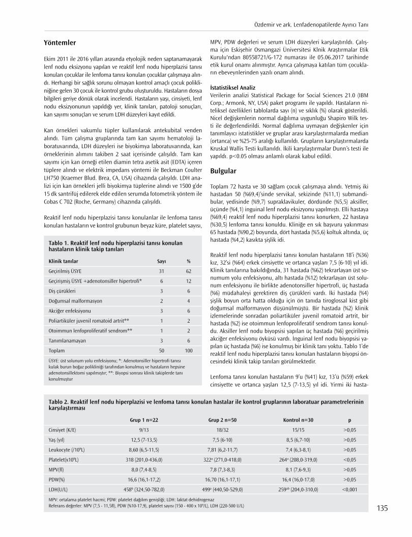
Yöntemler
Ekim 2011 ile 2016 yılları arasında etyolojik neden saptanamayarak lenf nodu eksizyonu yapılan ve reaktif lenf nodu hiperplazisi tanısı konulan çocuklar ile lenfoma tanısı konulan çocuklar çalışmaya alın-dı. Herhangi bir sağlık sorunu olmayan kontrol amaçlı çocuk polikli-niğine gelen 30 çocuk ile kontrol grubu oluşturuldu. Hastaların dosya bilgileri geriye dönük olarak incelendi. Hastaların yaşı, cinsiyeti, lenf nodu eksizyonunun yapıldığı yer, klinik tanıları, patoloji sonuçları, kan sayımı sonuçları ve serum LDH düzeyleri kayıt edildi.
Kan örnekleri vakumlu tüpler kullanılarak antekubital venden alındı. Tüm çalışma gruplarında tam kan sayımı hematoloji la-boratuvarında, LDH düzeyleri ise biyokimya laboratuvarında, kan örneklerinin alımını takiben 2 saat içerisinde çalışıldı. Tam kan sayımı için kan örneği etilen diamin tetra asetik asit (EDTA) içeren tüplere alındı ve elektrik impedans yöntemi ile Beckman Coulter LH750 (Kraemer Blud. Brea, CA, USA) cihazında çalışıldı. LDH ana-lizi için kan örnekleri jelli biyokimya tüplerine alındı ve 1500 g’de 15 dk santrifüj edilerek elde edilen serumda fotometrik yöntem ile Cobas C 702 (Roche, Germany) cihazında çalışıldı.
Reaktif lenf nodu hiperplazisi tanısı konulanlar ile lenfoma tanısı konulan hastaların ve kontrol grubunun beyaz küre, platelet sayısı,
MPV, PDW değerleri ve serum LDH düzeyleri karşılaştırıldı. Çalış-ma için Eskişehir Osmangazi Üniversitesi Klnik Araştırmalar Etik Kurulu'ndan 80558721/G-172 numarası ile 05.06.2017 tarihinde etik kurul onamı alınmıştır. Ayrıca çalışmaya katılan tüm çocukla-rın ebeveynlerinden yazılı onam alındı.
İstatistiksel Analiz Verilerin analizi Statistical Package for Social Sciences 21.0 (IBM Corp.; Armonk, NY, USA) paket programı ile yapıldı. Hastaların ni-teliksel özellikleri tablolarda sayı (n) ve sıklık (%) olarak gösterildi. Nicel değişkenlerin normal dağılıma uygunluğu Shapiro Wilk tes-ti ile değerlendirildi. Normal dağılıma uymayan değişkenler için tanımlayıcı istatistikler ve gruplar arası karşılaştırmalarda median (ortanca) ve %25-75 aralığı kullanıldı. Grupların karşılaştırmalarda Kruskal Wallis Testi kullanıldı. İkili karşılaştırmalar Dunn’s testi ile yapıldı. p<0.05 olması anlamlı olarak kabul edildi.
Bulgular
Toplam 72 hasta ve 30 sağlam çocuk çalışmaya alındı. Yetmiş iki hastadan 50 (%69,4)’sinde servikal, sekizinde (%11,1) submandi-bular, yedisinde (%9,7) supraklavikuler, dördünde (%5,5) aksiller, üçünde (%4,1) inguinal lenf nodu eksizyonu yapılmıştı. Elli hastaya (%69,4) reaktif lenf nodu hiperplazisi tanısı konurken, 22 hastaya (%30,5) lenfoma tanısı konuldu. Kliniğe en sık başvuru yakınması 65 hastada (%90,2) boyunda, dört hastada (%5,6) koltuk altında, üç hastada (%4,2) kasıkta şişlik idi.
Reaktif lenf nodu hiperplazisi tanısı konulan hastaların 18’i (%36) kız, 32’si (%64) erkek cinsiyette ve ortanca yaşları 7,5 (6-10) yıl idi. Klinik tanılarına bakıldığında, 31 hastada (%62) tekrarlayan üst so-numum yolu enfeksiyonu, altı hastada (%12) tekrarlayan üst solu-num enfeksiyonu ile birlikte adenotonsiller hipertrofi, üç hastada (%6) müdahaleyi gerektiren diş çürükleri vardı. İki hastada (%4) şişlik boyun orta hatta olduğu için ön tanıda tiroglossal kist gibi doğumsal malformasyon düşünülmüştü. Bir hastada (%2) klinik izlemelerinde sonradan poliartiküler juvenil romatoid artrit, bir hastada (%2) ise otoimmun lenfoproliferatif sendrom tanısı konul-du. Aksiller lenf nodu biyopsisi yapılan üç hastada (%6) geçirilmiş akciğer enfeksiyonu öyküsü vardı. İnguinal lenf nodu biyopsisi ya-pılan üç hastada (%6) ise konulmuş bir klinik tanı yoktu. Tablo 1’de reaktif lenf nodu hiperplazisi tanısı konulan hastaların biyopsi ön-cesindeki klinik takip tanıları görülmektedir.
Lenfoma tanısı konulan hastaların 9’u (%41) kız, 13’ü (%59) erkek cinsiyette ve ortanca yaşları 12,5 (7-13,5) yıl idi. Yirmi iki hasta-
Tablo 1. Reaktif lenf nodu hiperplazisi tanısı konulan hastaların klinik takip tanıları
Klinik tanılar Sayı %
Geçirilmiş ÜSYE 31 62
Geçirişmiş ÜSYE +adenotonsiller hipertrofi* 6 12
Diş çürükleri 3 6
Doğumsal malformasyon 2 4
Akciğer enfeksiyonu 3 6
Poliartiküler juvenil romatoid artrit** 1 2
Otoimmun lenfoproliferatif sendrom** 1 2
Tanımlanamayan 3 6
Toplam 50 100
ÜSYE: üst solunum yolu enfeksiyonu; *: Adenotonsiller hipertrofi tanısı kulak burun boğaz polikliniği tarafından konulmuş ve hastaların hepsine adenotonsillektomi yapılmıştır; **: Biyopsi sonrası klinik takiplerde tanı konulmuştur
Tablo 2. Reaktif lenf nodu hiperplazisi ve lenfoma tanısı konulan hastalar ile kontrol gruplarının laboratuar parametrelerinin karşılaştırması
Grup 1 n=22 Grup 2 n=50 Kontrol n=30 p
Cinsiyet (K/E) 9/13 18/32 15/15 >0,05
Yaş (yıl) 12,5 (7-13,5) 7,5 (6-10) 8,5 (6,7-10) >0,05
Leukocyte (/109L) 8,60 (6,5-11,5) 7,81 (6,2-11,7) 7,4 (6,3-8,1) >0,05
Platelet(x109L) 318 (201,0-436,0) 322a (271,0-418,0) 264a (208,0-319,0) <0,05
MPV(fl) 8,0 (7,4-8,5) 7,8 (7,3-8,3) 8,1 (7,6-9,3) >0,05
PDW(%) 16,6 (16,1-17,2) 16,70 (16,1-17,1) 16,4 (16,0-17,0) >0,05
LDH(U/L) 458b (324,50-782,0) 499a (440,50-529,0) 259ab (204,0-310,0) <0,001
MPV: ortalama platelet hacmi; PDW: platelet dağılım genişliği; LDH: laktat dehidrogenazReferans değerler: MPV (7,5 - 11,5fl), PDW (%10-17,9), platelet sayısı (150 - 400 x 109/L), LDH (220-500 U/L)
Özdemir ve ark. Lenfadenopatilerde Ayırıcı Tanı
135
dan 15 (%68)’ine Hodgkin lenfoma, yedisine (%32) Non-Hodgkin lenfoma tanısı konulmuştu. Non-Hodgkin lenfoma tanısı konulan hastalardan üçü T hücreli, üçü B hücreli lenfoblastik lenfoma, biri foliküler lenfoma idi. Lenfomalı hastaların dördü Evre II, 12’si Evre III, altısı Evre IV hastalığa sahipti.
Kontrol grubunun 15’i (%50) kız, 15’i (%50) erkek, ortanca yaşı 8,5 (6,7-10) yıl idi ve hasta grupları ile arasında yaş ve cinsiyet açısın-dan istatistiksel olarak anlamlı fark bulunmadı (p>0.05) (Tablo 2). Hasta grupları ile kontrol grupları arasında beyaz küre sayısı, MPV, PDW değerleri açısından farklılık yoktu (p>0.05, hepsi için). Reaktif lenf nodu hiperplazisi tansı konulan hastaların platelet sa-yısı kontrol grubundan yüksekti (p=0.018). Hasta gruplarının LDH düzeyi kontrol grubundan anlamlı şekilde yüksekti (p<0.001, her ikisi için). Ancak hasta grupları arasında LDH düzeyi açısından ista-tistiksel fark bulunmadı (p>0.05) (Tablo 2).
Tartışma
Lenfadenopatiler lokal veya sistemik inflamasyona sekonder ola-rak, lenforetiküler malignite ve metastatik malin hastalıklara bağlı olarak veya otoimmun hastalıklar ve aşılamaya cevap olarak geli-şebilir (13). Lenfadenopatili çocukların büyük kısmında altta yatan infeksiyöz veya benign nedenler vardır (14). Nedeni açıklanama-yan lenfadenopatili hastalarda malignite riskini değerlendiren üç çalışma bulunmaktadır. Bu çalışmalardan birinde 238 hastadan 3’üne malignite tanısı konulduğu, diğerinde 80 hastadan hiçbi-rinde malignite tanısı konulmadığı, üçüncüsünde ise malignite prevalansının %1,1 olduğu bildirilmiştir (15-17). Bizim çalışmamız-da lenf nodu biyopsisi yapılan hastalar arasında malignite sıklığı %30,5 bulundu. Bizim çalışmamız bir prevalans çalışması olmadığı için bu konuda genelleme yapmak mümkün değildir. Bu sonucun kliniğimize malignite şüphesi yüksek hastaların gönderilmesinden kaynaklanmış olma ihtimali yüksektir.
Platelet indekslerinin kan sayımından kolaylıkla elde edilir olması ve bazı hastalık durumlarında bu parametrelerde bir takım deği-şikliklerin oluştuğunun keşfedilmesinden sonra bu konu üzerinde çalışmalar giderek yoğunlaşmıştır. Son yıllarda, platelet sayısı ve platelet indekslerinin kanserli hastalarda inflamatuar bir belirteç olarak kullanılabileceği kabul görmeye başlamıştır.
Dolaşımdaki plateletlerin hacmi heterojendir. Yapıları ve meta-bolik aktiviteleri farklıdır. Sağlıklı kişilerde plateletlerin ortalama hacimleri 7,2-11,7 fl’dir (18, 19). MPV, megakaryositler tarafından belirlenir. Kanserli hastalarda birtakım sitokinlerin megakaryosit olgunlaşmasını artırarak genç ve daha büyük plateletlerin üretimi-ni uyardığı öne sürülmüştür (20). Artmış MPV, artmış platelet çapı üretim hızının ve platelet aktivasyonunun göstergesi olarak kula-nılabilir. Endometrial, ovarian, kolorektal ve gastrik kanserlerde MPV’nin sağlıklı kontrol gruba göre önemli ölçüde yüksek olduğu gösterilmiştir (21).
PDW, platelet dağılım hacmini gösterir. PDW’nin yükselmesi, dola-şımda anormal derecede küçük ve büyük plateletlerin olduğunu gösterir. PDW’nin over kanserlerinde sağlıklı kontrollerden yüksek, akciğer, meme ve malin adneksial tümörlerde ise düşük olduğu gösterilmiştir (22-24). Hematolojik malignitelerden akut lösemiler-de MPV’nin kontrol grubu ile benzer olduğu, PDW’nin düşük oldu-ğu, lenfoblastik lösemiler ile myeloblastik lösemiler arasında MPV ve PDW açısından farklılık olmadığı gösterilmiştir (25). Literatürde lenfadenopatili hastalarda platelet indekslerinin değerlendirildiği
bir çalışmaya rastlayamadık. Bizim çalışmamızda reaktif lenf nodu hiperplazisi olan hastalar, lenfoma hastaları ve sağlıklı kontroller arasında MPV ve PDW değerleri açısından farklılık saptayamadık. Laktat dehidrogenaz, glikolizin son basamağında prüvatın laktata indirgenmesinde ve glukoneogenez sırasında laktatın prüvata dö-nüştürülmesinde görev yapar (12). Solid tümörlerde, lösemilerde ve difüz lenfomalarda özellikle Burkitt lenfomada LDH düzeyinin arttığı bildirilmiştir (26-30).
Non-malign ve malign lenfadenopatilerin laboratuar paramet-relerinin araştırıldığı çalışmalarda, serum LDH düzeyinin ve lenf nodu çapının malign grupta non-malign gruptan yüksek olduğu gösterilmiştir (26-28). Ancak bu grupların sağlıklı kontroller ile kar-şılaştırması yapılmamış ve aradaki farklılığın ne düzeyde olduğu bildirilmemiştir (26-28). Yapılan bir çalışmada, Hodgkin lenfomalı olguların hiçbirinde LDH düzeylerinin normal sınırın üzerinde ol-madığı, lenfomaların histolojik alt tipi açısından LDH düzeyinin bir değer taşımadığı, ancak hastalığın evresi ile LDH düzeyi arasın-da önemli bir ilişki olduğu gösterilmiştir (12). Ayrıca kemoterapi-ye cevaplı ve cevapsız olanlarda da serum LDH düzeyinin normal aralıklara gerilediği ve LDH düzeyleri açısından farklılık olmadığı gösterilmiştir (12). Çalışmamızın sonuçlarına göre lenfomalı has-talarda serum LDH düzeyleri kontrol grubundan yüksekti ancak reaktif lenf nodu hiperplazisi olan hastalar ile benzerdi. Hastaları-mızın % 68 gibi büyük kısmının Hodgkin lenfoma hastası olması ve bu tümörlerin agresif olmamaları nedeni ile LDH düzeylerinin çok yükselmemiş olması muhtemeldir.
Bozlak ve ark. (26) çocuklarda servikal lenf adenopatilerin neden-lerini ve malinite için risk faktörlerini araştırdıkları çalışmalarında benign lenfadenopatisi olan çocukların yarısından fazlasında se-rum LDH düzeyinin normal aralığın üzerinde olduğunu göstermiş-lerdir. Serum LDH düzeylerinin diagnostik öneminin araştırıldığı bir çalışmada da LDH düzeyleri normalin 2 katından yüksek olan hastaların %60’ında benign, %36’sında malign nedenlerin olduğu, %5’inde etyolojik nedenin belirlenemediği bildirilmiştir (31). Çalış-mamızda reaktif lenf nodu hiperplazisi olan hastaların LDH dü-zeylerinin normal değerlerin üzerinde bulunması bu çalışmalarda elde edilen sonuçlar ile uyuşmakta olup, bu sonuç birçok lenfopro-liferatif hastalıkta LDH’nın yükselmesi bilgisi ile açıklanabilir (32).
Sonuç
Çalışmamızın sonuçları, nedeni açıklanamayan lenfadenopatiler-de malignite ayırıcı tanısında platelet indekslerinin kullanışlı ol-madığını, yükselmiş LDH düzeyinin lenfoma için spesifik bir bulgu olmadığını ve inflamatuar olaylarda da LDH düzeylerinin yüksele-bileceğini gösterdi. Laktat dehidrogenaz, malignite şüphesinin ol-duğu durumlarda ölçülmelidir, ancak diğer tanısal testler ve klinik bulgular ile birlikte dikkatli şekilde değerlendirilmelidir. Çalışma-mızın az sayıda hasta içermesi çalışmamızın kısıtlılığı olup daha fazla sayıda hastayı içeren kapsamlı çalışmalara ihtiyaç vardır.
Etik Komite Onayı: Bu çalışma için Eskişehir Osmangazi Üniversitesi Klinik Araştırmalar Etik Kurulu'ndan etik komite onayı alınmıştır. (Onay Ta rihi: 05.06.2017; Onay Numarası: 80558721/G-172)
Hasta Onamı: Hasta onamı bu çalışmaya katılan hastaların ailelerinden alın-mıştır.
Hakem Değerlendirmesi: Dış Bağımsız.
İstanbul Med J 2018; 19: 134-7
136
Yazar Katkıları: Fikir - Z.C.Ö., Ö.B.; Tasarım - Z.C.Ö., Ö.B.; Denetleme - Z.C.Ö.; Kaynaklar - Z.C.Ö., A.D., Y.D.K.; Veri Toplanması ve/veya işlemesi - A.D., Y.D.K., H.Ö.; Analiz ve/veya Yorum - Z.C.Ö., Y.D.K.,H.Ö.; Literatür taraması - Z.C.Ö., A.D., Y.D.K.; Yazıyı Yazan - Z.C.Ö., Ö.B.; Eleştirel İnceleme - Ö.B.
Çıkar Çatışması: Yazarların beyan edecek çıkar çatışması yoktur.
Finansal Destek: Yazarlar bu çalışma için finansal destek almadıklarını beyan etmişlerdir.
Ethics Committee Approval: Ethics committee approval was received for this study from Eskişehir Osmangazi University School of Medicine Clinical Rese-arches Center (Approvel Date: June 5th, 2017; Approval No: 80558721/G-172).
Informed Consent: Informed consent was obtained from the parents of the patient who paticipated in this study.
Peer-review: Externally peer-reviewed.
Author contributions: Concept - Z.C.Ö., Ö.B.; Design - Z.C.Ö., Ö.B.; Supervisi-on - Z.C.Ö.; Resource - Z.C.Ö., A.D., Y.D.K.; Data Collection and/or Processing - A.D., Y.D.K., H.Ö.; Analysis and/or Interpretation - Z.C.Ö.,Y.D.K.,H.Ö.; Litera-ture Search - Z.C.Ö., A.D., Y.D.K.; Writing - Z.C.Ö., Ö.B.; Critical Reviews - Ö.B.
Conflict of Interest: Authors have no conflicts of interest to declare.
Financial Disclosure: The authors declared that this study has received no financial support.
Kaynaklar
1. Moore SW, Schneider JW, Schaaf HS. Diagnostic aspects of cervical lymphadenopathy in children in the developing world: a study of 1,877 surgical specimens. Pediatr Surg Int 2003; 19: 240-4. [CrossRef]
2. Mohan A, Reddy MK, Phaneendra BV, Chandra A. Aetiology of perip-heral lymphadenopathy in adults: analysis of 1724 cases seen at a tertiary care teaching hospital in southern India. Natl Med J India 2007; 20: 78-80.
3. Darnal HK, Karim N, Kamini K, Angela K. The profile of lymphadeno-pathy in adults and children. Med J Malaysia 2005; 60: 590-8.
4. Mohseni S, Shojaiefard A, Khorgami A, Alinejad S, Ghorbani A, Ghafo-uri A. Peripheral lympadenopathy: Approach and dianostic tools. Iran J Med Sci 2014; 39: 158-70.
5. Gasparyan AY, Ayvazyan L, Mikhailidis DP, Kitas GD. Mean platelet vo-lume: a link between thrombosis and inflammation? Curr Pharm Des 2011; 17: 47-58. [CrossRef]
6. Jackson SR, Carter JM. Platelet volume: laboratory measurement and clinical application. Blood Rev 1993; 7: 104-13. [CrossRef]
7. Oncel M, Kiyici A, Oncel M, Sunam GS, Sahin E, Adam B. Evaluation of platelet indices in lung cancer patients. Asian Pac J Cancer Prev 2015; 16: 7599-602. [CrossRef]
8. Thachil J. Platelets in Inflammatory Disorders: A Pathophysiological and Clinical Perspective. Semin Thromb Hemost 2015; 41: 572-81. [CrossRef]
9. Öztürk ZA, Dag MS, Kuyumcu ME, Cam H, Yesil Y, Yilmaz N, et al. Could platelet indices be new biomarkers for inflammatory bowel diseases? Eur Rev Med Pharmacol Sci 2013; 17: 334-41.
10. Güneş A, Ece A, Şen V, Uluca Ü, Aktar F, Tan İ, et al. Correlation of mean platelet volume, neutrophil-to-lymphocyte ratio, and disease activity in children with juvenile idiopathic arthritis. Int J Clin Exp Med 2015; 8: 11337-41.
11. Templeton AJ, Ace O, McNamara MG, Al-Mubarak M, Vera-Badillo FE, Hermanns T, et al. Prognostic role of platelet to lymphocyte ratio in solid tumors: A systematic review and meta-analysis. Cancer Epidemi-ol Biomarkers Prev 2014; 23: 1204-12. [CrossRef]
12. Shamoon RP, Polus RK. Serum lactic dehydrogenase (LDH) activity in lymphomas: Prognostic significance and relationship to presentation, stage and histologic type. Zanco J Med Sci 2010; 14: 85-9.
13. Matsumoto F, Itoh S, Ohba S, Yokoi H, Furukawa M, Ikeda K. Biopsy of cervical lymph node. Auris Nasus Larynx 2009; 36: 71-4. [CrossRef]
14. Bazemore AW, Smucker DR. Lymphadenopathy and malignancy. Am Fam Physician 2002; 66: 2103-10.
15. Allhiser J, McKnight TA, Shank JC. Lymphadenopathy in a family prac-tice. J Fam Pract 1981; 12: 27-32.
16. Williamson HA Jr. Lymphadenopathy in a family practice: a descripti-ve study of 249 cases. J Fam Pract 1985; 20: 449-52.
17. Fijten GH, Blijham GH. Unexplained lymphadenopathy in family practice. An evaluation of the probability of malignant causes and the effectiveness of physicians' workup. J Fam Pract 1988; 27: 373-6.
18. Demirin H, Ozhan H, Ucgun T, Celer A, Bulur S, Cil H, et al. Normal range of mean platelet volume in healthy subjects: insight from a large epidemiologic study. Thromb Res 2011; 128: 358-60. [CrossRef]
19. Wiwanitkit V. Plateletcrit, mean platelet volume, platelet distribution width: its expected values and correlation with parallel red blood cell parameters. Clin Appl Thromb Hemost 2004; 10: 175-8. [CrossRef]
20. Coupland LA, Parish CR. Platelets, selectins, and the control of tumor metastasis. Semin Oncol 2014; 41: 422-34. [CrossRef]
21. Dincel O, Bayraktar C. Evaluation of platelet indices as a useful marker in papillary thyroid carcinoma. Bratisl Lek Listy Med J 2017; 118: 153-5.
22. Ma X, Wang Y, Sheng H, Tian W, Qi Z, Teng F, et al. Prognostic signifi-cance of thrombocytosis, platelet parameters and aggregation rates in epithelial ovarian cancer. J Obstet Gynaecol Res 2014; 40: 178-83. [CrossRef]
23. Ozaksit G, Tokmak A, Kalkan H, Yesilyurt H. Value of the platelet to lymphocyte ratio in the diagnosis of ovarian neoplasms in adoles-cents. Asian Pac J Cancer Prev 2015; 16: 2037-41. [CrossRef]
24. Okuturlar Y, Gunaldi M, Tiken EE, Oztosun B, Inan YO, Ercan T. Utility of peripheral blood parameters in predicting breast cancer risk. Asian Pac J Cancer Prev 2015; 16: 2409-12. [CrossRef]
25. Alsweedan SA, Al-Shurman A, Mahmoud AS. Diagnostic value of plate-let indices in children with leukemia. J Pediatr Hematol Oncol 2008; 30: 953-5. [CrossRef]
26. Bozlak S, Varkal MA, Yildiz I, Toprak S, Karaman S, Erol OB, et al. Cervical lymphadenopathies in children: A prospective clinical cohort study. Int J Pediatr Otorhinolaryngol 2016; 82: 81-7. [CrossRef]
27. Oğuz A, Karadeniz C, Temel EA, Citak EC, Okur FV. Evaluation of perip-heral lymphadenopathy in children. Pediatr Hematol Oncol 2006; 23: 549-61. [CrossRef]
28. Yaris N, Çakır M, Sözen E, Cobanoğlu U. Analysis of children with peripheral lymphadenopathy. Clin Pediatr(Phila) 2006; 45: 544-9. [CrossRef]
29. García R1, Hernández JM, Caballero MD, González M, Galende J, del Cañizo MC, et al. Serum lactate dehydrogenase level as a prognostic factor in Hodgkin's disease. Br J Cancer 1993; 68: 1227-31. [CrossRef]
30. Blay J, Gomez F, Sebban C, Bachelot T, Biron P, Guglielmi C, et al. The International Prognostic Index correlates to survival in patients with aggressive lymphoma in relapse: analysis of the PARMA trial. Parma Group. Blood 1998; 92: 3562-8.
31. Berthier S, Bertrand MR, Ghirenghelli F, Bonnotte B, Besancenot JF, Lorce-rie B. [Elevation of serum lactate dehydrogenase. Diagnostic, prognostic and evolutive values]. Presse Med 2002; 31: 107-12.[Article in French]
32. Yadav C, Ahmad A, D'Souza B, Agarwal A, Nandini M, Ashok Prabhu K, et al. Serum Lactate Dehydrogenase in Non-Hodgkin’s Lymphoma: A prognostic indicator. Indian J Clin Biochem 2016; 31: 240-2. [CrossRef]
Cite this article as: Özdemir ZC, Deniz A, Kar YD, Özen H, Bör Ö. Can Platelet Indices and Serum Lactate Dehydrogenase Levels be Used for the Differential Diagnosis of Malignancy in Children with Lymphadenopathies? İstanbul Med J 2018; 134-7.
Özdemir ve ark. Lenfadenopatilerde Ayırıcı Tanı
137

Introduction
Helicobacter pylori is a common pathogen that colonizes the gastric epithelium with a high in-fection prevalence (1). H. pylori infection risk increases due to insufficiency of socio-economic conditions and inability to create healthy living conditions. Many gastrointestinal illnesses are associated with H. pylori such as gastritis, gastric and duodenal ulcer, and malignancies (2).
Gastroduodenal ulcers can cause gastrointestinal bleeding. H. pylori is detected in more than 70% of cases of gastric ulcer and 90% of cases of duodenal ulcer (3). Its eradication leads to a significant reduction in the incidence of recurrent upper gastrointestinal bleeding (4). Many conventional diag-nostic methods fail to identify H. pylori especially in patients with upper gastrointestinal bleeding. However, some studies have shown that polymerase chain reaction (PCR)-based methods are more reliable than the other techniques for the diagnosis of cases with upper gastrointestinal bleeding (5).
The accurate detection of H. pylori is essential for the management and eradication of bacteria in related cases. The diagnosis is based on both invasive and non-invasive methods. Invasive diagnostic tests include endoscopy followed by histopathological examination of biopsy speci-mens, fast urease test, and direct identification of the microorganism using culture. Non-invasive methods comprise urea breath test, antibody detection using serology, and stool antigen test (6).
The aim of the current study was to compare invasive and non-invasive tests for the detection of H. pylori in patients with gastroduodenal disease. Antibiotic susceptibility test results were also evaluated for H. pylori strains.
Comparison of culture, Real- time-PCR, ELISA, and histopathological examination methods for identification of Helicobacter pyloriHelicobacter pylori Tanısında Kültür, Real-Time-PCR, Elisa ve Histopatolojik İnceleme Yöntemlerinin Karşılaştırılması
Amaç: Helicobacter pylori enfeksiyonlarının tanısında çeşitli yöntem-ler kullanılmaktadır. Invazif olmayan, daha hızlı testlerin kullanımı için artan bir talep vardır. Bu çalışmanın amacı farklı tanı yöntemle-rini karşılaştırmaktır.
Yöntemler: Çalışma özofagogastroduodenoskopi uygulanan 87 has-tayı kapsamaktadır. Bu hastalardan alınan biyopsi örneklerinden kültür, Real-time PCR ve histopatolojik inceleme yapıldı. Ayrıca tüm hastalardan dışkı örnekleri de alınıp H.pylori dışkı antijen testi (HpSA) uygulandı. Histopatolojik inceleme altın standart test olarak kabul edildi.
Bulgular: Histopatolojik inceleme sonucu; 87 hastanın 77’si H. pylori pozitif (%87.5), 10’u ise (%12.5) negatif olarak belirlenmiştir. Kültür, HpSA ve Real-time PCR’nin duyarlılık ve özgüllükleri sırasıyla %71.4 ve %100, %87 ve %60, %97.4 ve % 80 olarak saptanmıştır. Kültür yönte-miyle pozitif saptanan 55 örneğin 48’inde antibiyotik duyarlılık testle-ri çalışılmıştır. Klaritromisin direnci 28 (% 58.3), metronidazol direnci 14 (% 29.2) ve levofloksasin direnci 4 (% 8.3) izolatta saptandı. Hiçbir izolatta amoksisilin ve tetrasiklin direnci saptanmadı.
Sonuç: Günümüzde, Helicobacter pylori enfeksiyonlarının tanısında invazif olan ve invazif olmayan çeşitli tanı testleri kullanılmaktadır. Her testin bazı avantajları ve dezavantajları vardır. Seçilen tanı yönte-mi, tüm yaş gruplarına kolaylıkla uygulanabilir olmalıdır.
Anahtar Kelimeler: Antimikrobiyal direnç, tanı, Helicobacter pylori, yöntemler, üst gastrointestinal kanama
Introduction: There are several methods used for the diagnosis of He-licobacter pylori infections, and there is an increasing demand for the use of non-invasive, more rapid tests. The aim of the present study was to compare different diagnostic methods.
Methods: A total of 87 patients who had undergone esophagogastro-duodenoscopy were included in the study. Biopsy samples obtained from these patients were used for culture, real-time polymerase chain reaction (RT-PCR), and histopathological examination. Stool samples were also collected from these patients and were tested using the Heli-cobacter pylori stool antigen (HpSA) kit. Histopathological examination was accepted as the gold standard test.
Results: H. pylori was identified by histological examination in 77/87 (87.5%) patients, whereas it was negative in 10/87 (12.5%) patients. Furt-hermore, positive results were obtained in 55 (63.2%), 71 (81.6%), and 77 (87.5%) patients using the culture method, HpSA analysis, and RT-PCR met-hod, respectively. The sensitivity and specificity of culture, HpSA, and PCR tests were determined as 71.4% and 100%, 87% and 60%, and 97.4% and 80%, respectively. Antibiotic susceptibility tests were performed on 48 out of the 55 culture positive samples. Resistance to clarithromycin was found in 28 (58.3%), metronidazole in 14 (29.2%), and levofloxacin in 4 (8.3%) of the isolates. Resistance to amoxicillin and tetracycline was not observed.
Conclusion: There are currently several invasive and non-invasive diag-nostic tests for the detection of H. pylori infections. Each test has some advantages and disadvantages. The diagnostic method of choice sho-uld be easy and applicable to all age groups.
Keywords: Antimicrobial resistance, diagnosis, Helicobacter pylori, met-hods, upper gastrointestinal bleeding
Abst
ract
/ Ö
z
ORCID IDs of the authors: S.M. 0000-0002-9202-4047; A.A. 0000-0001-7856-7590; B.Ş. 0000-0002-0724-3166; C.S. 0000-0001-7637-8745; D.O. 0000-0003-3637-5392; H.Ö. 0000-0002-9063-3893; T.K. 0000-0003-2879-7739; Y.A. 0000-0002-0919-5508.
This study was presented in XXXVII. Turkish Microbiology Congress (November 16-20, 2016, Antalya, Türkiye).
1Department of Medical Microbiology, Selçuk University School of Medicine, Konya, Türkiye2Department of Medical Microbiology, Hacettepe School Faculty of Medicine, Ankara, Türkiye3Department of Medical Pathology, Hacettepe School of Medicine, Ankara, Türkiye4Departments of Pediatrics, Gastroenterology Unit, Hacettepe University School of Medicine, Ankara, Türkiye5Departments of Internal Medicine, Gastroenterology Unit, Hacettepe University School of Medicine, Ankara, Türkiye
Address for CorrespondenceYazışma Adresi:Salih MaçinE-mail: [email protected]
Received/Geliş Tarihi: 04.01.2017Accepted/Kabul Tarihi: 22.11.2017
© Telif Hakkı 2018 Makale metnine istanbultipdergisi.org web sayfasından ulaşılabilir.© Copyright 2018 by Available online at istanbulmedicaljournal.org
Original Investigation/Orijinal Araştırmaİstanbul Med J 2018; 19: 138-42DOI: 10.5152/imj.2018.94834
Salih Maçin1 , Alpaslan Alp2 , Burçin Şener2 , Cenk Sökmensüer3 , Diclehan Orhan3 , Hasan Özen4 , Taylan Kav5 , Yakut Akyön2
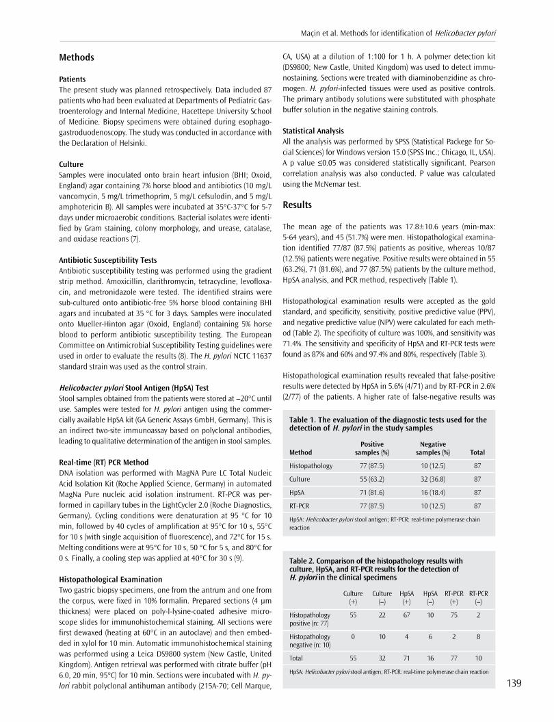
Methods
PatientsThe present study was planned retrospectively. Data included 87 patients who had been evaluated at Departments of Pediatric Gas-troenterology and Internal Medicine, Hacettepe University School of Medicine. Biopsy specimens were obtained during esophago-gastroduodenoscopy. The study was conducted in accordance with the Declaration of Helsinki.
CultureSamples were inoculated onto brain heart infusion (BHI; Oxoid, England) agar containing 7% horse blood and antibiotics (10 mg/L vancomycin, 5 mg/L trimethoprim, 5 mg/L cefsulodin, and 5 mg/L amphotericin B). All samples were incubated at 35°C-37°C for 5-7 days under microaerobic conditions. Bacterial isolates were identi-fied by Gram staining, colony morphology, and urease, catalase, and oxidase reactions (7).
Antibiotic Susceptibility TestsAntibiotic susceptibility testing was performed using the gradient strip method. Amoxicillin, clarithromycin, tetracycline, levofloxa-cin, and metronidazole were tested. The identified strains were sub-cultured onto antibiotic-free 5% horse blood containing BHI agars and incubated at 35 °C for 3 days. Samples were inoculated onto Mueller-Hinton agar (Oxoid, England) containing 5% horse blood to perform antibiotic susceptibility testing. The European Committee on Antimicrobial Susceptibility Testing guidelines were used in order to evaluate the results (8). The H. pylori NCTC 11637 standard strain was used as the control strain.
Helicobacter pylori Stool Antigen (HpSA) TestStool samples obtained from the patients were stored at −20°C until use. Samples were tested for H. pylori antigen using the commer-cially available HpSA kit (GA Generic Assays GmbH, Germany). This is an indirect two-site immunoassay based on polyclonal antibodies, leading to qualitative determination of the antigen in stool samples.
Real-time (RT) PCR MethodDNA isolation was performed with MagNA Pure LC Total Nucleic Acid Isolation Kit (Roche Applied Science, Germany) in automated MagNa Pure nucleic acid isolation instrument. RT-PCR was per-formed in capillary tubes in the LightCycler 2.0 (Roche Diagnostics, Germany). Cycling conditions were denaturation at 95 °C for 10 min, followed by 40 cycles of amplification at 95°C for 10 s, 55°C for 10 s (with single acquisition of fluorescence), and 72°C for 15 s. Melting conditions were at 95°C for 10 s, 50 °C for 5 s, and 80°C for 0 s. Finally, a cooling step was applied at 40°C for 30 s (9).
Histopathological ExaminationTwo gastric biopsy specimens, one from the antrum and one from the corpus, were fixed in 10% formalin. Prepared sections (4 μm thickness) were placed on poly-l-lysine-coated adhesive micro-scope slides for immunohistochemical staining. All sections were first dewaxed (heating at 60°C in an autoclave) and then embed-ded in xylol for 10 min. Automatic immunohistochemical staining was performed using a Leica DS9800 system (New Castle, United Kingdom). Antigen retrieval was performed with citrate buffer (pH 6.0, 20 min, 95°C) for 10 min. Sections were incubated with H. py-lori rabbit polyclonal antihuman antibody (215A-70; Cell Marque,
CA, USA) at a dilution of 1:100 for 1 h. A polymer detection kit (DS9800; New Castle, United Kingdom) was used to detect immu-nostaining. Sections were treated with diaminobenzidine as chro-mogen. H. pylori-infected tissues were used as positive controls. The primary antibody solutions were substituted with phosphate buffer solution in the negative staining controls.
Statistical Analysis All the analysis was performed by SPSS (Statistical Packege for So-cial Sciences) for Windows version 15.0 (SPSS Inc.; Chicago, IL, USA). A p value ≤0.05 was considered statistically significant. Pearson correlation analysis was also conducted. P value was calculated using the McNemar test.
Results
The mean age of the patients was 17.8±10.6 years (min-max: 5-64 years), and 45 (51.7%) were men. Histopathological examina-tion identified 77/87 (87.5%) patients as positive, whereas 10/87 (12.5%) patients were negative. Positive results were obtained in 55 (63.2%), 71 (81.6%), and 77 (87.5%) patients by the culture method, HpSA analysis, and PCR method, respectively (Table 1).
Histopathological examination results were accepted as the gold standard, and specificity, sensitivity, positive predictive value (PPV), and negative predictive value (NPV) were calculated for each meth-od (Table 2). The specificity of culture was 100%, and sensitivity was 71.4%. The sensitivity and specificity of HpSA and RT-PCR tests were found as 87% and 60% and 97.4% and 80%, respectively (Table 3).
Histopathological examination results revealed that false-positive results were detected by HpSA in 5.6% (4/71) and by RT-PCR in 2.6% (2/77) of the patients. A higher rate of false-negative results was
Table 1. The evaluation of the diagnostic tests used for the detection of H. pylori in the study samples
Positive Negative Method samples (%) samples (%) Total
Histopathology 77 (87.5) 10 (12.5) 87
Culture 55 (63.2) 32 (36.8) 87
HpSA 71 (81.6) 16 (18.4) 87
RT-PCR 77 (87.5) 10 (12.5) 87
HpSA: Helicobacter pylori stool antigen; RT-PCR: real-time polymerase chain reaction
Table 2. Comparison of the histopathology results with culture, HpSA, and RT-PCR results for the detection of H. pylori in the clinical specimens
Culture Culture HpSA HpSA RT-PCR RT-PCR (+) (−) (+) (−) (+) (−)
Histopathology 55 22 67 10 75 2 positive (n: 77)
Histopathology 0 10 4 6 2 8 negative (n: 10)
Total 55 32 71 16 77 10
HpSA: Helicobacter pylori stool antigen; RT-PCR: real-time polymerase chain reaction
Maçin et al. Methods for identification of Helicobacter pylori
139

obtained with the culture method (22/32; 68.7%). Culture method, RT-PCR, and HpSA tests were found to correlate with the Pearson correlation analysis (Table 4).
In 20 patients who had upper gastrointestinal bleeding, H. pylori was detected in all patients by RT-PCR, detected in 14 patients by culture, and detected in 16 patients by HpSA (Table 5).
Antibiotic susceptibility tests were performed on 48 out of the 55 culture positive samples. Among 48 isolates, none of them were resistant to amoxicillin and tetracycline. Resistance to clarithro-mycin was found in 28 (58.3%), metronidazole in 14 (29.2%), and levofloxacin in 4 (8.3%) of the isolates (Table 6).
Discussion
Currently, several different tests for the diagnosis of H. pylori in-fections exist. Each test has both advantages and disadvantages. Several studies have examined the diagnostic performance of in-vasive and non-invasive methods (10, 11). However, these studies demonstrated a lack of agreement. Discrepancies in the diag-nostic performance of different tests in different studies might be attributed to the selection of different methods as the gold standard.
Among various diagnostic methods, histopathological examina-tion of endoscopic biopsy specimens provides more information about the degree of inflammation and associated pathology (12). It is also the most reliable test in the presence of upper gastroin-testinal bleeding. Proton pump inhibitors should be stopped prior to gastroduodenoscopy, since they may decrease the sensitivity of the histopathological examination.
Routine cultivation is difficult to perform in microbiology labora-tories, since it is time consuming and hard to maintain microaero-philic conditions. However, bacterial growth in cultures provides definitive diagnosis and also enables antibiotic susceptibility test-ing to guide specific treatment. Gisbert and Abraria (13) reported three studies with culture sensitivity of 45% and specificity of 98%. Aktepe et al. (14) reported that the sensitivity of the culture meth-od is 61%, and specificity is 91%. In the present study, sensitivity was 71.4%, and specificity was 100% for culture. The low sensitivity and high specificity of the culture-based methods might be cor-related with inappropriate biopsy site and inadequate specimens. However, culture-based methods enable specific antibiotic suscep-tibility testing of the strains, thus providing important data espe-cially in populations with a high rate of drug resistance among H. pylori strains.
HpSA test, which has been introduced as a non-invasive diagnos-tic alternative, has the advantages of being relatively inexpensive, easy to perform, and can be used in pregnant women, children, and the elderly. It can easily be performed in routine laborato-ries as it does not require complicated laboratory facilities. In a Japanese study, the sensitivity and specificity of the HpSA test were 93.9% and 95.7%, respectively, when compared with histopatho-logical examination (15). A study from Turkey reported the HpSA test sensitivity as 72% and specificity as 67% (14). In our study, sen-sitivity and specificity were 87% and 60%, respectively. There were four false-positive samples using the HpSA test. Two of these false-positive samples were negative using culture and RT-PCR. The two other samples were negative using histopathology and culture, but positive with RT-PCR method. Thus, this false-positive may be at-tributed to the lack of detection by histopathology that was con-sidered as the gold standard.
The presence of H. pylori and specific antibiotic-resistant genes can be investigated by RT-PCR from gastric biopsy specimens. RT-PCR has a high sensitivity and specificity and can be used as a follow-up assessment after therapy (16). The contamina-tion may occur during the DNA extraction step or the presence of inanimate microorganisms residual chromosomal DNA, and this may lead to false-positive results (17). In our study, biopsy PCR studies had a sensitivity of 97.4%, specificity of 80%, NPV
Table 5. Detection of H. pylori infection in patients with upper gastrointestinal bleeding
Positive samples Negative samples Method (%) (%) Total
Histopathology 20 (100) 0 20
Culture 14 (70) 6 (30) 20
HpSA 16 (80) 4 (20) 20
RT-PCR 20 (100) 0 20
HpSA: Helicobacter pylori stool antigen; RT-PCR: real-time polymerase chain reaction
Table 6. Antimicrobial resistance of the tested H. pylori strains (n=48)
Antibiotics Resistance, n (%)
Amoxicillin 0
Clarithromycin 28/48 (58.3%)
Levofloxacin 4/48 (8.3%)
Metronidazole 14/48 (29.2%)
Tetracycline 0
Table 3. Comparison of the results of the H. pylori diagnostic methods when histopathology was considered as the gold standard
Sensitivity Specificity PPV NPV Method (%) (%) (%) (%)
Culture 71.4 100 100 31.2
HpSA 87 60 94.4 37.5
RT-PCR 97.4 80 97.4 10.4
PPV: positive predictive value; NPV: negative predictive value; HpSA: Helicobacter pylori stool antigen; RT-PCR: real-time polymerase chain reaction
Table 4. Correlation of H. pylori test results to histopathology results
Method False-positive False-negative ra P
Culture 0 22 0.472 <0.0001
HpSA 4 10 0.387 0.180
RT-PCR 2 2 0.774 <1
aCorrelation is significant at the 0.01 level.HpSA: Helicobacter pylori stool antigen; RT-PCR: real-time polymerase chain reaction
İstanbul Med J 2018; 19: 138-42
140

of 10.4%, and PPV of 97.4%. There were only two false-positive samples by RT-PCR. These two samples were also positive by HpSA test. Thus, obtaining positive results with two different methods provided strong information that this patient was in-fected with H. pylori, and the “false” false-positive evaluation in that specific case was attributed to choosing histopathology as the gold standard.
Many conventional H. pylori diagnostic methods show a significant decrease in their sensitivity in patients with upper gastrointestinal bleeding. However, in cases of upper gastrointestinal bleeding, PCR techniques and histopathological examination are more reli-able than rapid urease test, HpSA test, or culture (5, 18). A Korean study that evaluated the patients with gastrointestinal bleeding (n=157) found that sensitivity and specificity were 92.5% and 96% for histopathological examination, 40% and 100% for culture, and 97% and 56% for serology. The HpSA method showed relatively high sensitivity, but cannot be recommended as the primary diag-nostic method in the bleeding situation, because of its low speci-ficity (19). In our study, H. pylori was correctly detected by RT-PCR and histopathological examination in 20 patients who had upper gastrointestinal bleeding, culture in 14, and HpSA in 16. The find-ings of the current study suggested that histopathological exami-nation and RT-PCR assay were the most appropriate methods for the detection of H. pylori in patients with upper gastrointestinal bleeding.
Antibiotic susceptibility testing provides valuable information for choosing the right treatment. Resistance to metronidazole is re-ported to be between 15.9% and 77.9% (20, 21). In accordance with the literature, metronidazole resistance was 29.2% in the current study. Resistance to clarithromycin was reported to be 34% in Aus-tria, 54.6% in Spain, and 30.1% in Turkey (22-24). Clarithromycin resistance was also found to be high (58.3%) in the present study. Resistance to amoxicillin and tetracycline is very rare. Agudo et al. (23) revealed no resistance to amoxicillin, rifampicin, and tetra-cycline. Vécsei et al. (20) reported tetracycline resistance as 0.9%; however, they did not report any amoxicillin resistance. In our study, no resistance to amoxicillin and tetracycline was found. The frequency of levofloxacin resistance is reported to be 5.9%-18.2% in Turkey, 22.1% in Italy, and 34.5% in China (25-28). In our study, levofloxacin resistance was also found to be compatible with previ-ous studies from Turkey (8.3%).
Conclusion
There are a variety of tests available for the diagnosis of H. pylori. In cases where endoscopy could not be performed, non-invasive, simple, rapid, and practical HpSA test with acceptable results can be used to provide diagnosis and to monitor treatment. RT-PCR method has a high sensitivity and specificity in comparison with histopathological examination accepted as the gold standard. Both RT-PCR and histopathological examination are also reliable diagnostic methods in patients with upper gastrointestinal bleed-ing. When available, culture should be performed for antibiotic susceptibility tests, especially in the case of treatment failure. Thus, each laboratory should better establish its own diagnostic algorithm for the accurate and appropriate diagnosis of H. pylori infection in their patient population, according to their laboratory facilities and clinical setting.
Ethics Committee Approval: Since our study was retrospective and we compared the tests previously used for Helicobacter pylori in our hospital, no ethics committee approval was applied.
Informed Consent: Informed consent is not obtained due to the retrospec-tive nature of this study.
Peer-review: Externally peer-reviewed.
Author contributions: Concept - S.M, Y.A; Design -S.M, A.A, B.Ş., Y.A.; Su-pervision - Y.A, B.Ş, H.Ö, T.K; Materials - C.S., D.O., SM; Data Collection and/ or Processing - A.A., B.Ş., C.S., D.O.; Analysis and/or Interpretation - S.M., Y.A.; Literature Review - S.M., B.Ş., Y.A.; Writing - S.M., A.A., B.Ş., C.S., D.O., H.O., T.K., Y.A.; Critical Review - A.A., B.Ş., Y.A.
Conflict of Interest: Authors have no conflicts of interest to declare.
Financial Disclosure: The authors declared that this study has received no financial support
Etik Komite Onayı: Çalışmamız retrospektif olduğu ve hastanemizde daha önceden Helicobacter pylori tanısında kullanılan testler karşılaştırıldığı için, etik komite onayı başvurusu yapılmamıştır.
Hasta Onamı: Bu çalışma retrospektif veriler kullanılarak yapıldığından hasta onamı alınmamıştır.
Hakem Değerlendirmesi: Dış Bağımsız.
Yazar Katkıları: Fikir - S.M, Y.A; Tasarım - S.M, A.A, B.Ş., Y.A.; Denetleme - Y.A, B.Ş, H.Ö, T.K; Gereçler - C.S., D.O., S.M.; Veri Toplanmasi ve/veya İşle-mesi - A.A., B.Ş., C.S., D.O.; Analiz ve/veya Yorum - S.M., Y.A.; Literatur Tara-masi - S.M., B.Ş., Y.A.; Yaziyi Yazan - S.M., A.A., B.Ş., C.S., D.O., H.O., T.K.,Y.A..; Elestirel Inceleme - A.A., B.Ş., Y.A.
Çıkar Çatışması: Yazarların beyan edecek çıkar çatışması yoktur.
Finansal Destek: Yazarlar bu çalışma için finansal destek almadıklarını beyan etmişlerdir.
References
1. Dunn BE, Cohen H, Blaser MJ. Helicobacter pylori. Clin Microbiol Rev 1997; 42: 720-41.
2. Peterson WL. Review article: Helicobacter pylori and gastric adeno-carcinoma. Aliment Pharmacol Ther 2002; 16: 40-6. [CrossRef]
3. Kuipers EJ, Thijs JC, Festan HP. The prevalence of Helicobacter pylori in peptic ulcer disease. Aliment Pharmacol Ther 1995; 9: 59-69
4. Graham DY, Hepps KS, Ramirez FC, Lew GM, Saeed ZA. Treatment of Helicobacter pylori reduces the rate of rebleeding in peptic ulcer dis-ease. Scand J Gastroenterol 1993; 28: 939-42. [CrossRef]
5. Saez J, Belda S, Santibanez M, Rodriguez JC, Sola-Vera J, Galiana A, et al. Real-time PCR for diagnosing Helicobacter pylori infection in patients with upper gastrointestinal bleeding: comparison with other classical diagnostic methods. J Clin Microbiol 2012; 50: 3233-7. [CrossRef]
6. Bonamico M, Strappini PM, Bonci E, Ferri M, Crisogianni M, Guido M et al. Evaluation of stool antigen test PCR on oral samples and serol-ogy for the noninvasive detection of Helicobacter pylori infection in children. Helicobacter 2004; 9: 69-76. [CrossRef]
7. Vibrios, Campylobacter, Helicobacter, and associated bacteria in Jawetz, Melnick & Adelberg's Medical Microbiology. 25th edition. Mc Graw Hill Companies, International Edition. 2010; 17, 235-43.
8. The European Committee on Antimicrobial Susceptibility Testing. Breakpoint tables for interpretation of MICs and zone diameters. Ver-sion 6.0, 2016. http://www.eucast.org.
9. Matsumura M, Hikiba Y, Ogura K, Togo G, Tsukuda I, Ushikawa K, et al. Rapid detection of mutations in the 23S rRNA gene of Helicobacter
Maçin et al. Methods for identification of Helicobacter pylori
141

pylori that confers resistance to clarithromycin treatment to the bac-terium. J Clin Microbiol 2001; 39: 691-5. [CrossRef]
10. Cutler AF, Havstad S, Ma CK, Blaser MJ, Perez-Perez GI, Schubert TT. Accuracy of invasive and noninvasive tests to diagnose Helicobacter pylori infection. Gastroenterology 1995; 109: 136-41. [CrossRef]
11. Vaira D, Malfertheiner P, Mégraud F, Axon AT, Deltenre M, Gasbarrini G et al. Noninvasive antigen-based assay for assessing Helicobacter pylori eradication: a European multicenter study. The European Heli-cobacter pylori HpSA Study Group. Am J Gastroenterol 2000; 95: 925-9. [CrossRef]
12. Tepes B, Ferlan-Marolt V, Jutersek A, Kavcic B, Zaletel-Kragelj L. In-terobserver agreement in the assessment of gastritis reversibility af-ter Helicobacter pylori eradication. Histopathology 1999; 34: 124-33. [CrossRef]
13. Gisbert JP, Abraira V. Accuracy of Helicobacter pylori diagnostic tests in patients with bleeding peptic ulcer: a systematic review and meta-analysis. Am J Gastroenterol 2006; 101: 848-63. [CrossRef]
14. Aktepe OC, Çiftçi IH, Şafak B, Uslan I, Dilek FH. Five methods for detec-tion of Helicobacter pylori in Turkish population. World J Gastroen-terol 2011; 17: 5172-6. [CrossRef]
15. Ohkura R, Miwa H, Murai T, Nagahara A, Ohta K, Sato K, et al. Useful-ness of a novel enzyme immunoassay for the detection of Helicobacter pylori in feces. Scand J Gastroenterol 2000; 35: 49-53. [CrossRef]
16. Agudo S, Pérez-Pérez G, Alarcón T, López-Brea M. High prevalence of clarithromycin-resistant Helicobacter pylori strains and risk factors associated with resistance in Madrid, Spain. J Clin Microbiol 2010; 48: 3703-7 [CrossRef]
17. He Q, Wang JP, Osato M, Lachman LB. Real-time quantitative PCR for detection of Helicobacter pylori. J Clin Microbiol 2002; 40: 3720-8. [CrossRef]
18. Grinó P, Pascual S, Such J, Casellas JA, Niveiro M, Andreu M et al. Com-parison of diagnostic methods for Helicobacter pylori infection in patients with upper gastrointestinal bleeding. Scand J Gastroenterol 2001; 36: 1254-8. [CrossRef]
19. Choi YJ, Kim N, Lim J, Jo SY, Shin CM, Lee HS, et al. Accuracy of diag-nostic tests for Helicobacter pylori in patients with peptic ulcer bleed-ing. Helicobacter 2012; 17: 77-85. [CrossRef]
20. Vécsei A, Kipet A, Innerhofer A, Graf U, Binder C, Gizci H, et al. Time trends of Helicobacter pylori resistance to antibiotics in children living in Vienna, Austria. Helicobacter 2010; 15: 214-20. [CrossRef]
21. Boyanova L, Mentis A, Gubina M, Rozynek E, Gosciniak G, Kalenic S, et al. The status of antimicrobial resistance of Helicobacter pylori in eastern Europe. Clin Microbial Infect 2002; 8: 388-96. [CrossRef]
22. Mégraud F. H. pylori antibiotic resistance: prevalence, importance, and advances in testing. Gut 2004; 53: 1374-84. [CrossRef]
23. Agudo S, Alarcón T, Cibrelus L, Urruzuno P, Martínez MJ, López-Brea M. High percentage of clarithromycin and metronidazole resistance in Helicobacter pylori clinical isolates obtained from Spanish children. Rev Esp Quimioter 2009; 22: 88-92.
24. Maçin S, Demir H, Özen H, Yüce A, Akyön Y. Determination of Heli-cobacter pylori antibiotic resistance patterns in pediatric gastroen-terology patients: the Hacettepe experience. Turk J Pediatr 2015; 57: 254-7.
25. Kantarceken B, Yıldırım B, Karıncaoğlu M, Aladağ M, Hilmioğlu F. He-licobacter pylori and antibiotic resistance. Turk J Gastroenterol 2000; 11: 141-5.
26. Çağdaş U, Otağ F, Tezcan S, Sezgin O, Aslan G, Emekdaş G. [Detec-tion of Helicobacter pylori and Antimicrobial Resistance in Gastric Biopsy Specimens]. Mikrobiyol Bul 2012; 46: 398-409. [Article in Turkish]
27. Saracino IM, Zullo A, Holton J, Castelli V, Fiorini G, Zaccaro C, et al. High prevalence of primary antibiotic resistance in Helicobacter py-lori isolates in Italy. J Gastrointestin Liver Dis 2012; 21: 363-5.
28. Zhang YX, Zhou LY, Song ZQ, Zhang JZ, He LH, Ding Y. Primary anti-biotic resistance of Helicobacter pylori strains isolated from patients with dyspeptic symptoms in Beijing: a prospective serial study. World J Gastroenterol 2015; 21: 2786-92. [CrossRef]
Cite this article as: Maçin S, Alp A, Şener B, Sökmensüer C, Orhan D, Özen H. Comparison of culture, Real- time-PCR, ELISA, and histopathological examination methods for identification of Helicobacter pylori. İstanbul Med J 2018; 19: 138-42.
İstanbul Med J 2018; 19: 138-42
142
Introduction
Peritoneal carcinomatosis (PC) which is called advanced stage of abdominal cancer continues to be a problem that not fully solved at the present time. Due to both medical cost burdens and infe-rior treatment results of oncologic diseases with palliative chemo-radiotherapy or supportive care in advance, in 1989 Sugarbaker et al. (1) presented cytoreductive surgery (CRS) and hyperthermic intraperitoneal chemotherapy (HIPEC) as a stunning method for appropriate patients with PC. Initially, this technique had excessive treatment-related morbidity and mortality with a major concern. However, completion of learning curve in the experienced centers resulted an overall decreasein postoperative morbidity and mortality, as shown in the recent studies (2, 3).
Regardless of enhanced survival with admissible morbidity and mortality of surgery, quality of life (QoL) in patients with PC is still controversial. Due to broad resection and possible chemotherapy toxicity, changes in QoL patients with CRS and HIPEC could be more than simple surgery.
These studies were performed in Western and Asian population;nevertheless, have not been ap-plied in Turkish population. Thus, in the present study, we aimed to share out QoL results of our
Short-term Quality of Life after Cytoreductive Surgery and Hyperthermic Intraperitoneal ChemotherapySitoredüktif Cerrahi ile İntraperitoneal Sıcak Kemoterapinin Erken Dönem Yaşam Kalitesi Üzerine Etkisi
Amaç: Sitoredüktif cerrahi (CRS) ile birlikte hipertermik intraperitone-al sıcak kemoterapi (HIPEC) ülkemizde ileri merkezlerde son yıllarda giderek artan sıklıkla yapılmaktadır. Çalışmamızın amacı peritoneal karsinomatozis (PK) teşhisiyle ameliyat edilen hastaların hayat kalite-sini (QoL) değerlendirmektir.
Yöntemler: Çalışmamızda 2012-2015 yılları arasında PK tanısıy-la SRC+HIPEC yapılan 42 hastanın ameliyat sonrası hayat kaliteleri ile SRC+HIPEC olmaksızın onkolojik cerrahi sonrası takip edilen 92 hastanın hayat kaliteleri karşılaştırıldı. Avrupa Kanser Araştırma ve Tedavi Organizasyonu (EORTC) tarafından hazırlanan QLQ-C30 soru-ları tüm hastalarımıza ameliyat sonrası 6. ayda uygulandı ve her iki grup karşılaştırıldı. İstatistiksel analiz one sample t-test ile yapılarak, p<0.05 anlamlı kabul edildi.
Bulgular: 42 hastanın 30’u (%71.4) bayan, 12’si (% 28.5) erkek idi. Has-taların yaş ortalaması 52 (range, 69-22) idi. PK orijinleri 15hastada (%35.7) kolorektal malignite, 12hastada (%28.5) over kanseri, 6hasta-da (%14.2) mide malignitesi, 3hastada (%7.1) pseudomiksoma perito-nei, 3hastada (%7.1) sarkom ve 3hastada (%7.1) da mezenkimal tümör olarak bulundu. Ortalama intraoperatif peritoneal karsinomatöz in-deksi (PCI) 15 (range 6-29) olarak bulundu. Rezeksiyon tamlık skoru (CCS) 0 ve 1 olarak bulundu. Tüm hastalarda ortalama takip süresi 12 ay (range 2-26) idi. Her iki grup arasında fonksiyonel ve semptom skorları açısından istatiksel fark saptanmadı (p>005).
Sonuç: SRC+HIPEC olmaksızın onkolojik cerrahi yapılan hastalar ile karşılaştırıldığında,PK sebebiyle SRC+HIPEC yapılan olguların kısa dö-nemde yaşam kalitelerinin (QoL) benzer olduğu bulunmuştur.
Anahtar Kelimeler: Sitoredüktif cerrahi (CRS), Hipertermik intraperi-toneal kemoterapi (HIPEC), Turkish population, Yaşam kalitesi (QoL)
Introduction: Cytoreductive surgery (CRS) and hyperthermic intra-peritoneal chemotherapy (HIPEC) have been increasingly performed in many advanced centers of our country. Short-term quality of life (QoL) after the CRS with HIPEC was investigated in Turkish population. The purpose of our study was to investigate the QoL in patients ope-rated for peritoneal carcinomatosis (PC).
Methods: Retrospectively collected data from 42 consecutive pati-ents, who had undergone CRS and HIPEC, and 92 abdominal malig-nancy patients after oncological surgery without CRS and HIPEC were selected in the study between 2012 and 2015. A Turkish version of the European Organization for Research and Treatment of Cancer ques-tionnaire (QLQ-C30) was used at the 6th month following surgery for the QoL assessment. Statistical analysis was done with one sample t-test, and p<0.05 was accepted as statistically significant.
Results: In total, 42 CRS+HIPEC patients were analyzed; of which, 30 (71.4%) were female and 12 (28.5%) were male. The median age was 52 years (range, 22-69). CRS with HIPEC was performed for colorec-tal carcinoma in 15 patients (35.7%), ovarian cancer in 12 patients (28.5%), gastric cancer in 6 patients (14.2%), pseudomixsoma peritonei in 3 patients (7.1%), sarcoma in 3 patients (7.1%), and mesenchymal tumor in 3 patients (7.1%). The median intraoperative peritoneal car-cinomatosis index score was 15 (6-29), whereas the completeness of the cytoreductive score was 0 and 1. The median follow-up period for all patients was 12 months (range, 6-22 months). The functional and symptom scores were equal between the CRS with HIPEC and without CRS+HIPEC cancer patients (p>0.05).
Conclusion: Short-term QoL after the CRS with HIPEC patients and without the CRS+HIPEC oncologic patients is found to be similar in Turkish population.
Keywords: Cytoreductive surgery (CRS), hyperthermic intraperitoneal chemotherapy (HIPEC), Turkish population, quality of life (QoL)
Abst
ract
/ Ö
z
This study was presented 20th National Surgery Kongress (April 13-17, 2016, Antalya, Türkiye)
1Deapartment of Oncological Surgery, Çukurova University School of Medicine, Adana, Türkiye2Department of General Surgery, Kanuni Sultan Süleyman Training and Research Hospital, İstanbul, Türkiye3Department of Medical Oncology, Çukurova Üniversitesi School of Medicine, Adana, Türkiye
Address for Correspondence/Yazışma Adresi:İnanc Şamil SarıcıE-mail: [email protected]
Received/Geliş Tarihi: 28.03.2017
Accepted/Kabul Tarihi: 23.11.2017
© Copyright 2018 by Available online at istanbulmedicaljournal.org
© Telif Hakkı 2018 Makale metnine istanbultipdergisi.org web sayfasından ulaşılabilir.
Original Investigation/Orijinal Araştırmaİstanbul Med J 2018; 19: 143-6DOI: 10.5152/imj.2018.04828
Özgül Düzgün1, İnanç Şamil Sarıcı2, Serkan Gökçay3
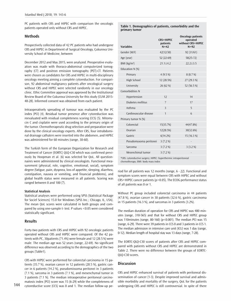
PC patients with CRS and HIPEC with comparison the oncologic patients operated only without CRS and HIPEC.
Methods
Prospectively collected data of 42 PC patients who had undergone CRS and HIPEC in Department of Surgical Oncology, Çukurova Uni-versity School of Medicine, between
December 2012 and May 2015, were analyzed. Preoperative evalu-ation was made with thoraco-abdominal computerized tomog-raphy (CT) and positron emission tomography (PET)-CT. Patients were chosen as candidates for CRS and HIPEC in multi-disciplinary oncology meeting aiming a complete cytoreduction. For compari-son, 92 abdominal malignancy patients after oncological surgery without CRS and HIPEC were selected randomly in our oncology clinic. Ethic Committee approval was approved by the Institutional Review Board of the Cukurova University for this study (CUM 2015-48-28). Informed consent was obtained from each patient.
Intraoperatively spreading of tumour was evaluated by the PC index (PCI) (4). Residual tumor presence after cytoreduction was reevaluated with residual completeness scoring (CCS) (5). Mitomy-cin C and cisplatin were used according to the primary origin of the tumor. Chemotherapeutic drug selection and preparation were done by the clinical oncology experts. After CRS, four intrabdomi-nal drainage cathaters were inserted into the abdomen, and HIPEC was administered for 60 minutes (range, 30-60).
The Turkish form of the European Organization for Research and Treatment of Cancer (EORTC) QLQ-C30 which was confirmed previ-ously by Hoopman et al. (6) was selected for QoL. All question-naires were administered by clinical oncologists. Functional mea-surement (physical, role, cognitive, emotional, social), symptom degree (fatigue, pain, dyspnea, loss of appetite, sleeping, diarrhea, constipation, nausea or vomiting, and financial problems), and global health status were measured in all patients. Scoring was ranged between 0 and 100 (7).
Statistical AnalysisStatistical analyses were performed using SPSS (Statistical Package for Sociel Sciences) 15.0 for Windows (SPSS Inc.; Chicago, IL, USA). The mean QoL scores were calculated in both groups and com-pared by using one-sample t- test. P values <0.05 were considered statistically significant.
Results
Forty-two patients with CRS and HIPEC with 92 oncologic patients operated without CRS and HIPEC were compared. Of the 42 pa-tients with PC, 30patients (71.4%) were female and 12 (28.5 %) were male. The median age was 52 years (range, 22-69). No significant difference was observed according to the demographics of the two groups (Table1).
CRS with HIPEC were performed for colorectal carcinoma in 15 pa-tients (35.7 %), ovarian cancer in 12 patients (28.5 %), gastric can-cer in 6 patients (14.2 %), pseudomixsoma peritonei in 3 patients (7.1 %), sarcoma in 3 patients (7.1 %), and mesenchymal tumor in 3 patients (7.1 %). The median intraoperative peritoneal carcino-matosis index (PCI) score was 15 (6-29) while the completeness of cytoreductive score (CCS) was 0 and 1. The median follow-up pe-
riod for all patients was 12 months (range, 6 - 22). Functional and symptom scores were equal between CRS with HIPEC and without CRS+HIPEC cancer patients (p>0.05). The ECOG performance status of all patients was 0 or 1.
Without PC group included colorectal carcinoma in 44 patients (47.8 %), ovarian cancer in 30 patients (32.6 %), gastric carcinoma in 15 patients (16.3 %), and sarcomas in 3 patients (3.2%).
The median duration of operation for CRS and HIPEC was 480 min-utes (range, 310-565) and that for without CRS and HIPEC group was 110minutes (range, 80-160) (p<0.001). The median PCI was 15 (range, 6-29). There were 39 patients in CCS 0 and 3 patients in CCS 1. The median admission in intensive care unit (ICU) was 1 day (range, 0-12). Median length of hospital stay was 13 days (range, 7-28).
The EORTC-QLQ-C30 scores of patients after CRS and HIPEC com-pared with patients without CRS and HIPEC are demonstrated in Table 2. There were no difference between the groups of EORTC- QLQ-C30 scores.
Discussion
CRS and HIPEC enhanced survival of patients with peritoneal dis-semination of cancer (1-3). Despite improved survival and admis-sible morbidity and mortality of the surgery, QoL for the patients undergoing CRS and HIPEC is still controversial. In spite of there
İstanbul Med J 2018; 19: 143-6
144
Table 1. Demographics of patients, comorbidity and the primary tumor
Oncology patients CRS+HIPEC operated patients without CRS+HIPEC Variables N=42 N=92
Gender (M/F) 42(12/30) 92 (31/61)
Age (year) 52 (22-69) 58(25-72)
BMI (kg/m2) 21.1±4.2 22.2±3.5
Education N (%)
Primary 4 (9.5 %) 8 (8.7 %)
High School 12 (28.5%) 27 (29.3 %)
University 26 (62 %) 52 (56.5 %)
Comorbidities N
Hypertension 12 14
Diabetes mellitus 7 17
Asthma 3 5
Cardiovascular disease 1 6
Primary tumor N (%)
Colorectal 15(35.7%) 44(47.8%)
Ovarian 12(28.5%) 30(32.6%)
Gastric 6(14.2%) 15 (16.3 %)
Pseudomyxoma peritonei 3 (7.2 %) -
Sarcoma 3 (7.2 %) 3 (3.2 %)
Mesenchimal tumor 3 (7.2 %) -
*CRS: cytoreductive surgery; HIPEC: hyperthermic intraperitoneal chemotherapy; BMI: body mass index

Düzgün et al. Quality of Life after CRS HIPEC
145
have been many articles with respect to QoL scoring calculated for patients with CRS and HIPEC, there is no information about these scores in Turkish PC patients. To our knowledge, this is the first study from a Turkish university hospital referral oncology unit that assesses QoL outcomes who undergone CRS with HIPEC.
There have been recent publications in the literature about EORTC QLQ C-30 to assess the QoL for PC patients. Schmidt et al. (8) evalu-ated the QoL of patients who underwent CRS combined with HIPEC, and found that QoL is associated with an increased morbidity and mortality according to the difficulty of surgery. On the other hand, in the study of Alves et al. (9) applied EORTC QLQ C-30 questionnaire to their PC patients who undergone CRS with HIPEC. The authors found an improvement in QoL at one year following the procedure. Furthermore, Tan et al. (10) alcompared CRS plus HIPEC patients with their cancer free patients. They found cognitive functioning scores and fatigue scores better in their CRS+HIPEC patients.
In the present study, the Turkish version of EORTC QLQ C-30 was eval-uated between PC patients with CRS and HIPEC and patients without CRS and HIPEC, 6 months after surgery, and no significant difference was found between two groups. However, our study have some limi-tations. First one is its retrospective design. Lack of the patients basic QoL scores for reference values after CRS and HIPEC is the second limitation. Third limitation is the variety of primary cancer origins. Compared to the population of oncology patients operated without CRS and HIPEC in our center, those patients that underwent CRS and HIPEC had smilar scores in global health, physical functioning, emo-tional functioning and social functioning.
Developing hospital care, advances in ICU for postoperative pa-tient care, and a significant increase in the numbers of specialized oncology units in private sector as well as in the state hospitals can
be the reason of no difference of QoL. On the other hand, success-ful tumor free surgery, lack of serious morbidity, adequate enteral nutrition, achievement of longer disease-free survival and rapid return to normal life of the oncology patients are important fac-tors affecting QoL. This is likely due to the fact that many of the pa-tients after cancer surgery in Turkey starting to take chemotherapy in this period, quick return to their normal economic, social and physical lives that can be explained why scores are better in both CRS and HIPEC and without CRS and HIPEC groups.
Conclusion
Short term QoL after CRS with HIPEC and oncology patients op-erated without CRS and HIPEC seems to be equal in our Turkish population.
Ethics Committee Approval: Ethic Committee approval was recieved by the Institutional Review Board of the Çukurova University for this study (CUM 2015-48-28).
Informed Consent: Informed consent was obtained from the patients who participated in this study
Peer-review: Externally peer-reviewed.
Author contributions: Concept - Ö.D. İ.Ş.S.; Design - İ.Ş.S, Ö.D., S.G.; Super-vision İ.Ş.S .; Resource - Ö.D., S.G. Materials - Ö.D., S.G.; Data Collection and/or Processing - Ö.D., S.G.; Analysis and/or Interpretation - İ.Ş.S., Ö.D.; Lit-erature Search - İ.Ş.S, Ö.D; Writing - İ.Ş.S., Ö.D.; Critical Reviews - İ.Ş.S., Ö.D.
Conflict of Interest: Authors have no conflicts of interest to declare.
Financial Disclosure: The authors declared that this study has received no financial support.
Table 2. Comparison of EORTC QLQ C-30 scoresin patients with CRS and HIPEC and oncology patients operated without CRS and HIPEC
Oncology patients operated EORTC QLQ-C30 CRS+HIPEC patients without CRS+HIPECparameters (N=42) (N=92) P
Physical functioning 78(70-87) 84 (75-90) 0.524
Role functioning 81(76-91) 85 (79-93) 0.406
Cognitive functioning 87(81-94) 89 (85-96 0.305
Emotional functioning 82(75-88) 84 (73-90) 0.555
Social functioning 81(75-90) 82 (75-90) 0.465
Global quality of life 65(57-79) 68 (55-82) 0.343
Fatigue 14(10-22) 16 (11-25) 0.245
Pain 15(6-24) 16 (7-25) 0.324
Nausea and vomiting 8(2-15) 9 (1-17) 0.405
Dyspnea 10(3-17) 9 (2-17) 0.325
Insomnia 19(8-28) 17 (8-26) 0.308
Appetite loss 10(2-18) 9 (3-17) 0.243
Constipation 15(6-22) 13 (5-21) 0.543
Diarrhea 9 (3-15) 8(2-17) 0.550
Financial problems 23(11-35) 24 (11-37) 0.605
*CRS: cytoreductive surgery; HIPEC: hyperthermic intraperitoneal chemotherapy; EORTC QLQ-C30: European Organization for research and treatment of cancer quality of life

Etik Komite Onayı: Bu çalışma için etik komite onayı Çukurova Üniversitesi Kurumsal İnceleme Kurulu’ndan alınmıştır (CUM 2015-48-28).
Hasta Onamı: Hasta onamı bu çalışmaya katılan hastalardan alınmıştır.
Hakem Değerlendirmesi: Dış Bağımsız.
Yazar Katkıları: Fikir - Ö.D., İ.Ş.S; Tasarım - İ.Ş.S, Ö.D., S,G.; Denetleme - İ.Ş.S; Kaynaklar - Ö.D., S.G.; Malzemeler - Ö.D., S.G.; Veri Toplanması ve/veya işlemesi - Ö.D., S.G.; Analiz ve/veya Yorum - İ.Ş.S., Ö.D.; Literatür taraması - İ.Ş.S, Ö.D; Yazıyı Yazan - İ.Ş.S., Ö.D.; Eleştirel İnceleme - İ.Ş.S., Ö.D.
Çıkar Çatışması: Yazarlar çıkar çatışması bildirmemişlerdir.
Finansal Destek: Yazarlar bu çalışma için finansal destek almadıklarını beyan etmişlerdir.
References
1. Sugarbaker PH, Cunliffe WJ, Belliveau J, de Bruijn EA, Graves T, Mullins RE, et al. Rationale for integrating early postoperative intraperitoneal chemotherapy into the surgical treatment of gastrointestinal cancer. Semin Oncol 1989; 16: 83-97.
2. Chua TC, Yan TD, Saxena A, Morris DL. Should the treatment of peri-toneal carcinomatosis by cytoreductive surgery and hyperthermic intraperitoneal chemotherapy still be regarded as a highly morbid procedure?: a systematic review of morbidity and mortality. Ann Surg 2009; 249: 900-7. [CrossRef]
3. Wilson KA, Dowling AJ, Abdolell M, Tannock IF. Perception of qual-ity of life by patients, partners and treating physicians. Qual Life Res 2000; 9: 1041-52. [CrossRef]
4. Jacquet P, Sugarbaker PH. Current methodologies for clinical assess-ment of patients with peritoneal carcinomatosis. J Exp Clin Cancer Res 1996; 15: 49-58.
5. Esquivel J, Elias D, Baratti D, Kusamura S, Deraco M. Consensus state-ment on the loco regional treatment of colorectal cancer with perito-neal dissemination. J Surg Oncol 2008; 98: 263-7. [CrossRef]
6. Hoopman R, Muller MJ, Terwee CB, Aaronson NK. Translation and validation of the EORTC QLQ-C30 for use among Turkish and Moroc-can ethnic minority cancer patients in the Netherlands. Eur J Cancer 2006; 42: 1839-47. [CrossRef]
7. Fayers PM, Aaronson NK, Bjordal K, Curran D, Groenvold M. The EORTC QLQ-C30 Scoring Manual. 2nd Ed., Brussels: In: European Orga-nization for Research and Treatment of Cancer; 1999.
8. Schmidt U, Dahlke MH, Klempnauer J,Schlitt HJ, Piso P. Perioperative morbidity and quality of life in long-term survivors following cytore-ductive surgery and hyperthermic intraperitoneal chemotherapy. Eur J Surg Oncol. 2005; 31: 53-8. [CrossRef]
9. Alves S, Mohamed F, Yadegarfar G, Youssef H, Moran BJ. Prospective longitudinal study of quality of life following cytoreductive surgery and intraperitoneal chemotherapy for pseudomyxoma peritonei. Eur J Surg Oncol 2010; 36: 1156-61. [CrossRef]
10. Tan WJ, Wong JF, Chia CS, Tan GH, Soo KC, Teo MC. Quality of life after cytoreductive surgery and hyperthermic intraperitoneal che-motherapy: an Asian perspective. Ann Surg Oncol 2013; 20: 4219-23. [CrossRef]
Cite this article as: Düzgün Ö, Sarıcı İŞ, Gökçay S. Short-term Quality of Life after Cytoreductive Surgery and Hyperthermic Intraperitoneal Chemotherapy. İstanbul Med J 2018; 19: 143-6.
İstanbul Med J 2018; 19: 143-6
146
Introduction
Endometriosis is the presence of endometrial glands and stroma outside the uterine cavity af-fecting mostly women of reproductive age and is encountered in 10%-15% of women during their reproductive age. This disease is usually manifested by dysmenorrhea (62.2%), infertility (14%), and chronic pelvic pain (13.3%) (1).
Endometriosis can be divided into three types: superficial endometriosis, deep endometriosis, and ovarian endometriosis called endometrioma. Endometrioma is the formation of the cyst con-taining chocolate-colored content and is encountered in 17%-44% of patients with endometriosis. It is hypothesized that endometriomas arise as coelemic metaplasia of the ovarian epithelium or invaginations of the inverted ovarian cortex after implantation of the endometriotic foci on the ovarian surface (2).
There is no consensus about definitive treatment of endometriomas to relieve pain, optimize fer-tility, and delay recurrence. At present, the optimal treatment is the surgical excision of the ovar-ian endometriomas. Fenestration or aspiration of endometriomas is considered an alternative to minimize damage to the ovarian reserve (3). Although laparoscopic excision of the entire cyst wall is believed to be more effective than excision by laparotomy, ovarian injury is inevitable in lapa-roscopic surgery (4). Characteristics of the cyst, surgical method, and experience of the surgeon affect the ovarian damage, particularly in repeated operations for recurrent endometriomas (5).
Endometriomas tend to recur after the operation, and repeated surgery for recurrent endome-triomas is harmful to the ovarian reserve. Guo (6) reported that the recurrence rate was 21.5% and 40%-50% at 2 and 5 years, respectively, after primary surgery. Repeated surgery for these recurrent cysts is harmful to the ovarian reserve, especially because of dense adhesions. There is a debate
Is there Any Difference between the Patients with Primary Endometriomas and those with Recurrent Endometriomas?Reküren Endometriyoma Nedeniyle Opere Olan Hastalar ile İlk Kez Opere Olan Hastalar Arasında Herhangi Bir Fark Var mıdır?
Amaç: Reküren endometriyoma nedeniyle tekrarlayan cerrahi, over rezervine zarar vermektedir. Çalışmamızın amacı, endometriyoması olan hastaların demografik ve klinik özelliklerine bakarak rekürensini öngören bir belirteç olup olmadığını tanımlamaktır.
Yöntemler: Mayıs 2014 ve Mayıs 2016 tarihleri arasında endometri-yoma nedeniyle kliniğimizde opere edilmiş 151 hasta bu retrospektif kohort çalışmaya dahil edildi. Hastalar önceden endometriyoma ne-deniyle opere olup olmamalarına göre gruplandırıldı ve demografik ve klinik özeliklerine bakılarak karşılaştırma yapıldı.
Bulgular: Populasyonda 8 hasta ikinci kez endometriyoma endikas-yonuyla opere edilmekteydi. Reküren endometriyoması olan hastalar, ilk kez opere olan hastalara göre daha büyük boyutlu kistlere sahipti ve kanser antijeni-125 (CA125) değerleri daha yüksekti. Demografik ve klinik özellikler bakımından iki grup arasında istatistiksel anlamlı bir fark bulunmadı.
Sonuç: Primer ve sekonder endometriyoma cerrahisi geçiren hasta-lar arasında demografik ve klinik özellikler bakımından istatistiksel anlamlı bir farklılık izlenmedi. Hangi hastada endometriyomanın nüksedeceğini öngörmek mümkün gözükmemektedir. Dolayısıyla endometriyoma nedeniyle opere olan hastalarda overyan hasarını önlemek ve rekürensi azaltmak için ilk cerrahi dikkatlice yapılmalıdır.
Anahtar Kelimeler: Endometriyoma, rekürens, laparoskopi, over hasarı
Introduction: Repeated surgery for recurrent endometriomas is harm-ful to the ovarian reserve. The aim of the study was to identify predicti-ve factors determining recurrence of endometriomas based on demog-raphic and clinical characteristics.
Methods: A total of 151 patients who underwent surgery for endomet-riomas between May 2014 and May 2016 were included in this retros-pective cohort study. The patients were grouped according to the pre-sence of recurrent surgery for endometriomas and compared based on demographic and clinical characteristics.
Results: A total of 8 patients had repeated surgery for recurrent en-dometriomas. The patients with recurrent endometriomas had larger cysts and higher cancer antigen-125 levels. There is no statistically sig-nificant difference between patients who underwent surgical treatment primarily or secondarily.
Conclusion: There was no significant difference found in patients who underwent primary or secondary surgery for endometriomas according to demographic and clinical characteristics. It was found that it is not possible to predict the patients in whom endometriomas will recur. Therefore, excision of the cyst in the first surgery should be carefully performed to minimize the ovarian damage and delay recurrence.
Keywords: Endometrioma, recurrence, laparoscopy, ovarian damage
Abst
ract
/ Ö
z
ORCID IDs of the all authors: H.G.Ç. 0000-0002-5162-3262; E.Ç. 0000-0001-7646-6091; G.T. 0000-0002-2443-1927; İ.P. 0000-0001-9434-3976.
This Study was presented in 7th National Reproductive Endocrinology and Infertility Congress (November 17-20, 2016, Antalya, Türkiye)
1Department of Obstetrics and Gynecology, Health Sciences University, İstanbul Kanuni Sultan Süleyman Training and Research Hospital, İstanbul, Türkiye2Department of Obstetrics and Gynecology, İstanbul University School of Medicine, İstanbul, Türkiye
Address for Correspondence/Yazışma Adresi:Hale Göksever ÇelikE-mail: [email protected]
Geliş Tarihi/Received: 02.10.2017
Kabul Tarihi/Accepted: 07.12.2017
© Telif Hakkı 2018 Makale metnine istanbultipdergisi.org web sayfasından ulaşılabilir.
© Copyright 2018 by Available online at istanbulmedicaljournal.org
Original Investigation/Orijinal Araştırmaİstanbul Med J 2018; 19: 147-51DOI: 10.5152/imj.2018.27136
Hale Göksever Çelik1 , Engin Çelik2 , Gökçe Turan1 , İbrahim Polat1

about the postoperative medical suppression of ovulation with oral contraceptives, danazol, or gonadotropin-releasing hormone agonists to prevent recurrence (7).
We aimed to identify whether there is a predictive factor for deter-mining the correlation between the recurrence of endometriomas and demographic and clinical characteristics.
Methods
A total of 151 patients who underwent surgery for endometrio-mas between May 2014 and May 2016 were included in this ret-rospective cohort study. Inclusion criteria were as follows: (1) the presence of endometrioma with a diameter of at least 2 cm, (2) reproductive age (18-45 years) with regular menstrual cycles, and (3) absence of any endocrine disease. Exclusion criteria were as follows: (1) high levels of follicle-stimulating hormone compatible with postmenopausal period and (2) any suspicion of malignant ovarian disease. Clinical and demographic characteristics, includ-ing age, gravidity parity, the complaint of the patient on admis-sion, cyst size, serum cancer antigen-125 (CA 125) level, surgical characteristics, such as the presence of adhesion, and laterality of the endometrioma were obtained from the patients’ medical re-cords. Details such as surgical method (laparoscopy or laparotomy, cystectomy, oophorectomy, or hysterectomy), postoperative medi-cations, and recurrence time were obtained from the medical re-ports of the patients. The operative findings at the first operation in recurrent cases could not be compared with the findings at the second operation. Because the first operation in most patients had been done in other clinics, the information about the findings dur-ing the operation was not available.
The recurrence was described as the occurrence of endometrio-mas sized at least 2 cm. The recurrence time was defined as the time from the primary surgery in years. Oral contraceptives had been generally preferred as postoperative medication to suppress ovulation. All the cystectomies were performed under general an-esthesia by the experienced surgeons. The laparoscopic surgery was preferred and performed as follows. After establishment of pneumoperitoneum using the Verres needle through a 10 mm subumbilical vertical incision, a 10 mm laparoscope was inserted. Then, two to three additional 5 mm trocars were suprapubically placed to introduce ancillary instruments. At entrance to the ab-domen pelvis, abdomen, and the surface of the cyst and ovaries were assessed for any suspicious sign of malignancy. The cleavage plane was identified at first, and the cyst wall was stripped off the ovary by traction and countertraction by two atraumatic grasping forceps. Bipolar electrocoagulation was occasionally applied for hemostasis on the ovarian parenchyma with caution not to dam-age ovarian reserve. The cyst wall was removed from the abdomen by means of an endobag. If the oopherectomy was performed, the utero-ovarian ligament and the infundibulopelvic ligament were ligated, and the ovary was removed. The laparotomy was performed in the appropriate anatomic planes. The postoperative pathological examinations confirmed that the cysts were endome-triomas.
Our study was designed as a retrospective cohort study and con-ducted according to the guidelines of the Helsinki Declaration. There was no ethical approval required because we collected data of the patients from the records in the archive, and we did not
document any personal information. Furthermore, in our hospi-tal, informed consent is taken from every patient concerning the medical information which may be used in scientific publications. The possibility of receiving subsequent phone calls to ascertain follow up is also stated in this informed consent, which has been signed by all participating women.
Statistical AnalysisData are given as mean±standard deviation for normally dis-tributed continuous variables and as frequencies for categorical variables. Relevant parameters were separately examined using chi-square test or independent samples t-test, depending on the parameter type and its relationship with the delivery method. The SPSS (Statistical Package for Social Sciences) for Windows version 22.0 software (IBM Corp.; Armonk, NY, USA) was used for the analy-sis, and a two-sided p<0.05 was considered significant.
Results
The mean age in our study population was 34.5±7.1. The cyst size ranged between 2 and 20 cm with mean values of 6.9±3.0 cm. The mean serum CA 125 level was 148.9±420.6. Clinical and demo-graphic characteristics are shown in Table 1. A total of 8 patients underwent repeated surgery for recurrent endometriomas. The time to recurrence was 2.6±0.7 years. Only 25% of these patients with recurrent endometriomas had used medication for ovulation suppression after primary surgery.
When patients were compared with regard to primary or second-ary surgery for endometriomas, there was no any statistically sig-nificant difference between the groups, except the surgical meth-od (Table 2). Patients with recurrent endometriomas had larger cysts and higher CA 125 levels. Pelvic pain was the most common complaint on admission in both groups. Although there is no sta-tistically significant difference, the patients in the recurrent group had received more postoperative treatment. Bilaterality and ad-hesion formation were observed more frequently in the recurrent group. The surgical method included laparoscopic cystectomy, cys-tectomy with laparotomy, oophorectomy, and hysterectomy. In the recurrent group, oophorectomy was not performed.
Discussion
Endometriomas affect 17%-44% of patients with endometriosis. Because both the cyst itself and surgical intervention create prob-lems in the ovarian reserve, there is no consensus about the pre-ferred approach for these patients (8). According to recent guide-lines, surgical excision of the entire cyst wall is the gold standard treatment for endometriomas. Surgical intervention improves patients’ symptoms, but could also be harmful to the ovarian reserve. Because of these reasons, the first operation should be carefully performed. As mentioned before, endometriomas have a very high recurrence rate and short recurrence time after the first operation, particularly in the treated ovary (9). Based on the reproductive capacity, repeated surgery via either laparoscopy or laparotomy can cause damage to the ovaries.
Several methods have been tried to minimize the time interval of recurrence. Chang et al. (10) used the combination of transvaginal-ultrasound-guided aspiration and sclerotherapy in recurrent endo-metriomas and found it as an effective and safe alternative to treat
İstanbul Med J 2018; 19: 147-51
148
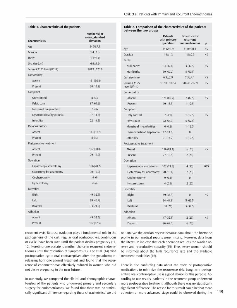
Çelik et al. Patients with Primary and Recurrent Endometriomas
149
recurrent cysts. Because ovulation plays a fundamental role in the pathogenesis of the cyst, regular oral contraceptives, continuous or cyclic, have been used until the patient desires pregnancy (11, 12). Noretindrone acetate is another choice in recurrent endome-triomas until the resolution of symptoms (13). Lee et al. (14) tried postoperative cyclic oral contraceptives after the gonadotropin-releasing hormone agonist treatment and found that the recur-rence of endometriomas effectively reduced in women who did not desire pregnancy in the near future.
In our study, we compared the clinical and demographic charac-teristics of the patients who underwent primary and secondary surgery for endometriomas. We found that there was no statisti-cally significant difference regarding these characteristics. We did
not analyze the ovarian reserve because data about the hormone profile in our medical reports were missing. However, data from the literature indicate that each operation reduces the ovarian re-serve and reproductive capacity (15). Thus, every woman should be informed about the high recurrence rate and the available treatment modalities (16).
There is also conflicting data about the effect of postoperative medications to minimize the recurrence risk. Long-term postop-erative oral contraceptive use is a good choice for this purpose. Ac-cording to our results, patients in the recurrent group underwent more postoperative treatment, although there was no statistically significant difference. The reason for this result could be that more adhesion or more advanced stage could be observed during the
Table 2. Comparison of the characteristics of the patients between the two groups
Patients Patients with with primary recurrent operation endometriomas p
Age 34.6±6.9 33.0±10.1 NS
Gravida 1.4±1.3 1.8±2.3 NS
Parity
Nulliparity 54 (37.8) 3 (37.5) NS
Multiparity 89 (62.2) 5 (62.5)
Cyst size (cm) 6.9±2.9 7.3±4.1 NS
Serum CA125 137.8±107.4 348.4±212.9 NS level (U/mL)
Comorbidity
Absent 124 (86.7) 7 (87.5) NS
Present 19 (13.3) 1 (12.5)
Complaint
Only control 7 (4.9) 1 (12.5) NS
Pelvic pain 92 (64.3) 5 (62.5)
Menstrual irregularities 6 (4.2) 1 (12.5)
Dysmenorrhea/Dysparonia 17 (11.9) 0
Infertility 21 (14.7) 1 (12.5)
Postoperative treatment
Absent 116 (81.1) 6 (75) NS
Present 27 (18.9) 2 (25)
Operation
Laparoscopic cystectomy 102 (71.3) 4 (50) .015
Cystectomy by laparotomy 28 (19.6) 2 (25)
Oopherectomy 9 (6.3) 0
Hysterectomy 4 (2.8) 2 (25)
Laterality
Right 49 (34.3) 0 NS
Left 64 (44.8) 5 (62.5)
Bilateral 30 (21) 3 (37.5)
Adhesion
Absent 47 (32.9) 2 (25) NS
Present 96 (67.1) 6 (75)
Table 1. Characteristics of the patients
number(%) or mean±standard Characteristics deviation
Age 34.5±7.1
Gravida 1.4±1.3
Parity 1.1±1.0
Cyst size (cm) 6.9±3.0
Serum CA125 level (U/mL) 148.9±120.6
Comorbidity
Absent 131 (86.8)
Present 20 (13.2)
Complaint
Only control 8 (5.3)
Pelvic pain 97 (64.2)
Menstrual irregularities 7 (4.6)
Dysmenorrhea/dysparonia 17 (11.3)
Infertility 22 (14.6)
Previous history
Absent 143 (94.7)
Present 8 (5.3)
Postoperative treatment
Absent 122 (80.8)
Present 29 (19.2)
Operation
Laparoscopic cystectomy 106 (70.2)
Cystectomy by laparotomy 30 (19.9)
Oopherectomy 9 (6)
Hysterectomy 6 (4)
Laterality
Right 49 (32.5)
Left 69 (45.7)
Bilateral 33 (21.9)
Adhesion
Absent 49 (32.5)
Present 102 (67.5)

first operation, and these patients could be managed with postop-erative treatment.
In case of recurrence, medical treatment is the first step, particu-larly in small cysts. If medication is ineffective, if the cyst is rapidly growing, if infertility or suspicion of malignancy is present, or if patient is suffering from pain, surgery may be offered for the re-current endometriomas to such patients. In our study, the main indication for surgery in both primary and secondary cases was pelvic pain.
Many studies have been performed to determine the risk factors for the recurrence of endometriomas after operation. Cyst size, op-erative time, intraoperative blood loss, and several serum markers change the recurrence rate of endometriomas (17, 18). Some stud-ies showed that there were several risk factors for endometrioma recurrence such as the cyst size >8 cm, young age (<25 years), preoperative cyst rupture, or preoperative dysmenorrhea. Authors explained that recurrent cysts occur in the lesions in which a cys-tectomy was performed, whereby endometrial cells touched the peritoneal surface. The impact of postoperative hormone therapy on ovarian endometriosis remains unclear. Maul et al. (19) found that patients did not significantly benefit from additional postop-erative hormone therapy (gonadotropin-releasing hormone ago-nist, oral contraceptive, medroxyprogesterone acetate, or danazol) in terms of reducing the risk of disease and pain recurrence. A retrospective study performed by Koga et al. (20) indicated that prior use of medical treatment for endometriosis is a significant risk factor for recurrence in later life. Higher diameter of the mass, previous pelvic surgery, operation type, presence of adhesion, and higher CA 125 levels were found as risk factors for endometrioma recurrence, whereas demographic and obstetric past history had no difference for endometrioma recurrence in another study (21). The reason for this could be that larger endometriomas are more prone to rupture before or during surgery resulting in more adhe-sion formation and peritoneal spread of endometrial cells. But we did not find any correlation between the clinical and demographic characteristics and the recurrence of endometriomas after surgery. This could be explained with the small number of recurrent endo-metriomas in our population. Fedele et al. (22) did not find any difference between the patients with endometriomas and patients with recurrent endometriomas according to these characteristics, similar to our results.
According to recent guidelines, there is a strong recommendation for clinicians about the use of oxidized regenerated cellulose dur-ing endometriosis surgery to prevent adhesion formation (23, 24).
Conclusion
It is not possible to accurately predict the patients in whom en-dometriomas will recur. As a result, maximum excision of the cyst should be performed in the first operation and should be carefully done to minimize damage to the ovaries and delay recurrence of the cyst.
Ethics Committee Approval: Since our study was designed as retrospective cohort study, no ethics committee approval was applied.
Informed Consent: Informed consent was obtained from every patient who participated in this study.
Peer-review: Externally peer-reviewed.
Author contributions: Concept - H.G.C., E.C.; Design - H.G.C., E.C.; Super-vision - E.C., I.P.; Resource - H.G.C., G.T., I.P.; Materials - H.G.C., G.T.; Data Collection and/or Processing - H.G.C., G.T.; Analysis and/or Interpretation - H.G.C., E.C.; Literature Search - H.G.C., E.C.; Writing - H.G.C., E.C.; Critical Reviews - E.C., I.P.
Conflict of Interest: Authors have no conflicts of interest to declare.
Financial Disclosure: The authors declared that this study has received no financial support
Etik Komite Onayı: Çalışmamız retrospektif olarak hazırlandığı için, etik kurul onayı başvurusu yapılmamıştır.
Hasta Onamı: Hasta onamı bu çalışmaya katılan hastalardan alınmıştır.
Hakem Değerlendirmesi: Dış Bağımsız.
Yazar Katkıları: Fikir - H.G.C., E.C.; Tasarım - H.G.C., E.C.; Denetleme - E.C., I.P.; Kaynaklar - H.G.C., G.T., I.P.; Malzemeler - H.G.C., G.T.; Veri Toplanması ve/veya işlemesi - H.G.C., G.T.; Analiz ve/veya Yorum - H.G.C., E.C.; Literatür taraması - H.G.C., E.C.; Yazıyı Yazan - H.G.C., E.C.; Eleştirel İnceleme - E.C., I.P.
Çıkar Çatışması: Yazarların beyan edecek çıkar çatışması yoktur.
Finansal Destek: Yazarlar bu çalışma için finansal destek almamışlardır.
References
1. Gonçalves FC, Andres MP, Passman LJ, Gonçalves MO, Podgaec S. A systematic review of ultrasonography-guided transvaginal aspiration of recurrent ovarian endometrioma. Intern J Gynecol Obstet 2016; 134: 3-7. [CrossRef]
2. Somigliana E, Benaglia L, Vercellini P, Paffoni A, Ragni G, Fedele L. Recurrent endometrioma and ovarian reserve: biological connection or surgical paradox? Am J Obstet Gynecol 2011; 204: 529. [CrossRef]
3. Seong SJ, Kim D, Lee KH, Kim TJ, Chung HH, Chang SJ, et al. Role of hormone therapy after primary surgery for endometrioma: A multicenter retrospective cohort study. Reprod Sci 2016; 23: 1011-8. [CrossRef]
4. Saleh A, Tulandi T. Reoperation after laparoscopic treatment of ovar-ian endometriomas by excision and by fenestration. Fertil Steril 1999; 72: 322-4. [CrossRef]
5. Muzii L, Achilli C, Lecce F, Bianchi A, Francesschetti S, Marchetti C, et al. Second surgery for recurrent endometriomas is more harmful to healthy ovarian tissue and ovarian reserve than first surgery. Fertil Steril 2015; 103: 738-43. [CrossRef]
6. Guo SW. Recurrence of endometriosis and its control. Hum Reprod Update 2009; 15:n441-61.
7. Chan LY, So WW, Lao TT. Rapid recurrence of endometrioma after transvaginal ultrasound-guided aspiration. Eur J Obstet Gynecol Re-prod Biol 2003; 109: 196-8. [CrossRef]
8. Xing W, Lin H, Wu Z, Li Y, Zhang Q. Effect of pelvic endometriosis, endometriomas and recurrent endometriomas on IVF-ET/ICSI out-comes. Mater Sociomed 2016; 28: 91-4. [CrossRef]
9. Lee DY, Kim HJ, Yoon BK, Choi D. Factors associated with the lateral-ity of recurrent endometriomas after conservative surgery. Gyencol Endocrinol 2013; 29: 978-81. [CrossRef]
10. Chang MY, Hsieh CL, Shiau CS, Hsieh TT, Chiang RD, Chan CH. Ultra-sound-guided aspiration and ethanol sclerotherapy (EST) for treat-ment of cyst recurrence in patients after previous endometriosis surgery: analysis of influencing factors using a decision tree. J Minim Invasive Gynecol 2013; 20: 595-603. [CrossRef]
İstanbul Med J 2018; 19: 147-51
150

11. Cucinella G, Granese R, Clagna G, Svelato A, Saitta S, Tonni G, et al. Oral contraceptives in the prevention of endometrioma recurrence: does the different progestins used make a difference? Arch Gynecol Obstet 2013; 288: 821-7. [CrossRef]
12. Vercellini P, DE Matteis S, Somigliana E, Buggio L, Frattaruolo MP, Fe-dele L. Long-term adjuvant therapy for the prevention of postopera-tive endometrioma recurrence: a systematic review and meta-analy-sis. Acta Obstet Gynecol Scand 2013; 92: 8-16. [CrossRef]
13. Muneyyirci-Delale O, Anopa J, Charles C, Mathur D, Parris R, Cutler JB, et al. Medical management of recurrent endometrioma with long-term norethindrone acetate. Int J Womens Health 2012; 4: 149-54. [CrossRef]
14. Lee DY, Bae DS, Yoon BK, Choi D. Post-operative cyclic oral contracep-tive use after gonadotropin-releasing hormone agonist treatment ef-fectively prevents endometrioma recurrence. Hum Reprod 2010; 25: 3050-4. [CrossRef]
15. Celik HG, Dogan E, Okyay E, Ulukus C, Saatli B, Uysal S, et al. Effect of laparoscopic excision of endometriomas on ovarian reserve: se-rial changes in the serum antimullerian hormone levels. Fertil Steril 2012; 97: 1472-8. [CrossRef]
16. Vercellini P, Somigliana E, Vigano P, De Matteis S, Barbara G, Fedele L. Post-operative endometirosis recurrence: a plea for prevention based on pathogenic, epidemiological and clinical evidence. Reprod Biomed Online 2010; 21: 259-65. [CrossRef]
17. Han AR, Lee TH, Kim S, Lee HY. Risk factors and biomarkers for the recurrence of ovarian endometrioma: about the immunoreactivity of progesterone receptor isoform B and nuclear factor kappa B. Gynecol Endocrinol. 2016; 23: 70-4.
18. Tobiume T, Kotani Y, Takaya H, Nakai H, Tsuji I, Suzuki A, et al. Deter-minant factors of postoperative recurrence of endometriosis: differ-
ence between endometrioma and pain. Eur J Obstet Gynecol Reprod Biol 2016; 205: 54-9. [CrossRef]
19. Maul LV, Morrision JE, Schollmeyer T, Alkatout I, Mettler L. Surgical therapy of ovarian endometrioma: recurrence and pregnancy rates. JSLS 2014; 18: pii.
20. Koga K, Takemura Y, Osuga Y, Yoshino O, Hirota Y, Hirata T, et al. Re-currence of ovarian endometrioma after laparoscopic excision. Hum Reprod 2006; 21: 2171-4 [CrossRef]
21. Guzel AI, Topcu HO, Ekilinc S, Tokmak A, Kokanali MK, Cavkaytar S, et al. Recurrence factors in women underwent laparoscopic surgery for endometrioma. Minerva Chir 2014; 69: 277-82.
22. Fedele L, Bianchi S, Zanconato G, Berlanda N, Raffaelli R, Fontana E. Laparoscopic excision of recurrent endometriomas: long-term out-come and comparison with primary surgery. Fertil Steril 2006; 85: 694-9. [CrossRef]
23. Ahmad G, Duffy JM, Farquhar C, Vail A, Vandekerckhove P, Watson A, et al. Barrier agents for adhesion prevention after gynecological surgery. Cochrane Database Syst Rev 2008; 2: CD000475
24. Trew G, Pistofidis G, Pados G, Lower A, Mettler L, Wallwiener D, et al. Gynecological endoscopic evaluation of 4% icodextrin solution: a Eu-ropean, multicentre, double-blind, randomized study of the efficacy and safety in the reduction of de novo adhesions after laparoscopic gynaecological surgery. Hum Reprod 2011; 26: 2015-27. [CrossRef]
Cite this article as: Çelik HG, Çelik E, Turan G, Polat İ. Is there Any Difference between the Patients with Primary Endometriomas and those with Recurrent Endometriomas? İstanbul Med J 2018; 19: 147-51.
Çelik et al. Patients with Primary and Recurrent Endometriomas
151

Introduction
Psoriasis (Ps) (MIM 177900) is a chronic inflammatory skin disease that manifests as red, scaly skin plaques with various degrees of severity and affects almost 2% of the population (1). Psoriasis vulgaris is the most common type of the disease. Several factors associated with the skin, immune system,and human genome have been implicated in the pathogenesis of Ps. Histologically, Ps is characterized by the hyperproliferation and impaired differentiation of keratinocytes, dilated hyperplastic blood vessels, and inflammatory infiltration of leucocytes mainly into the dermis(2). It is suggested that the infiltration of activated T-cells induces kera-tinocyte proliferation in Ps (3).
Nitric oxide (NO), a multifunctional secondary messenger,which acts as a vasodilator, immuno-modulator, antioxidant, and bactericide, contributes to the modulation of gene expression, and alters their functions thorough the nitrosylation of involved proteins. NO plays a role in the main-tenance of skin homeostasis,and it is a strong regulator of keratinocyte growth and differentiation (4). NO is produced in and released from keratinocytes in high amounts and is the key inhibitor of cellular proliferation and inducer of differentiation in vitro. NO is synthesized from L-arginine by three NO synthases (NOS), namely NOS1 or neuronal, NOS2 or inducible, and NOS3 or endothelial NO synthase (5). These NOS are expressed by skin cell types, and NOS2 is expressed by keratino-cytes in Ps and other inflammatory skin disorders (6). The NOS3 gene is found on chromosome 7q35-36, within a length of 4.4 kb (7). The NOS3 gene has several allelic variants, and some of them are functional. The exon 7 Glu298Asp missense variant (rs1799983) and the intron-4 27-bp
Is there any Association between the Functional Variants of the NOS3 Gene and Psoriasis? Psoriazis ve NOS3 Gen Fonksiyonel Varyantları Arasında Bir İlişki Var mıdır?
Amaç: Psoriazis (Ps) etyolojsi tam olarak anlaşılamayan kronik, im-münite ile ilişkili enflamatuar bir deri hastalığıdır. Bu çalışmanın amacı Ps hastalığı yatkınlığı ile endotelyal nitrik asit sentaz (NOS3) geni G894T (rs1799983) ve değişken sayılı ardışık tekrar (VNTR) var-yantları arasındaki ilişkiyi saptamaktır.
Yöntemler: Bu calışma aynı bölgeden seçilen 74 sağlıklı kontrole ila-veten 74 Ps hastası olmak üzere kontrollü vaka çalışmasıdır. NOS3 gen variantları Polimeraz zincir reaksiyonu (PZR) ve/veya PZR-Sınırlayıcı enzim parça uzunluk polimorfizmi (RFLP) ile analiz edilmiştir.
Bulgular: NOS3 G894T TT genotipi ve T alleli sağlıklı kontrollere göre Ps hastalarında daha fazla bulunuyordu (p=0.000, p=0.001, sırasıyla). NOS3 VNTR varyant BB genotipi ve B alleli hasta grubunda konrol grubuna göre daha fazlaydı (p=0.005, p=0.000 sırasıyla). NOS3 VNTR AA genotipi hasta grubunda azdı (p=0.027). Ancak artrit, Ps alan ve şiddet indeksi (PASI), başlangıç yaşı ve aile hikayesine göre yapılan analiz NOS3 G894T ve NOS3 VNTR genotipleri arasında önemli bir iliş-ki ortaya koymadı (p>0.05).
Sonuç: Bu sonuçlar NOS3 G894T ve VNTR varyantların Türk toplulu-ğunda Ps ile ilişkili olduğunu öne sürmektedir. Ancak, Ps oluşmasın-da NOS3 varyantlarının genetik rolünü anlamak için ileri çalışmalar gerekmektedir.
Anahtar Kelimeler: Psoriazis, Endotelyal nitrik oksit sentaz geni, G894T, VNTR
Introduction: Psoriasis (Ps) is a chronic, immune-mediated inflamma-tory skin disorder with an incompletely understood etiology. The aim of this study was to investigate the relationship between the suspecti-bility to Ps and G894T (rs1799983) and variable number tandem repeat (VNTR) variants of the endothelial nitric oxide synthase (NOS3) gene.
Methods: This is a case-controlled study that included 74 Ps patients in addition to 74 matched healthy unrelated controls from the same locality. The NOS3 gene variants were analyzed by polymerase chain reaction (PCR) and/or PCR- restriction fragment lenght polymorp-hism (PCR-RFLP).
Results: The NOS3 G894T TT genotype and T allele were more com-mon in the Ps group compared to the healthy controls (p=0.000, p=0.001, respectively). The NOS3 VNTR variant BB genotype and B allele were higher in the patient group than in the control group (p=0.005, p=0.000 respectively). The NOS3 VNTR AA genotype was lower in the patient group (p=0.027). However, a stratified analysis including arthritis, the Ps area and severity index (PASI), the age of onset, and family history revealed no significant correlation between the NOS3 G894T and NOS3 VNTR genotypes (p>0.05).
Conclusion: These results suggest that the NOS3 G894T and VNTR va-riants are associated with Ps in a Turkish cohort. However, future stu-dies are needed to understand the genetic role of the NOS3 variants in the development of Ps.
Keywords: Psoriasis, endothelial nitric oxide synthase gene, G894T, VNTR
Abst
ract
/ Ö
z
ORCID ID of the authors: A.F.N. 0000-0001-7639-1122; M.P. 0000-0002-6692-085X; S.P. 0000-0003-1272-5845.
1Department of Medical Biology, İstanbul University İstanbul School of Medicine, İstanbul, Türkiye2Department of Dermatology, Gaziantep University School of Medicine, Gaziantep, Türkiye3Department of Medical Genetics, Hitit University School of Medicine, Çorum, Türkiye4Department of Hematology, Gaziantep University School of Medicine, Gaziantep, Türkiye
Address for Correspondence/Yazışma Adresi:Ayşe Feyda NursalE-mail: [email protected]
Received/Geliş Tarihi: 06.09.2017
Accepted/Kabul Tarihi: 08.12.2017
© Telif Hakkı 2018 Makale metnine istanbultipdergisi.org web sayfasından ulaşılabilir.
© Copyright 2018 by Available online at istanbulmedicaljournal.org
Original Investigation/Orijinal Araştırmaİstanbul Med J 2018; 19: 152-7DOI: 10.5152/imj.2018.05658
Sacide Pehlivan1 , Hüseyin Serhat İnalöz2, Ayşe Feyda Nursal3 , Aslıhan Gülel2, Mustafa Pehlivan4

variable number tandem repeat (VNTR) variant of NOS3 are pro-posed to be related with NOS3 dysfunction, resulting in impair-ment of NO production (8).
Therefore, in present study, we aimed to investigate the relation-ship of Ps with the NOS3 gene functional variants in a Turkish cohort. The results obtained from the study were compared both between each other and with the clinical parameters.
Methods
Study PopulationIn total, 74 Ps patients (mean age: 42 years; male-to-female ratio 1.1:1) and 74 healthy controls participated in the study. Both the study group and control group were of Turkish origin, from the south of Turkey. Clinical diagnosis was performed by dermatolo-gists, and all the patients had at least two skin lesions. The par-ticipants were well informed about the nature of this study and they participated voluntarily. Clinical data were collected from all the patients. Disease severity was quantified by the psoriasis area and severity index (PASI) (9). For all the patients, the PASI score was recorded. In our study, patients with Ps were sorted into two groups according to their PASI score. Based on the median distri-bution, PASI≤10 was defined as mild Ps (Type I) and PASI>10 (Type II) as severe Ps. High PASI score represents a high degree of psori-atic severity. Seventy-four healthy subjects, individually matched to patients by both gender and age, were recruited as controls. Informed written consent was obtained from all individuals before enrollment in the study, according to the ethical guidelines of the Declaration of Helsinki, and the investigation was approved by the Gaziantep University Instutional Ethical Committee.
Genotyping Analysis DNA was extracted from whole blood samples using a salting out method (10). The NOS3 gene variant G894T was analyzed by poly-merase chain reaction-restriction fragment lenght polymorphism (PCR-RFLP) method as described previously (11). The G894T variant was genotyped by PCR amplification using the following primers 5′-CATGAGGCTCAGCCCCAGAAC-3′ (forward) and 5′-AGTCAATCCCTTTG-GTGCTCAC-3′ (reverse). The PCR products were digested using MboI enzyme (Invitrogen, Carlsbad, CA, USA) at 37°C overnight. PCR products were loaded onto 3% agarose gels previously stained with ethidium bromide and were visualized by using an ultraviolet transilluminator.The 206 bp products had a consistent restriction site resulting in a 119 bp and an 87 bp fragment.
The 27 bp-VNTR AB genotype in intron 4 of NOS3 were evaluated by PCR amplification using primers 5′-AGGCCCTATGGTAGTGCCTTT-3′ (forward) and 5′-TCTCTTAGTGCTGTGGTCAC-3′ (reverse) (12). The am-plified products were separated by electrophoresis on a 3% agarose gel and visualized by ethidium bromide staining. The wild type allele contained five tandem repeats of 27 bp and 420 bp, and the mutant allele four tandem repeats of 27 bp and 393 bp.
Statistical AnalysisStatistical analysis was performed using the SPSS (Statistical Package for Social Sciences) version 14.0 for Windows (SPSS Inc., Chicago, IL, USA). Statistical significance of differences between groups was esti-mated by logistic regression analysis. Odds ratio (OR) and 95% con-fidence interval (CI) were also calculated. Distribution of the NOS3 G894T and VNTR variants and deviation from the Hardy-Weinberg equilibrium (HWE) for each control group were examined using the
chi-square test. All the analyses were two-tailed, and a p-value <0.05 was considered to be significant.
Results
This analysis included 74 Turkish patients with Ps and 74 age- and sex-matched controls. The mean age of the patients was 42 years. Tables 1 and 2 demonstrate the genotype distribution and allele frequency of the G894T and VNTR variants of the NOS3 gene in the patients and the controls.
NOS3 G894T Frequencies of the GG, GT, and TT genotypes for the NOS3 G894T variant were 50%, 32.4%, and 17.6%, respectively, among the pa-tients, and 64.9%, 35.1%, and 0%, respectively, among the controls. The alleles and genotypes of the NOS3 G894T variant were found to be significantly associated between the patients and controls. Patients with the NOS3 TT genotype had a 0.542-fold increased risk for Ps (OR=0.542, 95% Cl: 0.280-1.048; p=0.000) (Table 1). Al-lelic frequencies of the NOS3 G894T variant among the patients and controls were 66.2% and 82.4%, respectively, for allele G (wild type) and 33.8% and 17.6%, respectively, for allele T (mutant). There was a significant association between the allele frequencies of the NOS3 G894T variant between patients and controls. The NOS3 G894T T al-lele was higher in patients with Ps (OR=0.418, 95% Cl: 0.191-0.837; p=0.001) (Table 1).
NOS3 VNTRFor the NOS3 VNTR variant, a significant difference in both the genotype distribution and allele frequency between the Ps pa-tients and control subjects was observed (Table 2). The NOS3 VNTR AA genotype was lower in the patient group (OR=0.442, 95% Cl: 0.223-0.872; p=0.005), while the NOS3 VNTR variant BB genotype was higher (OR=0.130, 95% Cl: 0.028-0.600; p=0.005).
Allelic frequencies of the NOS3 VNTR variant among the patients and controls were 67.6% and 84.5%, respectively, for allele A (wild type), and 32.4% and 15.5 %, respectively, for allele B (mutant). There was a significant association between allele frequencies of NOS3 VNTR between patients and controls (OR=2.609, 95% Cl: 1.487-4.578; P=0.000). Allele B was higher in the patient group than in the control group.
When the patients and controls were compared in terms of HWE, deviations were observed in both NOS3 variants (p=0.017 and p=0.005, respectively) (Table 1 and 2).
Clinical Features According to the NOS3 G894T and VNTR Genotypes We compared the genotype distribution of the groups with a posi-tive and negative family history in order to establish any possible influence of family history on the relation between the NOS3 genotypes and Ps. The distributions of the NOS3 gene variants ac-cording to family history are given Table 3. We found no significant difference between the groups.
We evaluated the NOS3 G894T and VNTR variants genotype distri-bution according to arthritis status in patients with Ps. The results are presented in Table 4. We found no significant difference be-tween the patients and controls. When the patient groups were compared with regard to the presence of arthritis, there was no statistically significant difference between the arthritic (psoriatic arthritis) and non-arthritic (psoriasis vulgaris) groups.
Pehlivan et al. The NOS3 Gene Variants in Psoriasis
153
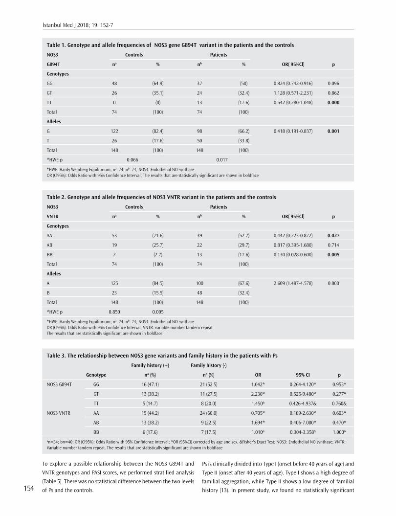
To explore a possible relationship between the NOS3 G894T and
VNTR genotypes and PASI scores, we performed stratified analysis
(Table 5). There was no statistical difference between the two levels
of Ps and the controls.
Ps is clinically divided into Type I (onset before 40 years of age) and
Type II (onset after 40 years of age). Type I shows a high degree of
familial aggregation, while Type II shows a low degree of familial
history (13). In present study, we found no statistically significant
İstanbul Med J 2018; 19: 152-7
154
Table 1. Genotype and allele frequencies of NOS3 gene G894T variant in the patients and the controls
NOS3 Controls Patients
G894T na % nb % OR( 95%Cl) p
Genotypes
GG 48 (64.9) 37 (50) 0.824 (0.742-0.916) 0.096
GT 26 (35.1) 24 (32.4) 1.128 (0.571-2.231) 0.862
TT 0 (0) 13 (17.6) 0.542 (0.280-1.048) 0.000
Total 74 (100) 74 (100)
Alleles
G 122 (82.4) 98 (66.2) 0.418 (0.191-0.837) 0.001
T 26 (17.6) 50 (33.8)
Total 148 (100) 148 (100)
*HWE p 0.066 0.017
*HWE: Hardy Weinberg Equilibrium; na: 74; nb: 74; NOS3: Endothelial NO synthaseOR (Cl95%): Odds Ratio with 95% Confidence Interval; The results that are statistically significant are shown in boldface
Table 2. Genotype and allele frequencies of NOS3 VNTR variant in the patients and the controls
NOS3 Controls Patients
VNTR na % nb % OR( 95%Cl) p
Genotypes
AA 53 (71.6) 39 (52.7) 0.442 (0.223-0.872) 0.027
AB 19 (25.7) 22 (29.7) 0.817 (0.395-1.680) 0.714
BB 2 (2.7) 13 (17.6) 0.130 (0.028-0.600) 0.005
Total 74 (100) 74 (100)
Alleles
A 125 (84.5) 100 (67.6) 2.609 (1.487-4.578) 0.000
B 23 (15.5) 48 (32.4)
Total 148 (100) 148 (100)
*HWE p 0.850 0.005
*HWE: Hardy Weinberg Equilibrium; na: 74; nb: 74; NOS3: Endothelial NO synthaseOR (Cl95%): Odds Ratio with 95% Confidence Interval; VNTR: variable number tandem repeatThe results that are statistically significant are shown in boldface
Table 3. The relationship between NOS3 gene variants and family history in the patients with Ps
Family history (+) Family history (-)
Genotype na (%) nb (%) OR 95% CI p
NOS3 G894T GG 16 (47.1) 21 (52.5) 1.042* 0.264-4.120* 0.953*
GT 13 (38.2) 11 (27.5) 2.230* 0.525-9.480* 0.277*
TT 5 (14.7) 8 (20.0) 1.450& 0.426-4.937& 0.760&
NOS3 VNTR AA 15 (44.2) 24 (60.0) 0.705* 0.189-2.630* 0.603*
AB 13 (38.2) 9 (22.5) 1.694* 0.406-7.080* 0.470*
BB 6 (17.6) 7 (17.5) 1.010& 0.304-3.358& 1.000&
an=34; bn=40; OR (Cl95%): Odds Ratio with 95% Confidence Interval; *OR (95%CI) corrected by age and sex, &Fisher's Exact Test; NOS3: Endothelial NO synthase; VNTR: Variable number tandem repeat. The results that are statistically significant are shown in boldface

association between the NOS3 (G894T and VNTR) genotypes and the onset age (Table 6).
Discussion
Ps is a chronic inflammatory immunologic genodermatosis, mani-fested by an increased activity of proliferative keratinocytes. While the exact cause remains unclear, environmental and genetic fac-tors are believed to be involved in the pathogenesis of Ps. Ps has been related to several gene variants that are implicated in many different processes, such as skin barrier functions and the regula-tion of inflammatory and immune responses.
Reactive species or free radicals include reactive oxygen, and ni-trogen species are called reactive oxygen nitrogen species (RONS).
Most of the RONS carry unpaired electrons and are called free radicals. In mammalian species, one of the main functions of specialized enzymes is the production of RONS (14). Skin is the main target of oxidative stress as it is repetitively exposed to UV radiation and other environmental stresses. Therefore, it has been proposed that elevated ROS synthesis, impaired function of an-tioxidant system, and oxidative stress may be responsible in the pathogenesis of Ps. Involvement of ROS in the etiopathogenesis of Ps has been studied by several researchers in recent years (15, 16). A recent study (17) reported significantly high levels of oxidative stress markers, such as plasma or serum malondialdehyde (MDA), and NO end products, in patients with Ps. Besides, blood NO lev-els were significantly elevated in patients with Ps and exhibited a positive correlation with the severity and duration of the lesions (18-20). Some of these studies, further suggested a positive corre-
Pehlivan et al. The NOS3 Gene Variants in Psoriasis
155
Table 4. The relationship between NOS3 gene variants and arthritis in the patients with Ps
Arthritis (+) Arthritis (-)
Genotype na (%) nb (%) OR 95% CI p
NOS3 G894T GG 12 (52.2) 25 (49.0) 1.898* 0.460-7.835* 0.376*
GT 6 (26.1) 18 (35.3) 1.904* 0.418-8.660* 0.405*
TT 5 (21.7) 8 (15.7) 1.493& 0.430-5.188& 0.526&
NOS3 VNTR AA 13 (56.5) 26 (51.0) 1.020* 0. 245-4.244* 0.978*
AB 6 (26.1) 16 (31.4) 1.397* 0. 287-6.792* 0.679*
BB 4 (17.4) 9 (17.6) 1.018& 0.278-3.722& 1.000&
an=23; bn=51; NOS3: Endothelial NO synthase; OR (Cl95%): Odds Ratio with 95% Confidence Interval; *OR (95%CI) corrected with age and sex, &Fisher's Exact Test; VNTR: Variable number tandem repeat.
Table 5. The relationship between NOS3 gene variants and PASI in patients with Ps
PASI 1 (PASI≤10) PASI 2 (PASI>10)
Genotype na (%) nb (%) OR 95% CI p
NOS3 G894T GG 19 (51.4) 18 (48.6) 0.794* 0.210-3.002* 0.733*
GT 11 (29.7) 13 (35.2) 0.785* 0.194-3.002* 0.735*
TT 7 (18.9) 6 (16.2) 0.829& 0.250-2.755& 1.000&
NOS3 VNTR AA 20 (54.1) 19 (51.4) 0.906* 0.247-3.324* 0.881*
AB 10 (27.0) 12 (32.4) 0.695* 0.168-2.872* 0.616*
BB 7 (18.9) 6 (16.2) 1.206& 0.363-4.004& 1.000&
an=37; bn=37; OR (Cl95%): Odds Ratio with 95% Confidence Interval; *OR (95%CI) corrected by age and sex, &Fisher's Exact Test; PASI (psoriasis area severity index); NOS3: Endothelial NO synthase; VNTR: Variable number tandem repeat
Table 6. The relationship between NOS3 gene variants and disease onset in the patients with Ps
Early onset (≤40 age) Late onset (>40 age) Type I Type II
Genotype na (%) nb (%) OR 95% CI p
NOS3 G894T GG 26 (53.1) 11 (44.0) 1.318& 0.129-13.499& 0.816&
GT 15 (30.6) 9 (36.0) 1.882* 0.163-21.690* 0.612*
TT 8 (16.3) 5 (20.0) 1.281& 0.371-4.422& 0.752&
NOS3 VNTR AA 25 (51.0) 14 (56.0) 0.338* 0.039-2.940* 0.326*
AB 14 (28.6) 8 (32.0) 0.195* 0.018-2.161* 0.183*
BB 10 (20.4) 3 (12.0) 1.880& 0.467-7.565& 0.523&
an=49; bn=25; *OR (95%CI) corrected with age and sex, &Fisher's Exact Test; NOS3: Endothelial NO synthase; VNTR: Variable number tandem repeat
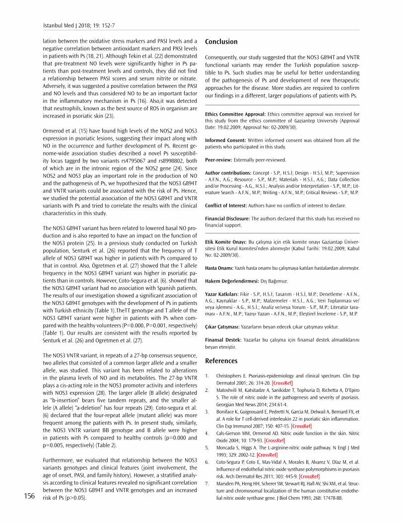
lation between the oxidative stress markers and PASI levels and a negative correlation between antioxidant markers and PASI levels in patients with Ps (18, 21). Although Tekin et al. (22) demonstrated that pre-treatment NO levels were significantly higher in Ps pa-tients than post-treatment levels and controls, they did not find a relationship between PASI scores and serum nitrite or nitrate. Adversely, it was suggested a positive correlation between the PASI and NO levels and thus considered NO to be an important factor in the inflammatory mechanism in Ps (16). Also,it was detected that neutrophils, known as the best source of ROS in organism are increased in psoriatic skin (23).
Ormerod et al. (15) have found high levels of the NOS2 and NOS3 expression in psoriatic lesions, suggesting their impact along with NO in the occurrence and further development of Ps. Recent ge-nome-wide association studies described a novel Ps susceptibil-ity locus tagged by two variants rs4795067 and rs8998802, both of which are in the intronic region of the NOS2 gene (24). Since NOS2 and NOS3 play an important role in the production of NO and the pathogenesis of Ps, we hypothesized that the NOS3 G894T and VNTR variants could be associated with the risk of Ps. Hence, we studied the potential association of the NOS3 G894T and VNTR variants with Ps and tried to correlate the results with the clinical characteristics in this study.
The NOS3 G894T variant has been related to lowered basal NO pro-duction and is also reported to have an impact on the function of the NOS3 protein (25). In a previous study conducted on Turkish population, Senturk et al. (26) reported that the frequency of T allele of NOS3 G894T was higher in patients with Ps compared to that in control. Also, Ögretmen et al. (27) showed that the T allele frequency in the NOS3 G894T variant was higher in psoriatic pa-tients than in controls. However, Coto-Segura et al. (6). showed that the NOS3 G894T variant had no association with Spanish patients. The results of our investigation showed a significant association of the NOS3 G894T genotypes with the development of Ps in patients with Turkish ethnicity (Table 1).TheTT genotype and T allele of the NOS3 G894T variant were higher in patients with Ps when com-pared with the healthy volunteers (P=0.000, P=0.001, respectively) (Table 1). Our results are consistent with the results reported by Senturk et al. (26) and Ogretmen et al. (27).
The NOS3 VNTR variant, in repeats of a 27-bp consensus sequence, two alleles that consisted of a common larger allele and a smaller allele, was studied. This variant has been related to alterations in the plasma levels of NO and its metabolites. The 27-bp VNTR plays a cis-acting role in the NOS3 promoter activity and interferes with NOS3 expression (28). The larger allele (B allele) designated as “b-insertion” bears five tandem repeats, and the smaller al-lele (A allele) “a-deletion” has four repeats (29). Coto-segura et al. (6) declared that the four-repeat allele (mutant allele) was more frequent among the patients with Ps. In present study, similarly, the NOS3 VNTR variant BB genotype and B allele were higher in patients with Ps compared to healthy controls (p=0.000 and p=0.005, respectively) (Table 2).
Furthermore, we evaluated that relationship between the NOS3 variants genotypes and clinical features (joint involvement, the age of onset, PASI, and family history). However, a stratified analy-sis according to clinical features revealed no significant correlation between the NOS3 G894T and VNTR genotypes and an increased risk of Ps (p>0.05).
Conclusion
Consequently, our study suggested that the NOS3 G894T and VNTR functional variants may render the Turkish population suscep-tible to Ps. Such studies may be useful for better understanding of the pathogenesis of Ps and development of new therapeutic approaches for the disease. More studies are required to confirm our findings in a different, larger populations of patients with Ps.
Ethics Committee Approval: Ethics committee approval was received for this study from the ethics committee of Gaziantep University (Approval Date: 19.02.2009; Approval No: 02-2009/30).
Informed Consent: Written informed consent was obtained from all the patients who participated in this study.
Peer-review: Externally peer-reviewed.
Author contributions: Concept - S.P., H.S.İ; Design - H.S.İ, M.P.; Supervision - A.F.N., A.G.; Resource - S.P., M.P.; Materials - H.S.İ., A.G.; Data Collection and/or Processing - A.G., H.S.İ.; Analysis and/or Interpretation - S.P., M.P.; Lit-erature Search - A.F.N., M.P.; Writing - A.F.N., M.P.; Critical Reviews - S.P., M.P.
Conflict of Interest: Authors have no conflicts of interest to declare.
Financial Disclosure: The authors declared that this study has received nofinancial support.
Etik Komite Onayı: Bu çalışma için etik komite onayı Gaziantap Üniver-sitesi Etik Kurul Komitesi’nden alınmıştır (Kabul Tarihi: 19.02.2009; Kabul No: 02-2009/30).
Hasta Onamı: Yazılı hasta onamı bu çalışmaya katılan hastalardan alınmıştır.
Hakem Değerlendirmesi: Dış Bağımsız.
Yazar Katkıları: Fikir - S.P., H.S.İ; Tasarım - H.S.İ, M.P.; Denetleme - A.F.N., A.G.; Kaynaklar - S.P., M.P.; Malzemeler - H.S.İ., A.G.; Veri Toplanması ve/veya işlemesi - A.G., H.S.İ.; Analiz ve/veya Yorum - S.P., M.P.; Literatür tara-ması - A.F.N., M.P.; Yazıyı Yazan - A.F.N., M.P.; Eleştirel İnceleme - S.P., M.P.
Çıkar Çatışması: Yazarların beyan edecek çıkar çatışması yoktur.
Finansal Destek: Yazarlar bu çalışma için finansal destek almadıklarını beyan etmiştir.
References
1. Christophers E. Psoriasis-epidemiology and clinical spectrum. Clin Exp Dermatol 2001; 26: 314-20. [CrossRef]
2. Matoshvili M, Katsitadze A, Sanikidze T, Tophuria D, Richetta A, D'Epiro S. The role of nitric oxide in the pathogenesis and severity of psoriasis. Georgian Med News 2014; 234:61-4.
3. Boniface K, Guignouard E, Pedretti N, Garcia M, Delwail A, Bernard FX, et al. A role for T cell-derived interleukin 22 in psoriatic skin inflammation. Clin Exp Immunol 2007; 150: 407-15. [CrossRef]
4. Cals-Gierson MM, Ormerod AD. Nitric oxide function in the skin. Nitric Oxide 2004; 10: 179-93. [CrossRef]
5. Moncada S, Higgs A. The L-arginine-nitric oxide pathway. N Engl J Med 1993; 329: 2002-12. [CrossRef]
6. Coto-Segura P, Coto E, Mas-Vidal A, Morales B, Alvarez V, Díaz M, et al. Influence of endothelial nitric oxide synthase polymorphisms in psoriasis risk. Arch Dermatol Res 2011; 303: 445-9. [CrossRef]
7. Marsden PA, Heng HH, Scherer SW, Stewart RJ, Hall AV, Shi XM, et al. Struc-ture and chromosomal localization of the human constitutive endothe-lial nitric oxide synthase gene. J Biol Chem 1993; 268: 17478-88.
İstanbul Med J 2018; 19: 152-7
156

8. Wang XL, Mahaney MC, Sim AS, Wang J, Wang J, Blangero J, et al. Ge-netic contribution of endothelial constitutive nitric oxide synthase gene to plasma nitric oxide levels. Arterioscler Thromb Vasc Biol 1997; 17: 3147-53. [CrossRef]
9. de Rie MA, Goedkoop AY, Bos JD. Overview of psoriasis. Dermatol Ther 2004;17: 341-9. [CrossRef]
10. Miller SA, Dykes DD, Polesky HF. A simple salting out procedure for ex-tracting DNA from human nucleated cells. Nucleic Acids Res 1988;16: 1215. [CrossRef]
11. Ozturk E, Balat O, Pehlivan S, Ugur MG, Ozcan C, Sever T, et al. En-dothelial nitric oxide synthase gene polymorphisms in preeclampsia with or without eclampsia in a Turkish population. J Obstet Gynaecol Res 2011;37: 1778-83. [CrossRef]
12. Safarinejad MR, Safarinejad S, Shafiei N, Safarinejad S. Effects of the T-786C, G894T, and intron 4 VNTR (4a/b)polymorphisms of the endo-thelial nitric oxide synthase gene on the risk of prostate cancer. Urol Oncol 2013; 31: 1132-40. [CrossRef]
13. Henseler T, Christophers E. Psoriasis of early and late onset: character-ization of two types of psoriasis vulgaris. J Am Acad Dermatol 1985; 13: 450-6. [CrossRef]
14. Weidinger A, Kozlov AV. Biological Activities of Reactive Oxygen and Nitrogen Species: Oxidative stress versus Signal Transduction. Biomol-ecules 2015; 5: 472-84. [CrossRef]
15. Ormerod AD, Weller R, Copeland P, Benjamin N, Ralston SH, Grabowk-si P, et al. Detection of nitric oxide and nitric oxide synthases in pso-riasis. Arch Dermatol Res 1998; 290: 3-8. [CrossRef]
16. Zalewska A, Wyczo’łkowska J, Narbutt J, Sysa-Jedrzejowska A. Nitric oxide levels in plasma and fibroblast cultures of psoriasis vulgaris patients. J Dermatol Sci 2007; 48: 237-40. [CrossRef]
17. Kadam DP, Suryakar AN, Ankush RD, Kadam CY, Deshpande KH. Role of oxidative stress in various stages of psoriasis. Indian J Clin Biochem 2010; 2: 388-92. [CrossRef]
18. Gabr SA, Al-Ghadir AH. Role of cellular oxidative stress and cyto-chrome c in the pathogenesis of psoriasis. Arch Dermatol Res 2012; 304: 451-7. [CrossRef]
19. Gokhale NR, Belgaumkar VA, Pandit DP, Deshpande S, Damle DK. A study of serum nitric oxide levels in psoriasis. Indian J Dermatol Ve-nereol Leprol 2005; 71: 175-8. [CrossRef]
20. Orem A, Aliyazicioglu R, Kiran E, Vanizor B, Cimnocodeit G, Deger O. The relationship between nitric oxide production and activity of the disease in patients with psoriasis. Arch Dermatol 1997; 133:1606-7. [CrossRef]
21. Nemati H, Khodarahmi R, Sadeghi M, Ebrahimi A, Rezaei M, Vaisi-Raygani A. Antioxidant status in patients with psoriasis. Cell Biochem Funct 2014; 32: 268-73. [CrossRef]
22. Tekin NS, Ilter N, Sancak B, Ozden MG, Gurer MA. Nitric oxide levels in patients withpsoriasis treated with methotrexate. Mediators Inflamm 2006; 2006: 16043. [CrossRef]
23. Turner CP, Toye AM, Jones OTG. Keratinocyte superoxide generation. Free Radic Biol Med 1998;24: 401-7. [CrossRef]
24. Stuart PE, Nair RP, Ellinghaus E, Ding J, Tejasvi T, Gudjonsson JE, et al. Genome-wide association analysis identifies three psoriasis suscepti-bility loci. Nat Genet 2010; 42: 1000-4. [CrossRef]
25. Veldman BA, Spiering W, Doevendans PA, Vervoort G, Kroon AA, de Leeuw PW, et al. The Glu298Asp polymorphism of the NOS3 gene as determinant of the baseline production of nitric oxide. J Hypertens 2002; 20: 2023-7. [CrossRef]
26. Senturk N, Kara N, Aydin F, Gunes S, Yuksel EP, Canturk T, et al. As-sociation of eNOS gene polymorphism (Glu298Asp) with psoriasis. J Dermatol Sci 2006; 44: 52-5. [CrossRef]
27. Ogretmen Z, Hiz MM, Silan F, Uludag A, Ozdemirc O. Association of endothelial nitric oxide synthase Glu298Asp gene polymorphism in psoriasis cases with hypertension. Ann Saudi Med 2014; 34: 340-5. [CrossRef]
28. Zhang MX, Zhang C, Shen YH, Wang J, Li XN, Zhang Y, et al. Biogen-esis of short intronic repeat 27-nucleotide small RNA from endo-thelial nitric-oxide synthase gene. J Biol Chem 2008; 283: 14685-93. [CrossRef]
29. Nadaud S, Bonnardeaux A, Lathrop M, Soubrier F. Gene structure, polymorphism and mapping of the human endothelial nitric oxide synthase gene. Biochem Biophys Res Commun 1994; 198: 1027-33. [CrossRef]
Cite this article as: Pehlivan S, İnalöz S, Nursal AF, Gülel A, Pehlivan M. Is there any Association between the Functional Variants of the NOS3 Gene and Psoriasis? İstanbul Med J 2018; 19: 152-7.
Pehlivan et al. The NOS3 Gene Variants in Psoriasis
157
Giriş
Ön segment muayenesinde tanıya yardımcı olmak amaçlı, cerrahi öncesi değerlendirme veya has-ta takibi için biyomikroskopik muayeneye ek olarak tarayıcı-slit topografi, interferometri, Sche-impflug görüntüleme, optik koherens tomografi cihazlarının kullanımı artmaktadır. Görüntüleme yöntemleri ile ön segment parametrelerinin daha doğru ölçülmesi tanıda ve tedavide hataları azaltmaktadır. Cihazların teknik özellikleri, ölçüm tekniği, hastaya ait özellikler ölçümlerin tekrar-lanabilirliğini ve karşılaştırılabilirliğini etkilemektedir.
Nidek AL-Scan (Nidek, Aichi, Japonya) cihazı katarakt cerrahisi öncesinde göz içi lensi (GİL) numa-rasını hesaplamak amaçlı kullanıma sunulan optik biyometri cihazıdır. Cihazın başlıca avantajları, non-kontakt bir ölçüm cihazı olması nedeniyle kornea indentasyonundan kaynaklanan ölçüm hatalarının olmaması, 3 boyutlu otomatik göz hareketi izleme sistemi olması nedeniyle gözün hareketi ve kullanıcı kaynaklı ölçüm hatalarının minimalize edilmesidir (1).
Sirius topografi cihazı (Costruzione Strumenti Oftalmici, Florence, İtalya) dönen Scheimpflug kamera ve Plasido disk görüntüleme sisteminden gelen verileri birlikte değerlendiren ön segment analiz cihazıdır. Kornea kalınlığı, ön-arka korneal elevasyon haritalarını içeren topografik ölçümler, ön ka-mara derinliği (ÖKD), keratometri temel ölçüm parametreleridir (2). Kornea hastalıklarının tanı ve takibi, refraktif cerrahi öncesi değerlendirme ve sonrasında takip amaçlı kullanılmaktadır.
Nidek-AL Scan ve Sirius Topografi Cihazları ile Ölçülen Keratometri, Merkezi Kornea Kalınlığı ve Ön Kamara Derinliği Ölçümlerinin KarşılaştırılmasıComparison of Keratometry, Central Corneal Thickness, and Anterior Chamber Depth Results Measured With Nidek-AL Scan Biometry and Sirius Topography Devices
Introduction: To investigate whether the central corneal thickness (CCT), anterior chamber depth (ACD), and keratometric measurements change according to biometric and topographic measurements in childhood and in young adult age.
Methods: In total, 147 eyes of 78 patients were included in the study. Patients with an ocular disease other than refractive error were not included in the study. CCT, ACD, and keratometry values of all patients were measured with Nidek-AL Scan optical biometry and Sirius com-bined Scheimpflug-Plasido disc Corneal Topography devices.
Results: Of the seventy-eight patients, 46 were female and 32 were male. The mean age was 14.46±5.15 years (range, 5-29 years), the mean spherical equivalence was -0.30±1.13 diopters (D), and the mean axial length was 23.28±0.77 mm. The Sirius and AL-Scan CCT were 555.66±39.58 μm and 548.01±38.14 μm, respectively. The Sirius and AL-Scan ACD were 3.65±0.28 mm and 3.57±0.26 mm μm, respec-tively. The Sirius SimK, AL-Scan K 2.4 mm, and AL-Scan K 3.3 mm va-lues were 43.09±1.45 D, 43.22 ±1.50 D, and 43.2±1.47 D, respectively. The values of CCT and ACD measured on the Sirius device were found to be statistically significantly higher than those measured by the AL-Scan device (p=.00). A high level of correlation was found between the Sirius CCT and the AL-Scan CCT (p=0.000, r=0.974). A high level of correlation was found between the Sirius ACD and AL-Scan ACD (p=0.000, r=0.918). A high level of correlation was found between the the Sirius SimK value and the AL-Scan K 2.4 mm and K 3.3 mm values (p=0.000, r values were 0.979 and 0.982, respectively).
Conclusion: In childhood and young adult age, between the Sirius and AL-Scan devices, although CCT, ACD, SimK, and 2.4-3.3 mm K were statistically different, a high correlation and agreement were found between the measurements.
Keywords: Biometry, corneal topography, pachymetry, sirius, AL-Scan
Amaç: Çocukluk ve genç erişkin çağında, merkezi kornea kalınlığı (MKK), ön kamara derinliği (ÖKD) ve keratometrik ölçümlerin biyo-metrik ve topografik ölçüme göre değişim gösterip göstermediğini araştırmak.
Yöntemler: Yetmiş sekiz hastanın 147 gözü çalışmaya dahil edildi. Refraksiyon kusuru dışında oküler hastalığı bulunan hastalar çalışma-ya dahil edilmedi. Tüm hastaların MKK, ÖKD ve keratometri değerleri Nidek AL Scan optik biyometri ve Sirius kombine Scheimpflug-Plasido disk Kornea Topografi cihazları ile ölçüldü.
Bulgular: Yetmiş sekiz hastanın 46’sı kadın, 32’si erkek idi. Ortalama yaş 14,46±5,15 yıl(5-29 yaş arası),ortalama sferik eşdeğer -0,30±1,13 diyoptri(D), ortalama aksiyel uzunluk 23,28±0.77 mm saptandı. Sirius MKK 555,66±39.58 μm, AL-Scan MKK 548,01±38,14 μm saptandı. Si-rius ÖKD 3,65±0,28 mm, AL-Scan ÖKD 3,57±0,26 mm saptandı. Sirius SimK 43,09±1,45 D, AL-Scan K 2.4 mm 43,22±1,50 D, AL-Scan K 3.3mm 43,2±1,47 D saptandı. Sirius cihazında ölçülen MKK ve ÖKD değerleri, AL Scan cihazından ölçülen değerlere göre istatistiksel olarak anlamlı düzeyde daha yüksek saptandı (p=0,00). Sirius MKK ile AL-Scan MKK arasında yüksek düzeyde korelasyon saptandı(p=0.000,r=0.974). Si-rius ÖKD ile AL-Scan ÖKD arasında yüksek düzeyde korelasyon sap-tandı (p=0.000,r=0.918). Sirius SimK değeri ile AL-Scan K 2.4mm ve K 3.3mm arasında yüksek düzeyde korelasyon saptandı (p=0.000, r değerleri sırasıyla 0.979 ve 0.982 saptandı).
Sonuç: Çocukluk ve genç erişkin çağında, Sirius ve AL-Scan cihazla-rı arasında MKK, ÖKD, SimK ile 2.4-3.3 mm K değerleri istatistiksel olarak farklı saptanmış olmasına rağmen ölçümler arasında yüksek korelasyon ve uyum saptandı.
Anahtar Kelimeler: Biyometri, kornea topografisi, pakimetri, sirius, AL-Scan
Öz
/ Abs
trac
t
Bu çalışma TOD 1. Canlı Cerrahi Günleri’nde poster olarak sunulmuştur (16-18 Haziran 2017, İstanbul, Türkiye).
ORCID IDs of all the authors: S.E.B. 0000-0002-9502-4368; N.S. 0000-0002-1442-9743; D.Y.E. 0000-0002-5535-264X, M.E. 0000-0002-2556-7383.
Sağlık Bilimleri Üniversiteri, Kanuni Sultan Süleyman Eğitim ve Araştırma Hastanesi, Göz Hastalıkları Kliniği, İstanbul, Türkiye
Yazışma Adresi/Address for Correspondence:Sadık Etka BayramoğluE-mail: [email protected]
Geliş Tarihi/Received: 30.09.2017Kabul Tarihi/Accepted: 17.12.2017
© Telif Hakkı 2018 Makale metnine istanbultipdergisi.org web sayfasından ulaşılabilir.
© Copyright 2018 by Available online at istanbulmedicaljournal.org
Orijinal Araştırma/Original Investigationİstanbul Med J 2018; 19: 158-61DOI: 10.5152/imj.2018.89106
Sadık Etka Bayramoğlu , Nihat Sayın , Dilbade Yıldız Ekinci , Mehmet Erdoğan
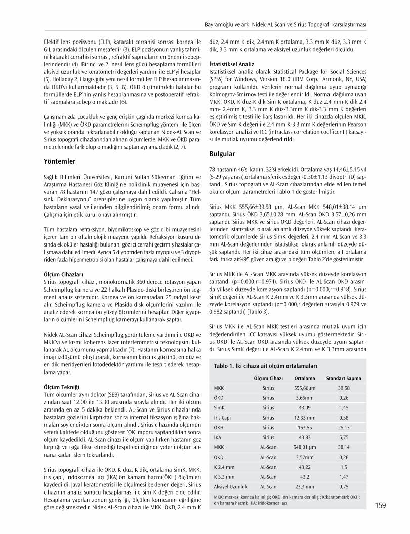
Efektif lens pozisyonu (ELP), katarakt cerrahisi sonrası kornea ile GİL arasındaki ölçülen mesafedir (3). ELP pozisyonun yanlış tahmi-ni katarakt cerrahisi sonrası, refraktif sapmaların en önemli sebep-lerindendir (4). Birinci ve 2. nesil lens gücü hesaplama formülleri aksiyel uzunluk ve keratometri değerleri yardımı ile ELP’yi hesaplar (5). Holladay 2, Haigis gibi yeni nesil formüller ELP hesaplanmasın-da ÖKD’yi kullanmaktadır (3, 5, 6). ÖKD ölçümündeki hatalar bu formüllerde ELP’nin yanlış hesaplanmasına ve postoperatif refrak-tif sapmalara sebep olmaktadır (6).
Çalışmamızda çocukluk ve genç erişkin çağında merkezi kornea ka-lınlığı (MKK) ve ÖKD parametrelerini Scheimpflug yöntemi ile ölçen ve yüksek oranda tekrarlanabilir olduğu saptanan Nidek-AL Scan ve Sirius topografi cihazlarından alınan ölçümlerde, MKK ve ÖKD para-metrelerinde fark olup olmadığını saptamayı amaçladık (2, 7).
Yöntemler
Sağlık Bilimleri Üniversitesi, Kanuni Sultan Süleyman Eğitim ve Araştırma Hastanesi Göz Kliniğine poliklinik muayenesi için baş-vuran 78 hastanın 147 gözü çalışmaya dahil edildi. Çalışma “Hel-sinki Deklarasyonu” prensiplerine uygun olarak yapılmıştır. Tüm hastaların yasal velilerinden bilgilendirilmiş onam formu alındı. Çalışma için etik kurul onayı alınmıştır.
Tüm hastalara refraksiyon, biyomikroskop ve göz dibi muayenesini içeren tam bir oftalmolojik muayene yapıldı. Refraksiyon kusuru dı-şında ek oküler hastalığı bulunan, göz içi cerrahi geçirmiş hastalar ça-lışmaya dahil edilmedi. Ayrıca 5 diyoptriden fazla myopisi ve 3 diyopt-riden fazla hipermetropisi olan hastalar çalışmaya dahil edilmedi.
Ölçüm CihazlarıSirius topografi cihazı, monokromatik 360 derece rotasyon yapan Scheimpflug kamera ve 22 halkalı Plasido-diski birleştiren ön seg-ment analiz sistemidir. Kornea ve ön kamaradan 25 radyal kesit alır. Scheimpflug kamera ve Plasido-disk ölçümlerini yazılım ile analiz ederek kornea ön yüzey ölçümlerini hesaplar. Diğer içyapı-ların ölçümlerini Scheimpflug kamerayı kullanarak saptar.
Nidek AL-Scan cihazı Scheimpflug görüntüleme yardımı ile ÖKD ve MKK’yi ve kısmi koherens lazer interferometrisi teknolojisini kul-lanarak AL ölçümünü yapmaktadır (7). Hastanın korneasına halka imajı izdüşümü oluşturarak, korneanın kırıcılık gücünü, en düz ve en dik meridyenleri fotodedektör yardımı ile tespit ederek hesap-lama yapar.
Ölçüm TekniğiTüm ölçümler aynı doktor (SEB) tarafından, Sirius ve AL-Scan ciha-zından saat 12.00 ile 13.30 arasında sırayla alındı. Her iki ölçüm arasında en az 5 dakika beklendi. AL-Scan ve Sirius cihazlarında hastalara gözlerini kırptıktan sonra internal fiksasyon ışığına bak-maları söylendikten sonra ölçüm alındı. Sirius cihazında ölçümün yeterli kalitede olduğunu gösteren ‘OK’ raporu saptandıktan sonra ölçüm kaydedildi. AL-Scan cihazı ile ölçüm yapılırken hastanın göz kırptığı ve ışığa fikse etmediği tespit edildiğinde yeterli ölçüm alı-nana kadar işlem tekrarlandı.
Sirius topografi cihazı ile ÖKD, K düz, K dik, ortalama SimK, MKK, iris çapı, iridokorneal açı (İKA),ön kamara hacmi(ÖKH) ölçümleri kaydedildi. Javal keratometrisi ile ölçülmesi beklenen değeri, Sirius cihazının analiz sonucu hesaplaması ile Sim K değeri elde edilir. Hesaplama yapılan zonun genişliği, ölçülen korneanın eğriliğine göre değişmektedir. Nidek AL-Scan cihazı ile MKK, ÖKD, 2.4 mm K
düz, 2.4 mm K dik, 2.4mm K ortalama, 3.3 mm K düz, 3.3 mm K dik, 3.3 mm K ortalama ve aksiyel uzunluk değerleri ölçüldü.
İstatistiksel Analizİstatistiksel analiz olarak Statistical Package for Social Sciences (SPSS) for Windows, Version 18.0 (IBM Corp.; Armonk, NY, USA) programı kullanıldı. Verilerin normal dağılıma uyup uymadığı Kolmogrov-Smirnov testi ile değerlendirildi. Normal dağılıma uyan MKK, ÖKD, K düz-K dik-Sim K ortalama, K düz 2.4 mm-K dik 2.4 mm- 2.4mm K, 3.3 mm K düz-3.3mm K dik-3.3 mm K değerleri eşleştirilmiş t testi ile karşılaştırıldı. Her iki cihazda ölçülen MKK, ÖKD ve Sim K değeri ile 2.4 mm K-3.3 mm K değerlerinin Pearson korelasyon analizi ve ICC (intraclass correlation coefficent ) katsayı-sı ile mutlak uyumu değerlendirildi.
Bulgular
78 hastanın 46’sı kadın, 32’si erkek idi. Ortalama yaş 14,46±5.15 yıl (5-29 yaş arası),ortalama sferik eşdeğer -0.30±1.13 diyoptri (D) sap-tandı. Sirius topografi ve AL-Scan cihazlarından elde edilen temel oküler ölçüm parametreleri Tablo 1'de gösterilmiştir.
Sirius MKK 555,66±39.58 μm, AL-Scan MKK 548,01±38.14 μm saptandı. Sirius ÖKD 3,65±0,28 mm, AL-Scan ÖKD 3,57±0,26 mm saptandı. Sirius MKK ve Sirius ÖKD değerleri, AL-Scan cihazı değer-lerinden istatistiksel olarak anlamlı düzeyde yüksek saptandı. Kera-tometrik ölçümlerde Sirius SimK değerleri, 2.4 mm AL-Scan ve 3.3 mm AL-Scan değerlerinden istatistiksel olarak anlamlı düzeyde dü-şük saptandı. Her iki cihaz arasındaki tüm ölçümlere ait ortalama fark, farka ait%95 güven aralığı ve p değeri Tablo 2’de gösterilmiştir.
Sirius MKK ile AL-Scan MKK arasında yüksek düzeyde korelasyon saptandı (p=0.000,r=0.974). Sirius ÖKD ile AL-Scan ÖKD arasın-da yüksek düzeyde korelasyon saptandı (p=0.000,r=0.918). Sirius SimK değeri ile AL-Scan K 2.4mm ve K 3.3mm arasında yüksek dü-zeyde korelasyon saptandı (p=0.000,r değerleri sırasıyla 0.979 ve 0.982 saptandı) (Tablo 3).
Sirius MKK ile AL-Scan MKK testleri arasında mutlak uyum için değerlendirilen ICC katsayısı yüksek uyumu göstermektedir. Siri-us ÖKD ile AL-Scan ÖKD arasında yüksek düzeyde uyum saptan-dı. Sirius SimK değeri ile AL-Scan K 2.4mm ve K 3.3mm arasında
Bayramoğlu ve ark. Nidek-AL Scan ve Sirius Topografi karşılaştırması
159
Tablo 1. İki cihaza ait ölçüm ortalamaları
Ölçüm Cihazı Ortalama Standart Sapma
MKK Sirius 555,66μm 39,58
ÖKD Sirius 3,65mm 0,26
SimK Sirius 43,09 1,45
İris Çapı Sirius 12,33 mm 0,38
ÖKH Sirius 163,55 25,13
İKA Sirius 43,83 5,75
MKK AL-Scan 548,01 μm 38,14
ÖKD AL-Scan 3,57mm 0,26
K 2.4 mm AL-Scan 43,22 1,5
K 3.3 mm AL-Scan 43,2 1,47
Aksiyel Uzunluk AL-Scan 23,3 mm 0,75
MKK: merkezi kornea kalınlığı; ÖKD: ön kamara derinliği; K:keratometri; ÖKH: ön kamara hacmi; İKA: iridokorneal açı
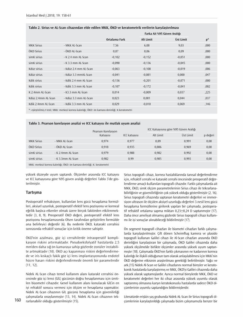
yüksek düzeyde uyum saptandı. Ölçümler arasında ICC katsayısı ve ICC katsayısına göre %95 güven aralığı değerleri Tablo 3’de gös-terilmiştir.
Tartışma
Postoperatif refraksiyon, kullanılan lens gücü hesaplama formül-leri, aksiyel uzunluk, postoperatif efektif lens pozisyonu ve korneal eğrilik başlıca etkenler olmak üzere birçok faktörden etkilenmek-tedir (3, 8, 9). Preoperatif ÖKD değeri, postoperatif efektif lens pozisyonu hesaplamasında Olsen tarafından geliştirilen formülde ana belirleyici değerdir (6). Bu nedenle ÖKD, katarakt cerrahisi sonrasında refraktif sonuçlar için kritik öneme sahiptir.
ÖKD’nin azalması, göz içi cerrahilerde intraoperatif kompli-kasyon riskini artırmaktadır. Pseudoeksfolyatif hastalarda 2,5 mm’den daha sığ ön kamaraya sahip gözlerde zonüler instabili-te artmaktadır (10). ÖKD açı kapanması riskini değerlendirme-de ve iris kıskaçlı fakik göz içi lens implantasyonunda endotel hücre hasarı riskini değerlendirmede önemli bir parametredir (11, 12).
Nidek AL-Scan cihazı temel kullanım alanı katarakt cerrahisi ön-cesinde göz içi lensi (GİL) gücünün doğru hesaplanması için üreti-len biyometri cihazıdır. Genel kullanım alanı konulacak GİL’in en iyi refraktif sonucu vermesi için ölçüm ve hesaplama yapmaktır. Nidek AL-Scan cihazının GİL gücünü hesaplama için güvenilirliği çalışmalarla onaylanmıştır (13, 14). Nidek AL-Scan cihazının tek-rarlanabilir olduğu gösterilmiştir (15).
Sirius topografi cihazı, kornea hastalıklarında tanısal değerlendirme için, refraktif cerrahi ve katarakt cerrahi öncesinde preoperatif değer-lendirme amaçlı kullanılan topografi cihazıdır. Farklı çalışmalarda ait MKK, ÖKD, simK ölçüm parametrelerinin Sirius cihazı ile tekrarlana-bilirliğinin ve güvenilirliğinin çok yüksek olduğu gösterilmiştir (2, 16). Sirius topografi cihazında saptanan keratometri değerleri ve immer-siyon ultrason ile ölçülen aksiyel uzunluğu değerleri 3.nesil lens gücü hesaplama formüllerine girilerek yapılan bir çalışmada, postopera-tif refraktif ortalama sapma miktarı 0,23±0,24 D saptanmıştır (17). Daha önce ameliyat olmamış gözlerde Sirius topografi cihazı kullanı-mı ile iyi sonuçlar alınabileceği bildirilmiştir (17).
Ön segment topografi cihazları ile biometri cihazları farklı çalışma-larda karşılaştırılmıştır. Çift dönen Scheimflug kamera ve plasido topografi kullanan Galilei cihazı ile Al-Scan cihazları arasında ÖKD derinliğini karşılaştıran bir çalışmada, ÖKD Galilei cihazında daha yüksek ölçülmekle birlikte ölçümler arasında yüksek uyum saptan-mıştır (18). Çalışmada ÖKD’nin farklı çıkmasının ne kadarının kornea kalınlığı ile ilişkili olduğunun tam olarak anlaşılabilmesi için MKK’nin ÖKD değerine etkisinin araştırılması gerektiği belirtilmiştir. Yağcı ve ark.(15) Nidek Al-Scan ve Galilei cihazlarını normal bireyler ve kerato-konik hastalarda karşılaştırmış ve MKK, ÖKD’yi Galilei cihazında daha yüksek olarak saptamışlardır. Ayrıca normal bireylerde MKK, ÖKD ve keratometri değerleri her iki cihaz arasında yüksek uyumlu olarak saptanmış olmasına karşın keratokonuslu hastalarda sadece ÖKD öl-çümlerinin uyumlu saptandığını bildirmişlerdir.
Literatürde erişkin yaş grubunda Nidek AL-Scan ile Sirius topografi öl-çümlerinin karşılaştırıldığı çalışmada bizim çalışmamızla benzer bir
İstanbul Med J 2018; 19: 158-61
160
Tablo 2. Sirius ve AL-Scan cihazından elde edilen MKK, ÖKD ve keratometrik verilerin karşılaştırılması
Farka Ait %95 Güven Aralığı
Ortalama Fark Alt Limit Üst Limit p*
MKK Sirius - MKK AL-Scan 7,56 6,08 9,03 ,000
ÖKD Sirius - ÖKD AL-Scan 0,07 0,06 0,09 ,000
simK sirius - K 2.4 mm AL-Scan -0,102 -0,152 -0,051 ,000
simK sirius - K 3.3 mm AL-Scan -0,090 -0,136 -0,045 ,000
Kdüz sirius - Kdüz 2.4 mm AL-Scan -0,063 -0,108 -0,019 ,006
Kdüz sirius - Kdüz 3.3 mmAL-Scan -0,041 -0,081 0,000 ,047
Kdik sirius - Kdik 2.4 mm AL-Scan -0,136 -0,201 -0,071 ,000
Kdik sirius - Kdik 3.3 mm AL-Scan -0,107 -0,172 -0,041 ,002
K 2.4mm AL-Scan - K3.3 mm AL-Scan 0,014 -0,009 0,037 ,225
Kdüz 2.4mm AL-Scan - Kdüz 3.3 mm AL-Scan 0,023 0,001 0,044 ,037
Kdik 2.4mm AL-Scan - Kdik 3.3 mm AL-Scan 0,029 -0,010 0,069 ,146
*: eşleştirilmiş t testi; MKK: merkezi kornea kalınlığı; ÖKD: ön kamara derinliği; K:keratometri
Tablo 3. Pearson korelasyon analizi ve ICC katsayısı ile mutlak uyum analizi
Pearson Korelasyon
ICC Katsayısına göre %95 Güven Aralığı
Katsayısı ICC katsayısı Alt Limit Üst Limit p değeri
MKK Sirius - MKK AL-Scan 0,974 0,977 0,89 0,991 0,00
ÖKD Sirius - ÖKD AL-Scan 0,918 0,935 0,806 0,969 0,00
simK sirius - K 2.4mm AL-Scan 0,979 0,988 0,982 0,992 0,00
simK sirius - K 3.3mm AL-Scan 0,982 0,99 0,985 0,993 0,00
MKK: merkezi kornea kalınlığı; ÖKD: ön kamara derinliği; K: keratometri

şekilde MKK, ÖKD ve keratometri ölçümleri arasında yüksek oranda korelasyon saptanmıştır (19). Çalışmamızda MKK, ÖKD, SimK Sirius ile K 2,4-3,3 mm AL-Scan arasında yüksek korelasyon saptanması, korelasyonun tüm yaş gruplarında devam ettiğini desteklemektedir.
Normal bireylerde Galilei ile AL-Scan ve Sirius ile AL-Scan ölçümle-rini karşılaştıran çalışmalarda Galilei ve Sirius cihazları ile ölçülen MKK ve ÖKD değerlerinin AL-Scan ölçümlerinden daha yüksek fa-kat uyumlu çıkması, Galilei ve Sirius cihazlarının benzer mekaniz-ma ile çalışmasından kaynaklanması olabilir.
MKK’nin doğru hesaplanması refraktif cerrahi öncesi ve glokom hastalarında GİB hesaplanmasında çok önemlidir. Çalışmamızda Sirius cihazında MKK kalınlığının daha kalın saptanmış olması, glokom hastası değerlendirilirken göz önünde bulundurulması ge-reken bir durumdur.
Sonuç
Çalışmamızda biyometrik ÖKD’nin topografik ÖKD’ye göre daha düşük saptanmış olması, cihazlar arasındaki farklı ölçümler sebebi ile lens gücü hesaplama formüllerinin farklı cihazlarda elde edilen ölçümlere göre farklı sonuçlar verebileceğini göstermektedir. Her iki cihaz ölçümleri arasında yüksek korelasyon ve uyum saptanmış olmasına karşın, aradaki farkın klinik olarak önemli olup olmadığı, her iki cihazdan elden edilen verilen ne kadar postoperatif refraktif sapmaya sebep olacağını araştıracak klinik çalışmalar ile saptanabilir.
Etik Komite Onayı: Bu çalışma için etik komite onayı Sağlık Bilimleri Üniversitesi Kanuni Sultan Süleyman Eğitim ve Araştırma Hastanesi Etik Kurulu'ndan alınmıştır (Kabul No: KAEK/2018.3.3).
Hasta Onamı: Hasta onamı bu çalışmaya katılan hastaların ailelerinden ve hastalardan alınmıştır.
Hakem Değerlendirmesi: Dış Bağımsız.
Yazar Katkıları: Fikir - S.E.B.; Tasarım - S.E.B.; Denetleme - N.S., D.Y.E., M.E.; Kaynaklar - S.E.B.; Malzemeler - N.S., D.Y.E., M.E., S.E.B.; Veri Top-lanması ve/veya işlemesi - S.E.B.; Analiz ve/veya Yorum - S.E.B.; Literatür taraması - S.E.B.; Yazıyı Yazan - S.E.B.; Eleştirel İnceleme - N.S., D.Y.E., M.E.
Çıkar Çatışması: Yazarların beyan edecek çıkar çatışması yoktur.
Finansal Destek: Yazarlar bu çalışma için finansal destek almadıklarını beyan etmişlerdir.
Ethics Committee Approval: The ethics committee approval has been received by the Ethics Committee of Sağlık Bilimleri Üniversitesi Kanuni Sultan Süleyman Training and Research Hospital (Approval Number: KAEK/2018.3.3).
Informed Consent: Informed consent was obtained from parents of the patients and patients who participated in this study.
Peer-review: Externally peer-reviewed.
Author contributions: Concept - S.E.B.; Design - S.E.B.; Supervision - N.S., D.Y.E., M.E.; Resource - S.E.B.; Materials - N.S., D.Y.E., M.E., S.E.B.; Data Col-lection and/or Processing - S.E.B.; Analysis and/or Interpretation - S.E.B.; Literature Search - S.E.B.; Writing - S.E.B.; Critical Reviews - N.S., D.Y.E., M.E.
Conflict of Interest: Authors have no conflicts of interest to declare.
Financial Disclosure: The authors declared that this study has received no financial support.
Kaynaklar
1. Kola M, Duran H, Turk A, Mollamehmetoglu S, Kalkisim A, Erdöl H. Evaluation of the repeatability and the reproducibility of AL-scan me-asurements obtained by residents. J Ophthalmol. 2014. [CrossRef]
2. Savini G, Barboni P, Carbonelli M, Hoffer KJ. Repeatability of automatic measurements by a new Scheimpflug camera combined with Placido topography. J Cataract Refract Surg. 2011; 37: 1809-16. [CrossRef]
3. Olsen T. Prediction of the effective postoperative (intraocular lens) anterior chamber depth. J Cataract Refract Surg 2006; 32: 419-24. [CrossRef]
4. Olsen T. Sources of error in intraocular lens power calculation. J Cata-ract Refract Surg 1992; 18: 125-9. [CrossRef]
5. Retzlaff JA, Sanders DR, Kraff MC. Development of the SRK/T int-raocular lens implant power calculation formula. J Cataract Refract Surg. 1990; 16: 333-40. [CrossRef]
6. Olsen T. Calculation of intraocular lens power: a review. Acta Ophthal-mol Scand 2007; 85: 472-85. [CrossRef]
7. Yağcı R, Güler E, Kulak AE, Erdoğan BD, Balcı M, Hepşen İF. Repeatabi-lity and reproducibility of a new optical biometer in normal and kera-toconic eyes. J Cataract Refract Surg. 2015; 41: 171-7. [CrossRef]
8. Hosny M, Alió JL, Claramonte P, Attia WH, Pérez-Santonja JJ. Relations-hip between anterior chamber depth, refractive state, corneal diame-ter, and axial length. J Refract Surg 2000; 16: 336-40.
9. Holladay JT, Musgrove KH, Prager TC, Lewis JW, Chandler TY, Ruiz RS. A three-part system for refining intraocular lens power calculations. J Cataract Refract Surg 1988; 14: 17-24. [CrossRef]
10. Küchle M, Viestenz A, Martus P, Händel A, Jünemann A, Naumann GO. Anterior chamber depth and complications during cataract surgery in eyes with pseudoexfoliation syndrome. Am J Ophthalmol. 2000; 129: 281-5. [CrossRef]
11. Saxena R, Boekhoorn SS, Mulder PG, Noordzij B, van Rij G, Luyten GP. Long-term follow-up of endothelial cell change after Artisan phakic intraocular lens implantation. Ophthalmology 2008; 115: 608-13. [CrossRef]
12. Devereux JG, Foster PJ, Baasanhu J, Uranchimeg D, Lee PS, Erdenbe-leig T, et al. Anterior chamber depth measurement as a screening tool for primary angle-closure glaucoma in an East Asian population. Arch Ophthalmol 2000; 118: 257-63. [CrossRef]
13. Kaswin G, Rousseau A, Mgarrech M, Barreau E, Labetoulle M. Bio-metry and intraocular lens power calculation results with a new op-tical biometry device: comparison with the gold standard. J Cataract Refract Surg. 2014; 40: 593-600. [CrossRef]
14. Suto C, Shimamura E, Watanabe I. Comparison of 2 optical biometers and evaluation of the Camellin-Calossi intraocular lens formula for normal cataractous eyes. J Cataract Refract Surg 2015; 41: 2366-72. [CrossRef]
15. Yağcı R, Kulak AE, Güler E, Tenlik A, Gürağaç FB, Hepşen İF. Compa-rison of anterior segment measurements with a dual Scheimpflug Placido corneal topographer and a new partial coherence interfero-meter in keratoconic eyes. Cornea 2015; 34: 1012-8. [CrossRef]
16. Masoud M, Livny E, Bahar I. Repeatability and intrasession reproduci-bility obtained by the Sirius anterior segment analysis system. Eye contact lens. 2015; 41: 107-10. [CrossRef]
17. Savini G, Barboni P, Carbonelli M, Hoffer KJ. Accuracy of corneal power measurements by a new Scheimpflug camera combined with Placi-do-disk corneal topography for intraocular lens power calculation in unoperated eyes. J Cataract Refract Surg 2012; 38: 787-92. [CrossRef]
18. Dervişoğulları MS, Totan Y, Gürağaç B. Comparison of anterior chamber depth measurements of Nidek AL-Scan and Galilei Dual Scheimpflug Analyzer. Cont Lens Anterior Eye 2015; 38: 85-8. [CrossRef]
19. Çağlar Ç, Kocamış Sİ, Demir E, Durmuş M. Comparison of the measu-rements of a novel optical biometry: Nidek AL-Scan with Sirius and a ultrasound biometry. Int Ophthalmol 2017; 37: 491-8. [CrossRef]
Cite this article as: Bayramoğlu SE, Sayın N, Ekinci DY, Erdoğan M. Comparison of Keratometry, Central Corneal Thickness, and Anterior Chamber Depth Results Measured With Nidek-AL Scan Biometry and Sirius Topography Devices. İstanbul Med J 2018; 19: 158-61.
Bayramoğlu ve ark. Nidek-AL Scan ve Sirius Topografi karşılaştırması
161

Introduction
Chronic otitis media (COM) leads to a chronic inflammation of the middle ear mucosa which can be accompanied by ossicular erosions and/or bony destructions. When there is also an accumulation of keratinizing squamous epithelium in these spaces, it is called COM with cholesteatoma. Oxidative stress and inflammation have been shown to play important roles in several middle ear infections (1-4).
Lymphopenia and neutrophilia show the general inflammatory status of the body (5-7). The neu-trophil-to-lymphocyte ratio (NLR) can be used as a systemic inflammatory marker, as it reflects both the increase in neutrophils and the decrease in lymphocytes (5, 8). Mean platelet volume (MPV) can also be used as a marker of inflammation (9, 10). It measures the volume of circulat-ing thrombocytes, and an increased MPV indicates that the inflammation is more intense (9, 10).
The NLR and MPV values have been previously studied in COM, but, to our knowledge, the difference in inflammatory values between patients with different presentations of COM has not been previ-ously investigated. The aim of the present study was to show the NLR and MPV values in patients who have COM with or without ossicular/bony destruction or cholesteatoma, to compare the results in different groups, and to investigate the predictive values of these parameters in COM.
Methods
This was a retrospective study. İstanbul Training and Research Hospital Ethics Committee (29.12.2017-1158) approved the study in accordance with the Declaration of Helsinki. Patients with COM who had
Neutrophil-to-Lymphocyte Ratio and Mean Platelet Volume in Chronic Otitis Media with or Without CholesteatomaKolesletaomlu ya da Kolesteatomsuz Kronik Otitis Media’da Nötrofil-Lenfosit Oranı ve Platelet Hacminin Prediktif Değeri
Amaç: Nötrofil-lenfosit oranı sistemik bir inflamatuar belirteç olarak kullanılabilir ve artmış platelet hacmi inflamasyonun daha yoğun ol-duğunu gösterir. Bu çalışmanın amacı, Kemikçik zincir destrüksiyonu veya kolesteatomu olan ve olmayan kronik otitis media hastalarında Nötrofil-Lenfosit oranlarının ve Platelet Hacminin prediktif ve prog-nostik değerlerinin incelenmesidir.
Yöntemler: Hastanemizde opere olan kronik otit hastaları retrospektif analiz ile incelenerek üç gruba ayrıldı: Grup 1: sadece timpanik membran perforasyonu olan hastalar, Grup 2: eşlik eden kemikçik zincir deformitesi olan ancak kolesteatomu olmayan hastalar, Grup 3: Kolesteatomu olan hastalar. Kontrol Grup (Grup 0) otolojik bir semptomu ve bulgusu olmayıp hastanemizde septoplasti olan hastalardan oluşmaktaydı. Tüm hastaların ve kontrol grubundaki vakaların ameliyat öncesi kan değerleri incelendi, Platelet Hacmi, Nötrofil ve Lenfosit değerleri kaydedildi, Nötrofil/Lenfosit oranı hesaplandı. Değerler, gruplar ararında karşılaştırıldı.
Bulgular: Çalışmaya 157 kronik otit hastası ve 50 kontrol vakası dahil edildi. Grup 1 ve 2 de 50 hasta, Grup 3’te 57 hasta, kontrol grubunda 50 hasta vardı. Nötrofil, Lenfosit, Nötrofil/Lenfosit oranı ve Platelet Hacmi gruplar arasında fark göstermemekteydi.
Sonuç: Nötrofil/Lenfosit oranı ve Platelet Hacmi birçok hastalığın seyri ve şiddetiyle ilişkili olmasına rağmen, çalışmamızda bu oranlar ile kronik otit arasında bir ilişki bulunamamıştır. Bu değerlerin aynı zamanda kronik otitte kemikçik zincir destruksiyonu ve kolesteatom varlığını ve prognozu ön görmekte prediktif değeri yoktur.
Anahtar Kelimeler: NLR, MPV, kronik otit
Introduction: The neutrophil-to-lymphocyte ratio (NLR) can be used as a systemic inflammatory marker and the increased MPV indicates that the inflammation is more intense. The aim of the study is to show the NLR and MPV values in chronic otitis media with or without ossicular/bony destruction or cholesteatoma and to investigate the predictive values of these parameters..
Methods: Patients with chronic otitis media were retrospectively analy-zed and divided into three groups: group 1 had only tympanic memb-rane perforation without any ossicular/bony erosion or cholesteatoma, group 2 had ossicular/bony erosion but no cholesteatoma, and group 3 had cholesteatoma. The control group (group 0) included subjects who were scheduled for septoplasty in our hospital, who did not have any otologic complaints, and who had normal otologic examination. Blood samples including complete blood count were obtained from all patients in their preoperative visit. The neutrophil, lymphocyte, plate-let, and MPV values were obtained. NLR was calculated by dividing the number of neutrophils by the number of lymphocytes.
Results: The study included 157 patients and 50 controls. There were 50 patients in groups 1 and 2 and 57 patients in group 3. The neutrop-hil, lymphocyte, NLR, MPV, and red blood cell distribution width valu-es showed no statistically significant difference between the groups.
Conclusion: Although NLR is related to the prognosis and severity of se-veral diseases, we found no association between NLR, MPV and chronic ear disease. NLR and MPV also have no value in predicting prognosis and the accompanying ossicular or bony erosion accompanying COM
Keywords: NLR, MPV, chronic otitis
Abst
ract
/ Ö
z
ORCID IDs of all the authors: SE.Y. 0000-0002-9853-6238; Ö.Ö.Ç. 0000-0001-8170-7443; E.A.S. 0000-0002-8462-3605; E.S.L. 0000-0001-6256-2015.
1Department of Otorhinolaryngology/Head and Neck Surgery, Lüleburgaz State Hospital, Kırklareli, Turkey2Department of Otorhinolaryngology/Head and Neck Surgery, İstanbul Training and Research Hospital, İstanbul, Turkey
Address for Correspondence/Yazışma Adresi:Ela Araz Server E-mail: [email protected]
Received/Geliş Tarihi: 03.03.2018Accepted/Kabul Tarihi: 18.04.2018
© Telif Hakkı 2018 Makale metnine istanbultipdergisi.org web sayfasından ulaşılabilir.
© Copyright 2018 by Available online at istanbulmedicaljournal.org
Original Investigation / Orijinal Araştırmaİstanbul Med J 2018; 19: 162-6DOI: 10.5152/imj.2018.77854
Enes Yiğit1 , Özlem Önerci Çelebi2 , Ela Araz Server2 , Ecem Sevim Longur2
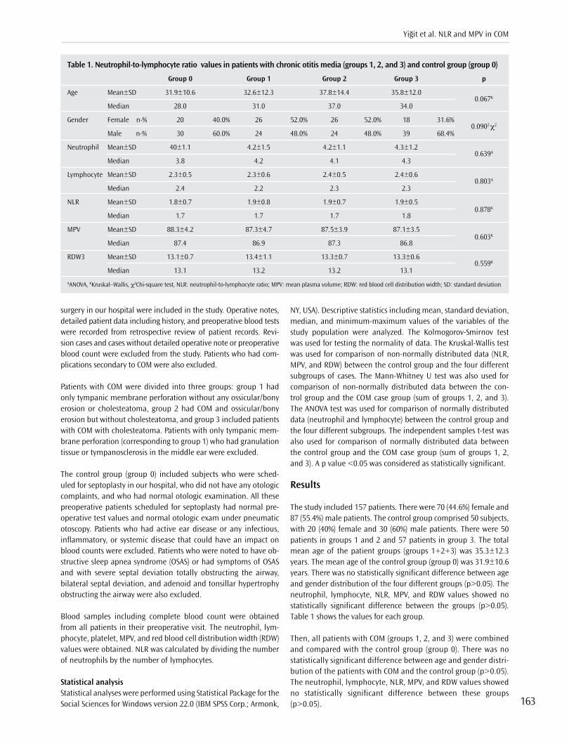
surgery in our hospital were included in the study. Operative notes, detailed patient data including history, and preoperative blood tests were recorded from retrospective review of patient records. Revi-sion cases and cases without detailed operative note or preoperative blood count were excluded from the study. Patients who had com-plications secondary to COM were also excluded.
Patients with COM were divided into three groups: group 1 had only tympanic membrane perforation without any ossicular/bony erosion or cholesteatoma, group 2 had COM and ossicular/bony erosion but without cholesteatoma, and group 3 included patients with COM with cholesteatoma. Patients with only tympanic mem-brane perforation (corresponding to group 1) who had granulation tissue or tympanosclerosis in the middle ear were excluded.
The control group (group 0) included subjects who were sched-uled for septoplasty in our hospital, who did not have any otologic complaints, and who had normal otologic examination. All these preoperative patients scheduled for septoplasty had normal pre-operative test values and normal otologic exam under pneumatic otoscopy. Patients who had active ear disease or any infectious, inflammatory, or systemic disease that could have an impact on blood counts were excluded. Patients who were noted to have ob-structive sleep apnea syndrome (OSAS) or had symptoms of OSAS and with severe septal deviation totally obstructing the airway, bilateral septal deviation, and adenoid and tonsillar hypertrophy obstructing the airway were also excluded.
Blood samples including complete blood count were obtained from all patients in their preoperative visit. The neutrophil, lym-phocyte, platelet, MPV, and red blood cell distribution width (RDW) values were obtained. NLR was calculated by dividing the number of neutrophils by the number of lymphocytes.
Statistical analysisStatistical analyses were performed using Statistical Package for the Social Sciences for Windows version 22.0 (IBM SPSS Corp.; Armonk,
NY, USA). Descriptive statistics including mean, standard deviation, median, and minimum-maximum values of the variables of the study population were analyzed. The Kolmogorov-Smirnov test was used for testing the normality of data. The Kruskal-Wallis test was used for comparison of non-normally distributed data (NLR, MPV, and RDW) between the control group and the four different subgroups of cases. The Mann-Whitney U test was also used for comparison of non-normally distributed data between the con-trol group and the COM case group (sum of groups 1, 2, and 3). The ANOVA test was used for comparison of normally distributed data (neutrophil and lymphocyte) between the control group and the four different subgroups. The independent samples t-test was also used for comparison of normally distributed data between the control group and the COM case group (sum of groups 1, 2, and 3). A p value <0.05 was considered as statistically significant.
Results
The study included 157 patients. There were 70 (44.6%) female and 87 (55.4%) male patients. The control group comprised 50 subjects, with 20 (40%) female and 30 (60%) male patients. There were 50 patients in groups 1 and 2 and 57 patients in group 3. The total mean age of the patient groups (groups 1+2+3) was 35.3±12.3 years. The mean age of the control group (group 0) was 31.9±10.6 years. There was no statistically significant difference between age and gender distribution of the four different groups (p>0.05). The neutrophil, lymphocyte, NLR, MPV, and RDW values showed no statistically significant difference between the groups (p>0.05). Table 1 shows the values for each group.
Then, all patients with COM (groups 1, 2, and 3) were combined and compared with the control group (group 0). There was no statistically significant difference between age and gender distri-bution of the patients with COM and the control group (p>0.05). The neutrophil, lymphocyte, NLR, MPV, and RDW values showed no statistically significant difference between these groups (p>0.05).
Yiğit et al. NLR and MPV in COM
163
Table 1. Neutrophil-to-lymphocyte ratio values in patients with chronic otitis media (groups 1, 2, and 3) and control group (group 0)
Group 0 Group 1 Group 2 Group 3 p
Age Mean±SD 31.9±10.6 32.6±12.3 37.8±14.4 35.8±12.0 0.067K
Median 28.0 31.0 37.0 34.0
Gender Female n-% 20 40.0% 26 52.0% 26 52.0% 18 31.6% 0.0902 χ2
Male n-% 30 60.0% 24 48.0% 24 48.0% 39 68.4%
Neutrophil Mean±SD 40±1.1 4.2±1.5 4.2±1.1 4.3±1.2 0.639A
Median 3.8 4.2 4.1 4.3
Lymphocyte Mean±SD 2.3±0.5 2.3±0.6 2.4±0.5 2.4±0.6 0.803A
Median 2.4 2.2 2.3 2.3
NLR Mean±SD 1.8±0.7 1.9±0.8 1.9±0.7 1.9±0.5 0.878K
Median 1.7 1.7 1.7 1.8
MPV Mean±SD 88.3±4.2 87.3±4.7 87.5±3.9 87.1±3.5 0.603K
Median 87.4 86.9 87.3 86.8
RDW3 Mean±SD 13.1±0.7 13.4±1.1 13.3±0.7 13.3±0.6 0.559K
Median 13.1 13.2 13.2 13.1
AANOVA, KKruskal–Wallis, χ2Chi-square test, NLR: neutrophil-to-lymphocyte ratio; MPV: mean plasma volume; RDW: red blood cell distribution width; SD: standard deviation
Discussion
Chronic otitis media is the inflammation of the middle ear mu-cosa and the temporal bone for >3 months, accompanied by the perforation of the tympanic membrane. Chronic inflammation may act differently in different patients. In some patients, there is only a perforation of the tympanic membrane without any os-sicular or bony destruction. However, in some patients, in addition to tympanic membrane perforation, there is also an erosion or even destruction of the ossicles. This may or may not be accom-panied by growth of the keratinized epithelium in the middle ear, cholesteatoma, which is a lytic process and is generally accompa-nied by bony and ossicular erosions. These different inflammatory processes of the middle ear act differently, and the surgery to be performed to each of these pathologies is different. It is not easy to foresee which patients with COM will develop ossicular/bony erosion or cholesteatoma. It is also hard to predict what to expect during surgery, as the surgical approaches to all these patholo-gies differ. Thus, predictors that would differentiate these different types of chronic ear disease prior to surgery are necessary. This would help a physician to inform his patient about the progress and prognosis of his chronic ear disease and about what to expect after surgery. It would also be beneficial for a physician to pre-dict the status of the middle ear prior to surgery. Thus, we aimed to compare the inflammatory values in these different types of COM. Although one would expect that all these processes causing inflammation would affect the NLR and MPV values, the present study showed that COM with or without ossicular/bony erosion or cholesteatoma had no impact on these values. Even the bone erosion and the osteoclastic activity seen in cholesteatoma had no effect on these values. These findings show that the inflam-mation in these pathologies remains local and does not lead to a systemic inflammatory reaction. Inflammations of the middle ear cavity with or without an accompanying ossicular/bony erosion or cholesteatoma do not increase the NLR or MPV values.
Neutrophil-to-lymphocyte ratio and MPV have been recognized as a systemic inflammatory marker and predictor of poor clinical out-comes for many diseases (11-16). Many biomarkers are available to detect inflammatory cytokines; however, determination of NLR and MPV is simple and readily available ways to detect inflamma-tion without an extra cost. A complete blood cell count is sufficient to determine both values.
Neutrophil-to-lymphocyte ratio has been studied in various diseas-es, such as cardiovascular diseases, chronic renal disease and dia-betic nephropathy, and several types of cancer (11-15, 17), and in various otorhinolaryngological disorders including malignities (18-25), OSAS, Bell’s palsy, sudden hearing loss, tinnitus, adenoidectomy, and tonsillectomy (7, 26-30). It has also been studied in patients with otitis media to detect the effects of inflammation in the middle ear on the inflammatory blood parameters. The effects of oxidative stress on acute otitis media, otitis media with effusion (OME), COM, and tympanosclerosis have been shown, suggesting the role of in-flammation in various inflammatory otologic disorders (1-4, 30-33). The studies showed that NLR is higher in patients with OME and could help in determining the viscosity of the fluid accumulated in the middle ear (30-33). OME can act as a precursor for COM, and the inflammation observed in these studies may act by triggering the inflammatory pathway leading to COM with or without choles-
teatoma. Tansuker et al. investigated the predictive value of NLR to differentiate active from inactive COM and concluded that NLR did not help to differentiate active from inactive COM (16).
The NLR value has also been studied in patients with cholestea-toma. Inflammation causes squamous epithelial transformation in the middle ear leading to cholesteatoma, which may cause lytic and destructive inflammatory reaction. Kilickaya et al. (34) investigated the systemic inflammatory effect of COM with cho-lesteatoma and showed that NLR has no predictive value with re-spect to bone erosions and associated complications in patients with cholesteatoma. Other than that, Eryilmaz et al. (10) studied the NLR values in pediatric patients compared with those in COM with and without cholesteatoma. NLR in the two groups showed no statistically significant difference. However, MPV was lower in patients with cholesteatoma than in the controls, which they sug-gested could act as a predictor for cholesteatoma.
Mean platelet volume has also been shown to be a marker of inflammation and has been studied in various otolaryngological diseases (35, 36). In addition, it has been studied in chronic ear disease. It was reported that MPV is lower in pediatric patients with cholesteatoma, and that it has a role in predicting cholestea-toma in children (10). However, this decrease in MPV has not been shown in our study.
As complications of COM may lead to leukocytosis and systemic toxicity, patients who had complications secondary to COM were excluded in the present study. This is important as these inflam-matory results could affect our results. Other than that, as hypoxia can cause inflammatory and oxidative response (7, 36, 37), pa-tients who were prone to hypoxic conditions, who had OSAS or had symptoms of OSAS, and with severe septal deviation totally ob-structing the airway, bilateral septal deviations, and adenoid and tonsillar hypertrophy were excluded to eliminate the impact of hypoxia on blood test results. One limitation of the present study is its retrospective design and lack of follow-up data after surgery.
To the best of our knowledge, this is the first study to investigate NLR and MPV in patients with COM with or without ossicular/bony destruction or cholesteatoma. Our study shows that a chronic in-flammation in the middle ear cavity with or without ossicular/bony erosion or cholesteatoma remains local and does not trigger a systemic inflammatory reaction.
Conclusion
To our knowledge, the present study is the first to assess the MPV and NLR values in patients with COM and to investigate whether these values have any value in predicting the ossicular and bony erosions and presence of cholesteatoma in advance. However, this effect has not been shown in our study. Although NLR is related to the prognosis and severity of several diseases, we found no asso-ciation between NLR, MPV, and chronic ear disease. NLR and MPV also have no value in predicting the prognosis and the ossicular or bony erosion accompanying COM.
Ethics Committee Approval: Ethics committee approval was received for this study from the ethics committee of İstanbul Training and Research Hospital (29.12.2017-1158).
İstanbul Med J 2018; 19: 162-6
164
Informed Consent: Due to the retrospective design of the study, informed consent was not taken.
Peer-review: Externally peer-reviewed.
Author Contributions: Concept - E.Y., Ö.Ö.Ç., E.A.S., E.S.L.; Design - E.Y., Ö.Ö.Ç., E.A.S., E.S.L.; Supervision - E.Y., Ö.Ö.Ç., E.A.S., E.S.L.; Resources - E.Y., Ö.Ö.Ç., E.A.S., E.S.L.; Materials - E.Y., Ö.Ö.Ç., E.A.S., E.S.L.; Data Collection and/or Processing - E.Y., Ö.Ö.Ç., E.A.S., E.S.L.; Analysis and/or Interpreta-tion - E.Y., Ö.Ö.Ç., E.A.S., E.S.L.; Literature Search - E.Y., Ö.Ö.Ç., E.A.S., E.S.L.; Writing Manuscript - E.Y., Ö.Ö.Ç., E.A.S., E.S.L.; Critical Review - E.Y., Ö.Ö.Ç., E.A.S., E.S.L.; Other - E.Y., Ö.Ö.Ç., E.A.S., E.S.L.
Conflict of Interest: The authors have no conflicts of interest to declare.
Financial Disclosure: The authors declared that this study has received no financial support.
Etik Komite Onayı: Bu çalışma için etik komite onayı İstanbul Eğitim ve Araştırma Hastanesi’nden (29.12.2017-1158) alınmıştır.
Hasta Onamı: Çalışmanın retrospektif tasarımından dolayı hasta onamı alınamamıştır.
Peer-review: Externally peer-reviewed.
Yazar Katkıları: Fikir - E.Y., Ö.Ö.Ç., E.A.S., E.S.L.; Tasarım - E.Y., Ö.Ö.Ç., E.A.S., E.S.L.; Denetleme - E.Y., Ö.Ö.Ç., E.A.S., E.S.L.; Kaynaklar - E.Y., Ö.Ö.Ç., E.A.S., E.S.L.; Malzemeler - E.Y., Ö.Ö.Ç., E.A.S., E.S.L.; Veri Toplanması ve/veya İşlemesi - E.Y., Ö.Ö.Ç., E.A.S., E.S.L.; Analiz ve/veya Yorum - E.Y., Ö.Ö.Ç., E.A.S., E.S.L.; Literatür Taraması - E.Y., Ö.Ö.Ç., E.A.S., E.S.L.; Yazıyı Yazan - E.Y., Ö.Ö.Ç., E.A.S., E.S.L.; Eleştirel İnceleme - E.Y., Ö.Ö.Ç., E.A.S., E.S.L.; Diğer - E.Y., Ö.Ö.Ç., E.A.S., E.S.L.
Çıkar Çatışması: Yazarlar çıkar çatışması bildirmemişlerdir.
Finansal Destek: Yazarlar bu çalışma için finansal destek almadıklarını beyan etmişlerdir.
References
1. Yilmaz T, Kocan EG, Besler HT, Yilmaz G, Gursel B. The role of oxidants and antioxidants in otitis media with effusion in children. Otolaryn-gol Head Neck Surg 2004; 131: 797-803. [CrossRef]
2. Karlidag T, Ilhan N, Kaygusuz I, Keles E, Yalcin S. Comparison of free radicals and antioxidant enzymes in chronic otitis media with and without tympanosclerosis. Laryngoscope 2004; 114: 85-9. [CrossRef]
3. Baysal E, Aksoy N, Kara F, Taysi S, Taşkın A, Bilinç H, et al. Oxidative stress in chronic otitis media. Eur Arch Otorhinolaryngol 2013; 270: 1203-8. [CrossRef]
4. Garça MF, Aslan M, Tuna B, Kozan A, Cankaya H. Serum Myeloperoxidase Activity, Total Antioxidant Capacity and Nitric Oxide Levels in Patients with Chronic Otitis Media. J Membrane Biol 2013; 246: 519-24. [CrossRef]
5. Yenigun A. The efficacy of tonsillectomy in chronic tonsillitis patients as demonstrated by the neutrophil-to-lymphocyte ratio. J Laryngol Otol 2015; 129: 386-91. [CrossRef]
6. Zahorec R. Ratio of neutrophil to lymphocyte counts-rapid and simp-le parameter of systemic inflammation and stress in critically ill. Bra-tisl Lek Listy 2001; 102: 5-14.
7. Derin S, Erdogan S, Sahan M, Topal H, Sozen H. Neutrophil-Lymphocyte Ratio in Patients with Adenoidectomy. J Clin Diagn Res 2016; 10: MC03-5.
8. Roxburgh CS, McMillan DC. Role of systemic inflammatory response in predicting survival in patients with primary operable cancer. Futu-re Oncol 2010; 6: 149-63. [CrossRef]
9. Gasparyan AY, Ayvazyan L, Mikhailidis DP, Kitas GD. Mean Platelet Vo-lume: A Link Between Thrombosis and Inflammation? Curr Pharm Des 2011; 17: 47-58.
10. Eryilmaz, MA, Derin S. Mean Platelet Volume as a Potential Predictor of Cholesteatoma in Children. J Craniofac Surg 2016; 27: e575-8
11. Tamhane UU, Aneja S, Montgomery D, et al. Association between ad-mission neutrophil to lymphocyte ratio and outcomes in patients with acute coronary syndrome. Am J Cardiol 2008;102:653–7 [CrossRef]
12. Halazun KJ, Aldoori A, Malik HZ, Rogers EK, Eagle KA, Gurm HS. Ele-vated preoperative neutrophil to lymphocyte ratio predicts survival following hepatic resection for colorectal liver metastases. Eur J Surg Oncol 2008; 34: 55-60. [CrossRef]
13. Arruda-Olson AM, Reeder GS, Bell MR, et al. Neutrophilia predicts death and heart failure after myocardial infarction: a community-ba-sed study. Circ Cardiovasc Qual Outcomes 2009;2:656–62. [CrossRef]
14. Gibson PH, Croal BL, Cuthbertson BH, Weston SA, Roger VL. Preopera-tive neutrophil lymphocyte ratio and outcome from coronary artery bypass grafting. Am Heart J 2007; 154: 995-1002. [CrossRef]
15. Cedrés S, Torrejon D, Martínez A, Martinez P, Navarro A, Zamora E, et al. Neutrophil to lymphocyte ratio (NLR) as an indicator of poor prog-nosis in stage IV non-small cell lung cancer. Clin Transl Oncol 2012; 14: 864-9. [CrossRef]
16. Tansuker HD, Eroğlu S, Yenigun A, Taskin U, Oktay MF. Can Serum Neutrophil-to-Lymphocyte Ratio Be a Predictive Biomarker to Help Differentiate Active Chronic Otitis Media From Inactive Chronic Otitis Media? J Craniofac Surg 2017; 28: e260-3.
17. Huang W, Huang J, Liu Q, Lin F, He Z, Zeng Z, et al. Neutrophil-lymp-hocyteratio is a reliablepredictive marker forearly-stage diabetic nephropathy. Clin Endocrinol (Oxf) 2015; 82: 229-33. [CrossRef]
18. Kara M, Uysal S, Altinisik U, Cevizci S, Güçlü O, Derekoy FS. The pre-treatment neutrophil-to-lymphocyte ratio, platelet-to-lymphocy-te ratio, and red cell distribution width predict prognosis in patients with laryngeal carcinoma. Eur Arch Otorhinolaryngol 2017; 274: 535-42. [CrossRef]
19. Perisanidis C, Kornek G, Poschl PW, Holzinger D, Pirklbauer K, Schop-per C, et al. High neutrophil to-lymphocyte ratio is an independent marker of poor disease specific survival in patients with oral cancer. Med Oncol 2013; 30: 334-5. [CrossRef]
20. Ozturk K, Akyildiz NS, Uslu M, Gode S, Uluoz U. The effect of preope-rative neutrophil, platelet and lymphocyte counts on local recurrence and survival in early-stage tongue cancer. Eur Arch Otorhinolaryngol 2016; 273: 4425-9. [CrossRef]
21. Kum RO, Ozcan M, Baklacı D. Elevated neutrophil-to lymphocyte ra-tio in squamous cell carcinoma of larynx compared to benign and precancerous laryngeal lesions. Asian Pac J Cancer Prev 2014; 15: 7351-5. [CrossRef]
22. Zeng YC, Chi F, Xing R, Xue M, Wu LN, Tang MY, et al. Pre-treatment neutrophil-to-lymphocyte ratio predicts prognosis in patients with locoregionally advanced laryngeal carcinoma treated with chemora-diotherapy. Jpn J Clin Oncol 2016; 46: 126-31.
23. Salim DK, Mutlu H, Eryilmaz MK, Salim O, Musri FY, Tural D, et al. Neutrophil to lymphocyte ratio is an independent prognostic factor in patient with recurrent or metastatic head and neck squamous cell cancer. Mol Clin Oncol 2015; 3: 839-42. [CrossRef]
24. Haddad CR, Guo L, Clarke S, Guminski A, Back M, Eade T. Neutrop-hil-to-lymphocyte ratio in head and neck cancer. J Med Imaging Ra-diat Oncol 2015; 59: 514-9. [CrossRef]
25. Rachidi S, Wallace K, Wrangle JM, Day TA, Alberg AJ, Li Z. Neutrophil-to lymphocyte ratio and overall survival in all sites of head and neck squamous cell carcinoma. Head Neck 2015; 38: E1068-74.
26. Ozbay I, Kahraman C, Balikci HH, Kucur C, Kahraman NK, Ozkaya DP, et al. Neutrophil-to-lymphocyte ratio in patients with severe tinnitus: prospecti-ve, controlled clinical study. J Laryngol Otol 2015; 129: 544-7. [CrossRef]
27. Chung JH, Lim J, Jeong JH, Kim KR, Park CW, Lee SH. The significance of neutrophil to lymphocyte ratio and platelet to lymphocyte ratio in vestibular neuritis. Laryngoscope 2015; 125: E257-61.
28. Bucak A, Ulu S, Oruc S, Yucedag F, Tekin MS, Karakaya F, et al. Neut-rophil-to-lymphocyte ratio as a novel-potential marker for predicting prognosis of Bell palsy. Laryngoscope 2014; 124: 1678-81. [CrossRef]
29. Karli R, Alacam H, Unal R, Kucuk H, Aksoy A, Ayhan E. Mean platelet volume: is it a predictive parameter in the diagnosis of sudden senso-rineural hearing loss? Indian J Otolaryngol Head Neck Surg 2013; 65: 350-3.
Yiğit et al. NLR and MPV in COM
165
30. Elbistanli MS, Kocak HE, Acipayam H, Yigider AP, Keskin M, Kayhan FT. The Predictive Value of Neutrophil-Lymphocyte and Platelet-Lymp-hocyte Ratio for the EffusionViscosity in Otitis Media With Chronic Effusion. J Craniofac Surg 2017; 28: e244-7.
31. Yellon RF, Leonard G, Marucha PT, Craven R, Carpenter RJ, Lehmann WB, et al. Characterization of cytokines present in middle ear effu-sions. Laryngoscope 1991; 101: 165-9. [CrossRef]
32. Smirnova MG, Kiselev SL, Gnuchev NV, Birchall JP, Pearson JP. Role of the proinflammatory cytokines tumor necrosis factor-alpha, interleu-kin-1 beta, interleukin-6 and interleukin-8 in the pathogenesis of the otitis media with effusion. Eur Cytokine Netw 2002; 13: 161-72.
33. Atan D, Apaydin E, Ozcan KM, Dere H. New diagnostic indicators in chronic otitis media with effusion: neutrophil to lymphocyte ratio and thrombocyte lymphocyte ratio. ENT Updates 2016; 6: 12-5. [CrossRef]
34. Kılıckaya MM, Aynali G, Tuz M, Bagcı O. Is There A Systemic Inflam-matory Effect of Cholesteatoma? Int Arch Otorhinolaryngol 2017; 21: 42-5.
35. Unlu I, Kesici GG, Onec B, Yaman H, Guclu E. The effect of duration of nasal obstruction on mean platelet volume in patients with marked nasal septal deviation. Eur Arch Otorhinolaryngol 2016; 273: 401-5. [CrossRef]
36. Rahangdale S, Yeh SY, Novack V, Stevenson K, Barnard MR, Furman MI, et al. The influence of intermittent hypoxemia on platelet activa-tion in obese patients with obstructive sleep apnea. J Clin Sleep Med 2011; 7: 172-8.
37. Korkmaz M, Korkmaz H, Küçüker F, Ayyıldız SN, Çankaya S. Evalua-tion of the Association of Sleep Apnea-Related Systemic Inflammation with CRP, ESR, and Neutrophil-to-Lymphocyte Ratio. Med Sci Monit 2015; 21: 477-81. [CrossRef]
Cite this article as: Yiğit E, Önerci Çelebi Ö, Araz Server E, Longur ES. Neutrophil-to-Lymphocyte Ratio and Mean Platelet Volume in Chronic Otitis Media with or Without Cholesteatoma. İstanbul Med J 2018; 19: 162-6.
İstanbul Med J 2018; 19: 162-6
166

Introduction
Hyperhidrosis (HH) is a pathological condition of excessive secretion of the eccrine sweat glands in amounts greater than that required for physiological needs. HH occurs more commonly in women and is usually bilateral and symmetrical. It often begins in early childhood and usually persists throughout adulthood, resulting in severe occupational, emotional, and social disabilities (1). Although the prevalence of HH is thought to be low, it has been reported as 2.8% in the United States and 4.9% in China (2). Various treatment methods have been recommended for HH. Gener-ally, the preferred treatment method varies according to the clinician. The success and compli-ance rates of medical treatments are significantly lower, particularly in patients with life-long or moderate to severe HH. Permanent management can be achieved through invasive techniques such as sympathectomy. Except for one series, no mortality has been reported, with a morbidity not exceeding 5%, in the English literature. Pneumothorax is the most common complication (2% of patients; a total of 25%-30% exclusively require thoracic drainage), followed by subcutaneous emphysema (1%) and pleural effusion (0.3%-0.5%), which rarely requires drainage. Solitary cases of complications, such as chylothorax, hemopericardium, lesions of the superior intercostal vein, pulmonary edema, or brachial plexus lesions, have been reported in a previous study (1). Herein, we describe a patient who was treated with autologous blood pleurodesis for ductus thoracicus injury after endoscopic thoracic sympathectomy.
Case Report
A 23-year-old woman was admitted to our clinic with a complaint of bilateral pronounced axil-lary HH, minimal sweating of the hands, and bruising. She underwent bilateral thoracic sympa-thectomy at levels T3 and T4 via a diathermy hook inserted through the thoracoscope (Karl Storz Hopkins 0° 26034 AA Laparoscope Tuttlingen,Germany). Electrocautery ablation of the accessory nerve and Kuntz branches was composed of approximately 3 cm to prevent recurrence at the level of the third rib. Chest X-ray was obtained in the recovery room after surgery to verify full
Chylothorax: A Rare Complication of Endoscopic Thoracic SympathectomyŞilotoraks: Endoskopik Torasik Sempatektominin Nadir Bir Komplikasyonu
Hyperhidrosis (HH) is a pathological condition of excessive secretion of the eccrine sweat glands in amounts greater than that required for physiological needs. Herein, we describe a patient who was treated with autologous blood pleurodesis for ductus thoracicus injury after endoscopic thoracic sympathectomy. A 23-year-old woman was ad-mitted to our clinic with a complaint of bilateral pronounced axillary HH, minimal sweating of the hands, and bruising. She underwent bilateral thoracic sympathectomy at levels T3 and T4. A milky fluid was observed in the left chest tube and was diagnosed as chylothorax. No similar case of postoperative chylothorax treated with autologous blood pleurodesis has been found in the English literature. According to anatomical variations, the ductus thoracicus is susceptible to in-jury even in the hands of an experienced surgeon. In case an injury has occurred shortly after thoracic sympathectomy, autologous blood pleurodesis is an effective treatment for chylothorax. This procedure is safe and cheap and can be easily performed at the bedside.
Keywords: Chylothorax, Thoracic, Sympathectomy, Pleurodesis, Au-tologous Blood
Terleme, vücut sıcaklığını sabit tutulmasında önemli bir mekanizma-dır. Hiperhidrozis ekrin ter bezlerin fizyolojik ihtiyaçları için gerekli olandan daha fazla miktarlarda aşırı ter salgılanması durumudur. Bu-rada, torakal sempatektomi sonrası gelişen ve otolog kan plörezisiyle tedavi edilen vakayı sunduk. 23 yaşında bayan hasta aksillada daha belirgin olmak üzere ellerde minimal terleme; soğukta morarma şikâyeti ile kliniğimize başvurdu. Hastaya T3-T4 seviyesinden bilateral torakal sempatektomi yapıldı. Sol göğüs tüpünde süt gibi bir sıvı göz-lendi ve şilotoraks teşhisi kondu. İngilizce literatür taramasında oto-log kan ile plöredezis yapılan başka bir vakaya rastlamadık. Duktus torasikusun anatomik varyasyonları nedeniyle en deneyimli cerrahın elinde bile yaralanma açıktır. Torakal sempatektomiden sonra hemen sonra gelişen şilotoraks için otolog kanla yapılan plörodezis etkili bir tedavi yöntemidir. Bu işlem ucuz, kolay ve hasta başında rahatlıkla yapılabilecek bir tedavidir.
Anahtar Kelimeler: Şilotoraks, Torasik, sempatektomi, Plörodez, Oto-log Kan
Abst
ract
/ Ö
z
This study was presented in the American College of Chest Physicians CHEST Congress (October 26-31, 2013, Chicago, USA)
ORCID IDs of all the authors: S0000-0001-9963-5724; H.E. 0000-0002-6569-8193; T.T.B. 0000-0002-5705-8452; S.G.Ç. 0000-0002-6569-8193.
1Department of Thoracic Surgery, Health Sciences University, Konya Training and Research Hospital, Konya, Turkey.2Department of Pulmonary Medicine, Health Sciences University, Konya Training and Research Hospital, Konya, Turkey.3Emergency and First Aid Program, Vocational School of Health Services KTO Karatay University, Konya, Turkey.
Address for Correspondence/Yazışma Adresi:Mustafa ÇalıkE-mail: [email protected]
Received/Geliş Tarihi: 16.04.2017
Accepted/Kabul Tarihi: 28.10.2017
© Telif Hakkı 2018 Makale metnine istanbultipdergisi.org web sayfasından ulaşılabilir.
© Copyright 2018 by Available online at istanbulmedicaljournal.org
Case Report/Olgu Sunumuİstanbul Med J 2018; 19: 167-9DOI: 10.5152/imj.2018.63308
Mustafa Çalık1 , Hıdır Esme1 , Taha Tahir Bekçi2 , Saniye Göknil Çalık3
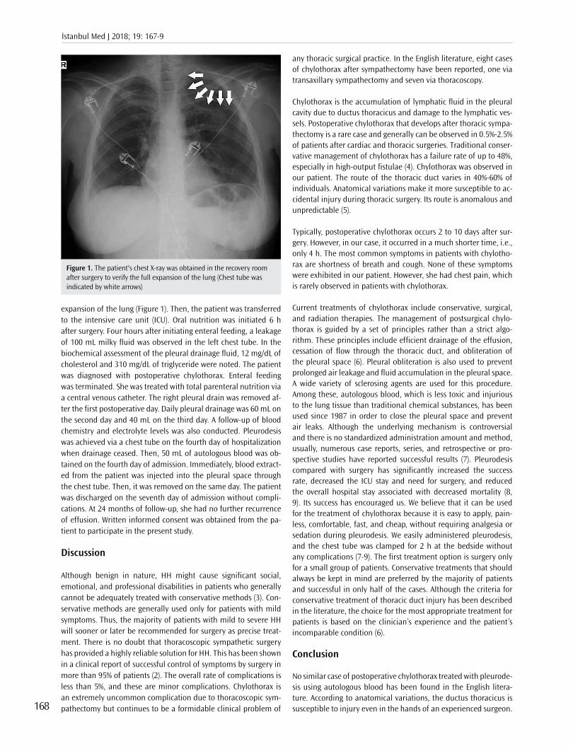
expansion of the lung (Figure 1). Then, the patient was transferred to the intensive care unit (ICU). Oral nutrition was initiated 6 h after surgery. Four hours after initiating enteral feeding, a leakage of 100 mL milky fluid was observed in the left chest tube. In the biochemical assessment of the pleural drainage fluid, 12 mg/dL of cholesterol and 310 mg/dL of triglyceride were noted. The patient was diagnosed with postoperative chylothorax. Enteral feeding was terminated. She was treated with total parenteral nutrition via a central venous catheter. The right pleural drain was removed af-ter the first postoperative day. Daily pleural drainage was 60 mL on the second day and 40 mL on the third day. A follow-up of blood chemistry and electrolyte levels was also conducted. Pleurodesis was achieved via a chest tube on the fourth day of hospitalization when drainage ceased. Then, 50 mL of autologous blood was ob-tained on the fourth day of admission. Immediately, blood extract-ed from the patient was injected into the pleural space through the chest tube. Then, it was removed on the same day. The patient was discharged on the seventh day of admission without compli-cations. At 24 months of follow-up, she had no further recurrence of effusion. Written informed consent was obtained from the pa-tient to participate in the present study.
Discussion
Although benign in nature, HH might cause significant social, emotional, and professional disabilities in patients who generally cannot be adequately treated with conservative methods (3). Con-servative methods are generally used only for patients with mild symptoms. Thus, the majority of patients with mild to severe HH will sooner or later be recommended for surgery as precise treat-ment. There is no doubt that thoracoscopic sympathetic surgery has provided a highly reliable solution for HH. This has been shown in a clinical report of successful control of symptoms by surgery in more than 95% of patients (2). The overall rate of complications is less than 5%, and these are minor complications. Chylothorax is an extremely uncommon complication due to thoracoscopic sym-pathectomy but continues to be a formidable clinical problem of
any thoracic surgical practice. In the English literature, eight cases of chylothorax after sympathectomy have been reported, one via transaxillary sympathectomy and seven via thoracoscopy.
Chylothorax is the accumulation of lymphatic fluid in the pleural cavity due to ductus thoracicus and damage to the lymphatic ves-sels. Postoperative chylothorax that develops after thoracic sympa-thectomy is a rare case and generally can be observed in 0.5%-2.5% of patients after cardiac and thoracic surgeries. Traditional conser-vative management of chylothorax has a failure rate of up to 48%, especially in high-output fistulae (4). Chylothorax was observed in our patient. The route of the thoracic duct varies in 40%-60% of individuals. Anatomical variations make it more susceptible to ac-cidental injury during thoracic surgery. Its route is anomalous and unpredictable (5).
Typically, postoperative chylothorax occurs 2 to 10 days after sur-gery. However, in our case, it occurred in a much shorter time, i.e., only 4 h. The most common symptoms in patients with chylotho-rax are shortness of breath and cough. None of these symptoms were exhibited in our patient. However, she had chest pain, which is rarely observed in patients with chylothorax.
Current treatments of chylothorax include conservative, surgical, and radiation therapies. The management of postsurgical chylo-thorax is guided by a set of principles rather than a strict algo-rithm. These principles include efficient drainage of the effusion, cessation of flow through the thoracic duct, and obliteration of the pleural space (6). Pleural obliteration is also used to prevent prolonged air leakage and fluid accumulation in the pleural space. A wide variety of sclerosing agents are used for this procedure. Among these, autologous blood, which is less toxic and injurious to the lung tissue than traditional chemical substances, has been used since 1987 in order to close the pleural space and prevent air leaks. Although the underlying mechanism is controversial and there is no standardized administration amount and method, usually, numerous case reports, series, and retrospective or pro-spective studies have reported successful results (7). Pleurodesis compared with surgery has significantly increased the success rate, decreased the ICU stay and need for surgery, and reduced the overall hospital stay associated with decreased mortality (8, 9). Its success has encouraged us. We believe that it can be used for the treatment of chylothorax because it is easy to apply, pain-less, comfortable, fast, and cheap, without requiring analgesia or sedation during pleurodesis. We easily administered pleurodesis, and the chest tube was clamped for 2 h at the bedside without any complications (7-9). The first treatment option is surgery only for a small group of patients. Conservative treatments that should always be kept in mind are preferred by the majority of patients and successful in only half of the cases. Although the criteria for conservative treatment of thoracic duct injury has been described in the literature, the choice for the most appropriate treatment for patients is based on the clinician’s experience and the patient’s incomparable condition (6).
Conclusion
No similar case of postoperative chylothorax treated with pleurode-sis using autologous blood has been found in the English litera-ture. According to anatomical variations, the ductus thoracicus is susceptible to injury even in the hands of an experienced surgeon.
Figure 1. The patient's chest X-ray was obtained in the recovery room after surgery to verify the full expansion of the lung (Chest tube was indicated by white arrows)
İstanbul Med J 2018; 19: 167-9
168

In case an injury has occurred shortly after thoracic sympathec-tomy, autologous blood pleurodesis is an effective treatment for chylothorax. This procedure is safe and cheap and can be easily performed at the bedside.
Informed Consent: Written informed consent was obtained from patient who participated in this study.
Peer-review: Externally peer-reviewed.
Author Contributions: Concept - M.Ç., H.E., T.T.B., S.G.Ç.; Design - M.Ç., H.E., T.T.B., S.G.Ç.; Supervision - M.Ç., H.E., T.T.B., S.G.Ç.; Resource - M.Ç., H.E., T.T.B., S.G.Ç.; Materials - M.Ç., H.E., T.T.B., S.G.Ç.; Data Collection and/or Processing - M.Ç., S.G.Ç. ; Analysis and/or Interpretation - M.Ç., S.G.Ç.; Literature Search - M.Ç., S.G.Ç.; Writing - M.Ç., S.G.Ç.; Critical Reviews - M.Ç., S.G.Ç.
Conflict of Interest: Authors have no conflicts of interest to declare.
Financial Disclosure: The authors declared that this study has received no financial support.
Hasta Onamı: Yazılı hasta onamı bu çalışmaya katılan hastadan alınmıştır.
Hakem Değerlendirmesi: Dış Bağımsız.
Yazar Katkıları: Fikir - M.Ç., H.E., T.T.B., S.G.Ç.; Tasarım - M.Ç., H.E., T.T.B., S.G.Ç. ; Denetleme - M.Ç., H.E., T.T.B., S.G.Ç.; Kaynaklar - M.Ç., H.E., T.T.B., S.G.Ç.; Malzemeler - M.Ç., H.E., T.T.B., S.G.Ç.; Veri Toplanması ve/veya işlemesi - M.Ç., S.G.Ç.; Analiz ve/veya Yorum - M.Ç., S.G.Ç.; Literatür taraması - M.Ç., S.G.Ç.; Yazıyı Yazan - M.Ç., S.G.Ç.; Eleştirel İnceleme - M.Ç., S.G.Ç.
Çıkar Çatışması: Yazarların beyan edecek çıkar çatışması yoktur.
Finansal Destek: Yazarlar bu çalışma için finansal destek almadıklarını beyan etmişlerdir.
References
1. Macía I, Moya J, Ramos R, Rivas F, Urena A, Rosado G, et al. [Primary hyperhidrosis. Current status of surgical treatment]. Cir Esp 2010; 88: 146-51. [Article in Spanish] [CrossRef]
2. Sugimura H, Spratt EH, Compeau CG, Kattail D, Shargall Y. Thoraco-scopic sympathetic clipping for hyperhidrosis: long-term results and reversibility. J Thorac Cardiovasc Surg 2009; 137: 1370-6. [CrossRef]
3. Reisfeld R, Nguyen R, Pnini A. Endoscopic thoracic sympathectomy for hyperhidrosis: experience with both cauterization and clamping meth-ods. Surg Laparosc Endosc Percutan Tech 2002; 12: 255-67. [CrossRef]
4. Kumar S, Kumar A, Pawar DK. Thoracoscopic management of thoracic duct injury: Is there a place for conservatism? J Postgrad Med 200; 50: 57-9.
5. Apiliogullari B, Esme H, Yoldas B, Duran M, Duzgun N, Calik M. Early and midterm results of single-port video-assisted thoracoscopic sym-pathectomy. Thorac Cardiovasc Surg 2012; 60: 285-9. [CrossRef]
6. Stager V, Le L, Wood RE. Postoperative chylothorax successfully treat-ed using conservative strategies Proc (Bayl Univ Med Cent) 2010; 23: 134-8 [CrossRef]
7. Muruganandan, S, Kumar S, Lee, YCG. Blood Patch for Pneumothorax: A Literature Review. Cur Pulmonol Rep, 2017; 6: 30-8. [CrossRef]
8. Alamdari DH, Asadi M, Rahim AN, Maddah G, Azizi S, Shahidsales S, et al. Efficacy and Safety of Pleurodesis Using Platelet-Rich Plasma and Fibrin Glue in Management of Postoperative Chylothorax After Esophagectomy. World J Surg 2018; 42: 1046-55. [CrossRef]
9. Akin H, Olcmen A, Isgorucu O, Denizkiran I, Dincer I. Approach to patients with chylothorax complicating pulmonary resection. Thorac Cardiovasc Surg 2012; 60: 135-9. [CrossRef]
Cite this article as: Çalık M, Esme H, Bekçi TT, Göknil Çalık S. Chylothorax: A Rare Complication of Endoscopic Thoracic Sympathectomy İstanbul Med J 2018; 19: 167-9.
Mustafa Çalık. Chylothorax: A Complication of Sympathectomy
169

Giriş
Klinefelter sendromu primer hipogonadizme neden olan en yaygın konjenital anomalidir. Bin canlı erkek doğumun yaklaşık 1’inde meydana gelir (1). En sık görülen genotipi 46, XXY'dir. Fakat 48, XXXY ve 46, XY / 46, XXY mozaikliği gibi karyotiplere yol açan daha büyük ve daha az sayıda X kromozomu da rapor edilmiştir (2). Testosteron düzeyinin azalması, FSH-LH artması testislerde atrofiye, infertiliteye ve azalmış virilizasyon bulgularına sebep olur (3). Yaşamın ileri dönemlerin-de testosteron eksikliği ile ilişkisi olmayan morbiditeye neden olabilir (4). Bunlara kronik bron-şit, bronşektazi ve amfizem gibi akciğer hastalıkları, germ hücreli tümörler, meme kanseri ve non-Hodgkin lenfomayı da içeren kanserler; bacak ülserlerine yol açan variköz damarlar, ekstra X kromozomuna bağlı sistemik lupus eritematosus ve diabetes mellitus dahildir (5-9). Bu olgu sunumunda, Klinefelter sendromu tanısı ile izlenen hastada, edinsel trombotik trombositopenik turpura (TTP) gelişimini bildirdik.
Olgu Sunumu
Klinefelter sendromu tanılı 36 yaşında erkek hasta, 1 haftadır olan baş ağrısının şiddetlenmesi ve bilinç değişikliği olması üzerine acil servise getirilmiş. Acil serviste değerlendirilen hastanın bilinci kapalı, ağrılı uyarana yanıtı yoktu. Kan basıncı 90/60 mmHg, nabız 116/dk, ateş 36,7°C idi. Yapılan tetkiklerinde wbc 10 900 /μL(4000-10000), hemoglobin 6,2 g/dL (13-17), trombosit sayısı (plt) 7000 /μL (150000-450000), retikülosit yüzdesi %17,2 (0,5-2), kreatinin 1,39 mg/dL (0,7-1,2), LDH: 1749 U/L (125-220), total bilirübin 1,99 mg/dL (0,2-1,2), indirekt bilirübin: 1,33 mg/dL (0,1-0,7), SGPT 33 U/L (0-55), SGOT: 68 U/L (5-34), direkt ve indirekt coombs testleri negatif, haptoglobulin <9 mg/dL (40-240), B12: 291 pg/mL (195-961), folik asit 3,51 ng/mL (3,1-19,9), anti nükleer antikor nega-tif olarak saptandı. Periferik yaymasında her alanda yaygın şistositler ve trombositopeni izlendi (Resim 1).
TTP düşünülerek ADAMTS13 için kan örneği alınıp metilprednizolon 1 mg/kg/gün başlandı ve 1 plazma volümü plazma değişimi yapıldı. İlk plazmaferezden sonra bilinci açıldı, LDH 377 U/L’ye düştü, trombosit sayısı 47 000 /μL’ye yükseldi. Plazma değişimine devam edildi. Üçüncü plazma-ferez sonrası platelet sayısı 164 000 /μL oldu. Tedaviye asetilsalisilik asit 100 mg/gün eklendi. ADAMTS13 aktivitesi <%0,2 (40-1330), ADAMTS13 antijeni 0,06 μg/mL (0,6-1,6), ADAMTS13 inhi-bitör düzeyi 60,90 U/mL (<12) saptandı. Hasta kazanılmış TTP olarak kabul edildi. Toplam 11 kez plazma değişimi yapıldı, metilprednizolon azaltılarak 3 ayda kesildi. Hasta halen remisyonda izlenmektedir. Hasta onamı alınarak sunulmuştur.
Klinefelter Sendromlu Hastada Gelişen Trombotik Trombositopenik PurpuraThrombotic Thrombocytopenic Purpura in a Patient with Klinefelter Syndrome
Thrombotic thrombocytopenic purpura is a rare disease associated with microangiopathic hemolytic anemia, thrombocytopenia, fever, neurological disorders, and renal insufficiency pentad. It is a fatal he-matologic emergency if left untreated. If plasma exchange is feasible, treatment is easy and comfortable. It should be kept in mind that such as in our patient may be acquired at all congenital illnesses.
Keywords: Klinefelter syndrome, thrombotic thrombocytopenic pur-pura, plasma exchange
Trombotik trombositopenik purpura, mikroanjiyopatik hemolitik ane-mi, trombositopeni, ateş, nörolojik bozukluklar ve böbrek yetmezliği pentadı ile tanınan nadir görülen bir hastalıktır. Tedavi edilmezse ölüm-cül olan hematolojik acil durumdur. Plazma değişimi yapılabiliyorsa tedavi oldukça kolay ve yüzgüldürücüdür. Bizim hastamızda olduğu gibi konjenital tüm hastalıklarda edinsel olarak görülebileceği akılda tutul-malıdır.
Anahtar Kelimeler: Klinefelter sendromu, trombotik trombositopenik purpura, plazma değişimi
Öz
/ Abs
trac
t
ORCID IDs of the authors: S.D. 0000-0003-1277-5105; S.Y. 0000-0002-7222-499X; Ö.B. 0000-0002-2670-022X; Ö.Ç. 0000-0003-2541-1335.
Bu çalışma 2. Selim Hematoloji Güncelleme Sempozyumu’nda sunulmuştur (24-26 Şubat 2017, Antalya, Türkiye).
Necmettin Erbakan Üniversitesi, Meram Tıp Fakültesi, Hematoloji Bilim Dalı, Konya, Türkiye
Yazışma Adresi/Address for Correspondence:Sinan Demircioğlu E-mail: [email protected]
Geliş Tarihi/Received: 17.07.2017
Kabul Tarihi/Accepted: 29.11.2017
© Telif Hakkı 2018 Makale metnine istanbultipdergisi.org web sayfasından ulaşılabilir.
© Copyright 2018 by Available online at istanbulmedicaljournal.org
Olgu Sunumu/Case Reportİstanbul Med J 2018; 19: 170-2DOI: 10.5152/imj.2018.62681
Sinan Demircioğlu , Seda Yılmaz , Özlen Bektaş , Özcan Çeneli
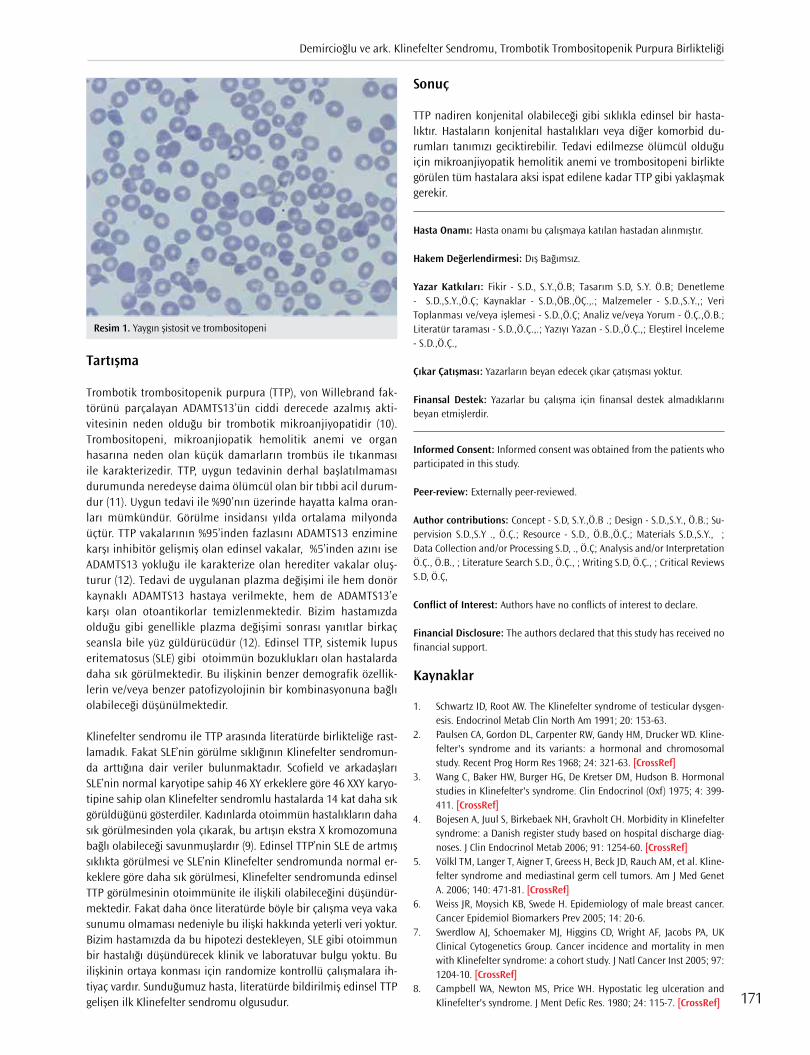
Tartışma
Trombotik trombositopenik purpura (TTP), von Willebrand fak-törünü parçalayan ADAMTS13’ün ciddi derecede azalmış akti-vitesinin neden olduğu bir trombotik mikroanjiyopatidir (10). Trombositopeni, mikroanjiopatik hemolitik anemi ve organ hasarına neden olan küçük damarların trombüs ile tıkanması ile karakterizedir. TTP, uygun tedavinin derhal başlatılmaması durumunda neredeyse daima ölümcül olan bir tıbbi acil durum-dur (11). Uygun tedavi ile %90’nın üzerinde hayatta kalma oran-ları mümkündür. Görülme insidansı yılda ortalama milyonda üçtür. TTP vakalarının %95’inden fazlasını ADAMTS13 enzimine karşı inhibitör gelişmiş olan edinsel vakalar, %5’inden azını ise ADAMTS13 yokluğu ile karakterize olan herediter vakalar oluş-turur (12). Tedavi de uygulanan plazma değişimi ile hem donör kaynaklı ADAMTS13 hastaya verilmekte, hem de ADAMTS13’e karşı olan otoantikorlar temizlenmektedir. Bizim hastamızda olduğu gibi genellikle plazma değişimi sonrası yanıtlar birkaç seansla bile yüz güldürücüdür (12). Edinsel TTP, sistemik lupus eritematosus (SLE) gibi otoimmün bozuklukları olan hastalarda daha sık görülmektedir. Bu ilişkinin benzer demografik özellik-lerin ve/veya benzer patofizyolojinin bir kombinasyonuna bağlı olabileceği düşünülmektedir.
Klinefelter sendromu ile TTP arasında literatürde birlikteliğe rast-lamadık. Fakat SLE’nin görülme sıklığının Klinefelter sendromun-da arttığına dair veriler bulunmaktadır. Scofield ve arkadaşları SLE’nin normal karyotipe sahip 46 XY erkeklere göre 46 XXY karyo-tipine sahip olan Klinefelter sendromlu hastalarda 14 kat daha sık görüldüğünü gösterdiler. Kadınlarda otoimmün hastalıkların daha sık görülmesinden yola çıkarak, bu artışın ekstra X kromozomuna bağlı olabileceği savunmuşlardır (9). Edinsel TTP’nin SLE de artmış sıklıkta görülmesi ve SLE’nin Klinefelter sendromunda normal er-keklere göre daha sık görülmesi, Klinefelter sendromunda edinsel TTP görülmesinin otoimmünite ile ilişkili olabileceğini düşündür-mektedir. Fakat daha önce literatürde böyle bir çalışma veya vaka sunumu olmaması nedeniyle bu ilişki hakkında yeterli veri yoktur. Bizim hastamızda da bu hipotezi destekleyen, SLE gibi otoimmun bir hastalığı düşündürecek klinik ve laboratuvar bulgu yoktu. Bu ilişkinin ortaya konması için randomize kontrollü çalışmalara ih-tiyaç vardır. Sunduğumuz hasta, literatürde bildirilmiş edinsel TTP gelişen ilk Klinefelter sendromu olgusudur.
Sonuç
TTP nadiren konjenital olabileceği gibi sıklıkla edinsel bir hasta-lıktır. Hastaların konjenital hastalıkları veya diğer komorbid du-rumları tanımızı geciktirebilir. Tedavi edilmezse ölümcül olduğu için mikroanjiyopatik hemolitik anemi ve trombositopeni birlikte görülen tüm hastalara aksi ispat edilene kadar TTP gibi yaklaşmak gerekir.
Hasta Onamı: Hasta onamı bu çalışmaya katılan hastadan alınmıştır.
Hakem Değerlendirmesi: Dış Bağımsız.
Yazar Katkıları: Fikir - S.D., S.Y.,Ö.B; Tasarım S.D, S.Y. Ö.B; Denetleme - S.D.,S.Y.,Ö.Ç; Kaynaklar - S.D.,ÖB.,ÖÇ.,.; Malzemeler - S.D.,S.Y.,; Veri Toplanması ve/veya işlemesi - S.D.,Ö.Ç; Analiz ve/veya Yorum - Ö.Ç.,Ö.B.; Literatür taraması - S.D.,Ö.Ç.,.; Yazıyı Yazan - S.D.,Ö.Ç.,; Eleştirel İnceleme - S.D.,Ö.Ç.,
Çıkar Çatışması: Yazarların beyan edecek çıkar çatışması yoktur.
Finansal Destek: Yazarlar bu çalışma için finansal destek almadıklarını beyan etmişlerdir.
Informed Consent: Informed consent was obtained from the patients who participated in this study.
Peer-review: Externally peer-reviewed.
Author contributions: Concept - S.D, S.Y.,Ö.B .; Design - S.D.,S.Y., Ö.B.; Su-pervision S.D.,S.Y ., Ö.Ç.; Resource - S.D., Ö.B.,Ö.Ç.; Materials S.D.,S.Y., ; Data Collection and/or Processing S.D, ., Ö.Ç; Analysis and/or Interpretation Ö.Ç., Ö.B., ; Literature Search S.D., Ö.Ç., ; Writing S.D, Ö.Ç., ; Critical Reviews S.D, Ö.Ç,
Conflict of Interest: Authors have no conflicts of interest to declare.
Financial Disclosure: The authors declared that this study has received no financial support.
Kaynaklar
1. Schwartz ID, Root AW. The Klinefelter syndrome of testicular dysgen-esis. Endocrinol Metab Clin North Am 1991; 20: 153-63.
2. Paulsen CA, Gordon DL, Carpenter RW, Gandy HM, Drucker WD. Kline-felter's syndrome and its variants: a hormonal and chromosomal study. Recent Prog Horm Res 1968; 24: 321-63. [CrossRef]
3. Wang C, Baker HW, Burger HG, De Kretser DM, Hudson B. Hormonal studies in Klinefelter's syndrome. Clin Endocrinol (Oxf) 1975; 4: 399-411. [CrossRef]
4. Bojesen A, Juul S, Birkebaek NH, Gravholt CH. Morbidity in Klinefelter syndrome: a Danish register study based on hospital discharge diag-noses. J Clin Endocrinol Metab 2006; 91: 1254-60. [CrossRef]
5. Völkl TM, Langer T, Aigner T, Greess H, Beck JD, Rauch AM, et al. Kline-felter syndrome and mediastinal germ cell tumors. Am J Med Genet A. 2006; 140: 471-81. [CrossRef]
6. Weiss JR, Moysich KB, Swede H. Epidemiology of male breast cancer. Cancer Epidemiol Biomarkers Prev 2005; 14: 20-6.
7. Swerdlow AJ, Schoemaker MJ, Higgins CD, Wright AF, Jacobs PA, UK Clinical Cytogenetics Group. Cancer incidence and mortality in men with Klinefelter syndrome: a cohort study. J Natl Cancer Inst 2005; 97: 1204-10. [CrossRef]
8. Campbell WA, Newton MS, Price WH. Hypostatic leg ulceration and Klinefelter's syndrome. J Ment Defic Res. 1980; 24: 115-7. [CrossRef]
Resim 1. Yaygın şistosit ve trombositopeni
Demircioğlu ve ark. Klinefelter Sendromu, Trombotik Trombositopenik Purpura Birlikteliği
171

9. Scofield RH, Bruner GR, Namjou B, Kimberly RP, Ramsey-Goldman R, Petri M, et al. Klinefelter's syndrome (47, XXY) in male systemic lupus erythematosus patients: support for the notion of a gene-dose effect from the X chromosome. Arthritis Rheum 2008; 58: 2511-7. [CrossRef]
10. George JN, Nester CM. Syndromes of thrombotic microangiopathy. N Engl J Med 2014; 371: 654-66. [CrossRef]
11. Rock GA, Shumak KH, Buskard NA, Blanchette VS, Kelton JG, Nair RC, et al. Comparison of plasma exchange with plasma infusion in the treatment of thrombotic thrombocytopenic purpura. Canadian Apheresis Study Group. N Engl J Med 199; 325: 393-7.
12. Moatti-Cohen M, Garrec C, Wolf M, Boisseau P, Galicier L, Azoulay E, et al. Unexpected frequency of Upshaw-Schulman syndrome in preg-nancy-onset thrombotic thrombocytopenic purpura. Blood 2012; 119: 5888-97. [CrossRef]
Cite this article as: Demircioğlu S, Yılmaz S, Bektaş Ö, Çeneli Ö. Thrombotic Thrombocytopenic Purpura in a Patient with Klinefelter Syndrome. İstanbul Med J 2018; 19: 170-2.
İstanbul Med J 2018; 19: 170-2
172
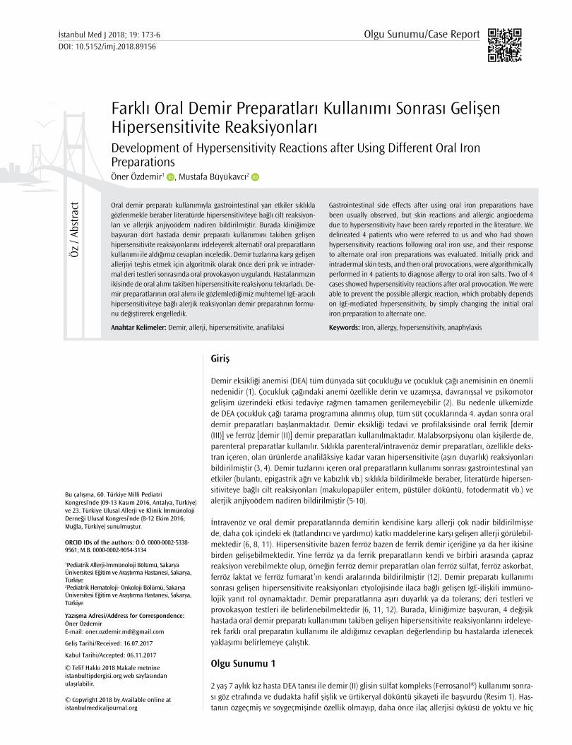
Giriş
Demir eksikliği anemisi (DEA) tüm dünyada süt çocukluğu ve çocukluk çağı anemisinin en önemli nedenidir (1). Çocukluk çağındaki anemi özellikle derin ve uzamışsa, davranışsal ve psikomotor gelişim üzerindeki etkisi tedaviye rağmen tamamen gerilemeyebilir (2). Bu nedenle ülkemizde de DEA çocukluk çağı tarama programına alınmış olup, tüm süt çocuklarında 4. aydan sonra oral demir preparatları başlanmaktadır. Demir eksikliği tedavi ve profilaksisinde oral ferrik [demir (III)] ve ferröz [demir (II)] demir preparatları kullanılmaktadır. Malabsorpsiyonu olan kişilerde de, parenteral preparatlar kullanılır. Sıklıkla parenteral/intravenöz demir preparatları, özellikle deks-tran içeren, olan ürünlerde anafilâksiye kadar varan hipersensitivite (aşırı duyarlık) reaksiyonları bildirilmiştir (3, 4). Demir tuzlarını içeren oral preparatların kullanımı sonrası gastrointestinal yan etkiler (bulantı, epigastrik ağrı ve kabızlık vb.) sıklıkla bildirilmekle beraber, literatürde hipersen-sitiviteye bağlı cilt reaksiyonları (makulopapüler eritem, püstüler döküntü, fotodermatit vb.) ve alerjik anjiyoödem nadiren bildirilmiştir (5-10).
İntravenöz ve oral demir preparatlarında demirin kendisine karşı allerji çok nadir bildirilmişse de, daha çok içindeki ek (tatlandırıcı ve yardımcı) katkı maddelerine karşı gelişen allerji görülebil-mektedir (6, 8, 11). Hipersensitivite bazen ferröz bazen de ferrik demir içeriğine ya da her ikisine birden gelişebilmektedir. Yine ferröz ya da ferrik preparatların kendi ve birbiri arasında çapraz reaksiyon verebilmekte olup, örneğin ferröz demir preparatları olan ferröz sülfat, ferröz askorbat, ferröz laktat ve ferröz fumarat’ın kendi aralarında bildirilmiştir (12). Demir preparatı kullanımı sonrası gelişen hipersensitivite reaksiyonları etyolojisinde ilaca bağlı gelişen IgE-ilişkili immüno-lojik yanıt rol oynamaktadır. Demir preparatlarına aşırı duyarlık ya da tolerans; deri testleri ve provokasyon testleri ile belirlenebilmektedir (6, 11, 12). Burada, kliniğimize başvuran, 4 değişik hastada oral demir preparatı kullanımını takiben gelişen hipersensitivite reaksiyonlarını irdeleye-rek farklı oral preparatın kullanımı ile aldığımız cevapları değerlendirip bu hastalarda izlenecek yaklaşımı belirlemeye çalıştık.
Olgu Sunumu 1
2 yaş 7 aylık kız hasta DEA tanısı ile demir (II) glisin sülfat kompleks (Ferrosanol®) kullanımı sonra-sı göz etrafında ve dudakta hafif şişlik ve ürtikeryal döküntü şikayeti ile başvurdu (Resim 1). Has-tanın özgeçmiş ve soygeçmişinde özellik olmayıp, daha önce ilaç allerjisi öyküsü de yoktu ve hiç
Farklı Oral Demir Preparatları Kullanımı Sonrası Gelişen Hipersensitivite ReaksiyonlarıDevelopment of Hypersensitivity Reactions after Using Different Oral Iron Preparations
Oral demir preparatı kullanımıyla gastrointestinal yan etkiler sıklıkla gözlenmekle beraber literatürde hipersensitiviteye bağlı cilt reaksiyon-ları ve allerjik anjiyoödem nadiren bildirilmiştir. Burada kliniğimize başvuran dört hastada demir preparatı kullanımını takiben gelişen hipersensitivite reaksiyonlarını irdeleyerek alternatif oral preparatların kullanımı ile aldığımız cevapları inceledik. Demir tuzlarına karşı gelişen allerjiyi teşhis etmek için algoritmik olarak önce deri prik ve intrader-mal deri testleri sonrasında oral provokasyon uygulandı. Hastalarımızın ikisinde de oral alımı takiben hipersensitivite reaksiyonu tekrarladı. De-mir preparatlarının oral alımı ile gözlemlediğimiz muhtemel IgE-aracılı hipersensitiviteye bağlı alerjik reaksiyonları demir preparatının formu-nu değiştirerek engelledik.
Anahtar Kelimeler: Demir, allerji, hipersensitivite, anafilaksi
Gastrointestinal side effects after using oral iron preparations have been usually observed, but skin reactions and allergic angioedema due to hypersensitivity have been rarely reported in the literature. We delineated 4 patients who were referred to us and who had shown hypersensitivity reactions following oral iron use, and their response to alternate oral iron preparations was evaluated. Initially prick and intradermal skin tests, and then oral provocations, were algorithmically performed in 4 patients to diagnose allergy to oral iron salts. Two of 4 cases showed hypersensitivity reactions after oral provocation. We were able to prevent the possible allergic reaction, which probably depends on IgE-mediated hypersensitivity, by simply changing the initial oral iron preparation to alternate one.
Keywords: Iron, allergy, hypersensitivity, anaphylaxis
Öz
/ Abs
trac
t
Bu çalışma, 60. Türkiye Milli Pediatri Kongresi’nde (09-13 Kasım 2016, Antalya, Türkiye) ve 23. Türkiye Ulusal Allerji ve Klinik İmmünoloji Derneği Ulusal Kongresi’nde (8-12 Ekim 2016, Muğla, Türkiye) sunulmuştur.
ORCID IDs of the authors: Ö.Ö. 0000-0002-5338-9561; M.B. 0000-0002-9054-3134
1Pediatrik Allerji-İmmünoloji Bölümü, Sakarya Üniversitesi Eğitim ve Araştırma Hastanesi, Sakarya, Türkiye2Pediatrik Hematoloji- Onkoloji Bölümü, Sakarya Üniversitesi Eğitim ve Araştırma Hastanesi, Sakarya, Türkiye
Yazışma Adresi/Address for Correspondence:Öner ÖzdemirE-mail: [email protected]
Geliş Tarihi/Received: 16.07.2017
Kabul Tarihi/Accepted: 06.11.2017
© Telif Hakkı 2018 Makale metnine istanbultipdergisi.org web sayfasından ulaşılabilir.
© Copyright 2018 by Available online at istanbulmedicaljournal.org
Olgu Sunumu/Case Reportİstanbul Med J 2018; 19: 173-6DOI: 10.5152/imj.2018.89156
Öner Özdemir1 , Mustafa Büyükavcı2
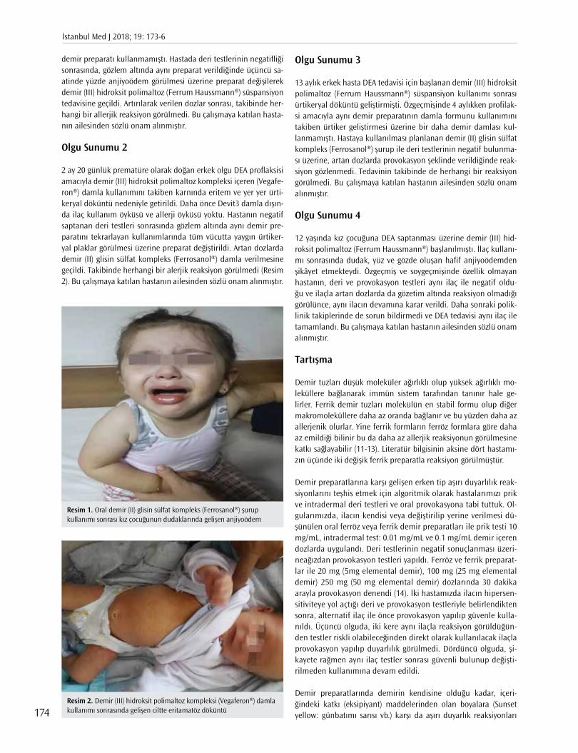
demir preparatı kullanmamıştı. Hastada deri testlerinin negatifliği sonrasında, gözlem altında aynı preparat verildiğinde üçüncü sa-atinde yüzde anjiyoödem görülmesi üzerine preparat değişilerek demir (III) hidroksit polimaltoz (Ferrum Haussmann®) süspansiyon tedavisine geçildi. Artırılarak verilen dozlar sonrası, takibinde her-hangi bir allerjik reaksiyon görülmedi. Bu çalışmaya katılan hasta-nın ailesinden sözlü onam alınmıştır. Olgu Sunumu 2
2 ay 20 günlük prematüre olarak doğan erkek olgu DEA proflaksisi amacıyla demir (III) hidroksit polimaltoz kompleksi içeren (Vegafe-ron®) damla kullanımını takiben karnında eritem ve yer yer ürti-keryal döküntü nedeniyle getirildi. Daha önce Devit3 damla dışın-da ilaç kullanım öyküsü ve allerji öyküsü yoktu. Hastanın negatif saptanan deri testleri sonrasında gözlem altında aynı demir pre-paratını tekrarlayan kullanımlarında tüm vücutta yaygın ürtiker-yal plaklar görülmesi üzerine preparat değiştirildi. Artan dozlarda demir (II) glisin sülfat kompleks (Ferrosanol®) damla verilmesine geçildi. Takibinde herhangi bir alerjik reaksiyon görülmedi (Resim 2). Bu çalışmaya katılan hastanın ailesinden sözlü onam alınmıştır.
Olgu Sunumu 3
13 aylık erkek hasta DEA tedavisi için başlanan demir (III) hidroksit polimaltoz (Ferrum Haussmann®) süspansiyon kullanımı sonrası ürtikeryal döküntü geliştirmişti. Özgeçmişinde 4 aylıkken profilak-si amacıyla aynı demir preparatının damla formunu kullanımını takiben ürtiker geliştirmesi üzerine bir daha demir damlası kul-lanmamıştı. Hastaya kullanılması planlanan demir (II) glisin sülfat kompleks (Ferrosanol®) şurup ile deri testlerinin negatif bulunma-sı üzerine, artan dozlarda provokasyon şeklinde verildiğinde reak-siyon gözlenmedi. Tedavinin takibinde de herhangi bir reaksiyon görülmedi. Bu çalışmaya katılan hastanın ailesinden sözlü onam alınmıştır.
Olgu Sunumu 4
12 yaşında kız çocuğuna DEA saptanması üzerine demir (III) hid-roksit polimaltoz (Ferrum Haussmann®) başlanılmıştı. İlaç kullanı-mı sonrasında dudak, yüz ve gözde oluşan hafif anjiyoödemden şikâyet etmekteydi. Özgeçmiş ve soygeçmişinde özellik olmayan hastanın, deri ve provokasyon testleri aynı ilaç ile negatif oldu-ğu ve ilaçla artan dozlarda da gözetim altında reaksiyon olmadığı görülünce, aynı ilacın devamına karar verildi. Daha sonraki polik-linik takiplerinde de sorun bildirmedi ve DEA tedavisi aynı ilaç ile tamamlandı. Bu çalışmaya katılan hastanın ailesinden sözlü onam alınmıştır.
Tartışma
Demir tuzları düşük moleküler ağırlıklı olup yüksek ağırlıklı mo-leküllere bağlanarak immün sistem tarafından tanınır hale ge-lirler. Ferrik demir tuzları molekülün en stabil formu olup diğer makromoleküllere daha az oranda bağlanır ve bu yüzden daha az allerjenik olurlar. Yine ferrik formların ferröz formlara göre daha az emildiği bilinir bu da daha az allerjik reaksiyonun görülmesine katkı sağlayabilir (11-13). Literatür bilgisinin aksine dört hastamı-zın üçünde iki değişik ferrik preparatla reaksiyon görülmüştür.
Demir preparatlarına karşı gelişen erken tip aşırı duyarlılık reak-siyonlarını teşhis etmek için algoritmik olarak hastalarımızı prik ve intradermal deri testleri ve oral provokasyona tabi tuttuk. Ol-gularımızda, ilacın kendisi veya değiştirilip yerine verilmesi dü-şünülen oral ferröz veya ferrik demir preparatları ile prik testi 10 mg/mL, intradermal test: 0.01 mg/mL ve 0.1 mg/mL demir içeren dozlarda uygulandı. Deri testlerinin negatif sonuçlanması üzeri-neağızdan provokasyon testleri yapıldı. Ferröz ve ferrik preparat-lar ile 20 mg (5mg elemental demir), 100 mg (25 mg elemental demir) 250 mg (50 mg elemental demir) dozlarında 30 dakika arayla provokasyon denendi (14). İki hastamızda ilacın hipersen-sitiviteye yol açtığı deri ve provokasyon testleriyle belirlendikten sonra, alternatif ilaç ile önce provokasyon yapılıp güvenle kulla-nıldı. Üçüncü olguda, iki kere aynı ilaçla reaksiyon görüldüğün-den testler riskli olabileceğinden direkt olarak kullanılacak ilaçla provokasyon yapılıp duyarlılık görülmedi. Dördüncü olguda, şi-kayete rağmen aynı ilaç testler sonrası güvenli bulunup değişti-rilmeden kullanımına devam edildi.
Demir preparatlarında demirin kendisine olduğu kadar, içeri-ğindeki katkı (eksipiyant) maddelerinden olan boyalara (Sunset yellow: günbatımı sarısı vb.) karşı da aşırı duyarlık reaksiyonları
Resim 1. Oral demir (II) glisin sülfat kompleks (Ferrosanol®) şurup kullanımı sonrası kız çocuğunun dudaklarında gelişen anjiyoödem
Resim 2. Demir (III) hidroksit polimaltoz kompleksi (Vegaferon®) damla kullanımı sonrasında gelişen ciltte eritamatöz döküntü
İstanbul Med J 2018; 19: 173-6
174

bildirilmiştir (8). Çalışmamızda hastalarımızın kullandığı demir preparatlarının güncel ilaç rehberlerine (vademekum) ve ilaçların tanıtıldığı değişik websitelerine göre içerikleri şöyle idi. Vegaferon® damla: demir (III) hidroksit polimaltoz, sakkaroz, metil paraben, propil paraben, krem esansı, sodyum hidroksit; Ferrum Hauss-mann® şurup: demir (III) hidroksit polimaltoz, şeker, sorbitol, Ni-pagin M, Nipasol, krem esansı R22, sodyum hidroksit; Ferrosanol® damla: ferro glikokol (ferröz) sülfat, sorbitol, sakkarin, sülfirik asit, portakal esansı (etanollü) içeriyordu.
Vakalarımızın kullandığı preparatların içinde ortak olarak krem esansı, sodyum hidroksit ve sorbitol vardı. İntravenöz demir preparatına bağlı hipersensitivite reaksiyonlarında içeriğindeki dekstran sorumlu tutulmaktadır (3, 4, 11, 13). Oral preparatlar-la ilgili bir veri olmamakla beraber, olgularımızda yukarıdaki üç maddeden herhangi birine bağlı gelişebileceğini düşünmekteyiz. Dördüncü olguda doğrulanamamasına rağmen, hastalarımızın üçünde de oral demir preparat alımını takiben hipersensitivite reaksiyonu gelişmişti. Eksipiyantlarla (katkı madddeleri) ile test etme imkânımız olmamasına rağmen, içeriği ve kuvveti değişen demir preparatlarında sorun olmaması daha çok bu sonucu dü-şündürmüştür.
Demir preparatlarına karşı allerjik reaksiyon gelişme durumunda, desensitizasyon protokolleri kullanılmak zorunda kalınabilir (6, 15, 16). Fakat özellikle oral demir preparatlarında tolerans geli-şebilmesi bazen Ortega ve ark. (17) bildirdiği gibi 4 günde bile ge-lişen reaksiyondan ötürü tedavi dozuna ulaşılamamış; de Barrio ve ark. (6) ise 18 günde tedavi dozuna ulaşmış ve hızlıca desen-sitizasyon mümkün olmamıştır. Parenteral preparatlar için birkaç saatlik desensitizasyon protokolleri Rodríguez-Jiménez ve ark. (10) tarafından tanımlanmıştır. Bu yüzden oral preparatla karşılaşılan allerjik reaksiyonlarda parenteral desensitizasyonun tercih edildiği de olmuştur. Desensitizasyon öncesinde antihistaminik, kortikos-teroid, ve montelukast hatta bazen asetil salisilik asidin kullanıl-dığı bildirilmiştir (3, 11, 14-18). Olgularımızın hiçbirinde içeriği ve kuvveti değiştirilmiş preparatla riskli ve uzun süren desensitizasyo-na ihtiyaç kalmamıştır.
Hastalarımızda preparat değişikliği yaparken preparat içeriğindeki demir kuvvetinin -iki ya da üç değerlikli- ve katkı ve boya (eksipi-yant) maddelerinin farklı olmasına dikkat ederek olguların soru-nunu çözmüş olduk. İki olguda demir (III) preparatı demir (II) pre-paratına, bir olguda da demir (II) preparatı demir (III) preparatına değiştirilerek güvenle uygulanabildi. Bu deneyimlerimiz, güncel literatürle de uyumluydu. Türkiye’den bildirilen bir çalışmada fer-röz preparatlara alerji geliştiğinde, ferrik preparatın tolere edilebil-diği de gösterilmiştir (12).
Sonuç olarak; DEA profilaksi ve tedavisi amacıyla kullanılan farklı hastalardaki değişik oral demir preparatları ile gözlemlediğimiz IgE-aracılı hipersensitivite mekanizmasına bağlı alerjik reaksiyon-ları oral demir preparatının tipini değiştirerek engelledik. Anemi-nin derinliği ve tedavideki preparatın zaruriliği değerlendirilerek öncelikli ve pratik olarak içeriği farklı preparatlar desensitizasyon öncesi denenmeli ardından uygun tedavi yaklaşımı belirlenmeli-dir.
Hasta Onamı: Sözlü hasta onamı bu çalışmaya katılan hastaların ailele-rinden alınmıştır.
Hakem Değerlendirmesi: Dış Bağımsız.
Yazar Katkıları: Fikir - Ö.Ö., M.B.; Tasarım - Ö.Ö., M.B.; Denetleme - Ö.Ö., MB; Kaynaklar - Ö.Ö.; Malzemeler - Ö.Ö.; Veri Toplanması ve/veya işlemesi - Ö.Ö.; Analiz ve/veya Yorum - Ö.Ö., M.B.; Literatür taraması - Ö.Ö.; Yazıyı Yazan - Ö.Ö.; Eleştirel İnceleme - M.B.
Çıkar Çatışması: Yazarların beyan edecek çıkar çatışması yoktur.
Finansal Destek: Yazarlar bu çalışma için finansal destek almadıklarını beyan etmişlerdir.
Informed Consent: Verbal informed consent was obtained from parents of the patiens’ who participated in this study.
Peer-review: Externally peer-reviewed.
Author contributions: Concept - Ö.Ö., MB; Design - Ö.Ö., M.B.; Supervi-sion - Ö.Ö., M.B.; Resource - Ö.Ö.; Materials - Ö.Ö.; Data Collection and/or Process ing - Ö.Ö.; Analysis and/or Interpretation - Ö.Ö., M.B.; Literature Search - Ö.Ö.; Writing - Ö.Ö.; Critical Reviews - M.B.
Conflict of Interest: Authors have no conflicts of interest to declare.
Financial Disclosure: The authors declared that this study has received no financial support.
Kaynaklar
1. Panagiotou JP, Douros K. Clinicolaboratory findings and treatment of iron-deficiency anemia in childhood. Pediatr Hematol Oncol 2004; 21: 521-34. [CrossRef]
2. de Andraca I, Castillo M, Walter T. Psychomotor development and behavior in iron-deficient anemic infants. Nutr Rev 1997; 55:125-32. [CrossRef]
3. Morales Mateluna CA, Scherer Hofmeier K, Bircher AJ. Approach to hypersensitivity reactions from intravenous iron preparations. Allergy 2017; 72: 827-30. [CrossRef]
4. Bailie GR, Clark JA, Lane CE, Lane PL. Hypersensitivity reactions and deaths associated with intravenous iron preparations. Nephrol Dial Transplant 2005; 20: 1443-9. [CrossRef]
5. Choulis NH, Dukes MNG. Metals. In: Dukes MNG and Aronson JK, eds. Meyler's side effects of drugs. Amsterdam: Elsevier, 2000; 683-713.
6. de Barrio M, Fuentes V, Tornero P, Sánchez I, Zubeldia J, Herrero T. Anaphylaxis to oral iron salts. desensitization protocol for tolerance induction. J Investig Allergol Clin Immunol 2008; 18: 305-8.
7. Kawada A, Hiruma M, Noguchi H, Kimura M, Ishibashi A, Banba H, et al. Photosensitivity due to sodium ferrous citrate. Contact Dermatitis 1996; 34: 77. [CrossRef]
8. Rogkakou A, Guerra L, Scordamaglia A, Canonica GW, Passalacqua G. Severe skin reaction due to excipients of an oral iron treatment. Al-lergy 2007; 62: 334-5. [CrossRef]
9. Ito A, Nomura K, Hashimoto I. Pustular drug eruption induced by fer-rous fumarate. Dermatology 1996; 192: 294-5. [CrossRef]
10. Rodríguez-Jiménez B, Domínguez-Ortega J, Nu-ez-Acevedo B, Cava- Sumner B, Kindelan-Recarte C, Montojo-Guillén C. Rapid iron desen-sitization after generalized urticaria and facial angioedema. J Investig Allergol Clin Immunol 2014; 24: 69-71.
11. Rampton D, Folkersen J, Fishbane S, Hedenus M, Howaldt S, Locatelli F, et al. Hypersensitivity reactions to intravenous iron: guidance for risk minimization and management. Haematologica 2014; 99: 1671-6. [CrossRef]
12. Yilmaz O, Erkasar Citak F. A case report with hypersensitivity to ferrous but tolerance of ferric iron salts. J Allergy Clin Immunol Pract 2016; 4: 343-4. [CrossRef]
Özdemir ve Büyükavcı. Demir Preparatlarına Allerji
175

13. Fletes R, Lazarus JM, Gage J, Chertow GM. Suspected iron dextran-
related adverse drug events in hemodialysis patients. Am J Kidney Dis
2001; 37: 743-9. [CrossRef]
14. Celik GE. İlaç aşırı duyarlılık reaksiyonlarına yaklaşım. Ulusal rehber.
Ankara: Bilimsel Tıp Yayınevi; 2014.
15. Alam MG, Krause MW, Shah SV. Parenteral iron therapy: beyond ana-phylaxis. Kidney Int 2004; 66:457-8. [CrossRef]
16. Monaghan MS, Glasco G, St John G, Bradsher RW, Olsen KM. Safe ad-ministration of iron dextran to a patient who reacted to the test dose. South Med J 1994; 87: 1010-2. [CrossRef]
17. Ortega N, Castillo R, Blanco C, Alvarez M, Carrillo T. Oral iron cuta-neous adverse reaction and successful desensitization. Ann Allergy Asthma Immunol 2000; 84: 43-5. [CrossRef]
18. Demir S, Olgac M, Unal D, Gelincik A, Colakoglu B, Buyukozturk S. A practical and successful desensitization protocol for immediate hypersensitivity re-actions to iron salts. Int Arch Allergy Immunol 2014; 165: 100-3. [CrossRef]
Cite this article as: Özdemir Ö, Büyükavcı M. Development of Hypersensitivity Reactions after Using Different Oral Iron Preparations. İstanbul Med J 2018; 19: 173-6.
İstanbul Med J 2018; 19: 173-6
176
Introduction
Spinal chondrosarcomas are uncommon tumors of childhood. It requires multimodality imag-ing techniques for differentiation from other tumors. Low-grade chondrosarcomas rarely mimic nerve sheet tumors with the compressive effects on the surrounding structures without distinct destruction (1). Herein we report a pediatric case of this tumor.
Case Report
A 7-year-old boy was admitted to department of pediatrics with dorsal pain and weakness of lower extremities that lasted for a month without any history of trauma.
There was an extramedullar mass in the spinal canal at the Th1 and Th2 levels, indicated by the magnetic resonance imaging (MRI) scans. The mass was centrally hyperintense on T2-weighted (T2w) imagesbesides enhancing peripherally and heterogeneously on T1-weighted (T1w) images after the administration of gadolinium. The spinal cord was compressed, and the spinal canal was enlarged together with the right neural foramen. Hyperintense signal changes which seemed to be present at the level of cerebrospinal fluid intensity were also noticed around the mass (Figure 1). Posterior compressive scalloping of the Th1 vertebral body and other findings mentioned above were suggesting an intradural-extramedullar mass, pos-sibly a nerve sheet tumor.
Because of the complex anatomic location and for further evaluation of the lesion, a computed tomography (CT) scan was performed. The CT scan of the spine revealed an expansile, peripher-ally calcified lobulated mass in the spinal canal with mild central hypodensity (compared to the density of muscle, 20 HU) that was extending into the right neural foramen at the Th1-Th2 level (Figure 2a). There was no distinct destruction of adjacent bony structures, with the exception of the right-superior articular process of the Th2 vertebra. It was concluded that the mass was an ex-tradural exophytic tumor arising from this vertebral element (Figure 2b). We re-evaluated the MRI scans for this challenging appearance and confirmed the presence of a dural interface between the tumor and spinal cord (Figure 3).
Cause of Paraparesia in Childhood: Spinal ChondrosarcomaÇocukluk Çağının Nadir Paraparesi Nedeni, Spinal Kondrosarkom
Chondrosarcomas are common primary malignant bone tumors that are mostly seen in adulthood. Spinal involvement is uncommon among all sites of involvement of the tumor; spinal chondrosarcoma in child-hood is particularly rare. Here we present the imaging findings of a spinal low-grade chondrosarcoma in a 7-year-old boy, which was later surgically excised and pathologically proven. Differentiation of chond-rosarcoma from other spinal tumors may be difficult in cases of inde-terminate tumoral origin in spinal compartments, and it may require a multi-modal imaging approach. Demonstration of dural interface is very important for an accurate diagnosis. Also, changes in the neural fo-raminal width and vertebral architecture and the integrity of the spinal cord are important criteria for differential diagnosis.
Keywords: Spina, chondrosarcoma, childhood, extramedullary tumor, dural interface
Kondrosarkomlar daha çok yetişkinlerde görülen yaygın primer malign kemik tümörleridir. Diğer tüm tümör tutulum yerleri arasında spinal tutulum sık değildir. Bu nedenle çocukluk çağında spinal kondrosar-kom oldukça nadirdir. Burada 7 yaşında bir hastada görülen, sonrasın-da cerrahi eksizyon yapılan ve patolojik tanı alan düşük dereceli spinal kondrosarkoma ait görüntüleme bulgularını paylaştık. Bu tümörün spi-nal kompartmanlarda orjininin tespit edilememesi diğer olası spinal tümörlerden ayrımını güçleştirmekte, çoklu görüntüleme tekniklerinin kullanılmasını gerektirebilmektedir. Dural arayüzün tespiti doğru tanı için çok önemlidir. Ayrıca nöral foramen genişliği ve vertebral yapıdaki değişiklikler ile spinal kordun bütünlüğü ayırıcı tanıda önemli nokta-lardır.
Anahtar Kelimeler: Omurga, kondrosarkom, çocukluk çağı, extrame-düller tümör, dural arayüz
Abst
ract
/ Ö
z
ORCID IDs of the authors: Ç.D. 0000-0001-7208-1290; A.M.K. 0000-0001-6824-4990; A.G.A. 0000-0002-4339-4236; B.E.D. 0000-0003-2434-1398; A.Ö.B. 0000-0002-6222-382X; N.T. 0000-0003-2812-1528.
1Department of Radiology, Gaziantep Maternity and Childrens Hospital, Gaziantep, Türkiye2Department of Radiology, İzmir Bozyaka Training and Research Hospital, İzmir, Türkiye3Department of Radiology, Gazi University School of Medicine, Ankara, Türkiye 4Department of Neurochirurgy, Gazi University School of Medicine, Ankara, Türkiye
Address for Correspondence/Yazışma Adresi:Çağrı DamarE-mail: [email protected]
Received/Geliş Tarihi: 08.08.2016
Accepted/Kabul Tarihi: 19.11.2017
© Copyright 2018 by Available online at istanbulmedicaljournal.org
© Telif Hakkı 2018 Makale metnine istanbultipdergisi.org web sayfasından ulaşılabilir.
Case Report / Olgu Sunumuİstanbul Med J 2018; 19: 177-80DOI: 10.5152/imj.2018.08379
Çağrı Damar1 , Ali Murat Koç3 , Ayşe Gül Alımlı2 , Betül Emine Derinkuyu2 , Alp Özgün Börcek4 , Nil Tokgöz2
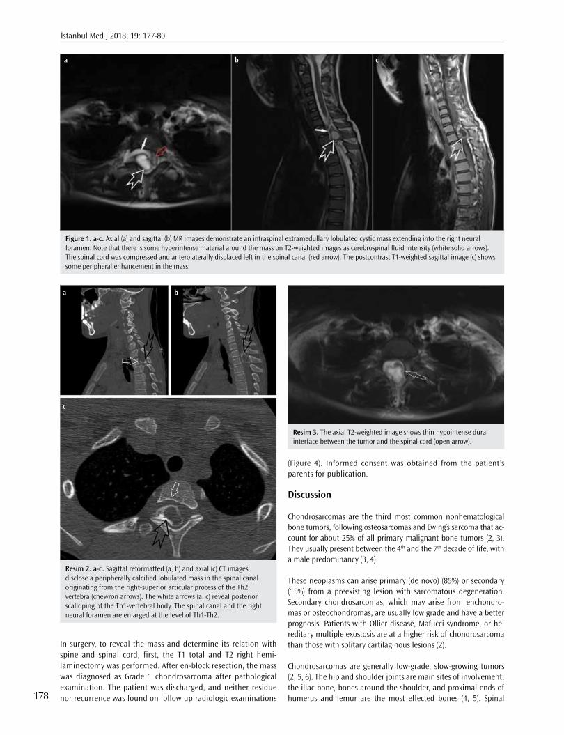
In surgery, to reveal the mass and determine its relation with spine and spinal cord, first, the T1 total and T2 right hemi-laminectomy was performed. After en-block resection, the mass was diagnosed as Grade 1 chondrosarcoma after pathological examination. The patient was discharged, and neither residue nor recurrence was found on follow up radiologic examinations
(Figure 4). Informed consent was obtained from the patient’s parents for publication.
Discussion
Chondrosarcomas are the third most common nonhematological bone tumors, following osteosarcomas and Ewing’s sarcoma that ac-count for about 25% of all primary malignant bone tumors (2, 3). They usually present between the 4th and the 7th decade of life, with a male predominancy (3, 4).
These neoplasms can arise primary (de novo) (85%) or secondary (15%) from a preexisting lesion with sarcomatous degeneration. Secondary chondrosarcomas, which may arise from enchondro-mas or osteochondromas, are usually low grade and have a better prognosis. Patients with Ollier disease, Mafucci syndrome, or he-reditary multiple exostosis are at a higher risk of chondrosarcoma than those with solitary cartilaginous lesions (2).
Chondrosarcomas are generally low-grade, slow-growing tumors (2, 5, 6). The hip and shoulder joints are main sites of involvement; the iliac bone, bones around the shoulder, and proximal ends of humerus and femur are the most effected bones (4, 5). Spinal
Figure 1. a-c. Axial (a) and sagittal (b) MR images demonstrate an intraspinal extramedullary lobulated cystic mass extending into the right neural foramen. Note that there is some hyperintense material around the mass on T2-weighted images as cerebrospinal fluid intensity (white solid arrows). The spinal cord was compressed and anterolaterally displaced left in the spinal canal (red arrow). The postcontrast T1-weighted sagittal image (c) shows some peripheral enhancement in the mass.
a b c
Resim 2. a-c. Sagittal reformatted (a, b) and axial (c) CT images disclose a peripherally calcified lobulated mass in the spinal canal originating from the right-superior articular process of the Th2 vertebra (chewron arrows). The white arrows (a, c) reveal posterior scalloping of the Th1-vertebral body. The spinal canal and the right neural foramen are enlarged at the level of Th1-Th2.
a
c
b
Resim 3. The axial T2-weighted image shows thin hypointense dural interface between the tumor and the spinal cord (open arrow).
İstanbul Med J 2018; 19: 177-80
178

involvement is seen in approximately 10% of patients (2, 6). The thoracic region is the most common site of involvement in spine, with a predilection of posterior vertebral elements (1-3). Primary chondrosarcomas arise mainly from vertebral bodies; on the other hand, main locations of the secondary types are the posterior ele-ments (7). Our case did not have any known prior lesions, but the tumor had involved the articular facet unlike most of the primary chondrosarcomas originating from the vertebral body.
Less than 10% of chondrosarcomas occur in childhood (4). They usually present with slow-growing night pain and sometimes a palpable mass. Occasionally, neurologic symptoms appear be-cause of spinal cord compression from chondrosarcomas, like in the current case (2, 3, 6).
Chondrosarcomas can be defined as central (intramedullary), peripheral, and juxtacortical (periosteal) on the basis of their location in the bone (2, 3, 8). Because of its pathologic character-istics, chondrosarcomas have been classified into the following conventional and variant subtypes: clear cell, myxoid, mesen-chymal, and dedifferentiated. The grades of these variant types are variable between low to high grade (2, 5, 8). It is known that spinal chondrosarcomas are extradural tumors, but there are some reports about intradural lesions regarding these variant subtypes (9-11). The grading system is based on tumor cellularity, and there are three main histologic grades (2, 7, 8). Low-grade, centrally (intramedullary) located conventional type is the most common, and it represents up to 90% of all chondrosarcomas (2-4, 9). Our patient had a low-grade juxtacortical-exophytic located tumor, which is not very aggressive, so it could be successfully resected en-bloc.
Chemotherapy and radiotherapy have not been demonstrated as successful treatment options. The pelvis, sacrum, or spine are dif-ficult locations for en-bloc resection, which is the sole therapeutic option. Hence, the life expectancy of patients with these tumor locations is lower than others. Although surgery is the best option, the recurrence rates are quite high after tumor debulking without safe surgical margins (1, 2, 4, 12, 13).
These cartilaginous tumors produce a chondroid matrix (2-5). Ra-diological findings correlate well with histological grades (5, 8, 12). Low-grade lesions have dense spicules of calcification, whereas high-grade lesions have amorphous areas of calcification and con-centric growth of this soft tissue component (7). Nonmineralized central hypodensity reflects the high water content of hyaline car-tilage on CT images. MRI characteristics of the tumor are iso- to
hypointense on T1w and heterogenous on T2w images. Peripheral and lobulated rim enhancement is seen on postcontrast T1w im-ages. Mineralized areas are responsible for hypointense signals, contrary to predominant nonmineralized areas which are hyperin-tense because of high water content of matrices on T2w images (8).
Findings may vary from lysis that interferes with enchondromas to moth-eaten destruction and the discriminative “rings-and-arcs” type of tumoral calcification that represents enchondral calcifica-tion (2-4, 8, 12). The involvement of axial skeleton; peripheral, nodular, and septal enhancement; peritumoral edema and end-osteal scalloping of >2/3rd of cortical thickness are all in favor of chondrosarcomas rather than enchondroma (4).
Chondrosarcomas rarely present as a dumbbell-shaped mass caus-ing neural foraminal widening, as in our present report (5, 11, 14, 15). Peripheral calcification with central nonmineralized areas and peripheral and heterogenous enhancement supported the diagno-sis of chondrosarcoma, which was pathologically made after surgi-cal excision in our patient.
Conclusion
Spinal low-grade chondrosarcomas in childhood may mimic the imaging findings of nerve sheet tumors in terms of spinal cord compression, bony scalloping, and neural foraminal enlargement. It is difficult to detect the dural interface, but once it is observed, it definitely helps initial diagnosis.
Informed Consent: Informed consent was obtained from parents of the patient who participated in this study.
Peer-review: Externally peer-reviewed.
Author Contributions: Concept - Ç.D., A.M.K.; Design - Ç.D., A.M.K.; Super-vision - N.T., A.Ö.B.; Resource - A.G.A., B.D.; Materials - A.G.A., B.D.; Analysis and/or Interpretation - N.T., A.Ö.B.; Literature Search - A.G.A., B.D.; Writing - Ç.D., A.M.K.; Critical Reviews - N.T., A.Ö.B.
Conflict of Interest: Authors have no conflicts of interest to declare.
Financial Disclosure: The authors declared that this study has received no financial support
Hasta Onamı: Bilgilendirilmiş hasta onamı bu çalışmaya katılan hastanın ailesinden alınmıştır.
Hakem Değerlendirmesi: Dış Bağımsız.
Figure 4. a-c. Early postoperative axial CT image (a) shows that there is no residue of the mass. Postoperative followup images (1 year later); axial (b) and sagittal (c) postcontrast T1-weighted MR images reveal that there is no pathologic signal intensity or enhancement compatible with recurrence. Note that there are laminectomy defects in the posterior elements of the Th1 and Th2 vertebrae. Posterior scalloping of the Th1 vertebral body improved.
a b c
Damar et al. Spinal Chondrosarcoma
179
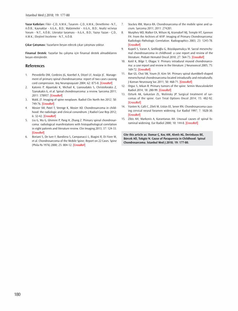
Yazar Katkıları: Fikir - Ç.D., A.M.K.; Tasarım - Ç.D., A.M.K.; Denetleme - N.T., A.Ö.B.; Kaynaklar - A.G.A., B.D.; Malzemeler - A.G.A., B.D.; Analiz ve/veya Yorum - N.T., A.Ö.B.; Literatür taraması - A.G.A., B.D.; Yazıyı Yazan - Ç.D., A.M.K.; Eleştirel İnceleme - N.T., A.Ö.B.
Çıkar Çatışması: Yazarların beyan edecek çıkar çatışması yoktur.
Finansal Destek: Yazarlar bu çalışma için finansal destek almadıklarını beyan etmişlerdir.
References
1. Prevedello DM, Cordeiro JG, Koerbel A, Ditzel LF, Araújo JC. Manage-ment of primary spinal chondrosarcoma: report of two cases causing cord compression. Arq Neuropsiquiatr 2004; 62: 875-8. [CrossRef]
2. Katonis P, Alpantaki K, Michail K, Lianoudakis S, Christoforakis Z, Tzanakakis G, et al. Spinal chondrosarcoma: a review. Sarcoma 2011; 2011: 378957. [CrossRef]
3. Wald, JT. Imaging of spine neoplasm. Radiol Clin North Am 2012; 50: 749-76. [CrossRef]
4. Mosier SM, Patel T, Strenge K, Mosier AD. Chondrosarcoma in child-hood: the radiologic and clinical conundrum. J Radiol Case Rep 2012; 6: 32-42. [CrossRef]
5. Liu G, Wu G, Ghimire P, Pang H, Zhang Z. Primary spinal chondrosar-coma: radiological manifestations with histopathological correlation in eight patients and literature review. Clin Imaging 2013; 37: 124-33. [CrossRef]
6. Boriani S, De Iure F, Bandiera S, Campanacci L, Biagini R, Di Fiore M, et al. Chondrosarcoma of the Mobile Spine; Report on 22 Cases. Spine (Phila Pa 1976) 2000; 25: 804-12. [CrossRef]
7. Stuckey RM, Marco RA. Chondrosarcoma of the mobile spine and sa-crum. Sarcoma 2011; 2011: 274281.
8. Murphey MD, Walker EA, Wilson AJ, Kransdorf MJ, Temple HT, Gannon FH. From the Archives of AFIP: Imaging of Primary Chondrosarcoma: Radiologic-Pathologic Correlation. Radiographics 2003; 23: 1245-78. [CrossRef]
9. Kupeli S, Varan A, Gedikoğlu G, Büyükpamukçu M. Sacral mesenchy-mal chondrosarcoma in childhood: a case report and review of the literature. Pediatr Hematol Oncol 2010; 27: 564-73. [CrossRef]
10. Kotil K, Bilge T, Olagac V. Primary intradural myxoid chondrosarco-ma: a case report and review in the literature. J Neurooncol 2005; 75: 169-72. [CrossRef]
11. Bae GS, Choi SW, Youm JY, Kim SH. Primary spinal dumbbell-shaped mesenchymal chondrosarcoma located intradurally and extradurally. J Korean Neurosurg Soc 2011; 50: 468-71. [CrossRef]
12. Orguc S, Arkun R. Primary tumors of the spine. Semin Musculoskelet Radiol 2014; 18: 280-99. [CrossRef]
13. Ozturk AK, Gokaslan ZL, Wolinsky JP. Surgical treatment of sar-comas of the spine. Curr Treat Options Oncol 2014; 15: 482-92. [CrossRef]
14. Yünten N, Calli C, Zileli M, Ustün EE, Sener RN. Chondrosarcoma caus-ing cervical neural foramen widening. Eur Radiol 1997; 7: 1028-30. [CrossRef]
15. Zibis AH, Markonis A, Karantanas AH. Unusual causes of spinal fo-raminal widening. Eur Radiol 2000; 10: 144-8. [CrossRef]
Cite this article as: Damar Ç, Koç AM, Alımlı AG, Derinkuyu BE, Börcek AÖ, Tokgöz N. Cause of Paraparesia in Childhood: Spinal Chondrosarcoma. İstanbul Med J 2018; 19: 177-80.
İstanbul Med J 2018; 19: 177-80
180
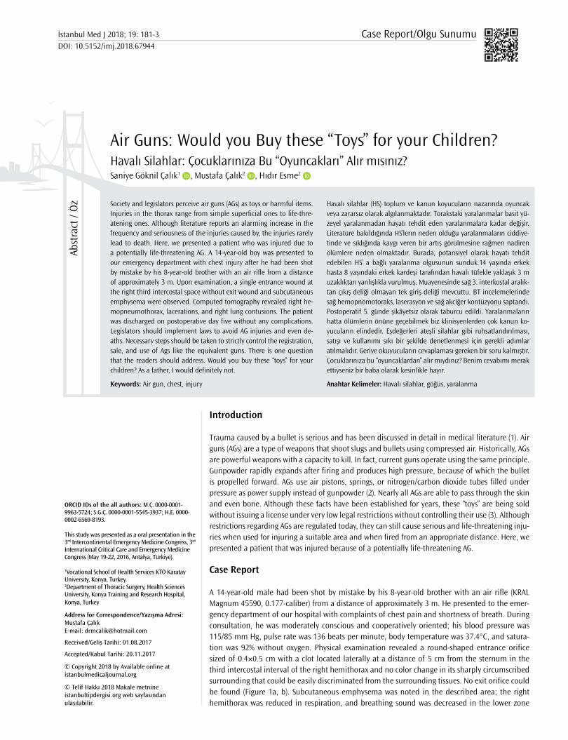
Introduction
Trauma caused by a bullet is serious and has been discussed in detail in medical literature (1). Air guns (AGs) are a type of weapons that shoot slugs and bullets using compressed air. Historically, AGs are powerful weapons with a capacity to kill. In fact, current guns operate using the same principle. Gunpowder rapidly expands after firing and produces high pressure, because of which the bullet is propelled forward. AGs use air pistons, springs, or nitrogen/carbon dioxide tubes filled under pressure as power supply instead of gunpowder (2). Nearly all AGs are able to pass through the skin and even bone. Although these facts have been established for years, these “toys” are being sold without issuing a license under very low legal restrictions without controlling their use (3). Although restrictions regarding AGs are regulated today, they can still cause serious and life-threatening inju-ries when used for injuring a suitable area and when fired from an appropriate distance. Here, we presented a patient that was injured because of a potentially life-threatening AG.
Case Report
A 14-year-old male had been shot by mistake by his 8-year-old brother with an air rifle (KRAL Magnum 45590, 0.177-caliber) from a distance of approximately 3 m. He presented to the emer-gency department of our hospital with complaints of chest pain and shortness of breath. During consultation, he was moderately conscious and cooperatively oriented; his blood pressure was 115/85 mm Hg, pulse rate was 136 beats per minute, body temperature was 37.4°C, and satura-tion was 92% without oxygen. Physical examination revealed a round-shaped entrance orifice sized of 0.4×0.5 cm with a clot located laterally at a distance of 5 cm from the sternum in the third intercostal interval of the right hemithorax and no color change in its sharply circumscribed surrounding that could be easily discriminated from the surrounding tissues. No exit orifice could be found (Figure 1a, b). Subcutaneous emphysema was noted in the described area; the right hemithorax was reduced in respiration, and breathing sound was decreased in the lower zone
Air Guns: Would you Buy these “Toys” for your Children?Havalı Silahlar: Çocuklarınıza Bu “Oyuncakları” Alır mısınız?
Havalı silahlar (HS) toplum ve kanun koyucuların nazarında oyuncak veya zararsız olarak algılanmaktadır. Torakstaki yaralanmalar basit yü-zeyel yaralanmadan hayatı tehdit eden yaralanmalara kadar değişir. Literatüre bakıldığında HS’lerın neden olduğu yaralanmaların ciddiye-tinde ve sıklığında kaygı veren bir artış görülmesine rağmen nadiren ölümlere neden olmaktadır. Burada, potansiyel olarak hayatı tehdit edebilen HS’ a bağlı yaralanma olgusunun sunduk.14 yaşında erkek hasta 8 yaşındaki erkek kardeşi tarafından havalı tüfekle yaklaşık 3 m uzaklıktan yanlışlıkla vurulmuş. Muayenesinde sağ 3. interkostal aralık-tan çıkış deliği olmayan tek giriş deliği mevcuttu. BT incelemelerinde sağ hemopnömotoraks, laserasyon ve sağ akciğer kontüzyonu saptandı. Postoperatif 5. günde şikâyetsiz olarak taburcu edildi. Yaralanmaların hatta ölümlerin önüne geçebilmek biz klinisyenlerden çok kanun ko-yucuların elindedir. Eşdeğerleri ateşli silahlar gibi ruhsatlandırılması, satışı ve kullanımı sıkı bir şekilde denetlenmesi için gerekli adımlar atılmalıdır. Geriye okuyucuların cevaplaması gereken bir soru kalmıştır. Çocuklarınıza bu “oyuncaklardan” alır mıydınız? Benim cevabımı merak ettiyseniz bir baba olarak kesinlikle hayır.
Anahtar Kelimeler: Havalı silahlar, göğüs, yaralanma
Society and legislators perceive air guns (AGs) as toys or harmful items. Injuries in the thorax range from simple superficial ones to life-thre-atening ones. Although literature reports an alarming increase in the frequency and seriousness of the injuries caused by, the injuries rarely lead to death. Here, we presented a patient who was injured due to a potentially life-threatening AG. A 14-year-old boy was presented to our emergency department with chest injury after he had been shot by mistake by his 8-year-old brother with an air rifle from a distance of approximately 3 m. Upon examination, a single entrance wound at the right third intercostal space without exit wound and subcutaneous emphysema were observed. Computed tomography revealed right he-mopneumothorax, lacerations, and right lung contusions. The patient was discharged on postoperative day five without any complications. Legislators should implement laws to avoid AG injuries and even de-aths. Necessary steps should be taken to strictly control the registration, sale, and use of Ags like the equivalent guns. There is one question that the readers should address. Would you buy these “toys” for your children? As a father, I would definitely not.
Keywords: Air gun, chest, injury
Abst
ract
/ Ö
z
ORCID IDs of the all authors: M.Ç. 0000-0001-9963-5724; S.G.Ç. 0000-0001-5545-3937; H.E. 0000-0002-6569-8193.
This study was presented as a oral presentation in the 3rd Intercontinental Emergency Medicine Congress, 3rd International Critical Care and Emergency Medicine Congress (May 19-22, 2016, Antalya, Türkiye).
1Vocational School of Health Services KTO Karatay University, Konya, Turkey.2Department of Thoracic Surgery, Health Sciences University, Konya Training and Research Hospital, Konya, Turkey
Address for Correspondence/Yazışma Adresi:Mustafa ÇalıkE-mail: [email protected]
Received/Geliş Tarihi: 01.08.2017
Accepted/Kabul Tarihi: 20.11.2017
© Copyright 2018 by Available online at istanbulmedicaljournal.org
© Telif Hakkı 2018 Makale metnine istanbultipdergisi.org web sayfasından ulaşılabilir.
Case Report/Olgu Sunumuİstanbul Med J 2018; 19: 181-3DOI: 10.5152/imj.2018.67944
Saniye Göknil Çalık1 , Mustafa Çalık2 , Hıdır Esme2
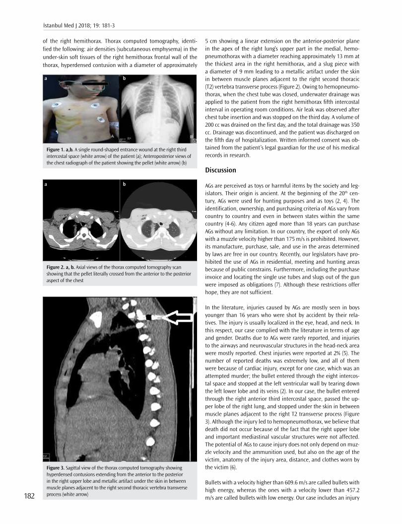
of the right hemithorax. Thorax computed tomography, identi-fied the following: air densities (subcutaneous emphysema) in the under-skin soft tissues of the right hemithorax frontal wall of the thorax, hyperdensed contusion with a diameter of approximately
5 cm showing a linear extension on the anterior-posterior plane in the apex of the right lung’s upper part in the medial, hemo-pneumothorax with a diameter reaching approximately 13 mm at the thickest area in the right hemithorax, and a slug piece with a diameter of 9 mm leading to a metallic artifact under the skin in between muscle planes adjacent to the right second thoracic (T2) vertebra transverse process (Figure 2). Owing to hemopneumo-thorax, when the chest tube was closed, underwater drainage was applied to the patient from the right hemithorax fifth intercostal interval in operating room conditions. Air leak was observed after chest tube insertion and was stopped on the third day. A volume of 200 cc was drained on the first day, and the total drainage was 350 cc. Drainage was discontinued, and the patient was discharged on the fifth day of hospitalization. Written informed consent was ob-tained from the patient’s legal guardian for the use of his medical records in research.
Discussion
AGs are perceived as toys or harmful items by the society and leg-islators. Their origin is ancient. At the beginning of the 20th cen-tury, AGs were used for hunting purposes and as toys (2, 4). The identification, ownership, and purchasing criteria of AGs vary from country to country and even in between states within the same country (4-6). Any citizen aged more than 18 years can purchase AGs without any limitation. In our country, the export of only AGs with a muzzle velocity higher than 175 m/s is prohibited. However, its manufacture, purchase, sale, and use in the areas determined by laws are free in our country. Recently, our legislators have pro-hibited the use of AGs in residential, meeting and hunting areas because of public constrains. Furthermore, including the purchase invoice and locating the single use tubes and slugs out of the gun were imposed as obligations (7). Although these restrictions offer hope, they are not sufficient.
In the literature, injuries caused by AGs are mostly seen in boys younger than 16 years who were shot by accident by their rela-tives. The injury is usually localized in the eye, head, and neck. In this respect, our case complied with the literature in terms of age and gender. Deaths due to AGs were rarely reported, and injuries to the airways and neurovascular structures in the head-neck area were mostly reported. Chest injuries were reported at 2% (5). The number of reported deaths was extremely low, and all of them were because of cardiac injury, except for one case, which was an attempted murder; the bullet entered through the eight intercos-tal space and stopped at the left ventricular wall by tearing down the left lower lobe and its veins (2). In our case, the bullet entered through the right anterior third intercostal space, passed the up-per lobe of the right lung, and stopped under the skin in between muscle planes adjacent to the right T2 transverse process (Figure 3). Although the injury led to hemopneumothorax, we believe that death did not occur because of the fact that the right upper lobe and important mediastinal vascular structures were not affected. The potential of AGs to cause injury does not only depend on muz-zle velocity and the ammunition used, but also on the age of the victim, anatomy of the injury area, distance, and clothes worn by the victim (6).
Bullets with a velocity higher than 609.6 m/s are called bullets with high energy, whereas the ones with a velocity lower than 457.2 m/s are called bullets with low energy. Our case includes an injury
Figure 1. a,b. A single round-shaped entrance wound at the right third intercostal space (white arrow) of the patient (a); Anteroposterior views of the chest radiograph of the patient showing the pellet (white arrow) (b)
a b
Figure 2. a, b. Axial views of the thorax computed tomography scan showing that the pellet literally crossed from the anterior to the posterior aspect of the chest
a b
Figure 3. Sagittal view of the thorax computed tomography showing hyperdensed contusions extending from the anterior to the posterior in the right upper lobe and metallic artifact under the skin in between muscle planes adjacent to the right second thoracic vertebra transverse process (white arrow)
İstanbul Med J 2018; 19: 181-3
182
caused by a bullet with low energy. It is different from injuries of bullets with low energy such as our case. The damages caused are laceration, penetration, crushing/contusion, and temporary cav-ity for a short period (4, 6, 8). A hyperdensed contusion with a diameter of approximately 5 cm showing linear extension on the anterior-posterior plane in the right lung upper lobe apex medial was present in our patient, as identified in literature (Figure 3). The bullet passed the thorax with a kinetic energy of 32.67 J from end to end and stopped under the skin adjacent to the right T2 trans-verse process. It traveled 147.2 mm in the skin (9).
Injuries in the thorax range from simple superficial ones to life-threatening injuries. Every year, one or two people die because of injuries caused by AGs in the United Kingdom, where there are supposedly 4 million AGs; four people die for the same reason in the United States (1, 10). It may escape attention owing to the small entrance orifice in crowded and busy emergency services such as the ones in our country. Despite the restricted power and low ki-netic energy of AGs, they may lead to serious or life-threatening in-juries in suitable injury area and distance. Each AG injury has to be accepted as an injury caused by a gun with low energy and should be treated accordingly. Surgical exploration and pellet removal are contradictive. Although emboli and migration cases have been reported in the literature, discussions show that leaving it in the body is less risky than its removal (4). In our case, surgery was not performed due to limited drainage and because the pellet was far from important neurovascular structures.
Conclusion
Although the frequency and seriousness of the injuries caused by AGs is increasingly alarming when considering the literature, they rarely lead to death. Majority of the reported cases are neurovas-cular injuries around the head-neck area, and more rarely cardiac injuries (4). Injuries caused by AGs should be treated like the inju-ries caused by guns with low energy. It should be always kept in mind that they may cause serious, and even fatal, injuries under suitable conditions. It is in the power of legislators rather than us as clinicians to avoid injuries and even deaths. Necessary steps should be taken to strictly control their registration, sale, and use like the equivalent firearms guns.
Informed Consent: Informed consent was obtained from patient who par-ticipated in this study.
Peer-review: Externally peer-reviewed.
Author contributions: Concept - S.G.Ç., M.Ç., H.E.; Design - S.G.Ç., M.Ç.; Su-pervision - S.G.Ç., M.Ç., H.E.; Resource - S.G.Ç., M.Ç., H.E.; Materials - S.G.Ç., M.Ç., H.E.; Data Collection and/or Processing - S.G.Ç., M.Ç., H.E.; Analysis and/or Interpretation - S.G.Ç., M.Ç.; Literature Search - S.G.Ç., M.Ç.; Writing - S.G.Ç., M.Ç.; Critical Reviews - S.G.Ç., M.Ç.
Conflict of Interest: Authors have no conflicts of interest to declare.
Financial Disclosure: The authors declared that this study has received no financial support.
Hasta Onamı: Bu çalışmaya katılan hastadan hasta onamı alınmıştır.
Hakem Değerlendirmesi: Dış Bağımsız.
Yazar Katkıları: Fikir - S.G.Ç., M.Ç., H.E.; Tasarım - S.G.Ç., M.Ç., H.E.; Denet-leme - S.G.Ç., M.Ç., H.E.; Kaynaklar - S.G.Ç., M.Ç., H.E.; Malzemeler - S.G.Ç., M.Ç., H.E.; Veri Toplanması ve/veya işlemesi - S.G.Ç., M.Ç., H.E.; Analiz ve/veya Yorum - S.G.Ç., M.Ç.; Literatür taraması - S.G.Ç., M.Ç.; Yazıyı Yazan - S.G.Ç., M.Ç.; Eleştirel İnceleme - S.G.Ç., M.Ç.
Çıkar Çatışması: Yazarların beyan edecek çıkar çatışması yoktur.
Finansal Destek: Yazarlar bu çalışma için finansal destek almadıklarını beyan etmişlerdir.
References
1. Wightman G, Cochrane R, Gray RA, Linton M. A contribution to the discus-sion on the safety of air weapons. Sci Justice 2013; 53: 343-9. [CrossRef]
2. Bligh-Glover WZ. One-in-a-million shot: a homicidal thoracic air rifle wound, a case report, and a review of the literature. Am J Forensic Med Pathol 2012; 33: 98-101. [CrossRef]
3. Di Maio VJ. Homicidal death by air rifle. J Trauma 1975; 15: 1034-7. [CrossRef]
4. Abad S, McHenry ID, Carter LM, Mitchell DA. Carotid artery injury from an air gun pellet: a case report and review of the literature. Head Face Med 2009; 17; 5:3.
5. Veenstra M, Prasad J, Schaewe H, Donoghue L, Langenburg S. Non powder firearms cause significant pediatric injuries. J Trauma Acute Care Surg 2015; 78: 1138-42. [CrossRef]
6. Bakovic M, Petrovecki V, Strinovic D, Mayer D. Shot through the heart-fire power and potential lethality of air weapons. J Forensic Sci 2014; 59: 1658-61. [CrossRef]
7. 2521 sayılı avda ve sporda kullanılan tüfekler, nişan tabancaları ve av bıçaklarının yapımı, alımı, satımı ve bulundurulmasına dair kanun Resmî Gazete No:2720822 Nisan 2009 http://www.resmigazete.gov.tr/eskiler/2009/04/20090422-10.htm
8. Stankov A, Jakovski Z, Pavlovski G, Muric N, Dwork AJ, Cakar Z. Air gun injury with deadly aftermath-case report. Leg Med(Tokyo) 2013;15: 35-7. [CrossRef]
9. Hallikeri VR, Gouda HS, Kadagoudar SA. Country made scare gun vs. airgun-a comparative study of terminal ballistics using gelatine blocks. Forensic Sci Int 2012; 214: 148-51. [CrossRef]
10. Laraque D; American Academy of Pediatrics Committee on Injury, Violence, and Poison Prevention. Injury risk of non-powder guns. Pe-diatrics 2004; 114: 1357-61. [CrossRef]
Cite this article as: Çalık SG, Çalık M, Esme H. Air Guns: Would you Buy these “Toys” for your Children? İstanbul Med J 2018; 19: 181-3.
Göknil Çalık et al. Air Guns
183

Introduction
Transverse testicular ectopia (TTE) is a rarely seen congenital anomaly, generally associated with inguinal hernia, where both testes migrate into the same hemiscrotum. An ectopic testis may be situated in the contralateral hemiscrotum, inguinal canal, or internal inguinal ring. In most patients with TTE, a nonpalpable testis along with an ectopic testis associated with ipsilateral inguinal hernia is present. Furhtermore, TTE may be associated with hydrocele, disorders of sex-ual development, and karyotypic anomalies in nearly 20% of the cases, TTE is associated with Persistent Müllerian duct syndrome (PMDS) (1). Regression of Müllerian structures is expected in men. However, sometimes, these structures may be localized in the abdominal cavity or herniated through the inguinal canal. Although the mechanism of this pathologic descensus is unknown, various hypotheses have been suggested including descensus of both testes into the same canal because of a defect occurring during the fetal development of testes, premature fusion of Wolff-ian ducts, and origination of both testes from the same germinal structure (2).
Different approaches have been described in the treatment of TTE. In this article, we have pre-sented two cases with TTE.
Case Reports
Case Report 1A 9-year-old male was referred to our clinic with the absence of testis in the left hemiscrotum. He was operated at another center for right inguinal hernia. On physical examination, the testis could not be palpated in the left inguinal canal and left hemiscrotum. In the right hemiscrotum and the upper part of the scrotum, an anatomical formation thought to be two separate testes was palpated. On a scrotal ultrasound (US), both testes and epididymis were detected in the right hemiscrotum. The dimensions and echogenicities of both testes were within the normal limits. An US revealed that both spermatic chords passed through the right inguinal canal, but the left testis could not be visualized. On laparoscopic exploration, a patent left inguinal canal was seen. How-ever, Müllerian duct remnants were not observed, and the vessels of two separate testes entered the right inguinal canal. On inguinal exploration, two separate ducts and two testicular structures, which were adhered to each other, were observed (Figure 1). The adhesions were excised, and two testes were separated from each other (Figure 2). Through an incision made on the scrotal septum, the left testis was fixed to the left and the right testis was fixed to the right hemiscrotum. The pa-tient also underwent left inguinal hernia repair. No relevant medical problems were encountered
Transverse Testicular Ectopia: Two Case ReportsTransvers Testiküler Ektopi: İki Olgu Sunumu
Transvers testiküler ektopi (TTE) nadir görülen, her iki testisin birlikte aynı hemiskrotuma göç etmesi ile oluşan ve genellikle inguinal herni ile ilişkili olan konjenital bir anomalidir. Olguların yaklaşık %20’ sinde Persistant Müllerian Kanal Sendromu ile birliktelik vardır. Bu çalışmada 1 olgumuza Persistant Müllerian Kanal Sendromunun eşlik ettiği TTE’li 2 olgumuzu sunduk. TTE tedavisinde farklı yaklaşımlar tarif edilmiştir. Tedavinin temeli testis dokularının korunması ve varsa Müllerian Ka-nalın çıkarılmasıdır.
Anahtar Kelimeler: Transvers testiküler ektopi, Persistant Müllerian ka-nal sendromu, testis, cerrahi
Transverse testicular ectopia (TTE) is a rare congenital anomaly caused by migration of the testes to the same half of the scrotum; it is associa-ted with inguinal hernia. In addition, it is present along with persistent Müllerian duct syndrome (PMDS) in approximately 20% of patients. In such cases, Müllerian duct excision is recommended. In this article, we present two cases of TTE including one case of PMDS accompanied by TTE. Different approaches have been described in the treatment of TTE. Protection of testicular tissue and removal of the Müllerian duct, when TTE is present along with PMDS, are the basis of treatment.
Keywords: Transverse testicular ectopia, Persistent Müllerian duct syndro-me, testis, surgery
Abst
ract
/ Ö
z
Bu çalışma 7. Ulusal Pediatrik Üroloji Kongresi'nde sunulmuştur (26-30 Ekim, 2016, Girne, KKTC).
ORCID IDs of all the authors: T.T. 0000-0002-7755-4736; M.S. 0000-0002-6660-5243; Ü.B. 0000-0002-5140-8618; Ş.C. 0000-0003-2318-5653; A.K. 0000-0003-0440-3555.
Department of Pediatric Surgery, Fırat University School of Medicine, Elazığ, Turkey
Address for Correspondence/Yazışma Adresi:Tugay Tartar E-mail: [email protected]
Received/Geliş Tarihi: 12.05.2017
Accepted/Kabul Tarihi: 29.12.2017
© Copyright 2018 by Available online at istanbulmedicaljournal.org
© Telif Hakkı 2018 Makale metnine istanbultipdergisi.org web sayfasından ulaşılabilir.
Case Report/Olgu Sunumuİstanbul Med J 2018; 19: 184-6DOI: 10.5152/imj.2018.88557
Tugay Tartar , Mehmet Saraç , Ünal Bakal , Şenay Canpolat , Ahmet Kazez
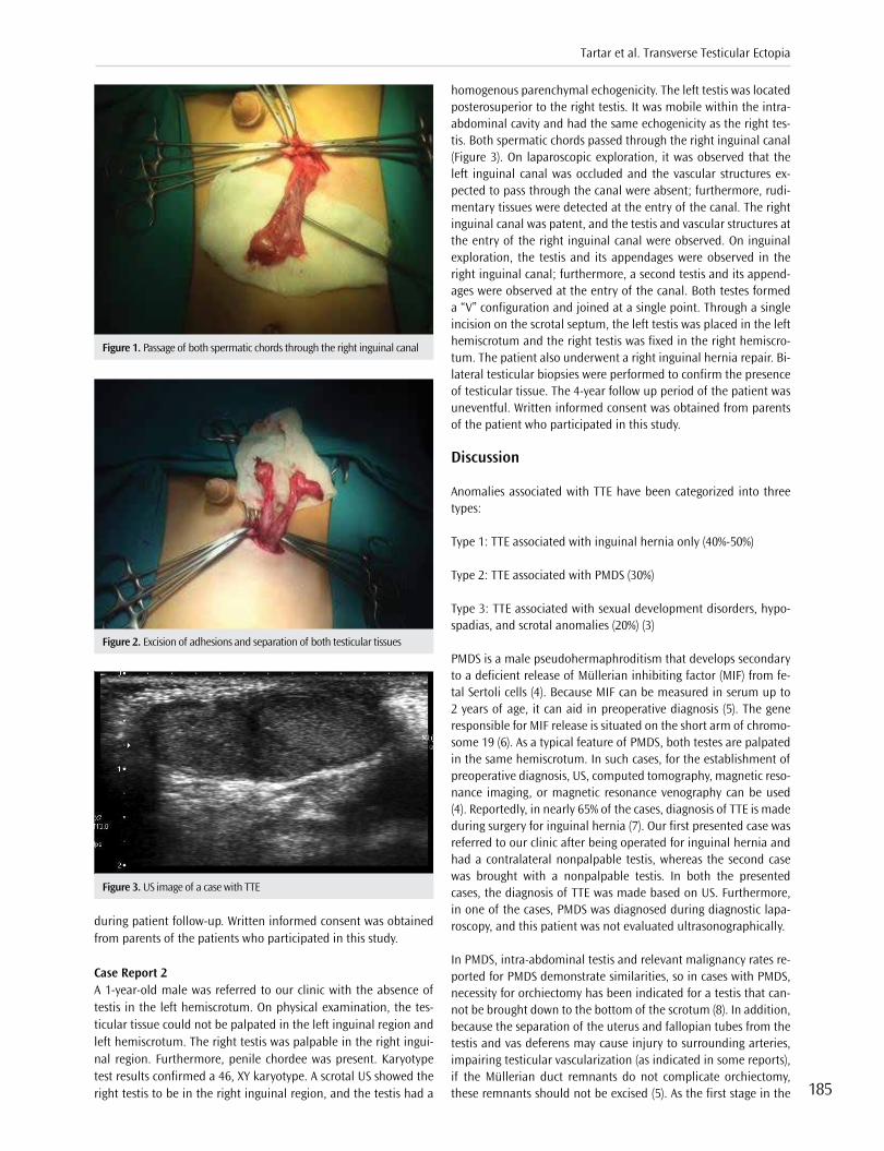
during patient follow-up. Written informed consent was obtained from parents of the patients who participated in this study.
Case Report 2A 1-year-old male was referred to our clinic with the absence of testis in the left hemiscrotum. On physical examination, the tes-ticular tissue could not be palpated in the left inguinal region and left hemiscrotum. The right testis was palpable in the right ingui-nal region. Furthermore, penile chordee was present. Karyotype test results confirmed a 46, XY karyotype. A scrotal US showed the right testis to be in the right inguinal region, and the testis had a
homogenous parenchymal echogenicity. The left testis was located posterosuperior to the right testis. It was mobile within the intra-abdominal cavity and had the same echogenicity as the right tes-tis. Both spermatic chords passed through the right inguinal canal (Figure 3). On laparoscopic exploration, it was observed that the left inguinal canal was occluded and the vascular structures ex-pected to pass through the canal were absent; furthermore, rudi-mentary tissues were detected at the entry of the canal. The right inguinal canal was patent, and the testis and vascular structures at the entry of the right inguinal canal were observed. On inguinal exploration, the testis and its appendages were observed in the right inguinal canal; furthermore, a second testis and its append-ages were observed at the entry of the canal. Both testes formed a “V” configuration and joined at a single point. Through a single incision on the scrotal septum, the left testis was placed in the left hemiscrotum and the right testis was fixed in the right hemiscro-tum. The patient also underwent a right inguinal hernia repair. Bi-lateral testicular biopsies were performed to confirm the presence of testicular tissue. The 4-year follow up period of the patient was uneventful. Written informed consent was obtained from parents of the patient who participated in this study.
Discussion
Anomalies associated with TTE have been categorized into three types:
Type 1: TTE associated with inguinal hernia only (40%-50%)
Type 2: TTE associated with PMDS (30%)
Type 3: TTE associated with sexual development disorders, hypo-spadias, and scrotal anomalies (20%) (3)
PMDS is a male pseudohermaphroditism that develops secondary to a deficient release of Müllerian inhibiting factor (MIF) from fe-tal Sertoli cells (4). Because MIF can be measured in serum up to 2 years of age, it can aid in preoperative diagnosis (5). The gene responsible for MIF release is situated on the short arm of chromo-some 19 (6). As a typical feature of PMDS, both testes are palpated in the same hemiscrotum. In such cases, for the establishment of preoperative diagnosis, US, computed tomography, magnetic reso-nance imaging, or magnetic resonance venography can be used (4). Reportedly, in nearly 65% of the cases, diagnosis of TTE is made during surgery for inguinal hernia (7). Our first presented case was referred to our clinic after being operated for inguinal hernia and had a contralateral nonpalpable testis, whereas the second case was brought with a nonpalpable testis. In both the presented cases, the diagnosis of TTE was made based on US. Furthermore, in one of the cases, PMDS was diagnosed during diagnostic lapa-roscopy, and this patient was not evaluated ultrasonographically.
In PMDS, intra-abdominal testis and relevant malignancy rates re-ported for PMDS demonstrate similarities, so in cases with PMDS, necessity for orchiectomy has been indicated for a testis that can-not be brought down to the bottom of the scrotum (8). In addition, because the separation of the uterus and fallopian tubes from the testis and vas deferens may cause injury to surrounding arteries, impairing testicular vascularization (as indicated in some reports), if the Müllerian duct remnants do not complicate orchiectomy, these remnants should not be excised (5). As the first stage in the
Figure 1. Passage of both spermatic chords through the right inguinal canal
Figure 2. Excision of adhesions and separation of both testicular tissues
Figure 3. US image of a case with TTE
Tartar et al. Transverse Testicular Ectopia
185

surgical treatment of PMDS, gonadal biopsy followed by orchio-pexy has been recommended by some authors, whereas others have reported uselessness of testicular biopsy in patients with a male karyotype (4). In one of our patients, during laparoscopic exploration of the undescended testis, rudimentary tissues were observed at the entry of the inguinal canal, and testicular biopsy performed during orchiopexy helped confirm the presence of tes-ticular tissue. Furthermore, we decided not to remove the rudi-mentary tissues because of the possibility of injury to testicular vessels.
No malignant tumors originating from the Müllerian duct remnants (tuba uterina, uterus, and one-third of the vagina) have been report-ed because these structures are probably not affected by estrogens. Therefore, as indicated in some case reports, removal of these struc-tures does not render additional benefits to the patient (4).
Conclusion
In the management of TTE, an ectopic testis should be placed in its normal anatomical position through transseptal orchiopexy or extraperitoneal transposition. In addition, in cases with TTE, PMDS should be kept in mind, and the Müllerian duct remnants should be investigated via diagnostic laparoscopy. In such cases, because a malignancy may develop, the Müllerian duct remnants should be removed to as much extent as possible without traumatizing the testis and its annexes. The patients should be followed up for a long time to check for infertility and malignancy.
Informed Consent: Written informed consent was obtained from parents of the patients who participated in this study. .
Peer-review: Externally peer-reviewed.
Author contributions: Concept - T.T., M.S.; Design - T.T., M.S., Ü.B.; Super-vision - A.K.; Resource - T.T., Ş.C.; Materials - T.T., M.S., ÜB.; Data Collection and/or Processing - T.T., M.S., Ü.B.; Analysis and/or Interpretation - T.T., A.K.; Literature Search - T.T., Ş.C.; Writing - T.T.; Critical Reviews - A.K.
Conflict of Interest: Authors have no conflicts of interest to declare.
Financial Disclosure: The authors declared that this study has received no financial support.
Hasta Onamı: Yazılı hasta onamı bu çalışmaya katılan hastaların aileler-inden alınmıştır.
Hakem Değerlendirmesi: Dış Bağımsız.
Yazar Katkıları: Fikir - T.T., M.S.,; Tasarım - T.T., M.S., Ü.B.,; Denetleme - A.K.; Kaynaklar - T.T., Ş.C..; Malzemeler - T.T., M.S., ÜB.Veri Toplanması ve/veya işlemesi - T.T., M.S., Ü.B., ; Analiz ve/veya Yorum - T.T., A.K. , ; Literatür taraması - T.T., Ş.C. , ; Yazıyı Yazan - T.T.; Eleştirel İnceleme - A.K. .
Çıkar Çatışması: Yazarlar çıkar çatışması bildirmemişlerdir.
Finansal Destek: Yazarlar bu çalışma için finansal destek almadıklarını beyan etmişlerdir.
References
1. Karnak I, Tanyel FC, Akçören Z, Hiçsönmez A. Transverse testicular ectopia with persistent müllerian duct syndrome. J Pediatr Surg 1997; 32: 1362-4. [CrossRef]
2. Gupta RL, Das P. Ectopia testis transversa. J Indian Med Assoc 1960; 35: 547-9.
3. Ramareddy RS, Alladi A, Siddappa OS. Ectopic testis in children: expe-rience with seven cases. J Pediatr Surg 2013; 48: 538-41. [CrossRef]
4. Tunçay S. Persistent Müllerian Duct Syndrome with Transverse Tes-ticular Ectopia and Hernia Uteri Inguinalis: A Case Report. Turk J Urol 2003; 29: 215-8.
5. Martin EL, Bennet AH, Cromia WJ. Persistent Müllerian duct syndrome with transverse testicular ectopia and spermatogenesis. J Urol 1992; 147: 1615-7. [CrossRef]
6. Cohen-Hanguenauer O, Picard JY, Mattéi MG, Serero S, Nguyen VC, de Tand MF, et al. Mapping of the gene for antimüllerian hormone to the short arm of human chromosome 19. Cytogenet Cell Genet 1987; 44: 2-6. [CrossRef]
7. Gauderer MW, Grisoni ER, Stellato TA, Ponsky JL, Izant RJ Jr. Transverse testicular ectopia. J Pediatr Surg 1982; 17: 43-7. [CrossRef]
8. Berkmen F. Persistent müllerian duct syndrome with or without transverse testicular ectopia and testis tumors. Br J Urol 1997; 79: 122-6. [CrossRef]
Cite this article as: Tatar T, Saraç M, Bakal Ü, Canpolat Ş, Kazez E. Transverse Testicular Ectopia: Two Case Reports. İstanbul Med J 2018; 19: 184-6.
İstanbul Med J 2018; 19: 184-6
186
Introduction
The etiology of notalgia paresthetica has not yet been fully elucidated. However, notalgia par-esthetica is defined as a disease characterized by neuropathic pain accompanied by dysesthetic symptoms, such as burning, stinging, tingling, pruritus, and numbness (1). Hyperpigmentation of the skin localized to the painful region, which is not accompanied by primary dermatologic pathology, is the most striking clinical finding (1, 2).
Notalgia paresthetica was first described in 1934 by the Russian neurologist Michail Astwazaturow as a clinical picture presenting with a painful, itchy hyperpigmented skin lesion usually localized to the dermatome distribution of the spinal nerves T2-T6. Notalgia paresthetica is considered to be a frequently encountered but easily overlooked condition, although its actual incidence remains unknown (2, 3).
Notalgia paresthetica mostly has a benign nature; however, because it reduces the quality of life, has a hereditary form, albeit rare, and is accompanied by multiple endocrine neoplasia, early diagnosis and screening for accompanying conditions is important (3). Diagnosis and treatment of a rare clinical presentation are noted in this case report.
Case report
A 65-year-old man was admitted to the physical medicine and rehabilitation polyclinics with com-plaints of pain, burning, stinging, and pruritus on his back for the past 6 months.
Background and family history of the patient did not exhibit any specific conditions. Physical examination revealed a hyperpigmented macular skin lesion 10×7 cm in size located under the left scapula in the dorsal region (Figure 1a, b).
Complaints of burning and stinging by the patient were evaluated with pre-diagnosis of neuro-pathic pain, and diagnosis was made based on the Leeds Assessment of Neuropathic Symptoms and Signs (LANSS) scale. The LANSS scale included seven domains that assessed pain characteris-tics and sensation. This combines five simple questions and two examination findings to give a score for pain out of 24, with a score ≥12 suggesting a neuropathic component (4). The Turkish
Notalgia paresthetica: A Rare Cause of Neuropathic PainNotaljia Parestetika: Nöropatik Ağrının Nadir Bir Nedeni
Notaljia parestetika T2-T6 spinal sinirlerin dağılım alanında yanıcı, ba-tıcı vasıfta sırt ağrısına eşlik eden kaşıntılı hiperpigmente cilt lezyonu ile giden klinik bir tablodur. Etiyolojisi tam olarak aydınlatılamamıştır. Notaljia parestetika görece olarak sık karşımıza çıkan fakat çoğu zaman gözden kaçan bir patolojidir. Hastalar kaşıntı şikayetlerinin ön planda olması nedeni ile genellikle dermatoloji kliniklerine başvurmakta ve tanıda gecikme ya da atlanma yaşanmaktadır. Yaşam kalitesini düşür-mesi nedeniyle erken tanı ve tedavisi önemlidir. Bu yazıda kliniğimize başvuran altmış beş yaşında erkek hasta literatür eşliğinde tartışılmıştır.
Anahtar Kelimeler: Nöropatik ağrı, notaljia parestetika, hiperpigmen-tasyon
Notalgia paresthetica is a clinical condition characterized by burning and stinging pain accompanied by an itchy hyperpigmented skin le-sion in the distribution area of spinal nerves T2-T6. Its etiology is not completely understood. It is a relatively common pathology that is often overlooked. Patients usually refer to dermatology clinics with complaints of pruritus, and the diagnosis is usually delayed, or some symptoms can go unnoticed. Early diagnosis and treatment are impor-tant because notalgia paresthetica reduces the quality of life. In this case study, we present a 65-year-old male patient who referred to our clinic and discussed this case for research.
Keywords: Neuropathic pain, notalgia paresthetica, hyperpigmentation
Abst
ract
/ Ö
z
ORCID IDs of all the authors: E.K. 0000-0003-0121-9082; Ç.A. 0000-0001-5476-9454.
1Department of Orthopedics and Traumatology, Süleyman Demirel University School of Medicine, Isparta, Türkiye2Clinic of Physical Medicine and Rehabilitation, Artvin State Hospital, Artvin, Türkiye
Address for Correspondence/Yazışma Adresi:Çiğdem Aydoğan E-mail: [email protected]
Received/Geliş Tarihi: 19.10.2017
Accepted/Kabul Tarihi: 02.01.2018
© Copyright 2018 by Available online at istanbulmedicaljournal.org
© Telif Hakkı 2018 Makale metnine istanbultipdergisi.org web sayfasından ulaşılabilir.
Case Report/Olgu Sunumuİstanbul Med J 2018; 19: 187-90DOI: 10.5152/imj.2018.72687
Emrah Kovalak1 , Çiğdem Aydoğan2
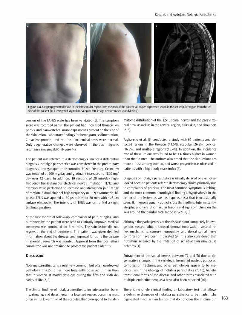
version of the LANSS scale has been validated (5). The symptom score was recorded as 19. The patient had increased thoracic ky-phosis, and paravertebral muscle spasm was present on the side of the skin lesion. Laboratory findings for hemogram, sedimentation, C-reactive protein, and routine biochemical tests were normal. Only degenerative changes were observed in thoracic magnetic resonance imaging (MRI) (Figure 1c).
The patient was referred to a dermatology clinic for a differential diagnosis. Notalgia paresthetica was considered in the preliminary diagnosis, and gabapentin (Neurontin; Pfizer, Freiburg, Germany) was initiated at 600 mg/day and gradually increased to 1800 mg/day over 12 days. In addition, 10 sessions of 20 min/day high-frequency transcutaneous electrical nerve stimulation (TENS) and exercises were performed to increase and strengthen joint range of motion. A dual-channel high-frequency (80 Hz) asymmetric, bi-phasic TENS was applied at 30 μs pulses for 20 min with 4×5 cm surface electrodes. The intensity of TENS was set to feel a slight tingling sensation.
At the first month of follow-up, complaints of pain, stinging, and numbness by the patient were seen to clinically improve. Medical treatment was continued for 6 months. The skin lesion did not regress at the end of treatment. The patient was given detailed information about the disease, and approval for using the disease in scientific research was granted. Approval from the local ethics committee was not obtained to protect the patient’s identity.
Discussion
Notalgia paresthetica is a relatively common but often overlooked pathology. It is 2-3 times more frequently observed in men than that in women. It mostly develops during the fifth and sixth de-cades of life (2, 3).
The clinical findings of notalgia paresthetica include pruritus, burn-ing, stinging, and dysesthesia in a localized region, occurring most often in the lower third of the scapulae that correspond to the der-
matome distribution of the T2-T6 spinal nerves and the paraverte-bral area, as well as in the cervical region, hairy skin, and shoulders (2, 3).
Pagliarello et al. (6) conducted a study with 65 patients and de-tected lesions in the thoracic (41.5%), scapular (26.2%), cervical (16.9%), and multiple regions (15.4%). In addition, the incidence rate of these lesions was found to be 1.6 times higher in women than that in men. The authors also noted that the skin lesions are more diffuse among women, and worse prognosis was observed in patients with a high body mass index (6).
Diagnosis of notalgia paresthetica is usually delayed or even over-looked because patients refer to dermatology clinics primarily due to complaints of pruritus. The most common symptom is itching, and the most common neurological finding is hypoesthesia in the center of the lesion, as well as hyperesthesia that is occasionally seen. Skin lesions usually do not cross the midline. Intermittently, atrophic and keratotic macular lesions and signs of itching on the skin around the painful area are observed (7, 8).
Although the pathogenesis of the disease is not completely known, genetic susceptibility, increased dermal innervation, visceral re-flex mechanisms, sensory neuropathy, and dorsal spinal nerve compression have been implicated (9). It is also considered that histamine released by the irritation of sensitive skin may cause itchiness (1).
Entrapment of the spinal nerves between T2 and T6 due to de-generative changes in the vertebrae, herniated nucleus pulposus, compression fractures, and other pathologies appear to be ma-jor causes in the etiology of notalgia paresthetica (7, 10). Genetic transitional forms of the disease and other forms associated with multiple endocrine neoplasia have also been reported (10).
There is no single clinical finding or laboratory test that allows a definitive diagnosis of notalgia paresthetica to be made. Itchy pigmented macular skin lesions that do not cross the midline but
Kovalak and Aydoğan. Notalgia Paresthetica
188
Figure 1. a-c. Hyperpigmented lesion in the left scapular region from the back of the patient (a); Hyper-pigmented lesion in the left scapular region from the left side of the patient (b); T1 weighted sagittal dorsal spine MRI image demonstrated spondylosis (c)
a b c
accompany pain and paresthetic complaints in the T2-T6 spinal nerves are pathognomonic (10).
Radiological examinations mostly reveal cervical and thoracic degenerative changes. In a study evaluating spinal pathologies in 43 patients using MRI, Savk et al. (11) reported degenerative disc hernias in 24.6% of the cases, degenerative changes in the thoracic vertebrae in 32.8%, and degenerative changes together with tho-racic disc hernias in 3.3%.In their study with 10 patients, Savk et al. (12) reported that elec-trophysiological examinations have normal findings in all patients. Since the diagnosis of the disease is mostly clinical, an electromyo-graphic examination was not performed in our case. Electroneuro-myographic studies performed in other studies also mostly provide normal findings. In the same study by Savk et al. (12), histopatho-logical examination revealed a hyperkeratosis of the epidermis in a patient as well as extensive inflammation and melanophages in the papillary dermis in all patients. Histopathological examination was not performed in the present case. Skin disorders including atopic dermatitis, post-inflammatory hyperpigmentation, prurigo nodularis, and macular amyloidosis should be considered in the differential diagnosis (13).
Systemic and local agents have been used for the treatment of notalgia paresthetica. Topical capsaicin, corticosteroids, tacro-limus, and local anesthesia drugs can be used (1, 14). Antiepi-leptic drugs, such as gabapentin, pregabalin, oxcarbazepine, and tricyclic antidepressants, are preferred for the treatment of neu-ropathic pain. Antihistamines can be prescribed to relieve com-plaints of pruritus (14).
In another study conducted by Savk et al. (15), TENS therapy was shown to be effective in 15 patients. Maari et al. (16) ad-ministered botulinum toxin to 10 patients to prevent substance P release to reduce the symptoms of pruritus and neuropathic pain and compared the results with a placebo group with an equal number of patients. The authors found no difference be-tween the groups in terms of the symptoms of pruritus and the extent of hyperpigmentation (16). Tension and strengthening exercises on the lower back, rhomboid, pectoralis major, and latissimus dorsi muscles have been shown to be beneficial (17). In a single-case study, Subasi et al. (18) performed dry needling on the fibrous bands in the dorsal region combined with kinesio taping on macular skin lesions using the inhibition technique and found the treatment to be effective.
Conclusion
Owing to neuropathic pain complaints, rather than dermatology, patients with notalgia paresthetica may refer to other branches of medicine, such as physical medicine and rehabilitation, neu-rology, orthopedics and traumatology, brain and neurosurgery, and algology. A multidisciplinary approach is important in these patients. Positive outcomes can be achieved with early diagnosis and treatment.
Informed Consent: Informed concent was obtained from the patient who participated in this study.
Peer-review: Externally peer-reviewed.
Author contributions: Concept - E.K., Ç.A.; Design - E.K., Ç.A.; Supervision - E.K., Ç.A.; Resource - E.K., Ç.A.; Materials - E.K., Ç.A.; Data Collection and/or Processing - E.K., Ç.A.; Analysis and/or Interpretation - E.K., Ç.A.; Literature Search - E.K., Ç.A.; Writing - E.K., Ç.A.; Critical Reviews - E.K., Ç.A.
Conflict of Interest: Authors have no conflicts of interest to declare.
Financial Disclosure: The authors declared that this study has received no financial support.
Hasta Onamı: Hasta onamı bu çalışmaya katılan hastalardan alınmıştır.Hakem Değerlendirmesi: Dış Bağımsız.
Yazar Katkıları: Fikir - E.K., Ç.A.; Tasarım - E.K., Ç.A.; Denetleme - E.K., Ç.A.; Kaynaklar - E.K., Ç.A.; Malzemeler - E.K., Ç.A.; Veri Toplanması ve/veya işlemesi - E.K., Ç.A.; Analiz ve/veya Yorum - E.K., Ç.A.; Literatür taraması - E.K., Ç.A.; Yazıyı Yazan - E.K., Ç.A.; Eleştirel İnceleme - E.K., Ç.A.
Çıkar Çatışması: Yazarların beyan edecek çıkar çatışması yoktur.
Finansal Destek: Yazarlar bu çalışma için finansal destek almadıklarını beyan etmişlerdir.
References
1. Chiriac A, Podoleanu C, Moldovan C, Stolnicu S. Notalgia Paresthetica, A Clinical Series and Review. Pain Pract 2016; 16: 90-1. [CrossRef]
2. Pleet AB, Massey EW. Notalgia paresthetica. Neurology 1978; 28: 1310-2. [CrossRef]
3. Karahan AY. Gabapentin and TENS in the Treatment of Notalgia Pares-thetica: A Case Report. J PMR Sci. 2012; 15: 86-8.
4. Bennett M. The LANSS Pain Scale: the Leeds assessment of neuropath-ic symptoms and signs. Pain 2001; 92: 147-57. [CrossRef]
5. Yucel A, Senocak M, Kocasoy Orhan E, Cimen A, Ertas M. Results of the Leeds assessment of neuropathic symptoms and signs pain scale in Turkey: a validation study. J Pain 2004; 5: 427-32. [CrossRef]
6. Pagliarello C, Fabrizi G, De Felici B, Casanova D, Feliciani C, Di Nuzzo S. Notalgia paresthetica: factors associated with its perceived sever-ity, duration, side, and localization. Int J Dermatol 2017; 56: 932-8. [CrossRef]
7. Richardson BS, Way BV, Sepeece AJ 3rd. Osteopathic manipulative treatment in the management of notalgia paresthetica. J Am Osteo-path Assoc 2009; 109: 605-8.
8. Eisenberg E, Barmeir E, Bergman R. Notalgia paresthetica associated with nerve root impingement. J Am Acad Dermatol. 1997; 37: 998-1000. [CrossRef]
9. Springall DR, Karanth SS, Kirkham N, Darley CR, Polak JM. Symptoms of no-talgia paresthetica may be explained by increased dermal in-nervation. J Invest Dermatol 1991; 97: 555-61. [CrossRef]
10. Alcantara F, Feito M, Albizuri F, Beato M, De Lucas R. Notalgia Pares-thetica anf Multiple Endocrine Neoplasia Syndrome 2A: Case Report. Pediatr Dermatol 2016; 33: 303-5. [CrossRef]
11. Savk O, Savk E. Investigation of spinal pathology in notalgia pares-thetica. J Am Acad Dermatol 2005; 52: 1085-7. [CrossRef]
12. Savk E, Savk O, Bolukbasi O, Culhaci N, Dikicioğlu E, Karaman G, Sen-dur N. Notalgia paresthetica: a study on pathogenesis. Int J Dermatol 2000; 39: 754-9. [CrossRef]
13. Savk E, Savk SO. On brachioradial pruritus and notalgia paresthetica. J Am Acad Dermatol 2004; 50: 800-1. [CrossRef]
14. Pérez-Pérez LC. General features and treatment of notalgia paresthet-ica. Skinmed 2011; 9: 353-8.
İstanbul Med J 2018; 19: 187-90
189

15. Savk E, Savk O, Sendur F. Transcutaneous electrical nerve stimulation
offers partial relief in notalgia paresthetica patients with a relevant
spinal pathology. J Dermatol 2007; 34: 315-9. [CrossRef]
16. Maari C, Marchessault P, Bissonete R. Treatment of notalgia pares-
thetica with botulinum toxin A: a double-blind random-ized con-
trolled trial. J Am Acad Dermatol 2014; 70: 1139-41. [CrossRef]
17. Fleischer AB, Meade TJ, Fleischer AB. Notalgia paresthetica: success-
ful treatment with exercises. Acta Derm Venereol 2011; 91: 356-7.
[CrossRef]
18. Subaşı V, Çakır T, Atasoy MF. Notaljia parestetika tedavisinde kuru iğleneme ve kinezyolojik bantlama kombinasyonun etkinliği: Olgu sunumu. Turk J Phys Med Rehab 2016; 62: 273-6. [CrossRef]
Cite this article as: Kovalak E, Aydoğan Ç. Notalgia paresthetica: A rare cause of neuropathic pain. İstanbul Med J 2018; 19: 187-90
Kovalak and Aydoğan. Notalgia Paresthetica
190