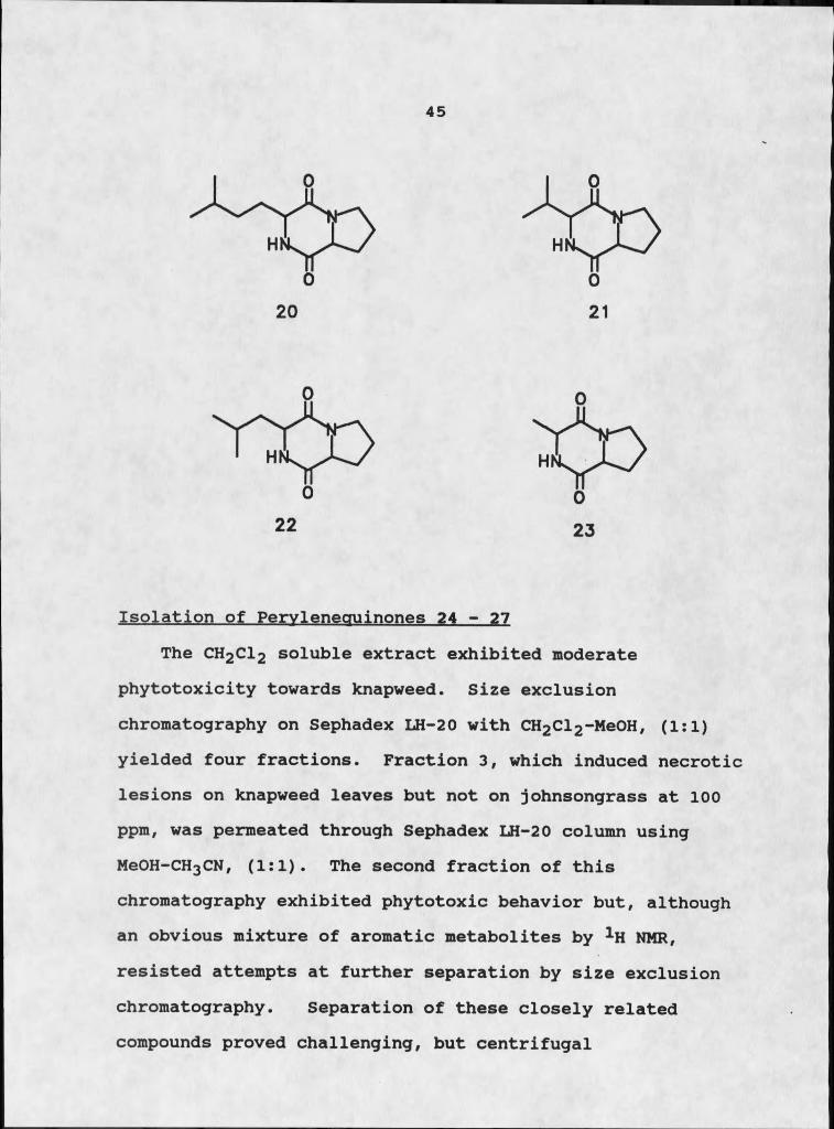
31762100613247.pdf - ScholarWorks
236
Investigation of biologically active metabolites from symbiotic microorganisms by Andrea Anne Stierle A thesis submitted in partial fulfillment of the requirements for the degree of Doctor of Philosophy in Chemistry Montana State University © Copyright by Andrea Anne Stierle (1988) Abstract: The investigation of symbiotic relationships between macroorganisms and microorganisms is a complex, fascinating research endeavor. Throughout the course of this project we explored two such interactions: the relationship between a terrestrial weed and a plant pathogenic fungus, and the relationship between a marine sponge and a gram positive bacterium. Each of these interactions represents a different aspect of symbiosis. Centaurea maculosa Lam, spotted knapweed, was the focus of a study concerning a parasitic fungus-weed interaction. Alternaria alternate was isolated from a black leaf blighted specimen of spotted knapweed and was determined to be the causative organism of the observed plant disease. A series of diketopiperazines were consistently isolated from liquid cultures of Alternaria alternate. The most active of these compounds, maculosin, cyclo(L-Pro-L-Tyr), caused black necrotic lesions in a nicked leaf bioassay at 10μm. In tests against nineteen plant species, both monocots and dicots, maculosin was phytotoxic only to spotted knapweed. Several other diketopiperazines isolated from the fungus exhibited either reduced phytotoxicity or none at all. This is the first report of a host specific toxin from a weed pathogen. Additional phytotoxic fractions from the organic soluble extracts of the fungus yielded a series of perylenequinones. Two of these compounds were novel and phytotoxic, and two were known but inactive. The organic extracts also yielded the known toxin tenuazonic acid. All three classes of compounds were compared individually and in combination with respect to phytotoxicity and host specificity. Not all symbioses are as well-defined as the host-parasite relationship. A Micrococcus sp. was consistently isolated from tissues excised from the marine sponge Tedania ignis, despite the collection locale, suggesting some form of symbiosis, but not clarifying the degree of interaction. A relationship which we initially considered ambiguous between the sponge and the bacterium evolved into a true case of mutualism. Of particular interest was the isolation of three compounds from the liquid culture of the bacterium that were previously attributed to the sponge. In addition to these three compounds, several aromatic metabolites not usually associated with living organisms were also isolated from the liquid culture, characterized, and tested for biological activity.
-
Upload
khangminh22 -
Category
Documents
-
view
5 -
download
0
Transcript of 31762100613247.pdf - ScholarWorks

Investigation of biologically active metabolites from symbiotic microorganismsby Andrea Anne Stierle
A thesis submitted in partial fulfillment of the requirements for the degree of Doctor of Philosophy inChemistryMontana State University© Copyright by Andrea Anne Stierle (1988)
Abstract:The investigation of symbiotic relationships between macroorganisms and microorganisms is acomplex, fascinating research endeavor. Throughout the course of this project we explored two suchinteractions: the relationship between a terrestrial weed and a plant pathogenic fungus, and therelationship between a marine sponge and a gram positive bacterium. Each of these interactionsrepresents a different aspect of symbiosis.
Centaurea maculosa Lam, spotted knapweed, was the focus of a study concerning a parasiticfungus-weed interaction. Alternaria alternate was isolated from a black leaf blighted specimen ofspotted knapweed and was determined to be the causative organism of the observed plant disease. Aseries of diketopiperazines were consistently isolated from liquid cultures of Alternaria alternate. Themost active of these compounds, maculosin, cyclo(L-Pro-L-Tyr), caused black necrotic lesions in anicked leaf bioassay at 10μm. In tests against nineteen plant species, both monocots and dicots,maculosin was phytotoxic only to spotted knapweed. Several other diketopiperazines isolated from thefungus exhibited either reduced phytotoxicity or none at all. This is the first report of a host specifictoxin from a weed pathogen.
Additional phytotoxic fractions from the organic soluble extracts of the fungus yielded a series ofperylenequinones. Two of these compounds were novel and phytotoxic, and two were known butinactive. The organic extracts also yielded the known toxin tenuazonic acid. All three classes ofcompounds were compared individually and in combination with respect to phytotoxicity and hostspecificity.
Not all symbioses are as well-defined as the host-parasite relationship. A Micrococcus sp. wasconsistently isolated from tissues excised from the marine sponge Tedania ignis, despite the collectionlocale, suggesting some form of symbiosis, but not clarifying the degree of interaction. A relationshipwhich we initially considered ambiguous between the sponge and the bacterium evolved into a truecase of mutualism. Of particular interest was the isolation of three compounds from the liquid cultureof the bacterium that were previously attributed to the sponge. In addition to these three compounds,several aromatic metabolites not usually associated with living organisms were also isolated from theliquid culture, characterized, and tested for biological activity.

INVESTIGATION OF BIOLOGICALLY ACTIVE METABOLITES
FROM SYMBIOTIC MICROORGANISMS
byAndrea Anne Stierle
A thesis submitted in partial fulfillment of the requirements for the degree
ofDoctor of Philosophy
inChemistry
MONTANA STATE UNIVERSITY Bozeman, Montana
August 1988
St̂ 5
ii
APPROVALof a thesis submitted by
Andrea Anne Stierle
This thesis has been read by each member of the thesis committee and has been found to be satisfactory regarding content, English usage, format, citations, bibliographic style, and consistency, and is ready for submission to the College of Graduate Studies.
2% szr"Date ^
Approved for the Major Department
Date f Head, Major Department
Approved for the College of Graduate Studies
Date Graduate D4an
iii
STATEMENT OF PERMISSION TO USE
In presenting this thesis in partial fulfillment of the requirements for a doctoral degree at Montana State University.,. I agree that the Library shall make it available to borrowers under rules of the Library. I further agree that copying of this thesis is allowable only for scholarly purposes, consistent with "fair use" as prescribed in the U.S. Copyright Law. Requests for extensive copying or reproduction of this thesis should be referred to University Microfilms International, 300 North Zeeb. Road, Ann Arbor, Michigan 48106, to whom I have granted "the exclusive right to reproduce and distribute copies of the dissertation in and from microfilm and the right to reproduce and distribute by abstract in any format."
Date

iv
ACKNOWLEDGEMENTS
There are so many people to thank. Joe Sears who provided mass spectral analyses whenever needed. The various members of my research group who shared expertise and support, in so many ways. Professor John Cardellina, my primary advisor, who provided the freedom I needed to be truly creative, and the discipline I required to develop a professional approach to research. Professor Gary Strobel who always tried to broaden my horizons, always listened when I needed to talk, and provided a beautiful example of grace under pressure. The faculty of Montana Tech who generously provided research space, facilities, and moral support.
But above all others, is Donald. His support and love have been overwhelming. From the first day of graduate school until the last feverish moments of thesis preparation, Don has given so much. Thank you, my love, you've helped to make another dream come true.
And finally. Tortilla, our beautiful little girl who taught us what symbiosis was all about. And how important such relationships can be. We miss her deeply.
V
TABLE OF CONTENTS
LIST OF TABLES........LIST OF FIGURES.......INTRODUCTION.......TERRESTRIAL PARASITISM
Page viii . . ix . I . 5
Background.................................... „. „ „Biorationale for Research Design. ..!.‘! ! ! ! X X !Phytotoxins: Historical Perspective..........!!Selection of a Suitable Target Plant.......... !Discovery of a Pathogen................ ....... .Results............. !!!!!!!!
Isolation of Diketopiperazines.............Structure Elucidation of Diketopiperazines Isolation of Peryleneguinones 24 - 27.....Structure Elucidation of Perylenequinones. Isolation of Tenuazonic Acid, 34..........Structural Elucidation of Tenuazonic Acid, Biological Activity........... .............
... 5
. . . 7
. . . 13 . . . 21
. . . 25
. . . 29
. . . 32
... 34
... 45
. . . 48
. . . 63 34. 65 . . . 67
MARINE MUTUALISM 82Background.... ..........Compounds of Possible or Proven Microbial Origin!Biochemical Studies of Endosymbionts............Biorationale for Research Design.................Suitability of Micrococcus sp. as a Symbiont.....Results.............................. .
Isolation of Diketopiperazines................Structure Elucidation of Diketopiperazines...Isolation of Tedanazine, 60...................Structure Elucidation of Tedanazine, 60......Isolation of Benzothiazoles and Indoles......Structure Elucidation of Benzothiazoles......Structure Elucidation of Indole Derivatives..Isolation of Daidzein.................. .......Structure Elucidation of Daidzein... .........Isolation of Glucose.... ......................Structure Elucidation of Glucose........!!!!!Biological Activity and Significance.......
Conclusion............... ........... .....
. 82
. 91
. 99
.108
.114
.117
.123
.124
.128
.130
.147
.149
.166
.175
.175
.177
.178
.178
.183
TABLE OF CONTENTS- ContinuedPage
EXPERIMENTAL. ............ ................................. 188Materials and Methods....................... iss
General Instrumentation.... ............. 188Fungal Culture Maintenance...... 188Marine Bacterial Culture Maintenance...........189Fungal Culture Growth and Extraction.......... 189Marine Bacterial Culture Growth andExtraction............ 190Artifact Control................................ 192
Bioassay Protocols.................................. .195Leaf Assay. ............... 195Hypocotyl Assay. ...................... .196Antimicrobial Assay......................... 196Brine Shrimp Toxicity....... 197
Isolation of Compounds............... 198Maculosin, 17......... ...........................19 8Cyclo(L-Pro-L-Phe), 18 andCyclo (L-Pro-D-Phe), 19............. 199Cyclo(Pro-Hleu) , 20............... ...199Cyclo(Pro-Val), 21, Cyclo(Pro-Leu),22, and Cyclo(Pro-Ala), 23..................... 199Cyclo(Pro-Met) , 57............ 200.Cyclo(Phe-Ala), 58................ . 200Cyclo(Pro-Trp) , 59...............................200Perylenequinones .24-27.......... .201Tenuazonic Acid, 34 ...............................201Tedanazine, 60......................... 2012-Mercaptpbenzothiazole, 63,2-Methylbenzothiazole, 64,3 (2-hydroxyacetyi)indole, 70, and2-Hydroxybenzothiazole, 65.................... .202
. 6-Hydroxy-3-methyl-2-benzothiazolone,67, and Indole-3-methylthiocarboxylate, 71....203Daidzein, 74.......... ..203
Characterization of C o m p o u n d s . ....... 204Maculosin, 17.................................... 204Synthesis of Maculosin, '17................... .204Cyclo(L-Pro-L-Phe), 18.... ............ ........ 205Cyclo(L-Pro-D-Phe) , 19............... 206Cyclo(Pro-Hleu), 20................... 206Cyclo(Pro-Val), 21....... 206Cyclo(Pro-Leu) , 22......... 207Cyclo(Pro-Ala) , 23......... .207Cyclo(Pro-Met), 57................. 207Cyclo(Phe-Ala), 58................. 207Cyclo(Pro-Trp), 59.......... .208Alterlosin I, 26. ....... 208
vi
TABLE OF CONTENTS-ContinuedPage
Alterlosin TI, 27.................................208NaBH4 Reduction of Alterlosin II, 27..........„209NaBH4 Reduction of Altertoxin I, 24.......... ..209Tenuazonic Acid, 34........ ....210Tedanazine, 60.................................... 2102-Mercaptobenzothiazole, 63..................... 2112-Methylbenzothiazole, 64....................... 2112-Hydroxybenzothiazole, 65...................... 2116-Hydroxy-3-methyl-2-benzothiazolone, 67...... 2113 (2-hydroxyacetyl) indole, 70.................... 212Indole-3-methyl thiocarboxylate, 71............. 212Daidzein, 74.............................. 212
Vii
REFERENCES 214

viii
LIST OF TABLES
Table Page1. Host-Specific Toxins of Alternaria alternate...... 302. Diketopiperazines Isolated, Fraction of
Origin, Separation Technique, % Yield and Molecular Weight.............................. 33
3. Comparison of 1H NMR Data for Diketopiperazines,Maculosin, 17, (L-Pro-L-Phe), 18, and (L-pro-D-Phe), 19, Listing ChemicalShifts (6), Multiplicities, and Coupling Constants(Hz).......................................... 44
4. Comparison of 1H NMR Data (in CDCl3) forAlterlosin I, 26 and 11,27........................ 51
5. Comparison of Spectral Data of Maculosin andCompound of Tatsuno et a 1 ............................. 69
6. Comparison of Phytotoxicity of Naturally Occurring Diketopiperazines towards Knapweedand Johnsongrass...................................... 71
7. Phytotoxicity Data for Determining Host Specificity of Maculosiri and SyntheticMaculosin Using Leaf Assays.......................... 73
8. Summary of Phytotoxicity Data for Determining Host Specificity of Maculosin, Tenuazonic Acidand Alterlosin II...................................... 75
9. Synergy Study of Diketopiperazine Toxin andTenuazonic Acid........................................ 78
10. Activity Profile of the Microbes Isolated from the Various Sponges and the Bryozoan Collected in Bermuda................................................. .

i
ix
LIST OF FIGURESFigure Page1. Spotted Knapweed....................................... 222. Spread of Knapweed in Montana Since Mid-1920's..... 243. Structures of Host-Specific Toxins Isolated
from Various Form-Species of Alternariaalternata........................................ 31
4. 1H NMR Spectrum of Maculosin................... 365. Mass Spectrum of Maculosin............................ 376. Tyrosyl Fragment.... ....... 387. 1H NMR Spectrum of 18.............................. 418 . 1H NMR Spectrum of 19................................. 429. CCC Trace of Separation of Diastereomers of
Cyclo(Pro-Phe) and Peryleneguinones................ 4610. Conformational Rotamers of Cycle (Pro-Phe).......... 4711. Peryleneguinone Skeleton of 24................... 4912. Mass Spectrum of 26............... ................... 5213. 1H NMR Spectrum of 26........... 5314. Mass Spectrum of 27..................... 5715. 1H NMR Spectrum of 27..... 5816. NMR Simulation of ABX System Using NMRCALC......... 5917. Mass Spectrum of 60....................................13218. Infrared Spectrum of 60............................. ...13319. 1H NMR Spectrum of 60..................................13520. Two Potential Skeletons for 60....................... 137
LIST OF FIGURES-Continued
Figure Page21. 1H NMR Assignments for SI..................... ....... 14022. Lowest Energy Conformers of [2,2,3] Cyclazine___ ..14223. Lowest Energy Conformers of 60...................... .24. Mass Spectral Fragmentation of 60................ ...14425. 1H NMR Assignments for 62................ ............ 14526. Lowest Energy Conformers for Lactam Derivative.... 14727. Mass Spectrum of 63................................... .28. 1H NMR Spectrum of 63................................. .29. Mass Spectral Fragmentation of 63................... .30. Mass Spectrum of 64................................... 15631. 1H NMR Spectrum of 64........ ........... ............. 2.5732. 1H NMR Spectrum Of 65................................. 15933. Mass Spectrum of 65.............. ..................... 2.6034. Mass Spectral Fragmentation of 65................... .35. Mass Spectrum of 67................................... 16336. 1H NMR Spectrum of 67................. ............... 15437. Mass Spectrum of 70................................... I6938. 1H NMR Spectrum of 70................................. .39. Mass Spectrum of 71.............................. .....17240. 1H NMR Spectrum of 71..................................173
X
ABSTRACT
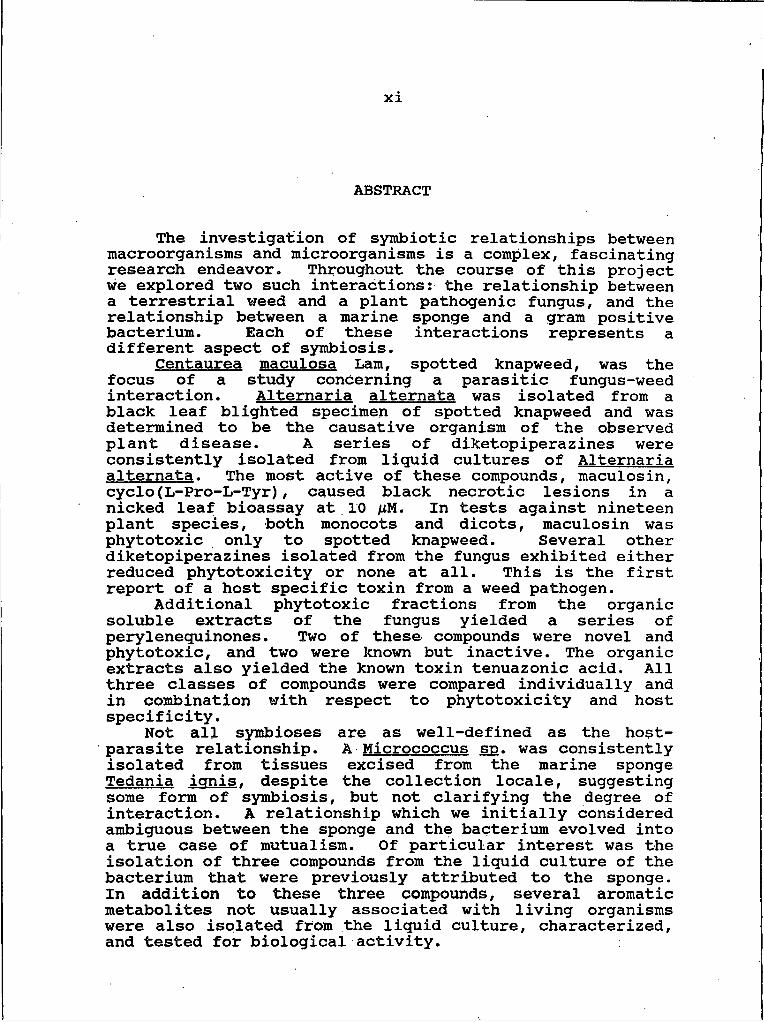
The investigation of symbiotic relationships between macroorganisms and microorganisms is a complex, fascinating research endeavor. Throughout the course of this project we explored two such interactions: the relationship between a terrestrial weed and a plant pathogenic fungus, and the relationship between a marine sponge and a gram positive bacterium. Each of these interactions represents a different aspect of symbiosis.
Centaurea maculosa Lam, spotted knapweed, was the focus of a study concerning a parasitic fungus-weed interaction. Alternaria alternate was isolated from a black leaf blighted specimen of spotted knapweed and was determined to be the causative organism of the observed plant disease. A series of diketopiperazines were consistently isolated from liquid cultures of Alternaria alternate. The most active of these compounds, maculosin, cyclo(L-Pro-L-Tyr), caused black necrotic lesions in a nicked leaf bioassay at 10 /iM. In tests against nineteen plant species, both monocots and dicots, maculosin was phytotoxic only to spotted knapweed. Several other diketopiperazines isolated from the fungus exhibited either reduced phytotoxicity or none at all. This is the first report of a host specific toxin from a weed pathogen.
Additional phytotoxic fractions from the organic soluble extracts of the fungus yielded a series of perylenequinones. Two of these compounds were novel and phytotoxic, and two were known but inactive. The organic extracts also yielded the known toxin tenuazonic acid. All three classes of compounds were compared individually and in combination with respect to phytotoxicity and host specificity.
Not all symbioses are as well-defined as the host- parasite relationship. A Micrococcus sp. was consistently isolated from tissues excised from the marine sponge Tedania ignis, despite the collection locale, suggesting some form of symbiosis, but not clarifying the degree of interaction. A relationship which we initially considered ambiguous between the sponge and the bacterium evolved into a true case of mutualism. Of particular interest was the isolation of three compounds from the liquid culture of the bacterium that were previously attributed to the sponge. In addition to these three compounds, several aromatic metabolites not usually associated with living organisms were also isolated from the liquid culture, characterized, and tested for biological activity.
I
INTRODUCTION
The investigation of symbiotic relationships requires an understanding of the scope and variability of possible interactions between organisms. In a literal sense, symbiosis simply means "living together"; in the true biological sense, however, it involves a close association between two members of different species (I). There are three extremes of symbiotic relationships. If the relationship is beneficial to both species it is called mutualism. A classic example of mutualism is the lichen, which is part fungus and part alga. The two unrelated organisms together form a closely integrated unit capable of growth under conditions that neither the fungus nor the alga could survive alone (I).
A relationship that benefits one species while neither harming nor benefiting the other species is called commensalism. The remora enjoys such a relationship with the shark. An indifferent swimmer, the remora attaches itself to the shark by means of an adhesive organ on its head. The "guest" is not only transported courtesy of its host, but also appropriates a portion of the shark's kill when it feeds (2).
If one species is harmed and the other species benefits it is called parasitism. Parasitism is considered to be a
2
special form of predation in which the predator is considerably smaller than the prey (3). Familiar examples of parasites are mosquitoes, fleas, and ticks.
An intriguing aspect of symbiosis is the interaction of macroorganisms and microorganisms in intimate contact with one another. Just as in interactions between two macroorganisms, such relationships can be mutualistic, commensal, or parasitic. In most cases, it is the microorganism that derives consistent benefit from such associations, and any harm is usually derived by the macroorganism (4). The exploration of such relationships and, in particular, the chemical manifestations of such interactions, provide a rich arena for natural products research.
It is sometimes difficult to determine the nature of a particular symbiotic association. All of the details of a relationship may not be known or fully understood, and relationships are subject to change with time or various circumstances. This is especially true in ascertaining the nature of macroorganism-microorganism interactions. Human beings play host to a rich population of skin bacteria, such as Staphylococcus aureus. which generally coexist as harmless commensals on the surface of the epidermis. When aspirated into the lungs or introduced into a wound, these harmless commensals may become parasitic (4). Saprophytic fungi may parasitize live plants under the proper
conditions (5). Any suspected instance of symbiosis must be carefully examined to verify both the existence and the nature of such a relationship.
In general, a relationship is considered symbiotic if two organisms are consistently found together even if the scope of their interaction is unknown or undefined (I). in the course of this study we chose to examine such relationships in both marine and terrestrial ecosystems.The target macroorganisms, Centaurea maculosa and Tedania ignis, were chosen because both possess intrinsic biological significance.
Spotted knapweed is the most important weed pest in Montana, and we were most interested in discovering a bacterium or fungus capable of producing phytotoxins deleterious to this plant. The first part of this study centers on the discovery of such a pathogen, an examination of its disease-inductive propensity towards knapweed, and the determination of its ability to produce substances injurious to the plant.
Organic extracts of the marine sponge Tedania ignis exhibit both cytotoxicity and in vivo tumor inhibition, and have yielded the potent cytotoxic macrolide designated tedanolide, I (6). The extremely low yields of tedanolide (lxl0“4% dry weight) suggested to Schmitz that this metabolite might actually prove to be of microbial origin, as was found with tetrodotoxin (7). It was our intention
3
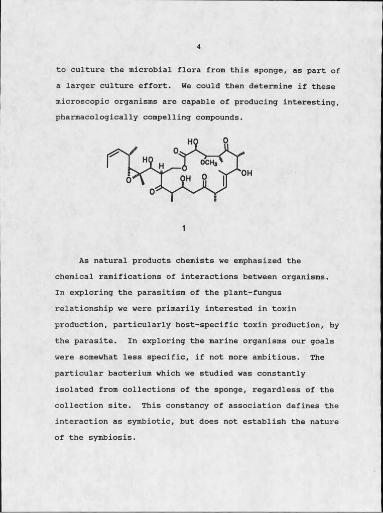
4
to culture the microbial flora from this sponge, as part of a larger culture effort. We could then determine if these microscopic organisms are capable of producing interesting, pharmacologically compelling compounds.
I
As natural products chemists we emphasized the chemical ramifications of interactions between organisms.In exploring the parasitism of the plant-fungus relationship we were primarily interested in toxin production, particularly host-specific toxin production, by the parasite. In exploring the marine organisms our goals were somewhat less specific, if not more ambitious. The particular bacterium which we studied was constantly isolated from collections of the sponge, regardless of the collection site. This constancy of association defines the interaction as symbiotic, but does not establish the nature of the symbiosis.
5
TERRESTRIAL PARASITISM
Background
The examination of the parasitism of plants by fungi is supported by extensive plant pathological lore. Fungi have long been recognized as causal agents of plant diseases. Many disease symptoms are often associated with the elaboration of one or more phytotoxins by a pathogenic fungus. To date, phytotoxins capable of expressing host specificity at the species or cultivar level are known only as metabolites of pathogens of crop plants. This is probably because crop plants are typically agro-ecosystems with a strictly homogeneous genetic base, which serves as a huge reservoir of effectively identical plant material.This homogeneity renders these plants susceptible to widespread devastation by one or more pathogens. The 1970 Southern corn leaf blight epidemic in the US-Canada, attributed to the pathogenic fungus Drechslera mavdis. supports this supposition (8). This epidemic caused the greatest crop loss in the shortest time span of any plant disease on record. Corn plants with Texas-male-sterile (Tms) cytoplasm, which comprised most of the corn planted, were particularly vulnerable. A pathogen has the potential to develop and spread quickly under such conditions and can be easily observed and isolated.
6
This does not appear to be the case with weeds, which can be defined as opportunistic plants that compete economically or aesthetically with whatever plants are intentionally cultivated (9). Common weedy plants usually exist in a population having a heterogeneous genetic base which tends to preclude the development of such an epidemic, making discovery of the pathogen difficult, if not impossible.
The isolation and investigation of weed pathogens isimportant not only because of their intrinsic ability toserve as biocontrol agents, but also because of theirpropensity for producing novel bioactive substances. Thesesubstances may act as herbicides or provide importantchemical leads to the herbicide industry (10). It was ourintention to harness this propensity for phytotoxinproduction exhibited by a number of pathogenic fungi anddirect it towards an important weed pest.
Phytotoxins vary greatly in their disease inductiveabilities and in their degree of host-selectivity. We weremost interested in isolating a pathogen specific to aparticular weed capable of producing toxins that were alsohost-specific for that weed. R. K. S. Wood (S) definedhost-specific toxins as follows:
Microorganism X (but not others) produces substance Y which damages plant or plant group A but not others, and only A is parasitized.
I
The concept of a host specific toxin is certainly not new. Over 40 years ago Meehan and Murphy (1.1) recognized the influence of such a compound in the disease symptontology of victoria blight of oats, which will be discussed in more detail later. Several compounds with apparent true host specificity have been isolated and characterized. It is interesting to note that no host specific toxins have ever been found to weed plants.
The focus of this study was the discovery of a weed pathogen capable of producing compounds with phytotoxicity only towards a particular weed. Once such a weed pathogen was discovered, the chemistry responsible for its virulence and host-specificity would be thoroughly investigated.
Biorationale for Research Design
There are two approaches to the utilization of plant pathogenic microorganisms as biocontrol agents. The first approach involves the inoculation of the target organism, in this case a weed, with the live pathogen, in an attempt to induce disease symptoms in the plant. The second approach involves application of selected, bioactive chemicals (phytotoxins) produced by the pathogen to the target weed to produce disease symptoms. In both cases it is necessary to follow the dictates of Koch9 s postulates (12) to establish absolutely an isolated pathogen as the instigator of plant disease:
8
1) The pathogen must be isolated from the tissues of a diseased host and established in a pure culture.2) The pure cultural isolate must be capable of
consistently inducing disease symptoms in a healthy host.3) The pathogen must then be reisolated from the disease-
induced host and re-established as a pure culture.The deliberate use of live plant pathogenic bacteria,
fungi, and viruses to achieve economic control of weeds is a relatively new endeavor. Throughout the world, the past twenty-five years have witnessed numerous attempts to control important weed pests through the introduction of selected microorganisms. Plants that have been transported into new regions either by accident or by design often assume the status of weeds due to a lack of natural predators (13). Pathogens are sought from the original geographic location of the plant for introduction into its new environs. Ideally, the pathogen will become established, reach epiphytotic levels, and eventually stabilize as an endemic population once the weed is reduced to subeconomic levels. Skeletonweed (Chondrilla iuncea L.) has been successfully controlled in Australia by the introduction of the rust fungus Puccinia chondrillina from the Mediterranean region where the weed originated (14). Attempts to duplicate this success in the United States include the control of pamakani weed (Ageratina rineria) in Hawaii by introduction of the deuteromycete Cercosnorella
9
aaeratina from the Caribbean area, where the weed originated.
A variant of this tactic involves the importation of pathogenic strains from the same or related host species to infest indigenous weeds which have evolved in the absence of the pathogen. Historically crucial diseases such as white pine blister rust and Dutch Elm disease in the United States, and grape powdery mildew rust in Europe, all involved the accidental introduction of pathogens to indigenous, vulnerable plant populations with devasting results (15). The intentional introduction of pathogenic organisms to weed populations may prove similarly devastating to economically undesirable plants. The deployment of fungal pathogens to indigenous weed populations is approaching commercial use in at least three cases. Water hyacinth fEichhornia crassipes Sohms) control by application of Cercosnora rodmanii is currently being tested by the U.S. Army Corps of Engineers. The fungus is expected to be marketed by Abbott Laboratories in the near future (14). The fungus Phvtonhthora naImivora has recently been registered, also by Abbott Laboratories, as a mycoherbicide for the control of strangler vine (Morrenia odorata Lindl.) in Florida citrus groves (14). The indigenous fungus Colletotrichum aloeosnoroides f . sp. aeschvnomene is being field-tested in Arkansas as a control agent for northern jointvetch in rice and soybeans, and

10
should be available commercially pending approval from the Environmental Protection Agency (14). Such successful applications of plant pathogens as control agents in the fight against weeds should inspire further investigation of this area of biocontrol.
The utilization of live pathogens as bioherbicidal agents is not without risk. The pathogen may actually prove more virulent to important crop plants in the area than to the desired weedy target, with predictable dire results. The pathogen may be spread beyond the target environs by the natural but somewhat unpredictable dispersal mechanisms of wind, water, and unwitting animal vectors (16).
The uncontrolled spread of an unnaturally enriched microorganism unleashed on an unsuspecting public is, of course, the stuff of nightmares and Hollywood horror movies. The more realistic concern is actually the inability of a chosen pathogen to propagate disease in a target area. There are many factors that govern the ability of a pathogen to enter the host plant and produce disease, an ability that can be termed the inoculum potential of a particular plant pathogen (17).
Inoculum potential is a function of inoculum density, nutrient availability to the propagules of infection, environmental factors, virulence of the pathogen, and host susceptibility (17). A successful biocontrol program which
11
involves the direct application of a pathogen to a target weed must consider all of these factors, and manipulate them as much as possible to insure infection and disease production.
Inoculum density refers to the number of viable propagules per unit area of a leaf or stem, or per unit volume of soil or water (17). The inoculum density requisite to the induction of disease in a plant community is variable, and many investigators have attempted to quantify the effects of varying densities. Researchers at the Boyce Thompson Institute have studied the dynamics of inoculum density for fungal pathogens that attack the aerial portions of plants. They found that numerous spores were required to produce one lesion on a leaf. For instance, four hundred uredospores of the wheat stem rust fungus are required to produce one lesion, while only fifteen sporangiospores of the potato late-blight fungus are needed per lesion. Spores of many obligate parasites such as the rusts produce a volatile chemical that inhibits the germination of neighboring spores. In this case, as inoculum density increases, the percentage of spores that germinate decreases. One cannot simply conclude that by doubling or tripling the inoculum density, the inoculum potential is linearly affected (17).
Nutrient availability would not be a limiting factor in disease induction as the direct interaction of the
pathogen with the weed provides a ready source of simple sugars, fats and complex carbohydrates usually vital for spore germination (17). Of much greater importance in determining whether a propagule will germinate, grow, and infect a host are environmental factors such as humidity and temperature, factors which are unpredictable at best. Most plant pathogens germinate and grow at temperatures similar to those which are optimum for the growth and germination of higher plants. Unexpected frosts can severely retard the growth and development of most plants and, therefore, of most pathogens. Moisture also affects germination of fungal spores and establishment of bacterial pathogens in the host plant. Most fungal spores require high moisture conditions for germination. Alternaria sp. require a relative humidity of 90-95% for germination, while the uredospores of rust fungi require free water for germination. It is important to note that in the cases involving successful control of weeds using live pathogens cited above, all of the weeds were found in areas with warm, moist climates. Since it was our intention to isolate a plant pathogen against a Montana weed we would not enjoy either optimum temperatures or humidity for pathogen growth and establishment. Indeed, once an appropriate pathogenic fungus was isolated from our target weed we could not successfully inoculate healthy host
12
13
plants with the fungus unless artificially high humidity was provided (17).
After considering our options, we decided to utilize the chemical approach to weed control rather than the live pathogen approach. This method also involves a search for an appropriately diseased plant and the isolation of the responsible pathogen. The pathogen would be established in pure culture and determined to be the true cause of deleterious symptoms in our target weed. It would then be grown in culture and its organic and aqueous extracts analyzed for phytotoxicity. The extracts would be subjected to a variety of separation techniques including gel permeation, centrifugal countercurrent and high performance liquid chromatographies, following a bioassay guided fractionation scheme. This process should culminate in the isolation of one or more phytotoxic metabolites. These metabolites would then be characterized and further tested to establish their host range and relative phytotoxicity profiles.
Phvtotoxins; Historical Perspective
Man has been aware of the link between plant disease and blights for many centuries. The Romans were aware of the periodic plagues of rust on their wheat and actually attempted to control its ravages with "scientific" methods. They believed that a particularly capricious god, Robigus,
14
was responsible for the occurrence of the rust epidemics. They endeavored to appease the anger of the god by holding an annual festival. The faithful gathered in a sacred grove: red wine was poured over an altar and a red dog was sacrificed (18). Other than the obvious effects on the red dog, no mention is made of the success or failure of this endeavor.
Since Roman times, attempts to identify and control the causes of plant disease have met with much greater success. The earliest authenticated bacterium induced plant disease was fire blight of pear (19). This was the first of many diseases proven to be of microbial origin.The causative agent is often found to be a bacterium or fungus capable of elaborating a variety of phytotoxic compounds deleterious to the host plant. Phytotoxins vary in their degree of disease induction as well as in their degree of host-specificity. Toxins may be quite cosmopolitan in their effects or they may target a single cultivar of a given plant species, with disastrous results for that particular plant, but no other (5).
A variety of phytotoxic compounds have been isolated and identified to date, with activity expressed almost exclusively towards important crop plants such as corn, oats, and sugar cane. A disproportionately high percentage of the phytotoxins characterized to date are metabolites of the saprophytic fungal genera Alternaria, Fusarium. and
15
Helminthosporium (also called DrechsIera) and the perfect stage of Helminthosoorium. Cochliobolus (20). No attempt will be made to enumerate all of the phytotoxins isolated and characterized to date, but several representative toxin types will be presented.
In 1971 Steiner and Byther (21) reported that a host- specific toxin was produced by Helminthosoorium sacchari. the causal agent of eye spot disease of sugar cane. The fungus caused eye-shaped lesions on leaves followed by the development of reddish brown runners extending from the lesion towards the leaf tip. Since the fungus could only be isolated from the lesion and not from the runners the investigators concluded that a toxin was involved in the symptomology. An unidentified toxin was isolated from the fungus which produced runners only on susceptible cultivars of sugar cane. Steiner and Strobel isolated the same toxin from H. sacchari and designated it helminthosporoside (22). The original structure proposed for the toxin was revised by Beier et al. (23); however, Hacko et al. proposed the definitive structure for helminthosporoside, 2 (24). Intensive investigation into the mechanism of toxicity as well as the determinant for the demonstrated host- specificity of helminthosporoside indicated the involvement of a membrane-binding effect. An interesting,result of this work is the use of helminthosporoside as a screening tool for disease resistance in sugar cane varieties (20).
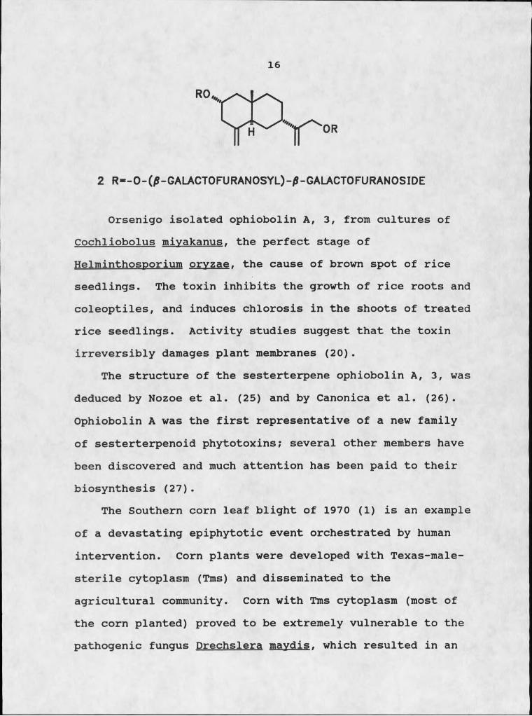
16
2 R*-0-(/?-GALACT0RJRAN0SYL)-/S-GALACT0FURAN0SIDE
Orsenigo isolated ophiobolin A, 3, from cultures of Cochliobolus mivakanus. the perfect stage of Helminthosoorium orvzae. the cause of brown spot of rice seedlings. The toxin inhibits the growth of rice roots and coleoptiles, and induces chlorosis in the shoots of treated rice seedlings. Activity studies suggest that the toxin irreversibly damages plant membranes (20) .
The structure of the sesterterpene ophiobolin A, 3, was deduced by Nozoe et al. (25) and by Canonica et al. (26). Ophiobolin A was the first representative of a new family of sesterterpenoid phytotoxins; several other members have been discovered and much attention has been paid to their biosynthesis (27).
The Southern corn leaf blight of 1970 (I) is an example of a devastating epiphytotic event orchestrated by human intervention. Corn plants were developed with Texas-male- sterile cytoplasm (Tms) and disseminated to the agricultural community. Corn with Tms cytoplasm (most of the corn planted) proved to be extremely vulnerable to the pathogenic fungus Drechslera mavdis. which resulted in an
17
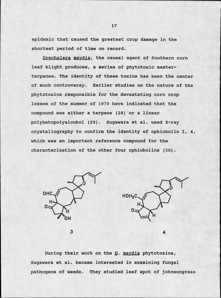
epidemic that caused the greatest crop damage in the shortest period of time on record.
Drechslera mavdis. the causal agent of Southern corn leaf blight produces, a series of phytotoxic sester- terpenes. The identity of these toxins has been the center of much controversy. Earlier studies on the nature of the phytotoxins responsible for the devastating corn crop losses of the summer of 1970 have indicated that the compound was either a terpene (28) or a linear polyketopolyalcohol (29). Sugawara et al. used X-ray crystallography to confirm the identity of ophiobolin I, 4, which was an important reference compound for the characterization of the other four ophiobolins (30).
During their work on the f). mavdis phytotoxins, Sugawara et al. became interested in examining fungal pathogens of weeds. They studied leaf spot of johnsongrass

18
fSQgqhum halapense L.), a serious weed in tropical and subtropical areas of the world. The disease is caused by D. sorqhicola whose perfect stage was found to be genetically compatible with the perfect stage of fi. mavdis (R.R. Nelson, personal communication to Sugawara et a!.). They examined the phytotoxins of both fungi in their activity towards corn bearing Tms cytoplasm. Both of these fungi induced disease symptoms on corn, and both produced ophiobolins, although the concentrations of the various members of this class of compounds differed. This study emphasizes the importance of a thorough investigation of phytotoxins produced by weeds, because the toxic secondary metabolites of weed pathogens may be injurious to crop plants as well (30).
Cochliobolus victbriae Nelson, the perfect stage of Helminthosnorium victgriae, was recognized as the causal agent of victoria blight of oats over 40 years (31). This disease was actually the unfortunate result of an attempt to render oat plants resistant to crown rust, Puccinia coronata. It was determined that susceptibility to crown rust was a function of a single gene locus; commercial varieties of oats were therefore introduced carrying the Pc gene for resistance to this disease. Unfortunately, it was discovered that crown rust resistance and victoria oat blight susceptibility were either closely linked or controlled by the same genetic locus. Subsequent studies
19
have shown that sensitivity to H. victoriae is controlled by a single dominant gene with the homozygous dominant genotype conferring sensitivity and the homozygous recessive genotype conferring insensitivity (32).
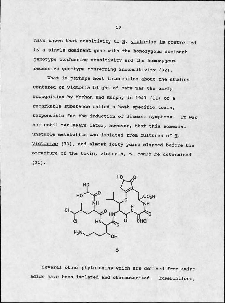
What is perhaps most interesting about the studies centered on victoria blight of oats was the early recognition by Meehan and Murphy in 1947 (ll) of a remarkable substance called a host specific toxin, responsible for the induction of disease symptoms. It was not until ten years later, however, that this somewhat unstable metabolite was isolated from cultures of H. victoriae (33), and almost forty years elapsed before the structure of the toxin, victorin, 5, could be determined (31) .
CO2H
5
Several other phytotoxins which are derived from amino acids have been isolated and characterized. Exserohilone,

20
6, was isolated from Exserohilum holmii. a pathogen of the weed Dactvloctenium aecrvotium (crowfoot grass), a serious grassy weed in all major tropical and subtropical parts of the world (34) . The toxin is a diketopiperazine with nonspecific activity towards crowfoot grass and several other plants against which it was tested. Other amino acid-derived phytotoxins include lycomarasmine, a nonspecific wilt toxin produced by the wilt fungus Fusarium oxvsporum (20), and the related compounds aspergillo- marasmine A, 7, and B, 8 (20).
OH
HO
6
7 8
21
Selection of a Suitable Target Plant
The choice of a target plant is an important one.The discovery of a pathogen that can produce compounds deleterious to lettuce might be an interesting academic exercise, but would have limited importance to the plant pathology community. Of course, pathogens that elaborate toxins to crop plants have an important place in the screening of disease-resistant cultivars for dissemination to the agricultural community. The discovery of a weed pathogen, however, has more immediate significance as a potential biocontrol tool. And the more economically important the weed, the greater the significance of a biocontrol agent.
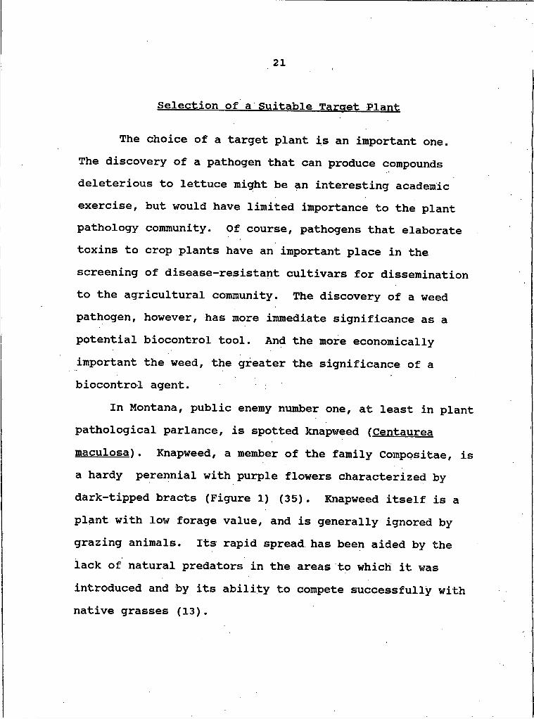
In Montana, public enemy number one, at least in plant pathological parlance, is spotted knapweed fCentaurea maculosa). Knapweed, a member of the family Compositae, is a hardy perennial with purple flowers characterized by dark-tipped bracts (Figure I) (35). Knapweed itself is a plant with low forage value, and is generally ignored by grazing animals. Its rapid spread has been aided by the lack of natural predators in the areas to which it was introduced and by its ability to compete successfully with native grasses (13).

22
Figure I. Spotted knapweed: a) habit; b) enlarged leaf; c) flower head; d) disk flower; e) achenes.
Knapweed plants actually produce their early season rosettes in the fall rather than the spring, unlike most plants. This hardy rosette overwinters and revives rapidly with the first spring thaw, a distinct advantage in the competition for space, moisture, and soil nutrients. The production of certain allelochemicals, in particular the sesquiterpene lactone cnicin, 9, has been attributed to various knapweed species (13). The weed is tolerant of

23
wide fluctuations in temperature and total precipitation, and competes particularly well in dry areas.
Knapweed reproduces in typical composite fashion by seed dispersal in late summer and early fall. A single plant can produce 900-1800 seeds per season (36).Mechanical agents such as the wind can disseminate the seeds over fairly large distances (10-20 m ) . Animals (including humans) may serve as unwitting carriers of the seeds into previously uninfected areas. In a study on diffuse knapweed (Centaurea diffusa), a close relative of spotted knapweed, the front of distribution in a particular area advanced 120 m over a three year period (13). The low forage value of this plant, its extreme hardiness, and its ability to replace economically important plants make spotted knapweed an ideal target for attempted biocontrol.
Spotted knapweed poses a significant threat as a weed species, as a result of its extensive incursion into rangelands of northwestern United States and southwestern Canada. It ranks as the number one weed problem on
0
OH
9
24
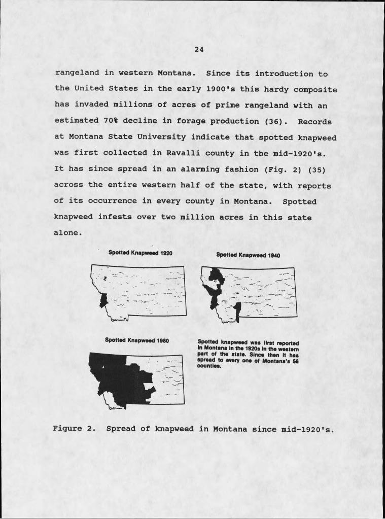
rangeland in western Montana. Since its introduction to the United States in the early 1900's this hardy composite has invaded millions of acres of prime rangeland with an estimated 70% decline in forage production (36). Records at Montana State University indicate that spotted knapweed was first collected in Ravalli county in the mid-1920's.It has since spread in an alarming fashion (Fig. 2) (35) across the entire western half of the state, with reports of its occurrence in every county in Montana. Spotted knapweed infests over two million acres in this state alone.
Spotted Knapweed 1920 Spotted Knapweed 1940
Spotted Knapweed 1980 SpoHed knapweed was first reported In Montana In the 1920s In the western pert of the state. Since then It has spread to every one of Montana’s 56 counties.
Figure 2. Spread of knapweed in Montana since mid-1920's.
25
Discovery of a Pathogen
Locating spotted knapweed plants posed no difficulties as the range of this regionally ubiquitous plant includes most of southwestern Montana. Locating a diseased knapweed plant was considerably more challenging. Knapweed is a hardy plant, not particularly susceptible to blights or pathogen induced diseases. The discovery of an obviously diseased plant required several weeks of exploring various regions in southwestern Montana.
A two month search for an appropriately infected plant culminated in the discovery of black leaf blighted Centaurea maculosa in Silver Bow County, Montana, in July, 1984. A seriously compromised individual was found on the northern slope of Big Butte, a five hundred foot cinder cone in Butte, Montana. Approximately thirty percent of the aerial leaves and ninety percent of the flowers were covered either by black fungal growth or by dark brown, weeping lesions. The plant was carefully collected, placed in a sterile plastic bag, and returned to the laboratory for study.
After returning to the laboratory, we endeavored to isolate the disease causal agent in pure culture. Diseased pieces of stems, leaves, and flowers were excised from the plant using sterile technique and placed on DIFCO bacto- agar plates. No additional nutrients were added to the
26
plates. It was hoped that by limiting the available nutrients to the remnants of the plant itself and bacto- agar we could select for microorganisms capable of utilizing knapweed for food.
Several fungi grew on the plates and great care was taken to isolate the individual organisms as pure cultures. Single spore transfers were used whenever possible to facilitate this process. Once the fungi were established as demonstrably pure cultures, we attempted to determine which, if any, of these isolates were responsible for the induction of black-leaf blight disease of knapweed.
Following the mandate of Koch's Postulates (12), the nature of the pathogen was determined by challenging healthy knapweed plants with pure isolates of the fungi obtained from the lesions. Healthy knapweed plants were nicked with a sterile razor blade on their stems and leaves. Each plant was nicked in ten locations, with no two nicks on the same branch. A piece of mycelia impregnated agar "27mm3 was applied to eight of the nicked sites on each plant. One plant was not infected with any of the fungi. The plants were allowed to grow undisturbed after they had been inoculated with the fungi, with daily checks to determine if the fungi were actually growing. After five days it was apparent that the fungi were not impacting the plants in any significant fashion. Fungi require fairly humid conditions (90%-95%) for growth and
27
sporulation (16), and the laboratory did not provide adequate moisture for the fungi to develop. The inoculation procedure was repeated with new plants which were encased in sterile, clear plastic bags to facilitate the need for increased humidity. Only one of the fungal isolates induced the formation of weeping necrotic lesions within a five day period. The pathogen was reisolated from the diseased plants and was morphologically identical to the applied fungus. The inoculation procedure was repeated with five healthy plants and in each case the fungus induced the formation of necrotic lesions at the site of inoculation. The organism was identified as Alternaria alternata Lam. by the CBS (Mycological Laboratory), Baarn, Netherlands.
Alternaria sp. are members of the deuteromycetes or imperfect fungi: no sexual stage has yet been isolated for this genus. The chronic asexual condition results in the production of large numbers of spores, the usual infective agents in most fungal plant diseases. Asexual reproduction and spore formation are usually highly repetitive processes, and are instrumental in the development of plant disease epidemics (37).
Alternaria sp. have been isolated as causative organisms in a variety of plant diseases and have been shown to produce a wide variety of phytotoxic compounds.The fungus is capable of elaborating toxins with broad-
28
spectrum activity and toxins with extremely limited host- range activity. Some of the toxins have only been isolated from single species while others appear to be quite widespread throughout the genus.
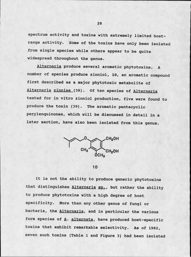
Alternarie produce several aromatic phytotoxins. A number of species produce zinniol, 10, an aromatic compound first described as a major phytotoxic metabolite of Alternaria zinniae (38). Of ten species of Alternaria tested for in vitro zinniol production, five were found to produce the toxin (39). The aromatic pentacyclic perylenquinones, which will be discussed in detail in a later section, have also been isolated from this genus.
It is not the ability to produce generic phytotoxins that distinguishes Alternaria so.. but rather the ability to produce phytotoxins with a high degree of host specificity. More than any other genus of fungi or bacteria, the Alternaria. and in particular the various form species of &. alternata, have produced host-specific toxins that exhibit remarkable selectivity. As of 1982, seven such toxins (Table I and Figure 3) had been isolated

29
and of these, six have been fully characterized (5). What is remarkable about the host-specific toxins isolated to date is that only one (or a few) cultivars of a given host species are susceptible to either the live pathogen or its toxins; all other cultivars are resistant. 1
Results
The various form species of A. alternate are credited with the production of a variety of host specific toxins on crop plants. We hoped that this propensity for phytotoxin production by A. alternate could be exploited against spotted knapweed.
In the course of this study we isolated a series of compounds that expressed varying degrees of phytotoxicity to knapweed. The phytotoxins represent three distinct classes of organic compounds; diketopiperazines, perylenequinpnes, and tetramic acids. Following the isolation and .elucidation of the various compounds a detailed bioactivity profile of each class was developedwith respect to the degree of phytotoxicity and the degree
;
of host selectivity towards knapweed.
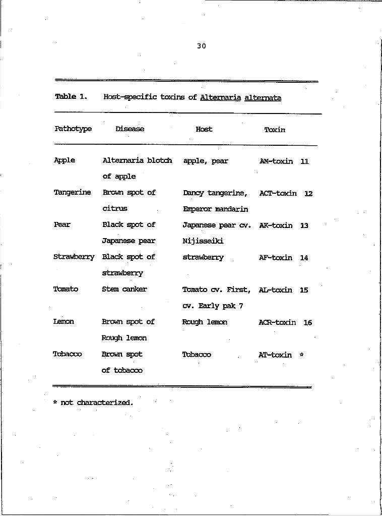
30
Table I. Host-specific toxins of Altemaria altemata
Pathotype n-iswas#* Host Toxin
Apple Alterraria blotch apple, pear AM-toxin 11of apple
Tangerine Brown spot of Dancy tangerine. ACT-toxin 12citrus Enperor mandarin
Pear Black spot of Japanese pear cv. AK-toxin 13Japanese pear Nijisseiki
Stravfcerry Black spot of strawberry AF-toxin 14strawberry
Tcanato Stem canker Tomato cv. First, Air-toxin 15cv. Early pak 7
Lemon Brown spot of Rough lemon ACR-toxin 16Rough lemon
Tcbacco Brown spot Tobacco AT-toxin Vt
of tobacco
* not characterized.
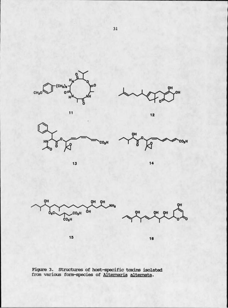
31
15 16
Figure 3. Structures of host-specific toxins isolated frcm various form-species of Altemaria altemata.
32
Isolation of DiketopjperazinesA series of diketopiperazines of varying toxicity to
knapweed was consistently isolated from 24 day cultures of A*— alternate. The EtOAc soluble extract of the culture filtrate was chromatographed on Bio-Beads S-X4 (hexane- CH2cl2"EtOAc/ 4:3si). This chromatography yielded twelve fractions; only fractions 4, 5, and 6 induced necrotic lesions on knapweed tissues. Size exclusion chromatography of fraction 5 on Sephadex LH-20 (CH2Cl2-MeOtBu-iPrOH,1:1:1) yielded maculosln, 17. Similar treatment of fraction 6 gave a mixture that was readily resolved by centrifugal counter-current chromatography (CHCl3-MeOH-H20, 25:34:20, lower phase mobile) into two compounds, 18 and 19. Fraction 4 yielded diketopiperazine 20 upon further size exclusion chromatography on Bio-Beads S-X8 (CH2Cl2- cyclohexane, 3:2).
The CH2Cl2 soluble extract of the culture filtrate was applied to a Sephadex LH-20 column (CH2Cl2-MeOH, 1:1). The fourth eluant yielded 21, 22, and 23, using centrifugal countercurrent chromatography as described above. Table 2 outlines the separation techniques used to isolate compounds 17-23, their percent yield relative to the total organic extract, and the molecular weight of each compound.
I
Ttible 2. Diketcpiperazines isolated, fraction of origin, separation technique, % yield, and molecular weight.
INITIAL CHRaMftTOGORftHfif Rt MDIECULftRFRACTION TECHNIQUE COMPOUND (hr) %YIEtDa WEIQfT
EtQftc ExtractE IH-20b 17 2.10 0.74% 260F U M O b
COCc 18 0.38 0.39% 244
19 0.45 0.44% 244
D S-XSd 20 1.33 0.23% 224
CHnCli ExtractJ CCCc 21 0 .70 0.10% 196
I cod5 22 0.63 0.23% 210
H cod5 23 0 .56 0.08% 168
a %Yield = weight of ccarpound/total organic wei^it x 100 ^ The solvent system used was CH2Cl2-^butylntethyl ether- isopropanol, 1/1/1.c The solvent system used was CH2Cl2-Cyclohexane, 3/2. d The solvent system used CH2Cl2-MeOH-H2O, 25/34/20.
34
Structure Elucidation of DiketonineraaineaThe first compound to be characterized was the most
active of the compounds isolated. The molecular formula c 14h 16n 2°3' which required eight sites of unsaturation, was assigned to compound 17 by high-resolution mass spectrometry. Phenolic and amide functional groups were indicated by infrared absorptions at 3590, 3380, and 1670 cm"1 respectively (42). These assignments were supported by a detailed analysis of 1H NMR and mass spectrometry.
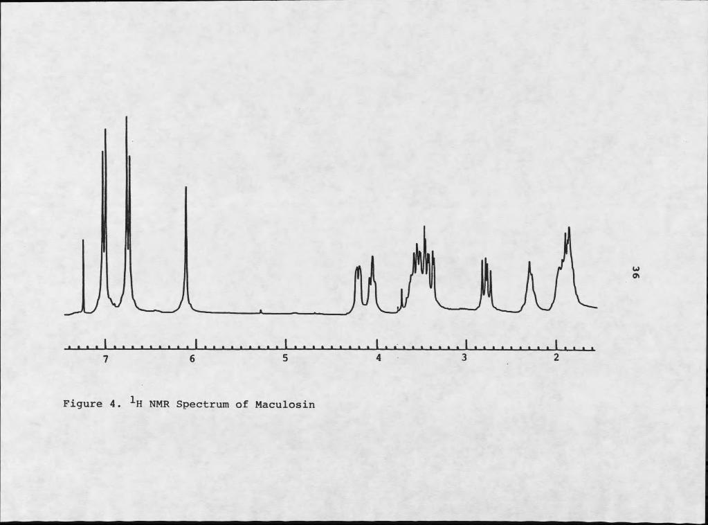
1H NMR analysis (Figure 4) began with examination of the aromatic region of the spectrum. The aromatic doublets at 66.76 and 67.03 were mutually coupled by 8.2 Hz, indicative of a para disubstituted benzene ring (41). The 0.51 ppm upfield shift of the doublet at 66.76, relative to classic benzene absorption of 67.27, is typical of aromatic protons adjacent to a hydroxyl group. The 0.24 ppm upfield shift of the companion doublet is typical of the ortho effect of an adjacent alkyl substituent. With the supporting phenolic infrared absorption at 3590 cm"1, the data presented a strong case for the presence of a phenolic moiety.
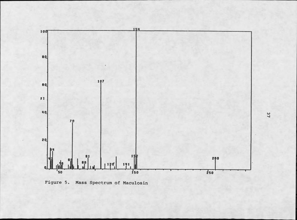
Mass spectral analysis revealed two major fragments resulting from electron impact: a m/z 107 fragment and a m/z 154 fragment (Figure 5). The m/z 107 fragment is typical of a hydroxytropyIium ion, C7H7O+. The m/z 154

35
fragment must be C^HgNgQg, accommodating the four remaining sites of unsaturation. This second fragment must be responsible for the amide absorptions at 3380 and 1670 cm-1 in the infrared spectrum.
The coexistence of a hydroxy benzylic moiety and amide functionality together in the same molecule suggested the presence of a tyrosyl derivative. The benzylic methylene should be coupled to a methine with an approximate absorbance of 63.8 (41). Extensive decoupling experiments indicated that the methine at 64.20 was coupled to two mutually coupled doublets of doublets at 63.44 and 62.76. These two multiplets displayed typical geminal coupling (15 Hz), indicative of methylene protons, with reasonable benzylic methylene proton chemical shifts. The disparity of their chemical shifts indicated proximity to a chiral center (42).
The region between 63.00 and 64.50 of the 1H NMR proved to be quite diagnostic in identifying this phytotoxin. It also established an excellent means of identifying other members of the same class of compounds. The chemical shifts of the two methines at 64.20 and 64.07 were amenable to adjacent oxygen functionalities, although somewhat downfield of protons in typical ether systems (63.6) (43).
V JL
I7
y
Figure 4. IH NMR Spectrum of Maculosin
100 151
Bd
Bd
Xl
Wd70
Figure 5. Mass Spectrum of Maculosin
w
280
250
38
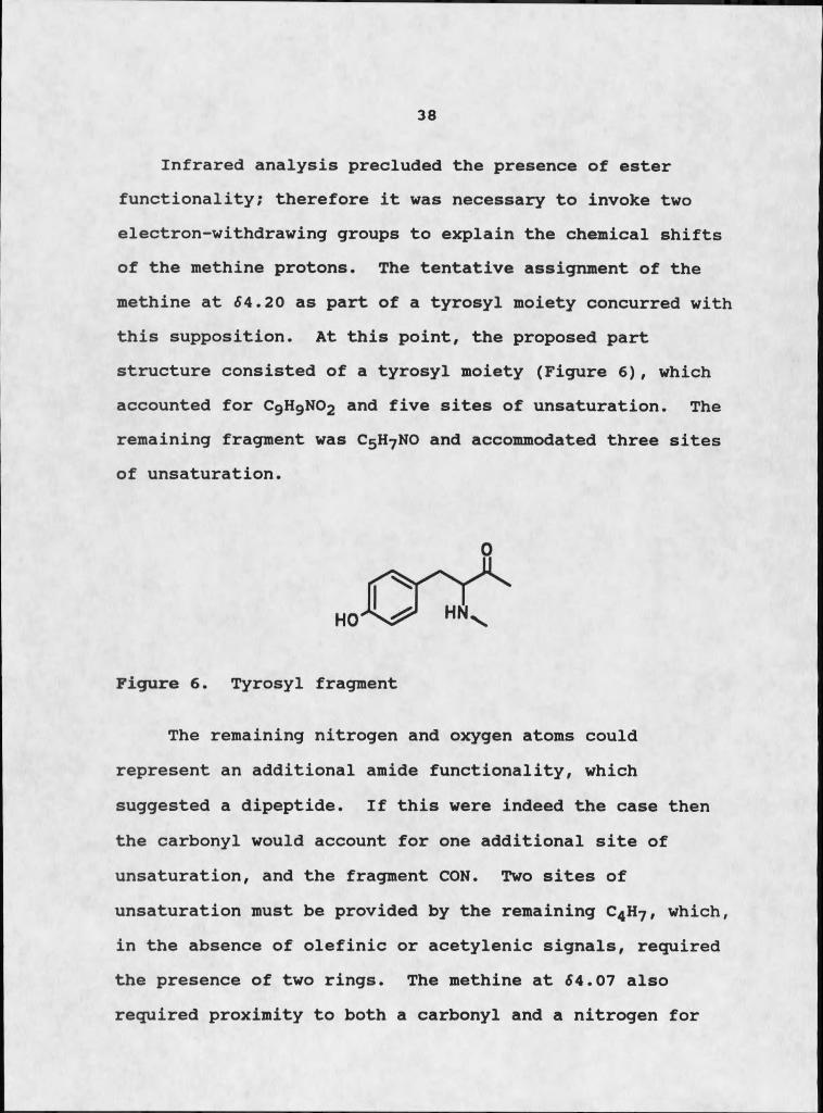
Infrared analysis precluded the presence of ester functionality; therefore it was necessary to invoke two electron-withdrawing groups to explain the chemical shifts of the methine protons. The tentative assignment of the methine at 54.20 as part of a tyrosyl moiety concurred with this supposition. At this point, the proposed part structure consisted of a tyrosyl moiety (Figure 6), which accounted for C9H9NO2 and five sites of unsaturation. The remaining fragment was C5H7NO and accommodated three sites of unsaturation.
Figure 6. Tyrosyl fragment
The remaining nitrogen and oxygen atoms could represent an additional amide functionality, which suggested a dipeptide. If this were indeed the case then the carbonyl would account for one additional site of unsaturation, and the fragment CON. Two sites of unsaturation must be provided by the remaining C4H7 , which, in the absence of olefinic or acetylenic signals, required the presence of two rings. The methine at 54.07 also required proximity to both a carbonyl and a nitrogen for
39
its chemical shift, supporting the presence of an additional amide moiety. Adjacent to the only remaining nitrogen, the methine was also coupled to the multiplets at 52.31 (IH, 7.1 Hz) and 51.93 (3H, 1.0Hz). The higher field multiplet was also coupled to the two proton multiplet at 53.57, a typical absorption for a ring methylene adjacent to a nitrogen. This created a five-membered ring, suggestive of proline. The need for an additional ring and the presence of two amides suggested a diketopiperazine skeleton, which completed the preliminary structural characterization. Compound 17 was designated maculosin in recognition of its phytotoxicity towards Centaurea maculosa.
The structure assignment did not address stereochemical determination of the two chiral centers. Although relative stereochemistry can be determined in a variety of ways, synthesis of the compound following a protocol that proceeds without racemization provides the absolute assignment. It also provides more material, which was an
40
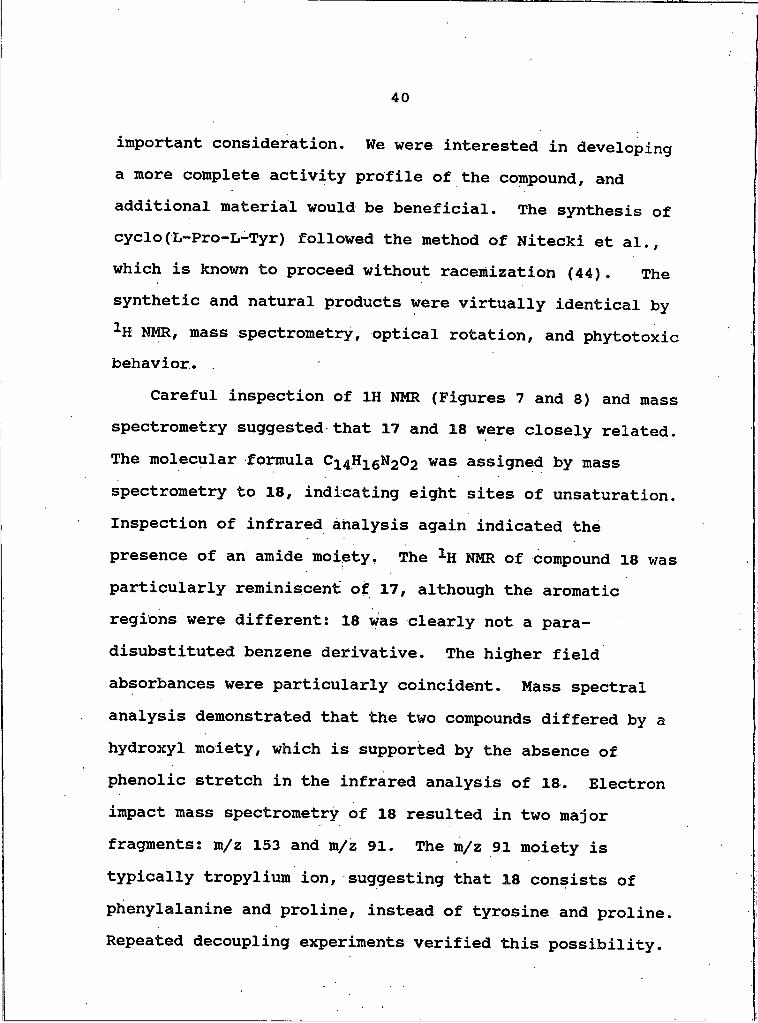
important consideration. We were interested in developing a more complete activity profile of the compound, and additional material would be beneficial. The synthesis of cyclo(L~Pro~L-Tyr) followed the method of Nitecki et al., which is known to proceed without racemization (44). The synthetic and natural products were virtually identical by 1H NHR, mass spectrometry, optical rotation, and phytotoxic behavior.
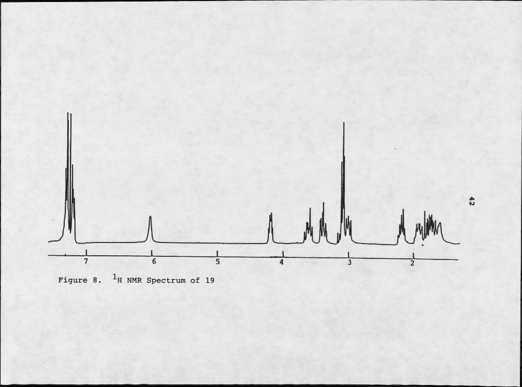
Careful inspection of IH NMR (Figures 7 and 8) and mass spectrometry suggested that 17 and 18 were closely related. The molecular formula C^^HigNgOg was assigned by mass spectrometry to 18, indicating eight sites of unsaturation. Inspection of infrared analysis again indicated the presence of an amide moiety. The 1H NMR of compound 18 was particularly reminiscent of 17, although the aromatic regions were different: 18 was clearly not a para- disubstituted benzene derivative. The higher field absorbances were particularly coincident. Mass spectral analysis demonstrated that the two compounds differed by a hydroxyl moiety, which is supported by the absence of phenolic stretch in the infrared analysis of 18. Electron impact mass spectrometry of 18 resulted in two major fragments: m/z 153 and m/z 91. The m/z 91 moiety is typically tropylium ion, suggesting that 18 consists of phenylalanine and proline, instead of tyrosine and proline. Repeated decoupling experiments verified this possibility.

7 6 5 4 3 2 1
Figure 7. NMR Spectrum of 18
H NMR Spectrum of 19
43
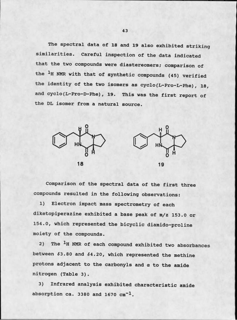
The spectral data of 18 and 19 also exhibited striking similarities. Careful inspection of the data indicated that the two compounds were diastereomers; comparison of the 1H NMR with that of synthetic compounds (45) verified the identity of the two isomers as cyclo(L-Pro-L-Phe), 18, and cyclo(L-Pro-D-Phe), 19. This was the first report of the DL isomer from a natural source.
Comparison of the spectral data of the first three compounds resulted in the following observations:
1) Electron impact mass spectrometry of each diketopiperazine exhibited a base peak of m/z 153.0 or 154.0, which represented the bicyclic diamido-proline moiety of the compounds.
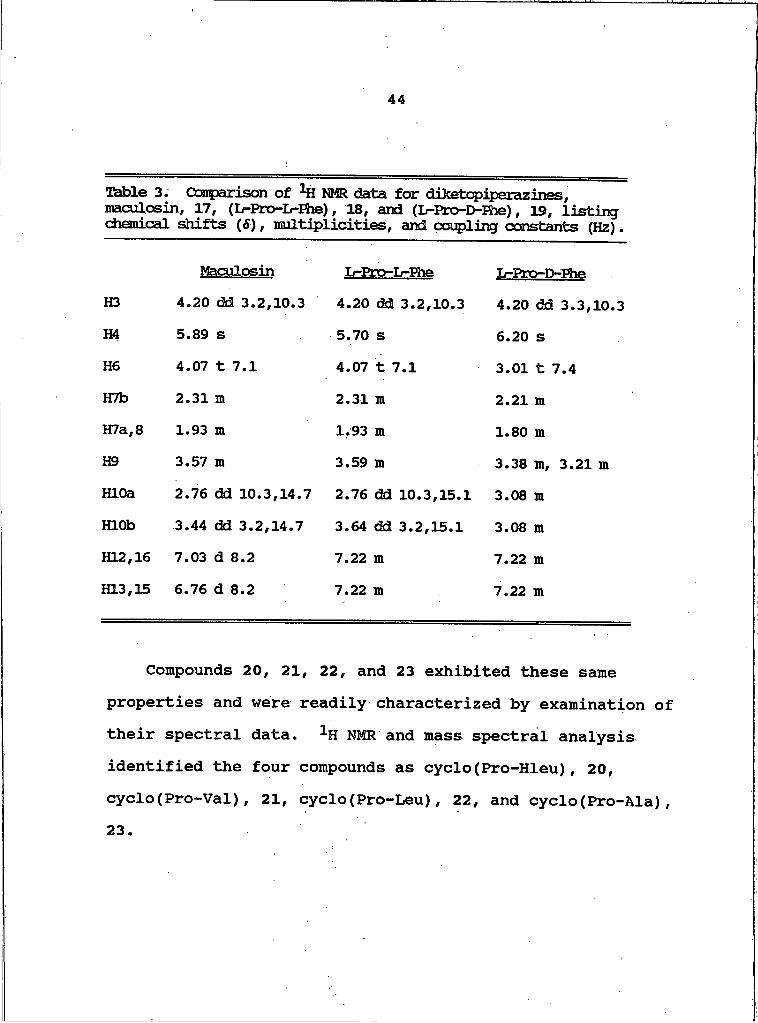
2) The 1H NMR of each compound exhibited two absorbances between <53.80 and <54.20, which represented the methine protons adjacent to the carbonyls and a to the amide nitrogen (Table 3).
3) Infrared analysis exhibited characteristic amide absorption ca. 3380 and 1670 cm”1.
44
Table 3. Ooarparison of NMR data for diketopiperazines, maculosin, 17, (L-Pro-Ir-Ehe), 18, and (L-Pro-D-Fhe), 19, listing chemical shifts (S), multiplicities, and coupling constants (Hz).
Maculosin L-ETo-L-Ehe L-Pro-D-FheH3 4.20 dd 3.2,10.3 4.20 dd 3.2,10.3 4.20 dd 3.3,10.3H4 5.89 s 5.70 s 6.20 sH6 4.07 t 7.1 4.07 t 7.1 3.01 t 7.4HTb 2.31 m 2.31 m 2.21 mH7a,8 1.93 m 1.93 m 1.80 mH9 3.57 m 3.59 Ht 3.38 m, 3.21 mHLOa 2.76 dd 10.3,14.7 2.76 dd 10.3,15.1 3.08 mHLOb 3.44 dd 3.2,14.7 3.64 dd 3.2,15.1 3.08 mKL2,16 7.03 d 8.2 7.22 m 7.22 m1313,15 6.76 d 8.2 7.22 m 7.22 m
Compounds 20, 21, 22, and 23 exhibited these same properties and were readily characterized by examination of their spectral data. 1H NMR and mass spectral analysis identified the four compounds as cyclo(Pro-Hleu), 20, cyclo(Pro-Val), 21, cyclo(Pro-Leu), 22, and cyclo(Pro-Ala),23
45
Isolation of Pervlenecminones 24 - 27The CH2Cl2 soluble extract exhibited moderate
phytotoxicity towards knapweed. Size exclusion chromatography on Sephadex LH-20 with CH2Cl2-MeOH, (1:1) yielded four fractions. Fraction 3, which induced necrotic lesions on knapweed leaves but not on johnsongrass at 100 ppm, was permeated through Sephadex LH-20 column using MeOH-CH3CN, (1:1). The second fraction of this chromatography exhibited phytotoxic behavior but, although an obvious mixture of aromatic metabolites by 1H NMR, resisted attempts at further separation by size exclusion chromatography. Separation of these closely related compounds proved challenging, but centrifugal
46
countercurrent chromatography provided baseline resolution of 24 - 27 using a solvent system of CHCl3-MeOH-H2O (25:34:20, lower phase mobile).
We were able to afford baseline resolution of several complex mixtures with great facility through the use of the CCC. Two such mixtures, one containing the diastereomers cyclo(L-Pro-L-Phe) and cyclo(L-Pro-D-Phe) and the other a mixture of structurally similar perylenequinones, were each resolved in less than one hour from time of injection (Figure 9). Separation in the CCC is primarily a partition phenomenon and conformational analysis of these compounds suggested a mechanism responsible for their resolution.
Figure 9. a) CCC trace of separation of diastereomers of cyclo(Pro-Phe); b) CCC trace of separation of perylenequinones.
Diketopiperazines usually assume one of three rotamers: folded, extended towards nitrogen, or extended towards
47
oxygen. Comparisons of the diastereomers of cyclo(Pro-Phe) by lanthanide-assisted 13C NMR and 1H NMR, and circular dichroism indicated that they assume different, often solvent related conformations (45, 46). The preferred rotamer for cyclo(L-Pro-L-Phe) in chloroform is extended towards nitrogen, which may be stabilized by induced polarization of the electronegative aromatic ring by the proximity of the electropositive amide hydrogen. Cyclo(L- Pro-D-Phe) assumes a folded conformation in a variety of solvents, including chloroform, consistent with observations derived from calculations of the energy differences of various rotamers (Figure 10) (45, 46).
Extended Folded
Figure 10. Conformational rotamers of cyclo(Pro-Phe).
These conformational differences are also responsible for the disparity in 1H NMR that was observed between the two diastereomers of cyclo(Pro-Phe). Under identical conditions, the amide proton of the L-proline moiety of
48
cyclo(L-Pro-D-Phe) is shifted approximately I ppm upfield of the amide proton of the L-proline moiety of its diastereomer. The folded conformation preferred by the LD isomer situates the amide proton in the shielding zone of the aromatic phenylalanine ring, creating this upfield shift.
Facile separation of the four perylenequinones by CCC was based on subtle differences in partition coefficients of the compounds. These differences were the result of enhanced polarity due to variation in the number of polar substituents or in sites of unsaturation.
Structure Elucidation of PervlenecminonesSeveral apparently related aromatic compounds were
isolated from the methylene chloride soluble extract. Compound 24 had a molecular formula of C2QHigO6 , requiring thirteen sites of unsaturation, based on high-resolution mass spectrometry. A strong infrared absorption near 1645 cm”1 indicated several conjugated, carbonyl systems: extended quinone, hydrogen bonded O-CO-C6H4-OH, or chelated, enolic, /3-keto esters (40). A phenolic absorption at 3580 cm”1 supported the second possibility, but did not preclude the coexistence of the other functionalities. The 1H NMR contained four doublets typical of para-disubstituted benzene; the size of the molecule and the large degree of unsaturation suggested a
49
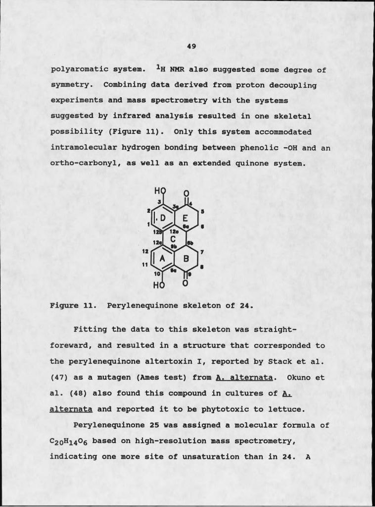
polyaromatic system. 1H NMR also suggested some degree of symmetry. Combining data derived from proton decoupling experiments and mass spectrometry with the systems suggested by infrared analysis resulted in one skeletal possibility (Figure 11). Only this system accommodated intramolecular hydrogen bonding between phenolic -OH and an ortho-carbonyl, as well as an extended quinone system.
Figure 11. Peryleneguinone skeleton of 24.
Fitting the data to this skeleton was straight- foreward, and resulted in a structure that corresponded to the perylenequinone altertoxin I, reported by Stack et al. (47) as a mutagen (Ames test) from A. alternata. Okuno et al. (48) also found this compound in cultures of A. alternata and reported it to be phytotoxic to lettuce.
Perylenequinone 25 was assigned a molecular formula of c 20h 14°6 based on high-resolution mass spectrometry, indicating one more site of unsaturation than in 24. A

50
comparison of the 1H NMR spectrum with that of 24 suggested that the two compounds differed by the presence of a double bond in the E ring. Careful examination of the spectral data showed that 25 was identical to alteichin, reported by Robeson et al. as a phytotoxin towards water hyacinth produced by Alternaria eichorniae (49). Okuno et al. also found this compound in an earlier study of A. alternata (48) .
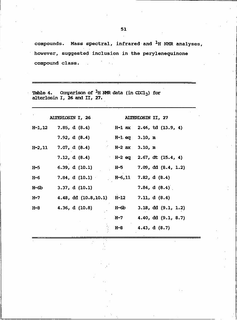
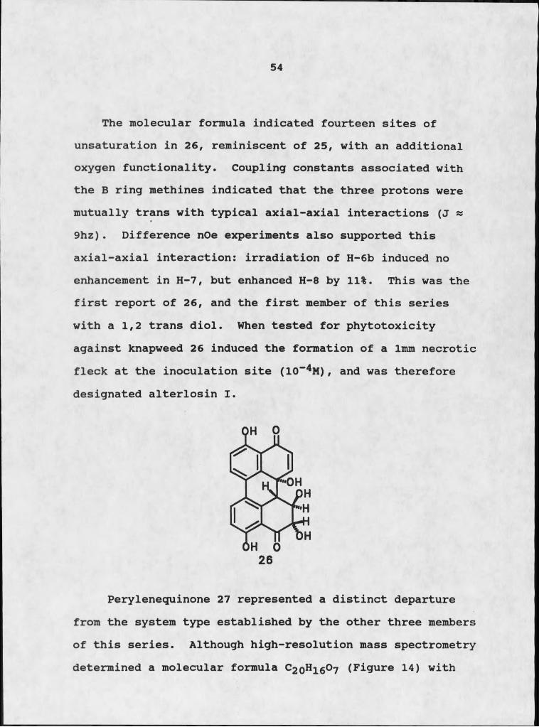
Perylenequinone 26, had a molecular weight of m/z 366.0752 which required a molecular formula of C2Qh H 0? (Figure 12). Inspection of 1H NMR data (Table 4 and Figure 13) indicated a double bond in the E ring, similar to 25, but with an unusual 1,2 diol system in the B ring.Detailed comparisons of the data collected for this compound, as well as 27, with data available for known perylenequinones suggested that they were both novel
51
compounds. Mass spectral, infrared and 1H NMR analyses, however, suggested inclusion in the perylenequinone compound class.
Table 4. Ompariscn of 1H NMR data (in CDClg) for alterlosin I, 26 and II, 27.
MJIERIOSIN I, 26 MJEERLiDSIN II, 27H-1,12 7.85, d (8.4) H-I ax 2.46, td (13.9, 4)
7.92, d (8.4) H-I eq 3.10, mH-2,11 7.07, d (8.4) H-2 ax 3.10, m
7.12, d (8.4) H-2 eq 2.67, dt (15.4, 4)H-5 6.39, d (10.1) H-5 7.09, dd (8.4, 1.2)H-6 7.84, d (10.1) H-6,11 7.82, d (8.4)H-6b 3.37, d (10.1) 7.84, d (8.4)H-7 4.48, dd (10.8,10.1) H-12 7.11, d (8.4)H-8 4.36, d (10.8) H-6b 3.18, dd (9.1, 1,2)
H-7 4.40, dd (9.1, 8.7)H-8 4.43, d (8.7)
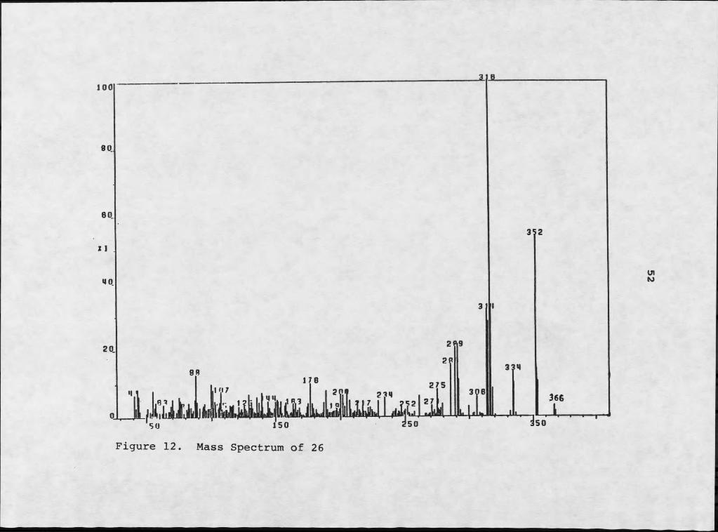
Figure 12. Mass Spectrum of 26

UlW
7 6 5Figure 13. NMR Spectrum of 26
4 3 2 I
54
The molecular formula indicated fourteen sites of unsaturation in 26, reminiscent of 25, with an additional oxygen functionality. Coupling constants associated with the B ring methines indicated that the three protons were mutually trans with typical axial-axial interactions (j « 9hz). Difference nOe experiments also supported this axial-axial interaction: irradiation of H-6b induced no enhancement in H-7, but enhanced H-8 by 11%. This was the first report of 26, and the first member of this series with a 1,2 trans diol. When tested for phytotoxicity against knapweed 26 induced the formation of a Imm necrotic fleck at the inoculation site (IO-4M ) , and was therefore designated alterlosin I .
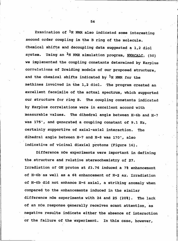
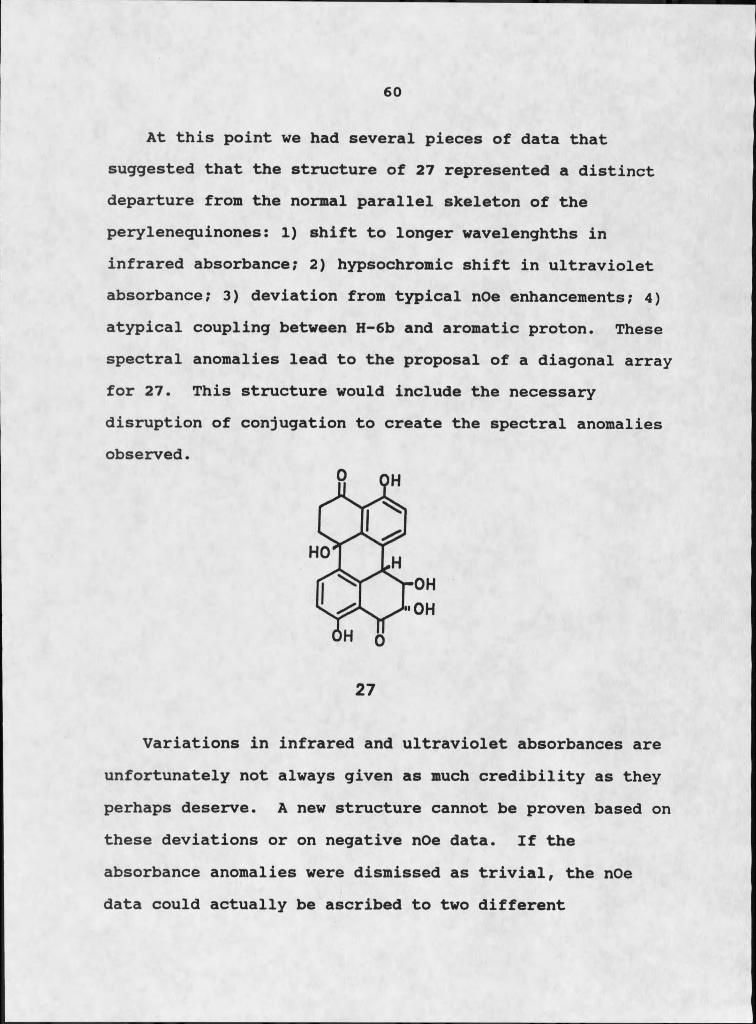
Perylenequinone 27 represented a distinct departure from the system type established by the other three members of this series. Although high-resolution mass spectrometry determined a molecular formula C2O11Ie0? (Figure 14) with

55
fragmentation patterns typical of perylemequinones, spectral anomalies indicated that the structure was clearly not the dihydro-analog of 26. Infrared analysis (liquid film) showed the carbonyl absorption of this molecule to be at a somewhat lower energy than was found in the rest of the series (1630 cm-1 compared to 1645 cm"1), which is more typical of an isolated, hydrogen-bonded Ortho-OH-C6H4-CO than of an extended quinone system (40). Compounds 24-26 were run both in chloroform and neat, as liquid films, and no difference was observed in the carbonyl absorbance at 1645 cm"1 . This constancy obviated solvent effects as the cause of variation in infrared absorbance between the first three compounds and 27.
The ultraviolet absorption of 27 appeared at 348 nm, as opposed to the more typical 358-380 nm (47). This hypsochromic shift suggested that the usual extensive conjugation was disrupted. The deviations from typical values observed in both infrared and ultraviolet spectroscopies suggested that 27 represented a departure from the usual parallel array.
Examination of the 1H NMR (Table 4 and Figure 15) and of data from repeated decoupling experiments indicated that aromatic H-5 and H-6b were coupled to each other by I .0 h z . This deviated from the usual data which we observed, as perylenequinones 24-26 did not exhibit this type of coupling.
56
Examination of NMR also indicated some interesting second order coupling in the B ring of the molecule. Chemical shifts and decoupling data suggested a 1,2 diol system. Using an 1H NMR simulation program, NMRCALC. (50) we implemented the coupling constants determined by Karplus correlations of Dreiding models of our proposed structure, and the chemical shifts indicated by 1H NMR for the methines involved in the 1,2 diol. The program created an excellent facsimile of the actual spectrum, which supported our structure for ring B i The coupling constants indicated by Karplus correlations were in excellent accord with measurable values. The dihedral angle between H-6b and H-7 was 175°, and generated a coupling constant of 9.1 Hz, certainly supportive of axial-axial interaction. The dihedral angle between H-7 and H-8 was 170°, also indicative of vicinal diaxial protons (Figure 16).
Difference nOe experiments were important in defining the structure and relative stereochemistry of 27. Irradiation of OH proton at 53.76 induced a 7% enhancement of H-6b as well as a 6% enhancement of H-2 ax. Irradiation of H-6b did not enhance K-6 axial, a striking anomaly when compared to the enhancements induced in the similar difference nOe experiments with 24 and 25 (20%). The lack of an nOe response generally receives scant attention, as negative results indicate either the absence of interaction or the failure of the experiment. In this case, however,
Figure 14. Mass Spectrum of 27

J______________ I______________ I8 7 6
Figure 15. NMR Spectrum of 27
_14
I35

59
the lack of nOe enhancement between H-6 axial and H-6b suggested a deviation from the normal parallel skeleton, a possibility that required further examination and corroboration.
AB x
Figure 16. a) NMR simulation of ABX system using NMRCALC; b) Actual 1H NMR spectrum.
60
At this point we had several pieces of data that suggested that the structure of 27 represented a distinct departure from the normal parallel skeleton of the perylenequinones: I) shift to longer wavelenghths in infrared absorbance; 2) hypsochromic shift in ultraviolet absorbance; 3) deviation from typical nOe enhancements; 4) atypical coupling between H-6b and aromatic proton. These spectral anomalies lead to the proposal of a diagonal array for 27. This structure would include the necessary disruption of conjugation to create the spectral anomalies observed.
27
Variations in infrared and ultraviolet absorbances are unfortunately not always given as much credibility as they perhaps deserve. A new structure cannot be proven based on these deviations or on negative nOe data. If the absorbance anomalies were dismissed as trivial, the nOe data could actually be ascribed to two different

61
structures, the diagonal structure which we proposed, and a more typical parallel compound, 27b.
27b
Stack et al. (47) provided an excellent means of distinguishing between these two possibilities and unequivocally determining the true nature of the skeleton. They isolated a mutagen designated altertoxin III, 28, which exhibited the same type of spectral anomalies which we have described. Altertoxin III possesses a diagonal structure, similar to that proposed for 27.
28

62
Stack et al. performed a simple experiment to determine the structure of 28 (47). They investigated changes in the ultraviolet absorption induced by reduction with NaBH^. Altertoxin I, 24, gave a product with ^ max at 299 nm (24,000) and 214 nm (31,000), while 28 gave a product with ^max at 287 nm (5,200) and 234 nm (14,000), typical of
phenoxide absorbance (51). In our hands, compound 24 gave a reduction product with ^ max at 297 nm ( 19,200), similar to that observed by Stack et al. Reduction of 27, however, gave a product with X max at 268 nm (7850), which is the ultraviolet absorption of phenol, rather than phenoxide.We then treated phenol with NaBH^ and saw no change in the X max at 270 nm. Only addition of NaOH produced the phenoxide absorbances observed by Stack et al. This experiment demonstrated that 27 lacked the extensive conjugation of the biphenyl system, and verified the diagonal array which we had proposed.
Other investigators have isolated perylenequinones, both parallel and diagonal, from fungal cultures. Arnone et al. isolated four compounds, designated stemphyltoxins 1-4, 29-32, with typical parallel skeletons, and stemphyperylenol, 33, which possesses the diagonal array, from Stemohvlium botryosum (52). They reported infrared and ultraviolet absorption deviations similar to those we observed in their diagonal molecule. They reported, however, that all five of their compounds possessed the H-5
63
coupling to H-6b which we observed solely in 27. They attributed this coupling to normal para-benzylic interactions. In our study, only 27 displayed this coupling, which suggested that its conformation differed from that of 24-26. The stemphyltoxins, 29-32, all possess epoxides in the E ring, which might alter the conformation of these molecules sufficiently to generate coupling not apparent in 24-26.
In phytotoxicity studies, 27 induced 2mm necrotic lesions on knapweed plants at 10”4 M, but was harmless towards johnsongrass. Because of this observed, somewhat selective phytotoxicity, 27 was designated alterlosin II.
Isolation of Tenuazonic Acid. 34In the course of isolating maculosin, 17, from the
EtOAc soluble extracts of the fungal culture, we consistently encountered a phytotoxic fraction which contained no evidence of diketopiperazines. size exclusion chromatography of the EtOAc soluble extract of A. alternata on Bio-Beads S-X4 gave twelve fractions. Fraction 7 induced large weeping necrotic lesions on knapweed leaves. This fraction was chromatographed on a Sephadex LH-20 column with CH2Cl2-HeOtBu-IPrOH (1:1:1). Fraction 2 induced 5mm lesions on knapweed at IO-4M.
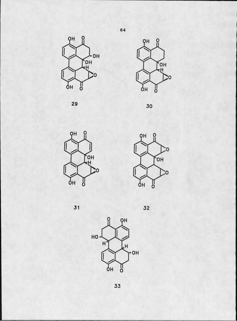
64
3 3
65
Structural Elucidation of Tenuazonic Acidr 3aThis phytotoxic compound had a molecular formula of
c 10h 15n o 3' based on a weight of m/z 197.1051, requiring four sites of unsaturation. This was an interesting elucidation exercise in which infrared analysis was instrumental in the structure determination. A sharp absorption at 3460 cm-^ and a broad absorption at 3250 cm--*- indicated intramolecular hydrogen-bonded O-H stretch and secondary amide N-H stretch, respectively (40). Three intense absorptions, a single peak at 1710 cm"1 and a doublet at 1650 and 1610 cm"1 , provided valuable information about the toxin skeleton. The doublet signified the keto-enol tautomers of a /8-diketone, an assignment that was supported by the both 1H and 13C NMR (40). The peak at 1710 cm"1 will be discussed in context with 13C NMR analysis.
Despite the high-resolution mass spectral evidence to the contrary, there were at least 18 carbon absorbances present in a broadband decoupled 13C NMR experiment. Such an obvious discrepancy could be caused by either a very dirty sample or by tautomerism. Mass spectral analysis eliminated the first option, while NMR analysis verified the second possibility. Carbonyl resonances at 5187 and 5190, in the presence of an absorbance at 5102, were diagnostic for the ehol tautomer form, while carbonyl
66
resonances at 6197 and 6201 indicated the presence of the oxo- form (53). The carbonyl resonance at 6175, in conjunction with the infrared carbonyl absorption at 1710 cm”l, was typical of an amide, and especially indicative of a Y lactam (40).
1H NMR also exhibited signal doubling for several peaks, with an approximate 3:1 intensity differential. A methyl singlet at 62.38 and a methine doublet at 63.76 were obviously twinned by proton resonances at 62.42 and 63.94. The methyl singlet was typical of a methyl group adjacent to an unsaturated ketone, and, considering the tautomerism proposed for this compound, such doubling was reasonable. The 1H NMR also suggested the presence of two additional methyl groups, one attached to a methylene, the other attached to a methine. Decoupling experiments indicated that these signals represented a sec-butyl moiety. The methine of this fragment was a complex multiplet that resonated at 61.35 and was coupled to the methine at 63.76.
Piecing the fragments together, and utilizing the lactam framework suggested by infrared analysis, generated an isoleucine derivative that was determined to be the known phytotoxin tenuazonic acid, 34. Stickings first isolated and characterized this metabolite from Alternaria tenuis Auct. (54), and Mikami et al. demonstrated its ability to induce chlorotic haloes on tobacco leaves (58).
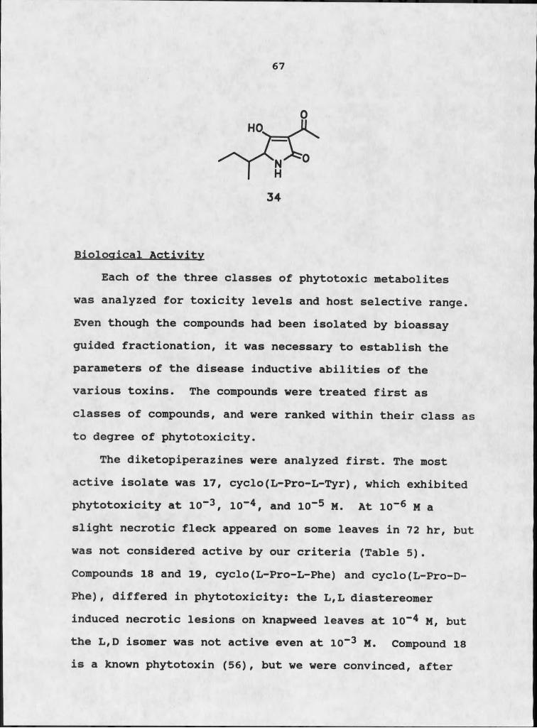
67
Biological ActivityEach of the three classes of phytotoxic metabolites
was analyzed for toxicity levels and host selective range. Even though the compounds had been isolated by bioassay guided fractionation, it was necessary to establish the parameters of the disease inductive abilities of the various toxins. The compounds were treated first as classes of compounds, and were ranked within their class as to degree of phytotoxicity.
The diketopiperazines were analyzed first. The most active isolate was 17, cyclo(L-Pro-L-Tyr), which exhibited phytotoxicity at 10“3 , IO"4, and IO"5 M. At IO"6 M a slight necrotic fleck appeared on some leaves in 72 hr, but was not considered active by our criteria (Table 5). Compounds 18 and 19, cyclo(L-Pro-L-Phe) and cyclo(L-Pro-D- Phe), differed in phytotoxicity: the L,L diastereomer induced necrotic lesions on knapweed leaves at IO"4 M, but the L,D isomer was not active even at IO"3 M. Compound 18 is a known phytotoxin (56), but we were convinced, after
68
several thorough literature searches, that 17 and 19 had not been previously reported from natural sources. Two papers were brought to our attention at this time, however. The first article was unabstracted (not recorded in Chemical Abstracts), written in French, and published in a Japanese journal. Tatsuno et al. (57) purported to have isolated cyclo(L-Pro-L-Tyr), 17, from a culture of Fusarium nivale. although no indication of isolated yield was provided. The authors did present their spectral data, and what was presented did not coincide with either our natural product or our synthetic product (Teible 5). The 1H NMR was recorded in the same solvent as our spectrum (CDCl3), but there were distinct disparities. Tatsuno et al. recrystallized their compound from benzene, which could complex with the molecule, and induce significant chemical shift deviations from the pure compound. In an effort to mimic this effect, we dissolved our crystalline material in benzene, and evaporated the excess solvent. The 1H NMR showed a strong benzene absorbance (67.27), but no discernible chemical shift was observed for any of the protons. We could only conclude that their material was not authentic.
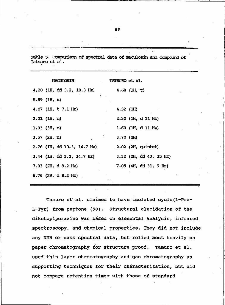
69
Table 5. Canpariscn of spectral data of maculosin and ccmpound of Tatsuno et al.
mCOmSIH TO3E$B£D et al.4.20 (1H, dd 3.2, 10.3 Hz) 4.68 (IN, t)5.89 (1H, s)4.07 (1H, t 7.1 Hz) 4.32 (IH)2.31 (1H, m) 2.30 (1H, d 11 Hz)1.93 (3H, m) 1.60 (1H, d 11 Hz)3.57 (2H, m) 3.70 (2H)2.76 (IN, dd 10.3, 14.7 Hz) 2.02 (2H, quintet)3.44 (IN, dd 3.2, 14.7 Hz) 3.32 (2H, dd 43, 15 Hz)7.03 (2H, d 8.2 Hz) 7.05 (4H, dd 31, 9 Hz)6.76 (2H, d 8.2 Hz)
Tamuro et al. claimed to have isolated cyclo(L-Pro- L-Tyr) from peptone (58). Structural elucidation of the diketopiperazine was based on elemental analysis, infrared spectroscopy, and chemical properties. They did not include any NMR or mass spectral data, but relied most heavily on paper chromatography for structure proof. Tamuro et al. used thin layer chromatography and gas chromatography as supporting techniques for their characterization, but did not compare retention times with those of standardj
70
material. Their evidence was not conclusive, and should not be considered unequivocal. Our own attempts to determine the actual source of maculosin in our fungal cultures (see Material and Methods) suggested that no discernible maculosin was produced as an artifact of our fermentation conditions.
Compounds 20, 21, 22, and 23 were not active towards knapweed at any of the test concentrations (Table 6). These tests were repeated with hypocotyl tissue, and similar results were obtained, although in 47% of these tests, Cyclo(L-Pro-L-Tyr) induced lesions at io~6 M.
Following the investigation of the comparative phytotoxicity of the diketopiperazines, the four perylenequinones were tested against knapweed to ascertain their relative phytotoxic behavior. Perylenequinones 26 and 27 induced necrotic lesions on knapweed at test concentrations of K T 3 and 10“4 M, with 27 inducing larger necrotic lesions than the small flecks induced by 26. Compounds 24 and 25 were not phytotoxic to knapweed at any test concentration.

71
Ttible 6. Comparison of phytotoxicity of naturally occurring diketqpiperazines towards knapweed and jchnsongrass.
CCMZHsFn̂ ATICN HKTOTOXICnY
COMPOUND (MOIE/UTER) KNAPWEED JCHNSONGRASS
17 L-Rro-IHiyr 10-3 H-+ -iH- -
18 L-Pro-L-HieI
+ -
IO"4 -19 L-Rro-D-Hie 10-3 - -i
- -20 Rro-Hleu IQ”3 - -
21 Pro-Val IQ-3 - -22 Pro-Leu IQ-3 - -23 Pro-Ala IO-3 — —
+++ : weeing necrotic lesion radius > 4mm from inoculation site; -H- : necrotic lesicm 2mm - 4mm; + ; necrotic lesion 0.5 - 2mm;- : no lesion present, or fleck < 0.5mm.
Tenuazonic acid, 34, was also tested against knapweed It appeared to act at levels analogous to maculosin, 17,
72
with strong activity at 10”3 M, that attenuated as the concentration was diluted to IO-5 M.
Once the initial comparative testing phase was completed our full attention turned to maculosin in an attempt to ascertain its potential against C. maculosa. Preliminary literature searches suggested that this was a novel dipeptide and worthy of further study. Maculosin was synthesized to facilitate establishing the absolute stereochemistry as L fL and to provide enough material for further testing. In all subsequent tests, synthetic maculosin behaved in an identical manner to the natural product.
Maculosin was established as a phytotoxin of some merit, but its selectivity or specificity was only suggested by its inability to induce necrosis in johnson- grass. A test designed to establish the host-range of the toxin involved a simple leaf assay utilizing a wide variety of plants. Both monocots and dicots were included in the test range. The assays were run at IO-3, IO""4 , and 10“5 M (Table 7). Maculosin consistently induced necrotic lesions on knapweed leaves, but in no case was lesion induction observed on any other test plant. The nineteen test plant species were chosen somewhat at random, although an attempt was made to include several composites and plants known to be hosts to other form species of A. alternate. These include apple, citrus, and tomato, although the actual host
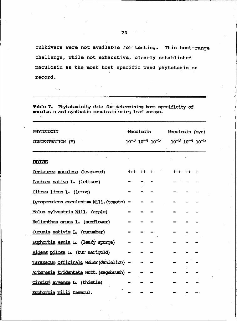
73
cultivars were not available for testing. This host-range challenge, while not exhaustive, clearly established maculosin as the most host specific weed phytotoxin on record.
Table 7. Riytotoxicity data for determining host specificity of maculosin and synthetic maculosin using leaf assays.
HttTOTOXIN Maculosin Maculosin (syn)
CONOMRAnON (M) H T 3 ItT 4 ICT5 10“3 IO**4 10“5
D I C C T S
Oentaurea maculosa (knapweed) +++ + + + -S-H- + + +Tactuca sativa L. (Ietbxie) - - -Citrus limon L. (Iesooi) - -Lvcoparsicon esculentum Mill, (tomato) - - - - -MmTus svlvestris Mill, (apple) -- -Helianthus anuus L. (sunflower) - - -Cucumis sativis L. (cucumber) - - -Euphorbia esula L. (leafy spurge) - - -Bidens oilosa L. (bur marigold) - - - -Taraxacum officinale Weber (dandelion) - - - ™Artemesia tridentata Nutt, (sagebrush) - - - -Cirsium arvense L. (thistle) - - - - -Euphorbia milii Dasmoul. - - - -
74
Table 7, continued.
Sorghum halepense L. (jtimsongrass) Poa annua L. (annual bluegrass) Avena sativa L. (park oat) Aqrcpyrens repens L. (guackgrass) Diqitaria ischaemum (Schreb) Muhl. (crabgrass)Zea mays L. (com)
Following the host-range selectivity study on knapweed we decided to test the most active perylenequinone, 27 and tenuazonic acid, 34, in a similar fashion. Maculosin was extremely selective in its choice of target plant, while tenuazonic acid was more cosmopolitan in its toxicity. Alterlosin II, 27, displayed host selectivity intermediate to that of the other two toxins (Table 8). All of these phytotoxicity test were conducted under similar conditions of temperature, humidity, illumination, manipulation, and duration. The determination of phytotoxicity was based on the appearance of a necrotic fleck or chlorotic region in the vicinity of the inoculation site.
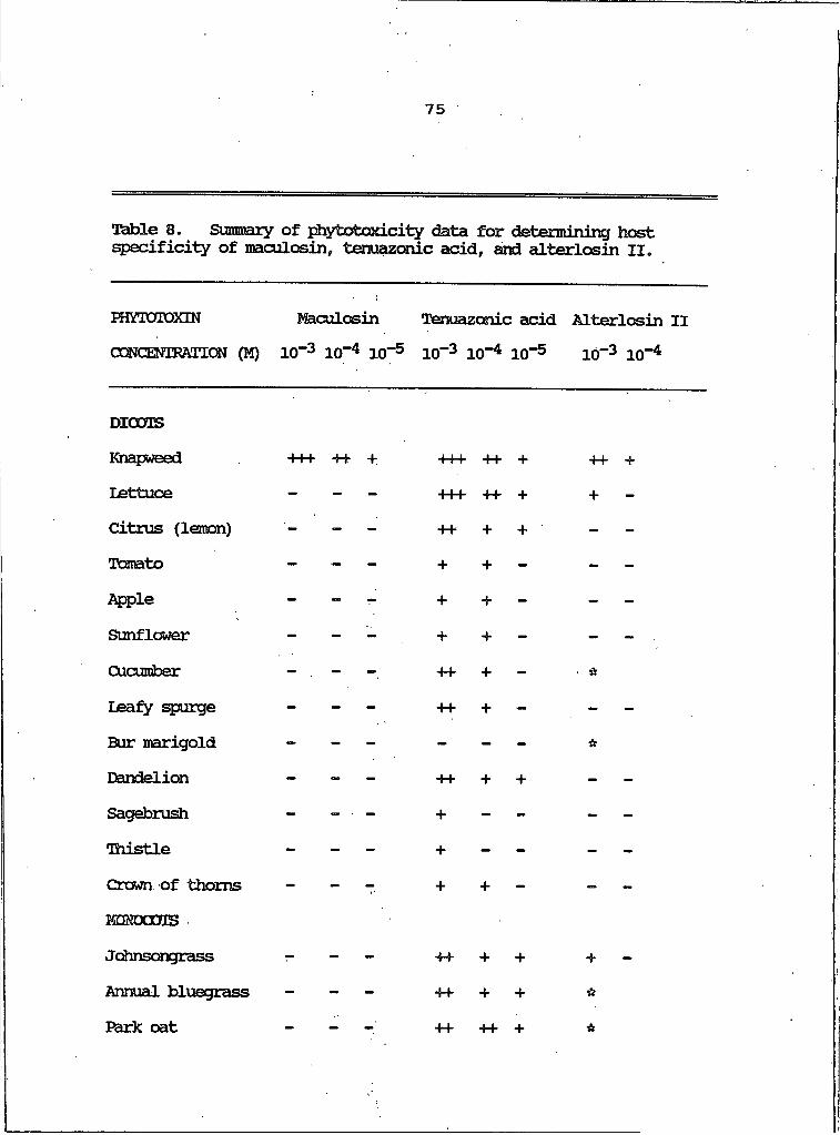
75
Table 8. Sunrnery of phytotoxicity data for determining host specificity of maculosin, tenuazonic acid, and alterlosin ii„
FHYTQTOXIN Macailosin Tenuazonic acid Alterlosin IICmCEMRMTON (M) IO"3 IO"4 IO"5 IO"3 IO"4 IO"5 10"3 IO"4
DIOOTSKi^weed +++ ++ + +++ ++ + -H-Lettuce - — — -H-+ -H- + +Citrus (lemon) ” — ++ + + ' -Tcanato - “ — + + - -Apple - ” — + + - -Sunflower - — — + + - -Cucumber - . — — -H- + - ■ *leafy spurge - — — -H- + — -Bur marigold - — — - - - itDandelion - ™1 — -H- + + -Sagebrush - ” ■ — + - - -Thistle - — ~ + - - -Crown of thorns - — T- + + - -MDKOOOTSJohnsongrass -H- + + +Annual bluegrass -- — — -H- + + *Bark oat _ ++ -H- + *
76
Ttible 8, continued.QuackgrassCrabgrassC o m
++ + + *+ + — “
-H- -H- + ** not tested (plants unavailable at time of testing.)
In examining the diketopiperazines isolated and tested in this study it was apparent that certain functional groups were concurrent with phytotoxicity. Maculosin, the most active isolate, possesses a phenolic moiety, not uncommon in phytotoxins. The acidity of the hydroxyl proton may be instrumental in this activity. All of the phytotoxins isolated in this study, maculosin, 17, tenuazonic acid, 34, and alterlosins I and II, 26 and 27, possess fairly rigid structures either due to specific conformational preferences or to inherent skeletal design. They also possess reasonably acidic protons and extended conjugation. The minor activity of the cyclo(L-Pro-L-Phe) isolate compared to the inactivity of its diastereomer supports the importance of conformation to bioactivity Studies on diketopiperazines containing proline clearly indicated that L,L isolates assume an extended conformation that would render both the aromatic moiety and its hydroxyl group accessible (9). The actual mode of activity of maculosin remains to be determined, although studies by
77
Ottenheijm et al. have demonstrated the inhibition of RNA- dependent DNA reverse transcriptase by certain dithio- diketopiperazines (59). It is interesting that the relative % yields of the various diketopiperazines are consistent in repeated fermentations and extractions. Maculosin, the most toxic to knapweed, is the most abundant diketopiperazine produced under our culture conditions, where knapweed effusion is present in the media (Table 2).
During the course of our investigation we had isolated and identified four compounds from three distinct chemical classes with significant toxicity to knapweed: the two novel perylenequinones, 26 and 27, tenuazonic acid, 34, and maculosin, 17. The coexistence of the three classes of phytotoxins within the fungal extract raised some interesting questions concerning the possibility of a synergistic relationship among the various toxins.Shiguera demonstrated that tenuazonic acid inhibits the release of newly formed proteins from the ribosomes, and thus inhibits further protein synthesis (60). Although the mode of action of maculosin has not yet been determined, it would be interesting to ascertain if the toxins isolated in the course of this study are mutually enhancing in their activities.
We attempted to explqre the possibility of synergistic activity by comparing toxic effects of the two most active phytotoxins, tenuazonic acid and maculosin, which also

78
showed the most disparate host-specificity. The analysis involved leaf assays with three different plants, leafy spurge, knapweed, and johnsongrass, all three of which were susceptible to tenuazonic acid. The plants were retested with the two toxins at IO**3 M, IO"4 M, and IO"5 M. Tenuazonic acid and maculosin were then combined to total concentrations of IO"4 M and IO"5 M and applied to the test plants (Table 9). It is interesting to note that with the combined molarities indicated, the effective activity to knapweed was doubled, while that to johnsongrass and leafy spurge was halved. The mitigating effect is most likely a function of dilution, but the augmentation effect suggests mutual enhancement by the two toxins.
Table 9. Synergy study of diketcpiperazine toxin and tenuazonic acid. All concentrations are in moles/liter.
HOST PLANT TENUAZONIC A d D10"3
TS
IO"5
KNAEVZEED +H- H +
JOHNSONGRASS H H +
IEAPY SPURGE H + +
a Represents total concentration in each of the two toxins.
MACULOSIN MIXTURE310"3 IO"4 IO"5 S Ul
+H- H + +H- H
+ ++ —
combined toxins: IO-4M is 5x10"%
79
Plant pathogenic organisms often express several phytotoxins, and each may play a different role in host- pathogen symbiosis (30). It is reasonable that a particular toxin might target a particular host mechanism, and that the elaboration of more than one phytotoxin by the pathogen may facilitate a multiphase attack of the host plant. Our studies suggest that two toxins can enhance mutual deleterious effects, increasing the pathogenicity of a fungus to a given plant host. Our study was by no means exhaustive but it certainly supported the concept of synergy relationships among phytotoxins.
The majority of studies on phytotoxins have been conducted on the effects of toxins on hybrid crop plants.An important factor in such systems is their extreme genetic similarity which actually renders such hybrids susceptible to the onslaught of pathogenic fungi. Weeds are a genetically diverse lot and may actually vary from acre to acre in a well-established biome. History does not record the devastating loss of any major weed due to a microbial pathogen? a particular pathogen might decimate an isolated population of weeds, while their neighbors are immune to the destruction.
Throughout this study we attempted to use knapweed test plants grown from seeds from a variety of geographic locations, and some diversity was seen in phytotoxicity results depending on the knapweed source. Knapweed
8 0
reproduces in typical composite fashion, from wind-borne seeds carried from the parent plant. Seeds collected from plants within a twenty foot radius of the initial damaged specimen were the most affected by maculosin. Indeed, plants grown from seeds collected in this radius actually exhibited necrotic lesions at 10”6 M maculosin in leaf assays.
Knapweed seeds were collected from plants from Missoula, Bozeman, and Butte, Montana. Plants grown from these various seeds exhibited some response variability to toxin challenges. One plant was actually somewhat resistant to maculosin: necrosis was only induced at a test concentration of 10”3 M. And an occasional leaf in a test group (one in twenty leaves) might not show any deleterious symptoms. The results reported in this study represent the average response of all of the plants tested.
The discovery of a host specific toxin against a significant weed pest has important biological implications. The selection of the pathogen was fortuitous in that it represented a species renowned for its host specificity. The seven host specific toxins isolated to date from A. alternate include compounds that are active against important cultivars of a variety of crop plants: pears, apples, citrus, and tomatoes (5). The value of a highly specific toxin to economically desirable plants resides not in its herbicidal potential but in its utility
81
as a screening agent. Cultivars of a particular plant species can be challenged by the phytotoxin and resistant individuals can be disseminated to the agricultural community. The discovery of a host specific weed toxin, however, represents even more compelling possibilities as a direct biocontrol agent in the war against undesirable weed pests.

82
KtARIME MOTOMiISM
Background
Over the past twenty years, the marine environment has provided a large number of chemically fascinating, pharmacologically important metabolites. Almost every phylum of marine invertebrate and plant has contributed to an impressive array of bioactive compounds. Of particular interest to the pharmaceutical community are those organisms that display significant antiviral, antitumor, or antineoplastic activity. Cancer is the leading cause of death in the Western World, while viral infections cause 60% of all illnesses in developed countries (61) . New sources of bioactive compounds which can effectively target these diseases are in increasing demand. Of course, there is always interest in new types of antibiotic and antifungal agents. The various denizens of the sea and their metabolic products are a likely source of future leads in this vital area of research.
Marine invertebrates have been the focus of natural products investigations for over twenty years. Field observations led to ecological considerations of the defense mechanisms employed by certain seemingly defenseless, but thriving organisms. Certain organisms can apparently modify their behavior or influence the behavior

83
of proximate organisms to prevent aggression or predation. They can overgrow neighboring species, or inhibit the growth of potentially encroaching organisms to maintain spatial integrity (61). These observations suggest a chemical defensive or offensive capability, and have provided impetus for further investigation. The chemicals involved are often cytotoxic (62, 63).
To date, compounds with significant antiviral, antitumor, or antineoplastic activity have been isolated from the following phyla: Echinodermata, Cyanophyta, Pyrrophyta, Phaeophyta, Rhodophyta, Chlorophyta, Bryozoa, Echuria, Porifera, Mollusca, Cnidaria, and Chordata (61). The compounds isolated from these organisms are as diverse as the organisms that produced them. Certain compounds, which will be discussed in detail later, are sufficiently active to warrant clinical trials as antineoplastic agents.
Microscopic inhabitants of the various marine ecosystems are as intriguing as their macroscopic counterparts. A diverse array of organisms can be classified as marine microorganisms, including bacteria, fungi, cyanobacteria, diatoms, and dinoflagellates. Early culturing ventures of seawater yielded a variety of microbes, many of which were apparently unique to the marine environment, or at least displayed salinity requirements comparable to that of seawater. Cognizant of the incredible contributions terrestrial microorganisms
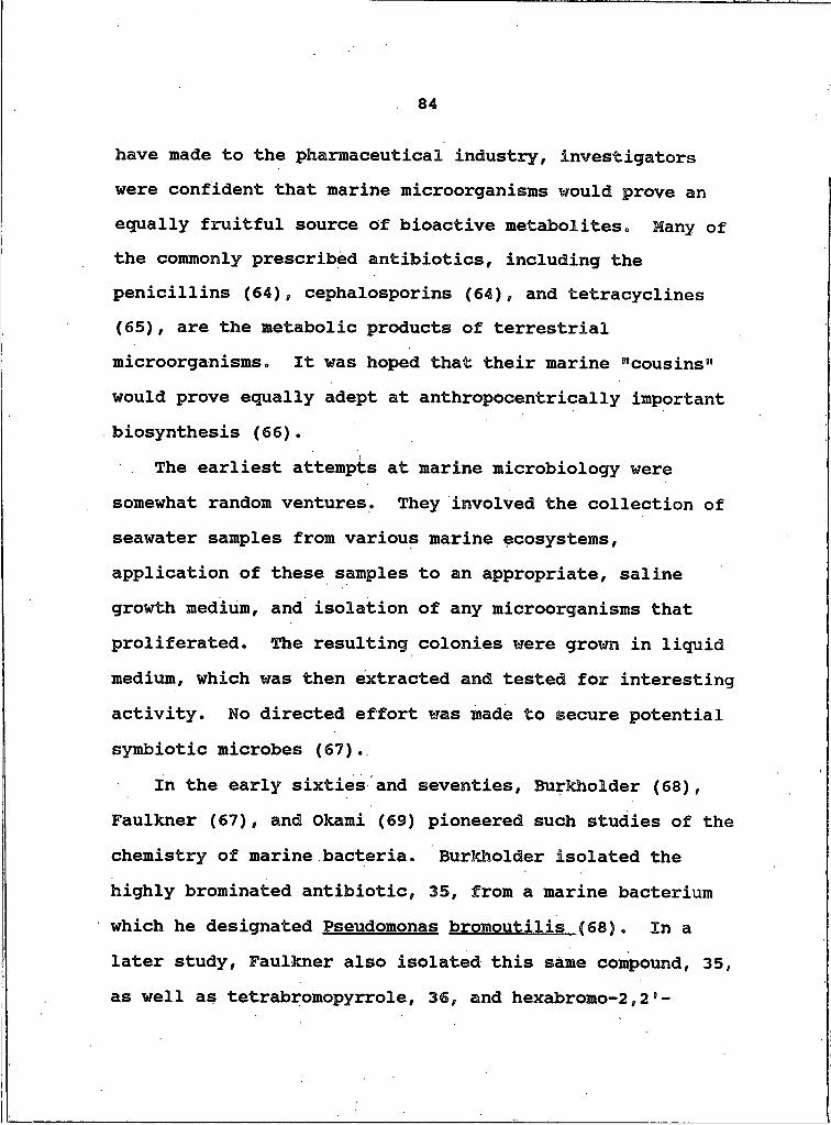
84
have made to the pharmaceutical industry, investigators were confident that marine microorganisms would prove an equally fruitful source of bioactive metabolites, Hany of the commonly prescribed antibiotics, including the penicillins (64), cephalosporins (64), and tetracyclines (65), are the metabolic products of terrestrial microorganisms. It was hoped that their marine "cousins" would prove equally adept at anthropocentrically important biosynthesis (66).
The earliest attempts at marine microbiology were somewhat random ventures. They involved the collection of seawater samples from various marine ecosystems, application of these samples to an appropriate, saline growth medium, and isolation of any microorganisms that proliferated. The resulting colonies were grown in liquid medium, which was then extracted and tested for interesting activity. No directed effort was made to secure potential symbiotic microbes (67).
In the early sixties and seventies, Burkholder (68), Faulkner (67), and Okami (69) pioneered such studies of the chemistry of marine bacteria. Burkholder isolated the highly brominated antibiotic, 35, from a marine bacterium which he designated Pseudomonas bromoutilis (68). In a later study, Faulkner also isolated this same compound, 35, as well as tetrabromopyrrole, 36, and hexabromo-2,26 -
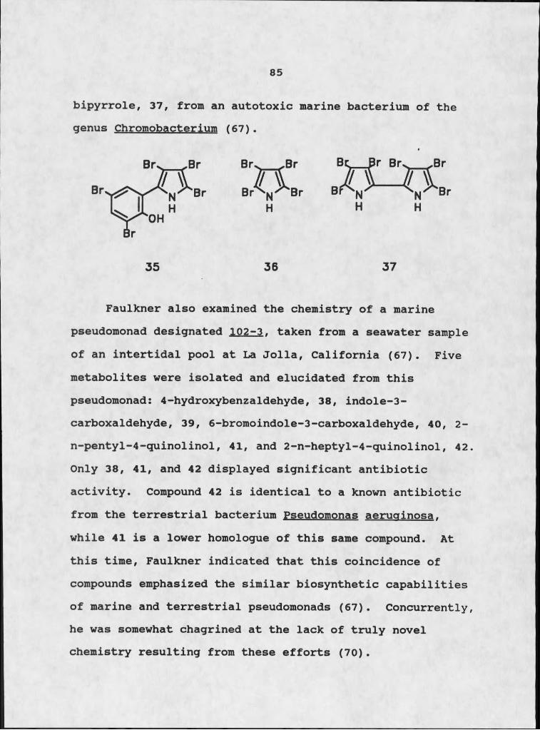
85
bipyrrole, 37, from an autotoxic marine bacterium of the genus Chromobacterium (67).
Faulkner also examined the chemistry of a marine pseudomonad designated 102-3. taken from a seawater sample of an intertidal pool at La Jolla, California (67). Five metabolites were isolated and elucidated from this pseudomonad: 4-hydroxybenzaldehyde, 38, indole-3- carboxaldehyde, 39, 6-bromoindole-3-carboxaldehyde, 40, 2- n-pentyl-4-quinolinol, 41, and 2-n-heptyl-4-quinolinol, 42. Only 38, 41, and 42 displayed significant antibiotic activity. Compound 42 is identical to a known antibiotic from the terrestrial bacterium Pseudomonas aeruginosa. while 41 is a lower homologue of this same compound. At this time, Faulkner indicated that this coincidence of compounds emphasized the similar biosynthetic capabilities of marine and terrestrial pseudomonads (67). Concurrently, he was somewhat chagrined at the lack of truly novel chemistry resulting from these efforts (70).
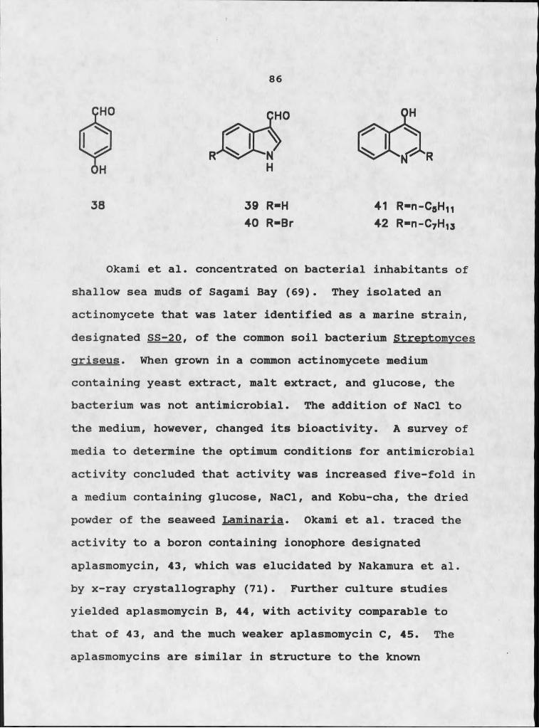
86
R
38 39 R-H40 R -Br
41 R-R-CgH1142 R-R-CyH1J
Okami et al. concentrated on bacterial inhabitants of shallow sea muds of Sagami Bay (69). They isolated an actinomycete that was later identified as a marine strain, designated SS-20. of the common soil bacterium Streptomvces griseus. When grown in a common actinomycete medium containing yeast extract, malt extract, and glucose, the bacterium was not antimicrobial. The addition of NaCl to the medium, however, changed its bioactivity. A survey of media to determine the optimum conditions for antimicrobial activity concluded that activity was increased five-fold in a medium containing glucose, NaCl, and Kobu-cha, the dried powder of the seaweed Laminaria. Okami et al. traced the activity to a boron containing ionophore designated aplasmomycin, 43, which was elucidated by Nakamura et al. by x-ray crystallography (71). Further culture studies yielded aplasmomycin B, 44, with activity comparable to that of 43, and the much weaker aplasmomycin C, 45. The aplasmomycins are similar in structure to the known
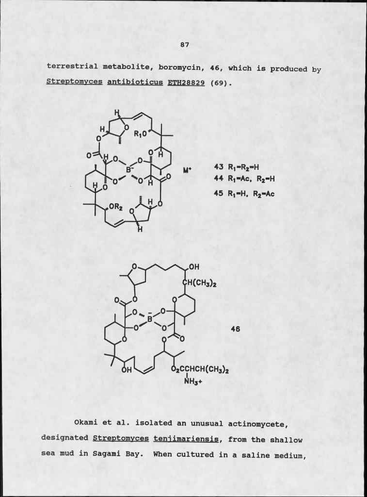
87
terrestrial metabolite, boromycin, 46, which is produced by Streptomvces antibioticus ETH28829 (69).
43 R1-R 2-H44 R1 -Ac, R2-H
45 R1-H , R2-Ac
Okami et al. isolated an unusual actinomycete, designated Streptomvces teniimariensis. from the shallow sea mud in Sagami Bay. When cultured in a saline medium,
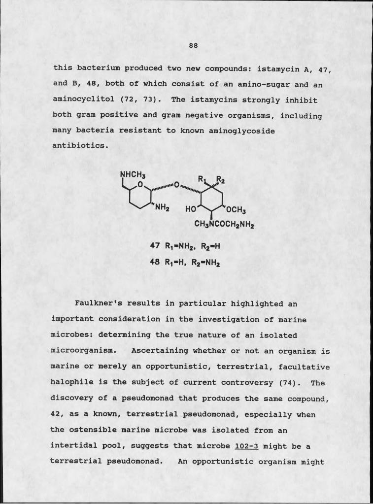
88
this bacterium produced two new compounds: istamycin A, 47, and B, 48, both of which consist of an amino-sugar and an aminocyclitol (72, 73). The istamycins strongly inhibit both gram positive and gram negative organisms, including many bacteria resistant to known aminoglycoside antibiotics.
Faulkner's results in particular highlighted an important consideration in the investigation of marine microbes: determining the true nature of an isolated microorganism. Ascertaining whether or not an organism is marine or merely an opportunistic, terrestrial, facultative halophile is the subject of current controversy (74). The discovery of a pseudomonad that produces the same compound, 42, as a known, terrestrial pseudomonad, especially when the ostensible marine microbe was isolated from an intertidal pool, suggests that microbe 102-3 might be a terrestrial pseudomonad. An opportunistic organism might
CH3NCOCh2NH247 R1-NH2, R2-H
48 R1-H , R2-N H 2

89
be washed into an intertidal pool and survive quite comfortably, and the pseudomonads are known for their ability to adapt to diverse environmental conditions (75). Determining the true nature of an organism is important from several perspectives, but efficiency and effective utilization of time and resources are perhaps of primary significance. Much screening has already been done on terrestrial microbes as sources of bioactive compounds. Their potential is certainly not exhausted, but focus is slowly turning to marine microbes as an untapped resource. If the same microbes are being isolated and tested, much valuable time is lost repeating investigations. Bioactive metabolites discovered as a result of these ventures might prove to be identical to known compounds.
These preliminary marine microbiological ventures transpired when marine natural products chemistry was still a relatively young science. Every macroorganism promised interesting, novel compounds to the curious investigator. Most of the marine invertebrates were uncharted territory at this time, and almost any abundant organism was a reasonable study target. The average marine invertebrate is relatively easy to manipulate, from a chemical standpoint. It can usually be homogenized in a Waring blender and sequentially extracted with appropriate solvents. The resulting extracts can then be resolved into

90
individual components using a variety of chromatographic techniques.
The problems involved with microbial studies - isolation of pure cultures, contamination, fermentation optimization, and the logistics of chemical manipulation of the culture - lessened the appeal of such endeavors. The few studies of marine microbial fermentation extracts failed to produce any truly novel compounds, further diminishing continued interest in this area (67). After these early attempts at combining microbiology and marine natural products chemistry, little attention was paid to the biosynthetic efforts of marine microorganisms.
The last decade, however, has witnessed a major change in the direction of natural products chemistry. Most researchers indulging in this specialty are concerned with more than the isolation and elucidation of new compounds. The desire for compounds displaying a variety of important pharmacological activities has superseded the quest for mere structural novelty. Many of the most active compounds isolated and characterized to date are produced in miniscule quantities that suggest a microbial associate as their actual source. These compounds include the bryostatins, 49 (76), dolastatin 10, 50 (77), and tedanolide, I (6). This possibility, coupled with the growing needs of the pharmaceutical industry for new, potent chemicals from a readily available and easily

91
manipulated source, has renewed interest in marine microbial chemistry.
Compounds of Possible or Proven Microbial Origin
The bryostatins are a class of compounds originally isolated from the bryozoan Bucmla neritina. In 1968 Pettit and co-workers initiated a systematic search for marine organisms exhibiting significant antineoplastic activity. The alcohol extracts of collections of B. neritina displayed significant in vivo activity (68-100% life extension in P388 leukemia infected mice) (78). Based on these results, Pettit undertook a detailed study of both this and another bryozoan with similar activity, Amathia convoluta. Seventeen bryostatins, 1-17, have been reported from these two bryozoans, although at this time, the structures and antineoplastic activities have only been determined for bryostatins 1-13 (61).
4 9

92
The toxic properties of an Indian Ocean sea hare of the genus Polabella were described nearly 2000 years ago by Pliny the Elder, (79) but the medical significance of this mollusc was not recognized until the 1970's. Dolastatin 10, 50, was isolated and characterized as a result of a fifteen year study of the antineoplastic constituents of the seahare Dolabella auricularia (77). Pettit claims that it is the most active antineoplastic compound currently known, with remarkable potency towards different melanoma and leukemia cell-types (77). The extremely low yield of this metabolite, a mere I mg isolated from over 100 kg of animal, suggests a possible microbial source for 50.
50
Tedanolide, I, an extremely cytotoxic macrolide isolated from the marine sponge Tedania ignis, is an intriguing candidate for potential marine microbial metabolism (6). T. ignis, the sole source of this
93
metabolism (6). T. ignis. the sole source of this macrolide, is a common, widely distributed sponge that is both accessible and abundant in the shallow coastal waters of Bermuda. The sponge was originally collected as part of an ongoing study of the metabolites of Bermudian invertebrates conducted by the Cardellina research group.It was established as an excellent target for investigation of microbe-invertebrate symbiosis by Schmitz's suggestion that the extremely low yield of tedanolide (I x 10”4% dry weight) evidenced its microbial origin (6).
These indications that some of the most active metabolites ever isolated from marine invertebrates were of microbial origin were sufficiently intriguing to warrant further investigation. The actual discovery of microbial origins for several compounds previously attributed to macroorganisms was considerably more compelling, and provided a more concrete rationale for further study.
Kosuge et al. (80) provided the first evidence of toxin production by bacteria in a marine organism, the Japanese ivory shell, Babylonia iaponica. This shellfish, harvested from Suruga Bay, was responsible for an outbreak of food poisoning in September, 1965. Kosuge et al. isolated nontoxic surugatoxin, 51 (81), and the causative toxins neosurugatoxin, 52 (82), and prosurugatoxin 53, (83), from the digestive gland of the shellfish and determined their

94
chemical structures. The authors attempted to ascertain if these compounds were ingested material, as was determined for saxitoxin, 54, the toxic principle of paralytic shellfish poisoning first associated with clams (84).
52 R-6'-(MYOINOSITOL-5-0-1 -XYLOPYRANOSE)
53 R-e'-MYOINOSITOL
5 4

95
Kosuge et al. first established a positive correlation between cultivation of the ivory shell in an area where poisonings had previously occurred, and accumulation of the toxin in its digestive gland. Toxic shellfish that were cultivated in "virgin" waters lost their toxicity within one month. Nontoxic shellfish transplanted to Suruga Bay developed a degree of toxicity after two months. These results implicated an environmental factor in the production of the toxins (80). Ivory shells derive a major portion of their nutrients from planktonic species, the prime suspects as toxin producers. Chemical investigation of the plankton in Suruga Bay failed to establish a nutritional source: the plankton populations were free of the toxins. It was noted that the shellfish were toxic from July to September, when the temperature of the water often reached 250C and Suruga Bay was heavily polluted.Both of these conditions favor bacterial growth; therefore, a bacterial source for the toxins was considered (80).
More than one hundred strains of bacteria isolated from the sea bed slime of Suruga Bay and from the toxic shellfish were investigated . A gram-positive, facultative halophilic bacterium isolated from the digestive gland of the ivory shell produced the toxins. Based on biological activity, the estimated yield of the toxins was 20 ng from I I of the cultured medium. Neosurugatoxin and prosurugatoxin exhibited more than five thousand times the

96
neuroactivity of currently prescribed ganglion blocking drugs. They specifically block nicotinic receptors in the ganglion, and are therefore excellent tools for studying the neurosystem (80).
Two important guanidine derivative are saxitoxin, 54, and tetrodotoxin, 55, which are two of the most toxic nonproteinaceous naturally occurring substances known (85). Saxitoxin was originally isolated from California clams, and was determined to be the cause of periodic paralytic shellfish poisoning. The possible dietary origin of the toxin in filter-feeding clams was suggested in 1928 (86), and verified in 1947 (87). Needier identified the nutritional source as the,dinoflagellate Gonvaulax tamarensis (88), which was confirmed by Prakash in 1963(89) .
One of the most fascinating natural products isolated from either a terrestrial or a marine source is tetrodotoxin, 55. Tetrodotoxin has been isolated from a variety of terrestrial and marine vertebrates, including the California newt Taricha torosa and the Japanese pufferfish of the genus Snhoeroides (Fugu) (85).Pufferfish have long been associated with 66Fugu poisoning61, a syndrome caused by the ingestion of improperly prepared fish. The toxic principle was identified as tetrodotoxin(90) . This toxin has recently earned international acclaim due to the efforts of an ethnobotanist, Wade Davis, eager
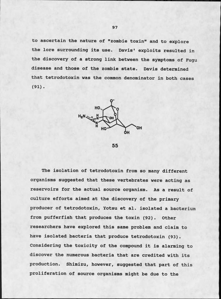
97
to ascertain the nature of "zombie toxin" and to explore the lore surrounding its use. Davis' exploits resulted in the discovery of a strong link between the symptoms of Fugu disease and those of the zombie state. Davis determined that tetrodotoxin was the common denominator in both cases(91) .
The isolation of tetrodotoxin from so many different organisms suggested that these vertebrates were acting as reservoirs for the actual source organism. As a result of culture efforts aimed at the discovery of the primary producer of tetrodotoxin, Yotsu et al. isolated a bacterium from pufferfish that produces the toxin (92) . Other researchers have explored this same problem and claim to have isolated bacteria that produce tetrodotoxin (93). Considering the toxicity of the compound it is alarming to discover the numerous bacteria that are credited with its production. Shimizu, however, suggested that part of this proliferation of source organisms might be due to the
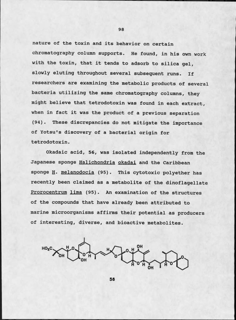
98
nature of the toxin and its behavior on certain chromatography column supports. He found, in his own work with the toxin, that it tends to adsorb to silica gel, slowly eluting throughout several subsequent runs. If researchers are examining the metabolic products of several bacteria utilizing the same chromatography columns, they might believe that tetrodotoxin was found in each extract, when in fact it was the product of a previous separation (94). These discrepancies do not mitigate the importance of Yotsu1s discovery of a bacterial origin for tetrodotoxin.
Okadaic acid, 56, was isolated independently from the Japanese sponge Halichondria okadai and the Caribbean sponge H. melanodocia (95). This cytotoxic polyether has recently been claimed as a metabolite of the dinoflagellate Prorocentrum lima (95). An examination of the structures of the compounds that have already been attributed to marine microorganisms affirms their potential as producers of interesting, diverse, and bioactive metabolites.
56

99
Biochemical Studies of Endosvmbionts
The isolation of bacteria and other microorganisms responsible for the production of known, bioactive compounds has been a tremendous incentive for continued investigation. The past four years witnessed a resurgence of interest in marine microbial chemistry, as well as a proliferation of biochemical studies of endosymbiotic bacteria. Once again, the majority of the chemical studies in the recent literature concerned microbes isolated from seawater or sea muds, and therefore did not exploit the directive potential of symbiotic associations. Several investigators, however, are currently addressing the possibility of symbiotic relationships and their affects on natural products chemistry by examining the microbial flora of a variety of marine invertebrates.
Biochemical studies of endosymbiotic microorganisms have flourished within the last few years. Giere and Langheld explored the bacterial populations of the thiobiotic Bermudian oligochaetes Phallodrilus planus and P.Ieukodermatus (96). A non-random, differentiated and consistent distribution pattern of the extracellular bacteria along the length of the worm's body emphasizes the regulated nature of bacterial colonization. The eggs become infected at oviposition by intrusion from large stores of bacteria in a genital pad adjacent to the female
100
pores. During ontogenesis a balance is maintained between extracellular active forms and intracellular lytic bacteria enclosed in epidermal cell vacuoles. In the early stages of development, intracellular forms predominate, but the pattern changes as the worms age. Older individuals harbor large populations of extracellular bacteria beneath their cuticles. Giere and Langheld were intrigued by the zoological aspects of this association and investigated three major issues: a) the spatial distribution pattern of extracellular bacteria on the body of the oligochaete; b) the establishment of bacterial colonies in the various developmental stages of the host; and c) the biological fate of bacteria in the host's cuticle (96).
The outer mantle of bacteria on the oligochaetes is most suitable for detoxification of seawater in the immediate environs of the worm. Platt's Inlet (Bermuda) is enriched with H2S, a toxic agent for most invertebrates. A bacterial population forming a protective shield around the oligochaete could utilize sulfide as a nutrient, fulfilling its own nutritional needs and diminishing the supply of sulfide directly impacting the tissues of the worm.
Giere and Langheld demonstrated the establishment of the symbiotic relationship between the oligochaete and its bacterial associates through detailed microscopic studies throughout ontogenesis of the worm (96). Their results showed a regular transmittance of bacteria from the
1 0 1
parental body to its eggs by external intrusion during a precise phase of oviposition. Apparently, lytic effects are involved in bacterial transfer. The bacteria invade the mucous sheath of an oocyte by tunneling into the egg's surface and creating entrance passages into the ooplasm.The complex regulation of transmittance was underscored by the fact that invasion was a strictly polar event: only the egg surface adhering to the parental body was invaded.
Examination of the zonation patterns of bacterial colonization in the epidermal-cuticle complex indicated a mutual advantage between bacterial activity and animal reaction. A marked lysis zone demonstrated that the host was able to utilize metabolic products excreted by their symbionts. The worm's epidermal cytoplasm is highly vesiculate, with a maximum concentration of exocytotic and Golgi organelles in close conjunction with encircling bacterial colonies. To justify their claim of mutualism, Giere and Langheld demonstrated that the bacteria also derived some benefit from close association with the oiigochaete. Extraction of the oligochaete yielded a surprisingly high percentage of polyhydroxybutyric acid (10% dry weight), a major bacterial reserve substance (96). This suggested that the bacteria were thriving as a result of their interaction with the worm. They probably benefit from both the protection of their host and from the

1 0 2
optimization of their position at the anoxic/oxic interface resulting from the motility of their host.
The discovery of the unique fauna of deep-sea hydrothermal vents delineated an unexpected type of symbiosis (97). In these systems the animal hosts (the pogonophoran worm, Riftia pachvptila. the giant vent clam, Calyptogena magnifies, and the vent mussel, Bathvmodiolus thermophilus) harbor chemosynthetic, sulfide-oxidizing, endosymbiotic bacteria within their tissues. Utilizing energy released by the oxidation of reduced sulfur compounds in their environment, these bacteria fix dissolved carbon dioxide into organic compounds which nurture their hosts. The greatly reduced or absent digestive systems of the host organisms indicate their nutritional dependence on bacterial chemosynthesis. Since these discoveries in the isolated and rare environments of the hydrothermal vents, similar associations were found in several species representing the phyla Pogonophora,Mollusca (98), and Annelida (99). These symbionts were found in habitats ranging from the abyssal to the intertidal depths, and from the subarctic to the tropical latitiudes (100, 101, 102). The common denominator linking these habitats was an interface between a sulfidegenerating zone (anoxic) and water with sufficient oxygen to sustain the host (97).
10,3
Distel and Felbeck (97) examined the incidence of endosymbiosis between lucinid clams and sulfide-oxidizing bacteria. Lucinoma aeouizonata. annulata. and Lucina floridana were collected from different depths: 550-570 m, 70-150 m, and intertidal flats, respectively. The authors examined morphological details of ultrastructure for evidence of endosymbiotic bacteria. The gills, which comprise approximately 35% of the clam's wet body weight, consist of three distinct regions: the ctenidial filament zone (CFZ), the transition zone (TZ), and the bacteriocyte zone. Light and electron microscopy indicated that the morphology of the ctenidial zone is typical of other eulamellibranch bivalves. Directly beneath this region is a thin layer of non-ciliated, non-pigmented, bacteria-free cells designated the transition zone. Medial to these two thin, transparent layers is a thick, deeply pigmented, highly specialized tissue called the bacteriocyte zone.It is within this third zone that large concentrations of bacteria are found (97).
The bacteriocyte zone is organized into tubular stacks called bacteriocyte cylinders, whose hollow centers are designated bacteriocyte channels. The channels penetrate the bacteriocyte and transition zones, and open into the ostia of the ctenidial filament zone, allowing free passage of sea water between the mantle cavity and interlamellar spaces. The predominant cell type of this region is the
104
bacteriocyte, a large spherical cell with a diameter rangeof 20 - 40 Atm. The nuclei of these cells are displaced toIthe side of the cell opposite the bacteriocyte channel. Numerous mitochondria and Golgi bodies are in the cytoplasm, as well as large quantities of glycogen granules. The most conspicuous structures in the cytoplasm, however, are the endosymbiotic bacteria, which comprise the greatest portion of the cell volume. The endosymbionts are large, pleomorphic, gram-negative rods, similar in appearance to bacteria found in other lucinids. They are apparently strictly confined to the bacteriocyte zone (97).
The spatial relationship and intimacy between bacterial endosymbionts and host cells, and the lack of evidence of endocytotic activity by bacteriocytes, suggest that the bacteria are not directly involved in the uptake of dissolved organic matter from the environment. The growth, proliferation, and maintenance of symbiotic bacteria evidently occur entirely within the bacteriocytes, and do not depend on transport of bacteria or particulate matter to and from the external environment (97). The value of this association to the lucinid host is not entirely clear. The location of the bacteriocyte zone and its endosymbiont population preclude their importance in primary sulfide detoxification. The gill infrastructure necessitates the initial passage of water through the bacteria-free
105
ctenidial and transition zones before reaching the bacteriocyte zone. Sulfide oxidation by the endosymbionts would afford no protection against sulfide toxicity to these outer regions. They could, however, participate in secondary detoxification by metabolizing compounds such as thiosulfate or sulfite which are commonly produced by animal tissues challenged by sulfides (97).
Gustafson and Reid (103) examined the incidence of associative bacteria in the gutless protobranch bivalve Solemva reidi, particularly the mechanism of transmission and infection. Rod-shaped bacteria were consistently observed by transmission electron microscopy in the locomotory test of larvae and in the perivisceral cavity of post-larvae of S. reidi. Adults of this species are known to harbor intracellular chemoautotrophic bacteria in their gills. Transmission of endosymbionts in invertebrate- microorganism proceeds in one of three ways: by vertical transmission (from parent to offspring); by horizontal transmission (between adult contemporaries); or by infection of the each adult generation from an environmental stock of microorganisms (104). Gustafson and Reid wanted to demonstrate the mode of infection in S. reidi.
The study consisted primarily of transmission and scanning electron microscopy studies of the various oocyte, larval, and adult tissues of this bivalve. They determined
106
that the bacteria are packaged in cryptic form, as prebacteria, in dense granular vesicles within the egg. It was found that these vesicles possess a homogeneous granular matrix and are present in the basal region of the larval test. No bacteria were discernible on the first three days of larval development. On day four, however, particles resembling procaryotic cells were seen within the vesicular matrix. An electron-lucent nuclear region is apparent in these particles. By the fifth day of development definite procaryotic cells were present within many of the vesicles, while distinct rod-shaped, gramnegative bacteria were evident in the free space between the larval test and definitive epithelium. Their configurations suggested that they had been released from the larval test. As the bacteria undergo transformation numerous membranes and small vesicles develop within the matrix (103).
Following metamorphosis of the bivalve, which includes auto-ingestion of much of the locomotory larval test, many rod-shaped bacteria were observed in the perivisceral cavity of the post-larvae. These bacteria lie in close proximity with potential hemocytes, which could engulf the bacteria and transport them to their ultimate destination, the gill tissue. This mode of concurrent host-guest development as a corollary to horizontal transmission of
107
associative organisms provides valuable insight into the scope of the symbiotic relationship (103).
All of the biochemical or histological studies of microbe-invertebrate symbioses emphasize that these relationships are not merely casual, accidental associations. Energy is apparently expended by both microbe and invertebrate to preserve and propagate the association. Spatial orientation and colonization by the microbe are not random but follow discrete patterns within the body of the host. It is important to remember that in these interactions, one or both symbionts are benefited in some intrinsic fashion, although the benefit might not be obvious to observers.
108
Biorationale for Research Desicm
The examples cited above create several interesting scenarios of microbe-invertebrate asssociation, and the biochemical and chemical ramifications of such close associations. We had already convinced ourselves that such a directed research study could prove fascinating, but there were more pragmatic reasons fOr this type of study that were equally important to consider. Development of a strategy for examining the microbial flora of a particular macroorganism involves the following: isolation of individual microbes, culturing the isolates, investigation of their chemistry, and optimization of growth conditions for production of desired metabolites. Undertaking this rather ambitious endeavor was predicated on the belief that such a venture would prove worthwhile not only as a purely academic exercise, but also as a means to isolate important metabolites. Microbial fermentation as a means of isolating bioactive substances with pharmaceutical potential has several advantages (69):
I. Industrial production of bioactive substances requires reproducible, dependable productivity. Many macroorganisms produce desirable metabolites in miniscule quantities, requiring thousands of kilograms of organism for the isolation of one milligram of metabolite (6, 76,77). Before a compound can be adequately tested, the
109
world's supply of the source organism could be obliterated,, and most of the compounds are sufficiently complicated to defy simple, efficient synthetic methodology. Microorganisms, however, can be stored indefinitely as stock cultures and grown as needed. The production of large quantities of metabolites.can be consistently and reproducibly achieved in tank fermenters in less than one
week (69).2. The average microorganism responds favorably to
fairly routine culture techniques. Cultivation of macroorganisms is considerably more challenging, and often years of growth are necessary before harvesting is feasible
(69) .3. Productivity amplification is relatively easy in
microorganisms. In the case of penicillin, improved cultural conditions and genetic manipulation of the producing strains of Penicillium increased the drug yield from a few micrograms per milliliter to several thousand micrograms per milliliter (105, 106). Few options for improved productivity from macroorganisms are available,
other than larger collection sizes.4. Different bioactive compounds can be produced by the
same strain of microbe by altering culture conditions. The aplasmomycins were produced by Streotomvces qrjeeus SS-20 only if NaCl were added to the growth medium (72).Directed changes in culture conditions can be explored
H O
indefinitely as a method of optimizing various biosynthetic pathways (69).
The disadvantages of a random approach to marine microbiology have already been addressed with regard to the less than inspiring results of such studies (67, 68). We chose a directed approach to the selection of marine microorganisms for investigation. A directed search for a particular microorganism that produces a particular metabolite ostensibly produced by a macroorganism has often proven successful. The discovery of microbial sources for saxitoxin (84), tetrodotoxin (92), okadaic acid (95), and surugatoxin (80) have already been discussed.
Ideally, a bioactive metabolite is selected for study.A small sample of the reputed source invertebrate is collected and placed on a saline medium to encourage the growth and proliferation of true marine microorganisms.The isolated microbes are grown in small liquid cultures and extracted with appropriate solvents. The extracts are tested with a bioassay that specifically and dramatically indicates the presence of the desired compound, and the second or third microbe tested proves to be the actual source of that compound.
Of course, the ideal situation is somewhat unlikely: hundreds of microbes will probably need to be analyzed before the desired microbial source is discovered. Finding a bioassay that will specifically indicate the existence of
Ill
a compound that might he present in nanogram amounts (in a 100 ml culture) is not trivial. And even the most specific, sensitive assay will not detect a particular metabolite if it is not present in a culture.
At an early point in this study we realized that all of our culture efforts might not yield a microbe that produces tedanolide; therefore we adopted a bilevel approach to the investigation. Our primary goal was still the discovery of a tedanolide producer. Our secondary goal, however, which gained significance as the project progressed, was the isolation and characterization of any novel, bioactive metabolites produced by the bacterium.This flexibility permitted maximization of our research efforts.
The dual goals of this project were both dependent on the development of expedient, effective assay techniques. Any project concerned with the isolation of bioactive metabolites requires appropriate testing methodology. The search for a tedanolide producer demanded an assay capable of detecting minute quantities of the macrolide. At the onset of this project, bioassays capable of indicating the presence of minute quantities of specific compounds were not routinely available. In the past year radioimmunoassay techniques, such as those operating in the phorbol ester binding assay, have provided more sensitive detection of potent antitumor activity at the nanogram level (107).
112
Prior to this, however, tedanolide detection was strictly a function of general cytotoxicity determination (6).
The search for generic bioactives, however, could be accomplished with the routine bioassay techniques already available in our laboratory. For preliminary screening purposes, antimicrobial activity can be determined with an impregnated disc assay, and "cytotoxicity" can be determined by the brine shrimp toxicity assay (see Experimental), Once compounds have been isolated and characterized they can be sent to the National Cancer Institute for more thorough testing.
The state of the art bioassays for cytotoxic, antineoplastic or antitumor activity, at least for the public sector, have been the National Cancer Institute's in vitro and in vivo test systems using various cancer cell lines. These include the KB cell culture derived from human epidermoid mouth cancer, which constitutes an effective in vitro prescreen, followed by in vivo tests using leukemia P388, lymphoid leukemia L1210, B16 melanoma, HS067 sarcoma, and the HX-I human mammary tumor (61).
The terms cytotoxicity, antitumor, antineoplastic, and anticancer are often used interchangeably. In order to eliminate confusion in the literature the National Cancer Institute has precisely defined these terms. Cytotoxicity should only refer to toxicity to tumor cells in culture. Terms such as antineoplastic, antitumor, or anticancer
113
should not be used when referring to in vitro results. Antineoplastic and antitinaor should be used to describe in vivo activity in experimental systems. The term anticancer should only be used to report data from clinical trials in humans (61).
Unfortunately, the National Cancer Institute screening program is not a viable choice for routine isolation procedures. Long distance bioassay guided fractionation is not especially feasible or expedient. It was necessary to establish an in-house screen that combined ease of operation and maintenance with a reasonable estimation of cytotoxicity. The brine shrimp test is a simple test to run, requires no microbiological or histological training, and provides an unequivocal indication of toxicity. If a certain concentration of a test sample kills brine shrimp, then it is toxic. Of course, our goal is not the isolation and characterization of compounds that would make effective rat poisons. We are interested in compounds with marked cytotoxicity, that translates into antitumor or antineoplastic activities with further testing. Several studies, however, have found that the correlation between brine shrimp lethality and cytotoxicity is excellent (108, 109, H O , 111). As a rapid, efficient prescreening tool, the brine shrimp test is ideal. Throughout this investigation brine shrimp toxicity was designated as cytotoxicity.
114
Any compound with bioactivity determined by any screening method must be tested extensively before it can be considered appropriate for clinical trials. Any good in-house bioassay procedure usually affords an indication of bioactivity which must be substantiated by additional testing.
Suitability of Micrococcus so. as a Svmbiont
The choice of Tedania ignis as a target invertebrate was predicated on its production of the cytotoxic macrolide tedanolide, I, as well as its availability for collection. The decision to study the bright orange, gram-positive bacterium isolated from the tissues of T. ignis was predicated on its behavior in the prescreening phase of the project. When grown in 100 ml of DIFCO marine broth 2216 for 2 weeks, the freezed dried culture residue exhibited antimicrobial activity and slight cytotoxicity. When the bacterium was grown in liquid cultures large enough to facilitate chemical investigation, the organic soluble extract was bioactive.
An aspect of the bacterium which gained significance with later collection and culturing efforts however, was the constancy of its occurrence in $. ignis. Samples of the sponge were collected from two locations in Bermuda during the summer of 1986. The same bacterium was isolated
115
from all of the tissues excised from the sponge from both collection sites.
During the summer of 1987, the sponge was recollected from two other sites in Bermuda, and once again the same bacterium appeared in culture. The constant association of a particular microorganism with a particular macroorganism suggests symbiosis, although not definitively. Even though the actual collection sites varied, the bacterium could have been a common, shallow water microorganism. As a control experiment, a sample of water was taken at each collection site from the vicinity of the sponge before it was disturbed. The water was applied to a marine agar plate, and checked for microbial growth. Several organisms developed on the plate within three days, but in no case was the orange bacterium apparent. These observations suggested that this particular association might be more than casual.
During the summer of 1988, £. ignis was collected off the coast of Florida? once again, the orange bacterium was isolated from the sponge tissue. At this point, some level of symbiosis was assured. Random organisms might colonize a particular macroorganism, but in most studies of macrobe- microbe interaction, the microbial genus, and sometimes species, is constant (103). What originated as the investigation of a microbe with dubious associative ties to

116
the sponge was developing into a respectable example of true symbiosis.
Once the bacterium was identified as a Micrococcus sp. by Professor Singleton of the University of Maryland, its generic proclivities towards symbiotic relationships, as well as its chemical potential, could be assessed. The genus Micrococcus consists of aerobic, cluster-forming, gram-positive cocci (112). Members of this genus are non- fermentative. Micrococcus are universally present on human skin and help prevent colonization of the skin by pathogenic microorganisms (113). The surface of the skin provides excellent growth facilities for the bacterium.This type of a relationship is a classic case of mutualism.
Few studies of the chemistry of Micrococcus sp. have been reported in the literature. Kato et al. investigated the incidence of microbial production of bacteriocins, bactericidal substances which are usually active against the same or closely related species. Eighty strains of amino or nucleic acid producing bacteria were analyzed, including six strains of Micrococcus sp. Two strains of M. roseus produced these unidentified toxins (114).
Fahey and Mewton detected several low molecular weight thiols in several Micrococcus sp. The thiols were labeled with a fluoresent indicator and the labeled derivatives were separated by high performance liquid chromatography.No attempt was made to identify these compounds (115).
117
The chemical and histological references to Micrococcus in the literature Here sparse but encouraging. The bacterium is an acknowledged symbiont and has proven capable of producing bioactive substances. Considering the nature of several of the metabolites isolated from this bacterium, the references to its sulfur utilization were important. The nature of the compounds produced by this organism, which will be discussed in detail, and its constant association with the sponge, created a strong case for mutualism.
Results
We collected selected marine organisms from various ecosystems in Bermuda during July, 1986 and June, 1987. These invertebrates were chosen because of their activity in our in-house bioassays for antimicrobial activity, cytotoxicity, and phytotoxicity. During our first trip to Bermuda we collected samples of the two marine sponges of the class Demospongea, Tedania ignis and Dvsidea etheria. and the bryozoan, Buaula neritina. The sponges were collected in shallow water from several locations in Bermuda. ignis was collected from both the east and west ends of Ferry Reach, and from Flatt's Inlet. A typical blue specimen of D. etheria was collected from the base of the Causeway across Castle Harbor, and an unusual purple specimen was removed from Ely's Harbor at the
1 1 8
extreme western tip of Bermuda, g. neritina was collected from Mangrove Lake. On our second trip to Bermuda, we recollected specimens from our original venture, and added several other invertebrates to our repertoire for culture work. These included the Demospongea Ulosa ruetzleri. Chondrilla nucula, Xqernella notabilis. Lissodendoryv isodictvalis and Pseudaxinella explicate, and the Calcarea Leucetta microranhis. All of the specimens that were collected for culture work were removed from their environments and immediately placed inside sterile plastic baggies. They were returned to the laboratory where they were sliced open and small pieces of tissue were carefully excised. These tissue samples were placed on marine agar plates and left undisturbed for 24 hr. Sterile technique was followed at all times.
After 24 hr the plates were examined for microbial growth, and the visible organisms were streaked for isolation. This procedure was repeated several times over the next few days until each plate harbored only one or two microorganisms. The plates were then sealed and returned to our laboratory in Bozeman.
When we returned to Bozeman, the cultures were examined once more and any multi-culture plates were again streaked for isolation. During the two collection trips we isolated 143 microorganisms based on gross morphology, although several of these isolates proved to be duplicates.
119
Once we were satisfied with the purity of our cultures, we grew each organism in 100 ml broth cultures to facilitate bioactivity testing. The 29 microbes isolated the first summer were cultured for periods of one, two, and three weeks before harvesting. For an active isolate, optimum activity was realized in both the two and three week cultures; therefore, all subsequent testing on the small liquid cultures was performed on two week cultures.
At the end of two weeks the cultures were killed by the addition of either MeOH or CH2Cl2 , and the water was removed by freeze drying. The dried residue was tested directly as an aqueous sample. As a control, a 100 ml sterile broth culture was also freeze-dried and the nutrient broth residue weighed. By comparing the weights of the two residues it was apparent that the inoculum residue was approximately 90% nutrient material. All test samples were weighed with the assumption that only 10% of the material was associated with the inoculum. All of the microorganisms were tested for antimicrobial activity, cytotoxicity, and phytotoxicity. For the early testing period, microbes were considered to be antimicrobial if they produced a zone of inhibition with a radius of at least 1.0 mm. Microbes were considered cytotoxic if they killed 25% of the brine shrimp in the assay in 24 hr, and phytotoxic if they induced a necrotic or chlorotic lesion on a j ohnsongrass leaf (Table 10).
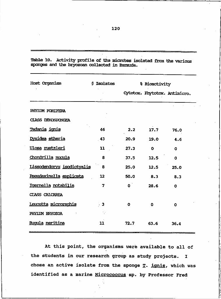
1 2 0
Table 10. Activity profile of the microbes isolated from the various sponges and the bryozoan collected in Bermuda.
Host Organism # Isolates % BioactivitycytotoK. Phytotox. Antimicro.
HKHJM PORIFERA CLASS DEMDSFONGEATedania ionis 46 2.2 17.7 76.0Dvsidea etheria 43 20.9 19.0 4.6Ulosa ruetzleri 11 27.3 O OChondrilla nucula 8 37.5 12.5 OLissodendorvx isodictvalis 8 25.0 12.5 25.0Pseudaxinella exolicata 12 50.0 8.3 8.3Iaemella notabilis 7 O 28.6 0CLASS CAICAREA Leuoetta microrachis . 3 O O 0HKHJM ERYOZQA Buaula neritina 11 72.7 63.6 36.4
At this point, the organisms were available to all of the students in our research group as study projects. I chose an active isolate from the sponge £. icmis. which was identified as a marine Micrococcus sp. by Professor Fred
1 2 1
Singleton of the University of Maryland. Microorganisms that were chosen were grown in large broth cultures (9-20 I) to produce enough cellular material and exudates for chemical analysis and biological testing. The large broth cultures were grown for 21 days as still cultures, although we did simulate wave action once a day. The organisms were harvested following the same protocol used for the small cultures. The microorganisms were killed by the addition of MeOH or CHgClg and water was removed by freeze drying. The residue was retested for bioactivity to ensure our earlier results. The culture was then ready for chemical analysis.
The entire residue was extracted with MeOH-MeCN, 4:1, to facilitate separation of the nutrient salts from the cellular materials, and still retain the more polar metabolites. The residue was then extracted with CH2Cl2 . The MeOH-MeCN soluble extract was partitioned between water and CH2Cl2 , and the two CH2Cl2 soluble extracts were combined. An emulsion formed between the two layers that was neither water soluble nor CH2Cl2 soluble. This was removed from the water soluble extract by suction filtration and was found to be soluble in MeOH. The aqueous and organic soluble extracts were tested for bioactivity. For Micrococcus sp., all of the activity was concentrated in the two organic soluble extracts.
1 2 2
The first three large liquid cultures of this organism were grown in Bozeman. The total volume of the three cultures was 49 I, and the total organic soluble extracts weighed 3.08 g, for an average productivity of 0.063 g/1. The final broth culture was grown by Professor Singleton at his facility in Maryland. He centrifuged the entire 220 I culture and separated the cellular solids from the media and cellular exudates. He personally extracted the entire 220 I liquid culture with EtOAc (500 ml at a time), and reduced this extract to dryness. He sent the EtOAc soluble extract (0.643 g) and the solids to us in Bozeman. The solids were first extracted with MeOH-MeCN, 4:1, followed by extraction with EtOAc-C^Cl2 , 1:1, a process which spanned 10 days. The combined organic soluble extracts from Singleton's 220 I culture weighed 5.01 g, a productivity of only 0.025 g/1. Singleton only grew the culture for I week, which evidently reduced productivity by 60%.
The organic soluble extracts of Micrococcus sp. exhibited consistent cytotoxicity and antimicrobial activity, which directed bioassay guided fractionation. In the course of this study I isolated, characterized and tested a series of compounds with varying degrees of bioactivity, representing several classes of organic compounds. Even though we did not realize our ultimate aspiration, the discovery of a tedanolide producer, the
123
compounds which were produced were sufficiently intriguing to justify the entire investigation.
Isolation of DiketooinerazinesA series of diketopiperazines was consistently
isolated from 21 day marine broth cultures of Micrococcus sp. The CH2Cl2 soluble extract of the freeze-dried culture residue was chromatographed on Sephadex LH-2O (CH2Cl2-MeOH, 1:1), yielding 11 fractions. Examination of the 1H NMR spectra of fractions 5, 6 and 7 indicated the presence of diketopiperazines, compounds with which we became intimately involved in the knapweed study. Schmitz et al. isolated three of these compounds, cyclo(Pro-Val), 21, cyclo (Pro-Leu), 22, and cyclo(Pro-Ala), 23, from extracts of Tedania ianis (116); we were interested in determining if these same compounds were produced by this associated bacterium. Size exclusion chromatography of fraction 5 on Bio-Beads S-X8 (CH2Cl2-cyclohexane, 3:2) divided the diketo- piperazines among fractions 5, 6, and 7. Fraction 6 was analyzed by mass spectrometry and 1H NMR, and designated compound 57 (0.9 mg, > 90% purity). Fractions 5 and I were readily resolved by centrifugal countercurrent chromatography (CHCl3-MeOH-H2O , 25:34:20) into three compounds that were shown by 1H NMR and mass spectrometry to be identical to diketopiperazines 21 (2.4 mg), 22 (1.2 mg), and 23 (1.3 mg).
124
The seventh fraction to elute from the original chromatography was chromatographed on Sephadex LH-20 (MeOH- MeCN, 4:1). Fraction 3 was marginally antimicrobial. It was further resolved into thirteen fractions by centrifugal countercurrent chromatography as described above. Fraction 11 was pure a compound, 58 (8.2 mg), that did not match any of the diketopiperazines previously characterized.
The bacterium was grown once more in a 16 liter culture, and treated as before. The organic soluble extract was again permeated through Sephadex LH-20 (CH2Cl2- MeOH, 1:1) and ten fractions eluted. Fractions 5 and 6 apparently contained diketopiperazines. They were combined and permeated through the same column, which further resolved the material into five fractions. The fifth fraction from this chromatography was shown by spectral analysis to fit into the diketopiperazine scheme and was designated 59 (1.9 mg).
Structure Elucidation of DiketopiperazinesThis proved to be a facile venture based on previous
experience characterizing various members of this class of compounds. Compounds 21, 22, and 23 were trivial to characterize as they were spectrally identical to metabolites isolated from Alternaria alternata. These compounds were also spectrally identical to the three diketopiperazines that were isolated from Tedania ignis by
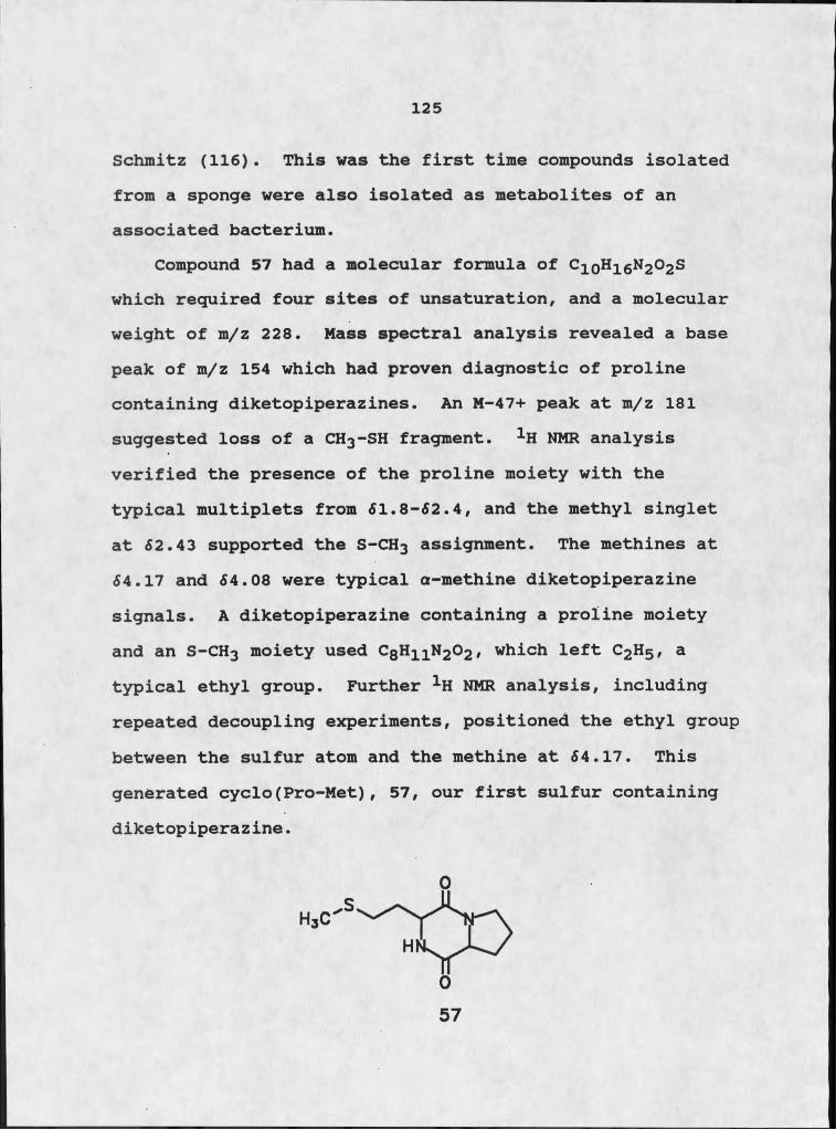
125
Schmitz (116). This was the first time compounds isolated from a sponge were also isolated as metabolites of an associated bacterium.
Compound 57 had a molecular formula of C;loh 16n 202s which required four sites of unsaturation, and a molecular weight of m/z 228. Mass spectral analysis revealed a base peak of m/z 154 which had proven diagnostic of proline containing diketopiperazines. An M-47+ peak at m/z 181 suggested loss of a CH3-SH fragment. 1H NMR analysis verified the presence of the proline moiety with the typical multiplets from 51.8-62.4, and the methyl singlet at 62.43 supported the S-CH3 assignment. The methines at 64.17 and 64.08 were typical a-methine diketopiperazine signals. A diketopiperazine containing a proline moiety and an S-CH3 moiety used C8H n N 3O2 , which left C2H5, a typical ethyl group. Further 1H NMR analysis, including repeated decoupling experiments, positioned the ethyl group between the sulfur atom and the methine at 64.17. This generated cyclo(Pro-Met), 57, our first sulfur containing diketopiperazine.
0
05 7
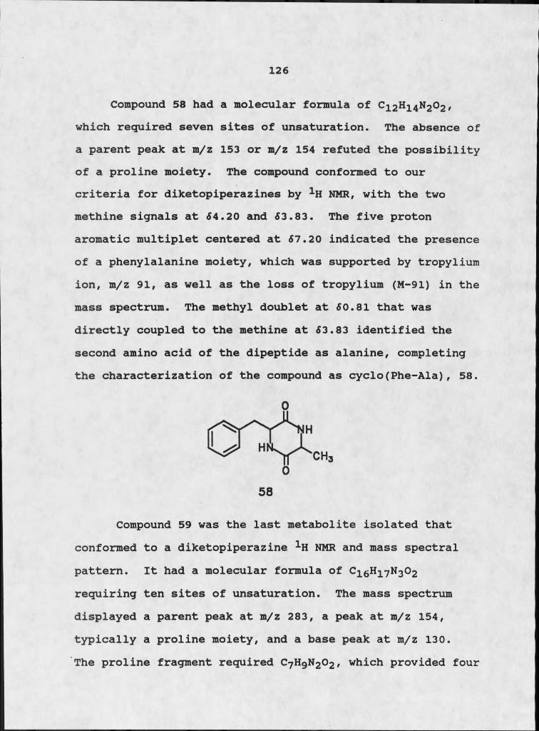
1 2 6
Compound 58 had a molecular formula of C12H14N2O2 , which required seven sites of unsaturation. The absence of a parent peak at m/z 153 or m/z 154 refuted the possibility of a proline moiety. The compound conformed to our criteria for diketopiperazines by 1H NMR, with the two methine signals at 54.20 and 53.83. The five proton aromatic multiplet centered at 57.20 indicated the presence of a phenylalanine moiety, which was supported by tropylium ion, m/z 91, as well as the loss of tropylium (M-91) in the mass spectrum. The methyl doublet at 50.81 that was directly coupled to the methine at 53.83 identified the second amino acid of the dipeptide as alanine, completing the characterization of the compound as cyclo(Phe-Ala), 58.
Compound 59 was the last metabolite isolated that conformed to a diketopiperazine 1H NMR and mass spectral pattern. It had a molecular formula of C16H 17N3O2 requiring ten sites of unsaturation. The mass spectrum displayed a parent peak at m/z 283, a peak at m/z 154, typically a proline moiety, and a base peak at m/z 130.The proline fragment required C7H9N2O2 , which provided four
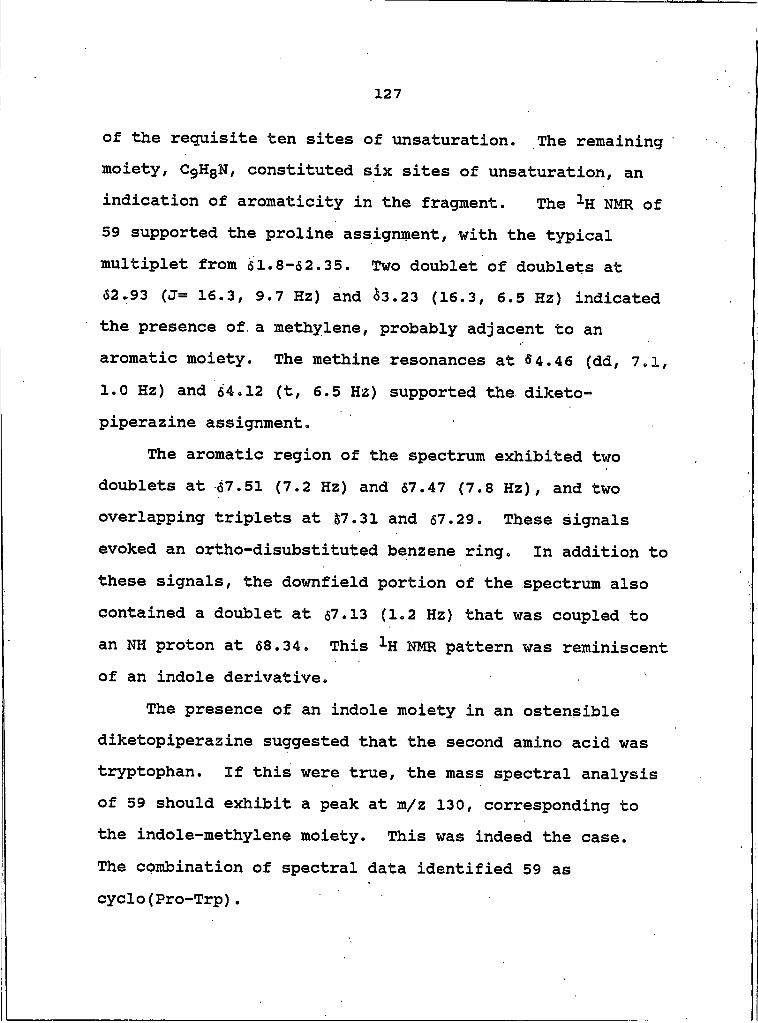
127
of the requisite ten sites of unsaturation. The remaining moiety, C9H8N, constituted six sites of unsaturation, an indication of aromaticity in the fragment. The 1H NMR of 59 supported the proline assignment, with the typical multiplet from 61.8-62.35. Two doublet of doublets at 52.93 (J= 16.3, 9.7 Hz) and 53.23 (16.3, 6.5 Hz) indicated the presence of. a methylene, probably adjacent to an aromatic moiety. The methine resonances at 54.46 (dd, 7 .1, 1.0 Hz) and 54.12 (t, 6.5 Hz) supported the diketo- piperazine assignment.
The aromatic region of the spectrum exhibited two doublets at 57.51 (7.2 Hz) and 57.47 (7.8 Hz), and two overlapping triplets at 57.31 and 57.29. These signals evoked an ortho-disubstituted benzene ring. In addition to these signals, the downfield portion of the spectrum also contained a doublet at 57.13 (1.2 Hz) that was coupled to an NH proton at 58.34. This ^H NMR pattern was reminiscent of an indole derivative. '
The presence of an indole moiety in an ostensible diketopiperazine suggested that the second amino acid was tryptophan. If this were true, the mass spectral analysis of 59 should exhibit a peak at m/z 130, corresponding to the indole-methylene moiety. This was indeed the case.The combination of spectral data identified 59 as cyclo(Pro-Trp).
1 2 8
Isolation of Tedanazine. 60One of the most active compounds isolated from this
bacterium was an aromatic compound, 60, which was toxic to brine shrimp in extremely dilute solutions. The CH2Cl2 soluble extract of the culture possessed both cytotoxic and antimicrobial activities, which were used to direct a bioassay guided fractionation scheme. The extract was chromatographed on Sephadex LH-20 (CH2Cl2-MeOH, 1:1), and was separated into ten fractions. Fractions 7, 8, and 9 displayed significant bioactivity. Fraction 7 coupled activity with some intriguing 1H NMR chemical shifts, including a putative methyl doublet at 62.37. It was permeated through Sephadex LH-20 using a more polar solvent system (MeOH), resulting in nine fractions. Cytotoxicity was concentrated in fraction 8, which appeared pure by 1H NMR. The fraction was analyzed by high resolution GC-MS. Several small impurities were present in the fraction along with the major compound. The fraction was resolved by
129
centrifugal countercurrent chromatography (CHCl3-MeOH-H2O , 25:34:20) into seven fractions. Fraction 4 exhibited strong uv absorptivity (254 nm) and potent cytotoxic activity. Spectral analysis confirmed the purity of this bright yellow compound, SO.
Host of my culturing and isolation efforts were inspired by the need for several milligrams of this compound. Unfortunately, only 0.1-0.2 mg of 60 was isolated from a 16 I culture, and the combined yield of all in-house liquid cultures totalled 0.6 mg. Of this prodigious yield, 0.2 mg was sacrificed for bioassay testing, and 0.2 mg was lost in a most unfortunate accident. Singleton's broth culture yielded 1.7 mg, which provided a grand total of 1.9 mg for structure elucidation. In an effort to consolidate any available source of 60, all fractions with strong uv absorption, characteristic retention time on a given chromatography column and a methyl doublet between 52.2 and 52.5 were combined. Resolution of this material into pure 60 followed the scheme cited above.
The extraction protocol for Singleton's liquid culture digressed from the routine followed for the smaller, in- house cultures. He centrifuged the culture and separated it into cellular solids and liquid broth containing bacterial exudates. He extracted the broth with EtOAc, reduced it to dryness and sent it to us in Bozeman. The
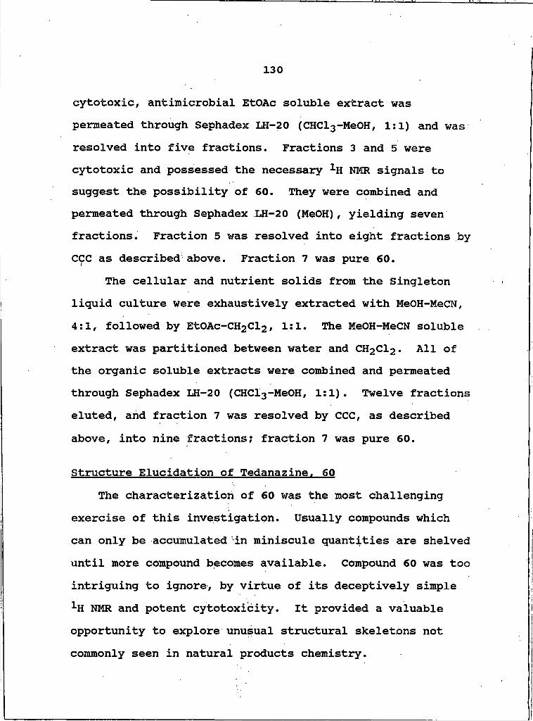
130
cytotoxic, antimicrobial EtOAc soluble extract was permeated through Sephadex LH-20 (CHCl3-MeOH, 1:1) and was resolved into five fractions. Fractions 3 and 5 were cytotoxic and possessed the necessary 1H NMR signals to suggest the possibility of 60. They were combined and permeated through Sephadex LH-20 (MeOH), yielding seven fractions. Fraction 5 was resolved into eight fractions by CCC as described above. Fraction 7 was pure 60.
The cellular and nutrient solids from the Singleton liquid culture were exhaustively extracted with MeOH-MeCN, 4:1, followed by EtOAc-CHgCl3 , 1:1. The MeOH-MeCN soluble extract was partitioned between water and CH2Cl2 . All of the organic soluble extracts were combined and permeated through Sephadex LH-20 (CHCl3-MeOH, 1:1). Twelve fractions eluted, and fraction 7 was resolved by CCC, as described above, into nine fractions; fraction 7 was pure 60.
Structure Elucidation of Tedanazine. 60The characterization of 60 was the most challenging
exercise of this investigation. Usually compounds which can only be accumulated in miniscule quantities are shelved until more compound becomes available. Compound 60 was too intriguing to ignore, by virtue of its deceptively simple 1H NMR and potent cytotoxicity. It provided a valuable opportunity to explore unusual structural skeletons not commonly seen in natural products chemistry.
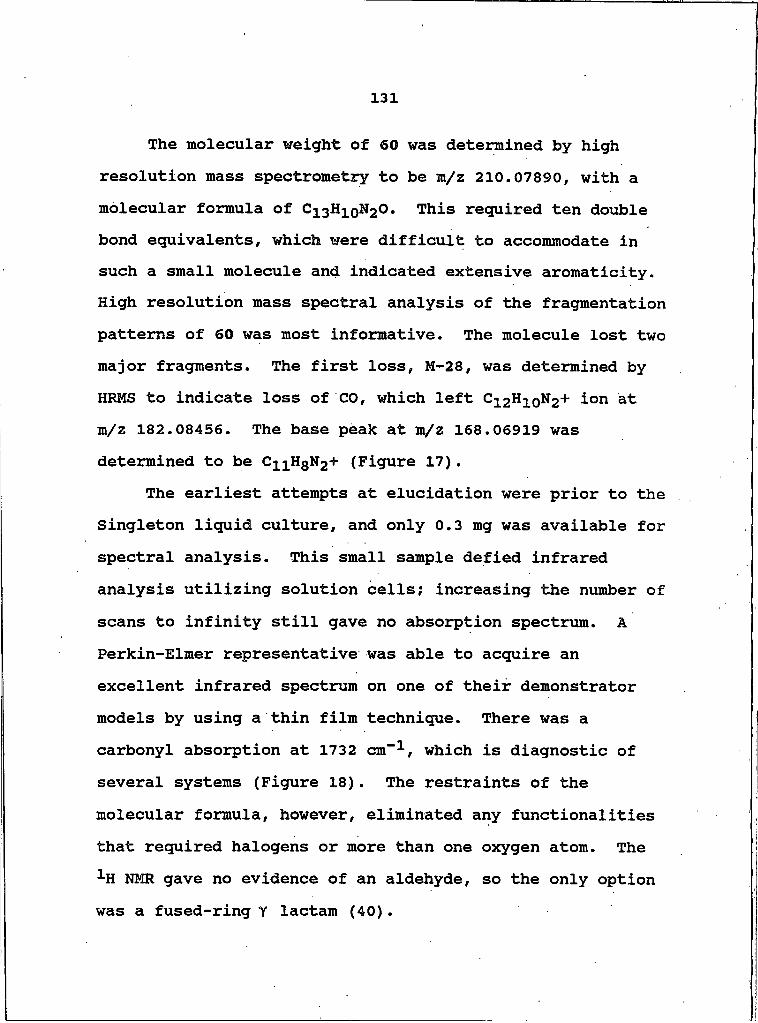
131
The molecular weight of 60 was determined by high resolution mass spectrometry to be m/z 210.07890, with a molecular formula of C13Hi0N2O. This required ten double bond equivalents, which were difficult to accommodate in such a small molecule and indicated extensive aromaticity. High resolution mass spectral analysis of the fragmentation patterns of 60 was most informative. The molecule lost two major fragments. The first loss, M-28, was determined by HRMS to indicate loss of CO, which left C12H 10N2+ ion at m/z 182.08456. The base peak at m/z 168.06919 was determined to be C11HgN2+ (Figure 17).
The earliest attempts at elucidation were prior to the Singleton liquid culture, and only 0.3 mg was available for spectral analysis. This small sample defied infrared analysis utilizing solution cells; increasing the number of scans to infinity still gave no absorption spectrum. A Perkin-Elmer representative was able to acquire an excellent infrared spectrum on one of their demonstrator models by using a thin film technique. There was a carbonyl absorption at 1732 cm"1, which is diagnostic of several systems (Figure 18). The restraints of the molecular formula, however, eliminated any functionalities that required halogens or more than one oxygen atom. The 1H NMR gave no evidence of an aldehyde, so the only option was a fused-ring Y lactam (40).
Figvue 17. Mass Spectrum of 60
132
87.00-Energy
78.002500 2000300035004000
Figure 18. Infrared Spectrum of 60
133
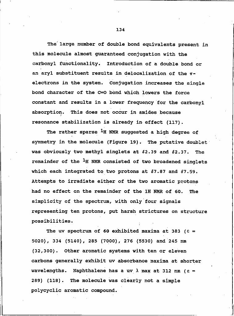
134
The' large number of double bond equivalents present in this molecule almost guaranteed conjugation with the carbonyl functionality. Introduction of a double bond or an aryl substituent results in delocalization of the ir- electrons in the system. Conjugation increases the single bond character of the C=O bond which lowers the force constant and results in a lower frequency for the carbonyl absorption. This does not occur in amides because resonance stabilization is already in effect (117).
The rather sparse 1H NMR suggested a high degree of symmetry in the molecule (Figure 19). The putative doublet was obviously two methyl singlets at 62.39 and 62.37. The remainder of the 1H NMR consisted of two broadened singlets which each integrated to two protons at 67.87 and 67.59. Attempts to irradiate either of the two aromatic protons had no effect on the remainder of the IH NMR of 60. The simplicity of the spectrum, with only four signals representing ten protons, put harsh strictures on structure possibilities.
The uv spectrum of 60 exhibited maxima at 383 (e = 5020), 334 (5140), 285 (7000), 276 (5530) and 245 nm (32,300). Other aromatic systems with ten or eleven carbons generally exhibit uv absorbance maxima at shorter wavelengths. Naphthalene has a uv A max at 312 nm (e =289) (118). The molecule was clearly not a simple polycyclic aromatic compound.
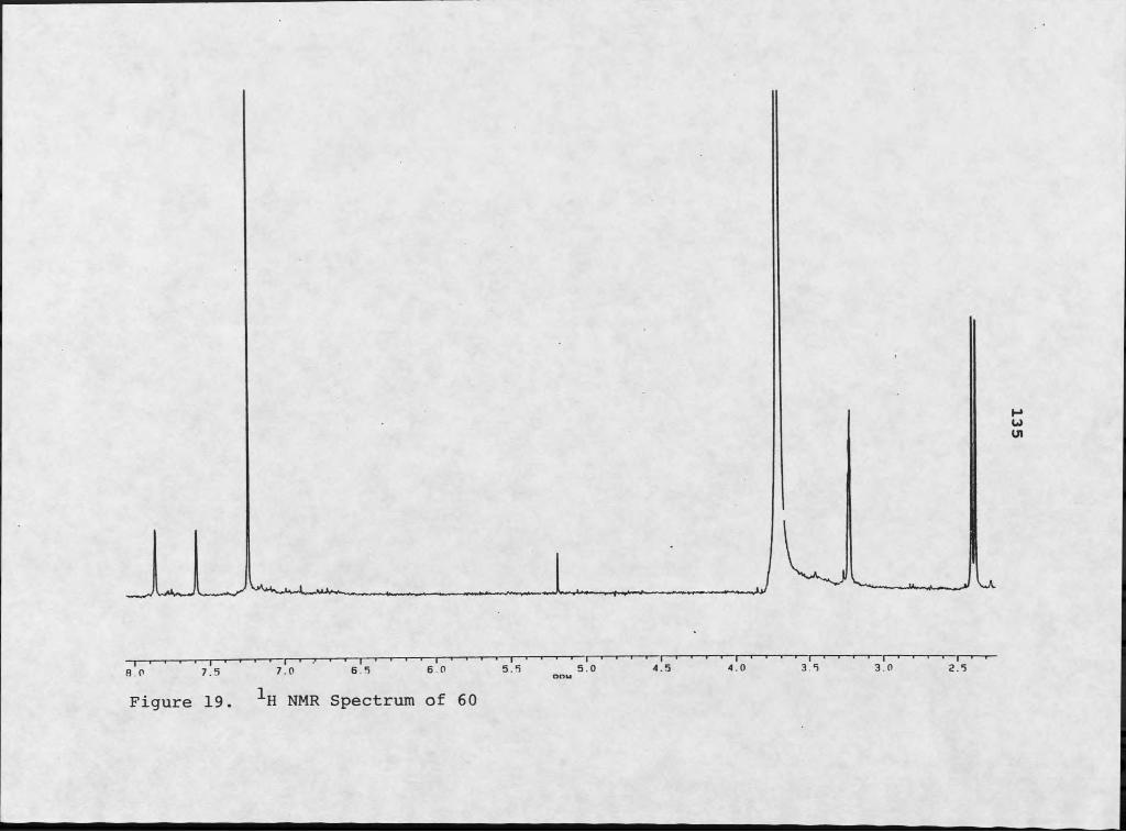
I 1 1 1 ' IB O 7.5
Figure 19
— I--- 1— i--- 1--- 1--- 1--- 1--- 1--- 1--- 1--- 1 ' 1 ' 1 I 1 1 1 1 I 1 1 1 1 I 1 ' 1 1 I 1 1 1 1 I 1 1 1 1 I ' ' 1 1 I 17,0 6 5 6.0 5.5 5.0 4.5 4.0 3.5 3.0 2.5
1H NMR Spectrum of 60
135
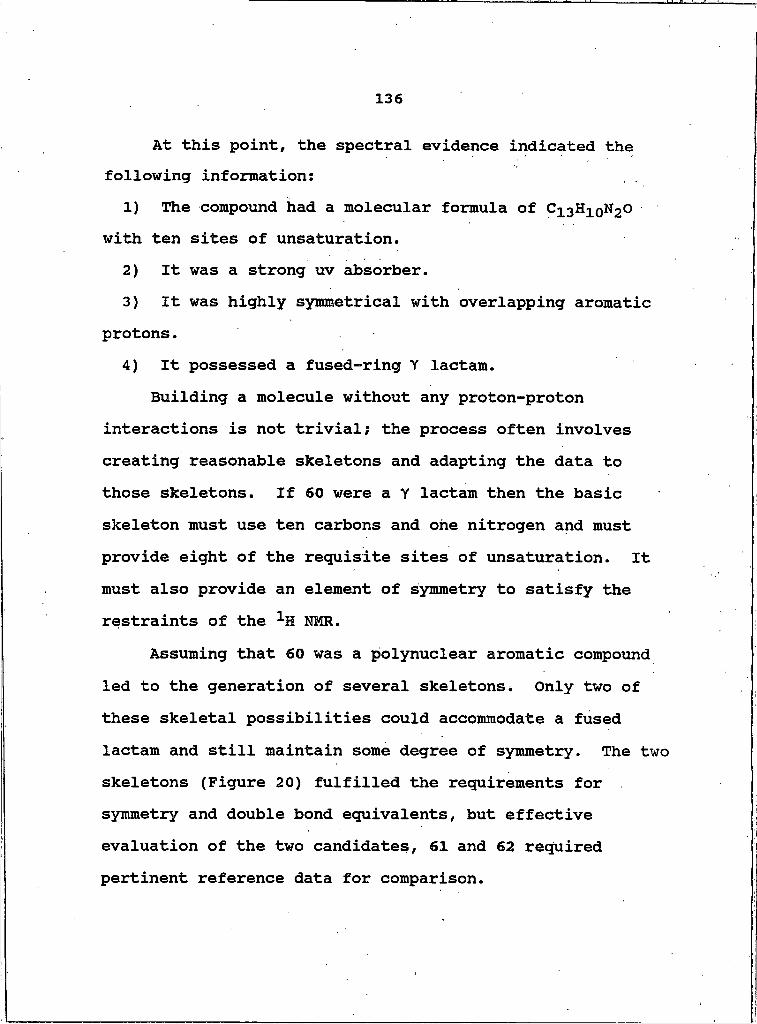
136
At this point, the spectral evidence indicated the following information:
1) The compound had a molecular formula of C13H10N2O with ten sites of unsaturation.
2) It was a strong uv absorber.3) It was highly symmetrical with overlapping aromatic
protons.4) It possessed a fused-ring Y lactam.
Building a molecule without any proton-protoninteractions is not trivial; the process often involves creating reasonable skeletons and adapting the data to those skeletons. If 60 were a Y lactam then the basic skeleton must use ten carbons and one nitrogen and must provide eight of the requisite sites of unsaturation. It must also provide an element of symmetry to satisfy the restraints of the 1H NMR.
Assuming that 60 was a polynuclear aromatic compound led to the generation of several skeletons ̂ Only two of these skeletal possibilities could accommodate a fused lactam and still maintain some degree of symmetry. The two skeletons (Figure 20) fulfilled the requirements for symmetry and double bond equivalents, but effective evaluation of the two candidates, 61 and 62 required pertinent reference data for comparison.
137
Figure 20. Two potential skeletons for 60. a) [2,2,3] cyclazine, 61; b) pyrrolo[3,2,1-hi]indole, 62.
An extensive literature search indicated that neither of these two skeletons were incorporated into any known metabolite from living organisms. This might have been discouraging if not for an experiment which was performed on the impure fraction from which 60 was separated. The high resolution GC-MS analysis of the various impurities which were too dilute to be obvious by 1H NMR included the following compounds: m/z 178.07881, C14H 10* with ten double bond equivalents; m/z 192.09308, C15H 12, with ten double bond equivalents; and m/z 206.11118, C16H14, with ten double bond equivalents. These hardly qualified as unequivocal elucidations, but they did suggest some interesting structures. All three of the molecules fit well into the polycyclic aromatic scheme. Comparison with mass spectral data of authentic samples suggested that the

138
three compounds were anthracene, and isomers of methyl and dimethyl anthracene (119). This analysis was important because it attested to the ability of our marine Micrococcus to produce polyaromatic compounds not usually associated with microorganisms. Anthracene and naphthalene are usually found as constituents of coal tar, not living organisms (120). Finding anthracene or its isomer in the organic extract does not verify either skeletal possibility, but does substantiate the ability of this microbe to produce polycyclic aromatic compounds.
A fourth compound which was identified by this analysis was Sg. It had no 1H NMR signals and a 'molecular weight of 256, with peaks representing repeated loss of sulfur (M- 32+, M-64+, etc.). The significance of this finding was establishing the bacterium's ability to sequester sulfur from its environment, an ability which may have important ramifications for the host macroorganism.
Both of the proposed skeletal types have been the foci of synthetic ventures. In the late fifties, Windgassen et al. were exploring various aromatic systems. They were particularly interested in creating large, conjugatedcarbocycles that were held planar by bonding to an internal nitrogen and determining whether or not such systems were truly aromatic (121). They synthesized several of these compounds, which they called cyclazines, from a series of 2-substituted-5-methylpyrrocolines.
139
Cursory inspection of this class of compounds suggested aromaticity because of the ten ^-electrons in the periphery of the molecule. As early as 1931, Hiickel recognized that 4n + 2 ^-electron systems imparted stability to completely conjugated monocyclic systems (122). Experimental evidence usually justifies the extension of Hiickel1 s rule to polynuclear aromatics such as naphthalene, anthracene and phenanthrene. Windgassen et al. were interested in determining if Hiickel1 s rule applied to the cyclazines. Utilizing the principles of molecular orbital theory, they approximated the resonance energy, or delocalization energy, of cyclazine compounds. For [2,2,3] cyclazines they calculated a resonance energy of -2.9440, which is consistent with values for polynuclear aromatic compounds (121).
Windgassen et al. reported the ultraviolet and infrared spectra of the [2,2,3]cyclazines. More recent synthetic attempts have also provided 1H NMR analyses of several compounds of this class (123, 124). The spectral data reported in these studies provided a means of evaluating the possibility of a [2,2,3]cyclazine skeleton for 60.
The 1H NMR spectrum of the parent [2,2,3]cyclazine molecule, 61, exhibits the symmetry and diamagnetic ring current effects expected of an aromatic system (Figure 21) (124). Comparison of the 1H NMR spectrum of 60 with 61

140
suggested the feasibility of this system as the basis of our compound.
7.50
61Figure 21. 1H NMR assignments for 61.
If one of the methyl groups were assigned to position 6, the 1H NMR of the "southern" portion of the molecule would be reduced to a single, two proton resonance. The chemical shifts of H-5 and H-7 (67.87) were consistent with those of the model compound. A typical chemical shift for a para-methyl substituent in pyridine is 62.37, which coincided with the chemical shift of one of the methyl groups in our compound (125).
The chemical shifts of the "northern" portion of the molecule varied from those of the model, which should be
expected with the additional amide functionality of 60. As a rule, the carbonyl and nitrogen portions of an amide do not exert an equivalent influence on the protons in an aromatic system. Protons adjacent to the carbonyl moiety are generally shifted downfield by as much as 0.5 ppm.

141
while protons adjacent to the amine portion are generally only shifted 0.30-0.35 ppm downfield (126). These differences are mitigated if the carbonyl moiety is not coplanar with the aromatic system. This is particularly important in systems in which the amido group is locked in a non-coplanar conformation. In such a system, both portions of the amido group would exert similar effects (+0.3-0.35 ppm) on the chemical shift of adjacent aromatic protons (126). This would shift H-I and H-4 from 57.19 in the parent system to 57.49-7.54, which compared favorably to a chemical shift of 57.57 for compound 60.
Empirical evidence is necessary to determine the conformation and planarity of a molecule. Windgassen et al. assumed that the carbocycle was held planar by the nitrogen atom bridging the system (121). They did not offer any evidence to corroborate this supposition. Using a program called Alchemy, we applied molecular mechanics (MM2) type calculations to the [2,2,3]cyclazine molecule to
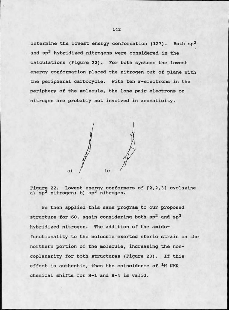
142
determine the lowest energy conformation (127). Both sp2 and sp3 hybridized nitrogens were considered in the calculations (Figure 22). For both systems the lowest energy conformation placed the nitrogen out of plane with the peripheral carbocycle. With ten f-electrons in the periphery of the molecule, the lone pair electrons on nitrogen are probably not involved in aromaticity.
Figure 22. Lowest energy conformers of [2,2,3] cyclazine a) sp2 nitrogen; b) sp3 nitrogen.
We then applied this same program to our proposed structure for 60, again considering both sp2 and sp3 hybridized nitrogen. The addition of the amido- functionality to the molecule exerted steric strain on the northern portion of the molecule, increasing the noncoplanarity for both structures (Figure 23). If this effect is authentic, then the coincidence of 1H NMR chemical shifts for H-I and H-4 is valid.

143
Figure 23. Lowest energy conformers of 60: a) sp2 nitrogen; b) sp3 nitrogen.
Additional spectral evidence provided further support for the [2,2,3] cyclazine skeleton. Windgassen et al. reported uv % max of the parent compound in ethanol at 419 ( E =4460), 408 (4570), 398 (3630) , 289 (7240), 274 (5490) and 244 nm (37,200) (121). The higher energy absorptionscompared favorably with the ultraviolet spectrum of 60. Discrepancies in the longer wavelength absorptions were apparent. Coplanarity is an important factor in ultraviolet absorption spectroscopy; steric factors that force a molecule or portions of a molecule out of plane with the rest of the structure can significantly decrease the maximum absorption wavelengths and their intensities. For example, the destruction of coplanarity by steric interference in cis-stilbene is reflected by the shorter wavelength and lower intensity of the K-band (tt-tt* transition) relative to that of trans-stilbene (283 nm e =
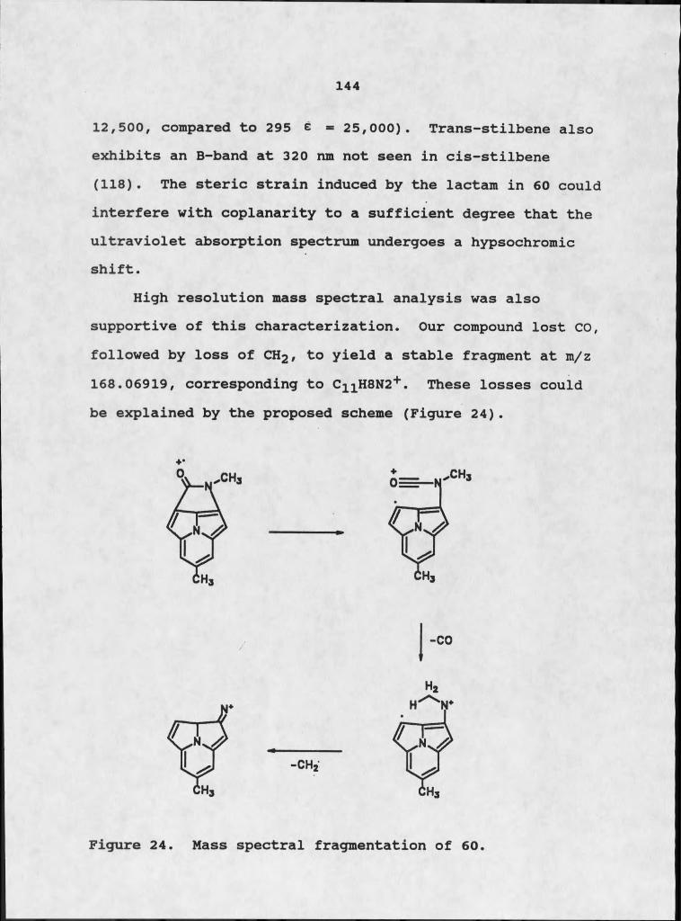
144
12,500, compared to 295 e = 25,000). Trans-stilbene also exhibits an B-band at 320 nm not seen in cis-stilbene (118). The steric strain induced by the lactam in 60 could interfere with coplanarity to a sufficient degree that the ultraviolet absorption spectrum undergoes a hypsochromic shift.
High resolution mass spectral analysis was also supportive of this characterization. Our compound lost CO, followed by loss of CH2 , to yield a stable fragment at m/z 168.06919, corresponding to C11H8N2+ . These losses could be explained by the proposed scheme (Figure 24).
+"
Figure 24. Mass spectral fragmentation of 60.

145
The second skeletal option for 60 was also a tricyclic heteroaromatic compound, pyrrolo[3,2,l-hi]indole, 62. Paudler and Shin synthesized this compound from N-amino- indoline and ethyl pyruvate (128). They examined the spectral properties of the molecule, which permitted comparison with those of 60. 1H NMR analysis emphasized the symmetry of 62, but careful consideration of the chemical shifts suggested that this was not the appropriate skeleton (Figure 25).
7.35
62Figure 25. 1H NMR assignments for 62.
In the southern portion of the molecule, methylation at position 7 would again reduce the aromatic doublet at 57.53 to a two proton singlet. The slight shielding effect of ortho-methylation would shift the absorbance to 57.45- 57.50. The comparable chemical shift for 60 was 57.57. The northern portion of the molecule was not as amenable to comparison. The chemical shift of H-I and H-5 was 56.60.It was improbable that a lactam ring could shift the
7.12
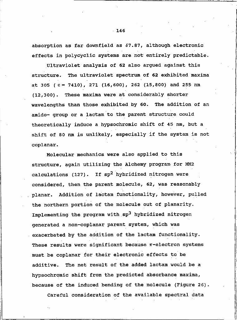
146
absorption as far downfield as 57.87, although electronic effects in polycyclic systems are not entirely predictable.
Ultraviolet analysis of 62 also argued against this structure. The ultraviolet spectrum of 62 exhibited maxima at 305 ( E = 7410), 271 (16,600), 262 (15,800) and 255 ran (12,300). These maxima were at considerably shorter wavelengths than those exhibited by 60. The addition of an amido- group or a lactam to the parent structure could theoretically induce a hypsochromic shift of 45 nm, but a shift of 80 nm is unlikely, especially if the system is not coplanar.
Molecular mechanics were also applied to this structure, again utilizing the Alchemy program for MM2 calculations (127). If sp2 hybridized nitrogen were considered, then the parent molecule, 62, was reasonably planar. Addition of lactam functionality, however, pulled the northern portion of the molecule out of planarity. Implementing the program with sp3 hybridized nitrogen generated a non-coplanar parent system, which was exacerbated by the addition of the lactam functionality. These results were significant because ^-electron systems must be coplanar for their electronic effects to be additive. The net result of the added lactam would be a hypsochromic shift from the predicted absorbance maxima, because of the induced bending of the molecule (Figure 26).
Careful consideration of the available spectral data

147
suggested that 61 was the correct skeleton, and 60 the correct structure. Compound 60 was designated tedanazine, based on the parent cyclazine. At this point, however, the elucidation is not unequivocal. The ultimate goal would be synthesis of the proposed structure and comparison of spectral data.
Figure 26. Lowest energy conformers for lactam derivative of pyrrolo[3,2,1-hi] indole, 62: a) sp2 nitrogen; b) sp3nitrogen.
Isolation of Benzothiazoles and IndolesA series of nitrogen and sulfur-containing aromatic
compounds was isolated from a 220 I marine broth culture of Micrococcus sp. The culture was centrifuged and separated into solid cellular material and the supernatant. The solids and the supernatant were extracted as described
above.The organic extract of the solid cellular material was
chromatographed on Sephadex LH-20 (CHgClg-MeOH, 1:1), which
148
yielded ten fractions. Several of these fractions were bioactive, including fraction 5, which displayed both antimicrobial activity and cytotoxicity. It was permeated through Bio-Beads S-X4 Chexane-CH2Cl2-EtOAc, 4:3:1). Bioactivity was concentrated in fractions 3, 4, and 5. The three fractions, which exhibited striking spectral similarities, were combined and further size exclusion chromatography on Sephadex LH-20 (MeOH) yielded seven fractions. Fractions 5, 6, and 7, which were bioactive, were recombined and were poorly resolved by centrifugal countercurrent chromatography (CHCl3-MeOH-H20, 25:34:20) into nine fractions. Fractions 5, 6, 7, and 8 were chromatographed on Bio-Beads S-X8 (CHCl3-MeCN, 1:1), which resolved the mixture into eleven fractions.
Both fractions 6 and 8 were further resolved into heteroaromatic compounds. Fraction 6 was finally resolved by CCC (Iiexane-CH2Cl2-MeCN, 50:15:35) into six fractions which included the benzothiazoles, 63 (0.4 mg) and 64 (1.8 mg), and the indole derivative, 70 (1.1 mg).
Fraction 8 was chromatographed on Sephadex LH-20 (CHCl3-MeOH, 1:1), yielding five fractions. The third fraction was resolved by permeation through Bio-Beads S-X8 (CH2Cl2-MeCN, 1:1) into six fractions; fraction 4 was a benzothiazole, 65 (Q.5 mg).
The EtOAc soluble extract of the supernatant was chromatographed on Sephadex LH-20 (CHCl3-MeOH, 1:1),
149
yielding five fractions. All of the fractions were cytotoxic, but fractions 3 and 5 coupled activity with intriguing spectral data. They were combined and permeated through Sephadex LH-20 (HeOH). Eight fractions eluted and the sixth fraction was resolved by CCC (CHCl3-MeOH-^O, 25:34:20) into seven fractions. Fraction 3 was a sulfur- containing indole derivative, 71 (0.2 mg) and fraction 6 was a benzothiazolone, 67.
Structure Elucidation of BenzothiazolesThree compounds, 63-65, were sulfur-containing
aromatics with similar spectral characteristics. Compound 63 had a molecular weight of m/z 167 with a relatively large M+2+ (11.27%) peak, suggesting the presence of two sulfur atoms. This fit a molecular formula of C7HgNS2 , which required six sites of unsaturation. Mass spectral analysis showed an M-27+ peak at m/z 140, an M-32+ peak at m/z 135, and an M-44+ fragment at m/z 123 (Figure 27).
The 1H NHR was almost exclusively aromatic. In CDCl3/ MeOH-d^ the 1H NMR consisted of two doublets at 67.44 and 67.32, which were mutually coupled by 8.3 Hz. The remainder of the aromatic signals were buried under the chloroform resonance at 67.24. The spectrum was then run in CDCl3/acetone-dg, which shifted the signals upfield and away from the chloroform peak. The two doublets shifted to 67.15 and 67.00, and the formerly buried multiplet was

150
cleanly resolved to two triplets at 56.98 and 66.88 (J= 7.3 Hz). A broad signal at 510.1 was typical of a proton on a heteroatom (Figure 28).
The infrared analysis of the molecule was not obviously diagnostic of any particular class of compounds. It did help to eliminate certain functional groups, which is often of equal importance. Retrospective analysis of the infrared spectrum of 63 supported the eventual characterization of the molecule.
Accommodating six double bond equivalents in a molecule with only seven carbons and no carbonyl or nitrile potential required a bicyclic heteroaromatic skeleton. The presence of four mutually coupled aromatic protons indicated an ortho-disubstituted benzene system. The magnitude of the ortho coupling constants precluded a monsubstituted five-membered ring aromatic compound. The position of the chemical shifts also argued against a pyridine moiety;.the protons on pyridine usually resonate further downfield, from 57.06 - 58.5. The facile loss of sulfur in the mass spectral analysis suggested that one of the sulfur atoms in the molecule was not incorporated into the aromatic skeleton, but was a substituent on one of the rings. These data generated a 5,6-bicyolic heteroaromatic compound with sulfur and nitrogen atoms incorporated into the five-membered ring. This described the benzothiazole class of compounds. The M-32+ at m/z 135 and M-44+ peak at
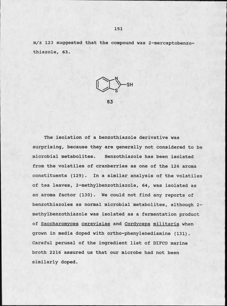
151
m/z 123 suggested that the compound was 2-mercaptobenzo- thiazole, 63.
The isolation of a benzothiazole derivative was surprising, because they are generally not considered to be microbial metabolites. Benzothiazole has been isolated from the volatiles of cranberries as one of the 126 aroma constituents (129). In a similar analysis of the volatiles of tea leaves, 2-methylbenzothiazole, 64, was isolated as an aroma factor (130). We could not find any reports of benzothiazoles as normal microbial metabolites, although 2- methylbenzothiazole was isolated as a fermentation product of Saccharomvces cerevisiae and Cordvceos militaris when grown in media doped with ortho-phenylenediamine (131). Careful perusal of the ingredient list of DIFCO marine broth 2216 assured us that our microbe had not been similarly doped.
63
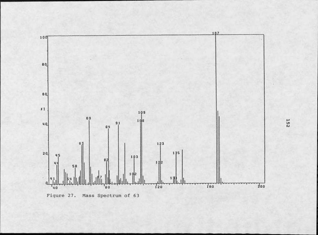
Figure 27. Mass Spectrum of 63
152
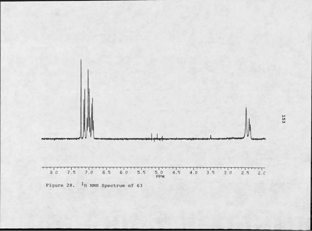
T I f— [ I I I i I I I i I I I r n i j i i i i pn i i i i I I j I i i I pn I I i j i i rn j i i i i j m f t — j— i— i— i— i j—3.0 7.5 7.0 6.5 6.0 5.5 5.0 4.5 4.0 3.5 3.0 2.5 2 0
PPM
. NMR Spectrum of 63Figure 28
153
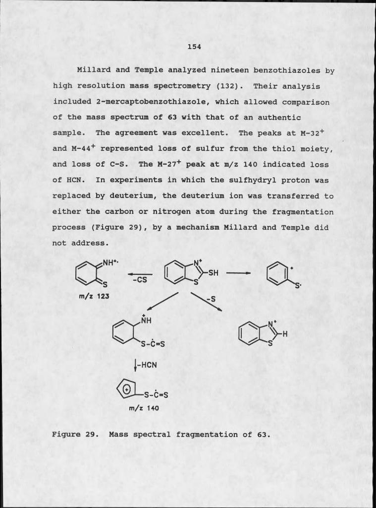
154
Millard and Temple analyzed nineteen benzothiazoles by high resolution mass spectrometry (132). Their analysis included 2-mercaptobenzothiazole, which allowed comparison of the mass spectrum of 63 with that of an authentic sample. The agreement was excellent. The peaks at M-32+ and M-44+ represented loss of sulfur from the thiol moiety, and loss of C-S. The M-27+ peak at m/z 140 indicated loss of HCN. In experiments in which the sulfhydryl proton was replaced by deuterium, the deuterium ion was transferred to either the carbon or nitrogen atom during the fragmentation process (Figure 29), by a mechanism Millard and Temple did not address.
m/z 123
j-HCN
m/z 140
Figure 29. Mass spectral fragmentation of 63.

155
The isolation of 63 from the organic soluble extract of Micrococcus sp. and subsequent characterization of the molecule as 2-mercaptobenzothiazole aided in the characterization of two other compounds isolated from the bacterial culture. Compound 64 had a molecular weight of m/z 149 and a molecular formula of C8H7NS, which required six sites of unsaturation. Mass spectral analysis exhibited an M-41+ peak at m/z 108 (Figure 30). The 1H NMR spectral pattern of two downfield, mutually coupled doublets and two higher field triplets was reminiscent of 63 (Figure 31). In CDCl3/ acetone-d6, the two doublets resonated at 57.92 and 57.80 (J= 7.8 Hz); the two triplets were at 57.43 and 57.32 (7.2 Hz). A methyl singlet at 52.80 suggested that 64 was a methyl-substituted benzothiazole. Katritzky and Takeuchi determined the 1H NMR spectra of a series of 2-methylbenzothiazoles; the 1H NMR spectrum of their synthetic 2-methylbenzothiazole coincided with that of compound 64 (133).
64

Mass Spectrum of 64
156

T I I I • 1 I 1 r
0*5 0 0
Figure 31.
' I “i I— I » I 1 1 * * I ' 1 * * I 1 1 ' * I7.5 7.0 6.5 6.0 5.5
-*-H NMR Spectrum of 64
1 I ■ ■ ■ ■ ' 5.0 4.5PPM 4.0 3.5I r
3.0 2.5
157
158
Comparison of the 1H NMR spectra of 63 and 64 showed a downfield shift for all of the aromatic protons of 64.This was suprising because in typical aromatic systems a methyl group exerts a greater shielding effect than a sulfhydryl group (134). A reasonable explanation for this unexpected shielding is tautomerism. In the keto- form, electron donation by the nitrogen would be possible, which would exert a shielding effect on the aromatic ring.Studies by Mecke et al. determined that in many thiocarbdnyl compounds, the C=S tautomer predominates over the C=O form. In thioureas the C=S tautomer is often exclusive (135). Tautomerism could explain an interesting fragmentation pattern in mass spectral analysis: the loss of C-S, with retention of the sulfhydryl proton by the molecule (Figure 29). Millard and Temple examined this effect using 2-SD-benzothiazole. Loss of C-S was always accompanied by the transfer of the deuterium ion from the sulfur atom to the rest of the molecule. Tautomerism provides a reasonable mechanism for this phenomenon (132).
Compound 65 had a molecular weight of m/z 151, and a molecular formula of C7H5NOS, requiring six sites of unsaturation. The 1H NMR (CDCl3) exhibited two doublets at SI.38 and SI.24 (hidden under chloroform), which were coupled by 8.5 Hz and two triplets at SI.12 and SI.08 (J= 7.5 Hz) (Figure 32). In CDCl3/acetone-dg, the proton resonances were shifted upfield.
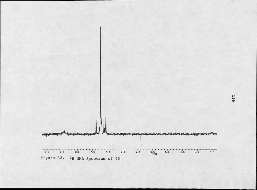
jJL9 0 8 5 8 0 7 5 7.0 6.5 6.0
Figure 32. Ijj NMR Spectrum of 655.5PPM
5.0 4.5 4.0 3 5
159

Il !■ aLL̂ I i4_IlL
Mass Spectrum of 65
160
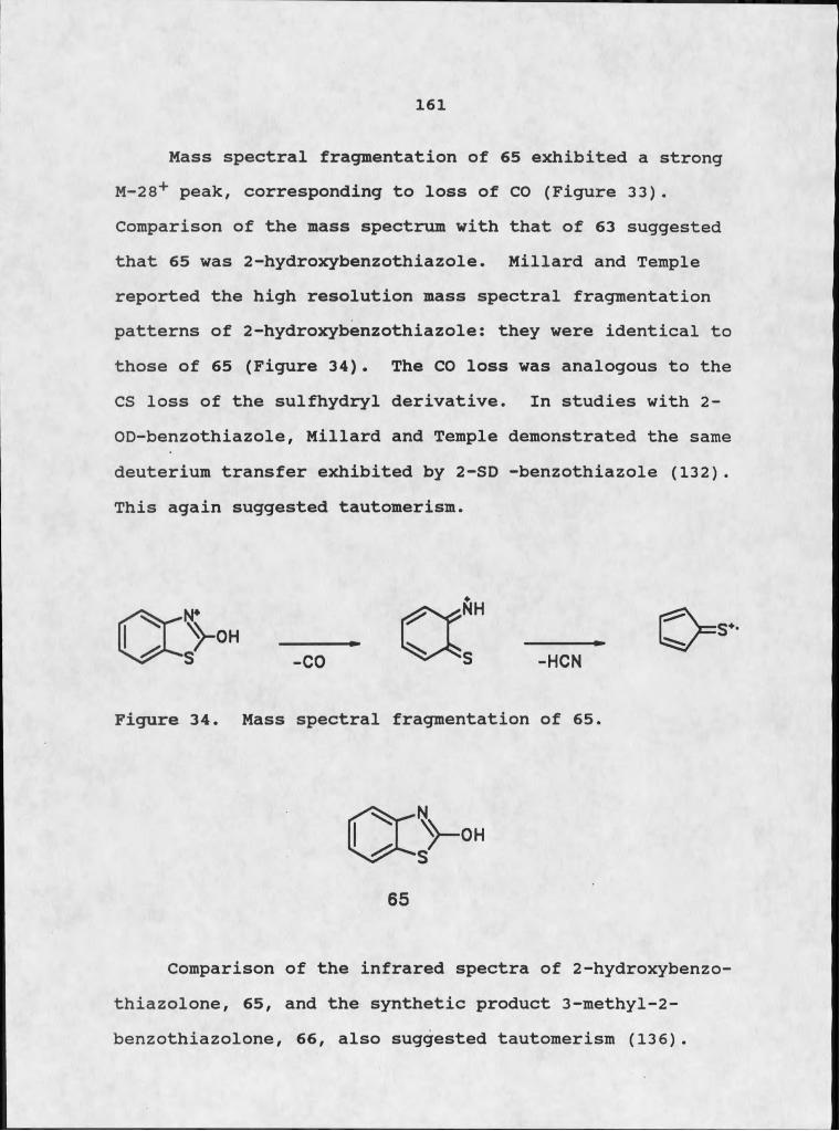
1 6 1
Mass spectral fragmentation of 65 exhibited a strong M-28+ peak, corresponding to loss of CO (Figure 33). Comparison of the mass spectrum with that of 63 suggested that 65 was 2-hydroxybenzothiazole. Millard and Temple reported the high resolution mass spectral fragmentation patterns of 2-hydroxybenzothiazole: they were identical to those of 65 (Figure 34). The CO loss was analogous to the CS loss of the sulfhydryl derivative. In studies with 2- OD-benzothiazole, Millard and Temple demonstrated the same deuterium transfer exhibited by 2-SD -benzothiazole (13 2) . This again suggested tautomerism.
Figure 34. Mass spectral fragmentation of 65.
65
Comparison of the infrared spectra of 2-hydroxybenzo-thiazolone, 65, and the synthetic product 3-methyl-2- benzothiazolone, 66, also suggested tautomerism (136).
162
Compound 65 exhibited -OH stretch at 3410 cm"1, which was not apparent in the spectrum of 66. Both of the compounds had two absorptions at 1700 and 1667 cm"1, but the ratios of their intensities varied. The 1700 cm"1 absorption was a function of the C=O stretch of the keto- form, which predominated to the point of exclusivity in 66. This frequency is typical of fused Y lactams. The absorption at 1667 cm"1, which in these systems indicated C=N stretch of the benzothiazole, was the dominant absorption in the alcohol.
The final member of the benzothiazole series was isolated from the EtOAc soluble extract of the culture supernatant. Compound 67 had a molecular weight of m/z 181.02381, which required a molecular formula of C8H7NO2S and six sites of unsaturation. The M-28+ at 152.01605 was determined to be loss of CO by high resolution mass spectrometry (Figure 35).
The 1H NMR spectrum of 67 exhibited resonances at 66.53 (1H, d, J= 8.6 Hz), 56.83 (1H, d, 2.6 Hz), 56.36 (1H, dd, 8 .6, 2.6 Hz), and 53.10 (3H, s) (Figure 36). The aromatic protons described a I,2,4-trisubstituted benzene ring, which suggested a 5- or 6- substituted benzothiazole derivative..
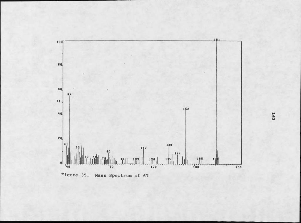
Figure 35. Mass Spectrum of 67
163
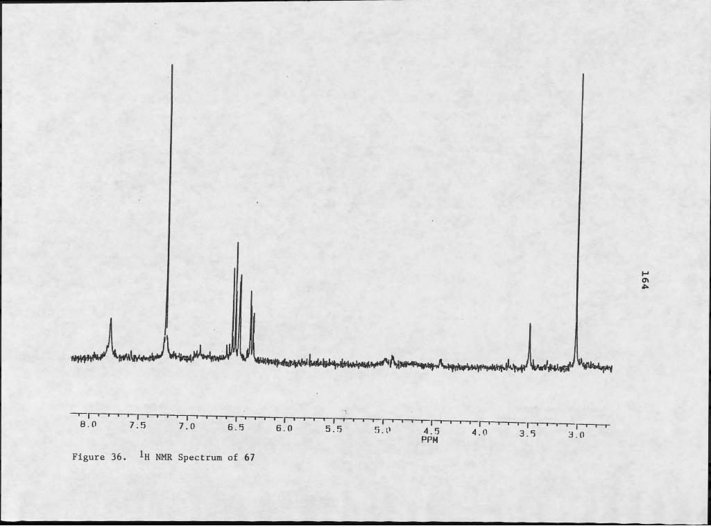
-̂ I ' I '— I— I— I— I— I— I— I— r8.0 7.5 7.0 I6.5 1 ' I 1 1 1 1 I '— I— I— I— I— I— I— I— I— I—
6.0 5.5 5.0 4.5PPM
I ' 1 r I— I—4.0 3.5 ' I 1 1 r3.0
Figure 36. NMR Spectrum of 67
164
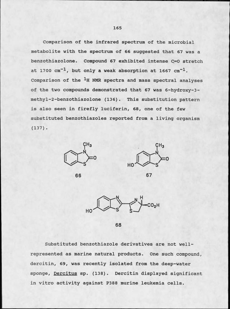
165
Comparison of the infrared spectrum of the microbial metabolite with the spectrum of 66 suggested that 67 was a benzothiazolone. Compound 67 exhibited intense C=O stretch at 1700 cm-1, but only a weak absorption at 1667 cm-1. Comparison of the 1H NMR spectra and mass spectral analyses of the two compounds demonstrated that 67 was 6-hydroxy-3- methyl-2-benzothiazolone (136). This substitution pattern is also seen in firefly luciferin, 68, one of the few substituted benzothiazoles reported from a living organism (137) .
Substituted benzothiazole derivatives are not well- represented as marine natural products. One such compound, dercitin, 69, was recently isolated from the deep-water sponge, Dercitus sp. (138). Dercitin displayed significant in vitro activity against P388 murine leukemia cells.
1 6 6
Benzothiazole derivatives are not common metabolites of any known macroorganism, except the firefly (137). The discovery of a series of simple benzothiazoles from a sponge symbiont and the discovery of a sponge producing complex benzothiazole derivatives, is certainly provocative. Dercitus would be an excellent candidate for culture work.
Structure Elucidation of Indole DerivativesTwo of the compounds isolated from the culture of
Micrococcus sp. exhibited the mass spectral fragmentation pattern indicative of indole derivatives. Compound 70 was isolated from the culture solids of the bacterium, and 71 was isolated from the supernatant.
Compound 70 had a molecular weight of m/z 175 and a molecular formula of CiQHgNOg, which required seven sites of unsaturation. The mass spectrum of 70 exhibited an M-
h3c"n'ch369

167
3I+ peak at m/z 144, which corresponded to a loss of OCH3 .A peak at m/z 116 indicated a loss of CO from this fragment. The fragments at m/z 116 and m/z 89 were diagnostic of an indole derivative, representing the parent structure and subsequent loss of HCN (139) (Figure 37).The indole moiety required C8H6N, accommodating six of the seven sites of unsaturation. The remaining portion of the molecule, CgH3O3 , must provide one double bond equivalent.
Infrared analysis of 70 suggested that the additional site of unsaturation was a carbonyl moiety. A strong absorption at 1650 cm-1 can indicate several functional groups. The constraints of the molecular formula, however, eliminated most of the options because they required more heteroatoms than the C2H3O2 fragment could provide. Two valid options were a hydrogen-bonded a,/? unsaturated ketone, or a conjugated aldehyde. Lack of the appropriate proton absorption in the ^H NMR argued against an aldehyde
functionality (41).1H NMR spectral analysis supported the indole
assignment, although the multiplicities were complicated by apparent second order coupling (Figure 38). In acetone- d6/MeOH-d4 the signals resonated at 68.29 (1H, s), 58.26 (IH, m ) , 57.50 (IH, m ) , 57.27 (2H, m) and 64.56 (2H, s). Repeated decoupling experiments, indicated that the signals at 58.26, 57.50, and 57.27 were mutually coupled. The four Spin-coupled aromatic protons were part of an ortho-

168
disubstituted benzene ring, which corresponded to a 2- or 3- substituted indole. Repeated decoupling experiments and examination of model systems indicated that the indole was substituted by an electron-withdrawing group at position 3.
acetylindole (140).Infrared analysis provided a reasonable candidate for
the electron-withdrawing substituent at position 3: a hydrogen-bonded a,B unsaturated ketone. The methylene singlet at <$4.56 was too far downfield to be influenced simply by a carbonyl moiety, however. Mass spectral analysis suggested a viable solution. Compound 70 exhibited an initial loss of CH2OH followed by loss of CO, which generated an a-hydroxy ketone. This complemented the infrared analysis which indicated either an a-hydroxy or B- hydroxy unsaturated ketone.
Indeed, the 1H NMR of 70 resembled the spectrum of 3
0
H
70
Figure 37. Mass Spectrum of 70
169

as an 7.S 7.0 6 S 6.0 5.5 5.0PPM 4.5 4.0
Figure 38. 1H NMR Spectrum of 70
171
Compound 71 was closely related to 70 both by 1H NMR and mass spectral analyses. The molecular weight of 71 was m/z 191.03746, which required a molecular formula of C10H9NOS and seven sites of unsaturation. Mass spectral fragmentation was again indicative of an indole (Figure 39). High resolution mass spectrometry exhibited an M-47+ peak at m/z 144.04577 which demonstrated loss of SCH3 .This was followed by an M-75+ peak at m/z 116, which indicated loss of COSCH3 from the parent peak, followed by the typical indole loss of HCN to give a peak at m/z 89.The facile loss of SCH3 in the mass spectral analysis suggested that it was not an aromatic substituent (141).
The 1H NMR analysis of 71 exhibited the following signals in CDCl^/MeOH-d^: 68.05. (1H, m) , 67.86 (1H, s), 67.08 (3H, m) and 62.31 (3H, s) (Figure 40). Decoupling experiments and comparison of the spectrum with those of model compounds, suggested that 71 was also substituted at position 3. The.chemical shifts of H-2 and H~7 demanded an electron withdrawing group as the substituent. The loss of COSCH3 in the mass spectral analysis suggested a thioester. The chemical shift of the methyl singlet at 62.31, mimicked the literature value for methyl thioesters (142). Ar-SCH3 absorbs at 62.18-62.20; aliphatic -SCH3 absorbs at 62.00. Infrared analysis of 71 exhibited a carbonyl absorption at 1673 cm-1 which approximated the typical C=S stretch of thioesters at 1675 cm-1.
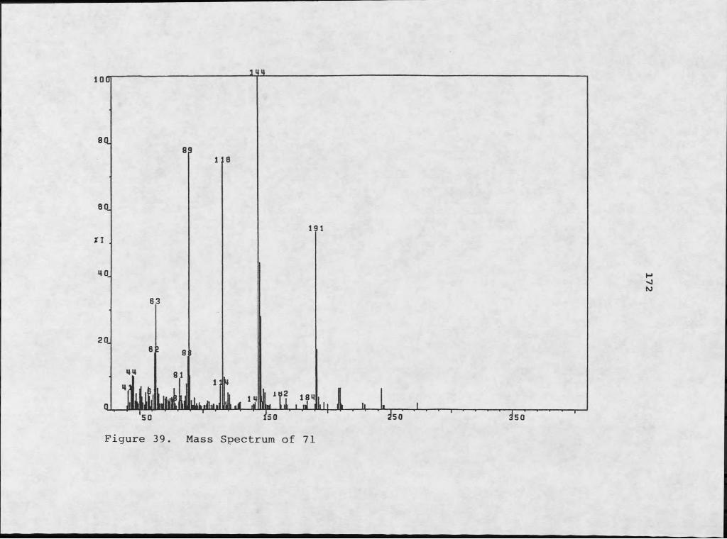
100 1M.
Figure 39. Mass Spectrum of 71

H NMR Spectrum of 71PPM
173

174
Thioesters are often incorporated into important biological compounds, such as acetyl-coenzyme A. Few thioesters have been reported from marine organisms, but an interesting group of compounds that combined indole functionality with the -SCH3 moiety were isolated from the hypobranchial glands of certain marine molluscs. These compounds include 2-methylthioindoleninone, 72, and 6- bromo-2-methylthioindoleninone, 73. Both 72 and 73, in conjunction with other similar metabolites, are believed to constitute the photosensitive complex called tyriverdin, which produces the dye Tyrian purple when exposed to light or heat (143). Marine molluscs have been shown to harbor symbiotic bacteria within their hypobranchial tissues (97, 103). The isolation of a closely related compound, 71, from a marine symbiont suggests that this might also be an interesting macroorganism for symbiotic investigation.
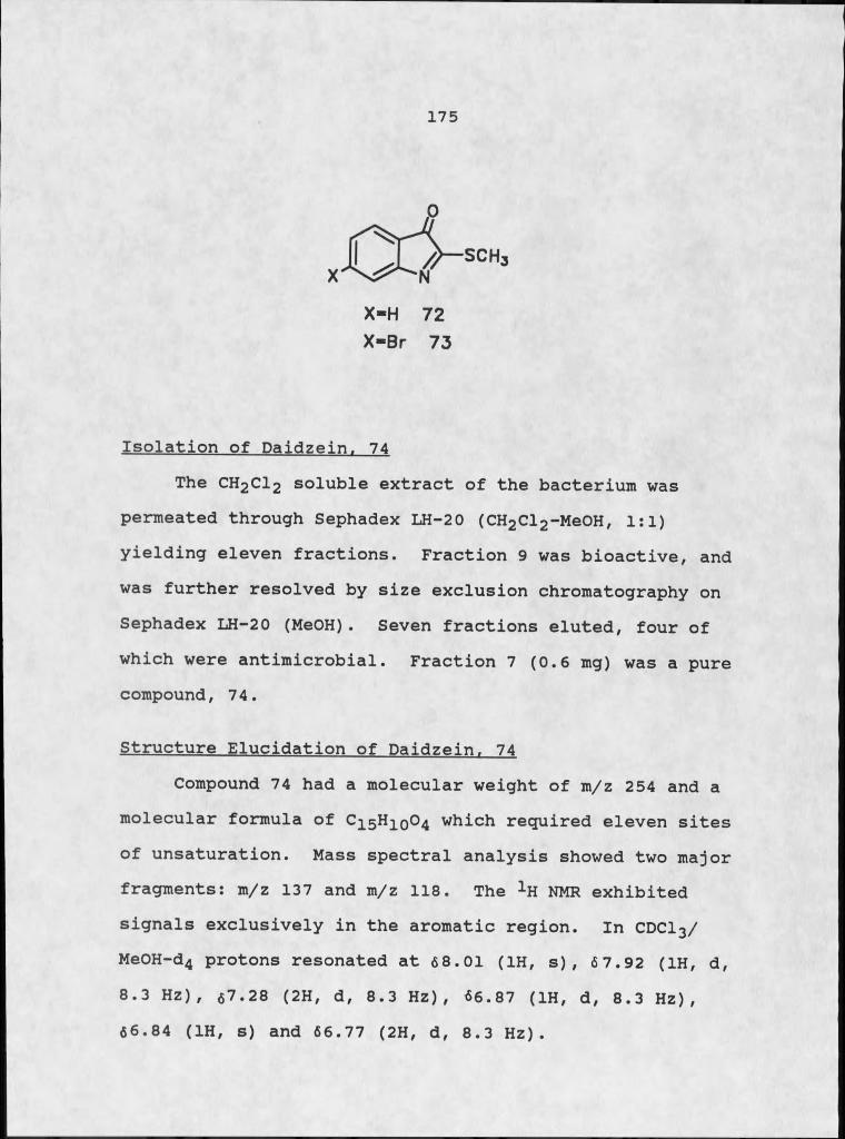
175
X-H 72
X -B r 73
Isolation of Daidzein. 74The CHgClg soluble extract of the bacterium was
permeated through Sephadex LH-20 (CH2Cl2-MeOH, 1:1) yielding eleven fractions. Fraction 9 was bioactive, and was further resolved by size exclusion chromatography on Sephadex LH-20 (MeOH). Seven fractions eluted, four of which were antimicrobial. Fraction 7 (0.6 mg) was a pure compound, 74.
Structure Elucidation of Daidzein. 74Compound 74 had a molecular weight of m/z 254 and a
molecular formula of C15H10O4 which required eleven sites of unsaturation. Mass spectral analysis showed two major fragments: m/z 137 and m/z 118. The 1H NMR exhibited signals exclusively in the aromatic region. In CDCl3/ MeOH-d4 protons resonated at 68.01 (1H, s), 67.92 (1H, d, 8.3 HZ), 67.28 (2H, d, 8.3 Hz), 66.87 (1H, d, 8.3 Hz), 66.84 (IH, s) and 66.77 (2H, d, 8.3 Hz).
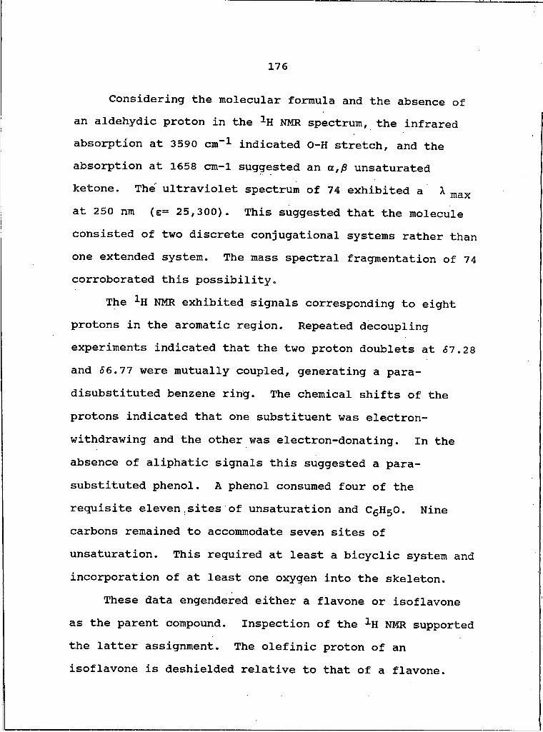
176
Considering the molecular formula and the absence of an aldehydic proton in the 1H NMR spectrum, the infrared absorption at 3590 cm-1 indicated O-H stretch, and the absorption at 1658 cm-1 suggested an a,/3 unsaturated ketone. The ultraviolet spectrum of 74 exhibited a X max at 250 nm (e= 25,300). This suggested that the molecule consisted of two discrete conjugational systems rather than one extended system. The mass spectral fragmentation of 74 corroborated this possibility.
The 1H NMR exhibited signals corresponding to eight protons in the aromatic region. Repeated decoupling experiments indicated that the two proton doublets at SI.28 and S6.ll were mutually coupled, generating a para- disubstituted benzene ring. The chemical shifts of the protons indicated that one substituent was electron- withdrawing and the other was electron-donating. In the absence of aliphatic signals this suggested a para- substituted phenol. A phenol consumed four of the requisite eleven sites of unsaturation and C6H5O. Nine carbons remained to accommodate seven sites of unsaturation. This required at least a bicyclic system and incorporation of at least one oxygen into the skeleton.
These data engendered either a flavone or isoflavone as the parent compound. Inspection of the 1H NMR supported the latter assignment. The olefinic proton of an isoflavone is deshielded relative to that of a flavone.
177
The singlet at 68.Ol supported this choice. The remainder of the aromatic signals indicated hydroxyl substitution at position 7. This structure corresponded to a known compound, the isoflavone daidzein. The two compounds were identical by comparison of spectral data and physical properties. Daidzein was isolated from red clover and from the mold Micromonosnora halonhvtica. This is the first report of an isoflavone from a marine source, or from any source other than a plant or fungi. Daidzein, 74, was also isolated from subsequent broth cultures of the bacterium.
74
Isolation of GlucoseIn all of our in-house culture efforts, partitioning
the freeze-dried inoculum residue between CH2Cl2 and H2O created an emulsion that resisted both solvents. The emulsion was separated from the water soluble extract by suction filtration and dissolved in MeOH. This MeOH soluble extract was chromatographed on Sephadex LH-20 (MeOH) yielding ten fractions. Fraction seven was further resolved into five fractions by CCC (CHCl3-MeOH-H2O,
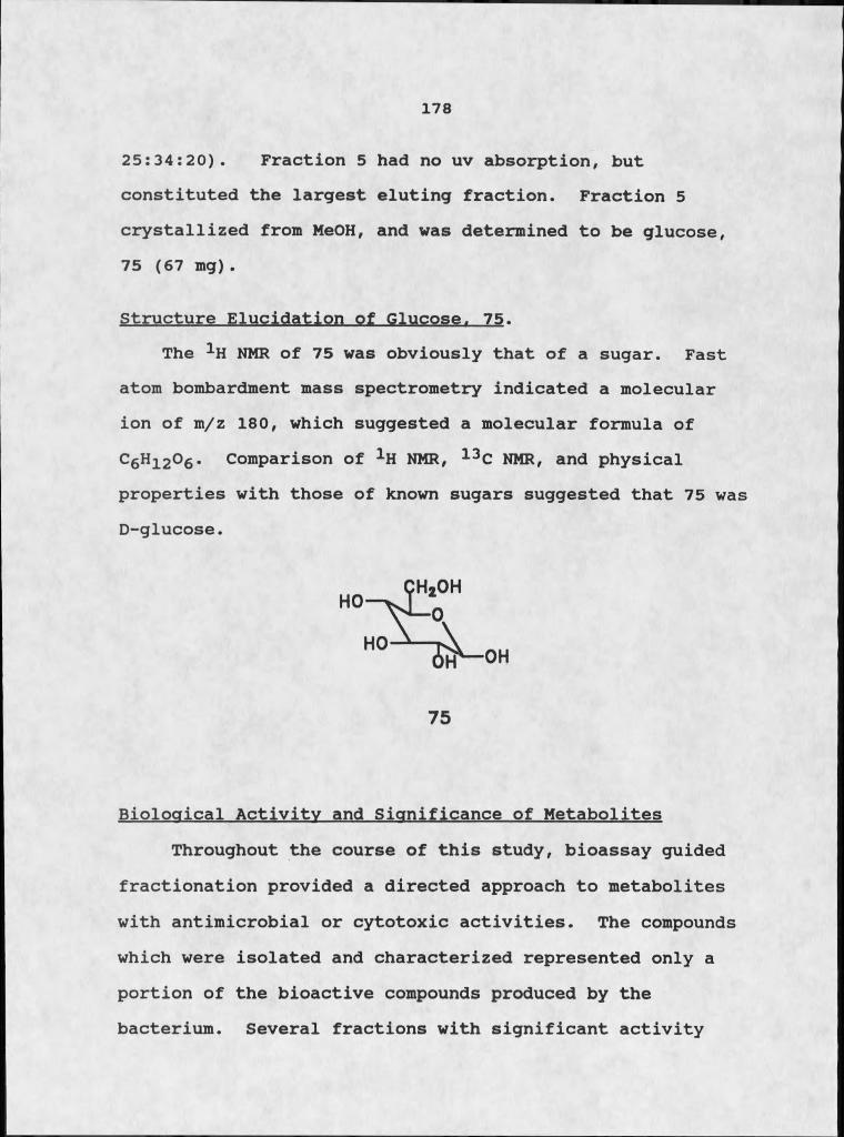
178
25:34:20). Fraction 5 had no uv absorption, but constituted the largest eluting fraction. Fraction 5 crystallized from MeOH, and was determined to be glucose,75 (67 mg).
Structure Elucidation of Glucose. 75.The 1H NMR of 75 was obviously that of a sugar. Fast
atom bombardment mass spectrometry indicated a molecular ion of m/z 180, which suggested a molecular formula of C6Hi2O6 . Comparison of 1H NMR, 13C NMR, and physical properties with those of known sugars suggested that 75 was D-glucose.
Biological Activity and Significance of MetabolitesThroughout the course of this study, bioassay guided
fractionation provided a directed approach to metabolites with antimicrobial or cytotoxic activities. The compounds which were isolated and characterized represented only a portion of the bioactive compounds produced by the bacterium. Several fractions with significant activity
75

179
were not further explored; the isolation, characterization and testing of the metabolites of a single organism could keep one researcher employed for several years. The compounds that were elucidated in this study, however, possessed intrinsic biological and chemical significance. Each of them demonstrated at least one of the following concepts: I) that the relationship between Micrococcus sp.and T . ignis was a true symbiosis; 2) that this symbiosis was mutualistic in nature; and 3) that marine bacteria are- capable of producing unusual compounds with important biological activity.
The biological significance of the microbe was: first realized with the isolation and characterization of the three diketopipefazines cyclo(Pro-Val), 21, cyclo(Pro-Leu), 22, and cyclo(Pro-Ala), 23. Finding these three compounds early in our investigation was quite encouraging. Schmitz had isolated them from the host sponge; isolating the same diketopiperazines from an associated bacterium suggested that the microbe was the actual producer. Diketopiperazines are not commonly isolated from marine macroorganisms. Lindgren et al. have reported the second instance of isolation of dipeptides from a sponge (144).The separate reports by Schmitz and Lindgren, in conjunction with Pettit's report of the isolation cyclo(Pro-Gly) from the starfish Luidia clathrata (145),
180
constitute the marine macrobe isolation reports of diketo- piperazines.
Considering the few accounts of their occurrence as macrobial metabolites, and the growing incidence of their isolation from microorganisms, it is reasonable to suspect that diketopiperazines are ultimately microbial in origin. If this is the case, then the coincidental isolation of three compounds from T. ignis and from its marine bacterial associate implies that bacteria could be responsible for other putative sponge metabolites.
Isolation.and characterization of D-glucose actually proved to be an interesting endeavor, with surprising ramifications. Many culture media contain D-glucose as an ingredient, and the relatively large quantities isolated from the bacterial culture were considered artifacts and were ignored. Careful examination of the constituents of DIFCO marine broth 2216 indicated that absolutely no sugars were included in the medium. Zobell demonstrated that the presence of sugar in the broth had no effect on growth of marine bacterium. Media should be as simple as possible, and sugars were eliminated (146).
The isolation of D-glucose was suddenly a bit more intriguing. D-glucose is not usually considered a storage product, of bacteria, but simple sugars are an important nutrient source for sponges (96). If Micrococcus sp. produces D-glucose and T. ignis uses it as a food source,

181
the biological implications are remarkable. An ambiguous association evolves into a classic mutualism.
The discovery of a series of bioactive benzothiazole derivatives in the organic soluble extracts of the bacterium was important for several reasons. This was the first instance of the isolation of benzothiazoles from the marine environment. The first report of a class of metabolites is always interesting. The limited number of benzothiazoles isolated from natural sources augments the significance. Only one benzothiazole derivative, dercitin, 69, has been reported from the marine environment. It is possible that dercitin is either a microbial metabolite, or that a benzothiazole precursor of dercitin is of microbial origin. Either possibility indicates the suitability of the sponge for microbial analysis.
The isolation of sulfur as S8 from the culture.implied that Micrococcus sp. served an additional purpose in the relationship. Giere and Langheld demonstrated bacterial potential for protecting the host from high levels of H2S in the sea water or sediments (96). Their study on oligochaetes from Platt's Inlet, Bermuda, indicated that high, levels of H2S were present in the water and sediment. Giere and Langheld suggested that symbiotic bacteria can lower the local H2S concentration by sequestering sulfur as vacuolar inclusions, or by oxidation of sulfide to a less
182
toxic form. One of our collection sites was Flatt's Inlet. The discovery of molecules of Sg, as well as the isolation of sulfur containing thioesters, suggests that Micrococons sp. might serve a detoxification function for T. ignis.
Isolation and characterization of the cyclazine 60 was a fascinating, frustrating endeavor. The limited quantity of material and the prohibitive time involved in isolating additional material augmented the frustration factor. But the high level of cytotoxicity exhibited by tedanazine, and its unusual structural characteristics, were too compelling to ignore. Tedanazine, more than any other compound isolated in this study, emphasized the potential for new classes of compounds from marine microbes. The concurrent isolation of anthracene and methylated anthracenes from the microbial culture, suggested that tedanazine was a product of a mixed polyacetate-polyketide biosynthesis. Efforts are underway to synthesize this compound.
Isolation of the isoflavone daidzein, 74, was interesting for two reasons. We found that 74 was antimicrobial, which had not been previously reported.This was also the first report of an isoflavone from a marine source or from any source other than a plant.
183
Conclusion
Two instances of symbiosis between microorganisms and macroorganisms were explored throughout the course of this investigation. The first association, between a terrestrial plant and a pathogenic fungus, was a classic case of parasitism. The host plant derived no apparent benefit from the fungus, while the fungus utilized the plant tissues as a food source. Such interactions are common and have been explored for many years. What was significant about this particular association was the status of the host plant, Centaurea maculosa, as the primary noxious weed pest in Montana. The search for a pathogen that could induce deleterious symptoms on this hardy composite was somewhat protracted. The eventual discovery of„such a pathogen initiated a study of its potential as a biocontrol agent against spotted knapweed.
The discovery of broad-spectrum phytotoxins as metabolites of pathogenic microorganisms has engendered interest in the use of naturally occurring herbicides for weed control. Of much greater interest, however, was the discovery that certain plant pathogenic fungi produce toxins that harm specific cultiyars of a particular plant. Such toxins, called host-specific toxins, have been found against several crop plants. These toxins have tremendous potential as screening agents for eradicating cultivars
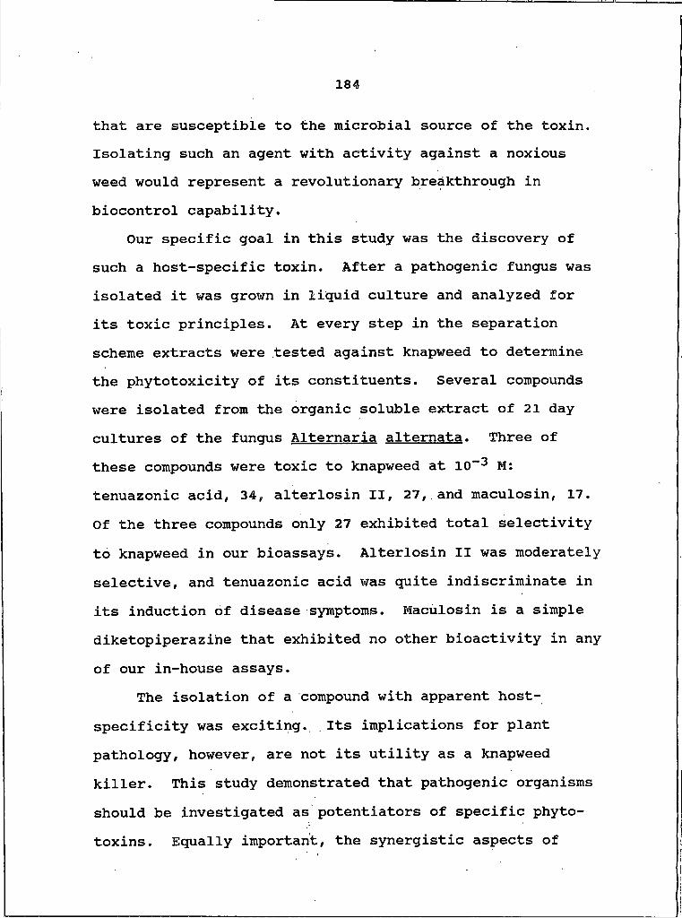
184
that are susceptible to the microbial source of the toxin. Isolating such an agent with activity against a noxious weed would represent a revolutionary breakthrough in biocontrol capability.
Our specific goal in this study was the discovery of such a host-specific toxin. After a pathogenic fungus was isolated it was grown in liquid culture and analyzed for its toxic principles. At every step in the separation scheme extracts were tested against knapweed to determine the phytotoxicity of its constituents. Several compounds were isolated from the organic soluble extract of 21 day cultures of the fungus Alternaria alternata. Three of these compounds were toxic to knapweed at 10”3 M: tenuazonic acid, 34, alterlosin II, 27, and maculosin, 17. Of the three compounds only 27 exhibited total selectivity to knapweed in our bioassays. Alterlosin II was moderately selective, and tenuazonic acid was quite indiscriminate in its induction of disease symptoms. Maculosin is a simple diketopiperazihe that exhibited no other bioactivity in any of our in-house assays.
The isolation of a compound with apparent host- specificity was exciting. Its implications for plant pathology, however, are not its utility as a knapweed killer. This study demonstrated that pathogenic organisms should be investigated as potentiators of specific phytotoxins. Equally important, the synergistic aspects of
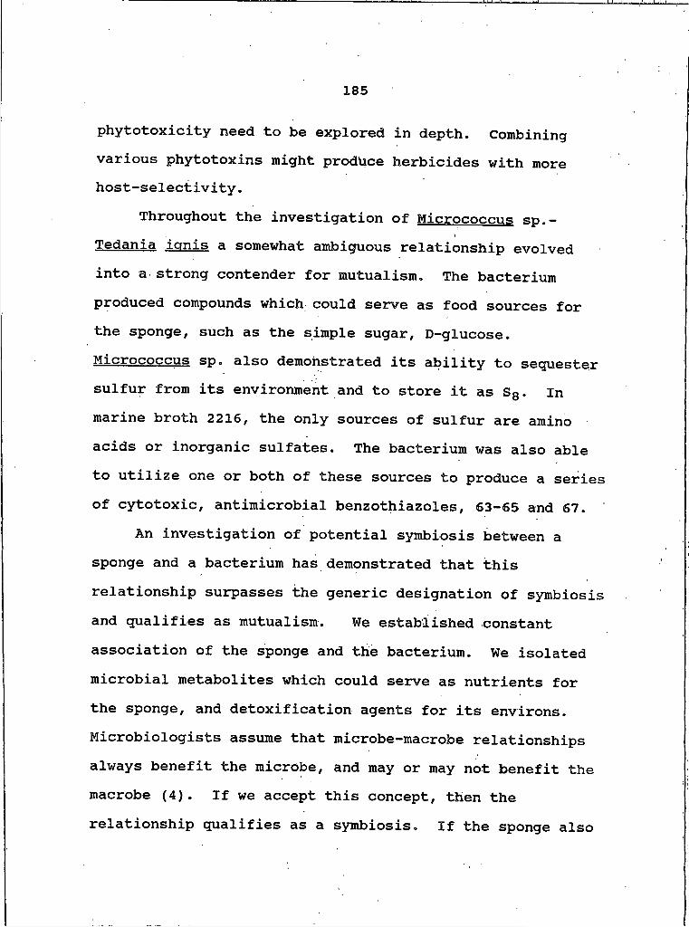
185
phytotoxicity need to be explored in depth. Combining various phytotoxins might produce herbicides with more host-selectivity.
Throughout the investigation of Micrococcus Sp.- Tedania ignis a somewhat ambiguous relationship evolved into a strong contender for mutualism. The bacterium produced compounds which could serve as food sources for the sponge, such as the simple sugar, D-glucose. Micrococcus sp. also demonstrated its ability to sequester sulfur from its environment and to store it as S8 . In marine broth 2216, the only sources of sulfur are amino acids or inorganic sulfates. The bacterium was also able to utilize one or both of these sources to produce a series of cytotoxic, antimicrobial benzothiazoles, 63-65 and 67.
An investigation of potential symbiosis between a sponge and a bacterium has demonstrated that this relationship surpasses the generic designation of symbiosis and qualifies as mutualism. We established constant association of the sponge and the bacterium. We isolated microbial metabolites which could serve as nutrients for the sponge, and detoxification agents for its environs. Microbiologists assume that microbe-macrobe relationships always benefit the microbe, and may or may not benefit the macrobe (4). If we accept this concept, then the relationship qualifies as a symbiosis. If the sponge also
186
benefits, then the relationship is a true case of mutualism.
The investigation of these two cases of symbiotic relationships between microorganisms and macroorganisms has both biological and chemical significance. Using such interactions in a directed bioactivity fashion can lead to the discovery of compounds with desirable biological activity. The isolation and characterization of such compounds is an important aspect 6f natural products chemistry. Both of these studies entailed such directed investigation. In both cases, the endeavors were successful.
We used diseased knapweed plants as an indicator of a pathogenic microorganism. The disease-inductive potential of each extract and fraction facilitated isolation of phytotoxins. Comparative phytotoxicity tests enabled detection of selective, phytotoxicity. The ultimate result was the isolation of three phytotoxins with varying degrees of activity and host selectivity.
The same rationale operated in the marine symbiosis investigation. Invertebrates with demonstrated bioactivity were selected for culture work. Microbial isolates were screened for bioactivity, and only those organisms with reasonable levels of bioactivity were studied. The most active microbes were grown in cultures of sufficient volume to facilitate isolation and elucidation of metabolites.
187
All extracts and fractions were screened for desired bioactivity throughout the fractionation process. Several metabolites with antimicrobial activity or cytotoxicity were isolated as microbial metabolites.
The biological implications of these studies are of equal importance. The mechanisms of interspecies dynamics are complex at best. Understanding these mechanisms can lead to better utilization of living organisms as sources of bioactive metabolites.

1 8 8
EXPERIMENTAL
Materials and Methods
General InstrumentationNMR spectra were recorded on a Bruker WM250 and AC300
spectrometers. Mass spectra were obtained with VG instruments MM16F and 7070EHF spectrometers. Ultraviolet spectra were determined with a Perkin-Elmer LC-55B spectrophotometer. Infrared analyses were obtained with a Perkin- Elmer 1310 infrared spectrophotometer. Optical rotations were determined on a Perkin-Elmer 24IMC polarimeter. The centrifugal countercurrent chromatograph was manufactured by P.C. Inc. Chromatographic separations utilized either the ISCO UA-5 fixed wavelength detector or the ISCO V-4 variable wavelength detector.
Fungal Culture MaintenanceA. alternate was maintained on DIFCO mycological agar
plates containing a 2% knapweed effusion (wt/vol) to insure solicitation of toxin production. The appropriate quantity of leaves and stem pieces were removed from either fresh or frozen knapweed plants. The plant material was boiled in distilled water for thirty minutes and the resulting effusion was added to the mycological agar mixture prior to sterilization. The fungus was cultured in I liter still cultures of Czepak-Dox medium also modified by the addition
189
of the 2% knapweed effusion, prepared in the same manner. Crude extracts of the fungus grown without the effusion were less phytotoxic than those grown with the effusion, which exhibited no toxicity when applied directly to any of the test plants. Plants used in this study were grown under controlled environmental conditions of 12 hr of darkness and 12 hr of light at 250C, with a light intensity of 4xl04 lux.
Marine Bacterial Culture MaintenanceThe marine bacterial . cultures were maintained on
autoclaved DIFCO marine agar 2216 plates prepared according to standard directions. The cultures were streaked for i s o l a t i o n and the s e parate isolates were grown independently, with no attempt made at optimization of growth conditions for individual cultivars. All of the marine cultures were maintained at room temperature, with natural and i n c a n d e s c e n t lighting, with natural photoperiods. No additional nutrients were added to the growth medium.
Fungal Culture Growth and ExtractionA. alternata was grown in five I liter broth cultures which were harvested after a 24 day incubation at 250C. Suction filtration was used to separate the mycelial mat from the liquid media. The filtrate was sequentially, exhaustively extracted with four 500 ml portions of CHjCl2 , which were
190
combined and evaporated (0.041g). The filtrate was then extracted with four 500 ml aliquots of EtOAc which were also combined and evaporated to give an oily residue (0.675 g) • The aqueous fraction was freeze-dried and, along with the two organic fractions, was tested for phytotoxicity against knapweed at a concentration of 1000 ppm. Both organic fractions induced the formation of large necrotic lesions in 48 hr, but the aqueous fraction caused no apparent tissue damage even after 96 hr.
The fungal mat was extracted in a similar manner following homogenation in a Waring blender, but when tested for phytotoxicity did not induce lesions on knapweed leaves. All subsequent chemical manipulations were performed on the organic portions of the filtrate.
Marine Bacterial Culture Growth and ExtractionMarine bacteria were cultured following two different
protocols, one designed to facilitate testing of all of the isolates for bioactivity, and the other designed to procure large quantities of bacteria for chemical analysis. The bacteria were grown in liquid culture for both purposes, using autoclaved, unadulterated DIFCO marine broth 2216 and tap water as the growth medium.
Once individual isolates had been separated, they were grown in 100 ml marine broth for periods of one, two, and three weeks. At the end of these periods, 20 ml MeOH was
191
added to the liquid cultures to kill the bacteria prior to handling. The MeOH was then removed by rotoevaporation, arid the culture was placed on the freeze dryer. After the water was completely evaporated the dried residue was removed from the flask, weighed, and prepared for testing. By comparison with a control, We determined that the dried residue was approximately 90% salt and nutrient material, and only 10% bacterial residue. All of our testing was therefore performed at test concentrations that were more concentrated than usual by a factor of ten. Our standard protocol for antimicrobial assay requires a I mg sample of crude extract: for marine bacterial antimicrobial testing we used a 10 mg sample. The relative activities of the bacterial residues that were cultured for varying time periods were also compared.
Marine bacteria that were considered suitable for further study were cultured in 9-20 liter batches that were allowed to grow for 21 days. The cultures, which were grown in 3 or 4 liter erlenmeyer flasks, were swirled once a day.
As part of the inoculation protocol, 100 ml flasks of marine broth were prepared and autoclaved along with the large flasks. Upon cooling, the small flasks of marine broth were inoculated with a marine bacterium and allowed to grow for 24 hr. The large flasks of marine broth were cooled for 24 hr, and were then inoculated by carefully

192
adding the 24 hr cultures to the sterile marine broth. One small volume culture was used to inoculate 2 I of
broth. The mouths of the large erlenmeyer flasks were covered with foil to prevent contamination.
After the 21 day growth period, the liquid cultures were carefully prepared for subsequent chemical analysis. The cultures were first killed by the addition of 200 ml of MeOH, which was then removed by evaporation. The liquid culture material was then placed on the freeze dryer in 1000 ml round bottom flasks until all of the water was removed. The remaining residue, a mixture of bacteria, salts, and other nutrients, was ready for chemical manipulation.
The residue was thoroughly extracted, first with MeOH- MeCN, 4:1, and then with CH2Cl2 - The polar organic extract was evaporated to dryness and partitioned between CH2Cl2 and water. The two CH2Cl2 extracts were combined, and the organic and aqueous extracts were evaporated to dryness. Each of the two extracts were then tested for antimicrobial activity, phytotoxicity, and brine shrimp toxicity.
Artifact Control
As we began to isolate and elucidate compounds from the fungal extracts, particularly those of amino acid origin, we realized the need to establish their true source. Growth media represent a rich nutrient broth of sugars,
193
salts and amino acids that might react to produce interesting "metabolites" as a result of the high temperature and pressure of autoclaving . To eliminate the possibility of mistaking an artifact for a genuine metabolite, we chose three types of control experiments.
The first experiment mimicked the usual media preparation (2 I) but excluded inoculation with the fungus. The media was allowed to sit for 21 days, and was then extracted in the same manner as the fungal cultures. The resulting total organic extract was 0.015 g/1 , compared to0.140 g/1 from the fungal cultures. The 1H NMR of the media extraction did not resemble any of the metabolites isolated and characterized from the fungal extraction.
Following this experiment we tried a second approach: force the formation of diketopiperazines. We needed to determine if the heat and pressure of autoclaving could encourage the formation dipeptide bonds. We added 50 mg each of several amino acids (L-proline, DL-tryptophan, L- tyrosine, L-valine, L-Ieucine, and L-phenylalanine) to 2 I distilled water and autoclaved as usual. The mixture was allowed to sit at room temperature for 14 days, and was then extracted as usual. There was no organic extract. The diketopiperazines are organic soluble, and apparently did not form under the conditions of the experiment.
The final experiment involved the direct extraction of 25 g of dry mycological agar with CH2Cl2 and EtOAc.
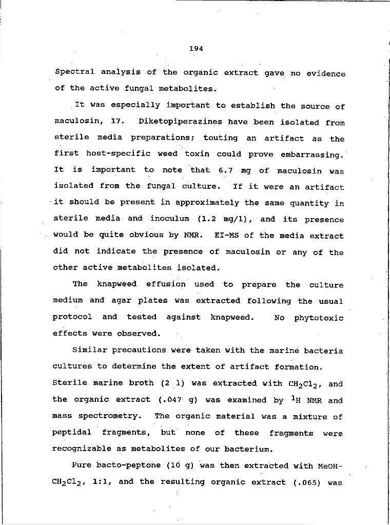
194
Spectral analysis of the organic extract gave no evidence of the active fungal metabolites.
It was especially important to establish the source of maculosin, 17. Diketopiperazines have been isolated from sterile media preparations; touting an artifact as the first host-specific weed toxin could prove embarrassing. It is important to note that 6.7 mg of maculosin was isolated from the fungal culture. If it were an artifact it should be present in approximately the same quantity in sterile media and inoculum (1.2 mg/1), and its presence would be quite obvious by NMR. EI-MS of the media extract did not indicate the presence of maculosin or any of the other active metabolites isolated.
The knapweed effusion used to prepare the culture medium and agar plates was extracted following the usual protocol and tested against knapweed. No phytotoxic effects were observed.
Similar precautions were taken with the marine bacteria cultures to determine the extent of artifact formation. Sterile marine broth (21) was extracted with CH2Cl2 , and the organic extract (.047 g) was examined by 1H NMR and mass spectrometry. The organic material was a mixture ofpeptidal fragments, but none of these fragments were recognizable as metabolites of our bacterium.
Pure bacto-peptohe (10 g) was then extracted with MeOH- CH2Cl2 , 1:1, and the resulting organic extract (.065) was
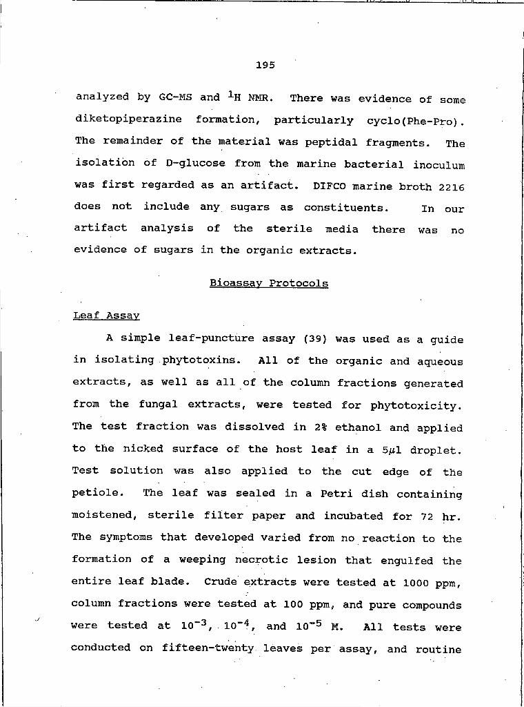
195
analyzed by GC-MS and 1H NMR. There was evidence of some diketopiperazine formation, particularly cyclo(Phe-Pro). The remainder of the material was peptidal fragments. The isolation of D-glucose from the marine bacterial inoculum was first regarded as an artifact. DIFCO marine broth 2216 does not include any sugars as constituents. In our artifact analysis of the sterile media there was no evidence of sugars in the organic extracts.
Bioassav Protocols
Leaf AssayA simple leaf-puncture assay (39) was used as a guide
in isolating phytotoxins. All of the organic and aqueous extracts, as well as all of the column fractions generated from the fungal extracts, were tested for phytotoxicity. The test fraction was dissolved in 2% ethanol and applied to the nicked surface of the host leaf in a 5^1 droplet. Test solution was also applied to the cut edge of the petiole. The leaf was sealed in a Petri dish containing moistened, sterile filter paper and incubated for 72 hr. The symptoms that developed varied from no reaction to the formation of a weeping necrotic lesion that engulfed the entire leaf blade. Crude extracts were tested at 1000 ppm, column fractions were tested at 100 ppm, and pure compounds were tested at 10“3, 10“4 , and IO"5 M. All tests were conducted on fifteen-twenty, leaves per assay, and routine
196
assays were conducted on knapweed and johnsongrass fSorahmn halapense L.).
Only crude marine bacterial extracts were tested for phytotoxicity.
Hvoocotvl AssayThis assay involved nicking the hypocotyl of
appropriate host seedlings and applying the test fraction in a 2% ethanol solution in a manner similar to the leaf assay. Symptoms usually developed within 48 hr and ranged from no reaction to total necrosis of the plant material. In no circumstances were deleterious symptoms induced by application of 2% ethanol solution in either the leaf or hypocotyl assays.
Antimicrobial AssayThe organic and aqueous extracts of the bacterial
culture were tested for antimicrobial activity against bothgram-positive and gram-negative bacteria, and fungi. The
(
protocols were the same regardless of the test organism. The first level of testing against the crude residue of the bacterial culture involved weighing out 10 mg of finely ground material, and dissolving it in .021 ml of sterile distilled water. This emulsion was applied to a sterile paper test disk and allowed to air dry. A DIFCO tryptic soy agar plate (or mycological agar plate) was inoculated with a single test bacterium (or fungus). The organism was
197
suspended in sterile distilled water and carefully spread across the surface of the plate until a smooth lawn was formed. The inoculated test disks were applied to the microbial lawn and allowed to sit undisturbed for 24-48 hr. The growth characteristics of the microbe were observed in the vicinity of the test disk, and zones of inhibition were determined by measuring areas devoid of growth radiating from the edge of the disk.
Brine Shrimp ToxicityThroughout the course of the marine bacterial study we
needed a means of determining the cytotoxicity of the culture extracts and various column fractions to facilitate bioassay guided fractionation. Ferrigni et al. suggested that a simple brine shrimp assay provided a reasonable facsimile of the standard anticancer assays (109). They were able to isolate the antileukemic principles from the seeds of Euphorbia lagascae Spreng using a fractionation scheme guided by brine shrimp and potato disc assays. Several studies have demonstrated good correlation between brine shrimp lethality and cytotoxicity (108-111).
The brine shrimp bioassay is a simple test. Brine shrimp are hatched in Instahit Ocean dissolved in tap water, which generates a solution that approximates the constituents of sea water. The test material is handled according to its polarity and purity. Crude culture
198
residues are tested at 10 mg, and simply dissolved in the saline water used to rear the brine shrimp. The test material is dissolved in 2 ml of salt water, and an additional 2 ml of salt w^ter containing the brine shrimp is added. The brine shrimp are counted, then recounted at various time intervals. Toxicity is determined by the number of brine shrimp deaths that result from the test material, relative to a control.
The organic crude extract of the bacterial culture is relatively free of salts and is therefore tested at a lower concentration (I mg) . It must be dissolved in a minimal amount of MeOH to facilitate dissolution. A MeOH concentration of .010 ml per ml of sea water will not harm brine shrimp and is usually sufficient to dissolve the test material.
Isolation of CompoundsMaculosin, 17
The EtOAc soluble extract (675 mg) of 24 day culture filtrates of Alternaria aIternata was permeated through BioBeads S-X4 (column 4.5 x 95 cm) with Iiexane-CH2Cl2- EtOAc, 4:3:1). The fifth (26 mg) of twelve fractions was chromatographed on Sephadex LH-20 (column 2.0 x 135 cm) using CH2Cl2-MeOH-IPrOH, 1:1:1). The third of the seven fractions was maculosin, 17 (6.8 mg).
199
CvclofL-Pro-L-PheK 18 and CvclofL-Pro-D-PheK 19Fraction 6 (13 mg) of the S-X4 separation of the EtOAc
soluble extract of the fungus was also permeated through Sephadex LH-20, as described above. Six fractions eluted and fraction 2 was resolved by centrifugal countercurrent chromatography (flow rate: 4 ml/min? volume: 285 ml; ID:1.6 mm) using CHClg-MeOH-K^0, 25:34:20, lower phase mobile, into eight fractions. Fraction 2 was 18 (4.0 mg) andfraction 3 was 19 (3.0 mg).
CvclofPro-Hleu), 20Fraction 4 (7 mg) of the S-X4 chromatography of the
EtOAc soluble extract of Alternaria alternata was permeated through Bio-Beads S-X8 (column 2.5 x 140 cm) using CH2Cl2- cyclohexane, 3:2. The second of the five fractions was cyclo(Pro-Hleu) (2.1 mg).
CvclofPro-Val). 21. CvclofPro-Leu), 22.CvclofPro-Ala). 23
The CH2Cl2 soluble extract (234 mg) of the culture filtrate of Alternaria alternata was applied to Sephadex LH-20 (column 2.5 x 185 cm) using CH2Cl2-MeOH, 1:1. Fraction 4 (28 mg) was resolved by CCC as described above into twelve fractions. Fraction 10 was cyclo(Pro-Val), 21 (0.9i mg), fraction nine was cyclo (Pro-Leu), 22 (2.1 mg), and fraction eight was cyclo(Pro-Ala), 23 (0.7 mg).
200
CvclofPro-Met). 57The CH2Cl2 soluble extract (710 mg) of 21 day marine
broth cultures of Micrococcus sp. was chromatographed on Sephadex LH-20 (CH2Cl2-MeOH, 1:1) yielding eleven fractions. Size exclusion chromatography of fraction 5 (145 mg) on Bio-Beads S-X8 (CH2Cl2-cyclohexane 3:2) yielded seven fractions. Fraction 6 (0.9 mg) was cyclo(Pro-Met),57.
Cvclo(Phe-Ala). 58Fraction I (35.5 mg) from the initial LH-20 separation
of the CH2Cl2 soluble extract of Micrococcus sp. was chromatographed on Sephadex LH-20 (column 2.0 x 180 cm) using MeOH-MeCN, 4:1). Four fractions eluted; fraction 3 (17.8 mg) was resolved into thirteen fractions by CCC as described above. Fraction 11 (8.2 mg) was cyclo(Phe-Ala),58.
Cvclo(Pro-Trp). 59The CH2Cl2 soluble extract of Micrococcus sp. was
permeated through Sephadex LH-20 (CH2Cl2-MeOH, 1:1), yielding ten fractions. Fractions 5 (138.4 mg) and 6 (40.6 mg) were combined and permeated through Sephadex LH-20 (CH2Cl2-MeOH, 1:1) yielding four fractions. Fraction 3 (8.6 mg) was resolved by CCC as described above into ten fractions. Fraction 5 (1.98 mg) was cyclo(Pro-Trp), 59.

201
Pervlenecruinones 24-^27The CH2Cl2 soluble extract (234.4 mg) of Alternaria
alternate was chromatographed on Sephadex LH-20 (CH2Cl2- MeOH, 1:1), yielding four fractions. Fraction 3 (32.5 mg) was chromatographed on Sephadex LH-20 (MeOH-MeCN, 4:1) yielding six fractions. The second fraction (23.6 mg) was resolved by CCC as described above into seven fractions. Fraction I was 24 (4.5 mg), fraction 3 was 25 (4.0 mg), fraction 4 was 27 (1.8 mg), and fraction 5 was 26 (2.9 mg).
Tenuazonic Acid. 34The EtOAc soluble extract of Alternaria alternate was
chromatographed on Bio-Beads S-X4 (hexane-CH2Cl2-EtOAc, 4:3:1) yielding twelve fractions. Fraction 5 (1.354 g) was permeated through Sephadex LH-20 (CH2Cl2-MeOtBu-iPrOH, 1:1:1) yielding eight fractions. The third fraction (182.5 mg) was resolved by size exclusion chromatography using Bio-Beads S-X8 (CH2Cl2-Cyclohexane, 3:2) into five fractions. Fraction 2 ( 84.1 mg) was tenuazonic acid, 34.
Tedanazine, 60The CH2Cl2 soluble extract (1.66 g) of Micrococcus sp.
was chromatographed on Sephadex LH-20 (CH2Cl2-MeOH, 1:1) and separated into ten fractions. Fraction 7 (16.0 mg) was permeated through Sephadex LH-20 (MeOH) and separated into eight fractions. Fraction 7 (2.2 mg) was resolved into
202
seven fractions by CCC, as described above. Fraction 4 (0.2 mg) was tedanazine, SO.
2-Mercaptobenzothiazole. S3. 2-Methvlbenzothiazole.64, 3-(2-hvdroxyacetvl)indole. 70. and2-Hvdroxvbenzothiazole. 65
The 220 I Singleton liquid culture of Micrococcus sp. was separated by centrifugation into cellular solids and s u p e r n a t a n t . The C H 2C l 2 soluble e xtract was chromatographed on Sephadex LH-20 (CH2Cl2-MeOH, 1:1), which yielded ten fractions. Fraction 5 (258.9 mg) was permeated through Bio-Beads S-X4 (Iiexane-CH2Cl2-EtOAc, 4:3:1) yielding five fractions. Fractions 3, 4, and 5 wererecombined and chromatographed on Sephadex LH-20 (MeOH), which yielded seven fractions. Fractions 5, 6, and I were combined and poorly resolved by CCC into nine fractions, as described above. Fractions 5-8 were combined and chromatographed on Bio-Beads S-X8 (column 2.0 x 135 cm) using CH2Cl2-MeCN, 1:1. This resolved the mixture into eleven fractions.
Both f r actions 6 and 8 were resolved into heteroaromatic compounds. Fraction 6 (8.2 mg) was resolved by CCC (Iiexane-CH2Cl2-MeCN, 50:15:35, lower phase mobile)into six fractions. Fraction 3 was 2-mercaptobenz o- thiazole, 63 (0.4 mg), fraction 4 was 3-(2-hydroxyacetyl)- indole, 70 (1.1 mg), and fraction 5 was 2-methylbenzo-thiazole, 64 (1.8 mg).
203
Fraction 8 (19.3 mg) was chromatographed on Sephadex LH-20 (column 1.6 x 200 cm) using CHCl3-MeOH, 1:1). Five fractions eluted; fraction 3 (2.9 mg) was permeated through Bio-Beads S-X8 (CH3Cl3-MeCN, 1:1). Fraction 4 was 2- hydroxybenzothiazole, 65 (0.5 mg).
6-Hydroxy-3-methvl-2-benzothiazolone. 67 and Indole-3(methylthiocarboxvlate>. 71
The EtOAc soluble extract (643.3 mg) of thesupernatant of the Singleton liquid culture of Micrococcussp.. was chromatographed on Sephadex LH-20 (CHCl3-MeOH, 1:1)yielding five fractions. Fractions 3 (133.3 mg) and 5(134.5 mg) were combined and permeated through Sephadex LH-20 (MeOH). Eight fractions eluted and the fraction 6 (7.1mg) was resolved into seven fractions by CCC (CHCl3-MeOH-H2O, 25:34:20). Fraction 3 was a sulfur-containing indolederivative, 71 (0.2 mg), and fraction 6 was abenzothiazolone, 67 (2.1 mg).
Daidzein, 74The CH2Cl2 soluble extract of Micrococcus sp. was
permeated through Sephadex LH-20 (CH2Cl2-MeOH, 1:1) yielding eleven fractions. Fraction 9 (22.2 mg) was resolved into eight fractions by size exclusionchromatography on Sephadex LH-20 (MeOH). Fraction 7 (0.6
-
mg) was daidzein, 74.

204
Characterization of Compounds
Maculosin. 17Yield: 6.8 mg from 5 liter fungal culture; mp. 151°C;
natural product: [g]D -72 ° (c 0.103, CHCl3); syntheticproduct: [a]p -786 (c 0.602, CHCl3); ir V max (CHCl3) 3490(broad), 3380, 3000, 1670, 1510, 1420 cm"1 ; 1H NMR (CDCl3) 56.76 (2H, d, J=8.2 Hz), 7.03 (2H, d, 8.2), 5.89 (1H, s) , 4.20 (IH, dd, 10.3, 3.2), 4.07 (IH, t, 7.1), 3.57 (2H, M ) , 3.44 (IHf dd, 14.7, 3.2) , 2.76 (IH, dd, 14.7, 10.3), 2.31 (IH, m ) , 1.93 (3H, m ) ; ms: m/z (%), 260 (M+, 7), 154 (100); HRMS: m/z 260.1141 (C14H16N2O3 requires 260.1161).
Synthesis of Maculosin. 17The synthesis of maculosin followed a two step
procedure that involved protection one of the amino acids with B0C-0N, using the method of Itoh (40). The BOC- protected amino acid was reacted with the methyl ester of the second amino acid, following the method of Nitecki et al., which is known to proceed without racemization (41).
In our typical synthesis, we reacted 0.115 g (1.0 mM) L-proline with 0.271 g (1.1 mM) BOC-ON, which was stirred at room temperature in 10 ml aqueous acetone and 200 ^l triethylamine. After two hours the mixture was evaporated and washed with EtOAc. The aqueous layer was acidified with I M HCl, then extracted with CH2Cl2 . A typical yield of crude product was 0.169 g (79%).

205
The BOC-proline was dissolved in an equimolar mixture of CH2Cl2 and triethylamine. The L-tyrosine methyl ester hydrochloride was added (1.0 mM) , followed by the addition of 0.192 g (1.0 mM) of N-ethyl-N6-(3-dimethylaminopropyI) - carbodiimide hydrochloride. The mixture was left in the freezer overnight at -5°C, then reduced in vacuo. The crude dipeptide was sequentially washed with water, citric acid (I N) , sodium bicarbonate, water, then evaporated to dryness.
The crude t-boc-dipeptide methyl ester was dissolved in 5-10 ml formic acid, and stirred at room temperature for two hours. After removal of the excess formic acid in vacuo, the crude dipeptide ester formate was dissolved in 5
i -ml toluene and 25 ml sec-butanol, and refluxed for two hours. The mixture was evaporated to dryness once more, and the crude diketopiperazine was purified by size exclusion chromatography. The mixture was permeated through Sephadex LH-20 (MeOH-MeCN, 4:1). Fraction 4 crystallized during evaporation, and was shown by spectral analysis to be pure cyclo(L-Pro-L-Tyr). The yield of the second reaction was 0.144 g (70%). Overall yield was 55%.
CvclofL-Pro-L-PheK 18
Yield: 4 mg from 5 liter fungal culture; mp. 126 0 - I3S 0C; ir v max (CHCl3) 3380, 3000, 1670, 1425 cm”1 ; 1H NMR (CDCl3) S 7.22 (5H, m) , 5.70 (1H, s) , 4.20 (1H dd, J=10.3,

3.2 Hz) , 4.07 (1H, t, 7.1), 3.64 (1H, dd, 15.1, 3.2), 3.59 (2H, m) , 2.76 (1H, dd 15.1, 10.3), 2.31 (1H, m) , 1.93 (3H, m ) ; ms: m/z (%), 244 (M+, 11), 153 (100).
CvclofL-Pro-D-Phe^. 19Yield: 3 mg from 5 liter fungal culture; mp. 136°-
1380C; ir V max (CHCl3) 3380, 3000, 1670, 1425 cm-1; 1H NMR (CDCl3) 5 7.22 (5H, m ) , 6.20 (1H, s), 4.20 (1H, dd, J=IO.3, 3.3), 3.38 (1H, m) , 3.08 (2H, m) , 3.01 (1H, t, 7.4), 2.21(IH, m ) , 1.80 (3H, m ) ; ms: m/z (%) 244 (M+, 12), 153 (100).
Cvclo(Pro-Hleu). 20Yield: 2.11 mg from 5 liter fungal culture; ir v max
(CHCl3) 1670, 1427 cm-1; 1H NMR (CDCl3) 64.15 (1H, m) , 4.02 (IH, m) , 3.59 (2H, m) , 2.40-1.80 (5H, m) , 1.70 (1H, m) , 1.50 (3H, m ) , 1.0 (3H, d, 6.2), 0.97 (3H, d ) ; ms: m/z (%) 224 (M+, 7), 209 (65), 154 (100).
Cvclo(Pro-Val). 21Yield: 0.91 mg from 5 liter fungal culture, 2.4 mg
from 9 liter marine bacterial culture; ir v max (CHCl3) 1669 cm-1; 1H N M R (CDCl3) 64.10 (1H, m) 3.90 (1H, m) , 3.59 (2H, m ) , 2.50 (IH, m ) , 2.4-1.8 (4H, m ) , 1.10 (3H, d J= 6.7 Hz), 0.92 (3H, d, 6.7); ms: m/z (%) 196 (M+, 4), 154 (90), 125 (36), 70 (100).
206
207
CvclofPro-LeuV. 22Yield: 2.1 mg from 5 liter culture, 1.2 mg from 9
liter marine bacterial culture; 1H NHR (CDCl3) 64.09 (1H,t, J= 7.8 Hz), 3.99 (1H, dd, 9.3, 3.7), 3.56 (2H, m), 2.40- 1.95 (4H, m ) , 1.9 (IH, m), 1.73 (1H, m), 1.50 (1H, m), 0.97 (3H, d, 6.2), 0.92 (3H, d, 6.2); ms: m/z (%) 210 (M+, I), 195 (M-15+,2), 154 (100).
CvclofPro-AlaV. 23
Yield: 0.7 mg from 5 liter culture, 1.3 mg from 9liter marine bacterial culture; 1H NMR (CDCl3) 64.20 (1H, q J= 8.5 Hz), 4.01 (1H, dd, 8.6, 2.8), 3.6 (2H, m ) , 2.4-1.8 (4H, m) , 1.0 (3H, d, 8.5); ms: m/z (%) 168 (M+, 6), 154(100), 125 (12), 70 (91).
CvclofPro-MetV. 57
Yield: 0.9 mg from 9 liter marine bacterial culture;1H NMR (CDCl3) 64.17 (1H, t, J= 3.8 Hz) , 4.08 (1H, t,7.10), 3.61 (2H, m) , 3.01 (1H, m) , 2.91 (1H, m) , 2.43 (3H,S), 2.6-2.3 (3H, m) and 2.25-1.8 (3H, m ) ; ms: m/z (%) 228(M+, 9.2), 181 (M-47+, 16.8), 154 (47.9) and 123 (6.0).
CvclofPhe-AlaV. 58
Yield: 8.2 mg from 9 liter marine bacterial culture;1H NMR (CDCl3/MeOH-d4) 67.20 (5H, m) , 4.20 (1H, t, J= 3.2Hz), 3.81 (IH, d, 6.1), 3.27 (1H, dd, 9.7, 3.2), 3.15 (IR,
208
d, 9.7) and 0.83 (3H, d, 6.1); ms: m/z (%) 218 (M+, 7.1),91 (100).
CvclofPro-Trp). 59
Yield: 1.98 mg from 16 liter marine bacterial culture; 1H NMR (CDCl3) 68.34 (NH, br s), 7.61 (NH, br s), 7.51 (IR, d/ J= 7.2 Hz), 7.47 (1H, d, 7.8), 67.31 (1H, t, 7.8), 67.29 (IH, t, 7.2) , 7.13 (1H, 1.2 Hz) , 4.46 (1H, dd, 7.1, 1.0), 4.12 (IH, t. 6.52), 3.5 (2H, m ) , 3.23 (1H, dd, 16.3, 6.52), 2.93 (IH, dd, 16.3, 9.7) ; ms: m/z (%) 283 (M+, 23.7) , 154(66.6), 130 (100).
Alterlosin I. 26Yield: 2.9 mg from 5 liter culture; mp. 191-193°C;
[a]D +122° (c 0.21, MeOH) ; ir v max (CHCl3) 3400 (broad) ,1646, 1600, 1486, 1457, 1369, 1336, 1231, 1170, 1062, 1018, 951 cm"1 ; UV X max (MeOH) 256 nrn (e= 31,450), 285 (15,7Q0), 366 (5080) ; H R M S : m/z . 366.0752 (^20^14^7 requires366.0740); ms: m/z 366 (M+), 348 (M-H2O+), 330 (m-2H20+) ,314.
Alterlosin II. 27Yield: 1.8 mg from 5 liter culture; mp. 185-1870C;
[a] D +131 (c 0.20 MeOH); ir V max (neat) 3400 (broad) , 1632, 1600, 1473 cm"1 ; uv ^ max (MeOH) 255 nm (e =30,400), 348 (5040); H R M S : Itt/Z 368.0869 (C20H 16O 7 requires
209
m
368.0896); ms: m/z 368 (M+) , 352 (M-2H2O+) „ 334 (M-3H2O+) , 316 (M-4H20+, 100%).
NaBH ̂ Reduction of Alterlosin II. 270.1 mg of 27 was dissolved in 2 ml MeOH in a I cm
cuvette. The uv ̂max was 348 nm (5040). NaBH^ was added to 27 in 0.1 mg increments. After addition of the first increment, the uv spectrum was analyzed following evolution of H2 : uv A max 348 nm (1021), 268 (2667). After I hr the sample was measured again; the uv A max was 268 nm (7850). More NaBH4 was added (0.1 mg), and the sample was again analyzed following the evolution of H2 : the spectrum was unchanged. The sample was analyzed every hour for the next 5 hr: the uv spectrum remained constant. More NaBH4 was added, but no uv shift was observed. For comparison,phenol was treated in a similar manner. The uv A max of0.1 mg phenol dissolved in MeOH was 270 nm (1450) . NaBH4 was added to phenol in 0.1 mg increments. Even after the addition of 0.4 mg of base, the uv A max did not shift. At this point 0.1 mg NaOH was added to the cuvette. Within 5 minutes, the uv A max shifted to 287 nm (2650), 235 (9500), which reflects the formation of phenolate anion (51).
NaBH4 Reduction of Altertbxin I. 24The uv A max of 0.1 mg of 24 dissolved in 4 ml of MeOH
was determined: 356 nm (5950), 296 (13,500), 285 (16,400), 256 (33,900). NaBH4 was added in 0.1 mg increments and the
210
uv spectrum was determined after evolution of H2 was complete. Within 5 minutes the uv spectrum shifted to 298 nm (19,200) . No changes were observed with time, or with further addition of the base.
Tenuazonic acid, 34Yield: 87 mg isolated from 5 liter fungal culture;
ir V max (CHCl3) 3460, 3250, 3008, 2870, 2920, 1710, 1650, 1610, 1453, 1380, 1230 cm"1 ; 1H NMR (CDCl3) S 7.60 (1H, br sj, 7.43 (IH, br s), 3.96 (1H, d, J= IHz), 3.76 (1H, d, I), 2.46 (3H, s) , 2.38 (3H, s) , 1.9 (1H, br s) , 1.35 (3H, m) 0.94 (3H, d, 6.6), 0.83 (3H, t, 6.6); m s : m/z 197 (M+), 182 (M-15+), 168, 75, 58; HRMS: m/z 197.1051 (C10H15NO3requires 197.1052).
Tedanazine. 60Yield: 0.2 mg from 20 liter marine bacterial culture;
ir v max (neat) 2956, 2922, 2853, 1732, 1653, 1464, 1563,1361 cm"1 ; uv A max (MeOH) 383 ( e = 5020), 334 (5140), 285 (7000), 276 (5530) and 245 nm (32,300); 1H NMR (CDCl3/MeOH- d4) 67.87 (2H, s), 7.57 (2H, s), 2.39 (3H, s) and 2.37 (3H, s) ; ms: m/z (%) 210 (M+, 91.4), 182 (M-28+, 45.0), 168 (M- 44+, 100) and 140 (22.1) . HRMS: m/z 210.07890 (C13H10N2Orequires 210.0793), 182.08456 (C12H10N2 requires 182.0844)and 168.06919 (C11H8N2 requires 168.0688).
211
2-Mercaptobenzothiazole. 63Yield: 0.4 mg from 200 liter marine bacterial culture;
ir V max (CHCl3) 3380, 2985, 1600, 1500, 1485, 1460, 1428, 1407, 1321, 1035 cm""1 ; 1H NMR (CDC^/MeOH-d^) 67.24 (4H, m) , 7.32 (IH, d, J= 8.28 Hz), 7.44 (1H, d, 8.28); ms: m/z (%) 169 (M+2+, 11.3), 167 (M+, 99.9), 140 (M-27+, 5.6), 135 (M-32+, 4.9), 123 (M-44+, 6.5).
2-Methvlbenzothiazole. 64Yield: 1.8 mg from 200 liter marine bacterial culture;
ir V max (CHCl3) 3060, 1595, 1550, 1450, 1414, 1312 cm”1 ; 1H NMR (CDCl3/acetone-d6) 62.80 (3H, s) , 7.32 (1H, t, J= 7.2 Hz), 7.43 (IH, t, 7.2), 7.80 (1H, d, 7.8), 7.92 (1H, d, 7.8); ms: m/z (%) 151 (M+2+, 5.1) 149 (M+ , 100), 108 (M- 41+, 24.3).
2-Hvdroxvbenzothiazoie. 65Yield: 0.5 mg from 200 liter marine bacterial culture;
ir V max (CHCl3) 3410, 3185, 3001, 1700 (sh), 1667, 1467 cm”1 ; 1H NMR (CDCl3) 67.08 (1H, t, J= 7.5 Hz), 7.12 (1H, t, 7.5), 7.24 (IH, d, 8.5), 7.38 (1H, d, 8.5); ms: m/z.(%) 151 (M+ , 100), 123 (M-28+).
6-Hvdroxv-3-methvl-2-benzothiazolone. 67Yield: 2.1 mg from 200 liter marine bacterial culture;
ir v max (CHCl3) 3400, 2995, 2920, 1700, 1667, 1360 cm"1 ; 1H NMR (CHCl3/acetone-d6) 63.29 (3H, s) , 6.36 (1H, dd, J=

212
8.62, 2.62 HZ), 6.53 (1H, d, 8.62), 6.83 (1H, d, 2.62); ms: xn/z (%) 182 (M+2+, 5.7) 181 (M+, 100), 152 (M-28+, 47.1). HRMS: m/z 181.02381 (C8H7NO2S requires) and 152.01605 (C7H 6NOS requires 152.0167).
3 (2-hvdroxvacetvl)indole. 70Yield: 1.1 mg from 200 liter marine bacterial culture;
ir u max (CHCl3) 3455, 3000, 2920, 1650, 1400 cm-1; 1H NMR (acetone-d6/MeOH-d4) <5 4.56, (2H, s) , 7.27 (1H, m,) , 7.50 (1H, m ) , 8.26 (1H, m), 8.29 (1H, s); ms: m/z (%) 175(M+ , 23.9), 144 (M-31+, 100)> 116 (34.0), 89 (28.7).
Indole(3-methvlthiocarboxvlateV. 71Yield: 0.2 mg from 16 liter marine bacterial culture;
ir U max 2940, 1673 cm"1 ; 1H NMR (CDCl3/MeOH-d4) 62.31 (3H, s) , 7.08 (3H, m)., 7.86 (1H, s) , 8.05 (1H, m) ; ms m/z (%)193 (M+2+, 0.9), 191 (M+, 13.3), 144 (M-44+, 100), 116 (M- 75+, 18.5), 89 (19.2); HRMS: m/z 191.03746 (C10H9NOS requires 191.0402) and 144.04577 (C9H6NO requires 144.0449).
Daidzein. 74
Yield: 0.6 mg from 9 liter marine bacterial culture; ir u max (CHCl3) 3590, 1658 cm"1 ; uv X max 250 nra ( e = 25,300) 1H NMR (CDCl3/MeOH-d4) 68.01 (1H, s), 7.92 (1H, d, 8.31 Hz), 7.28 (2H, d, 8.31), 6.87 (Iff, d, 8.3), 6.84 (1H,S), 6.77 (2H, d, 8.31); ms: m/z (%) 254 (M+, 15.2), 137 (M-
213
(M-136+, 18.0); HRMS; m/z 254.0577 (C15H10O4 requires254.0578) and 137.0240 (C7H5O3 requires 137.0238).
214
REFERENCES1. Curtis, H. and Barnes, N. S. 69Invitation to Biology" ;
Anderson, S., Ed., Worth Publishers: New York, 1981; p 543.
2. Jessop, N. M. "Biosphere"; McElroy, W. D. and Swanson, C . P., Eds., Prentice-Hall: Englewood Cliffs, New Jersey, 1970; p 220.
3. Curtis, H. and Barnes, N. S., o p . cit.. p 546.4. Nester, E. W.; McCarthy, B. J.; Roberts, C. E.; and
Pearsall, N . N., "Microbiology, Molecules, Microbes, and Man"; Holt, Rinehart, and Winston: New York, 1973; p 359.
5. Nishixnura, S. and Kohxnoto, K. (1983). Ann. Rev. Phvtooath. 21, 87-115.
6. Schmitz, F . J.; Gunasekera, S. P.; Yalamanchili, G. ; Hossain, M. B.; and van der Helm, D. (1984). J. A m . Chem. Soc. 106, 7251-7252.
7. Yotsu, M.; Yamazaki, T.; Mezuro, Y.; Endo, A.; Murata, M.; Naoki, H.; and Yasumoto, T. (1987). Toxicon 25(2), 225-228.
8. Tatum, L. A. (1971). Science 171, 1113-1115..9. Fuller, H. J.; Carothers, Z. B.; Payne, W. W. and
Balbach, M. K., "The Plant World"; Holt, Rinehart, and Winston: New York, 1972; p 505.
10. Strobel, G. A.; Sugawara, F.; and Clardy, J. In Allelochemicals: Role in Agriculture and Forestry; Waller, G. R., Ed., American Chemical Society: Washington, DC, 1987; pp 516-523.
11. "Phytotoxins in Plant Diseases"; Wood, R. K. S., Ballio, A. and Graniti, A., Eds., Academic Press: London, 1972; p 141.
12. Strobel, G. A. and Mathre, D. E. "Outlines of Plant Pathology"; Van Nostrand Reinhold: New York, 1970; p 23.Myers, J. H. and Berube, D. E. (1983). Can. J. Plant Sci. 63 981-987.
13.
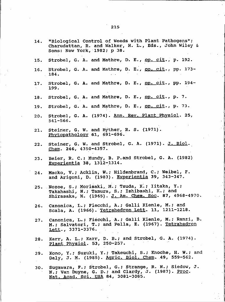
215
14. "Biological Control of Weeds with Plant Pathogens";Charudattan, R. and Walker, H. L., Eds., John Wiley & Sons: New York, 1982; p 38.
15. Strobel, G. A . and Mathre, D. E., O D . cit.„ D . 192.
16. Strobel, 184.
G. A . and Mathre, D. Eep O D . cit.„ D D . 173-
17. Strobel, 199.
G. A. and Mathre, D. E., O D . cit.. D D . 194-
18. Strobel, G. A. and Mathre, D. E., O D . cit.. D . 7.
19. Strobel, G. A . and Mathre, D. E., O D . cit.. D . 73.
20. Strobel, 541-566.
G. A. (1974). Ann . Rev. Plant Phvsiol. 25.
21. Steiner, G. W. and Byther, Phvtooatholoav 61. 691-696
R. S. (1971).
22. Steiner, G. Chem. 246.
W. and StrObel 4350-4357.
, G.. A. f!97n . J. Biol.
23. Beier, R . C .; Mundy, B. P. and Strobel, G. A. (1982)Experientia 38, 1312-1314.
24. Macko, V.? Acklin, W.; Hildenbrand, C.; Weibel, F . and Arigoni, D. (1983). Exoerientia 39, 343-347.
25. Nozoe, S.? Morisaki, M.? Tsuda, K.; Iitaka, Y.; Takahashi, N.; Tamura, S.,* Ishibashi, K.," and Shirasaka, M. (1965). J. Am. Chem. Soc. 87, 4968-4970.
26. Canonica, L.; Fiecchi, A.; Galli Kienle, M.; and Scala, A. (1966). Tetrahedron Lett. 11, 1211-1218.
27. Canonica, L.; Fiecchi, A . ; Galli Kienle, M.? Ranzi, B.M . ; Salvatori, T.; and Pella, E. (1967). Tetrahedron Lett., 3371-3376.
28. Karr, A. L.; Karr, D. B.; and Strobel, G. A. (1974). Plant Phvsiol. 53, 250-257.
29. Kono, Y.? Suzuki, Y.; Takeuchi, S.,* Knoche, H. W.; and Daly, J. M. (1985). Aoric. Biol. Chem. 49, 559-562.
30. Sugawara, F.,* Strobel, G.? Strange, R. N.; Siedow, J .N . ; Van Duyne, G. D.,* and Clardy, J . (1987) . Proc.Nat. Acad. Sci■ USA 84, 3081-3085.

216
31.
32.
33.
34.
35.
36.
37.38.39.
40.
41.
42.
43.
44.
45.
46.
Wolpert, T. J.; Macko, V.; Acklin, W.; Jaun, B.;Seibli J.; Meili, J.; and Arigoni, D. (1985). Experientia 41, 1524-1529.Scheffer, R. P. In "Encyclopedia of Plant Physiology"; Heitefuss, R. and Williams, P. H., Eds., Springer- Verlag: Berlin, 1976; Vol. 4, pp 245-269.Pringle, R. B. and Braun, A. C. (1957).Phytopathology 47, 369-071.Sugawara, K.; Sugawara, F. and Strobel, G. A. (1985). J. Ora. Chem. 50, 5631-5633.French, R. and Lacey, J. Knapweed; Its Cause. Effect, and Spread in Montana; Circular 307, Cooperative Extension Service, Montana State University: Bozeman, MT, 1983.Harris, P. and Cranston, J. (1979). Can. J. Plant Sci. 59, 375-382.Strobel, G. A. and Mathre, D. E., o p . cit.. p 200.Starratt, A. N. (1968). Can. J . Chem. 46, 767-770.Cotty, P. J. and Misaghi, I. J. (1984). Physiology and Biochemistry 74, 785-788.Parikh, V. M. "Absorption Spectroscopy of Organic Molecules"; Addison-Wesley; Reading, MA, 1974; pp 247-252.Silverstein, R. M.; Bassler, G. C.; and Morrill, T. C. "Spectrometric Identification of Organic Compounds"; John Wiley & Sons: New York, 1981; p 236.Silverstein, R. M.; Bassler, G. C.; and Morrill, T. C. ibid., p 208.Silverstein, R. M.; Bassler, G. C.; and Morrill, T. C. ibid., p 220.Nitecki, D. E.; Halpern, B.; and Westley, J. Ora. Chem. 33. 864-866.
J. W. (1967)
Young, P. E . ; Madison, V . ; and Blout, Am. Chem. Soc. 98. 5365-5371.
E. (1975). J-
Madison, V.; Young, P. E.; and Blout, E. (1975). J-
217
Am. Chem. Soc« 98, 5358-5364.47. Stack, M. E.; Mazzola, E. P.; Page, S. W.; Pohland,
A. E.; Highet, R. J.; Tempesta, M. S.; and Corley,D. G. (1986). J. Nat. Prod. 49, 1248.
48. Okuno, T.; Natsume, I.; Sawai, K.; Sawamura, K.; and Furasaki, A. (1983). Tetrahedron Letters 24,5653.
49. Robeson, D.; Strobel, G.; Matsumoto, G. K.; Fisher,E. L.; Chen, M. H.; and Clardy, J. (1984). Experientia 40, 1248-1250
50. Johnston, M. D., NMRCALC, Dept, of Chemistry, University of South Florida, 1985.
51. Silverstein, R. M.; Bassler, G. C.; and Morrill, T. C. op. cit.. p 322.
52. Arnone, A. and Nasini, G. (1986). J. Chem. Soc. Perkin Trans. I . 525-530.
53. Silverstein, R. M.; Bassler, G. C.; and Morrill, T. C. o p . cit.. p 270.
54. Stickings, C. E. (1958). Biochem. J . 72, 332-340.55. Mikami, Y . ; Nishijima, Y.; Iimufa, H.; Suzuki, A and
Tamura, S . (1971). Aar. Biol. Chem. 35, 611-618.56. Chen, Y. S. (1960). Aar. Biol. Chem. 24, 372.57. Tatsuno, T.; Sato, M.; Kubota, Y,; Kubota, Y.; and
Tsunoda, H. (1971). Chem. Pharm. Bull. 19, 1498-1500.58. Tamura, S.; Suzuki, A.; Aoki, Y.; and Otake, N.
(1964.) . Aar. Biol. Chem. 28, 650-652.59. Ottenheijm, H.; Herscheid, J. D. M. and Nivard, R.
(1977). J. Ora. Chem. 42, 925-926.60. Shiguera, H. T. In "Antibiotics I. Mechanism of
Action"; Gottleib, D. and Shaw, P. D., Eds., Springer-Verlag; New York, 1967; pp 360^365.
61. Munro, M. H. G.; Luibrand, R. T. and Blunt, J. W. In "Bioorganic Marine Chemistry"; Springef-Verlag:Berlin, 1987; Vol. I, pp 94-96.
62. Faulkner, D. J. (1979). Oceans 22, 44.

218
63. Ciereszko, L. S. In “Marine Natural Products Chemistry19? Faulkner, D. J. and Fenical, W . , Eds., Plenum Press: New York, 1977; p I.
64. "Antibiotics, Chemotherapeutics, and Antibacterial Agents for Disease Control"; Grayson, M., Ed., John Wiley & Sons: New York, 1982? p 92.
65. "Antibiotics, Chemotherapeutics, and Antibacterial Agents for Disease Control", ibid., p 318.
66. Colwell, R. R. (1983). Science 222, 19-24.67. Wratten, S. J.: Wolfe, M. S.; Andersen, R. J. and
Faulkner, D. J. (1977). Antimicrob. Agents Chemother. 11(3), 411-414.
68. Burkholder, P. R.; Pfister, R. M. and Leitz, R. M. (1966). Appl. Microbiol. 14, 649-653.
69. Okami, Y. (1986). Microbial Ecology 12, 67-78.70. Faulkner, D. J., Scripps Institution of Oceanography,
La Jolla, CA, personal communication , 1988.71. Nakamura, H.; Iitaka, Y.; Kitahara, T.; Okazaki, T .
and Okami, Y. (1977). J. Antib. 30, 714-719.72. Okami, Y.? Okazaki, T.s Kitahara, T. and Umezawa, H.
(1976). J. Antib. 29, 1019-1025.73. Okami, Y.; Hotta, K.; Yoshida, M.,* Ikeda, D.; Kondo,
S. and Umezawa, H. (1979). J. Antib. 32, 964-966.74. Colwell, R. R. Presented at the 1st International
Meeting of Biomedical Prospectives for Marine Microorganisms, La Jolla, CA, March, 1988.
75. Nester, E. W.; McCarthy, B. J.? Roberts, C. E.; and Pearsall, N. N., op. cit.. pp 250-251.
76. Pettit, G. R.; Kamano, Y . ? Herald, C. and Tozawa, M. (1984). J. Am. Chem. Soc. 106, 6768-6771.
77. Pettit, G. R . ; Kamano, Y,} Herald, C. ; Tuinman, A.; Boettner, F. ; Kizu, H . ; Schmidt, J. M.? Baczynskyj ,L.; Tomer, K. B. and Bontems, R. J. (1987). J . A m .Chem. Soc. 109, 6883-6885.
78. Pettit, G. R.; Hartwell, J. L. and Wood, H. B. (1968).
219
Cancer Res. 28, 2168.79. Plinius, G. S . (ca 60). Historia Naturalis, Lib. IX,
Lib. XXII.80. Kosuge, T.; Tsuji, K.; Hirpi, K. and Fukuyama, T.
(1985). Chem. Pharm. Bull. 33(7), 3059-3061.81. Kosuge, T.; Zenda, H.; Ochiai, A.; Masaki, N.;
Noguchi, M.; Kimura, S. and Nairita, H. (1972) . Tetrahedron Lett. 2545.
82. Kosuge, T.; Tsuji, K.; Hirai, K.; Yamaguchi, K.; Okamoto, T. and Iitaka, Y. (1981). Tetrahedron Lett. 22, 3417.
83. Kosuge, T.; Tsuji, K. and Hirai, K. (1982). Chem. Pharm. Bull. 30, 3255.
84. Shimizu, Y. In "Marine Natural Products"; Scheuer, P., Ed., Academic Press: New York, 1978; Vol. I, p 9.
85. Baker, J. T. and Murphy, V. "CRC Handbook of Marine Science"; CRC Press; Cleveland, Ohio, 1976; Vol. I, p 55.
86. Meyer, K. F.; Sommer, H. and Schoenholz, P. (1928).J. Prev. Med. 2, 365-394.
87. Medcof, J. C.; Leim, A. H.; Needier, A. B.; Needier,A. W. H.; Gibbard, J. and Naubert, J. (1947). Bull. Fish. Res. Board Can. 175, 1-32.
88. Needier, A. B* (1949). J. Fish. Res. Board Can. 7, 490-504.
89. Prakash, A. (1963). J. Fish. Res. Board Can. 20, 983-996.
90. Goto, T.? Kishi, Y.; Takahashi, S. and Hirata, Y. (1965). Tetrahedron 21, 2059.
91. Davis, E. W. "The Serpent and the Rainbow"; Simonand Schuster: Austin, Texas, 1986.
92. Yotsu, M.; Yamazaki, T.; Meguro, Y.; Endo, A.; Murata, M.; Naoki, H. and Yasumoto, T. (1987). Toxicon 25(2), 225-228.
220
93. Yasumoto, T. Presented at the 1st International Meeting of Biomedical Prospectives for Marine Microorganisms, La Jolla, CA, March, 1988.
94. Shimizu, Y . Presented at the 1st International Meeting of Biomedical Prospectives for Marine Microorganisms, La Jolla, CA, March, 1988.
95. Tachibana, K.; Scheuer, P. J.; Tsukitani, Y . ; Van Engen, D.;Clardy, J.; Gopichand, Y. and Schmitz, F. J. (1981). J. Am. Chem. Soc. 103, 2469-2470.
96. Giere, 0. and Langheld, C. (1987). Mar. Bot. 93, 641- 650.
97. Distel, D. L and Felbeck, H. (1987). Mar. Bot. 96, 79- 86.
98. Cavanaugh, C. M.; Gardiner, S. L.; Jones, M. L.; Jannasch, H. W. and Waterbury, J. B. (1981). Science 213, 340-342.
99. Giere, 0. (1981). Mar. Ecol. Proa. Ser. 5, 353-357.100. Felbeck, H. (1981). Nature 293, 291-293.101. Dando, P. R.; Southward, A. J. and Southward, E. C.
(1985). Mar. Ecol. Proa. Ser. 23, 85-98.102. Pauli, C. K.; Hecker, B.; Commeau, R.; Freeman-Lynde,
R. P.; Neumann, C.; Corsp, W. P.; Golubic, S.; Hook,J. E . ; Sikes, E. and Curray, J. (1984). Science 226, 965—967.
103. Gustafson, R. G. and Reid, R. G. B. (1988) . Mar. Bot. 97, 389-401.
104. Andrewes, C. H. (1957). Adv. Virus Res. 4, 1-24.105. "Antibiotics, Chemotherapeutics, and Antibacterial
Agents for Disease Control", o p . cit. pp 103-113.106. Demain, A. L. (1981). Science 214, 987-995.107. Blumberg, P. M.; Jaken, S.; Kdnig, B.; Sharkey, N. A.;
- Leach, K. L.; Jeng, A. Y. and Yeh, E. (1984). Biochem.Pharmacol. 33(6), 933-940.
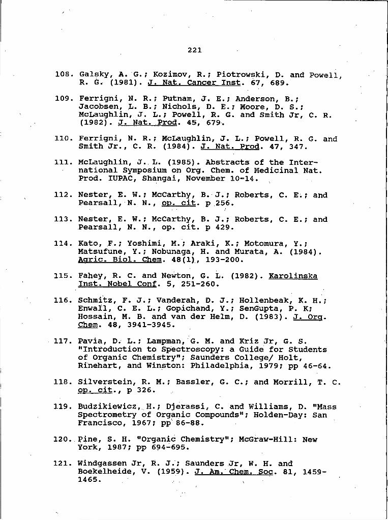
221
108. Gaisky, A. G . ; Kozimov, R.? Piotrowski, D. and Powell R. G . (1981). J. Nat. Cancer Inst. 67, 689.
109. Ferrigni, N. R . ; Putnam, J. E.; Anderson, B.y Jacobsen, L. B.; Nichols, D. E.; Moore, D. S.; McLaughlin, J. L.? Powell, R. G. and Smith Jr, C. R. (1982). J. Nat. Prod. 45, 679.
H O . Ferrigni, N. R.? McLaughlin, J. L.; Powell, R. G. and Smith Jr., C. R. (1984). J. Nat. Prod. 47, 347.
111. McLaughlin, J. L. (1985). Abstracts of the International Symposium on Org. Chem. of Medicinal Nat. Prod. IUPAC, Shangai, November 10-14.
112. Nester, E. W.; McCarthy, B. J . ; Roberts, C. E . ; andPearsall, N . N . . O D . Cit. D 256/ •
113. Nester, E. W . ; McCarthy, B. J . ; Roberts, C . E .; andPearsall, N. N ., op. cit. p 429 .
114. Kato, F.; Yoshimi, M.; Araki, K.; Motomura, Y.; Matsufune, Y.; Nobunaga, H. and Murata, A. (1984). Aaric. Biol. Chem. 48(1), 193-200.
115. Fahey, R. C. and Newton, G. L. (1982). Karolinska Inst. Nobel Conf. 5, 251-260.
116. Schmitz, F. J.; Vanderah, D. J.; Hollenbeak, K. H.? Enwall, C. E. L.; Gopichand, Y.; SenGupta, P. K; Hossain, M. B. and van der Helm, D. (1983). J . Ora. Chem. 48, 3941-3945.
117. Pavia, D. L.; Lampman, G. M. and Kriz Jr, G. S. "Introduction to Spectroscopy: a Guide for Students of Organic Chemistry"; Saunders College/ Holt, Rinehart, and Winston: Philadelphia, 1979; pp 46-64.
118. Silverstein, R. M.; Bassler, G. C.; and Morrill, T. C op. cit.. p 326.
119. Budzikiewicz, H .; Djerassi, C. and Williams, D. "Mass Spectrometry of Organic Compounds”; Holden-Day: San Francisco, 1967; pp 86-88.
120. Pine, S . H. "Organic Chemistry"; McGraw-Hill: New York, 1987; pp 694-695.
121. Windgassen Jr, R. J . ; Saunders Jr, W. H. andBoekelheide, V. (1959). J. Am. Chem. Soc. 81, 1459- 1465. ,
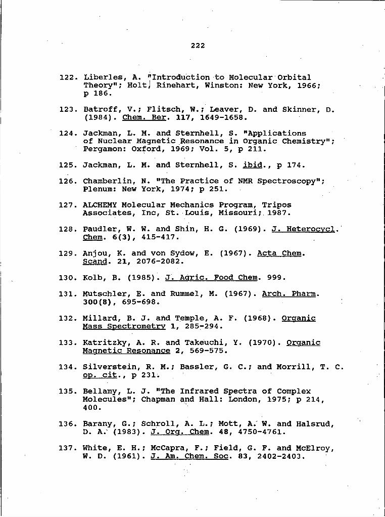
222
122. Liberies, A. "Introduction to Molecular Orbital Theory"; HoltJ Rinehart, Winston; New York, 1966;p 186.
123. Batroff, V.; Flitsch, W.; Leaver, D. and Skinner, D. (1984). Chem. Ber. 117, 1649-1658.
124. Jackman, L. M. and Sternhell, S. "Applicationsof Nuclear Magnetic Resonance in Organic Chemistry"; Pergamon; Oxford, 1969; Vol. 5, p 211.
125. Jackman, L. M. and Sternhell, S. ibid.. p 174.126. Chamberlin, N. "The Practice of NMR Spectroscopy";
Plenum: New York, 1974; p 251.127. ALCHEMY Molecular Mechanics Program, Tripos
Associates, Inc, St. Louis, Missouri; 1987.128. Paudler, W. W. and Shin, H. 6. (1969). J. Heterocvcl.
Chem. 6(3), 415-417.129. Anjou, K. and von Sydow, E. (1967). Acta Chem.
Scand. 21, 2076-2082.130. Kolb, B. (1985). J. Adric. Food Chem. 999.131. Mutschler, E. and Rummel, M. (1967). Arch. Pharm.
300(8), 695-698.132. Millard, B. J. and Temple, A. F. (1968). Organic
Mass Spectrometry I, 285-294.133. Katritzky, A. R. and Takeuchi, Y. (1970). Organic
Magnetic Resonance 2, 569-575.134. Silverstein, R. M.; Bassler, G. C.; and Morrill, T. C
op. cit.. p 231;135. Bellamy, L. J. "The Infrared Spectra of Complex
Molecules"; Chapman and Hall; London, 1975; p 214, 400.
136. Barany, G.; SchrolI, A. L.; Mott, A. W. and Halsrud, D. A. (1983). J. Org. Chem. 48, 4750-4761.
137. White, E. H.; McCapra, F.; Field, G. F. and McElroy, W. D. (1961). J. Am. Chem. Soc. 83, 2402-2403.

223
138. Gunawardana, G. P.; Kohmoto, S.; Gunasekera, S. P.; McConnell, 0. J. and Koehn, F. E. (1988). J. Am. Chem„ Soc. H O , 4856-4858.
139. Budzikiewicz, H.; Djerassi, C. and Williams, D., op. cit., pp 610-613.
140. Pouchert, C. J. and Campbell, J. R. "The Aldrich Library of NMR Spectra"; Aldrich Chemical Company: Milwaukee, W I ; 1974, p 62.
141. Budzikiewicz, H.; Djerassi, C. and Williams, D., pp. cit., pp 276-279.
142. Jackman, L. M. and Sternhell, S., o p . cit.. p 177.143. Baker, J. T. and Duke, C. C. (1973). Aust. J. Chem.
26, 2153-2157.144. Lindgren, G. Bohlin, L. and Bergman, J. (1986).
Tetrahedron Lett. 27, 4782.145. Baker, J. T. and Murphy, V., pp. pit., p 78.146. Zobell, C. E. (1941). J. Mar. Res. 4, 43-75.