217 000-year-old DNA sequences of green sulfur bacteria in Mediterranean sapropels and their...
34
1 1 2 3 4 5 6 7 8 9 217,000 year-old DNA sequences of green sulfur bacteria in Mediterranean 10 sapropels and their implications for the reconstruction of the 11 paleoenvironment 12 13 14 15 16 Marco J.L. Coolen 1 and Jörg Overmann 2 * 17 18 19 1 Woods Hole Oceanographic Institution, Department of Marine Chemistry and Geochemistry, 20 Woods Hole, MA 02543, USA 21 22 2 Section Microbiology, Ludwig-Maximilians-Universität München, Maria-Ward-Str. 1a, D- 23 80638 München, Germany 24 25 26 Running title: Fossil DNA sequences of green sulfur bacteria 27 28 Keywords: ancient DNA, oceanic anoxic events, Chlorobiaceae, fossil DNA, green sulfur 29 bacteria, sapropels 30 31 32 33 34 35 *For correspondence. Email [email protected]; Tel. +49-(0)89-2180-6123; Fax 36 +49-(0)89-2180-6125 37 38 39 40
Transcript of 217 000-year-old DNA sequences of green sulfur bacteria in Mediterranean sapropels and their...

1 1 2 3 4 5 6 7 8 9
217,000 year-old DNA sequences of green sulfur bacteria in Mediterranean 10
sapropels and their implications for the reconstruction of the 11
paleoenvironment 12
13 14 15
16
Marco J.L. Coolen1 and Jörg Overmann2* 17
18
19 1 Woods Hole Oceanographic Institution, Department of Marine Chemistry and Geochemistry, 20
Woods Hole, MA 02543, USA 21 22 2 Section Microbiology, Ludwig-Maximilians-Universität München, Maria-Ward-Str. 1a, D-23
80638 München, Germany 24 25
26
Running title: Fossil DNA sequences of green sulfur bacteria 27
28
Keywords: ancient DNA, oceanic anoxic events, Chlorobiaceae, fossil DNA, green sulfur 29
bacteria, sapropels 30
31
32
33
34
35
*For correspondence. Email [email protected]; Tel. +49-(0)89-2180-6123; Fax 36 +49-(0)89-2180-6125 37
38
39
40
2
SUMMARY 1
Deep-sea sediments of the eastern Mediterranean harbor a series of dark, organic carbon-rich 2
layers, so-called sapropels. Within these layers, the carotenoid isorenieratene was detected. Since 3
it is specific for the obligately anaerobic phototrophic green sulfur bacteria, the presence of 4
isorenieratene may suggest that extended water column anoxia occurred in the ancient 5
Mediterranean Sea during periods of sapropel formation. Only three carotenoids (isorenieratene, 6
β-isorenieratene and chlorobactene) are typical for green sulfur bacteria and thus do not permit 7
to differentiate between the ∼80 known phylotypes. In order to reconstruct the paleoecological 8
conditions in more detail, we searched for fossil 16S rRNA gene sequences of green sulfur 9
bacteria employing ancient DNA methodology. 540 bp-long fossil sequences could indeed be 10
amplified from up to 217,000-year-old sapropels. In addition, such sequences were also 11
recovered from carbon-lean intermediate sediment layers deposited during times of an entirely 12
oxic water column. Unexpectedly, however, all the recovered 16S rRNA gene sequences 13
grouped with freshwater or brackish, rather than truly marine, types of green sulfur bacteria. It is 14
therefore feasible that the molecular remains of green sulfur bacteria originated from populations 15
which thrived in adjacent freshwater or estuarine coastal environments rather than from an 16
indigenous pelagic population. 17
18

3
INTRODUCTION 1
All known green sulfur bacteria and about half of the species of purple sulfur bacteria are 2
obligate anaerobic photolithoautotrophs which can only grow in the presence of light and 3
reduced sulfur compounds as photosynthetic electron donators (Pfennig and Trüper, 1989; 4
Overmann, 2001). Their specific physiological requirements render these bacteria suitable 5
indicator organisms for past environmental conditions (Passier et al., 1999; Menzel et al., 2002). 6
Eastern Mediterranean sediments harbor more than 1 cm-thick, organic carbon-rich layers, 7
so-called sapropels. Sapropels contain >2 % and up to 30% per weight of organic carbon and are 8
embedded in hemipelagic carbonate oozes (< 0.5 wt% organic C) (Kidd et al., 1978). The 9
enhanced organic carbon preservation during sapropel formation has been explained by anoxia 10
of deep Mediterranean bottom water (Oceanic Anoxic Events; Rossignol-Strick, 1985; Rohling 11
and Hilgen, 1991; Passier et al., 1999) or by increased marine primary production (Calvert, 12
1983; Calvert et al., 1992). 1.8 to 3.0 million-year-old Pliocene sapropels were shown to contain 13
the carotenoid isorenieratene and its diagenetic derivatives (Passier et al., 1999; Menzel et al., 14
2002). Since isorenieratene occurs almost exclusively in green sulfur bacteria, it was concluded 15
that sulfidic bottom waters extended into the photic zone during sapropel formation and that the 16
Mediterranean Sea ecosystem underwent repeated and major oxic-anoxic shifts during the past 3 17
million years. 18
However, isorenieratene has also been detected in the actinobacteria Streptomyces griseus 19
and Brevibacterium linens (Krubasik and Sandmann, 2000; Krügel et al., 1999). On the other 20
hand, certain strains of green sulfur bacteria do not contain any detectable amounts of those 21
carotenoids which were originally thought to be typical for this group (Glaeser et al. 2002). 22
Since green sulfur bacteria form a distinct and coherent phylogenetic lineage (Overmann and 23
Tuschak, 1997; Imhoff, 2003), 16S rRNA gene sequences provide an alternative means to trace 24
their occurrence and species composition in the environment (Overmann et al., 1999). While 25
green sulfur bacteria contain only three specific carotenoids (isorenieratene, β-isorenieratene and 26
chlorobactene) (Overmann, 2001), 80 different 16S rRNA gene sequence types are recognized to 27
4
date (A. Manske and J. Overmann, submitted). The three carotenoids occur across all different 1
subgroups of the green sulfur bacteria (Imhoff, 2003), whereas species isolated from the marine 2
environment form a single well-separated sequence cluster (marine group 1) (Imhoff, 2003; 3
Manske et al., 2005). Therefore, analyses of their fossil 16S rRNA gene sequences in subsurface 4
sediments would not only provide independent evidence for the occurrence of green sulfur 5
bacteria but also permit a more detailed reconstruction of their paleoenvironment. To date, 6
however, such a paleomicrobiological analysis has not been performed in the marine 7
environment. 8
Fully hydrated DNA spontaneously decays over only hundreds of years, mainly through 9
hydrolysis and oxidation (Shapiro, 1981; Lindahl, 1993; Hofreiter et al., 2001). However, low 10
temperatures, high ionic strength, anoxic conditions and protection from enzymatic degradation 11
by adsorption extend the half-life of intact DNA by one or two orders of magnitude (Lindahl, 12
1993; Poinar et al., 1996; Willerslev et al., 2004a). So far, intact DNA of anoxygenic 13
phototrophic bacteria could be extracted from up to 9,100 year-old holocene lake sediments and 14
was analyzed by PCR amplification and sequencing (Coolen and Overmann, 1998). Recently, 15
amplification of fossil chloroplast DNA has been found to be reliable for samples which are 16
hundreds of thousands of years old (Willerslev et al., 2003). Under favorable conditions of 17
preservation, genomic DNA of anoxygenic phototrophic bacteria thus may have persisted for 18
similar time periods in Mediterranean sapropels. 19
In the present study, fossil DNA sequences and carotenoids of ancient green sulfur bacteria 20
could be recovered from sapropels deposited between 8,000 and 217,000 years ago in the 21
Eastern Mediterranean. Phylogenetic analyses were used to infer the most likely origin of the 22
respective bacteria and provide a more detailed picture of the development of this marine 23
ecosystem. 24
25
5
RESULTS 1
Vertical structure and age of sediment layers 2
A four meter-long sediment core obtained at a water depth of 2155 m from the Eastern 3
Mediterranean southeast of Crete contained four different sapropels which were identified as 4
sapropels S1, S6, S7, and S8 based on geochemical evidence (Coolen et al., 2002; Fig. 1). The 5
ages of these sapropels are 8,000 (S1), 172,000 (S6), 195,000 (S7) and 217,000 (S8) years (Emeis 6
et al., 2000). The organic carbon content of the sapropels ranged between 2.3 and 8.5% (w/w), 7
whereas intermediate layers contained only up to 0.16 % (Coolen et al., 2002). The intermediate 8
layer Z1 is likely to represent a turbidite (K.-C. Emeis; pers. comm.). 9
For subsequent analyses, the top of the sediment (Z0, 4-6 cm below surface), each sapropel 10
layer (S1, 13-17 cm; S6, 262-266 cm; S7, 314-318 cm; and S8, 364-368 cm), and three 11
intermediate hemipelagic layers between the sapropels (denoted Z1, 62-66 cm; Z6, 285-289 cm; 12
and Z7, 333-337 cm) were selected (Fig. 1). 13
Green sulfur bacterial carotenoids in late Pleistocene and Holocene sapropels 14
Isorenieratene and β-isorenieratene could be detected in all four sapropel layers where they 15
reached concentrations between 56.4 and 1280 ng·(g dry weight sediment)-1 (Fig. 1C). Initially, 16
both carotenoids were identified by HPLC based on their characteristic retention time and 17
absorption spectra (peaks at 454 and 482 nm and a shoulder at 428 nm). Analysis of these 18
compounds by LC-MS in the APCI positive ion mode yielded major signals at m/z values of 529 19
and 133. These masses match those of the protonated isorenieratene (i.e., [M+H]+) and of 20
1,2,3,4-tetramethylbenzene, respectively, the latter representing a fragment typical of 21
isorenieratene and β-isorenieratene. Our mass spectrometric analysis thus provides the first 22
evidence for the presence of carotenoids of green sulfur bacteria in late Pleistocene and 23
Holocene sapropels. 24
In contrast, only traces of carotenoid compounds with the retention times and absorption 25
spectra of isorenieratene and β-isorenieratene were detected in intermediate layers. 26
6
Concentrations ranged from (1.4 ± 0.1) to (5.9 ± 0.3) ng·(g dry weight sediment)-1 (Fig. 1C). 1
Although above the detection limit, these trace amounts did not allow further analyses by LC-2
MS. Therefore, it cannot be completely ruled out that other carotenoids were present, though 3
carotenoids with retention times and absorption spectra identical to isorenieratene/β-4
isorenieratene are currently not known. 5
Detection and quantification of DNA of green sulfur bacteria 6
After optimizing our extraction protocol, genomic DNA could be isolated from all eight 7
sediment layers (Fig. 1A). Between 0.7 and 5.0 µg of DNA were extracted per gram dry weight 8
of sediment from the four sapropels. These amounts significantly exceeded those obtained from 9
the intermediate layers (0.01 to 0.12 µg·(g dry wt)-1) and were only matched by the value from 10
the sediment surface (1.04 µg·(g dry wt)-1). Analysis by gel electrophoresis revealed that the 11
DNA mostly consisted of up to 23 - 30 kb long fragments (data not shown). The shortest 12
fragments detected measured 500 bp. This length distribution was even observed in extracts from 13
the 217,000-year-old Mediterranean sapropel S8. 14
The specific primer combination and PCR conditions used in the present study permit the 15
selective amplification of 16S rRNA gene fragments of green sulfur bacteria (Overmann et al., 16
1999). With the exception of the intermediate layer Z6, all samples yielded amplification 17
products. The DNA extract of sapropel S7 yielded only a faint amount of DNA of the correct 18
molecular size, but also some unspecific amplification products. Of all sapropel layers 19
investigated, S7 contains the by far highest amount of kerogen (total organic carbon content of 20
8.5% as compared to ≤4.4% in other sapropels; Coolen et al., 2002). Similarly, Z6 contained the 21
highest amount of total organic carbon of all intermediate layers (0.16 versus ≤0.01%). Most 22
likely, the failure to obtain appropriate amplification products from these two layers resulted 23
from the unfavourable ratio of fossil DNA to organic kerogen. 24
Each amplification reaction was rigorously checked for contamination with extraneous DNA 25
by routinely including a set of procedural blanks and PCR controls (as detailed in the Materials 26
7
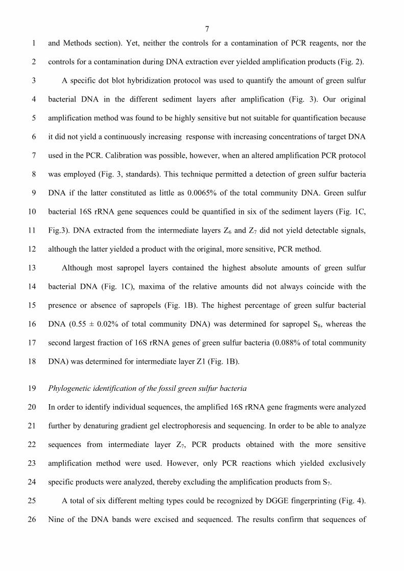
and Methods section). Yet, neither the controls for a contamination of PCR reagents, nor the 1
controls for a contamination during DNA extraction ever yielded amplification products (Fig. 2). 2
A specific dot blot hybridization protocol was used to quantify the amount of green sulfur 3
bacterial DNA in the different sediment layers after amplification (Fig. 3). Our original 4
amplification method was found to be highly sensitive but not suitable for quantification because 5
it did not yield a continuously increasing response with increasing concentrations of target DNA 6
used in the PCR. Calibration was possible, however, when an altered amplification PCR protocol 7
was employed (Fig. 3, standards). This technique permitted a detection of green sulfur bacteria 8
DNA if the latter constituted as little as 0.0065% of the total community DNA. Green sulfur 9
bacterial 16S rRNA gene sequences could be quantified in six of the sediment layers (Fig. 1C, 10
Fig.3). DNA extracted from the intermediate layers Z6 and Z7 did not yield detectable signals, 11
although the latter yielded a product with the original, more sensitive, PCR method. 12
Although most sapropel layers contained the highest absolute amounts of green sulfur 13
bacterial DNA (Fig. 1C), maxima of the relative amounts did not always coincide with the 14
presence or absence of sapropels (Fig. 1B). The highest percentage of green sulfur bacterial 15
DNA (0.55 ± 0.02% of total community DNA) was determined for sapropel S8, whereas the 16
second largest fraction of 16S rRNA genes of green sulfur bacteria (0.088% of total community 17
DNA) was determined for intermediate layer Z1 (Fig. 1B). 18
Phylogenetic identification of the fossil green sulfur bacteria 19
In order to identify individual sequences, the amplified 16S rRNA gene fragments were analyzed 20
further by denaturing gradient gel electrophoresis and sequencing. In order to be able to analyze 21
sequences from intermediate layer Z7, PCR products obtained with the more sensitive 22
amplification method were used. However, only PCR reactions which yielded exclusively 23
specific products were analyzed, thereby excluding the amplification products from S7. 24
A total of six different melting types could be recognized by DGGE fingerprinting (Fig. 4). 25
Nine of the DNA bands were excised and sequenced. The results confirm that sequences of 26
8
green sulfur bacteria had been selectively amplified from the extracted DNA. Seven of the 1
bands (A through G in Fig. 4) yielded unambiguous sequences. Of the latter, bands A, B, E and F 2
not only exhibited the same melting behavior during DGGE, but also contained the same 3
sequence. This phylotype was found to be identical to those of 11 other strains or environmental 4
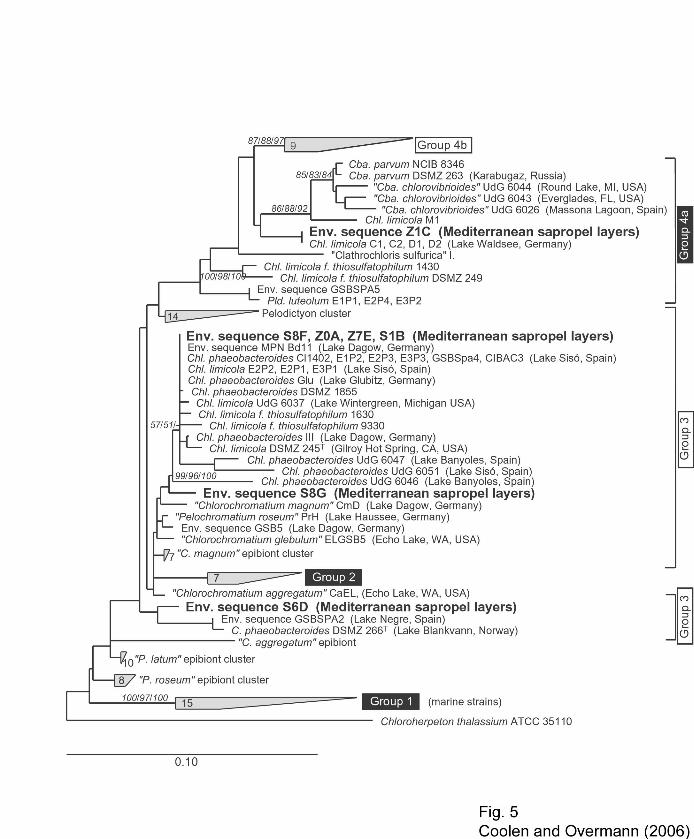
sequences (Fig. 5) and fell into Group 3 among the green sulfur bacteria. Sequence type C was a 5
member of Group 4a and only present in the intermediate layer Z1, yet could not be detected in 6
the adjacent sapropel S1 or the surface layer Z0. Two other sequences (D and G) fell into Group 3 7
(Fig. 5). Each represented a distinct, so far unknown sequence type (Fig. 5) and was found in 8
only a single sapropel layer but was missing in all other samples (Fig. 4). 9
Adsorptive binding of DNA to the sediments 10
Sediment material of the sapropel S6 and the intermediate layer Z6 was chosen for adsorption 11
assays since they contained comparatively low amounts of indigenous DNA compared to the 12
other layers (Fig. 1A). Since the concentration of indigenous DNA determined (1.44 µg·(g dry 13
weight sediment)-1 in S6) amounted to only a very minor fraction (0.003%) of the total 14
adsorption capacity, the DNA already present in the samples did not interfere with these 15
adsorption assays. 16
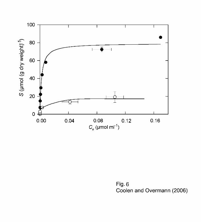
The sapropel exhibited an extraordinarily high maximum adsorption capacity Smax for DNA 17
((79.3 ± 2.8) µmol double-stranded DNA (g dry wt)-1; corresponding to 52.8 mg (g dry wt)-1) 18
(Fig. 6, Table 1). This value is comparable to that of pure montmorillonite and is only surpassed 19
by purified humic acids (Table 1). Albeit lower, the adsorption capacity of Z6 for double 20
stranded DNA was still significantly higher than that of soil (Table 1). 21
22
23
9
DISCUSSION 1
Authenticity of the green sulfur bacterial DNA 2
Analyses of ancient DNA are usually based on minute amounts of highly degraded template 3
molecules and thus are subject to a considerable risk of contamination (Cooper and Poinar, 4
2000). In order to avoid sources of extraneous DNA in the present study, subsampling of the 5
sediment cores was performed in a separate, PCR-product-free laboratory in which green sulfur 6
bacteria had never been worked with before, and a security level 2 laminar flow chamber 7
dedicated to work with low DNA template number samples was used for subsequent DNA 8
extraction and to set up amplification reactions. Each set of DNA extractions included controls 9
for contamination of the chemicals and vessels employed. None of these extraction controls 10
yielded an amplification product, indicating that no extraneous DNA from green sulfur bacteria 11
had been introduced during DNA isolation. Furthermore, each amplification run comprised two 12
reactions devoid of DNA template in order to control separately for a potential contamination of 13
PCR reagents and tubes. These negative controls also did not yield any PCR product. Finally, 14
quantification of fossil green sulfur bacterial DNA was performed in three independent parallels 15
to test reproducibility of the results. 16
Whereas parallel extraction trials for the identical sediment layer were not feasible due to the 17
restricted availability of deep sea sediment samples, a comparison of the results from the 18
consecutive sediment horizons further confirms that the DNA genuinely originated from these 19
samples. Sequences of green sulfur bacteria were not only detected on a single occasion, but 20
occurred in seven out of eight different sediment layers. Furthermore, amplification of green 21
sulfur bacterial sequences was reproducible for the same extract (compare standard deviations 22
for quantification of green sulfur bacterial amplification products in Fig. 1C). 23
Three of the four sequence types obtained (Z1-C, S6-D, S8-G) were detected in only a single 24
sample. It appears unlikely that each of the three sediment samples Z1, S6 and S8 was 25
contaminated with yet another, different type of green sulfur bacterium. Two of these sequence 26
types (S6-D, S8-G) have neither been isolated as a clone nor from a culture before. It is also 27
10
extremely unlikely that two samples were contaminated individually, yet by two different 1
sequences which are not available in any laboratory world wide. Our phylogenetic analysis of the 2
recovered sequences thus provides independent evidence for the conclusion that the green sulfur 3
bacterial DNA is indigenous to the Mediterranean sediments. 4
Fossil origin and mechanisms of persistence of green sulfur bacterial DNA 5
Compared to the multitude of studies targeting higher organisms, studies of ancient bacteria have 6
been limited to a few pathogenic species (Mycobacterium tuberculosis, M. leprae, Yersinia pestis 7
and Treponema pallidum) or to intestinal bacteria, and cover a time period of only the last 5,400 8
years (Rollo, 1998). Information on past bacterial communities in the environment is very sparse 9
(Coolen and Overmann, 1998; Coolen et al., 2006; Willerslev et al., 2004b) which can be 10
attributed to the difficulty to distinguish ancient from modern bacterial DNA. 11
All cultured representatives of the green sulfur bacteria are obligately anaerobic 12
photolithoautotrophs and, accordingly, require the simultaneous presence of light and sulfide for 13
growth. The family Chlorobiaceae comprises all known green sulfur bacteria plus numerous 14
environmental sequences (Overmann and Tuschak, 1997; Overmann et al., 1999; A. Manske and 15
J. Overmann, submitted). Similar to their cultured relatives, all so-far-uncultured members of 16
this group have been exclusively detected in illuminated sulfidic environments like the 17
chemocline of lakes, lagoons or benthic microbial mats. As the sequences recovered in the 18
present study clearly fall within this group, the available evidence indicates that they originated 19
from obligately anaerobic photolithoautotrophs. All our attempts to enrich green sulfur bacteria 20
by different cultivation methods (Overmann, 2001; Manske et al., 2005) failed, supporting the 21
view that viable green sulfur bacteria do not exist in these sediments. In conclusion, the green 22
sulfur bacterial sequences obtained in the present study are highly unlikely to originate from 23
bacteria growing within the sediments. Rather, the cells grew outside the sediments and the 24
aphotic portion of the water column and were subsequently deposited in Mediterranean deep sea 25
sediments. 26
11
DNA spontaneously decays into 100 - 600 bp short fragments after its deposition in 1
lacustrine sediments (Coolen and Overmann, 1998). Based on our data, green sulfur bacterial 2
genome fragments at least 540-bp in size persisted in Mediterranean sapropels over 217,000 3
years. Survival of ancient DNA in a lake sediment could be demonstrated for a time period of 4
9,100 years (Coolen and Overmann, 1998) due to the short lifespan of this ecosystem. The 5
partial 16S rRNA gene fragments obtained in this study are among the oldest authenticated 6
ancient bacterial sequences available to date. For animals and plants, the oldest authenticated 7
records come from 60,000-year-old remains of brown bears (Barnes et al., 2002), and from 8
~400,000-year-old chloroplast DNA (Willerslev et al., 2003), respectively, both recovered from 9
Alaskan permafrost. The age of the fossil 16S rRNA gene sequences of green sulfur bacteria 10
detected in the present study falls well within this time frame. Based on the relatively high in situ 11
temperature of 14°C, however, the efficient preservation of fossil DNA in eastern Mediterranean 12
sediments must be attributed to factors other than low temperature. 13
Besides low temperatures, the persistence of fossil DNA is significantly extended by high 14
ionic strength, anoxic conditions, and rapid dehydration and adsorption of DNA (Lindahl, 1993; 15
Poinar et al., 1996; 2003; Willerslev et al., 2004a). The ionic strength and anoxic conditions in 16
eastern Mediterranean sediments are comparable to other sediment environments. Adsorption to 17
clay and other mineral surfaces significantly decreases the degradation rates for organic 18
compounds in soils (Jones and Edwards, 1998), and marine sediments (Keil et al., 1994) by up to 19
five orders of magnitude. Adsorptive binding to hydroxyapatite retards the spontaneous decay of 20
DNA (Lindahl, 1993). In addition to spontaneous hydrolysis, microbial DNase activity leads to 21
rapid degradation of free DNA in water and sediments (Lorenz and Wackernagel, 1994), but is 22
also effectively prevented by adsorption even in sandy sediments which bind DNA much less 23
efficiently than the sapropels (Romanowski et al., 1991; Crecchio and Stotzky, 1998). Based on 24
our results, the outstanding adsorption capacity of eastern Mediterranean sediments, in particular 25
of the sapropel matrix, represents the major reason for the efficient preservation of fossil DNA of 26
12
green sulfur bacteria. Potentially, sapropels may also represent important archives of fossil DNA 1
from other microorganisms. 2
Implications for the reconstruction of past ecosystems 3
To date, isorenieratene and its degradation products have been used as an indicator of past water 4
column anoxia (Passier et al., 1999; Menzel et al., 2002). Because of their very limited diversity, 5
carotenoids do not permit a differentiation between species with contrasting physiology and 6
ecology. In the eastern Mediterranean, isorenieratene has previously been shown to occur in ≥1.8 7
million-year-old Pliocene sapropels (Passier et al., 1999; Menzel et al., 2002). However, only 8
four of the 83 currently recognized sapropels (Emeis et al., 2000) have been investigated so far. 9
It was therefore unknown whether isorenieratene represents a typical constituent of the 10
sapropels. The present study provides the first instance of occurrence of isorenieratene in marine 11
deposits which are sufficiently young to also harbor 16S rRNA gene sequences of green sulfur 12
bacteria. The latter are high resolution fossil biomarkers which permit an improved assessment 13
of their origin and hence a more detailed reconstruction of the paleoenvironment. 14
All but one (Z1C) of the 16S rRNA gene sequences recovered were phylogenetically 15
affiliated with typical freshwater or brackish water species, whereas no single representative of 16
the marine group 1 could be detected. Secondly, the presence of green sulfur bacterial 16S rRNA 17
genes in intermediate layers was unexpected since the latter were deposited under a fully 18
oxygenated water column (Schmiedl et al., 1998). Theoretically, DNA could have reached 19
intermediate layers through vertical percolation after deposition in the sapropel layers. However, 20
vertical migration of fossil DNA fragments should be independent of their actual base sequence 21
and, as a result, sequence types present in sapropels should also be detectable in the adjacent 22
intermediate layers. Yet, phylotype C was detected in intermediate layer Z1, but not in the 23
adjacent S1 (compare Fig. 4). This indicates that the strong adsorption to the sediment particles 24
has effectively immobilized the fossil DNA of green sulfur bacteria. Thirdly, a long-distance 25
transport of green sulfur bacterial biomarkers has been shown for North Atlantic deep-sea 26
13
sediments (Rosell-Melé et al., 1997). Indeed, a turbidite origin was proposed for the intermediate 1
layer Z1 (K.-C. Emeis, pers. comm.) which contained the by far highest fraction of green sulfur 2
bacterial DNA among all organic carbon lean intermediate layers. It thus appears feasible that at 3
least the DNA of green sulfur bacterial phylotypes affiliated with typical freshwater species may 4
have originated from environments like coastal lagoons where these bacteria frequently form 5
dense blooms. 6
In conclusion, our results suggest that not all green sulfur bacteria deposited in the ancient 7
Mediterranean Sea during the last 217,000 years were autochthonous but instead are more likely 8
to have originated from Mediterranean coastal environments. Our cumulative evidence casts 9
some doubts on the hypothesis of the presence of green sulfur bacteria in the open waters of the 10
eastern Mediterranean and, by inference, questions the alleged past photic zone anoxia. A future 11
isolation and physiological characterization of the two previously unknown phylotypes detected 12
in the present study will help to more precisely assign a certain habitat, hence origin, to the fossil 13
remains of green sulfur bacteria present in the sapropel layers. 14
15
14
EXPERIMENTAL PROCEDURES 1
Sampling and sample preparation 2
A gravity core (#69-2SL) was obtained during cruise 40 leg 4 of the R/V Meteor on January 30, 3
1998, at position 33°51,53'N and 24°54,46'E southeast of Crete. The core was collected at a 4
depth of 2155 m and was subsampled employing the aseptic techniques developed for the 5
recovery of fossil DNA from sediment samples (Coolen and Overmann, 1998; 2000). It was cut 6
longitudinally which left behind a potentially contaminated surface. This surface was rapidly 7
frozen with powdered dry ice and subsequently lifted off. Through the freshly exposed surface, 5 8
cm3 subsamples were retrieved aseptically using sterile plastic syringes which had their ends cut 9
off and were immediately stored in sterile vials at –800C until extraction. 10
Precautions and controls to prevent contamination 11
Preparation of sediment samples was performed directly after retrieval of sediment cores in the 12
laboratory on board of the R/V Meteor, employing the aseptic DNA techniques established 13
previously (Coolen and Overmann, 1998; Coolen et al., 2006). Green sulfur bacteria had never 14
been introduced or worked with in these premises. Further precautions against contamination of 15
the samples with foreign DNA included the use of a laminar flow hood dedicated to low 16
template number samples. Prior to each use, the hood was UV-sterilized for 4 hours and all 17
surfaces were subsequently sterilized with sodium hypochlorite. Nucleic acids-free disposable 18
plastics were used throughout and autoclaved before use. All solutions were prepared in fresh 19
double quartz-distilled water, sterile filtered, and autoclaved. As a control for contamination 20
during DNA-extraction, two procedural blanks without sediment were subjected to the whole 21
extraction and purification procedure along with the sediment samples. One µl of each of these 22
extraction controls (corresponding to the average volume of sediment extracts used for 23
amplifications) was included in subsequent PCR amplifications. As additional controls, each 24
amplification included reactions without DNA template to independently control for 25
contamination of PCR reagents. 26
15
Pigment analysis 1
Pigments were extracted in the dark from 10 grams of freeze-dried and finely ground sediment 2
samples. Acetone/methanol (7/2, v/v), acetone, and dichloromethane were used for successive 3
extractions, followed by alkaline hydrolysis of the sediment with 6% KOH in methanol (Glaeser 4
et al., 2002). To the latter, distilled water and dichloromethane were added and after phase 5
separation, the organic phases were combined and concentrated by rotary evaporation. The 6
extracts were subsequently dried under a flow of nitrogen. Individual pigments were quantified 7
by reverse-phase HPLC (Glaeser et al., 2002) employing a NovaPak C18 end-capped 60Å 4 µm 8
4.6 × 250 mm column (Waters) with a Spherisorb 5 ODS 2 4.6 × 10 mm guard column (Waters). 9
The detection limit of this method is 1.0 ng isorenieratene·(g dry weight sediment)-1. 10
For mass spectroscopy, the residue was redissolved in dichloromethane and applied to a 11
silica column. The apolar carotenoids were eluted with dichloromethane, dried under nitrogen 12
and the residue dissolved in acetone. This carotenoid fraction was then immediately analyzed on 13
a HP 1100 series LC/MS equipped with an auto-injector and photodiode array detector. 14
Separation was achieved on a ZORBAX Eclipse XDB-C18 column (2.1 × 150 mm, 5 µm; Agilent 15
Technologies, USA), maintained at 25ºC, with a linear gradient from 100% solvent A to 100% 16
solvent B in 50 min at a flow rate of 0.6 ml·min-1. Solvent A was methanol/water (4:1, v/v) and 17
solvent B acetone/methanol/water (19:1:1, v/v/v). Total run time was 60 minutes. Detection was 18
achieved by in-line UV-detection (250−700 nm) and positive ion APCI (Atmospheric Pressure 19
Chemical Ionization) of the eluent in either scanning or Single Ion Monitoring (SIM) mode. 20
Conditions for APCI-MS were as follows: nebulizer pressure 60 psi, vaporizer temperature 21
325°C, drying gas (N2) flow 7 L·min-1 and temperature 350°C, capillary voltage 3000V, corona 4 22
µA. Positive ion spectra were generated by scanning m/z 100-1000. In SIM mode m/z 529 23
(protonated molecule of isorenieratene) was monitored. Isorenieratene was quantified by 24
comparing its UV response at 454 nm (the λmax of isorenieratene in the mobile phase) to known 25
amounts of an authentic β-carotene standard (Aldrich) and correcting for the difference in 26
extinction coefficients (Britton, 1995). 27
16
Extraction of genomic DNA 1
Each sediment sample was aliquoted in ten 2 ml bead-beat vials and 0.7 g of glass beads 2
(0.1 mm diameter) and 0.9 ml lysis buffer (100 mM Tris-HCl, 500 mM sodium-EDTA and 3
1 wt% SDS; pH 8.0) were added to each vial. The sediment samples were pre-heated for 5 4
minutes at 70°C in a water bath. Cell-lysis was accomplished by bead-beating at 5,000 rpm for 5
80 sec (Biospec Mini Bead-Beater; Bartlesville, Oklahoma, USA) followed by another 6
incubation at 70°C for 30 min. Following centrifugation of the samples in a microfuge (2 min; 7
14,000 rpm), the supernatants were transferred to a 45 ml teflon centrifuge tube. For extraction 8
of extracellular, adsorbed DNA, each pellet was resuspended in 0.8 ml of 0.12 M sodium 9
phosphate buffer (pH 8.0) containing 1% wt/vol SDS, followed by a second round of preheating, 10
bead-beating and heating. Following centrifugation, the supernatants were recovered. Finally, 11
each subsample was washed three times with 0.8 ml of 0.12 M sodium phosphate buffer (without 12
SDS). For each sediment, the supernatants of all subsamples were pooled, yielding a total 13
volume of about 40 ml. 14
Organic carbon in Mediterranean sapropels contains long chains of polymethylenic carbon 15
(Petsch et al. 2001), which copurified with the genomic DNA and inhibited subsequent PCR. For 16
removal of the kerogen, several consecutive purification steps were required. Most of the 17
kerogen could be removed from the crude DNA extracts by adding 3 grams of autoclaved, acid 18
washed polyvinylpolypyrrolidone (PVPP; Zhou et al., 1996) to each sample. After an incubation 19
for 20 min on a rotary shaker, the samples were centrifuged for 20 min at 20,400 x g (Beckman 20
J2-HS, München, Germany), and the supernatants were transferred to a new sterile centrifuge 21
tube. Extractions with phenol, phenol/chloroform/isoamylalcohol, and chloroform followed 22
(Sambrook et al., 1989) and the DNA was recovered by standard ethanol precipitation. Finally, 23
the resulting pellet was washed with sterile double distilled water using a Centricon 50 24
ultrafiltration unit (Amicon; Witten, Germany) and purified with the Wizard PCR-preps DNA-25
purification kit (Promega, Mannheim, Germany). The DNA concentrations in the extracts were 26
quantified by fluorescent dye binding with PicoGreen (MoBiTec, Göttingen, Germany). 27
17
Amplification of 16S rRNA genes of green sulfur bacteria 1
16S rRNA sequences of green sulfur bacteria were selectively amplified with eubacterial primer 2
341f and the group-specific primer GSB822r, using the previously published cycling conditions 3
(Overmann et al., 1999). The phylum green sulfur bacteria consists of a crown group of closely 4
related green sulfur bacteria sensu strictu (the family Chlorobiaceae) as well as an increasing 5
number of deep-branching phylotypes which are all uncultured and hence of unknown 6
physiology. However, all members of the crown group investigated to date are typical 7
unicellular, obligate photolithoautotrophs. We reassessed the specificity of primer GSB822 by 8
also including the novel database entries for 16S rRNA gene sequences. Of a total of 86 9
sequences of Chlorobiaceae tested, 11 showed a base substitution (A for T) at the 3´-end of the 10
target sequence: 5´-AATACTAGATGTTGG(A instead of T)CAT-3´. In three other phylotypes, 11
different base substitutions were found. All but one of these sequences fell into group 4a (Fig. 5), 12
while the remaining was a member of group 3. In contrast, all truly marine phylotypes contained 13
the probe target sequence. Based on this analysis, the PCR method was therefore found to be 14
suitable to detect in particular the marine members of the green sulfur bacteria. 15
For separation by DGGE, a 40 bp-long GC-clamp has to be added at one end of the PCR 16
product during amplification, employing primer GC 341f which carries the GC clamp (5‘-17
CGCCCGCCGCGCCCCGCGCCCGGCCCGCCGCCCCCGCCCC-3‘) at its 5´-end (Muyzer et 18
al., 1998)). Initial experiments revealed a significantly reduced detection limit of green sulfur 19
bacterial 16S rRNA genes when GC341f was employed directly for amplification. Consequently, 20
the GC clamp was added by performing 15 additional cycles with 1 µl of the primary 21
amplification and with 0.5 pmoles each of primers GC341f and GSB822r. 22
In order to control for the specificity of the amplification conditions, genomic DNA of 23
Chlorobium phaeobacteroides strain MN1 (Overmann et al., 1992) and Chl. phaeovibrioides 24
DSMZ 269T were used as positive controls. Since the Bacteroidetes represent the sister group of 25
the phylum green sulfur bacteria, DNA of Cytophaga sp. strain 2b served as a negative control. 26
Purity of the PCR reagents was checked by including two reactions without DNA template in 27
18
each amplification trial. Possible contaminations during DNA extraction were checked by 1
including reactions spiked with one µl of each of the extraction controls (see above). 2
Denaturing gradient gel electrophoresis (DGGE) 3
PCR-products were separated by DGGE (Muyzer et al., 1998) in a Bio-Rad D Gene system 4
(Biorad, München, Germany). PCR samples were applied directly onto 6% (wt/vol) 5
polyacrylamide gels (acrylamide/N,N'-methylene bisacrylamide ratio, 37:1 [w/w]) in 1 x TAE 6
buffer (pH 7.4) which had been prepared from sterile solutions and were cast aseptically between 7
sterilized glass plates. The gels contained a linear gradient of 30% to 70% denaturant (100% 8
denaturant correspond to 7 M urea plus 40% [v/v] formamide). Electrophoresis proceeded for 5 h 9
at 200 V and 60°C. Afterwards, gels were stained for 20 min with sterile ethidium bromide 10
solution and photographed. Finally, individual fragments were excised with a sterile scalpel, 11
the DNA was eluted in sterile 1 x TAE (pH 8.0) by electrophoresis (3 h, 200 V) in Centricon 50 12
concentrators inserted into a Centrilutor Micro Electroelutor (Amicon, Witten, Germany). 13
Sequencing and phylogenetic analysis 14
One µl of the eluted DNA was reamplified with primers 341f and GSB840r. Primers and 15
deoxyribonucleoside triphosphates were removed using the QIAquick PCR Purification Spin Kit 16
(Qiagen, Hilden, Germany). After cycle sequencing with the SequiTherm EXCEL Long-Read 17
Sequencing Kit-LC (Biozym, Hess. Oldendorf, Germany) and employing primers 341f and 18
GSB840r, sequence data were collected with a LiCor-4000 automated sequencer (Lincoln, 19
Nebraska, USA). 20
Each sequence was checked for chimeras employing the CHECK_CHIMERA option of the 21
ribosomal database project (RDP). The 16S rRNA gene sequences were then analyzed using the 22
ARB phylogeny software package (Ludwig et al., 2004). The Fast Aligner V1.03 tool was used 23
for automatic alignment and the resulting alignments were corrected based on the 16S rRNA 24
secondary structure information for Chlorobium vibrioforme DSMZ 260T, as available through 25
The Comparative RNA Web (CRW) Site (www.rna.icmb.utexas.edu; Cannone et al., 2002). 26
19
Phylogenetic trees were constructed including 16S rRNA gene sequences of available strains and 1
environmental sequences. First, sequences longer than 1100 bp were used for the calculation, 2
employing the MAXIMUM LIKELIHOOD algorithm (Fast DNA_ML). The shorter 3
environmental sequences were inserted afterwards without changing overall tree topology 4
employing the PARSIMONY INTERACTIVE tool implemented in the ARB software package. 5
The 16S rRNA gene sequences obtained during the present study have been deposited in 6
GenBank under accession numbers AF298531-AF298537. 7
Quantification of DNA of green sulfur bacteria by dot blot hybridization 8
For dot blot quantification, 16S rRNA gene sequences of green sulfur bacteria were amplified 9
with an altered PCR protocol. Instead of a step down, primer annealing was performed at a 10
constant temperature of 52°C for 40 s and 31 cycles. Samples were denatured for 7 min at 100°C 11
and vacuum blotted onto positively charged nylon membranes (Boehringer Mannheim, 12
Germany). The membrane was baked (25 min at 120°C) and pre-hybridized in 10 ml of DIG 13
Easy Hyb buffer (Boehringer). Afterwards, 150 pmole of a highly specific probe for green sulfur 14
bacteria (5’- TGCCACCCCTGTATC-3’; E. coli positions 532 to 546; Tuschak et al., 1999), 5’ 15
labeled with dig-11-dUTP (MWG-Biotech, Germany), was added and hybridization was carried 16
out for 12 h. After hybridization, the blot was washed twice for 5 min in 2 x SSC (150 mM 17
NaCl, 15 mM Na-citrate, pH 7.0) plus 0.1% SDS, followed by two stringent washing steps 18
(15 min in 0.1xSSC/0.1% SDS). The hybridization signal was detected by chemiluminescence 19
with the DIG Luminescent Detection Kit (Boehringer). Lumi-Film (Boehringer) was exposed for 20
30 min, developed and the image digitized with a flatbed scanner. For quantification of the 21
individual dots, the ZERO-Dscan software (Scanalytics, Billerica, USA) was employed. 22
Genomic DNA of Chlorobium phaeovibrioides DSMZ 269T was used for calibration. The 23
homology of the 16S rRNA gene sequences of other Bacteria at the target site of probe 532 is 24
low, which reflects the large phylogenetic distance of green sulfur bacteria to other bacteria 25
(Overmann and Tuschak, 1997). Therefore we chose DNA of Cytophaga strain 2b (which, 26
20
however shows 5 mismatches) as a negative control for the specificity of the hybridization. For 1
each sample, quantification was conducted in three independent parallels. The quantification was 2
based on the assumption that genome sizes of green sulfur bacteria and the number of rRNA 3
operons are similar. Based on the currently available information, these assumptions appear to be 4
largely valid since genome sizes of green sulfur bacteria show comparatively little variation in 5
length (1.97 to 3.13 Mb) and typically comprise one (33% of the genomes) or two (66% of the 6
genomes) rrn operons (http://genome.jgi-psf.org/draft_ microbes/). 7
Adsorption capacity for DNA of the sapropels 8
In order to study the potential protection of fossil DNA by adsorption, equilibrium adsorption 9
isotherms were determined. Herring sperm DNA (Boehringer Mannheim) at concentrations 10
between 1 and 10 µg·ml-1 was incubated for 12 hours with aliquots (5 mg dry weight·ml-1) of 11
sapropel S6 and the intermediate layer Z6. Adsorption of DNA was observed to reach a 12
maximum and therefore yielded the best fit with the Langmuir equation (Ogram et al., 1987) 13
according to: 14
!
S = Smax
"K "C
e
(1+ K "Ce)
(1) 15
Equation (1) was fitted to the data points of the amount S of DNA adsorbed (µmol·(g dry wt)-1) 16
and the concentration Ce of the substance remaining in solution (in µmol·ml-1). This yielded an 17
estimate for the maximum adsorption capacity Smax (µmol·(g dry wt)-1) and the Langmuir affinity 18
coefficient K (ml·µmol-1). For comparison, equilibrium adsorption isotherms of DNA for 19
montmorillonite and cellulose were also measured. 20
21

21
ACKNOWLEDGEMENTS 1
We thank Daniel Dotschkal for help with the extraction of carotenoids and Jens Glaeser and 2
Ellen Hopmans for assistance during HPLC determinations. Andrea Smock, Henrik Sass, and 3
Heribert Cypionka are gratefully acknowledged for their support during the cruise and helpful 4
discussions, and the master and the crew of the R/V Meteor for their help during collection of the 5
sediment cores. Thanks to Ann Manske for help with the phylogenetic analyses. This work was 6
funded by the Deutsche Forschungsgemeinschaft (grants Ov 20/3-2 and Ov 20/8-1 to 8-3). 7
8
9
22
REFERENCES 1
Barnes, I., Matheus, P. Shapiro, B., Jensen, D., and Cooper, A. (2002) Dynamics of Pleistocene 2
population extinctions in Beringian brown bears. Science 295: 2267-2270 3
Britton, G. (1995) UV/Visible Spectroscopy. In: Britton, G., Liaaen-Jensen, S., Pfande, H. (eds.) 4
Carotenoids, vol. 1B: Spectroscopy. Birkhäuser Verlag, Basel. 5
Calvert, S.E. (1983) Geochemistry of the Pleistocene sapropels and associated sediments from 6
the eastern Mediterranean. Oceanol Acta 6: 225-267 7
Calvert, S.E., B. Nielsen, and Fontugne, M.R. (1992) The role of mat-forming diatoms in the 8
formation of Mediterranean sapropels. Nature 359: 223-225 9
Cannone, J. J., Subramanian, S., Schnare, M. N., Collett, J. R., D'Souza, L. M., Du, Y., et al. 10
(2002) The Comparative RNA Web (CRW) Site: An Online Database of Comparative 11
Sequence and Structure Information for Ribosomal, Intron, and other RNAs. BioMed 12
Central Bioinformatics 3: 2 [Correction: BioMed Central Bioinformatics 3: 15] 13
Coolen, M.J.L. and Overmann, J. (1998) Analysis of subfossil molecular remains of purple 14
sulfur bacteria in a lake sediment. Appl Environ Microbiol 64: 4513-4521 15
Coolen, M.J.L., and Overmann, J. (2000) Functional exoenzymes as indicators of metabolically 16
active bacteria in up to 124,000-year-old sapropel layers of the eastern Mediterranean Sea. 17
Appl Environ Microbiol 66: 2589-2598 18
Coolen, M.J.L., Cypionka, H., Smock, A., Sass, H., and Overmann, J. (2002) Ongoing 19
modification of Mediterranean Pleistocene sapropels mediated by prokaryotes. Science 296: 20
2407-2410 21
Coolen, M. J. L., Boere, A., Abbas, B., Baas, M., Wakeham, S. G., and Sinninghe Damsté, J. S. , 22
(2006) Fossil DNA derived from alkenone-biosynthesizing haptophytes and other algae in 23
Holocene sediment from the Black Sea, Paleoceanography 21: PA1005-24
doi:10.1029/2005PA001188. 25
Cooper, A., and Poinar, H.N. (2000) Ancient DNA: do it right or not at all. Science 289: 1 26
Crecchio, C., and Stotzky, G. (1998) Binding of DNA on humic acids: effect on transformation 27
of Bacillus subtilis and resistance to Dnase. Soil. Biol. Biochem. 30, 1061-1067 28
Emeis, K.-C., Sakamoto, T., Wehausen, R., and Brumsack, H.-J. (2000) The sapropel record of 29
the eastern Mediterranean Sea: Results of Ocean Drilling Program Leg 160. Palaeogeogr 30
Palaeoclimatol Palaeoecol 158: 371-395 31
23
Glaeser, J., Baneras, L, Rütters, H., and Overmann, J. (2002) Novel bacteriochlorophyll e 1
structures and species-specific variability of pigment composition in green sulfur bacteria. 2
Arch Microbiol 177: 475-485 3
Hofreiter, M., Serre, D., Poinar, H.N., Kuch, M., and Pääbo, S. (2001) Ancient DNA. Nature Rev 4
Gen 2: 353-359 5
Imhoff, J.F. (2003) Phylogenetic taxonomy of the family Chlorobiaceae on the basis of 16S 6
rRNA and fmo (Fenna-Matthews-Olson protein) gene sequences. Int J Syst Evol Microbiol 7
53: 941 8
Jones, D.L., and Edwards, A.C. (1998) Influence of sorption on the biological utilization of two 9
simple carbon substrates. Soil Biol Biochem 30: 1895-1902 10
Keil, R.G., Montluçon, D.B., Prahl, F.G., and Hedges, J.I. (1994) Sorptive preservation of labile 11
organic matter in marine sediments. Nature 370: 549-552 12
Kidd, R.B., Cita, M.B., and Ryan, W.B.F. (1978) Stratigraphy of eastern Mediterranean sapropel 13
sequences recovered during DSDP Leg 42A and their paleoenvironmental significance. Init 14
Reports DSDP 42: 421-443 15
Krubasik, P., and Sandmann, G. (2000) A carotenogenic gene cluster from Brevibacterium linens 16
with novel lycopene cyclase genes involved in the synthesis of aromatic carotenoids. Mol 17
Gen Genet 263: 423-432 18
Krügel, H., Krubasik, P., Weber, K., Saluz, H.P., and Sandmann, G. (1999) Functional analysis 19
of genes from Streptomyces griseus involved in the synthesis of isorenieratene, a carotenoid 20
with aromatic end groups, revealed a novel type of carotenoid desaturase. Biochim Biophys 21
Acta 1439: 57-64 22
Lindahl, T. (1993) Instability and decay of the primary structure of DNA. Nature 362: 709-715 23
Lorenz, M.G., and Wackernagel, W. (1994) Bacterial gene transfer by natural genetic 24
transformation in the environment. Microbiol Rev 58: 563-602 25
Ludwig, W., Strunk, O., Westram , R., Richter, L., Meier, H., Yadhukumar, A. et al. (2004) 26
ARB: a software environment for sequence data. Nucleic Acids Res 32:1363-1371. 27
Manske, A.K., Glaeser, J., Kuypers, M.M.M., and Overmann, J. (2005) Physiology and 28
phylogeny of green sulfur bacteria forming a monospecific phototrophic assemblage at 100 29
m depth in the Black Sea. Appl Environ Microbiol 71: 8049-8060 30
24
Menzel, D., Hopmans, E.C., van Bergen, P.F., de Leeuw, J.W., and Sinninghe Damsté, J.S. 1
(2002) Development of photic zone euxinia in the eastern Mediterranean Basin during 2
deposition of Pliocene sapropels. Mar. Geol. 189: 215-226 3
Muyzer, G., Brinkhoff, T., Nübel, U., Santegoeds, C., Schäfer, H., and Wawer, C. (1998) 4
Denaturing gradient gel electrophoresis (DGGE) in microbial ecology. In Akkermans, 5
A.D.L., van Elsas, J.D., and de Bruijn, F.J. (eds.), Molecular microbial ecology manual, vol. 6
3.4.4. Kluwer Academic Publishers, Dordrecht, The Netherlands. p. 1 - 27 7
Ogram, A., Sayler, G.S., and Barkay, T. (1987) The extraction and purification of microbial 8
DNA from sediments. J Microbiol Methods 7: 57-66 9
Overmann, J. (2001) Green Sulfur Bacteria. In: Boone, D.R., Castenholz, R.W. (eds) Bergey´s 10
Manual of Systematic Bacteriology, 2nd ed., Vol. 1. Williams and Wilkins, Baltimore, pp. 11
601-630 12
Overmann, J., Coolen, M.J.L., and Tuschak, C. (1999) Specific detection of different groups of 13
chemocline bacteria based on PCR and denaturing gradient gel electrophoresis of 16S rRNA 14
gene fragments. Arch Microbiol 172: 83-94 15
Overmann, J., Cypionka, H., and Pfennig, N. (1992) An extremely low-light adapted 16
phototrophic sulfur bacterium from the Black Sea. Limnol Oceanogr 37: 150-155 17
Overmann, J., and Tuschak, C. (1997) Phylogeny and molecular fingerprinting of green sulfur 18
bacteria. Arch Microbiol 167: 302-309 19
Paget, E., Monrozier, L.J., and Simonet, P. (1992) Adsorption of DNA on clay minerals: 20
protection against DNase I and influence on gene transfer. FEMS Microbiol Lett 97: 31-40 21
Passier, H.F., Bosch., H.J., Nijenhuis, I.A., Lourens, L.J., Böttcher, M.E., Leenders, A. et al. 22
(1999) Sulphidic Mediterranean surface waters during Pliocene sapropel formation. Nature 23
397: 146-149 24
Petsch, S. T., Eglinton, T. I., and Edwards, K. J. (2001) 14C-dead living biomass: evidence for 25
microbial assimilation of ancient organic carbon during shale weathering. Science 292: 1127 26
Pfennig, N., and Trüper, H.G. (1989) Anoxygenic phototrophic bacteria. In: Staley J.T., M.P. 27
Bryant, N. Pfennig and J.G. Holt (eds.): Bergey's Manual of Systematic Bacteriology, Vol. 28
III, Williams and Wilkins, Baltimore, pp. 1635-1709 29
Poinar, H.N., Höss, M., Bada, J.L., and Pääbo, S. (1996) Amino acid racemization and the 30
preservation of ancient DNA. Science 272:864-866 31
Poinar, H., Kuch, M., McDonald, G., Martin, P., and Pääbo, S. (2003) Nuclear gene sequences 32
from a late Pleistocene sloth coprolite. Curr Biol 13: 1150-1152 33

25
Rohling, E.J., and Hilgen, F.J. (1991) The eastern Mediterranean climate at times of sapropel 1
formation: a review. Geol Mijnbouw 70: 253-264 2
Rollo, F. (1998) Ancient DNA: Problems and perspectives for molecular microbial 3
palaeoecology. In: Carvalho, C.R. (ed.) Advances in Molecular Ecology, IOS Press, 4
Amsterdam. p. 133-149 5
Romanowski, G., Lorenz, M.G., and Wackernagel, W. (1991) Adsorption of plasmid DNA to 6
mineral surfaces and protection against Dnase I. Appl Environ Microbiol 57: 1057-1061 7
Rosell-Melé, A., Maslin, M.A., Maxwell, J.R., and Schaefer, P. (1997) Biomarker evidence for 8
“Heinrich” events. Geochim Cosmochim Acta 61: 1671-1678 9
Rossignol-Strick, M. (1985) Mediterranean Quaternary sapropels, an inmediate response of the 10
African Monsoon to variation of insolation. Paleogeogr Paleoclimatol Paleoecol 49: 237-11
263 12
Sambrook, J., Fritsch, E.F., and Maniatis, T. (1989) Molecular cloning: A laboratory manual. 13
2nd edition, Cold Spring Harbor Laboratory Press, New York 14
Schmiedl, G. Hemleben, C., Keller, J., and Segl, M. (1998) Impact of climatic changes on the 15
benthic foraminiferal fauna in the Ionian Sea during the last 330,000 year. 16
Paleoceanography 13: 447-458 17
Shapiro, R. (1981) Damage to DNA caused by hydrolysis. In: Seeberg, E., and Kleppe, K. (eds) 18
Chromosome Damage and Repair, pp. 3-18, Plenum Press 19
Tuschak, C., Glaeser, J., and Overmann, J. (1999) Specific detection of green sulfur bacteria by 20
in situ hybridization with a fluorescently labeled oligonucleotide probe. Arch Microbiol 171: 21
265-272 22
Willerslev, E., Hansen, A.J., Binladen, J., Brand, T.B., Gilbert, M.T.P., Shapiro, B., Bunce, M., 23
Wiuf, C., Gilichinsky, D.A., and Cooper, A. (2003) Diverse plant and animal genetic 24
records from Holocene and Pleistocene sediments. Science 300: 791-795 25
Willerslev, E., Hansen, A.J., Poinar, H.N. (2004a) Isolation of nucleic acids and cultures from 26
fossil ice and permafrost. TRENDS Ecol Evol 19: 141-147 27
Willerslev, E., Hansen, A.J., Rønn, R., Brand T.B., Barnes, I., Wiuf, C., Gilichinsky, D., 28
Mitchell, D., Cooper, A. (2004b) Long-term persistence of bacterial DNA. Curr Biol 14: 29
R9-R10 30
Zhou, J., Bruns, M.A., and Tiedje, J.M. (1996) DNA recovery from soils of diverse composition. 31
Appl Environm Microbiol 62: 316-322 32
33
26
FIGURE LEGENDS 1
Fig. 1. A. Total DNA content () at eight consecutive depths of core #69-2SL. The vertical 2
positions of the sapropel layers (black rectangles) and of sampling depths are denoted on 3
the right. B. DNA of green sulfur bacteria as fraction of total DNA (). C. Absolute 4
amounts of green sulfur bacterial DNA as determined by dot blot quantification () and 5
of the carotenoid isorenieratene (ο). Horizontal bars indicate one standard deviation. 6
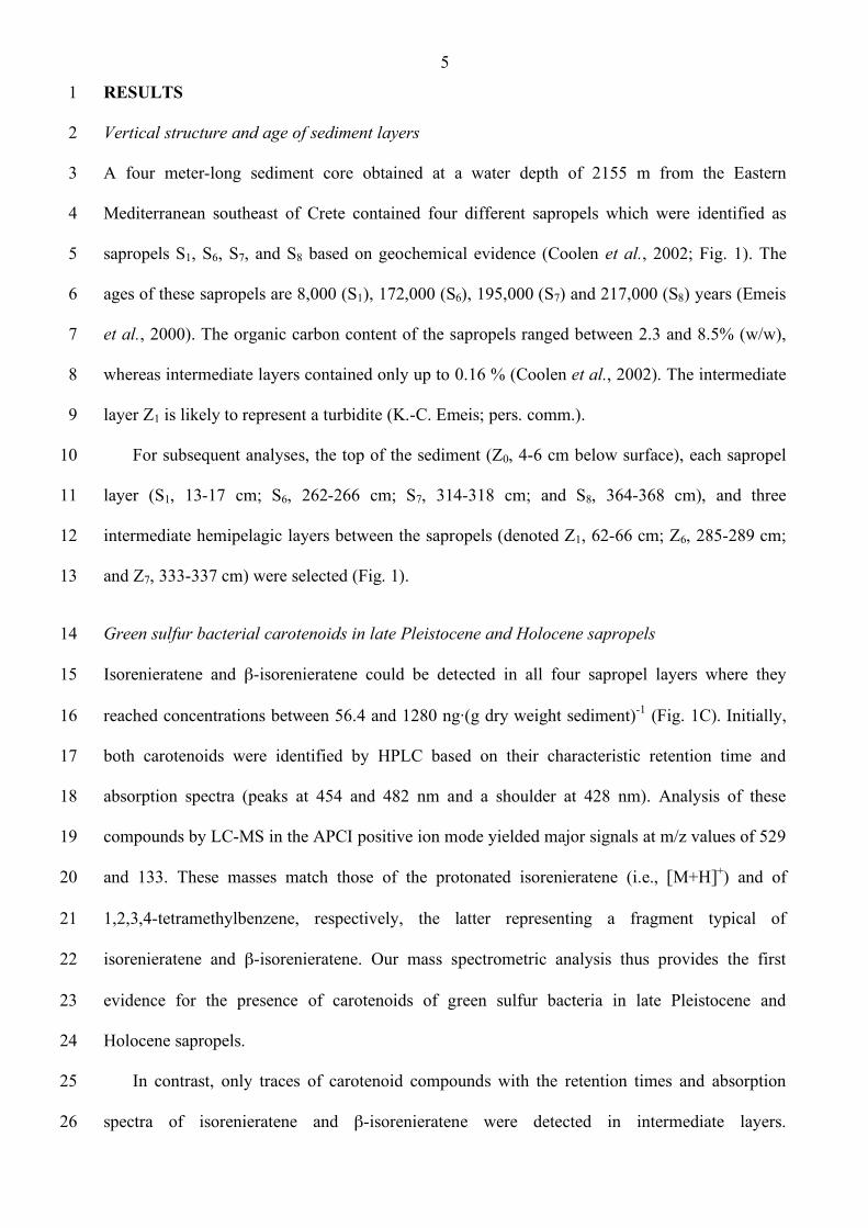
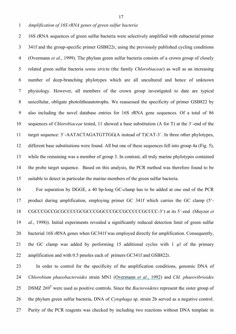
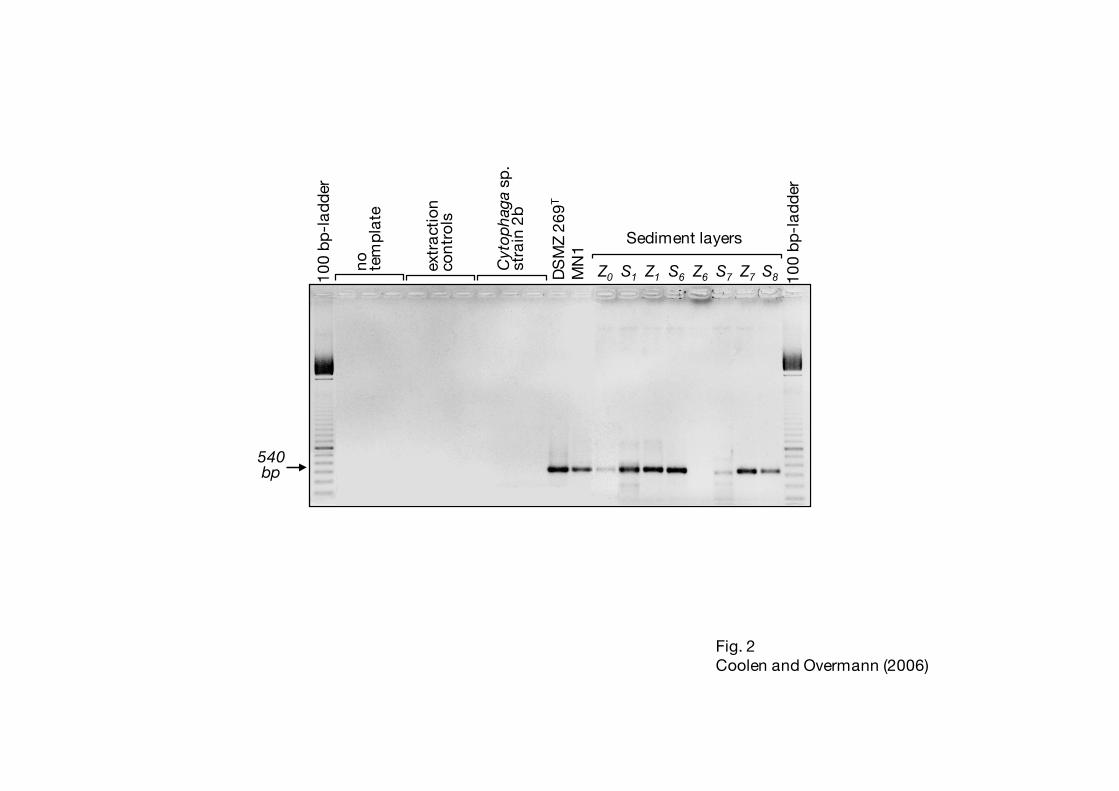
Fig. 2. Amplification products generated with primers GC341f and GSB822. Controls for 7
contamination of PCR reagents (no template), for contamination with extraneous DNA 8
(extraction controls, see Experimental procedures) and for primer specificity (Cytophaga 9
sp. strain 2b) were included in each PCR run. Chlorobium phaeovibrioides DSMZ 269T 10
and Chlorobium phaeobacteroides strain MN1 served as positive controls. A negative 11
image of an ethidium bromide-stained agarose gel is shown. 12
Fig. 3. Dot blot quantification of fossil DNA after amplification of 2 ng each of sample DNA. 13
The standard curve was obtained by using different amounts (given in pg) of genomic 14
DNA of Chlorobium phaeovibrioides DSMZ 269T as template. As negative controls, two 15
independent PCR amplifications were conducted either without DNA template (n.t.), with 16
1 µl of the extraction controls (Extr., compare Experimental Procedures), or with 2 ng of 17
genomic DNA of Cytophaga strain 2b (Cyt). 18
Fig. 4. Separation of green sulfur bacterial 16S rRNA gene fragments by DGGE. For 19
comparison, amplification products from the two brown-colored marine strains 20
Chlorobium phaeovibrioides DSMZ 269T and Chl. phaeobacteroides MN1 (Overmann et 21
al., 1992) were included. DNA bands used in the subsequent phylogenetic analyses are 22
denoted by labels A through G. Asterisks denote sequences which did not yield 23
unambiguous sequences. Six different melting types are marked with arrows. 24
Fig. 5. Phylogenetic position of the seven fossil partial 16S rRNA gene sequences of green 25
sulfur bacteria (given in bold face; labeling refers to layer of origin plus the band no. 26
according to Fig. 4) recovered from sapropels and intermediate layers. Currently 27
recognized groups are indicated (Imhoff, 2003; denoted in rectangles). Groups comprising 28
salt tolerant or salt-requiring strains marked by black rectangles. Bar indicates 0.1 fixed 29
point mutations per nucleotide. Numbers at nodes give bootstrap values out of 100 30
resamplings for phylogenetic trees calculated by Maximum Likelihood / Maximum 31
Parsimony / Neighbor Joining methods. C., Chlorochromatium; Cba., Chlorobaculum, 32
Chl., Chlorobium; Chp.; P. Pelochromatium; Pld., Pelodictyon. 33
34

27
Fig. 6. Equilibrium adsorption isotherms of double-stranded herring sperm DNA bound to 1
sapropel S6 () and the intermediate layer Z6 (ο). Bars indicate one standard deviation. 2
Lines indicate the curves fitted to the data according to the Langmuir equation (eq. 1, see 3
text). 4
5
28
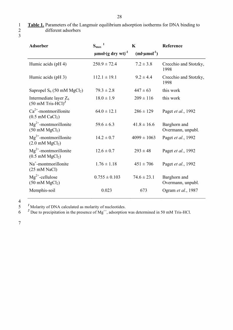
Table 1. Parameters of the Langmuir equilibrium adsorption isotherms for DNA binding to 1 different adsorbers 2
3
Adsorber Smax 1
µmol·(g dry wt)-1 K (ml·µmol-1)
Reference
Humic acids (pH 4) 250.9 ± 72.4 7.2 ± 3.8 Crecchio and Stotzky, 1998
Humic acids (pH 3) 112.1 ± 19.1 9.2 ± 4.4 Crecchio and Stotzky, 1998
Sapropel S6 (50 mM MgCl2) 79.3 ± 2.8 447 ± 63 this work Intermediate layer Z6 (50 mM Tris-HCl)2
18.0 ± 1.9 209 ± 116 this work
Ca2+-montmorillonite (0.5 mM CaCl2)
64.0 ± 12.1 286 ± 129 Paget et al., 1992
Mg2+-montmorillonite (50 mM MgCl2)
59.6 ± 6.3 41.8 ± 16.6 Barghorn and Overmann, unpubl.
Mg2+-montmorillonite (2.0 mM MgCl2)
14.2 ± 0.7 4099 ± 1063 Paget et al., 1992
Mg2+-montmorillonite (0.5 mM MgCl2)
12.6 ± 0.7 293 ± 48 Paget et al., 1992
Na+-montmorillonite (25 mM NaCl)
1.76 ± 1.18 451 ± 706 Paget et al., 1992
Mg2+-cellulose (50 mM MgCl2)
0.755 ± 0.103 74.6 ± 23.1 Barghorn and Overmann, unpubl.
Memphis-soil
0.023 673 Ogram et al., 1987
4 1 Molarity of DNA calculated as molarity of nucleotides. 5 2 Due to precipitation in the presence of Mg++, adsorption was determined in 50 mM Tris-HCl. 6
7

100
bp-l
adde
r
100
bp-l
adde
r
no tem
plat
e
extr
act
ion
cont
rols
Cyt
opha
ga s
p.st
rain
2b
DS
MZ
269T
MN
1
Z0 S1 Z1 S6 Z6 S7 Z7 S8
Sediment layers
Fig. 2Coolen and Overmann (2006)
540 bp


![Пилипчук Я.В. Кыпчаки в Китае [Kipchaks in China] // Метаморфозы истории. Вып. 5. Псков, 2014. С. 217-235 [in Russian]](https://static.fdokumen.com/doc/165x107/631382815cba183dbf07216f/pilipchuk-yav-kipchaki-v-kitae-kipchaks-in-china-metamorfozi.jpg)