20200826_Sierksma_manuscript.pdf - -ORCA - Cardiff ...
26
This is an Open Access document downloaded from ORCA, Cardiff University's institutional repository: http://orca.cf.ac.uk/134660/ This is the author’s version of a work that was submitted to / accepted for publication. Citation for final published version: Sierksma, Annerieke, Escott-Price, Valentina and De Strooper, Bart 2020. Translating genetic risk of Alzheimer’s disease into mechanistic insight and drug targets. Science 370 (6512) , pp. 61-66. 10.1126/science.abb8575 file Publishers page: http://doi.org/10.1126/science.abb8575 <http://doi.org/10.1126/science.abb8575> Please note: Changes made as a result of publishing processes such as copy-editing, formatting and page numbers may not be reflected in this version. For the definitive version of this publication, please refer to the published source. You are advised to consult the publisher’s version if you wish to cite this paper. This version is being made available in accordance with publisher policies. See http://orca.cf.ac.uk/policies.html for usage policies. Copyright and moral rights for publications made available in ORCA are retained by the copyright holders.
-
Upload
khangminh22 -
Category
Documents
-
view
0 -
download
0
Transcript of 20200826_Sierksma_manuscript.pdf - -ORCA - Cardiff ...

This is an Open Access document downloaded from ORCA, Cardiff University's institutional
repository: http://orca.cf.ac.uk/134660/
This is the author’s version of a work that was submitted to / accepted for publication.
Citation for final published version:
Sierksma, Annerieke, Escott-Price, Valentina and De Strooper, Bart 2020. Translating genetic risk
of Alzheimer’s disease into mechanistic insight and drug targets. Science 370 (6512) , pp. 61-66.
10.1126/science.abb8575 file
Publishers page: http://doi.org/10.1126/science.abb8575 <http://doi.org/10.1126/science.abb8575>
Please note:
Changes made as a result of publishing processes such as copy-editing, formatting and page
numbers may not be reflected in this version. For the definitive version of this publication, please
refer to the published source. You are advised to consult the publisher’s version if you wish to cite
this paper.
This version is being made available in accordance with publisher policies. See
http://orca.cf.ac.uk/policies.html for usage policies. Copyright and moral rights for publications
made available in ORCA are retained by the copyright holders.

1
Translating genetic risk of Alzheimer’s disease into
mechanistic insight and drug targets
Annerieke Sierksma1,2, Valentina Escott-Price3,4,*, Bart De Strooper1,2,5,*
1 VIB Center for Brain & Disease Research, Leuven, Belgium
2 Laboratory for the Research of Neurodegenerative Diseases, Department of Neurosciences, Leuven
Brain Institute (LBI), KU Leuven (University of Leuven), Leuven, Belgium
3 Medical Research Council Centre for Neuropsychiatric Genetics and Genomics, Cardiff University,
Cardiff
4 UK Dementia Research Institute, Cardiff University, Cardiff, UK
5 UK Dementia Research Institute, University College London, London, UK
*to whom correspondence can be addressed
Abstract
In order to provide better prevention and treatment we need to understand the environmental and
genetic risk of Alzheimer’s Disease (AD). However, the definition of AD has been confounded with
dementia in many studies. Thus, over interpretation of genetic findings with regard to mechanisms
and drug targets may explain in part controversies in the field. Here, we analyze the different forms of
genetic risk of AD and how these can be used to model disease. We stress the importance of studying
gene variants in the right cell types and in the right pathological context. The lack of mechanistic
understanding of genetic variation has become the major bottle neck in the search for novel drug
targets for AD.

2
Introduction
The number of people worldwide suffering from dementia is now already slightly exceeding the
number of people with cancer, and is poised to increase even more over the next decades. Dementia
is, however, a container term for the end symptoms of a wide variety of brain diseases, including
Alzheimer’s Disease (AD). AD is a slowly progressing disorder characterized by specific protein
accumulations in the brain. Clinical dementia manifests only late, which confounds many case-control
studies using this criterion as a proxy for AD. Prodromal AD patients become excluded and not yet
cognitively altered AD cases are mixed with controls. About 30% of clinically diagnosed patients have
no neuropathological or biomarker characteristics of AD (1) and 56% of cases defined as AD present
with common comorbidities such as Lewy body disease, vascular pathology or hippocampal sclerosis
(2). Unfortunately, the advice of the National Institute of Aging and the Alzheimer Association Research
Framework to define AD as a biological construct (3), is not yet widely adopted.
A recent comprehensive overview estimated that 35% of life time risk of dementia is
modifiable, including factors such as education, vascular aspects, hearing loss, social deprivation, etc.
(4). The Framingham Heart Study confirms the modifiable nature of dementia risk, with decreasing
incidence of dementia over the last decades (5). However, while this trend was very significant for
dementia overall and for vascular dementia in particular, it was not significant for AD alone (5). Given
the high heritability of AD (6, 7), studying genetic risk seems a more fruitful way forward to identify
molecular mechanisms of disease. The central question is whether there is one central route to AD,
and therefore one ‘type’ of AD, or whether various pathogenic mechanisms exist that converge on the
defining amyloid plaque and tangle pathology.
The heritability of AD
Heritability, formally defined as the proportion of phenotypic variance that is due to genetic
factors, can be used as a population-based measure for the risk of disease (see glossary in box 1).

3
Importantly, the inheritance of genetic risk variants does not necessarily imply disease and not all
individuals with AD carry the same risk variants.
The best studied risk (or better causal) genetic variants in AD are the fully penetrant mutations
in the genes encoding Amyloid Precursor protein (APP) and presenilin 1 and 2 (PSEN1&2). They affect
the processing of the amyloid-β peptide (Aβ) indicating that Aβ aggregation is an upstream event in
the pathogenesis of AD (8). These mutations were identified in families with a Mendelian, dominantly
inherited form of AD (8) which clinically manifests as early onset dementia (onset before <65 years).
Estimates for the heritability of early onset AD are very high, ranging between 0.92 and 1 (7). Even in
this smaller group (<10% of total AD patients), APP and PSEN1&2 mutations explain only about 10% of
these early onset cases (7). The remaining heritability is explained by APP duplications, by an increasing
number of rare variants in genes encoding e.g. the Sortilin-related receptor (SORL1), Triggering
receptor expressed on myeloid cells 2 (TREM2) and ATP Binding Cassette Subfamily A Member 7
(ABCA7) (9), and finally by not yet identified, but likely recessive, mutations (7). An example of a
recessive AD mutation is A673V in APP (10).
In the large group of patients in which dementia manifests after age 65, the heredity is also
large, estimated between 0.58 and 0.78 with rather large 95% confidential intervals [0.19-0.87] and
[0.67-0.88] respectively (6). This is high compared to other late onset diseases. The genetic
architecture underlying AD >65 years is far from fully charted (see Fig.1). Apolipoprotein ε4 allele
(APOE4) is the only common high risk genetic variant (odds ratio (OR)=3.32) (11, 12). Genome wide
association studies (GWAS) have further identified many common genetic variants with low risk
(OR=1.1-1.2) of which 40 have genome wide significance (12–14). Exome chip analyses have
additionally yielded rare variants in the very same genes, i.e. SORL1, TREM2, and ABCA7, that increase
strongly risk of early onset AD (9, 15). Variants that protect have also been discovered: the APOE2
allele (OR=0.6) (16), rare mutations in phosphatidylinositol specific phospholipase C-gamma 2 (PLCγ2)
(OR=0.68)(15) and the Icelandic mutation A673T in APP (OR=0.19) (10). The genetics of early and late

4
onset disease suggest that AD should be considered a continuum. As indicated by the broad
confidential intervals, the AD heritability estimates remain imprecise, as with many polygenetic
disorders (17). Increasing efforts to create larger datasets for GWAS studies, to directly sequence full
genomes and to develop novel data analyses methodologies are under way to tackle the “missing
heritability” in AD.
From heritability to mechanisms of disease
Translating genetic information into disease mechanisms is anything but trivial. It took twenty
years to understand that AD causing mutations destabilize presenilin leading to premature release of
long Aβ peptides (18). Similar efforts to understand how rare mutations in the open reading frame of
TREM2, PLCγ2, SORL1 and ABCA7 affect protein function will be needed. In addition, most available
genetic information in AD remains imprecise. The causal variant is known for only 40% of the identified
GWAS loci (14), the effect of these variants is only known in a minority of cases, and literally thousands
of these variants contribute to the heritability of the phenotype.
The question is what the core genes are, i.e. which genes execute a direct effect on the disease
process. Unfortunately, more than 70% of variants determining phenotypic variation are in
“peripheral” genes. Such genes have only indirect effects on expression or posttranslational
modification of core gene products and as such are not very informative for the molecular mechanisms
driving the phenotype (19). The individual ‘trans’ effects of these peripheral genes are small (19) but
since there are many, they underlie a large part of the heritability (20, 21) . Even using the p-value of
p<5x10⁻⁸ for genome wide significance to prioritize gene loci (which comprise now 40 loci in AD (14))
does not provide certainty of finding “core disease pathway genes”(19). The frustrating conclusion is
that the bulk of the heredity in AD likely only indirectly points to key biological pathways of disease.
One group of peripheral genes, i.e. master regulator genes, is nevertheless of particular interest. These
genes, encoding for example transcription factors, chromatin modifiers, regulatory RNA or enzymes,

5
regulate the expression or function of several disease core genes. For example the AD risk locus Spi-1
proto-oncogene (Spi1) codes for the transcription factor Pu.1 which regulates many microglia genes,
pointing to a role for inflammation in AD (22). Such master regulators are usually under strong
evolutionary constraint, and so not easily detected in GWAS (19).
One could try to investigate how peripheral genes affect the expression of core genes. A
prerequisite is to understand in which cells these peripheral genes exert their effect and hence single
cell analyses of gene expression in brain cells is crucial (23). Such trans-expression quantitative trait
loci (trans-eQTL) mapping, however, needs huge data sets and is only readily available for peripheral
blood cells. Another possibility is to focus on gene variants with large effects on heredity. ApoE4 is the
only example in AD. Finally, one could ignore the quantitative contributions of genes to heredity and
focus on rare variants, which are likely more central to the disease mechanisms because of their large
effect sizes. A potentially fruitful avenue of research is to investigate how the common variants that
define heredity regulate these rare variant genes.
The complexity of the APOE locus
The 3 major isoforms of ApoE (ε2, ε3 and ε4) are defined by two single nucleotide
polymorphisms or SNPs (rs429358 and rs7412) within exon 4 of the gene (11). The ε4 allele (frequency
0.14 in the Caucasian population) provides a 3-fold increased risk of AD which raises to 14-fold in ε4
homozygotes (11, 16). Conversely, the ε2 allele (frequency 0.08) confers a 1.7-fold decreased risk. This
risk is more pronounced in women then in men and is strongly depending on ethnic background, i.e.
the ε4 effect is much smaller in the African-American and Hispanic population (16). This illustrates the
importance of multi-ethnic genetic studies when studying the heritability of AD.
The APOE locus is highly complex, spanning almost 2MB and covering over 70 genes. Despite
being in low linkage disequilibrium with the APOE SNPs for ε2 and ε4, there are many other SNPs in
this large locus showing significant association to AD. This might point to other AD risk genes in this

6
locus. Several of these SNPs, however, likely affect expression of APOE. Understanding this will be of
tremendous value as it would clarify whether and under which conditions up- or down regulation of
this multifunctional protein could affect the risk of AD.
Under physiological conditions, APOE is mainly expressed by astrocytes, but microglia exposed
to Aβ plaques highly upregulate APOE. It will be critical to unravel how microglial function is affected
by different APOE isoforms and how this contributes to disease. Knock-out of the gene eliminates the
AD induced inflammatory response in mice (24).
While very relevant to AD, the role of APOE in brain inflammation remains badly understood.
APOE obviously plays a crucial role in cholesterol transport and lipid homeostasis, but also in Aβ-
aggregation, -clearance, and -cellular uptake, and affects, via less well understood molecular pathways,
synapse number and function, blood-brain barrier integrity, and TAU-mediated neurodegeneration
(16, 24, 25). It is important to decipher which roles of APOE are directly relevant to AD as the variety
of functional effects of APOE deficiency in different cell types and in different tissues suggest that the
APOE gene is a master peripheral regulator in the disease. Not all affected pathways are necessarily
relevant to AD. Directly modulating APOE to protect against AD is likely to have a variety of effects and
the outcome of such treatments will need careful monitoring.
Causal, high risk and protective variants are involved in APP processing and in
microglial function
Evidence of genotype-phenotype dose-responses in an allelic series strongly argues for a core
gene function. Such gene-dosage effects are observed with genes involved in Aβ-generation. Next to
the fully penetrant APP and PSEN mutations, APP gene duplications and triplications, including Down
syndrome, cause AD (8, 10). A recessive (673V) and a protective (A673T) allele (10) affect the
propensity of Aβ to aggregate. A673T also lowers β-secretase processing of APP (26). A common allele
(rs2154481) in the APP locus lowers risk (OR=0.95) although, counter intuitively, slightly increases APP
7
expression (14). Finally, variants in the gene locus of the α-secretases, ADAM17 and ADAM10 (27) all
demonstrate that APP itself and the enzymes processing it to Aβ carry risk and even cause AD.
SORL1 provides another example of an allelic series with increasing risk of AD. SORL1 encodes
the sorting-related receptor with A-type repeats SorlA (a.k.a. LR11) involved in retromer-related
endosomal traffic. SorlA contains functional domains that can bind monomeric Aβ or APP. Several of
the deleterious variants affect those domains (28). SorlA lowers Aβ production by redirecting APP to
the cell membrane and trans-Golgi network (TGN) and Aβ to lysosomes in neurons (see Fig.2)(28).
Interestingly, SORL1 expression is twentyfold higher in human than in mouse microglia, warranting for
further characterization of the impact of SORL1 deficiency on microglia functions (29). The different
SORL1 variants aggregate into categories with increasing risk burden (30) from OR=1.21 for missense
variants up to OR=16.73 for protein-truncating variants (9). These ORs are comparable to heterozygous
(OR=3.2) and homozygous (OR=14.9) APOE-ε4 carriers. Variants are present in 2% of AD, compared to
<1% for APP and PSEN1 mutations (30).
Common and rare ABCA7 variants provide a third allelic series. ABCA7 promotes the efflux of
phospholipids out of cells. Protein-truncating (OR=2.6) and missense mutations (OR=1.8) are
associated with AD (31). In addition, a tandem repeat in intron 18, ranging from 300 bps to more than
10kb, provides relative high risk of AD (OR=4.5). It remains unclear how loss of function of ABCA7
increases risk of AD, although in mice it causes higher Aβ plaque burden related to impaired Aβ
phagocytosis in macrophages and microglia (32). Loss of ABCA7, because of its role in lipid transport,
might have broad effects on cell physiology (see Fig.2). The relative low OR suggests that it is not
directly causally involved and its broad function suggest it acting as a master regulator peripheral gene
in AD.
Next to core and master regulator genes affecting Aβ, strong genetic evidence imply microglia
in AD. Many common variants associated to risk of AD occur in genes that are expressed in microglia

8
(see table 1 in (33)). Rare missense mutations in the open reading frames of TREM2, PLCγ2 and ABI3
(Abelson interactor family protein 3) which are genes mainly or exclusively expressed in myeloid cells,
also point into that direction (15). Many of the risk genes of AD become upregulated in microglia when
exposed to Aβ but less so to TAU pathology (34). Thus, a large part of the genetic risk of AD, as opposed
to genetic cause of AD, seems to converge into the microglial response to amyloid plaques.
One of the best studied genes in this series is TREM2. TREM2 is a receptor for anionic ligands
including phospholipids, lipopolysaccharide (LPS) and DNA (35). In mouse models of AD Trem2 is
required for the transit of microglia from homeostatic to activated cell states in response to amyloid
plaques. Interestingly these microglia strongly upregulate ApoE expression (36). Trem2 deficiency
leads to more diffuse plaques with greater neuritic damage and less recruitment of microglia to
amyloid plaques (36). The rare R47H and more common R62H variants of TREM2 (15) alter its stability,
affect phagocytic capacity and impair TREM2 affinity for APOE, clusterin (ApoJ), low-density
lipoproteins (LDL) and Aβ (33). The impact of other more common variants on TREM2 function remains
unclear. Since the R47H and R62H mutations cause partial loss of function of TREM2, and since Trem2
deficiency seems to aggravate amyloid plaque pathology in mice, most drug development efforts are
focused on enhancing TREM2 function (37). However, enhancing microglia activity might be a two
edged sword with opposite effects on Aβ and TAU pathology (24). It also remains a big question to
what extent observations in mice can be extrapolated to the human pathophysiology as the cellular
reactions around amyloid plaques are much more complex in human than in the available mouse
models (see for instance (38, 39)). The fact that several additional AD associated variants have been
observed in genes that act downstream of TREM2 nevertheless underlines the importance of TREM2-
signaling in AD (see Fig.2).
One example is the rare protective P522R variant in the PLCγ2 gene with moderate effect size
(OR=0.57-0.68) (15). This mutation increases the activity of the microglial signaling enzyme
phospholipase C gamma 2 downstream of TREM2 (see Fig.2). The variant is overrepresented in a

9
cohort of cognitively healthy centenarians and, anecdotally, provides full protection to APOE-ε4 in a
more than hundred years old homozygous carrier (40). PLCγ2 becomes phosphorylated upon
stimulation and affects phagocytosis, migration, chemokine and cytokine release (41). Structurally, the
P522R variant modifies an autoinhibitory domain of PLCγ2 leading to greater PIP2 conversion and
increased cellular calcium release (41). The P522R variant enhances Aβ endocytosis, suggesting that
this protective variant may facilitate microglial clearance of Aβ.
In conclusion, the genetics of AD provide strong evidence for a major pathway centered on Aβ
generation, aggregation and clearance that operates in early and late onset disease. The genetics also
strongly imply microglia responses to amyloid plaques in AD. Assuming that these responses are
directed by the genetic risk profile of the patient, one would predict that some patients are protected
from the damage caused by amyloid plaques because of their excellent microglia (34). Major questions
for the field are what aspects of the microglia response on amyloid plaques are beneficial or
detrimental, how genetic risk affects this balance, and whether this contributes to TAU pathology.
Drug development will have to move cautiously taking into account this fine Yin- and Yang of the
cellular response in AD (23).
Leveraging polygenic risk
A large proportion of the genetic risk of AD is explained by common variation in the genome
and is captured via single nucleotide polymorphisms (SNP) in GWAS (21). Such single variants on their
own do not predict an individual’s risk of AD but can be combined in a polygenic risk score (PRS). PRS
is a “genetic score” defined as the sum of the number of SNP risk alleles that an individual carries,
weighted by their contribution to the disease risk (effect size).
Most investigators currently use a partial AD PRS calculated with the lead SNP in the 40 canonic
GWAS genome loci mentioned before (12–14, 42). However, a more complete PRS calculation includes
the thousands of other SNPs in loci that are associated with risk of AD but did not reach the threshold

10
for genome wide significant association (p<5x10⁻⁸). Such calculation improves strongly the prediction
accuracy of AD, something also observed with psychiatric and other complex disorders (43). In fact,
the prediction accuracy of AD using the complete PRS is high, with area under the receiver-operator
curve (AUC) of 75% in clinical and 84% in pathologically confirmed samples (21, 44).
Using only the canonic GWAS loci biases the score to the effect of the APOE region (21). If all
genetic risk of AD is used as proposed for the complete PRS, the bulk of associated SNPs of small effect
sizes will eventually outperform the effect size of the APOE locus alone. Accordingly, the predictive
accuracy of complete PRS in pathologically confirmed E3 homozygotes is high, with AUC>80 (45). To
date, the PRS approach has mostly been assessed in European populations due to lack of multiethnic
GWAS data.
The field is currently struggling to translate the concept of PRS into meaningful functional
hypotheses. An interesting recent development is to include only SNPs associated with genes from
putative disease-specific pathways, for instance APP metabolism, lipid metabolism, endocytosis, to
generate pathway specific PRS (27). However, the definition of these disease pathways is based mostly
on the different functional categories defined by Gene Ontology (46). This is problematic (46) as there
is little expert scrutiny, inclusion thresholds are low and almost all AD genes are implicated in more
than one pathway (12, 47, 48). It turns out that the AD predictability using such categories is low (47).
Finally, it is important to mention that the PRS is currently designed as a linear combination of SNP
effect sizes without accounting for non-linear effects, also known as epistasis or SNPxSNP interaction.
Biologically, it is very unlikely that genetic risk of AD is the simple additive sum of the individual SNP
risks.
From polygenic risk to mechanisms of disease and drug targets: cellular state
and disease context matter
Drugs developed against targets supported by genetic evidence have a better chance to
become approved (49). However, in the AD field the causal SNPs are in many cases unknown or

11
assigned to the wrong gene. There are still large gaps in understanding how SNPs affect the functional
genomic architecture. At the other side, information on the effects of drugs on eQTL are usually not in
the public domain, making it difficult to link experimental drugs to candidate targets. Overall, the single
most important limiting factor in the translation of knowledge from genetics to drugs is, however, the
lack of good models for AD (see Fig.3).
Assessing the functional impact of non-coding risk variants is challenging and starts with the
question whether a particular SNP is functional or is only in linkage disequilibrium with the real
functional SNP. Risk mechanisms will only manifest in disease-relevant conditions and therefore cell
type and experimental context really matters when analyzing the functional consequences of SNPs
(23). Finally, SNP are frequently assigned to genes that are most close in the linear DNA sequence
representation of the genome. However, chromatin has a complex 3D structure and enhancers or
suppressors can exert their effects on the expression of genes that are remote from their location (50).
Recent work has indeed shown that many causal variants affect enhancers which are highly specific to
brain region, cell-type, and cell state (22, 50, 51). It was noted that mainly myeloid and microglial
enhancer regions, and not the promoter regions are significantly enriched for AD-associated variants
(50–52). An elegant knock-out experiment underscored this conclusion. Nott et al. deleted in human
induced pluripotent stem cells (iPSCs) the BIN1 (Bridging Integrator-1 and Amphiphysin-2) enhancer
which carries one of the higher AD risk variants, rs6733839 (OR=1.2)(12, 50). When they differentiated
these cells into microglia, astroglia, and neurons, expression of BIN1 was only affected in microglia
(50).
The AD field really struggles to generate good models that reproduce all features of disease
(see Fig.3). Double and even triple transgenic mice overexpressing human TAU, PSEN and APP, all with
FAD or FTD mutations, are needed to obtain amyloid plaques and tangles and it remains a tantalizing
question to what extent cellular phenotypes induced in these mice mimic the situation in human. Sixty-
12
five million years of evolution divergence cannot be ignored when modeling a human polygenic
disease. In order to research human-specific cell biology, research on human iPSCs has taken a flight,
including in vitro 3D (53) and organoid cultures (54). All are promising, but each approach comes with
its own limitations (see Fig.3). For instance, the 3D in vitro cultures provide a very artificial
conformation to grow the cells and uses high overexpression of the APP gene in the neurons to obtain
AD phenotypes (53). The human organoid culture is promising but their usefulness to study non-
developmental disorders remains debated (54).
The xenograft or chimeric, mouse model approach, where human iPSC-derived brain cells are
transplanted into the mouse brain (55–57) provides an interesting alternative combining several
advantages. The rodent brain functions as a superior “physiological” 3D matrix for human cells
compared to other more artificial environments. Human neurons (55), microglia (56, 57) and astroglia
have been grown in rodent brains for over one year and reproduce many human features. While the
rodent brain background and the immune suppression are confounders in these experiments,
microglia cells, even after exposure to a cell culture environment, fully regain their identity when
returned to the CNS and transcriptionally closely resemble freshly isolated human microglia from
surgical samples (57).
In theory, human iPSCs and their derived models can be used to functionally evaluate the
impact of PRS-defined risk in different cell types and AD-relevant contexts. Obviously, the genomic
variants captured from different patients will be different, but, because the pathological phenotype of
AD patients is very similar, it is assumed that the cellular pathology converges and that shared
pathways leading to disease may be identified. Once a critical mass of PRS-defined iPSCs will have been
analysed, one can also envision eQTL and regulatory landscape analyses to define how specific AD-
associated variants may exert their effects. This can subsequently refine the list of SNPs, including only
core variants driving AD pathogenesis. Ultimately, such functional insights will lead to better and more

13
relevant PRS that will be used for diagnostics, stratification of patients for clinical trials, and
personalized medicine based on genetic profile.
Conclusions
The genetic component in AD risk is surprisingly large for a late onset disorder. Tremendous
progress has been made to map this genetic landscape, but now it becomes critically dependant on a
better definition of AD and the underlying mechanisms of disease. “More” cases is never going to
replace “quality” of cases and deeper clinical phenotyping and biomarkers are needed to better
interpret the role of genetic variation in specific aspects of the AD phenotype.
It is crucial, while working further along those lines, but also from therapeutic development
perspective, to take into account the long preclinical phase of AD (23). At the functional level we need
to get away from the classical molecular biology paradigms of gene-function-drug target. Gene variants
affect gene function in specific genetic backgrounds (mice are not humans), in specific cell types, in
specific cell states, and in specific stages of the disease. In silico predictions and simple cell biological
experiments, although tempting because of the high throughput, can be very misleading and can bring
a whole drug development campaign in jeopardy. Finding drugs for a complicated multifactorial
disease like AD requires deep knowledge of the mechanisms that are targeted. The full mapping of the
cellular phase of AD is now a priority for the field (23).
One should, however, acknowledge the tremendous progress made in AD research. We can
now further build on the many hints coming from genetic work over the last decade to generate the
more sophisticated models that will better represent specific mechanisms underlying AD. This novel
thinking will open many opportunities for drug development, while better stratification of patients will
accelerate the road from concept to clinic.

14
References and Notes
1. T. G. Beach, S. E. Monsell, L. E. Phillips, W. Kukull, Accuracy of the Clinical Diagnosis of Alzheimer
Disease at National Institute on Aging Alzheimer Disease Centers, 2005–2010. J. Neuropathol.
Exp. Neurol. 71, 266–273 (2012).
2. G. D. Rabinovici et al., Multiple comorbid neuropathologies in the setting of Alzheimer’s disease
neuropathology and implications for drug development. Alzheimer’s Dement. Transl. Res. Clin.
Interv. 3 (2017), pp. 83–91.
3. C. R. Jack et al., NIA-AA Research Framework: Toward a biological definition of Alzheimer’s
disease. Alzheimer’s Dement. 14 (2018), pp. 535–562.
4. G. Livingston et al., Dementia prevention, intervention, and care. Lancet. 390, 2673–2734
(2017).
5. C. L. Satizabal et al., Incidence of dementia over three decades in the Framingham heart study.
N. Engl. J. Med. 374, 523–532 (2016).
6. M. Gatz et al., Role of genes and environments for explaining Alzheimer’s disease. Arch. Gen.
Psychiatry. 63, 168–74 (2006).
7. T. S. Wingo, J. J. Lah, A. I. Levey, D. J. Cutler, Autosomal recessive causes likely in early-onset
Alzheimer disease. Arch. Neurol. 69, 59–64 (2012).
8. R. E. Tanzi, L. Bertram, Twenty years of the Alzheimer’s disease amyloid hypothesis: A genetic
perspective. Cell. 120, 545–555 (2005).
9. C. Bellenguez et al., Neurobiol. Aging, in press, doi:10.1016/j.neurobiolaging.2017.07.001.
10. J. TCW, A. M. Goate, Genetics of β-Amyloid Precursor Protein in Alzheimer’s Disease. Cold
Spring Harb. Perspect. Med. 7 (2017), p. a024539.
11. E. H. Corder et al., Gene dose of apolipoprotein E type 4 allele and the risk of Alzheimer’s
disease in late onset families. Science (80-. ). 261, 921–923 (1993).
12. B. W. Kunkle et al., Genetic meta-analysis of diagnosed Alzheimer’s disease identifies new risk

15
loci and implicates Aβ, tau, immunity and lipid processing. Nat. Genet. 51, 414–430 (2019).
13. I. E. Jansen et al., Genome-wide meta-analysis identifies new loci and functional pathways
influencing Alzheimer’s disease risk. Nat. Genet. 51, 404–413 (2019).
14. I. de Rojas et al., Common variants in Alzheimer’s disease: Novel association of six genetic
variants with AD and risk stratification by polygenic risk scores. medRxiv, 19012021 (2020).
15. R. Sims et al., Rare coding variants in PLCG2, ABI3, and TREM2 implicate microglial-mediated
innate immunity in Alzheimer’s disease. Nat. Genet. 49, 1373–1384 (2017).
16. M. E. Belloy, V. Napolioni, M. D. Greicius, Review A Quarter Century of APOE and Alzheimer’s
Disease: Progress to Date and the Path Forward. Neuron. 101, 820–838 (2019).
17. A. J. Mayhew, D. Meyre, Assessing the Heritability of Complex Traits in Humans: Methodological
Challenges and Opportunities. Curr. Genomics. 18, 332–340 (2017).
18. M. Szaruga et al., Alzheimer’s-Causing Mutations Shift Aβ Length by Destabilizing γ-Secretase-
Aβn Interactions. Cell. 170, 443-456.e14 (2017).
19. X. Liu, Y. I. Li, J. K. Pritchard, Trans Effects on Gene Expression Can Drive Omnigenic Inheritance.
Cell. 177, 1022–1034 (2019).
20. R. S. Desikan et al., Genetic assessment of age-associated Alzheimer disease risk: Development
and validation of a polygenic hazard score. PLOS Med. 14, e1002258 (2017).
21. V. Escott-Price et al., Common polygenic variation enhances risk prediction for Alzheimer’s
disease. Brain. 138, 3673–84 (2015).
22. K. Huang et al., A common haplotype lowers PU.1 expression in myeloid cells and delays onset
of Alzheimer’s disease. Nat. Neurosci. 20, 1052–1061 (2017).
23. B. De Strooper, E. Karran, The Cellular Phase of Alzheimer’s Disease. Cell. 164, 603–615 (2016).
24. J. M. Long, D. M. Holtzman, Alzheimer Disease: An Update on Pathobiology and Treatment
Strategies. Cell. 179 (2019), pp. 312–339.
25. A. Montagne et al., APOE4 leads to blood–brain barrier dysfunction predicting cognitive
16
decline. Nature. 581 (2020), doi:10.1038/s41586-020-2247-3.
26. I. Benilova et al., The Alzheimer disease protective mutation A2T modulates kinetic and
thermodynamic properties of amyloid-β (Aβ) aggregation. J. Biol. Chem. 289, 30977–30989
(2014).
27. R. Sims, M. Hill, J. Williams, The multiplex model of the genetics of Alzheimer’s disease. Nat.
Neurosci. 23, 311–322 (2020).
28. D. Campion, C. Charbonnier, G. Nicolas, SORL1 genetic variants and Alzheimer disease risk: a
literature review and meta-analysis of sequencing data. Acta Neuropathol. 138 (2019), pp. 173–
186.
29. D. Gosselin et al., An environment-dependent transcriptional network specifies human
microglia identity. Science (80-. ). 356, 1248–1259 (2017).
30. H. Holstege et al., Characterization of pathogenic SORL1 genetic variants for association with
Alzheimer’s disease: A clinical interpretation strategy. Eur. J. Hum. Genet. 25, 973–981 (2017).
31. A. De Roeck, C. Van Broeckhoven, K. Sleegers, The role of ABCA7 in Alzheimer’s disease:
evidence from genomics, transcriptomics and methylomics. Acta Neuropathol. 138 (2019), pp.
201–220.
32. T. Aikawa et al., ABCA7 haplodeficiency disturbs microglial immune responses in the mouse
brain. PNAS. 116, 23790–23796 (2019).
33. D. V Hansen, J. E. Hanson, M. Sheng, Microglia in Alzheimer ’ s disease. J. Cell Biol. 217, 459–
472 (2017).
34. A. Sierksma et al., Novel Alzheimer risk genes determine the microglia response to amyloid‐β
but not to TAU pathology. EMBO Mol. Med. 12 (2020), doi:10.15252/emmm.201910606.
35. T. K. Ulland, M. Colonna, TREM2 — a key player in microglial biology and Alzheimer disease.
Nat. Rev. Neurol. 14 (2018), pp. 667–675.
36. S. Parhizkar et al., Loss of TREM2 function increases amyloid seeding but reduces plaque-

17
associated ApoE. Nat. Neurosci. 22, 191–204 (2019).
37. K. Schlepckow et al., Enhancing protective microglial activities with a dual function TREM 2
antibody to the stalk region . EMBO Mol. Med. 12, 1–22 (2020).
38. Y. Zhou et al., Human and mouse single-nucleus transcriptomics reveal TREM2-dependent and
TREM2-independent cellular responses in Alzheimer’s disease. Nat. Med. 26, 131–142 (2020).
39. W.-T. Chen et al., Spatial Transcriptomics and In Situ Sequencing to Study Alzheimer’s Disease.
Cell, 1–16 (2020).
40. S. J. van der Lee et al., A nonsynonymous mutation in PLCG2 reduces the risk of Alzheimer’s
disease, dementia with Lewy bodies and frontotemporal dementia, and increases the likelihood
of longevity. Acta Neuropathol. 138, 237–250 (2019).
41. L. Magno et al., Alzheimer’s disease phospholipase C-gamma-2 (PLCG2) protective variant is a
functional hypermorph. Alzheimer’s Res. Ther. 11, 16 (2019).
42. R. E. Marioni et al., GWAS on family history of Alzheimer’s disease. Transl. Psychiatry. 8, 99
(2018).
43. S. M. Purcell et al., Common polygenic variation contributes to risk of schizophrenia and bipolar
disorder. Nature. 460, 748–752 (2009).
44. V. Escott-Price, A. J. Myers, M. Huentelman, J. Hardy, Polygenic risk score analysis of
pathologically confirmed Alzheimer disease. Ann. Neurol. 82, 311–314 (2017).
45. V. Escott-Price, A. Myers, M. Huentelman, M. Shoai, J. Hardy, Polygenic Risk Score Analysis of
Alzheimer’s Disease in Cases without APOE4 or APOE2 Alleles. J. Prev. Alzheimer’s Dis. 6, 16–19
(2019).
46. F. Koopmans, P. van Nierop, M. Andres-Alonso, P. D. Thomas, A. B. Smit, SynGO: An Evidence-
Based, Expert-Curated Knowledge Base for the Synapse. Neuron. 103, 217-234.e4 (2019).
47. E. Bellou et al., Age-dependent effect of APOE and polygenic component on Alzheimer’s
disease. Neurobiol. Aging (2020), doi:10.1016/j.neurobiolaging.2020.04.024.

18
48. G. Leonenko et al., Genetic risk for alzheimer disease is distinct from genetic risk for amyloid
deposition. Ann. Neurol. 86, 427–435 (2019).
49. E. A. King, J. Wade Davis, J. F. Degner, Are drug targets with genetic support twice as likely to
be approved? Revised estimates of the impact of genetic support for drug mechanisms on the
probability of drug approval. PLoS Genet. 15, 1–20 (2019).
50. A. Nott et al., Brain cell type-specific enhancer-promoter interactome maps and disease-risk
association. Science (80-. ). 366, 1134–1139 (2019).
51. G. Novikova et al., Integration of Alzheimer’s disease genetics and myeloid genomics reveals
novel disease risk mechanisms. bioRxiv, 694281 (2019).
52. E. Gjoneska et al., Conserved epigenomic signals in mice and humans reveal immune basis of
Alzheimer’s disease. Nature. 518, 365–369 (2015).
53. J. Park et al., A 3D human triculture system modeling neurodegeneration and
neuroinflammation in Alzheimer’s disease. Nat. Neurosci. 21, 941–951 (2018).
54. S. Wray, Modelling neurodegenerative disease using brain organoids. Semin. Cell Dev. Biol., 0–
1 (2020).
55. I. Espuny-Camacho et al., Hallmarks of Alzheimer’s Disease in Stem-Cell-Derived Human
Neurons Transplanted into Mouse Brain. Neuron. 0, 1066-1081.e8 (2017).
56. J. Hasselmann et al., Development of a Chimeric Model to Study and Manipulate Human
Microglia In Vivo. Neuron. 103, 1016-1033.e10 (2019).
57. R. Mancuso et al., Stem-cell-derived human microglia transplanted in mouse brain to study
human disease. Nat. Neurosci. 22, 2111–2116 (2019).
19
Figure legends
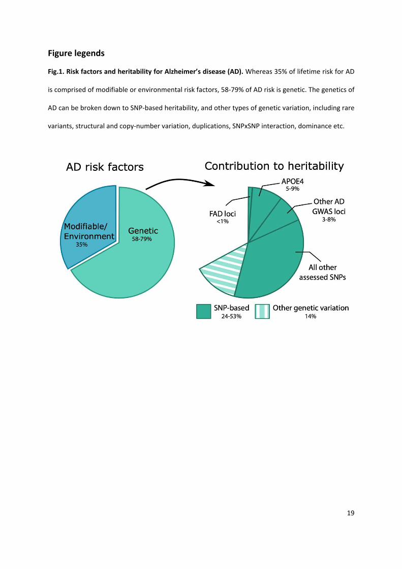
Fig.1. Risk factors and heritability for Alzheimer’s disease (AD). Whereas 35% of lifetime risk for AD
is comprised of modifiable or environmental risk factors, 58-79% of AD risk is genetic. The genetics of
AD can be broken down to SNP-based heritability, and other types of genetic variation, including rare
variants, structural and copy-number variation, duplications, SNPxSNP interaction, dominance etc.

20
Fig.2. Emerging signaling pathways in AD. TREM2, SORL1 and ABCA7 interact with other genetic risk
genes for AD (protein names highlighted in red), impacting microglial function and APP processing. A)
AD pathway in microglia. TREM2 can bind amyloid-β (Aβ) that may need to be lipidated by ApoE or
ApoJ (CLU) and associates with DNAX-activating protein of 12 kDa (DAP12) to constitute intracellular
signaling via its immunoreceptor tyrosine-based activating motif (ITAM). The ITAM domain undergoes
double phosphorylation by the SRC family kinases (SFK, e.g. LYN) to allow binding of spleen tyrosine
kinase (Syk). Syk can phosphorylate phosphoinositide 3-kinase (PI3K) and PLCγ2. Activation of these
proteins ultimately leads to calcium and mitogen-activated protein kinase (MAPK) signaling and
nuclear factor kappa β (NF-κβ) transcription. Protein kinase C (PKC) can also activate proline-rich
tyrosine kinase 2 (Pyk2; PTK2B), which can activate MAPK signaling, but also associates with Cas
scaffold protein family member 4 (CASS4) and focal adhesion kinase (FAK; PTK2) to affect actin
polymerization, as does Abelson interactor family protein 3 (ABI3). Overall these signaling pathways
affect cytoskeletal rearrangements associated with microglial motility and increase phagocytosis. B)
Endocytosis and Alzheimer genes. SORL1 can transfer APP to the trans-Golgi network and late
endosomes where it undergoes amyloidogenic processing to Aβ. SORL1 can also directly bind Aβ and
facilitate its degradation in the lysosomes. Although ABCA7 is involved in cellular lipid homeostasis,
e.g. regulating the efflux of lysophosphatidyl choline (LPC) and phosphatidyl choline (PC), ABCA7 can
also impact amyloidogenic proteolysis by affecting beta-site APP cleaving enzyme 1 (BACE1) expression
levels. Several other AD risk genes involved in endocytic pathway are indicated in red.

21

22
Fig.3. The opportunities and limitations of commonly used models in AD research.

23

24
Box 1 – Glossary
• Heritability: the proportion of phenotypic variance that is due to genetic factors.
• Missing heritability: the difference between the genetic heritability observed in families and
the estimated heritability of identified genetic variants in the population.
• Core gene: a mutation in this gene will directly impact disease
• Peripheral gene: a mutation in this gene will only indirectly impact disease, most likely through
a trans-regulatory effect on core genes
• Core disease pathway genes: genes directly impacting pathways that determine disease onset
• Master regulatory gene: a peripheral gene that regulates the expression or function of several
core genes in the disease. Examples include transcription factors, regulatory RNAs or enzymes,
or chromatin modifiers.
• Genotype-phenotype dose-response: several alleles of a gene impact disease risk, possibly to
different degrees, e.g. common and rare, or loss- and gain-of-function variants. Either multiple
alleles can affect the same gene or causal alleles are present in different genes that cooperate
within the same disease pathway. An example is the amyloid-β pathway, where mutations in
APP, the presenilins and α-secretase impact the same pathway and both protective and risk
variants have been identified.
• Polygenic risk score: a single genetic score indicating a person’s risk of developing a trait.
Calculated by summing the number of risk alleles present and multiplying this by their effect
size, i.e. the weight of disease risk.
• Linkage disequilibrium: the observation that specific alleles at a particular genomic locus or
region are more often co-inherited within the population than expected by chance.

25
Acknowledgements
Funding: The authors wish to thank the Dementia Research Institute (UKDRI supported by Medical
Research Council (MRC), Alzheimer’s Research UK and Alzheimer’s Society), MRC Centre for
Neuropsychiatric Genetics and Genomics (MR/L010305/1), Welsh Government, Joint Programming for
Neurodegeneration (JPND), VIB and KU Leuven (Methusalem grant), the European Union (grant no.
ERC-834682 CELLPHASE_AD), the ‘Fonds voor Wetenschappelijk Onderzoek’, the ‘Geneeskundige
Stichting Koningin Elisabeth’, Opening the Future campaign of the Leuven Universitair Fonds, the
Belgian Alzheimer Research Foundation and the Alzheimer’s Association USA. B.D.S. is holder of the
Bax-Vanluffelen Chair for Alzheimer’s disease.
Author contributions: A.S, V.E-P and B.D.S wrote the manuscript.
Competing interests: B.D.S. is ad hoc consultant for various companies but has no direct financial
interest in the current manuscript.