2014 focused update of the Canadian Cardiovascular Society Guidelines for the management of atrial...
17
Society Guidelines 2014 Focused Update of the Canadian Cardiovascular Society Guidelines for the Management of Atrial Fibrillation Atul Verma, MD, a John A. Cairns, MD, b L. Brent Mitchell, MD, c Laurent Macle, MD, d Ian G. Stiell, MD, e David Gladstone, MD, f Michael Sean McMurtry, MD, g Stuart Connolly, MD, h Jafna L. Cox, MD, i Paul Dorian, MD, j Noah Ivers, MD, k Kori Leblanc, PharmD, l Stanley Nattel, MD, d and Jeff S. Healey, MD h ; for the CCS Atrial Fibrillation Guidelines Committee a Southlake Regional Health Centre, University of Toronto, Newmarket, Ontario, Canada b GLD Health Care Centre, University of British Columbia, Vancouver, British Columbia, Canada c Foothills Medical Centre, University of Calgary, Calgary, Alberta, Canada d Montreal Heart Institute, University of Montreal, Montreal, Quebec, Canada e Ottawa Hospital Research Institute, University of Ottawa, Ottawa, Ontario, Canada f Sunnybrook Health Sciences Centre, University of Toronto, Toronto, Ontario, Canada g University of Alberta Hospital, University of Alberta, Edmonton, Alberta, Canada h Hamilton General Hospital, McMaster University, Hamilton, Ontario, Canada i QEII Health Sciences Centre, Dalhousie University, Hailfax, Nova Scotia, Canada j St Michael’s Hospital, University of Toronto, Toronto, Ontario, Canada k Women’s College Hospital, University of Toronto, Toronto, Ontario, Canada l University Health Network, University of Toronto, Toronto, Ontario, Canada ABSTRACT Atrial fibrillation (AF) is an extremely common clinical problem with an important population morbidity and mortality burden. The manage- ment of AF is complex and fraught with many uncertain and conten- tious issues, which are being addressed by extensive ongoing basic and clinical research. The Canadian Cardiovascular Society AF Guide- lines Committee produced an extensive set of evidence-based AF management guidelines in 2010 and updated them in the areas of anticoagulation and rate/rhythm control in 2012. In late 2013, the committee judged that sufficient new information regarding AF man- agement had become available since 2012 to warrant an update to R ESUM E La fibrillation auriculaire (FA) est un problème clinique très fr equent repr esentant un fardeau important de la morbidit e et de la mortalit e de la population. La prise en charge de la FA est complexe et com- porte plusieurs questions incertaines et controvers ees, qui sont actuellement abord ees par la recherche fondamentale et clinique approfondie. En 2010, le comit e des lignes directrices sur la FA de la Soci et e canadienne de cardiologie a produit un vaste ensemble de lignes directrices sur la prise en charge de la FA fond ees sur des donn ees probantes et les a mises à jour en 2012 dans les domaines de l’anticoagulation du contrôle de la fr equence et du rythme. À la fin Canadian Journal of Cardiology 30 (2014) 1114e1130 Received for publication July 30, 2014. Accepted August 3, 2014. Corresponding author: Dr Atul Verma, Heart Rhythm Program, South- lake Regional Health Centre, Suite 602, 581 Davis Dr, Newmarket, Ontario L3Y 2P6, Canada. Tel.: þ1-905-953-7917; fax: þ1-905-953-0046. E-mail: [email protected] The disclosure information of the authors and reviewers is available from the CCS on their guidelines library at www.ccs.ca. This statement was developed following a thorough consideration of medical literature and the best available evidence and clinical experience. It represents the consensus of a Canadian panel comprised of multidisciplinary experts on this topic with a mandate to formulate disease-specific recommendations. These recommendations are aimed to provide a reason- able and practical approach to care for specialists and allied health pro- fessionals obliged with the duty of bestowing optimal care to patients and families, and can be subject to change as scientific knowledge and tech- nology advance and as practice patterns evolve. The statement is not intended to be a substitute for physicians using their individual judgment in managing clinical care in consultation with the patient, with appropriate regard to all the individual circumstances of the patient, diagnostic and treatment options available and available resources. Adherence to these recommendations will not necessarily produce successful outcomes in every case. http://dx.doi.org/10.1016/j.cjca.2014.08.001 0828-282X/Ó 2014 Canadian Cardiovascular Society. Published by Elsevier Inc. All rights reserved.
Transcript of 2014 focused update of the Canadian Cardiovascular Society Guidelines for the management of atrial...

iology 30 (2014) 1114e1130
Canadian Journal of CardSociety Guidelines
2014 Focused Update of the Canadian CardiovascularSociety Guidelines for the Management of Atrial Fibrillation
Atul Verma, MD,a John A. Cairns, MD,b L. Brent Mitchell, MD,c Laurent Macle, MD,d
Ian G. Stiell, MD,e David Gladstone, MD,f Michael Sean McMurtry, MD,g Stuart Connolly, MD,h
Jafna L. Cox, MD,i Paul Dorian, MD,j Noah Ivers, MD,k Kori Leblanc, PharmD,l
Stanley Nattel, MD,d and Jeff S. Healey, MDh; for the CCS Atrial
Fibrillation Guidelines Committeea Southlake Regional Health Centre, University of Toronto, Newmarket, Ontario, Canada
bGLD Health Care Centre, University of British Columbia, Vancouver, British Columbia, CanadacFoothills Medical Centre, University of Calgary, Calgary, Alberta, Canada
dMontreal Heart Institute, University of Montreal, Montreal, Quebec, CanadaeOttawa Hospital Research Institute, University of Ottawa, Ottawa, Ontario, Canadaf Sunnybrook Health Sciences Centre, University of Toronto, Toronto, Ontario, CanadagUniversity of Alberta Hospital, University of Alberta, Edmonton, Alberta, CanadahHamilton General Hospital, McMaster University, Hamilton, Ontario, Canada
iQEII Health Sciences Centre, Dalhousie University, Hailfax, Nova Scotia, Canadaj St Michael’s Hospital, University of Toronto, Toronto, Ontario, Canada
kWomen’s College Hospital, University of Toronto, Toronto, Ontario, CanadalUniversity Health Network, University of Toronto, Toronto, Ontario, Canada
ABSTRACTAtrial fibrillation (AF) is an extremely common clinical problem with animportant population morbidity and mortality burden. The manage-ment of AF is complex and fraught with many uncertain and conten-tious issues, which are being addressed by extensive ongoing basicand clinical research. The Canadian Cardiovascular Society AF Guide-lines Committee produced an extensive set of evidence-based AFmanagement guidelines in 2010 and updated them in the areas ofanticoagulation and rate/rhythm control in 2012. In late 2013, thecommittee judged that sufficient new information regarding AF man-agement had become available since 2012 to warrant an update to
Received for publication July 30, 2014. Accepted August 3, 2014.
Corresponding author: Dr Atul Verma, Heart Rhythm Program, South-lake Regional Health Centre, Suite 602, 581 Davis Dr, Newmarket, OntarioL3Y 2P6, Canada. Tel.: þ1-905-953-7917; fax: þ1-905-953-0046.
E-mail: [email protected] disclosure information of the authors and reviewers is available from
the CCS on their guidelines library at www.ccs.ca.This statement was developed following a thorough consideration of
medical literature and the best available evidence and clinical experience. Itrepresents the consensus of a Canadian panel comprised of multidisciplinaryexperts on this topic with a mandate to formulate disease-specific
http://dx.doi.org/10.1016/j.cjca.2014.08.0010828-282X/� 2014 Canadian Cardiovascular Society. Published by Elsevier Inc. A
R�ESUM�ELa fibrillation auriculaire (FA) est un problème clinique très fr�equentrepr�esentant un fardeau important de la morbidit�e et de la mortalit�ede la population. La prise en charge de la FA est complexe et com-porte plusieurs questions incertaines et controvers�ees, qui sontactuellement abord�ees par la recherche fondamentale et cliniqueapprofondie. En 2010, le comit�e des lignes directrices sur la FA de laSoci�et�e canadienne de cardiologie a produit un vaste ensemble delignes directrices sur la prise en charge de la FA fond�ees sur desdonn�ees probantes et les a mises à jour en 2012 dans les domainesde l’anticoagulation du contrôle de la fr�equence et du rythme. À la fin
recommendations. These recommendations are aimed to provide a reason-able and practical approach to care for specialists and allied health pro-fessionals obliged with the duty of bestowing optimal care to patients andfamilies, and can be subject to change as scientific knowledge and tech-nology advance and as practice patterns evolve. The statement is notintended to be a substitute for physicians using their individual judgment inmanaging clinical care in consultation with the patient, with appropriateregard to all the individual circumstances of the patient, diagnostic andtreatment options available and available resources. Adherence to theserecommendations will not necessarily produce successful outcomes in everycase.
ll rights reserved.

the Canadian Cardiovascular Society AF Guidelines. After extensiveevaluation of the new evidence, the committee has updated theguidelines for: (1) stroke prevention principles; (2) anticoagulation ofAF patients with chronic kidney disease; (3) detection of AF in patientswith stroke; (4) investigation and management of subclinical AF; (5)left atrial appendage closure in stroke prevention; (6) emergencydepartment management of AF; (7) periprocedural anticoagulationmanagement; and (8) rate and rhythm control including catheterablation. This report presents the details of the updated recommen-dations, along with their background and rationale. In addition, acomplete set of presently applicable recommendations, those thathave been updated and those that remain in force from previousguideline versions, is provided in the Supplementary Material.
de 2013, le comit�e a estim�e que les nouvelles informations sur la priseen charge de la FA qui sont disponibles depuis 2012 �etaient suffi-santes pour justifier une mise à jour des lignes directrices sur la FA dela Soci�et�e canadienne de cardiologie. Après l’�evaluation approfondiedes nouvelles donn�ees probantes, le comit�e a mis à jour les lignesdirectrices sur : 1) les principes de pr�evention de l’accident vasculairec�er�ebral; 2) l’anticoagulation des patients atteints d’une maladier�enale chronique qui ont une FA; 3) la d�etection de la FA chez lespatients qui subissent un accident vasculaire c�er�ebral; 4) l’�evaluationet la prise en charge de la FA subclinique; 5) la fermeture de l’ap-pendice auriculaire gauche dans la pr�evention de l’accident vasculairec�er�ebral; 6) la prise en charge de la FA par le service des urgences; 7)la prise en charge p�eri-interventionnelle de l’anticoagulation;8) le contrôle de la fr�equence et du rythme, y compris l’ablation parcath�eter. Ce rapport pr�esente de manière d�etaill�ee les recommanda-tions mises à jour, ainsi que leur fondement et leurs justifications. Deplus, l’ensemble des recommandations actuellement applicables,celles qui ont �et�e mises à jour et celles des versions pr�ec�edentes deslignes directrices qui demeurent en vigueur, est fourni dans une Listecomplète des lignes directrices comme documentationcompl�ementaire.
Verma et al. 11152014 Focused Update of the CCS AF Guidelines
The management of atrial fibrillation (AF) has changeddramatically over the past 5-10 years, with the introductionof novel direct oral anticoagulants (NOACs), the wideapplication of AF ablation as an effective therapeuticapproach, and the development of new technologies to detectAF. These innovations have the potential to lead tosubstantially reduced morbidity and mortality among AFpatients. Since the 2012 iteration of the Canadian Cardio-vascular Society (CCS) AF guidelines, progress has shiftedsomewhat from the introduction of new therapies to a morerefined understanding of how to better apply recent in-novations. New developments have nevertheless occurred,including the successful development of another NOAC,edoxaban. There have also been important studies showingthe benefits of various monitoring technologies to detect AFin high-risk populations, such as patients with cryptogenicstroke.
The 2014 Focused Update of the CCS Guidelines dealswith advances in oral anticoagulant (OAC) therapy and pre-sents a new “CCS algorithm” that will allow clinicians toeasily determine which AF patients will benefit from OACtherapy. The update also outlines the optimal approach toperioperative OAC management and updates rate and rhythmmanagement in AF including catheter ablation.
This document is also linked to the Complete GuidelinesListing as Supplementary Material, which summarizes allpresently applicable CCS AF guidelines including the mostrecently updated version for recommendations that havechanged, and recommendations that remain in force un-modified since 2010.
I. Stroke Prevention PrinciplesIn the 2010 and 2012 CCS guidelines1,2 it was recom-
mended that the Congestive Heart Failure, Hypertension,Age, Diabetes, Stroke/Transient Ischemic Attack (CHADS2)
schema3 be used to estimate stroke risk, because of itssimplicity, familiarity, and extensive validation. However, inthe 2012 guidelines, elements of the Congestive HeartFailure, Hypertension, Age (� 75 years), Diabetes, Stroke/Transient Ischemic Attack, Vascular Disease, Age (65-74years), Sex (Female) (CHA2DS2-VASc) schema4 wereincorporated in an algorithm for the selection of antith-rombotic therapy, particularly for patients with a CHADS2score of 0.
For 2014, the CCS continues to recommend that theCHADS2 schema be used to estimate stroke risk, com-plemented by the inclusion of some, but not all, of theCHA2DS2-VASc criteria. We are recommending an updatedsimple “CCS algorithm” (Fig. 1) to select appropriateantithrombotic therapy. Based on clinical significance of age� 65 years as a risk factor, the clinician should use OAC formost patients who are aged � 65 years or have a CHADS2score � 1. Acetylsalicylic acid (ASA) should be used for pa-tients aged < 65 years with CHADS2 score ¼ 0 who havevascular disease. No antithrombotic therapy should be givenfor patients aged < 65 years with CHADS2 score ¼ 0 and novascular disease. We do not consider female sex or vasculardisease alone as sufficient reasons to prescribe OAC therapy,in contrast to the European Society of Cardiology guideline touse OACs for anyone with a CHA2DS2-VASc score � 1. Wedo, however, recommend OAC therapy for patients � 65years old in agreement with the European Society of Cardi-ology guidelines, but different from the American Heart As-sociation guidelines which suggests a choice of noantithrombotic, ASA, or OAC therapy5 in these patients. Oursimplified “CCS algorithm” differs from the 2012 algorithmin only 1 respect: women with vascular disease no longerqualify for OAC therapy unless they are age � 65 or have anadditional CHADS2 risk factor.
The rationale for our choice to exclude female sex andvascular disease but include age � 65 years as an indication

Figure 1. The simplified “CCS algorithm” for deciding which patients with atrial fibrillation (AF) or atrial flutter (AFL) should receive oral anti-coagulation (OAC) therapy. * We suggest that a NOAC be used in preference to warfarin for non-valvular AF. y Might require lower dosing. ASA,acetylsalicylic acid; CAD, coronary artery disease; CCS, Canadian Cardiovascular Society; CHADS2, Congestive Heart Failure, Hypertension, Age,Diabetes, Stroke/Transient Ischemic Attack; eGFR, estimated glomerular filtration rate; INR, international normalized ratio; NOAC, novel oralanticoagulant; NSAID, nonsteroidal anti-inflammatory drug; TIA, transient ischemic attack.
1116 Canadian Journal of CardiologyVolume 30 2014
for OAC therapy is based on a Danish national cohortstudy,6 which delineated the annual stroke risk associatedwith individual risk factors of the CHADS2 and CHA2DS2-VASc schemes. The main limitation to CHADS2 is that nopoints are given for age 65-74 years, yet the annual risk ofstroke in this group is 2.1% compared with an annual risk ofstroke of 1.6% for hypertension which does contribute 1point to the CHADS2 score. However, female sex alone hasa 0.9% annual stroke risk (hazard ratio, 1.25; P ¼ 0.10compared with men) and the risk with vascular diseasealone is approximately 1.4%. The Guidelines Committeeconcluded that female sex should not factor into antith-rombotic therapy selection because of the low associatedstroke risk and the nonsignificant hazard ratio comparedwith men.
The benefit of anticoagulant therapy must be weighedagainst the risk of hemorrhage. The rationale for choosingOAC therapy for all patients with age � 65 years orCHADS2 � 1 is based on the effects of OAC on the ab-solute risk reduction of stroke compared with the increase inmajor hemorrhage. In patients aged � 65 years and withoutother risk factors for stroke, warfarin decreased the annualrisk of stroke from 2.1% to 0.7% and increased the risk ofmajor bleeding by approximately 0.5% per year to 1.0%.7,8
Although the risk of major bleeding increases withincreasing CHADS2 scores, the rate of increase is not assteep as that for stroke; therefore, the benefit to risk ratio forOAC actually increases as stroke risk factors accumulate.Furthermore, although 70% of strokes result in death ormajor disability,9 most patients survive major hemorrhagewithout long-term effects. Thus, these results favour the use
of OAC in patients with age � 65 years or CHADS2 � 1.The relative importance of a stroke prevented and majorbleed caused is a subjective judgement. There is considerablescope for physician-patient discussion (ie, shared decision-making) to ensure that patient values are concordant withthe decision to prescribe OAC therapy, particularly whenthe annual risk of stroke is in the 2% per year range.Physician-patient discussions are necessary to ensure thepatient understands the importance of long-term adherenceto OAC therapy.
For patients with an annual stroke risk of approximately1%-1.5%, for example, patients with vascular disease in theabsence of other risk factors, the CCS recommends ASA inpreference to OAC based on 2 lines of evidence. ASA reducesthe risk of stroke in AF by approximately 20% with only aminor increase in the risk of major bleeding.10-12 Althoughwarfarin is more efficacious than ASA for stroke prevention(relative risk [RR], 0.6)7 in this population, it clearly causesmore major bleeding (RR, 1.5 in trials of AF,7,8 but up to 2.5in non-AF trials8).
When the annual risk of stroke is < 1%, the risk ofincreased major bleeding even with ASA and the in-conveniences associated with regular medication mightoutweigh its benefit, and no antithrombotic therapymight be appropriate. Recent randomized controlled trials(RCTs) among AF patients,13,14 for example, have foundhigher rates of major bleeding with ASA, likely because themean age is older than in the earlier trials. The greatersafety of NOACs might alter the risk-benefit calculationfor OAC use further, but data to allow this are presentlyinsufficient.

RECOMMENDATION
1. We recommend that all patients with AF or atrialflutter (AFL), whether paroxysmal or persistent, shouldbe stratified using a predictive index for stroke risk(for example, the “CCS algorithm” based on theCHADS2 model) (Strong Recommendation, High-Quality Evidence).
2. We recommend that OAC therapy be prescribed formost patients aged � 65 years or CHADS2 score � 1(the “CCS algorithm”; Fig. 1) (Strong Recommenda-tion, Moderate-Quality Evidence).
3. We suggest that ASA (81 mg/d) be prescribed forpatients with none of the risks outlined in the “CCSalgorithm” (age< 65 years and no CHADS2 risk factors)who have arterial disease (coronary, aortic, or peripheral)(Conditional Recommendation, Moderate-QualityEvidence).
4. We suggest no antithrombotic therapy for patients withnone of the risks outlined in the “CCS algorithm” (age< 65 and no CHADS2 risk factors) and free of arterialvascular disease (coronary, aortic, peripheral) (Condi-tional Recommendation, Low-Quality Evidence).
RECOMMENDATION
5. We recommend that when OAC therapy is indicatedfor patients with nonvalvular AF, most patients shouldreceive dabigatran, rivaroxaban, apixaban, or edoxaban(when approved) in preference to warfarin (StrongRecommendation, High-Quality Evidence).
Verma et al. 11172014 Focused Update of the CCS AF Guidelines
The NOACs apixaban, dabigatran, edoxaban, and rivar-oxaban were developed to overcome the major limitationsassociated with warfarin and other vitamin K antagonists. All4 agents have been evaluated in large, blinded, RCTsinvolving > 70,000 patients.15-18 The trials of apixaban,dabigatran, and rivaroxaban were summarized in the 2012CCS update2 and since then numerous publications haveprovided details about interactions with age, time in thetherapeutic range, renal dysfunction, and previous stroke ortransient ischemic attack (TIA). Edoxaban, the most recentagent, was compared with warfarin in the Effective Anti-coagulation With Factor Xa Next Generation in AtrialFibrillation-Thrombolysis in Myocardial Infarction 48(ENGAGE AF-TIMI 48) trial.18 There were 21,105 patients(mean CHADS2 score ¼ 2.8) randomized double blind toedoxaban 30 mg once daily, edoxaban 60 mg once daily, orwarfarin. The principal outcome rates (stroke or systemicembolism) were noninferior to warfarin for both doses ofedoxaban and neither dose was superior to warfarin, althoughthere was a trend toward benefit with the higher dose.Bleeding rates were significantly lower for both doses ofedoxaban. Intracranial bleeding was significantly less withboth doses of edoxaban than warfarin.
Each of the NOACs was found to be noninferior towarfarin for the outcome of all stroke or systemic embolism.None of them caused more major bleeding and all weresuperior for the outcome of intracranial hemorrhage. Ameta-analysis of the 4 RCTs19 found the following for thehigher-dose regimens vs warfarin: stroke or systemic em-bolism (RR, 0.81; 95% confidence interval [CI], 0.73-0.91;P < 0.0001), intracranial hemorrhage (RR, 0.48; 95% CI,0.39-0.59; P < 0.0001), gastrointestinal bleeding (RR,1.25; 95% CI, 1.01-1.55; P ¼ 0.04), all-cause mortality(RR, 0.90; 95% CI, 0.85-0.95; P ¼ 0.0003). Comparisonof the lower-dose regimens with warfarin showed similar
rates of stroke or systemic embolism, significantly lessintracranial bleeding, and significantly less mortality. Basedon these observations coupled with greater conveniencefor patients and physicians, the Guidelines Committeecontinues to recommend NOACs over warfarin for non-valvular AF.
There have been no published trials directly comparing thevarious NOACs, and it is unlikely that any will be conductedin the near future. In the absence of any such directcomparative data, the CCS expert panel discussed the possi-bility that the balance of efficacy and safety might influenceclinicians to choose one agent over another. Indirect com-parisons using appropriate statistical methods have been per-formed but their limitations have to be acknowledged.20,21
Differences in baseline populations and in the designs of thekey RCTs affect the ability to make any definitive compari-sons between the NOACs. Any differences in efficacy thatmight exist among the NOACs, and even the difference inefficacy between warfarin and each of the NOACs, is verysmall compared with the reduction of stroke with any OACcompared with no OAC.
The panel also reviewed the data within various subgroupsof patients in the RCTs. It appears that the benefit-to-riskratio of dabigatran 150 mg vs warfarin is more favourableamong patients aged < 75 years, but less favourable in thoseaged � 75 years, among whom dabigatran 110 mg is thebetter choice.22 For apixaban and rivaroxaban, the balance ofefficacy and safety does not differ between patients � 75 vs <75 years. Dabigatran elimination is more dependent on renalclearance, so rivaroxaban and apixaban might be preferred forestimated glomerular filtration rates (eGFRs) of 30-50 mL/min/1.73 m2. The initial publication from the RandomizedEvaluation of Long-term Anticoagulation Therapy (RE-LY)trial showed an excess of myocardial infarction with dabiga-tran over warfarin but the difference was insignificant whenadditional events were considered. Meta-analyses haveconsistently shown more myocardial infarction with dabiga-tran, although less total mortality, but a recent Food andDrug Administration study showed equivalent rates ofmyocardial infarction in patients taking dabigatran comparedwith patients taking warfarin.23 Gastrointestinal bleeding ismore common in patients taking dabigatran 150 mg twicedaily and rivaroxaban vs warfarin. The finding for dabigatranwas confirmed by a large Food and Drug Administrationcohort study.23 Patients taking dabigatran also had signifi-cantly more dyspepsia and earlier discontinuation of studytherapy.
Practical tip. Nonvalvular AF refers to AF in the absenceof rheumatic mitral stenosis, a mechanical or bioprostheticheart valve, or mitral valve repair.

1118 Canadian Journal of CardiologyVolume 30 2014
A major challenge for clinicians in following this recom-mendation is that the current reimbursement systems inCanada are not aligned with it.
RECOMMENDATION
6. We recommend that when OAC is indicated, warfarinbe used rather than a NOAC for patients with amechanical prosthetic valve, rheumatic mitral stenosisor eGFR of 15-30 mL/min/1.73 m2 (Strong Recom-mendation, Moderate-Quality Evidence).
Practical tip. Patients should generally receive the higherof the 2 doses of the NOACs evaluated in the RCTs unlessthere is a specific reason to use the lower dose (eg, advancedage, renal failure, small body weight).
Practical tip. Among patients aged � 75 years who receivedabigatran, the dose should be 110 mg twice per day, becauseof better balance between risks of stroke and major bleeding.
RECOMMENDATION
7. We recommend that patients whose risk of strokewarrants OAC therapy, but who refuse any OAC,should receive ASA 81 mg/d with clopidogrel 75 mg/d (Strong Recommendation, High-Quality Evidence).
II. Patients With Chronic Kidney DiseaseIn the Rivaroxaban Once Daily Oral Direct Factor Xa
Inhibition Compared With Vitamin K Antagonist for Pre-vention of Stroke and Embolism Trial in Atrial Fibrillation(ROCKET AF) trial (n ¼ 14,264) and the Anticoagulationand Risk Factors in Atrial Fibrillation (ATRIA) cohort (n ¼13,559) chronic kidney disease (CKD) with a creatinineclearance < 60 mL/min was independently associated withrisk for stroke after adjusting for CHADS2 or CHA2DS2-VASc parameters.24 Similar results were obtained in 547nonvalvular AF patients after catheter ablation: an eGFR < 60mL/min/1.73 m2 was independently associated with strokeafter adjustment for CHA2DS2-VASc parameters.25 However,renal dysfunction was not found to be independently pre-dictive in at least 3 other cohorts, including a cohort of 978patients,26 the 4576 patients of the A Multicenter, Ran-domized, Open-label, Assessor Blind, Non-inferiority StudyEvaluating the Use of SR34006 Compared to Warfarin orAcenocoumarol in Patients With Atrial Fibrillation (AMA-DEUS) trial,27 and 5912 patients in the Loire Valley AtrialFibrillation Project.28
Whether CKD is an independent stroke risk factor or not,patients with CKD have high rates of AF-related stroke. Forexample, in patients not taking OAC therapy, the stroke ratewas 7.5% per year for patients with an eGFR of 30-59 mL/min/1.73 m2 and 8.1% for patients with an eGFR < 30 mL/min/1.73 m2.29 Because randomized trials have demonstratedefficacy and safety of OACs for subjects with nonvalvular AFand eGFR > 30 mL/min/1.73 m2,30 most subjects withnonvalvular AF and CKD benefit from oral anticoagulation.
However, there are no randomized trials data for non-valvular AF patients who are dialysis-dependent, and wetherefore cannot recommend their routine anticoagulation(Supplementary Material).
Practical tip. Most patients with nonvalvular AF andCKD who are not dialysis-dependent have sufficient risk forstroke to consider oral anticoagulation.
Practical tip. Among patients with an eGFR of 30-50 mL/min/1.73m2, the lower dose forms of theNOACs are preferred.Therefore, patients taking one of the NOACs should have theirrenal function repeatedly monitored (at least once per year).
III. Detection of AF in Patients With StrokeIdentification of AF has particular importance in patients
with acute ischemic stroke or TIA because of the treatmentimplications for secondary stroke prevention. Without AF, theusual secondary stroke prevention treatment is antiplatelettherapy. However, when AF is documented in stroke/TIApatients (whether paroxysmal or persistent/permanent), OACtherapy is superior to antiplatelet therapy and strongly rec-ommended for recurrent stroke prevention.
Diagnostic evaluation of patients with ischemic stroke orTIA aims to determine the most likely etiology, which can bebroadly divided into 5 categories: cardio-embolism, large ar-tery atherosclerosis, small vessel occlusion, other determinedetiologies, and undetermined. AF is the leading cardiac causeof stroke, but can be difficult to detect when paroxysmal andasymptomatic. Diagnosed cases of AF account for more than1 in 6 ischemic strokes, with this proportion increasing withage.31 However, the true proportion of strokes related to AF islikely even larger, considering that embolic strokes mightresult from subclinical or “covert” AF, which might representone of the most commonly underdiagnosed and untreated riskfactors for recurrent strokes.
Cardiogenic brain embolism might be the first manifesta-tion of previously-unrecognized AF, appearing on electrocar-diogram (ECG) at the time of stroke or detected later duringthe post-stroke investigation. A 24-hour Holter monitor willdetect a new diagnosis of paroxysmal AF in 5% of strokepatients, and prolonged monitoring (for up to 30 days) detectsnew AF in an additional 5%-20% of patients.32-34
In practice, post-stroke screening for AF is usually limited to12-lead ECG and/or short duration (eg, 24-hour) ECG moni-toring, and therefore the diagnosis of AF can bemissed. As manyas 1 in 4 ischemic strokes, with no cause identified after the usualpost-stroke diagnostic evaluation, is classified as ‘cryptogenicstroke’ or ‘embolic stroke of undetermined source.’35 Unde-tected AF is likely the cause of a substantial number of these.
Three RCTs comparing different AF detection strategiesafter a stroke have recently been completed. A UK randomizedtrial (n¼ 100) showed that a strategy of 7-day ECGmonitoringin the acute phase post-stroke was superior to standard care forAF detection (18% vs 2%; P< 0.05) and significantly increasedOAC use.36 The 30-Day Cardiac Event Monitor Belt forRecording Atrial Fibrillation After a Cerebral Ischemic Event(EMBRACE) trial (n ¼ 572) in patients with recent crypto-genic ischemic stroke found that the 30-daymonitoring strategysignificantly improvedAFdetection at 90 days (16%vs 3%;P<0.001) and nearly doubled OAC treatment rates. The Cryp-togenic Stroke and Underlying Atrial Fibrillation Trial

RECOMMENDATION
10. We suggest that it is reasonable to prescribe OAC ther-
Verma et al. 11192014 Focused Update of the CCS AF Guidelines
(CRYSTAL AF) trial (n ¼ 441) randomized patients after anacute cryptogenic stroke to longer-term monitoring with animplantable device or conventional follow-up.37 At 6 months,AF detection was significantly greater in the intervention group(9% vs 1%; P ¼ 0.0006) and OAC rates were doubled; by 3years, AF was detected in 30% of the intervention group vs 3%of the control group (P < 0.0001).
Note that none of the available trials were powered to assesseffects of prolonged monitoring on stroke or death rates. Theminimum clinically significant duration of AF is uncertain andthere was considerable debate among our committee membersregarding the clinical significance and therapeutic implications offinding only very brief subclinical AF (SCAF). The optimal de-vice and monitoring duration has not been established. Futurestudies are needed to determine the most clinically and costeffective strategy. Limited availability or access to ambulatoryECG monitoring is an acknowledged barrier in some regions.
RECOMMENDATION
8. For patients being investigated for an acute embolicischemic stroke or TIA, we recommend at least 24hours of ECG monitoring to identify paroxysmal AF inpotential candidates for OAC therapy (StrongRecommendation, Moderate-Quality Evidence).
9. For selected older patients with an acute, nonlacunar,embolic stroke of undetermined source for which AF issuspected but unproven, we suggest additional ambu-latory monitoring (beyond 24 hours) for AF detection,where available, if it is likely that OAC therapy wouldbe prescribed if prolonged AF is detected (there arecurrently insufficient data to indicate what the mini-mum AF duration should be for OAC to be instituted,and expert opinion varies widely) (ConditionalRecommendation, Moderate-Quality Evidence).
apy for patients with age� 65 years orCHADS2 score�1 (“CCS algorithm”) who have episodes of SCAF lasting> 24 hours, or for shorter episodes in high-risk patients(such as those with a recent cryptogenic stroke) (Con-ditional Recommendation, Low-Quality Evidence).
Practical tip. The yield of monitoring for AF detectiondepends on many factors including the type and duration ofmonitoring, patient adherence to monitoring, patient age38
and other characteristics. The presence of frequent atrialpremature beats during a 24-hour Holter recording is a strongindependent predictor of AF in this population.39-41
IV. Investigation and Management of SCAFThe Asymptomatic Atrial Fibrillation and Stroke Evalua-
tion in Pacemaker Patients and the Atrial FibrillationReduction Atrial Pacing Trial (ASSERT) and A ProspectiveStudy of the Clinical Significance of Atrial ArrhythmiasDetected by Implanted Device Diagnostics (TRENDS)studies demonstrated that episodes of SCAF as short as 5-6minutes are common among patients with implanted devicesand that SCAF is associated with a 2- to 2.5-fold increased riskof stroke.42,43 Clinical risk factors influenced the absolutestroke risk among patients with SCAF; however, this riskappeared lower than among patients with clinical AF.44 Theduration and burden of SCAF were less clearly associated withstroke risk and uncertainty remains regarding the AF durationbelow which stroke risk is not increased. Finally, TRENDSand ASSERT showed no temporal association between SCAF
and stroke in most patients.45,46 Most patients with SCAF donot presently receive OAC,47 because of uncertainty abouttheir risk-benefit ratio for treatment. The only randomizedevaluation of OAC in this setting is the recently-presented butas yet unpublished IMPACT (The IMPACT of BiotronikHome Monitoring Guided Anticoagulation on Stroke Risk inPatients With ICD and CRT-D Devices) study. In theabsence of publication, its results are difficult to interpret.Uncertainty regarding the role of oral anticoagulation in thispopulation will remain until the completion of Apixabanfor the Reduction of Thrombo-Embolism Due to Sub-Clinical Atrial Fibrillation (ARTESiA) (ClinicalTrials.govNCT01938248) and other trials addressing this question.
V. Left Atrial Appendage Closure in StrokePrevention
The concept of left atrial appendage (LAA) removal orocclusion to prevent ischemic stroke in AF has existed formany years. This can be achieved surgically at the time ofanother cardiac surgical procedure or as a stand-alone surgery.It can also be achieved through a transvenous LAA occlusiondevice. Such devices are approved in Europe, but not inCanada. There are no major trials of surgical removal and thereis only one reported randomized trial of a LAA occlusion de-vice, Watchman Left Atrial Appendage System for EmbolicProtection in Patients With Atrial Fibrillation (PROTECT-AF).48 The 2.3-year follow-up data of PROTECT-AF werepublished in January 2013.49 Patients with AF and an indi-cation for warfarin (n ¼ 707) were randomized 2:1 to eitherreceive the Watchman LAA occlusion device (Boston Scien-tific, Marlborough, MA) or continue warfarin. After deviceimplantation, warfarin was continued for 45 days followed byclopidogrel for 4.5 months and eventually long-term aspirin.The primary efficacy outcome included ischemic stroke,hemorrhagic stroke, systemic embolism, and cardiovasculardeath. The study met its preset noninferiority end point. Ofparticular importance are the outcomes of ischemic stroke andsystemic embolism because device occlusion is designed spe-cifically to prevent these events. At the 2.3-year follow-up therewere only 27 occurrences of ischemic stroke and 3 of systemicemboli. The annual rates of ischemic stroke and systemicembolism were 1.9% and 0.3%, respectively, for device ther-apy vs 1.4% and 0% for warfarin (no significant differences).
Device therapy was also associated with significant acutecomplications including serious pericardial effusion (4.8%),procedure related ischemic stroke (1.1%), and device embo-lism (0.6%).48 At present there is insufficient evidence torecommend LAA occlusion device therapy as an alternative towarfarin or to NOACs. LAA occlusion device therapy mightbe considered reasonable in rare AF patients at high risk of

1120 Canadian Journal of CardiologyVolume 30 2014
stroke who, because of a very high risk of hemorrhage, are notcandidates for either warfarin or a NOAC.
RECOMMENDATION
11. We suggest these nonapproved LAA closure devicesnot be used, except in research protocols or in sys-tematically documented use protocols in patients athigh risk of stroke (CHADS2 score � 2) for whomantithrombotic therapy is precluded (ConditionalRecommendation, Low-Quality Evidence).
VI. Emergency Department ManagementThis section focuses on stroke prevention for patients with
symptomatic, recent-onset AF/AFL, the most commonarrhythmia in the Emergency Department (ED). There are 2competing strategies for ED management; rate-control andrhythm control treatment.50,51 The rate control approach con-sists of ventricular rate control, OAC, and delayed cardioversion
Figure 2. Decision algorithm for management of oral anticoagulation (OAC) threcent-onset atrial fibrillation (AF) requiring rate control or cardioversion (CV) icardioversion; either a novel direct oral anticoagulant (NOAC) or a dose of hepis contraindicated. ASA, acetylsalicylic acid; CAD, coronary artery disease; CHTransient Ischemic Attack; TIA, transient ischemic attack.
after 4 weeks if indicated. With the rhythm control approach,attempts are made to cardiovert patients to sinus rhythm in theED, either pharmacologically or electrically. The rhythm controlapproach is widely used in Canadian EDs and details of care areprovided in the 2010 CCS AF guidelines.52,53
Clinical experience and published reports support the gen-eral belief that it is safe to proceed with cardioversion if theduration of AF/AFL is clearly less than 48 hours and the patienthas no high-risk stroke characteristics.54-58 A recent Finnishstudy followed patients who were successfully cardioverted withAF onset< 48 hours, and who had neither oral anticoagulationnor periprocedural heparin therapy.59 Of 5112 cases, 0.7%developed thromboembolic events within 30 days, with thehighest risk in patients with older age, heart failure, or diabetes.Before proceeding with cardioversion, physicians must be awareof the characteristics that put patients at high risk of immediatestroke and how to mitigate that risk. Furthermore, physiciansmust carefully consider the need to initiate OAC therapy beforepatient discharge from the ED, based on Figure 1. Early follow-up with a specialist is recommended to review the need for long-term OAC therapy. The updated approach is summarized in arevised ED treatment algorithm (Fig. 2).
erapy for patients who present to the emergency department (ED) withn the ED. y Immediate OAC¼ a dose of OAC should be given just beforearin or low molecular weight heparin with bridging to warfarin if a NOACADS2, Congestive Heart Failure, Hypertension, Age, Diabetes, Stroke/

RECOMMENDATION
12. For patients with no high-risk factors for stroke (recentstroke or TIAwithin 6months; rheumatic heart disease;mechanical valve) and clear AF onset within 48 hours ortherapeutic OAC therapy for � 3 weeks, we recom-mend that they may undergo cardioversion in the EDwithout immediate initiation of anticoagulation. Afterattempted or successful cardioversion, antithrombotictherapy should be initiated as per the CCS algorithm(Strong Recommendation, Moderate-QualityEvidence).
RECOMMENDATION
15. For patients whose recent-onset AF/AFL is the directcause of instability with hypotension, acute coronarysyndrome, or florid pulmonary edema, we recom-mend that immediate electrical cardioversion beconsidered with immediate initiation of intravenousor LMWH before cardioversion followed by thera-peutic OAC for 4 weeks afterward (unless AF onsetwas clearly within 48 hours or the patient has receivedtherapeutic OAC for � 3 weeks) followed by thera-peutic OAC for at least 4 weeks after cardioversion(Strong Recommendation, Low-Quality Evidence).
Verma et al. 11212014 Focused Update of the CCS AF Guidelines
Practical tip. Either pharmacological or electrical cardio-version may be selected, depending on physician and patientpreference. For electrical cardioversion of AF, an initial QRSsynchronized energy level of 150-200 J is appropriate.
Practical tip. Patients who are discharged from the EDafter receiving or being considered for cardioversion of AFshould have early expert follow-up to review the need forongoing antithrombotic therapy.
RECOMMENDATION
13. For patients at high risk of stroke with cardioversion(not receiving therapeutic OAC therapy for � 3 weekswith any of the following: AF episode duration notclearly < 48 hours, stroke or TIA within 6 months,rheumatic heart disease, mechanical valve), werecommend optimized rate control and therapeuticOAC for 3 weeks before and at least 4 weeks aftercardioversion (Strong Recommendation, Moderate-Quality Evidence).
Practical tip.When OAC therapy is indicated, a NOAC ispreferred over warfarin for most patients.
Practical tip. Before discharge, patients should have theirresting heart rate reduced to < 100 beats per minute and theirwalking heart rate to < 110 beats per minute.
Practical tip. Patients should receive OAC therapytogether with appropriate oral rate-controlling agents(b-blocker or nondihydropyridine calcium channel blocker).
RECOMMENDATION
14. We suggest that patients at high risk of stroke (notreceiving therapeutic OAC therapy for � 3 weekswith any of the following: AF episode duration notclearly < 48 hours, stroke or TIA within 6 months,rheumatic heart disease, mechanical valve) mayundergo cardioversion guided by transesophagealechocardiography with immediate initiation of intra-venous or low molecular weight heparin (LMWH)before cardioversion followed by therapeutic OAC forat least 4 weeks after cardioversion (ConditionalRecommendation, Moderate-Quality Evidence).
Practical tip. If a NOAC is not used after transesophagealechocardiography-guided cardioversion, heparin bridgingshould be started immediately while warfarin is initiated.
Practical tip. Caution is required in patients with per-manent AF who present with instability because they mightnot benefit from cardioversion and might be made worse byattempts to do so.
Practical tip. In patients with longstanding AF and he-modynamic instability and a ventricular rate less than 150beats per minute, the instability is likely from causes otherthan AF (eg, hypoxia, pain, sepsis) and is unlikely to respondto cardioversion.
Practical tip. Immediate and adequate rate control mightalleviate the clinical instability and obviate the need for im-mediate cardioversion.
VII. Periprocedural Anticoagulation ManagementWhen a patient receiving an OAC or an antiplatelet agent is
to undergo a surgical or diagnostic procedure that has a risk ofmajor bleeding, the risk of a thromboembolic event while theantithrombotic agent is reduced or stopped must be weighedagainst the risk of bleeding during or after the procedure.60,61
The major patient factors that suggest a greater risk of athromboembolic event are captured by a higher CHADS2score, recent (< 3 months) stroke or TIA, mechanical pros-thetic heart valve, or rheumatic heart disease. The major pa-tient factors that suggest a higher risk of bleeding are reflectedin a higher HASBLED (Hypertension, Abnormal liver orkidney function, Stroke, Bleeding, Labile INRs, Elderly,Drugs) score. The major procedural factors that suggest ahigher risk of bleeding are when periprocedural hemostasismight be difficult to achieve and/or when a patient would beplaced at significant risk should bleeding occur.
Population risks for major bleeding have been categorizedas very low, low, intermediate, and high risk by the Throm-bosis Interest Group of Canada (Table 1).62
Procedures that have a very low or low probability of majorbleeding can generally be safely performed without interrup-tion of antithrombotic therapy (provided, in the case ofwarfarin, that the international normalized ratio [INR] is notsupratherapeutic). Very low-risk procedures include mostdental procedures (especially if supplemented with the use ofhemostatic mouthwash), minor dermatological procedures, oranterior eye chamber surgery.61,62,64
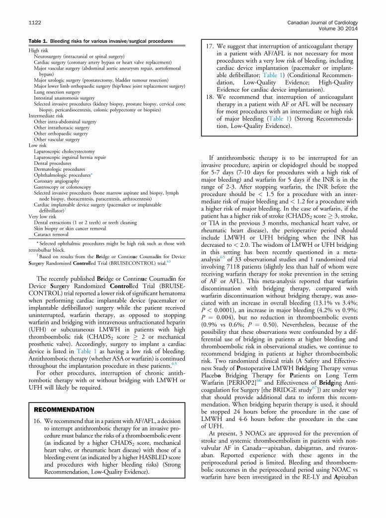
Table 1. Bleeding risks for various invasive/surgical procedures
High riskNeurosurgery (intracranial or spinal surgery)Cardiac surgery (coronary artery bypass or heart valve replacement)Major vascular surgery (abdominal aortic aneurysm repair, aortofemoral
bypass)Major urologic surgery (prostatectomy, bladder tumour resection)Major lower limb orthopaedic surgery (hip/knee joint replacement surgery)Lung resection surgeryIntestinal anastomosis surgerySelected invasive procedures (kidney biopsy, prostate biopsy, cervical cone
biopsy, pericardiocentesis, colonic polypectomy or biopsies)Intermediate risk
Other intra-abdominal surgeryOther intrathoracic surgeryOther orthopaedic surgeryOther vascular surgery
Low riskLaparoscopic cholecystectomyLaparoscopic inguinal hernia repairDental proceduresDermatologic proceduresOphthalmologic procedures*Coronary angiographyGastroscopy or colonoscopySelected invasive procedures (bone marrow aspirate and biopsy, lymph
node biopsy, thoracentesis, paracentesis, arthrocentesis)Cardiac implantable device surgery (pacemaker or implantable
defibrillator)y
Very low riskDental extractions (1 or 2 teeth) or teeth cleaningSkin biopsy or skin cancer removalCataract removal
* Selected ophthalmic procedures might be high risk such as those withretrobulbar block.
yBased on results from the Bridge or Continue Coumadin for DeviceSurgery Randomized Controlled Trial (BRUISECONTROL) trial.63
17. We suggest that interruption of anticoagulant therapyin a patient with AF/AFL is not necessary for mostprocedures with a very low risk of bleeding, includingcardiac device implantation (pacemaker or implant-able defibrillator; Table 1) (Conditional Recommen-dation, Low-Quality Evidence; High-QualityEvidence for cardiac device implantation).
18. We recommend that interruption of anticoagulanttherapy in a patient with AF or AFL will be necessaryfor most procedures with an intermediate or high riskof major bleeding (Table 1) (Strong Recommenda-tion, Low-Quality Evidence).
1122 Canadian Journal of CardiologyVolume 30 2014
The recently published Bridge or Continue Coumadin forDevice Surgery Randomized Controlled Trial (BRUISE-CONTROL) trial reported a lower risk of significant hematomawhen performing cardiac implantable device (pacemaker orimplantable defibrillator) surgery while the patient receiveduninterrupted, warfarin therapy, as opposed to stoppingwarfarin and bridging with intravenous unfractionated heparin(UFH) or subcutaneous LMWH in patients with highthromboembolic risk (CHADS2 score � 2 or mechanicalprosthetic valve). Accordingly, surgery to implant a cardiacdevice is listed in Table 1 as having a low risk of bleeding.Antithrombotic therapy (whether ASA or warfarin) is continuedthroughout the implantation procedure in these patients.63
For other procedures, interruption of chronic antith-rombotic therapy with or without bridging with LMWH orUFH will likely be required.
RECOMMENDATION
16. We recommend that in a patient withAF/AFL, a decisionto interrupt antithrombotic therapy for an invasive pro-cedure must balance the risks of a thromboembolic event(as indicated by a higher CHADS2 score, mechanicalheart valve, or rheumatic heart disease) with those of ableeding event (as indicated by a higher HASBLED scoreand procedures with higher bleeding risks) (StrongRecommendation, Low-Quality Evidence).
If antithrombotic therapy is to be interrupted for aninvasive procedure, aspirin or clopidogrel should be stoppedfor 5-7 days (7-10 days for procedures with a high risk ofmajor bleeding) and warfarin for 5 days if the INR is in therange of 2-3. After stopping warfarin, the INR before theprocedure should be < 1.5 for a procedure with an inter-mediate risk of major bleeding and < 1.2 for a procedure witha higher risk of major bleeding. In the case of warfarin, if thepatient has a higher risk of stroke (CHADS2 score � 3, stroke,or TIA in the previous 3 months, mechanical heart valve, orrheumatic heart disease), the perioperative period shouldinclude LMWH or UFH bridging when the INR hasdecreased to < 2.0. The wisdom of LMWH or UFH bridgingin this setting has been recently questioned in a meta-analysis65 of 33 observational studies and 1 randomized trialinvolving 7118 patients (slightly less than half of whom werereceiving warfarin therapy for stoke prevention in the settingof AF or AFL). This meta-analysis reported that warfarindiscontinuation with bridging therapy, compared withwarfarin discontinuation without bridging therapy, was asso-ciated with an increase in overall bleeding (13.1% vs 3.4%;P < 0.0001), an increase in major bleeding (4.2% vs 0.9%;P ¼ 0.004), but no reduction in thromboembolic events(0.9% vs 0.6%; P ¼ 0.50). Nevertheless, because of thepossibility that these observations were confounded by a dif-ferential use of bridging in patients at higher bleeding andthromboembolic risk in observational studies, we continue torecommend bridging in patients at higher thromboembolicrisk. Two randomized clinical trials (A Safety and Effective-ness Study of Postoperative LMWH Bridging Therapy versusPlacebo Bridging Therapy for Patients on Long TermWarfarin [PERIOP2]66 and Effectiveness of Bridging Anti-coagulation for Surgery [the BRIDGE study67]) are under waythat should provide additional data to inform this recom-mendation. When bridging heparin therapy is used, it shouldbe stopped 24 hours before the procedure in the case ofLMWH and 4-6 hours before the procedure in the caseof UFH.
At present, 3 NOACs are approved for the prevention ofstroke and systemic thromboembolism in patients with non-valvular AF in Canadadapixaban, dabigatran, and rivarox-aban. Reported experience with these agents in theperiprocedural period is limited. Bleeding and thromboem-bolic outcomes in the periprocedural period using NOAC vswarfarin have been investigated in the RE-LY and Apixaban

RECOMMENDATIONBefore the procedure
19. When a decision to interrupt aspirin or clopidogreltherapy for an invasive procedure has been made for apatient with AF/AFL, we suggest that interruptionbegin 5-7 days before the procedure, except for pro-cedures with a very high risk of bleeding, in whichcase we suggest interruption 7-10 days before theprocedure (Conditional Recommendation, Low-Quality Evidence).
20. When a decision to interrupt warfarin therapy for aninvasive procedure has been made for a patient withAF/AFL, we suggest that interruption begin 5 daysbefore the procedure. A procedure with low bleedingrisk may proceed when the INR is < 1.5 and a pro-cedure with an intermediate or high bleeding risk mayproceed when the INR is < 1.2 (ConditionalRecommendation, Low-Quality Evidence).
21. When a decision to interrupt warfarin therapy for aninvasive procedure has been made for a patient withAF/AFL, we suggest that bridging therapy withLMWH or UFH be instituted when the INR is belowthat patient’s therapeutic INR target in a patient athigh risk of thromboembolic events (CHADS2 score� 3, mechanical heart valve, stroke or TIA within 3months, rheumatic heart disease) (ConditionalRecommendation, Low-Quality Evidence).
22. When a decision to interrupt apixaban or rivaroxabantherapy for an invasive procedure has been made for apatient with AF/AFL, we suggest that interruptionbegin 1-2 days before a procedure with low risk ofmajor bleeding and 2-3 days before a procedure withan intermediate or high risk of major bleeding (Con-ditional Recommendation, Low-Quality Evidence).
23. When a decision to interrupt dabigatran therapy foran invasive procedure has been made for a patientwith AF/AFL, we suggest that interruption begin 1-2days before a procedure with low risk of majorbleeding and 2-3 days before a procedure with anintermediate or high risk of major bleeding wheneGFR is � 80 mL/min/1.73 m2 (ConditionalRecommendation, Low-Quality Evidence). The up-per end of these ranges should be used if eGFR is 50-80 mL/min/1.73 m2, an additional day should beadded when eGFR is 30-50 mL/min/1.73 m2, and incase eGFR is < 30 mL/min/1.73 m2, yet 1 more dayof dabigatran withdrawal should be added (Condi-tional Recommendation, Low-Quality Evidence).
24. We recommend that when LMWH or UFHbridging is used for an invasive procedure, the ther-apy should be started before the procedure when theINR is < 2.0 and be stopped 24 hours before theprocedure for LMWH and 4-6 hours befolre theprocedure for UFH (Strong recommendation, Low-Quality Evidence).
Verma et al. 11232014 Focused Update of the CCS AF Guidelines
for Reduction in Stroke and Other Thromboembolic Eventsin Atrial Fibrillation (ARISTOTLE) trials.9,68 There were nostatistically significant differences between the dabigatran orthe apixaban group and their respective warfarin groups withrespect to bleeding or thromboembolic complications in theperiprocedural period.69,70 Comparable data have not yetbeen published regarding rivaroxaban from the ROCKET AFtrial, but an observational analysis from the Dresden NOACRegistry suggests no difference in bleeding or thromboemboliccomplications in the periprocedural period in a comparison ofpatients who had been receiving rivaroxaban with patientswho received dabigatran.68
Because of the limited reported experience with NOACs inthe periprocedural period, recommendations regarding theiruse are necessarily empirical. The offset kinetics of theiranticoagulant effects might be reasonably predicted by theirelimination half-lives.71-73 Dabigatran is eliminated to agreater extent by the kidneys, so the time it should be with-held for an invasive procedure depends on renal function.Renal function is less critical to the elimination half-lives ofapixaban and rivaroxaban, provided the eGFR is > 30 mL/min/1.73 m2. We recommend that the withdrawal period forapixaban or rivaroxaban be 1-2 days for a procedure with alower risk of major bleeding and 2-3 days for a procedure witha higher risk of major bleeding. The same periods are rec-ommended for withdrawal of dabigatran when the eGFR is �80 mL/min/1.73 m2. The higher end of these ranges is rec-ommended for dabigatran if the eGFR is 50-80 mL/min/1.73 m2, and an additional day should be added for dabiga-tran if the eGFR is 30-50 mL/min/1.73 m2. For eGFR< 30 mL/min/1.73 m2, yet an additional day should be addedfor dabigatran, although NOACs are not generally recom-mended in these patients.
Because of the rapid onset and offset kinetics of theNOACs, bridging with LMWH or UFH is not recommendedduring the periprocedural period unless the NOAC has beenwithdrawn for a period longer than that recommended andthe patient has a greater risk of thromboembolism (CHADS2score � 3, mechanical heart valve, stroke or TIA within 3months, rheumatic heart disease).
The absence of a proven “antidote” for the NOACs alongwith interindividual variability in their respective terminalelimination half-lives, suggest a conservative approach forrecommendations regarding the use of these agents around thetime of procedures during which any bleeding could conferserious adverse outcomes. Thus, the NOACs should not beused in the perioperative period when neuraxial anaesthesia orlumbar puncture is planned.
After the invasive procedure, antithrombotic therapy isreintroduced after hemostasis has been secured (usually 24-48hours after a procedure with a lower risk of major bleedingand 48-72 hours after a procedure with a higher risk of majorbleeding). Because of the rapid onset of the effects of theNOACs, these agents should be restarted nearer the latterportion of each of these suggested ranges. When bridgingLMWH is used, the LMWH should be given in prophylacticdosages for 24-72 hours depending on the risk of proceduralbleeding. LMWH may be increased to therapeutic dosagesthereafter, providing postoperative bleeding has not occurred.Warfarin therapy is reintroduced after the same time periods,recognizing that several days will be needed for the INR to
achieve the therapeutic range at which time bridging LMWHor UFH may be discontinued.

After the procedure
25. When LMWH or UFH bridging is used for aninvasive procedure, we suggest that such therapy berestarted after the procedure when hemostasis isestablished (usually 24 hours for a procedure with alow risk of bleeding and 48-72 hours for a procedurewith an intermediate or high risk of bleeding) inprophylactic dosages for the first 24-72 hours andthen increased to therapeutic dosages. Bridging is thencontinued until an OAC is therapeutic (ConditionalRecommendation, Low-Quality Evidence).
26. When warfarin, ASA, or clopidogrel therapy has beeninterrupted for an invasive procedure, we suggest thatsuch therapy be restarted after the procedure whenhemostasis is established (usually 24-48 hours for aprocedure with a low risk of bleeding and 48-72 hoursfor a procedure with an intermediate or high risk ofbleeding) (Conditional Recommendation, Low-Quality Evidence).
27. When apixaban, dabigatran, or rivaroxaban have beenwithdrawn for an invasive procedure we suggest thatsuch therapy be restarted after the procedure 1 dayafter hemostasis is established (usually 48 hours for aprocedure with low risk of bleeding and 72 hours for aprocedure with an intermediate or high risk ofbleeding) (Conditional Recommendation, Low-Quality Evidence).
1124 Canadian Journal of CardiologyVolume 30 2014
VIII. Rate and Rhythm ControlSince the publication of the 2012 focused update of CCS
AF guidelines,2 there have been some new data that reinforcethe rate and rhythm control recommendations in the 2010guidelines and the focused update.74
Target for rate control
In the 2010 guidelines, the committee recommended thatthe goals of ventricular rate control “should be to improvesymptoms and clinical outcomes which are attributable toexcessive ventricular rates.”74 Although the historical target forrate control was a resting heart rate < 80 beats per minute, the2010 guidelines liberalized the target to a resting heart rate <100 beats per minute based on randomized clinical trial datasuggesting no difference between strict and lenient rate con-trol targets.
A recent substudy of 608 patients analyzed quality of lifewith 3 measurement tools in patients randomized to lenient vsstrict rate control.75 After an average of 2 years of follow-up,there was no difference between groups for any of the qualityof life measures. This evidence further reinforces the guide-lines original recommendation for a more lenient target heartrate of < 100 beats per minute.
Use of digoxin for rate control
In the 2010 guidelines, digoxin was downgraded tosecond-line therapy for rate control of AF, behind calciumchannel blockers and b-blockers. Specifically, we suggestedthat “digoxin not be used as initial therapy for active patients
and be reserved for rate control in patients who are sedentaryor who have left ventricular dysfunction.”74 Since that time,additional evidence has further questioned the use of digoxinfor rate control. Two substudies from the Atrial FibrillationFollow-up Investigation of Rhythm Management (AFFIRM)trial comparing rate control with rhythm control were pub-lished in 2013.76,77 In the first substudy, use of digoxin wastreated as a time-dependent covariate to take into accountthat patients might be using therapy or not at various timesin follow-up. After controlling for clinical variables and usingpropensity adjustment, the results showed a significant in-crease in all-cause mortality associated with digoxin use(hazard ratio, 1.41; 95% CI, 1.19-1.67; P < 0.001).76 In thesame journal issue, however, another substudy from theAFFIRM trial showed no increased mortality, but also nobenefit, associated with digoxin use (hazard ratio, 1.06; 95%CI, 0.83-1.37; P ¼ 0.64).77 The second study excluded alarge number of patients based on inclusion criteria andmethod of propensity matching, and did not account forchanges in digoxin use over time. Furthermore, there hasbeen a greater understanding about drug interactions thatmight elevate digoxin levels to toxic ranges. Dronedarone, forexample, has been shown to increase steady-state digoxinlevels by 2.5 times.78 These data reinforce the 2010 guide-lines recommendation that digoxin only be used as second-line therapy in selected cases.
Algorithm for choice of rate vs rhythm control for AF
As mentioned earlier, the main goal of AF treatment is torelieve symptoms, improve functional capacity and qualityof life, and where possible, improve left ventricular func-tion. Trial results suggest that this can be accomplished byeither rate control or rhythm control, without systematicbenefit of either strategy in terms of mortality or morbidity.Thus, the choice of rate vs rhythm control must be indi-vidualized for each patient. Furthermore, the preferredtreatment strategy might change over time if the patient’ssymptoms change or if one strategy has not proveneffective.
The Guidelines Committee has developed a new practicealgorithm (Fig. 3) to help in treatment selection. For pa-tients with recently diagnosed symptomatic AF, rate controlto keep the heart rate < 100 beats per minute seems anappropriate first step. If symptoms do not resolve with ratecontrol or therapy-related adverse effects appear, thenrhythm control might be considered. The choice of rhythmcontrol relates to AF characteristics. For patients with a lowburden of infrequent paroxysmal AF, pill-in-pocket antiar-rhythmic therapy might be reasonable. For more frequentparoxysmal AF, daily maintenance antiarrhythmic therapymight be tried, followed by consideration of catheter abla-tion if the response is not adequate. For patients withpersistent AF, a trial of cardioversion might help to deter-mine further treatment strategies. If sinus rhythm restora-tion substantially improves symptoms and AF does notrecur, the patient might simply be observed until AF recurs.If sinus rhythm restoration improves symptoms but AFrecurs, maintenance antiarrhythmic drug (AAD) therapyshould be considered, with catheter ablation if drugs are noteffective. However, if sinus rhythm restoration does not

Figure 3. Approach to rate and/or rhythm control of atrial fibrillation (AF) in patients presenting with symptomatic AF. QOL, quality of life.
Verma et al. 11252014 Focused Update of the CCS AF Guidelines
change symptoms, long-term rate control would bepreferred if AF recurs.
In selected cases with highly symptomatic AF, multiplerecurrences, or extreme impairment in quality of life, earlyconsideration of rhythm control in conjunction with ratecontrol might be indicated. Patients with arrhythmia-inducedcardiomyopathy might also be considered for early rhythmcontrol, although cardiomyopathy might resolve withadequate rate control alone.
For patients with asymptomatic persistent AF, rate controlis usually the therapy of choice. However, AF symptomsmight be difficult to ascertain because they can be quitenonspecific. Fatigue, declining exercise tolerance, decreasedmotivation, and exertional dyspnea might all be attributed toaging or other conditions, but might actually be due to AF.When it is difficult to establish whether symptoms are due toAF, a trial of cardioversion might be helpful. Clear symptomrelief after restoration of sinus rhythm would encourage arhythm control approach.
Recent evidence suggests that 4 weeks of antiarrhythmictherapy after cardioversion might reduce the incidence oflonger-term AF recurrence beyond the 4 weeks.79 Patientswere randomized after cardioversion to no antiarrhythmictherapy, flecainide for 4 weeks after cardioversion, or flecai-nide for 6 months after cardioversion. Although short-term
flecainide was less effective than long-term treatment, thedifference in recurrence was small (56% vs 48%), reflectingthe predominant recurrence rate early after cardioversion andsuggesting that short-duration antiarrhythmic therapy aftercardioversion might produce enduring AF recurrencereduction.
IX. Catheter Ablation for AF
AF ablation as first-line therapy
Recent studies examined the value of AF ablation as first-line therapy. The Medical Antiarrhythmic Treatment orRadiofrequency Ablation in Paroxysmal Atrial Fibrillation(MANTRA-PAF) study randomized 294 patients withparoxysmal AF and no history of AAD use to an initialstrategy of catheter ablation (n ¼ 146) or AADs (n ¼ 148).After 24 months, AF burden was not different but signifi-cantly more patients in the ablation group were AF-free.80
The Radiofrequency Ablation vs Antiarrhythmic Drugs asFirst-Line Therapy of Atrial Fibrillation 2 (RAAFT-2) trialrandomized 127 AAD-naive patients with paroxysmal AF tofirst-line ablation (n ¼ 66) vs AAD-therapy (n ¼ 61)75 andfollowed them for 2 years: ablation significantly reducedsymptomatic and asymptomatic AF vs AADs. This new

1126 Canadian Journal of CardiologyVolume 30 2014
evidence supports the 2010 recommendation suggestingcatheter ablation as first-line therapy in highly-selected pa-tients with symptomatic paroxysmal AF. In Table 2 thebenefit-to-risk ratio for catheter ablation in various situationsare summarized.
Alternate energy sources for catheter ablation
AF ablation most commonly uses radiofrequency (RF)energy to isolate pulmonary veins (PVs). However, perform-ing PV isolation (PVI) with a “point-by-point” approachmight be technically challenging. Ablation technologies havetherefore been developed to achieve PVI with a singlecircumferential energy application.
The cryoballoon system consists of a steerable catheter thatdelivers pressurized cryorefrigerant to a distally mountedballoon. The multicentre, randomized Sustained Treatmentof Paroxysmal Atrial Fibrillation (STOP AF) trial comparedAAD therapy with cryoballoon-based PVI in 245 patientswith drug-refractory paroxysmal AF.81 At 1 year, 69.9% of163 cryoballoon-ablated patients were AF-free, comparedwith 7.3% of 82 patients randomized to AADs. Early expe-rience suggests that the efficacy and safety of cryoballoonablation are comparable to RF energy for paroxysmal AF.82,83
The PV ablation catheter (PVAC) consists of a circularcatheter that enables mapping and circumferential PV abla-tion using RF. PVAC ablation efficacy compares favourablywith standard RF energy delivery,82,84,85 but PVAC ablationproduced an excessively high rate (38%-45%) of silent cere-bral ischemic lesions.86,87 With procedural modifications,there has been a significant decrease in cerebral ischemicevents to rates similar to those obtained using othertechnologies.88
Complications of catheter ablation
In recent studies the important issue of risks associatedwith AF ablation have been examined.89-92 In a single-centre study in which risks were assessed during 1190procedures over 9 years, the major complication rates were4.7%, including vascular complications (1.5%), tamponade(1.1%), and stroke/TIA (1.1%).89 In a US inpatientdatabase analysis 93,801 ablations performed between2000 and 2010 were examined.91 The overall complicationrate was 6.29% (vascular, 1.53%; pericardial, 1.52%; res-piratory, 1.3%; stroke/TIA, 1.02%) with a 0.42% in-hospital mortality rate. Annual operator volume (< 25AF ablation procedures) and hospital volume (< 50 AFablation procedures) were significant predictors of majoradverse outcomes. The experience threshhold for reducedrisk seems to be 25-50 procedures per operator per year,
Table 2. Balance of benefit to risk for catheter ablation in patientswith symptomatic atrial fibrillation
Long-standing* Persistent Paroxysmal
First line d d þFailed first drug d þ þþFailed second drug þ þþ þþþFailed multiple drugs þþ þþþ þþþ
þ Indicates balance of benefit to risk in favour of catheter ablation.*Ongoing symptomatic atrial fibrillation � 1 year.
which aligns with the 2010 CCS/Canadian Heart RhythmSociety training standards for adult clinical cardiacelectrophysiology.93
Long-term efficacy of catheter ablation
Most large-scale observational studies and RCTs hadlimited follow-up.94-98 Recently, several studies havedemonstrated that very late arrhythmia recurrences (> 1 year)are not uncommon after initial “successful” ablation.99-102 In1 study, arrhythmia-free survival rates were 87%, 81%, and63% after the last catheter ablation procedure at 1, 2, and 5years, respectively.99 In another study, the long-termarrhythmia-free outcomes of 123 patients AF-free at 1 yearwere 85% after 3 years and 71% after 5 years.100 Catheterablation of AF should thus be considered an effective treat-ment rather than a cure. This new evidence reinforces theimportance of pursuing oral anticoagulation in patients withhigh thromboembolic risk, regardless of short- or mid-termprocedural success.
Antithrombotic therapy in relation to catheter ablation
AF ablation is associated with a risk of thromboembolismeven in patients without stroke risk factors. Therefore,careful anticoagulation is critical before, during, and afterAF ablation procedures. Recent studies have demonstratedthat performing AF ablation with uninterrupted warfarin(therapeutic INR of 2.0-3.0) is safe and associated withfewer complications (vascular access complications, strokes)compared with bridging with LMWH.103-105 Evidence forthe periprocedural use of NOACs for AF ablation is limited.Nonrandomized studies that evaluated interrupted dabiga-tran vs uninterrupted warfarin have yielded conflictingresults.106-109 Recently, no differences in periproceduralbleeding or thromboembolic complications were observedbetween uninterrupted rivaroxaban and uninterruptedwarfarin therapy.110 Results from RCTs of periproceduralNOAC vs warfarin should further define the relative role ofthese agents. After ablation during warfarin treatment, itshould be continued to maintain an INR of > 2.0 to avoidthe use of heparin bridging. When NOACs are used, theycan be reinitiated within 4-12 hours of sheath removalwithout the need for heparin bridging. In all cases, oralanticoagulation with warfarin or NOACs should becontinued for at least 2 months after AF ablation. Althoughit has been suggested that AF ablation could lower the long-term stroke risk, this remains unproven.111-114 In additionto delayed symptomatic AF recurrence discussed previouslyherein, asymptomatic recurrences are not infrequent;accordingly, evaluation of procedural success according tosymptoms alone is unreliable.115 Therefore, decisionsregarding long-term anticoagulation should be based onthe patient’s stroke risk factors, not on the apparentabsence of AF recurrence. A desire to avoid long-termanticoagulation should not be considered an indicationfor AF ablation. Further long-term studies such as theOptimal Anticoagulation for Enhanced Risk Patients Post-Catheter Ablation for Atrial Fibrillation (OCEAN) trial(ClinicalTrials.gov NCT02168829) will help to assess therisk of stroke after AF ablation.

RECOMMENDATION
28. We recommend catheter ablation of AF in patientswho remain symptomatic after an adequate trial ofAAD therapy and in whom a rhythm control strategyremains desired (Strong Recommendation, Moderate-Quality Evidence).
29. We suggest catheter ablation to maintain sinusrhythm as first-line therapy for relief of symptoms inhighly selected patients with symptomatic, paroxysmalAF (Conditional Recommendation, Moderate-Quality Evidence).
30. We suggest that catheter ablation of AF shouldbe performed by electrophysiologists with a highdegree of expertise and high annual procedural vol-umes (Conditional Recommendation, Low-QualityEvidence).
Verma et al. 11272014 Focused Update of the CCS AF Guidelines
Practical tip. The following represents an ideal, but notexclusive, profile of a patient who is referred for consider-ation of AF ablation today: age < 80 years, symptomaticwith their AF, has tried but treatment has failed or isintolerant of AAD therapy, has paroxysmal AF or short-standing persistent AF, and minimal to moderate structuralheart disease (such as left ventricular dysfunction or valvulardisease).
Practical tip. AF ablation should not be considered as analternative to oral anticoagulation. If a patient has a highthromboembolic risk profile, then the patient shouldcontinue oral anticoagulation even after successful AF abla-tion. Studies of long-term monitoring have consistentlyshown asymptomatic episodes of AF before and after abla-tion. Initiation of oral anticoagulation should also not bedelayed when indicated in patients pending referral for AFablation.
AcknowledgementsFor a full list of Guideline Committee Members, see the
Canadian Cardiovascular Society Atrial Fibrillation Guidelines ePrimary Panel and Canadian Cardiovascular Society AtrialFibrillation Guidelines e Secondary Panel sections of theSupplementary Material.
References
1. Cairns JA, Yusuf S, Cook RJ, et al. Canadian network and centre fortrials internationally (cannectin): A national network for canadian-ledtrials in cardiovascular diseases and diabetes mellitus. Can J Cardiol2010;26:353-8.
2. Skanes AC, Healey JS, Cairns JA, et al. Focused 2012 update of theCanadian Cardiovascular Society atrial fibrillation guidelines: recom-mendations for stroke prevention and rate/rhythm control. Can J Car-diol 2012;28:125-36.
3. Gage BF, Waterman AD, Shannon W, et al. Validation of clinicalclassification schemes for predicting stroke: results from the nationalregistry of atrial fibrillation. JAMA 2001;285:2864-70.
4. Lip GY, Nieuwlaat R, Pisters R, Lane DA, Crijns HJ. Refining clinicalrisk stratification for predicting stroke and thromboembolism in atrialfibrillation using a novel risk factor-based approach: the euro heartsurvey on atrial fibrillation. Chest 2010;137:263-72.
5. January CT, Wann LS, Alpert JS, et al. 2014 AHA/ACC/HRS guidelinefor the management of patients with atrial fibrillation: executive sum-mary: a report of the American College of Cardiology/American HeartAssociation task force on practice guidelines and the Heart RhythmSociety. Circulation 2014 Apr 10. [Epub ahead of print]
6. Olesen JB, Lip GY, Hansen ML, et al. Validation of risk stratificationschemes for predicting stroke and thromboembolism in patients withatrial fibrillation: nationwide cohort study. BMJ 2011;342:d124.
7. Hart RG, Pearce LA, Aguilar MI. Meta-analysis: antithrombotic therapyto prevent stroke in patients who have nonvalvular atrial fibrillation.Ann Intern Med 2007;146:857-67.
8. You JJ, Singer DE, Howard PA, et al. Antithrombotic therapy for atrialfibrillation: antithrombotic therapy and prevention of thrombosis, 9thed: American College of Chest Physicians evidence-based clinical prac-tice guidelines. Chest 2012;141:e531S-75S.
9. Saposnik G, Gladstone D, Raptis R, Zhou L, Hart RG. Atrial fibrilla-tion in ischemic stroke: Predicting response to thrombolysis and clinicaloutcomes. Stroke 2013;44:99-104.
10. Hart RG, Pearce LA, Aguilar MI. Adjusted-dose warfarin versus aspirinfor preventing stroke in patients with atrial fibrillation. Ann Int Med2007;147:590-2.
11. Aguilar M, Hart R. Antiplatelet therapy for preventing stroke in patientswith non-valvular atrial fibrillation and no previous history of stroke ortransient ischemic attacks. Cochrane Database Syst Rev 2005:CD001925.
12. The efficacy of aspirin in patients with atrial fibrillation. Analysis ofpooled data from 3 randomized trials. The atrial fibrillation in-vestigators. Arch Intern Med 1997;157:1237-40.
13. Mant J, Hobbs FD, Fletcher K, et al. Warfarin versus aspirin for strokeprevention in an elderly community population with atrial fibrillation(the Birmingham atrial fibrillation treatment of the aged study,BAFTA): a randomised controlled trial. Lancet 2007;370:493-503.
14. Connolly SJ, Eikelboom J, Joyner C, et al. Apixaban in patients withatrial fibrillation. N Engl J Med 2011;364:806-17.
15. Connolly SJ, Ezekowitz MD, Yusuf S, et al. Dabigatran versus warfarinin patients with atrial fibrillation. N Engl J Med 2009;361:1139-51.
16. Patel MR, Mahaffey KW, Garg J, et al. Rivaroxaban versus warfarin innonvalvular atrial fibrillation. N Engl J Med 2011;365:883-91.
17. Granger CB, Alexander JH, McMurray JJ, et al. Apixaban versuswarfarin in patients with atrial fibrillation. N Engl J Med 2011;365:981-92.
18. Giugliano RP, Ruff CT, Braunwald E, et al. Edoxaban versus warfarinin patients with atrial fibrillation. N Engl J Med 2013;369:2093-104.
19. Ruff CT, Giugliano RP, Braunwald E, et al. Comparison of the efficacyand safety of new oral anticoagulants with warfarin in patients with atrialfibrillation: a meta-analysis of randomised trials. Lancet 2014;383:955-62.
20. Lip GY, Larsen TB, Skjoth F, Rasmussen LH. Indirect comparisons ofnew oral anticoagulant drugs for efficacy and safety when used for strokeprevention in atrial fibrillation. J Am Coll Cardiol 2012;60:738-46.
21. Mantha S, Ansell J. An indirect comparison of dabigatran, rivaroxabanand apixaban for atrial fibrillation. Thromb Haemost 2012;108:476-84.

1128 Canadian Journal of CardiologyVolume 30 2014
22. Majeed A, Hwang HG, Connolly SJ, et al. Management and outcomesof major bleeding during treatment with dabigatran or warfarin. Cir-culation 2013;128:2325-32.
23. FDA Drug Safety Communication: FDA study of Medicare patientsfinds risks lower for stroke and death but higher for gastrointestinalbleeding with Pradaxa (dabigatran) compared to warfarin. Available at:http://www.fda.gov/Drugs/DrugSafety/ucm396470.htm. Accessed May13, 2014.
24. Piccini JP, Stevens SR, Chang Y, et al. Renal dysfunction as a predictorof stroke and systemic embolism in patients with nonvalvular atrialfibrillation: validation of the R(2)CHADS(2) index in the ROCKET AF(Rivaroxaban Once-daily, oral, direct factor Xa inhibition Comparedwith vitamin K antagonism for prevention of stroke and Embolism Trialin Atrial Fibrillation) and ATRIA (AnTicoagulation and Risk factors InAtrial fibrillation) study cohorts. Circulation 2013;127:224-32.
25. Chao TF, Tsao HM, Ambrose K, et al. Renal dysfunction and the riskof thromboembolic events in patients with atrial fibrillation after cath-eter ablationethe potential role beyond the CHA2DS2-VASc score.Heart Rhythm 2012;9:1755-60.
26. Roldan V, Marin F, Manzano-Fernandez S, et al. Does chronic kidneydisease improve the predictive value of the CHADS2 and CHA2DS2-VASc stroke stratification risk scores for atrial fibrillation? ThrombHaemost 2013;109:956-60.
27. Apostolakis S, Guo Y, Lane DA, Buller H, Lip GY. Renal function andoutcomes in anticoagulated patients with non-valvular atrial fibrillation:the AMADEUS trial. Eur Heart J 2013;34:3572-9.
28. Banerjee A, Fauchier L, Vourc’h P, et al. Renal impairment andischemic stroke risk assessment in patients with atrial fibrillation: theLoire Valley Atrial Fibrillation Project. J Am Coll Cardiol 2013;61:2079-87.
29. Banerjee A, Fauchier L, Vourc’h P, et al. A prospective study of esti-mated glomerular filtration rate and outcomes in patients with atrialfibrillation: the Loire Valley Atrial Fibrillation Project. Chest 2014;145:1370-82.
30. Hart RG, Pearce LA, Asinger RW, Herzog CA. Warfarin in atrialfibrillation patients with moderate chronic kidney disease. Clin J AmSoc Nephrol 2011;6:2599-604.
31. McGrath ER, Kapral MK, Fang J, et al. Association of atrial fibrillationwith mortality and disability after ischemic stroke. Neurology 2013;81:825-32.
32. Kishore A, Vail A, Majid A, et al. Detection of atrial fibrillation afterischemic stroke or transient ischemic attack: a systematic review andmeta-analysis. Stroke 2014;45:520-6.
33. Seet RC, Friedman PA, Rabinstein AA. Prolonged rhythm monitoringfor the detection of occult paroxysmal atrial fibrillation in ischemicstroke of unknown cause. Circulation 2011;124:477-86.
34. Culebras A, Messe SR, Chaturvedi S, Kase CS, Gronseth G. Summaryof evidence-based guideline update: prevention of stroke in nonvalvularatrial fibrillation: report of the guideline development subcommittee ofthe American Academy of Neurology. Neurology 2014;82:716-24.
35. Hart RG, Diener HC, Coutts SB, et al. Embolic strokes of undeter-mined source: the case for a new clinical construct. Lancet Neurol2014;13:429-38.
36. Higgins P, MacFarlane PW, Dawson J, et al. Noninvasive cardiac eventmonitoring to detect atrial fibrillation after ischemic stroke: a random-ized, controlled trial. Stroke 2013;44:2525-31.
37. Sanna T, Diener HC, Passman RS, et al. Cryptogenic stroke and un-derlying atrial fibrillation. N Engl J Med 2014;370:2478-86.
38. Wachter R, Weber-Kruger M, Seegers J, et al. Age-dependent yield ofscreening for undetected atrial fibrillation in stroke patients: the Find-AF study. J Neurol 2013;260:2042-5.
39. Wallmann D, Tuller D, Wustmann K, et al. Frequent atrial prematurebeats predict paroxysmal atrial fibrillation in stroke patients: an op-portunity for a new diagnostic strategy. Stroke 2007;38:2292-4.
40. Kochhauser S, Dechering DG, Dittrich R, et al. Supraventricular pre-mature beats and short atrial runs predict atrial fibrillation in continu-ously monitored patients with cryptogenic stroke. Stroke 2014;45:884-6.
41. Weber-Kruger M, Groschel K, Mende M, et al. Excessive supraven-tricular ectopic activity is indicative of paroxysmal atrial fibrillation inpatients with cerebral ischemia. PLoS One 2013;8:e67602.
42. Glotzer TV, Daoud EG, Wyse DG, et al. The relationship betweendaily atrial tachyarrhythmia burden from implantable device diagnosticsand stroke risk: the TRENDS study. Circ Arrhythm Electrophysiol2009;2:474-80.
43. Healey JS, Connolly SJ, Gold MR, et al. Subclinical atrial fibrillationand the risk of stroke. N Engl J Med 2012;366:120-9.
44. Gage BF, van Walraven C, Pearce L, et al. Selecting patients with atrialfibrillation for anticoagulation: stroke risk stratification in patients tak-ing aspirin. Circulation 2004;110:2287-92.
45. Brambatti M, Connolly SJ, Gold MR, et al. Temporal relationshipbetween subclinical atrial fibrillation and embolic events. Circulation2014;129:2094-9.
46. Daoud EG, Glotzer TV, Wyse DG, et al. Temporal relationship of atrialtachyarrhythmias, cerebrovascular events, and systemic emboli based onstored device data: a subgroup analysis of trends. Heart Rhythm 2011;8:1416-23.
47. Healey JS, Martin JL, Duncan A, et al. Pacemaker-detected atrialfibrillation in patients with pacemakers: Prevalence, predictors, andcurrent use of oral anticoagulation. Can J Cardiol 2013;29:224-8.
48. Holmes DR, Reddy VY, Turi ZG, et al. Percutaneous closure of the leftatrial appendage versus warfarin therapy for prevention of stroke inpatients with atrial fibrillation: a randomised non-inferiority trial. Lancet2009;374:534-42.
49. Reddy VY, Doshi SK, Sievert H, et al. Percutaneous left atrialappendage closure for stroke prophylaxis in patients with atrial fibril-lation: 2.3-Year Follow-up of the PROTECT AF (Watchman LeftAtrial Appendage System for Embolic Protection in Patients with AtrialFibrillation) Trial. Circulation 2013;127:720-9.
50. Decker WW, Stead LG. Selecting rate control for recent-onset atrialfibrillation. Ann Emerg Med 2011;57:32-3.
51. Stiell IG, Birnie D. Managing recent-onset atrial fibrillation in theemergency department. Ann Emerg Med 2011;57:31-2.
52. Rogenstein C, Kelly AM, Mason S, et al. An international view of howrecent-onset atrial fibrillation is treated in the emergency department.Acad Emerg Med 2012;19:1255-60.
53. Stiell IG, Macle L. Canadian Cardiovascular Society atrial fibrillationguidelines 2010: management of recent-onset atrial fibrillation andflutter in the emergency department. Can J Cardiol 2011;27:38-46.
54. Michael JA, Stiell IG, Agarwal S, Mandavia DP. Cardioversion ofparoxysmal atrial fibrillation in the emergency department. Ann EmergMed 1999;33:379-87.

Verma et al. 11292014 Focused Update of the CCS AF Guidelines
55. Burton JH, Vinson DR, Drummond K, et al. Electrical cardioversion ofemergency department patients with atrial fibrillation. Ann Emerg Med2004;44:20-30.
56. Gallagher MM, Hennessy BJ, Edvardsson N, et al. Embolic compli-cations of direct current cardioversion of atrial arrhythmias: associationwith low intensity of anticoagulation at the time of cardioversion. J AmColl Cardiol 2002;40:926-33.
57. Stiell IG, Clement CM, Perry JJ, et al. Association of the OttawaAggressive Protocol with rapid discharge of emergency department pa-tients with recent-onset atrial fibrillation or flutter. CJEM 2010;12:181-91.
58. Xavier Scheuermeyer F, Grafstein E, Stenstrom R, et al. Thirty-dayoutcomes of emergency department patients undergoing electrical car-dioversion for atrial fibrillation or flutter. Acad Emerg Med 2010;17:408-15.
59. Airaksinen KE, Gronberg T, Nuotio I, et al. Thromboembolic com-plications after cardioversion of acute atrial fibrillation: the FINCV(Finnish Cardioversion) study. J Am Coll Cardiol 2013;62:1187-92.
60. Kearon C, Hirsh J. Management of anticoagulation before and afterelective surgery. N Engl J Med 1997;336:1506-11.
61. Douketis JD, Spyropoulos AC, Spencer FA, et al. Perioperative man-agement of antithrombotic therapy: antithrombotic therapy and pre-vention of thrombosis, 9th ed: American College of Chest Physiciansevidence-based clinical practice guidelines. Chest 2012;141:e326S-50S.
62. Thrombosis Canada. Peri-operative management of patients who arereceiving warfarin. Available at: http://www.thrombosiscanada.ca/guides/pdfs/Warfarin_perioperative_management.pdf. Accessed May13, 2014.
63. Birnie DH, Healey JS, Wells GA, et al. Pacemaker or defibrillatorsurgery without interruption of anticoagulation. N Engl J Med2013;368:2084-93.
64. Patatanian E, Fugate SE. Hemostatic mouthwashes in anticoagulatedpatients undergoing dental extraction. Ann Pharmacother 2006;40:2205-10.
65. Siegal D, Yudin J, Kaatz S, et al. Periprocedural heparin bridging inpatients receiving vitamin k antagonists: systematic review and meta-analysis of bleeding and thromboembolic rates. Circulation 2012;126:1630-9.
66. PERIOP 2-A Safety and Effectiveness Study of LMWH BridgingTherapy Versus Placebo Bridging Therapy for Patients on Long TermWarfarin and Require Temporary Interruption of Their Warfarin.Available at: http://clinicaltrials.gov/show/NCT00432796. AccessedJune 30, 2014.
67. Effectiveness of Bridging Anticoagulation for Surgery (The BRIDGEStudy). Available at: http://clinicaltrials.gov/show/NCT00786474.Accessed June 30, 2014.
68. Beyer-Westendorf J, Gelbricht V, Forster K, et al. Peri-interventionalmanagement of novel oral anticoagulants in daily care: results from theprospective Dresden NOAC registry. Eur Heart J 2014;35:1888-96.
69. Healey JS, Eikelboom J, Douketis J, et al. Periprocedural bleeding andthromboembolic events with dabigatran compared with warfarin: resultsfrom the randomized evaluation of long-term anticoagulation therapy(RE-LY) randomized trial. Circulation 2012;126:343-8.
70. Lopes RD, Garcia D, Wojdyla D, et al. Use of apixaban and warfarin inpatients undergoing invasive procedures: insights from Aristotle. Can JCardiol 2013;29:S232-3.
71. Eliquis productmonograph, l3 version 1.1. Available at: http://www.pfizer.ca/en/our_products/products/monograph/313. Accessed June 30, 2014.
72. Pradax product monograph. Available at: http://www.boehringer-ingelheim.ca/content/dam/internet/opu/ca_EN/documents/humanhealth/product_monograph/PradaxaPMEN.pdf. Accessed June 30, 2014.
73. Kubitza D, Becka M, Mueck W, et al. Effects of renal impairment onthe pharmacokinetics, pharmacodynamics and safety of rivaroxaban, anoral, direct factor Xa inhibitor. Br J Clin Pharmacol 2010;70:703-12.
74. Gillis AM, Verma A, Talajic M, Nattel S, Dorian P. Canadian Car-diovascular Society atrial fibrillation guidelines 2010: rate and rhythmmanagement. Can J Cardiol 2011;27:47-59.
75. Morillo CA, Verma A, Connolly SJ, et al. Radiofrequency ablation vsantiarrhythmic drugs as first-line treatment of paroxysmal atrial fibril-lation (RAAFT-2): a randomized trial. JAMA 2014;311:692-700.
76. Whitbeck MG, Charnigo RJ, Khairy P, et al. Increased mortality amongpatients taking digoxineanalysis from the affirm study. Eur Heart J2013;34:1481-8.
77. Gheorghiade M, Fonarow GC, van Veldhuisen DJ, et al. Lack of evi-dence of increased mortality among patients with atrial fibrillationtaking digoxin: findings from post hoc propensity-matched analysis ofthe AFFIRM trial. Eur Heart J 2013;34:1489-97.
78. Vallakati A, Chandra PA, Pednekar M, Frankel R, Shani J. Dronedar-one-induced digoxin toxicity: new drug, new interactions. Am J Ther2013;20:e717-9.
79. Kirchhof P, Andresen D, Bosch R, et al. Short-term versus long-termantiarrhythmic drug treatment after cardioversion of atrial fibrillation(flec-sl): a prospective, randomised, open-label, blinded end pointassessment trial. Lancet 2012;380:238-46.
80. Cosedis Nielsen J, Johannessen A, Raatikainen P, et al. Radiofrequencyablation as initial therapy in paroxysmal atrial fibrillation. N Engl J Med2012;367:1587-95.
81. Packer DL, Kowal RC, Wheelan KR, et al. Cryoballoon ablation ofpulmonary veins for paroxysmal atrial fibrillation: first results of theNorth American Arctic front (STOP AF) pivotal trial. J Am Coll Car-diol 2013;61:1713-23.
82. Andrade JG, Khairy P, Guerra PG, et al. Efficacy and safety of cry-oballoon ablation for atrial fibrillation: a systematic review of publishedstudies. Heart Rhythm 2011;8:1444-51.
83. Schmidt M, Dorwarth U, Andresen D, et al. Cryoballoon versus RFablation in paroxysmal atrial fibrillation: results from the Germanablation registry. J Cardiovasc Electrophysiol 2014;25:1-7.
84. Calkins H, Reynolds MR, Spector P, et al. Treatment of atrial fibril-lation with antiarrhythmic drugs or radiofrequency ablation: two sys-tematic literature reviews and meta-analyses. Circ ArrhythmElectrophysiol 2009;2:349-61.
85. Hummel J, Michaud G, Hoyt R, et al. Phased RF ablation in persistentatrial fibrillation. Heart Rhythm 2014;11:202-9.
86. Herrera Siklody C, Deneke T, Hocini M, et al. Incidence of asymp-tomatic intracranial embolic events after pulmonary vein isolationcomparison of different atrial fibrillation ablation technologies in amulticenter study. J Am Coll Cardiol 2011;58:681-8.
87. Gaita F, Leclercq JF, Schumacher B, et al. Incidence of silent cerebralthromboembolic lesions after atrial fibrillation ablation may changeaccording to technology used: comparison of irrigated radiofrequency,multipolar nonirrigated catheter and cryoballoon. J Cardiovasc Elec-trophysiol 2011;22:961-8.

1130 Canadian Journal of CardiologyVolume 30 2014
88. Verma A, Debruyne P, Nardi S, et al. Evaluation and reduction ofasymptomatic cerebral embolism in ablation of atrial fibrillation, buthigh prevalence of chronic silent infarction: results of the Evaluation ofReduction of Asymptomatic Cerebral Embolism trial. Circ ArrhythmElectrophysiol 2013;6:835-42.
89. Hoyt H, Bhonsale A, Chilukuri K, et al. Complications arising fromcatheter ablation of atrial fibrillation: temporal trends and predictors.Heart Rhythm 2011;8:1869-74.
90. Shah RU, Freeman JV, Shilane D, et al. Procedural complications,rehospitalizations, and repeat procedures after catheter ablation for atrialfibrillation. J Am Coll Cardiol 2012;59:143-9.
91. Deshmukh A, Patel NJ, Pant S, et al. In-hospital complications asso-ciated with catheter ablation of atrial fibrillation in the United Statesbetween 2000 and 2010: analysis of 93 801 procedures. Circulation2013;128:2104-12.
92. Gupta A, Perera T, Ganesan A, et al. Complications of catheter ablationof atrial fibrillation: a systematic review. Circ Arrhythm Electrophysiol2013;6:1082-8.
93. Green MS, Guerra PG, Krahn AD. 2010 Canadian CardiovascularSociety/Canadian Heart Rhythm Society training standards and main-tenance of competency in adult clinical cardiac electrophysiology. Can JCardiol 2011;27:859-61.
94. Jais P, Cauchemez B, Macle L, et al. Catheter ablation versus antiar-rhythmic drugs for atrial fibrillation: the A4 study. Circulation2008;118:2498-505.
95. Packer DL, Irwin JM, Champagne J, et al. Cryoballoon ablation ofpulmonary veins for paroxysmal atrial fibrillation: first results of theNorth American Arctic Front Stop-AF pivotal trial. J Am Coll Cardiol2010;55:E3015-6.
96. Stabile G, Bertaglia E, Senatore G, et al. Catheter ablation treatment inpatients with drug-refractory atrial fibrillation: a prospective, multi-centre, randomized, controlled study (catheter ablation for the cure ofatrial fibrillation study). Eur Heart J 2006;27:216-21.
97. Wilber DJ, Pappone C, Neuzil P, et al. Comparison of antiarrhythmicdrug therapy and radiofrequency catheter ablation in patients withparoxysmal atrial fibrillation: a randomized controlled trial. JAMA2010;303:333-40.
98. Piccini JP, Lopes RD, Kong MH, et al. Pulmonary vein isolation for themaintenance of sinus rhythm in patients with atrial fibrillation: a meta-analysis of randomized, controlled trials. Circ Arrhythm Electrophysiol2009;2:626-33.
99. Weerasooriya R, Khairy P, Litalien J, et al. Catheter ablation for atrialfibrillation: are results maintained at 5 years of follow-up? J Am CollCardiol 2011;57:160-6.
100. Tzou WS, Marchlinski FE, Zado ES, et al. Long-term outcome aftersuccessful catheter ablation of atrial fibrillation. Circ Arrhythm Elec-trophysiol 2010;3:237-42.
101. Ganesan AN, Shipp NJ, Brooks AG, et al. Long-term outcomes ofcatheter ablation of atrial fibrillation: a systematic review and meta-analysis. J Am Heart Assoc 2013;2:e004549.
102. Vogt J, Heintze J, Gutleben KJ, et al. Long-term outcomes after cry-oballoon pulmonary vein isolation: results from a prospective study in605 patients. J Am Coll Cardiol 2013;61:1707-12.
103. Di Biase L, Burkhardt JD, Mohanty P, et al. Periprocedural stroke andmanagement of major bleeding complications in patients undergoing
catheter ablation of atrial fibrillation: the impact of periproceduraltherapeutic international normalized ratio. Circulation 2010;121:2550-6.
104. Santangeli P, Di Biase L, Horton R, et al. Ablation of atrial fibrillationunder therapeutic warfarin reduces periprocedural complications: evi-dence from a meta-analysis. Circ Arrhythm Electrophysiol 2012;5:302-11.
105. Di Biase L. Periprocedural stroke and bleeding complications in patientsundergoing catheter ablation of atrial fibrillation with different anti-coagulation management: results from the “compare” randomized trial(abstract LB01e1). Heart Rhythm Society 2013 scientific sessions; May09, 2013. Denver, CO 2013.
106. Lakkireddy D, Reddy YM, Di Biase L, et al. Feasibility and safety ofdabigatran versus warfarin for periprocedural anticoagulation in patientsundergoing radiofrequency ablation for atrial fibrillation: Results from amulticenter prospective registry. J Am Coll Cardiol 2012;59:1168-74.
107. Kim JS, She F, Jongnarangsin K, et al. Dabigatran vs warfarin forradiofrequency catheter ablation of atrial fibrillation. Heart Rhythm2013;10:483-9.
108. Bassiouny M, Saliba W, Rickard J, et al. Use of dabigatran for peri-procedural anticoagulation in patients undergoing catheter ablation foratrial fibrillation. Circ Arrhythm Electrophysiol 2013;6:460-6.
109. Providencia R, Albenque JP, Combes S, et al. Safety and efficacy ofdabigatran versus warfarin in patients undergoing catheter ablation ofatrial fibrillation: a systematic review and meta-analysis. Heart2014;100:324-35.
110. Lakkireddy D, Reddy YM, Di Biase L, et al. Feasibility and safety ofuninterrupted rivaroxaban for periprocedural anticoagulation in patientsundergoing radiofrequency ablation for atrial fibrillation: results from amulticenter prospective registry. J Am Coll Cardiol 2014;63:982-8.
111. Tsuchiya T. Can we stop warfarin after AF ablation? Circ J 2011;75:2312-3.
112. Hunter RJ, McCready J, Diab I, et al. Maintenance of sinus rhythmwith an ablation strategy in patients with atrial fibrillation is associatedwith a lower risk of stroke and death. Heart 2012;98:48-53.
113. Reynolds MR, Gunnarsson CL, Hunter TD, et al. Health outcomeswith catheter ablation or antiarrhythmic drug therapy in atrial fibrilla-tion: results of a propensity-matched analysis. Circ Cardiovasc QualOutcomes 2012;5:171-81.
114. Bunch TJ, May HT, Bair TL, et al. Atrial fibrillation ablation patientshave long-term stroke rates similar to patients without atrial fibrillationregardless of CHADS2 score. Heart Rhythm 2013;10:1272-7.
115. Verma A, Champagne J, Sapp J, et al. Discerning the incidence ofsymptomatic and asymptomatic episodes of atrial fibrillation before andafter catheter ablation (DISCERN AF): a prospective, multicenterstudy. JAMA Intern Med 2013;173:149-56.
Supplementary MaterialTo access the supplementary material accompanying this
article, visit the online version of the Canadian Journal ofCardiology at www.onlinecjc.ca and at http://dx.doi.org/10.1016/j.cjca.2014.08.001.