
1.EPG-PATHSHALA-3.4.7-new.pdf - Galgotias University
553
Galgotias University Plot No. 2, Yamuna Expressway, Opposite, Buddha International Circuit, Sector 17A, Greater Noida, Uttar Pradesh 203201, India 3.4.7 E-content developed by Teachers
-
Upload
khangminh22 -
Category
Documents
-
view
0 -
download
0
Transcript of 1.EPG-PATHSHALA-3.4.7-new.pdf - Galgotias University

Galgotias University Plot No. 2, Yamuna Expressway,
Opposite, Buddha International Circuit,
Sector 17A, Greater Noida,
Uttar Pradesh 203201, India
3.4.7
E-content developed by Teachers

INDEX
1. For e-PG-Pathshala
2. For SWAYAM
3. For other MOOCs platforms
4. Government Initiatives (Uttar Pradesh Higher
Education Digital Library)
5. For Institutional LMS

E-content developed by Teachers
1. For e-PG-Pathshala
Galgotias University Plot No. 2, Yamuna Expressway,
Opposite, Buddha International Circuit,
Sector 17A, Greater Noida,
Uttar Pradesh 203201, India

Sr no
Name of the teacher
Name of the module developed
Platform on which
module is developed
Date of launching e content
Link to the relevant
document and facility available in the institution
1.
Mr Saroj K. Amar
Origin of species from biological fluids, Blood grouping (ABO, MN, Rh) from dried blood stains epg-pathshala 30-Jun-17
https://epgp. inflibnet. ac. in/Home/ViewSubject?catid=1608
2.
Mrs. Vinny Sharma
Types of fingerprints, location, collection and preservation. Development: conventional and nonconventional methods epg-pathshala 30-Jun-17
https://epgp. inflibnet. ac. in/Home/ViewSubject?catid=1608
3.
Dr Mamta
Vegetable and animal poisons: Abrus, calotropis, castor, croton, nux vomica, oleander, marking nut, Cantharides, scorpion, snake venom. epg-pathshala 30-Jun-17
https://epgp. inflibnet. ac. in/Home/ViewSubject?catid=1608
4.
Mr Saroj K. Amar
Determination of age, sex, Race and stature from human skeleton remains epg-pathshala 30-Jun-17
https://epgp. inflibnet. ac. in/Home/ViewSubject?catid=1608
5.
Mrs. Vinny Sharma
Classification of Fingerprints Comparison of finger prints. epg-pathshala 30-Jun-17
https://epgp. inflibnet. ac. in/Home/ViewSubject?catid=1608
6.
Dr Mamta
Isolation of poisons from viscera: Ammonium sulphate method, Stassotto method, dry ashing method, wet digestion, dialysis and total alcoholic extract method. epg-pathshala 30-Jun-17
https://epgp. inflibnet. ac. in/Home/ViewSubject?catid=1608
7.
Dr Priti Sharma
Drugs of abuse: classification of drugs, opium, barbiturate, cannabis, cocaine, LSD. NDPS act. epg-pathshala 30-Jun-17
https://epgp. inflibnet. ac. in/Home/ViewSubject?catid=1608
8.
Dr. Rajeev Kumar
Forensic Science and Forensic Medicine/Forensic Science: History, principles, Division and Ethics in Forensic Science epg-pathshala 30-Jun-17
https://epgp. inflibnet. ac. in/Home/ViewSubject?catid=1608
9.
Dr. Rajeev Kumar
Forensic Science and Forensic Medicine/Collection and analysis of biological evidences: Blood, semen, saliva, sweat, Hair epg-pathshala 30-Jun-17
https://epgp. inflibnet. ac. in/Home/ViewSubject?catid=1608
10.
Dr. Rajeev Kumar
Forensic Science and Forensic Medicine/Indian evidence act: section 45, 46, 47, 48, 49, 50 and 51. Cr. P. C. - Section 293. Expert witness and admissibility of evidence. epg-pathshala 30-Jun-17
https://epgp. inflibnet. ac. in/Home/ViewSubject?catid=1608
11. Mrs. Vinny Sharma
Speaker identification and tape authentication. epg-pathshala 30-Jun-17
https://epgp. inflibnet. ac.

in/Home/ViewSubject?catid=1608
12.
Dr Mamta Forensic toxicology, classification of poisons epg-pathshala 30-Jun-17
https://epgp. inflibnet. ac. in/Home/ViewSubject?catid=1608
13.
Dr. Rajeev Kumar
Forensic Science and Forensic Medicine/Collection and analysis of Non-Biological evidences: glass, soil, paint, fibres and tool marks. epg-pathshala 30-Jun-17
https://epgp. inflibnet. ac. in/Home/ViewSubject?catid=1608
14.
Mrs. Vinny Sharma
Finger Prints: Definition, general characteristics: Patterns, delta, core, ridge count, ridge trace. Individual ridge characteristics epg-pathshala 30-Jun-17
https://epgp. inflibnet. ac. in/Home/ViewSubject?catid=1608
15.
Mr Saroj K. Amar DNA profiling: Technique and Forensic Application epg-pathshala 30-Jun-17
https://epgp. inflibnet. ac. in/Home/ViewSubject?catid=1608
16.
Dr. Rajeev Kumar
Forensic Science and Forensic Medicine/Crime scene investigation and management of crime scene: Searching Methods, documentation and Types of physical Evidence, Importance of evidence epg-pathshala 30-Jun-17
https://epgp. inflibnet. ac. in/epgpdata/uploads/epgp_content/S001608/P001746/M022163/ET/1504500890et. pdf
17.
Mrs. Vinny Sharma
Examination of currency, passport, computer printouts and e-documents epg-pathshala 30-Jun-17
https://epgp. inflibnet. ac. in/Home/ViewSubject?catid=1608
18.
Dr Mamta
Insecticides: organochlorines, organophosphates and carbamates. epg-pathshala 30-Jun-17
https://epgp. inflibnet. ac. in/Home/ViewSubject?catid=1608
19.
Dr Amitabh Biswas
Questioned document: History, types epg-pathshala 30-Jun-17
https://epgp. inflibnet. ac. in/Home/ViewSubject?catid=1608
20.
Dr Amitabh Biswas
Forgery and forged documents: methods, types. Signature forgery and its examination epg-pathshala 30-Jun-17
https://epgp. inflibnet. ac. in/Home/ViewSubject?catid=1608
21.
Dr Amitabh Biswas
Handwriting: general characteristics, individual characteristics, examination: standard, questioned, disguised, rules epg-pathshala 30-Jun-17
https://epgp. inflibnet. ac. in/Home/ViewSubject?catid=1608
22.
Dr Amitabh Biswas
Ink and paper analysis and Techniques epg-pathshala 30-Jun-17
https://epgp. inflibnet. ac. in/Home/ViewSubject?catid=1608

23.
Dr Prashant Agrawal
Ballistics: history, classification of firearms and Ammunition epg-pathshala 30-Jun-17
https://epgp. inflibnet. ac. in/Home/ViewSubject?catid=1608
24.
Dr Prashant Agrawal
Internal and external ballistics, identification of firearms and ammunition epg-pathshala 30-Jun-17
https://epgp. inflibnet. ac. in/Home/ViewSubject?catid=1608
25.
Dr Prashant Agrawal
Gun Shot Residue, Determination of range of fire, firearm injuries epg-pathshala 30-Jun-17
https://epgp. inflibnet. ac. in/Home/ViewSubject?catid=1608
26.
Dr Prashant Agrawal
Forensic Odontology: personal identification, determination of age, Bite marks Analysis epg-pathshala 30-Jun-17
https://epgp. inflibnet. ac. in/Home/ViewSubject?catid=1608
27.
Dr Priti Sharma
Alcohol: Types of Alcoholic beverages, country made liquor, illicit liquor, detection of alcohol in blood and breath (breathalyzer) epg-pathshala 30-Jun-17
https://epgp. inflibnet. ac. in/Home/ViewSubject?catid=1608
28.
Dr Priti Sharma Arson: definition, chemistry of fire, analysis of arson exhibits. epg-pathshala 30-Jun-17
https://epgp. inflibnet. ac. in/Home/ViewSubject?catid=1608
29. Dr. Harish Kumar
Representation of Minority (Women, Media & Films) epg-pathshala 2017-18
http://epgp. inflibnet. ac. in/
30. Dr. Harish Kumar Advertising in Television epg-pathshala 2018-19
http://epgp. inflibnet. ac. in/
31. Dr. Harish Kumar
Need for Mass Communication: Mass Communication as a bridge epg-pathshala 2018-19
http://epgp. inflibnet. ac. in/
32.
Mr Saroj K. Amar Forensic Entomology: Life cycle blow flies, Forensic application epg-pathshala 23-Jun-17
https://epgp. inflibnet. ac. in/Home/ViewSubject?catid=1608
33.
Neeharika Srivastava
Forensic medicine and medical jurisprudence, inquest, dying declaration, exhumation. Law in relation to medical profession. epg-pathshala 30-Jun-17
https://epgp. inflibnet. ac. in/Home/ViewSubject?catid=1608
34.
Neeharika Srivastava
Autopsy: Internal and external examination epg-pathshala 30-Jun-17
https://epgp. inflibnet. ac. in/Home/ViewSubject?catid=1608
35.
Neeharika Srivastava
Time since death: rigor mortis, liver mortis, algor mortis, decomposition. epg-pathshala 30-Jun-17
https://epgp. inflibnet. ac. in/Home/ViewSubject?catid=1608
36.
Neeharika Srivastava
Asphyxial death: hanging, strangulation, throttling, drowning epg-pathshala 30-Jun-17
https://epgp. inflibnet. ac. in/Home/ViewSubject?catid=1608

37.
Prof (Dr) Sally Lukose
Injuries and their medico legal aspects: Abrasion, bruise, laceration, Incised, stab epg-pathshala 30-Jun-17
https://epgp. inflibnet. ac. in/Home/ViewSubject?catid=1608
38.
Prof (Dr) Sally Lukose
Antemortem and postmortem injuries, Burns, scalds and electrocution epg-pathshala 30-Jun-17
https://epgp. inflibnet. ac. in/Home/ViewSubject?catid=1608
39.
Prof (Dr) Sally Lukose
Sexual Offences: Natural and unnatural, examination of victim and accused. Abortion and infanticide. epg-pathshala 30-Jun-17
https://epgp. inflibnet. ac. in/Home/ViewSubject?catid=1608
40.
Prof (Dr) Sally Lukose
Forensic psychiatry: definition, classification, McNaugtens rule epg-pathshala 30-Jun-17
https://epgp. inflibnet. ac. in/Home/ViewSubject?catid=1608

Criminology
Forensic Science and Forensic Medicine
Origin of species from biological fluids, Blood grouping (ABO, MN, Rh) from dried blood stains

DESCRIPTION OF MODULE
Items Description of Module Subject Name Criminology Paper Name Forensic Science and Forensic Medicine
Module Name/Title
Origin of species from biological fluids, Blood grouping
(ABO, MN, Rh) from dried blood stains Module Id ------
Objectives
Learning Outcome:
To make the learners understand the source of different biological fluid and its forensic significance.
To help the learners in determination of species to which the biological fluid belongs.
To educate the learners to the procedure of determination of blood grouping (ABO, MN, Rh factor) from dried blood stains.
To teach the learners understand the composition and identifying features of different biological fluids.
Prerequisites General understanding about different biological fluids like blood, semen, saliva, urine, milk etc.
Key words
Origin of species, Blood grouping, ABO, MN, Rh factor
Role Name Affiliation Principal Investigator Prof. (Dr.) Ranbir
Singh Vice Chancellor, National Law University, Delhi
Co-Principal Investigator Prof. (Dr.) G.S. Bajpai Registrar, National Law University Delhi
Paper Coordinator Prof. (Dr) Sally Lukose Dean, School of Basic and
Applied Sciences, Galgotias
University Content Writer/Author Mr. Saroj Kumar Amar Assistant Professor, Forensic
sc. School of Basic and
Applied Sciences, Galgotias
University Content Reviewer Dr. Tanya Chauhan Assistant Professor, LNJN-
National Institute of Criminology and Forensic Science, Delhi.

Determination of origin of species Before the invention of DNA fingerprinting, forensic serology was important tool for
criminal investigation. To find out the actual source of body fluids specially blood,
semen, veginal secretions, milk, saliva etc.. which are utmost important clue left
behind the scene of crime. Biological sample collected from scene of crime first has
to identify, then it is essential to establish whether it is of human origin or non human
origin. In the cases of non- human origin, we have to find out the species it belongs.
The most frequent encountered species in the cases of non human origin are dog, cat,
cattle, fowl, got, rat and rabbit. The specific antibody plays key role in identification
of species, the antigen or specific proteins present in the bloodstains or other body
fluid/tissues get bind to the specific antibodies which help in detection of species.
Procedure:-
The species specific proteins/antigens from the bloodstain/fluid or tissue are extracted
in 0.85% normal saline or in 5% ammonia extract. Ammonia solution is better extract
for old blood stains or stains on fibers and calcified tissues.
Bloodstains extract
1. Small quantity of the bloodstain taken was dipped in minimum amount of
0.85% normal saline or 5% ammonia solution.
2. Incubate for 60-90 minutes for proper extraction or till brown coloured
solution, after centrifugation supernatant can be used.
How to prepare extract from Calcified/keratinized tissues?
1. Macerate the bone or nail tissue to get a very fine powder.
2. Dip the pulverized tissue in 5% ammonia solution.
3. After overnight incubation use the supernatant for determination of origin of
species.
Preparation of extract from soft tissues

1. Take small fraction of the tissue and add one drop of normal saline/ 5%
ammonia solution.
2. Pulverize tissue systematically and incubate up to 90 minutes.
3. After centrifugation use the supernatant.
Methods for detection of origin of species
1. Precipitin Tube Method
Take different precipitin tube for different anti sera and label it properly.
Add a drop of the bloodstain/ fluid/ tissue extract in the each tubes.
Add one drop of antiserum of different species origin like anti Human serum,
anti Fowl serum, anti Dog serum, anti Cow Serum, anti Goat serum etc. along
the walls of tube and leave undisturbed for 30 minutes at room temperature.
Observe a white ring at the interface of two solutions.
2 .Double Diffusion method
When an antigen (blood stain/fluid/tissue etc.) binds to the specific antibody/anti sera
by diffusion reaction inside the gel, a precipitin arc forms.
Procedure:-
1. Prepare 1% agarose gel in normal saline.
2. Pour it over a levelled petri dish or glass slide to make a 1-2mm thick agar
layer. Wait until the agar solidifies.
3. Punch wells in gel, each about 5 mm apart, in a hexagonal way.
4. Seal the bottoms of punched wells with dilute agar (0.5%).

5. Fill the central well with tissue extract and peripheral wells with different
antisera for species origin (anti Human serum, anti Fowl serum, anti Dog
serum, anti Cow Serum, anti Goat serum etc.).
6. Cover the petri dish and keep gel in a moist chamber for overnight.
7. Examine gel for the presence of specific precipitin arcs respective to origin of
species.
3. Cross over electrophoresis
Principle
Agarose gel prepared in electrophoretic buffer should be use for cross over
electrophoresis. The sample in question or blood stain/fluid/tissue extract is treated as
antigen due to presence of negative charge and placed in the cathodic well.
Antiserum/Antibody having positive charge is kept at anodic well. The -globulin
antibodies migrate toward opposite charged cathode, while the other serum proteins/
antigen migrate towards anode when electric current is supplied. A precipitin reaction
can be observed between the two wells when an antigen combines with its specific
antibody.
Procedure:-
1. Prepare 1% agarose gel in gel buffer.
2. Pour it over in a leveled glass slide to make 1-2mm thick agar layer, wait until
the agar solidifies.
3. Punch wells in pairs, each about 5 mm apart.
4. Add few drop of sample/antigen extract in the well near to cathode end and
species specific antiserum/antibody (anti Human serum, anti Fowl serum, anti
Dog serum, anti Cow Serum, anti Goat serum etc.) in the well near to anode.

Arrangement of cathode and anode with respect to antigen/extract can be in
following fashion.
Extract O O Anti Human Serum
Cathode (-Ve) Extract O O Anti Fowl Serum Anode (+Ve)
{ANTIGEN} Extract O O Anti Dog Serum {ANTIBODY}
5. Place the slide on the electrophoresis chamber and connect the gel to tank buffer
chambers by two pieces of filter papers (wick) on each side.
6. Run the electrophoresis for 20 minutes at 150 volts.
7. Remove the slide and observed it with the help of lamp for a fine white line of
precipitate between opposite wells.
Staining with amido Black stain
Procedure
1. Dip the slide in saline for overnight at RT to wash out any unreact proteins.
2. Rinse the slide in distilled water for 60 min at room temperature to remove
any saline.
3. Take out the slide and cover with a piece of damp filter paper and dry it in hot
air oven.
4. When dry remove the filter paper, and wash the plate under running tap water
to remove fragments of filter paper.
5. Dip with amido Black stain for 10 minutes and transfer to destain solution till
the background is clear and precipitin bands are stained a deep blue/black.

Chemical requirements for cross over electrophoresis
Tank buffer*
(pH- 8.6)
Sodium barbiturate
Diethylbarbituric acid
Calcium lactate
Add Distilled water to make one
Litre
8.1g
1.38g
0.39g
Gel buffer*
(pH 8.6)
Sodium barbiturate
Diethylbarbituric acid
Calcium lactate
Add Distilled water to make one
Litre
7.01g
1.38g
1.03g
Support medium 1% Agar or agarose in Gel Buffer
Voltage 150V for 20 minutes
Stain (amido black) # Naphthalene black 10B
Methanol
Glacial acetic acid
Distilled water
0.1 g
50mL
10 mL
50mL
Destaining solution Methanol
Glacial acetic acid
Distilled water
1 Litre
200 mL
1 Litre
*Barbital buffer can be substituted with the following Tris- glycine buffer:
Tank Buffer (pH- 8.4): 0.037 M Tris 4.50g
0.29M Glycine 21.8g
Dissolve in distilled water, adjust pH with HCl and
make final volume 1 litre.
Gel Buffer (pH- 8.4): Same as tank buffer.
# Coomassie Brilliant Blue (R- 250) (1g in 500mL of Destain solution) stain can be
used instead of Amido Black.

BLOOD GROUPING A blood group is a characteristic of individuals’ red blood cells; define in terms of
specific substances commonly known as antigens present on the surface of the cell
membrane. The two most important classifications to describe blood type are ABO
and Rhesus factor (Rh factor). There are 46 other known antigens in humans, most of
which are much rarer than ABO and Rh.
Blood type
Characteristic A B AB O
Ag on RBC A B Both A and B Neither A nor B
Ab in plasma Anti-B Anti-A Neither anti A nor anti-B Both anti-A and anti-B
Ag antigens, Ab antibodies 1. How to collect blood sample for blood grouping?
1. Sterlize the finger tip with alcohol swab and prick it with a disposable needle.
2. Collect the blood in a tube having normal saline.
3. Centrifuge the contents at 3000rpm for 1-2 minute.
4. Wash the blood cells 2-3 times by discarding the supernatant and resuspending
the blood cells in fresh normal saline.
5. Prepare 2% or 0.2% (as per requirement) cell suspension in normal saline.
ABO grouping of dried blood stain
In case the blood stain is not completely dried and having microbial growth over the
stains, the stains can be kept at 100oC for 60 min to destroy the microbes then
proceeded for typing.
1. Blood grouping of bloodstains by Lattes test
This method is very use full for the identification of mixture of blood stains, since it is
based on the identification of antibody present in the blood stains not the antigen.
Procedure:-
1. Prepare the extract of blood stain in normal sline.

2. For 0.5 cm2 bloodstain 2 drops of normal saline is needed and incubate it for
60 min.
3. Squeeze the stain and centrifuge the extract and take the supernatant.
4. Take one clean dry cavity tile and mark two cavities as A and B.
5. In a cavity slide mark A and B and put one drop of bloodstain extract in each
cavity.
6. Add one drop of 2% cell suspension of A and B cells in the respective cavities.
Mix it and rotate the tile for five minutes.
7. Observed the cavity slide for agglutination macroscopically as well as
microscopically.
Agglutination in cavity
A B
Antibody
present
Blood
group
- + Anti B A
+ - Anti A B
- - NIL AB
+ + Anti A & Anti B O
Note: - This method is less sensitive, since antibodies are less stable in comparison to
antigens and can be better typed for the fresh stains.
2. Absorption Elution Method
1. Take three test tubes and mark it as A, B and H.
2. Take about 2 mm2 blood stain or 2- 5mm long threads in each test tube.
3. Dip the fabric in anti- A serum, anti- B serum and anti- H lectin respectively for
overnight at 40C.
4. Wash the fabric 3-4 times with ice chilled normal saline, finally at last add one
drop of fresh normal saline.
5. Cap the test tubes with cotton swab and incubate in water bath at 56oC for 20
minutes.
6. Add one drop of 0.2% indicator cells A, B and O in the respective tubes and keep at 40C for 30 min.

7. Centrifuge, shake well and observe the contents for agglutination both macroscopically and microscopically.
Agglutination in cavity
A B H
Blood group
+ - - or + A
- + - or + B
+ + - or + AB
- - + O
Preparation of Anti- H Lectin
1. Soak about 2g seed of Ulex europaeus in 10ML of normal saline for
overnight.
2. Macerate seeds and agitate paste for an hour. Centrifuge at 3,000rpm for 5
minutes and discard sediment.
3. If supernantant is cloudy, centrifuge at about 10,000rpm for 15minutes and
use the supernatant.
4. Check the specificity of the lectin with group O cells & titrate. Anti- H
lectin with minimum titre of 32 should be used for grouping reactions.
3. Absorption Elution Method: Howard Martin
1. Take a cellulose acetate sheet (thickness 0.4mm or more) and mark as A, B
and H. stick 1 cm long bloodstained thread with acetone to each of 3 areas.
2. Allow the threads to fix on the sheet for 15 minutes.
3. After 15 minutes put one drop of the anti- A serum, anti- B serum and anti- H
lectin respectively on the fixed threads
4. Put the sheet in a moist chamber for overnight at 4oC in the refrigerator.
5. Take out from the refrigerator and rinse off the excess antisera and blot the
threads dry with a paper towel by inverting the plate face down on a paper
towel and rubbing the back of the glass plate with another towel.
6. Incubate at 40C for 2 hours. Longer wash times will not have a negative effect
on the results provided the temperature does not exceed 40C. Shorter wash
times may result in incomplete rinsing of unbound antibody.
7. Remove and dry with a paper towel as previously above. Add one drop of
appropriate 0.2% indicator cells to each thread.

8. Keep in moist chamber and elute in 560C incubator for 20 minutes.
9. Place the sheet in a moist chamber and rotate on rotator for 30 minutes.
10. Read results microscopically.
4. Absorption Elution: Ammonia Method
This method is useful for very old or insoluble bloodstains or when typing on
substrates which do not lend themselves to Howard-Martin absorption elution
technique.
1. Extract the stain with 4-5 drops of 5% ammonia solution and add 1 drop of
the extract in each of three wells of cavity slide marked A, B, and H.
2. Heat-fix the extract for 60 minutes at 560C.
3. Add one drop of anti- A serum, anti- B serum and anti- H lectin respectively to
each well and allow to absorb for 5 minutes in a moisture chamber at RT.
4. Rinse off the antiserum and put in a normal at 40C for 10 minutes. Saline
should be changed frequently.
5. Carefully blot dry each well and add one drop of respective 0.2% cell
suspension to each well.
6. Keep slides in moist chamber at 370C for 15 minutes for the elution process.
7. Transfer the slide to RT moist chamber and rotate for ten minutes.
8. Read results microscopically.
5. Mixed Agglutination Method
1. Take three clean and dry test tubes and mark them A, B and H.
2. Put around 2 mm2 of blood stain or 2- 5mm long threads in each test tube.
3. Dip the fabric in anti- A serum, anti- B serum and anti- H lectin respectively and keep at 40C for overnight.
4. Remove the antiserum and give 3-4 wash with ice chilled normal saline.

5. After the last wash remove whole of the normal saline and add one drop of
0.2-0.5% A, B and O indicator cells in the respective tubes.
6. Cap the test tubes with cotton swab and keep in water bath at 500C for 10 minutes.
7. Keep tubes at 40C for half an hour, centrifuge, shake and examine the contents for agglutination attached to fabrics, both macroscopically and microscopically.
Agglutination in cavity A B H
Blood group
+ - - or + A
- + -or + B
+ + -or + AB
- - + O
Rh Typing
1. Take a clean and dry cavity slide and mark as C, c, D, E & e.
2. Place one drop of anti C serum, anti c serum, anti D serum, anti E serum and
anti e serum in the respective cavities.
3. Add one drop of 2% cell suspension in each cavity and mixed and rotate the
tile for 5 minutes at room temperature.
4. Examine for agglutination both macroscopically as well as microscopically.
5. If there is no agglutination, keep the tile at 370 C for 15 minutes, rotate and
reexamine for agglutination. Presence of agglutination indicates the presence
of respective antigen.
MN Typing
1. Take a clean and dry cavity slide and mark as M and N.
2. Wash the R.B.C in normal saline thrice.
3. Add one drop of the appropriate antiserum in the wells.

4. Add one drop of 2.0% cell suspension of R.B.C.
5. Rotate the cavity tile at room temperature for five minutes.
6. Observe for agglutination both macroscopically as well as microscopically.
Precaution: - Properly washed cells are absolutely critical in MN typing in order to
avoid false positive results.
Agglutination in cavity
M N
Antigen on RBC's Blood group
+ - M M
- + N N
+ + M, N M,N
References:-
1. Direcrorate of forensic science laboratory manual, 2005. 2. American academy of forensic science (www.aafs.org) 3. Shetty M, Premalatha K. ABO blood grouping from tooth material. J Indian
Acad Forensic Med. 1972;32(4):336–38. 4. Da Silva RH, Sales-Peres A, de Oliveira RN, de Oliveira FT, Sales-Peres SH. Use
of DNA technology in forensic dentistry. J Appl Oral Sci. 2007; 15:156–61.

Criminology
Forensic Science and Forensic Medicine
Types of fingerprints, location, collection and preservation. Development: conventional and nonconventional methods.

DESCRIPTION OF MODULE
Items Description of Module Subject Name Criminology Paper Name Forensic Science and Forensic Medicine Module Name/Title
Types of fingerprints, location, collection and preservation. Development: conventional and nonconventional methods.
Module Id CRIMINOLOGY/FSFM/XX
Objectives
Learning Outcome:
To make the learners understand about the fingerprint evidence.
To acquaint the learners with different types of scene of crime prints.
To make the learners understand the different methods of development of fingerprint evidence.
To acquaint learners with the scientific methods used to develop the scene of crime prints
Prerequisites Fingerprint: pattern types, plain prints, rolled prints, fingerprint evidence.
Key words
Latent prints, Patent prints, plastic prints, powder method, silver nitrate method, ninhydrin method, iodine fuming.
Role Name Affiliation Principal Investigator Prof. (Dr.) G.S. Bajpai Registrar, National Law
University Delhi Co-Principal Investigator
Paper Coordinator Prof. (Dr) Sally Lukose, Dean, School of Basic and
Applied Sciences, Galgotias
University Content Writer/Author Ms Vinny Sharma Assistant Professor, Division
of Forensic Science, SBAS, Galgotias University
Content Reviewer

1. Introduction:
At a scene of crime, the perpetrator tries to remove all possible physical evidences
he/she may have left. In doing so the perpetrator forgets that during this removal
process also he/she is leaving behind another physical evidence i.e. fingerprints.
At the scene of crime the perpetrator unknowingly leave behind the fingerprint
impression, which are either visible or invisible to the naked eye. These prints are
generally referred to as the chance prints, as they are left by chance at the scene of
crime.
A fingerprint is usually formed by the papillary ridges leaving a deposit of
perspiration on the surface with which the finger has been brought into contact. The
perspiration or sweat is composed of 99% water and the remaining 1% includes
inorganic and organic substances. Inorganic substances include sodium, potassium,
magnesium, calcium, chlorides, sulphates and phosphates. Organic substances include
amino acids, urea, uric acid, fats, oils, sugar, etc. The constituents of sweat react with
different chemicals and form a coloured component making the invisible prints
visible. Whenever a person touches a surface with one’s finger an invisible
impression of the finger is left on the surface due to the contact, which are then
developed by the investigators or fingerprint experts and can be used for solving the
crime.
2. Scene of crime prints:
Fingerprint evidence is the most common physical evidence found at the scene of
crime, whose proper collection, preservation and identification can help the
investigators to put the criminal behind the bars. Due to the amount and nature of this
evidence it is extremely difficult for the criminal to remove all of them from the scene
of crime. At the scene of crime the different types of fingerprint evidence that can be
found are categorised into:
1. Visible prints
2. Invisible prints

Visible prints are those prints which are easily visible to the naked eye. These prints
does not require any other external aid to make them visible. These prints are further
categorised into:
1. Patent prints
2. Plastic prints
Patent prints or inked prints are those fingerprints which are coloured in nature, i.e.
when any foreign colour substance gets adhered to the finger or hand of an individual
who then uses the same coloured hand to touch a surface then a coloured impression
of the ridges is left on the surface of contact. These prints are easily visible and can be
directly photographed for the purpose of comparison. For example, prints of hand
immersed with ink, blood, grease, etc.
Figure: Patent fingerprints a. on glass, b. on paper, both are immersed with blood.
(https://s-media-cache-ak0.pinimg.com/originals/4f/09/cb/4f09cb2f96b780c68180bb322123d214.jpg)
Plastic prints are those fingerprints which are formed when an individual touches a
pliable surface with his/her hands/fingers. In this case a mould of the print is casted on
the surface of contact which is a negative impression of the original print i.e. the
ridges becomes the furrows and the furrows become the ridges on the surface of
contact. These prints are photographed under oblique light so that the ridges and
furrows can be easily seen. Also for the purpose of comparison a cast of these prints is
created so as to obtain a positive impression making the comparison process easy. For
example, prints left on soap, molten wax, wet paint, wet putty, clay, etc.

Figure: Plastic fingerprints a. on wet putty, b. on clay, both are photographed under oblique light
(https://www.bevfitchett.us/forensic-science/images/3041_6_3-physical-matching-forensic.jpg)
Invisible prints are those prints which are not visible to the naked eye and needed be
made visible by using some external scientific aid to make them visible. These
invisible prints are also known as latent prints, where latent means hidden. These
latent prints are the impression of the ridges of the perpetrator hands or fingers and
are left behind on the surface of contact due to the presence of sweat. These prints are
easily found at the smooth, polished surfaces which when touched by the finger, leave
behind an invisible image of the hand known as latent fingerprint. For example, prints
left on door knobs, glass, walls, table, etc.
Figure: Latent fingerprints a. on car, b. on table, both developed using powder method
(http://www.crime-scene-investigator.net/video-magnetic-
powderlift.files/html5video/Magnetic_Powder_Lifting.jpg)

3. Location of Fingerprint evidence:
When a criminal enters a scene of crime he/she may touches a number of things and
surfaces including the crime articles. Therefore, when an investigator is searching for
the fingerprint evidence, he/she should firstly be try to reconstruct the events of crime,
which will make it convenient for him/her to locate the fingerprint evidence and also
in doing so he/she will not lose a spot where the fingerprint evidence might be left
behind by the perpetrator. Secondly, the investigator must be quick to take the
photographs of any of the fingerprint evidence of any kind whether latent, patent or
plastic, he/she may come across. At the scene of crime the fingerprint evidence can be
found at:
1. Glass object
2. Table top
3. Door knob
4. Window molding
5. Telephone
6. Finished leather
7. Paper
Scene of Crime Prints
Visible Prints
Invisible Prints
Patent Prints
Plastic Prints
Latent Prints

8. Cupboard
9. Safe
10. Refrigerator
11. Crime articles like knife, rope, guns, etc.
12. Pipes
13. Mirror, etc.
Fingerprints are generally located using various light sources, for example, oblique
light, UV light, poly-light, poly-view, poly-ray, etc.
When a surface is viewed under oblique light then the latent prints which are present
on that surface can be seen.
Poly-light is a heavy equipment which is difficult to carry at the scene of crime. It
includes various lamps which corresponds to various light radiation and filters.
Poly-view is a high intensity light source which is used inn laboratories. It consist of
various lamps which correspond to light radiations. Also it has special arrangement
for inclining the light source.
Poly-ray is like a gun, quite handy and can be freely used at a scene of crime for
searching latent prints. It can be operated using a battery, so it is carried to crime
scene even in remote areas. It contains various light sources which corresponds to
various light radiations but the inclination of the light in this instrument is not
applicable.
Sometimes fingerprints are found on dusty surfaces. These prints are not to be treated
with any powder or chemical. These prints are taken with the help of DLK (dust lift
kit). With the help of this kit, dusty prints are lifted on aluminium foil by electrifying
the surface with the help of a charger.
4. Collection and preservation of fingerprint evidence:
Collection and preservation of fingerprint evidence is a job that must be done with
extreme precision and accuracy, reason being the process of comparison and
identification of such prints lies entirely on the correctness of this process of
collection and preservation. If anything goes wrong in this process then the entire

investigation may come to a standstill. Therefore, for doing this procedure of
collection and preservation trained professionals should be employed.
The procedure to be followed for collection and preservation depends on the surface
of the article on which the print is present.
1. Print present on moveable surfaces: for the prints which are present on
movable surfaces, no attempt should be made to lift such prints, rather the
entire article or that surface should be taken into account and packed carefully
and dispatched to the forensic laboratory for further proceeding. But while
packing care must be taken that the particular article in question should not
break and also the surface on which the print is present should not come in
contact with walls of the container in which it is packed otherwise the print
might get damaged.
2. Photography: it is one of the most frequent technique used for preserving and
collecting the fingerprint evidence, as the photographs can be easily taken and
stored for a longer time span. While taking the photographs the lightening
conditions should be appropriate and required filters should also be used to
make the print clearly visible. Care should be taken while photographing the
plastic prints, oblique lightening should be used for doing this.
3. Lifting of fingerprint evidence: when the prints are present on the
immoveable articles then the prints are lifted using lifting tapes. While lifting
of the prints care should be taken that excess of pressure should not be applied
as it may result in smudging of prints. When the lifting is carried out care must
be taken as to ensure that the tape should not get folded or has creases in the
process. After lifting the tape should be pasted on perfectly smooth surface
with a colour contrast of the colour in which the print is developed.
4. Prints present on pliable surface: when the prints are present on pliable
surfaces, i.e. when the plastic prints are found at the scene of crime, they must
be first photographed under the oblique light and then cast of them should be
prepared to preserve such prints.

5. Fingerprints present in dust: sometimes fingerprints are found on the dust
deposited on the surface of the articles. Such prints cannot be developed by
powder or chemical methods. Prints like these are first photographed and then
are lifted electrostatically on aluminium sheet or by using DLK (dust lifting
kit).
5. Development of latent prints using conventional methods:
When the chance prints are of the category of latent prints i.e. invisible prints, then
these prints are needed to be developed in order to be visualised with the naked eyes.
Latent prints are formed due to the deposition of sweat on the surface of contact. This
sweat is composed of numerous components such as water, urea, fats, oils, amino
acids, chlorides and sulphates salts, etc., which when react with different chemicals
forms coloured compounds which are visible to the naked eyes.
Development methods/techniques of latent prints are broadly classified inn two
categories:
a. Conventional methods
b. Non-conventional methods
By conventional methods we mean those methods which are being used from a long
period of time and are tested and relied upon by a number of experts. Non-
conventional methods are comparatively new techniques which are recently
developed and are used by some of the experts
Conventional methods includes:
(i) Physical methods
(ii) Fuming methods
(iii) Chemical methods
Non-conventional methods includes: Laser method.

PHYSICAL METHOD
The physical method of latent print development is basically carried out by using
fingerprint development powders. These powders are chemically inert in nature,
amorphous, dry and have uniform consistency in regard of the particle size. These
powders are chemically stable compounds and are sticky in nature. When the prints
are found on the non-porous surfaces such as glass, table –top, polished wood
surfaces, window panes, door knobs, etc. The investigator uses the physical method –
powder method to develop the latent fingerprint. This method works because of the
process of adsorption, i.e. the physical interaction between the powder and the sweat.
When the powder is applied on the surface with the presence of latent prints the
powder gets stick on the print due to the water content in the sweat and because of
this physical interaction the powder remains attached there and the latent prints can
easily be visualised.
Commercially, a lot variety of the powders are available in different colours. The
colour of the powder to be used by the investigator depends on the colour of the
background surface on which the print is present. Generally for:
Light coloured surfaces black powder is used.
Dark coloured surfaces grey powder is used.
Multi-coloured surfaces florescent powders are used.
Latent Print
Development Techniques
Conventional
Methods
Non-Conventional
Methods
Physical methods
Fuming methods
Chemical methods
Laser
Powder method
Iodine fuming
Cyanoacrylate fuming
Silver Nitrate method
Ninhyrin method

FUMING METHOD
Fuming method is a method in which the direct application of the chemical on the
contact surface is avoided and in place of that the fumes of the chemical to be used
are made to come in contact with the surface and wherever the latent prints are
present, due to chemical reaction between the fumes and the sweat the latent prints are
developed as the result. Generally two types of fuming methods are used:
a. IODINE FUMING: in this method the fumes of iodine crystals are made to
come in contact with the surface and due to the presence of the oils in the
sweat, this iodine fumes forms a yellow brown coloured compound due to
which the latent prints are developed.
b. CYANOACRYLATE FUMING: when the cyanoacrylate fumes come in
contact with the amino acids present in the sweat, they form a chalky white
coloured compound. This method is found quite useful when the prints are to
be developed from fabrics, skin, etc. or the surfaces where the powder method
cannot be used.
CHEMICAL METHOD
These methods are used in laboratories when the crime articles or suspected surfaces
with the latent prints present on them are send to the laboratory for the development.
The experts use these chemical methods to develop those prints. Generally employed
chemical methods are:
a. SILVER NITRATE METHOD: in this method a 3% solution of silver
nitrate (AgNO3) is used which is sprayed on the surface with the print present
on it. Due to the presence of sodium chloride (NaCl) in the sweat, the silver
nitrate reacts with it and forms silver chloride which is when exposed to light
form metallic silver which forms the outline of the ridges of the print to be
developed. The drawback of this method is that the surface darkens because of
the exposure of light therefore in this methods the expert has to very quick in
taking the photographs of the prints as soon as they develop.
AgNO3 + NaCl AgCl + NaNO3
2AgCl + LIGHT 2Ag + Cl2

b. NINHYDRIN METHOD: in this method, 1.5% solution of ninhydrin is used
(which is made in ethyl alcohol, ether or acetone) to develop the latent
fingerprints. The suspected surface is sandwiched between two filter papers
which are soaked in ninhydrin solution. This arrangement is then kept in oven
at 60 to 65oCfor 20 to 25 minutes to heat. The amino acids present in the sweat
(which is present due to the latent fingerprint) react with ninhydrin and forms
a purple-pink coloured dye and therefore the latent prints become visible in the
purple-pink colour.
This method is considered to be most effective one for developing the latent
fingerprint as scientists have been able to develop fingerprint from a 5000
years old document using this technique. Therefore it is considered to be one
of the most sensitive technique for the development off latent fingerprint.
NON-CONVENTIONAL METHOD
Non-conventional methods for development of latent fingerprints includes the use of
laser.
In has been observed that when the laser (Ar-Laser) is focused on the surface bearing
the latent fingerprint, then the fingerprint show florescence. This technique is found to
be very useful in developing old fingerprints. Also, the type of surface of contact (on
which the fingerprint is present) does not matter in this case, if the print is present it
can be developed using laser. Another advantage of this technique is that the surface
which is pre-treated by the conventional methods, even those surfaces when are
subject to laser light, show fingerprint giving florescence meaning if the conventional
methods are unable to develop the latent fingerprint, this non-conventional laser
method can be used instead to develop the latent fingerprint.

Law
Forensic Science and Forensic Medicine Vegetable and animal poisons: Abrus, calotropis, castor, croton, nux
vomica, oleander, marking nut, Cantharides, scorpion, snake venom.

DESCRIPTION OF MODULE
Items Description of Module Subject Name Forensic Toxicology Paper Name
Forensic Science and Forensic Medicine Module Name/Title
Vegetable and animal poisons: Abrus, calotropis,
castor, croton, nux vomica, oleander, marking
nut, Cantharides, scorpion, snake venom. Module Id LAW/CJA/VIII /11
Objectives
Learning Outcome:
To make the learners understand about various types of poisonous plants.
To make the learners understand about various active constituents posses by plants and animals venom which are responsible for toxicity.
To ascertain the learners about the type of poisoning on the basis of pattern of toxicity caused.
Prerequisites To aware about different types of poisons plants and active principles which are responsible for their toxic effects.
Key words
Plant poisons, active part, active principle, scorpion, cantharides and snake poison.
Role Name Affiliation Principal Investigator Prof. (Dr.) G.S. Bajpai Registrar, National Law
University Delhi Co-Principal Investigator
Paper Coordinator Prof. (Dr) Sally Lukose Dean, School of Basic and
Applied Sciences, Galgotias
University, Greater Noida. Content Writer/Author Dr. Mamta Assistant Professor, School
of Basic and Applied
Sciences, Galgotias
University, Greater Noida. Content Reviewer

1. Introduction:
Plants are very important to human requirement as they are gaining importance in
modern medicines due to their characteristic features of bioactivity. Though there are
certain plants which are toxic due to their active constituents present in their different
parts and cause damage to biological system during their exposure. Active constituent
is a chemical compound present in the plant which is responsible for its toxic action
and the part of plant which constitutes that active principle is known as active part. In
this section, toxicity of Abrus precatorius, Calotropis, Castor, Croton, Nux vomica,
Oleander and Marking nut have been discussed along with animal poisons such
cantharides, scorpion and snake venom.
2. Vegetable poisons:
2.1. Abrus precatorius
Abrus precatorius is known for its irritant action. It is found all over the India. It is
native to India and other tropical region of the world. All parts of this plant are toxic.
However, seeds are commonly used as a poison.
Raw seeds are swallowed they are not poisonous. However, when extract of this seed
is injected under skin in the form of „sui‟ by pressing them into the skin of a person by
holding them between the fingers its poisonous symptoms resemble those of viper
bite. Injection of these suies result in local oedema, necrosis and hemorrhages from
the site of injection. It causes agglutination of red blood corpuscles.This preparation is
used for human as well as cattle poisoning. These suies are very fine, prepared by
powdering the seeds followed by mixing with opium, onion, dhatura and spirit or
water and then shaped into small sharp needles and are allowed to hard by drying in
the sunlight1.
1 K Vij , Textbook of Forensic Medicine and Toxicology: Principles and Practice, Chapter 35 p. 476,
Irritants of plant poisons, 5th
edn., (Elsevier, 2011).

Fig. 1: Ripe Abrus precatorius2
Fig.2: Whole plant of Abrus precatorius3
Fig. 3: Seeds of Abrus precatorius4
2 http://dir.indiamart.com/impcat/abrus-precatorius.html
3 http://08hachi.blogspot.in/2011/08/rosary-pea-abrus-precatorius.html
4 http://dir.indiamart.com/impact/abrus-precatorius.html

Botanical Name : Abrus precatorius
Family Name : Fabaceae
Common Name : Jequerity, Indian liquorice, gunchi or ratti etc5.
Plant
Characteristics
It has 10-15 pairs of narrow leaves, small pinkish flowers with
seedpods. When it is ripe, exposes 4-6 seeds within seedpods5
[Fig. 1]. These seeds are bright red in color with black spot in one
pole which weigh about 105 mg [Fig. 2]. Earlier these seeds were
used for weighing of gold by goldsmiths6.
Active Part Whole plant is poisonous however seed is more poisonous. Size
of seeds are as small as of a small pea which is about 0.80 cm
long, 0.60 cm broad and have average wt. of 105 mg. Seeds are
tasteless, odorless, oval and red in color with black spot on one
pole6 [Fig. 3].
Active
Principle
Active principle is abrin, a toxalbumin5.
Sign and
symptoms The animal becomes apathetic and drowsy and is disinclined
to take food.
In three or four days, it is unable to move, drops down,
becomes comatose and dies.
The symptoms resemble those of viper snake bite, for which
they may be mistaken.
Human poisoning is characterized by a local painful swelling,
ecchymosis followed by necrosis.
The patient suffers from vertigo, cardiac arrhythmia,
convulsion and death.
When ingested, there is a nausea, vomiting, abdominal pain,
diarrhoea and collapse1.
Fatal Dose : 1-2 seeds orally or 90-120 mg abrin by injection.
Fatal Period : 1-5 days6.
Postmortem
Appearances
The injured site is swollen, inflamed with necrosis.
Fragments of sui are found in the wound.
Hemorrhagic patches are also seen under mucous membrane.

Internal organs are congested with hemorrhages1.
Medicolegal
aspects Suis are used to kill cattle.
Human poisoning is also recorded by keeping a sui-spike
between fingers and giving a slap or injected into the
contaminating wounds.
Malingerers use the powered seeds to produce conjunctivitis
to escape duty or work.
Abrus precatorius is also used as an arrow poison1.
5 Kishor S. Chaudhari, R Sharma, Pradeep S. Pawar, and Vidyadhish A. Kashikar, Pharmacological
activities of Abrus precatorius Linn. International Journal of Ayurvedic and Herbal Medicine, 2012,
2(2), 336:348
6 http://www.planetayurveda.com/abrus_precatorius.htm

2.2. Calotropis
This plant grows almost everywhere in India and it is also known as madar. There are
two varieties of Calotropis namely Calotropis gigantean and Calotropis procera.
Fig 4: Calotropis plant7
Botanical Name
Variety I : Calotropis gigantea (Purple flower)
Variety II : Calotropis procera (White flower)
Family Name : Apocynaceae
Common Name : Madar8
Active part An acrid milky juice is obtained by incision of leaves and
stem and is when heated and allowed to stand, forms a clear
serum which contains gigantin and is highly toxic9
Active
principle
Uscharin, calotoxin, calotropin and gigantin9.
Sign and
symptoms Skin becomes red and vesicated when applied externally on
skin.
When instilled into the eyes, it causes conjuctivitis which may
result in permanent impairment of vision.
Internally, it acts as gastrointestine and cerebrospinal poison.
There is an acrid taste, burning pain in throat and stomach with
nausea, vomiting and diarrhea followed by dilated pupils,
convulsion, collapse and death.
When powdered madar root is used (snuffing), death ensues

immediately7.
Fatal dose : Uncertain
Fatal period : 12 hrs7
Postmortem
appearances Froth at the nostrils, stomatitis, and acute inflammation of the
alimentary tract with dilated dilated pupils.
Stomach may show an acute ulcer or perforation.
Viscera including brain and its meninges are congested7
Medicolegal
aspects
Madar juice has been used:
Sometimes for infanticide
To cause abortion by ingestion or by local application on an
abortion stick.
As a cattle poison
To produce artificial bruises
Rarely for suicide or homicide7
7 http://www.arkive.org/sodoms-apple-milkweed/calotropis-procera
8 L B Gaur, S.S Bornare, A.S Chavan, Mukh Ram, S P Singh, S.C Gaur and Sudhir Kumar, Biological
Activities and Medicinal Properties of Madar (Calotropis Gigantean R.Br). Punarna V An International
Peer Reviewed Ayurved Journa, 2013, 1(1), 11:19
9 S Quazi, K Mathur, S Arora, Calotropis Procera: An Overview of Its Phytochemistry and
Pharmacology, Indian Journal of Drugs, 2013, 1(2), 63-69

2.3. Castor
Castor plant grows naturally all over the India and also cultivated for its oil seeds
known as Castor oil. It is almost 2-4 m in height and seeds are oval and glossy brown
in colour. The entire plant is poisonous including Castor seeds. The oil is obtained
after extraction of seeds, known as Castor oil which contains active principle called
ricin, a toxalbumin and it also an irritant in action.
Fig. 5: Castor Plant10
Fig. 6: Castro seeds11
10 http://www.library.illinois.edu/vex/toxic/castor/castor.html
11
http://www.thedangergarden.com/2010/03/seeds.html

Botanical Name : Ricinus communis
Family Name : Euphorbiaceae
Common Name : Castor12
Active part Entire plant is poisons and seed contains Castor oil which contains
ricin which is toxalbumin12
.
Active
principle
Ricin12
Sign and
symptoms Raw seeds when eaten, cause burning in throat, salivation,
nausea, and painful vomiting, abdominl pain, bloody purging,
followed by collapse.
Dehydration and cramps are common.
Coma and convulsions may precede death.
The dust from residue may cause dermatitis, conjuctivitis,
rhinitis and occasionally asthma and allergy 13
.
Fatal dose : About 10 seeds (6 mg of ricin).
Fatal period : Several days after consumption1.
Postmortem
appearances
Fragments of seeds may found in stomach.
Bowel is inflamed and there are occasional erosions and
submucous hemorrhages.
Mucosa of GI tract is congested, inflamed and also produced
haemorrhages.
Hemorrhage may also found in internal organs13
.
Medicolegal
aspect
Accidental cases occur among children from eating seeds.
Administration in food with homicidal intent.
Powder of seeds causes local irritation of skin and mucous
membrane of nose and eyes13
.
12 P. L. Ladda, R. P. Kamthane and Resinus communis (Castor): An overview, International Journal of
Research in Pharmacology & Pharmacotherapeutics, 3(2), 2014, 136-144.
13
K. S. N. Reddy and O.P. Murty, The Essential of Forensic Science and Toxicology, Chapter 29 p.
479, 25th
edi. (2006).
2.4. Croton
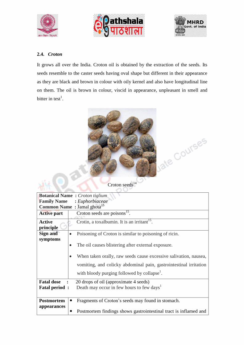
It grows all over the India. Croton oil is obtained by the extraction of the seeds. Its
seeds resemble to the caster seeds having oval shape but different in their appearance
as they are black and brown in colour with oily kernel and also have longitudinal line
on them. The oil is brown in colour, viscid in appearance, unpleasant in smell and
bitter in test1.
Croton seeds14
Botanical Name : Croton tiglium
Family Name : Euphorbiaceae
Common Name : Jamal ghota15
Active part Croton seeds are poisons15
.
Active
principle
Crotin, a toxalbumin. It is an irritant15
.
Sign and
symptoms Poisoning of Croton is similar to poisoning of ricin.
The oil causes blistering after external exposure.
When taken orally, raw seeds cause excessive salivation, nausea,
vomiting, and colicky abdominal pain, gastrointestinal irritation
with bloody purging followed by collapse1.
Fatal dose : 20 drops of oil (approximate 4 seeds)
Fatal period : Death may occur in few hours to few days1
Postmortem
appearances
Fragments of Croton‟s seeds may found in stomach.
Postmortem findings shows gastrointestinal tract is inflamed and

congested16
.
Medicolegal
aspects
Croton oil is admininistered with food for homicidal intention.
Accidental poisoning is resulted from Croton oil as a purgative.
It also is used as abortifacient.
Wild tribes use oil as arrow poisons1.
14 http://toptropicals.com/catalog/uid/Croton_sylvaticus.htm
15 J M Ganer, V V Nikam, Baragi Umapati C, 4Baragi Pramod C, International Pharmacognostic,
Phytochemical and Physicochemical Investigation of Croton Tiglium Seeds. International Journal of
Pharmacy, 2014, 4(3), 140-145
16 N K Rao, Textbook of Forensic Medicine and Toxicology, Chapter 21, p. 369, Irritant Poioson, 2
nd
edi., (JP Brothers Medical Pulisher P. LTD, 2006)

2.5 Nux vomica
Nux vomica is also known as strycnos Nux vomica and it grows in tropical region
throughout the India. It belongs to Loganiaceae family17
.
Seeds of Strychnos Nux vomica18
Botanical Name : Strychnos Nux vomica
Family Name : Loganiaceae
Common Name : Locally it‟s known as Kuchila, Kuchla etc19
.
Active part Seeds, leaves, bark and wood contain active principle20
.
Active
principle
The seeds of Nux vomica are poisonous which contains active
principle strychnine while leaves bark and wood contain brucine20
.
Sign and
symptoms
Unbroken seeds of Nux vomica are not poisonous as hard pericarp
is insoluble in digestive juices while, when seeds are broken or
chewed, there is an intense bitter taste in mouth and symptoms
appear within 15 min to an hour20
.
Strychnine is a powerful alkaloid. It stimulates all parts of central
nervous system and particularly spinal cord and causes muscles
to contract. This can lead to convulsions and death.
Muscles become so stiff and rigid that the body is arched. The
facial muscles are also contracted Sometimes the chest is fixed
so that breathing is difficult and therefore, cyanosis and blood

stained froth may be seen at the mouth.
Death usually occurs due to asphyxia from spasm of respiratory
muscles or from exhaustion due to repetition of spasms21
.
Fatal dose : Ingestion of one crushed seed (about 15-16 mg of
strychnine).
Fatal period : 1-2 hrs21
.
Postmortem
appearances
Signs of asphyxia are common.
Rigor mortis sets in almost immediately after death and passes
off early.
Strychnine resists putrefaction. Therefore, the remains of seeds
may be found in viscera (stomach)20
.
Medicolegal
aspects
Mostly accidental.
Homicide is rare due to bitter taste.
Suicide is rare due to incredibly painful death.
Used as cattle poison, also used to kill stray dogs, as
rodenticide and sometimes as an arrow poison20
.
17 J Chen, Y Qu , D Wang, P. Peng, H Cai, Y Gao, Z Chen and B Cai, Pharmacological Evaluation of
Total Alkaloids from Nux Vomica: Effect of Reducing Strychnine Contents. Molecules, 2014, 19,
4395-4408
18 http://vnspice.com/strychnos-nux-vomica-seeds
19 R. Bhati, A. Singh, V. A. Saharan, V. Ram and A. Bhandari,
Strychnos nuxvomica seeds: Pharmacognostical standardization, extraction,
and antidiabetic activity. J Ayurveda Integr Med. 2012, 3(2): 80–84.
20 K Vij , Textbook of Forensic Medicine and Toxicology: Principles and Practice, Chapter 41 p. 521,
Spinal poisons, 5th
edn., (Elsevier, 2011).
21 I. Makarovsky, G. Markel, A. Hoffman, O. Schein, T. Brosh-Nissimov, Z. Tashma, T. Dushnitsky
and A. Eisenkraft, Toxic chemical compound: Strychnine – A Killer from the Past. Israel Medical
Association Journal, 2008, 10, 142-145.

2.6. Oleander
There are two variety of Oleander exist in nature i.e. Nerium odorum and Cerbera
thevetia. These plants grow widely in India. Nerium odorum bears white or pink
flowers and known as true Oleander while, Cerbera thevetia bears yellow bell-shaped
flowers and known as pila kaner. It and belongs to Apocynaceae family22
.
Nerium odorum
Cerbera thevetia
Botanical Name
Variety I : Nerium odorum (While and pink flower) and
Variety II : Cerbera thevetia (Yellow flower)
Family Name : Apocynacea
Common Name : Kaner
Active part Milky juice which exudes from all parts of plant25
.

Active
principle
Cerbera thevetia: Thevetin, Thevetoxin, Cerberin (glycosides).
Nerium odorum: The active principles are 3 glycosides, i.e.
neriodorin, neriodorein and Karabin.
The principal action of neriodorin is similar to that of digitalis
causing death from cardiac failure.
Neriodorein causes muscular twitching and tetanic spasms more
powerful than those of strychnine.
Sign and
symptoms
Cerbera thevetia:
Burning sensation in mouth with tingling of the tongue, dryness
of throat, vomiting, diarrhoea, headache, dizziness, dilated
pupils, irregular action of the heart, drowsiness, collapse, coma
and death.
Tetanic convulsions are occasionally observed.
Nerium odorum:
Vomiting, pain in abdomen and frothy salivation usually occur,
followed by restlessness. There is difficulty in swallowing and
often lock jaw.
The pulse is slow and weak, and a respiration is hurried.
Muscular twitching of the extremities results into tetanic spasms.
This is followed by coma and death from heart failure.
Fatal dose:
Cerbera thevetia: 8-10 seeds or 15-20 gm of root
Nerium odorum: About 15 gm of root
Fatal period:
Cerbera thevetia: 24 hrs.
Nerium odorum: 24 hrs.
Postmortem
appearances
Cerbera thevetia:
Signs of gastrointestinal irritation, congestion of various
organs, and endocardinal ecchymoses caused by bruising.
It resists putrefaction and can be detected even years after

death in exhumed putrefied bodies.
Nerium odorum:
Not specific.
Petechial hemorrhages on heart are a characteristic feature.
Nerium odorum resists heat and can therefore be detected even
from the burnt remains of the dead bodyVij25
.
Medicolegal
aspects
Suicide with roots leaves and seeds are common among village
women.
The root is commonly used for procuring abortion.
Accidental poisoning is sometimes met when the decoction of
leaves is applied externally to reduce swellings
Use as cattle poison has also been recorded.
22 K. S. N. Reddy and O.P. Murty, The Essential of Forensic Science and Toxicology, Chapter 36 p.
538, 25th
edi. (2006).
23http://www.kew.org/science-conservation/plants-fungi/nerium-oleander-oleander
24 http://www.delange.org/OleanderYellow/OleanderYellow.htm
25 K Vij , Textbook of Forensic Medicine and Toxicology: Principles and Practice, Chapter 42 p. 525,
Cardiac poisons, 5th
edn., (Elsevier, 2011).

2.7. Marking nut
It is distributed in Himalayan region, tropical and central parts of India. It is closely
related to the cashew26
. Its Fruit is black which yields oily resin is often used as
“marking ink” on clothes1.
Fruits of Marking nut27
Botanical Name : Semecarpus anacardium.
Family Name : Anacardiaceae
Common Name : In Sanskrit it is known as Agnimukha and in Hindi as
„Bhilwa‟. It is also known by some other names i.e. Bhela and
Oriental Cashew26
.
Active part Fruit
Active
principle
Semicarpol (monohydroxy phenol compound) & Bhilawanol
(alkaloid)1.
Sign and
symptoms When juice is applied on the skin, it causes irritation and painful
blisters. The lesion resembles a bruise which may ulcerate later
and slough.
When administered internally, it is causes less irritation. In larger
doses, it causes blisters in the mouth and throat with severe
gastroenteritis, followed by dyspnoea, cyanosis, tachycardia
coma and death in some cases1.
Fatal dose : 5 to 10 gm
Fatal period : 12 to 1 hrs1
Postmortem The blister formation will be there. Blister fluid containing acrid

appearances serum should be preserved in rectified spirit and sent to a forensic
science laboratory for the analysis1.
Medicolegal
aspects The juice is used as an abortifacient by means of an abortion
stick on to the uterus.
It is also used by malingeres to produce an artificial bruise to
support a false charge.
Accidental poisoning may occur from internal administration by
quack.
The juice can be thrown like vitriol on the face intentionally.
Homicidal poisoning by internal application is rare1.
26
M. Semalty, A. Semalty, A. Badola, G. P. Joshi and M. S. M. Rawat, Semearpus anacardium Linn.:
A review. Pharmacogn Rev, 2010, 4(7): 88–94.
27
http://dir.indiamart.com/impcat/bhilawa-seeds.html

3. Animal poisons:
In this section, animal poisons such cantharides, scorpion and snake venom have been
discussed as below.
3.1. Scorpions
There are various species of scorpions but few of them are unsafe for human.
Scorpions have a cephalothorax, an abdomen and segments in tail with venom
secreting glands and one sting. Besides, the scorpion has two claws to grasp the prey.
Scorpion venom contains both haemotoxin and neurotoxin. It stimulates the release of
catecholamines from the adrenal glands and release into the circulation and hence,
affects the myocardium which results in cardiac arrhythmias, hypertension. After
release of catecholamines, it results in hypotension and bradycardia28
.
Scorpion29
Scorpion venom
Sign and
symptoms
There is local irritation manifested by redness and burning pain
at the site of bite.
The presence of swelling and hemorrhages help in locating the
site of bite.
Scorpion sting will have a single hole at the center of reddish
area.
As consequences, there may be headache, giddiness, nausea,
excessive sweating, fever, paralysis, cardiac problems, cynosis
and muscular cramps followed by coma13
.

Fatal dose : Death is rare in adults while, in children it may causes fatality due
to pulmonary oedema.
Fatal period : Death may occur within an hour from pulmonary oedema13
.
Postmortem
appearances
There is widespread hemorrhage.
Sting may be found at the site of bite.
Myocardial damage may also found30
.
Medicolegal
aspects
Poisoning from scorpion is accidental30
.
28 K Vij , Textbook of Forensic Medicine and Toxicology: Principles and Practice, Chapter 36 p. 483,
Irritants of animal origin, 5th
edn., (Elsevier, 2011).
29
http://www.factzoo.com/invertebrates/emperor-scorpion-glow-in-the-dark.html
30
G. Biswas, Review of Forensic Medicines & Toxicology Including Clinical and Pathological aspects,
Chapter 46 p. 464, 2nd
edi. (Jaypee Brothers Medial Publisher P, 2012)

3.2. Cantherides
The Spanish fly or Blister Beetle is a type of insect. It contains active principle
cantharidin which is an irritant. Cantharides may be administered in the form of
powdered beetles, or the tincture and it readily absorbs from skin28
.
Cantherdes31
Cantharides
Sign and
symptoms When applied externally on the skin, it causes redness, burning
pain followed by vesication.
On internal application, there is burning sensation in mouth,
throat, pain in stomach and intense feeling of thirst.
There is difficulty in swallowing, nausea, vomiting with
abdominal pain.
The urine is scanty and tinged with blood.
Abortion may occur in pregnant woman.
In fatal cases, coma with convulsions usually precedes death.
Fatal dose : 15 to 50 of cantharidine and one and half g to 3 g of cantharides.
Fetal period : 24 to 36 hours
Postmortem
appearances There is inflammation of mouth. Sometimes vesications may
appear.
There may be swelling of mucous membrane of oesophagus with
ulcer formation.
There may be congestion of mucous membrane of stomach

which may be extended to the small intestine.
There is acute inflammation of kidney with the haemorrhages in
renal, pelvis and bladder.
Medicolegal
aspects Accidental poisoning may occur due to over dose application
because of its aphrodisiac properties.
Homicidal poisoning is rare.
It is may be used as aphrodisiac.
It is also used as abortifacient13
.
31
http://www.wisegeek.com/what-is-cantharidin.htm

3.3. Snake Venom
Snakes are classified into two groups, i.e. poisonous and non-poisonous. Poisonous
snakes are further divided into three categories on the basis of poison secreted by
them such as Elapids (neurotoxic venom), Vipers (vasculotoxic venom) and Sea
snakes (myotoxic venom)28
.
Neurotoxic venom causes muscular weakness of legs and paralysis of muscles of face,
throat and respiratory track while, vasculartoxic venom causes coagulation disorders
and destroy endothelium of blood vessels. Myotoxic venom produces muscular pain,
followed by myoglobinuria and later on by failure of respiration after 3-5 hrs in fatal
cases.
Snake29
Snake venom
Sign and
symptoms
The hallmark of attack of a snake is presence of fang marks.
After 1or 2 hrs of attack, generalized muscular pain and stiffness
develop.
There will be slight burning at the site of bite followed by
neurotoxic effects such as giddiness, lethargy, muscular
weakness and paralysis. Weaknesses in the legs are manifested
by staggereing.
There will be difficulty in speaking and swallowing. Ptosis and
paralysis of ocular muscles may occur with slow and labored
breathing. After couples of hours, respiration may cease and

heart stops.
Skin and tissues shows necrosis surrounding the bite mark along
with intense local pain, swelling, discoloration of skin and
severe oozing of hemolytic blood. Blisters may appear
sometimes.
Hemolysis may lead to hemoglobinuric nephritis.
Petechial hemorrhages, bleeding from the gums, and bleeding
from mucous membrane of the rectum and other orifices of body
are common.
Collapse sets in with cold calmly skin with rapid feeble pulse
and dilated pupils, followed by coma and death.
Respiratory failure may occur13
.
Fatal dose : mg (dried form) Type of snake
15 mg – Cobra dried cobra venom
12 mg – King Cobra
2.5 mg-6 mg – Common Krait
40 mg – Russell‟s Viper
8 mg – Saw-scaled viper
Fetal period : Death occur from
Cobra venom- within a few min to few hours
Viper venom- in a few days
Sea snake bite is mostly not fatal30.
Postmortem
appearances
There will be the presence of fang marks which are about 1 cm
to 2.5 cm deep depending upon the type of snake bitten with
some swelling around the bitten part.
There are no characteristics findings indicating the cause of
death except the signs of asphyxia.
There will be severe oozing of blood from puncture site.
There are hemorrhages in lungs membranes. Endocardial
hemorrhages are seen especially in left ventricle. Petechiae are
also found within kidney and mucousa of urinary bladder,

stomach and intestines.
Arterioles and capillaries are characterized by blurred walls and
swollen endothelial cells13
.
Medicolegal
aspects
Generally accidental
Rarely homicidal and suicidal.
Sometimes used to kill cattles28
.
29 http://www.dkfindout.com/us/animals-and-nature/reptiles/cobras

Criminology
Forensic Science and Forensic Medicine
Determination of age, sex, Race and stature from human skeleton
remains

Role Name Affiliation
Principal Investigator Prof. (Dr.) Ranbir
Singh
Vice Chancellor, National
Law University, Delhi
Co-Principal Investigator Prof. (Dr.) G.S. Bajpai Registrar, National Law
University Delhi
Paper Coordinator Prof. (Dr) Sally Lukose Dean, School of Basic and
Applied Sciences, Galgotias
University
Content Writer/Author Mr. Saroj Kumar Amar Assistant Professor, Forensic
sc. School of Basic and
Applied Sciences, Galgotias

DESCRIPTION OF MODULE
Items Description of Module
Subject Name Criminology
Paper Name Forensic Science and Forensic Medicine
Module Name/Title Determination of age, sex, Race and stature from human
skeleton remains.
Module Id -----------
Objectives
Learning Outcome:
To make the learners understand the basic of
forensic anthropology.
To help the learners in determination of age from
bones.
To educate the learners how to estimate the race
and stature of individual from skeleton remains.
To teach the learners how the skeleton of male
and female are differ.
Prerequisites General understanding about anthropology and its
forensic significance.
Key words
Anthropology, sex determination, age determination,
stature, human skeleton.
University
Content Reviewer Dr. Tanya Chauhan Assistant Professor, LNJN-
National Institute of
Criminology and Forensic
Science, Delhi.

1. Sex determination from skeleton remains:
Sex differentiation from skeleton does not appear until puberty. Until the puberty
skeletons of two sexes differ only in size. Sex determination is dependable only if the
vital parts of the skeleton are present in good condition. Pelvis and skull are the most
important one for determining sex. The round of ball joints as well provides reliable
means of determining sex.
Characteristic feature of skeleton
Male Female
1. Skeleton are bigger and stouter. Skeleton comparatively smaller
and slender.
2. Muscular ridges, depression and
process are more prominent.
Muscular ridges, depression and
process are less prominent.
3. Shaft of the long bones relatively
rough and the articular surfaces
and ends larger.
Shaft of the long bones relatively
smooth and the articular surfaces
and ends small.
Skull:
The adult female skull is usually lighter and smaller, 10% less spacious than that of
the males. It is very difficult to determine sex of skull below the age of adolescence.
The differentiating features between the two sexes are as follows:

Fig: - DFS manual 2005.
2. Age Estimation from skeleton
(i)
(A) AGE OF INFANT BELOW 6 MONTH FROM
SKELETON
The ramous of the mandible is short, oblique and forms
obtuse angle with the body.
(ii) The coronoid process projects about the level of the
condyloid process.
(iii) The mental formation remains near the lower margin of the
jaw.
(iv) The body is shallow.
(i)
(B) 6 MONTHS TO 2 YEARS:
Outbreak of almost all the temporary teeth.
(ii) Emergence of ossification centers in the heads of humerus,

femur, ulna, tarsal and carpal bones.
(iii) Closure of the anterior fontanelle at about 3/2 years.
(i)
(C) 2 TO 6 YEARS:
Appearance of centers of ossification in the epiphysis of long
bones.
(ii) Closure of metopic suture.
(iii) Union of condylar part of occipital bone with the squama and
basi-occiput.
(iv) Blending of greater and lesser tubercles to head of humerus.
(i)
(D) 6 TO 12 YEARS:
Union of rami of ischium with pubis at the age of 7-9 years.
(ii) Ossification of pisiform bones occurs at the age of 9 to 12
years in females.
(iii) Union of lateral epicondyles of the humerus with the shaft.
(iv) Shedding of all temporary teeth and eruption of all
permanent teeth except the 3rd molars at the age of 12 to14
years.
(v) Appearance of secondary sexual character around 12 years.
(i)
(E) 12 TO 18 YEARS:
Fusion of upper end of Radius with shaft.
(ii) Fusion of Olecranon to the ulna.
(iii) Eruption of 3rd molar teeth with calcification of roots of all
the previously erupted teeth.
(iv) Union of lower ends of Radius and Ulna with the respective
shafts.
(i)
(F) 18 TO 25 YEARS:
Fusion of epyphysis of long bones with their respective
shafts.
(ii) Fusion of iliac crest with ilieum, ischial tuberosity with
ischium.
(iii) Fusion of coracoid and acromion with their respective
bodies.
(iv) Union of the epiphysis at sternal ends of the clavicles; the
secondary centers in the pelvis and articular facets of ribs
usually get completed at the age of 20-25 years.
(v) Bony replacement of the cartilage between the basiocciput
and the basisphenoid commences at about 17th year in both
sexes; union is completed by 22 years in females and 24
years in males usually.
(i)
(G) 24 TO 35 YEARS:
Closure of the lambdoid suture is not complete before 50
years, though coronal sutures get closed at the age of 40
years.
At the age of 40 years, the articular surfaces of lumbodorsal
vertebrae show lipping, loss of joint space, presence of
punched out areas of osteoporosis.
X-ray examination shows such changes, first in the glenoid
fossa of the scapulae; lipping becomes well advanced by 45-
50 years but does not get complete before 60 years.
(I) ABOVE 50 YEARS:
(i)
In old age the bone density is very low, long bones become
lighter and brittle. Skull becomes thinner and lighter and
hence is more liable to fracture from even slight violence.
(ii) Closure of mastoid, occipital, squamous and parietomastoid
sutures will occur in very old age. Parietal suture may not
even close throughout life.
(iii) Above 60-65 years, the angle of lower jaw opens out and the
medico legal angles become obtuse.
(iv) Manubrium and body of sternum may fuse above 60 years.
(e) The calcification of the laryngeal and costal cartilages
becomes more apparent.
3. Determination of Race from skeleton remains
Racial differences in skeletal structure originally arose when small genetic changes
developed in populations isolated by geography.
Now, as the migration increases worldwide and inter race marriage are also endorsed.
Thus the people of different racial backgrounds intermix and the progeny of those
individuals is harder to differentiate. Still there are some key features of the skull that
can help forensic investigation:
I. Mouth: Whites tends to have smaller teeth, with significant crowding and impacted
third molars, Blacks not often have crowding and the upper teeth often project
outwards.

II. The palate: In American Indians, the palate is elliptical, with the ‘U’ shape
angling in at the back teeth. In blacks, the palate is hyperbolic—a perfect ‘U’ shape
with straight lines. And in whites, the palate is parabolic with the ends of the ‘U’
flaring outwards.
III. Incisors: American Indians (and East Asians, both of Mongoloid ancestry), have
the incisors shovel-shaped. Black and whites both have blade-form incisors.

IV. The nose: Whites have long and narrow nasal aperture, with a high bridge and a
sharp nasal sill. The nasal aperture of blacks has short and wide with a low bridge and
a guttered or trough-like nasal sill. , the nasal aperture of American Indians is
medium-sized with both a medium bridge and nasal sill.

V. The mastoid process: Blacks have the wide bony projection, whites have narrow
and pointed. American Indians, a secondary smaller projection forms on the back
surface of the mastoid process.
4. Estimation of Height from Long Bones
An approximate height of the deceased from the skeletal remains is obtained by
multiplying the length (in inches) of a long bone with a suitable multiplication
factor. These are the factors for long bones:
Long bone Multiplication factor
Humerus 5.2
Radius 6.6
Ulna 6.0
Femur 3.7
Tibia 4.3
Fibula 4.4

The exact multiplication factor may vary from males to females and children for
diverse groups of population.
References:-
1. Direcrorate of forensic science laboratory manual, 2005.
2. American academy of forensic science (www.aafs.org)
3. Shetty M, Premalatha K. ABO blood grouping from tooth material. J Indian
Acad Forensic Med. 1972;32(4):336–38.
4. Da Silva RH, Sales-Peres A, de Oliveira RN, de Oliveira FT, Sales-Peres SH.
Use of DNA technology in forensic dentistry. J Appl Oral Sci. 2007; 15:156–
61.

Criminology
Forensic Science and Forensic Medicine
Classification of Fingerprints Comparison of finger prints.

DESCRIPTION OF MODULE
Items Description of Module Subject Name Criminology Paper Name Forensic Science and Forensic Medicine Module Name/Title
Classification of Fingerprints Comparison of finger prints.
Module Id CRIMINOLOGY/FSFM/XIX
Objectives
Learning Outcome:
To make the learners understand the numbering and naming of fingerprints for the classification purposes.
To acquaint the learners with the henry’s classification system of fingerprints.
To acquaint the learners about the basis for comparison of fingerprints.
To inform learners about the process of comparison of fingerprints.
Prerequisites Fingerprints – definition, pattern types, general and individual fingerprint characteristics.
Key words
Henry’s system of classification, valued and non-valued patterns, suspect and standard fingerprints.
Role Name Affiliation Principal Investigator Prof. (Dr.) G.S. Bajpai Registrar, National Law
University Delhi Co-Principal Investigator
Paper Coordinator Prof. (Dr) Sally Lukose, Dean, School of Basic and
Applied Sciences, Galgotias
University Content Writer/Author Ms Vinny Sharma Assistant Professor, Division
of Forensic Science, SBAS, Galgotias University
Content Reviewer

1. Introduction:
Fingerprints or finger impressions are unique and highly individualistic, even the
same person cannot have the same fingerprints on two different fingers also they are
different in monoclonal twins also.
Due such vast variation in the finger impressions a need to classify them was felt.
Also for the purposes of comparison of scene of crime prints with the suspect’s prints
required a means of classification which ease the process of comparison. Apart from
this section 75 IPC that has a provision for extended punishment for repeat offenders
requires the search of pervious records of culprits and since fingerprints does not
change with time therefore are a perfect medium to act as a basic searching tool for
such records. This searching process again requires a classification method for
classifying the recorded fingerprints records.
For the classification purposes the all the fingerprints of all the digits are recorded on
the 10-digit fingerprint slip (sample below). The impressions recorded are both the
rolled and plain impressions.
By rolled impressions we mean that the fingers are rotated from nail to nail from left
to right to obtain the inked impression of the fingers. These rolled impression are
widen and provides a detailed account of the fingerprints.
By plain impressions we mean that the inked prints are obtained by merely slapping
the fingers perpendicularly on the record slip to obtain the prints. The purpose of
taking these prints is to match the sequence of the rolled prints obtained earlier of the
same individual.

Figure: 10-digit fingerprint record slip.
CASE NO.: TIME: DATE: INVESTIGATING TEAM: EVIDENCE NO.: SIGNATURE OF OFFICER:
ROLLED PRINTS
RIGHT HAND
RT RI RM RR RL
LEFT HAND
LT LI LM LR LL
PLAIN PRINTS
LEFT HAND RIGHT HAND
LT RT

2. Various system of fingerprint classification:
Fingerprints are classified on various basis by different researchers devising a
different classification each time. There are approximately 50 different system of
classification of fingerprints are available which are practiced by different countries.
The most commonly used system for classification of fingerprints is the Henry’s ten-
digit classification system. This system is widely used across the world for the
purposes of filing and searching of fingerprints records (discussed later).
Other than this system of classification some other system are also in use, for
example,
The Vcetich or Argentine system
The Budapest or Hungarian system
The Valladares system
The Bertillon system
The Daae system
Then Klatt system
The When system
The Proivenski system
The Brussels system
3. Henry’s System of classification:
The classification system used in all parts of our country and elsewhere is based on
henry system. The classification formulae consist of letters and numbers arranged in
the form of a fraction with a numerator and denominator. The formula is based on a
study of the ten fingerprints of an individual. Every individual pattern is first
identified and marked on the slip. Then the classification formula is worked in a
logical order in the following different steps:
1. Primary classification
2. Secondary classification
3. Sub-secondary classification
4. Final classification.
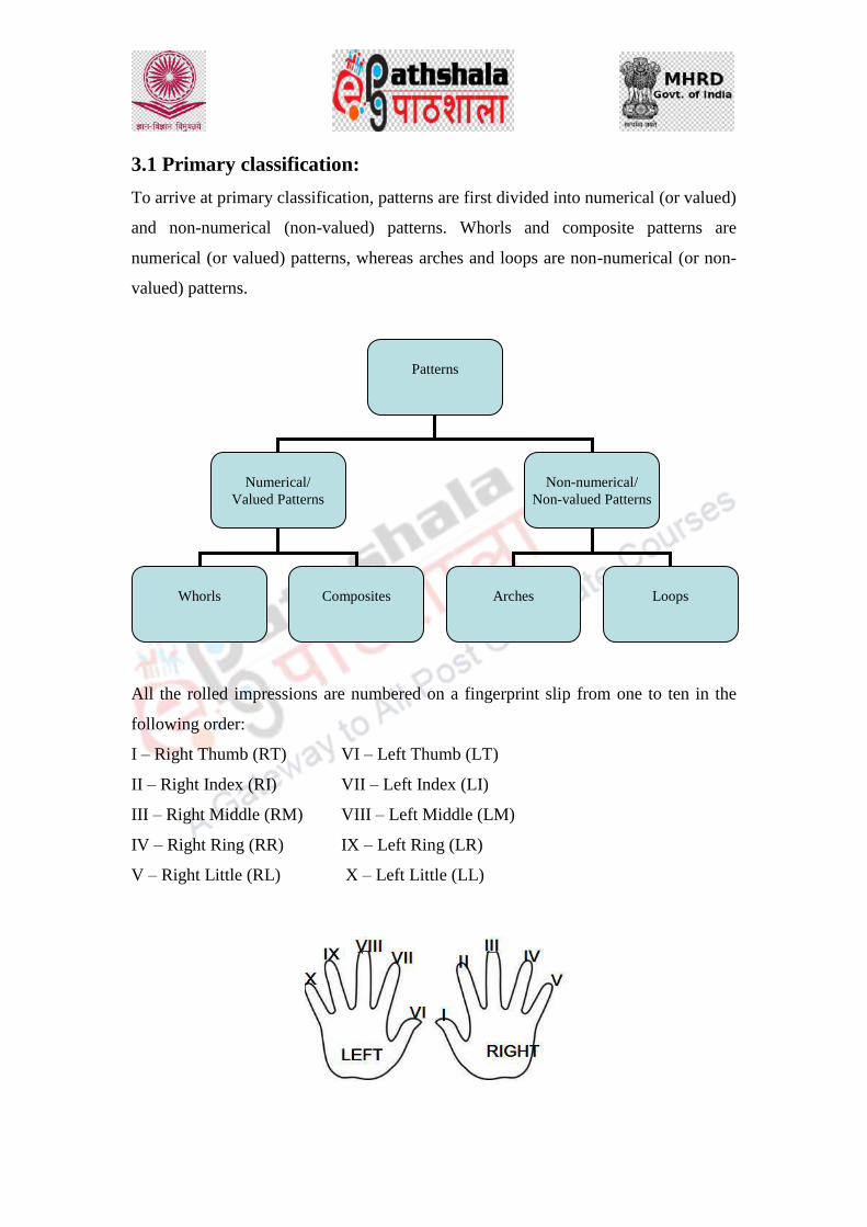
3.1 Primary classification:
To arrive at primary classification, patterns are first divided into numerical (or valued)
and non-numerical (non-valued) patterns. Whorls and composite patterns are
numerical (or valued) patterns, whereas arches and loops are non-numerical (or non-
valued) patterns.
All the rolled impressions are numbered on a fingerprint slip from one to ten in the
following order:
I – Right Thumb (RT) VI – Left Thumb (LT)
II – Right Index (RI) VII – Left Index (LI)
III – Right Middle (RM) VIII – Left Middle (LM)
IV – Right Ring (RR) IX – Left Ring (LR)
V – Right Little (RL) X – Left Little (LL)
Patterns
Numerical/
Valued Patterns
Non-numerical/
Non-valued Patterns
Whorls
Composites
Arches
Loops

Numerical values are assigned to the fingers in the following order:
I – Right Thumb (RT) = 16 VI – Left Thumb (LT) = 4
II – Right Index (RI) = 16 VII – Left Index (LI) = 2
III – Right Middle (RM) = 8 VIII – Left Middle (LM) = 2
IV – Right Ring (RR) = 8 IX – Left Ring (LR) = 1
V – Right Little (RL) = 4 X – Left Little (LL) = 1
Digit
number I II III IV V
Digit name Right
Thumb
Right
Index
Right
Middle
Right
Ring
Right
Little
Numerical
value 16 16 8 8 4
Digit
number VI VII VIII IX X
Digit name Left
Thumb
Left
Index
Left
Middle
Left
Ring
Left
Little
Numerical
value 4 2 2 1 1
If the pattern identified come under the valued/numerical pattern group then the value
assigned will be taken into account. If the pattern identified come under non-
valued/non-numerical pattern group then the vale assigned to these positions will not
be taken into account and will be taken as zero (0).
After assigning the values, the values obtained for all the even numbered fingers are
placed in the numerator, and the values obtained for all the odd-numbered fingers are
placed in the denominator. To obtain the primary classification all the values in the
numerator are totalled and 1 is added. Similarly, all the values in denominator are
totalled and 1 is added. The fraction thus obtained constitutes the primary
classification.
For numerator = RI, RR, LT, LM, LL
For denominator = RT, RM, RL, LI, LR
Formula,
Primary classification = RI+RR+LT+LM+LM+1/RT+RM+RL+LI+LR+1
As many as 210
= 1024 combinations are possible through this classification.
3.2 Secondary classification: (or fulcrum classification)
The secondary classification subdivides large groups of fingerprint slips having the
same primary classification. The secondary classification consists of the capital letter
symbols for the patterns of the two index fingers, with the right index finger symbol
appearing in the numerator and the left index finger symbol in the denominator. The
five basic pattern types that can appear on index fingers are:
Arch (A), Tented arch (T), Radial Loop (R), Ulnar Loop (U) and Whorl (W).
For the purpose of this classification, composite patterns are classified as plain whorls
under the symbol W. As many as 25 possible combinations are possible out of thus
classification.
Formula:
Secondary classification = Pattern of RI/Pattern of LI
3.3 Sub-secondary classification:
The sub-secondary classification further subdivides large groups having same primary
and secondary classification. The sub-secondary classification is represented by
symbols that may be I, M, or O. These symbols are given to the index, middle and
ring fingers of the right hand and make –up the numerator; the same fingers for the
left hand make-up the denominator. These symbols are derived from the ridge tracing
of the whorls appearing on the index, middle and ring fingers, or from the ridge
counts of loops on these fingers.
In case of ridge counting,
For index finger: I = 1 to 5 ridge count
M = 6 to 12 ridge count

O = 13 and above ridge count
For middle finger: I = 1 to 6 ridge count
M = 7 to 13 ridge count
O = 14 and above ridge count
For ring finger: I = 1 to 7 ridge count
M = 8 to 14 ridge count
O = 15 and above ridge count
Formula:
Sub-secondary classification = Ridge tracing or count of: RI, RM, RR/ LI, LM,
LR
3.4 Final Classification:
The final classification is a mere number, which denotes the actual ridge count for the
patter of the little finger of right hand.
Formula:
Final classification = Ridge count (RL)
Henry Classification formulae:
Primary classification = RI+RR+LT+LM+LM+1/RT+RM+RL+LI+LR+1
Secondary classification = Pattern of RI/Pattern of LI
Sub-secondary classification = Ridge tracing or count of: RI, RM, RR/ LI,
LM, LR
Final classification = Ridge count (RL)

5. Process of Comparison:
Fingerprints are infallible means of physical evidence. They are persistent and highly
individualistic. They are left at the scene of crime by the perpetrator without his/her
knowledge, unknowingly and by chance. The tendency of leaving a fingerprint at the
scene of crime is enhanced due to the reason that they are left behind in the form of
sweat deposited in the exact same manner as that the ridges are present and whicle
committing the crime an individual perspires more as compared to the normal day-to-
day scenario. Therefore, the frequency of finding a fingerprint at scene of crime is
increased. The prints which are found at the scene of crime are termed as suspected
prints and are need to be compared with the standard prints.
Suspected prints in other words are those prints whose identity is unknown, whereas
standard prints are those prints whose identity is known.
Standard prints are obtained from the individuals on the standard 10-digit fingerprint
slip, while taking the standard prints both rolled and plain prints are obtained. These
slips are then used to compare with the suspect prints.
The evidentiary value of the fingerprint in the court of law lies on the top of all other
scientific evidences.
The process of fingerprint comparison involves the photography of the prints,
followed by the comparison of general and individual characteristics. The comparison
process is majorly an elimination process. First the expert has to identify the pattern
type of the suspected print followed by the type of core and delta, ridge count and
ridge tracing. Then he/she start comparing it with the standard prints. Those prints
with the same pattern type are considered and rest all are eliminated. After that the
prints with similar delta and core type are considered rest are eliminated. Then the
expert look for the similar ridge count and ridge tracing. In doing this the expert is
eliminating the most unlikeable fingerprints which will not be matching with the
suspected print. After comparing all the general characteristics, the expert is able to
narrow down the list of standard prints with the most likeable prints remaining only.
The expert then look for the 8 ridge characteristics (minimum, as required by the law)
in the suspect print and then he/she looks for the same ridge characteristics in the
standard prints at the exact same location. When the expert does this he/she is able to
tell the identity of the suspect print with a 100% match.

Comparison of suspect (latent) and standard (known) print.

References:
Cummins, H. and Midlo, C. (1961) “Finger Prints, Palms and Soles.
An Introduction to Dermatoglyphics”, Dover, New York.
Henry, E.R., (1900) “Classification and uses of fingerprints”, George
Routledge and Sons, London.
Nath, S. (2010) “Fingerprint Identification”. Shiv Shakti Book Traders,
New Delhi.
Nayar, P.S. (2016) “Fingerprint Identification & Procedure Manual”,
Selective &Scientific Books, New Delhi.
The Science of Fingerprints Classification and Uses, United States
Department of Justice, Federal Bureau of Investigation, John Edgar
Hoover, Director

Law
Forensic Science & Forensic Medicine
Ammonium sulphate method, Stas-Otto method, dry ashing
method, wet digestion, dialysis and total alcoholic extract method.

DESCRIPTION OF MODULE
Items Description of Module Subject Name Law Paper Name Forensic Science and Forensic Medicine
Module Name/Title
Isolation of poisons from viscera: Ammonium
sulphate method, Stas-Otto method, dry ashing
method, wet digestion, dialysis and total
alcoholic extract method. Module Id LAW/CJA/VIII /10
Objectives
Learning Outcome:
To make the learners understand about the extraction, isolation/ purification of poisons.
To make the learner aware about various methods of extraction for different classes of drugs.
To make the learners understand that extraction procedure depends upon physiochemical properties of poisons.
Prerequisites To aware about different extraction procedures for the extraction of different classes of compounds/ drugs.
Key words
Extraction, isolation, purification, non-volatile organic poisons, volatile organic poisons, metallic poisons, anions etc.
Role Name Affiliation Principal Investigator Prof. (Dr.) G.S. Bajpai Registrar, National Law
University Delhi Co-Principal Investigator
Paper Coordinator Prof. (Dr) Sally Lukose Dean, School of Basic and
Applied Sciences, Galgotias
University, Greater Noida. Content Writer/Author Dr. Mamta Assistant Professor, School
of Basic and Applied
Sciences, Galgotias
University, Greater Noida. Content Reviewer

1. Introduction:
There are various traditional as well as modern methods are employed for the
extraction of poison from biological matter. The factors which affect the selection of
method of extraction depends upon are type of poison, biological matter (matrix)
constitute poison and quantity of matrix available for the analysis.
There are various types of poisons which have been discussed in earlier chapter.
Depending upon the type of poison to be extracted from the given the matrix
(biological matter intagged with poison), following are the methods used for
extraction and isolation (purified) of poisons.
2. Isolation of non-volatile organic toxic poisons
2.1. Ammonium Sulphate Method:
Ammonium sulphate method is used the screening non-volatile drugs such as
barbiturates, alkaloids, tranquilizers etc and the procedure involved is as follow:
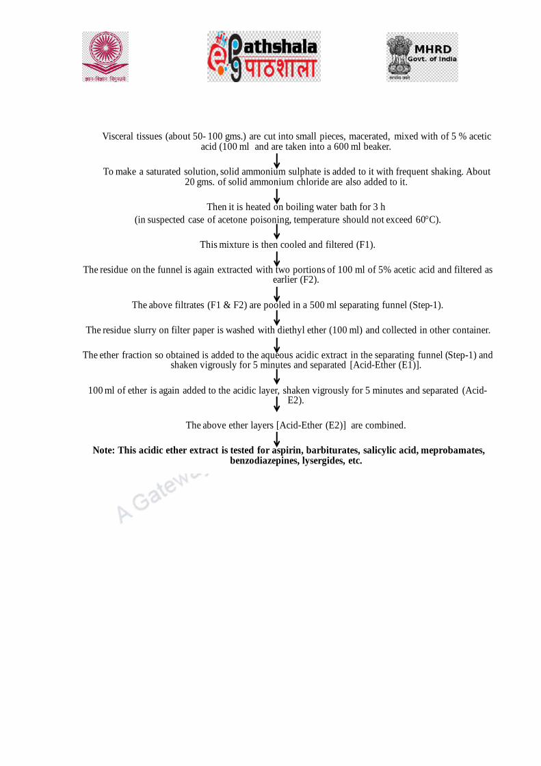
Visceral tissues (about 50- 100 gms.) are cut into small pieces, macerated, mixed with of 5 % acetic acid (100 ml and are taken into a 600 ml beaker.
To make a saturated solution, solid ammonium sulphate is added to it with frequent shaking. About 20 gms. of solid ammonium chloride are also added to it.
Then it is heated on boiling water bath for 3 h
(in suspected case of acetone poisoning, temperature should not exceed 60C).
This mixture is then cooled and filtered (F1).
The residue on the funnel is again extracted with two portions of 100 ml of 5% acetic acid and filtered as earlier (F2).
The above filtrates (F1 & F2) are pooled in a 500 ml separating funnel (Step-1).
The residue slurry on filter paper is washed with diethyl ether (100 ml) and collected in other container.
The ether fraction so obtained is added to the aqueous acidic extract in the separating funnel (Step-1) and shaken vigrously for 5 minutes and separated [Acid-Ether (E1)].
100 ml of ether is again added to the acidic layer, shaken vigrously for 5 minutes and separated (Acid-E2).
The above ether layers [Acid-Ether (E2)] are combined.
Note: This acidic ether extract is tested for aspirin, barbiturates, salicylic acid, meprobamates, benzodiazepines, lysergides, etc.

The aqueous solution remaining in the separating funnel after separation of acidic drugs (Acid-ether
layer) is made alkaline by addition of ammonium hydroxide and it is extracted thrice with 100 ml
portions of chloroform ether mixture (1 : 3)
(Organic layer) [Step-2].
Note: The aqueous layer is retained for the extraction of opium alkaloids.
The organic layers after separation [obtained at Step-2] are combined and washed with 50 ml of distilled
water. It is extracted thrice with 25 ml of 10% sulphuric acid.
The sulphuric acid fractions are combined and taken into another separating funnel. (Organic layer is
discarded). To it 50 ml mixture of chloroform-ether (1 : 3) is added to it. To make the solution
alkaline dilute ammonium hydroxide solution is added it and shaken for 5 minutes [Step-3].
The organic layer [obtained at Step-3] is separated. The extraction is repeated thrice.
The organic layer after separation are combined, washed with l50 ml of water, dried by passing through
anhydrous sodium sulphate and evaporated to dryness.
Note: This extract is tested for aconite, , datura, opium alkaloids, amphetamines, methaqualone,
meprobamate and etc.
The acid – ether extract may be further be separated into following three
fractions:
(i)
The acid-ether layer is shaken with 25 ml of 5% sodium bicarbonate solution.
The aqueous layer is removed and taken into another separating funnel.
Acidified with dilute sulphuric acid and again extracted with 25 ml of ether.
This ether fraction is passed through hydrous sodium sulphate and dried.
Note: The residual fraction 1 contains salicylates.

(ii)
The ether layer (of acid ether fraction) after washing with sodium bicarbonate is extracted twice
with 25 ml portions of N-sodium hydroxide solution.
The aqueous layer are separated from ether layer, combined and taken into another separating
funnel.
Made acidic by adding dilute sulphuric acid and extracted twice 25 ml ether.
Then this ether fraction is washed with 25 ml of distilled water and then dried by adding
anhydrous sodium sulphate and evaporated to dryness.
Note: The residuul fraction 2 contains purified form of barbiturates.
(iii)
The ether layer is washed with water after extracting with NaOH and then evaporated to
dryness.
Note: The residue fraction 3 contains carbamates, meprobamate and other neutral
drugs.

2.2.1. Stas-Otto Process1:
50 gm of biological material is macerated with sufficient quantity of spirit (about 2-3 times the
weight of material) and acidified with tartaric acid in a flask.
This mixture is then heated on steam bath for 1-2 hours with frequent shakings at fixed intervals.
The extraction is carried out for about 24 hours with steam off.
Filtration is then carried out using filter paper. The filtrate obtained is evaporated and the residue is
again extracted with acidified alcohol, filtered and washed with hot rectified spirit.
The filtrates are combined and evaporated in a porcelain basin on the steam bath to a syrupy mass.
To this about 100 ml of rectified spirit is added slowly with constant stirring. (If the alcohol is added
rapidly, the insoluble matter will become sticky and resulted into loss of alkaloids).
It is warmed with stirring for about half an hour and then filtered. This process is repeated again for one
more time then alcoholic extracts are combined and evaporated to dryness.
The residue is dissolved in about 50 ml of water acidified with dilute sulphuric acid and filtered after
about an hour. The poisons are thus come in aqueous layer and transferred to a separating funnel and
extracted with about 25 ml ether, chloroform etc.
[Acid solution contains colouring matters, toxic oils, resins, salicylic acid and its derivatives (aspirin,
salol etc.), barbiturates, sulphonal, acetanilide, narcotine and alkaloids of ergot, certain glycosides
such as thevetin which has escaped initial treatments for purification.]
The acid aqueous solution is then made alkaline by adding sodium carbonate or ammonia solution to
separate the free base from its salt.
The alkaline solution is then extracted with 25ml chloroform in the same way. It will constitute all the
alkaloids except morphine (only traces are extracted) and those feebly basic compounds which are
partially extracted in the acidic layer. The extraction is repeated 2 or 3 times.
In suspected case of morphine poisoning, it can be extracted at this stage by adding amyl alcohol or
mixture of chloroform-ether in 3:1 ration or chloroform -alcohol mixture in 9:1 ratio.
The chloroform or amyl alcohol fractions are combined, evaporated to dryness and now it is ready for
further purification and analysis.
It is purified by dissolving it in about 20 ml of water acidified with sulphuric acid and then filtered.
This filtrate is again extracted with chloroform, first in acid and then in alkaline medium as earlier.
These extracts are evaporated to dryness for analysis.
1.
http://wincenprado.tripod.com/toxicology.html

2.2.2. Modified Stas-Otto Procedure :
Modification in Stas-Otto method has been carried out to minimise the chances of loss
of poison at each step as Stas-Otto method involves so many steps which may also
result in complete loss of poison. Therefore, the method has been modified with the
minimum following steps2.
Extraction of biological material is carried with absolute alcohol (to prevent hydrolysis) at room
temperature (temperature should not exceed 40oC) for suspected case of aconite, belladonna,
datura or cocaine poisoning. The fraction so obtained is evaporated under reduced pressure and
following points are taken into consideration while extraction.
• The extraction with rectified spirit is carried out for 48 hours if biological material is preserved in
saturated saline solution.
• As stomach contents consist of too much fluid, the extraction should be done with absolute
alcohol for 3 or 4 times.
• In case of extraction of stomach wash, add absolute alcohol just 2 times the quantity of stomach
wash, acidified with tartaric acid and allowed to evaporate on a steam bath.
(For filtration, Buchner funnel is preferred).
• Agitation (2-3 time in the beginning and not exceeding 12 times at the end with organic solvents
such as ether or chloroform or amyl alcohol) is carried out to prevent loss due to emulsion
formation. In case of formation of emulsion, it is evaporated on a steam bath and then residue is
taken in fresh solvent.
2. http://shodhganga.inflibnet.ac.in/bitstream/10603/77515/8/08_chapter%202.pdf

2.3. Isolation of Toxic Cations (Metals)
2.3.1. Dry Ashing Method3:
10-50 gm of biological material is taken into a silica crucible and heated in a Bunsen burner to remove
the moisture content and to partially destroy the organic material.
The crucible is then kept in a muffle furnace and the temperature is raised up to 550C to perform
incineration of the organic matter for one hour.
After incineration, the crucible is taken out.
The color of the residue is noted at this stage (when it is hot).
(In the presence of zinc, residue shows yellow color while in presence of copper, residue is bluish green
in colour)
The residue is then boiled with 10 ml of 4N hydrochloric acid in silica basin and then it is filtered.
The clear acidic solution so obtained is tested for the presence of metallic poisons such as arsenic,
mercury, lead, copper, bismuth, zinc, barium etc. by group analysis or using micro methods,
chromatographic techniques etc.
3. N. R. Bader, sample preparation for flame atomic absorption spectroscopy: an overview, Rasayan
J.com, CODEN: RJCABP, 4(1) 2011, 49-55, ISSN: 0974-1496

2.3.2. Wet Digestion Method4:
50 gm of visceral tissues/10 ml of blood is taken into a Kjeldahl flask and 20-40 ml of concentrated
nitric acid is added to it and heated with care in a small flame when the mass begins to liquefy.
The heating is continued till liquefaction of material is not completed and is carried out in the
presence of brown fumes of NO2 in the flask.
At this stage about 20-30 ml of concentrated sulphuric acid is added and the flask is heated over a
wire gauge and conc. nitric acid is also added drops wise (using dropping funnel) at the rate of about
10 drops/minute so that it result in the continuous production of brown fumes in the flask.
Heating is continued until all organic matter is destroyed and at this stage liquid becomes clear and
colorless or straw colored.
To find out whether oxidation is complete or not, flask is heated without adding any HNO3, if there
is any un-burnt organic content left, the liquid begins to darken and if the digestion is completed, no
darkening takes place.
In the earlier case, when the organic matter is not completely oxidised, addition of HNO3 and
heating should be continued.
To expel the nitric acid completely, heating is continued for 15 minutes more.
After cooling, 25 ml of saturated ammonium oxalate solution is added and is boiled until SO3
fumes appear. This confirms complete removal of HNO3.
It is then cooled, diluted with an equal quantity of water and carefully transferred into a beaker. The beaker is heated on a hot plate or sand bath to expel the excess H2SO4.
The solution is then cooled and diluted with distilled water in such a manner that the strength of acid is remain about10%.
Precipitate may be formed which contains salts of lead, bismuth, tin, barium, strontium or silver etc. The precipitate is filtered and tested for these metals by group analysis and quantitative
analysis if required.
4. https://www.scribd.com/doc/310201137/Toxicology-Manual, Laboratory procedure manual forensic
toxicology, Directorate of Forensic Science, MHA, Govt. of India

2.4. Isolation of Toxic Anions
Dialysis Method5:
10 gm of the tissue is chopped into small pieces and placed in a cellophane membrane bag.
The bag is slowly rotated in a beaker containing 100 ml of distilled water employing an electrical motor.
The separation of particles in a liquid is carried out on the basis of the their ability to pass through this
membrane.
After one hour, water of the beaker is taken out and replaced by fresh water and the bag is rotated for
another half an hour.
This water is then taken out, pooled with the previous fraction and evaporated to a small fraction using
water bath.
If required, it is filtered and tested for the presence of toxic anions such as halides, azides etc.
5. G. Sarathchandra, A. Albert, A.T. Venugopalan, Manual on Analytical Toxicology, Tamilnadu
Veterinary & Animal Sciences University. Toxicology Unit, Central University Laboratory, Centre for
Animal health Studies, Madhavram Milk Colony, Madras.

2.5. Total Alcoholic Extraction:
10 gm of the tissue is copped into small pieces or biological material is taken in an evaporation dish
to remove moisture completely on hot water bath.
It is then macerated with rectified sprit with twice the quantity of biological material and transferred to
an Erlenmeyer flask.
To it 0.5 ml of acetic acid is added and fitted with a reflux condenser.
It is then refluxed on boiling water bath for 4 hours, cooled and then filtered.
The filtrate is evaporated to dryness.
This total alcoholic extract is evaluate for the presence of mainly indigenous poisonous drugs such as
croton, castor, bhilawan, kaner, madar, mushrooms,oduvan etc.

Criminology
Forensic Science and Forensic Medicine Drugs of abuse: Classification of drugs, opium, barbiturate, cannabis, cocaine, LSD, NDPS act.

DESCRIPTION OF MODULE
Items Description of Module
Subject Name Criminology
Paper Name Forensic Science and Forensic Medicine
Module Name/Title Drugs of abuse: classification of drugs, opium,
barbiturate, cannabis, cocaine, LSD. NDPS act.
Module Id
Objectives
Learning Outcome:
To make learners understand the impact of drug
abuse
Name and classify the commonly abused drugs
Describe Narcotic Drugs and Psychotropic
Substances Act.
Describe the laboratory tests normally used to
perform a routine drug identification analysis
Prerequisites General understanding of Forensic Science and its
application in crime solving.
Key words
Drug, Abuse, Narcotic, Sedative, Barbiturate, Cocaine,
LSD, Opium, Identification
Role Name Affiliation
Principal Investigator Dr G. S. Bajpai
Co-Principal Investigator
Paper Coordinator
Content Writer/Author Dr Priti Sharma
Content Reviewer

1. Introduction:
Drug abuse is an important issue nowadays. Keeping in mind, the large number of
populations getting in trap of drugs abuse, there requires a great deal of understanding
on how they affect human beings. This knowledge helps law enforcement agencies in
recognizing the type of drug consumed by the person and to assess whether drug
abuse is involved in the crime. Such knowledge is also required by medical
practitioner to assist the addict in undergoing treatment for deaddiction and toxicity.
About 190 million people all over the world consume one drug or the other. In India,
an increasing use of illicit drugs has been reported since many decades. Although, the
World Health Organization notes significant difficulty in estimating drug usage and
addiction rates in India, some resources point to number over 3 million drug addicts in
India.2 Along with various issues arising from drug addiction and usage including
health and social, HIV is a significant issue with over 2.4 million people infected in
India. This places India as the third-highest country in terms of rate of infection in the
world.
This module contains description about classification of drugs, their nature and effects
on human being. This knowledge is forensically important to enforce law in crimes
where drug abuse is a key element.
2. Definition of Drug
A drug can be defined as a natural or synthetic substance that is used to produce
physiological or psychological effects in humans or other higher order animals.
However, with the differential effects from use and abuse of drug, drugs, to some,
may be as a necessity for sustaining and prolonging life; to others, drugs provide an
escape from the pressures of life; to still others, they are a means of ending it.
3. Classification of Drugs
Drugs are classified in many ways, on the basis of their usage, pharmacological
action, etc. These categories help to understand better about the drug consumed based
on their behaviour, appearance, effects, etc.
Understanding how different classes of drugs can affect individuals helps law enforce
-ment officers react to, control and question suspects in drug-related cases.
For example, a person who has taken a stimulant drug such as methamphetamines will
react or respond to police differently than a person who has consumed a narcotic or
marijuana.
3.1. Narcotic Drugs
The term narcotic is derived from the Greek word narkotikos, which implies a state of
lethargy or sluggishness. As per pharmacological definition, narcotic drugs are
analgesic substances that bring relief from pain and produce sleep by depressing the
central nervous system. However, the regular use of a narcotic drug leads to physical
dependence, with all its dire consequences.
The source of most narcotics is opium, a gummy, milky juice exuded through a cut
made in the unripe pod of the poppy (Papaver somniferium), a plant grown mostly in
parts of Asia. Opium is brownish in color and has a morphine content ranging from 4
to 21 percent. Morphine is then used to produce its derivative, heroin, which is the
most commonly used drug by addicts. Heroin’s high solubility in water makes its
street preparation for intravenous administration rather simple. Codeine is also one of
the derivative from opium, but it is commonly used as a cough suppressant in
prescription cough syrup. Codeine, only one-sixth as strong as morphine, is not
commonly used as drug by addicts.
3.1.1. Heroin
Heroin is one of the most recognized and widely abused drugs. The correct chemical
nomenclature for heroin is O3, O -diacetylmorphine. The first synthesis of heroin i.e.,
diacetylmorphine reported in the literature was in 1875 by two English chemists, G.H.
Beckett and C.P. Alder Wright. In 1898 in Eberfield, Germany, the Farbenfarbriken
vorm Friedrich Bayer and Company produced the drug commercially for patients
suffering from lung diseases such as tuberculosis. The Bayer Company also
advertised heroin as a cure for morphine addiction. The analgesic properties of the
drug were very effective. However, the addictive properties were quite devastating.

The “Golden Triangle” areas of Burma, China, and Laos are the three major source
countries in part of the world for the production of illicit opium. Of these three
countries, 60 to 80% of the total world supply of heorin comes from Burma. India is
also known to be one of the primary consumers of heroin and the largest grower of
licit opium in the world, which is used to make a range of prescription medications.
However, there are reports by law enforcement agencies that the licit opium is being
diverted to illegal markets inside India and for trafficking beyond its borders for
synthesis of morphine and its derivatives. Additionally, India is also at increased treat
of drug abuse due to its geographical location between the two largest illicit opium
growers, Burma and Afghanistan and use as a major trafficking route between the
two.
A form of heroin known as brown sugar is commonly used in the country, which is
made of a mixture of heroin which typically ranges between 20 to 60 percent purity,
and adulterants in the form of chalk, zinc or other chemicals. Due to cheap price and
easy availability, this drug is very popular in India among wide variety of people.
Recent trend suggests that not only adults but children as young as 13 are falling in
trap of drug addiction. Due to less purity and toxic adulterants present, there are
serious adverse effects from taking this drug.
This process of heroin production involves following steps:
(1) The opium poppy (Papaver Somniferum L.) is cultivated
(2) The poppy head is scored and the opium latex is collected
(3) Isolation of morphine from the latex
(4) Treating morphine with an acetylating agent such as acetic anhydride.
Acetylation of Morphine to Diacetylmorphine (Heroin) involves placing dried
morphine into a reaction vessel and adding excess acetic anhydride (Figure 1).
Sometimes a co-solvent is also used. The mixture is heated to boiling and stirred for
varying periods of time ranging from 30 min up to 3 or 4 h. The vessel and contents
are cooled and diluted in cold water. A sodium carbonate solution is then added until
precipitation of the heroin base is complete and settles to the bottom of the reaction
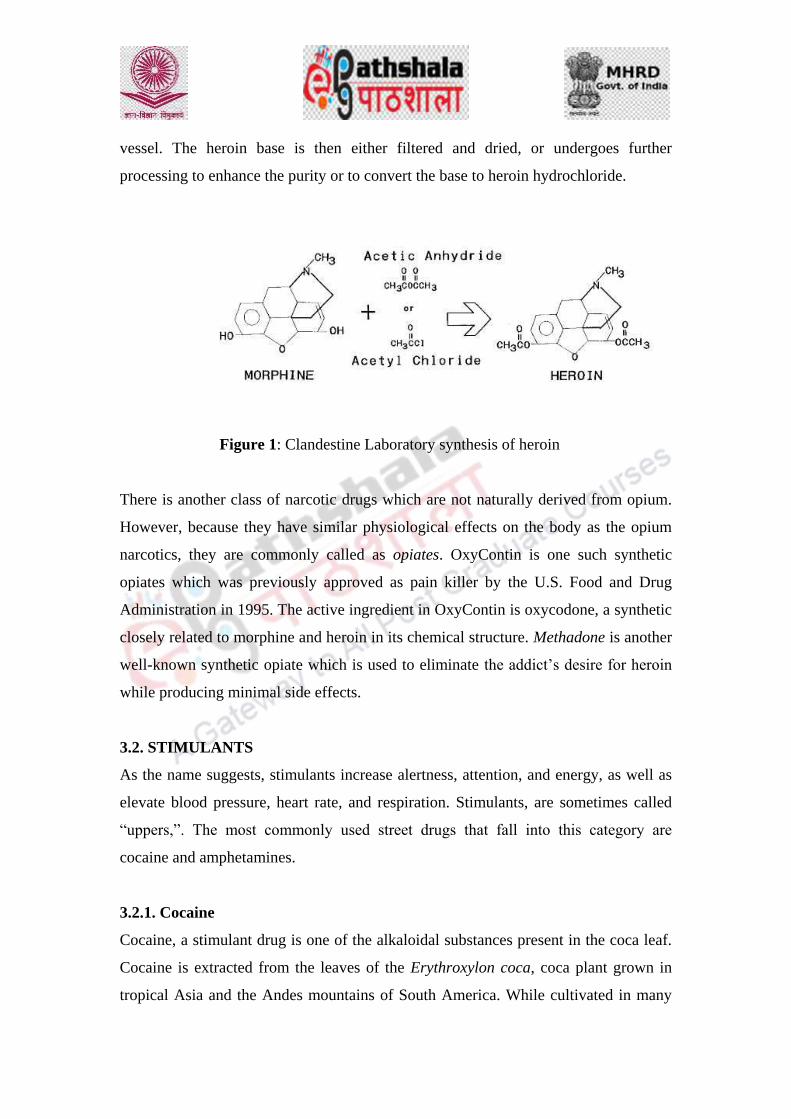
vessel. The heroin base is then either filtered and dried, or undergoes further
processing to enhance the purity or to convert the base to heroin hydrochloride.
Figure 1: Clandestine Laboratory synthesis of heroin
There is another class of narcotic drugs which are not naturally derived from opium.
However, because they have similar physiological effects on the body as the opium
narcotics, they are commonly called as opiates. OxyContin is one such synthetic
opiates which was previously approved as pain killer by the U.S. Food and Drug
Administration in 1995. The active ingredient in OxyContin is oxycodone, a synthetic
closely related to morphine and heroin in its chemical structure. Methadone is another
well-known synthetic opiate which is used to eliminate the addict’s desire for heroin
while producing minimal side effects.
3.2. STIMULANTS
As the name suggests, stimulants increase alertness, attention, and energy, as well as
elevate blood pressure, heart rate, and respiration. Stimulants, are sometimes called
“uppers,”. The most commonly used street drugs that fall into this category are
cocaine and amphetamines.
3.2.1. Cocaine
Cocaine, a stimulant drug is one of the alkaloidal substances present in the coca leaf.
Cocaine is extracted from the leaves of the Erythroxylon coca, coca plant grown in
tropical Asia and the Andes mountains of South America. While cultivated in many
countries, Peru and Bolivia are the world’s leading producers of the coca plant.
Cocaine is present in the coca leaves from these countries at dry weight
concentrations of from 0.1 to 1%. The average concentration of cocaine in the leaf is
0.7%.
At one time, cocaine had wide medical application as a local painkiller or anaesthetic.
However, this function has now been largely replaced by other drugs, primarily
procaine and lidocaine. Cocaine is also a powerful stimulant to the central nervous
system, and its effects resemble those caused by the amphetamines—namely,
increased alertness and vigor, accompanied by the suppression of hunger, fatigue, and
boredom. Most commonly, cocaine is sniffed or “snorted” and is absorbed into the
body through the mucous membranes of the nose.
The method of isolating cocaine from the coca leaf does not require a high degree of
technical expertise or experience. All of the known production techniques involve
three primary steps: (1) extraction of crude coca paste from the coca leaf; (2)
purification of coca paste to cocaine base; and (3) conversion of cocaine base to
cocaine hydrochloride.
One form of cocaine that has gained widespread popularity in the drug culture is
known as crack. The process used to make crack is simple. Ordinary cocaine is mixed
with baking soda and water into a solution that is then heated in a pot. This material is
then dried and broken into tiny chunks that dealers sell as crack rocks. Crack is
freebase cocaine and is sufficiently volatile to be smoked, usually in glass pipes.
Crack, like cocaine that is snorted, produces a feeling of euphoria-a feeling of
increased energy, of being mentally more alert, of feeling really good. The faster the
cocaine level rises in the brain, the greater the euphoria, and the surest way to obtain a
fast rise in the brain’s cocaine level is to smoke crack. Inhaling the cocaine vapor gets
a large wallop of the drug to the brain in less than fifteen seconds—about as fast as
injecting it and much faster than snorting it. Crack. However, euphoria fades quickly
as cocaine levels drop, leaving the user feeling depressed, anxious, pleasure less. The
desire to return to a euphoric feeling is so intense that crack users quickly develop a

habit for the drug that is almost impossible to overcome. Only a small percentage of
crack abusers will ever be cured of this drug habit. While there is no evidence of
physical dependency accompanying cocaine’s repeated use, cocaine produces the
strongest psychological compulsions for continued use.
3.2.2. Amphetamines
Amphetamines are a group of synthetic drugs that stimulate the central nervous
system. They are commonly referred to in the terminology of the drug culture as
“uppers” or “speed.” Methamphetamine is a major problem throughout the world and
there are increasing reports of its use in India. The first clandestine methamphetamine
laboratory was discovered in 2003, and many more have been found since then. India
is a key producer of precursor chemicals for methamphetamine which include
ephedrine and chemicals that are used in the processing. The majority of these
chemicals are trafficked to major manufacturing countries like China, Thailand,
Mexico and America.
Ordinarily, therapeutic doses of 5–20 milligrams per day, taken orally, provide a
feeling of well-being and increased alertness that is followed by a decrease in fatigue
and a loss of appetite. However, these apparent benefits of the drug are accompanied
by restlessness and instability or apprehension, and once the stimulant effect wears
off, depression may set in.
Methamphetamine use is linked to many serious physical and mental health concerns
including violence, paranoia, insomnia, organ damage and brain damage.
Amphetamine drugs are often used by the working class poor as a stimulant to help
them work harder, for longer and require less food. Using drugs for this purpose is
incredibly dangerous and can lead to major problems with families and
communities. Not only are they highly addictive but their use can damage motor
skills, response times, problem solving skills and cause sexual dysfunction.
A new smokable form of methamphetamine known as “ice” is reportedly in heavy
demand in some countries. Ice is prepared by slow evaporation of a

methamphetamine solution to produce large, crystal-clear “rocks.” Like crack
cocaine, ice is smoked and produces effects similar to those of crack cocaine, but the
effects last for a longer period of time. Once the effects of ice wear off, users often
become depressed and may sleep for days. Repeated use of amphetamines leads to a
strong psychological dependency, which encourages their continued administration.
3.3. Depressants
A depressant, or central depressant, is a drug that lowers neurotransmission levels,
which is to depress or reduce arousal or stimulation, in various areas of the brain.
Depressants are also occasionally referred to as "downers" as they lower the level of
arousal when taken. Depressants are widely used throughout the world as prescription
medicines and as illicit substances. Eg., Barbiturates.
3.3.1. Barbiturates
Barbiturates create a feeling of well-being, and produce sleep to the individual. Like
alcohol, barbiturates suppress the vital functions of the central nervous system.
Collectively, barbiturates can be described as derivatives of barbituric acid, which
was first synthesized by a German chemist, Adolf Von Bayer, more than a hundred
years ago. Twenty-five barbiturate derivatives are currently used in medical practice
in the United States; however, five—amobarbital, secobarbital, phenobarbital,
pentobarbital, and butabarbital—tend to be used for most medical applications. Slang
terms for “barbs” usually stem from the color of the capsule or tablet (for example,
“yellow jackets,” “blue devils,” and “reds”).
Normally, barbiturate users take these drugs orally. The average sedative dose is
about 10–70 milligrams. When taken in this fashion, the drug enters the blood through
the walls of the small intestine. Some barbiturates, such as phenobarbital, are
absorbed more slowly than others and are therefore classified as long-acting
barbiturates. Undoubtedly, the slow action of phenobarbital accounts for its low
incidence of abuse. Apparently, barbiturate abusers prefer the faster-acting ones—
secobarbital, pentobarbital, and amobarbital. When taken in prescribed amounts,

barbiturates are relatively safe, but in instances of extensive and prolonged use,
physical dependence can develop.
3.4. Hallucinogens
These are group of drugs that can cause marked alterations in mood, attitude, thought
processes, and perceptions. Perhaps the most popular and controversial member of
this class of drugs is marijuana. Other drugs which come in this class include lysergic
acid diethylamide (LSD), mescaline, phencyclidine (PCP), psilocybin, and
methylenedioxymethamphetamine, also known as MDMA or Ecstasy.
3.4.1. Marijuana
Cannabis is an incredibly popular and widely used drug in India and is known
as ganja, charas or bhang. India has a long history of cannabis use, and the drug is one
of the five sacred plants mentioned in the Hindu texts, the Vedas. It is typically
associated with the Hindu deity Shiva, who is believed to like the hemp plant. The
drug is often smoked or drunk in a beverage at Hindu ceremonies. Cannabis is
combined with a mixture of milk or yoghurt and boiled with nuts and spices to make a
refreshing beverage. This drink is consumed by many all over the country, especially
by labor workers who find the relaxing properties beneficial.
Despite its widespread and accepted use, cannabis is illegal to use and possess in any
form. Some figures place regular cannabis users as high as 10 million in India, many
of these among the working poor. Many of these heavy users are dependent on the
drug and suffer from major health problems including respiratory disorders, memory
impairment, mental disturbance, digestive tract problems, major weight loss and
problems with sleep. Almost half that number may be regular users.
Marijuana is a preparation derived from the plant Cannabis. Most botanists believe
there is only one species of the plant, Cannabis sativa L. The marijuana preparation
normally consists of crushed leaves mixed in varying proportions with the plant’s
flower, stem, and seed (See Figure 2). The plant secretes a sticky resin known as
hashish. The resinous material can also be extracted from the plant by soaking in a

solvent such as alcohol. On the illicit-drug market, hashish usually appears in the
form of compressed vegetation containing a high percentage of resin.
Figure 2: Marijuana preparation
Marijuana is a weed that grows wild under most climatic conditions. The plant grows
to a height of 5 to 15 feet and is characterized by an odd number of leaflets on each
leaf. Normally, each leaf contains five to nine leaflets, all having serrated or saw-tooth
edges, as shown in Figure 3.
Figure 3: A photograph showing Marijuana leaves

In 1964 scientists isolated the chemical substance largely responsible for the
hallucinogenic properties of marijuana. This substance is known as
tetrahydrocannabinol, or THC. The THC content of Cannabis varies in different parts
of the plant, generally decreasing in the following sequence: resin, flowers, and
leaves. Little THC is found in the stem, roots, or seeds. The potency and resulting
effect of the drug fluctuate, depending on the relative proportion of these plant parts
in the marijuana mixture.
The potency of marijuana depends on its form. Marijuana in the form of loose
vegetation has an average THC content of about 3–4.5 percent, while hashish
preparations average about 2–8 percent. Another form of hashish is known as liquid
hashish or hashish oil. Hashish in this form is normally a viscous substance, dark
green with a tarry consistency. Liquid hashish is produced by efficiently extracting
the THC-rich resin from the marijuana plant with an appropriate solvent. Liquid
hashish typically varies between 8 and 20 percent in THC content. Because of its
extraordinary potency, one drop of the material can produce a “high.” Ordinarily a
drop is placed on a regular cigarette or on a marijuana cigarette before smoking.
No current evidence suggests that experimental or intermittent use causes physical or
psychological harm. Marijuana does not cause physical dependency. However, the
risk of harm lies instead in heavy, long-term use of the drug, particularly of the more
potent preparations. Heavy users can develop a strong psychological dependence on
the drug. Some effects of marijuana use include increased heart rate, dry mouth,
reddened eyes, impaired motor skills and concentration, and frequently hunger and an
increased desire for sweets. Long-term chronic marijuana use is associated with a
motivational syndrome characterized by apathy; impairment of judgment, memory,
and concentration; and loss of interest in personal appearance and the pursuit of
conventional goals.

3.4.2. LSD
LSD is synthesized from lysergic acid, a substance derived from ergot, which is a type
of fungus that attacks certain grasses and grains. Its hallucinogenic effects were first
described by the Swiss chemist Albert Hofmann after he accidentally ingested some
of the material in his laboratory in 1943. The drug is very potent; as little as 25
micrograms is enough to start vivid visual hallucinations that can last for about twelve
hours. The drug also produces marked changes in mood, leading to laughing or crying
at the slightest provocation. Feelings of anxiety and tension almost always accompany
LSD use. Although physical dependence does not develop with continued use, the
individual user may be prone to flashbacks and psychotic reactions even after use is
discontinued.
4. Drug Control Law in India- Narcotic Drugs and Psychotropic Substances
(NDPS) Act, 1985
4.1. Elements of NDPS Act
The NDPS Act 1985 sets out the statutory framework for drug law enforcement in
India. The main elements of the control regime mandated by the Act are as follows:
1. Control and regulation of NDPS: The cultivation, production, manufacture,
possession, sale, purchase, transportation, warehousing, consumption, inter-
State movement, shipment and import and export of narcotic drugs and
psychotropic substances is prohibited, except for medical or scientific
purposes and in accordance with the terms and conditions of any license,
permit or authorization given by the Government. (Section 8)
2. Power of CG: The Central Government is empowered to regulate the
cultivation production, manufacture, import, export, sale, consumption, use etc
of narcotic drugs and psychotropic substances. (Section 9).
3. Power of SG: State Governments are empowered to permit and regulate
possession and inter-State movement of opium, poppy straw, the manufacture

of medicinal opium and the cultivation of cannabis excluding hashish.
(Section 10).
4. Prohibition of trade outside India: All persons in India are prohibited from
engaging in or controlling any trade whereby narcotic drugs or psychotropic
substances are obtained outside India and supplied to any person outside India
except with the previous authorisation of the Central Government and subject
to such conditions as may be imposed by the Central Government. (Section
12).
5. Assessment of NDPS as controlled substance by CG: The Central Government
is empowered to declare any substance, based on an assessment of its likely
use in the manufacture of narcotics drugs and psychotropic substances as a
controlled substance. (Section 9-A).
6. Assets derived from drugs trafficking are liable to forfeiture (Chapter V-A).
7. Power to appoint officers: Both the Central Government and State
Governments are empowered to appoint officers for the purposes of the Act.
(Sections 4, 5 and 7).
8. The NDPS Act is in effect a comprehensive code not only for the control and
regulation of Narcotics Drugs and Psychotropic Substances; but also for the
control of selected chemicals - commonly known as precursors - which can be
used in the illicit manufacture of narcotic drugs and psychotropic substances,
as well as for the investigation and forfeiture of drug related assets.
A number of agencies both at the Centre and in the States have been empowered to
enforce the provisions of the Act. These agencies include
the Department of Customs and Central Excise,
the Directorate of Revenue Intelligence,
the Central Bureau of Narcotics and
the Central Bureau of Investigation at the Central level
State Police and Excise Departments at the State level.

Section 4(3) of the Act envisages the creation of a Central Authority to coordinate the
activities of the various Central and State agencies involved in drug law enforcement,
to implement India's obligations under various international conventions, and to
coordinate with international organizations and authorities in foreign countries in the
prevention and suppression of the illicit traffic in narcotic drugs and psychotropic
substances. In terms of this provision, the Narcotics Control Bureau was set up by the
Central Government in 1986 with the broad remit to coordinate drug law enforcement
nationally.
4.2. Investigative procedures
Chapter V of the NDPS Act (Sections 41 to 68) sets out the powers as well as the
procedures for the investigation of offences under the Act. This Chapter empowers
officers duly authorized by the Central Government or a State Government to issue
warrants, to enter and search premises, to stop and search conveyances, to seize
narcotic drugs and psychotropic substances, to take statements and to arrest persons
suspected of having committed an offence, punishable under the Act.
The power to issue search and arrest warrants, is in terms of Section 41, been vested
both in Magistrates as well as in specially designated (Gazetted) officers of the
Central and State Governments. This is designed to ensure both timely and effective
action in response to any information.
In addition, both the Central and the State Governments are authorized to entrust any
Officer duly empowered under the Act with the powers of an Officer-in-Charge of a
Police Station for the investigation of offences under the Act.
4.3. Offences and Penalties
Chapter IV, (Sections 15 to 40) sets out the penalties for offences under the Act.
These offences are essentially related to violations of the various prohibitions
imposed under the Act on the cultivation, production, manufacture, distribution, sale,
import and export etc. of narcotic drugs and psychotropic substances.
All these offences are triable by Special Courts and the punishments prescribed range
from imprisonment from 10 to 20 years for first offences to 15 to 30 years for any
subsequent offences together with monetory fines. In addition to persons directly
involved in trafficking narcotic drugs and psychotropic substances, any person who
finances trafficking or harbours a person involved in trafficking, or abets, or is a party
to a criminal conspiracy, including a criminal conspiracy to commit an offence
outside India, is also liable to the same scale of punishments. The Act was amended in
May 1989 to mandate the death penalty for second offences relating to contraventions
involving more than certain quantities of specified narcotic drugs and psychotropic
substances. The Act, however, makes a distinction between possession for personal
consumption and trafficking, the punishment for the former being limited to between
six months and one year only.
4.4. Precursor Control
The 1988 U.N. Convention against Illicit Traffic in Narcotic Drugs and Psychotropic
Substances to which India is a signatory, requires Parties to impose controls on the
manufacture, internal distribution and import and export of chemicals which can be
used in the illicit manufacture of narcotic drugs and psychotropic substances.
In exercise of its powers under the Act, the Central Government has so far notified
Acetic Anhydride, which is used in the processing of opium into heroin, N-
Acetylanthranilic acid which is used in the illicit manufacture of Methaqualone and
Ephedrine and Pseudoephedrine, which are used in the illicit manufacture of
amphetamine type stimulants, as controlled substances.
5. Identification of Drugs
Any forensic lab should be equipped with reagents and instruments to analyse
identification of drug. The task of a drug analyst is very crucial knowing that the
drugs in markets are highly variable. This is made further complex by newly
developing drugs which are modifications of already known drugs. These drugs are
designed to confer difficulty in their identification at laboratories.

To perform drug identification, the analyst must employ series of tests which range
from screening i.e., preliminary or non-specific to confirmatory or specific tests. The
screening tests reduce the possibilities for a definite group of drugs to a small and
manageable number. These tests are considered important in case of negative result
and so their value lies in having excluded certain drugs from further consideration.
Once the number of possibilities is substantially reduced, the second phase of the
analysis is conducted which is confirming the drug’s identity. This consists of
confirmatory tests that identifies a substance. The analytical scheme sometimes
consists of a series of nonspecific or presumptive tests followed by confirmatory tests.
Forensic chemists normally rely on several tests for a routine drug-identification
scheme: color tests, microcrystalline tests, chromatography, spectrophotometry, and
mass spectrometry.
5.1. Color Tests
This consists of subjecting the sample to a series of color tests that produce
characteristic colors when brought into contact with specific chemical reagents. Not
only do these tests provide a useful indicator of a drug’s presence, but they are also
used by investigators in the field to examine materials suspected of containing a drug.
Five primary color test reagents are as follows:
1. Marquis (2 percent formaldehyde in sulfuric acid). The reagent turns purple in the
presence of heroin and morphine and most opium derivatives. Marquis also becomes
orange-brown when mixed with amphetamines and methamphetamines.
2. Dillie-Koppanyi (1 percent cobalt acetate in methanol is first added to the suspect
material, followed by 5 percent isopropylamine in methanol). This is a valuable
screening test for barbiturates, in whose presence the reagent turns violet-blue in
color.
3. Duquenois-Levine (solution A is a mixture of 2 percent vanillin and 1 percent
acetaldehyde in ethyl alcohol; solution B is concentrated hydrochloric acid; solution C
is chloroform). This is 2. Dillie-Koppanyi (1 percent cobalt acetate in methanol is first
added to the suspect material, followed by 5 percent isopropylamine in methanol).

This is a valuable screening test for barbiturates, in whose presence the reagent turns
violet-blue in color.
4. Van Urk (1 percent solution of p-dimethylaminobenzaldehyde in 10 percent
concentrated hydrochloric acid and ethyl alcohol). The reagent turns blue-purple in
the presence of LSD. However, owing to the extremely small quantities of LSD in
illicit preparations, this test is difficult to conduct under field conditions.
5. Scott Test (solution A is 2 percent cobalt thiocyanate dissolved in water and
glycerine [1:1]; solution B is concentrated hydrochloric acid; solution C is
chloroform). This is a color test for cocaine. A powder containing cocaine turns
solution A blue. Upon addition of B, the blue color is transformed to a clear pink
color. Upon addition of C, if cocaine is present, the blue color reappears in the
chloroform layer.
5.2. Microcrystalline Tests
A technique considerably more specific than color tests is the microcrystalline test. In
this test, a drop of a chemical reagent is added to a small quantity of the sample of
drug on a microscopic slide. After a short time, a chemical reaction ensues, producing
a crystalline precipitate. The size and shape of the crystals, under microscope
examination, are highly characteristic of the drug. These tests are rapid and often do
not require the isolation of a drug from its diluents; however, because diluents can
sometimes alter or modify the shape of the crystal, the examiner must develop
experience in interpreting the results of the test. The microcrystals of cocaine and
methamphetamine is shown in Figure 4.

Figure 4: Microcrystals of Cocaine and Methamphetamine
5.3. Chromatography
Chromatography is a good method of choice for separation of components of a
mixture. This technique separates mixtures on the basis of their distribution between
affinity for stationary liquid phase and a moving gas phase. Many compounds have a
tendency to become closely associated with other compounds through attractive
forces, while others do not. This attraction or tendency for association is often called
affinity. Chromatography utilizes the differences in affinity of compounds for
separation. Thin-layer and gas chromatography are especially well suited to the needs
of the drug analyst, because they separate drugs from their diluents while providing
for their tentative identification.
TLC of drugs is used as a presumptive test to provide an indication of the nature of
the drug under question. To perform TLC, the sample must first be dissolved in a
solvent – unless it is already in a liquid state – and then it can be spotted onto a TLC
plate along with a known standard and run under a solvent system.
It is useful for identification because many of the colour change presumptive tests
used will only provide an indication of a certain active group in the molecule. This
variation produces visible differences as well as difference in Rf (Retention Factor)
value. Rf value is the ratio of distance travelled by sample to distance travelled by
solvent on TLC plate. Since there can be some overlap of Rf values and confusion in

colour judgement, TLC is considered as a presumptive test to inform further analysis
rather than producing a definitive result on its own.
In gas chromatography, separation is on the basis of affinity with phases called as
mobile phase and stationary phase. The mobile phase is nothing but a gas called the
carrier gas, which flows through a column. The carrier gas is chemically inert and is
generally nitrogen or helium. The stationary phase is a thin film of liquid within the
column. Because chromatography requires a comparison of Rf values between
questioned and known drugs, the analyst must have some clue to the identity of the
illicit material before using these techniques. Hence, in a typical drug analysis,
chromatography accompanies and complements color and crystal tests.
5.4. Spectrophotometry
Selective absorption of UV and IR light by drugs provides a valuable technique for
characterizing drugs. The ultraviolet spectrum is not conclusive for positive
identification of a drug, because other materials may very well produce an
indistinguishable spectrum. Nevertheless, UV spectrophotometry is often a useful
technique for establishing the probable identity of a drug. For example, if an unknown
substance yields a UV spectrum that resembles that of amphetamine, thousands of
substances are immediately eliminated from consideration, and the analyst can begin
to identify the material from a relatively small number of possibilities.
The pattern of an infrared spectrum is unique for each compound and can thus serve
as a “fingerprint” of the compound. The combination of preliminary screening tests
with a final verification by infrared spectrophotometry offers an ideal approach to
drug identification. Unfortunately, the technique does present some problems because
the substance to be identified must be as pure as possible. This requirement often
necessitates lengthy purification steps to prepare the sample for IR analysis. For this
purpose, GC is used initially to purify the drug from hindering components to produce
a GC-IR spectra. The GC-IR spectra of methamphetamine is shown in Figure 5.

Figure 5: GC-IR Spectra of Methamphetamine
5.5. Mass Spectrometry
The technique of chromatography is particularly suited for analyzing illicit drugs,
because it can readily separate a drug from other substances that may be present in the
drug preparation. Chromatography does, however, have the drawback of not being
able to provide a specific identification of the material under investigation. This
deficiency has been overcome by linking the gas chromatograph to a mass
spectrometer to yield a very powerful combination known as gas
chromatography/mass spectrometry (GC/MS). As a sample emerges from the gas
chromatograph, it immediately enters the mass spectrometer.
In the mass spectrometer, the material enters a high-vacuum chamber where a beam
of high-energy electrons is targeted at the sample molecules. The electrons collide
with the molecules, causing them to lose electrons and to acquire a positive charge
(commonly called ions). These positively charged molecules or ions are very unstable
or are formed with excess energy and almost instantaneously decompose into
numerous smaller fragments. The fragments then pass through an electric or magnetic
field, where they are separated according to their masses generating a highly
characteristic line pattern where each line represents a fragment of a different mass
(actually the ratio of mass to charge), and the line height reflects the relative
abundance of each fragment. The unique feature of mass spectrometry is that under
carefully controlled conditions, no two substances produce the same fragmentation
pattern. This generates a pattern like “fingerprint” of the substance which is unique,
with a few exceptions. With data obtained from a GC/MS determination, a forensic
analyst can, with one instrument, separate the components of a complex drug mixture
and then unequivocally identify each substance present in the mixture.
Summary
A drug can be defined as a natural or synthetic substance that is used to produce
physiological or psychological effects in humans. Drugs are classified on the basis of
their effects on humans. Narcotic drugs relieve pain by depressing the central nervous
system and may cause physical dependence on regular use. The most common source
of narcotic drugs is opium. Morphine is readily extracted from opium and is used to
synthesize heroin. Opiates, which include methadone and oxycodone, are not derived
from opium, but they have the same physiological effects on the body as do opium
narcotics.
Depressants are another class of drugs which slows the action of central nervous
system. These include alcohol (ethanol), barbiturates, tranquilizers, and various
substances that can be sniffed. Stimulants include amphetamines, sometimes known
as “uppers” or “speed,” and cocaine, which in its free-base form is known as crack.
Another class of drugs is hallucinogens which cause marked alterations in mood,
attitude, thought processes, and perceptions. Marijuana is the most controversial drug
in this class because its long-term effects on health are still largely unknown. Other
hallucinogens include LSD, mescaline, PCP, psilocybin, and MDMA (Ecstasy).
Each country has its federal laws to regulate and control drugs transport and abuse. In
India, Narcotic Drugs and Psychotropic Substance act established in 1985, controls
and regulate drugs in India. Whenever any questioned sample of drug is seized, it is
sent to laboratory for identification. Since unknown substance may any one of a
thousand of drugs, the analyst employs screening tests to reduce these possibilities to

a small number. This is done by subjecting the sample to a series of color tests that
produce characteristic colors for the more commonly encountered illicit drugs.
Microcrystalline tests which are more specific than color test are also conducted to
identify sample on the basis of characteristic crystal formed with specific reagents.
Once the preliminary analysis is completed, a confirmation is pursued using
combination of techniques such as chromatography and spectrophotometry. Typically
Gas chromatography is used for separation of drug compound from the mixture and
infrared spectrophotometry or mass spectrometry is used to specifically identify a
drug substance.

Items Description of Module
Subject Name Criminology
Paper Name Forensic Science and Forensic Medicine
Module Name/Title Collection and analysis of Biological evidences:
Blood, semen, saliva, sweat, Hair
Module Id
Goals of Biological Evidence Collection
1. Each individual stain should be collected separately. Do not collect or
package two separate stains together in the same container.
2. Do not allow evidence samples to come into contact with any surface, which
contains residue from another biological sample (e.g. dirty tweezers,
bloodstained glove, contaminated work surface).
3. Use tweezers with smooth, easy-to-clean working surfaces.
4. Tools (e.g. tweezers, scissors) can be cleaned by thoroughly rinsing with a
stream of distilled water and drying thoroughly with paper tissue. Repeat
this process twice before using tool to manipulate another sample.
5. Do not talk or cough over biological evidence.
6. Small biological evidence (e.g. 2-mm size bloodstain or hair) is most
susceptible to contamination.
7. Put on a new pair of gloves before handling these small stains. Consider
wearing a mask.
8. Package all biological evidence in paper bags or envelopes. Do not use
plastic.
9. Handle the sample in a manner, which minimizes deterioration of the
sample.
10. When feasible, take object with stain. Do not remove stain. If stain can be
easily dislodged (e.g. stain is on non-porous surface), protect it from contact
with another object. One way this can be done is by immobilizing the
evidence item in a cardboard container.
11. If evidence stains are found on objects that can be cut (e.g. a rug), the
evidence stain can be removed by cutting it out with a pair of clean scissors.

Remember to also take an “unstained” control cutting (e.g. the substrate
without the stain) from the object.
12. If object cannot be moved, use a slightly dampened substrate (cotton swab
or piece of plain, cotton cloth or gauze) to collect stain. Remember to collect
an unstained control by swabbing an unstained area of the evidence object as
very few stains can be safely collected by scraping them.
13. The size of the stain should influence the size of a substrate used to collect
the stain. Thus, use a small part of a swab or a small piece of cotton cloth or
piece of gauze to collect a small stain. Do not smear a small stain over a
large surface.
14. Use a minimum amount of distilled water to dampen the swab or cloth/gauze
substrate.
15. To keep the stain concentrated, collect the stain on the smallest area of the
swab or cotton cloth.
16. Wet blood should be dried first and then scraped or can be collected on a
swab.
17. Blood-stained materials must be stored in paper bags or manila envelopes.
18. Blood-soaked clothing must not be stored in air-tight containers because the
trapped moisture may cause the growth of mildew & mold and destroy the
blood.
19. All clothing in fact, must be air-dried and individually stored in paper bags.
20. If bloodstained materials are stored in airtight containers, the accumulation
of moisture may encourage the growth of mold, which can destroy the
evidential value of blood.
21. For damp or bloody items one should use:
a) Brown paper bags of appropriate size
b) Earthguard bags
c) Butcher paper that can be folded and properly taped shut.
SWEAT
Sweat is the watery fluid produced and excreted by the sweat gland. Sweat glands
are simple tubular glands (almost two to four million sweat glands) found in
almost every part of the skin.
Sweat glands are stimulated in response to
a) high temperature.
b) exercise.

c) hormones.
d) emotional stress (emotionally induced sweating is restricted to palms ,soles
, armpits and forehead while temperature induced sweating causes sweating
throughout the body).
Composition of sweat
Sweat is composed mainly of water and 0.2 – 1% solutes, such as; sodium,
potassium lactate, urea, ammonia etc
Mechanism of sweat secretion
When the sweat glands are stimulated the secretory portion of the sweat gland
secrets a fluid which is absorbed by the gland cells from the interstitial fluid and
is produced and secreted into the gland lumen by active secretory activity of the
epithelial cells lining the coiled portion of the sweat gland. As the precursor
flows through the duct portion it is modified by the reabsorbtion Na+ and Cl- .
Function of sweat
1- Thermoregulation:- The principle function of sweat. Sweating allows the body
to rid itself of excessive heat production, through the evaporation of water
which brings about cooling of the body.
2- Sweat accounts for a large proportion of the water that remains on the skin,
forming part of the hydrolipid film which is the indispensable protective covering
keeping the skin in good condition and, allowing it to perform its many essential
functions.
3- It plays a minor excretory role ( some drugs and toxins are excreted in sweat).
HAIR
1. Hair should be stored in unbreakable plastic pill bottles with pressure lids or
in Manila envelopes, screw-cap glass vials, or cardboard pillboxes.

hair analysis can be a forensic tool because human hairs are routinely shed and can
be discovered at a crime scene. Examination of hair found at the scene and
compared with hair from a known source may be helpful to the extent that it
reflects similarities. The extent of the similarity in those samples and the
examiner’s ability to discover and compare those similarities is the principal issue
when hair analysis is offered in a criminal case.
Microscopic Hair Analysis
Traditionally, forensic evidence comparing human hair was based on a
microscopic comparison of the evidentiary sample with a known sample from the
defendant. To make a microscopic hair comparison, a control group of hairs from a
known source must be properly collected by pulling or combing hairs from a
subject. That requires a total of fifty hairs from different areas of the scalp or a
total of twenty-five hairs from a pubic region. The samples are then examined
macroscopically for gross feature comparison, such as color, form, and thickness.
In the microscopic stage, hairs are mounted on slides using a mounting medium
that has the same refractive index as the hair. The hair analyst then attempts to
identify the part of the body from which the hair might have come, based on
certain area characteristics.
Features of the hair are divided into “major and secondary characteristics.” Major
characteristics include color, treatment, pigment aggregation, shaft form, pigment
distribution, medulla appearance, hair diameter, medullary index, and the presence
or absence of a root or shaft. Secondary characteristics include cuticular margin,
pigment density, pigment size, tip shape, and shaft diameter. The examiner then
formulates an opinion as to exclusion or consistency.
SEMEN

Normal male can ejaculate 2.5-6 ml of seminal fluid. Each ml contains 100 million
or more spermatozoa. Many of the cases sent to a forensic laboratory involve
sexual offenses, making it necessary to examine exhibits for the presence of
seminal stains.
PRESUMPTIVE TESTS
Acid Phosphatase Test (Walker Test or Brentamine spot test)
The male prostate gland produces and secrets into semen a high amount of the
enzyme acid phosphatase (AP). In the presence of Alpha-Naphthyl acid phosphate
and Brentamine Fast Blue, AP will produce a dark purple color in less than a
minute.
Precautions: The shade of purple color will depend on the activity of the enzyme,
which can be negatively impacted by the age of the stain and the storage
conditions. This test is highly presumptive because vaginal secretions and other
bodily fluids contain detectable levels of this enzyme as well.
Alternative Light Sources
Under specialized lights, semen will fluoresce due to the presence of molecules
such as Flavin and Choline-conjugated proteins. This color will vary from blue to
yellow depending on the light equipment used.
Precautions: This detection technique is highly presumptive because many
molecules (natural and artificial) will fluoresce in a similar way as semen. Also,
not all semen stains will fluoresce. Exposure to different environments, different
types of fabrics, and different fabric treatments can affect this fluorescent activity.
Prostate Specific Antigen

Test detects prostate specific antigen (PSA). PSA is produced in high amounts by
male prostate gland.
Precautions: This antigen can also be found in very small amounts of fecal material
and sweat. Studies also indicate that PSA can exist in female urine and breast milk.
Caution is urged when interpreting positive PSA results which are not confirmed
by actual presence of sperm.
CONFIRMATORY TESTS
Christmas Tree Stain
Positive visual identification of sperm cells using a stain. Two main reagents are
used consecutively to produce this distinctive stain: Picroindigocarmine stains the
neck and tail portions of the sperm in green and blue, while the Nuclear Fast Red
(AKA Kernechtrot) gives the sperm heads a read color and the tip of the heads a
pink color.
Precautions: Sperm cells deteriorate quickly after ejaculation. Sperm survival will
depend on the surrounding environment and type of surface. The sperm tails are
the most susceptible to damage and will break down first. Therefore, the analyst
must be trained to make visual distinctions between sperm heads and other types of
cells in the mix. Other cells will also stain red
RSID Test for Semen
Identifies the presence of the seminal vesicle-specific antigen, or semonogelin.
This antigen is unique to human semen; therefore, there is no cross reactivity with
other bodily fluids in males and females or with semen from other mammals. This
test can also identify semen even if the stain was stored in less favorable
conditions.
Saliva

Saliva is a watery substance formed in the mouths of humans and animals, secreted
by the salivary glands. Human saliva comprises 99.5% water,
plus electrolytes, mucus, white blood cells, epithelial cells (which can be used to
extract DNA), glycoproteins, enzymes (such as amylase), antimicrobial agents
such as secretory IgA and lysozyme. The enzymes found in saliva are essential in
beginning the process of digestion of dietary starches and fats. These enzymes also
play a role in breaking down food particles entrapped within dental crevices, thus
protecting teeth from bacterial decay. Saliva tests can reveal certain disease
markers, viral infections, and the presence of therapeutic as well as illicit drugs in
the body.
PRESUMPTIVE TESTS
Phadebas Test
A chemical reagent called Phadebas is used to detect the enzymatic activity of the
alpha-amylase enzyme, which is found in saliva. This enzyme is found in other
organisms as well. In humans, there are four variants of alpha-amylase, two of
which are found in saliva, and the other two are secreted by the pancreas. This test
is presumptive because it will give a positive result if the alpha-amylase enzyme
from any organism is present.
CONFIRMATORY TESTS
Phadebas Test and RSID Test for Human Saliva
The RSID Test for Human Saliva detects the alpha-amylase molecule itself, and
specifically, the alpha-amylase from human saliva (in comparison to the testing for
enzymatic activity as seen in the Phadebas test). Performing both of these tests is
considered a confirmatory test
Precautions: The RSID test has produced positive reactions in samples containing
alpha-amylases from mammals such as gorillas and rats. Positive reactions were
also noted in other bodily fluids, such as semen, blood, vaginal discharge, sweat,
and breast milk. High reactivity of this test is observed in samples containing
human feces. Reactivity was also noticed in urine samples. Improper swabbing and
other factors relating to personal hygiene, personal behavior, and indirect saliva
transfer from mouth to surface can result in "false" positives.
Blood

Blood is a body fluid in humans and other animals that delivers necessary
substances such as nutrients and oxygen to the cells and transports metabolic
waste products away from those same cells. Blood is circulated around the body
through blood vessels by the pumping action of the heart. Blood performs many
important functions within the body including supplying of oxygen to tissues
(bound to hemoglobin, which is carried in red cells)
It is composed of blood cells suspended in blood plasma. Plasma, which
constitutes 55% of blood fluid, is mostly water (92% by volume)] and contains
dissipated proteins, glucose, mineral ions, hormones, carbon dioxide (plasma being
the main medium for excretory product transportation), and blood cells themselves.
Blood consists of 7% of the human body weight. The average adult has a blood
volume of roughly 5 litres which is composed of plasma and several kinds of cells.
These blood cells consist of erythrocytes (red blood cells, RBCs), leukocytes
(white blood cells), and thrombocytes (platelets). By volume, the red blood cells
constitute about 45% of whole blood, the plasma about 54.3%, and white cells
about 0.7%.
Species of origin test
The Ouchterlony test is used to determine if a blood sample is human or animal
through the comparison of its reactions to specific antibodies. A sample of the
unknown bloodstain is placed in a well in an agar gel. Antibodies from human and
animal sources of blood are placed in other wells in the gel. Antigens from the
sample and the antibodies will spread out of their respective wells and will pair up
to form an immune complex if the antigen and antibody are from the same animal

source. The immunne complexes can be observed as a line in the gel, thus
indicating the source of the blood.
Precautions: A control sample consisting of a sample from the unstained area near
a stained area of interest must be tested. It may take several hours up to 72 hours
for the reaction to occur. This test requires expertise in interpretation.
ABO typing - identifies a person's blood type
ABO typing requires a multi-step procedure in which the sample is observed
reacting with Anti-A and Anti-B antibodies. Next, the liquid part of the blood
without cells, the serum, is mixed with blood that is known to be Type A or Type
B and the reaction is observed.
Precautions: Appropriate negative controls must be run as false positives are
possible, especially with Type B blood. Stains on denim fabric or soiled shoes may
also yield false positives. Weak results may be read differently by analysts, so it is
essential to have a second analyst read the results.
PRESUMPTIVE TESTS
Phenolphthalein Test (Also known as the Kastle Meyer Test)
A Phenolphthalein solution is used to show the possible presence of blood based
upon a peroxidase reaction of hemoglobin which produces a pink color.
Precautions: This test is presumptive because it has produced false positives from
other substances, such as saliva, pus, malt extract, vegetable extracts, and the salts
of certain heavy metals. A false positive reaction has also been observed with rust.
Luminol Test
A chemical compound, known as Luminol, is used in solution or sprayed onto
suspected surfaces. This compound gives a strong blue fluorescence when viewed
with a UV light. The Luminol reacts with hematin, a substanced formed as
bloodstains age, and produces a luminescence which is best observed in the dark.
The luminescence lasts for several minutes and can be photographed. Aged
bloodstains tend to give more intense and longer-lasting luminescence than fresh
blood, and can be re-sprayed with Luminol to be viewed again.

Precautions: False positives have been observed with the presence of copper salts.
Most brass, bronze, and similar alloys which contain copper gave a false positive
reaction, which is important to consider when dealing with locks, door handles,
and other fixtures made of these materials.
Alternative Light Sources
Alternative Light Sources such as the CrimeScope use ultraviolet, visible or
infrared light to cause certain substances to fluoresce (glow) or absorb light
(darken). Blood stains will darken rather than glow when certain light wavelengths
are used. See the user's manual for the alternative light source for more
information.
CONFIRMATORY TESTS
Takayama Test
Through the application of a specific solution developed by Takayama,
hemochromogen crystals form by treating a small amount of blood or a stain
fragment. The crystals are observable under a microscope and look like salmon-
pink rhomboid crystals. This test does not require heating, and can be used on
older samples.
Precautions: This test requires a relatively large amount of sample (0.1 mg
hemoglobin).
RSID Test for Human Blood
This test uses two specialized antibodies to detect the presence of human
Glycophorin A which is found in red blood cell membranes. The antibodies are
applied to the suspected sample by using a strip test assay. At the end of the test,
certain markings will indicate whether human blood was detected or if the test
failed.
Precautions: This test should be evaluated exactly 10 minutes after the addition of
the sample. An appropriate sample size and dilution of the sample must be used.
Kits should be stored at room temperature and buffers should be stored at 4 C.
ABAcard HemaTrace test strips

HemaTrace test strips are used to detect blood by indentifying the presence of
human hemoglobin. The test strip contains an antihuman hemoglobin antibody. A
blood sample is applied to the bottom of the test strip. If human hemoglobin is
present, then a mobile antibody-antigen complex will be formed. This complex
will then migrate through the test strip to a test window. This window will indicate
if there is a positive result for human hemoglobin with a pink dye band.

Items Description of Module
Subject Name Criminology
Paper Name Forensic Science and Forensic Medicine
Module Name/Title Indian evidence act: section 45, 46, 47, 48, 49, 50 and
51. Cr.P.C.- Section 293. Expert witness and
admissibility of evidence.
Module Id
The Indian Evidence Act, 1872
The Indian Evidence Act,originally passed in India by the Imperial Legislative Council in 1872,
during the British Raj. It comprises a set of rules and allied issues governing admissibility of
evidence in the Indian courts of law.
This Act introduced a standard set of law applicable to all Indians. The law is mainly based upon
the firm work by Sir James Fitzjames Stephen, who could be called the founding father of this
comprehensive piece of legislation.
The Indian Evidence Act, 1872, is divided into three parts has eleven chapters and 167 sections.
This act came into force on 1st September 1872.
After independence of India on 15 August 1947, the Act continued to be in force throughout
the Republic of India and Pakistan, except the state of Jammu and Kashmir. Then, the Act
continues in force in India.
Few important sections of the said act are discussed below:
Section 45
Opinions of experts —When the Court has to form an opinion upon a point of foreign law or of
science or art, or as to identity of handwriting [or finger impressions], the opinions upon that
point of persons specially skilled in such foreign law, science or art, [or in questions as to identity
of handwriting] [or finger impressions] are relevant facts. Such persons are called experts.
Illustrations
(a) The question is, whether the death of A was caused by poison. The opinions of experts as to
the symptoms produced by the poison by which A is supposed to have died are relevant.
(b) The question is, whether A, at the time of doing a certain act, was, by reason of unsoundness
of mind, incapable of knowing the nature of the Act, or that he was doing what was either wrong
or contrary to law. The opinions of experts upon the question whether the symptoms exhibited by
A commonly show unsoundness of mind, and whether such unsoundness of mind usually renders
persons incapable of knowing the nature of the acts which they do, or of knowing that what they
do is either wrong or contrary to law, are relevant.

(c) The question is, whether a certain document was written by A. Another document is
produced which is proved or admitted to have been written by A. The opinions of experts on the
question whether the two documents were written by the same person or by different persons, are
relevant. Comments Conflict of opinion of Experts When there is a conflict of opinion between
the experts, then the Court is competent to form its own opinion with regard to signatures on a
document; Kishan Chand v. Sita Ram, AIR 2005 P&H 156. Expert opinion admissibility
Requirement of expert evidence about test firing to find out whether double barrel gun is in
working condition or not, not necessary; Jarnail Singh v. State of Punjab, AIR 1999 SC 321. The
evidence of a doctor conducting post mortem without producing any authority in support of his
opinion is insufficient to grant conviction to an accused; Mohd Zahid v. State of Tamil Nadu,
1999 Cr LJ 3699 (SC). Opinion to be received with great caution The opinion of a handwriting
expert given in evidence is no less fallible than any other expert opinion adduced in evidence
with the result that such evidence has to be received with great caution; Ram Narain v. State of
Uttar Pradesh, AIR 1973 SC 2200.
Section 46
Facts bearing upon opinions of experts—Facts not otherwise relevant, are relevant if they
support or are inconsistent with the opinions of experts, when such opinions are relevant.
Illustrations
(a) The question is, whether A was poisoned by a certain poison. The fact that other persons,
who were poisoned by that person, exhibited certain symptoms which experts affirm or deny to
be the symptoms of that poison, is relevant.
(b) The question is, whether an obstruction to a harbour is caused by a certain sea-wall. The fact
that other harbours similarly situated in other respects, but where there were no such sea-walls,
began to be obstructed at about the same time, is relevant. COMMENTS tc "COMMENTS"
Admissibility The science of identification of footprints is not a fully developed science and
therefore if in a given case, evidence relating to the same is found satisfactory it may be used
only to reinforce the conclusions as to the identity of a culprit already arrived at on the basis of
other evidence; Mohd. Aman v. State of Rajasthan, (1997) 4 Supreme 635.
Section 47
Opinion as to handwriting, when relevant—When the Court has to form an opinion as to the
person by whom any document was written or signed, the opinion of any person acquainted with
the handwriting of the person by whom it is supposed to be written or signed that it was or was
not written or signed by that person, is a relevant fact. Explanation.—A person is said to be
acquainted with the handwriting of another person when he has seen that person write, or when
he has received documents purporting to be written by that person in answer to documents
written by himself or under his authority and addressed to that person, or when, in the ordinary
course of business, documents purporting to be written by that person have been habitually

submitted to him. Illustration The question is, whether a given letter is in the underwriting of A,
a merchant in London. B is a merchant in Calcutta, who has written letters addressed to A and
received letters purporting to be written by him. C is B's clerk, whose duty it was to examine and
file B's correspondence. D is B's broker, to whom B habitually submitted the letters purporting to
be written by A for the purpose of advising him thereon. The opinions of B, C and D on the
question whether the letter is in the handwriting of A are relevant, though neither B, C nor D
ever saw A write.
Section 48
Opinion as to existence of right or custom, when relevant—When the Court has to form an
opinion as to the existence of any general custom or right, the opinions, as to the existence of
such custom or right, of persons who would be likely to know of its existence if it existed, are
relevant. Explanation.—The expression ―general custom or right‖ includes customs or rights
common to any considerable class of persons. Illustration The right of the villagers of a
particular village to use the water of a particular well is a general right within the meaning of this
section.
Section 49
Opinions as to usages, tenets, etc., when relevant—When the Court has to form an opinion as
to— the usages and tenets of any body of men or family, the constitution and government of any
religious or charitable foundation, or the meaning of words or terms used in particular districts or
by particular classes of people, the opinions of persons having special means of knowledge
thereon, are relevant facts.
Section 50
Opinion on relationship, when relevant—When the Court has to form an opinion as to the
relationship of one person to another, the opinion, expressed by conduct, as to the existence of
such relationship, or any person who, as a member of the family or otherwise, has special means
of knowledge on the subject, is a relevant fact: Provided that such opinion shall not be sufficient
to prove a marriage in proceedings under the Indian Divorce Act, 1869 (4 of 1869) or in
prosecutions under section 494, 495, 497 or 498 of the Indian Penal Code (45 of 1860).
Illustrations
(a) The question is, whether A and B were married. The fact that they were usually received and
treated by their friends as husband and wife, is relevant.
(b) The question is, whether A was the legitimate son of B. The fact that A was always treated as
such by members of the family, is relevant. Comments Contradiction in evidence of relationship
of witness of triffle nature, not material in a partition suit; Gowhari Das v. Santilata Singh, AIR
1999 Ori 61.

Section 51
Grounds of opinion, when relevant—Whenever the opinion of any living person is relevant,
the grounds on which such opinion is based are also relevant. Illustration An expert may give an
account of experiments performed by him for the purpose of forming his opinion.
The Code Of Criminal Procedure, 1973
Section 293
Reports of certain Government scientific experts.
(1) Any document purporting to be a report under the hand of a Government scientific expert to
whom this section applies, upon any matter or thing duly submitted to him for examination or
analysis and report in the course of any proceeding under this Code, may be used as evidence in
any inquiry, trial or other proceeding under this Code.
(2) The Court may, if it thinks fit, summon and examine any such expert as to the subject- matter
of his report.
(3) Where any such expert is summoned by a Court and he is unable to attend personally, he
may, unless the Court has expressly directed him to appear personally, depute any responsible
officer working with him to attend the Court, if such officer is conversant with the facts of the
case and can satisfactorily depose in Court on his behalf.
(4) This section applies to the following Government scientific experts, namely:-
(a) any Chemical Examiner or Assistant Chemical Examiner to Government;
(b) the Chief Inspector of- Explosives;
(c) the Director of the Finger Print Bureau;
(d) the Director, Haffkeine Institute, Bombay;
(e) the Director 1
, Deputy Director or Assistant Director] of a Central Forensic Science
Laboratory or a State Forensic Science Laboratory;
(f) the Serologist to the Government.
Expert Witness
1. A person whose level of specialized knowledge or skill in a particular field qualifies them
to present their opinion about the facts of a case during legal proceedings.
In other words an expert witness can be defined as a person whose opinion by virtue
of training, education, certification, experience or skills, (technical, scientific or other) is
accepted by the judge as an expert.
An expert offers special expertise in a particular field. As an expert witness however, he or she
needs to offer additional skills and abilities – courtroom skills and report writing, for example --
which can be enhanced by training and developed over time.

When in court, the expert witness methodically presents opinion evidence based on evidence of
fact. The subsequent report which the expert witness also prepares would be written within a
specified time scale in compliance with specific legal guidelines.
Admissibility of evidence
Admissible evidence is any document, testimony, or tangible evidence used in a court of
law. Evidence is typically introduced to a judge or a jury to prove a point or element in a case.
Criminal Law: In criminal law, evidence is used to prove a defendant's guilt beyond a reasonable
doubt.
Relevance
For evidence to be admissible, it must tend to prove or disprove some fact at issue in the
proceeding. However, if the utility of this evidence is outweighed by its tendency to cause the
fact finder to disapprove of the party it is introduced against for some unrelated reason, it is not
admissible. Furthermore, certain public-policy considerations bar the admission of otherwise
relevant evidence.
Reliability
For evidence to be admissible enough to be admitted, the party proffering the evidence must be
able to show that the source if the evidence makes it so. If evidence is in the form
of witness testimony, the party that introduces the evidence must lay the groundwork for the
witness's credibility and knowledge if he attests to. Hearsay is generally barred for its lack of
reliability. If the evidence is documentary, the party proffering the evidence must be able to
show that it is authentic, and must be able to demonstrate the chain of custody from the original
author to the present holder. The trial judge performs a "gatekeeping" role in excluding
unreliable testimony. The United States Supreme Court first addressed the reliability requirement
for experts in the landmark case Daubert v. Merrell Dow Pharmaceuticals, Inc. 509 U.S. 579
(1993).
The Court laid out four non-exclusive factors that trial courts may consider when evaluating
scientific expert reliability:
(1) whether scientific evidence has been tested and the methodology with which it has been
tested;
(2) whether the evidence has been subjected to peer review or publication;
(3) whether a potential rate of error is known; and
(4) whether the evidence is generally accepted in the scientific community
Admissibility and Inadmissibility
Evidence generally falls into four categories:
Real
Demonstrative
Documentary
Testimonial
Real evidence is any actual object that was directly involved in an event in the case. It could be
the weapon used to murder a victim, like a gun or a hammer, or the tool used to break into a
house, like a crowbar.
Demonstrative evidence, on the other hand, is an illustration of evidence -- something like a
map of the crime scene.
Documentary evidence, also a type of real evidence, describes letters, contracts, newspapers or
anything that contains human language.
Testimonial, or anecdotal evidence, is oral or written evidence from victims, suspects and
witnesses involved with the case.
All of the above types of evidence need to follow the rules of admissibility, which are there to
make sure that anything introduced to the court as evidence meets three criteria:
Relevance
Materiality
Competence
Relevant evidence proves or disproves a fact of a crime, but it doesn't necessarily prove
anyone's guilt or innocence -- it's simply a broad term that describes any piece of evidence
related to the case. A tool stained with a suspect's blood might be relevant, for example, but so is
the person who sold that tool to the suspect. However, testimony from a toddler discussing a
broken house contract would be deemed irrelevant and therefore inadmissible because a child
would be too young to understand the case.
Material evidence, on the other hand, needs to prove an essential fact of the case. For example,
if a lawyer attempts to prove that the drapes in the room of a murder scene were blue, chances
are a judge will deem such evidence immaterial. The room itself may be relevant to the case, but
it's likely the color of the drapes doesn't have anything to do with the murder. Finally, competent
evidence is an object or testimony proven to be reliable, like matching fingerprints, the results of
a DNA test, or an expert on footwear impressions. An expert giving an opinion that isn't
generally accepted in his field, on the other hand, is neither competent nor admissible.
There's an endless amount of inconsistency and several exceptions, but one of the most important
rules of evidence is the hearsay rule. This rule prohibits secondhand testimony, or evidence of

the "he said, she said" variety, during a trial. If an eyewitness to an accident tells his friend the
details after the event, the eyewitness's friend's testimony would be hearsay and considered
inadmissible.
A judge can dismiss evidence for several other reasons. A presentation will take an unnecessarily
long time; upsetting photographs will unfairly incite a jury, or forensics experts might have
gathered evidence illegally. However, the main reason for declaring inadmissibility is to make
sure evidence is reliable and fair to both sides of a case.

Criminology
Forensic Science and Forensic Medicine
Speaker identification and tape authentication.

DESCRIPTION OF MODULE
Items Description of Module Subject Name Criminology Paper Name Forensic Science and Forensic Medicine Module Name/Title
Speaker identification and tape authentication.
Module Id CRIMINOLOGY/FSFM/XXXII
Objectives
Learning Outcome:
To make the learners understand the process of voice production
To make the learners understand the forensic importance of voice analysis and speaker identification
To acquaint the learners with the process of speaker identification
To make the learners understand the process of tape authentication
Prerequisites General understanding of the human anatomy, organs and brief knowledge pf phonetics
Key words
Anatomy of voice, mechanism of voice production, speaker identification, tape authentication, forensic phonetics
Role Name Affiliation Principal Investigator Prof. (Dr.) G.S. Bajpai Registrar, National Law
University Delhi Co-Principal Investigator
Paper Coordinator Prof. (Dr) Sally Lukose, Dean, School of Basic and
Applied Sciences, Galgotias
University Content Writer/Author Ms Vinny Sharma Assistant Professor, Division
of Forensic Science, SBAS, Galgotias University
Content Reviewer

Introduction:
The human voice is unique and distinguishing characteristic of humans which make
them stand apart from the entire animal kingdom. The ability of flexibility of the
human voice allows us to portray our thoughts, emotions, joys, and fears. Each voice
is unique in itself and acts as one of the identifying characteristic of an individual.
The voice is produced when muscles press air from the lungs through the larynx. The
vocal cords vibrates, and interrupt the air and produce a periodic pressure wave. The
pressure impulse are called pitch impulse. The frequency of the pressure signal is the
pitch frequency or fundamental frequency. The frequency of the pressured signal is
the part that define the speech melody. The frequency of the vocal cord is determined
by serval factors: the tension exerted by the muscles, its mass and its length. These
factors vary between sexes and according to age. The pressure impulse are stimulating
the air in the oral tract and for certain sounds also the nasal tract. When the cavities
resonate, they radiate a sound wave which is the speech signal. Both tracts (Vocal and
nasal) act as resonators with characteristic resonance frequencies called formant
frequency. These formant frequencies are used for identifying and individualization
of a particular voice. It is possible to change the cavities of the mouth by moving the
jaw, tongue, velum, lips and mouth helping us in producing various sounds.
Forensic Phonetics focuses on the analysis of spoken communication for the purposes
of justice, for identification of suspected speaker of a questioned voice or speech
sample. It includes speaker identification, enhancing and decoding spoken messages,
analysis of emotions in voice, authentication of recordings and related.
The Forensic Phonetics comprises of two major elements. First which involves the
analysis (usually electro-acoustical) of those speech signals which have been
transmitted and stored and second is that of analyzing the communicative acts
themselves (Hollien 1990). The first of these two domains addresses the enhancement
of speech intelligibility, speech decoding (including accuracy of transcripts), the
authentication of recordings and the like. The second area involves issues such as

recognition of speakers from their voices, identification of the health, emotional or
psychological states of the talker and the analysis of speech for evidence of deception.
Tape-authentication involves the forensic analysis of standard monophonic and
stereophonic cassette and micro-cassette tape recordings. The time-domain waveform
for recorded signatures are analyzed in terms of relative timing offset for determining
azimuth of the record head used in making a specimen tape. Additionally, excursion
of the erase signature into the guard band region is a reliable indicator of an original
versus a copied erase signature.
2. Production of Voice - voice organs, voice anatomy, mechanism of
voice production:
Voice Organs: The main components of the human speech system are: The lungs,
trachea (windpipe), larynx, pharyngeal cavity (throat), oral or buccal cavity (mouth),
nasal cavity (nose). Normally the pharyngeal and the oral cavity are grouped into one
unit called the oral tract. The nasal cavity is normally called the nasal tract.
Voice Anatomy: Sound consists of variations in pressure caused by the movement
of molecules, in order for these to start moving, there must be a source of energy; in
human beings this role is played by the respiratory organs. In most cases, speech
sounds rely on air supplied by the lungs. During inhalation (or inspiration), the
diaphragm contracts (and moves down), whereas the rib cage is elevated as a result of
the action of the muscles between the ribs, thus enlarging the thorax. As the
compartment enlarges, the lungs expand, which, in turn, results in the pressure inside
the lungs dropping below the air pressure that surrounds us (known as the ambient or
atmospheric pressure). It is through the inflow of air that the two pressures are
equalized. Conversely, when we breathe out, i.e. during exhalation (or expiration),
there is a downward movement of the rib cage and a relaxation of the diaphragm,
which then returns to its normal dome shape; this results in a reduction of the thoracic

cavity, and the compression of the lungs, which, in turn, leads to an increase in the
pressure inside them. And as the pressure inside the lungs is higher than the
atmospheric pressure, air flows out in order to equalize both pressures. The entire
process of breathing in and out is known as the respiratory cycle. At rest, this takes
place some 12-15 times a minute, while it increases to thirty times during strenuous
exercise, etc.
However, during phonic respiration, i.e. our breathing during speech, the inhalation
time is reduced (sometimes to as little as half a second), whereas the exhalation time
is increased to about 5-10 seconds, though in rapid, excited speech – when we wish to
convey more speech – it can go up considerably.
The larynx and vocal folds
The larynx is popularly known as the Adam‟s apple. It lies immediately below the
root of the tongue and is composed of a framework of cartilages, connected by means
of joints and fibrous bands (ligaments) and membranes. The larynx has both speech
and physical (i.e. anatomical) functions. Its main physical functions are to protect the
entry to the lower air passages, and to prevent food, etc. from going down the trachea.
To understand the speech function of the larynx i.e. its role as sound producing organ
of the vocal apparatus, we need to discuss its major components.
The larynx includes two big cartilages in the larynx. The lower one is called the
cricoid cartilage, which is situated just above the highest ring in the trachea. Resting
on this cartilage is the so-called thyroid cartilage, which is made up of two square-
shaped cartilages that are joined at the front of the larynx. At the top of where the two
surfaces join together, there is a small V-shaped gap. This is actually what is known
colloquially as the Adam’s apple. Inside the „box‟ formed by the thyroid and cricoid
cartilages we find the vocal folds (- lips/ - cords), i.e. two three-sided pyramid-shaped
muscles, which project into the cavity of the larynx. The vocal folds are stretched
across the larynx from front to back. The opening between the vocal folds is called the
glottis. At the front they are joined together and fixed to the inside of the thyroid
cartilage, whereas at the back they are attached to a pair of small cartilages, the
arytenoid cartilages. These are the most vital organs of speech since it is they that
move the vocal folds; the arytenoids articulate with the cricoid through a joint which
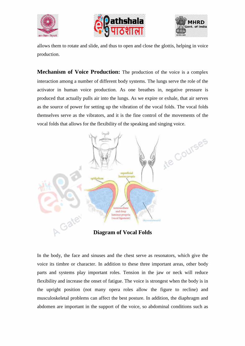
allows them to rotate and slide, and thus to open and close the glottis, helping in voice
production.
Mechanism of Voice Production: The production of the voice is a complex
interaction among a number of different body systems. The lungs serve the role of the
activator in human voice production. As one breathes in, negative pressure is
produced that actually pulls air into the lungs. As we expire or exhale, that air serves
as the source of power for setting up the vibration of the vocal folds. The vocal folds
themselves serve as the vibrators, and it is the fine control of the movements of the
vocal folds that allows for the flexibility of the speaking and singing voice.
Diagram of Vocal Folds
In the body, the face and sinuses and the chest serve as resonators, which give the
voice its timbre or character. In addition to these three important areas, other body
parts and systems play important roles. Tension in the jaw or neck will reduce
flexibility and increase the onset of fatigue. The voice is strongest when the body is in
the upright position (not many opera roles allow the figure to recline) and
musculoskeletal problems can affect the best posture. In addition, the diaphragm and
abdomen are important in the support of the voice, so abdominal conditions such as

cramping or bowel disorder may have a negative impact on voice. The psychological
system is important in the confidence one has in his or her voice, as can be seen by
the fluttering of the voice when a person is nervous or anxious. Finally, refined
coordination of muscle movement and sensory control are adjusted by the
neurological system.
Diagram of Voice Box Cartilages and Muscles Voice box muscles are named according to the cartilages to which they are attached.
3. Speaker identification: Speaker recognition comprises those activities which attempt to link a speech sample
to its speaker through its acoustic or perceptual properties. Speech signal provides
information regarding speaker characteristics, spoken phrase, speaker emotions,

additional noise, channel transformations etc. The process of Speaker recognition has
two broad application areas, explicitly, Speaker identification and Speaker
verification. Speaker identification deals with identifying a speaker of a given
utterance amongst a set of known speakers. The unknown speaker is identified as the
speaker whose model best matches the input utterance. Speaker verification is a more
direct and converged effort leading to either acceptance or rejection of the claimed
identity of a speaker.
Components of Speaker Recognition
The main components of speaker recognition system are feature extraction and
classification. The classification module is further divided into two parts: pattern
matching and decision.
Feature extraction
This is the foremost step in the process of speaker recognition. This segment
processes the acquired data, i.e., a set of feature vectors or parameters from the speech
signal representing some speaker-specific information, which results from complex
transformations occurring at different levels of the speech production: semantic,
phonologic, phonetic, and acoustic. There are different ways to categorize the
features. From the viewpoint of their physical interpretation, following categories
have been proposed:
1. Short-term spectral features–These features, as the name suggests, are computed
from the short frames of about 20 to 30 milliseconds in duration. They are
usually the descriptors of the resonance properties of the supralaryngeal vocal
tract.
2. Voice source features –These features characterize the glottal excitation signal of
voiced sounds such as glottal pulse shape and fundamental frequency, and it is
reasonable to assume that they carry speaker-specific information.
3. Spectro-temporal features-It is very much a rational assumption that the spectro
temporal. Signal details such as formant transitions and energy modulations
contain useful speaker-specific information.
4. Prosodic features-Prosody refers to non-segmental aspects of speech, including
syllable stress, intonation patterns, speaking rate and rhythm. These features
depends upon the long segments like syllables, words, and utterances and reflects

differences in speaking style, language background, sentence type and emotion
of the speaker.
5. High level features–These features attempt to capture conversation-level
characteristics of speakers, such as characteristic use of words („„uh-huh”, “you
know”, “oh yeah”, etc.). Other features are the dialect of any language used in
the conversation by the speaker, accent of the speaker and the style of
speaking.
Pattern matching and decision
The pattern matching module deals with comparison between the estimated features
to the speaker models. Some of the pattern matching methods used in speaker
recognition include Hidden markov models (HMM), dynamic time warping (DTW),
neural networks and vector quantization (VQ) [20]. In case of verification, this
module provides an expert with a similarity score between the test sample and the
claimed identity. While, in case of identification, the module gives similarity score
between the test sample and all the available samples in the database. The evaluation
of these scores is done using decision module and the results are accordingly
presented.
Various approaches to speaker recognition system
In the discipline of speaker recognition a wide range of methods and procedures are
adopted by the experts for identification.
Auditory analysis
Such type of analysis involves a group of trained phoneticians giving their judgement
regarding the similarity and dissimilarity between the two speech events, after hearing
the samples again and again to find out some similarities in their linguistic, phonetic
and acoustic features. Human listeners are robust speaker recognizers when presented
with the degraded speech. Listener performance free from all types of limitations like
the signal to noise ratio, speech bandwidth, the amount of speech material, distortions
occurring in the speech signals as a result of speech coding, transmission systems, etc.
In this technique, different utterances of the speakers are segregated in respect of each
speaker by way of repeated listening of recorded conversation. The segregated
conversations of each speaker are repeatedly heard to identify linguistic features and

phonetic features like articulation rate, flow of speech, degree of vowels and
consonant formation, rhythm, striking time, pauses etc. There are cues in voice and
speech behaviour, which are individual and thus make it possible to recognize the
familiar voices. Experts working in several governments forensic laboratories
including laboratories in Germany, Austria, Sweden, the Netherlands and Spain, and
in private practice in countries like the United Kingdom and Germany, are still
practising this phonetic-acoustic technique for identification. However, with any
human decision process, it is generally believed that the auditory analysis by a listener
leads to a subjective decision.
Spectrographic approach or voiceprint identification
This involves the semi-automatic measurements of particular acoustic speech
parameters such as vowel formants, articulation rate, which is sometimes combined
with the results of auditory phonetic analysis by a human expert. In 1941, an electro
mechanical acoustic spectrograph was developed by Dr. Raleph Potter, Bell
Telephone Laboratory, with an idea to convert sounds into pictures. A sound
spectrograph is an instrument which is able to give a permanent record of changing
energy-frequency distribution throughout the time of a speech wave. The
spectrograms are the graphic displays of the amplitude as a function of both frequency
and time. Examiners visually inspect and compare similarities or differences of
patterns of the energy distribution in the spectrograms. It is generally believed that
formant structures and other spectral characteristics which are evident from a
spectrogram are unique for each individual. The most widely used features are
fundamental frequencies, formant bandwidths, formant frequencies, spectral
composition of fricatives and plosives for individual segments, and transitions.
However, the main drawback of this voiceprint analysis is that the spectrograms of the
speech signal from same individual will show large intraspeaker variations, because
of the fact that no speaker actually is capable of producing two identical speech
utterances.

4. Tape authentication
In many cases, an audio forensic expert is called upon to examine taped evidence to
provide an opinion on whether or not a tape has been “edited” or “doctored” in any
way. Specifically, this translates into an analysis of the temporal sequence of events
found on the tape that correspond to record start, pause, and stop operations of one or
more tape recording devices. This typically includes the analysis of “record event
signatures” corresponding to the interaction of the tape surface with the electrical
activation and deactivation of AC-bias record and erase heads, and/or contact with a
permanent magnet erase head. The analysis includes aural analysis (“critical
listening”); visual analysis of the time-domain waveform (“waveform analysis”); and
via visual analysis of the bitter pattern of the magnetized surface of the tape
(“magnetic development”). The bitter pattern, a visual representation of the
magnetized portions of the tape rendered visible through a low-power microscope,
results from the application of micron-sized iron particles in a liquid suspension to the
tape known as “ferro fluid”. A camera or video recorder can be used to document the
event. In addition to examining record event signatures to determine continuity or
editing, audio forensic experts are also sometimes asked to confirm if an evidence
tape is an original recording, or a copy. Evidence of an original versus a copied tape
usually results from similar record event signature analysis, whenever possible. While
it is possible to determine that a record stop event is a copy via the analysis
techniques just described, an audio forensic expert can never determine with absolute
certainty if a recording is truly “original”; one can only state that the recording is
consistent with an original recording. As an example, an original recording can be
recorded to a computer, digitally edited, and then played back to a cassette recorder
containing a tape with “leader” at the start and end. The leader cannot be recorded
upon since it has no metallic surface that can be magnetized. The action of the tape
recorder corresponding to the record start and record stop events cannot be observed
via either waveform or magnetic development analysis.
Another area of authentication not considered here is matching a tape to a specific
tape recorder used to make the evidence recording. It is on occasion useful for an
attorney to have an audio forensic expert impugn the reliability of a witness‟s

testimony in cases where a specific tape recorder is claimed to have been used to
make a specific evidence tape. Intra-machine variability viewed with inter-machine
variability makes it far easier for an expert to eliminate a specific machine rather to
identify it. Finally, the forensic audio expert and legal practitioners must bear in mind
the difference between what has been termed “Technical Authentication” versus
“Legal Authentication”. While related, the audio forensic expert is not required nor
are they responsible to legally authenticate evidence in the legal sense. Technical
authentication is within the expertise of the audio forensic expert.

Law
Forensic Science & Forensic Medicine
Forensic Toxicology and Classification of Poisons
DESCRIPTION OF MODULE
Items Description of Module Subject Name Law Paper Name Forensic Science and Forensic Medicine Module Name/Title
Forensic Toxicology and Classification of Poisons
Module Id LAW/CJA/VIII /9
Objectives
Learning Outcome:
To make the learners understand about the concept of forensic toxicology.
To make the learner aware about various branches of toxicology such as analytical
toxicology, clinical toxicology, veterinary
toxicology, aquatic toxicology,
environmental toxicology etc.
To make the learners understand how poisons can be classified in various categories.
To aware the learners about the mode of action of different poisons.
Prerequisites To aware about the different branches of toxicology and how the toxic compounds can be categorized into different groups depending upon their properties.
Key words
Forensic toxicology, poison, corrosives, irritant, asphyxiants, cerebral poison, cardiac poison, spinal poison, peripheral poisons, metallic, non-metallic, anions, animal poisons, vegetable poisons, volatile, non-volatile, gaseous poisons, pesticides, drugs etc.
Role Name Affiliation Principal Investigator Prof. (Dr.) G.S. Bajpai Registrar, National Law
University Delhi Co-Principal Investigator
Paper Coordinator Prof. (Dr) Sally Lukose Dean, School of Basic and
Applied Sciences, Galgotias
University, Greater Noida. Content Writer/Author Dr. Mamta Assistant Professor, School
of Basic and Applied
Sciences, Galgotias
University, Greater Noida. Content Reviewer

1. Introduction:
Toxicology is the study of adverse effects of chemical substances on biological
system due to their exposure. It includes study of mode of action, symptoms,
detection and treatments of toxic agent in biological system.
2. Branches of toxicology
There are various branches of toxicology i.e. analytical toxicology, clinical toxicology,
forensic toxicology, veterinary toxicology, aquatic toxicology, environmental toxicology,
etc. These branches have been summarised in following figure.
2.1. Analytical toxicology: Analytical toxicology is deals with evaluation of toxic
substance present in biological specimen as well as in raw form. Evaluation
involves detection, identification and quantification of poisons using various
analytical techniques1.
2.2. Clinical toxicology: Clinical toxicology is deals with diagnosis and
treatment of human poisoning.
2.3. Forensic toxicology: Forensic toxicology deals with the analysis of toxic
substances which involve various methods to study these poisonous substances
to aid the legal investigation to know the cause of poisoning. It is medico-legal
aspect of clinical poisoning2.
2.4. Veterinary toxicology: Veterinary toxicology is the specialised branch of
toxicology which deals with study, diagnosis and treatment of various toxic
compounds in animal kingdom3.
1. R. J. Flanagan, A. Taylor, I. D. Watson, R. Whelpton, Fundamental of Analytical Toxicology, (John
Wiley & Sons Ltd., 2007).
2. https://toxlearn.nlm.nih.gov/htmlversion/module1.html
3. R.C. Gupta, Veterinary Toxicology Basic and Clinical Principles, Chapter 1, 1st edi., (Elsevier, 2007).
2.5. Aquatic toxicology: Aquatic toxicology deals with adverse effect caused by
toxic substance on aquatic system4.
2.6. Environmental toxicology: Environmental toxicology deals with the ill-
effects of environmental toxicants on human, animal, plants and the
environment. Environmental toxicants are the compounds which are released
into environment from various sources such as disposal from industries,
domestic disposal etc which may be degradable or non-degradable.
3. Poison
Poison is a substance which when administered, inhaled or swallowed by living
organism causes ill effects on the body. It is defined also as a medicine in a toxic
dose. Toxic substance may be solid, liquid, gas or any environmental agent.
3.1. Classification of poison
Poisons can be classified in various categories depending upon their (A) mode of
action and (B) physical state and (C) medicolegal classification (D) toxico-analytical
as follow:
(A) On the basis of mode of action: On the basis of mode of action poison can be
categorized into (i) Corrosive Poisons (ii) Irritant poisons, (iii) Neurotic
Poisons, (iv) Cardiac Poisons and (v) Asphyxiants.
(i) Corrosive Poisons: Corrosive poisons are the poisons which cause
inflammation at the site of contact. Both strong acids and alkalis are comes
under this category.
(a) Strong acids: Concentrated sulphuric acid, nitric acid and hydrochloric acid
(b) Strong bases: Sodium hydroxide, potassium hydroxide and ammonium
hydroxide.
4 Mikko Nikinmaa, Aquatic Toxicology: An Introduction To Aquatic Toxicology, Chapter 1.
(Elsevier, 2014).

(ii) Irritant poisons: Irritants are those substances which cause irritation, pain,
excessive vomiting. These are further divided into (a) inorganic poisons, (b)
organic poisons and (c) mechanical poisons.
(a) Inorganic poisons: Inorganic poisons are consisted of:
Metallic – Lead, arsenic, mercury, antimony, copper and zinc etc.
Non Metallic - Chlorine, bromine and iodine etc.
(b) Organic Poisons: Organic poisons consist of poisons of both vegetable
origin and animal origin.
Animal poisons – Snakes venom, scorpion venom, spiders etc.
Vegetable poisons – Croton oil, castor oil etc.
(c. Mechanical Poisons: Mechanical poisons are the poisons which cause
irritation, perforation obstruction in the gastrointestinal tract. For example,
powder glass, diamond dust etc.
(iii) Neurotic Poisons: - Neurotic poisons are the poisons which affects the
different part of central nervous system such as cerebral poisons, spinal
poison, peripheral poisons etc. and these constitute of following poisons:
a. Cerebral Poisons –Alcohol, opium, barbiturates, benzodiazepines etc.
b. Spinal Poison – Strychnine.
c. Peripheral – Curare
(iv) Cardiac Poisons – Digitalis, tobacco.
(v) Asphyxiants – CO, CO2 etc5.
5 K Vij , Textbook of Forensic Medicine and Toxicology: Principles and Practice, Chapter 30 p. 429,
Basic Consideration in Drugs/Chemicals, 5th edn., (Elsevier, 2011).

Poisons
Corrosive Poisons
Strong acidFor e.g. Sulphuric acid, nitric acid,
hydrochloric acid etc.
Strong baseFor e.g. Sodium hydroxide, potassium hydroxide, ammonium hydroxide etc.
Irritant poison
Inorganic poison
MetallicFor e.g. Lead, arsenic, mercury, antimony,
copper and zinc etc
Non-metallic For e.g. Chlorine, bromine and iodine etc.
Organic Poison
Vegetable For e.g. Croton oil, castor oil etc.
AnimalFor e.g. Snakes venom, scorpion venom,
spiders etc.
Mechanical Poisons
For e.g. Powder glass, diamond dust etc.
Neurotic Poisons
Cerebral Poisons
For e.g. Alcohol, Opium, barbiturates, benzodiazepines etc.
Spinal Poison For e.g. Strychnine
Peripheral For e.g. CurareCardiac Poisons
Asphyxiants For e.g. CO, CO2 etc.

(B) On the basis of their physical state: Based on physical state, poisons can be
grouped into (i) solid poisons, (ii) liquid poisons and (iii) gaseous poisons.
(i) Solid poisons: Solid poisons don not get absorbed easily into the blood.
These should be dissolved properly in liquid to get absorbed. For example-
lead, arsenic, mercury etc.
(ii) Liquid poisons: Liquid poisons contain both organic and inorganic liquid.
Organic liquids are more volatile than inorganic liquids.
Liquid poison can be absorbed when administered orally or by inhalation or
through skin. As organic liquids are vaporises and organic vapours are
absorbed by lungs such as chloroform, acetone etc. and the organic vapours
which are soluble in lipids get easily absorbed through skin for e.g. furfural.
(iii) Gaseous poisons: Gaseous poisons are absorbed by inhalation such as
carbon dioxide, carbon monoxide etc.
In atmosphere, there are aerosols particles in air and these are either inhaled
or deposited on the skin. They causes marked effect on lungs when inhaled
during breathing and causes obstruction into the lungs (asbestos) in
comparison to those absorbed through skin. Fibres and dust are also the
examples of aerosols6.
6. Toxicological Chemistry, LVIV–2009, Universitatis Medici Leopoliensis Sigillum.
AD 1784.

Poisons
Solid Poisons
For e.g. lead, arsenic,
mercury etc.
Liquid Poisons
Organic liquid
For e.g. ethanol,
methanol, chloroform, aceton etc.
Inorganic liquid
For e.g. liquid ammonia,
liquid sulfurdioxide etc
Gaseous Poisons
carbon dioxide, carbon
monoxide etc.
(C) Medicolegal classification of poison: Based on intention behind the crime,
poisons are classified into (i): Homicidal poisons, (ii) Suicidal poisons, (iii)
Accidental poisons, (iv) Abortifacient poisons, (v) Stupefying agent/poisons,
(vi) Agents used to cause bodily injury (vii) Cattle Poison (viii) Used for
malingering.
(i) Homicidal poisons: The poisons which are used to kill the other person are
known as homicidal poisons such as aconite, abrus precatorius, strychnos
nux vomica etc.
(ii) Suicidal poisons: Suicidal poisons are those poisons which are used for
self killing such as opium, barbiturate, organophosphorus, organochloro
compounds.

(iii) Accidental poisons: Accidental poisons are those poisons which cause
toxicity by accidents or used in mistaken of others: Snakes bite, CO, and
child takes dhatura’s seeds in mistaken of capsicum seeds as it resemble to
capsicum seed.
(iv) Abortifacient poisons: The poisons winch are used to procure abortion are
know as abortifacient poisons for exemples Quinine (alkaloid), Calotropis
etc.
(v) Stupefying agent/poisons: Stupefying agents are those poisons which are
used to stupefy or fool the person for example Dhatura and chloral hydrate
are used to rob the stranger or for the commitment of other crimes.
(vi) Agents used to cause bodily injury: Usually corrosive acids are used to
cause injury on faces and known as vitriol throwing.
(vii) Cattle Poison: Agents which are used to kill livestocks are known as cattle
poisons. Abrus precatorius, Calotropis are used to kill cattle.
(viii) Used for malingering: Some times semicarpus anacardium is used by
malinger to escape from duties as these produces to produce an artificial
bruise.

Poisons
Homicidal poisons
Aconite, abrusprecatorius,
strychnos nux vomicaetc.
Suicidal poisons
Opium, barbiturate, organophosphorus,
organochlorocompounds.
Accidental poisons
Snakes bite, CO, dhatura’s seeds as it
resemble to capsicum seed.
Abortifacientpoisons
Quinine, Calotropisetc.
Stupefying agents
Dhatura, chloral hydrate etc.
Agents used to cause body
injuryCorrosive acids
Cattle poison Abrus precatorius,
calotropis
Used for malingering
Semicarpusanacardium
Examples

(D) Toxico-analytical classification: Poisons can also be classified on the bases
of method used for extraction or analysis into (i) Gaseous and volatile
substances, (ii) Organic non-volatile substances, (iii) Metallic poisons and
(iv) Anion. Therefore, this classification is known as toxico-analytical
classification.
(i) Gaseous and volatile substances: Poisons which are isolated by distillation
or by headspace are categorised into this group. Volatile substances
(methanol, ethanol, benzene, toluene, acetone etc.) can be separated from
gaseous (ethane, butane etc.) poisons because they can be extracted with
alternate methods.
(ii) Organic non-volatile substances: Compounds which are isolated by
solvent extraction methods include in this group. For example drugs such as
opiates and synthetic narcotics, sedatives and hypnotics, stimulants,
antidepressants etc and pesticides which include insecticides, fungicides,
herbicides, rodenticides, nematocides etc. In this group pesticides can be
separated from drugs, though both are organic non-volatile substances which
are isolated by solvent extraction method.
(iii) Metallic poisons: The substance which are isolated by dry ash method or by
wet digestion method constituting this group. The commonest poisons are
arsenic, lead, mercury, etc.
(iv) Anions poisons: Anions are isolated by dialysis. Most commonly
encountered anions in poisoning cases are bromide, cyanide, fluoride,
hypochlorite, nitrate, nitrite, phosphate, sulfide, sulfate etc8.
8 A. C. Moffat, M. D. Osselton, B. Widdop, S. Jickells and A. Negrusz, Clarke’s Analytical Forensic
Toxicology, Introduction to Forensic Toxicology, Chapter 1, p. 1, 2nd edi., Editor: Adam negrusz,
Gail A A Cooper, (Pharmaceutical Press, 2013).

Gaseous and volatile
substances
Organic non-
volatile substances
Metallic poisons
Anions poisons
Poisons
Gaseous poisons: Methanol, ethanol, benzene,toluene, acetone etc.Volatile substances: ethane, butane etc.
Drugs: Opiates and synthetic narcotics,sedatives and hypnotics, stimulants,antidepressants etc.Pesticides: Insecticides, fungicides,herbicides, rodenticides, nematocides etc.
Arsenic, lead, mercury, antimony,zinc, copper etc.
Bromide, cyanide, fluoride, hypochlorite, nitrate, nitrite,phosphate, sulfide, sulfate etc.

Items Description of Module
Subject Name Criminology
Paper Name Forensic Science and Forensic Medicine
Module Name/Title Collection and analysis of Non-Biological evidences:
glass, soil, paint, fibres and tool marks.
Module Id
Non biological evidence
Fiber
A fiber is the smallest unit of a textile material that has a length many times
greater than its diameter. Fibers can occur naturally as plant and animal fibers,
but they can also be man-made. A fiber can be spun with other fibers to form a
yarn that can be woven or knitted to form a fabric.
The type and length of fiber used, the type of spinning method, and the type of
fabric construction all affect the transfer of fibers and the significance of fiber
associations.
Fiber Evidence
Fibers are gathered at a crime scene with tweezers, tape, or a vacuum. They
generally come from clothing, drapery, wigs, carpeting, furniture, and blankets.
For analysis, they are first determined to be natural, manufactured, or a mix of
both.
Fiber Number
The number of fibers on the clothing of a victim identified as matching the
clothing of a suspect is important in determining actual contact. The greater the
number of fibers, the more likely that contact actually occurred between these
individuals
Forensics of Fiber Analysis

Cross transfers of fiber often occur in cases in which there is person-to-person
contact. Investigators hope that fiber traceable back to the offender can be found
at the crime scene, as well as vice versa. Success in solving crimes often hinge on
the ability to narrow the sources for the type of fiber found, as the prosecution did
with their probability theory on the fibers
The world produced approximately 80 billion pounds of fabric in 1995, about half
of which was cotton. The other approximately 44 billion pounds of fiber were
manufactured or synthetic. It could be argued that the large volume of fibers
produced reduces the significance of a fiber association discovered in a criminal
case.Considering the volume of textiles produced worldwide each year, the
number of textiles produced with any one fiber type and color is extremely small.
The likelihood of two or more manufacturers exactly duplicating all of the
aspects of the textile is extremely remote
Fiber Evidence
The problem with fiber evidence is that fibers are not unique. Unlike fingerprints
or DNA, they cannot pinpoint an offender in any definitive manner. There must
be other factors involved, such as evidence that the fibers can corroborate or
something unique to the fibers that set them apart.
Microscopic Examination
A compound microscope uses light reflected from the surface of a fiber and
magnified through a series of lenses, The comparison microscope (two compound
microscopes joined by an optical bridge) is used for more precise identification.
The phase-contrast microscope, reveals some of the structure of a fiber.Electron
microscopes either pass beams through samples to provide a highly magnified
image, or reflect electrons off the sample's surface.
A scanning electron microscope converts the emitted electrons into a
photographic image for display. This affords high resolution and depth of focus.
Spectrometer
The spectrometer, which separates light into component wavelengths. Every
organic element has a uniqueness to its constituent parts. By passing light through
something to produce a spectrum, the analyst can read the resulting lines, called
"absorption lines."

That is, the specific wavelengths are characteristic of its component molecules of
the substance.
Micro-spectrophotometer
This microscope locates minute traces or shows how light interacts with the
material under analysis. Linking this to a computerized spectrophotometer
increases the accuracy. The scientist can get both a magnified visual and an
infrared pattern at the same time, which increases the number of identifying
characteristics of any given material.
Natural Fibers vs Manufactured
Natural fibers come from plants (cotton) or animals (wool). Manufactured fibers
are synthetics like rayon, acetate, and polyester, which are made from long chains
of molecules called polymers. Many different natural fibers originating from
plants and animals are used in the production of fabric. Cotton fibers are the plant
fibers most commonly used in textile materials. The type of cotton, fiber length,
and degree of twist contributing to the diversity of these fibers.
Other plant fibers :- Flax (linen), ramie, sisal, jute, hemp, kapok, and coir.
The identification of less common plant fibers at a crime scene or on the clothing
of a suspect or victim would have increased significance.
Animal Fiber: Wool is the most frequently used in the production of textile
materials and the most common wool fibers originate from sheep. Finer woolen
fibers are used in the production of clothing coarser fibers are found in carpet.
Fiber diameter and degree of scale protrusion of the fibers are other important
characteristics.
Other Animal Fibers:- Although sheep's wool is most common, woolen fibers
from other animals may also be found. These include camel, alpaca, cashmere,
mohair, and others. The identification of less common animal fibers at a crime
scene or on the clothing of a suspect or victim would have increased significance.
Man-Made Fibers:- More than half of all fibers used in the production of textile
materials are man-made. Some man-made fibers originate from natural materials
such as cotton or wood; others originate from synthetic materials. Polyester and
nylon fibers are the most commonly encountered man-made fibers, followed by
acrylics, rayons, and acetates. There are also many other less common man-made

fibers. The amount of production of a particular man-made fiber and its end use
influence the degree of rarity of a given fiber.
The cross section of a man-made fiber can be manufacturer-specific. Some cross
sections are more common than others, and some shapes may only be produced
for a short period of time. Unusual cross sections encountered through
examination can add increased significance to a fiber association. Generally, the
analyst gets only a limited number of fibers to work with—sometimes only one.
Whatever has been gathered from the crime scene is then compared against fibers
from a suspect source, such as a car or home. Fibers are laid side by side for
visual inspection through a microscope.
Important Considerations
An is the length of time between the actual physical contact and the collection of
clothing items from the suspect or victim. If the victim is immobile, very little
fiber loss will take place, whereas the suspect's clothing will lose transferred
fibers quickly. The likelihood of finding transferred fibers on the clothing of the
suspect a day after the alleged contact may be remote, depending on the
subsequent use or handling of that clothing.
Steps of Fiber Analysis
The first step in fiber analysis is to compare color and diameter. Dyes can also be
further analyzed with chromatography, which uses solvents to separate the dye's
chemical constituents.
Fiber Color
Color influences the value given to a particular fiber identification. Often several
dyes are used to give a fiber a desired color. Individual fibers can be colored prior
to being spun into yarns. Yarns can be dyed, and fabrics made from them can be
dyed. Color can also be applied to the surface of fabric, as found in printed
fabrics. How color is applied and absorbed along the length of the fiber are
important comparison characteristics. Color-fading and discoloration can also
lend increased value to a fiber association.
Microscopy
Fibers should be first examined with a stereomicroscope. Physical features such
as crimp, length, color, relative diameter, luster, apparent cross section, damage,

and adhering debris should be noted. Fibers are then tentatively classified into
broad groups such as synthetic, natural, or inorganic. If the sample contains
yarns, threads, or sections of fabric, construction should be recorded
Illumination and Magnification
Comparisons should be made under the same illumination conditions at the same
magnifications. For comparison microscopes, this requires color balancing the
light sources. This is best achieved with two fibers or fiber samples from the
same source mounted on two microscope slides, which are then compared. A
balanced neutral background color is optimal.
Side-by-Side Comparisons.
If all of the characteristics are the same under the stereoscope, the next step is to
examine the fibers with a comparison microscope. This side-by-side and point-
by-point examination is the best technique to discriminate between fibers,
especially those that appear to be similar. The physical characteristics of the must
be compared visually with the comparison microscope to determine if they are
the same in the known and questioned samples. Photography is recommended to
capture the salient features for later demonstration.
Glass
What is Glass?
It is a uniform amorphous solid. Glass have no specific melting point. It Softens
over a temperature range. Because of this, glass breaks in a variety of fracture
patterns.
What’s in Glass?
Fusion of sand (SiO2), soda (Na2CO3) & lime (CaO) produces a transparent solid
when cooled called glass. It is a 3D network of atoms which lacks the repeated,
orderly arrangement typical of crystalline materials.
Soil

Physical properties of glass: Glass is hard, elastic, brittle, non-conductor of
electricity, density, refractive index, etc.
Chemical properties of glass: it is resistant to all but fluorine and very strong
bases.
Forensic Examination of Glass
The primary uses for glass are in windows, containers, light bulbs and eyewear.
a) Borosilicate Glass (pyrex): 5% borax (Na2B4O7) is added to resist breaking
when heated or cooled.
b) Colored Glass: metal oxides or colloidal iron (Fe) & sulfur (S) are added to
change its color.
c) Lead glass: Pb increases refractive index & density
d) Flat glass: made by a ―float glass process‖; molten glass is floated on a
pool of tin while cooling. Commonly found in doors and windows.
e) Laminated glass: used in windshields, two sheets of glass with plastic
between them.
f) Tempered safety glass: used in car side windows and designed to break into
tiny pieces; potassium (K) replaces sodium (Na) on the surface.
How Do Glass Windows Break?
Each force causes a deformation that may leave a visible mark or fracture the
glass. This can be used to determine the direction and amount of force. Glass acts
initially as an elastic surface and bends away when a force is applied. When the
force increases beyond its tensile strength, it cracks.

Radial cracks form first and are propagated in short segments on the side opposite
the force.
Concentric cracks come later from continued pressure on the same side as the
force applied.
Edges of broken pieces of glass will show rib (―stress‖) marks. In a radial crack,
the rib marks are perpendicular to unloaded side and parallel to loaded side. The
arrow shows the side that received the impact.
3R rule:
– Radial cracks give rib marks that make
– Right angles on the
– Reverse side from where the force was applied
Exceptions to the Three R Rule
a) Tempered glass ―dices‖ without forming ridges, Very small windows held
tightly in frame can’t bend or bulge appreciably

b) Windows broken by heat or explosion no ―point of impact‖ curved, smooth
edges at break point
Types of Fractures by Projectiles
Bullets are a projectile force (load) that can pass through glass. Load side is the
entrance side; unloaded side is the exit side. Low-speed projectiles: rib marks
may indicate where breaking force was applied.
As the bullet’s velocity increases, the central hole becomes smaller, cracking
patterns become simpler, and the exit hole becomes wider than the entrance hole.
– –
Which side was the bullet fired from? Exit (unloaded) side is wider
than entry (load) side.

– –
Stress lines on the glass edge of radial cracks form a right angle on the reverse
side from the force. Stress lines on the glass edge of concentric cracks form a
right angle on the same side as the force.
•
Which Bullet Hole Was First?
The sequence of impacts can be determined since crack propagation is stopped by
earlier cracks.

Examiners can fit together two or more pieces of glass that were broken from the
same object. Because glass is amorphous, no two glass objects will break the
same way.
Glass Transfer Evidence
• When glass objects are broken, glass flies backward from all parts of the
object where cracks appear not just from point of impact.
This creates a shower of minute glass particles and a transfer of evidence. Glass
fragment comparison depends finding and measuring properties that will
associate one glass fragment with another while eliminating other sources.
Collection of Glass Samples
a) The glass fragments should be packaged in boxes to avoid further
breakage.
b) If evidence is to be examined for glass fragments, it should be taken whole
and each item individually wrapped in paper and boxed.
c) If even the remotest possibility exists that glass fragments may be pieced
together, every effort must be made to collect all glass fragments.
d) Submit glass evidence along with a representative sample of each type of
glass from the crime scene.
Optical Properties of Glass
Make side-by-side comparisons using similar-sized fragments. Place samples on
a white surface using natural light. Use both fluorescent and incandescent light to

determine the glass’s color. Visual color analysis is very subjective. Dyes and
pigments can be almost impossible to extract.
Non- optical Physical Properties of Glass
Surface striations and markings
– Rollers leave parallel ream marks on sheet glass
– Markings may indicate the glass’s orientation when pieces are
missing
– Surface scratches, etchings, and other markings may also be used to
individualize evidence
• Other Properties
– Hardness=5-6 on Mohs scale; use a scratch test.
– Determinations of curvature can distinguish flat glass from
container, decorative, or ophthalmic.
Glass Density
• Density can be measured by directly determining mass and volume (usually
by displacement), by comparison by flotation and by comparison using a
density gradient column
• Density gradient column method:
• Fragments of different densities settle at different levels in the
column of liquid of varying density.
• Technique is not accurate for fragments that are cracked or contain
an inclusion.
Refractive Index by Immersion
Immersing a glass particle in a liquid medium (silicone oil) whose refractive
index can varied with temperature until it is equal to that of the glass particle. At
this point, known as the match point, the Becke line disappears and minimum
contrast between liquid and particle is observed: RI oil = RI glass. The Becke line
is a bright halo near the boarder of a particle that is immersed in a liquid of a
different refractive index.
Becke Lines
Glass has higher refractive index-note white line inside.

• Glass has lower refractive index-note white line outside
Chemical Analysis of Glass
• Fluorescence
– Under UV radiation, many glasses exhibit fluorescence (glow)
– Caused by heavy metals (including tin) from ―float‖ process or
organic coatings
– Scanning Electron Microscopy Energy Dispersive X-ray Analysis
– Can determine many elements simultaneously
– Surfaces of samples (>50 mg) can be analyzed

Tool Marks
Tool marks are made when a harder object comes in contact with a softer object,
leaving marks on it.A tool mark is considered to be any impression, cut, scratch,
gouge, or abrasion caused by a tool coming into contact with another object.
Tools often used in burglaries may leave a mark.
For example, if you attempted to pry open a locked window with a screwdriver,
the screwdriver would leave a tool mark on the window and windowsill. Most
often, tool marks are encountered at burglary scenes that involve forcible entry
into a building or safe.
Typically, an indented impression is left on the frame of a door or window as a
result of the prying action of a screwdriver or crowbar.
One of the first things an investigator looks for at a suspect’s home is the
suspect’s toolbox. Any tools in the commission o f a crime leave unique scratch
marks behind.
These striation marks can be used to match a tool to an object it came into contact
with at the crime scene. A careful examination of these impressions can reveal
important class characteristics. That is, the size and shape of the tool. However,
they rarely reveal any significant individual characteristics that could permit the
examiner to individualize the mark to a single tool. Such characteristics, when
they do exist, usually take the form of discernible random nicks and breaks that
the tool has acquired through wear and use.
Microscopic Irregularities
Just as the machined surfaces of a firearm are impressed with random striations
during its manufacture, the edges of a pry bar, chisel, screwdriver, knife, and
cutting tool will likewise display a series of microscopic irregularities having the
appearance of ridges and valleys.
The Machining Process
Such markings are left as a result of the machining processes used to cut and
finish tools.
These markings are called striation marks. The shape and pattern of such minute
imperfections are further modified by damage and wear during the life of the tool.
When a screwdriver is first made, the microscopic imperfections in the blade
make it unique. As it is used, more imperfections are added and the blade
becomes more unique.
Imperfections Cause Individuality
Considering the unending variety of patterns that the hills and valleys can
assume, it is highly unlikely that any two tools will be identical. Hence, it is the
presence of these minute imperfections that imparts individuality to each tool.
One of the major problems associated with tool mark comparisons is the
difficulty in duplicating in the lab the tool mark left at the crime scene.
Striations

If the edge of a tool is scraped against a softer surface, it may cut a series of
striated lines that reflect that pattern of the tool’s edge. Markings left in this
manner are compared in the lab through a comparison microscope with test tool
marks made from the suspect tool.
Positive Comparisons
Once at the crime lab, a cast is made of the scratch marks left on the window lock
from the forced entry. The result can be a positive comparison, and hence a
definitive association of the tool with the evidence mark, when a sufficient
quantity of striations match between the evidence and test markings.
Test Marks
A thorough comparison requires the preparation of a series of test marks obtained
by applying the suspect tool at various angles and pressures to a soft metal
surface (lead is commonly used). The cast and the lead brick with the scrapings
are placed under a comparison microscope to see if the striation marks match.
This approach gives the examiner ample opportunity to duplicate many of the
details of the original evidence markings. Bring the entire object to the crime lab.
Whenever practical, the entire object or part of the object bearing a tool mark
should be submitted to the crime lab for examination.
When removal of the tool mark is impractical, the only recourse left, is to
photograph the marked area to scale and make a cast of the mark.
Photograph the mark.
However, even under the most optimum conditions, the clarity of many of the
tool mark’s minute details will be lost or obscured in a photograph or cast.
Of course, this will reduce the possibility that the criminalist could individualize
the mark to a single tool.
Use liquid silicone or dental stone for casting.
Under these circumstances, liquid silicone casting material or dental plaster has
been found to be the most satisfactory for reproducing most of the fine details of
the mark.
Matching Toolmarks

Photography and casting are important to match tool with mark.
Soil
What Is Soil?
Mixture of organic and inorganic material. It may range from 100% inorganic
(sand) to nearly 100% organic (peat). Inorganic part is minerals. Organic part is
decayed plant and animal material and is sometimes called humas
Forensic Significance of Soil
Soil is class evidence - cannot be individualized to a particular location. There is
no classification system for soils. Soils can be easily transported. Soils within a
few meters horizontally or vertically differ.
Forensic Soil Analysis is the use of soil sciences and other disciplines to aid in
criminal investigation. Soils are like fingerprints because every type of soil that
exists has unique properties that act as identification markers. This means that the
origin of the soil sample can be identified. For example, clay embedded in the
sneaker of a criminal can be traced back to a specific clay type found along a lake
where a murder victim was discovered. The majority of soil cases involves
footprints or tire marks that have been left in the soil.
The unique properties of soil are as follows:

Sediment– the original solid particles that were weathered and transported. This
could be in the form of a grain of rock that breaks off of the larger parent material
(larger version of rock). Soils can develop on these sediments due to physical and
chemical alteration.
Color– indicates its history as well as the compounds present in the soil. For
example, white or gray soil could mean that the soil contains lime or has been
leeched((a chemical, a metal, etc.) from a substance by the action of a liquid
passing through the substance. Gray soil can also mean that the organic material or
moisture is present, black soil suggests the same. Soil that is red,brown or yellow
generally suggests that there is iron present.
Structure-indicates whether a soil is composed of a single grain particle or not.
This is determined by the presence of peds (clumps). These peds are formed due to
cementing agents such as calcium carbonate attracting the soil particles so that they
adhere to each other to form either bulky peds which are small conglomerates
(masses) or platy peds which are flat and sheet like.
Soil samples can be collected in different ways depending on where the sample is
being collected from. If samples are being collected indoors or from a vehicle
vacuuming is generally used. If the sample is outdoors it’s collected by placing a
teaspoon of soil into a plastic vial. When found on a tool, it is wrapped in plastic
and then sent to the lab for testing. Collecting soil samples off of a body isn’t any
harder than collecting a sample from anywhere else but it takes more work and
care so that the evidence doesn’t get contaminated. When collecting samples from
a body, samples should be taken at regular intervals and a different spoon should
be used each time.
Once the soil samples are collected they are sent to the laboratory. At the
laboratory samples should be separated by samples from the victim and samples
pertaining to the suspect. Also, each sample set should get its own examiner. This
is to avoid contamination; if possible the samples should be kept in different
rooms. To examine the samples the examiner will first want to use microscopic
analysis to perform testing on the mineral content. Another test that can be done to
help try to identify the origin of the soil is a density test. The density test is called
the density gradient tube. This test consists of adding liquid to two glass tubes. The

liquid in both tubes is the same, but the rations are different. This represents two
different densities. The soil sample is added to both liquid samples. After the soil
samples become suspended in the liquid the separation of the bands can then be
analyzed to reveal the profile of the soil. Heat tests can also be used to test the soils
reaction and electron microscopes can be used to examine the structure of the
minerals in the soil. During examination, an examiner might find that some soil
samples may contain biological evidence such as saliva, semen or blood. If
biological evidence is found in the sample the whole soil sample should be sent to
the laboratory for testing.
Paint
One of the most commonly encountered types of physical evidence, most
frequently in hit-and-run and burglary cases. Usually the forensic scientist will be
asked to compare two or more paints for the purpose of determining common
origin.
The paint from an automobile can be used to determine the color, make, and
model of a car.
Paint is composed of pigments (to give color and hiding quality) and a binder
which provides the support medium for the pigment and other additives. The
binder is usually a polymer.
Automotive paint – there are four basic coatings for auto paint.
a. The ELECTROCOAT PRIMER is the first layer. It is electroplated onto the
steel body to provide corrosion resistance. Usually grey or black in color.
b. The PRIMER SURFACER is applied over the electrocoat primer. Its function
is to smooth out and hide seams or imperfections.
c. The BASECOAT is the layer that provides the color and appearance of the
finish. Mica pigments, aluminum flakes, and other materials are added to give
paint an individual and unique appearance.
d. The CLEARCOAT has no color and is used to provide gloss and add
durability.

The microscope has traditionally been and remains the most important instrument
for locating and comparing paint specimens. Color imparts paint with its most
distinctive forensic characteristics. The importance of layer structure for
evaluating the evidential significance of paint is very important. When paint
specimens posses colored layers that match with respect to number and sequence
of colors a common origin can is probable.
The diverse chemical composition of paint can provide for additional points of
comparison between specimens. A thorough comparison of paint must include a
chemical analysis of either the paint’s pigment, binder, or both.
Pyrolysis gas chromatography is a valuable and accepted technique for
distinguishing most paint formulations. In this process paint chips as small as 20
micrograms are decomposed by heat into numerous gas products. These products
are sent through a gas chromatograph. What emerges and is recorded are the
separated decomposition products of the polymer used in the paint.
It is the pattern of this chromatogen or ―pyrogram‖ that distinguishes one
polymer from another. What results is a pyrogram that is sufficiently detailed
enough to reflect the chemical make-up of the binder.
Infrared spectrophotometry is another analytical technique used to provide
information about the binder composition of paint. Binders will selectively
absorb infrared radiation to yield a spectrum that is highly characteristic of a
paint specimen.
The elements that comprise the inorganic pigments of paint can be identified by
either emission spectroscopy, neutron activation analysis, X-ray diffraction, and
X-ray spectroscopy. The emission spectrograph can simultaneously detect 15 to
20 elements in most automobile paint.
Crime labs are often asked to identify the make and model of a car from a very
small amount of paint left behind at a crime scene.
Color charts for automobile finishes are available from paint manufacturers.


Criminology
Forensic Science and Forensic Medicine
Finger Prints: Definition, general characteristics: Patterns, delta, core, ridge count, ridge trace. Individual ridge characteristics

DESCRIPTION OF MODULE
Items Description of Module Subject Name Criminology Paper Name Forensic Science and Forensic Medicine Module Name/Title
Finger Prints: Definition, General Characteristics: Patterns, Delta, Core, Ridge count, Ridge Trace; Individual Characteristics.
Module Id CRIMINOLOGY/FSFM/XVIII
Objectives
Learning Outcome:
To make the learners understand the basics of fingerprint science.
To inform learners regarding the brief history of the fingerprint science.
To acquaint learners with the identifying characteristics of fingerprints.
Prerequisites General understanding of the fingerprint science and its importance in forensic science.
Key words
Fingerprints, core, delta, ridge count, ridge trace, individual characteristics.
Role Name Affiliation Principal Investigator Prof. (Dr.) G.S. Bajpai Registrar, National Law
University Delhi Co-Principal Investigator
Paper Coordinator Prof. (Dr) Sally Lukose, Dean, School of Basic and
Applied Sciences, Galgotias
University Content Writer/Author Ms Vinny Sharma Assistant Professor, Division
of Forensic Science, SBAS, Galgotias University
Content Reviewer
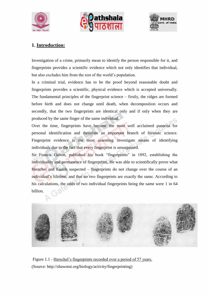
1. Introduction:
Investigation of a crime, primarily mean to identify the person responsible for it, and
fingerprints provides a scientific evidence which not only identifies that individual,
but also excludes him from the rest of the world‘s population.
In a criminal trial, evidence has to be the proof beyond reasonable doubt and
fingerprints provides a scientific, physical evidence which is accepted universally.
The fundamental principles of the fingerprint science – firstly, the ridges are formed
before birth and does not change until death, when decomposition occurs and
secondly, that the two fingerprints are identical only and if only when they are
produced by the same finger of the same individual.
Over the time, fingerprints have become the most well acclaimed panacea for
personal identification and therefore an important branch of forensic science.
Fingerprint evidence is the most assenting investigate means of identifying
individuals due to the fact that every fingerprint is unsurpassed.
Sir Francis Galton, published his book ―fingerprints‖ in 1892, establishing the
individuality and permanence of fingerprints. He was able to scientifically prove what
Herschel and Faulds suspected – fingerprints do not change over the course of an
individual‘s lifetime, and that no two fingerprints are exactly the same. According to
his calculations, the odds of two individual fingerprints being the same were 1 in 64
billion.
Figure 1.1 - Herschel‘s fingerprints recorded over a period of 57 years.
(Source: http://shawmst.org/biology/activity/fingerprinting)

Galton identifies the characteristics by which fingerprints can be identified, these
same characteristics (minutia) are basically still in use today, and are often referred to
as Galton‘s Details.
The present module discusses the fingerprint science, a brief introduction, its
biological origin, deals with the brief historical background, and explains fingerprints
as an important aid in identification and individualization. Delineates the forensic
importance of fingerprint evidence. Explains the various general and individual
characteristics of fingerprints.
2. Fingerprints – Definition and its principles:
The skin covering the anterior surface (palm) of human hand and plantar surface
(sole) of the human foot is different in texture and appearance than the one which
covers the rest of the human body. The skin on the palmar and plantar surfaces is
continuously wrinkled with narrow minute ridges (also known as frictional ridges)
and is also completely free from hair and oil glands. However, there is a profusion of
sweat glands and those are relatively large in size.
The study of the skin patterning on fingers, palm, soles and toes is termed as
dermatoglyphics.
Dermatoglyphics.
Glyphe = a Carving

2.1 Definition of Fingerprints:
A study of ridge patterning is known as fingerprints, which is a reproduction of the
friction ridges of the finger on a surface of contact.
In other words, a fingerprint is an impression of the friction ridges of all or any part of
the finger. A friction ridge is a raised portion of the epidermis layer on the skin of
palmar: palm and fingers or plantar: sole and toes, consisting of one or more
connected ridge units on skin. These ridges are also known as dermal ridges or dermal
papillae.
Fingerprints may be deposited in natural secretions from the exocrine glands present
in the friction ridge skin (secretions consisting primarily of water) or they may be
made by ink or other contaminants transferred from the peaks of friction skin ridges to
a relatively smooth surface. The term fingerprint normally refers to impressions
transferred from the pad on the last joint of the fingers and thumbs.
The science of fingerprint identification stands out among all other forensic science
branches for the following reasons:
a) The fingerprint science has served almost all governments worldwide for
nearly 120 years to provide accurate identification of criminals. No two
fingerprints have yet been found alike in billions of fingerprints.
Fingerprints are the very basis for criminal history foundation at every law
enforcement agency.
b) It remains the most commonly used forensic evidence around the globe. In
most jurisdictions fingerprints examination cases out number all other
forensic examination casework combined.
c) It continues to expand as the premier method for personal identification,
with tens of thousands of persons added to fingerprint records.
d) Fingerprints obtained from crime scenes lead to more suspects and
generate more evidence in court than all other forensic techniques
combined.

2.2 Properties of Fingerprints:
a) They are unique to an individual.
b) They are persistent and does not change with age.
c) They cannot be destroyed, even if damaged. The parameters considered in
case of identification are so wide that it‘s nearly impossible to destroy all the
details of one‘s fingerprint.
d) They can be lost only with the loss of limb/phalanges but still since all ten
phalanges prints are recorded, in case of a criminal, so impersonation is not
possible.
2.3 Fundamental Principles of Fingerprints:
Like any other science, fingerprint science lays its basis in its three fundamental
principles:
a) Principle of individuality
b) Principle of persistency
c) Principle of variety.
2.3.1 Principle of Individuality:
It states that ―No two fingers have yet been observed to possess identical ridge
characteristics‖.
Explanation:
According to Galton‘s calculation, the odds of two individual fingerprints
being the same are 1 in 64 billion.
Also, the general shape and pattern can be same but ridge characteristics –
identity, number, relative position – imparts individuality to fingerprints.
Fingerprints remain constant for the life and cannot be altered except by the
destruction of skin.
Fingerprints are present from birth both on epidermis and dermis layers of
skin.
Fingerprints form patterns that are absolutely individual and classifiable.

Fingerprints are easily collectible.
2.3.2 Principle of Persistency:
It states that ―A fingerprint will remain unchanged during an individual‘s lifetime‖.
Explanation:
In the cross-section of the skin the boundary of cells is observed separating the
epidermis and dermis (layers of skin). It is the shape of this boundary, made
up of dermal papillae, that determines the form and pattern of the ridge on the
surface of the skin. Once the dermal papillae develop in the human foetus
during embryonic state, the ridge pattern will remain unchanged throughout
the lifespan of an individual except for enlargement during growth of the
limbs, i.e., the hands and feet.
If an injury reaches deeply enough into the skin and damages the dermal
papillae, a permanent scar will form. For this to happen, such a wound would
have to penetrate up to 2mm beneath the skin‘s surface.
The presence of permanent scars would serve as a new and additional
characteristic for the purpose of identification.
2.3.3 Principle of Variety:
It states that ―Fingerprints have general ridge patterns that permit them to be
systematically classified‖.
Explanation:
Fingerprints have general ridge patterns that permit them to be systematically
classified.
All fingerprints are divided into three major classes: Loops, Arches and
Whorls.
In general population, the percentage frequency of these patterns is
o Loops - 60 – 65%
o Whorls - 30 – 35%
o Arches - 5 – 10%

Based on these grounds, the fingerprints became a highly effective tool in
establishing the identity of any individual.
3. Biological Origin of Fingerprints:
Fingerprints are formed during the second trimester of foetal development and when a
child is born the fingerprints are already present on his palms, fingers, soles and toes.
Once formed the fingerprints remain unaltered throughout the lifespan of an
individual (except for the increase in size due to growth of the human body) until the
skin decomposes or degenerates after death.
Fingerprints are the impressions produced by the papillary ridges present at the apex
of the top phalanges (tips of the fingers) of one‘s hand. The ridges are the raised
portions on the palm skin bearing numerous amounts of sweat glands. The
arrangement of papillary ridges of every individual is unique to themselves and are
permanent throughout in life. Their ability of being highly individual and permanent
has made them an indispensable mode of identification for more than 100 years and
still counting.
4. Historical Account:
The study of fingerprints dates back to the earlier civilizations. The exact person
hence cannot be named who first time used the fingerprints for identification purpose.
But the idea that fingerprints could be used for personal identification was first used
by Sir William Herschel, district magistrate of Hooghly district, Bengal, 1858.
4.1 Ancient History:
• Earthenware estimated to be 6000yrs old, North-west China: has clear friction
ridge impressions

• Thousands of years before BC, fingerprints were found on pottery as a mark
and seal of the maker.
• Pre-historic cave wall etching found in Nova Scotia, Canda, showing a hand
with ridge patterns sketched on it.
• Fingerprints were used as seals during Tong dynasty, China.
• The deeds were signed by means of fingerprints during Sung period.
• Persian physician Rashid-al-Din Hamadani or ―Rashideddin‖ in his collections
called Jami-al-Tawarikh, commented on the Chinese practice of identifying
people through fingerprints.
• Fingerprints along with palm prints known as ―PANJA‖ were used for
identification. In India, fingerprints were embossed on clay tablets and seals
during Indus Valley Civilization.
• Slabs of clay were found in King Tut-en-khamen‘s tomb in Egypt.
• In ancient China, thumb prints were found on clay seals.
4.2 From 17th
Century to 20th
Century:
• Grew (1684):
• in his paper stated ― Innumerable little ridges of equal size on the ends
of the first joints of the fingers‖.
• also mentioned about sweat pores, epidermal ridges and their
arrangements.
• Referred sweat pores as ― Little Fountain‖.
• Bidloo (1685):
• in his book human anatomy incorporated a diagram of friction ridges
and pore structure but failed to mention about the individuality of the
friction ridges.
• Malpighi (1685 - 86):
• Referred about the function, form, and structure of the friction skin as a
touchable organ

• Mentioned about spirals and loops in fingerprints but no reference to
its importance
• 1.8 mm thick layer of the skin was named after him as ―Malpighi
layer‖.
• Thomas Bewick (1753 - 1828):
• Used fingerprints in the form of stamp.
• Purkinje (1823):
• Classified fingerprints into nine principal configuration groups and
assigned name to each one of them. But he never mentioned anything
regarding individuality of the ridge structure and their use in personal
identification.
• William Herschel (1858):
• Chief Magistrate of the Hooghly district, Jungipoor, India – First
Person to use fingerprints on native contracts.
• Henry Faulds (1880):
• Examined fingerprints in depth
• Mentioned that fingerprints can be classified and also the ridge details
are distinctive
• Also, maintained that fingerprints are useful in medico-legal studies as
the photographs of people may change but friction ridges never change
• Also emphasized apprehending criminals by locating fingerprints at
crime scene.
• Kollmann (1883):
• First researcher who made a mention regarding formation of friction
ridges in the embryos and the topographical physical stressors that may
have been part of their growth.

• He identified the presence and location of volar pads on human hand
and foot.
• Thomas Taylor (1887):
• Made an effort to identify criminals, especially murderer, by
comparing the marks of the hand left upon any object wit impressions
in wax taken from the hands of suspected persons.
• Sir Francis Galton (1822 – 1911):
• Published book entitled ―Finger Prints‖ (1892), which included the
first classification system for fingerprints
• Scientifically proved that fingerprints do not change over the course of
an individual‘s lifetime, and no two fingerprints are exactly the same.
• According to his calculations, the odds of two individual fingerprints
being the same were 1 in 64 billion.
• Responsible for identifying the ridge characteristics (minutia) that are
responsible for establishing the supremacy of fingerprints over
Bertillon‘s anthropometric method of identification in 1901
• Established it as the primary means of personal identification.
• June Vucetich (1855 - 1925):
• In Argentina, set up his own equipment for taking criminal‘s prints.
• In 1891, he independently worked out a fingerprint classification
system and was filing criminal‘s fingerprints using his new system,
which was practically used in solving the first ever murder case ―The
Rojas Murders‖ in 1892.
• By 1896, Argentina, become the first country in the world to abolish
anthropometry and file criminal records solely by fingerprint
classification.

• Sir Edward Henry (1850 – 1931):
• In 1891 added taking of the left thumb impression to each
anthropometric file card.
• In 1894 he instructed that all the ten fingers of each prisoner be printed
and added to the anthropometric cards and assigned two Bengali
officers, KHAN BHADUR AZIZUL HAQUE and RAI BHADUR
HEM CHANDRA BOSE, to study the classification problem.
• Eventually succeeded in setting up a classification system with 1024
primary positions and secondary breakdowns in each.
• In 1897 fingerprints were adopted as the official method for the
identification of criminals in British India.
• In December, 1900, the Belper Committee, recommended that the
fingerprints of criminals be taken and classified by the Indian System.
• These recommendations were implemented in 1901 and the Indian
system eventually replaced anthropometry.
• Subsequently systems of Henry and Vucetich form the basis of most of
the ten digit classification systems used at present.
• Dr. Edmond Locard (1912):
• Established the Science of Poroscopy.
• Salil K. Chatterjee (1962):
• Coined the term Edgeoscopy – to use ridge edges in concert with other
friction ridge formations to establish individuality.
• Observed different shapes on the friction ridge edges that tended to
reappear frequently and named them.
• Having an understanding of the relevance of edge shapes enhances
comparison and improve the chances of establishing individuality.

5. Fingerprints – An Aid in Identification and Individualization.
Personal identification through fingerprint is regarded as the greatest contribution to
law enforcement. The science of fingerprints provides unique service in the
administration of justice and also in other areas where positive identification is of
paramount importance. Identification is the comparing of the fingerprints of a suspect
with the chance fingerprints obtained from a scene of crime to determine whether an
identification can be made. An identification can be made by comparing a very small
portion of ridges left at a scene of crime with those of a suspect and classification is
not required for this comparison.
Fingerprint identification is the process of comparing questioned and suspected
fingerprints, to check whether or not they are from the same finger of the same hand
of the same individual. Identification otherwise referred to as individualization occurs
when an expert or a computer based software determines that two fingerprint
impressions are identified i.e., they are from the same finger of the same hand of the
same individual.
Fingerprints have been in use as a mark of individual way back since they were
legally adopted and possessed a proper classification. Fingerprints were used on the
pottery to indicate the maker and the brand of the pottery.
With the development of the methods to identify the individual/suspect on the basis of
the portrait parley, the method of individualization using fingerprints is now being
used by many judicial organization all over the globe.
There are various reasons put forward for using the fingerprints for identification and
individualization. Some of them are:
1. The fingerprint patterns are unique – no two individuals (including identical
monozygotic twins) have not yet till date being reported to possess exactly
same fingerprints.
2. Whatever surface /object we touch we leave impressions on everything we
touch with pressure.
3. The fingerprints left can be visible or latent (invisible). Visible when the
fingers are coated with dirt, ink, colored substances, etc. Latent (invisible),
when they are made by sweat. In other words, finger impressions or
fingerprints are always present.
4. Superficial injuries does not change the fingerprints and even when the new
skin grows the same pattern/design appears as it was before.
5. Fingerprints has served governments all over the globe during the past 100
years providing accurate identification of criminals. They are the very basis
for recording criminal history at every police agency all over the globe.
6. Fingerprints are the most commonly used forensic evidence out numbering the
other evidences.
7. Fingerprints has also out performed DNA and all other human identification
systems to identify more criminals.

5.1 Importance in Criminal Justice System:
Personal Identification through fingerprint is regarded as the greatest contribution to
law enforcement. The science of fingerprints provides unique service in the
administration of justice and also in other areas where positive identification is of
paramount importance. Fingerprint science can help in:
• Identification of criminals where fingerprints are found at scene of crime
• Identification of fugitives
• Assistance to Prosecutors in presenting their cases in the light of defendant‘s
previous records
• Exchange of criminal identifying information with identification bureaus of
foreign countries in cases of mutual interest
• Assistance to probation or parole officers and to parole boards for their
enlightenment in decision making
• Imposition of more equitable sentences by the courts
• Identification of persons and maintenance of identity records (service or
criminal)
• Identification of unknown deceased persons
• Recognition by the government of honoured dead
• Identification of persons suffering from amnesia
• Identification of disaster victims
• Prevention of hospital mistakes in the identification of infants
• Identification of missing persons
• Identification of licensing procedures for automobiles, firearm, aircraft and
other equipment.
• Identification of unconscious persons
• Problems of mistaken identity
• Establishing correct identity in cases of kidnapping
• Detection of bank forgeries.

6. General Characteristics of Fingerprints.
6.1 Introduction
The identification of fingerprints rely on matching their general and individual
characteristics. The general characteristics are those characteristics based on which
the fingerprints can be grouped into separate groups or in other words they are those
characteristics which are common to a group of population. The various general
characteristics are:
a. Delta
b. Core
c. Pattern types.
d. Ridge Counting
e. Ridge Tracing
6.2 Delta
Delta corresponds to the outer terminus of the fingerprint pattern or the outer
boundary of the pattern. As the name suggest Delta is a triangular shaped structure
which marks the boundary of the pattern area. For identification purposes, the Delta
can be easily located in a fingerprint pattern as it resembles a ‗horizontally lying ―Y‖
shaped structure made by the ridges‖.
By definition, a Delta is a triangular plot which may be formed either by the
bifurcation of a single ridge or by the divergence of two parallel ridges.
The delta is that point on a ridge at or in front of and nearest the center of the
divergence of the ridges. It may be: (Figure 1.2)
a. A bifurcation
b. A dot
c. An abrupt ending ridge
d. A short ridge

e. A point on the first recurving ridge located nearest to the center and in front of
the divergence of the ridges.
f. A meeting of two ridges
Figure 1.2 – Types of Delta
(Source: http://www.ireadhands.com/blog/dermatoglyphics/vestige)
6.3 Core.
The core as the name suggests is the centre of the fingerprint pattern, it is also known
as inner terminus of the pattern. A core may consist either of an even or an uneven
number of ridges not joined together at the top or may consist of two ridges joined at
their summit. It may be the centre of the first ring in a whorl pattern or appoint from
which the spiral begins to revolve.
Figure 1.3 – Types of Core
(Source: The Science of Fingerprints Classification and Uses, United States Department of
Justice, Federal Bureau of Investigation, John Edgar Hoover, Director)

6.4 Pattern types.
The classification of fingerprints has long been an important part of any fingerprinting
system, which is, the partition of fingerprints into groups of broadly similar patterns.
The most widely used system of fingerprint classification is the Henry system and its
variants. In Henry Classification, fingerprint patterns are divided into four main types:
Arches
Loops
Whorls
Composites
Accidentals
Table 1.1 – Types of Fingerprint Patterns

Figure 1.4 – Types of Fingerprint Patterns.
6.4.1 The Loop
A loop is that type of fingerprint pattern in which one or more of the ridges enter on
either side of the impression, recurve, touch or pass an imaginary line drawn from the
delta to the core, and terminate or tend to terminate on or toward the same side of the
impression from whence such ridge or ridges entered.

Essentials of a loop
● A sufficient recurve.
● A delta.
● A ridge count across a looping ridge.
A sufficient recurve may be defined as that part of a recurving ridge between the
shoulders of a loop. It must be free of any appendages abutting upon the outside of the
recurve at a right angle.
Radial and Ulnar loops
The terms "radial" and "ulnar" are derived from the radius and ulna bones of the
forearm. Loops which flow in the direction of the ulna bone (toward the little finger)
are called ulnar loops and those which flow in the direction of the radius bone are
called radial loops.
6.4.2 The Arch
The plain arch
In plain arches the ridges enter on one side of the impression and flow or tend to flow
out the other with a rise or wave in the center.
The tented arch
In the tented arch, most of the ridges enter upon one side of the impression and flow
or tend to flow out upon the other side, as in the plain arch type; however, the ridge or
ridges at the center do not. There are two types of tented arches:
● The type in which ridges at the center form a definite angle; i.e., 90° or less.

● The type in which one or more ridges at the center form an upthrust. An upthrust is
an ending ridge of any length rising at a sufficient degree from the horizontal plane;
i.e., 45° or more.
● The type approaching the loop type, possessing two of the basic or essential
characteristics of the loop, but lacking the third.
6.4.3 The Whorl
The whorl is that type of pattern in which at least two deltas are present with a recurve
in front in each. The "plain whorl" consists of the simplest form of whorl construction
and is the most common of the whorl subdivisions.
The plain whorl has two deltas and at least one ridge making a complete circuit,
which may be spiral, oval, circular, or any variant of a circle. An imaginary line
drawn between the two deltas must touch or cross at least one of the recurving ridges
within the inner pattern area.
6.4.4 The Composites
Composites are compound patterns in which two or more designs are combined in one
pattern area. Two o more triradii are present.
6.4.4.1 Central pocket loop
The central pocket loop type of whorl has two deltas and at least one ridge making a
complete circuit, which may be spiral, oval, circular, or any variant of a circle.
In the central pocket loop, one or more of the simple recurves of the plain loop type
usually recurve a second time to form a pocket within the loop. The second recurve,
however, need not be a continuation of—or even connected with—the first. It may be
an independent ridge.
6.4.4.2 Lateral pocket loop

This pattern type is characterized by the presence of two interlocked loops whose
ridges when traced from the cores of the loops emerge on the same digital margin
(radial or ulnar).
6.4.4.3 Twin loop
This pattern type is characterized by the presence of two interlocked loops whose
ridges when traced from the cores of the loops emerge on the opposite digital margin.
6.4.5 Accidental
The accidental whorl is a pattern consisting of a combination of two different types of
pattern, with the exception of the plain arch, with two or more deltas; or a pattern
which possesses some of the requirements for two or more different types; or a pattern
which conforms to none of the definitions. It may be a combination of loop and tented
arch, loop and whorl, loop and central pocket loop, double loop and central pocket
loop, or other such combinations.
6.5 Ridge Counting.
Ridge count is the number of ridges that intervene between the core and the delta, the
points of core and delta are excluded while counting. A line which may be drawn to
connect the core and delta is called a ‗line of count‘.
In counting the ridges in a loop pattern, it is first necessary to determine accurately the
location of the delta and the core. The core and delta are not included in the actual
count, and only those ridges, or portions thereof, that lie, exactly between the core and
delta are considered.
The count is made by establishing an imaginary straight line from the delta to the core
and all the ridges or ridge fragments touched by this line are enumerated. If the line of
count cross a point where a ridge-forking occurs both sides of the fork ae counted,
provided that the line of count falls at the point of bifurcation.
Since this imaginary line is one having no diameter, and only one dimension, namely
that of length, it is extremely important that its establishment be determined with

exactitude, since a difference of even one ridge count may alter the classification
formula and change the filing-place of a set of fingerprints.
Ridge counting in whorls is required for classification purposes where there is a larger
database, in case of whorls in the right hand, the cunt is done from left delta to core
and in case of whorls in left hand, the count is done from right delta to core.
Figure 1.5 – Ridge Counting
(Source: http://medind.nic.in/jao/t13/i4/jaot13i4p245.htm)
6.5 Ridge Tracing.
Valued patterns likes whorls and composites have two deltas. The lower arm of the
left delta is followed, it may either meet the lower arm of the right delta or go inside
or go outside
If the lower arm of the left delta happens to meet the lower arm of the right
delta then it is called ―M‖ (meeting) tracing.
If the lower arm of the left delta goes inside the lower arm of the right delta by
three or more than three ridges, the it is called ―I‖ (inner) tracing.
If the lower arm of the left delta goes outside by the lower arm of the right
delta by three or more than three ridges, then it is ―O‖ (outer) tracing.

While traversing the path, if the lower arm of the left delta stops or breaks down
abruptly, then the ridge next (beneath) to it should be followed and if it forks out, the
ridge going down should be followed.
(a) M tracing
(b) O tracing
Figure 1.6 – Ridge Tracing
(Source: https://www.australianpolice.com.au/dactyloscopy/fingerprint-patterns/)
6. Individual Characteristics of Fingerprints.
Individual characteristics are those that are unique to a single person. They are also
known as ridge characteristics.
Ridge characterize themselves in as many as 16 ways, thus giving rise to 16 types of
ridge characteristics. These ridge characteristics are commonly occurring though with

varying frequencies and are free to occur independent of one another unbiased
manner.
The various ridge characteristics are:
1. Ridge Dot: An isolated ridge whose length is also the same as its width.
2. Ridge Ending: A single ridge that terminates within the ridge structure.
3. Bifurcation: The point at which one ridge divides into two ridges.
4. Trifurcation: The point at which one ridge divides into 3 ridges.
5. Hook or Spur: A bifurcation with one short ridge branching of a long ridge.
6. Convergence: It is the mirror image of bifurcation and is formulated when
two or more or less parallel ridges emerging from the left side of the pattern
fuse or converge to for a single ridge.
7. Enclosure: A single ridge bifurcates and re-joins after short while and
continue as a single ridge.

8. Lake: an elongated enclosure.
9. Island or Short Ridge: ridge of varying length.
10. Change over: It is formed when two parallel ridges change their places. One
ridge is interrupted while the other takes its place by passing through the
break.
11. Intersection or Ridge Crossing: A point where two ridges intersects.
12. Deviated break: It is an interruption formed by two ridges which, instead of
stopping just before they meet suddenly deviate, forming two ridge endings
with a furrow between them.
13. Ridge Return: It is formed when a ridge starting from one margin of the print
suddenly turns upon itself and returns its way it has come, forming a round
loop without a core.

14. Interjunction or Bridge: A connecting ridge between parallel running ridges.
15. Ridge Fragment: It is a ridge with ends which finishes abruptly. The
fragment may be small or large in size and may appear between two parallel
ridges.
16. Natural Break: It is formed by the interruption in the ridge. It is in its
formation similar to two ridge endings facing each other.

References:
Cummins, H. and Midlo, C. (1961) ―Finger Prints, Palms and Soles.
An Introduction to Dermatoglyphics‖, Dover, New York.
Herschel, W.J. (1916) ―The Origin of Finger Printing‖, London,
Oxford University Press, 32–33
http://medind.nic.in/jao/t13/i4/jaot13i4p245.htm
http://shawmst.org/biology/activity/fingerprinting
http://www.ireadhands.com/blog/dermatoglyphics/vestige
https://www.australianpolice.com.au/dactyloscopy/fingerprint-patterns
Kumbnani H K (1976). Dermatoglyphics: A review. Anthropologist.
3:285-295.
Nath, S. (2010) ―Fingerprint Identification‖. Shiv Shakti Book Traders,
New Delhi.
Nayar, P.S. (2016) ―Fingerprint Identification & Procedure Manual‖,
Selective &Scientific Books, New Delhi.
The Science of Fingerprints Classification and Uses, United States
Department of Justice, Federal Bureau of Investigation, John Edgar
Hoover, Director


Criminology
Forensic Science and Forensic Medicine
DNA profiling: Technique and Forensic Application

DESCRIPTION OF MODULE
Items Description of Module Subject Name Criminology Paper Name Forensic Science and Forensic Medicine Module Name/Title
DNA profiling: Technique and Forensic Application
Module Id ---------
Objectives
Learning Outcome:
To understand the genetic basis of DNA Fingerprinting.
To teach the learners different techniques of DNA fingerprinting.
To educate the learners the forensic importance of DNA Fingerprinting
To know the types of evidences those are collected for conducting DNA Fingerprinting.
Prerequisites General understanding about genetic makeup of human being. Familiar with the basic biological instruments.
Key words
DNA fingerprinting, RFLP, VNTR, PCR. STR.
Role Name Affiliation Principal Investigator Prof. (Dr.) Ranbir
Singh Vice Chancellor, National Law University, Delhi
Co-Principal Investigator Prof. (Dr.) G.S. Bajpai Registrar, National Law University Delhi
Paper Coordinator Prof. (Dr) Sally Lukose Dean, School of Basic and
Applied Sciences, Galgotias
University Content Writer/Author Mr. Saroj Kumar Amar Assistant Professor, Forensic
sc. School of Basic and
Applied Sciences, Galgotias
University Content Reviewer Dr. Tanya Chauhan Assistant Professor, LNJN-
National Institute of Criminology and Forensic Science, Delhi.

DNA fingerprinting
The chemical component of everyone's, either animal or human DNA is the same.
The only difference between people and any animals DNA is the order of the base
pairs or the numbers of repeat of a particular sequence. Due to the presence of
millions of base pairs of DNA that every person has a different sequence.
Using these sequences, every person could be identified by the sequence of their base
pairs.
These patterns do not, however, give an individual "fingerprint," but they are able to
determine whether two DNA samples are from the same person or not. Scientists are
now using a very small number of sequences of DNA that are known to vary among
individuals a great deal. The process of DNA fingerprinting was invented by Alec
Jeffreys at the University of Leicester in 1985.

DNA fingerprinting: technique
1. RFLP technique
Restriction fragment length polymorphism (RFLP) is significant to find out variations
in homologous DNA sequences. With the help of same restriction enzyme two
different samples is digested and the length of that fragmented sequence can be
revealed by electrophoresis. It was the first technique for DNA fingerprinting with
widespread application. RFLP analysis is an important technique of molecular
biology and useful in paternity dispute, gene mapping, localization of genes for
genetic disorders and determination of risk for disease.
Nucleotide sequence variations in a region of DNA that generates fragment length
differences according to the presence or absence of restriction enzyme recognition
sites. The RFLP fragments can be separated by gel electrophoresis.
Fig:- Graphical representation of RFLP technique.

2.Southern Blot
Sir Edwin Mellor Southern (1938)
The Southern Blot is technique to analyze the genomic patterns which appear in a
person's DNA. Southern blotting technique involve the following steps
1. Isolating the DNA in question. Isolation can be done either chemically or by
applying a large amount of pressure in order to "squeeze out" the DNA.
2. Cutting the DNA into pieces by restriction enzymes.
3. Sorting the DNA. The process by which the size separation, "size fractionation," is
done. The DNA is poured into a gel and an electrical charge is applied to the gel. The
different-sized of DNA will be separated by size, with the smaller pieces towards the
bottom and the larger pieces towards the well.
4. Denaturing the DNA, so that it becomes single-stranded. Done either by heating or
chemically treating the DNA in the gel.
5. Blotting the DNA. The gel with the size-fractionated DNA is applied to a sheet of
nitrocellulose paper, and then baked to permanently attach the DNA to the sheet. The
Southern Blot is now ready to be analyzed.
In order to analyze a Southern Blot, a radioactive genetic probe is used in
a hybridization reaction with the DNA in question. X-ray is also useful for Southern
Blot after a radioactive probe has been allowed to bond with the denatured DNA on
the paper, only the areas where the radioactive probe binds [red] will show up on the
film. This allows researchers to identify, in a particular person's DNA, the occurrence
and frequency of the particular genetic pattern contained in the probe.

Southern blotting
Southern blot hybridization: overview
-transfer of DNA from a gel to a membrane (e.g., nitrocellulose, nylon)
-developed by Edwin Southern
VNTRs
Every pieces of DNA contain genetic information called exons and pieces with no
relevant genetic information called introns. Inspite introns seems useless, but it
contain repeated sequences of base pairs. These sequences, called Variable Number
Tandem Repeats (VNTRs), can contain anywhere from 20- 100 or more base pairs.
Every individual has some VNTRs. Southern Blot is performed to determine a
particular VNTR, and then the Blot is probed, through a hybridization reaction, with a

radioactive version of the VNTR in question. The pattern which results from this
process is what is often referred to as a DNA fingerprint.
An example of paternity dispute:-
A given person's VNTRs come from the genetic information donated by his or her
parents; he or she could have VNTRs inherited from his or her mother or father, or a
combination, but never a VNTR either of his or her parents do not have. Shown below
are the VNTR patterns for Mrs. Nguyen [blue], Mr. Nguyen [yellow], and their four
children: D1 (the Nguyens' biological daughter), D2 (Mr. Nguyen's step-daughter,
child of Mrs. Nguyen and her former husband [red]), S1 (the Nguyens' biological
son), and S2 (the Nguyens' adopted son, not biologically related [his parents are light
and dark green]).
Because VNTR patterns are inherited genetically, a given person's VNTR pattern is
unique. The more VNTR probes used to analyze a person's VNTR pattern, the more
distinctive and individualized that pattern, or DNA fingerprint.

STR Typing
Short Tandem Repeats (STRs)
Currently the most used technique of all forensic markers for personal identification. People differ in length at these loci

13 loci used in CODIS
STR Marker Chromosome Repeat Sequence Repeat units Other Alleles
TPOX 2 AATG 6 - 14
CSF1PO 5 AGAT 6 - 15 10.3
D5S818 5 AGAT 7 -15
D7S820 7 GATA 6 - 14
D8S1179 8 TATC 8 - 19
D13S317 13 TATC 7 - 15
D16S539 16 GATA 5, 8 - 15
D3S1358 3 TCTA* 9, 11- 20 15.2, 16.2
FGA 4 CTTT* 15 - 30 16.2 -30.2
22.3, 34.2, 46.2
TH01 11 AATG* 3, 5 - 12 8.3, 9.3, 10.3, 13.3
VWA 12 TCTA* 11 - 22 15.2
D18S51 18 AGAA* 8 - 27 13.2, 14.2, 15.2
17.2, 19.2
D21S11 21 TCTA* 24 - 38 24.2 - 35.2

Practical Applications of DNA
Fingerprinting
1. Paternity and Maternity
A person inherits his/her VNTRs from his/her parents only, thus the VNTR patterns
establish paternity & maternity. The patterns are so unique that a parental VNTR
pattern can be reconstructed with the known children's VNTR patterns. Parent-child
VNTR pattern analysis used to solve paternity dispute as well as legal nationality and,
in instances of adoption, biological parenthood.
2. Criminal Identification and Forensics
DNA isolated from different biological samples like blood, hair, skin cells, semen,
veginal secretion left at the scene of a crime can be compared, through VNTR
patterns, with the DNA of suspect. VNTR patterns can establish the identity of a
homicide victim, either from DNA found as evidence or from the body itself.
3. Personal Identification
The concept of DNA fingerprints as a sort of genetic bar code to identify individuals.
The technology required to isolate, keep on file, and then analyze millions of very
specified VNTR and STR patterns.
References:-
1. DFS manuals 2005. 2. Tautz D (1989). "Hypervariability of simple sequences as a general
source for polymorphic DNA markers". Nucleic Acids Research. 17: 6463–6471.
3. Patent Jäckle H & Tautz D (1989) "Process For Analyzing Length Polymorphisms in DNA Regions" europäische Patent Nr. 0 438 512.
4. Miller, K.W.P.; Dawson, J.L.; Hagelberg, E. (1996). "A concordance of nucleotide substitutions in the first and second hypervariable segments of the human mtDNA control region". International Journal of Legal Medicine. 109: 107–113.

5. Suter, Sonia. “All in The Family: Privacy and DNA Familial Searching" (2010). Harvard Journal of Law and Technology, Vol. 23,328, 2010.
6. Butler. Forensic DNA typing.

Items Description of Module Subject Name Criminology Paper Name Forensic Science and Forensic Medicine
Module Name/Title
Crime scene investigation and management of
crime scene: Searching Methods, documentation
and Types of physical Evidence, Importance of
evidence Module Id
TABLE OF CONTENTS
1. Learning Outcomes
2. Physical Evidence
2.1 Definition
2.2 Role of Physical Evidence in Forensic Science
2.3 Processing of Physical Evidence
3. Types of physical evidence
3.1 Blood
3.2 Semen
3.3 Saliva
3.4 Documents
3.4.1 Types of document examined
3.4.2 Handwriting examinations
3.4.3 Tools for document examination
3.5 Explosives
3.6 Ballistics Evidence such as Firearms and ammunition
3.6.1 Firearm Evidence Collection
3.6.2 Analysis of firearm Evidence
3.7 Fingerprints
3.8 Tiremarks
3.9 Footprints
3.10 Fibers
3.11 Paint
3.12 Building Materials

1. Learning Outcomes
At the completion of this module, you shall be able to attain the insights of physical evidence, its
role in forensic science, steps to process physical evidence and various types thereof, in depth,
which can be encountered at the crime scene.
2. Physical Evidence
2.1 Definition
Physical evidence is any tangible material or object that plays some vital role in the matter that
gave rise to the litigation, introduced in a trial, intended to prove a fact in issue based on its
demonstrable physical characteristics.
2.2 Physical Evidence in Forensic Science- Role
There is a vital role of physical evidence in the investigation of a crime scene more or less it all
depends on different factors, namely, number of physical evidence recovered and how much
potential it is. Evidences recovered from the crime spot are very crucial in reconstruction of
events. The physical evidence alone does not describe everything that happened but it can
support or contradict accounts given by witnesses and/or suspects. The information obtained
from physical evidences recovered can also generates leads to the information and also confirms
the reconstruction of a crime to a jury.
2.3 How to process Physical Evidence
There are myriads of steps involved and to be followed while processing physical evidences.
These are stated under:
1. Discovering physical evidence
2. Recognizing physical evidence
3. Examining physical evidence
4. Collecting physical evidence
5. Recording physical evidence
6. Identifying physical evidence
7. Packaging, conveying and storing physical evidence
8. Exhibiting physical evidence in court
9. Disposing of physical evidence when the case is closed.

3. Types of Physical Evidences
Different types of physical evidences recovered at the crime scene. A few of them have been
discussed in depth below:
1. Blood
2. Semen
3. Saliva
4. Documents
5. Explosives
6. Firearms and ammunition
Other vital physical evidences are:-
7. Fingerprints
8. DNA Evidence
9. Skeletal Evidence
10. Petroleum products
11. Plastic bags
12. Plastic, rubber, and other polymers
13. Vehicle lights
14. Wood and other vegetative matter
Now, let us throw light on a few crucial physical evidences:-
3.1 Blood
Blood is a bodily fluid in animals that delivers essential substances, namely, nutrients and
oxygen to the cells and transports metabolic waste products away from those same cells.
Blood is often encountered at many crime scenes specifically in heinous crimes, for example
murder, rape, suicide, hit and run and many more. Many potential details can be discovered from
blood evidence regarding the crime scene which can further be used in the investigation. DNA,
proteins & cells are the vital components of blood allowing the laboratory to perform
examination. Plenitude of useful information can be ascertained from blood.
The examinations can be performed to ascertain:
1. Source of Origin- If the source of origin of blood is human or non human.
2. Specific Animal Family- The specific animal family can be determined for non-human
blood.
3. Exclusion and Inclusion of Possible suspects- If blood belongs to Human origin, then it
can be compared with the specimen blood for exclusion or inclusion of possible suspects.

3.2 Semen
Semen (seminal fluid) is an organic fluid that may contain spermatozoa. It is secreted from the
sexual glands and other sexual organs of male or hermaphroditic animals and
can fertilize female ova.
In case of sexual offenses and when the perpetrator is a male, then semen stains are common to
encounter from the crime scene. The stains may be found on the body of victim, clothings, rags,
upholstery, beddings and multitude of sources as well.
The below-mentioned are a few steps involved in the collection of Semen Stains from the
crime scene:
1. All the stained material under suspicion should be recovered carefully.
2. In order to avoid cross contamination of evidences and to prevent their loss, each item
should be separately packaged.
3. All damp stains should be air-dried.
4. The location of moist stain on the evidences itself must be marked. On Air dry, it may not
be visible.
5. Use clean paper, spread it under the item to catch any kind of debris which may be
dislodged during the drying process, and between items hanging next to each other to
prevent cross contamination.
6. Avail assistance of forensic staff to recover the stains in case when semen stains are on
object that cannot be easily submitted to the laboratory.
3.3 Saliva
Saliva is a watery substance located in the mouths of animals. It is secreted by the salivary
glands. Human saliva contains
99.5% - Water and
0.5% consists of electrolytes, mucus, glycoproteins, enzymes,
and antibacterial compounds.
Saliva stains are not usually evident from a visual examination. Certain types of evidences
frequently contain traces of saliva, namely, cigarette butts, gummed surfaces of envelopes,
chewing gum, bite marks, ski and/or nylon masks, and many more.
3.3.1 A few points must be kept in consideration while collecting Saliva Evidence
If the saliva stained object is transportable, then it is advisable to submit the object intact. And in
case it is not possible to transport the stained object, e.g. bite mark on a body, then following
points might be kept in mind while collecting the saliva stain:

1) Moisten a sterile cotton swab with distilled or tap water.
2) Shake the swab to remove excess water.
3) Gently swab the suspected saliva stain.
4) Allow the swab to thoroughly air-dry prior to packaging in a paper envelope and seal.
In order to collect the negative control, select an unstained area and collect a sample in the same
manner as described before. This swab will serve as a negative control.
3.4 Documents
Section 3 of Indian Evidence Act describes Document as below:
Document means any matter expressed or described upon any substance by means of letters,
figures or marks, or by more than one of those means, intended to be used, or which may be
used, for the purpose of recording that matter.
A document examiner is often asked to determine whether a questioned item is originated from
the same source as that of known item(s) followed by presenting their opinion on the matter in
court as an expert witness. A document examiner also determine what has happened to a
document, its absolute/relative date of production, decipher information on the document that has
been obscured, obliterated or erased.
3.4.1 Types of document examined
Documents play a vital role in our daily lives including our business and personal affairs.
Authenticity of almost any kind of document can be questioned.
In broadest sense, a document is anything bearing marks, signs or symbols which conveys
message or meaning to someone. It includes traditional paper documents, things like graffiti on a
wall, stamp impressions, or covert markings hidden in a written letter, among other things etc.
A questioned document may be a sheet of paper bearing handwriting or mechanically-produced
text such as a ransom note, a forged cheque or a business contract. That is to say, it may be some
material which is not normally thought of as a 'document'.
A forensic document examiner conduct examinations and comparisons which can be quite
diverse. The following examinations are usually performed by a forensic document examiner:-
Handwriting (cursive / printing) and Signatures
Typewriters, Photocopiers, Laser printers, Ink Jet Printers, Fax machines
Chequewriters, Rubber stamps, Price markers, Label makers

Printing Processes
Ink, Pencil, Paper
Alterations, additions, erasures, obliterations
Indentation detection and/or decipherment
Sequence Determination
Physical Matching
3.4.2 Handwriting examinations
Generally, there are three stages in the process of handwriting examination. In brief, they are:
1. Analysis
2. Comparison (questioned item against the known standard)
3. Evaluation
4. Optionally, the fourth step consisting of verification/validation or peer review may also
be there
3.4.3 Tools for document examination
Excellent eyesight
Handlens
Stereomicroscope
Electrostatic detection device (EDD)
Video Spectral Comparator (VSC)
Docubox Dragon
Docucenter
Raman Spectrophotmeter
3.5 Explosives
An explosive material, also called an explosive, is a reactive substance that contains humongous
of potential energy that can result into an explosion if released suddenly, usually accompanied by
the production of light, heat, sound, and pressure. That is to say, an explosive is a substance, may
be an element, a compound or mixture, which is capable of exerting pressure on its surroundings
on explosion/transformation. Forensic Science plays a role vis-a-vis explosives. Explosives are
studied by forensic personnel mainly related to mass destruction episodes where bombs are used
for illicit activities. The explosive residues discovered from the crime scene are examined for

many causes especially so as to identify the explosive material, the source and intention of
explosion. An explosive has myriads of applications which are legitimate and do not cause harm
to any animal or human being.
Legitimate Uses: An explosive might be used in blasting rocks for Mining, Oil Explorations, in
Satellite and space craft propulsions, in constructing roads railway line etc., in fireworks
displays, and may also be used as Military Explosives which we will discuss in forthcoming
slides.
Illegitimate Uses: The criminals are using the explosives for causing havoc to individuals or a
nation by blasting bombs. The illegitimate use of explosive causes high destruction to the
integrity of any nation and is severely punitive under Indian Penal Code, Explosive Act and The
Explosives Substance Act.
Some common examples of explosives are RDX, TNT, TETN, ANFO, Dynamite etc.
Forensics plays a vital role in the investigation of explosions where
explosive substances/materials are the prime ingredients. Explosives can be
detected prior to explosions (during trafficking) and also in the wake of the explosion by
forensic spot tests and also by hi-tech forensic analytical tools. In simplest term, we can define
an explosion as rapid increase in volume and release of energy along with the generation of high
temperature and release of gases.
Due to the presence of organic compounds containing -NO2, -ONO2 and -NHNO2 groups and
others, an explosion is a spontaneous chemical reaction which is driven by humongous release of
heat and energy. This type of explosion is called Chemical Explosion. The chemical explosion is
of three types, namely, Decomposition, Deflagration and Detonation.
The chemical decomposition of an explosive is a slower process which takes place in storage.
This may take years, days, hours, or a fraction of a second. Deflagration and Detonation are two
rapid forms of Chemical decomposition.

Deflagration of the explosive material is propagated by a flame front which moves slowly
through the explosive material. Low explosive undergoes the process of Deflagration.
Detonation of an explosive is propagated by an explosive shock wave traversing the explosive
material. The shock front is capable of passing through the high explosive material at great
speeds, typically thousands of metres per second. Detonation takes place in High Explosives.
The Explosives can be classified on the basis of composition, velocity, sensitivity and physical
forms. But broadly explosives are of three types. Low explosives, High Explosives and
Miscellaneous which we can further divide into Homemade Explosives, Nuclear Explosives and
others.
Availability and cost, Sensitivity, Sensitivity to initiation, Velocity of detonation, Stability,
Power, performance, and strength, Brisance, Density, Volatility, Toxicity, Explosive train,
Oxygen balance and Chemical composition are some of the important characteristics of an
explosive which are very important to ascertain whether the explosive is suitable for a particular
use.
There are some important rules which an investigating officer must keep in mind for Evidence
Collection. A few of them are mentioned below
a) Make Sure That There Are No Suspected Devices
b) Use Aerial Photography
c) Use Wire Mesh Screens to collect post blast residues
d) Do not- Handle potential explosives yourself
e) Clear and secure the area from unauthorized persons
f) Call the Bomb Squad
g) Use Bomb Disposal Suit
h) Always photograph the item "as found”
i) Always note the Evidence and its location
j) Do not restrict to a limited area of blast scene.
k) Always wear latex gloves to collect evidence.
l) Search far and wide from Epicentre

m) Always use clean, suitable, and unused containers.
n) Always label properly.
o) Always change gloves between collecting different items.
p) Always clean tools between collecting different items.
q) Always keep evidence in a secure location on the scene.
r) Always maintain the chain of custody.
s) Size of explosive does Not Matter
t) Collect soot deposits
u) Interview the Witness
v) Search for explosive device/bomb fragments.
3.6 Ballistics Evidence such as Firearms and ammunition
The range of evidence in firearms-related cases can be as small as a piece of a bullet fragment
having rifling marks or as large as hundreds of bullets and cartridge cases and numerous
firearms.
A firearm is a portable gun, being a barreled weapon that launches one or more projectiles often
driven by the action of an explosive force, whereas ammunition may be defined as a
propellant and projectile, or anything that can be used in combat including bombs, missiles,
landmines and anti-personnel mines.
A ballistics expert is a forensic specialist who is responsible for accumulating and analyzing
ballistics-related evidence, which includes firearms and ammunition.
The Ballistics evidence which may encounter at the crime scene may include:
Firearms
Spent cartridges
Spent shell casings/bullets
Shot shell wadding
Live ammunition
Clothing

3.6.1 Firearm Evidence Collection
Firearms evidence can be recovered in plenitude of ways and areas.
Firearms themselves can be recovered at shooting scenes by crime scene investigators and sent to
the laboratory.
Bullets, bullet fragments, cartridge cases, shotshell wadding, etc., are normally collected
individually after proper documentation/photography and sent to the laboratory.
Bullet evidence can also be obtained at autopsy or in an emergency room setting. In these cases,
the sample should be marked as a biohazard and then sent to the laboratory. Each laboratory has
written procedures for packaging and submitting evidence.
Bullets/slugs that do not strike a person are often imbedded into a nearby surface such as
wood/drywall. This evidence is best gathered by cutting out a section of the material and
submitting it to the laboratory to allow a firearms examiner to carefully extract it. This prevents
adding or destroying any markings that could be crucial to identifying or matching the suspected
firearm.
3.6.2 Analysis of firearm Evidence
A well-trained firearms examiner should perform the evaluation and comparison of this
evidence.
a) The marks left on ammunition may help in determining which firearm was used to fire
the bullet.
b) From the bullets fired to calibers and rifling patterns it is possible to identify the
characteristics of firearms.
c) Cartridges and cases are analyzed to search for signs of firing pin impression, ejector
marks, extractor marks, and other tool marks.
d) Even from small samples, information can be developed to indicate the type of firearm
used and possibly identify the actual firearm that was used.
e) Other firearms evidence that could be found at a shooting scene includes shotshell wads
and shot pellets; these can indicate the gauge of the shotgun.
f) Wads and pellets can be gathered and preserved in the same manner as bullets and
cartridge cases.

g) By examining wadding materials, the examiner may be able to determine the gauge of the
shotgun, the manufacturer or marketer, a range of possible shot sizes based on
impressions in the shotshell wad, individual characteristics (in some cases).
4. Summary
1. Discovering, recognizing, examining, collecting, recording, identifying, packaging,
conveying & storing, exhibiting and disposing of physical evidence are a few steps
involved in processing of physical evidence discovered from the crime scene.
2. Various types of Physical evidences encountered from the crime scene such as blood,
Semen, Saliva, Documents, Explosives, Firearms and ammunition, Fingerprints, DNA,
Skeletal Evidence, Petroleum products, Plastic bags, Plastic, rubber & other polymers,
Vehicle lights, Wood and other vegetative matter etc.
3. Blood is a bodily fluid in animals that delivers nutrients and oxygen to the cells and
transports metabolic waste products away from those same cells.
4. Blood is usually encountered in heinous crimes, namely, murder, rape, suicide, hit and
run etc.
5. Potential details regarding the crime scene can be discovered from blood evidence which
can drive the investigation.
6. DNA, proteins & cells are the vital components of blood allowing the laboratory to
perform examination.
7. Source of Origin (whether human or non human), Specific Animal Family (in case of
non-human blood), Exclusion and Inclusion of Possible suspects (in case of Human
origin) can be find out from blood.
8. Semen is an organic fluid that may contain spermatozoa.
9. Semen is secreted by the sexual glands and other sexual organs of male or
hermaphroditic animals and can fertilize female ova.
10. Seminal stains are usually encountered in sexual offenses and when the perpetrator is a
male.
11. The seminal stains may be found on the body of victim, clothing’s, rags, upholstery,
beddings and on other sources also.

12. All the stained material under suspicion should be recovered cautiously.
13. In order to banish cross contamination of evidences and to thwart their loss, each item
should be separately packaged.
14. All damp seminal stains should be air-dried.
15. The location of moist seminal stain on the evidences itself must be marked. On Air dry, it
may not be visible.
16. Use clean paper, spread it under the item to catch any kind of debris which may be
dislodged during the drying process, and between items hanging next to each other to
thwart cross contamination.
17. Avail assistance of forensic staff to recover the stains in case when semen stains are on
object that cannot be easily submitted to the laboratory.
18. Saliva is a watery substance located in the mouths of animals and secreted by the salivary
glands.
19. Human saliva contains 99.5% water and 0.5% consists of electrolytes, mucus,
glycoproteins, enzymes and antibacterial compounds.
20. Saliva stains evidences are usually found at cigarette butts, gummed surfaces of
envelopes, chewing gum, bite marks, ski and/or nylon masks, etc.
21. Submit the object intact if the saliva stained object is transportable.
22. If it is not possible to transport the stained object such as bite mark on a body, then
moisten a sterile cotton swab with distilled or tap water, shake the swab to banish excess
water, gently swab the suspected saliva stain and allow the swab to thoroughly air-dry
prior to packaging in a paper envelope and seal.
23. Select an unstained area so as to accumulate the negative control and a sample in the
same manner as the saliva stains. This swab will serve as a negative control.
24. Section 3 of Indian Evidence Act describes Document as “Document means any matter
expressed or described upon any substance by means of letters, figures or marks, or by
more than one of those means, intended to be used, or which may be used, for the
purpose of recording that matter”.
25. Document can also be described as “Anything bearing marks, signs or symbols which
conveys message or meaning to someone including traditional paper documents, things
like graffiti on a wall, stamp impressions, or covert markings hidden in a written letter,
among other things etc.”

26. A questioned document may be a sheet of paper bearing handwriting or mechanically-
produced text, namely, a ransom note, a forged cheque or a business contract.
27. A forensic document examiner conducts examinations and comparisons usually for
Handwriting (cursive / printing) and Signatures, Typewriters, Photocopiers, Laser
printers, Ink Jet Printers, Fax machines, Cheque writers, Rubber stamps, Price
markers, Label makers, Printing Processes, Ink, Pencil, Paper, Alterations, additions,
erasures, obliterations, Indentation detection and/or decipherment, Sequence
Determination and Physical Matching.
28. Generally, there are three stages involved in the process of handwriting
examination. In brief, they are: Analysis, Comparison, Evaluation and
verification/validation or peer review may also be there.
29. Excellent eyesight, Handlense, Stereomicroscope, Electrostatic detection device, Video
Spectral Comparator, Docubox Dragon, Docucenter, Raman Spectrophotmeter etc are the
tools for document examination.
30. An explosive material, also called an explosive, is a reactive substance that contains a
humongous of potential energy that can result into an explosion if released suddenly,
usually accompanied by the generation of light, heat, sound, and pressure.
31. An explosive is a substance, may be an element, a compound or mixture, which is
capable of exerting pressure on its surroundings on explosion/transformation.
32. Legitimate Uses of explosives includes blasting rocks for Mining, Oil Explorations, in
Satellite and space craft propulsions, in constructing roads railway line, fireworks
displays, and may also used as Military Explosives.
33. The illegitimate use of explosive includes blasting bombs causing substantial loss to life,
property as well as to the integrity of any nation and is severely punitive under Indian
Penal Code, Explosive Act and The Explosives Substance Act.
34. RDX, TNT, TETN, ANFO, Dynamite etc. are few examples of explosives.
35. Explosives can be investigated prior to explosions (during trafficking) and also after the
explosion by forensic spot tests and also by hi-tech forensic analytical tools.

36. An explosion is a rapid increase in volume and release of energy along with the
generation of high temperature and release of gases.
37. Due to the presence of organic compounds containing -NO2, -ONO2 and -NHNO2 groups
and others, an explosion is a spontaneous chemical reaction driven by humongous release
of heat and energy. This type of explosion is called Chemical Explosion.
38. Decomposition, Deflagration and Detonation are the three types of chemical explosion.
39. The chemical decomposition of an explosive is a slower process which takes place in
storage and may take years, days, hours, or a fraction of a second.
40. Deflagration and Detonation are two rapid forms of Chemical decomposition.
41. Broadly, explosives possess three types. Low explosives, High Explosives and
Miscellaneous which we can further divide into Homemade Explosives, Nuclear
Explosives and others.
42. Availability and cost, Sensitivity, Sensitivity to initiation, Velocity of
detonation, Stability, Power, performance, and strength, Brisance,
Density, Volatility, Toxicity, Explosive train, Oxygen balance and Chemical
composition are some of the vital characteristics of an explosive which are very
important to ascertain whether the explosive is suitable for a particular use or not.
43. While accumulating evidence, make sure that there are no suspected devices, Use Aerial
Photography, Use Wire Mesh Screens to collect post blast residues, Do not- Handle
potential explosives yourself, Clear and secure the area from unauthorized persons,Call
the Bomb Squad, Use Bomb Disposal Suit, Always photograph the item "as found”,
Always note the Evidence and its location, Do not restrict to a limited area of blast scene,
Always wear latex gloves to collect evidence, Search far and wide from Epicentre,
Always use clean, suitable, and unused containers, Always label properly, Always
change gloves and clean tools between collecting different items, Always keep evidence
in a secure location on the scene, maintain the chain of custody, Collect soot deposits,
Interview the Witness and Search for explosive device/bomb fragments.
44. A firearm is a portable gun, being a barreled weapon, that launches one or more
projectiles often driven by the action of an explosive force.

45. Ammunition is a propellant and projectile, or anything that can be used
in combat including bombs, missiles, landmines and anti-personnel mines.
46. A ballistics expert is a forensic specialist who is responsible for collecting and analyzing
ballistics-related evidence.
47. Firearms, Spent cartridges, Spent shell casings/bullets, Shot shell wadding, Live
ammunition and Clothing are the ballistics evidence which may encounter at the crime
scene.
48. Firearms evidence can be recovered at shooting scenes by crime scene investigators and
sent to the laboratory.
49. Bullets, bullet fragments, cartridge cases, shotshell wadding, etc., are normally
accumulated individually in the wake of proper documentation/photography and sent to
the laboratory.
50. Bullet evidence can also be obtained at autopsy or in an emergency room setting. In these
cases, the sample should be marked as a biohazard and then sent to the laboratory.
51. The marks left on ammunition may help in determining which firearm was used to fire
the bullet.
52. From the bullets fired to calibers and rifling patterns, it is possible to detect the
characteristics of firearms.
53. Cartridges and cases are analyzed to search for signs of firing pin impression, ejector
marks, extractor marks, and other tool marks.
54. Wads and pellets can be gathered and preserved in the same manner as bullets and
cartridge cases.
Tiremarks ;- Tiremarks are the class evidence means that may not be enough to convict
individually/
Shoeprints:- they ar also lie in the category of class evidence the shoeprint of a relatively new
shoe ,me rely suggest the make, style and size of the shoe. although no shoe wears down in the
same way. every person has their own individualistic gait pattern. people takes a unique path
when they walk.

Criminology
Forensic Science and Forensic Medicine
Examination of currency, passport, computer printouts and e-documents

DESCRIPTION OF MODULE
Items Description of Module Subject Name Criminology Paper Name Forensic Science and Forensic Medicine Module Name/Title
Examination of currency, passport, computer printouts and e-documents
Module Id CRIMINOLOGY/FSFM/XXV
Objectives
Learning Outcome:
To make the learners understand the need for having security documents.
To acquaint learners with the security features of currency notes.
To acquaint learners with the examination of passports.
To make learners understand about e-documents
To make learners understand about computer printouts.
Prerequisites General understanding of the currency notes, passports, e-documents and computer printouts.
Key words
Currency notes, passports, e-documents, computer printouts
Role Name Affiliation Principal Investigator Prof. (Dr.) G.S. Bajpai Registrar, National Law
University Delhi Co-Principal Investigator
Paper Coordinator Prof. (Dr) Sally Lukose, Dean, School of Basic and
Applied Sciences, Galgotias
University Content Writer/Author Ms Vinny Sharma Assistant Professor, Division
of Forensic Science, SBAS, Galgotias University
Content Reviewer

1. Introduction:
The word document implies anything which is written on any surface using alphabets,
numerical, symbols, signs or pictures which has some meaning or which delivers
some message. The term questioned document stands for those documents on which
doubt is being raised about their authorship, authenticity and genuineness.
The branch of forensic science which deals with the examination of documents and
questioned documents is the questioned document division.
The general perception is that the questioned document division deals with the cases
related to handwriting and signatures examination only but as the name suggest the
division handles the cases which involves documents. The cases include the
examination of currency notes, passports, stamp papers, credit cards, e-documents etc.
The present module discuss in the detail the examination of currency notes, passports,
computer printouts and e-documents.
2. Examination of currency notes:
Use of currency notes in India was started in the year of 1770 when Bank of hindostan
introduced its own currency note in the denomination of Rs. 16/- . later on following
the footsteps of Bank of Hindostan others banks also introduced their own notes in the
denominations they wanted like Bank of Bengal introduced a note of Rs. 250/- . This
process of having own notes by different banks continued for about 100 years till the
year of 1861 when government of India passed the currency regulation act and took
over all the control of currency in its own hands at that time the notes were printed
and made in England and were imported from there only. In the year 1934 on 1st Jan
RBI – Reserve Bank of India was established and it started its functioning from 1st
April 1935 and took over the full control of currency. From this time on the printing,
design denomination, size and colour scheme was the responsibility of RBI with the
approval of Ministry of Finance, Government of India. The RBI was also responsible
for the destruction of solid notes also. In the year of 1928 first currency note printing
press was established in India in Nasik, Maharashtra under the name of Currency
Note Press (CNP). Later on three other printing presses were established namely:
1. Bank Note Press (BNP), Dewas, Madhya Pradesh.

2. New Note Press, Mysore, Karnataka.
3. New Note Press, Salbony, West Bengal.
Security Features in Indian Currency Notes:
There are lot many security features in Indian currency starting from raw material
which is made up of 100% cotton pulp to water marks, security thread, optical fibers
etc. these are discussed in detail below one after the other.
1. PAPER: It is made from 100% cotton pulp. The forge notes are made from
wood pulp paper which can be identified by the perception of touch and its
crackle sound. Also the forge notes made from the cotton paper are
identifiable as their grammage is more and they are thicker in feel.
2. WATERMARKS: These are the marks made on the paper at the pulp stage
with the removal of water under pressure applied which also carry with some
fibers making a design on paper. This design made cannot be destroyed nor
can it be made or altered in fully made paper hence it act as a good security
feature. In Indian currency three water marks are made. These are
(a) Portrait of Mahatma Gandhi – in the window
(b) RBI – in vertical
(c) Denomination (10,20,50,100,500,2000) on the right corner.
In addition to these one more water mark is added in the note of Rs. 2000/- which 2K
on the right hand side of the note. 1K means one thousand hence found only in
Rs.2000/- notes. These are either absent in forge notes but even if present they show
difference in the fine details of design and spacing of watermarks.
3. OPTICAL FIBRES: These are added as a special pulp in the cotton pulp
when it is almost ready to make paper. In original notes there are three
coloured optical fibres are present :
$ yellow
$ blue
$ green

These are tiny fibres that can be seen on both the sides of the note under ultraviolet
light. In forged notes these are absent.
4. SECURITY THREAD: It is an OVI coated synthetic filament introduced in notes
at the stage of pulp. The following features of it should be noted:
(a) there is micro printing on it of original notes. Under transmitted light, the words
‗RBI‘ and ‗BHARAT‘ (in Hindi) can be read. Throughout the length of the thread
(alternatively). In forge notes no such printing is found.
(b) In original notes, it can be seen as a regular and complete line under
transmitted light. Whereas in forged notes, this line is visible as a broken or
segmented line.
(c) In original notes, the segmented security thread gives deep blue fluorescence
under ultraviolet light, whereas in the forged notes, it gives violet or dark or no
fluorescence.
(d) Shinning segments in the original notes are six in number, whereas in some of the
forge notes, the number of segments is more than this.
5. INTAGLIO PRINTING: On the obverse side of the original note, the signatures
of Governor, Promissory note, Mahatma Gandhi emblem, Braille Mark and portion of
design is printed in Intaglio style with a raised printing. This type of printing can be
felt by rubbing the obverse side of the note with finger. In forge notes, the intaglio
printing is absent.
6. UV FLUORESCENE: Paper of original currency notes does not give any
fluorescence under UV light, whereas the paper of forged currency note (other than
made by cotton pulp) gives blue fluorescence under UV light. Also the features like
optical threads, series number and portion of printing is also fluorescence under UV
light.

7. BRAILLE OR IDENTIFICATION MARK: In the notes a special design called
identification or Braille mark is created for visually impaired persons. In new notes
this feature is replaced by Bleed Lines, these lines are four in number in 100/-, five in
number in 500/- and seven in number in 2000/- note. This feature printed in Intaglio
is present at the left side of watermarked window.
8. SEE-THROUGH REGISTER: Since the front and backside of the original notes
are printed simultaneously as such, it gives perfect alignment of front and back
printing. In forge notes perfect alignment is absent.
9. MICROPRINTING: Micro printing is been created at the backside of the head
and ear of Mahatma Gandhi in original notes. This area is filled with the letters RBI
and denomination of note, which can be read with the help of 6 x-illuminated
magnifying glasses. In forged notes, the micro printing is absent or shows only dots or
dashes at this area instead of micro printing.
10. LATENT IMAGE: On the right side of the original note, there is a green
coloured vertical band. In this area, a hidden design the denomination of note called
Latent image has been created by a special technology. These figures can only be seen
when the note is held on the palm and seen against light parallel to the eye and note.
In forged notes, this latent image is absent.
11. OVI FEATURE: The value of note, is printed at the middle of the note with
shinning green OVI (optically variable ink) in Intaglio printing. These figures change
their colour from green to dark blue when the note is titled or its angle is changed.
This feature is absent in forged notes.
12. FONT SIZE: A careful examination and comparison of the numbers and series
show difference in the font size. In forged notes, the font size is normally smaller in
comparison with the font size of the original currency notes.

3. Examination of passports:
The passport is an official document of identification which is issued by the
Government of India to the Indian citizen. In our country, three types of passports are
issued by the government, Red, white and blue. Red passport is issued to the
diplomats it is known as diplomatic passport. White passport is known as the official
passport and is issued to the citizens who are visiting the foreign countries on official
work as asked by government of India. Blues passport is known as the citizen
passport, it is issued to all the citizens of our country. At one particular time an
individual can possess only on of either three of the passport, possession of more than
one type of passport is an offence.
Passport carries the information of an individual certifying the holder‘s identity,
nationality, citizenship, residential address, age, date of birth, date of issue, date of
expiry, parents and spouse name and address, travelling details, etc. The passport
issued by Government of India is either of 36 pages or 60 pages not more or less.
The passport being an important document contains various security features, such as
1. Paper
2. Optical fibres
3. Watermark
4. Chemical sensitization
5. Micro printing
6. Embossing
7. Photograph and signature security
8. Punched numbers
9. Florescent features
PAPER: paper is the key component in the security of a passport as it gives excellent
protection against the counterfeiting and in conjunction with other compatible
components, it protects them against fraudulent alterations. For the passport, high
quality paper is used which is prepared by cylinder mould process due to which
watermarks are produced during paper manufacture process.

OPTICAL FIBRES: tiny fibres of blue are mixed in pulp at the paper manufacturing
stage which are invisible to the naked eye but become visible on both the sides of the
paper of passport under ultraviolet light.
WATERMARK: at the middle of each page of the passport there are watermark in
the form of Ashoka Pillar in upright and downward design. When the pages of the
passport are held against transmitted light, watermark can be seen distributed over the
surface.
CHEMICAL SENSITISATION: the base printing of the passport is done with
water-soluble fugitive ink of light blue colour and on application of a few drops of
water or alcohol, this base printing gets dissolved forming smear of ink which gives
look of tempering or erasing. This feature is created to avoid the chemical erasures in
the passport.
MICRO PRINTING: all the vertical and horizontal black printed lines on the visa
pages of the passport are printed with a special technology. These lines during visual
inspection look like normal lack lines but if the lines are magnified with magnifying
lens, one can observe evidence of micro printing in these lines which when
deciphered can be read as ―VISA PAGE (respective page number figure like 5, 12, 13
etc.). This feature has been created to avoid scanning and photocopying of visa pages
as scanners do not read micro printing while preparing same size papers.
EMBOSING: design of ashoka pillar is embossed on laminated sheet while heating
and affixing it on the first page of passport paper above photograph etc. This can be
seen at an angle in the form of diagonal horizontal and vertical lines with the words
―SATYA MAV JAYATE‖ below the ashoka pillar.
PHOTOGRAPH AND SIGNATURE SECURITY: in each and every passport of
the world, the photograph and the signature of the holder are affixed on the first page
of the passport. To avoid the problem of substitution of photograph and signature as

on the passport, both of them are scanned and printed at the relevant page and the
laminated sheet is affixed above the scanned photograph and signature.
PUNCHED NUMBER: on the second half portion of the passport i.e., pages 19 to
36 and back cover page, punched passport numbers are present by laser technique and
the pages are smooth from both the sides.
FLORESCENT FEATURES:
A special laminated sheet with invisible printed security feature is affixed on
the left inner cover page of the passport. This sheet is affixed by heating it at
the temperature of 160 degree Celsius to 180 degree Celsius. On the
laminated sheet invisible printing, which gives fluorescent in yellow and red
colour and hidden text i.e. ―Bharat Sarkar‖ (in Hindi) in yellow and
Government of India in Red colour becomes visible when the passport is
placed in UV light. Discontinuity in this printing proves tampering.
On each page of the passport beside visible printed number, the page number
is also printed with special invisible inks which gives deep blue fluorescence
under UV light.
At the centre of each page of the passport, there is secret invisible circular
printed design around the upper portion of printed ashokas pillar, which also
give deep blue florescence under UV light.
The thread used for stitching passport gives deep yellow florescence under UV
light. Lately this thread is replaced by red type of thread which gives
multicolour fluorescence under UV light.
Most of visas shows various florescent features in the design under UV light.
The absence of these fluorescent designs indicates forgery of visas.
4. Examination of computer printouts:
Printers are hardware devices that help to create a hard copy of the e-document. The
printers can be classified into two categories impact and non-impact printers. Impact
printers – dot-matrix printers and non-impact printers – inkjet and laser printers. For

examination of printed documents, wee study general and individual characteristics.
General characteristics includes category of printers whether impact or non-impact.
The individual characteristics included defects of the printers due to wear and tear or
defects due to the use of non-standard usable parts of printers viz., ribbon, cartridge,
toner, etc.
FIGURE: Types of Printers
DOT-MATRIX PRINTERS:
Dot-Matrix is a kind of impact matrix printer with a print head that runs back and
forth on the page and prints by impact, striking an ink soaked cloth ribbon against a
paper.
Each dot is produced by a tiny metal rod also called as wire or pin which is driven
forward by the power of a tiny electromagnet or solenoid. As the printer head moves
in horizontal direction, the print head controller sends electrical signals which forces
the appropriate wires to stroke against the inked ribbon making dots on the paper and
forming the desired character.
After magnification of print out, the dot-matrix will show bigger circles compared to
inkjet as dot matrix works on impact principles containing matrix of pins whereas
inkjet printer works on non-impact principle which contains nozzles through which
ink comes out.
PRINTERS
IMPACT PRINTERS
NON-IMPACT
PRINTERS
DOT-MATRIX
PRINTERS
INK-JET PRINTERS
LASER PRINTERS

Print impressions appears at the back of the print out document in case of dot matrix
printers where as it is absent in in inkjet or laser printers.
In case of laser printers print is fine as there is neither pins as in dot matrix printers
nor nozzles as in ink-jet printers but it works on scanning principles with the help of
lasers.
General Characteristics of Dot-Matrix:
1. Low resolution.
2. Being an impact printer, depth of dots can be seen under magnification.
3. A standard ribbon of dot matrix printer contains carbon-wax based ink which
is non-soluble in water.
Individual characteristics of Dot-Matrix:
1. Problems due to print-head.
a. Print head data ribbon cable are missing dots.
b. Uneven or intermittent printing.
c. Black or white horizontal line along each line.
2. Defect in motor may cause misalignment of printed text.
3. Problems due to old ribbon, head‘s print pin is worn-out, maladjustment of
plates and print head are non-uniform printing density/faint printing, absence
of the printed portion of the dot at the location from where print head is worn
out.
General Characteristics of Ink-Jet Printers:
1. High-resolution printing.
2. Being a non-impact printer, depth of dots cannot be seen under magnification
but distortion of image can be observed.
3. Overspray of in in te form of red and green dots can be seen under
magnification.
4. Two cartridges of inkjet printers contain colour ink in the form of secondary
colours, i.e., cyan, yellow, magenta and other is black ink cartridge. These
inks are soluble in water.

Individual Characteristics of Ink-Jet Printers:
1. Vertical horizontal lines in the graphics printout are due to software problem.
2. Vertically shaky image is due to cartridge problem, images being misaligned.
3. Disappearing characters are due to faulty buffers, excess data or protocol
mismatch.
4. Change in font style or design is due to computer software problem.
Laser Printer:
When we print something, our computer sends a vast stream of electronic data
(typically a few megabytes or million characters) to laser printer. An electronic circuit
in the printer figures out what all this data means and what it needs to look like on the
page. It makes a laser beam scan back and forth across a drum inside the printer,
building up a pattern of static electricity. The static electricity attracts onto the page a
kind of powdered ink called toner. Finally, as in a photocopier, a fuser unit bonds the
toner to the paper. Millions of bytes (characters) of data stream into the printer from
your computer.
1. An electronic circuit in the printer (effectively, a small computer in its own right)
figures out how to print this data so it looks correct on the page.
2. The electronic circuit activates the corona wire. This is a high-voltage wire that
gives a static electric charge to anything nearby.
3. The corona wire charges up the photoreceptor drum so the drum gains a positive
charge spread uniformly across its surface.
4. At the same time, the circuit activates the laser to make it draw
5. The image of the page onto the drum. The laser beam doesn't actually move: it
bounces off a moving mirror that scans it over the drum. Where the laser beam
hits the drum, it erases the positive charge that was there and creates an area of
negative charge instead. Gradually, an image of the entire page builds up on the
drum: where the page should be white, there are areas with a positive charge;
where the page should be black, there are areas of negative charge.

6. An ink roller touching the photoreceptor drum coats it with tiny particles of
powdered ink (toner). The toner has been given a positive electrical charge, so it
sticks to the parts of the photoreceptor drum that have a negative charge
(remember that opposite electrical charges attract in the same way that opposite
poles of a magnet attract). No ink is attracted to the parts of the drum that have a
positive charge. An inked image of the page builds up on the drum.
7. A sheet of paper from a hopper on the other side of the printer feeds up toward
the drum. As it moves along, the paper is given a strong positive electrical charge
by another corona wire.
8. When the paper moves near the drum, its positive charge attracts the negatively
charged toner particles away from the drum. The image is transferred from the
drum onto the paper but, for the moment, the toner particles are just resting lightly
on the paper's surface.
FIGURE: Working of Laser Printer
(Source: Scientific Examination of Questioned Documents by Kelly)

General Characteristics of Laser Printer:
1. High resolution printing
2. Distortion of image can be seen at a very high magnification
depending up on the resolution i.e. dpi of the printer
3. Toner is used as ink in the cartridges of secondary colours and black
cartridge. The toner is non-soluble in water and also gives glossy
effects in printing.
Individual characteristics of Laser Printer:
1. Image defects due to toner cartridge sealing tape not
completely removed, toner not available, drum is not rotating,
high voltage circuit or DC controller assembly is defective.
2. Faint printout or character void problem is due to print density
is not set correctly, toner cartridge is nearly empty or transfer
roller is defective.
5. Examination of e-documents:
An electronic document is any electronic media content (other than computer
programs or system files) that are intended to be used in either an electronic form or
as printed output. Originally, any computer data were considered as something
internal — the final data output was always on paper. However, the development of
computer networks has made it so that in most cases it is much more convenient to
distribute electronic documents than printed ones. And the improvements in electronic
display technologies mean that in most cases it is possible to view documents on
screen instead of printing them (thus saving paper and the space required to store the
printed copies).
However, using electronic documents for final presentation instead of paper has
created the problem of multiple incompatible file formats. Even plain text computer

files are not free from this problem — e.g. under MS-DOS, most programs could not
work correctly with UNIX-style text files (see newline), and for non-English
speakers, the different code pages always have been a source of trouble.
Even more problems are connected with complex file formats of various word
processors, spreadsheets and graphics software. To alleviate the problem, many
software companies distribute free file viewers for their proprietary file formats (one
example is Adobe's Acrobat Reader). The other solution is the development of
standardized non-proprietary file formats (such as HTML and OpenDocument), and
electronic documents for specialized uses have specialized formats.
6. Summary:
The questioned document division in a forensic science laboratory deals with all types
of cases related with doubts about document raised in the court of law. These days the
job of the expert is to not only check these forgeries but also to make people aware of
the security features of the security documents so as to avoid the cases of cheating.
After reading through this module you are been informed about the security features
of Indian currency notes, passports, and are also made informed about the
examination of computer print outs and e-documents.

Law
Forensic Science & Forensic Medicine
Insecticides: organochlorines, organophosphates and carbamates.

DESCRIPTION OF MODULE
Items Description of Module Subject Name Forensic Toxicology Paper Name Forensic Medicine & Toxicology Module Name/Title
Insecticides: organochlorines, organophosphates
and carbamates.
Module Id LAW/CJA/VIII /12
Objectives
Learning Outcome:
To make the learners understand about various types of insecticides.
To make the learners understand about action mechanism i.e. how different insecticides affect different biological system.
To acquaint the learners about the type of insecticidal poisoning on the basis of the pattern of toxicity caused.
Prerequisites To aware about different type of synthetic insecticides which are responsible for their toxic effects.
Key words
Insecticides, agricultural poisons, organophosphorus,
organochloro, carbamates etc.
Role Name Affiliation Principal Investigator Prof. (Dr.) G.S. Bajpai Registrar, National Law
University Delhi Co-Principal Investigator
Paper Coordinator Prof. (Dr) Sally Lukose Dean, School of Basic and
Applied Sciences, Galgotias
University, Greater Noida. Content Writer/Author Dr. Mamta Assistant Professor, School
of Basic and Applied
Sciences, Galgotias
University, Greater Noida. Content Reviewer
1. Introduction:
Insecticides are used in agriculture to control the growth of insects which could harm
the crop or vegetation. Most of them are synthetics. These are also known as
agriculture poison. Exposure to insecticides comes from food or vegetables and
consumption of water. Besides, workers who are engaged in their manufacturing and
spreading are at high risk.
Synthetic organic insecticides can be classified into three categories: (1)
Organophosphorus insecticides (2) Carbamates and (3) Organochloro insecticides.
These have been described as follow.
2.1. Organophosphorus insecticides
These are the esters of phosphoric acid and can be classified in two different classes
i.e. (A) alkyl phosphate and (B) aryl phosphate. HETP (Hexaethyl tetraphosphate),
TEPP (Tetraethyl pyrophosphate), OMPA (Octamethyl pyrophosphoramide),
Demifox, Melathion etc. are the example of alkyl group while, Parathion, Paraoxon,
Methyl parathion, Chlorthion, Diazinon etc. are belong to aryl phosphate group.
Action Mechanism: Poisoning can occur from inhalation, ingestion and absorption
through skin. These compounds are powerful inhibitors of cholinesterase at the
myoneural junctions and synapses of the ganglions. Acetylcholine (neurotransmitter)
therefore accumulates and results in hyper-excitation of the voluntary and involuntary
muscles1.
Sign and
symptoms
Toxic effects are as follow:
a) Muscarine-like effects
b) Nicotine-like effects
c) On the central nervous system
a) Muscarine-like effects (acts on smooth muscles) :
It affects the bronchial tree and results in tightness of the chest

with prolonged wheezing expiration. Therefore, there is
discomfort or pain in chest, difficulty respiration, froth at the
mouth and nose with cynosis. The effects simulate bronchial
asthma.
It causes gastrointestinal disturbances which include anorexia,
nausea, vomiting, abdominal cramps, epigastric tightness wih
diarrhoea, tenesmus and involuntary defaecation.
It also affects sweat glands (increases sweating), salivary glands
(increases salivation), lacrimal glands (increases lacrimation).
Pupils are slightly become miosis, Ciliary body may blurred and
results in dimness of vision. It also acts on urinary bladder
which may result in frequent and involuntary micturition.
b) Signs and symptoms of nicotine-like effects:
It affects striated muscle which results in early fatigue, muscular
twitching, cramps, difficulty in respiration with dyspnoea and
cynosis.
Sympathetic ganglia is also affected which causes high blood
pressure.
b) Signs and symptoms of action on the CNS
It causes irritability, restlessness and muscular weakness (with
tremors in hands, eye lids, face or tongue with convulsions.
It also results in mental confusion. Coma with depression of
respiratory and circulatory centers.
Fatal Dose :
Compound i.m. orally
TEPP 50 mg 100 mg
Parathion 80 mg 175 mg
HETP 60 mg 350 mg

OMPA 80 mg 175 mg
Malathion & 1 g orally
Diazinone
Fatal Period : usually 1 hour to 10 days.
Postmortem
Appearances
There are sign of asphyxia. Externally, the face is cyanosed.
There is froth with blood stained at nose and mouth. A kerosene-
like smell may also be perceived.
Internally, the stomach contains greenish oily substance with
kerosene-like smell. The mucous of stomach is congested and
petechial hemorrhages are also seen.
There is pulmonary edema and hyperaemia of lungs, brain and
other organs.
Delayed paralysis of the extremities may found in case of
parathion, malathion and there is degeneration of motor horn
cells.
It resist putrefaction hence poisoning can be detected in buried
bodies.
Medicolegal
aspects These are used commonly agriculturally as insecticides.
They are used for suicide as these are easily available.
They have been used for homicide by mixing with alcohol to
mask their smell.
A number of accidental deaths have also been recorded through
contamination and leakage of these compounds to edibles2.
1https://www.epa.gov/sites/production/files/documents/rmpp_6thed_ch5_organophos
phates.pdf

2. K. S. N. Reddy and O.P. Murty, The Essential of Forensic Science and Toxicology, Chapter 25 p.
448, 25th
edi. (2006).
2.2. Organochlorines insecticides
Organochloro compounds are the chlorinated hydrocarbons and are also used in
agriculture like organophosphorus insecticide. DDT
(dichlorodiphenyltrichloroethane), BHC (benzene hexachloride) are used as a general
garden insecticide, Gamma-benzene hexachloride (GBH), Dieldrin, etc. are the other
example of this class3. Besides, Endrin is most toxic among the cholrinated
insecticides and is mainly applied to control the growth of insect pests of cotton,
sugarcane and tobacco, paddy. As endrin it shows wide spectrum of activity against
variety of plant pests, it is therefore, known as plant penicillin4.
Action Mechanism
These affect the sodium conductance across the neuronal membrane, central nervous
system and also affect metabolism of neurotransmitters.
Sign and
symptoms Organochloro poisoning results in salivation, nausea vomiting,
pain in abdomen.
Froth are seen at the respiratory airways, giddiness, ataxia,
tremors, convulsions, coma and death is ensues due to
respiratory failure.
Fatal Dose : About5 to 6 gm
Fatal Period : An hour to several hours
Postmortem
Appearances
There are signs of asphyxia.
Smell of kerosene is perceived from mouth and stomach content.
Endrine can be detected in viscera as it resists putrefaction.
Medicolegal
aspects As these are easily available it is therefore, used for suicidal
purposes.
It is used for homicidal purposes by mixing it with alcohol. It

may also be mixed with other foods.
Accidental poisoning also occurs due to contamination and
leakage2.
3. http://dhss.delaware.gov/dhss/dph/files/organochlorpestfaq.pdf
4. R. N. Karmakar, Forensic Science and Toxicology, p. 471, 3rd
edi., (Academic Publisher, 2010).
2.3. Carbamates
Carbamate insecticides are the derivatives of carbonic acid. Carbamates are
commonly used as surface sprays or baits to control of household pests. Carbamates
have wide spectrum of activity. For example, Propoxur is very effective against
cockroaches. Bendiocarb is mostly used as turf and ornamental insecticide while,
Methomyl is applied as adult fly bait. Apocarb (Baygon), carbyl (Sevin), carbofuran
(Fuaxdan) etc. are the other carbamates available in market.
Action Mechanism: The mode of action of carbamate insecticides is very similar to
that of the organophosphate insecticides as they inhibit cholinesterase enzymes5.
Sign and
symptoms Like organophosphates, carbamates are also inhibitors of
acetylcholinesterase as organophosporus insecticides but they
differ from carbamates as they itself hydrolyse from
cholinesterase enzymatic site.
Carbamates do not penetrate the CNS effectively. Therefore,
toxicity to CNS is very limited.
Other manifestations are similar to the organophosphorus
poisoning6.
Fatal Dose : For carbamates it ranges between 25 mg to 350 mg orally
Fatal Period : 30 min to three weeks7.
Postmortem
Appearances
Sign of asphyxia are common as in case of organochloro and
organophosphorus insecticides.
Medicolegal
aspects It is used for suicidal purposes.

Commonly used as surface sprays or baits to control of
household pests.
Accidental poisoning may also occurs2.
5. K Vij , Textbook of Forensic Medicine and Toxicology: Principles and Practice, Chapter 43 p. 531,
Agro-chemical Poisoning, 5th
edn., (Elsevier, 2011).
6. L. D. Schulze, C. L. Ogg, E. F. Vitzthum, University of Nebraska Cooperative Extension EC97-2505
- A Signs and Symptoms of Pesticide Poisoning,
http://www.rst2.edu/ties/ddts/university/docs/toxic.pdf.
7. Lecture on Insecticide Poisoning, DOU, University of Health Sciences, D.U.H.S.,
http://www.duhs.edu.pk/curriculum/downloads/lec10-sem6-ENO2WK3-20130826.pdf.

MODULE 21

Component-I (A) - Personal Detail
Role Name Affiliation
Principal Investigator Dr.G.S.Bajpai Registrar, National Law University Delhi
Paper Coordinator, if any Dr. Sally Lukose
Content Writer/Author (CW) Dr. Amitabh Biswas
Assistant Prof., Division of Forensic Science, Galgotias University, Greater Noida, Uttar Pradesh
Content Reviewer (CR) Dr. Ali Raza Associate Prof., Arba Minch University, Ethopia
Language Editor (LE)

Component-I (B) Description of Module:
Items Description of Module
Subject Name Criminology
Paper Name History of the Questioned document examination and classification of Documents

Module 21 - History of the Questioned document examination and classification of Documents:
1. Introduction
In recent times, although digital world had taken advantage over all the areas, still documentation is important and necessary part of any legal, official or personal issue. Abundance of vital information can be extracted from documents which are related to a civil or criminal case. The will of a rich guy- was it altered or changed so a relative could be benefitted? Is the suicide note found written by the victim or someone else? Does the bank robbers left any invisible expression on hold up note which can probably give his address to locate?
The questioned document is discipline, often referred as forensic document examination, which is associated with the white collar crimes such as fraudulent cheques; while it can be used in any cases involving forgeries through writing. Before understanding questioned documents in detail, let us understand what define “document”.
Ordway Hilton gave the following definition for the word “document”(1)
“In a broadest sense a document is any material containing marks, symbols, or signs that convey meaning or message to someone; may be in the form of pencil, ink writing, typewriting or printing on paper”
It means almost everything we touch, manipulate and share is a form of document. We go through many documents in our day to day life, such as tickets, ID cards, bank notes, cheques, credit cards, letters, and any contracts and so on. When a child take birth, a document is generated called birth certificate and after death, called death certificate.
With reference to all contexts, Questioned Documents can be defined as (2),
“One in which the facts appearing therein are not true, and are contests either in whole or in part with respect to its authenticity identity, or origin. It may be deed, contract, will, election ballots, marriage contracts, cheque, visa, application forms, cheque writers, certificates, etc.”
Now we understand the term questioned documents, now we need to understand what forensic document examination implies. The terms forensic document examination or questioned document examiners would indicate the analysis of questioned documents to answer the court’s query by using various scientific tools and methods.
The query’s or questions would vary from document to document which need to be addressed such as Does the writing belongs to A or B? Is document is original? Age of documents or is there is any alterations in documents? All sorts of examinations are conducted to answer all possible questions.
The Basic QD Examinations include:-
- Comparisons of signatures or handwritings - Detection of any changes in documents such as alterations, deletions or insertions or
substitutions. - Decipherment of erased or obliterated matter - Identification of counterfeits - Analysis of charred, torn, water soaked, stained documents - Ink and paper analysis

- Age of documents - Sequencing of entry made - Examination of indented or accidental or secret writings - Examination of matters wriiten by machines like typewriters, photocopier or printer
For questioned document examination, combinations of examination techniques are employed to address the questions particular to type of documents. There are few common examples of documents submitted for the examination such as wills, cheques, contracts, birth certificates, suicide notes, deeds, financial records, legal files, personal diaries etc (3).

Learning Objectives
At the end of this chapter, students will be able to:
This module will provide definition of the Questioned Documents and Forensic Document Examination and what are the basic components of the forensic question document examination.
History and development of forensic document examination are described in chrological ways around the world and India in this module.
Module also gives basic principles of questioned document examination and work of forensic document examiner for the civil and criminal cases.

2. Historical overview of questioned documents
Before we consider, detailed knowledge about how questioned documents are examined, let us know about history and types of document, we may encounter for forensic document examination.
The documents are ancient methods for communication or recording various activities, with documents of importance, there will be a question on its originality to prove. There is crucial face where forensic document analysis is required to check the genuineness of documents. Since the beginning of civilization, forgers tend to deceive people by simulating the writings of others. In the Egyptian rule, pharaoh used the seal for first time to identify royalty, but forger would alter it by melting and reseal it (4). Forensic document examination was first time used in third century in Rome where it becomes common to make fraudulent official documents. In the third century in the time of Titus and Anthony, protocols for the identification of the any forgery and way of forgeries detected were established by jurists under Roman law (5, 6). These protocols and procedures were not developed as it is today but fairly effective to prevent forgery.
In the sixth century (539 AD), Justinian Code on handwriting comparison was conducted for the guidelines for the handwriting examination in Roman courts. The Roman emperor, Justinian established further in the court of roman law guidelines for the using of handwriting comparisons. The judges may use power of discretion if required to examine the written documents by the person who had special skills for writing and provide testimony as the originality of the contested text. Although, the handwriting examination was purely based on likeness and similarities in two subsets without any science application. These guidelines improved the detection of
forgery. Years after, the Justinian Code on handwriting comparison was accepted and practiced in European countries
(7). These handwriting comparison methods were practiced in Spain where public servants were appointed to detect any forgery in official documents by sage Alfonso X. In France, “Master writers” were those experts who do handwriting comparisons.
In 1887, with reference to case of Brewster vs Bell, The Supreme court of Ohio realized the importance of handwriting to identify the person. On the basis of standards of comparison, handwriting experts may provide testimony that the writing belongs to specific writer or not (8). This led to use of scientific approaches to strongly opine on the handwriting comparison.
2.1 Document Examination in Scientific World
The field of handwriting comparison was given support to the invention of photography in 1890s as the photographs could be enlarged and could provide a better visualization to study even a minute detail in the documents consisted of extended handwritings and signatures.
Figure 1 Third Century- Roman Empire
Figure 2 Roman Emperor - Justinian
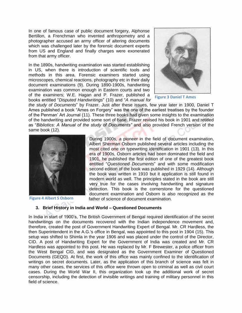
In one of famous case of public document forgery, Alphonse Bertillon, a Frenchman who invented anthropometry and a photographer accused an army officer of altering documents which was challenged later by the forensic document experts from US and England and finally charges were exonerated from that army officer.
In the 1890s, handwriting examination was started establishing in US, when there is introduction of scientific tools and methods in this area. Forensic examiners started using microscopes, chemical reactions, photography etc in their daily document examinations (9). During 1890-1900s, handwriting examination was common enough in Eastern courts and two of the examiners; W.E. Hagan and P. Frazer, published a books entitled “Disputed Handwritings” (10) and “A manual for the study of Documents” by Frazer. Just after these issues, few year later in 1900, Daniel T Ames published a book “Ames on Forgery” was the one of the earliest treatises by the founder of the Penman’ Art Journal (11). These three books had given some insights to the examination of the handwriting and provided some sort of base. Frazer revised his book in 1901 and retitled as “Bibliotics: A Manual of the study of Documents” and also provided French version of the same book (12).
During 1900s, a pioneer in the field of document examination, Albert Sherman Osborn published several articles including the most cited one on typewriting identification in 1901 (13). In this era of 1900s, Osborn articles had been dominated the field and 1901, he published the first edition of one of the greatest book entitled “Questioned Documents” and with some modification second edition of the book was published in 1929 (14). Although the book was written in 1910 but it application is still found in modern world as well. The principles stated in the book are still very true for the cases involving handwriting and signature detection. This book is the cornerstone for the questioned document examination and Osborn is also recognized as the father of science of document examination.
3. Brief History in India and World – Questioned Documents
In India in start of 1900’s, The British Government of Bengal required identification of the secret handwritings on the documents recovered with the Indian independence movement and, therefore, created the post of Government Handwriting Expert of Bengal. Mr. CR Hardless, the then Superintendent in the A.G.'s office in Bengal, was appointed to this post in 1904 (15). This setup was shifted to Shimla in the year 1906 and was placed under the control of the Director, CID. A post of Handwriting Expert for the Government of India was created and Mr. CR Hardless was appointed to this post. He was replaced by Mr. F Brewester, a police officer from the West Bengal CID, and was designated as the Government Examiner of Questioned Documents (GEQD). At first, the work of this office was mainly confined to the identification of writings on secret documents. Later, as the application of this branch of science was felt in many other cases, the services of this office were thrown open to criminal as well as civil court cases. During the World War II, this organization took up the additional work of secret censorship, including the detection of invisible writings and training of military personnel in this field of science.
Figure 3 Daniel T Ames
Figure 4 Albert S Osborn

The comprehensive textbook “Questioned Documents” which covered almost all the aspects of handwriting and signature identification in detail and also explained other types of evidences on documents including pen, paper, ink, typewriting and alterations etc. Osborn was one of the legal document examiners in major cases and was concerned for any document work such as examination methods, new techniques employed, court presentation and legal matters related to documents. During his career lifetime, he had published many articles on legal issues and document examinations (2).
In 1923, one of the landmark case of Frye vs United States (293 F, 1013; DC Cir. 1923) provided standard known as Frye Standard or Frye test or general acceptance test. The District court of Colombia had rejected the validity of polygraph test as it did not had significant acceptance in scientific community and court provided guideline for admissibility of scientific evidences (16). These guidelines for the acceptance for document examinations expert testimony were also applicable. Another landmark case in Virginia (Adams vs Ristine), court raises questions on scope of cross examination of expert testimony, qualification of expert to give testimony and use of photograph as evidence. It provided certain rules and guidelines before testimony is accepted in court of law (17).
In lieu of all the examinations testimony, the first organization forensic science laboratory, “the Scientific Crime Detection Laboratory in Chicago, IL, was established in 1929. Initially, the lab was under private firm, but with the continuous efforts of Professor John H Wigmore, the laboratory was affiliated by School of Law of Northwestern University.
Osborn had long discussion meetings with other experts in the field to constitute an organization which can formally related questioned document examination. In 1942, American Society of Questioned Document Examiners was formally founded under Albert S Osborn was established. The ASQDE focused on educational fulfillment and annual gathering for the full participation in the program and the organization was considered primarily as a professional institution giving support to research in the field of document examination.
Another organization, The American Academy of Forensic Sciences was founded in 1950 (18) and the forensic document examination to be considered as a discipline to be included. In 1952, two examiners were included as a member and Ordway Hilton was designated as chairman and he took forward to make a active section. After second revision of Osborn’s book, Hilton tried to update through his writings “Scientific examination of Questioned Documents” in 1956 and after few years another book followed “Evidential Documents” by James V.P. Convay in 1959 (1).
Later in 1969, as revised version of Charles Scott’s “Photographic evidences” was published which mentioned the importance and suitability of photographic documentation in examinations in detail. Various important articles were published in Journal of Forensic Sciences and the Journal of Criminal Law. With the help of these and other foreign journals, common consensus would have made on the examinations of questioned documents.
For the certification of the document examiners and to improve quality of questioned document work, a national certification program was developed. Officially, The American Board of Forensic Document Examiners, Inc was organized for the first time in 1977 under the chairmanship of John J Harris for certifying questioned document examiners. The ABFDE stated two clear objectives of the board; first to maintain, establish and improve standards of qualification for those who are forensic document examiners and certifying applicants who match with guidelines and requirements by ABFDE (3).

Historically questioned document examination is a comprehensive profession, but so-called pseudo-experts (in palmistry and fortune-telling) were sometimes encouraged to call so, and even today, it suffers from a bit of identity crisis in that at least eight different, or related, areas can be identified:
Questioned Document Examiners - Analyzes questioned document of any source and is capable of more than just questions of authorship limited only by their access to laboratory equipment
Historical Dating - These is work involving the verification of age and worth of a document or object, sometimes done by a document examiner, and can get as complicated as Carbon-14 dating
Fraud Investigators - This is work that often overlaps with that of the document examiner and focuses on the money trail and criminal intent
Paper & Ink Specialists - These are public or private experts who date, type, source, and/or catalogue various types of paper, watermarks, ink, printing/copy/fax machines, computer cartridges, etc., using chemical methods
Forgery Specialists - These are public or private experts who analyze altered, obliterated, changed, or doctored documents and photos using infrared lighting, expensive spectrography equipment, or digital enhancement techniques
Handwriting Analysts - These are usually psychology experts who assess personality traits from handwriting samples, also called graphologists or graphoanalysts;
Forensic stylistics- Refers to the same purpose, but by looking at semantics, spelling, word choice, syntax, and phraseology.
Typewriting Analysts - These are experts on the origin, make, and model used in typewritten material
Computer Crime Investigators - This is an emerging group that relates to QDE through some common investigative and testimonial procedures.
4. Types of Questioned Documents Evidences
Questioned Document forgery is very common among the most reported crime in civil and criminal cases. The type of evidences varies from case to case and can be characterized into two major categories; Patent and Latent. Latent document evidences are those which are not visible to the naked eye and need external aid to detect or identified the things and patent are those which have individual characteristics and visibly identified.

Fig 5. Types of documents which are encountered in forensic departments.
In the Fig 5, we can see the types of documents encountered by the experts like a fingerprints on the paper or secret writings employed to convey messages or watermarks etc. The patent documents are majorly found in a forensic scenario such forgery found in bank cheques, forged signatures on wills, deeds or official documents, fabricated documents using computer applications etc. All such documents are deposited by the investigating agencies to examine for its authenticity.
5. Principles and Guidelines for the Questioned Documents
For the better understanding of the handwriting or signature identification, some major principles were constructed by the pioneers of the experts to address the problem. These principles are universally applied to all sorts of the writings in any languages. These principles are followed all over the world by most of questioned documents experts in legal cases to assist the court of law. We have discussed here principles in different categories to understand.
5.1 Principles of Identification
The basic principle behind the identification of handwriting is based on the idea that "people are all alike; people are all different". To be able to make a positive identification of a person, a document examiner must be able to observe the distinguishing individual features which separate one person from all others.
All handwritings are alike. If this were not so, we would not be able to read another person's handwriting. All handwriting is different. If this were not so, we would not be able to identify the handwriting of close relatives and friends with whom we correspond (19).
5.2 Similarities and Differences
Any two writings in the same language and especially those based on the same writing system, naturally, have similarities and when closely examined, two writings by the same person show some natural and inevitable variation in letter form, which must be weighed, measured, noted and accounted for by the expert.

In the identification of handwriting all characteristics of both the questioned and specimen writings must be considered. Basic handwriting habits common to both must agree if all are the work of the same writer. Any single fundamental dissimilarity between two samples of writing is a strong indication of two writers, unless this divergence can be logically accounted for.
Several repeated fundamental dissimilarities establish that two writings are not the work of a single person. Under no circumstances, however, can identity be established by one, two or even several unusual characteristics. Rather, if two writings have been produced by one individual, there will be a combination of a sufficient number of similarities without any fundamental dissimilarity so that all chance of accidental co-incidence is excluded (1).
6. Class and Individual Characteristics
All the factors which identify handwriting fall into two general and somewhat overlapping groups - class and individual characteristics.
1. Class characteristics are those common to a number of writers and may result from such influences as the writing system studied, family associations, trade training, or foreign education as well as carelessness and haste in execution.
2. Individual characteristics are those which are highly personal or peculiar and unlikely to occur in combination in other instances (2).
All these individual characteristics are explained in the next chapters.
The Questioned document examination is now well established and demanding field in forensic science as it requires skills and extensive evaluation. As the rate of crime related to documents is far more than any other crime, need of document experts are much more required than any other forensic field. With the help of forensic document expert opinion on disputed documents, the court takes decisions on various criminal and civil cases.
Summary
Questioned document examination is very primitive science, used in pre-medieval periods and also one of the most demanding science around the world. The forensic document examiners examine all the questioned documents whose authenticity are challenged. Starting from the Egyptian rule and till date, history had been extensively filled with documents, presentations and their examination prove its authenticity. This module brief discusses about all the major historical development in the field of forensic questioned document and its examination. Documents are also categorized into different types on the basis of their use. Some of the important involved in the identification of handwriting hab been briefly discussed with reference to the class and individual characteristics. Handwriting and its genuinity had been put in question many time in past history and people have tried to put efforts in identifying genuine among forged documents.

References
1. Hilton O. Scientific examination of questioned documents: CRC press; 1992. 2. Osborn AS. Questioned document: Boyd; 1946. 3. Kelly JS, Lindblom BS. Scientific examination of questioned documents: CRC press; 2006. 4. Gardiner AH. Egypt of the Pharaohs: an Introduction: Oxford University Press on Demand; 1964. 5. Webster G. The Roman Imperial Army of the first and second centuries AD: University of Oklahoma Press; 1998. 6. Haighton A. Roman methods of authentication in the first two centuries AD. Journal of the Society of Archivists. 2010;31(1):29-49. 7. Vastrick TW. Forensic Handwriting Comparison Examination in the Courtroom. The Judges' Journal. 2015;54(3):32. 8. Fairman C. History of the Supreme Court of the United States. 1971. 9. Hilton O. History of questioned document examination in the United States. Journal of Forensic Science. 1979;24(4):890-7. 10. Hagan WE. Disputed Handwriting. Banks & Brooks, Albany. 1894. 11. Ames DT. Ames on Forgery: Boston Book Company; 1901. 12. Abt J. Objectifying the book: the impact of science on books and manuscripts. ILLINOIS. 1987:23. 13. Crown DA. Landmarks in Typewriting Identification. J Crim L Criminology & Police Sci. 1967;58:105. 14. Osborn A. Questioned Documents Boyd Printing Company. Inc, New York. 1929. 15. Tewari R, Ravikumar K. History and development of forensic science in India. Journal of postgraduate medicine. 2000;46(4):303. 16. Behringer JW. Introduction. Jurimetrics J. 1985;26:237. 17. Giannelli PC. The admissibility of novel scientific evidence: Frye v. United States, a half-century later. Columbia Law Review. 1980;80(6):1197-250. 18. Eckert WG. Introduction to forensic sciences: CRC press; 1996. 19. Saks MJ, VanderHaar H. On the" general acceptance" of handwriting identification principles. Journal of forensic sciences. 2005;50(1):119-26.

Criminology
Forensic Science and Forensic Medicine
Forgery in Documents

Component-I (A) - Personal Detail Role Name Affiliation
Principal Investigator Dr.G.S.Bajpai Registrar, National Law University Delhi
Paper Coordinator, if any Dr. Sally Lukose
Content Writer/Author (CW) Dr. Amitabh Biswas
Assistant Prof., Division of Forensic Science, Galgotias University, Greater Noida, Uttar Pradesh
Content Reviewer (CR) Dr. Ali Raza Associate Prof., Arba Minch University, Ethopia
Language Editor (LE)
Component-I (B) Description of Module:
Items Description of Module
Subject Name Criminology
Paper Name Forgery in Documents
Module Name/Title Module Id Pre-requisites Objectives Keywords
Table of Contents

Module 22: Forgery in Documents 1. Introduction
Every individual at one time or another was asked to sign a document rather than merely putting a fingerprint to the paper as a method of identification. But why signature had replaced fingerprints? It would be simple to just place a our fingerprint on a cheque instead of a payer‟s signature? It may also prevent forgery. Yes or No? The answer behind the use of signature is that it is easier to read is illogical, as lots of signatures have evolved until, they are symbolic illustrations of ancient handwriting and are now illegible. The answer to this question lies in the word “intent.” By signing a document, the signatory are entailing intent on his/her part to consent with consequences provided by that cheque, postscript, agreement, contract, etc. One could intentionally and easily put the fingerprints of somebody recently dead or unconscious upon a document, if the fingerprint was only required for authentication. This does not assume, though, that the placing an inked thumbprint next to a payer‟s signature on a cheque, about to be consulted at a cheque cashing counter in a bank, would not be a help out. The fingerprint‟s universal implication would certainly, at the very least, be a prevention to the individual intent upon passing a forged instrument. Handwriting and signatures are prone to be forged, over the years, many ways of forgeries had been employed to defy the concern person by the forger. Signatures are very common means of authentication in terms of banks transactions, cheques are vulnerable to be forged by imitating or copying signature of victim. These forgeries are needed to be addressed and what are the ways to identify these forgeries need to be discussed. Learning Outcome In this module, we are going to learn several points which are mentioned below:
What is a forgery? Types of forgeries which are classified as per execution.
Different characteristics of forged signature or handwriting, which identify the forge documents.
Who should examine these forged documents on the basis of qualification and expertise.
2. Forgery: Defnition and characteristics

What do we understand with the term „forgery‟? When the documents are called forged in the law of forgery? To understand forgery, we should well versed with the definition of forgery of documents in the legal term. “Forgery” in a stringent way is a legal term and its use as a result should most likely be avoided by the questioned document examiner. There have been proposed four characteristics for making a false document which need a attention:- Making a false document by (1) Signing a fictitious name; (2) Passing off an otherwise true document by false representations dehors the instruments (3) Signing one's own name; (4) Writing a false address or description to a true signature (1). Legally forgery is well defined in our criminal justice system. An underlying intent to defraud, based on knowledge of the false nature of the instrument, must accompany the act. Forgery is defined in our Indian Penal Code (IPC) under section 464 which states that; Making a false document. — [A person is said to make a false document or false electronic record— First —Who dishonestly or fradulently—(a) makes, signs, seals or executes a document or part of a document; (b) makes or transmits any electronic record or part of any electronic record; (c) affixes any 342 [electronic signature] on any electronic record; (d) makes any mark denoting the execution of a document or the authenticity of the electronic signature.(2) Tools of forgery may include bills of exchange, bills of lending, any notes of promise, bank cheques, legal bonds, cash or payment receipts, money order or order of goods, mortgages, discharges of mortgages, deeds, public records, account books, and certain kinds of tickets or passes for transportation or events. The compositions of forgery are Making false documents, liability to legal systems, forger identification and defraud intention. An apt fraud requires superior to anything normal dexterity and a lot of practice. Creative ability helps, as well. Penmanship is an exceedingly complex neuromuscular action that requires coordination between the hand, arm, and fingers, and also the eyes. When realistic development has been come to – at the end of the day, once a kid has figured out how to compose – composing turns into a characteristic demonstration, obtained after some time. It is no more important to stop and consider every stroke and how to take care of business.Unless there is a physiological or psychological problem, genuine handwriting is usually smooth, spontaneous, and free-flowing, with less focus on the movement and more on the content (3). The counterfeiter, then again, is compelled to keep up steady control over the pen, focusing eagerly on every moment point of interest. Under the magnifying instrument, the written work line will demonstrate successive stops and falterings, and the tight hold the counterfeiter must keep up on the pen, thusly, creates heavier weight on the paper. When he achieves the end of the mark, the counterfeiter has typically neglected to adhere to the casualty's

style and his own particular characteristic style creeps in. Subsequently, the penmanship inspector gives careful consideration to the finishes of letters, words, and lines, as opposed to the beginnings.
3. Types of forgeries in Questioned Documents
3.1 Simulation With regards to falsification, one size does not fit all. There are different types, the most common type of which is the simulation. Simulation is a type of forgery that involves the perpetrator's use of a model of the victim's handwriting. The falsifier deliberately watches the genuine, and after that makes duplicates of the individual's mark to work on imitating it. As indicated by penmanship master Sheila Lowe, the falsifier pays consideration on the way capital letters look, drawing instead of composing the signature. His slip-up, in any case, is neglecting to notice such attributes as the spaces amongst words and letters, and the upper and lower lengths and the arrangement of the letters that makes an individual's penmanship novel. Counterfeiter endeavors to duplicate the pictorial attributes – the way the composition looks – with a specific end goal to make it as near the bonafide signature as could be expected under the circumstances. This is a significantly more burdensome assignment than it may sound. Attempting to duplicate another's writings or signature is likened to emulating the way somebody strolls or talks (any individual who supposes this is simple ought to attempt it!). Counterfeiter neglects to go to, yet what the report inspector takes a gander at, is the measure of space left amongst words and letters, the extents of the upper and lower lengths, the arrangement, and other unwittingly rendered qualities (4). Various techniques are accessible for a counterfeiter to use in the development of a signature that may appear to the laypersons as real. These incorporate free-hand reenactment, following, and propagation by an electrostatic copier or PC. Free-hand recreation with a model accessible starts with the falsifier putting the original signature close to the paper on which the forged signature will be set. At that point, utilizing pen or pencil, the counterfeiter draws a photo of the authentic signature. Under amplification, such reproductions may uncover various pen stops and lifts or indications of rectification, alongside signs of a moderate, tremulous line quality. Less aesthetic counterfeiters regularly utilize tracings by different intends to deliver recreated marks (5). 3.2 Tracing Another type of forgery is the tracing. Tracing is very crude method of forging the signature or handwriting. At the end of the day, the counterfeiter has a model of the honest to goodness genuine signature, which he may hold against a window, or use carbon paper or a light box, and place another sheet of paper over the top, and actually follow the line. Under the process of act, numerous begins and stops the counterfeiter gains as he checks his ground – called resting dots – which are promptly seen. Likewise, the forged written work is slower, and once in a while there is a indentation in the paper, which can be seen close by the ink line (6).

Fig 1. Classification of different types of tracing forgery Tracing forgery is classified on the basis of the method of execution (Fig 1). Traced Forgeries display the rules which are present underneath the signatures. These rules might be as indentations or carbon outlines and ink strokes will be discovered holding fast to the rules. Direct tracing is the method where the forger uses direct original to trace in signature in forged documents without using any aid, whereas when a transmitted light is used beneath the document for the tracing are classified as transmitted forgery. Another way is to use carbon paper, carbon print is taken to forge document from original and carbon is covered up by ink using pine. By using tracing paper or butter paper, original signatures are traced by indentation and then writing it with an ink pen to cover the indented part made to document. These classifications of tracing forgeries provided the execution style, which is necessary to understand the nature of forgery. Knowledge of these methods of forgery are very important to identify these forgeries (7). 3.3 Cut and Paste Forgery The cut-and-paste forgery is closely as the term suggests. A real signature is cut from one report and set on the spurious record, then photocopied. In the event that the lighting and determination is legitimately balanced, the record will seem genuine. Since one of the principle on which penmanship examination rests is that it is difficult to compose a signature 100% the same way twice, the simplest approach to demonstrate a cut and paste falsification is to find the report from which the name was copied. They will be indistinguishable, or to a great degree close if the counterfeiter is sufficiently astute to change some minor points of interest, for example, the last strokes. Indictment depends on the law implementation criminologist's capacity to locate the original signed document from where it had been copied, and analyze the suspect's penmanship on the false to that of the first. Since no two parts of penmanship are indistinguishable, the counterfeiter abandons himself open to conviction by overlooking a couple of minor points of interest, for example, the way his casualty works out her numbers (7, 8). 3.4 Electronic Forgery Electronic forgery is also closely related to the cut-and-paste forgery. In the advanced computer era, the scan and-drop strategy has developed. The PC gives the counterfeiter a strong weapon. The falsifier basically digitizes a genuine signature by high resolution scanning, then embeds it into the spurious archive and prints it. Under the magnifying lens, in any case, the pixelation uncovers that it has been digitized (9).

3.5 Freehand Forgery This is another kind of fabrication where the falsifier essentially composes the another's name without making any effort to duplicate. He basically signs a structure in his own particular penmanship, now and again picking wording or stating of his own. The Fraud and Forgery Division of the forensic science labs is said to have many illustrations where the counterfeiter has kept in touch with some made-up expression, camouflaged as a signature. As per Lowe, this is the most effortless kind of signature for a falsifier to attempt. It will diminish his odds of arraignment, since it is more troublesome for analysts to peruse the two penmanship tests with a specific end goal to make a positive correlation (3, 10).
4. General indications of non-genuineness may include the following:
4.1 Blunt beginings and endings: The falsifier puts the pen point in contact with the paper, and afterward begins composing. When he is done with the
name or some bit thereof, he stops the pen and lifts it from the surface (Fig 2). This may bring about an underscored blunt start or completion where the pen was set in contact with the surface. Now this contact is held so long that if the pen contains a liquid ink it will wet the paper and relocate outward from the contact indicate or even through the back of the paper. There might be pointless and incidental imprints brought on by pen begins and stops. The author may choose subsequent to put his pen in contact with the paper, that it is in the wrong
spot, lifts it up and moves it to a position considered more right. Regularly a mark's begins and stops are a great
deal more dynamic. The pen is moving on a level plane before it contacts the paper and is lifted toward the end while still in flight. This leaves a decreased appearance at the beginnings and endings of names or letters. There are, obviously, special cases for this (11). 4.2 Unnatural Pen lifts and unusual hesitation: Sometimes, when the pen stops at an uncommon point in the composition of matter; maybe where a radical alter in the course is going to happen or another letter development is going to be begun. This may look like presence of little gap in the composed line where one is not expected, or a covering of two ink lines where there
Figure 2 Signs of Forgery shown with the arrow marks.

ought to be one and only consistent line. The pen lifts and hesitation point out the signs of forgery in the document (7). 4.3 Forger’s Tremor: Tremors may occur in writing or signature due to old age, or some illness or poor skill in writing (12), it may look similar to forgery, either by tracing or simulated. Since the formation of most types of n fake signatures is more similar to the drawings, the pen moves so gradually that little, now and then microscopic variations in course occur in what ought to be a liquid-looking line. The resultant line is not smooth, but rather look like the "shaking" pen. For the identifying forger‟s tremor two ways are basically employed. First is by elimination, elimination of any other doubts by analyzing the samples from the original writer or admitted writings during that time of forgery (2, 13). 4.4 Speed and pressure: Ordinary penmanship is uninhibitedly and quickly executed and in this way typically described, particularly, when a nib-pen is utilized, by light and hairline upstrokes and shaded or substantial down strokes. Although ball point or felt tip pens have a tendency to minimize this difference between strokes, it is still a big element in composing. Again in forged writings, in light of the fact that the pen is moving gradually instead of with the dynamic movement connected with most authentic compositions, the ink line stays steady in thickness because of the same consistent pressure applied to a gradually moving pen. There will be less, assuming any, decreasing of inner lines (14). 4.5 Patching: Occasionally, however, at some time, a most of us have made a mistake while composing our own signature. A few people may allow the signature to sign unbothered, thinking minimal about the oversight or flaw, while others will essentially "alter" the signature by adjusting the culpable part. This might be done keeping in mind the end goal to make the signature more discernable, or in light of the fact that an imperfection in the pen or paper has influenced what we see to be our "ordinary" signature, or for some other reason that may even be intuitive. These "fixes" are generally patent, with no endeavor made with respect to the author to veil or generally shroud the redress (7). These signature amendments are not exactly the same as the settling that is frequently found in non-authentic signatures. On these occasions, the creator is not attempting to make the signature more clear, but instead to appear satisfactory. He is changing an obvious blemish that he sees as discernible, and might uncover his forged thing and foil his arrangement. These generally show up as a change in accordance with a blemish in the structure line rather than as a letter. Enlargements to section or terminal strokes, or to cut down jumping bits of letters, nearby amendments to embellishments, are ordinary of non-true blue settling (15). 4.6 Carbon Outlines and Indentations As showed before, traced signatures may hold evidence of their technique for generation, for example, carbon or graphite follows or out-of-line indentations. Forger tries to eradicate carbon-paper or graphite imprints may yet leave hints of those substances: if not, confirmation of the deletion itself might be

distinguished by different means. As well, demonstration of erasing may harm or dull the signature or other traced composing, and this might be seen by microscopic perception. Infrared photography will infiltrate numerous overlying inks and along these lines uncover the carbon following (carbon being dark to infrared). Diagonal or oblique lighting may improve the indents visualization and show that they don't precisely relate with the inked line of composing (16). There are times when some of these same forgery indicators will be displayed in genuine signatures. Aged or informed writers will frequently display similar patterns. The mere presence of these indicators does not mean that the signature under scrutiny is non-genuine, but should contribute to the overall determination as to genuineness. Alternately, the signature devoid of these indications may not be assumed to be genuine. The signature of an elderly individual may, for instance, be expected to contain tremor and hesitation. If, however, the questioned signature appears to be written in a fluid manner and/or on a higher skill level than what is expected, the red warning flag should be waving. This occurrence may itself be indicative of non-genuineness. Often, a forger, because of an inherent high skill level in his writing, may produce a product that contains fewer indications of forgery than a counterpart with a lower skill-level (3).
5. Selecting a Handwriting Expert
At the point when the help of a penmanship master is required to reveal reality about the realness a signature (or other penmanship), the legitimate consultants or legal advisors is all around encouraged to choose the experts with consideration, as there is no proper licensing in the field of penmanship examination except for the government document examiners. In this field, as in others, there are the individuals who have earned themselves the notoriety of an enlisted firearm. To maintain a strategic distance from potential issues, the experts ought to be a present part on favorable terms of a legitimate penmanship examination association (not only an extortion analysts association, for example, the National Association of Document Examiners or Association of Forensic Document Examiners, for instance or working with presumed scientific science research facility with great scholastic and useful foundation. Summary Forgery of documents is common now a days, signature and handwriting forgery are need to be tackled in more technical and scientific ways. Any deliberate attempt to alter or modify the documents by adding or editing the marks, symbol or other written material is termed as a forgery. Forgery is categorized in different ways on the basis of the method used for the forgery. From crude to technical forgery are considered for the examination by the document forensic examiners. Tracing and simulation are the common form forgeries identified and examined. There are well known characteristics of these forgeries which are found in the cases. These features of non-genuiness should be analyzed by the document specialist to address the problems. Slow speed, blunt starts and stops, unusual tremors, evenness in

pressure, carbon outline or indentation in terms of tracing forgeries are some of the major identification features for the forged signatures or handwritings. For identification of these forgeries, a document experts are needed. The document expert should be specialist and known with the facts and procedure for the conduction of examination. Forensic document examiners had gained their knowledge and expertise through proper channel and licensed to practice.

MODULE 23

Component-I (A) - Personal Detail
Role Name Affiliation
Principal Investigator Dr.G.S.Bajpai Registrar, National Law University Delhi
Paper Coordinator, if any Dr. Sally Lukose
Content Writer/Author (CW) Dr. Amitabh Biswas
Assistant Prof., Division of Forensic Science, Galgotias University, Greater Noida, Uttar Pradesh
Content Reviewer (CR) Dr. Ali Raza Associate Prof., Division of Forensic Science, Arba Minch University, Ethopia
Language Editor (LE) NA NA

Component-I (B) Description of Module:
Items Description of Module
Subject Name Criminology
Paper Name Handwriting examination: General and individual characteristics and rules for handwriting examination

Module - 23
Handwriting examination: General and individual characteristics and rules for handwriting examination
1. Introduction
Handwriting or Penmanship is a procured aptitude and unmistakably one that is a complex perceptual-engine undertaking, now and then alluded to as a neuromuscular errand (Hilton, 1992). Talented writing movements are so common nowadays that person may ignore their multifaceted nature. Without distortion, be that as it may, writing is a standout amongst the most progressive accomplishments of the human hand. Handwriting is a yield of adjusted coordination between hand muscles and mind. Three districts regions of mind plays vital parts in writing; a first primary motor area which controls over fine developments; second pre-motor region which offers control to our visual direction and third is supplementary region which manages muscle and sequencing of terminating. The hand is a to a great degree intricate and sensitive component, containing approximately 27 bones controlled by more than 40 muscles (Marquardt and Mai, 1994; Wing, 2000). The majority of the muscles are arranged in the lower arm and interface with the fingers by an unpredictable arrangement of ligaments. Their capacity in controlling a written work instrument is exactly organized by a planning framework under a neural control of developments of the arm, the hand, and the fingers. The exact requesting and timing of the developments decides the structure of the example that is recorded by the pen or pencil (Osborn, 1946).
The development of writing is complex because it is, in part, culture dependent, and cultures differ with locales and undergo constant change. The evidence of this dependence is manifest in class, system, or national characteristics. Writing is a continuous or flowing task, not one of discrete or separated actions. There are apparent interruptions at word boundaries, but in many cases the pen movement may be continuous and uninterrupted, although not recorded as an inked line (Luria, 1977).

Learning Outcome
From this module a student will learn:-
Definition of handwriting and handwriting examination. Importance of handwriting in forensics
Examination of handwritings and signatures.
Priniciples involved in handwriting identification
General and class characteristics of handwriting identification.
Characteristics of forged handwriting or signatures

2. Why handwriting is important?
Writing has a very long history. It began as simple pictographs drawn on a rock, which were then combined to represent ideas and developed into more abstract symbols. Just like our writing today, early symbols were used to store information and communicate it to others. In recent years, modern technology has dramatically changed the way we communicate through writing. However, despite the increased use of computers for writing, the skill of handwriting remains important in education, employment and in everyday life.
Time devoted to the teaching and learning of letter formation in the early years will pay off. Legible writing that can be produced comfortably, at speed and with little conscious effort allows a child to attend to the higher-level aspects of writing composition and content. This is important when assessments are based on written work, particularly in time-limited written examinations, which remain as a major form of assessment for many formal qualifications. Without fast and legible handwriting, students may miss out on learning opportunities and under-achieve academically (Feder and Majnemer, 2007).
3. Principles involved in handwriting identification
Forensic document examiners often deal with questions of document authenticity. To determine whether a document is genuine, an examiner may attempt to confirm who created the document, determine the time frame in which it was created, identify the materials used in its preparation or uncover modifications to the original text.
I. All other things being equal, the significance of any combination of characteristics, qualities, and features as evidence to establish the identity of a writer is (Koppenhaver, 2007):
A. Directly proportional to the speed, spontaneity, and naturalness with which the writing is written, and
B. The frequency of occurrence in random writings of the combination of those particular characteristics, qualities, and features of the writing that when considered collectively making it identifiable.
II. Given a sufficient amount of handwriting, no two skilled writers exhibit identical handwriting features. The examined writing, questioned and known, must be suitable and sufficient in quantity and quality to conduct a meaningful examination and comparison. In some instances, either the questioned or known writing is so limited or lacks sufficient individuality that a meaningful examination and comparison to determine authorship is not possible. Some letters and letter combinations, written singularly or in a limited combination, such as “eo,” “ee,” “ion,” a name like “Lee,” consists of repetitive garland or arcade movements that are supposed to represent letters and letter combinations, or numerals, such as a “1,” may not contain sufficient individuality for a meaningful examination and comparison and determine their authorship. Initials are frequently unidentifiable because by themselves, they may not be sufficient to identify their writer. When any of these factors are present, either singularly or in some combination (Huber and Headrick, 1999):
A. It may not be possible to reach any conclusion concerning the identity of the writer of the writing. In other words, the examiner begins the examination and comparison process from a position of neutrality and the observable evidence in the writing is not sufficient or significant enough to move him from that starting position.
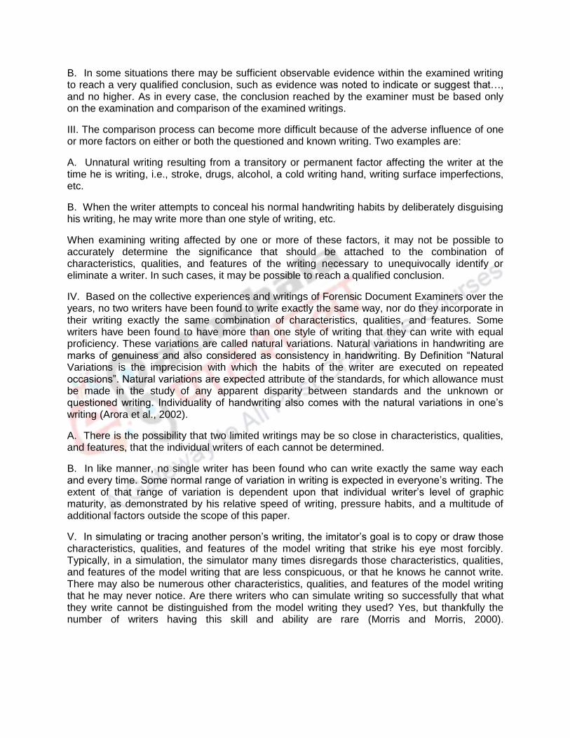
B. In some situations there may be sufficient observable evidence within the examined writing to reach a very qualified conclusion, such as evidence was noted to indicate or suggest that…, and no higher. As in every case, the conclusion reached by the examiner must be based only on the examination and comparison of the examined writings.
III. The comparison process can become more difficult because of the adverse influence of one or more factors on either or both the questioned and known writing. Two examples are:
A. Unnatural writing resulting from a transitory or permanent factor affecting the writer at the time he is writing, i.e., stroke, drugs, alcohol, a cold writing hand, writing surface imperfections, etc.
B. When the writer attempts to conceal his normal handwriting habits by deliberately disguising his writing, he may write more than one style of writing, etc.
When examining writing affected by one or more of these factors, it may not be possible to accurately determine the significance that should be attached to the combination of characteristics, qualities, and features of the writing necessary to unequivocally identify or eliminate a writer. In such cases, it may be possible to reach a qualified conclusion.
IV. Based on the collective experiences and writings of Forensic Document Examiners over the years, no two writers have been found to write exactly the same way, nor do they incorporate in their writing exactly the same combination of characteristics, qualities, and features. Some writers have been found to have more than one style of writing that they can write with equal proficiency. These variations are called natural variations. Natural variations in handwriting are marks of genuiness and also considered as consistency in handwriting. By Definition “Natural Variations is the imprecision with which the habits of the writer are executed on repeated occasions”. Natural variations are expected attribute of the standards, for which allowance must be made in the study of any apparent disparity between standards and the unknown or questioned writing. Individuality of handwriting also comes with the natural variations in one’s writing (Arora et al., 2002).
A. There is the possibility that two limited writings may be so close in characteristics, qualities, and features, that the individual writers of each cannot be determined.
B. In like manner, no single writer has been found who can write exactly the same way each and every time. Some normal range of variation in writing is expected in everyone’s writing. The extent of that range of variation is dependent upon that individual writer’s level of graphic maturity, as demonstrated by his relative speed of writing, pressure habits, and a multitude of additional factors outside the scope of this paper.
V. In simulating or tracing another person’s writing, the imitator’s goal is to copy or draw those characteristics, qualities, and features of the model writing that strike his eye most forcibly. Typically, in a simulation, the simulator many times disregards those characteristics, qualities, and features of the model writing that are less conspicuous, or that he knows he cannot write. There may also be numerous other characteristics, qualities, and features of the model writing that he may never notice. Are there writers who can simulate writing so successfully that what they write cannot be distinguished from the model writing they used? Yes, but thankfully the number of writers having this skill and ability are rare (Morris and Morris, 2000).

4. Class Characteristics of handwriting
The class characteristics of handwriting are defined by number of times by different experts. Ordway Hilton defines it as “Not all characteristics encountered in document examination are peculiar to a single person or thing, and one that is common to a group may be described as class characteristics (Osborn, 1929). In a paper entitled, “Handwriting Identification for the Investigator”, the definition is, “Class characteristics belong to the system or style of writing the person learned. They may also be forms or features added to the letters by environmental or cultural influences (Morris and Morris, 2000).
Everyone’s writing consists of combination of “class” and “individual” characteristics. To what extent and in what combination is dependent upon the individual. This is one of the basic reasons why handwriting is identifiable.
Experts usually examine the following characteristics when examining handwriting samples:
1. Line quality: Do the letters flow or are they written with very intent strokes?
2. Spacing of words and letters: What is the average space between words and letters?
3. Ratio of height, width, and size of letters: Are the letters consistent in height, width, and size?
4. Lifting pen: Does the author lift his or her pen to stop writing a word and start a new word?
5. Connecting strokes: How are capital letters connected to lower-case letters?
6. Strokes to begin and end: Where does the letter begin and end on a page?
7. Unusual letter formation: Are any letters written with unusual slants or angles? Are some letters printed rather than in cursive?
8. Pen pressure: How much pen pressure is applied on upward and downward strokes?
9. Slant: Do letters slant to the left or right? If slant is pronounced, a protractor may be used to determine the degree.
10. Baseline habits: Does the author write on the line or does the writing go above or below the line?
11. Fancy writing habits; Are there any unusual curls or loops or unique styles?
12. Placement of diacritics: How does the author cross the t's or dot the i's?
It is virtually impossible to find any writer who can imitate exactly all of the characteristics, qualities, and features of another person’s handwriting while simultaneously suppressing his own. This does not mean the simulator can be identified as the writer of the simulation based on the presence of their handwriting characteristics in the simulation (Turnbull et al., 2010).
5. Individual Characteristics of handwriting
Handwritng is unique to each individual. Although some peoples’ handwriting may have similar styles and characteristics in common, acquired when these people learned to write by copying letters and words, they tend to take on individual styles with age. Also, as a person ages, their handwriting will show additional changes (Hilton, 1983).

Depending upon his success at simulating the model writing, he may incorporate some of his writing characteristics, qualities, and features in the attempted simulation. When this occurs, it may or may not be possible to reach some qualified conclusion concerning the probability of his writing the simulation. By definition, if the writing is a simulation, the writer is copying a model that is either in front of him at the time he is writing, or he is writing the simulation from memory. It is virtually impossible to find sufficient evidence in a simulation to identify the writer of the simulation (Morris and Morris, 2000).
The following is a universally accepted truth or axiom of handwriting and hand printing identification. The result or opinion rendered in all examinations and comparisons performed by any Forensic Document Examiner is based only on the evidence present in the actual document or documents examined, giving consideration to any factor(s) affecting the writer at the time of that writing.
All conclusions concerning authorship of writing on documents is based on the examination and consideration of the evidence on a specific, examined document. A conclusion reached based on the examination and comparison of a copy, photocopy, fax, microfilm, carbonless paper, etc., purportedly of an original document of whatever type and generation cannot be extrapolated back to the original the copy purports to represent.
It is not prudent, nor can it be justified to make the assumption that the examined copy is an accurate representation of an original document without comparing the copy to the original it purports to be a copy of. Rather than an original, the copied document may actually be a copy of a copy or even a fabricated document and no truly original document exist. There will always be evidence on the original document that will not be present on the copy. The examiner has an ethical obligation to advise the submitter of this fact.
There are several reasons why this is a universal truth or axiom. First, the process of photocopying and/or faxing documents introduces into the copies defects and distortion. Therefore, the writing on the copy cannot be exactly the same as the original document that is being copied. The most recent and accurate work in this area was done by Mr. Robert Gervais.1 His work demonstrates the introduction of defects and distortion in photocopies when the controlled original is written with blue and black ink, for both slow and rapid writing, and at different degrees of copy darkness, and different generations of the copy.
In some situations, the examined copy may actually be a first, second, third, or greater generation copy, all of which were produced on different photocopiers. In some instances, a photocopy and not an original was placed in the fax machine for transmission. In this situation, the problematic nature of the received fax is compounded. If the copy submitted for examination is a fax of a fax, the submitted document is even more problematic for a meaningful examination and comparison. In every case, the original document is the best evidence. A first generation photocopy of an original is second best (Hilton, 1988).
6. Handwriting & Signature Examination
When there's a suspect in a crime and the evidence includes a handwritten note, investigators may call in handwriting experts to see if there's a match. In some cases, it might be the one piece of evidence that gets a suspect charged and eventually convicted. But what if it's a false match? How exactly do experts go about analyzing someone's handwriting?

In handwriting analysis, Original documents must be maintained for evidence. Handle original documents as little as possible. Keep them protected in archival sleeves or folders. Never work with original documents. Always make copies to work with (Osborn, 1946).
First of all, examine the class characteristics of handwriting as discussed in previous topic. Like examine the spacing between letters and words. Use a ruler to measure typical spacing, examine the relative height, width, and size of letters. Use a ruler to measure these for comparison, examine pen lifts and separations, some letters and combinations will be continued while others may not be connected, look at the beginning and ending strokes of words and letters and any connecting strokes, are there any unusual letter formations such as loops and curls or a mixture of cursive and printing of letters?, Is there any shading of letters due to uneven pressure applied in writing? Examine the slant of the letters. Do they slant left, right, or not at all? Are the slants consistent throughout the writing sample? Measure the angles of slant using a protractor. Examine the baseline habits. Are the words and letters on the baseline or are they above and below. A ruler will be helpful in determining this. Look for flourishes and embellishments. Using small circles to dot i’s or for periods, loops of capital letters or ending letters, etc. Are there any unusual letter formations? Look at the placement of diacritics. Are the i’s dotted and t’s crossed? Individuals tend to dot i’s and cross t’s in unique fashions.
Class characteristics as mentioned, thorough examination may rule out various important factors, before examination of individual characteristics and signs of forgery.
7. Signatures and their examination
Signatures serves as mean of identification and are typically used in the course of business to authorize financial transactions or even establish the legality of important documents. Sometimes, the addition of a signature can increase the value of an item, such is the case of celebrity memorabilia. As such, the high value items attached to such signatures make them likely targets of forgery as compared to handwriting.
In a typical signature case, detailed microscopic comparative examinations are made of a set of known ("specimen") signatures and one or more questioned signatures, the writer of which is in doubt. Signature forgery can occur in many ways, including freehand simulation, tracing and image transfer. Alternatively, a signature may be written with some disguise with a view to disavow it at a later time. Chance coincidence in the signatures of two persons is rare but may be possible if the signature is particularly simple. It must be noted that a finding that a signature is genuine does not establish the genuineness of the document as a whole as it may be the product of some form of document manipulation (Fielding et al., 2001).
Signatures can be disputed on all kinds of documents including Wills, financial documents, contracts, agreements, cheques, application forms and receipts. Signature verification may be applied to cases involving alleged cases. The authenticity of signatures on business documents such as contracts or agreements often forms the point of contention between two parties in a civil dispute. In such cases, a forensic handwriting expert will compare the signatures on the questioned documents with specimens to determine if the said person could have signed those documents.
Attempts to forge signatures usually occur in three ways:
Freehand simulation Tracing and Cut-and-paste: either physically or by electronic means using digital software.

FDS is also aware of the emerging use of electronic (biometric) signatures. We are familiar with certain technologies used to capture electronic signatures and the forensic analysis software required to interrogate them.
At times, there may also be deliberate acts of disguise by the writer, with the intention of denying the authorship of the signature afterwards. For examination of signatures, a forensic document examiner would have to consider the above possibilities and more. Our experienced document examiners are able to provide advice on the relevance of signature examination to your case and answer your queries.
8. Characteristics of Forged handwriting and signatures
Writing in forged documents tends to be slowly written and will show a lack of individuality. Letters tend to have an unnatural appearance as if the forger was drawing the letters. This makes letters inconsistent in the document, shows unnatural starts and stops and a general lack of rhythm to the writing. Any mistakes will show a careful correction. Signatures will be identical.
8.1 Disguised Writing
If a suspect attempts to disguise their writing, they will generally exhibit inconsistent slant and letter formations with a major change in the size of their letters. Capital letters will be different and they often will use block lettering. As they write, there will be a lack of rhythm, irregular spacing, and unnatural starts and stops. Occasionally they will add excessive ornamentation. Some individuals will try to write with the wrong hand. Signatures are the most frequent type of questioned writing encountered, although a person’s handwriting also frequently attracts attention in litigation. Forensic signature examination and forensic handwriting examination are both discussed below. The terms "examination" and "analysis" are often used interchangeably but, in the context of the work that FDS undertakes, their meaning is strictly scientific. Our examiners are forensic experts, not graphologists, and our work does not involve attempting to tell one's personality from their writing ("graphology") (Harris, 1952).
8.2 Normal Hand Forgery
During the creation of this class of non-genuine signature, the writer simply writes someone else’s name. There is no attempt made to duplicate or make the forgery look like a genuine signature. Any resemblance to the genuine signature is coincidental. Usually, the perpetrator of this signature does not have a model signature at hand and/or the skill level or forethought to attempt an emulation. If he does not attempt to impart disguise to the writing, the resultant product will display characteristics of the forger’s own handwriting. Armed with adequate standards of both the individual whose name is being used and exemplars of the suspect, the document examination may be definitive to the point that not only is the signature declared not genuine, but the forger is also identified (Herkt, 1986).
8.3 Simulation
The simulated signature, or “free hand forgery” as it is sometime known, is the usual bill of fare for the questioned document examiner. This forgery is constructed by using a genuine signature as a model. The forger generates an artistic reproduction of this model. Depending on his skill and amount of practice, the simulation may be quite good and bear remarkable pictorial similarity to the genuine signature.
Many simulations created with a model at hand will contain at least some of the general indicators of forgery, such as tremor, hesitation, pen lifts, blunt starts and stops, patching, and
static pressure. They will have a slow “drawn” appearance. The practiced simulation is most often a higher quality creation in that the model signature has been memorized and some of the movements used to produce it have become semi-automatic. This simulation can be written with a more natural fluid manner.
Both practiced and non-practiced simulations will still have notable shortcomings. The forger naturally puts his greatest effort into those parts of the name that he expects to fall under the greatest scrutiny. Although letter forms (especially the more prominent, large or beginning letters) may almost duplicate the genuine letters, proportions and height ratios will seldom be correct. Internal portions of the names (smaller, less prominent letters and pen movements) will usually display the greatest divergence from the correct form and movements found in the genuine signature.
8.4 Signature forged by tracing Methods
Traced forgeries are generally created by one of three methods: “transmitted light,” “carbon intermediate,” or “pressure indented image.” While tracings may not normally present much of a challenge to the document examiner trying to determine genuineness, the ability to identify the perpetrator is totally precluded. Tracing another’s signature, or for that matter another’s handwriting, is the paramount form of disguise.
Total agreement between the model and the questioned signature dictate that the questioned signature was a product of tracing. No two signatures or handwritings, even from the same person are ever totally duplicated (due to natural variation). Just as certainly, total agreement between two, three or more questioned signatures is adequate demonstrative proof of tracing. Of course, the document examiner faced with total agreement between a number of signatures must take care that the model signature (genuine signature) is not one of the signatures in question (Morris and Morris, 2000).
8.5 Transmitted Light Tracing
The transmitted light tracing is the simplest of the tracings to produce and the one most often encountered. The paper that is to receive the spurious signature is placed over a document bearing the genuine signature. These documents are then aligned so as to put the genuine signature directly under the selected location for the forgery. These two papers are then held up to a window or other light source, and the transmitted signature image is traced on the receiving document.
The indicators of a transmitted light tracing are similar to that of a simulation and the two are difficult to tell apart (unless the model for the tracing is located). Height ratios and proportions in the transmitted light tracing are generally right on the money, however. These two features are frequently incorrect in the simulation (Hilton, 1992).
8.6 Carbon-Medium Tracing
At times, a carbon-medium tracing is the method of choice, especially if the document to receive the tracing is too heavy a weight, such as cardboard, to allow for a good light transmitted image. The carbon tracing is crude method of tracing as it involves two steps; first tracing the writing with the help of the carbon paper and then retracing the drawn traced mark of carbon by pen. During the covering up the traced line, carbon deposition are seen around the periphery of the written line by pen. These are major signs of carbon tracing apart from slow writing speed, blunt start and ends, tremors etc.

Because of the almost non-existent use of this sensitized paper in modern day machine copying processes, most document examiners will likely never encounter this problem. However, on occasion a similar phenomenon can be found when NCR (National Cash Register – no carbon required) paper is employed (Huber and Headrick, 1999).
8.7 Pressure Indented Tracing
Similar to a carbon paper tracing, the indented line tracing is produced in essentially the same manner, but does not employ any intermediate reproduction medium. Heavier pressure is used when tracing over the model signature. This pressure leaves an indented “signature” on the receiving document. This is then covered over with a broad-tipped pen, although ballpoint is found on occasion.
Almost invariably, the writer misses portions of the indented line. This error may be easily observed using glancing (oblique) light. Other general indications of non-genuineness are similar to those found in simulated forgeries (Nickell, 2005).
9. Summary
Handwriting is the one of the primitive form of identification and mode of communication. It is developed from childhood with practice. Handwriting is an outcome from the process involving brain, hand and eye coordination. Through lifetime, a unique set of pattern is developed by the individual. There are some basic principles involved in handwriting examination like no two skill writers can have identical writing, if provided with sufficient writing and all the writings would not be same for an individual, it may vary from the same word to the letters which is referred as natural variations. On the basis of different class and individual characteristics, handwriting and signatures are identified and matched with the suspected or victims writing. The signatures are more prone to be forged during the course of financial transaction between two parties, the forensic experts utilize these identify features of handwriting, apply principles and distinguish between genuine and non-genuine writings. Handwriting and signatures identification and examination need expertise and proper knowledge to give as evidence in court of law.
References
Arora, H., Lee, S., Srihari, S.N., Cha, S.-H., 2002. Individuality of handwriting. Journal of forensic science 47, 1-17. Feder, K.P., Majnemer, A., 2007. Handwriting development, competency, and intervention. Developmental Medicine & Child Neurology 49, 312-317. Fielding, G., Gummadidala, K., Kam, M., Conn, R., 2001. Signature authentication by forensic document examiners. Journal of Forensic Science 46, 884-888. Harris, J.J., 1952. Disguised handwriting. J. Crim. L. Criminology & Police Sci. 43, 685. Herkt, A., 1986. Signature Disguise or Signature Forgery? Journal of the Forensic Science Society 26, 257-266. Hilton, O., 1983. How individual are personal writing habits? Journal of Forensic Science 28, 683-685. Hilton, O., 1988. The evolution of questioned document examination in the last 50 years. Journal of Forensic Science 33, 1310-1318. Hilton, O., 1992. Scientific examination of questioned documents. CRC press. Huber, R.A., Headrick, A.M., 1999. Handwriting identification: facts and fundamentals. CRC press. Koppenhaver, K.M., 2007. Principles of Handwriting. Forensic Document Examination: Principles and Practice, 7-25.

Luria, A.R., 1977. The development of writing in the child. Soviet Psychology 16, 65-114. Marquardt, C., Mai, N., 1994. A computational procedure for movement analysis in handwriting. Journal of neuroscience methods 52, 39-45. Morris, R., Morris, R.N., 2000. Forensic handwriting identification: fundamental concepts and principles. Academic press. Nickell, J., 2005. Detecting forgery: forensic investigation of documents. University Press of Kentucky. Osborn, A., 1929. Questioned Documents Boyd Printing Company. Inc., New York. Osborn, A.S., 1946. Questioned document. Boyd. Turnbull, S.J., Jones, A.E., Allen, M., 2010. Identification of the class characteristics in the handwriting of Polish people writing in English. Journal of forensic sciences 55, 1296-1303. Wing, A.M., 2000. Motor control: Mechanisms of motor equivalence in handwriting. Curr. Biol. 10, R245-R248.

MODULE 24

Component-I (A) - Personal Detail
Role Name Affiliation
Principal Investigator Dr.G.S.Bajpai Registrar, National Law University Delhi
Paper Coordinator, if any Dr. Sally Lukose
Content Writer/Author (CW) Dr. Amitabh Biswas
Assistant Prof., Division of Forensic Science, Galgotias University, Greater Noida, Uttar Pradesh
Content Reviewer (CR) Dr. Ali Raza Associate Prof., Division of Forensic Science, Arba Minch University, Ethopia
Language Editor (LE) NA NA

Component-I (B) Description of Module:
Items Description of Module
Subject Name Criminology
Paper Name Ink and paper analysis and techniques

Module – 24
Ink and paper analysis and techniques
1. Introduction
Document authentication is so important for any kind of trade commercially, security of border and law enforcement organizations that these kinds of examinations correspond to a discrete division of Forensic Science. Questioned document (QD) experts make use of an extensive range of techniques to aid in their examinations ranging from initial visual assessment tools to advanced instrumentation for chemical analysis experiments. Within the last 20 years, numerous potential new instruments of analyzing the physical and chemical structure of historical artifacts have been introduced. Rapid development in technology and modern techniques had unclear relative importance, especially to experts in fields where these analytical techniques recently been introduced (1).
The rational analysis of material form in manuscripts and printed books without the use of electronic aids has a much longer, if erratic, history. In systematic bibliography, for example, the study conducted by Carter and Pollard of the forgeries by Thomas Wise in 1934 was one of the initial attempts at using thorough physical evidence of paper material and lettering to display convincingly the falseness of documents (2). Their findings were well-designed in their sense and straightforwardness and provided a practical example of the worth of vigilant and methodical physical observation, a point of view that had in the past been neglected or even ignored in support of the document's substance.
The requirement of non-invasive forensic examinations of papers as well as writing and printing inks are very much needed. It is necessary to do so in matters where a question arises as to: whether a document has been altered; when an entry was written and/or a document created; the order in which entries were written; whether a document is genuine or counterfeit; recovering and deciphering faded/bleached/obliterated/erased entries; whether documents or entries thereon are original or reproduction; the method of production of a document; whether multiple documents have a common source; and so on. Therefore, critical analysis of the paper material and ink used is very important in many cases and can be useful in a court of law.

2. Learning Outcome
From this module, students will learn:
How paper and ink evidences plays role in forensic investigations
The paper analysis and method used for the analysis of paper fibres
Type of paper documents involved for the investigations purpose.
How ink analysis is useful in age estimation of the documents
Different techniques available for the paper and ink analysis

3. Evidence That May Be Examined
Questioned matter may consist of identity cards, agreements, titles and deeds, wills, seals, stamps, bank cheques and notes, handwritten communication, documents generated by machined (ex: fax machines, photocopiers and computer printers), currency and other electronic documents. In various conditions, graffiti and digital signatures may also be analyzed; however, the examinee should be responsive that the assessment of these types of evidence can be challenging.
Documents without visible, identifiable marks may consist of important evidence if they were below some other documents when the writing was carried out. Documents that were tattered or burned may also provide evidence if reconstructed by using proper techniques. Even writing instruments, rubber stamps, used envelopes and makes/models of office apparatus or equipments in the perpetrator’s custody may be taken into custody and analyzed by the investigator. Evidence could even be picked from the electronic signature file's metadata in digital documents, providing information such as who the person behind this and when the document was written (3).
3.1 Collecting the Known and Unknown
Forensic document experts must have known specimens for the comparison to the material in question. Known samples may come from any number of known sources, such as a particular ink or paper manufacturer or machine.
In cases concerning handwriting, samples are typically classified into two types: requested writing specimens and collected writing (Admitted) specimens. Investigator dictates a known writing material for the writer and writing obtained are called requested specimens. These specimens are carefully generated under controlled conditions, with the writer being closely observed. However, writings that were written by the suspect or victim prior to the examination are called collected writing specimens. Good quality basis of writing specimens may comprise items such as cancelled cheques, letters, diaries, signed receipts, medical records, real estate contracts, tax records or other signed legal documents (4).
4. Paper Examinations
Questioned document experts often are engaged to investigate a single page or many pages of paper upon which there are markings, handwriting, printing, copying and/or graphics. Preliminary examinations of questioned documents, especially paper entail testing the color, thickness, weight, weave pattern, and fiber examination consecutively to find the origin of the paper. Further analysis may consist of the spectrophotometric examination to determine ultraviolet characteristics and comparisons using instrumental assessment (5).
Through a side-by-side comparison from other sources of paper or matched up against a standard white paper, the color of a part or parts of the paper may be investigated. Along with a visual examination of all the sides of the paper, the expert should observe the internal color of the paper as well. By using a scalpel, scrape a paper carefully without disturbing other important elements and observe, as this may change during the processing of printing paper. Measuring calipers for paper or micrometers may be used to determine the paper thickness, while weight of paper may be determined by using a sensitive weighing scale. Forensic examiners also evaluate paper for internal or surface weave patterns. Surface weaves are unusual, however these are easily visualized with oblique lighting. Internal weaves are more frequently seen on paper and can be observed with the help of transmitting light. To finish, analysis of paper fiber is
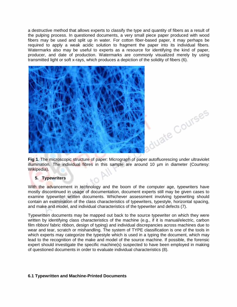
a destructive method that allows experts to classify the type and quantity of fibers as a result of the pulping process. In questioned documents, a very small piece paper produced with wood fibers may be used and split up in water. For cotton fiber-based paper, it may perhaps be required to apply a weak acidic solution to fragment the paper into its individual fibers. Watermarks also may be useful to experts as a resource for identifying the kind of paper, producer, and date of production. Watermarks are commonly visualized merely by using transmitted light or soft x-rays, which produces a depiction of the solidity of fibers (6).
Fig 1. The microscopic structure of paper: Micrograph of paper autofluorescing under ultraviolet illumination. The individual fibres in this sample are around 10 µm in diameter (Courtesy: Wikipedia).
5. Typewriters
With the advancement in technology and the boom of the computer age, typewriters have mostly discontinued in usage of documentation, document experts still may be given cases to examine typewriter written documents. Whichever assessment involving typewriting should contain an examination of the class characteristics of typewriters, typestyle, horizontal spacing, and make and model, and individual characteristics of the typewriter and defects (7).
Typewritten documents may be mapped out back to the source typewriter on which they were written by identifying class characteristics of the machine (e.g., if it is manual/electric, carbon film ribbon/ fabric ribbon, design of typing) and individual discrepancies across machines due to wear and tear, scratch or mishandling. The system of TYPE classification is one of the tools in which experts may categorize the typestyle which is used in a typing the document, which may lead to the recognition of the make and model of the source machine. If possible, the forensic expert should investigate the specific machine(s) suspected to have been employed in making of questioned documents in order to evaluate individual characteristics (8).
6.1 Typewritten and Machine-Printed Documents

Documents which are typewritten, printed, faxed, and photocopied are exposed to modification and extension, replacement of page, and cut and paste from original to fake documents. The quality of fax documents and photocopies is relatively not good, in comparison to the original document which makes these documents, particularly vulnerable to modifications or alterations for the purpose of fraudulent documents. Even though structural characteristics are frequent still apparent, slight details, such as hesitations, pen lifting, and retouching may not be replicated.
The primary step that is taken while examining any typewritten and printed documents is to establish the printing technology types involved in generating and printing the particular document. Secondly, it should be determined whether the document which is questioned is an original or a forged, and if there were numerous methods involved in the printing of the document (ex: a document which is printed that was again photocopied and/or faxed). When these factors are identified, the examiner may start looking for other factors such as typography, formatting, and copy deformation. Adobe Photoshop® or similar software might be used to evaluate line direction and spacing, which may be specifically useful to identify irregularities or alterations in the text.
If typewritten or machine-produced documents are in question, every page of the questioned document should be vigilantly compared to all of the other pages of the document, and, whenever possible, compared to samples which are collected from the source machine(s).
6.2 Photocopy and Facsimile (FAX) Machines
Questioned document (QD) examinations where facsimile (FAX) machines generated documents are involved often comprise queries regarding the genuiness of a replicated signature or writings, apprehensions around whether data or information has been changed or altered, added, or erased from a document, or queries about precisely when a fax was sent. Transmitted faxes are frequently in low resolution and printed likewise, compared to documents that are photocopied or printed (9). Consequently, fine details are not clear which need special attention during the examination of handwriting or typed writing may not be imitated. Efficient document examination in forensics may be hindered if there is an inadequate amount of material which are under question or if the quality of evidence is not good. It had been reported in previous research that during transmission faxes may experience considerable dimensional changes that can affect the size or even deformation, which further complicate the examination of transmitted questioned documents. Generally the poor quality of faxes are transmitted, they are more prone to modification, though, a careful examination should be done to look for any discrepancies in pixilation in an entire document, in addition to any changes in handwriting or print type, any spelling, punctuation or formatting of the documents (10).
Any document investigation in forensics comprising a fax machine also should take account of of the Transmit Terminal Identifier (TTI), which is basically found in the caption of any transmitted document. The TTI consist of data, such as the name of the sender and fax number of the sender and recipient, and the transmission date and time (11). These data may be very useful to the examiner as a way of recognizing the make and model of the source and recipient machine, and the date and time of transmission. Though, this data should be analyzed carefully, as the TTI is programmable, hence alteration is possible by a trained executor. Some of the fax machines also permit the user to readjust programs of some of the formatting features, which makes it further harder to correctly distinguish the source machine’s make and model.
To conclude, for any other technology, fax machines are vulnerable to built-up defects or defects obtained through regular use or abuse. These types of defects may happen in the source machine, the recipient machine, or in both, and must be distinguished from the background that may take place during communication. Consequently, when faxed documents are questioned for originality, both the source and receiving fax machines should be taken as evidence, and sample faxes from both machines transmitted around the date created by the TTI.
6. Computers and Printers
The progressively more ever-present utilization of computers ensuing in computer-generated documents has made more difficult for the document examiner to analyze these documents. Documents generated through computers are very vulnerable to modifications, such as addition of text, cut and paste, and even replacement of page. Consequently, investigators must think about the uniformity of the entire document in order to find any signs of modifications or tampering. For example, the investigator must establish whether numerous printers or other technologies were employed in making the document, in addition to examining any other possible irregularities. If any substitution of page is suspected, the writing or printing date also should be examined, as sequential discrepancies can be helpful in disgracing questioned documents.
When computer-generated document are encountered, the examiner initially tries to categorize the printing process by recognizing the technology used for printing. For example, the investigator establishes whether the text was printed in color or black-and-white ink, if ink or toner was used, and whether the process of printing was impacted or non-impacted. Examination of computer printouts should be carried out carefully which may permit investigators to recognizing the printer type which is used; it is likely that a number of overall mechanical deficiency exclusive to a printer discloses individualized imperfections that permit the printout to be identified back to the particular machine. Though, in contrast to typewriters, it is very hard to connect the document in question back to the particular computer on which it was typed (1).
In addition to identifying the printer’s type that generated the document in question, the typeface itself should be examined. Examination of the font and line spacing should be done, as discrepancies may be slight but pinpointing of a modification or addition to the genuine document. The forensic investigator will also analyze the printout to establish the type of paper and ink used which is common with other documents, as well as taking cautious note of any watermarks, and hole patterns by staple, etc. These recognizing characteristics do not always gives confirmative evidence that a document has been modified or tampered with, but in a number of cases may show convincingly that a document is not genuine. For example, in a case where a page is replaced in a property will that is printed on paper or with ink that was not existing at the time of the signing of the original (12).
7. Writing and Printing Inks
Usually ink analysis is important part of the investigation procedure, and ink analysis is the determination of specific composition of chemical compounds used for the manufacturing of ink by using wide variety of chemical and physical testing. Non-destructive methods for the examinations, such as visual analysis, examination under microscope, observing under ultraviolet and infrared lights are generally the primary step taken to categorize the type of ink used in a questioned document (13). Additionally, destructive method of analysis may also be required to differentiate among types of ink. Chromatographic technique, especially Liquid

chromatography may be performed to facilitate the determination of the chemical composition of the inks found in a questioned document. One of the few traditional destructive methods employed by forensic experts, a tiny piece of the paper in the document containing the ink is taken, dissolved in solvent to analyze the ink composition. Comparison of ink’s composition can be done with International Ink Library, more than 9,500 inks database that is preserved by the U.S. Secret Service (14). Chemical testing are essential in the assessment of printing inks because these tests also permit examiners to give an estimate of when the document was written. This may be meticulously prominent in the forgery cases; Ex: it may be ascertained that the ink used in a document is recently added as per its purported age.
Document experts are frequently asked not only to recognize the type of ink used in a questioned document, but also the origin of the ink. Identifying the ink’s source on a questioned document may be helpful for examiners to detect any fraud entries or alterations or omissions in a document. Ex: - If an entry in a contract/will may be added with another ink that may have similar color and texture, but will be chemically different from original writing on the rest of the document, which suggests that it was not present at the time period of its original writing when the document was made. Differentiating inks can be carried out initially by visual examination, and then non-destructive methods, and chemical methods, true identification of the precise source of the ink (i.e., the exact pen) is improbable. More probably, the assessor will be able to mark out the ink to the particular type, class, and or ink brand, and recommends that it is scientifically not matched from the suspected source. Spectroscopic examination by Infrared or ultraviolet rays gives different fluorescence patterns which can easily distinguish two different inks.
Fig 2. White light and Raman image of crossing ink (Courtesy : http://www.renishaw.com/en/forensics--7980).
8. Seals and Stamps Analysis
Official documents may consist of markings from rubber stamps, embossed seals, watermarks, or other mechanically-printed marks which symbolizes genuineness of documents. Classification of Stamps is done on the basis of the source of ink rather than the type of dye material. There are mainly four classifications of stamps: first the pre-inked stamp, the flat-dye stamp, the hand stamp, and the self-inking stamp (15). When seals and stamps are examined, it is vital that the forensic investigator initially identify that the marking(s) were certainly made by a seal or stamp, as not being generated by computer. Once this is established, the experts may analyze the specific features of the markings, basically identifying such defects that may be exclusive to an individual stamp. These defects may be made during manufacturing, such as defective die substance, deformation, or misalignment, or defects due to use (e.g., dirt, accumulated ink, or fibers) or misuse. The initial inspection of a impression of questioned stamp is to inspect it microscopically, during that time experts may be able to recognize a number of factors, including the ink source and condition of the ink die (16).
If possible, the expert should try to evaluate the questioned stamp impression that is suspected of making it. Before performing any method, it is essential to photograph the stamp impression before making any additional impressions so as to conserve its original nature (e.g., ink saturation, dirt, fibers). Many impressions from sample should then be taken with the suspected stamp, to understand angle variations and pressure with which the stamp is applied. A side-by-side comparison can then be carried out with the questioned impression, and results as to the source of the impression may be made (17).
9. Techniques and advancement for the paper and ink analysis
The examination of inks for investigations of counterfeiting, fraud, forgery, and other crimes, such as letters related to terrorist attacks is one of the most important forensic applications of advance techniques where precision is very much required. The terms thin-layer chromatography (TLC) and high-performance TLC (HPTLC) (18) will often be used indistinguishably to represent either or both techniques, because the elements discussed are very similar between them. These methods are the most widely used chromatographic techniques, for the analysis of inks. Inks of interest include those from fountain pens, ballpoint pens, roller ball pens, toner-based printers, inkjet printers, and solid ink printers. Forensic analysts look for the source and date of an ink and comparison with other inks. Major advantages of TLC in an ink analysis are ease of use and low cost, high resolution of the components of ink formulations, especially for HPTLC, and visual detection of the colored components allowing convenient and rapid comparison of different ink formulations and batches.
In 1954, Brown and Kirk reported an application of paper electrophoresis to analysis of ink initially and its comparison to paper chromatography. These scientista used a diethylbarbiturate buffer and an low-cost, commercially available electrophoresis apparatus for the partition of more than 50 blue, blue-black and black writing inks. They achieved chromatographic separation using horizontal paper chromatography and two solvent systems (19). In 1963, Crown explained construction of power sources for electrophoresis using commonly available electronic equipment, such as radios (20). Soon afterwards, Thompson included aluminum citrate or acetic acid–butyric acid–water electrophoretic buffers, describing results superior to electrophoretic separations obtained using weak acidic or basic buffers or paper chromatography. Electrophoresis have not attracted as much attention as the various chromatographic techniques and many researchers consider it inferior to chromatography for ink separations.

Then Capillary electrophoresis, which is new analytical techniques and proved its importance in may forensic purposes and has many advantages. These advantages include high resolution, ability to quantitate, and short analysis time. The novelty of the method prompts a concise review of its operation and capabilities. Most importantly, CE has extraordinarily small sample requirements and it is possible to enact a separation with nanogram (or even picogram) quantities. This is advantageous in minimizing the destruction of the document being tested (21).
As a non-destructive analytical method, Raman spectroscopy often provides insufficient information to identify or differentiate the ink used for the preparation of a questioned document. Inks were successfully classified based on the total number of prominent bands in Raman spectra. It was found that more than 90% of the samples of the same type and color could be differentiated visually using only Raman spectra i.e. 94-95% for blue and black inks, respectively (22). As a result of this study, a flow chart has been constructed for blue and black ballpoint pen inks allowing their systematic identification. Raman spectroscopy proved to be a fast and precise technique for forensic ink analysis (23). Raman spectroscopy in conjugation with nanotechnology is also employed for the identification and detection of even very small quantity of ink which is quite common in forensics (24). Photocopy papers are identified using Fourier transform infrared spectroscopy (FTIR) in real quick time. This method is fast, sensitive and veracious identifying similarity in two photocopy papers (25). With these advancements in technologies, examination of the paper and ink had becomes more easy and reliable which helps experts to come to the definite opnion for the case .
Utlisation of advance techniques made the ink and paper analysis more promising and evidences from these techniques are quite strong then the conventional techniques. These should be employed in regular basis in the forensic labs for the betterment of evidence procurement.
10. Summary
Paper and ink are the integral part of the any document. Forgery of documents generally involve pen and material on which it was written, paper is commonly used material for the documents. Analysis of the paper and ink is very much essential in determination of possible forgery, age of the document, any crossing over of pen marks, involvement of two pens in single documents, addition or deletion of new pages in previous documents. Paper analysis is generally conducted to match the paper source and differentiate between to paper material. Analysis involves weighing, measing thickness and uevaluate paper for internal or surface weave patterns. Surface weaves are unusual, however, these are easily visualized with oblique lighting. Whereas Ink analysis is equally important in forensic point of view. Ink analysis is done to differentiate two inks by using different physical and chemical methods. Now a days, more advance techniques are available for the identification and detection of ink and paper. The senstitvity of these is quite high, even a minute quantity of ink may be detected by using these techniques. The paper and ink analysis are vey important and should be done by using appropriate advance techniques for the evaluation of forensic cases.

References
1. Kelly JS, Lindblom BS. Scientific examination of questioned documents: CRC press; 2006. 2. Woodward D. The analysis of paper and ink in early maps. Library trends. 1987;36:85-107. 3. Hilton O. The evolution of questioned document examination in the last 50 years. Journal of Forensic Science. 1988;33(6):1310-8. 4. Osborn AS. Questioned document: Boyd; 1946. 5. Browning BL. Analysis of paper: Dekker; 1977. 6. Robertson JR, Roux C, Wiggins K. Forensic examination of fibres: CRC Press; 2002. 7. Mally W. The Identification of Typewriters. Kriminalistik; 1956. 8. Crown DA. Landmarks in Typewriting Identification. J Crim L Criminology & Police Sci. 1967;58:105. 9. Low SH, Maxemchuk NF, Brassil J, O'Gorman L, editors. Document marking and identification using both line and word shifting. INFOCOM'95 Fourteenth Annual Joint Conference of the IEEE Computer and Communications Societies Bringing Information to People Proceedings IEEE; 1995: IEEE. 10. Baran P. Method and apparatus for the primary and secondary routing of fax mesages using hand printed characters. Google Patents; 1993. 11. Lindblom BS. The Role of Indentation Analysis, Offsetting, and. Scientific examination of questioned documents. 2006:341. 12. Leslie A, Stimpson T. Identification of printout devices. Forensic Science International. 1982;19(1):11-38. 13. Brunelle RL, Pro MJ. A systematic approach to ink identification. Journal of the Association of Official Analytical Chemists. 1972;55(4):823-6. 14. Bowen R, Schneider J. Forensic databases: Paint, shoe prints, and beyond. NIJ Journal. 2007;258:34-8. 15. Kelly JS. Forensic Examination of Rubber Stamps: A Practical Guide: Charles C Thomas Publisher; 2002. 16. BAO R, ZHANG C-x. Research on the Application of Spectral Imaging in Sequencing Writing and Stamp Impressions [J]. Chinese Journal of Forensic Sciences. 2008;5:012. 17. Ussishkin D. Royal Judean Storage Jars and Private Seal Impressions. Bulletin of the American Schools of Oriental Research. 1976(223):1-13. 18. Zlatkis A, Kaiser RE. HPTLC-high performance thin-layer chromatography: Elsevier; 2011. 19. Brown C, Kirk PL. Paper Electrophoresis in the Identification of Writing Inks--Comparison with Horizontal Paper Chromatography. J Crim L Criminology & Police Sci. 1954;45:473. 20. Crown DA. Simple power sources for electrophoresis. Journal of forensic sciences. 1963;8(1):117. 21. Jorgenson JW, Lukacs KD. Capillary zone electrophoresis. Science. 1983;222(4621):266-72. 22. Zięba-Palus J, Kunicki M. Application of the micro-FTIR spectroscopy, Raman spectroscopy and XRF method examination of inks. Forensic science international. 2006;158(2):164-72. 23. Lee LC, Ab Samad MI, Ismail MAM. Nondestructive classification and identification of ballpoint pen inks by Raman spectroscopy for forensic document examinations. Journal of Analytical Chemistry. 2016;71(7):723-9.

24. Raza A, Saha B. Silver nanoparticles doped agarose disk: highly sensitive surface-enhanced Raman scattering substrate for in situ analysis of ink dyes. Forensic science international. 2013;233(1):21-7. 25. Hong-wu Z. Fast Identification of Photocopy Papers with Fourier Transform Infrared Spectroscopy [J]. Chinese Journal of Forensic Sciences. 2005;3:010.

Law
Forensic Science & Forensic Medicine
26, Ballistics: history, classification of firearms and
Ammunition

DESCRIPTION OF MODULE
Items Description of Module
Subject Name Law
Paper Name Forensic Science and Forensic Medicine
Module Name/Title Ballistics: history, classification of firearms and
Ammunition
Module Id LAW/CJA/VIII /
Objectives
To understand the role of ballistics in Forensic Science, classification of firearms
Terminology related with the firearms and
ballistics
To learn about the history of firearms, different
types of firing mechanisms used in ancient
firearms
Prerequisites Basic knowledge of Forensic Science, physics
Key words
Firearms, bullet, cartridge, history, terminology
Role Name Affiliation
Principal Investigator Prof. (Dr.) G.S. Bajpai Registrar, National Law
University Delhi
Co-Principal Investigator
Paper Coordinator Prof. (Dr) Sally Lukose Dean, School of Basic and
Applied Sciences, Galgotias
University, Greater Noida.
Content Writer/Author Dr Prashant Agrawal Division chair-Forensic
Science, Galgotias University
Content Reviewer Dr Uday Pratap Singh
Scientific Officer, Forensic
Science Laboratory,
Moradabad, Uttar Pradesh

Forensic ballistics is the analysis and interpretation of evidences by using scientific
methods to establish the facts in any gunshot case.
Forensics Ballistics may divided into 3 sub-categories
(a) Internal
(b) External
(c) Terminal ballistics or wound ballistics
Internal Ballistics: The mechanism that occurs inside a firearm when a shot is fired.
It may includes ignition mechanisms, barrel manufacturing techniques; factors
responsible for creating and maintaining internal gas pressure; firearm recoil etc.
External Ballistics: It starts from the time when projectile leaves the muzzle end of the
barrel and continue until it strikes the target.
Terminal Ballistics: The study of the projectile‟s effect on the target. The „target‟ can
be any solid or liquid object. If the target is a human or animal it is termed as “wound
ballistics”
Classification of Firearms:
Small Arms
1. HANDGUNS: Firearms which can be fired from the single hand. Pistols,
Derringers and Revolvers are the examples of handguns
Revolver is a firearm designed to be fired from the hand. It has rifled barrel and a
revolving cylinder containing 5 to 7 chambers each of which holds one cartridge.
They may be Solid frame (Samuel Colt 1835) type, Swingout type and Breakopen
type.
Pistol is a firearm designed to be fired from the hand. It has a rifled barrel. It may be
single shot, semiautomatic and fully automatic. It automatic pistol a removable
magazine for storing cartridges is present.
2. RIFLES: Firearms which have rifled barrel and needs a shoulder support to fire.
They may be
a. Single shot
b. Lever action
c. Bolt action
d. Pump action

e. Auto-loading (erroneously called "automatic rifles")
3. SHOTGUNS: Firearms with a smooth bored barrel and needs a shoulder support to
fire. They may be
a. Single shot
b. Over and under
c. Double barrel
d. Bolt action
e. Lever action
f. Pump action
g. Auto-loading
4. SUBMACHINE GUNS: Fully automatic firearms having a rifled barrel and which
fires pistol ammunition. They need a support of shoulder or the hip to fire.
5. MACHINE GUNS: Fully automatic firearms having a rifled barrel and which fires
rifle ammunition. They need a support of shoulder or the hip to fire.
History of Firearms
1232: The Chinese who invented gunpowder (black powder) first used it in
a weapon - gunpowder filled tubes aka rockets.
1364: First recorded use of a firearm - shooter lit wicks by hand that
ingnited gunpowder that was loaded into the gun barrel.
1400s: Matchlock guns - first mechanically firing of guns. Wicks were now
attached to a clamp that sprang into gunpowder that was placed in a "flash
pan".
1509: Wheel lock guns - wicks were replaced the wheel lock that generated
a spark for igniting the gunpowder.
1630: Flintlock guns - the flintlock did two things mechanically, it opened
the lid of the flash pan and provided an igniting spark.
1825: Percussion-cap guns invented
1835: Colt revolver - first mass-produced, multi-shot, revolving firearms
1840: Pin-fire cartridges
1850: Shotguns
1859: Full rim-fire cartridge
1860: Spencer repeating carbine patented
1861: Breech loaded guns
1869: Center-fire cartridge

1871: Cartridge revolver
1873: Winchester rifle
1877: Invention of Double-action revolver
1892: Automatic handguns were used
Terminology:
Action: One of the three major parts of a firearm. It loads, fires, and
ejects an empty cartridge.
Barrel Second part of the firearm. It is the metal tube through which
the bullet is fired.
Black
Powder
The old form of gunpowder invented over a thousand years
ago and consisting of nitrate, charcoal, and sulfur.
Bore The internal of the barrel
Breech The rear end the barrel of a firearm. It is attached to action
part of the firearm.
Bullets Projectile
Butt or
buttstock
Third part of the firearm. Also known as grip. It is the portion
which is used to hold or support the gun
Caliber The diameter of the bore in rifled weapons
Cartridge Also called a "round". Made up of a case, primer, powder,
and bullet.
Centerfire The cartridge contains the primer in the center of the base,
where it can be struck by the firing pin
Chamber The portion of the "action" that holds the cartridge ready for
firing.
Choke A constriction of the barrel at the muzzle end in shotguns to
reduce the dispersion of the pallets.
Double-
action
Pulling the trigger both cocks the hammer and fires the gun.
Double
barrel
Firearm with two barrels may be side by side or one on top of
the other
Gauge Diameter of the barrel on a shotgun in terms of the number of
lead balls the size of the bore it would take to weigh one
pound (10 gauge, 12 gauge, etc.)

Hammer A metal rod or plate that strikes the cartridge primer to
detonate the powder.
Magazine Device for storing cartridges in a repeating firearm for
loading into the chamber. Also referred to as a "clip"
Muzzle The end of the barrel out of which the bullet comes.
Pistol A handgun that uses magazine to store the cartridge
Primer volatile substance used to ignite the main propellant powder
Revolver Handgun that has a cylinder with holes to contain the
cartridges. The cylinder revolves to bring the cartridge into
position to be fired.
Rifling
Internal of the barrel is cut in to spiral grooves that give the
bullet a spinning motion. The raised part between the grooves
is called a "land".
Rimfire The cartridge has the primer distributed around the periphery
of the base
Safety A mechanism on an action to prevent firing of the gun.
Shotgun Firearm with smooth barrel. Small pellets (of lead) are used
as the projectiles.
Sights The device on top of a barrel to aim the target
Silencer An extra device that fits over the muzzle of the barrel to
muffle the sound of firearm
Single-
action
Hammer must be pulled back manually every time to cock
the firearm.
Smokeless
powder
Propellant powder used in modern firearms. Produces less
smoke comparative to old black powder. May be single base
(Nitrocellulose alone) or double base (Nitrocellulose along
with Nitroglycerine)
Stock The part of the firearm used to hold it. Made up of Metal,
wood or plastic
Classification of Ammunition:
SMALL ARMS AMMUNITION: A typical cartridge comprise of four major parts
viz. cartridge case, primer, propellant and projectile.
1. CARTRIDGE CASE: It houses the other components of a cartridge. In rifled
weapons usually made up of brass (70% copper 30% zinc). Shotgun cartridge case
may be of plastic or paper. The main function of the cartridge case is to expand and
seals chamber against rearward escape of gases at the time of firing.

Types of cartridge case:
On the basis of their shape:
a. straight ("always" pistol ammunition)
b. bottleneck ("always" rifle ammunition)is
c. tapered ("obsolete").
On the basis of the extractor flange and base of the cartridge:
a. Rimmed
b. semi-rimmed
c. rimless
d. belted,
e. rebated.
2. PRIMER: it is present near the base of the cartridge where firing pin or hammer
strikes when trigger is pulled. It is a very sensitive material and explodes on
compression and ignite the propellant. Commonly used primers are lead styphnate,
barium nitrate and antimony sulphide.
On the basis of the location of the primer they may be-
a. Centre fire: Centrally placed primer assembly comprising primer cup (struck
by firing pin), primer, anvil with flash holes. Boxer design (USA) or Berdan
design (Europe).
b. Rimfire: No primer assembly. Primer spun into rim of cartridge case (rim
struck by firing pin) and in contact with propellant.
3. PROPELLANT: Third component of the cartridge which burns to produce
large volumes of gases under pressure.
Types of Propellants:
Black powder (charcoal, sulphur, potassium nitrate) now obsolete.
Smokeless powder
a. Single base: Nitocellulose alone
b. Double base: Nitrocellulose with nitroglycerine
Smokeless powder may be in the form of disc, flake or cylinder shapes. It may be ball
and flattened ball (Winchester) which may be coated with silver-black graphite.
4. BULLET
function: the part of the cartridge which exits the muzzle.
composition:
(a) lead alloyed with tin and/or antimony with/without copper or copper alloy

"gilding" (less than 0.0002 inches thick).
(b) metal jacketed with lead or steel core and jacket of cupro-zinc, cupro-nickel or
aluminium (0.0165 to 0.03 inches thick).
shape: (a) lead bullets - roundnose, wadcutter, semi-wadcutter, hollowpoint;
generally all have cannelures or grooves. (b) metal-jacketed (i) full jacketing in
military ammunition (ii) partial jacketing in hunting rifle and semi-automatic pistol
ammunition: semi-jacketed soft point, semi-jacketed hollow point, Silver-tip
(aluminium).
History and Development of Firearms
Hand cannons:
The earliest type of handgun was simply small cannon of wrought iron or bronze,
fitted to a frame or stock with metal bands or leather thongs. These weapons were
loaded from the muzzle end of the barrel with powder, wad and ball. A small hole at
the breech end of the barrel, the touch hole, was provided with a pan into which a
priming charge of powder was placed. On igniting this priming charge, either with a
hot iron or lighted match, fire flashed through the touch hole and into the main
powder charge to discharge the weapon.
Disadvantages:
Slow to fire and difficult to aim
Rain or damp weather had an adverse effect on the priming charge making it
impossible to ignite.
Their first reported use is diffi cult to ascertain with any degree of certainty, but a
number of instances are reported in Spain between 1247 and 1311. In the records for
the Belgian city of Ghent, there are confi rmed sightings of the use of hand cannons in
Germany in 1313. One of the earliest illustrations concerning the use of hand cannons
appears in the fi fteenth century fresco in the Palazzo Publico, Sienna, Italy. The fi rst
recorded use of the hand cannon as a cavalry weapon appeared in 1449 in the
manuscripts of Marianus Jacobus. This shows a mounted soldier with such a weapon

resting on a fork attached to the pommel of the saddle. It is interesting to note that the
use of the saddle pommel to either carry or aim
the hand guns could be the origin of the word „ pistol ‟ , the early cavalry word for the
pommel of the saddle being „ pistallo ‟ . Combinations of the battle axe and hand
cannon were used in the sixteenth century, and a number of these can be found in the
Tower of London. One English development of this consisted of a large mace, the
head of which had a number of separate barrels. At the rear of the barrels, a concealed
chamber containing priming powder led to all the barrels. When the priming
compound was ignited, all the barrels discharged at once.
Matchlock Firearm
This was really the first major advance in pistols as it enabled the weapon to be fired
in one hand and also gave some opportunity to aim it as well. The construction of the
matchlock was exactly the same as the hand cannon in that it was muzzle loaded and
had a touch hole covered with a priming charge. The only difference was that the
match, a slow - burning piece of cord used to ignite the priming charge, was held in a
curved hook screwed to the side of the frame. To fire the gun, the hook was merely
pushed forward to drop the burning end of the match into the priming charge.
Disadvantages:
The major defect with the matchlock design was that it required a slow - burning „
match ‟for ignition. As a result, it was of little use for surprise attack or in damp or
rainy conditions.
Wheel lock:
With the advent of the wheel lock the lighted match used in the matchlock was no
longer necessary. This important innovation in the field of firearms design made
ambush possible as well as making the firearm a practical weapon for hunting.
The wheel lock consisted of a serrated steel wheel, mounted on the side of the weapon
at the rear of the barrel. The wheel was spring - loaded via a chain round its axle with
a small key or spanner similar to a watch drum. When the wheel was turned with a
spanner, the chain wound round the axle and the spring was tensioned. A simple bar
inside the lock work kept the wheel from unwinding until released with the trigger.

Part of the wheel protruded into a small pan, the flash pan or priming pan , which
contained the priming charge for the touch hole. The serpentine, instead of containing
a slow - burning match, had a piece of iron pyrite fixed in its jaws. This was kept in
tight contact with the serrated wheel by means of a strong spring. On pressing the
trigger, the bar was withdrawn from the grooved wheel which then turned on its axle.
Sparks produced from the friction of the pyrite on the serrated wheel ignited the
priming charge which in turn ignited the main powder charge and fired the weapon.
Disadvantages:
The mechanism was complicated and expensive, and if the spanner to tension the
spring was lost, the gun was useless.
Flintlock:
The ignition system which superseded that of the wheel lock was a simple
mechanism which provided a spark by striking a piece of flint against a steel plate.
The flint was held in the jaws of a small vice on a pivoted arm, called the cock. The
steel, which was called the frizzen, was placed on another pivoting arm opposite the
cock, and the pan containing the priming compound was placed directly below the
frizzen.
When the trigger was pulled, a strong spring swung the cock in an arc so that the flint
struck the steel a glancing blow. The glancing blow produced a shower of sparks
which dropped into the priming pan igniting the priming powder. The flash produced
by the ignited priming powder travelled through the touch hole, thus igniting the main
charge and discharging the weapon.
The flintlock represented a great advance in weapon design. It was cheap, reliable and
not overly susceptible to damp or rainy conditions. Unlike the complicated and
expensive wheel lock, this was a weapon which could be issued in large numbers to
foot soldiers and cavalry alike.
Percussion system:
The flintlock continued to be used for almost 200 years and it was not until 1807 that
a Scottish minister, Alexander John Forsyth, revolutionized the ignition of gunpowder
by using a highly sensitive compound which exploded on being struck. This

compound, mercury fulminate, when struck by a hammer, produced a flash strong
enough to ignite the main charge of powder in the barrel. A separate priming powder
and sparking system was now no longer required.
With this invention, the basis for the self - contained cartridge was laid and a whole
new field of possibilities was opened up. Once this type of ignition, known as
percussion priming, had been invented, it still took some time to perfect ways of
applying it.
Shaw employed a small iron cup into which was placed a small quantity of mercury
fulminate. This was placed over a small tube, called a nipple, projecting from the rear
of the barrel. The hammer striking the mercury fulminate in the cup caused it to
detonate and so send a flame down the nipple tube igniting the main charge in the
barrel.
Pinfire system:
Introduced to the United Kingdom at the Great Exhibition in London in 1851 by
Lefaucheux, the pinfire weapon was one of the earliest true breech - loading weapons
using a self - contained cartridge in which the propellant, missile and primer were all
held together in a brass case. In this system, the percussion cup was inside the
cartridge case whilst a pin, which rested on the percussion cup, protruded through the
side of the cartridge case. Striking the pin with the weapon‟s hammer drove the pin
into the priming compound causing it to detonate and so ignite the main propellant
charge. The pin, which protruded through the weapon‟s chamber, not only served to
locate the round in its correct position, but also aided extraction of the fired cartridge
case.
Rimfire cartridge:
It is a thin - walled cartridge with a hollow flanged rim. Into this rim is spun a small
quantity of a priming compound. Crushing the rim with the firing pin causes the
priming compound to explode, thus igniting the propellant inside the case. In the year
1855 Smith and Wesson manufactured the first revolver to fire rimfire cartridges.
Centre fire system:

This was the great milestone in weapon and ammunition development. In centre fire
ammunition, only the primer cup needed to be soft enough to be crushed by the firing
pin. The cartridge case could thus be made of a more substantial material which
would act as a gas seal for much higher pressures than could be obtained with rimfire
ammunition.

Law
Forensic Science & Forensic Medicine
27, Internal and external ballistics, identification of
firearms and ammunition

DESCRIPTION OF MODULE
Items Description of Module
Subject Name Law
Paper Name Forensic Science and Forensic Medicine
Module Name/Title Internal and external ballistics, identification of
firearms and ammunition
Module Id LAW/CJA/VIII /
Objectives
To understand the role of ballistics in Forensic Science, internal and external ballistics, factors affecting external ballistics
To correlate a suspected firearm recovered from the crime scene and fired bullet/cartridge
Study of the different marks produced on the bullet, cartridge after firing
Prerequisites Introduction to ballistics, ballistics terminology.
Key words
External ballistics, bullet, cartridge, striation marks,
firing pin marks, identification
Role Name Affiliation
Principal Investigator Prof. (Dr.) G.S. Bajpai Registrar, National Law
University Delhi
Co-Principal Investigator
Paper Coordinator Prof. (Dr) Sally Lukose Dean, School of Basic and
Applied Sciences, Galgotias
University, Greater Noida.
Content Writer/Author Dr Prashant Agrawal Division chair-Forensic
Science, Galgotias University
Content Reviewer Dr Uday Pratap Singh
Scientific Officer, Forensic
Science Laboratory,
Moradabad, Uttar Pradesh

The term ballistics refers to the science of the travel of a projectile in flight. The flight
path of a bullet includes: travel down the barrel, path through the air, and path through
a target.
There are three types of ballistics, internal, external and terminal.
Internal Ballistics:
Internal ballistics is the study of what happens inside the firearm from the moment the
trigger is pulled, firing pin hits the primer to the time until the bullet exits from the
barrel. It is mainly concerned with propellant pressures, acceleration of the missile
whilst it is in the bore, muzzle velocity and recoil.
When the firing pin strikes the primer, the priming compound explodes causing an
extremely high temperature jet of flame to pass through the flash hole and into the
propellant charge. This jet of flame, which is about 2000°C, ignites the propellant
powder which burns at high speed to form a large volume of gas. This high - pressure
gas accelerates the bullet down the barrel and out of the muzzle.
Nitrocellulose, which is the main propellant in modern firearms if ignited in an
unconfined space burns gently. If it is in a confined space, the heat and pressure built
up will accelerate the rate of combustion exponentially.
In a weapon, the propellant is confined within the cartridge case, the mouth of which
is closed with a bullet. The round of ammunition is then supported by the chamber
walls and standing breech of the weapon. Under these conditions, the pressure build -
up will continue until it is sufficient to overcome the inertia of the bullet and start its
acceleration down the bore. The heavier the bullet, the greater the resistance and the
higher the pressure. The higher the pressure, the greater the rate of combustion.
External Ballistics:
External ballistics is the study of the missile’s flight from when it leaves the muzzle
until it strikes the target. The two main factors which affect the performance of a
bullet on leaving the barrel are air resistance on its nose and the effect of the
gravitational pull of the earth. As a result of these forces, the bullet will, on leaving
the barrel, describe a downward curved path or trajectory.
The exact shape of this trajectory can be predetermined by knowing:

• The gravitational effect;
• The muzzle velocity;
• The angle of elevation of the barrel;
• The sectional density of the bullet;
• The bullet shape.
Terminal Ballistics:
Terminal ballistics deals with the behaviour of the missile once it reaches the target.
This is obviously not concerned with simply piercing a paper target, but what the
missile does once it encounters a material considerably denser than air. Whilst this
will usually be concerned with the missile’s performance and wounding capabilities
in animal tissue, this could also include its performance in water, soil, brick, concrete,
wood or bullet resistant materials.
Terminal ballistics is the study of missile penetration in solids and liquids. It can be
subdivided into penetration potential, which is the capability of a missile to penetrate
various materials and wound ballistics, which is the effect the missile has on living
tissue.
Identification of firearms and ammunition
Firearms identification is often treated as a subspeciality of toolmark identification. A
toolmark expert attempts to match tools like screwdrivers and crowbars to the marks
they make when used on objects. “Ballistics” experts are more than toolmark
specialists. They are generally experts in many aspects of firearms and testify about
topics ranging from whether a specific object is, legally, a firearm, to intricate
reconstructions of crime scene evidence.
When investigators find a bullet at a crime scene, it can tell an examiner the caliber of
the gun that fired it, the type of bullet, and possibly the manufacturer and model of the
firearm. If police find expended cartridge cases, these also indicate the caliber of the
weapon used, its type (rifle/shotgun/revolver/semiautomatic pistol), and possibly the
firearm’s manufacturer.
If police also recover a gun from a suspect, an expert would likely be able to match
the bullet and cartridge case to that specific firearm. Experts can do this by looking at

the marks the firearm makes on the cartridge and the bullet as it is fired. When a
cartridge is fired, the firing pin strikes the primer. This impresses the firing pin’s mark
into the soft metal of the primer. The primer contains a tiny bit of explosive, which,
when hit, ignites the propellant. The propellant burns rapidly, producing gases that
exert pressure in all directions—on the head of the cartridge case, on the walls of the
cartridge case, and on the bullet.
The bullet is the only part able to move, and is forced out of the barrel, leaving the
cartridge case behind. Most firearms have a rifled barrel. Parallel spiral grooves are
cut into the inner surface of the barrel. The space between the grooves is called the
lands. The grooves twist to the right or left. The number of grooves, their width and
depth, and the angle of the twist (pitch) vary by manufacturer.
As a bullet passes through the barrel, it engages the lands, forcing the bullet to rotate.
The spin acts like a gyroscope to stabilize the bullet and keep its nose pointed in a
consistent direction. The spin makes the bullet more accurate over longer ranges.
Because the bullet literally scrapes along the side of the barrel, the land and groove’s
impressions and other microscopic details are etched into the side of the bullet. These
fine microscopic details are called striations or striae.
A cartridge case may also receive striated marks from the extractor and magazine lips
in firearms that have these features. Newton’s third law requires an equal and opposite
reaction to any action. When a bullet is fired, the cartridge is pressed into the breech
by the gas pressure. This impresses any marks on the steel breech face onto the back
of the softer metal cartridge and the primer. The primer is also pressed back toward
the firing pin, which may further impress its mark. The cartridge case may also be
marked by the ejector in a firearm, which has this mechanism. These marks are called
impressed marks. The marks, which identify the gross properties of the firearm—
caliber, number of lands and grooves, and direction of rifling twist—are the firearm’s
class characteristics. The marks are often visible to the naked eye. These will be the
same for any bullet fired from any firearm of the same make and model, and often of
several different makes and models.

Trying to match a recovered bullet or cartridge case to a specific firearm is more
difficult. Firearm identification assumes that there are individual characteristics that
are unique and consistent to one specific firearm. In theory, it is not possible to make
two machined surfaces that are microscopically identical. Even rifled barrels
manufactured consecutively can be distinguished because the cutting and grinding
tools are blunted and worn each time they are used, leaving minute variations.
Similarly, firing pins and the breech are believed to leave unique markings. Normal
wear and maintenance, corrosion, rust, dirt, and debris will change markings over
time, creating both permanent individual characteristics and temporary accidental
characteristics. These changes can make it easier to tell one firearm from others made
by the same manufacturer.
If a firearm is recovered, the examiner compares microscopic marks on the cartridge
or bullet recovered from the crime scene with test bullets and cartridge cases fired
from the recovered weapon into a water tank or bullet trap to see if the markings are
consistent. If no weapon has been recovered, the examiner compares the crime scene
bullets to each other, and the cartridge cases to each other, to see if the markings are
consistent
Bullet class characteristics:
Caliber of Bullet
Composition of bullet
Number of lands and grooves (usually 4 to 6 but range from 2 to 22).
Diameter of lands and grooves.
Width of lands and grooves.
Depth of grooves.
Degree of twist (twist is the number of inches/cms of bore required for one
complete rifling spiral).
Direction of rifling twist (commonly right/clockwise, less commonly
left/counter- clockwise e.g. Colt).
Bullet individual characteristics:
Imperfections of grooves (most pronounced in lead bullets).
Imperfections of lands (most pronounced in jacketed bullets).

Striation Marks produced on the bullet surface due to the imperfection of the
internal of the barrel
Class and individual characteristics on the cartridge case
Type of breech block marking
Size, shape and location of extractor marks
Size, shape and location of ejector marks
Size, shape and location of firing pin marks and firing pin drag marks
Chamber marks
Magazine marks
Fingerprints are rarely recovered from firearms but may be obtained from cartridge
cases.
Number and twist of Lands and grooves: The Number of lands and grooves are the
amount of hill and valley protruding up and down in a helical pattern within the bore,
while the twist of the lands and grooves is the direction in which the helical patterns
move in the bore of a firearm which has been subjected to conventional rifling.
The width, depth, and pitch of lands and grooves: The width of the lands and
grooves is the distance between two lands; the depth of the lands and grooves is how
deep the raised portion of the barrel is to the actual caliber of the firearm; the pitch of
the lands and grooves is the angle of the groove edge relative to the width and
steepness of the groove. These are all class characteristics which also imparted from a
firearm to a fired bullet.
Caliber of the weapon: The caliber of the weapon is the diameter of the bore
measured from land to land. The caliber is one of the most obvious class
characteristics which is also imparted during the making of bullets
Class characteristics of cartridge case
Some Class Characteristics of cartridges are due to the impressions/markings
imparted from the weapon to which it was fired from; other class characteristics can
be linked to the manufacturer, and make of the cartridge for a particular weapon and
purpose
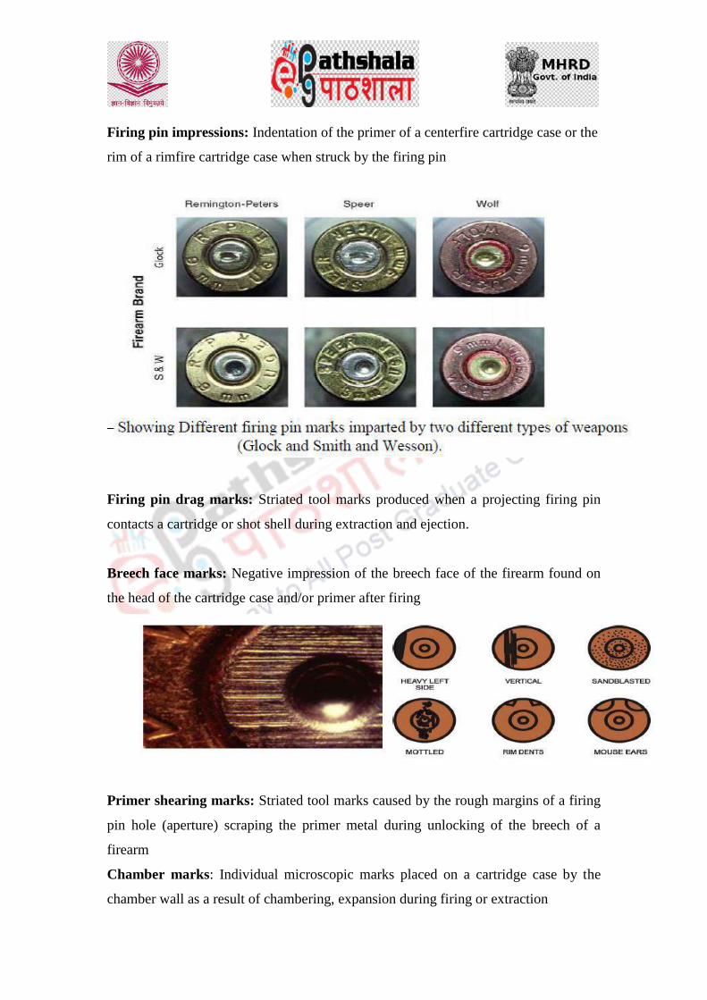
Firing pin impressions: Indentation of the primer of a centerfire cartridge case or the
rim of a rimfire cartridge case when struck by the firing pin
Firing pin drag marks: Striated tool marks produced when a projecting firing pin
contacts a cartridge or shot shell during extraction and ejection.
Breech face marks: Negative impression of the breech face of the firearm found on
the head of the cartridge case and/or primer after firing
Primer shearing marks: Striated tool marks caused by the rough margins of a firing
pin hole (aperture) scraping the primer metal during unlocking of the breech of a
firearm
Chamber marks: Individual microscopic marks placed on a cartridge case by the
chamber wall as a result of chambering, expansion during firing or extraction

Extractor marks: Striated tool marks produced on a cartridge or cartridge case from
the operation of an extractor (usually found on or just ahead of the rim)
Ejector marks: Tool marks produced on the head of a cartridge case, from contact
with the ejector (generally at or near the rim)
Ejection port marks: Striated marks produced by hard contact between the ejection
port of a firearm and a rapidly moving ejected cartridge case
Magazine marks: Striated marks produced on the periphery of a cartridge as it moves
from the lips of a magazine towards the chamber during feeding
The Manufacturer of the Cartridge: This is the company which manufactures
cartridges for various types or a specific type of weapon
Shape and Caliber of Cartridge: The Shape of the cartridge speaks to whether the
cartridge was designed for rimmed, rimless or belted firing; while the caliber is the
size of the cartridge as it relates to the type of weapon which it can be fired from.
Cartridge Composition: This speaks to the material used to compose the cartridge.
Some cartridges are designed with harder materials to facilitate the high pressure heat
during firing, some of these cartridges spent casings can be reused; Other cartridges
are designed in order to not degrade or to lose lustier and strength over time; while
some are made with materials such as paper or plastic relative to particular weapon.
Individualizing characteristics of Firearm and Bullet:
Striations in the lands and grooves of a firearm, and the lands and grooves of a fired
bullet. Striations are imparted into the bore of a firearm during rifling, as the lands
and grooves or are being formed inside the barrel of a firearm. These lands and
grooves along with their striations then impart themselves onto the bullet when it is
fired from a weapon. The striations in the lands and grooves of the weapon will
provide a direct link to the rifling process and manufacturer that made the weapon;
while the striations in the lands and grooves from a fired bullet will provide a direct
link to the bore of the specific firearm it was fired from, as well as a direct link to
other bullets fired from the same weapon.

Individualizing characteristics of Firearm and Cartridge
Type of Breech-face mark: The area around the firing pin, which is against the head
of the cartridge or shotshell during firing is the Breech face. Different weapons have
different types of Breech face marks. Eg. Glock pistols possess a rectangular Breech
face pattern, as opposed to a 9mm Lugar LC pistol. Within the Breech face pattern,
there are minute impressions which can be identified microscopically; these are the
individual characteristics which are imparted onto the cartridge casing when its head
is forced back onto the Breech block during discharge of the firearm.
Shape of ejector and extractor marks: The shape of ejector and extractor marks are
also marks imparted onto the cartridge after firing. The ejector and extractor can
impart individualizing characteristics on the head of the shell casing, based on the
theory that no two ejector and no two extractor is shaped the same way.
References:
1. http://library.med.utah.edu/WebPath/TUTORIAL/GUNS/GUNBLST.html.
accessed on January 2017.
2. Handbook of Firearms and Ballistics: Second Edition Brian J. Heard © 2008
John Wiley & Sons, Ltd.
3. Vincenzo D. Crawford, Class, Individual, & Sub-Class Characteristics of
Firearm & their
Constituents.https://www.academia.edu/12309195/Class_Characteristics_Indi
vidual_Characteristics_and_Sub-
Class_Characteristics_Of_Firearms_and_their_Constituents

4. http://projects.nfstc.org/firearms/module12/fir_m12_t01.htm
Law
Forensic Science & Forensic Medicine
28, Gun Shot Residue, Determination of range of fire,
firearm injuries

DESCRIPTION OF MODULE
Items Description of Module
Subject Name Law
Paper Name Forensic Science and Forensic Medicine
Module Name/Title Gun Shot Residue, Determination of range of
fire, firearm injuries
Module Id LAW/CJA/VIII /
Objectives
To understand the role of ballistics in Forensic Science
To learn about Gun shot residue, its composition, identification, collection and analysis
To determine the range of fire by which bullet is fired
To understand the types of injuries inflicted to the body
Prerequisites Introduction to ballistics, type of powders, their
composition.
Key words
GSR, range of fire, injuries
Role Name Affiliation
Principal Investigator Prof. (Dr.) G.S. Bajpai Registrar, National Law
University Delhi
Co-Principal Investigator
Paper Coordinator Prof. (Dr) Sally Lukose Dean, School of Basic and
Applied Sciences, Galgotias
University, Greater Noida.
Content Writer/Author Dr Prashant Agrawal Division chair-Forensic
Science, Galgotias University
Content Reviewer Dr Uday Pratap Singh
Scientific Officer, Forensic
Science Laboratory,
Moradabad, Uttar Pradesh

On pulling the trigger the firing pin of a firearm strikes the primer of a cartridge
the primer compound ignites sending a flame into the cartridge case. Propellant
in the cartridge case starts to burn, causing it to change from a solid material to
a gas. This change creates a large pressure within the cartridge, which in turn
forces the bullet down the barrel and down range. Pressure building behind the
bullet decreases as the bullet exits the muzzle of the firearm.
When the bullet exits the muzzle, pressure behind it blows the gunshot residues
out of the firearm's barrel with high velocity. The residues are expelled from the
barrel in a smoky cone shaped pattern. Gunshot residue travels a certain
distance after emitting the muzzle of the barrel and leaves a pattern around the
entry hole.
Gunshot residues can also be emitted from other areas of a firearm. The muzzle-
to-garment distance can vary considerably depending on the firearm and type of
ammunition being used. Short-barreled firearms and lower velocity cartridges
will not normally expel residues as far as a high velocity rifle. At shorter
distances however, they may deposit greater concentrations of gunshot
residues. Also, gunpowder can come in several forms such as ball, flake, disc,
and others. Ball powder being spherical in shape is more aerodynamic than say
a particle of flake gunpowder and as a result will travel farther. A number of
other variables can influence the amount of gunshot residues that may reach a
target; therefore, it is essential that the firearm and ammunition used in the
shooting incident be recovered.
Composition of Gunshot Residue:
Gunshot residue is normally a combination of gunpowder residues and lead
residues. More and more ammunition manufacturers are using lead free or low
lead propellants because of the toxicity of lead. Gunpowder residue can contain
unburned gunpowder particles, partially burned gunpowder particles or the
carbonaceous soot from completely burned gunpowder.
Modern smokeless gunpowder, and black powder, contains nitrate compounds.
Black powder normally contains a combination of potassium nitrate (75%),
charcoal (15%), and sulfur (10%). Smokeless powders can either be single
based or double based. Single based gunpowder will
contain nitrocellulose (cellulose hexanitrate) as its main ingredient. Double
based gunpowder contains nitrocellulose and nitroglycerin (glyceral trinitrate)
as its base. Some triple-based powders are also now available.
When either of these types of gunpowder burns the residue left behind will be in
the form of a nitrite-based compound. Nitrite particles when emitted from the
muzzle of a firearm will strike a nearby target and either be imbedded in the
target's surface or leave a deposit of nitrite residue.
Lead residues will be in a vaporous or particulate form and can come from a
couple sources within a discharged cartridge. The most common source is
the primer. Primers are used to start the ignition process in cartridges and
commonly contain lead styphnate, barium nitrate, and antimony
sulfide compounds. However, some newer primer compounds are being used
that are lead and/or barium free.
Cartridges containing lead based primers, when ignited, produce a vaporous
cloud of residue that is expelled from the muzzle of the firearm. Additional
vaporous lead residues can be produced when the hot gases pushing a lead
bullet down a barrel melt lead from the base of the bullet.
A third form of lead residue will be in a particulate form. Particulate lead
residue comes from minute lead particles that are shaved from the sides of a
lead bullet as it travels down the barrel. Lead particulate has more mass than
vaporous lead and travels greater distances. Also, gunpowder particles can be
coated by the vaporous lead residues and leave what appears to be a lead
particulate deposit upon striking the target.
The amount of lead residue emitted from a gun can vary slightly from shot to
shot. Fouling in the barrel from previous shots can slightly increase the amount
of lead residue emitted from one shot to the next.
Gunshot residue collection:
When a firearm is shot, in addition to the projectile(s), a mass of debris comes
out the muzzle. These gunshot residues (GSR) can include various primer
residues, residues from projectiles, and partially burned and unburned gun
powered particles. The examination and analysis of GSR on items of evidence
can allow determinations to be made as to weather a hole or defect is consistent
with being caused by a bullet (or other firearm- Relatedprojectiles).
Additionally, GSR can be transferred to an individual by discharging a firearm,
handling a firearm or fired ammunition components, or by contact with another
object that has GSR on it. The presence of GSR on a person may provide
useful information linking an individual with an action that could transfer this
residue to them. As a very general guide, after four to eight hours it is unlikely
that residues will be found on a live and mobile individual's hands unless steps
have been taken to preserve such evidence (e.g. bagging the hands). The
residue can persist for longer periods of time on some areas of interest such as
on the deceased, on clothing or other stationary objects. The decision to collect
a sample is affected by many variables and must be based on the investigative
information available.
Since 1933 several collection and analysis methods relating to GSR have been
employed. The most popular method of collection is lifting micro-traces from a
substrate upon which GSR has been deposited by multiple pressings of adhesive
material until the tackiness of the adhesive has gone. The adhesive material is
attached to the head (smooth and flat) surface of a SEM stub as that does not
require carbon coating, one that is polished smooth and flat, and does not
contain elements of high atomic number.
Both skin and clothing are suitable for the stub method of collection.
Specifically, in relation to the hands of the shooter one stub is pressed along the
thumb and forefinger and a second along the palm of each hand (resulting in
four stubs).
Sticky tape, specialized Micro vacuum cleaners, vacuum lifting onto a filter
disc for clothing, plastic shafted, alcohol moistened cotton swabs are other
means of collection.
Collection by tape and aluminium stub for use in a SEM is superior to that of
liquid adhesives and swabs.

Particle Type Elemental Composition(s) Designation
3 – Component Particle Pb-Ba-Sb Characteristic of GSR
2 – Component Particle Pb-Ba; Pb-Sb; or Ba-Sb Consistent with GSR
1 – Component Particle Pb-rich; Ba-rich; or Sb-rich Commonly associated with GSR
GSR particle types formed from the discharge of a firearm.
Examination of GSR:
When a firearm is discharged and the bullet strikes a surface, gunshot residues
are deposited, particularly at close range. These residues may be reproducible
and therefore have evidentiary value. Some residues are visible and others
require chemical treatment in order to visualize them.
The preliminary visual and microscopic examinations of gunshot residues
should be given first priority because subsequent chemical testing can dislodge
residues or alter the appearance of physical effects.
Residues should initially be observed and evaluated by the unaided eye and
with a low power (3x-30x) stereomicroscope. Infrared (IR) imaging may be
used to visualize heavy soot on dark or bloody clothing.
Chemical Tests:
Modified Griess Test: The Modified Griess Test is performed first on the
exhibit because it will not interfere with later tests for lead residues. The
Modified Griess Test is a test to detect the presence of nitrite residues. As
described earlier, nitrite residues are a by-product of the combustion of
smokeless gunpowder. When a firearm is discharged nitrite particles are
expelled from the muzzle of a firearm and can be imbedded in or deposited on
the surface of a target. The Modified Griess Test is the primary test used by
firearms examiners to determine a muzzle-to-garment distance.
The Modified Griess Test is performed by first treating a piece of desensitized
photographic paper with a chemical mixture of sulfanilic acid in distilled water
and alpha-naphthol in methanol. Desensitized photographic paper is obtained

by exposing the paper to a hypo solution. The photographic paper will no
longer be light-sensitive but will be reactive to the presence of nitrite residues.
The exhibit being processed is placed face down against a piece of treated photo
paper, with the bullet hole centered on the paper. The back of the exhibit being
examined is then steam ironed with a dilute acetic acid solution in the iron
instead of water. The acetic acid vapors will penetrate the exhibit and a reaction
takes place between any nitrite residues on the exhibit and the chemicals
contained in the photographic paper. The resulting reaction will appear as
orange specks on the piece photographic paper.
Dithiooxamide Test:
It is also known as the Rubeanic Acid Test and is a chemically specific
chromophoric test for the presence of cuprous (copper-bearing) material.
Copper-jacketed bullets represent a considerable percentage of ammunition
evidence in criminal cases.
Copper is used in the following types of ammunition:
Military and sporting jacketed bullets fabricated from gilding metal (a
90/10 copper/zinc alloy) or commercial bronze (a 95/5 copper/tin alloy)
Rimfire bullets coated or plated with copper or brass (a 70/30 copper/zinc
alloy)
Revolver bullets with copper jazckets
Nickel-plated bullets, e.g. Silvertip bullets
The test identifies a bullet wipe or bullet splash caused by the copper-bearing
particulate in the form of bullet jacket fragments found around the perimeter of
a bullet hole. While the test is not particularly useful for distance
determinations, it can detect residues consistent with the discharge of a firearm
or the passage or impact of a copper-jacketed bullet.
The chemistry of the Dithiooxamide Test is comprised of the following process:
Material from the perimeter of a suspected bullet hole is exposed to an
ammonia solution.
The same material is exposed to a solution of dithiooxamide dissolved in
ethanol. If cuprous material is present, a copper complex with a
characteristic color forms.

o A dark gray-green color indicates the presence of copper-bearing
material in bullet wipe.
o A blue-pink color indicates the presence of nickel (e.g., silver-
tipped bullets).
Sodium Rhodizonate Test: This chemical test is designed to determine if lead
residues are present on the exhibit. The Sodium Rhodizonate Test is performed by
spraying the exhibit with a weak solution of a mixture of Sodium Rhodizonate and
distilled water. This solution has a dark yellowish/orange color. The exhibit is
then sprayed with a buffer solution which causes the background color to
disappear.
The Sodium Rhodizionate reacts with any lead that may be present and turns the
lead a very bright pink. The pink color is only an indication of the presence of
lead residue and to confirm the presence of lead residue the area can be treated
with a diluted Hydrochloric Acid solution. If the pink turns to a blue then the
presence of lead is confirmed.
Instruments useful in detection of Gunshot Residue
The major methods for detection of primer residues are analytical and qualitative.
Analytical methods include neutron activation analysis (NAA) as well as atomic
absorption spectrophotometry (AAS) and inductively coupled plasma mass
spectroscopy (ICP-MS).
Scanning electron microscopy with energy dispersive analysis by x-ray detector
(SEM-EDX) and atomic force microscopy (AFM) are used to identify the primer
residue qualitatively. An X-ray analyzer can be beamed directly onto the particles
visualized with SEM, so that the energy dispersive pattern can be generated,
giving the elemental composition of the particles.
For these methods, samples must be obtained from the skin surfaces of a victim at
the scene. Delay in obtaining residues, movement, or washing of the body prior to
autopsy will diminish or destroy gunshot residues. A rapid loss in numbers of
GSR particles occurs from 1 to 3 hours post firearm discharge, though maximum
recovery times of 1 to 48 hours have been reported.
Bullet Wounds
Gunshot residues emitted from the muzzle will travel out to distances of
approximately 3 and 5 feet in most firearms but in some cases can travel even
greater distances. At the 3-5 foot range the gunshot residues may only consist of a
few trace particles and make determining the firing distance difficult if not
impossible.
As the firearm gets closer to its target the residue concentrations increase and the
actual size or diameter to the pattern gets smaller. At around 18-24 inches most
firearms will start to deposit considerable concentrations of gunshot residues that
may or may not be visible to the eye.
At distances of less than around 12 inches heavy concentrations of visible gunshot
residues will normally be deposited.
When the muzzle of the firearm gets next to or is in contact with the target, hot
gases escaping from the muzzle at high velocity will typically rip, tear, shred,
and/or melt the material of the target. A very intense deposit of gunshot residues
will be found around the margins of a contact or near contact entrance hole.
There have actually been cases where a hard contact gunshot (muzzle pressed hard
against the victim) caused the residues to blow through the wound tract in the
victim and be deposited around the inside of the exit hole of the victim's clothing.
Entry Wounds
The features vary depending on the range from which the weapon is fired—contact,
close (intermediate) range or longer (indeterminate) range.
A gunshot wound is a controlled explosion and the bullet is accompanied from the gun
by a jet of flame, a cloud of gas, burning and unburnt grains of gunpowder and soot
from burnt gunpowder. Entry wounds may show the stigmata of the explosion to a
lesser or greater extent.
(A) Contact wound
The muzzle is pressed against the skin. The heat of the discharge causes scorching or
charring of the wound. The gases produced by the explosion of the cartridge enter,
stretch and split the skin producing a stellate or cruciform tear. The tissue at the margin
of the wound may contain soot and powder.

(B) Close range (Intermediate range)
The wound is inflicted at less than arm's length i.e. < 2 - 3 feet. The particles of partly
burnt or unburnt powder from the muzzle are driven into the skin around the entrance
wound giving a stippled appearance called "powder tattooing" or "powder burns". The
area may be blackened by soot. Soot may be wiped off the skin, but powder tattooing
cannot be wiped off. The bullet hole may be round or split, the latter being relatively
common when there is underlying bone.
(C) Longer (Indeterminate) `range
The range is > 2 - 3 feet. The gun is too far from the skin for the products of the
explosion to have any effect. Therefore the appearance of the wound is due entirely to
the bullet. The wound is usually round (but may be split by "tail-wag" if the gun is fired
from the extreme of its effective range causing the bullet to lose its gyroscopic spin and
start to tumble).
Marginal abrasion/Abrasion collar/Abrasion ring
The margin of the entry wound in some close range and longer range injuries may be
abraded (“marginal abrasion”, “abrasion collar” or “abrasion ring”) as the bullet inverts
the skin and abrades the epidermis as it enters. The shape of this abrasion may help in
determining trajectory.
Grease ring
The inner edge of the abrasion collar may be black due to grease or lubricating oil and
metal particles from the bullet.
Exit wounds
These show none of the stigmata of the explosion or soiling seen in the entry wound.
An exit would may be the same size as the entry wound, but may be smaller or larger
depending on the range, type of weapon, type of bullet, the tissues being traversed by
the bullet, etc.
In a contact shot the entry wound is split by the explosive gases and is therefore usually
larger than its corresponding exit wound. However, if the bullet comes out carrying
bone e.g. a shot to the skull, the exit wound may be larger than the entry.

In a distant shot the exit wound may be the same size or slightly smaller than the entry.
In general, exit wounds tend to be split with irregular, everted edges. As a rule, exit
wounds DO NOT show an abrasion collar, but exceptionally, this may occur if the skin
was pushed up against a hard surface, e.g. concrete wall or floor at the time the bullet
exited. This is known as a shored exit wound.
References:
1. https://dps.mn.gov/divisions/bca/bca-divisions/forensic-
science/Pages/forensic-programs-crime-scene-gsr.aspx
2. http://forensic-review.com.au/gunshot-residue/
3. http://www.forensicmag.com/article/2014/08/gunshot-residue-collection-
decisions-make-or-break-
casehttp://projects.nfstc.org/firearms/module12/fir_m12_t05_03_a.htm
4. http://www.firearmsid.com/a_distancegsr.htm
5. http://library.med.utah.edu/WebPath/TUTORIAL/GUNS/GUNGSR.html
6. Handbook of Firearms and Ballistics: Second Edition Brian J. Heard © 2008
John Wiley & Sons, Ltd.


Law
Forensic Science & Forensic Medicine
29 Forensic Odontology: personal identification,
determination of age, Bite marks Analysis

DESCRIPTION OF MODULE
Items Description of Module
Subject Name Law
Paper Name Forensic Science and Forensic Medicine
Module Name/Title Forensic Odontology: personal identification,
determination of age, Bite marks Analysis
Module Id LAW/CJA/VIII /
Objectives
To learn about the different types of dentition present in child and adult person, their age of eruption and shading, mixed dentition
Determination of age from dental examination
Identification of person from bite marks analysis in cases of sexual offences.
Prerequisites Basic knowledge of Biology
Key words
Teeth, mixed dentition, bite marks,
Role Name Affiliation
Principal Investigator Prof. (Dr.) G.S. Bajpai Registrar, National Law
University Delhi
Co-Principal Investigator
Paper Coordinator Prof. (Dr) Sally Lukose Dean, School of Basic and
Applied Sciences, Galgotias
University, Greater Noida.
Content Writer/Author Dr Prashant Agrawal Division Chair-Forensic
Science, Galgotias University
Content Reviewer Dr Uday Pratap Singh
Scientific Officer, Forensic
Science Laboratory,
Moradabad, Uttar Pradesh

Forensic odontology is the application of dental science for the purpose of legal
investigations. It involves identification of the offender with certainty by comparing
dental records of bite mark left on the victim or at the scene. It may be identification
of human remains on the basis of dental records.
Forensic odontologists are experienced and specialized dentists who use their
knowledge to identify unknown remains and bite marks of an individual. Forensic
odontologist may be called by police officers to assist the investigation.
Forensic odontologists are generally called to:
Identify unknown human body or remains which cannot be identified by other
means like face, fingerprints etc.
In case of mass disasters such as plane crashes and natural calamities.
To link a person with bite mark injuries, in cases of assault or suspected abuse
Estimate the age of the person from skeletal remains
Personal identification for establishing the individuality of the person:
In humans, there are twenty temporary teeth and thirty-two permanent teeth in both
jaws. The deciduous and permanent teeth erupt earlier in the lower jaw than upper jaw
except lateral incisors. In females teeth erupt about a year earlier. The eruption of
tooth depends on the several factors like hereditary, environmental, nutritional and
endocrinal factors.
Among 32 permanent teeth there are four incisors, two canines, four premolars and
six molars in each of jaw. The temporary teeth are twenty comprising of four incisors,
two canines, and four molars in each jaw. The development of tooth begins with the
formation of cellular tooth germ within the alveolar bone in the shape of the crown.
The tooth has three parts a crown, neck and root that are embedded in the jaw. There
are certain differences in the appearance of deciduous and permanent teeth.
Incisors—Incisors have chisel shaped crown which is convex on labial surface and
concave on lingual surface, with constricted neck and has single root. They have
chewing surface and cutting edges.
Canines—Canines are large compared to the incisors. The crown is conical shaped
and concave on its labial surface but slightly concave on its lingual surface. The

masticatory edges tapers and projects beyond the level of other teeth. It also has single
root. They have single cusps.
Premolars—Premolars are also known as bicuspids. They are smaller and shorter than
the incisors and canines and the crown is circular. The chewing surface is bicuspid
with a groove between them. They also have a single root.
Molars—Molars are the largest of the entire tooth. The crown is cubical and is convex
on both lingual and labial surfaces. In upper jaw, first molar has stable configuration
with 4 cusps, second molar is very different individually and has 3 cusps. Third molar
is less mineralized than others and may have chalky spots. Also, upper molar has
three roots. In lower jaw, first molar has 5 cusps, 3 buccally and 2 lingually whereas
second and third molars have 4 cusps each.
Differences between primary and secondary dentition
Determination of age of the person from dental records
Approximate age of the individual can be determined by teeth examination.
Estimation of age can be done by the
Presence of deciduous dentition and stages of eruption
Period of mixed dentition
Different stages of eruption of permanent teeth
Loss of deciduous teeth
In Children At birth, the rudiments of all temporary teeth and first permanent molars
are there. In children and young person the chronological calcification and eruption of
teeth gives their ages. Wisdom tooth (3rd
Molar): During the age of 14-20 years, the
stage of development of third molar is of particular importance to determine the age.
The lateral oblique X-ray of maxilla and mandible show the development of third
molar and root completion of second molar. The body of jaw grows posteriorly and

ramus is elongated after eruption of second molar and a space is created for the third
molar.
Mixed dentition: It is the time when both the temporary and permanent teeth are
present in the jaw. The period starts from 6th year of life when the first molar erupts
and continue till the canine falls (up to 11 years).
Successional teeth: These are ten in number in each jaw. Permanent tooth replaces
the temporary except the permanent premolars that erupt in place of deciduous
molars.
Superadded teeth: Those teeth which do not have deciduous predecessors and erupt
behind the temporary teeth. All permanent molars are superadded as the first
permanent molar erupts while other deciduous teeth are present.
There are a particular number of teeth in different ages in children that is:
5 years—There are 20 teeth, all are temporary
6 years—Mixed dentition and the number of teeth is 21-24 due to eruption of
first permanent molar.
7-11 years—24 teeth (mixed dentition) with eruption of other permanent teeth
replacing the temporary ones.
12-14 years—24-28 teeth due to eruption of second molar
14-17 years—28 teeth as there is no eruption.
17-25 years—32 teeth due to eruption of third permanent molar
25 years and above—thirty-two teeth
Age of appearance of deciduous and permanent teeth
Determination of Age from Dentition in Adults and Older Age

In mandible the center of ossification of jaw appears at 2nd
month of intrauterine life
and symphysis menti fuses by 18-20 years. The changes in mandible with age are:
When eruption has ended and the person has achieved adult dentition, it is necessary
to apply other methods that occur due to the normal wear and tear and age of the
person.
Age changes in Mandible
Gustafson’s method: Estimation of age in the adults can be determined by studying
the progressive changes in an individual tooth.
• Attrition: It is wearing and tearing on the opposing mastication surface of the teeth
of the upper and lower gums due to continuous friction. Attrition is categorized in
four degrees: A0—No attrition, A1—Attrition within enamel A2—Attrition within
dentine A3—Attrition exposes to soft pulp
Gustafson’s method
Periodontosis: When the maintenance of teeth and gums is not proper, there is
regression of gums and periodontal tissue. There is loosening of the teeth, exposure of
length of root and deposition of stony hard debris over a long period. According to the
length of exposure of root, periodontosis may be categorized in following degrees

P0—No Periodontosis or no root exposure P1—Exposure of less than 1/3rd of the
part of the root next to crown P2—Exposure of more than 1/3rd of the of the root but
less than 2/3rd. P3—Periodontosis beyond 2/3rd of the length of the root near the
crown.
• Secondary dentine: As age advances there is deposition of secondary dentine tissue
in pulp cavity. The process progress until whole of pulp cavity is replenished and the
size of cavity decreases. In mandibular teeth, it starts from above and in maxillary
from below. The reasons may be ageing, pathological conditions, dental caries etc.
There are categorized in four degrees of secondary dentine formation: S0—No
deposition S1—Deposition in the upper part S2—Deposition in the half of the pulp
cavity from above S3—Almost whole of the pulp cavity is involved.
• Root resorption: This decaying change involves both cementum and dentine that
shows grooves. First apex is involved then extends upwards. It is categorized as R0—
No root resorption. R1—Seen only in some parts. R2—Seen over a large area. R3—
Involves both cementum and dentine.
• Cementum apposition: Cementum deposition increases due to change in the surface
of tooth near the end of roof and forms incremental lines. It is categorized in four
degrees: C0—Only normal layer of cementum is noticed. C1—Slightly greater than
normal. C2—Thick layer has occurred. C3—When a heavy layer is present.
• Root transparency: It occurs in the root from below upwards in the lower jaw and
above downwards in the upper jaw due to rarefaction of dentine tissue. It is
categorized as R0—No transparency noticed anywhere R1—Transparency noticeable
mostly over apical region R2—Transparency up to 1/3rd from the apical region R3—
Transparency noticeable up to 2/3rd of the length of the root from apex.
Out of the six criteria, transparency of the root is the most reliable one for age
estimation.
The total score obtained is applied to a regression formula and the age is estimated
below:
Estimated age (years) = 11.43 + 4.56 x (Total score)
Other criteria for personal identification:
1. Any extractions, recent or old from the condition of the socket.
2. Any fillings, number, position and composition.

3. Artificial teeth may be of gold, porcelain or stainless stain.
4. Prosthetic work in mouth such as bridge work or braces.
5. Crowned teeth.
6. Broken teeth.
7. Pathological conditions in teeth, jaws or gums
8. Congenital defects like enamel pearls, carabelli’s cusps or ectopic teeth.
9. Malpositioned teeth which are rotated or tilted.
10. General state of care and hygiene like caries, plaque, tobacco staining, gingivitis
etc.
Bite Marks Analysis:
Criminals may leave bite mark impressions at the crime scene, they may be in food,
chewing gum or, more commonly, on the victim. When a bite mark is found,
numerous steps should be taken. Once the mark has been sufficiently photographed,
saliva sample is taken from the area for DNA evidence. Casts or moulds can then be
made. Comparison can be done if another bite impression is found or standard teeth
impression is taken from a suspect.
Bite marks have been divided into seven classifications:
Haemorrhage: A small bleeding spot is found
Abrasion: Undamaging mark on the skin is observed
Contusion: Ruptured blood vessels, bruising etc.
Laceration: presence of punctured or torn skin
Incision: Neat and clear puncture of the skin.
Avulsion: Removal of the skin.
Artefact: Bitten off piece of the body
Police may sometimes found apple or piece of cheese with teeth marks. The unusual
bite mark on the objects may also be there due to some dental abnormality. ‘Plastic’
marks on objects such as butter, cheese, lard, wax or chocolates should be stored in
refrigerator to preserve them and prevent from melting. Fruits especially Apples are

preserved in Campden solution (Meta bisulphate fluid) used for fruit bottling.
Photographs are taken at right angles to the bite. Salivary traces should be swabbed.
Bites are commonly seen in cases of:
1. Child abuse cases: The bite marks may be present on any area of the body. The
common sites are the arms, hands, cheeks, buttocks, trunk and shoulders. Bites are
examined whether the size of the mark is same as of adult dentition. If bite mark is of
small size it could be due to bite by the sibling and when it is of different size it could
be animal bite.
2. Sexual assault: Sexually oriented bites may be caused at any parts of the body
namely Breasts, neck, shoulders, thighs, abdomen, pubis or vulva.
3. Police officers by the resisting offenders.
4. Sporting events: Bite marks are produced when the victim manages to bite the
assailant during any type of sports or games.
5. In assaults anywhere on the body.
6. Self inflicted bite marks: Mostly found on the forearms of the children caused by
themselves. Sometimes arms may be pushed into the child’s mouth to stop crying or
due to intense pain; Mentally challenged and psychologically disturbed people may
also inflict bite on themselves.
7. At scene of crime: In some cases, the criminals may leave their teeth marks on the
substance left at the scene. These bite marks may be encountered on inanimate objects
like food stuffs or fruits etc.
Nature of Bite Marks:
Bite mark comprise of punctate haemorrhages varying from small petechiae to large
Ecchymoses merging in to a confluent central bruise. Front teeth causes bite marks
from canine to canine with an invariable gap at either side representing the separation
of upper and lower jaw. A circular or shallow oval bite by human and deep parabolic
arch or U shaped is characteristic of an animal bite. Teeth may cause clear separate
marks that run in to each other as continuous, intermittently broken lines. There may
cause abrasions, bruises and lacerations or a combination of all. Identification from
bite marks is possible if incisors and canines has some characteristic and unique
features.

Images of different bite marks
Bite Mark Investigation
1. Photograph of Bite mark should be taken from different angles; from a directly
perpendicular viewpoint with a plane of film at right angles to that of the lesion, with
an accurate scale
2. Swabbing of saliva: To identify or exclude assailant; 80% of the people are
‘secretors’ who exude blood group enzymes in the saliva. Cotton swabs are rubbed on
to the bite to get the sample. These are then preserved and sent for further
examination.
3. Impression of bite mark: Plastic substance is laid over the bite mark that hardens
and produces permanent negative cast of the lesion. Plastic substance is made with a
rubber or silicone based medium containing a catalytic hardener or Plaster of Paris
(water based plaster).
Matching the bite mark with the suspect:
1. Informed consent should be taken in writing before examination of the suspect
2. Oral consent with at least one witness, if not written to be taken
3. Dentition examined and points determined and recorded by diagram and writing
4. Photographs should be taken.
The points to be noted in bite marks are:
1. Presence of full or partial denture; were they worn at the time of incidence?
2. Number of teeth in the upper and lower jaw.
3. Charting of missing teeth.
4. Estimate of bite overhang; whether there is an edge-to-edge occlusion or an
undershoot projection of lower teeth.
5. Recording of any broken teeth or teeth with significant individual abnormalities are
charted and described.
6. Any irregularity or marked variation in cutting edge profile of any front teeth.
7. Evaluation of size and prominence of any teeth especially canines and incisors.
8. Any developmental abnormalities are to be noted.
9. Recording of any abnormality in orientation of any tooth such as twisting (rotation)
of antero-posterior tilting or double row of teeth; any gap or irregular spacing: (i) Six
upper and six lower front teeth give the most information. (ii) Canines may provide
particular help. (iii) Premolars and molars are rarely useful due to being posteriorly
positioned in the jaw.
Medico-legal Importance of Dental Study
1. Identification of the individual can be done from the dental study. Other factors for
identification include: • The habits such as chewing tobacco are also helpful for
identification. • Peculiarity in setting of the teeth may be familiar and may help in
identification. • As the teeth resist decomposition, they are important for identification
even for a long time after death as peculiarity of setting of teeth, evidence of any
missing tooth or any artificial dentures help in proving the identity of the person. •
Occupation can be known if there is a notch under tooth e.g. in case of tailors.
2. Grievous hurt: Fracture, Dislocation of tooth amounts to grievous hurt according to
section 320 (7) I.P.C..
3. The teeth resist putrefaction and the presence of heavy metals in suspected
poisoning case can be detected for a considerable period after death.
4. In chronic phosphorus poisoning, evidence of phossy jaw and affection of teeth and
gums are characteristic.
5. Bite mark over the articles or food help towards detection of the criminal.
6. Bite mark on private parts, breasts, cheeks, neck of a girl are suggestive of some
sexual instinct.
7. Artificial dentures when dislodged can sometimes cause choking in the elderly.
8. Pink teeth: In putrefied bodies, near gum line, teeth are of pink colour. It is due to
dentine being stained by haemoglobin products

References:
1. Dayal PK. Textbook of Forensic Odontology. 1st Ed. Hyderabad: Paras
Medical Publishers; 1998.p.69-80.
2. McLay WDS. Clinical Forensic Medicine. 2nd Ed. London: Greenwich
Medical Media; 1996.p.288-291.
3. Vij K. Textbook of Forensic Medicine and Toxicology. 3rd Ed. New Delhi:
Elsevier; 2005. p.85.
4. Sharma et al. Bite Mark Analysis - An Important Tool in Crime Investigation.
JIAFM, 2006 : 28 (2) ISSN : 0971-0973
5. http://aboutforensics.co.uk/forensic-odontology/
6. https://explorehealthcareers.org/career/forensic-science/forensic-odontologist/
7. P C Dikshit. Textbook of Forensic Medicine and Toxicology: Peepee
Publishers and Distributors (P) Ltd; 2 edition (2013)

Criminology
Forensic Science and Forensic Medicine
Alcohol: Types of Alcoholic beverages, countrymade liquor,
illicit liquor, detection of alcohol in blood and breath (breathalyzer)

DESCRIPTION OF MODULE
Items Description of Module
Subject Name Criminology
Paper Name Forensic Science and Forensic Medicine
Module Name/Title Alcohol: Types of Alcoholic beverages, country
made liquor, illicit liquor, detection of alcohol in
blood and breath (breathalyzer)
Module Id
Objectives
Learning Outcome:
To make the learners understand about alcohol
abuse and its legal elements.
To make learners understand the types of
alcoholic beverages and its effects on body
To describe procedure for field sobriety testing.
To describe laboratory procedure used to detect
alcohol in blood and breath.
Prerequisites General understanding of Forensic Science and its
application in crime solving.
Key words
Alcohol, Abuse, Breath, Body Alcohol Concentration,
breathlyzer. Drunk, road accident.
Role Name Affiliation
Principal Investigator Dr G. S. Bajpai
Co-Principal Investigator
Paper Coordinator
Content Writer/Author Dr Priti Sharma
Content Reviewer

1. Introduction
Alcohol is a legally available drug that is consumed pervasively due to it significant
mood altering effects. Since it causes addiction on prolonged consumption, it is
considered as a drug of abuse and so, a socioeconomic burden for the society. Given
this, alcohol is a significant element in a wide range of cases originating from
negligence to criminal act.
As per the World Health Organization, harmful use of alcohol results in
approximately 2.5 million deaths per year, and 4% of all deaths worldwide as
attributable to alcohol.1 The alcohol related harm is not limited to heavy drinkers or
those suffering from alcohol use disorders but also extends to persons who drink
moderately or occasionally as well as to persons who do not drink but become victims
of alcohol related accidents or aggression. A report showed 70% of road accidents in
India due to drunken driving which costs 1.34 lakh lives each year.2 There are also
incidents of mass death due to consumption of illicit forms alcohol which are easy to
be misused due to their cheap cost and easy availability. Alcohol imparts significant
impairing effects on a number of aspects of human cognition and performance. This
results in several cases of violations of the law in which consumption of alcohol is a
fundamental component.
This module covers knowledge about how to deal with cases caused due to alcohol
abuse. Apart from being a sociological burden, alcohol is a culprit being various
crimes. This requires knowledge of its nature, effect on the body, manner and
collection and preservation of evidence, etc.
2. Alcohol and Laws in India
At present, India does not have a national alcohol policy. However, the Ministry of
Social Justice and Empowerment has Alcohol and Drug Demand Reduction and
Prevention Policies.
Prohibition is incorporated in the Constitution of India among the directive principles
of state policy.3,4 According to this “The state shall regard the raising of the level of
nutrition and standard of living of its people as among its primary duties and in
particular, the state shall endeavor to bring about prohibition of the use except for

medicinal purposes of intoxicating drinks and of drugs which are injurious to health.”
Alcohol policy is under the legislative power of individual states. Prohibition,
enshrined as an aspiration in the Constitution, was introduced and then withdrawn in
Haryana and Andhra Pradesh in the midi-1990s, although it continues in Gujarat, with
partial restrictions in other states – Delhi, for example, has dry days. There was an
earlier failure of prohibition in Tamil Nadu. Excise department regulate and control
the sale of liquor in the NCT of Delhi. Retail supply of alcohol is also regulated which
prohibits consumption and service of liquor at public places.5 This also prohibits
employment to any person (male under the age of 25 years or any female) at any
licensed premises either with or without remuneration in part of such premises in
which liquor or intoxicating drug is consumed by the public. Similarly no individual
should possess liquor at one time more than the prescribed limit without special
permit. In Bombay, the production, manufacture, possession, exportation,
importation, transportation, purchase, sale, consumption and use of all intoxicants is
prohobited.6 The advertisements of cigarette and alcohol on TV/Cable is completely
prohibited.
There is rule in Bombay8 which provides that any person, who is found drunk or
drinking in a common drinking house or is found there present for the purpose of
drinking, shall on conviction, be punished with fine which may extend to five hundred
rupees. Further, any person found drunk and incapable of controlling himself or
behaves in a disorderly manner under the influence of drink in any street or
thoroughfare or public place or in any place to which public have or permitted to have
access, shall on conviction, be punished with imprisonment for a term which may
extend to one to three months and with fine which may extend to two hundred to five
hundred rupees.9
3. Types of Alcohol and Alcoholic Beverages
There are different types of alcohol. Some of them are used in chemistry laboratories
and industry, e.g. isopropyl and methyl alcohol. Isopropanol, or isopropyl alcohol is
also used in industrial processes as well as in home cleaning products and skin
lotions. It is also commonly known as "rubbing alcohol". Methanol, or methyl
alcohol or wood alcohol has been used as an industrial solvent and is also commonly
available as methylated spirit. It is found in cleaning solvents, paint removers,
photocopier developer and anti-freeze solutions. As such, it is often available in large
quantities inexpensively. It is similar to ethanol but the end product after it is digested
by the body is formaldehyde, which is poisonous. This is responsible for "alcohol
poisoning". Methanol poisoning leading to blindness has been known to occur on
consuming even small amounts.
Another type of alcohol is ethyl alcohol, also known as ethanol. It is a thin, clear
liquid with harsh burning taste and high volatility. Ethyl alcohol is used as a reagent
in some industrial applications. For such use, ethyl alcohol is combined with small
quantities of methanol, with the mixture being called "denatured ethanol" to prevent
theft for human consumption. Alcohol is the active ingredient of various drink, liquors
and sprits. It is notorious due to its consumption by human beings for its intoxicating
and mind-altering effects.
Alcoholic beverages are made by fermentation of sugary and starchy substances,
followed by distillation to increase alcohol concentration. The active ingredient in
them is ethyl alcohol or ethanol. Based on the distillation and percentage of alcohol,
alcoholic liquors are classified into distilled and undistilled as described below:
Distilled Products
Rum (50-60%), Whisky, Brandy, Gin (40-45%), Sprits
Undistilled Products
Beer (4-11%), Wines, Burgundy, Champagne (10-15%)
Brief description of alcoholic beverages:
Wines are made from a variety of fruits, such as grapes, peaches, plums or apricots.
The most common wines are produced from grapes. The soil in which the grapes are
grown and the weather conditions in the growing season determine the quality and
taste of the grapes which in turn affects the taste and quality of wines. When ripe, the
grapes are crushed and fermented in large vats to produce wine.
Beer is also made by the process of fermentation. A liquid mix, called wort, is
prepared by combining yeast and malted cereal, such as corn, rye, wheat or barely.

Fermentation of this liquid mix produces alcohol and carbon dioxide. The process of
fermentation is stopped before it is completed to limit the alcohol content. The alcohol
so produced is called beer. It contains 4 to 8 per cent of alcohol.
Whisky is made by distilling the fermented juice of cereal grains such as corn, rye or
barley. Scotch whisky was originally made in Scotland. The word "Scotch" has
become almost synonymous with whisky of good quality.
Rum is a distilled beverage made from fermented molasses or sugarcane juice and is
aged for at least three years. Caramel is sometimes used for colouring.
Brandy is distilled from fermented fruit juices. Brandy is usually aged in oak casks.
The colour of brandy comes either from the casks or from caramel that is added.
Gin is a distilled beverage. It is a combination of alcohol, water and various flavours.
Gin does not improve with age, so it is not stored in wooden casks.
Liqueurs are made by adding sugar and flavouring such as fruits, herbs or flowers to
brandy or to a combination of lcohol and water. Most liqueurs contain 20-65 per cent
alcohol. They are usually consumed in small quantities after dinner.
4. Country made Liquors
Country liquor, an unbranded, highly potent alcohol drink is produced by distilleries
and sold through separate distribution channels. Country liquor is made from a variety
of raw materials and has different names in different parts of the country. Their
manufacture is based on indigenous ingredients, eg., fenny, toddy, and arrack.
Country liquor are, the cheapest form of alcoholic beverages. Some locally available
fruit fly lures such as molasses, sugar, country liquor, fresh date palm juice and
fermented date palm juice are used as raw ingredients.
Arrack is a traditional drink produced (both legally and illegally) by distilling
fermented molasses, raw brown sugar, palm wine, rice or palm sugar. It has an
alcohol content ranging from 20% to 40%. Palm wine, another traditional beverage
produced from either the coconut tree or other palm trees, has an alcohol content
ranging from 20% to 40%. Daru, a drink distilled from the flowers of the mahwa tree,
has an alcohol content ranging from 20% to 40%.
There is a thriving market for illicitly made country liquor.10 Country liquor is
consumed in rural areas and by low-income groups in urban areas. The country liquor

market is regional market with small manufacturers spread across the country but is
estimated to be around 2.5 times the Indian Made Foreign Liquor (IMFL) market.
5. Illicit liquor
Any alcoholic beverage made under unlicensed conditions is called illicit liquor.
Usually sub-standard raw material is used in their preparation, which often are
contaminated with toxic chemicals and thus dangerous to consume.11 The illicit
market in India is increasing at an alarming rate, in seven main sectors (two of which
are alcoholic beverages and tobacco), according to a recent report by FICCI
CASCADE (Federation of Indian Chambers of Commerce & Industry Committee
Against Smuggling and Counterfeiting Activities Destroying the Economy).12
Moonshine, also known as white lightning, hooch or Tennessee white whiskey, is a
high-proof distilled spirit, generally produced illicitly. Moonshine can be
contaminated, mainly from materials used in construction of the still (an apparatus
used to distill miscible or immiscible). Some manufactures use old automotive
radiators as condensers in stills which is the source of contamination by glycol, lead
and products from antifreeze which are poisonous and potentially deadly.

Alcohol concentrations above about 50% alcohol by volume are flammable and
therefore dangerous to handle. This is especially true during the distilling process
when vaporized alcohol may accumulate in the air to dangerous concentrations if
adequate ventilation has not been provided.
Another form of alcohol i.e., methyl alcohol or methanol, is consumed by poor
section of society due to its similarity in taste and smell with ethyl alcohol and poor
production cost. However, methyl alcohol is extremely toxic - 10 ml can cause
blindness and 30 ml can cause death within 10 to 30 hours. Although methanol is not
produced in toxic amounts by fermentation of sugars from grain starches,
contamination is still possible by distillers using cheap methanol to increase the
apparent strength of the product. Sometimes, industrial methyl alcohol or denatured
spirit is added as a mixture of ethanol and methanol by illicit brewers to save costs.
There have been incidents where chemicals like organophosphorus compounds have
also been added to illicit liquor.
6. Effects of Alcohol
The effects of ethanol vary progressively as the blood alcohol concentration (BAC)
increases. At low BACs, the effects of alcohol consumption include euphoria,
talkativeness, and reductions in anxiety and inhibitions. At progressively greater
BACs, speech may become slurred, and dizziness or significant loss of coordination
may be observed. Further increases to BAC may be accompanied by drowsiness,
emotional lability, confusion, and loss of consciousness. Uncontrolled overdose can
result in fatal respiratory depression.13
Depending on BAC concentration, alcohol causes various other impairments to a
number of faculties related to psychomotor performance. These include impairment in
ability to divide attention over multiple tasks, reaction time, risk or hazard perception,
and motor coordination which affect the safe operation of a motor vehicle. This
results in causing various vehicle accidents. So, drunken driving is listed as one of the
most predominantly reported offenses directly related to alcohol consumption. To
control this, a legal limit is defined by the jurisdiction of each nation beyond which a
person is held under criminal act.

The consumption of alcohol may also be associated with an increased probability of
occurrence of a number of other kinds of offenses. One such offense is sexual assault.
A significant amount of research has examined the incidence of the use of alcohol
alone or mixed with drugs in various cases of sexual assault. Since alcohol suppresses
behavioural inhibitions, thus influencing decision-making skills or risk perception,
excessive alcohol consumption also leads to substantial impairment and perhaps even
unconsciousness, which can place an individual under considerable risk for attack.
7. Drunken Driving
Drunkenness is defined as the condition produced in a person who has taken alcohol
in a quantity sufficient to cause him to lose control of his faculties to such an extent
that he is unable to execute the occupation on which he is engaged at the material
time. Across the world, governments have defined different acceptable blood alcohol
levels for driving. However, there is no minimum threshold below which alcohol can
be consumed without risk. With rise in blood alcohol concentration, there is
progressive loss of driving ability due to increased reaction time, over confidence,
impaired concentration, degraded muscle coordination and decreased visual and
auditory acuity.
Figure 1: Diagram of increased driving risk in relation to blood-alcohol
concentration.
As per laws in India,14 " Driving by a drunken person or by a person under the
influence of drugs - whoever while driving or attempting to drive a motor vehicle or

riding or attempting to ride, a motor cycle - (a) has in his blood, alcohol in any
quantity, howsoever small the quantity may be or (b) is under the influence of a drug
to such an extent as to be incapable of exercising proper control over the vehicle shall
be punishable for the first offence with imprisonment for a term which may extend to
six months or with fine which may extend to two thousand rupees or with both; and
for a second or subsequent offence, if committed within three years of the commission
of the previous similar offence, with imprisonment for a term which may extend to
three thousand rupees, or with both". Till mid November, 1994, drinking and driving
was not allowed to be mixed up. Any alcohol in the blood, howsoever, small the
quantity has been an offence till November l994 but now after November 1994 the
law has been amended. Now up to 30 milligrams of intake per 100m1 of blood has
been permitted to driver before getting behind the wheel.
8. Field Sobriety Testing
A police officer who suspects that an individual is under the influence of alcohol
usually conducts a series of preliminary tests before ordering the suspect to submit to
an evidential breath or blood test. These preliminary, or field sobriety, tests are
normally performed to ascertain the degree of the suspect’s physical impairment and
whether an evidential test is justified. Field sobriety tests usually consist of a series of
psychophysical tests and a preliminary breath test. A portable handheld roadside
breath tester is used for this purpose.
Horizontal-gaze nystagmus, walk and turn, and the one-leg stand constitute a series of
reliable and effective psychophysical tests. Horizontal-gaze nystagmus is an
involuntary jerking of the eye as it moves to the side. A person experiencing
nystagmus is usually unaware that the jerking is happening and is unable to stop or
control it. The subject being tested is asked to follow a penlight or some other object
with his or her eye as far to the side as the eye can go. The more intoxicated the
person is, the less the eye has to move toward the side before jerking or nystagmus
begins. Usually, when a person’s blood-alcohol concentration is in the range of 0.10
percent, the jerking begins before the eyeball has moved 45 degrees to the side.
Higher blood-alcohol concentration causes jerking at smaller angles.
Walk and turn and the one-leg stand are divided-attention tasks, testing the subject’s
ability to comprehend and execute two or more simple instructions at one time. The
ability to understand and simultaneously carry out more than two instructions is
significantly affected by increasing blood-alcohol levels. Walk and turn requires the
suspect to maintain balance while standing heel-to-toe and at the same time listening
to and comprehending the test instructions.
During the walking stage, the suspect must walk a straight line, touching heel-to-toe
for nine steps, then turn around on the line and repeat the process. The one-leg stand
requires the suspect to maintain balance while standing with heels together listening
to the instructions. During the balancing stage, the suspect must stand on one foot
while holding the other foot several inches off the ground for 30 seconds;
simultaneously, the suspect must count out loud during the 30-second time period.
9. Forensic Analysis of Evidence
Forensic analysis of the role of alcohol in a particular case requires an understanding
of the extent of intoxication or impairment, which, in turn, is reflected by the BAC at
the time of the incident. In practice, measurement of BAC involves the collection of
blood samples or breath measurements, depending on the type of offense (for
example, breath analysis is typically done in driving-related offenses, whereas blood
sampling is generally done in the course of the examination of victims of sexual
assault). Breath alcohol analysis uses instrumentation specifically designed for that
particular purpose; analysis of blood samples is generally done by enzymatic methods
or gas chromatography. Once a BAC measurement is made, a correction is usually
applied to account for the amount of alcohol eliminated from the blood through
metabolism and other processes between the time of the incident and the time of
sample collection.
9.1. Collection of Samples
In cases where alcohol is suspected to be a drug of abuse, it is essential to determine
BAC. Ideally, brain should provide the exact alcohol content responsible for the
behavioral changes in subject, however, it is not possible to do so in live subjects due
to the obvious reason. So, toxicologists concentrate on the blood, which provides the
medium for circulating alcohol throughout the body, carrying it to all tissues,

including the brain. An alternative to venous blood is capillary blood from fingertip.
However, the blood sample through fingertip is relatively very small in volume and
does not allow procedural repeat and further analyses of sample. So, it is not
considered a sample of choice for this purpose. From the medicolegal point of view,
blood-alcohol levels have become the accepted standard for relating alcohol intake to
its effect on the body.
So, venous whole blood provides as an ideal medium to identify whether the person
has consumed alcohol beyond the statutory legal limits during the act. Venous blood
is collected by venipuncture of cubital vein. The sample is collected by the authorized
medical officer at the request of the police in clean uncontaminated, leak proof
containers. The vial containing blood sample is added with proper preservative to
prevent blood coagulation.
During collection, various precautions are warranted to maintain the integrity of
sample. Non-alcoholic swabs are one such requirements. To meet these requirements,
special kits for forensic purpose are available in the market. Further, skin at the
injection site may carry certain microorganism which may contaminate the sample.
These microorganism intervene with the accurate measurement either by consuming
alcohol in blood as energy source or by producing alcohol from sugar present in blood
through fermentation. In both cases, the results are compromised and thus hinder the
judicial process. Sample storage temperature also plays an important role. The
optimum storage condition for blood sample is refrigeration at 4 degree C. The
samples can be optimally stored for a period upto 6 months at this temperature.
In cases of death, venous blood from arm or leg should be collected during post-
mortem for BAC determination. In many cases, blood of uncertain origin (heart, vein
or artery) may accumulate in the chest and get mixed with interstitial fluid. This blood
sample if analyzed for alcohol content will give erroneous result and thus should be
avoided. In any case, blood sample which is mixed with body fluid or blood of
uncertain origin should not be collected.
9.2. Method for BAC Determination

9.2.1. Chemical Method: Wet chemical method based on reduction-oxidation
reaction was devised by E.M.P. Widmark, a Swedish scientist in 1920. In his pioneer
work, alcohol condensate distilled from whole blood was suspended above
dichromate-acid solution in an enclosed flask. Now, based on the amount of alcohol
present, the sodium dichromate solution is reduced which is measured. It is simple
and accurate method which has been used abundantly by laboratories to measure
BAC. This method modified in 1950 and known as modified Widmark method is still
in use by many laboratories due to its simplicity and accuracy.
Albeit being a precise method of choice for BAC determination, this method suffers
some limitations. One such limitation is the use of corrosive chemicals which make
the process less than desirable to use. Another limitation is that this method cannot
differentiate ethanol from similar alcoholic compounds such as methanol and
propanol and thus lacks in specificity.
9.2.2. Biochemical Method:
The biochemical method, first developed in 1950, was based on enzymatic oxidation
and has underwent lot of modifications since then. However, the basic mechanism of
this method has remained the same. Alcohol in the presence of enzyme alcohol
dehydrogenase (ADH) gets converted into acetaldehyde in the presence of coenzyme
nicotinamide adenine dinucleotide (NAD+) which is reduced to NADH. The
formation of NADH is directly proportional to concentration of alcohol in sample
being analyzed. The NADH is measured on the basis of absorption of ultraviolet (UV)
radiation at 340 nm.
ADH
Alcohol Acetaldehyde
NAD+ NADH
This principle has been utilized to develop kits dedicated for alcohol determination in
blood samples. It is a good method of choice for batch analyses in forensic
laboratories keeping in mind the high frequency of such cases reported on daily basis.
9.2.3. Instrumental Technique

Gas chromatography (GC) is a good method of choice for separation and
identification of volatile compounds. This technique is widely used because of its
ability to resolve a highly complex mixture into its components, usually within
minutes. This technique separates mixtures on the basis of their distribution between
affinity for stationary liquid phase and a moving gas phase. Many compounds have a
tendency to become closely associated with other compounds through attractive
forces, while others do not. For example, ethanol (the alcohol found in alcoholic
beverages) will easily mix with water, while vegetable oil will not; there is no
attraction. This attraction or tendency for association is often called affinity. Ethanol
has a high affinity for water, while vegetable oil does not. Chromatography utilizes
the differences in affinity of compounds for separation.
In gas chromatography, separation is on the basis of affinity with phases called as
mobile phase and stationary phase. The mobile phase is nothing but a gas called the
carrier gas, which flows through a column. The carrier gas is chemically inert and is
generally nitrogen or helium. The stationary phase is a thin film of liquid within the
column. Nowadays, a more advanced type of column called capillary column is used
instead of simple packed column where stationary phase is a thin film of liquid fixed
onto small granular particles packed into the column. This column is usually
constructed of stainless steel or glass and is 2 to 6 meters in length and about 3
millimeters in diameter. Capillary columns have a very thin film of liquid directly
onto the column’s inner wall and are composed of glass. These are much longer than
packed columns—15 to 60 meters in length and very narrow, ranging from 0.25 to
0.75 millimeter in diameter.
For the separation of components of a mixture, the sample under investigation is
injected into the heated injection port with a syringe where it is immediately
vaporized. The carrier gas fed into the column at a constant rate carries the
compounds through the column. The column itself is heated in an oven in order to
keep the sample in a vapour state as it travels through the column. Now, the affinity
principle plays its role. Components with a greater affinity for the moving gas phase
travel through the column more quickly than those with a greater affinity for the
stationary liquid phase. Eventually, after the mixture has passed the column, it

emerges separated into its components. A simplified scheme of the gas
chromatograph is shown in Figure 2.
Figure 2: A Typical Set-up of Gas Chromatography
The headspace technique is one of the accepted technique which is suitable for
analysis of volatile components in solid or liquid mixture samples. In this technique,
in a closed container, either the sample is taken directly from the gas phase or the gas
is trapped and concentrated prior to analysis. This type of extraction techniques are
known as headspace analysis.15 The headspace injection technique has great
advantages over other methodologies, namely providing a clean injection, resulting in
lower spending of gas chromatograph consumables; it is simple, minimizes the
possibility of artifacts during the analysis, diminishes the possibility of contamination
and accurately quantifies analyte.
As each component emerges from the column, it enters a detector. There are various
types of detectors which differ in function. One type of detector called Flame Ionizing
detector (FID) uses a flame to ionize the emerging chemical substance, thus
generating an electrical signal.
The signal from detector is recorded onto a strip-chart recorder as a function of time
which is called a chromatogram. A gas chromatogram is a plot of the recorder

response (vertical axis) versus time (horizontal axis). A typical chromatogram shows
a series of peaks, each peak corresponding to one component of the mixture. The time
required for a component to emerge from the column from the time of its injection
into the column is known as the retention time, which is a useful identifying
characteristic of a material. Figure 3 shows the chromatogram.
Figure 3: Gas chromatographic separation of common volatiles: (A) methanol, (B)
acetone, (C) ethanol, (D) isopropanol, (E) butanol.
Gas chromatography is an extremely sensitive technique which gives fast results. It
cannot only be used for identification of volatile compounds but can also yield
quantitative results. The amount of substance passing through the GC detector is
proportional to the peak area recorded; therefore, by chromatographing a known
concentration of a material and comparing it to the unknown, the amount of the
sample may be determined by proportion. Gas chromatography has sufficient
sensitivity to detect and quantitate materials at the nanogram (0.000000001 gram or 1
10-29 gram) level.

Another type of detector which has gained widespread application with GC is Mass
Spectrometry. The combination of gas chromatography and mass spectrometry
provides the toxicologist with a one-step confirmation test of unequal sensitivity and
specificity. The separated components of sample leave GC column and enters the
mass spectrometer, where it is bombarded with high-energy electrons which causes
the sample to break up into fragments producing a fragmentation pattern or mass
spectrum for each sample. This mass spectrum is a unique pattern for most
compounds, and thus referred as a “fingerprint” that can be used for identification.
9. Breath Analysis:
Over the past few years, breath testing has been established as an accepted method of
choice for testing of alcohol testing and its associated effects on body. Despite many
challenges, breath analyses has expanding forensic applications owing to following
reasons: 1. Less invasive sampling; 2. Field testing; 3. Fast analyses and reporting of
results; 4. Minimal training required for operation; 5. No health risk associated with
sample collection in contrary to blood sampling. These advantages have made breath
alcohol testing an easy and fast technique which can be used by surveillance team
after any incident take place in which alcohol consumption is a responsible factor. Not
only this, the testing can also be used to prevent any such possible incident in future
by stopping the person who has consumed alcohol beyond the statutory legal limits.
9.1. Biological Principle:
Following oral intake, alcohol is distributed through simple diffusion to all over the
body water. Blood transports this alcohol to lungs via pulmonary circulation where it
gets distributed to alveolar and bronchial air through simple diffusion. This
partitioning occurs at a fixed ratio (1:1750) which is governed by Henry’s Law.
According to this, when a volatile chemical (alcohol) is dissolved in a liquid (blood)
and is brought to equilibrium with air (alveolar breath), there is a fixed ratio between
the concentration of the volatile compound (alcohol) in air (alveolar breath) and its
concentration in the liquid (blood), and this ratio is constant for a given temperature.
The temperature at which the breath leaves the mouth is normally 34°C. At this
temperature, experimental evidence has shown that the ratio of alcohol in the blood to

alcohol in alveoli air is approximately 2,100 to 1. In other words, 1 milliliter of blood
will contain nearly the same amount of alcohol as 2,100 milliliters of alveolar breath.
9.2. Analytical Methods
A breath tester is simply a device for collecting and measuring the alcohol content of
alveolar breath. Many recent advances in alcohol measurement have led to differences
in the instrumentation of device. However, the basic mechanism of sampling remains
the same i.e., sampling of alveolar air. This requires the subject to blow into a
disposable mouthpiece that lead into a cylinder where last portion of breath (alveolar
breath) is trapped. Further, all the breath testing instruments are based on the principle
that the amount of alcohol in 2,100 milliliters of alveolar breath approximates that in
1 milliliter of blood at a temperature of 34 degree C.16
There are various types of devices for alcohol measurement in trapped breath sample.
The first successful commercial breath-test device, known as the Breathalyzer (Figure
4), was developed in 1954 by R. K. Borkenstein, who was a captain in the Indiana
State Police. This was based on wet chemistry where alcohol in breath sample is
oxidized in the presence of dichromate acid solution (potassium dichromate and
sulfuric acid). This brings change in color of dichromate-acid solution which is
measured optically to measure alcohol in accordance with Beer’s Law. 16
Figure 4: Photograph of a Breathalyzer

Around 1970, breathlyzer was phased out and replaced by computerized
instrumentation which were free of chemicals use. These devices made use of a beam
of Infra-red radiation wherein alcohol in sample cell was passed through infra-red
radiation filtered to select a particular wavelength at which alcohol shows absorption.
The alcohol concentration was measured on the basis of decrease in signal which is
proportional to the amount of alcohol in breath sample. The signal is detected by a
photoelectric detector. This information is processed by an electronic microprocessor,
and the percent blood-alcohol concentration is displayed on a digital readout (Figure
5).
Figure 5: Schematic diagram of an infrared breath-testing instrument.
Another approach for measuring alcohol in breath is to use a fuel cell detector. A fuel
cell converts a fuel and an oxidant into an electrical current. In evidential breath-
testing devices that use this concept, breath alcohol is the fuel and atmospheric
oxygen is the oxidant. Alcohol is converted in the fuel cell into acetic acid, generating
a current that is proportional to the quantity of alcohol present in the breath (Figure 6).
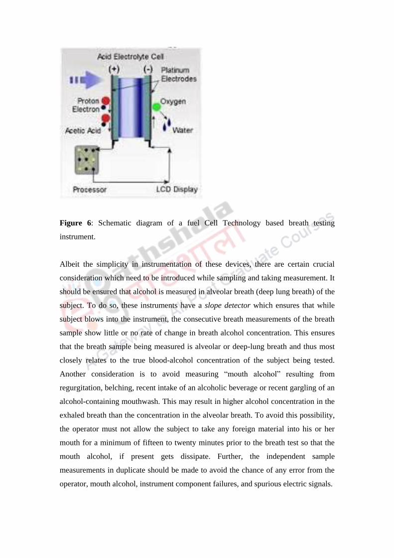
Figure 6: Schematic diagram of a fuel Cell Technology based breath testing
instrument.
Albeit the simplicity in instrumentation of these devices, there are certain crucial
consideration which need to be introduced while sampling and taking measurement. It
should be ensured that alcohol is measured in alveolar breath (deep lung breath) of the
subject. To do so, these instruments have a slope detector which ensures that while
subject blows into the instrument, the consecutive breath measurements of the breath
sample show little or no rate of change in breath alcohol concentration. This ensures
that the breath sample being measured is alveolar or deep-lung breath and thus most
closely relates to the true blood-alcohol concentration of the subject being tested.
Another consideration is to avoid measuring “mouth alcohol” resulting from
regurgitation, belching, recent intake of an alcoholic beverage or recent gargling of an
alcohol-containing mouthwash. This may result in higher alcohol concentration in the
exhaled breath than the concentration in the alveolar breath. To avoid this possibility,
the operator must not allow the subject to take any foreign material into his or her
mouth for a minimum of fifteen to twenty minutes prior to the breath test so that the
mouth alcohol, if present gets dissipate. Further, the independent sample
measurements in duplicate should be made to avoid the chance of any error from the
operator, mouth alcohol, instrument component failures, and spurious electric signals.

Summary:
Alcohol is abused largely due to its mood altering effects. Consumption of alcohol
creates a state of drunkenness to the subject which leads to various acts arising out of
negligence or with criminal intent. The extent to which an individual is under the
influence of alcohol is usually determined by measuring the quantity of alcohol in the
blood or the breath. In India, the legal limit for Blood Alcohol Concentration (BAC)
upto 30 mg per 100 ml of blood has been set, however, enactment of these legal
measures requires strict actions and seriousness among law enforcement agencies.
The legal limit is checked during field sobriety testing which also contains various
psychological tests such as horizontal-gaze nystagmus test, walk and turn, and the
one-leg stand. The BAC determination requires collection of blood or breath from
subject. Breath is tested on the basis of various principles such as oxidation reduction
in dichromate-acid solution, fuel cell chemistry, infrared spectroscopy. Among these,
infrared spectroscopy is one of the most commonly adopted method among law
enforcement agencies. Blood sample from vein is also tested for determination of
blood alcohol. Gas chromatography is the most widely used approach for determining
alcohol levels in blood. Blood must always be drawn under medically accepted
conditions by a qualified individual. A nonalcoholic disinfectant must be applied
before the suspect’s skin is penetrated with a sterile needle or lancet. Once blood is
removed from an individual, it is best preserved sealed in an airtight container after
adding an anticoagulant and a preservative.
Criminology
Forensic Science and Forensic Medicine
Arson: Definition, Chemistry of fire, Analysis of arson exhibits

DESCRIPTION OF MODULE
Items Description of Module
Subject Name Criminology
Paper Name Forensic Science and Forensic Medicine
Module Name/Title Arson: definition, chemistry of fire, analysis of
arson exhibits
Module Id
Objectives
Learning Outcome:
To make the learners understand definition of
arson and the chemistry of fire.
To make learners recognize the telltale signs of
arson
To describe how to collect physical evidence at
the scene of a suspected arson
To describe laboratory procedures used to detect
and identify hydrocarbon and explosive residues
Prerequisites General understanding of Forensic Science and its
application in crime solving.
Key words
Fire, hydrocarbon, residues, arson, origin, accelerant,
inflammable.
Role Name Affiliation
Principal Investigator Dr G. S. Bajpai
Co-Principal Investigator
Paper Coordinator
Content Writer/Author Dr Priti Sharma
Content Reviewer
1. Introduction:
Fire is a spontaneous burning process which has huge devastating effects on property
and person. Crime investigators often encounter with fire scenes which show
observable signs pointing towards its deliberate origin. The fire with deliberate origin
or the intentional fire has been differentially termed as ‘Arson’. Arson has been
committed throughout human history. Its definition as a crime originated in old
English common law, where the term “arson” referred specifically to a fire set by one
person against the dwelling of another.
There are a number of reasons why investigation team may suspect arson i.e.,
deliberate setting of fire. Arson often present complex and difficult circumstances to
investigate. The damage caused may range through structures, inhabited or not, as
well as vehicles or any other personal property. The perpetrator normally has
thoroughly planned the criminal act and has left the crime scene long before any
official investigation reaches. So, the criminalist’s function is very important since he
or she is trained to detect and identify relevant chemical materials collected at the
scene which help in reconstructing the crime and in identifying trace amounts of
gasoline or kerosene in debris or igniters used in setting the fire.
A fire can have many accidental causes, including faulty wiring, overheated electric
motors, improperly cleaned and regulated heating systems, and cigarette smoking
which usually leave no chemical traces. Thus, the final determination of the cause of a
fire or explosion must consider numerous factors and requires an extensive on-site
investigation.
In India, arson crimes are prosecuted under section 435 of Indian Penal Code. It
defines that whoever commits mischief by fire or any explosive substance
intending to cause, or knowing it to be likely that he will thereby cause, damage to
any property to the amount of one hundred rupees or upwards 1[or (where the
property is agricultural produce) ten rupees or upwards], shall be punished with

imprisonment of either description for a term which may extend to seven years,
and shall also be liable to fine
This chapter takes the reader from the beginning stages of a fire investigation in
evidence collection and evaluating a fire scene at the laboratory. Such knowledge
helps to reconstruct the case and finally provides evidences which will proof for or
against their intentional origin of fire. The primary aim of this module is to describe in
detail the intricacies about nature of fire and how a fire scene is investigated.
2. Chemistry of Fire
Fire may be defined as a transformation process during which oxygen is united with
some other substance i.e., fuel to produce noticeable quantities of heat and light (a
flame) by the process of oxidation. This is simple to understand by diagrammatic
representation of fire triangle as shown in Figure 1. The three corners depict that fuel,
oxygen and heat are required for sustaining fire.
Figure 1: Fire Triangle
This needs to be understood here that not all oxidation proceeds in the manner that
one associates with fire. For example, oxygen combines with many metals to form
oxides. Thus, iron forms a red-brown iron oxide, or rust. Furthermore, to start fire, an
energy input is needed to cross the energy barrier between the reactants and the
products of a reaction. The higher this barrier, the more energy required to initiate the
reaction. The energy barrier in the conversion of iron to rust is relatively small, and it
can be surmounted with the help of heat energy present in the surrounding
environment. But to ignite the gasoline, the energy barriers is quite high, and a high
temperature must be applied to start the oxidation of these fuels. Hence, before any
fire can result, the temperature of these fuels which is known as ignition temperature
must be raised to a value that will allow the heat energy input to exceed the energy
barrier. Once the combustion starts, enough heat is liberated to keep the reaction
going by itself. In essence, the fire becomes a chain reaction, absorbing a portion of
its own liberated heat to generate even more heat. The fire will burn until either the
oxygen or the fuel is exhausted.
So, we can picture an oxidation reaction, taking place when molecules combine or
collide with one another. Essentially, the faster the molecules move, the greater the
number of collisions between them and the faster the rate of reaction. The physical
state of fuel and temperature essentially affects this pace. A fuel achieves a reaction
rate with oxygen sufficient to produce a flame only when it is in the gaseous state, for
only in this state can molecules collide frequently enough to support a flaming fire.
This remains true whether the fuel is a solid such as wood, paper, cloth, or plastic, or
a liquid such as gasoline or kerosene. In the case of a liquid fuel, the temperature must
be high enough to vaporize the fuel. The vapour that forms burns when it mixes with
oxygen and combusts as a flame. The flash point is the lowest temperature at which a
liquid gives off sufficient vapour to form a mixture with air that will support
combustion. Once the flash point is reached, the fuel can be ignited by some outside
source of temperature to start a fire. With a solid fuel, the process of generating vapor
is more complex. Wood, or any other solid fuel, burns only when it is exposed to heat
that is hot enough to decompose the solid into gaseous products. This chemical
breakdown of solid material is known as pyrolysis. The numerous gaseous products
of pyrolysis combine with oxygen to produce a fire.
We may now consider the conversion of iron to rust as an example of an extremely
slow oxidation process, a situation that exists because of the inability of the iron
atoms to achieve a gaseous state. For this reason, the combination of oxygen with iron

to produce rust is restricted to the surface area of the metal exposed to air, a limitation
that severely reduces the rate of reaction. On the other hand, the reaction of methane
and oxygen is an example of oxidation in which all the reactants are in the gaseous
state. Hence, this reaction proceeds rapidly, as reflected by the production of
noticeable quantities of heat and light (a flame).
In summary, three requirements must be satisfied if combustion is to be initiated and
sustained:
1. A fuel must be present.
2. Oxygen must be available in sufficient quantity to combine with the fuel.
3. Heat must be applied to initiate the combustion, and sufficient heat must be
generated to sustain the reaction.
3. Examination of Scene
The investigation team following arrival at the scene should gather as much
information as possible from police officers and fire officers around them. The fire
fighters or police give first hand information about the fire which point towards the
possibility of ‘intentional fire’. Initially the CSI should thoroughly record the scene
both photographically and diagrammatically; video is an excellent tool for recording
the scene for possible future reconstruction as it records in low light conditions and
provides the viewer with a 360-degree view. A search of the fire scene must focus on
finding the fire’s origin, which will prove most productive in any search for an
accelerant or ignition device.
3.1. Locating the seat of fire
There are a number of clues which help in locating the seat of the fire. Low burning
where the floor is most damaged and high burning where the ceiling is most damaged
may help locate the seat of the fire. Burn patterns are classic indicators, with the ‘V’
pattern burn having the seat of the fire at its base. Figure 2 shows a photograph
indicating origin of fire. But the investigation team should be careful in jumping to
conclusions as some materials may burn more quickly than others or may be affected

by draughts or fire fighting. Glass and plastics melt towards the heat produced by the
fire, so called thermal indicators, can also be of use in locating the seat. Smoke
damage may travel away from the fire so will help in its location. Depth of charring to
wood may be a rough indicator to locate seat of fire (Dehaan 1997).
Figure 2: A photograph showing classic V pattern at origin of fire.
3.2. Signs of ‘deliberate fire’
In a search to determine the specific point of origin of a fire, the investigator may
uncover telltale signs of arson. The fire may be suspected to have originated
intentionally if it has multiple origins of fire (also called seat of fire). The firefighters
or the witness may also inform about the same person hanging around a number of
fire scene. If the fire had been very severe, developing rapidly and spreading through
a property, this indicates that an accelerant may have been used. An accelerant is
anything that speeds up the growth of the fire. Although usually associated with liquid
fuel, such as petrol or paraffin, an accelerant could equally be straw or newspaper. A

liquid fuel accelerant will typically leave a pool burn on the surface on which it has
been burning. As the vapour from the liquid fuel burns it creates a pool or halo shape
on the surface. There may be evidence of separate and unconnected fires or the use of
“streamers” to spread the fire from one area to another. Additionally, the presence of
containers capable of holding an accelerant or the finding of an ignition device
certainly will arouse suspicions of an arson-caused fire.
Other signs of deliberate fire are sign of breaking door or window, tempering of fire
appliances, unusual arrangement of furniture perhaps piled around the seat of the fire,
theft, removal of articles dear to the arsonist from the building for safe keeping prior
to the ignition of the fire. The fire may also be set to cover up and destroy evidence of
another crime such as a murder or theft.
3.3. Recovery of samples from the scene
While looking for evidences that may highlight the cause of fire, the investigators
have to look near area around the origin of a fire. Debris from areas around the origin,
as well as areas showing potential pour patterns, is collected to test for accelerant
residues at the laboratory. For the same purpose, an onsite screening may be made
using a highly sensitive portable vapor detector or “sniffer” (see Figure 3). This
device can rapidly screen suspect materials for the presence of volatile residues by
sucking in the air surrounding the questioned sample. The air is passed over a heated
filament; if a combustible vapor is present, it oxidizes and immediately increases the
temperature of the filament. The rise in filament temperature is then registered as a
deflection on the detector’s meter. Although, this test does not provide conclusive
remark, but it does provide the investigator with an excellent screening device for
checking suspect samples at the fire scene. The sniffer dogs that have been trained
and conditioned to recognize the odour of hydrocarbon accelerants are also commonly
used to search and screen the questioned sample for the presence of accelerant
residues.

Figure 3: Photograph of a vapour detector
As a matter of routine, two to three quarts of ash and soot debris must be collected at
the point of origin of a fire when arson is suspected. The collection should include all
porous materials and all other substances thought likely to contain flammable residues
such as wood flooring, rugs, upholstery, rags, etc. Specimens are to be immediately
packaged in airtight containers so no loss of possible residues can occur through
evaporation. New, clean paint cans with friction lids are good containers because they
are low cost, airtight, and unbreakable and are available in a variety of sizes (Figure
4). Wide-mouthed glass jars are also useful for packaging suspect specimens,
provided that they contain airtight lids. Cans and jars should be filled one-half to two-
thirds full, leaving an air space in the container above the debris. Large bulky samples
should be cut to size at the scene as needed so that they will fit into available
containers. Plastic polyethylene bags are not suitable for packaging specimens
because they react with hydrocarbons and permit volatile hydrocarbon vapors to be
depleted.

Figure 4: Evidence sampling Metal Cans
The collection of all materials suspected of containing volatile liquids must be
accompanied by a thorough sampling of similar but uncontaminated control
specimens from another area of the fire scene. This is known as substrate control. For
example, if an investigator collects carpeting at the point of origin, he or she must
sample the same carpet from unburnt part, where it can be reasonably assumed that no
flammable substance was placed. In the laboratory, the sample is tested to ensure that
that it is free of any flammables. This procedure reduces the possibility and that the
carpet was exposed to a flammable liquid such as a cleaning solution during normal
maintenance. In addition, laboratory tests on the unburned control material may help
analyze the breakdown products from the material’s exposure to intense heat during
the fire. This is because common materials such as plastic floor tiles, carpet, linoleum,
and adhesives can produce volatile hydrocarbons when they are burned. These
breakdown products can sometimes be mistaken for an accelerant.
Fluids found in open bottles or cans even if they appear empty must be collected and
sealed since they may contain trace amounts of liquids or vapors. Also, a thorough
search of the scene should be made for igniters. The most common igniter is a match.
Normally, the match is completely consumed during a fire. However, there have been
cases in which, by force of habit, the arsonist tossed aside the half burnt match which
can be recovered later by the investigator. This evidence may prove valuable if the

investigator can successfully fit the match to a match box found in the possession of a
suspect. In addition, an arsonist can construct many other types of devices to start a
fire. These include a burning cigarette, ammunition, a mechanical match striker,
electrical sparking devices, and a “Molotov cocktail (as shown in Figure 5).”
Relatively complex mechanical devices are much more likely to survive the fire for
later discovery. The broken glass and wick of the Molotov cocktail, if recovered, must
be preserved as well.
Figure 5: Molotov Cocktail
One important piece of evidence is the clothing of the suspect perpetrator. If the
suspect is arrested within a few hours after the fire, residual quantities of the
accelerant may still be present in the clothing. In such cases, the each clothing item
should be collected and placed in a separate airtight container, preferably a new, clean
paint can. The forensic laboratory can detect extremely small quantities of accelerant
materials in such clothing material making it a good piece of evidence especially if
the residues pattern match with the type recovered from the scene of fire.

When recovering samples, care must be taken by the CSI not to contaminate the
evidence. It is essential that clean, protective outer garments (including hand and
footwear) are worn, tools used to recover samples are thoroughly clean (some police
forces use disposable shovels) and samples recovered from the scene are transported
as soon as possible to the forensic laboratory.
4. Examination of exhibits at the laboratory
Debris from fire scene provides various evidences to prove or disprove arson. A
forensic chemist performs examination of debris to detect the presence of accelerant.
However, he/she should not only examine the residual content of debris, but also
perform the gross search of debris. For this, the debris is to be searched in controlled
laboratory conditions with good lighting to identify any item that may have caused the
fire, such as a lighter or cigarette. He should not rule out the possibility that the
arsonist may have intentionally left the device or device may have escaped the
damage caused by fire.
The forensic chemist will then examine the debris for the presence of liquid
accelerants, such as petrol or paraffin. The air left in the nylon bags containing the
debris is the source for the check for these accelerants. This air is recovered from the
sample debris contained inside the airtight jars by heating them. When the container is
heated, any volatile residue present in the debris is driven off and trapped in the
container’s enclosed airspace. The vapor is removed from the container using various
methods and then injected into the GC which is one of most commonly used
technique for detecting and characterizing flammable residues. Gas chromatograph
separates the hydrocarbon components and produces a chromatographic pattern
characteristic of a particular petroleum product.
To separate volatile accelerant residue from the fire scene debris, headspace method
as shown in Figure 6, is the most commonly used and conventional technique. In this,
the container containing fire debris is heated from outside at temperature not reaching
beyond 60oC. The collected vapour in the airspace is then collected using a syringe

which is punctured through the container wall and through suction, all the vapour is
collected. This sample air is then injected in the GC as described earlier for analysis.
Figure 6: Headspace technique
Albeit various advantages, there is also a shortcoming of headspace technique which
owes to limited volume of vapour that can be collected inside the syringe. This
shortcoming has been resolved by the introduction of another method called vapor
concentration. This is also called as Passive Headspace Method. In this method, as
shown in Figure 7, a charcoal-coated strip, similar to that used in environmental

monitoring badges, is used. This strip is placed within the container holding the fire
scene debris. The container is then heated to about 60°C for about one hour. As a
result, the accelerant vapour in significant quantity get collected into the container
airspace and finally get adsorbed onto the charcoal strip. Once the heating procedure
is complete, the strip is easily removed out from the container and the accelerant is
recovered from the strip by washing with a small volume of solvent (preferably
carbon disulfide). The solvent is then injected into the gas chromatograph where it is
again volatilized for analysis. The major advantage of using vapor concentration with
gas chromatography is its extreme sensitivity due to adsorption onto the charcoal
strip.
Figure 7: Vapour concentration using charcoal strip

When the vapor is injected into the gas chromatograph, it is separated into its
components, and each peak is recorded on the chromatogram in the form of a
characteristic pattern as shown in Figure 8. The typical set up of GC recording is
shown in Figure 9.
Figure 8: Typical Gas Chromatograph of known standards
Figure 9: A Typical Set-up of Gas Chromatography

The chromatogram pattern of vapour residue in question is then matched with patterns
of known standards of petroleum products to recognize the accelerant type. For
example, in Figure 10, a gas chromatographic analysis of debris recovered from a fire
site shows a chromatogram similar to a known gasoline standard, thus proving the
presence of gasoline. In the absence of any recognizable pattern, the individual peaks
can be identified when the investigator compares their retention times to known
hydrocarbon standards (such as hexane, benzene, toluene, and xylenes). At present, it
is not possible to determine the brand name of a gasoline sample by gas
chromatography or any other technique. Fluctuating gasoline markets and exchange
agreements among various oil companies make this difficult.
Figure 10: Gas Chromatograph of vapour from genuine gasoline standard (Top) and
of vapour recovered from debris at site of fire (Bottom). Courtesy: New Jersey State
Police.

Further, in some cases, the interpretation based on GC chromatogram is quite
difficult, if not impossible, due to mixture of accelerants or from heat generated
pyrolysis products of materials burnt at the fire scene. In these cases, gas
chromatography combined with mass spectrometry has proven to be a valuable
technique for solving difficult problems in the detection of accelerant residues.
In the mass spectrometer, the material enters a high-vacuum chamber where a beam
of high-energy electrons is targeted at the sample molecules. The electrons collide
with the molecules, causing them to lose electrons and to acquire a positive charge
(commonly called ions). These positively charged molecules or ions are very unstable
or are formed with excess energy and almost instantaneously decompose into
numerous smaller fragments. The fragments then pass through an electric or magnetic
field, where they are separated according to their masses generating a highly
characteristic line pattern where each line represents a fragment of a different mass
(actually the ratio of mass to charge), and the line height reflects the relative
abundance of each fragment. The unique feature of mass spectrometry is that under
carefully controlled conditions, no two substances produce the same fragmentation
pattern. This generates a unique pattern like “fingerprint” of the substance being
examined.
Complex chromatographic patterns thus can be simplified by passing the separated
components emerging from the gas chromatographic column through a mass
spectrometer. As each component enters the mass spectrometer, it is fragmented into a
collection of ions. The analyst can then control which ions will be detected and which
ones will go unnoticed. In essence, the mass spectrometer acts as a filter allowing the
analyst to see only the peaks associated with the ions selected for a particular
accelerant. In this manner, the chromatographic pattern can be simplified by
eliminating extraneous peaks that may obliterate the pattern.
Summary:
Arson is the act of deliberately setting things on fire. When a fire occurs, oxygen
combines with a fuel to produce noticeable quantities of heat and light (flames). If
combustion is to be initiated and sustained, a fuel must be present, oxygen must be
available, heat must be applied to initiate the combustion, and sufficient heat must be
generated to sustain the reaction. Fire requires heat, oxygen and fuel for its sustenance
however, to initiate it requires ignitor which may be flame, electric spark, blast, etc. In
any case of fire, the arson investigator needs to examine the fire scene for signs of
arson.
As soon as the fire has been extinguished, the investigator should search the fire scene
to find the fire’s origin. This site contains useful evidence to explain the cause of fire.
Some telltale signs of arson include evidence of separate and unconnected fires, the
use of “connectors” to spread the fire from one area to another, and evidence of severe
burning found on the floor as opposed to the ceiling of a structure. The importance of
porous materials at the fire origin is due to the fact that it contains the residues of
accelerant used by arsonist. So, porous materials such as carpet, bed sheet, curtains,
clothes, etc. should be collected and packed in air tight metal/glass containers to avoid
loss of volatile compounds.
In the laboratory, the volatile component is separated from the debris collected from
the fire scene using techniques such as headspace and vapour concentration which
involves application of heat to separate the volatile sample. They differ on the basis
that in headspace, the separated volatile sample is collected in syringe while in
passive headspace, it is adsorbed on charcoal strip. In any case, the sample is
identified using gas chromatograph which is the most sensitive and reliable
instrument for detecting and characterizing flammable residues. Most arsons are
initiated by petroleum distillates such as gasoline and kerosene. The gas
chromatograph separates the hydrocarbon components and produces a
chromatographic pattern characteristic of a particular petroleum product. By
comparing select gas chromatographic peaks recovered from fire-scene debris to

known flammable liquids, a forensic analyst may be able to identify the accelerant
used to initiate the fire.

Criminology
Forensic Science and Forensic Medicine
Forensic Entomology: Life cycle blow flies, Forensic
application.

DESCRIPTION OF MODULE
Items Description of Module Subject Name Criminology Paper Name Forensic Science and Forensic Medicine
Module Name/Title
Forensic Entomology: Life cycle blow flies, Forensic
application Module Id ----
Objectives
Learning Outcome:
To make the learners understand science of insect and its forensic significance.
To help the learners in determination of time since death.
To educate the learners to the Life cycle of blow flies.
To teach the learners to understand the decomposition time of dead body.
Prerequisites General understanding about entomology.
Key words
Forensic Entomology, Blow flies, Post Mortem Interval (PMI)
Role Name Affiliation Principal Investigator Prof. (Dr.) Ranbir
Singh Vice Chancellor, National Law University, Delhi
Co-Principal Investigator Prof. (Dr.) G.S. Bajpai Registrar, National Law University Delhi
Paper Coordinator Prof. (Dr) Sally Lukose Dean, School of Basic and
Applied Sciences, Galgotias
University Content Writer/Author Mr. Saroj Kumar Amar Assistant Professor, Forensic
Sc. School of Basic and
Applied Sciences, Galgotias
University Content Reviewer Dr. Tanya Chauhan Assistant Professor, LNJN-
National Institute of Criminology and Forensic Science, Delhi.

Forensic Entomology: Study of insects to the
criminal investigation.
The forensic entomologist job includes number of techniques about the life cycle of
insect species like succession, larval weight, larval length, and accumulated degree
hour technique. The use of insect behaviour to estimate the time of death is not a
current concept. The initial recorded crime investigation with insects was in 1235
A.D. in China. Sung Tz'u, a Chinese “death investigator,” wrote a book entitled. The
Washing Away of Wrongs. This medico-criminal entomology case was recounted as a
murder by slashing, which occurred in a Chinese village, and the local death
investigator was deputized to solve the crime. After some ineffective questioning, the
investigator had all the villagers bring their sickles to one spot and lay them out
before the crowd. Flies landed on only one of the sickle sticks. This was due to blood
being spattered on the sickle. The owner subsequently broke down and confessed to
the crime.
Job of Forensic Entomologist
1. Identification of insects at various stages of their life cycle, such as eggs, larva,
and adults.
2. Collection and preservation of insects as evidence.
3. Estimate for the postmortem interval or PMI (the time since death). Based on
dependent factors such as identification of insects or their stages, weather
conditions, location and condition of the body etc.
4. Justification before the court to explain insect-related evidence found at a crime
scene.

Most insects used in investigations are in two major orders:
A. – Flies (Diptera)
B. – Beetles (Coleoptera)
Importance of Species succession:-
Some species feed only a fresh corpse, while another species prefer to feed on one the
dead for two weeks. Experts should find other insect species that prey on the insects
feeding on the corpse. Which may gives clue to missing species.
Importance of dependent factors:- Weather data is also an important tool in
analyzing insect evidence from a corpse. Investigators will make note of the
temperature of the air, ground surface, the interface area between the body and the
ground, and the soil under the body as well as the temperature inside any maggot
masses. They will also collect weather data related to daily temperature
(highs/lows) and precipitation for a period of time before the body was discovered to
the time the insect evidence was collected.
Other factors that might affect their PMI estimates:
1. Was the body enclosed in an area or wrapped in a material that would have
prevented flies from finding the corpse and laying eggs?
2. Were other insect species present that may have affected the development of
the collected species?
3. Were there drugs or other poisons in or on the body that might have affected
the larvae’s development?
Blow Fly Metamorphosis
Blow flies are fascinated to dead bodies and often arrive within minutes of the death
of an animal. Blow flies have a complete life cycle with egg, larva, pupa, and adult
stages.

1st – Adult flies lay eggs on the carcass especially at wound areas or around the
openings in the body such as the nose, eyes, ears, anus, etc.
2nd – Eggs hatch into larva (maggots) in 12-24 hours
3rd– Larvae continue to grow and molt (shed their exoskeletons) as they pass through
the various instar stages.
1st Instar - 5 mm long after 1.8 days
2nd Instar - 10 mm long after 2.5 days
3rd Instar – 14-16 mm long after 4-5 days
4th – The larvae (17 mm) develop into pupa after burrowing in surrounding soil.
5th – Adult flies emerge from pupa cases after 6-8 days.

Examples of Diptera (Flies)
Examples of Coleoptera (Beetles)
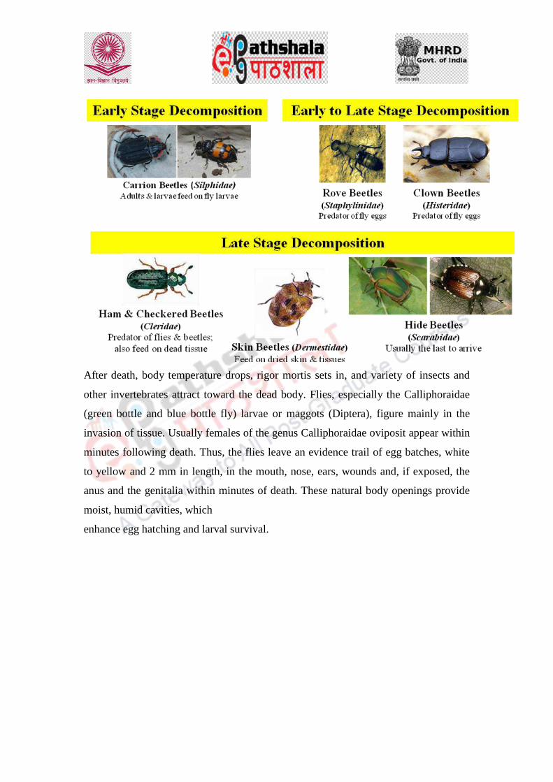
After death, body temperature drops, rigor mortis sets in, and variety of insects and
other invertebrates attract toward the dead body. Flies, especially the Calliphoraidae
(green bottle and blue bottle fly) larvae or maggots (Diptera), figure mainly in the
invasion of tissue. Usually females of the genus Calliphoraidae oviposit appear within
minutes following death. Thus, the flies leave an evidence trail of egg batches, white
to yellow and 2 mm in length, in the mouth, nose, ears, wounds and, if exposed, the
anus and the genitalia within minutes of death. These natural body openings provide
moist, humid cavities, which
enhance egg hatching and larval survival.
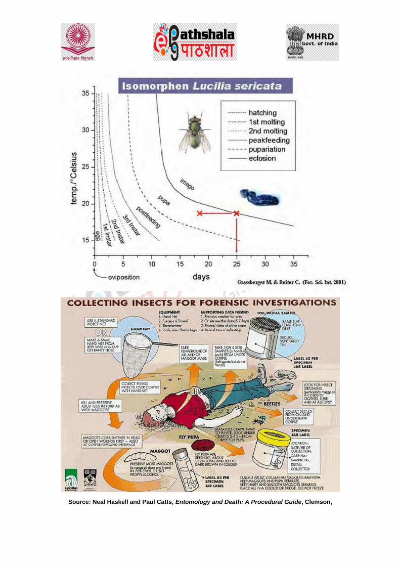
Source: Neal Haskell and Paul Catts, Entomology and Death: A Procedural Guide, Clemson,

Forensic significance of entomology
Forensic entomologist estimate PMI by examining the insect population on or near the
body. Expert can also estimate how long the deceased has been lying in a particular
location by the soil and insects beneath the deceased. If there is a difference in the
estimates, and the analysis of the soil suggests a short PMI while the analysis of the
body fauna suggests a longer PMI, one can determine that the body has been moved.
One can also estimate how long the body has been lying at a certain place by
assessing the plants and the soil surrounding the body.
Can insects mutely give us precious information about a crime? The experts at the
American Board of Forensic Entomology say we should be listening to insects
through the evidence they provide us. Can they tell us when and where someone was
murdered, or even who did it? Can they tell us what type of explosive material was
used in a bombing? This study and numerous green bottle blowflies may help us
decide that we should be listening.
References:-
1. http://www.forensicentomologist.org/
2. http://www.umext.maine.edu/images/FlyLife.jpg
3. http://www.kathyreichs.com/entomology.htm
4. http://www.forensicentomologist.org/
5. http://naturalsciences.org/files/documents/csi_tg_overview.do
c
6. www.cals.ncsu.edu/course/ent425/library/spotid/coleoptera/coleoptera.html
7. http://www.forensicflies.com/beetles.htm
8. Starkeby, M., “Ultimate Guide to Forensic Entomology: Introduction to Forensic
Entomology,” Web-only essay,URL< http://folk.uio.no/mostarke/forens_ent/
introduction.html>, accessed 5 January 2004.
9. K. Smith, A Manual of Forensic Entomology (New York: Cornell University Press,
1986), 20-23.

10. Starkeby, “Ultimate Guide to Forensic Entomology: Introduction to Forensic
Entomology,” 2004.

Law
Forensic Science and Forensic Medicine
Forensic medicine and medical jurisprudence, inquest, dying declaration,
exhumation. Law in relation to medical profession.

DESCRIPTION OF MODULE
Items Description of Module Subject Name Criminology
Paper Name Forensic Science and Forensic Medicine
Module Name/Title
Forensic medicine and medical jurisprudence, inquest,
dying declaration, exhumation. Law in relation to
medical profession.
Module Id LAW/CJA/VIII/33
Objectives
Learning Outcome:
To make the learners understand the need for
Medical laws concerning the rights and
responsibilities of medical professionals and
their patients
To make the learners understand of various
processes of jurisprudence in the application
of medical science to legal problems.
To acquaint the learners with the process relating
to exhumation.
Prerequisites General understanding of Medical law, concerning the
rights and responsibilities of medical professionals and
their patients including confidentiality, negligence and
other torts related to medical treatment
especially medical malpractice and criminal law and
ethics.
Key words
Jurisprudence, exhumation, medical negligence, malpractice, inquest.
Forensic science: 1,2
Role Name Affiliation Principal Investigator Prof. (Dr.) Ranbir
Singh Vice Chancellor, National Law University, Delhi
Co-Principal Investigator
Paper Coordinator Prof. (Dr.) Sally
Lukose Dean, School of Basic and
Applied Sciences, Galgotias
University, Greater Noida. Content Writer/Author Ms. Neeharika
Srivastava Assistant Professor, Galgotias
University, Greater Noida,
U.P. Content Reviewer

Forensic science is the application of science to criminal and civil laws. During the
course of an investigation, forensic scientist collect, preserve and analyze the
evidences scientifically. Or forensic science is the application of scientific knowledge
and methodology to legal problems and investigations.
Forensic science is comprised of different areas of science, biology, chemistry,
physics, genetics, medicine, anthropology, psychiatry and toxicology etc.
Apart from collection, preservation and examination, forensic scientist presents expert
testimony to courts.
With the use of fingerprint and other identification techniques in criminal
investigations, modern forensic science came in limelight. These days applications of
forensic science in legal issues have gained more popularity became more common as
every area of science has a potential bearing on law.
Forensic medicine: 2,3
Forensic implies something to forum while medicine is a science of cure and
preserving health. Forensic science is the branch of medical science which deals with
the application of medical knowledge in order to prove facts in criminal or civil cases.
Such applications can be applied in cases such as vehicular accidents, to establish the
fact that whether the accident is responsible for injuries or injuries are causes prior to
accident, in case of death to ascertain time of death, in case of sexual offences like
crime actually happened or not.

The area of a forensic scientist is not restricted to his own field only but he uses the
knowledge of other branches of medical science to solve the problem of forensic
medicine.
1 J. Prahlow, Forensic Pathology for Police, Death Investigators, Attorneys, 17 and
Forensic Scientists, DOI 10.1007/978-1-59745-404-9_2,
2 Parikh‟s Text book of medical jurisprudence Forensic Medicine and Toxicology
CBS publisher and distributors PVT.LTD.
3 anil aggrawal's internet journal of forensic medicine and toxicology, introduction to
forensic medicine and legal procedures prevalent in india - part i,volume 2, number 1,
january - june 2001.
Medical jurisprudence: 2,3,4
Medical jurisprudence is the legal aspect of practicing medicine.
Medical jurisprudence includes doctor-patient relationship, duties towards the patient,
rights and duties of doctor himself and medical negligence.
Doctors are obligated to certify persons for compensation, insurance plans, to certify
and issue birth and death certificates, to determine when a mentally disturbed person
to be detained to protect him and others. Such routine works are some frequent task
under Medical jurisprudence.
Medical jurisprudence may be involved in cases like paternity testing or genetic
relationships, in cases involving injuries or death from violence.

Medical ethics: 5, 6
Medical ethics is basically the moral principles that guide the medical professionals to
deal with each other, their patients and with the state as well. Few type of subjects that
may are relevant on medical ethics may include:
Patient autonomy and their right to refuse or choose treatment
Non-malaficence
Beneficence
Dignity
Honesty
justice
3 Mohammed Iliyas Sheikh, must know aspects of medical jurisprudence for medical
professionals, NHL journal of medica sciences/ Jan 2013/vol 2/issue, 1
4 Anil Aggrawal's internet journal of forensic medicine and toxicology, introduction
to forensic medicine
5 Dikshit P C, Text book of Forensic Medicine and Toxicology, PEEPEE publisher
and distributors (P) LTD.
6 Simpson's Forensic Medicine, 13th Edition, Jason Payne-James, Richard Jones,
Steven B Karch, John Manlove, August 26, 2011 by CRC Press.

Indian legal system: 5,6
The criminal procedure code, 1973 (CrPC) provides the machinery for crime
investigation, apprehension of suspected criminals, collection of evidences,
determining guilt or innocence and punishment of the guilty, public nuisance,
prevention of offences and maintenance of wife, child and parents within the
limitations of the union territories of India except Jammu & Kashmir and some other
tribal areas. 5,6 It also constructs the duties of police in arresting, dealing and
investigating the crime.
The Indian Penal Code, 1860 (I.P.C) describes various crimes with their punishment
in court of law.
The Indian evidence act, 1872 (I.E.A.) provides set of rules and allied issues which
govern the admissibility of evidences found at scene to reach a conclusion in the court
of law.
Criminal law describes the offences which are against the public interest namely
person, property, and safety of public and security of the state.
Civil law deals with disputes between two individual or parties.
Inquest : 2, 7
Inquest (in = in; quasitus= to seek) means judicial inquiry to ascertain a matter of fact.
An inquest is a judicial inquiry particularly into the cause of death where the death is
not natural. In such cases where death is under suspicion, investigation becomes
necessary to apprehend and punish the culprit.
The unnatural death, is defined under section 174 of CrPC, is that
- A person has committed suicide, or
- killed by another person or
- killed by an animal or
- Because of an accident or

- Or some other circumstances raising suspicion of some foul play
Inquest can be conducted in number of cases like suicide, homicide, industrial
accidents, domestic accidents, road accidents, infant deaths, operational deaths,
negligence, abortion, drug or poisoning cases, sudden death or death in custody. There
are four types of inquest, namely
- The Police inquest (conducted by a police officer)
- The Coroner‟s inquest ( conducted by a coroner, not followed in India
now)
- The Magistrate‟s inguest (conducted by a Magistrate)
- The Medical Examiner system (conducted by an official who is legally
and medically qualified, prevalent in USA)
7- 174 CrPC
Police inquest: 2,8
Police inquest (Section 174 Cr.P.C. ) to be conducted by officer in charge of a police
station or some other police officer empowered by the government.
Police inquest is conducted in cases like suicide, homicide, accidental (killing by an
animal or some machinery, death where suspicion arises that some other person has
committed the crime or in cases like where death of a women takes place within 7
years of her marriage and a relative of her make a request in this behalf.
On receipt of information of death, police reaches to the site and holds an inquiry with
the people of locality.
Police examines the situation and make a report describing the situation of body and
wound, bruise, fracture or any other mark which give indication of the method of

instrument used to inflict the injury. This proceeding comes under section 174 and
176 of the code of criminal procedure which will be conducted by the concerned
police officer and the Magistrate.
In such cases of unnatural deaths, In addition to sending the first information report, it
should be accompanied with inquest report and statements of witnesses attested by the
panchayats summoned under section 175 Cr.P.C.
If there is no foul play suspected, body is given to the relatives of the victim for
cremation.
In cases where there is suspicion of homicide, suicide, accident or any kind of
suspicion of unnatural death, the police officer conducting the inquiry sends the body
for post mortem to the nearest government hospital or to private hospital authorized to
conduct medico legal examination.
8- Committee on Reforms of Criminal Justice System Government of India, Ministry
of Home Affairs Report VOLUME I INDIA, March)

Magistrate’s inquest: 2,3,9
Magistrate‟s inquest (Section 176 Cr.P.C) to be conducted by District Magistrate/
Sub-divisional Magistrate, Executive Magistrate/ Judicial Magistrate.
Magistrate inquest is based on the assumption that inquiry made by police is not
always reliable.
In cases where women commit suicide within 7 years of her marriage or in case of
death of a woman where circumstances raise suspicion of some other person
committing the crime, Magistrate inquest is held.
Magistrate can hold an inquiry into the cause of death of any case mentioned under
police inquest.
In cases where person dies/disappears or cases like where suspicion of rape is there ,
inquiry is made by Judicial Magistrate in addition to the police inquest.
In cases where it is considered expedient or necessary to examine of a dead body
which already has been interred, the Magistrate may cause the body to disinter in
order to know the cause of death.
Medical Examiner system: 2,9
This kind of inquest is common in most of the stats of USA. In this system a medical
person is appointed to hold an inquest. The medical person owing his knowledge of
medical sciences gathers the evidences and performs the autopsy. He doesn‟t have the
power to summon and examine the witnesses. He submits his report to the district
attorney.

9 Vij K, Text book of Forensic Medicine and Toxicology, principle and practice,
Reed Elsevier India Private Limited.
Documentary evidences: 2
Documentary evidences comprise all the documents either written or printed to be
produced in court of law for examination during a trial. The term document here
includes any media by which information can be preserved. It may include the
following documents:
1- Medical certificate:
It is the certificate or statement from a physician which is issued in relation to ill
health, age , sex, insanity, pension disabilities or death. It can serve as a sick note
(employee is unfit for work) or an evidence of health condition.
2- Medical Report:
A medical report may be an injury report, post mortem report, report on sexual
offences, pregnancy, abortion or delivery etc.
3- Dying declaration: 2,5,10,11,12
Dying declaration is a legal term used for „words said before death‟. According to the
section 32(1) of the Indian Evidence Act “when the statement is made by a person as
to the cause of his/her death or as to any of the circumstances.
According to the section 157 IEA, Dying declaration from a person is made before his
death and should be supported by corroborative evidences.
Dying declaration is found relevant in case of, when it correlates with cause of death
and statement made by a person to any circumstances resulted in his death.

The provision of dying declaration was made basically on two grounds (1) when the
victim is the only eye witness and excluding his or her statement will not meet the
justice (2) person is believed to tell truth at the time of death.
Magistrate is called to record the declaration if time permits; otherwise a doctor can
take the declaration either at the site or in the hospital. Depending upon the situation
police officer, panchayat head or even relative can take the declaration.
According to section 162 CrPC, dying declaration may be taken in form of first
information report.
10 critical appraisal of dying declaration, dr. r.k.gorea, dr. 11o.p.aggarwal, jiafm,
2004; 26(1). issn 0971-0973.
11 Gupta bd, jani cb. status of compos mentis in relation to dying declaration in burn
patients. jiafm. 2004; 25(4):133 – 136
12 Indian Evidence Act, 1872, Criminal Manual. 14th ed. Lucknow: Eastern Book
Company, 2003: p15)
If the person dies before completion of the statement, the incomplete declaration is
not admissible in the court.
The doctor has to issue two certificates confirming the compos mentis of patient, one
before the declaration and other when the declaration is concluded.
The declaration may be oral but the person receiving it should record it in writing.
The declaration should be recorded in presence of two disinterested witnesses.
The declaration should be read over to the victim and he should affix his signature or
thumb impression when concluded. It should also be signed by doctors and witnesses.
The sealed declaration then sent to appropriate magistrate.
The investigating police officer should not be present
there when the declaration is being recorded.
In case of dying declaration, if the person survives, the
declaration does not has any legal value.
Under section 157 IEA, dying declaration can be
treated as corroborative evidence if the person survives.

Miscellaneous: it may include expert opinion from books or deposition from previous
case proceedings, etc.
Exhumation: 9
Aggrawal A. Exhumation – medical and legal aspects. Anil Aggrawal‟s Internet
Journel of Forensic Medicine and Toxicology 2001;2: available at
http://www.geradts.com/anil/ij).
Duff EJ, Johnson JS. Some social and forensic aspects of exhumation and reinterment
of industrial revolution remains. BMJ 1974; 1:563-7
Exhumation (ex= out of, humus= ground) means out of
ground or it is the authorized digging out of a dead person.
Exhumation is both in civil and criminal cases, to find
out a person‟s identity, establish cause of death or to decide some other relevant fact.
In India, majority of people are Hindus who believe in
cremation (burning) of body as soon as possible so exhumation is not of much
importance in India. Only a few communities here believe in burying of body after
death where exhumation might be useful to know the cause of death.
Exhumation can be done where the autopsy was not
done or had been done before burying the body but again needed after exhumation.
Exhumation can be done in civil cases like insurance
claims, accidental death claim, liability for medical negligence, compensation claims
or disputed identity and criminal cases like criminal abortion, homicide cases,
poisoning or criminal negligence.
There is no time limit for exhumation in India.
For exhumation written order is obtained from
magistrate. The place of burial is identified correctly and magistrate, police officer
and medical officer have to be present at the spot.
According to section 176 (4) Cr.P.C. relatives are
allowed to remain at the site throughout the process.

Procedure: 9, 13, 14 The grave is dug up to the level of coffin, photographs are taken
and sketch can also be drawn about the position of body and coffin and corpse should
be taken out carefully to avoid artefacts.
No disinfectant should be sprinkled on the body.
In case of skeletonisation the soil should be searched
for objects like bullet, teeth, hyoid bone or metallic objects etc. If any fluid or debris
is present in the coffin, it should also be collected.
If injuries are present, there are chances that injuries on
soft tissues may get distorted, so care should be taken while interpreting them.
If poisoning is suspected, viscera should be kept for
further analysis.
If body is decomposed so badly that no tissue is left
then hair, soil, teeth and bones should be collected. In such cases, ½ kg of soil from
top, bottom and sides should be preserved for further chemical analysis.
Whether the skeleton or a single bone is obtained for
examination, should be analyzed for its authenticity (actually it is bone or not), origin
of bone ( animal or human bone), they belong to one or more individual, race of the
person, sex determination, age, stature of the person it belongs, nature of injury, time
since death and cause of death.
13 ALEX KIRASI OLUMBE AND AHMED KALEBI YAKUB, Management,
exhumation and identification of human remains: A viewpoint of the developing
world, RICR Décembre IRRC December 2002 Vol. 84 No 848
14 M.A. Dada and D.J. McQuoid-Mason DJ, Introduction to Medico-legal Practice,
Butterworths, Durban, 2001, pp. 341-343.

Law in relation to medical profession: 2, 9, 15, 16
There are number of medicine system like western medicine, traditional Chinese
medicine, ayurvedic medicine in India and many native medicine systems from Africa
and Asia. In spite of having their own tradition, conventions and code of conduct, the
key principle espoused from the basis is called „medical ethics‟ for all the systems.
Changes influenced by society continue to evolve and change medical ethics time to
time. The laws governing the medical practice undoubtedly vary country to country
but principles of medical ethics are universal and followed by national and
international organizations. The medical ethics are nothing but the moral principles to
guide the medical professionals. In India, the enforcement is done by the Medical
Council of India and state medical council.
Medical council of India:
Medical council of India was established by Indian
Medical council Act of 1956. A medical register is maintained foe India and for the
matters concerned with it.
The council consists of one member from each state
nominated by the central government in consultation with the concerned state
government, One member from each university to be elected from its medical faculty,
One member from each state where the state medical register is maintained, who
possesses the qualification included in the 1st, 2
nd and part II of the third schedule,
seven members to be elected from any state medical register who possess a
qualification included under part I of the third schedule and eight members to be
nominated by the central government.
The main functions of Medical council of India
includes; maintenance of medical register, medical education where it maintain the
same standard for graduate and post graduate courses throughout India, recognition of
foreign medical qualification to an Indian national with foreign degree, appeal against
disciplinary action and warning notice in relation to professional misconduct.

Rights and privileges of a registered medical practitioner:
Right to practice medicine
Right to choose a patient
Right to issue a certificate
Right to add title to his name
Right to recover his fees
Right to appointment to public hospitals
Right to give expert opinion in court
Right to dispense medicine
Duties of a physician in general:
Maintain the highest standard of professional conduct
Respect the patient‟s right to accept or refuse the
treatment
Should avoid unfair discrimination
Deal honestly with patient and colleges
Should not provide specific products to patient to gain
financial benefits.
Certify only that which he personally verified.
Respect the national and international code of ethics.
Duties of physician to patient:
Examine the patient properly
Bear in mind the obligation to respect human life
Duty to exercise necessary care and skill
When providing medical care, should act in patient‟s
best interest
Should own patients loyalty
Give emergency service as a humanitarian duty
Not enter into a sexual relationship with his/her patient.
To prescribe medicine properly

Duties of physician towards colleagues:
never criticize the colleagues
never take fees from colleagues
always help the colleagues
consultation with the colleagues to provide the patient
best assistance
Should never undermine the patient-physician
relationship of colleagues to attract the patient.
Duties of physician towards state:
There are basically two duties of a physician towards state-
1- Notifiable diseases
2- Geneva conventions
Notifiable diseases:
In case of communicable diseases like cholera, plague etc, it is duty of a doctor to
inform the health authorities. In addition to this his duty also includes to inform the
authorities about the birth and death, failing of which a civil suit can be brought
against him but not the criminal suit.
15 FORENSIC MEDICINE AND TOXICOLOGY, W.G. AITCHISON
ROBERTSON, August 10, 2006, ISO-8859-1
16 Clinical forensic medicine, W.D.S McLay, Cambridge University Press 2009,
ISBN-13 978-0-521-70568-4

Geneva conventions: 2, 9
According to Geneva conventions, 1949, a doctor is bound to provide medical
assistance
- to a person of armed force (I convention)
- ship wrecked person ( II convention)
- prisoner of war (III convention)
- civilian of enemy nationalist (IV convention)
The mentioned categories of persons should be provided medical assistance without
any discrimination based on race, religion or political grounds.
Medical negligence (malpraxes) : 9
In general the negligence is “the omission to do
something which reasonable man could do or doing something which a prudent or a
reasonable man could not do.” While Medical negligence is defined as “want of
reasonable care and skill or willful negligence on the part of the medical practitioner
while treating a patient resulting in bodily injury, ill health or death.” Or in other
words since the doctor owes duty towards his patients, if there is some breach of the
duty by the doctor due to which patient suffered damage is medical negligence.
Medical negligence is a civil wrong, rarely the medical negligence may be renewed
from a civil action to the criminal courts.
In present times doctors too are very much prone to
threat with an action of negligence. Even the patients who are provided with the best
treatments with complete cure without cost are ready to sue the doctor. The matter of
fact is, severe disability resulting from an accident during the course of an accident is
not necessarily negligence.
The doctor has right to refuse a patient for treatment
but once he accepted to provide him medical assistance his duty towards the patient
starts.
There are certain circumstances where the doctor can
refuse to accept the patient for treatment. The conditions are:

- If the deceased does not belong to doctor‟s area of
specialization
- If the facilities needed are not available at that place
- the doctor himself is not well or any of the member of
his family is not well
- If the doctor is busy in some family functions.
During the emergency cases it is the moral and ethical
duty of the doctor to provide medical assistance to the patient in order to save the life.
If the patient cannot be accepted, doctor should make sure that the patient reaches to
the nearest hospital as early as possible. In such case if there occurs some damage to
the patient, he cannot sue the doctor.
It is the duty of a doctor to exercise reasonable skill
and care when the doctor-patient relationship is established.
When a doctor accepts a patient with intent to heal, a
doctor-patient relationship establishes and from that moment doctor is legally
responsible to provide due care, breach of which makes the ground for medical
negligence.
For all the doctors who qualified the examinations and
registered in the state medical council or Indian medical council, there is a minimum
level of competence to protect the public from insufficiently qualified doctors.
The doctor must use care, reasonable in circumstances.
Like responsibility of recovery of swabs used in operation theatre lies with the doctor.
Precaution against medical negligence:
Reasonable care, skill and attention to be taken
Should never guarantee a cure
Always obtain the informed consent from the patient
Doctor should maintain proper and accurate record.
Before injecting the hypersensitive drugs like penicillin
or streptomycin , sensivity test should be performed.
Medicine should be given in proper dosage
Specialist should be consulted when needed

A written informed consent should be taken from
patient before the surgery.
Contributory negligence: Contributory negligence is generally a defense based on
negligence. It is relevant when the claimants contributed to the harm they suffered
through their own negligence. In medical scenario, it is the absence of reasonable care
on part of patient or by his attendant that combines with the negligent action of the
doctor resulted in the damage, for eg- not to provide full history to the doctor. In cases
where doctor and patient boyh are negligent, doctor can take a good defense while
when only patient is negligent it is called „the negligence of the patient‟.
Therapeutic misadventure: Therapeutic misadventure is the unexpected damage to
the patient by the doctor while diagnosing or during the experiment. For eg-
radiological procedures during diagnosis may prove fatal or foetal death in uterus
while mother was given some drug during pregnancy.
The consumer protection act: 9
The consumer protection act 1986 came into force on
15 april 1987 is a welfare legislation that mainly favors the workers.
The complaint can be lodged in any places with or
without engaging a lawyer by paying a nominal fee. The complaint may be filled by
the district forum by
The consumer to whom the service has been provided
or agreed to be provided.
Consumer association
State or central government
The limitation period for the complain to be lodged in
all the forum is within two years from the date on which the cause of action has
arisen.
As penalty the person shall be punished with
imprisonment ranging from one month to three years and fine from rupees two
thousand to rupees ten thousand.

Law
Forensic Science and Forensic Medicine
Autopsy: Internal and external examination

DESCRIPTION OF MODULE
Items Description of Module
Subject Name Criminology
Paper Name Forensic Science and Forensic Medicine
Module Name/Title Autopsy: Internal and external examination
Module Id LAW/CJA/VIII/34
Objectives
Learning Outcome:
To make the learners understand about the
human autopsy in order to know suspicious,
violent or unknown cause of death.
Providing the learners an idea of Advantages
and benefits of autopsy by classifying in benefits
for medical practice, for deceased’s families and
for society.
The goal of an autopsy is to inform and help
clinicians learn from mistakes and improve
Role Name Affiliation
Principal Investigator Prof. (Dr.) Ranbir Singh
Vice Chancellor, National Law University, Delhi
Co-Principal Investigator
Paper Coordinator Prof. (Dr.) Sally
Lukose
Dean, School of Basic and
Applied Sciences, Galgotias
University, Greater Noida.
Content Writer/Author Ms. Neeharika
Srivastava
Assistant Professor, Galgotias
University, Greater Noida,
U.P.
Content Reviewer

medical future practice in favour of next patients.
Prerequisites General understanding of the medico legal and
pathological autopsy with an insight to get better
understanding of the causes of death.
Key words
Autopsy, thanatology, incision, haemorrhage,
histopathology.

Autopsy
Introduction
Autopsy refers to the examination of whole body after death. Autopsy is also referred
as post mortem, necropsy and thanatopsy. If the cause of death is known, still there is
a provision to examine all the body parts and all the cavities to find out any
contributory factor to death. To avoid miscarriage of injustice a proper autopsy is
needed and to establish facts between the eyewitnesses and investigation team. A
certified and experienced medical examiner carries out the post mortem to find out the
answers of queries asked by the investigator, often the cause of death. 1,2.
Autopsy can be of two types: (1) medico legal autopsy (2) pathological autopsy.
Medico legal autopsy is done on the request of investigative officer. In such autopsy
consent of the relative is not required. The medical practitioner performs the post
mortem to know the cause of death. (2) Pathological autopsy is one in which autopsy
is done on the request of relatives and performed in pathological department. In such
cases medical practitioner performs the autopsy of a particular or more organs where
pathology is suspected to find out the cause of death. 3,4
Autopsy is performed with following objects:
- To identify unknown bodies
- To know the cause of death
- To know the manner of death (suicide, homicide or accidental)
- To find out time since death
- To get trace evidence and preserve them when needed
- To preserve the viscera when needed.
Procedure for autopsy: 2,3,5,6
External examination:
- A brief description of age, sex, race, height and weight should be made.
- A brief note of articles found on body (shirt, t-shirt, colour, button etc)
- Degree and distribution of rigor mortis and post mortem staining.

- Colour of eye, hair, any unusual appearance, congenital malformations, scar ar
acne should be noted.
- Any evidence of disease, old injury, or medical surgery should be noted down.
If possible with age of injury should also be noted down.
- In case of gunshot wounds, size and length should be described in inches or
centimeters in relation to the head or sole and to the right nd left of midline.
- In case of injury caused by bullet, its place, intactness, condition of bullet, any
deformity to bullet should be noted. Then bullet should be kept in an envelope
with all the details like name of victim, date, postmortem details etc.
- When there is stab wounds, note should be made of its edges, dimensions and
estimation of depth.
- An examination of presence of trace evidences such as soot, grease, paint or
powder etc to be made.
Internal examination of the body during autopsy: 3,7,8
In the internal examination, the incision of the body is done through which we inspect
the various organs and examine the cavity systematically.
Weigh of the organs and checking for the pathological diseases are also done in
autopsy.
Incision must be adopted to suit the circumstances of case
The three major cavity of the body should be opened and examined these are:
Skull
Thorax
Abdomen
Three types of incisions: 2,3,9
I-shaped incision
1. Y-shaped incision
2. Modified Y-shaped incision

In I-shaped incision,
The incision is drawn from just above the thyroid cartilage to pubic symphysis
avoiding the umbilicus and any injuries in the line of incision.
This method is mainly used due to its simplicity and convenience.
In Y-shaped incision,
Two incisions commence on either side of neck from 2-3cm behind the lobe of
each ear to meet at manubrium sterni (called supersternal notch) and then
continued as a single incision down to pubic symphysis.
This method is used in which the more detail about neck is required.
In modified Y-shaped incision,
Two incisions are made commencing on either side of the chest from anterior
auxiliary fold, curving under the breast/nipples to xiphisternum and to be
continued as a single vertical incision down to pubic symphysis.
1 Wikipedia (https://en.wikipedia.org/wiki/Autopsy)
2 Dikshit P C, Text book of Forensic Medicine and Toxicology, PEEPEE
publisher and distributors (P) LTD.
3 Parikh’s Text book of medical jurisprudence Forensic Medicine and
Toxicology CBS publisher and distributors PVT.LTD.
4 Charlton, R: Autopsy and medical education: a review. J R Soc Med. 1984,
87 (4):232–236.)
5 verbal autopsy procedure, The 2012 WHO verbal autopsy instrument, ISBN
978 92 4 154847 2, World Health Organization 2012,
6 Horowitz, R.E., Naritoku, W.Y.: The autopsy as a performance measure and
teaching tool. In: Hum Pathol, 2007, 38 (5):688–695.
7 Nemetz, P.N., Ludwig, J., Kurland, L.T.: Assessing the autopsy. In: Amer J
Pathol 1987; 128:362-79.
8 Vij K, Text book of Forensic Medicine and Toxicology, principle and
practice, Reed Elsevier India Private Limited.

9 Roulson, J., Benbow, E.W., Hasleton, P.S.: Discrepancies between clinical
and autopsy diagnosis and the value of post mortem histology; a metaanalysis
and review. In: Histopathology 2005, 47:551-559.
_____________________________________________________________________
_
Order of examination: 8
Autopsy techniques may vary in order in which the organs are removed.
Virchow’s technique:
This method has been used most widely. In this method organ are removed one by
one. This technique follows the sequence: cranial cavity → the spinal cord →
thoracic, cervical and abdominal organs.
Rokitansky’s technique:

This technique includes in situ dissection combined with en block removal. This kind
of study is preferred where infections like HIV and hepatitis B have to be prevented.
Examination of neck: 2,3,10
In case of asphyxia deaths (hanging and strangulation) brain is taken out first
to completely drain the blood from neck in order to examine the neck in
bloodless field. The chest organs can be removed prior to neck dissection
avoiding injuring the neck veins.
In dissecting anterior neck structure the platysma muscle is examined for
presence of bruises.
The sternocleidomastoid muscles are left intact and the external jugular veins
are then examined.
Lateral clavicular head is detached. Suprahyoid and infrahyoid muscle groups
are then examined, carotid sheath containing carotid arteries, internal jugular
veins and the vagus nerve are examined.
While dissecting the posterior neck structure which is suspected because of
suffocation in traffic accidents and is recommended in all infant deaths
occurring outside the hospital, the prevertebral fascia can be examined for
evidence of traumatic injury, such as presence of crepitus. The fascia is then
reflected from the underlying bone.
The body is turned over and the superficial tissues including ligamentum
nuchae is reflected from the occipital region inferiorly to the base of the neck,
in order to expose the underlying soft tissues.
Internal examination of Thorax
Evaluation of coronaries:
Before any form of cardiac dissection is applied, coronaries should be
inspected for calcification & tortuosity.
Examine the size, shape, position, number & patency of coronary artery Ostia.
Heavily calcified coronary arteries, if possible, should be removed intact,
decalcified and opened transversely.

Coronary artery bypass grafts (saphenous veins, internal mammary arteries,
radial arteries etc.) may be examined with longitudinal or transverse cuts.
Examined by making serial cross section to check the ante mortem clots.
Air embolism in heart chambers should be also checked.
Subjects younger than 30 yrs or where the cause of death is non cardiac,
coronaries may be opened longitudinally.
Examination of the Heart
Open the right atrium by a cut from the inferior vena cava into the right
atrial appendage.
- Open the tricuspid valve laterally down to the apex of the right ventricle.
- Cut from the apex of the right ventricle through the pulmonary valve into
the pulmonary artery.
- Join two pulmonary veins across the roof of the left atrium to view the
inside of the chamber.
Cut through the mitral valve orifice laterally down to the apex of the left
ventricle.
Cut from the apex of the left ventricle up through its outflow tract into the
aorta.
There are two variations in this final cut.
It may be made through the anterior cusp of the mitral valve itself or pass
anteriorly, leaving a flap of anterior wall of the ventricle hinged on an
intact anterior cusp of the mitral valve.
The enterotome is inserted into the :
RA→ Tricuspid valve→ RV→ Pulmonary Trunk→ Pulmonary Vein→
LA→ Mitral Valve→ LV→ Ascending aorta.
Heart should be eventually weighed and the various measurements of the
circumference of the valves or the thickness of the ventricles etc are taken.
LUNGS:

Autopsy examination of the lungs includes dissection of the pulmonary
arteries, veins, removal of the lungs from the thoracic cavity and examination
of the outer lung surfaces (pleura).
Weighed before cutting as appreciable oedema fluid can run out during
dissection.
Dissect and examine the internal lung structures (arteries, veins and airways)
should be done first then open the lungs with a knife to reveal the lung tissue
(parenchyma).
Examined for consolidations, edema, atelectasis, emphysema, tardieu spots,
emboli etc.
Finally examined the lung tissue under the microscope to look for
abnormalities in the tissue and cellular level.
KIDNEYS:
Size and weight are noted.
Capsules are examined and excised carefully.
Kidney is sectioned longitudinally the convex border of the hilum so that it
splits into half and open the pelvis.
Checked for calculi, inflammation etc.
Ureter is cut and examined.
STOMACH:
Stomach is opened along the greater curvature from the cardiac to the pyloric
end after applying double ligatures.
Size of pyloric ring is noted.
Contents are examined for any nature of food which might be present and its
state of digestion, smell, color, character, presence of foreign bodies.
Mucous membrane is examined for congestion, haemorrhage, ulcerations or
any other abnormality.

POSTMORTEM EXAMINATION OF SPINAL COLUMN
AND SPINAL CORD
The spinal cord should be examined routinely in every postmortem
examination on a patient with a disorder of central nervous system or any
other head injury.
Removal of the spinal cord may disclose that there has been an unsuspected
fracture/dislocation of the cervical spine.
The basic principle in the anterior approach to the spinal cord is to cut through
the pedicles of the vertebrae so that the cord can be exposed by removing the
vertebral bodies.
After opening the brain the dura is cut open and checked for haemorrhage,
inflammation, tumours, pus etc.
Nerves are cut from below as they pass though the spinal foramina and the
cord is separated from the foramen magnum.
Cord is then sectioned transversely and serially, lastly, the vertebral column is
examined for fractures or dislocations.
POSTMORTEM APPEARANCE OF TESTIS AND FEMALE GENITLIA
TESTES: 8
Testes are examined for enlargement and malignancies.
Vertical cross-sections are made through the lateral and median lobes and the
prostate is split open for examination.
Inguinal canal is incised from the peritoneal aspect and the loop of the vas
deference is pulled to free it from the internal inguinal ring.
The testes is pushed with one hand and pulled out of the scrotum easily by the
other.

These are cut longitudinally and checked for any clotted blood inside the
scrotum and in the testis.
These should also be sectioned and examined wherever necessary.
Female genitlia-
Vagina and the uterus are cut either anteriorly or posteriorly up to the fundus.
Two short incisions are made at the fundus from the main incision towards
each cornu so as to expose the endometrium.
Ovaries are sectioned longitudinally and the fallopian tubes are cut
longitudinally.
If the uterus contains a fetus, its age should be determined.
PRESERVATION OF VISCERA AND OTHER TISSUES
Viscera should be preserved if death is suspected to be due to poisoning either by the
police or the doctor, Deceased was intoxicated or used to drugs, Cause of death not
found after autopsy, Death due to burns. Advanced decomposition and Accidental
death involving driver of a vehicle or machine operator.
The following must be preserved in all fatal cases of suspected poisoning.
(1) Stomach and its contents. If the stomach is empty, the wall should be preserved.
(2) The upper part of small intestine (about 30 cm. length) and its contents.
(3) Liver 200 to 300 gm.
(4) Kidney half of each as one kidney may be dysfunctional.
(5) Blood 30 ml. Minimum 10 ml.
(6) Urine 30 ml.
10 Shojania, K.G., Burton, E.C.: McDonald, K.M., Goldman, I.: Changes in rates of
autopsy - detected diagnostic errors over time: a systematic review. In: JAMA 2003;
289:2849–56.

CIRCUMSTANCES OF PRESERVATION 3,2
These are preserved for chemical analysis under the following circumstances:
1. When the investigating officer so requests
2. When the medical officer suspects the presence of poison by its smell or some other
evidence while conducting autopsy on injury cases.
3. To exclude poisoning in instances where the cause of death could not be arrived at
after a full autopsy and there is no natural disease or injury
4. In decomposed bodies
5. In alcoholics
Any tissue which is likely to provide evidence should be preserved in 10% formalin
for histopathological examination .
When blood or other fluid is found within a body cavity,the quantity should be
measured and recorded.
ROUTINE VISERA PRESERVATION INCLUDES:
Stomach with its contents and proximal 30 cms of intestine with its content.
Half of each kidney and 500gm of liver
100ml of blood
Only the preservatives, which act as control sample.
Special preservation includes i. e of liver, heart ,brain ,bile ,lungs , vitrous humor,
bone and uterus .
PRESEVATIVES USED
POISONING PRESERVATIVE
In all cases of poisioning but
acid poisoning (except
carbolic acid)
Saturated salt solution
For acid poisoning except
carbolic acid poisoning.
Rectified spirit

carbon monoxide poisoning
A layer of paraffin
All cases of poisoning
including alcohol poisoning for
blood.
Potassium oxalate and sodium
fluoride.
LIST OF EVIDENCE
A list should be made of all the articles removed from the body, e. g, clothes,
jewellery, bullets etc. They should be labelled, sealed, mentioned in the report and
handed over to the police officer in exchange for a signed and dated receipt.
CHAIN OF EVIDENCE
It is necessary to preserve the chain of evidence by identifying the body and
maintaining control of specimens removed at autopsy.
The defence attorney has a right to ask how specimen was taken, identified,
preserved, and dispatched to the appropriate laboratory to ensure that no mistake in
reporting has occurred.
Precautions to take while conducting a medico legal autopsy: 2,3
The following precautions are necessary:-
1) Authorisation:- Authorisation for a medicolegal autopsy is given by the coroner,
police, or magistrate. When a dead body is sent for autopsy, it is always accompanied
by a dead body challan.
A dead body challan is a requisition submitted by the investigating police officer to a
medical officer while handing over the body for postmortem examination.
2) Identification:- The body of the deceased should be identified by the police
constable who brought it or if necessary by the relatives in presence of the medical
officer who should make a note of the names and addresses of such persons.

Especially in the case of unknown bodies, it is necessary to note all particular such as
race, sex, religion, age, social status, height, weight, dental formula e.t.c.
3) Visit to the crime scene:- A crime scene visit is worth undertaking , when the
scene exists.
The medical officer should proceed with the examination only after the scene has
been documented by photograph, diagram, or sketch, and the search for physical and
trace evidence is concluded.
While removing the body from crime scene to mortuary, the head and each hand
should be enclosed in a paper bag secured by a tape.
4) History of the case:- The medical officer should obtain all available details of the
case so that:-
a) He can take special care to examine a particular body part, example neck in a case
of asphyxia death
b) He may use special equipment, example sexual assault kit
c) He can preserve finger nail scraping and clipping for trace evidence (hair, blood, or
skin of the assailant)
5) Examination:- The examination should be done in day light. if the body is brought
by the police at any time of night, an external examination should be carried out
immediately with special reference to body temperature, postmortem lividity, rigor
mortis, external injuies e.t.c.
The examination should be as thorough and complete as circumstances permit
because:-
a) The obvious cause of death may not be the real cause of death.
b) Coincident diseases contributory to the cause of death may be found in more than
one organ.
6) Verification of injuries:- The injuries recorded in the inquest report should be
verified. Postmortem changes/injuries may have been misinterpreted as antemortem
injuries. Misinterpreted should be carefully clarified. It is not commonly realized that
skin and most body soft tissues tend to darken as then dry.
7) Preservation of viscera and tissues:- These are preserved for chemical analysis
under the following circumstances:-
a) When the investigating officer so requests.

b) When the medical officer suspects the presence the poison.
c) To exclude poisoning
d) In decomposed bodies
e) In alcoholics
Any tissue which is likely to provide evidence should be preserved in 10% formalin
for histopathological examination.
8) List of articles:- A list should be made of all the articles removed from the body,
example clothes, jewellery, bullets e.t.c. they should be labeled, sealed, mentioned in
the report, and handed over to the police officer in exchange for a signed and dated
receipt.
9) Chain of evidence:- It is absolutely essential to preserve the chain of evidence by
identifying the body and maintaining absolute control of the specimens removed at
autopsy.
AUTOPSY ON DECOMPOSED, MUTILATED, FRAGMENTARY REMAINS
AND BONES: 11,2,8,3
DECOMPOSED BODIES : - For the purpose of identification , remnants of genitalia,
bones, hair, teeth are examined to determine the sex and age of the deceased since
they resist putrefaction.
Fingerprints can be extracted from the peeled off skin of the fingers.
Examination of teeth and hair’s peculiar characteristics can be useful in identity.
Personal properties like clothing ,rings, tattoo ,watch, belt ,etc provide valuable data.
CAUSE OF DEATH :
Nature of fractured bone give a lot of data .e .g-fracture of hyoid indicates case of
hanging.
Bullets or parts of weapon found in the body serves the same function.
Uterus examination may reveal attempts of criminal abortion.
Viscera must be preserved for subsequent examination.

MUTILATED BODIES AND FRAGMENTARY REMAINS:3
A mutilated body is one which is disfigured, deprived of any part of the body.
AGE: can be determined from state of epiphyses (end part of long bone); state of
teeth, lower jaw; calcification of laryngeal and sterna cartilages and hyoid bone;
changes in joints, etc.
SEX: By gross and microscopic examination of internal genitals.
RACE: from hair, skin, nasal aperture, incisors, skull, pelvis, etc.
IDENTITY: from fingerprints, dental status, tattoo marks, scars, blood grouping.
Evidence of any disease e. g, gall stones, uterine fibroids is corroborative.
BONES: The source can be determined from gross anatomical characteristics of the
bone, microscopic characteristics and by chemical analysis of bone ash.
Age can be determined by state of epiphyses, state of teeth, calcification of laryngeal
and sterna cartilage, cross section of mid shaft area of femur, tibia, fibula.
Malunited fractures, healing fractures or bone
Deformities if present are helpful in identity. Blood grouping and DNA profiling are
also useful.
It is difficult to establish the cause of death unless there are some clues for e. g,
fractures of hyoid, skull, ribs, etc.
11 Encyclopedia of forensic science


Law
Forensic Science and Forensic Medicine
Time since death: rigor mortis, liver mortis, algor mortis, decomposition

DESCRIPTION OF MODULE
Items Description of Module Subject Name Criminology
Paper Name Forensic Science and Forensic Medicine
Module Name/Title
Time since death: rigor mortis, liver mortis, algor
mortis, decomposition
Module Id LAW/CJA/VIII/35
Objectives
Learning Outcome: To make the learners understand about an
estimation of the time elapsed since death which
is a request almost invariably aimed at forensic
specialists summoned to the scene of a death.
To provide an estimation of the changes occurring at different stages of time.
To make the learners understand the changes
occurring in the central temperature of the body
as well as the environmental.
Prerequisites General understanding of the changes or
combination/integration of different estimators like
temperatue, enviornment occuring in body at early and
late stages.
Key words
Rigor mortis, lividity, algor morts, Cadaveric spasm,
putrefaction.
Role Name Affiliation Principal Investigator Prof. (Dr.) Ranbir
Singh Vice Chancellor, National Law University, Delhi
Co-Principal Investigator
Paper Coordinator Prof. (Dr.) Sally
Lukose
Dean, School of Basic and
Applied Sciences, Galgotias
University, Greater Noida.
Content Writer/Author Neeharika Srivastava Assistant Professor, Galgotias University, Greater Noida, U.P.
Content Reviewer

Time since death
The time passed since death which is also known as post mortem clocking is of great
medico-legal importance to correlate the crime with the criminal. The estimation of
time since death is one among the necessary information to be addressed after an
autopsy examination. Time of death can exonerate or focus the suspect; it can also
substantiate or refute suspect/witness statements hence it is one of the most important
functions of the medical examiner. Usually estimation of time since death is done
during the autopsy of the deceased or the pathologist/expert may also be called upon
at the scene of crime to find out the actual time of death through external
examinations. 1,2,3
The main principle of the determination of the time since death is the calculation of a
measurable date along a time-dependent curve back to the start point. Characteristics
of the curve (e.g. the slope) and the start point are influenced by internal and external,
antemortem and postmortem conditions.
4 (Source: C. Henssge, B. Madea / Forensic Science International 165 (2007) 182–
184)

1 Claus Henssge, Burkhard Madea, Estimation of the time since death, January 17,2007 volume 165,
isues 2-3, pages 182-184.
2 Parikh
3 DiMaio VJM, DiMaio D, eds. Forensic Pathology (Practical Aspects of Criminal and Forensic
Investigations). 2nd ed. Boca Raton, La: CRC Press, LLC; 2001.
Obviously, exact time cannot be calculated from a single parameter therefore, these
influencing factors have to be taken into consideration quantitatively in order to
improve the precision of death time estimation. However, a number of cadaveric or
environmental factors are there which can influence the normal rate of post mortem
changes. Longer the time since death, more difficult it is to estimate the time since
death with precision. Hence, apart from post mortem changes, histological,
biochemical and circumstantial evidences are also considered.5 Best estimate of time
since death can be offered with reasonable degree of medical and scientific
knowledge and experience. Although the estimation of time since death is impossible
to be 100% accurate except in case where a witness gives testimony of exact time and
events happened.
Categorising Time of Death 5
Time of death is broadly categorized in three ways:
Physiological: when the victim’s vital organs actually ceased to function.
Estimated: A guess based on available information by the medical examiner.
Legal: time that is shown - by law – time recorded on a death certificate.
4 (C. Henssge, B. Madea / Forensic Science International 165 (2007) 182–184).
5 (http://www.exploreforensics.co.uk/estimating-the-time-of-death.html)

Common estimator of time since death:
(Available at : http://what-when-how.com/forensic-sciences/time-since-death/)
Medico legal importance of determining time since death:
The fact, that most of the deaths are not witnessed. In case of natural death, it
might come when person is sleeping, in accidental/suicidal cases usually the
victim is alone and in cases like homicides, and the only witness is the culprit
who is not likely to talk about it. That is why it becomes important for medical
examiner to determine the time of death to exonerate the innocent or to
apprehend the culprit. In criminal cases where it can eliminate some suspect can
also focus attention on someone. But only an approximate estimate of time since

death can be made depending upon the environment surrounding the body, size
of the corpse, clothing and other factors.
Postmortem changes can be broadly divided in three groups: (parikh)
5 Parikh’s Text book of medical jurisprudence Forensic Medicine and
Toxicology CBS publisher and distributors PVT.LTD.
6 Dikshit P C, Text book of Forensic Medicine and Toxicology, PEEPEE
publisher and distributors (P) LTD.
7 Simpson's Forensic Medicine, 13th Edition, Jason Payne-James, Richard
Jones, Steven B Karch, John Manlove, August 26, 2011 by CRC Press.

1- Immediate Changes; includes
Permanent Cessation of Brain function:
Loss of sensation (touch, pain & temperature)
Absence of motor and sensory functions
Loss of voluntary power to move
Reflexes lost
Complete Cessation of Circulatory function (2,5,9)
Heart sounds are not found for continous five minutes
ECG can be taken in doubtful cases
Following are the tests that can be performed for stoppage of
circulation/heart
Icard’s test
Ligature test
Magnus test
Fingernail test
Heat test
Arterial spurting test
Entire and permanent cessation of Respiratory function (5)
Stoppage of respiration for more than 3-5 minutes (exception: yogic
exercises, cheyne’s stokes breathing, new born infants)
Following are the tests that can be performed for stoppage of
respiration
Feather test
Mirror test
Winslow’s test
2- Early Change (5, 7 )

Changes in the eye:
Dilated or fixed pupils
Absence of corneal and light reflex
Marked decrease in intra-ocular pressure
Cloudiness of cornea
Increase in potassium levels
Thin film observed over cornea within minutes
Taché noire
Absence of intraocular fluid suggests more than 4 days.
Changes in the skin
Pale/white appearance
Loses elasticity
Lips darken
Cooling of the body (Algor mortis)
1. Temperature fall after death is considered one of the prominent early sign of
death. Extent of cooling can be used to determine the time since death given the fact
that ambient temperature should be lower than the body temperature. As the time
passes temperature of the body progressively decreases until it achieve the
temperature of its surroundings. A body after death looses heat passively by three
different mechanism namely, conduction (heat passes to any other object which
comes in contact), convection (heat lost in air) and radiation (heat lost as infrared heat
rays)(parikh). As far as temperate climate is concerned, the process of body cooling at
the skin surface takes usually 8-10 h but the core of the body takes three times as long
to cool down. Liver and brain are the organs taken for measuring core temperature.
Such temperature estimates of time since death can be taken into consideration either
for cold or for temperate climates. To measure the body temperature, there is a special
type of chemical thermometer known as thanatometer. Temperature is measured
inserting the thanotometer in the rectum ensuring there is no homosexual activity. In
such cases, temperature is taken inserting the thermometer in auditory meatus or
notril. Rate by which temperature falls after death is dependent on the temperature
difference between the body and its surroundings. In a tropical country like India, fall
in temperature is nearly 0.5-0.7 ˚C after death and body takes nearly 16-20 h to attain
the ambient temperature. 8.9.10

A rough estimate of time since death can be drawn using a formula
Normal body temperature (37.2C) - rectal temperature
Average rate of fall of temperature per hour (0.6 C)
Or
Time since death = 98.6°F – Rectal Temp (°F)
1.5
Factors affecting the rate of cooling: (2,6,7)
Body weight: larger the body weight, slower will be the cooling while less body
weight indicates faster cooling.
Age and condition of the body: cooling of body directly depends on stature of the
body. Small stature like of children and adults of small stature cools rapidly because
of having large body surface as compared to their body weight. Lean or weak bodies
looses haet rapidly in comparison to fat bodies as fat is bad conductor of heat.
Mode of death: heat loss from a body may start before sometime of death if the
person was suffering from prolonged illness. If a healthy person dies suddenly,
process of cooling will be quite low.
Surface area of the body: Larger surface area of body speeds up cooling rate.
Emaciation and clothing: also affects the rate of cooling of body.
Environmental temperature: high humidity enhances cooling rate, similarly, high
air velocity also enhances cooling rate. Body looses heat rapidly if the difference in
temperature between environment and body is great.
Surroundings: convection accelerates the heat loss because of air movement.
Therefore, a body lying in an open area will lose heat more faster than one lying in a
closed area. Naked body colls rapidlt than the body well covered with a clothes.
Likewise, body found in water will lose heat rapidly than the body found on land
because of convection.

8- Suzutani T, Ishibashi H, Takatori T. Studies on the estimation of the postmortem interval. 2. The
postmortem lividity (author's translation). Hokkaido Igaku Zasshi 1978; 52:259-267
9- Henssge C, Knight B, Drompecher T, Madea B, Nokes L. The estimation of the time since death in
the early postmortem period. London: Edward Arnold; 1995.
10 Sund-Levander M, Forsberg C, Wahren LK. Normal oral, rectal, tympanic and axillary body
temperature in adult men and women: a systematic literature review. Scand J Caring Sci. 2002 Jun.
16(2):122-8.[Medline].
Post mortem lividity (Hypostasis)
Lividity is one of the reasonable post mortem change. It is also known as post mortem
staining, livor mortis, suggilations, hypostasis or vibices. Postmortem hypostasis is
defined as the intravascular pooling of blood in gravitationally dependent parts of the
body after death (9). Basically lividity is the accumulation of blood in the capillaries
and small veins in the dependent parts of the body due to gravitational forces and
show up through the skin as livid red area of discolouration. The cessation of blood
circulation at the time of death, the blood gravitates in the capillaries and venules and
settles in the lowest part of the body available. Staining is same as that of colour of
blood i.e. reddish-purple. (2) The lividity doesn't show where the body is in contact
with something. Thus a body lying on its back will show lividity in the small of its
back, its neck etc., but not parts of the body directly touching the ground. However,
intense lividity can be associated with small hemorrhages in the skin, so-called
postmortem hypostatic hemorrhages which is also known as Tardieu spots. 2, 9
9- Pollanen MS1, Perera SD, Clutterbuck DJ, Hemorrhagic lividity of the neck: controlled induction of
postmortem hypostatic hemorrhages. Am J Forensic Med Pathol. 2009 Dec;30(4):322-6. doi:
10.1097/PAF.0b013e3181c17ec2)
Medico-legal Importance of studying lividity:
Helps in determining-
It is a sign of death
Estimating the time of death
In determining position of the body after death
http://www.ncbi.nlm.nih.gov/pubmed/?term=Pollanen%20MS%5BAuthor%5D&cauthor=true&cauthor_uid=19901802

Determining Cause of death from color; generally post mortem staining colour
is livid but in some cases of poisoning colour of post mortem staining will
differ ( in CO poisoning- cherry red colour, in HCN poisoning- bright red, in
aniline dye poisoning- blue)
Appearance of lividity:
Lividity usually starts appearing within half an hour – one hour in case of a healthy
individual in form of patchy area of discoloration. These blotches within 6-12 hour
period gradually coalesce and manifests itself as reddish-purple discolouration. In
northern India, lividity appearance starts within an hour after death and takes nearly 6-
10 h to be well marked. In cases, where person is dying of narcotic poisoning, or
agonal period is prolonged, before death circulation became stagnant, in a slowly
dying person lividity starts appearing before death. Contrary to this, cases like death
due to anaemia, acute lobar neumonia and haemorrhage its appearance is late due to
intravascular clotting. 10 (basu and Krishan Vij)
10 Vij K, Text book of Forensic Medicine and Toxicology, principle and practice,
Reed Elsevier India Private Limited.
D.P.Lyle, carbon monoxide A Deadly Gas, april 4, 2013.

Livor mortis: Contact Flattening’ where the pressure exerted by these areas prevents
the pooling of blood in the vessels.
Tardieu spots are petechiae and purpuric hemorrhages that develop in areas of
dependency secondary to the rupture of degenerating vessels under the influence of
increased pressure from gravity (12 )
11 D.P.Lyle, carbon monoxide A Deadly Gas, april 4, 2013
12 (http://www.thepostmortempost.com/2015/10/01/stage-4-livor-mortis-2/)

Blanching: Thumb pressure indicates that the lividity is not fullyfixed.
Extent and distribution: 13
It depends upon amount of fibrin or fibrino-lysin in the blood.
Amount of still fluid blood in the circulation.
The distribution of lividity depends upon the posture of the body after death.
Where the body is lying on the back, lividity will be found on the posterior
and dependent parts like back of neck, against the lumbar region, extensor
surface of the upper limbs, sparing areas that prevent pooling of blood as these
areas are pressed against the ground. These areas include back of head, back
of thighs and calves, back of shoulders and are known as ‘areas of contact
flattening’. These areas will remain pale and blenched between the areas of
lividity. The areas which are covered with tight clothing like belt, constricting
terminal parts of socks will also not be having lividity, it may occur ad strips
or bands.
In case of hanging, livor mortis can be seen on dependent areas like lower
limbs, genitalia and hans.

In case of drowning, livor mortis can be seen on face, upper part of chest,
hands , feets and lower legs. If the body is constantly moving with water
current, hypostasis may not develop.
In face down death, in case of epilepsy or drunken victims, whitening can be
seen around nose and lips.
On the back if the body was in a supine position or on the face and front if the
body remained prone.
Hypostasis can also be seen in viscera (in heart, lungs and intestine)
Colour of hypostasis: 13
The colour appearing in hypostasis depends on oxygenation. The Usual colour is red
to purple but when the person is dying of hypoxic state due to reduced haemoglobin,
color apper is of little darker while those dying of hypothermia (cold or drowning),
colour is comparatively light (may be of pink color) due to presence of much
oxyhaemoglobin (cherry pink – in carbon monon oxide poisoning, coffee brown by
potassium chlorate, potassium bihromate, nitrobenzene and aniline, dark brown by
phosphorus and bright red in case of refrigerated dead body.
13 MEDICOLEGAL SIGNIFICANCE OF POSTMORTEM LIVIDITY IN DETERMINATION OF
TIME SINCE DEATH, Anand P Rayamane , M P Kumar , S D Nanandkar , G S Chavan, S S Bhise,
Dayananda R, Journal of Forensic Medicine & Toxicology Vol. 31 No. 1 & 2, January - June 2014.
Disappearance of post mortem staining:
Colour appeared in hypostasis tends to disappear with the onset of decomposition as
the gases produced during decomposition like H2S by the decomposed tissues acts
with the haemoglobin of blood anf convert it to sulphmethhaemoglobin and takes up
the usual colour of decomposition.

Hypostasis is affected by: 14, 2
Victim’s age
Deceased pre-morbid health
Type of drug taken/administered prior to death
Level of activity at time of death
Environmental conditions
In dark complexioned people: although it develops but difficult to see
In anaemics or person dyin of severe haemorrhage
In bodies which keep changing their position
14 I Gordon and H A Shapiro, Forensic Medicine. A Guide To Principles. The Diagnosis and the Early
Sign Death: the phenomena that Occur After Death, 1st ed. Churchill Livingstone, 1975. p.18 – 25. 5.
2 Dixit. P. C. Changes after Death, A Text Book of Forensic Medicine and Toxicology, 1st ed. New
Delhi: Peepee Publishers; 2007. p. 91-95.
Rigor mortis (Cadaveric rigidity)
Rigor mortis (rigor = rigid, mortis = death) which is also known as cadaveric rigidity,
is basically the stiffness caused in body after death. chemical changes in the body
makes muscle mass to become rigid. After death muscles are initially flaccid which
can be moved easily which is followed by stiffness of muscles that subcides graduall
and body becomes flaccid again. This can be broadly divided in three phases of
change in state of muscles where muscles pass through three different phases, i.e.
primary flaccidity, rigor mortis and secondary flaccidity. 15,16,17
Primary flaccidity: where muscles are comes just able to respond to electrical or
chemical stimuli. This phase comes just after somatic death. Rigor mortis: body
muscles start developing rigidity and no response towards electrical or chemical
stimuli.
Secondary flaccidity: rigor passes away and that is the onset of putrefaction.

18 http://www.documentingreality.com/forum/f10/rigor-mortis-lower-limbs-101806/
15 I Gordon and H A Shapiro, Forensic Medicine. A Guide To Principles. The Diagnosis and the Early
Sign Death: the phenomena that Occur After Death, 1st ed. Churchill Livingstone, 1975. p.18 – 25.
16 Parikh C.K. 6 th ed. New Delhi: C B S Publishers and distributors; 1999. p. 3.10-3.13.
17 Gradwohl’s Legal Medicine. Changes after death. Francis E. Camps. 3rd ed. Year Book Medical
Publishers INC; 17 1976. p. 81- 88 8. Modi’s Medical Jurisprudence and Toxicology. Postmortem
Change and Time since Death.
18 http://www.documentingreality.com/forum/f10/rigor-mortis-lower-limbs-101806/
Medico legal importance of rigor mortis:
Can tell Time of Death
Can tell whether the body has been moved
May be able to tell cause of death
Pathophysiology of rigor mortis: 19,20
The proteins, actin and myosin built sarcomere (contractile unit of muscle).
Contraction in the muscle is achieved by these two filaments. Myosin heads bind the
ATP and form myosin-ATP complex which has high affinity for actin, thus results in
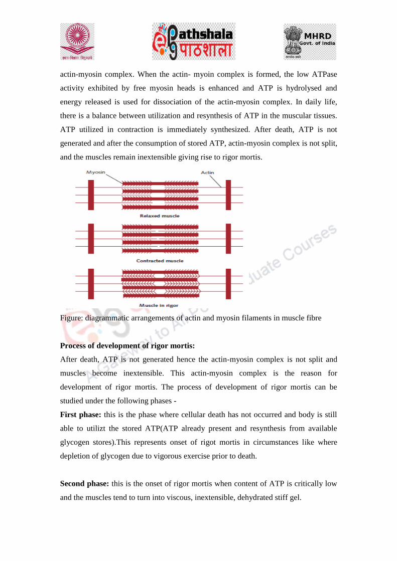
actin-myosin complex. When the actin- myoin complex is formed, the low ATPase
activity exhibited by free myosin heads is enhanced and ATP is hydrolysed and
energy released is used for dissociation of the actin-myosin complex. In daily life,
there is a balance between utilization and resynthesis of ATP in the muscular tissues.
ATP utilized in contraction is immediately synthesized. After death, ATP is not
generated and after the consumption of stored ATP, actin-myosin complex is not split,
and the muscles remain inextensible giving rise to rigor mortis.
Figure: diagrammatic arrangements of actin and myosin filaments in muscle fibre
Process of development of rigor mortis:
After death, ATP is not generated hence the actin-myosin complex is not split and
muscles become inextensible. This actin-myosin complex is the reason for
development of rigor mortis. The process of development of rigor mortis can be
studied under the following phases -
First phase: this is the phase where cellular death has not occurred and body is still
able to utilizt the stored ATP(ATP already present and resynthesis from available
glycogen stores).This represents onset of rigot mortis in circumstances like where
depletion of glycogen due to vigorous exercise prior to death.
Second phase: this is the onset of rigor mortis when content of ATP is critically low
and the muscles tend to turn into viscous, inextensible, dehydrated stiff gel.

Third phase: rigidity fully developed and is irreversible.
Fourth phase: in this phase muscles looses and rigidity disappears. This is also
known as phase of resolution.
Time of onset and its duration:
Rigor mortis can be seen within 30min- 1 hour following death.
Gets well established in 8-12 hours
complete rigor will Maintains for 8- 12 hours
rigor begins to dissipate over next 12 hours.(depends on environmental
temperature)
fully flaccid by 36 hours
in northern india, usual duration for rigor mortis during summer is 18-36 hour
in summer and 24-48 hours in winter.
Order of appearance and disappearance:
It is considered to appear first in eyelids by 1-2 hour followed by muscles of face,
neck , lower jaw, muscles of the chest, upper limb, abdomen and lower limbs. Since
rigor mortis as a physicochemical process, it affects all the body muscles
simultaneously.
Depending upon the progression of rigor mortis, corpses may be divided in three
categories:
Where the corpse is still warm, without showing any rigor indicates death
within 1-2 hours previously.
Body where the rigor is progressing but not established in full body, indicates
death within 4-12 hour previously.
Those corpses where the rigor is well developed in entire body, indicates death
beyond 9-12 hours.
The disappearance of rigor follows the same fashion as its appearance.
Factors influencing Rigor Mortis: 21

Environmental temperature: ‘rigidity persists longer in cold’
Cold and wet weather: onset will be slow, and duration will be longer
Hot and dry weather: onset will be fast while duration is shorter
Muscular activity:
If musles are healthy, robust and at rest before death: onset will be
slow, and duration will be longer
If muscles exhausted /fatigued: onset rapid
Nature of death:
bodies of those who are emaciated or die of wasting diseases pass
rapidly into state of rigidity which is of shorter duration.
Central nervous system:
nerve supply or even removal of brain does not affect the rigor. Rigor
mortis occur in amputated body parts too.
Age:
Not occur in foetus less than 7 months
May be found in stillborn infants
In healthy adults-well marked
Children and old people: weak and early
Cause of death:
Asphyxia, pneumonia, muscle paralysis & dehydration: slow
onset
Septicemia & poisoning: rapid onset, may even be absent,
especially in limbs affected by septicemia
Emaciated or died of wasting disease: rapid onset, short
duration

19 Perper J.A., Time of death and changes after death. Pt 1. Anatomical considerations, in Spitz W.U.
(ed): Spitz and Fisher’s Medicolegal Investigation of Death: Guidelines for the Application of
Pathology to Crime Investigation, 3rd ed. Springfield: Charles C. Thomas, 1993, pp 26-27.
20DiMaio D.J. and DiMaio V.J.M.: Forensic Pathology. New York: Elsevier, 1989. ). (Graham M.A.
and Hanzlick R.: Forensic Pathology in Criminal Cases. Carlsbad: Lexis Law Publishing, 1997.
21Subrahmanayam BV. Death in its medicolegal aspects. Medical Jurisprudence and Toxicology, 22nd
Ed, Butterworth’s India 1999; 140-43. 2. Parikh CK. Medicolegal aspects of Death.) (Parikh’s Text
Book of Medical Jurisprudence and Toxicology 6th Ed; CBS Publication, Bombay 1999; 148-54.
22 Vij K. Death and its medico-legal aspects. Text book of Forensic Medicine and Toxicology, 2nd
Ed.; BI Churchill Livingstone, New Delhi 2002; 159-69. ) Mant AK. Postmortem changes. Taylor’s
Principles and Practice of Medical Jusiprudence 13th Ed.; Sydney Smith, Chicago 1965; 140-43.
Cadaveric spasm (instant rigor/ cataleptic rigidity)
Cadaveric spasm is the stiffness of the muscles that has its onset immediately after
death and basis of such investigation is to find out items gripped in hand of victim
before the onset of rigor state.it is quite common but rare phenomenon. Usually after
death muscles become flaccid which is followed by rigor mortis but in case of
cadaveric spasm, the state of primary flaccidity does not come and muscles acquire
stiffness at the time of death. It is usually associated with the violent deaths. Its
mechanism or nature is still obscure but the reason can be attributed to the ATP stores
in the affected muscles. Most of the times cadaveric spasm is associated with the
emotional or physical stress. Cases are found where bodies are recovered from rivers
with weeds or twigs grasped in their hands or fingers of person committing suicide
found bend tightly.23,24,25,26,27,28
(23) Serafettin Demirci, Kamil Hakan Dogan, Zerrin Erkol, Idris Deniz Precautions Taken to Avoid
Abandoning the Act of Hanging and Reducing Pain in Suicidal Hanging Cases. Am. J. Forensic Med.
Pathol.2009; 30: 32–35. 2.
24 F.A. Benomran, S.E. Masood, A.I. Hassan. Masking and bondage in suicidal hanging: a case report.
Med. Sci. Law.2007; 47(2): 177- 80. 3. Krill A, Griller W, Wilske J. Modern variant of hanging: use of
timeprogrammed winch. Arch. Krminol.2002; 209(3-4): 110-5.
25 Kumar V. Hanging without knot in the noose. Journal of Forensic and legal medicine.2007; 14(1):
35-8.
26 Pollak S, Stellwag carion C. Deviations in findings in hanging by interposition of fingers between
noose and neck. Arch. Kriminol.1986; 177(3-4); 76-84.
27 Kanchan T, Menezes RJ, Manipady S. Haemorrhoids leading to postmortem bleeding artifact. J.
Clin. Forensic Med.2006; 13 (5):277-9.
28 Bharadwaj D N, Sharma SK, Gupta S. Haemorrhoids leading to postmortem artifact: a case report.
Med. Sci. Law.2005; 45(3): 265- 6.

Cadaveric spasm in case of drowning: victim had grass from the river bank firmly
grasped in the hand
Medico legal importance of cadaveric spasm:
Diagnosis of cause of death as in case of weapon found in hand
In cases like Drowning
ID of assailant may be found in hand
May allow one to know state of person prior to death

Difference between Rigor mortis and cadaveric spasm:
Rigor mortis cadaveric spasm
Onset Onset delayed after death (1-2 hours)
Onset is immediate
Duration Duration is approximately 12-24 hrs
Duration is a few hours,
until and unless it is
replaced by rigor mortis
Intensity Intensity comparatively moderate
Intensity comparatively
very strong
Mechanism When breakdown of ATP is below
critical level (less than 15%)
Mechanism of formation
unknown but factors like
Excitement, fear, fatigue,
exhaustion, nervous
tension, contraction of
muscles at time of death
might be the reasons.
Muscles
involved
All muscles of the body are affected
gradually
Selected muscles, which
were in a state of
contraction at the time of
death, are affected.
Medico legal
bearing
Mostly helps to know the time since
death
It helps to suggest the
manner of death, i.e.
whether
suicide, accident or
homicide

3- Late Changes; includes
Decomposition / Putrefaction.
There are changes in the body which takes place after 24 hour of death and is sure and
certain sign of death. It basically represents the decay or decomposition of body. The
process of decomposition include 29,30
1- Putrefaction
2- Adipocere formation
3- Mummification
___________________________________________________
29 Davis JB, Goff ML (2000) Decomposition patterns in terrestrial and intertidal habitats on O’ahu
Island and Coconut Island Hawai’i. J Forensic Sci 45:824–830
30 Early M, Goff ML (1986) Arthropod succession patterns in exposed carrion on the island of O”ahu,
Hawaiian Islands, USA. J Med Entomol 23:520–531
Putrefaction
It is Last stage in the resolution of the body where complex organic body constituents
get converted to the simple inorganic state. Putrefaction is mainly brought about by
two processes: Autolysis and bacterial action. 30
Autolysis:
This is rise in level of enzyme in tissues after death.
They soften and liquefy the dead tissues as the part of digestive action of the
enzyme.
The earliest autolytic changes can be seen in parenchymatous tissues or soft
tissues.
Autolysis Starts 3-4 hours after death and continues for 2-3 days, sometimes
even longer.
The process can be stopped by freezing.
Bacterial action:

Action of bacterial enzymes (aerobic and anaerobic) on tissue components like
carbohydrates/fat/proteins.
For bacterial growth – warmth, moisture are conditions favorable conditions.
Clostridium, streptococci, E coli, Bacillus proteus are the bacteria known to
decompose body tissue components.
Clostridium welchii is the bacteria which initiates haemolysis of blood and
starts the process of putrefaction.
In warm climate putrefaction starts comparatively faster while in cold climate
the process is retarded.
Development of gases under the skin and hollow viscera takes 18-36 hrs. 24-
48 hrs in case of solid viscera.
Purefaction changes include:
Change in colour:
discolouration varies fron green to black due to formation of
sulphmethaemoglobin.
Foul smelling gases:
Hydrogen sulphide ammonia phosphorated hydrogen and methane are the
gases which formation creates foul smell.
Sufficient accumulation of these gases is the reason why body floats in case of
drowning.
Pressure effect of putrefactive gases:
Pressure created displaces the diaphragm upwards.
Discolored fluid and liquefied tissue mixes with gases producing froth.
Shifting of the area of hypostasis.
Changes in skin, hair and wound.
Extrusion of fluid from the mouth and nose.
Emptying of the heart.
Changes in appearance of genitals.

Other sequale:
Fall of teeth
Skull sutures separation
Liquefied brain matter oozes out
Factors affecting putrefaction:
Warmth and clothing
Moisture
Air
Age
Manner of burial
Condition of body
Sex
Mode of death
Adipocere
After death, bacterial enzyme act upon body fats causing their hydrolysis and
hydrogenation. Palmitic acid is the main component of adipocere. Its appearance is
yellowish white with rancid smell. Adipocere can be formed at any site where fatty
tissues are present.it takes nearly 3 weeks in summer while in tropics 5-15 days. 2,6,7
Medico legal importance of adipocere
In establishing identity
Determining cause of death
To establish time since death
To determine place of death

Optimal conditions for the formation of adipocere are:
Abundant moisture
Bacteria (clostridium welchii)
Optimal temperature
Relative air
Abundance of adipose tissue
Mummification
It is a peculiar desiccation and modified form of putrefaction of a dead body where by
its soft parts shrivel up but retain the natural appearance and the features of the body
instead of liquefaction. Body attains rusty brown color, dry leathery skin which is
adherent to bones while internal organs get transformed into thick brown mass.
Mummification occurs in bodies buried in shallow graves, in dry sandy soils and takes
time nearly 3 months to 1-2 yrs.
Medico legal importance:
Identification
Cause of death
Time since death
Place of death
The conditions necessary for its formation are:
Warm and dry atmosphere
Moisture deprived area so proliferation of putrefying bacteria or
microorganisms can be prohibited.
Free air circulation around the body.
Forensic entomology
Flies and maggots also provide an approximate time of death, very useful for
cases where the body has been long dead. Only certain insects will feed and
lay eggs on a dead corpse and forensic entomologists study these insects, their

larvae cycles and thereafter can determine whether a body has been dead for
just one day or up to 3 or 4 weeks. 31
Time Physical appearance of
body
Insect present at that stage
0-3 days Proteins and carbohydrates
in the deceased body begin
to break down.
Blowflies e.g. Bluebottle
flies, Syrphidae flies
4-7 days Body is starting to decay
and causes the abdomen to
inflate because of the gases
inside.
Fly larvae and beetle e.g.
Rove Beetles
8-18 days Decay is well and truly
setting in; the abdomen
wall begins to break down.
Ants, cockroaches, beetles
and flies
19-30 days The decaying body enters a
stage know as 'post-
decay'; in wet, humid
conditionsthe body is
sticky and wet; in hot dry
conditions, the body is
dried out.
Beetles and mites e.g.
Springtail beetle, Acari,
as 'post-decay'; in
wet, humid conditions,
Nematocera
(present only during the
winter months),
31 days and onwards The bones, skin and hair
that remain no longer give
off a powerful stench and
smell
just like the soil
surrounding it.
30 Gill-King H (1996) Chemical and ultrastructural aspects of decomposition. In: Haglund
WD, Sorg MH (eds) Forensic taphonomy: the postmortem fate of human remains. CRC, New
York
31 Lord WD, Goff ML (2003) Forensic entomology: application of entomological methods to
the investigation of death. In: Froede RC (ed) Handbook of Forensic Pathology, 2nd Edn,
College of American Pathologists, Illinois.
Law
Forensic Science and Forensic Medicine
Asphyxial death: hanging, strangulation, drowning

DESCRIPTION OF MODULE
Items Description of Module
Subject Name Criminology
Paper Name Forensic Science and Forensic Medicine
Module Name/Title Asphyxial death: hanging, strangulation, drowning
Module Id LAW/CJA/VIII/36
Objectives
Learning Outcome:
To make the learners understand the need of studying
asphyxial deaths based on their mechanism.
To make the learners understand processes to determine
the various type of asphyxia operating in different case
and manner of death.
To make the learners understand the mechanism of
oxygen supply to the blood and tissues with respect to
interference with respiration.
Prerequisites General understanding of the Obstructions to the air
passages due to hanging, strangulation or other asphyxia
deaths and occlusion of the air passages within as in
drowning or laryngeal spasm.
Key words
Asphyxia, hanging, strangulation, drowning,
entomology.
Asphyxia
Asphyxia is a common term applied to the condition where to a more or less degree
interference with the respiratory exchange occurs. It is the condition where oxygen
supply to blood and tissue is restricted below the normal level causing any
interference with respiratory system. 1,2. It can also be considered as state of hypoxia
Role Name Affiliation
Principal Investigator Prof. (Dr.) Ranbir
Singh
Vice Chancellor, National
Law University, Delhi
Co-Principal Investigator
Paper Coordinator Prof. (Dr.) Sally
Lukose
Dean, School of Basic and
Applied Sciences, Galgotias
University, Greater Noida.
Content Writer/Author Ms. Neeharika
Srivastava
Assistant Professor, Galgotias
University, Greater Noida,
U.P.
Content Reviewer

or the lack of oxygen below the required level. Effect of asphyxia can be divided in
four stages: 3,4
Stage of inspiratory dyspnoes where deep and forceful respiration occurs, cyanosis
lasts for a minute or so.
Stage of expiratory dyspnoea with spasmodic efforts at expiration. At this stage,
consciousness is lost, pupils dialated and high blood pressure are the symptoms.
Next is the fall in blood pressure, resulting in increased pulse rate. Sometimes
defecation occurs. Erection of penis and ejaculation of semen may also occur.
At last, respiratory movements cease except for terminal irregular occasional
respiration. Heart beats for next 10-15 minutes.
Causes of asphyxia: 2
Obstruction in air passage due to mechanical asphyxia
Causes blockage of external respiratory orifices, as in smothering.
In case of drowning, occlusion of the air passage
As in case of traumatic asphyxia, pressure on chest.
Inhaling the toxic gases like carbon monooxide.
In case of strychnine poisoning, spasm of the respiratory muscles.
in case of narcotics and anesthetia administered, paralysis of the respiratory
centre.
1 (Premature and Congenitally Diseased Infants, by Julius H. Hess, M.D., Chapter X,
Diseases of the Respiratory Tract, parikh).
2 parikh
3 chapter 11, asphyxia, Ass. Prof.Dr. Atef Foda & Dr. Eslam Samy, 117-129). (Nandy
A.; Principles of Forensic Medicine; Edition 2005; p. 319-327.
4 Dixit P.C.; Textbook of Forensic Medicine and Toxicology; Edition 2007; p. 294,
300.)
Signs of asphyxia: 5,2,6,7,8,9
1- Cyanosis: (greek ‘dark blue’)
The quantity of oxyhaemoglobin and reduced haemoglobin in red blood cells
determine the Cyanoses.
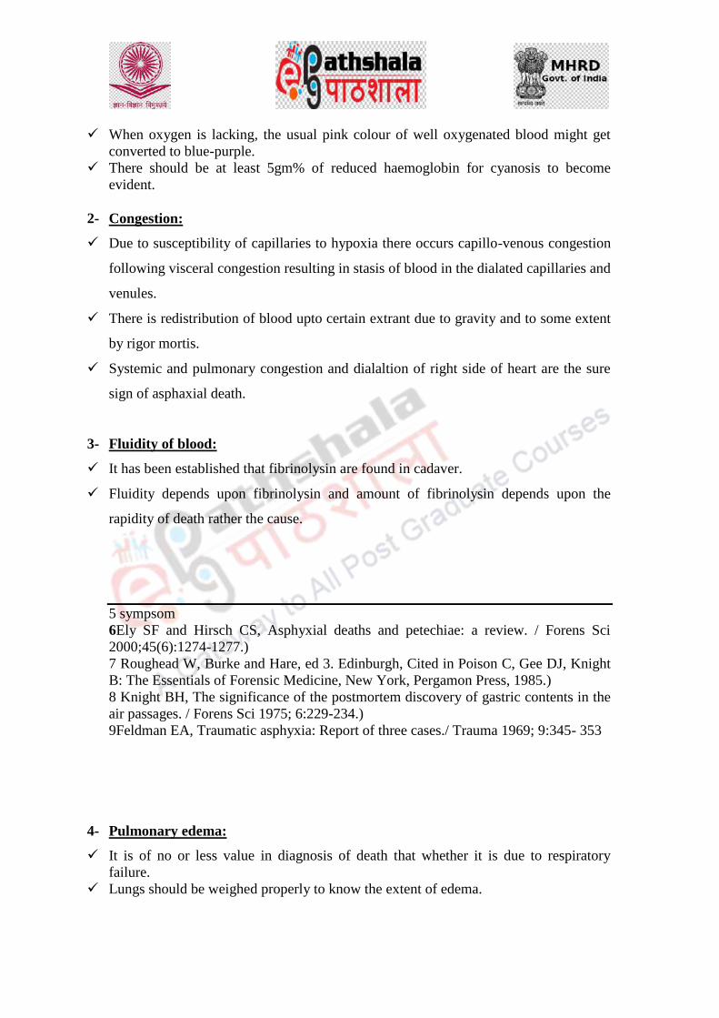
When oxygen is lacking, the usual pink colour of well oxygenated blood might get
converted to blue-purple.
There should be at least 5gm% of reduced haemoglobin for cyanosis to become
evident.
2- Congestion:
Due to susceptibility of capillaries to hypoxia there occurs capillo-venous congestion
following visceral congestion resulting in stasis of blood in the dialated capillaries and
venules.
There is redistribution of blood upto certain extrant due to gravity and to some extent
by rigor mortis.
Systemic and pulmonary congestion and dialaltion of right side of heart are the sure
sign of asphaxial death.
3- Fluidity of blood:
It has been established that fibrinolysin are found in cadaver.
Fluidity depends upon fibrinolysin and amount of fibrinolysin depends upon the
rapidity of death rather the cause.
5 sympsom
6Ely SF and Hirsch CS, Asphyxial deaths and petechiae: a review. / Forens Sci
2000;45(6):1274-1277.)
7 Roughead W, Burke and Hare, ed 3. Edinburgh, Cited in Poison C, Gee DJ, Knight
B: The Essentials of Forensic Medicine, New York, Pergamon Press, 1985.)
8 Knight BH, The significance of the postmortem discovery of gastric contents in the
air passages. / Forens Sci 1975; 6:229-234.)
9Feldman EA, Traumatic asphyxia: Report of three cases./ Trauma 1969; 9:345- 353
4- Pulmonary edema:
It is of no or less value in diagnosis of death that whether it is due to respiratory
failure.
Lungs should be weighed properly to know the extent of edema.

5- Pulmonary hemorrhages:
Due to trauma of pharynx against the anterior surface of spine , there is larfe
submucosal hemorrhages.
Rupture of submusous venous plexus at this site is due to serious venous congestion.
6- Petechial hemorrhages:
Petechial hemorrhages or tardieu spots are develop from:
Due to Increased venous stasis causing congestion which results in increased pressure
causing rupture of the vessels.
There occurs increased permeability due to hypoxia.
Tardieu spots are usually pinhead in appearance,but vary in size and shape.
7- Stasis hemorrhages:
Found in case of homicidal strangulation
Usually found beneath the mucosa of larynx in the subglottic space.
Classification of asphyxia death 2,10
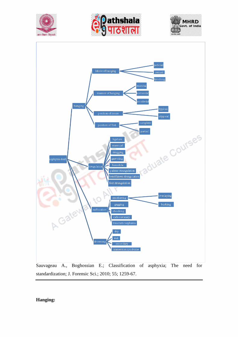
Sauvageau A., Boghossian E.; Classification of asphyxia; The need for
standardization; J. Forensic Sci.; 2010; 55; 1259-67.
Hanging:

Hanging is a mode of death in which death occur because of the compression of neck
as a result of suspension of body through a ligature. The constricting force results
from the weight of the body or a part of body weight acts as a constricting force.
Virtually all the suicide cases are suicidal in nature. Diagnosis of antemortem
hanging is important to acertain the death from hanging. There are few features which
can ascertain death by hanging: Ligature mark with vital reaction, Saliva dribbing
from mouth, Ecchymosis of larynx or epiglottis, Fracture of hyoid cartilage and hyoid
bone. 2,5,11
There are chances of complete or incomplete suspension of the body:
Complete hanging:
Here the body is completely suspended in air
Constricting force is the weight of the body.
Partial/incomplete hanging:
Here the constricting force is weight of the haed.
Lower part of the body like toes, feet , knee and buttocks generate the constricting
force.
Figure: (a) complete hanging (b) partial hanging
11 A Study of Gross Postmortem Findings in Cases of Hanging and Ligature
Strangulation, Mohammed Musaib M. Shaikh, H. J. Chotaliya, A.D. Modi, A. P.
Parmar, S. D. Kalele, J Indian Acad Forensic Med. Jan-March 2013, Vol. 35, No. 1
ISSN 0971-0973. ,
2 Parikh’s Text book of medical jurisprudence Forensic Medicine and Toxicology
CBS publisher and distributors PVT.LTD.
5 Simpson's Forensic Medicine, 13th Edition, Jason Payne-James, Richard Jones,
Steven B Karch, John Manlove, August 26, 2011 by CRC Press.

Cause of death in hanging:
Hanging may resulting death due to one or combination of following reasons of
immidiate death: 12,13,14,5
1- Asphyxia:
ligature forces the root of tongue aginst the pharynx and folds the epiglottis over the
entrance of larynx to block the passage of air.
A minimum weight of 15 kg is required to compee the trachea
2- Injuries to spinal cord:
When hanging is exercised with a long drop
Upper cervical cord is stretched
Immediate unconsciousness
Heart and respiration may continue for upto 10-15 minutes
Congestive changes are absent
3- Vagal inhibition:
By compression of the neck
Cab be considered as a possible cause where there is no or minimal congestive
changes.
4- Mechanical constrictions of the structures of the neck:
Combined obstructive asphyxia and interfered cerebral circulation is most common
cause of death
5- Cerebral anoxia:
Carotid artery occludes with 4-5 kg tension
Vertebral artery with 20 kgs tension
12 (Ashok Kumar Samanta, Soumya Rajan Nayak. Newer trends in hanging death.
Journal of Indian Academy of Forensic Medicine. 2012, Vol. 34 (1) pp 37 – 39.)
13 (Paliwal P. K, Basant Lal Sirohiwal, Vijay Pal Khanagwal, Hitesh Chawla. A drop of
saliva de-codes the mystery of hanging body. Journal of Indian Academy of Forensic
Medicine. July-Sep 2011, Vol. 33, No. 3.pp280-282.
14 Chormunge Patil, Mahajan S. V, Bhusari P. A. Hanging vs. strangulation a
comparative study. Journal of Forensic Medicine, Science and Law. Jul-Dec 2011.
Vol. 20, No.2 pp 1-5)
5 siympson
Reasons for delayed death:
Aspiration pneumonia
Infections
Hypoxic encephalopathy
Edema of lungs
Encephalitis
Cerebral abscess
Autopsy findings: 2,5,15,16
External:
Neck may be stretched or elongated
Head bend is opposite to the knot
Face is pale usually
Face may be swollen but swelling disappears when rope is cut down
Petechiae present on the skin
Dribbing of saliva
Bloody froth may present caused by congestion of lungs
Pulmonary edema
In the middle ear hemorrhage may be seen
Semen drops may come out of penis
Lower limbs show hypostasis
Internal:
Absence of Petechiae because of complete obstruction in the arterial system
Saliva run from mouth down the chest
Fracture of the hyoid bone and thyroid cartilage (considered antemortem)
Vertebral artry may show rupture
Thyrohyoid ligament is torn
Trachea show Petechiae haemorrhages over the epiglottis, trachea and larynx.
Abdominal organs are congested.
15 Zine K. U., Tandle R. M., Varma N. M., Jambure M. P. Accidental Ligature
Strangulation with Avulsion of Scalp. Journal Indian Academy Forensic Medicine.
July-Sept 2011, Vol. 33, No.3, pp 267 – 268.

16 Punia R. K, Gupta B. M. A rare case of ligature strangulation with salivary
staining. Journa)( Sauvageau A., LaHarpe R, Geberth VJ; Agonal sequences in eight
filmed hangings: Analysis of respiratory and movement responses to asphyxia by
hanging; J Forensic Sci. 2010; 55; 1278 – 81.
Strangulation
Strangulation is another form of asphaxia where death is due to constriction of neck
by a ligature or other mean bvut without suspending the body. Broadly it is of two
types (1) ligature strangulation and (2) manual strangulation. There also may be some
more type:
Ligature Strangulation: strangulation is by means of ligature.
Throttling or manual strangulation: compressing the victim;s neck by the hand
Mugging: compressing the victim’s neck against the forearm
Garroting: attacking the victim from behind, grabbing his neck or throwing a ligature
over neck and tightening it.
Bansodala: compressing the neck by means of two sticks.
Areas which are more presumptive of strangulation are head and neck and a thorough
examination of these area can be used to ascertain death by strangulation.
Medico legal importance of strangulation:
In homicidal cases:
Single turn of ligature with one or more knot
Abrasion around the neck due to ligature movement
Mark of struggle (absent in case of weak or frail individual)
In Accidental cases:
Children are more prone as they can get entangled in rope while playing.
Intoxicated person by tie, scarf or collar.
Umbilicle cord can strangulate the foetus.
In suicide cases:
Injuries are less marked as less force is applied.
No sign of struggle
Ligature is found at scene
17 (Chormunge Patil, Mahajan S. V, Bhusari P. A. Hanging vs. strangulation a
comparative study. Journal of Forensic Medicine, Science and Law. Jul-Dec 2011.
Vol. 20, No.2 pp 1-5)

Cause of death:
Asphyxia
Anoxia
Congestion
Vagal inhibition
Autopsy:
External:
Ligature mark on the middle or lower part of neck
Depending upon the compostion of the ligature it will shown regular or irregular
pattern around the neck
Mark is depressed at any level on the neck-usually at the level of thyroid cartilage or
below.
Mark may be found around the neck but is more prominent in front and sides of neck.
Marks may be interrupted by clothing or victim’s finger or ornaments.
Mark is usually transverse but sometimes oblique as well.
Internal:
Laceration of muscles.
Injuries to blood vessels
Hyoid bone fracture usually not seen.
Fracture thyroid is more common.
Fracture of cricoids is rare, but if pressure is used may be seen.
Organs are congested.
18 (Sheikh M. I. and Agarwal S. S. Medico-legal implications of hyoid bone fracture-
a study paper. Journal of Indian Academy of Forensic Medicine. 2001 Vol. 23.
Number 4. pp 61 - 63.)
19 (Zine K. U., Tandle R. M., Varma N. M., Jambure M. P. Accidental Ligature
Strangulation with Avulsion of Scalp. Journal Indian Academy Forensic Medicine.
July-Sept 2011, Vol. 33, No.3, pp 267 – 268.)
Drowning

Drowning is a form of asphyxial death due to aspiration of fluid into the air
passages by submersion of the body in water or some other liquid medium.20,21
Complete submersion not necessary, submersion of nose and mouth of a living
person under water is enough. Types of drowning:
Drowning is mainly of four types:
1- Wet drowning: when more water goes to lungs.
2- Dry drowning: no water goes to lungs, death is due to
laryngeal spasm.
3- Secondary drowning:
Survival beyond 24 hours, victim may survive or die
later
Injury to CNS is reported
Hypothermia and low oxygen delivery to vital tissues
are the main factors towards morbidity and mortality.
4- Immersion drowning: Death is due to vagal inhibition
following cardiac arrest. In drowning this may be brought about by
A sudden water entry to larynx.
Falling in water in a way such that it strikes the
abdomen suddenly, especially the epigastric region.
Sudden entry of cold water into the ears.
Medico legal importance of drowning:
Sure signs of drowning,
Could still be identified in putrefied bodies,
Could give an evidence of the site of drowning (fresh or salt water species).
Whether the death was due to drowning or other cause?
Length of time the body was in water.
Whether it was accidental/suicidal/homicide
20(Auer A, Möttönen M (1988). Diatoms and drowning. Z Rechtsmed 101: 87-98.)
21Davis J (1986). Bodies found in the water: an investigative approach. Am. J.
Forensic Med Pathol 7: 281-287
The change occur in body simply tells about the duration of submersion of body in
water. Changes are result of imbibition of water in outer layer of body. Changes start
to develop first in finger tips (3-4 hours) later spreading to entire hand (24 hours). By
careful examination of these changes duration of submersion can be determined.
In a couple of hours only, skin starts developing wrinkles
In about 12 hours of submersion, cuticle becomes bleached.
In 24 hours saddening of skin can be seen.

In about 48 hours of submersion, cuticle of skin starts separating from palm and foot
and can be peeled off by 3-4 days.
Usually body starts to float in about 24 hours of submersion in summer in 2- 3 days in
winter.
Autopsy/ diagnosis of death by drowning can be ascertained by (a) External sign (b)
internal sign (c) biochemical test and (d) analysis through diatoms. 22,23,24,25
External sign:
Clothing and skin is wet, cold and pale
Hypostasis is pink in color due to oxygenation or may be cyanotic
Upper parts of body show postmortem staining
Petechial haemorrhage may be present
Pupil dialated
Cyanosis may be present
Rigor mortis takes place early
White and leathery froth sometimes with blood is present over the mouth and nostril.
When chest is pressed more froth comes out.
Cutis anserine (Due to spasm of the erector pilae muscles and due to exposure to cold
water at the time of death) or goose skin is present
Cadaveris spasm: presence of weeds in grass in hands.
Washerwomen’s hand: saddening of the skin due to absorption of water.
22 DiMaio DJ, DiMaio VJM (1989) Drowning. In: DiMaio DJ, DiMaio VJM, eds.
Forensic pathology. Elsevier, Amsterdam pp. 357-365.)(Hendey NI (1973).
23 The diagnosis value of diatoms in the cases of drowning. Med Sci Law 13(1): 23-
34.)
24(Knight B (1991). Immersion deaths. In: Knight B, ed. Forensic Pathology,
E.Arnold, London pp. 360-374.
25 Ludes B, Fornes P. (2003). Drowning in : Forensic Medicine : Clinical and
Pathological Aspects. Payne-James J, Busuttil A, Smock W eds, Greenwich Medical
Media, pp 247-257)
Internal signs:
Paltauff’s haemorrhages:
Present in the lower lobes of lungs
Red or grey patchy appearance
This condition of lung is also known as emphysema aquosum and trockenes oedema.
Microscopic findings of drowning lungs:
Following stages are encountered

Thickness of the alveolar wall is reduced to capillary width
Alveolar are more distended and capillaries lie separately.
Capillaries appearance is thread like, lumina is observed occasionally.
Involve only scattered alveoli.
Water in stomach and duodenum:
When water is taken in excess, it passes to the duodenum.
Froth in air passage:
In case of fresh drowned bodies, froth comes out from mouth and nostril.
Froth is odema fluid from lungs
Froth consist of proteinaceous exudates and surfactant mixed with the drowning
medium water.
Over-inflation of lungs:
Lungs may be over-inflated filling the thoracic cavity.
Biochemical tests:
Many workers have worked on the biochemical and biophysical tests on drowning
related cases including calcium, magnesium or specific gravity of plasma but the
results are not rewarding. The rapid onset of post mortem changes in case of
drowning may be considered as one of the reason which is obscuring the reliability of
biochemical tests. 26
Diatoms (bacillariophyceae) is a photosynthetic unicellular algae with silicaceous
exoskeleton is found in water.
There are nearly 15,000 species of diatom found in fresh and sea water.
A broad classification of diatom can be (1) oligohalophilic: live in fresh water (2)
mesohalophilic: in sea water with salinity more than 0.05%.
26 Analysis of diatoms: (Ludes B, Coste M, North N, Doray S, Tracqui A, Kintz P
(1999). Diatom analysis in victim's tissues as an indicator of the site of drowning. Int
J Legal Med 112: 163-166.)

Different types of diatoms27
When a person drowns in water containing diatoms, many diatoms reaches to
pulmonary parenchyma. From there they enter into blood stream through the alveolar
walls during forceful inspiratory or expiratory efforts. Once they enter in blood , are
distributed in whole body through blood stream. This presence of diatom makes the
basis of diatom test in drowned individuals.
When person is not drowned oe a body is thrown in water after death, diatom are
though able to reach lungs by passive perlocation but cannot reach to distant organs.
Hence organs examined routinely for the presence of diatoms are lung, liver, brain
and bone marrow. 28
27 Forensic diatomology, ANU SASIDHARAN*, RESMI S, HEALTH SCIENCES,
2014; Vol.3,No.1 January –March, ISSN 2319 – 4154.
28 (Ludes B, Quantin S, Coste M, Mangin P (1994). Application of a simple
enzymatic digestion method for diatom detection in the diagnosis of drowning in
putrefied corpses by diatom analysis. Int J Med 107: 37-41.)

Figure: Diagrammatic representation of transmission of diatoms
There may be cases where the person was not drowned but diatoms were present or
may be absent when person was drowned. Such conditions can arise due to:
Seasonal variation
Due to havinf raw fruits or vegetables which have been in contact of soil diatoms
Having their ubiquitous nature, they are present in soil, water and in air.
Sometimes they can penetrate the intestinal lining and can directly get in the blood
stream.
Certain food like selfish contain diatoms.
In such conditions, following are the requirements which should be fulfilled:
Diatoms species recovered from body organs and from water of submersion should be
same.
They must be present in approximate same proportion. 29, 30,31
29 Neidhart DA, Greedyke RM (1967). The significance of diatom demonstration in
the diagnosis of death by drowning. Am J Clin Pathol 48(4): 377-382.
30 Pollanen MS (1997). The diagnosis value of the diatom test for drowning, II.
Validity: analysis of diatoms in bone marrow and drowning medium. J Forensic Sci
42(2): 286-290.
31Pollanen MS (1998). Forensic diatomology and drowning. Elsevier (Amsterdam)
ed.

DESCRIPTION OF MODULE
Items Description of Module Subject Name Criminology
Paper Name Forensic Science and Forensic Medicine
Module Name/Title
Injuries and their medico legal aspects: Abrasion, bruise,
laceration, Incised, stab wounds
Module Id 37
Objectives
Learning Outcome:
To make the learners understand the various types of injuries sustained by individuals and the medico-legal aspects associated with such injuries.
To make the learners understand the nature of different injuries and the estimation of age of injuries.
To understand the importance of assessing the ante-mortem and post-mortem injuries.
Prerequisites General understanding of the human anatomy.
Key words
Forensic Medicine, Medico-legal aspects, injuries

1. Introduction:
An injury or a wound means a solution or disruption of the anatomical continuity
of any of the tissues of the body.
Section 44 IPC : An injury is defined as any harm whatever illegally caused to any
person in body, mind, reputation or property.
The Mechanism of Injuries:
The routine movements of our body are a resultant of elasticity and flexibility of
our soft tissues and rigid skeletal framework, subsequently; injuries result when the
forces acting on the human body crosses its limits of this elasticity or resistance.
The factors responsible for the mechanism of injuries are:
1. Force
2. Area over which it acts
3. Specific effect of the force
4. Time taken over which the kinetic energy is transferred
Within a forensic context, the interpretation and classification of injuries are
important in helping to gain an insight into how they were caused. The
classification of injuries can be made based on the influencing factors and on the
legal basis.
Classification of Injuries based on influencing factors:
1. Mechanical Injuries
Abrasion
Contusion
Laceration
Incised
Stab
Fire Arms
Fractures and Dislocations

2. Thermal Injuries
Due to Cold
- Frost Bite
- Trench Foot
- Immersion Foot
Due to Heat
- Burns
- Scalds
3. Chemical Injuries
Corrosive Acids
Corrosive Alkalis
4. Miscellaneous
Injuries due to electricity
Injuries due to lightning
X-Rays
Radioactive Substances
Legal Classification of Injuries:
1. Simple Injuries
2. Grievous Injuries
3. Others
Self-inflicted Injuries
Defense Injuries
Unintentional Injuries
In the following sections we will discuss few commonly encountered injuries:
1. Abrasions: It is a superficial injury involving only the superficial layers of
the skin. Caused by friction or pressure between the skin and some rough
object or surface. It is sub-classified as:
a) Scratch: caused by a sharp object and carries the epithelium in front of it:
indicates the direction
b) Graze: Broad surface of skin slides against a rough surface: Found in traffic
accidents.
c) Imprint, pressure, or contact abrasion: Caused as a result of direct impact
or pressure of or contact with some object which crushes the cuticle and
stamps the reproduction of its shape upon the skin.

Fig1: Showing an abrasion Source: www.slideshare.net
Determining the age of an Abrasion:
Time after
Injury
Appearance
Fresh bright red colour
12-24 hours Lymph and blood dries up forming bright red scab
2 days Reddish brown Scab
4-7 days Epithelium starts to come under the scab
After 7 days Scab dries and falls off
Ante-mortem and Post-Mortem Abrasion:
Ante-mortem Abrasion Post-Mortem Abrasion
It is seen anywhere on the body It is seen usually on the bony
prominences
It is Reddish brown in colour It is yellowish and translucent
Scab is slightly raised Scab if seen is slightly depressed
Vital reactions are visible No vital reactions are visible
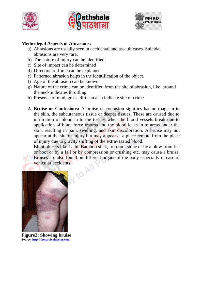
Medicolegal Aspects of Abrasions:
a) Abrasions are usually seen in accidental and assault cases. Suicidal
abrasions are very rare.
b) The nature of injury can be identified.
c) Site of impact can be determined
d) Direction of force can be explained
e) Patterned abrasion helps in the identification of the object.
f) Age of the abrasion can be known.
g) Nature of the crime can be identified from the site of abrasion, like around
the neck indicates throttling
h) Presence of mud, grass, dirt can also indicate site of crime
2. Bruise or Contusions: A bruise or contusion signifies haemorrhage in to
the skin, the subcutaneous tissue or deeper tissues. These are caused due to
infiltration of blood in to the tissues when the blood vessels break due to
application of blunt force trauma and the blood leaks in to areas under the
skin, resulting in pain, swelling, and skin discoloration. A bruise may not
appear at the site of injury but may appear at a place remote from the place
of injury due to gravity shifting of the extravasated blood.
Blunt objects like Lathi, Bamboo stick, iron rod, stone or by a blow from fist
or boot or by a fall or by compression or crushing etc, may cause a bruise.
Bruises are also found on different organs of the body especially in case of
vehicular accidents.
Figure2: Showing bruise Source: http://thesurvivaldoctor.com

Age determination of bruises:
Time after Injury Appearance
In fresh bruise Red
Few hours to 3 days Swollen, tender and blue
4 days Bluish black to brown colour
5-6 days Greenish colour
7-12 days Yellow colour
2 weeks Normal
Ante-mortem and post –mortem Bruise:
Ante-mortem bruise Post Mortem Bruise
The site is swollen in recent bruises Absent
If the person survives for some time
colour changes signifying vital reactions
are noted
Skin discolouration can be seen due to
post mortem staining but with no vital
reactions
Coagulation of extravasated blood in the
subcutaneous tissues and muscle fibers
are evident.
Presence of coagulated blood in the
subcutaneous tissues and muscle
fibers are absent.
Medico-legal Aspects of Bruise:
1. Bruises are resultants of either assaults or accidents. Self-inflicted assaults
are rare as they are extremely painful.
2. Self-inflicted bruises are though known to be made by application of some
plant parts such as "Bhilawan"(Marking nut) or the root of
Chitra"(Plumbago Rosae).
3. These self-inflicted bruises can be differentiated by the following characters -
They are present on the accessible parts of the body.
These are dark brown in colour.
Their margins are covered with tiny vesicles.
The surrounding skin is red and inflamed.
The scrapings of the area give positive test, on chemical analysis.
4. Age of the injury can be determined
5. Degree of violence can be known.
6. Type of weapon used can also be determined especially when the entire
length of the object used as a weapon is applied on the body as in case of a rod,
chain or stick.

Lacerations:
These are the wounds caused by the blunt force resulting in the tearing of the
skin and the underlying tissue, with a minimal bleeding.
Characteristic Features of Lacerated Injuries:
Edges or margins of the wound are ragged, irregular and contused.
These margins are abraded due to impact of blunt force.
The underlying deep tissues are crushed and the damage is gross and
extensive.
Hair bulbs present in the deeper tissues are crushed.
Less bleeding is as a result of crushing of underneath vessels.
Presence of foreign materials like dust, mud, gravel at the injury site is a
common feature.
The shape of the injury is usually irregular devoid of any definite shape.
The size of the injury may or may not correspond to the size or shape of the
weapon used
The healing process is delayed due to gross damage and infection at the
injury site.
Scars are formed due to the gross damage to underlying skin and tissue.
Types of Lacerated Injuries:
1. Split laceration:
https://med.pdn.ac.lk/departments/forensic/
This kind of laceration is usually found on parts overlying bones such
as the scalp, face, hands and lower limbs.
Split lacerations occur due to perpendicular impact by a blunt object.
The resulting injury is due to crushing of skin between two hard
objects.
This injury in some instances simulates an incised injury.
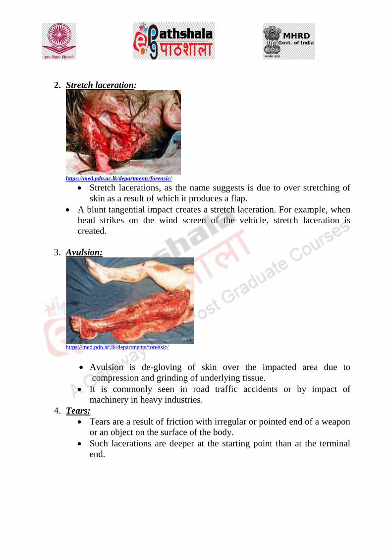
2. Stretch laceration:
https://med.pdn.ac.lk/departments/forensic/
Stretch lacerations, as the name suggests is due to over stretching of
skin as a result of which it produces a flap.
A blunt tangential impact creates a stretch laceration. For example, when
head strikes on the wind screen of the vehicle, stretch laceration is
created.
3. Avulsion:
https://med.pdn.ac.lk/departments/forensic/
Avulsion is de-gloving of skin over the impacted area due to
compression and grinding of underlying tissue.
It is commonly seen in road traffic accidents or by impact of
machinery in heavy industries.
4. Tears:
Tears are a result of friction with irregular or pointed end of a weapon
or an object on the surface of the body.
Such lacerations are deeper at the starting point than at the terminal
end.

5. Cut laceration:
https://med.pdn.ac.lk/departments/forensic/
This type of lacerated wound is produced by “blunt-sharp” edge of a
weapon like an ice pick.
Margins are not clear cut as would be seen with a sharp weapon.
Abrasions or contusions are seen on the margins.
Medico-Legal importance
Lacerations can been seen most often in homicidal and accidental cases.
It may occur on any part of the body and are produced by blows with
hard and blunt weapon.
Lacerations in suicidal cases are known to be very rare.
In accidental cases, lacerations are more prominent in road traffic
accident cases and accidental fall from height.
The location of the accident or crime can be determined by assessing the
foreign bodies present in the injured site such as mud, gravel, oil etc.
A split laceration may mimic an incision.
Incised Wounds
Fig: Showing an incised wound Source: http://etmcourse.com/
It is produced by any sharp cutting instrument such as knife, razor, blade,
sword, chopper, axe, etc.
The edges are clear cut, retracted and averted. An exception to this is found
in the neck and scrotum where the edges are inverted. The edges may be

irregular when the skin is loose in the affected site or if the cutting edge of
the knife is blunt.
The wound is spindle shaped due to retraction of the tissue.
The length of the wound is greater than its breadth. Additionally, the
breadth of the wound may be greater than the thickness of the cutting
blade.
Wound gaping is greater if underlying muscles are cut obliquely.
Excessive Bleeding or Hemorrhage is imminent due to the clear incision
of blood vessels.
The cuts are deeper at the beginning and gradually become shallow
towards the end when only skin is cut. This is called as “Tailing of the
wound”.
Determination of Age of the Incised Wound:
Medico-legal Importance:
The injury gives an indication of the nature of the weapon.
It gives an idea about the site of impact and direction of force.
Age of the injury can be estimated.
Position and characteristics of the wound can give an insight in assessing
whether it is a suicidal, homicidal or accidental injury.
Time after Injury Appearance
When fresh Haematoma formation
12 hrs Blood clot and lymph dry up, margins
are red and swollen. Histologically there
is infiltration of leucocytes.
24 hrs Proliferation of connective tissue cells
and vascular endothelium for neo-
vascularization.
36 hrs Fibroblastic infiltration and capillary
2-3 days Capillary network is completed.
Fibroblasts run across the new vessels.
3-5days Definite Fibrils are seen. Vessels are
obliterated and thickened
1-2 weeks Scar formation is completed.

Stab Injuries
These are also called as punctured injuries.
These injuries are generally caused by sharp and pointed weapons such as
knives, dagger, arrow, pick-axe, broken glass pieces, etc.
A stab wound caused by a sharp pointed and cutting instrument has clean cut
edges and sharp angles at the two extremities.
These are deeper than its length on the skin. Perforating wounds can also be
seen in stab wounds.
The shape of the wound will vary as per the type of piercing weapon used:
Type of Piercing Weapon Shape of injury Produced
Weapon with only one cutting edge Triangular or wedge shaped
Double edged weapon Elliptical
Round sharp pointed end (Spear) Circular injury with clean cut edges
Round blunt pointed end Circular injury with ragged and
bruised edges
Tapering Blades Perforating injury (entry wounds are
larger with inverted edges as compared
to exit wounds which are smaller and
with inverted edges)
Medicolegal Importance :
Shape of the injury will idicate the type of weapon used.
Depth of the injury will indicate the force of penetration.
Direction of the wound can indicate the relative position between the victim
and the assailant.
Age of the injury can be determined.
Stab wounds are mostly suicidal or homicidal.
Accidental stab wounds are rare.
References:-
http://thesurvivaldoctor.com
http://etmcourse.com/
https://med.pdn.ac.lk/departments/forensic/
Parikh, C.K. (2012) Medical Jurisprudence, Forensic Medicine and
Toxicology, CBS Publishers and Distributors Pvt. Ltd.

Modi’s Medical Jurisprudence and Toxicology,23rd
Edition, by K.
Mathiharan & Amrit K. Patnaik, Third reprint, 2009, LexisNexis,
Butterworth, New Delhi
Sharma, B.R., Forensic Science in Criminal Investigation and Trials (3rd
Edn.) Universal Law Publishing Co. Ltd. New Delhi

DESCRIPTION OF MODULE
Items Description of Module
Subject Name Criminology
Paper Name Forensic Science and Forensic Medicine
Module
Name/Title
Ante-mortem and Post-mortem injuries,
Burns, scalds and electrocution
Module Id 38
Objectives
Learning Outcome:
To make the learners understand the
various medico-legal aspects associated
with Burns, scalds and electrocution
injuries.
To make the learners understand the
nature of different injuries
To understand the importance of
assessing the ante-mortem and post-
mortem injuries.
Prerequisites General understanding of the human anatomy.
Key words
Forensic Medicine, Medico-legal aspects,
injuries
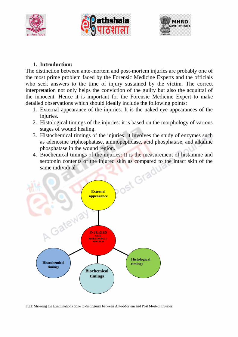
1. Introduction:
The distinction between ante-mortem and post-mortem injuries are probably one of
the most prime problem faced by the Forensic Medicine Experts and the officials
who seek answers to the time of injury sustained by the victim. The correct
interpretation not only helps the conviction of the guilty but also the acquittal of
the innocent. Hence it is important for the Forensic Medicine Expert to make
detailed observations which should ideally include the following points:
1. External appearance of the injuries: It is the naked eye appearances of the
injuries.
2. Histological timings of the injuries: it is based on the morphology of various
stages of wound healing.
3. Histochemical timings of the injuries: it involves the study of enzymes such
as adenosine triphosphatase, aminopeptidase, acid phosphatase, and alkaline
phosphatase in the wound region.
4. Biochemical timings of the injuries: It is the measurement of histamine and
serotonin contents of the injured skin as compared to the intact skin of the
same individual
Fig1: Showing the Examinations done to distinguish between Ante-Mortem and Post Mortem Injuries.
Histochemical
timings
Histological
timings
External
appearance
INJURIES ANTE-
MORTEM/POST-
MORTEM
Biochemical
timings

1.2 Difference between Ante-mortem and Post-mortem injuries:
The salient distinguishing features of Ante-mortem and Post-mortem injuries are as
follows:
S.No. Characteristics Ante-mortem Injuries Post-mortem injuries
1. Haemorrhage Profuse and arterial
Arterial spurting of
blood
Clotting of Blood
Blood clot when
found is laminated
Clot firmly adheres to
endothelium lining
Due to deep
infiltration the edges
of wound are stained
which cannot be
washed away.
Slight and venous
Venous oozing of blood
Blood clotting absent
Blood clot if found non-
laminated
Clot weakly adheres to
endothelium lining.
No infiltration and
hence the edges are not
stained and the stains if
present can be washed
away.
2. Wound edges Gaping, everted and swollen Apposed and not swollen
3. Vital Reaction Inflammation present
Granulation present
Inflammation absent
Granulation absent
4. Microscopy Leucocyte and RBC
infiltration between
muscle fibers
Clot composed of
fibrin, RBC and
platelets
Vessels distended with
postmortem clot
Clot composed of fibrin
and RBC
5. Enzyme Histo-
chemistry Negative and positive
vital reactions seen
Vital Reactions absent
6. Serotonin and
Histamine
biochemistry
Increase in Serotonin
and Histamine content
No increase in
Serotonin and
Histamine content
2. BURNS
A burn is defined as a traumatic injury to the skin or other organic tissue
primarily caused by thermal or other acute exposures namely heat, cold,

electricity, radiation, or chemicals. Burns may be classified in to three
degrees:
1. Epidermal Burns:
Effected part is red
Blister Formation
Inflammation
Singeing of hair
Very painful
Repair without Scar
2. Dermo-epidermal Burns:
Whole thickness of skin destroyed
Coagulation necrosis of epidermis and dermis
Lesions have a dry white leathery appearance.
Scar formation
Scars contact causing disfigurement
Considerable pain and shock
3. Deep Burns:
Complete charring
Gross destruction of subcutaneous tissue, muscles, and bones
Nerve endings destroyed. Burns therefore less painless

Fig2: Showing the different degree of Burns
Source: https://s-media-cache-ak0.pinimg.com/originals/
The estimation of burnt surface area can be worked out by the “Rule of 9”.

Fig 3: Showing Rule of 9 in an adult and a child
Medicolegal Significance:
Due to heat stiffening and coagulation of proteins the body presents a
pugilistic attitude. This condition is present in both dead and alive and hence
has no medicolegal significance.
Presence of soot in esophagus or stomach depicts that the victim was alive
when the fire was in progress.
Mucosa and trachea necrosed due to inhalation of hot air.
Blood thick due to haemo-concentration and cherry red in colour due to due
to carboxy-haemoglobin.
Suicidal burning is common.
Homicidal is rare but have known to occur with criminal intent.
Accidental burns are also common

3. SCALDS: A scald is an injury sustained due to application of liquid at or
near the boiling point or from steam. The injury is limited to skin, mouth,
throat when hot liquids are drunk. Moist heat usually causes bleaching effect
and soddening of the skin.
Classification of Scalds:
Fig: Classification of Scalds
Medicolegal Significance:
Scalding is usually accidental.
Severe scalds of the mouth and throat when hot liquid is taken orally.
Deliberate scalding is common in child abuse cases.
Forceful immersion is also common.
4. ELECTROCUTION:
3.1 Introduction
Electricity is the flow of electrons from atom to atom. Electrons, which comprise
the current, are passed along from atom to atom. Every time 6.242 x 10¹⁵ electrons
pass a given point in 1 second, 1 ampere of current has passed. Electric voltage of
380 volts or less is considered low voltage and above 380 volts, high voltage.
SCALDS
(Due to Moist Heat)
ERYTHEMA
(Reddening of skin)
Appears
Immediately
VESICATION
(Blister Formation)
Takes place within
few minutes
NECROSIS OF
DERMIS (Deep skin
Layer)

Fig.4: Showing Injury by Electrocution
Source: http://www.eplasty.com/article_images
3.2 Injury by Electrocution:
Resistance offered by the callused palm may reach 1,000,000 ohms/cm2,
while the average resistance of dry normal skin is 5000 ohms/cm2.
This resistance may decrease to 1000 ohms/cm2 if hands are wet.
Skin resistance is encountered primarily in the stratum corneum that serves
as an insulator for the body.
Exposure of the skin to 50 volts for 6-7 seconds results in blisters that have a
considerably diminished resistance.
3.3 Classification of Electric Burns:
4.3.1 Contact Burns:
Caused due to close contact with a live electric object.
The injury may vary from small to charring depending on the length of
contact
The point of entry is a raised blister containing gas or fluid.
The lesion is often seen on the pads of fingers or thumb.
Point of exit injury looks like lacerated of punctured wounds.
Image 5: Showing contact Burns
Source: https://encrypted-tbn0.gstatic.com/images

4.3.2 Spark Burns
These are due to poor or intermittent contact with electrical
equipment and resistance of dry skin.
Yellowish parchment like scab formation is seen with a pale halo
around it due to capillary contraction.
These lesions may be multiple.
Image 6: Showing spark Burns
Source: https://encrypted-tbn0.gstatic.com/images
4.3.3 Flash Burns
This is due to contact with very high voltage lines (more than 1000
Volts).
Commonly seen in lines-men working on grid system.
Charring of tissues with carbonization is common.
Arborescent pattern of lightening burn is seen.
“Crocodile skin effect” is commonly seen
Image 7: Showing Flash burn symptom
Source: https://encrypted-tbn0.gstatic.com/image
3.4 Medicolegal Aspects:

Death by electric currents is usually accidental in nature caused due to defective
electric appliances or negligence in the use of equipment. Suicide is rare.
References:
https://encrypted-tbn0.gstatic.com/image
http://www.eplasty.com/article_images
https://s-media-cache-ak0.pinimg.com/originals/
Parikh, C.K. (2012) Medical Jurisprudence, Forensic Medicine and
Toxicology, CBS Publishers and Distributors Pvt. Ltd.
Modi’s Medical Jurisprudence and Toxicology,23rd
Edition, by K.
Mathiharan & Amrit K. Patnaik, Third reprint, 2009, LexisNexis,
Butterworth, New Delhi
Sharma, B.R., Forensic Science in Criminal Investigation and Trials (3rd
Edn.) Universal Law Publishing Co. Ltd. New Delhi

DESCRIPTION OF MODULE
Items Description of Module
Subject Name Criminology
Paper Name Forensic Science and Forensic Medicine
Module
Name/Title
Sexual Offences: Natural and unnatural,
examination of victim and accused.
Module Id 39
Objectives
Learning Outcome:
To make the learners understand the
various types of sexual offences, medico-
legal aspects associated with these
offences.
To make the learners understand the
procedure to examine the victim and
accused of these sexual offences.
To understand the Medicolegal aspects of
associated with sexual offences.
Prerequisites General understanding of the human anatomy.
Key words
Forensic Medicine, Medico-legal aspects,
Sexual Offences
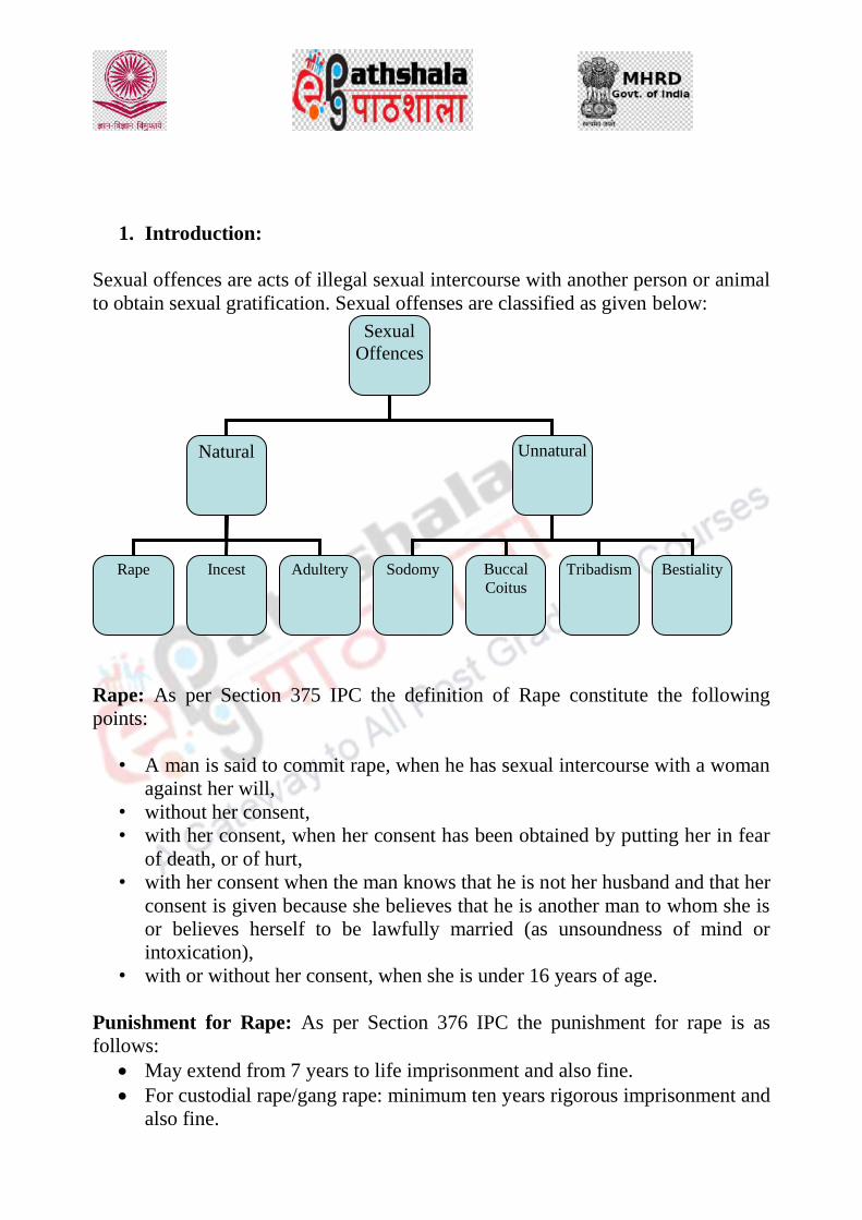
1. Introduction:
Sexual offences are acts of illegal sexual intercourse with another person or animal
to obtain sexual gratification. Sexual offenses are classified as given below:
Rape: As per Section 375 IPC the definition of Rape constitute the following
points:
• A man is said to commit rape, when he has sexual intercourse with a woman
against her will,
• without her consent,
• with her consent, when her consent has been obtained by putting her in fear
of death, or of hurt,
• with her consent when the man knows that he is not her husband and that her
consent is given because she believes that he is another man to whom she is
or believes herself to be lawfully married (as unsoundness of mind or
intoxication),
• with or without her consent, when she is under 16 years of age.
Punishment for Rape: As per Section 376 IPC the punishment for rape is as
follows:
May extend from 7 years to life imprisonment and also fine.
For custodial rape/gang rape: minimum ten years rigorous imprisonment and
also fine.
Sexual
Offences
Natural Unnatural
Rape Adultery Incest Sodomy Bestiality Tribadism Buccal
Coitus

Sec 228 IPC prohibits disclosure of identity of victim of rape.
Examination of Victim
The Objective for examination of a rape victim covers the following points:
1. To search for physical signs that will corroborate the history given by the victim.
2. To search for, collect and preserve all trace evidence for laboratory examination.
3. To treat the victim for any injuries and any venereal disease or pregnancy.
Preliminary steps: The Examination process of the victim should proceed only when asked by the
Police Officer or the Magistrate.
1. The first step is to obtain a written consent from the victim for the following:
NOTE: If the victim is under 12 years, or is insane, written consent has to be
obtained from her parents or guardians.
I. Preliminary Steps
1. Examination 3. Collection of Specimen
2. Photographs 4. Release of Information to
concerned authorities

3. Obtain and write history in the victim’s words which should include the
following:
,
4. Examination of Clothes
• Ascertain whether the clothes are those worn at the time of attack.
• The clothing should be retained if possible and labelled and handed over to
police.
• Foreign hair, fibers, etc., must be preserved.
Nail
scrapping
collection
Photographs of
the injuries on
the body of the
Victim
Events after the alleged
assault
Appearance
of Discharge
Pain
&
Haemorrhage
Details of
Struggle/Resi
stance/call for
help
Time and
place of
offence
Preliminary
Affairs
Victim’s Case
History

5. Examination of the Victim: This aspect should broadly include the followings
points:
Presence of
sperms in
the vagina
Injuries to
the Vagina
Condition/t
earing of
the Hymen
Signs of
violence in
other parts
of the body
Ascertainin
g the
clothing of
the Victim
Examination
of Victim

6. Examination of the Accused:
7. Samples collected for lab examination:
Samples Collected Purpose
Urethral swabs and smears For detection of semen, spermatozoa,
gonococci, etc
Loose foreign pubic hair or fiber
present on the body of accused.
For correlation with the event and the
victim
Stains of semen, blood, mud, grass
or other foreign materials on the
body of the accused.
For correlation with the event and the
place of occurrence.
Penile washings
For examining the presence of
vaginal epithelia
Examination of
the dental
structure of the
accused
Examination of
the genitals
Events after the
assault
Any history of
S.T.D of the
accused
Examination of
the Clothing of
the Accused
Examination for
any injury on the
body
Prior
Acquaintance
with the Victim
Examination of
the Accused

Nail Scrapings For examination of tissues embedded
in them during struggle
Buccal mucosa swabs Collected for saliva examination
Urethral swabs Collected for examination of any
venereal disease
Blood
Collected for grouping/serological
examinations
X-ray For age estimation especially in case
of delinquency plea
References:
Modi, Jaishing P, Textbook of Medical Jurisprudence & Toxicology, M.M.
Tripathi Pub. 2001.
Parikh, Textbook of Medical Jurisprudence & Toxicology, 2001.
Sharma, B.R., Forensic Science in Criminal Investigation and Trials (3rd
Edn.)
Universal Law Publishing Co. Ltd. New Delhi
James, S. H. and Nordby, J. J. (2003) Forensic Science: An introduction to
scientific and investigative techniques CRC Press, USA.
Guharaj, P. V., Chandran M. R. (2006) Forensic Medicine, 2nd Ed.,
Universities Press (India) Pvt. Ltd., Hyderabad.
Di Maio J. M. Vincent, Dana S. E. (2006) Handbook of Forensic Pathology,
VIVA Books Pvt. Ltd., India.

DESCRIPTION OF MODULE
Items Description of Module
Subject Name Criminology
Paper Name Forensic Science and Forensic Medicine
Module
Name/Title
Forensic psychiatry: definition, classification,
McNaugtens rule
Module Id 40
Objectives
Learning Outcome:
To make the learners understand the
various aspects associated with Forensic
Psychiatry
To make the learners understand the
various types of mental disorders
To understand the importance of legal
aspects associated with mental disorders
Prerequisites General understanding of the human
psychology, behaviours and psychiatric
disorders.
Key words
Forensic Psychiatry, mental disorders, McNaugtens Rule

1. Introduction:
Forensic psychiatry is a branch of medicine which focuses on the interface
of law and mental health. It includes psychiatric consultation in a wide
variety of legal matters (including expert testimony), as well as clinical work
with perpetrators and victims.
A Psychiatrist is a medical doctor who has completed several years of
additional training in the understanding, diagnosis, and treatment of mental
disorders.
A Forensic Psychiatrist is a psychiatrist who has additional training and/or
experience related to the interface of mental health (or mental illness) and
the law.
The Mental Health Act of 1987 was introduced to consolidate and amend the
law relating to the treatment and care of mentally ill persons, to make better
provisions with respect to their property and affairs and for matters
connected therewith or incidental thereto.
A mentally ill person needs to be restrained for his own safety or for the
safety of others. If the lawful guardian of such a patient is unable to keep
him under control for any reason, or if there be no guardian at all, the State
is legally justified to take charge of such a patient and to put him under
restraint.
Restraint may be (1) immediate, or (2) by admission to a psychiatric hospital
or registered psychiatric nursing home after certain conditions have been
complied with.
Scope of Forensic Psychiatry
Forensic Psychiatry
Criminal Forensic Psychiatry
Civil Forensic Psychiatry
Administrative Forensic Psychiatry

Classification of Mental Disorders
• Mental Retardation (mental sub normality, mental handicap)
• Organic Psychoses
Dementia
Drug induced psychoses
Confusional states and psychoses following epilepsy, pregnancy and
child birth, trauma, and general diseases.
• Functional Psychoses
Schizophrenia
Paranoid States
Affective disorders (mania, depression)
• Neurotic Disorders
• Personality disorders (e.g., sociopathic personality)
• Sexual deviations
Classification and Diagnosis of Mental Illnesses
In 1952, the American Psychiatric Association first published the Diagnostic
and Statistical Manual of Mental disorders (DSM-I). The latest edition,
DSM-IV was published in 1994.
The International Classification of Disease, 10th Revision, Clinical
Modification (ICD-10-CM), published by World Health Organization.
Diagnostic methods:
Computed Tomography (CT)
Magnetic Resonance Imaging (MRI)
Positron Emission Tomography (PET)
Competency to
Stand Trial
Legal Insanity
Expert
Witness/Testifying
Treatment of the
mentally ill offender
Personal Injury
Cases
Domestic relations
matters
Testamentary
capacity
Testimonial
capacity
Competency to
vote
Confidentiality
Privileged
Communications
Privacy issues
Right to treatment
Commitment or
Involuntary
hospitalization

1. Dementia
Dementia is described as a deterioration of intellectual function and other
cognitive skills, leading to a decline in the ability to perform activities of
daily living.
Prevalence increases rapidly with age; it doubles every 5 years after age 60.
Alzheimer’s disease and vascular dementias are the two most common
types, accounting for up to 90% of cases of established dementia in about a
2:1 ratio.
Signs and Symptoms
Steady, inexorable decline in intellectual function over 2 to 10 years,
culminating in total dependence and death, often due to infection.
Most common symptom is diminished short memory.
Other symptoms include personality changes, emotional liability, and poor
judgment.
Occurrence of mood swings, depression and euphoria.
Diagnosis
Diagnostic and Statistical Manual of Mental Disorders, Fourth Edition (DSM-IV)
criteria for dementia:
Impairment of memory
Impairment of at least one other domain of cognition.
The key to diagnosis is a thorough history.
Detailed drug review (over-the-counter drugs & ophthalmic preparations).
History of alcohol to be obtained.
Screening set of laboratory tests ( e.g. CBC, electrolytes, albumin, renal
function, liver function, thyroid function, vitamin B12 levels).
Dynamic imaging of cerebral blood flow by single photon emission
computed tomography (SPECT).
Neuropsychological testing.
Psychosis

Psychosis is a generic term for mental states in which the components of
rational thought and perception are severely impaired.
Particularly associated with schizophrenia, bipolar disorder and severe
clinical depression.
It should be distinguished from the concept of insanity and psychopathy.
Also to be distinguished from the state of delirium and mental illness.
Causes
Physical Circumstances (Electrolyte disorder, urinary tract infections).
Mental Illness (Bipolar disorder and schizophrenia).
Drug Use (Amphetamines, LSD, PCP, Cocaine or scopolamine).
Chronic psychological stress.
Withdrawal from CNS depressant drugs (Alcohol and Benzodiazepines).
Signs and Symptoms
Hallucinations
Delusions and paranoia
Thought disorder
Lack of insight
Diagnosis
• Pneumoencephalography in 1935 to obtain the first brain image of person
with psychosis.
• Grey matter reduction in cortex of people with psychosis.
• Sensory perceptions active during psychosis.
• Increased activation in the right hemisphere of the brain.
2. Delirium
• Delirium is not a disease, but it develops as the result of a medical condition.
• Acute means that it happens suddenly.
• Change in thinking or consciousness develops over a few hours or days.
• Patient may die from exhaustion in a week or two.
Signs and Symptoms

• Patient seems less aware of the environment.
• Memory may be affected.
• Trouble speaking or understanding.
• Delusions and hallucinations common.
• Restlessness and irritability.
• Mood swings and sleeplessness.
Causes
• Severe infections and high fevers.
• Dehydration.
• Diseases of the kidney or liver.
• Lack of certain vitamins.
• Hormonal imbalances.
• Lack of oxygen.
• Head injury.
• Reaction to drugs, alcohol, or prescription medications.
• After surgery effects.
3. Schizophrenia
• Schizophrenia is derived from the Greek words ‘schizo’ (split) and ‘phren’
(mind) and was coined by Eugene Bleuler to refer to the lack of interaction
between thought processes and perception.
• Severe mental illness characterized by persistent defects in the perception or
expression of reality.
• Schizophrenia does not involve a person changing among distinct multiple
personalities.
• Onset of schizophrenia typically occurs in late adolescence or early
adulthood, with males tending to show symptoms earlier than females.
Causes
Stressful life events
Drug usage
Use of stimulant or hallucinogenic drugs
Cannabis use
Smoking tobacco
Diagnosis
Significant Loss of Brain Gray Matter

Coronal MR scans
Decreased Prefrontal Brain Function
Enlarged Ventricles in the Brain
Decreased brain activity
Figure 1: Showing Significant loss of brain gray matter
McNaghten’s Rules
Daniel McNaghten killed the Prime Minister's Secretary by mistake for the Prime
Minister under an insane belief that the Government was persecuting him. He was
tried and acquitted on the ground of insanity and committed to Bethlehem
Hospital. The House of Lords then sent a number of questions to the judges of
England, who returned answers in the form of rules which are reported in 10 C. &
F. 200. The salient points covered under the McNaughten’s Rules are as follows:
Persons acting under the influence of an insane delusion are punishable if
they knew at the time of committing the crime that they were acting contrary
to law.
Every man is presumed sane and to have sufficient reason to be held
responsible for his crimes.
To establish a defense on the ground of insanity it must be clearly proved
that, at the time of committing the act, the accused was laboring under such
a defect of reason, from disease of the mind, as not to know the nature and
quality of the act he was doing or, if he did know it, that he did not know he
was doing what was wrong. If the accused was conscious that the act was

one that he ought not to do, and if the act was at the same time contrary to
the law of the land, he is punishable.
A person under a partial delusion is to be considered as if the facts with
respect to which the delusion exists were real.
Legal Test of Insanity
Section 84 IPC: Nothing is an offence which is done by a person who at the time
of doing it is by reason of unsoundness of mind, incapable of knowing the nature
of the act, or that he is doing what is either wrong or contrary to law.
References :
1. Davidson, A.H., Forensic Psychiatry, 2nd
ed., Ronald Press, New York, 1965
2. Heilbrun, K., The role of psychological testing in forensic assessment, Law
Human Behav., 16, aa257, 1992
3. Hess, A.K. and Weiner, I.B., Handbook of forensic Psychology, 2nd
Ed.,
John Wiley & Sons, New York, 1999.
4. APA, Issues in Forensic Psychiatry, American Psychiatric Association
Press, Washignton, D.C., 1984.
5. Simon, R.I., Clinical Psychoiatry and the Law. 2d
ed., American Psychiatric
Press, Washignton, D.C., 1992
6. Stone, A.A., Mental Health and Law. A System in Transition, Crime and
Delindquency Series, NIMH, Rockville, MD., 1975.


Galgotias University Plot No. 2, Yamuna Expressway,
Opposite, Buddha International Circuit,
Sector 17A, Greater Noida,
Uttar Pradesh 203201, India
E-content developed by Teachers
2. For SWAYAM

Sr no Name of the teacher
Name of the module developed
Platform on which module is developed
Date of launching e
content
Link to the relevant document and facility available in the institution
1. Dr. A Ram Pandey
Society and Media SWAYAM 2019-20
https://swayam. gov. in/nd2_cec20_ge36/preview
2. Dr. Harish Kumar
Gender & Contemporary Issues SWAYAM 2019-20
https://youtu. be/_vKTyr4x1Eg
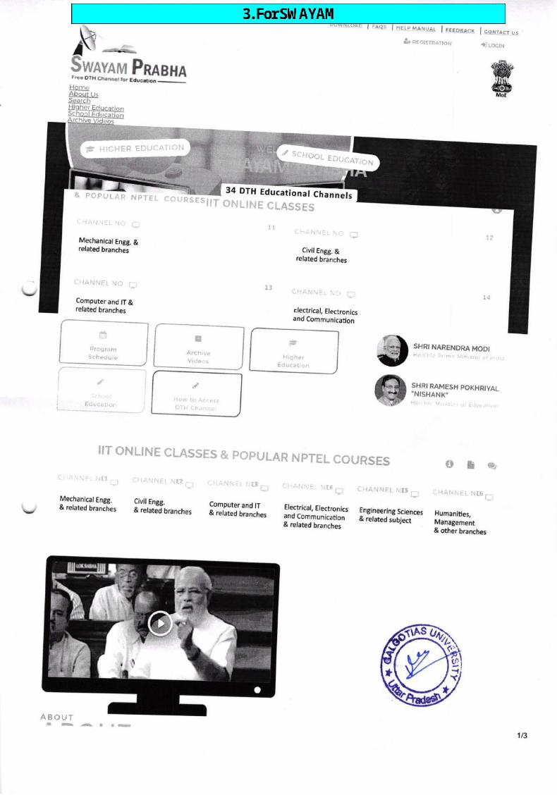
Ik -+rL$qynvarur Pnnaxarrec DIH Ch.nner ioi EdEiion _
r,ow*toac I r"oes I aEtp.t\.tA.N{.}A.t l tqe.pgr-sx I e_etlrct ,n&ceclsrnaxoN i-itoctr.r
EDUC,4T,ON
&&Mo€
R Nprrt cou
/ scHoor
34 DTH Educational ChannelsRSESIIT ONLINE CLASS ES
1r.(" I lr:lii\i:t. \it f;:j
Mechanical Engg. &related branches
cHANNET No * IZ
t4
SHRI NARENDRA MODI',,] }
i,r !,iri,t:i: ,i. i. l
..:Computer and lT &related branches
$ffi
CI.iA\JNEI- NIX
13 ci-lAN\il ili-i fr
Civil Engg. &related branches
rlectrical, Electronicsand Communication
SHRI RAMESH POXHRIYAL"NISHANK"
t.. ::, .j.i.,t!,..
IIT ONLINE CLAS$ES & POFULAR NPTEL COURSES Ott*,
Mechanical Engg.& related branches
FComputer and lT& related branches
Electrical, Electronicsand Communication& related branches
:.
Engineering Sciences& related subject
CH,A.NNEL NT6 f,,Civil Engg.& related branches Humanities,
Management& other branches
,/Houi lo AccpssDTt"i Chaon0i
HigherEducaticJn
A.r.h,veVider:s
ig HIfiHER EDUCATIOTI
ffPr0gramSchedule
ABOUTI
1/3
q 7
F
o
I H
*ffi
&
%
dell
Text Box
3. For SWAYAM

READ MORE
SWAYAM PRABHAThe SWAyAM pRABHA is a group of 34the CSAT-IS setellite Every day. therestuclents to choose the time of theirllTs. UCC. CEC, |CNOU. NCERT ancl N
from BISAC. Candhinagar. The
on 24X7 basis usingmes in a day.:llowing the
will be new content for at
DTH channels devoted to
least (4) hours
telecasting of high-quality edlicational programmeswhich would be repeated 5 more ticoltvenience. The channels are uplinked
iOS. The tNFLIBNET Centre ntaintains the web portal.contents are providecl by NpTEL,
\ ffiYF$ #$_$&trtr$|ffi*_rffi$"$ffiffi
^1
0
VAGEESH
Hurnanities- 1, Language atdLiterature
* cunp;li: l* upcoMlNc
SANSKRITI
Hum.rnitiesPhitosophy
Class-a pMeVtDyA
a, History, Culture &
VIEW ALL +
PRABODH
Sociat Science _r, IBehavioraL scienc*:
f atc*tvr.-
cEC/Usc
il CURRENT e upcoMij\ce
f ,arirHivi
CEC/UGC
[J CJPRENI A
= cufrpENr ? LjP
\ m"*-M tr${Af$N[?fri ff_##L
^1
.i,
VIEW ALt +
Ctass,3 pMeVlDyA
Class-r pMeVtDyA
!-:-.:-.----',' , \,lJl-llil ,\ l)J l J1i, ;)/:l\\,,
o
; c{:t{RENy ;, LiptotliNc f ARCHivi
PARTNERS AND CONTRIBUTERS
@
READ MOREREAD MORE
READ MORE
REAO MOREREAD MORE
READ MORE
I L b)Ntos
r@,ignou ffi ffi
ABOUT US CONTACT USvtstToRs
2t3
al
(].l
,:a
t@l @@@

The SWAYAM PRABHA is a group of34 DTHchannels devoted to telecasting of high_
:uality educational programm es on 24XZ
basis using the cSAT_lS sate,lite.
\. +9179-23268342
{Available on Monday To Friday from9gO AM To 6:0O pM)
p swayamprabha@infl ibnet.ac.in
Q vwwr.swayamprabha.gov.in
1179373
2C2: i\il.lB(*tT. all lghi: ,r"sery*j
v
READ MORE
3t3

10t1t2019Gmail - (no subject)
f"{ Gmail
(no subject)5 messages
Dr.Shrikant Dwivedi <[email protected]>
Mon, Feb 4, 2l,lg at 2:24 pM
F -90- 1 .63 / 20 I SAIIOS/A cad,. tTE/December 3l,20lg
Yours
Kanchan Kachroo <[email protected]>Reply-To: Kanchan Kachroo .t<ancnan_I'acfrroogyanoo.co.in>To: "Dr. Shrikant Dwivedi', <sh ree2g0@[r"it."om,
As you may be aware that NIoS has started a Live Telecast for_the D.Et.Ed programme which is beingtransmitted on the SWAYAM Prabha DTH channel no 32,.The timingoiitr''t"r".ast is Monday toFriday 3:00 pm to 5:00 pm (two hours).
Being an experienced person in the field of Teacher Education, we request for your expertise in this LiveTelecast of D'El.Ed. Programme on February 0s-06, 2019 in the Media uiit, om'rro";, *r* ;a,NOIDA at 3:00 pm lo 5:00 pm. Kindly be prisent ui i,oo p- for the preparation of this programme.
Please prepare PPTs for your topic following th" pr.r..iU"A fo"nlut as mentioned under Note.
Sir,
We look forward to your cooperation in the matter.
Needless to say, NIOS will reimburse local conveyance and provide a token honorarium.
With Regards,
sincerely,
(Sanjay Kumar Sinha)
Dr.Sreekant DwivediProfessorFaculty of Education,Galgotia UniversityNOIDA
Note: Please ensure that PPTs have Font Heading size 24-30 pnts; Subheading size 22-26 pnts; Body size 20-24 pnts; Framesize full screen 4:3 and fonts must be clear, legible against a soft coloured background with only 4-5 lines in each slide. Alsoinclude pictures and visuals to stlpport the content. Mail the final PPTs I day prior to your telecast date on [email protected]
With regardsDr. Kanchan Kachroo
https://mail.google.com/mail/u/o?ik=6cflg5f53d4&view=pt&search=all&permthid=thread-P/o3A 1624527821769464726&simpl=msg-fo/o3A16245278.. 1t3

't0t'v20,t9
Training Officer(Edu)NIOS [email protected]
Kanchan Kachroo <[email protected]>Reply-To: Kanchan Kachroo <[email protected]>To: "Dr.Shrikant Dwivedi" <shree2g0@[mail.com>
Sir,Any person is accompany you plz send the name.With regardsDr. Kanchan KachrooTraining Officer(Edu)NIOS HQsKanchankachrooT@gma il.comM-9654366679
[Quoted text hidden]
Gmail - (no subject)
Mon, Feb 4, 2019 at 2:26 pM
Dr.Shrikant Dwivedi <[email protected]>To: Kanchan Kach roo <kancha n_iach [email protected]. in>
Co presenter
MD FlRoz ALAM Designation Assistant Education officer NCPUL ,^pg.pJ orHigher Education , Ministry of HumanResource Govt. of lndia Email [email protected] Mobile No sgtttgoogg[Quoted text hidden]
Mon, Feb 4,2019 at2:27 pM
Mon, Feb 4,2019 at 11:05 pMDr.Shrikant Dwivedi <[email protected]>To: Kanchan Kachroo <[email protected]>, [email protected]
Plz find attached PPT for Live Telecast 3 to 5 pm slot.Topic: Learning Resources in Social SciencesThanks[Quoted text hidden]
Regards,Dr. Shri Kant DwivediAssoclate ProfessorSchool of Education, Galgotias U niversity,Room No. A-017A, Sec 1T A, Yamuna Expressway,Greater Noida, G. B. Naga4 (U.P.) - 20t308E Mail : [email protected] eb: hlp: //vrww,S Alg glpqAdyptllly. ed Ajn_Mobile No.- 9911487737
,U Dr. Shri Kant Dwivedi 05.02.2019.pptx80K
*
*.
Dr.Shrikant Dwivedi <[email protected]>To: [email protected]
Wed, Feb 6, 2019 at 12:38 PM
Plz find aftached PPT for Live Telecast 3 to 5 pm slot (06.02.2019).Topic: Learning Resources and Assessment in Social SciencesThanks
https://mail.google.com/maiUu/o?ik=6cfrl5f53d4&view=pt&search=all&permthid=thread-P/o3A1624527821769464726&simpl=msg-F/o3A16245278... 213

10t1t20,t9
[Quoted texl hidden]
Gmail - (no subject)
*il Dr. S.hri Kant Dwivedi 06.02.2019.pptx- 105K
.-J- ?Gl;:,Kr*<+
Mhttps://mail.google.com/maiuu/o?ik=6cre)5f53d4&view=pt&search=all&permthid=thread-P/o3A1624527821769464726&simpl=m sg-p/o3A16245278 313

10t1t2019Gmail - LIVE
Frl Gmail Dr.Shrikant Dwivedi <[email protected]>
LIVE2 messages
Kanchan Kachroo <[email protected]>Reply-To: Kanchan Kachroo <[email protected]>To : "D r. sh ri ka nt Dwived i" <s h ree2g0@[ma i l. com->, "firozm i mt@red iffma i l
Tue, Jan 9,2019 at 1:0S pM
.com" <[email protected]>
Sir,
As per our discussion, I will book a slot for your LIVE Telecast scheduled to be held on January 09,2019 (Wednesday).
The topic for the Telecast will be - Course Code 509 Assessment in Social Sciences
Please include the fo I lowing subtop_i cs iqyoulpreparatiol1l
Lecture42 509 Unit 9Continuous Comprehensive EvaluationLecture43 509 Unit 9Method of evaluation in social sciences based oninformation recall; understanding, applications andsynthesizing
Lecture44 509 Unit 9Alternative ways to evaluate learning (port Folio)
Lecture45 509 Unit 9Basis of evaluation, gpes of objective based questions,importance of grading and marking system
Kindly prepare your PPTs following the format:Please ensure that PPTs have Font Heading size 24-30 pnts; Subheading size 22-Z6pnts; Body size 20-24pnts; Frame size full screen 4:3 and fonts must be clear, legible againsti soft colourid background withonly 4-5 lines in each slide. Also include pictures and visuils to support the content. Mail thi final ppTsI day prior to your telecast date on [email protected]
Shall mailyour lnvitation Letters shortly.
With regardsDr. Kanchan KachrooTraining Office(Edu)NIOS HQsKanchankachrooT@g mail.comM-96s4366679
rl 509B4-U-9.pdf187K
Dr.Sh rikant Dwivedi <[email protected]>To: fi rozmimt <fi [email protected]>
rs,A
W
Tue, Jan 8, 2019 at 2:09 PM
https://mail.google.com/maiUu/0?ik=6c85f53d4&view=pt&search=all&permthid=thread-P/o3A1622076761907844690&simpl=msg-f/o3A16220767... 112

10t1t2019
[Quoted text hidden]
Gmail - LIVE
Regards,
Dr. ShriKant DwivediProgramme Chair (B.Ed.)
!9n9d of Education,Gatgotias UniversityPlot No.2, Sector 17-A
L?.rrI" Expressway, Greater Noida, Gautam Buddh Nagar,Uttar Pradesh Pin 201310Mob.:991'1481737
r1 509B-3-U-9.pdf"1B7K
https://mail.google.com/maiuu/0?ik=6cfr}5f53d4&view=pt&search=all&permthid=thread-P/o3A'1622076761907844690&simpt=msg-fo/o3A16220767... 2t2

10t1t2019
LIVE1 message
Kanchan Kachroo < [email protected]>
Gmail - L|VE
l-'1 Gmail
kanchan_l
Dr.Shrikant Dwivedi <[email protected]>
Thu, Feb 28,2019 at 11:20 AMReply-To: Kanchan Kachroo <kancha [email protected]>To: SATYENDRA GUPTA <satyendra [email protected]>,',Dr.Shrikant Dwivedi,, <[email protected]>
Sir,
As per our discussion, I will book a slot for your LIVE Telecast scheduled to be held on March 13,20lg(wednesday) from 3:00 pm to 5:00 pm. You u."."qu.ri"Jro urory be present by 2:00pm for thepreparation of the programme.
The topic for the Telecast will be - course code 507 Unit 12 MANAGEMENT AppRoACHES oF scHoolAND COMMUNITY PARTNERSHIP
Please include the following subtop_lcs iqyoulplgpalatia11:
Lecture 31 507 Unit 12Management Approaches - Meaning, nature and scope
Lecture 32 507 Unit 12Types of approaches man power,. Cost benefit,. Socialdemand,. Socialjustice
Lecture 33 507 Unit 12Relevance of each approach to strengthen school &Community Partnership
Lecture 34 507 Unit 12Differentiating management and organisation of School_Community Partnership and procesi of strengtheningrelationship between the two agencies
Lecture 35 507 Unit 12Supplementary Material: Video on Cases of CommunityMobilisation across states
Kindly_prepare_ypur PEfs folgyning the format:Please ensure that PPTs have Font Heading size 24-3opnts; Subheading size z2-26pnts; Body size 20-24pnts; Frame size full screen 4:3 and fonts must be clear, tegibte against-a soft colourid background wittronly 4-5 lines in each. slide. Also include pictures and visuils to support the content. Mail the final pprsI day prior to your telecast date on [email protected]
With regardsDr. Kanchan KachrooTraining Office(Edu)NIOS [email protected]
https://mail.google.com/maiuu/0?ik=6cff}5f53d4&view=pt&search=all&permthid=thread-p/o34'162669061332400g938&simpl=msg-f/o3A16266906... 112

'tot1t2019Gmail - LIVE
:l Block3_507.pdf392K
s
https://mail.google.com/mail/u/O?ik=6cf95f53d4&view=pt&search=all&permthid=thread-fo/o341626690613324009938&simpl=msg-fyo3A16266906... zlz

f.ri Gmail Dr.Shrikant Dwivedi <shree2g0@gmai t.com>
lnvitation for Live3 messages
Programme on SWAYAM PRABHA DTH CHANNEL
[email protected] <[email protected]>To: [email protected], shrikant.owivei'[email protected]
Dear Sir,This is in continuation to our telephonic conversation- and your consent,you are invited for live discussion for NloSSWAYAM PRABHA DTH CHANNEL on 20.03.2020 rrom 5 pru i, + pn,tl-y9l
_{" ;qr;;i;i i; reach Nlos srudio,.thfloor, A 24125, Sector 62, NoIDA by 2.30 PM. Kindty, a;nJ ppir;y', zo.os.zozo (by 11.30 AM) for our media unft forpreparation of live programme.
Regards,
Wed, Mar 18,ZOZO at 11:S0 AM
Dr. ManojThakurAsst. Director (CBC)
Dr.Shrikant Dwivedi <[email protected]>v To: [email protected]: [email protected]
I accept the invitation and will be there on time[Quoted text hidden]
Wed, Mar 18,Z0ZO at 1:4g pM
Dr.Shrikant Dwivedi <[email protected]>To: [email protected] Thu, Mar 19,2020 at 7:04 pM
Dear SirKindly find Attached PPT for live tetecast (3:00 to 4:00 pm)Topic: Assessment in EducationlQuoted te)d hiddenl
Regards,Dr. Shri Kant DwivediAssoclafe ProfessorSchool of Education, Galgotias University,Room No. A-017A, Sec 17 A, Yamuna Expressway,Greater Noida, G. B. Nagari (U.P) - 201308E Mail: [email protected] eb: h:!!p : //wutw. g a!99!ASUAers$y. et!.AMobile No.- 991148t737
,gi Assessment in Education.pptx- 100K
**

'1011t2019Gmail - LIVE
f'1 Gmail Dr.Shrikant Dwivedi <[email protected]>
LIVE4 messages
Kanchan Kachroo <[email protected]>Reply-To: Kanchan Kachroo <tancfran_[[email protected]>To: "Dr. Sh rikant Dwivedi" <sh [email protected], SATYEN DRAGUPTA <[email protected]
Sir,
Tue, Feb 19,2019 at 3:46 pM
GUPTA <[email protected]>, SATyENDRA
As per our discussion, I will book a slot for your LIVE Telecast scheduled to be held on February 21,2019 (Thursday) from 3:00 pm to 5:00 pm. You are requested to kindly be present by 2:00pm for thepreparation of the Programme.
The topic for the Telecast will be - Course Code 509 Assessment in Social Sciences
Please include the following subtopics in youlpreparatisnj.
Leclure42 509 Unit 9Evaluation
Continuous Comprehensive
Lecture43 509 Unit 9 Method of evaluation in socialsciences based on information recall; understanding,applications and synthesizing
Lecture44 509 Unit 9 alternative ways to evaluatelearning (Port Fotio)
Lecture45 509 Unit 9 basis of evaluation, types ofobjective based questions, importance of grading andmarking system
Ki n d ly_p_rcpare_you r PEfslqlowng th e fo rmat:Please ensure that PPTs have Font Heading size 24-30 pnts; Subheading size Z2-Z6pnts; Body size 20-24pnts; Frame size full screen 4:3 and fonts must be clear, legible against-a soft colourld background withonly 4-5 lines in each slide. Also include pictures and visuils to support the content. Mail the final ppTsI day prior to your telecast date on [email protected]
With regardsDr. Kanchan KachrooTraining Office(Edu)NIOS [email protected]
*:l Block3_509.pdf
947K
Dr.Shrikant Dwivedi <[email protected]> Tue, Feb 19, 2019 at 4:31 pMTo: Kanchan Kachroo <kanchan_kach [email protected]. in>Cc:SATYENDRA GUPTA <[email protected]>, SATYENDRA GUPTA <[email protected]>
*
https://mail.google.com/maiUu/0?ik=6cff)5f53d4&view=pt&search=all&permthid=thread-f/o3A1625891950002090522&simpl=msg-fo/o3A16288919... 1t2
10t1t2019
Thanks, I will be therelQuoted text hidden]
Lecture42 509 Unit 9Evaluation
[Quoted text hidden]
Gmail - LIVE
Regards,Dr. Shri Kant DwivediAssocrafe ProfessorSchool of Education, Galgotias lJniversity,Room No. A-017A, Sec 17 A, yamuna Expressway,Greater Noida, G. B. Nagar, (U.p) _ )Ot SOaE Mail : [email protected] eb: h,!!p1//www. S dSplbS!frUeISItV. edAJ nMobile No.- 991148L737
Dr.Shrikant Dwivedi <[email protected]>To: fi rozmimt <fi rozmimt@rediffmiiLom>
Please include rhe fbl lowing subtopjie s .igyeUlplgpalaliqg
Wed, Feb 20,2019 at 10:31AM
Continuous Comprehensive
Lecture43 509 Unit 9 Method of evaluation in socialsciences based on information recall; understanding,applications and synthesizing
Lecture44 509 Unit g alternative ways to evaluatelearning (Port Folio)
Lecture45 509 Unit 9 basis of evaluation, types ofobjective based questions, importance of griding anOmarking system
Dr.Shrikant Dwivedi <[email protected]>To: Kanchan Kachroo <[email protected]>, [email protected]
Plz find attached Ppr for Live Terecast 3 to 5 pm srot (21.02.2019).Thanks & Regards
Thu, Feb 21,2019 at 9:52 AM
on Tue, Feb 19, 2019 at 3:46 pM Kanchan Kachroo <[email protected]> wrote:[Quoted text hidden]
[Quoted text hidden]
*rl Dr.Shri Kant Dwivedi 21.02.2019.pptx.- 98K
**
https://mail.google.com/maiuu/O?ik=6cf95f53d4&view=pt&search=all&permthid=thread-p/o3A1625891950002090522&simpl=msg-F/o3A16258919.212

10t'v2019Gmail - LIVE dated July 11,2O1g
l*'f Gm*it Dr.Shrikant Dwivedi <[email protected]>
LIVE dated July 11,20192 messages
Kanchan Kachroo <[email protected]>Reply-To: Kanchan Kachroo .kancfran_i'Jctriooqyanoo.co.iTo: "Dr.Shrikant Dwivedi', <shree2g0@gr"ii..om
Sir,
As per our discussion, we extend our invitatiq! for your LlvE Telecast scheduled to be held,?l irt'rlJ;i:1fliTfir:'B?"Hl11oo pm' iou
"i" i"q-'"'ted to xinorv ue present bv 2:00 pm
Wed, Jul 10,2019 at 1:43 pMn>
1) Dr. Shrikant Dwivedi2) Md. Firoz Alam
The topic for the Telecast will be from course code 507 Unit 4 pRovlsloNs FORCOMMUNITYPARTICIPATION UNDER SSA AND RTE
With regardsDr. Kanchan KachrooTraining Office(Edu)NIOS [email protected]
M-9654366679
-r Blockl 507.odf- 829K
Lecture 7 507 Unit4Policy provision, Role of parent teacher association/mother teacher association, School ManagementCommittee; Self help group
Lecture 8 507 Unit 4Main objectives of panchayati Raj and their role as aninstitution in EE
Lecture 9 507 Unit 4
!E and partnership with community (pros and cons),Experiences and research studies
$dlV-nrerc-your PEfs following the format:Please ensure that ppTs have Font Heading s,'= e 24-30 pnts; Subheading size 22-26 pnts;Body size 20'24 pnts; Frame size full screen 4:3 and fonis must be ctear, legible against a softcoloured background with only 4-5 lines in each slide. Also inctude pictures and visuals tosupport the content. tvlail the final PPTs 1 day prior to your telecast date on [email protected]
U
https://mail.google.com/maiuu/0?ik=6cf95f53d4&view=pt&search=all&permthid=thread-P/o3A'163865g389048073g72&simpl=msg-f7o3A16386583.'U2

10t1t20,t9Gmail - LIVE dated July 11,2019
<[email protected]><[email protected]>, [email protected]
PFA[Quoted text hidden]
Regards,Dr. Shri Kant DwivediAssocrale ProfessorSchool of Education, Gatgotias University,Room No. A-017A, Sec 17 A, yamuna Expressway,Greater Noida, G. B. Nagan (U.p.) _ iOtSOAE Mail I [email protected] eb : blp;lw vvw.S AlS efusuljystsjly. ed4 nMobile No.- 9911481737
,31 ?J;in"
Kant Dwivedi 11.07.2019 Provisions for community participation Under SSA and RTE.pptx
T
+
Dr.Shrikant DwivediTo: Kanchan Kachroo Thu, Jul 11,2019 at 11:43 AM
https://mail.google.com/maiUu/O?ik=6cf95f53d4&view=pt&search=all&permthid=thread-P/o3A'1638658389048073972&simpl=msg-F/o3A,t 6386S83... 2t2

lY Gmail
LIVE dated October 14,20193 messages
Dr.Shrikant Dwivedi <[email protected]>
Kanchan Kachroo <[email protected]>Reply-To: Kanchan Kachroo <kanchan_[[email protected]>To: SATYENDRA GUpTA <satyendra.e?npgmai[iom>,,,Dr.ShrikantGUPTA <[email protected]>
Sir,
As per our discussion, we extend our invitation for your LIVE Telecast scheduled to be heldon October 14,2019 (Monday) from 3:00 pm to 5:00 pm. You are requested to kindly bepresent by 2:00 pm for the preparation of the programme.
1) Dr. Satyendra Gupta2) Dr. Shrikant Dwivedi
Kindly sonfirm the To@!Cd.
4i nd ly_pppa r. ysrfEtr&llowi n g the format:Please ensure that PPTs have Font Heading stze 24-30 pnts; subheading size 22-26 pnts;Body size 20-24 pnts; Frame size full screen 4:3 and fonis must be cleariegible against a'softcoloured backgroung yiln on[ 4-5 lines in each slide. Also include pictures and visuals tosupport the content. Mail the final PPTs 1 day prior to your telecast date on [email protected]
Thu, Oct ,10,2019 at 5:04 pM
Dwivedi" <[email protected]>, SATYENDRA
With regardsDr. Kanchan KachrooTraining Office(Edu)NIOS [email protected]
Dr.Shrikant Dwivedi<[email protected]> Fri, Oct 11,Z}1gat 11:19 AMTo: Kanchan Kach roo <kancha n_kach [email protected]. in>
Th.anks mam for your invitation. I am selecting topic from block 502 for LIVE Telecastscheduled to be held on october 14,201g (Monday) from 3:00 pm to 5:00 pm.
Topic: lntegrated Learning and Teaching process.
Thanks again for giving this opportunity.
[Quoted text hidden]
Regards,Dr. Shri Kant DwivediAssoclale ProfessorSchoo/ of Education, Galgotias University,Room No. A-017A, Sec 17 A, Yamuna Expressway,
E).>
i;6t Y,/

Greater Noida, G. B. Naga; (U.p.) _ 20130gE Mail; shrikant.dwivedi@lalgotiasu niversity.ed u. inW eb: bttp://wvrw. g aS g!jg1!!Uye rsll4.gdUt n -'Mobile No.- 9911481737
[email protected], Oct 1A,ZO1g at 9:34 pM
Dear Sir/MamPlease find attached ppr for Live Tetecast 3 to 5 pm srot (14.10.2019).Topic:lntegrated Learning and Teachin! Fio""",Thanks[Quoted text hidden]
,+i1 14.1'a goK0.2019 lntegrated Learning and Teaching process.pptx
*
Dr.Shrikant DwivediTo: Kanchan KachrooCc: SATYENDRA GUPTABcc:

MGmailLIVE dated October 21, 2O1g3 messages
Dr.Shrikant Dwivedi <[email protected]>
hoo.co.in>
Sent from Yahoo Mail
Sir,
1) Dr. Shrikant Dwivedi2)Ms. Deepa Bisht
Ktrullvsollrmlhe_lo p t c sele cted.IQplc co ntexluallsin g lea rn i n g-pWge d I ea rn e rsBlock 2
With regardsDr. Kanchan KachrooTraining Office(Edu)NIOS [email protected]
As per our discussion, we extend our invitation for your LIVE Telecast scheduled to be heldon october 21,2019 (Monday) from 3:0o,nato sitio irn. you are requested to kindly bepresent by 2:00 pm for the preparation of td" nrogium;e
Fri, Oct 18,2019 at 1:20 pM
-,
[email protected]&Iowxl g the format:Please ensure that PPTs nare ront Heaoing sL e 24-30 pnts; Subheading size 22-26 pnts;Body size 20'24 pnts; Frame size full screen 4:3 and fonis must be clear, legible against asoft coloured backgro.und with only 4-5 lines in each slide. Arso incluoe-fictures ano visualsto support the content. Mair the finar pprs 1 day prior to your terecast dateon [email protected]
+
Dr.Shrikant Dwivedi <[email protected]>To : Ka ncha n Ka ch roo <ka ncha n_kaih roo@ya hoo. co. in >
Thanks for the mail.[Quoted text hidden]
Fri, Oct 18,2019 at 1:31 pM
Dr.Shrikant Dwivedi <[email protected]>To: [email protected]
[Quoted text hidden]
Fri, Oct 18,2019 at 1:34 pM

10t1t2019Gmail - LIVE dated Sep 30, 2019
FtGmail Dr.Shrikant Dwivedi <[email protected]>
LIVE dated Sep 30, ZOlg2 messages
Kanchan Kachroo <[email protected]>Reply-To: Kanchan Kachroo .tancfran_i'acf,[email protected]:'Dr. Shrikant Dwivedi', <shree2g0@grrii."om
Sir,
As per our discussion, we extend our invitatio-n for your LlvE Telecast scheduted to be heldon september 30, 2o19 (Monday) from_3:00 pr io s:oo pm. i;;;;d;ested to kindry bepresent by 2:00 pm for the prepaiation of tne programm;.
Mon, Sep 30,2019 at12:48pMco.tn>
The topic for the Telecast will be from course code 507 community and school
5lnd. tv-o-rc0rc-vpur PPrs-follswrn g the format:
Please ensure that PPTs nave roit neaoinflze 24-30 pnts; Subheading size 22-26 pnts;Body size 20-24 pnts; Frame size full screen 4:3 and fonis must be clear, legible against a softcoloured backgroun{ yiln only 4-5 lines in each slide. Aro include pictures and visuals tosupport the content. Mair the finat pprs 1 day prio," io yorr terecast brt" * [email protected]
1) Dr. Shrikant Dwivedi2) Ms. Deepa Bisht
With regardsDr. Kanchan KachrooTraining Office(Edu)NIOS [email protected]
Dr.Shrikant Dwivedi <[email protected]>
- To: [email protected]
lQuoted text hiddenl
Mon, Sep 30, 2O1g at 12:56 pM
6"Y+
https://mail'google.com/maiuu/o?ik=6cff}5f53d4&view=pt&search=all&permthid=thread-p/o3A1646083893460818917&simpl=msg-fo/o3A16460838... .lt1
I
I
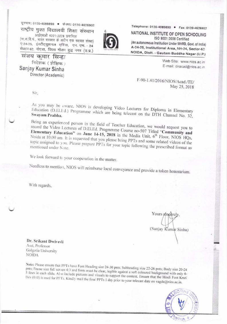
Eyqrs: 0r20-4089899 o swr: 0120_4626902qr*tq yffi Aqrtr$ trren *iw16
s{rfpi 9oo 1 :2008 ycrBrd(q'.{i.fr.d. , qnn YflzER d srfR go rcrrm nw)g-24-25, vr#qqrfi-a qRrn g{. vq. - 24
felephone: 0120-4089A99 . Fax; 0120-4626902
NATIONAL INSTITUTE OF OPEN SCHOOLINGtS0 9001 :2008 Certified
(An autonomous lnstitution Under MHRD, Govt. ol lndia)A-24-25, lnstitutional Area,. NH-24, Seclor-6cttqet-ez. drsr, fu"n .ilrrl ;rn (u.u.)
J
m)sq NOIDA, Distt. - Gautam Buddha Nagar (U.P.)rreq E$Trt frr6r
f{iflrar ( $rfil-n ,
Sanjay Kumar SinhaDirector (Academic)
Web Site: www.nios.ac inE-mail: [email protected] rn
F-90- t,4t /20 I 6lNIOS/Acact./TE/May 25,201g
Sir,
We lo<tk forward t(i ,vour cooperation in rhe matter
Needlcss to menti'rt' NIos will reirnburse local con'eyance and provide a token honorarium
With regards,
Yours
(Sanjay Sinha)
Dr. Srikant Dryir.ctliAssl- PlulbssorGaIgotia UnivcrsitvNOIDA
;\s ;'otr rnav bc ir\\'ilrc' NIos is cleveloping vicle. Lectures for Diploma in Ele,rentaryI*:'#ffi1?rii;|it ) r'rue,r,*u* ;il are-being
"[."ri'"" trie DrH channer No, 32.
Being an experienccd person in the fleld of rcacher Education, we would request you torecord the video I-ectures .f D.El.[d. trr:ogru*n.,* cource n.-507 Titled ..co*Irority andElementnry Ettucationo'or.rur*-rJ-il,1orr i,, irr..-rnr*oj" unir.6tr,FIo;., l.lioS Hes,Noicla ar r 0:0(l a'r Ir ir,,.qu.*r"ii ,lrr, ,",; prease rrri;,,; ppil'lnd .,rr," rerared vicreos of the
llli:,il:li'l;,.1j: i;: r,rcase p,."p,;;.b'r,;. io, y,,u, ,"pi.'n,ii"*ing the prescribect fo.nat as
:II
Note: l)leasc enstrre that 'l]l',tJ'"..
Font Hcading size z4-3apnts; subheacling size ?2-26 pnts;Body size 20-24pnts; Irranre size lull scrtctt 4:3 and forrrs ,nr., ti crear, legibre ";;;; ;; coloured background with onry a_'5 Iines in each sride. Ar.o ips;ucr, pr.rr,"r',rro rirrrr.io;rpp#;;;;;;;r. Ensure that the Hindi Font KrutiDcv (0 l0) is usecl fbr ppt.s. Kincllv ,na;t ttre i,rrai i;lf, I aor_.prio, ,, 1,or',, ,.l.cast date on [email protected].
v'

Erarrn: 01 20.4089899 . Sqqr: otao_a626902
Trc{lq gffi A-sr6{ff Pren titenq3rTf\nrsn 9oot 12 ro8 ycrftra
(qT.q.ft.ri., qrra vron d rr#{ q:fi.wFra rnefl)g-24-25, ir+c-Wrr-d strqr, v{. qEr. - 24
Telephone: 0120-4099899 . Fax: 0t2O-462690?
NATIONAL INSTITUTE OF OPEN SCHOOLINGlS0 9001 :2008 Certitied
(An autonomous lnstitution Under MHHD, Govt, of India)A-24-25, I nstitut iona I Area, N H-24, Sector-62NOIOA, Distt. - Gautam Buddha Nagar (U.P.),
Web Site. www.nios.ac.inE-mail: [email protected]. rn
m)*er-sz. d\.sl, ftrm rttTq
Trsq agrrp ftr{Tfr*vffi t ptfq{q. )
Sanjay Kumar SinhaDirector (Acadernic)
slr-
\\i irh Regarcls.
ga rw (v.n.)
F -90- I .63120 I 8/NIOs/Acad.lIENovember 30, 20lg
;)l,iliJlil,,?ff|'-t,at NIOS has srarted.a-Lrvr Radio counsering for rheln,g.,,rn,.;;ir';;'::""ttcd on Radio varrili-1vr d,r*rli".otion 9r.2 MHz. Th,3"',Y,It P;1sr1mme
iav to Frida)'2:45 pmm 3:30 prn ios'rrir,n*1. e timing of th"e nutriuIleing an expcrien""{t*T9n i'trre field of Teacher Education. \tr/e r.n,,aor +)_- _-_Ita d i' cr o u n se r r i n q .,p
f L;"r; il.+ o n D """m-ilJi iro;' rX; JiTH:
rff.r;:ffIf [],;;i: j1,,^il;lre' NorDA at 2:145 pm ro :':o prr."ri"iiv b, p,esenibf i,oi,p* f"r;d;6;;on of this programme.
We look furward to -).our cooperation in the rnatter. ' -----'"
,
\cetlless lo sav' NIoS will reimburse local conveyance and provide a token honorarium.
Yours
(Sanjay K".)
ryDr. Shri Kanr Dwiyerlirr0grar.r Chair (B.Ed)Sc:hool of EclucationGalgotias t.iniversity, ( ireater Noida
\,rirr lJrt l(,1(ijir (.,,rillir"r iirrt prutgrnrrrirrc is irl-so s1.oiyu1r1e orr NIOS rveh radio porta I rvwrv.n ios. iradiontlia.

Galgotias University Plot No. 2, Yamuna Expressway,
Opposite, Buddha International Circuit,
Sector 17A, Greater Noida,
Uttar Pradesh 203201, India
E-content developed by Teachers
3. For other MOOCs platforms (NIOS)

Sr no Name of the teacher
Name of the module developed
Platform on which module is developed
Date of launching e content
Link to the relevant document and facility available in the institution
1. Dr. Satyendra Gupta
D. El. Ed. 503 Learning- The Concept and Process
National Institute of Open Schooling (NIOS) 5-Nov-18
https://youtu. be/7JsY0AxTHzc
2. Dr. Satyendra Gupta
D. El. Ed. 501 Universalization of Elementary Education
National Institute of Open Schooling (NIOS) 17-May-18
https://youtu. be/-dbn5UL1Iew
3. Dr. Satyendra Gupta
D. El. Ed. 504 Learner and Learning Centered Methodologies- I
National Institute of Open Schooling (NIOS) 18-Jun-18
https://youtu. be/sB6W9eW-Hcc
4. Dr. Satyendra Gupta
D. El. Ed. 504 Learner and Learning Methodologies
National Institute of Open Schooling (NIOS) 7-Jun-18
https://youtu. be/lWpHAtYpE9M
5. Dr. Satyendra Gupta
D. El. Ed. 504 Pedagogy of Mathematics
National Institute of Open Schooling (NIOS) 13-Jul-18
https://youtu. be/_nT5rpLlvCg
6. Dr. Satyendra Gupta
D. El. Ed. 504 Data Handling
National Institute of Open Schooling (NIOS) 27-Aug-18
https://youtu. be/L1q5MRb5rww
7. Dr. Satyendra Gupta
D. El. Ed. Data Handling and Measures of Central Tedency
National Institute of Open Schooling (NIOS) 9-Oct-18
https://youtu. be/AOADulLOQ0M
8. Dr. Satyendra Gupta
D. El. Ed. 506 Development of Personality and Assessment
National Institute of Open Schooling (NIOS) 19-Nov-18
https://youtu. be/tE2Rs7ep-QY
9. Dr. Satyendra Gupta
D. El. Ed. 509 Nature of Social Science
National Institute of Open Schooling (NIOS) 26-Dec-18
https://youtu. be/c1neGi4kooU
10. Dr. Satyendra Gupta
D. El. Ed. Teaching Learning Strategies
National Institute of Open Schooling (NIOS) 2-Nov-19
https://youtu. be/gfDtoe1dhWc
11. Dr. Satyendra Gupta
D. El. Ed. 510 Different Approaches to Teaching of Science
National Institute of Open Schooling (NIOS) 25-Feb-19
https://youtu. be/84WhQHov71s
12. Dr. Satyendra Gupta
D. El. Ed. 501 Strategies for UEE-I
National Institute of Open Schooling (NIOS) 4-Mar-19
https://youtu. be/znDIXSLPsbQ
13. Dr. Satyendra Gupta
D. El. Ed. 504 Learner Assessment in Mathematics
National Institute of Open Schooling (NIOS) 25-Apr-19
https://youtu. be/gcuB8kGaYD8
14. Dr. Satyendra Gupta
D. El. Ed. 504 Characteristics of Learning Centred Approach
National Institute of Open Schooling (NIOS) 30-Apr-19
https://youtu. be/_UaEJmfnlao

15. Dr. Satyendra Gupta
D. El. Ed. 504 Follow up of Assessment of Learning Mathematics
National Institute of Open Schooling (NIOS) 7-Jan-20
https://youtu. be/-GnRI0YcLsA
16. Dr. Satyendra Gupta
D. El. Ed. Teacher's Training, Emerging Trends in Assessment
National Institute of Open Schooling (NIOS) 14-Feb-20
https://youtu. be/JKWSr2Ik7Hs
17. Dr. Satyendra Gupta, Dr Navita Malik
D. El. Ed. 506 Introduction of Inclusive Education
National Institute of Open Schooling (NIOS) 14-Jun-19
https://youtu. be/7c0rZrJx4Vo
18. Dr. Satyendra Gupta, Dr. Shri Kant Dwivedi
D. El. Ed. Management Approaches of School & Community Partnership
National Institute of Open Schooling (NIOS) 13-Mar-19
https://youtu. be/TIY_QwGut2M
19.
Dr. Shri Kant Dwivedi
D. El. Ed. Course-507
विविन्न राज्यों में
सामुदाविक िागीदारी के
प्रिास
National Institute of Open Schooling (NIOS) 14-Jun-18
https://youtu. be/MC6Bm4IrlT8
20. Dr. Shri Kant Dwivedi
D. El. Ed. Course-507
विक्षा का अविकार
आिाररत उपागम
National Institute of Open Schooling (NIOS) 14-Jun-18
https://youtu. be/h0jss75hGpg
21.
Dr. Shri Kant Dwivedi
D. El. Ed. Course-507
विक्षावथिियों के िाषा
विकास पर समुदाि का
प्रिाि
National Institute of Open Schooling (NIOS) 14-Jun-18
https://youtu. be/mX--SaStXwo
22. Dr. Shri Kant Dwivedi
D. El. Ed. Course-507
सूक्ष्म विियजि में समुदाि
की िूवमका
National Institute of Open Schooling (NIOS) 15-Jun-18
https://youtu. be/PwHU4tL5SnE
23.
Dr. Shri Kant Dwivedi
D. El. Ed. Course-507
स्थािीि सोंसािियों की
पहचाि और प्रचलि में
समुदाि की िूवमका
National Institute of Open Schooling (NIOS) 15-Jun-18
https://youtu. be/I0nxX9h5Uig
24. Dr. Shri Kant Dwivedi
D. El. Ed. Assessment in Social Sciences
National Institute of Open Schooling (NIOS) 9-Jan-19
https://youtu. be/2_Oo-n5-EJI
25. Dr. Shri Kant Dwivedi
D. El. Ed. Learning Resources in Social Sciences
National Institute of Open Schooling (NIOS) 5-Feb-19
https://youtu. be/bdaVxzgIy-0
26. Dr. Shri Kant Dwivedi
D. El. Ed. Learning Resources and Assessment in Social Sciences
National Institute of Open Schooling (NIOS) 2-Jun-19
https://youtu. be/tJjI-6dE150
27. Dr. Shri Kant Dwivedi
D. El. Ed. Assessment in Social Sciences
National Institute of Open Schooling (NIOS) 21-Feb-19
https://youtu. be/hsdXES4bGlg

28.
Dr. Shri Kant Dwivedi
D. El. Ed. Contextualizing Learning Processes of Disadvantaged Learners
National Institute of Open Schooling (NIOS) 21-Oct-19
https://youtu. be/BEr5mqOknIQ
29. Dr. Shri Kant Dwivedi
D. El. Ed. Teacher's Training, Assessment in Education
National Institute of Open Schooling (NIOS) 20-Mar-20
https://youtu. be/IGScqZNHY-Q
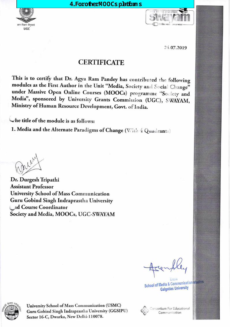
trt?-fun fry{riu6c
CERTIFICAIE
This is to certifr that Dr. Agya_Ram Pandey has contribrrted the followingmodules as the First Author in the Unit "Media, Sociery an,I S,,sia Cl,,*nge;under Massive open online courses (Moocs) p.ngramrne ,,sc ,"ty .r,dMefia", sponsored by university Grants commission (uGC), swdy4ivt,Ministry of Human Resource Development, Govt. of India.
--.he title of the module is as follows:
1. Media and the Alternate Paracligms of Change (\ ^rl, .i eu:r.^rant.
Dr. Durgesh TripathiAssistant ProfessorUniversity School of Mass ComnrunicationGuru Gobind Singh lndraprastha University
.-,d Course CoordinatorSociety and Media, MOOCs, UGC-SWAYAM
University School of Mass Communication (USMC)
Guru Gobind Singh Indraprastha University (GGSIPU)
Sector 16-C, Dwarka, New Delhi-l10078.w
,\
I rlhl rlt
? 1.o7.2019
School ot Media &
Galgotias Universi$
^,r r lortium For [:ducationalComnrurrication
{
6$-
rI
dell
Text Box
4. For other MOOCs platforms

t
I
rt
W
-=@
.
"@,
il
"$ ><)b
0rtsi?argE..
!,N
.&-6 LJ.!(
E-'i 'a*no.*
u5{u*s:rB
b
? f,er- E
.tE
6EJ-z
lt!(v!.,n.x.9 hll,E
ts#s
:]; il
{\l ,,
ul
"x.9 [€rE
t#$s
#
r;'!J
a
il
&
1.
-!
ffiw=
atd,tt=artat6b.::'6E
L-OE.>E
53s06=.cs 5E
8o
rfrItH
'I1l
8=E
{t^.5E
S*t*
SoE
*E
EE
:'E
$.6sEs*E*
.:DT,tl-d?sr#P". {r(t{ .4g
(*;
&6
.it.*, ,J7(E
*o!E
E
s}\i){t'gE{
[*6E
..
s,qE5Y
E
r."Ol
.-.! [
tpEt
fls5
oS,
TU
Uo{/}r.g)u$rsfi-q).={ut*il)y{u
s$.g{uHo{}?F
IfiT-gEsH
Itr}JC0.){u
..i r
_t