1992 volume 31 number 4 -
78
NENCKI INSTITUTE OF EXPERIMENTAL BIOLOGY WARSAW, POLAND 1992 VOLUME 31 NUMBER 4 ISSN 0065-1583 http://rcin.org.pl
-
Upload
khangminh22 -
Category
Documents
-
view
1 -
download
0
Transcript of 1992 volume 31 number 4 -

NENCKI INSTITUTE OF EXPERIMENTAL BIOLOGY
WARSAW, POLAND 1992 VOLUME 31 NUMBER 4
ISSN 0065-1583 http://rcin.org.pl

P o l i s h A c a d e m y of S c i e n c e s N e n c k i I n s t i t u t e of E x p e r i m e n t a l B i o l o g y
ACTA PROTOZOOLOGICA
International Journal on Protistology
Editor in Chief Stanisław L. KAZUBSKI
Editors Jerzy SIKORA and Anna WASIK
Managing Editor Małgorzata WORONOWICZ
Editorial Board
Andre ADOUTTE, Paris Christian F. BARDELE, Tübingen Magdolna Cs. BERECZKY, Göd Jacques BERGER, Toronto Y.-Z. CHEN, Beijing Jean COHEN, Gif-Sur-Yvette John O.CORLISS, Albuquerque Gyorgy CSABA, Budapest Isabelle DESPORTES-LIVAGE, Paris Stanisław DRYL, Warszawa Tom FENCHEL, Helsing0r Wilhelm FOISSNER, Salsburg Vassil GOLEMANSKY, Sofia Andrzej GRĘBECKI, Warszawa, Vice-Chairman Lucyna GRĘBECKA, Warszawa Donat-Peter HÄDER, Erlangen Janina KACZANOWSKA, Warszawa Witold KASPRZAK, Poznań
Stanisław L. KAZUBSKI, Warszawa Leszek KUZNICKI, Warszawa, Chairman John J. LEE, New York Jiri LOM, Ćeske Budejovice Pierangelo LUPORINI, Camerino Hans MACHEMER, Bochum Jean-Pierre MIGNOT, Aubiere Yutaka NAITOH, Tsukuba Eduardo ORIAS, Santa Barbara Dimitrii V. OSSIPOV, St. Petersburg George I. POLIANSKY, St. Petersburg Igor B. RAIKOV, St. Petersburg Leif RASMUSSEN, Odense Jerzy SIKORA, Warszawa Michael SLEIGHT, Southampton Ksenia M. SUKHANOVA, St. Petersburg Jiri VAVRA, Praha Anna WASIK, Warszawa
ACTA PROTOZOOLOGICA appears quarterly. Indexed in Current Contents in CABS and in Protozoological Abstracts.
Front cover: Thigmocoma acuminata Kazubski. Acta Protozool. 1963, Vol. 1 fasc. 25 p. 239, Fig. 1
http://rcin.org.pl

ACTA
PROTOZOOLOGICA
Gravireception and Graviresponses in Ciliates
Hans MACHEMER and Richard BRÄUCKER
Arbeitsgruppe Zelluläre Erregungsphysiologie, Fakultät für Biologie, Ruhr-Universität, Bochum, Federal Republic of Germany
Summary. An account is given of approaches to gravireception, terminology, mechanisms of responses to gravity as investigated and documented in the literature, and sensorimotor coupling properties in ciliates. Current theories and methods are discussed, and previously published experimental data on graviresponses are reviewed.
Key words. Gravireception, gravitaxis, gravikinesis, ciliates, electrophysiology, sensorimotor coupling
CONTENTS 8.2 Implications of the Electrophysiological Model 8.2.1 Masking of the Graviresponse
1. Introduction 8.2.2 Effects of Cell Orientation 2. Approaches to Gravireception 8.3 Circumstantial Experimental Conditions 2.1 Energetic Considerations 8.3.1 Cell Cultures and Experimental Solutions 2.2 Structures Suitable for Gravireception 8.3.2 Equilibration Time 2.3 Behavioural Analysis 8.3.3 Proportions of Chamber 3. Terminology 8.3.4 Mechanical Disturbances 4. Mechanisms in Graviresponses 8.3.5 Temperature 4.1 Static Hypothesis (Gravity-Buoyancy Model) 8.3.6 Illumination 4.2 Hydrostatic Hypothesis 8.3.7 Aeration 4.3 General Statocyst Hypothesis 8.3.8 Determination of Sedimentation Rates 4.4 Resistance Hypothesis 8.3.9 Acquisition of Data 4.5 Hydrodynamic Hypothesis 8.3.10 Processing and Presentation of Data 4.6 Propulsion Hypothesis 8.3.10.1 Behavioural Variety 4.7 Lifting Force Hypothesis 8.3.10.2 Statistical Treatment 4.8 Special Statocyst Hypothesis (Electrophysiological 8.3.10.3 Circular Histograms
Model) 8.3.10.4 Direction Coefficient (r-value) 4.9 Conclusions Regarding Various Hypotheses on 8.3.10.5 Orientation Coefficient
Graviresponses 8.3.10.6 Taxis Coefficient 5. Ciliates as Excitable Cells 8.3.10.7 Kinesis Coefficient 6. Mechanoreception in Ciliates 8.4 Current Experiments 7. Electromotor Coupling 8.4.1 Experiments under Normal Gravity 8. Physiology of Gravistimulation and Response 8.4.1.1 Free Locomotion 8.1 Basic Mechanisms 8.4.1.2 Velocity-Dependence of Gravikinesis 8.1.1 Gravikinesis 8.4.1.3 Cells under Galvanotactic Alignment 8.1.2 Gravitaxis 8.4.1.4 Swimming in Solutions of Adjusted Density
8.4.1.5 Nonswimming Locomotion
Address for correspondence: H. Machemer, Arbeitsgruppe 8.4.2 Microgravity Experiments Zell uläre Erregungsphysiologie, Fakultät für Biologie, Ruhr- 8.4.3 Hypergravity Experiments Universität, D-4630 Bochum 1, Federal Republic of Germany. 9. Conclusion and Perspective
Acta Protozoologica (1992) 31: 185 - 214
Review article
http://rcin.org.pl

186 H. Machemer and R. Braucker
1. INTRODUCTION
Gravireception in protists is an area of so far limited knowledge and understanding. A wealth of data, in particular of Paramecium, has been accumu-lated in the past century, but there is not much common agreement on the mechanisms of gravitaxis. The reason for this is closely associated with the nature of gravity which rules the universe across billions of light years and affects small living things on Earth in a subtle manner. Experiments using gravist imulation are diff icult to design and to analyze because gravity is not easy to manipulate. Moreover, gravist imulation persists. Unlike a mechanical stimulus, which induces sensory transduction, in the first line, by changing intensity (e.g., the magnitude of deformation of a structure), gravity maintains its amplitude under common cir-cumstances. Sensory structures in animals and in plants have evolved, which specialize in gravirecep-tion and guidance of gravity-related behaviour.
An archetype of a gravireceptor is the statocyst1
(Markl 1974, Vinnikov 1974). In an extracellular cavity of this organ, a body of higher density, a statolith1, is surrounded by (ideally) a globular shell of sensors, which may be represented by an epithelium or by intracellular organelles. The statolith exerts a constant load on a "lower" (or centrifugally placed) sensitive structure. Information about the organism's orientation in space arises from persistent stimulation and topology: the arrangement of the statocyst inside the animal, and the site of activation of the sensor via statolith inside the statocyst. The present review, in analyzing recent experimental data on behavioural responses of ciliates to gravity, assumes that the statocyst prin-ciple applies to gravisensation in protists, as it does in some algae and in the statocytes of roots in higher plants (for reviews see: Audus 1979, Bjorkman 1988, Sack 1991), although different signalling pathways may be employed in special cases. Comprehensive overviews of the effects of gravity on unicellular microorganisms have been published elsewhere (Haupt 1962, Roberts 1970, Machemer and de Peyer 1977, Sievers and Volkmann 1979, Bean 1984).
2. APPROACHES TO GRAVIRECEPTION
2.1 ENERGETIC CONSIDERATIONS
A fundamental requirement for gravireception is that a minimal stimulus energy can be supplied to the organism in the gravitransduction process. This energy (or work to be done) is approximated for a ciliate assuming a cylindrical body of 200 jam length and of 40 jam diameter. With a volume of 2.5x107 cm3, and a density of 1.04 g/cm3 (Paramecium: Koehler 1922, Fetter 1926, Taneda 1987, Kuroda and Kamiya 1989), the cytoplasm exceeds the mass of fresh water by 1.008x 10"8 g and exerts a force of 10"10 N (10 5 dyne) to the lower membrane. In a vertically swimming Paramecium, this force corresponds to an extra pressure of about 0.1 Pa (8x10~4 N/cm :), which is reduced to about 1/6 in a horizontally swimming cell due io the larger area of the lower membrane. Can this small value of pressure give rise to a biological response? Com-parison with gravitropic responses in plants shows that this is possible. In the green alga Chara a pressure increment of 10"4 Pa (Hejnowicz et al. 1985» and gravity-induced changes in shear stress in the pasma membrane modulate cytoplasmic streaming (Sievers et al. 1991), and a pressure difference of about 0.3 Pa thought to be exerted by starch grains (amyloplasts) in the statocytes of roots in higher plants induces pcsitive gravitropic growth (Volkmann 1974).
Apart from comparison with other gravisersitive systems, an adequate energy of a stimulus for gene'ation of a signal by the cell must exceed the energy le/el of thermal noise (2x10²¹ J). The minimal energy to hduce gravitransduction is estimated at 3x10211 J (Bjoikman 1988), corresponding to a force of 3x10'" N acting over a distance of 1 nm. In Paramecium, the cytoplasm-water density difference allows the available force of 10"10 N to deform the "lower" membrane by an unknown amount. If it is tentatively assumed that this force noves a gravity sensor over a path length of 1 nm, in analogy to effective deformation of an insect mechanore(eptor (Thurm et al. 1983), the work done will be 10 19 J and thus exceeds the energy of thermal noise by a facor of 50. Larger displacements of a membrane-integrated sensor give an even more favourable level of eiergy available for transduction.
1 Definitions of "statolith" vary in the literature. In the field of Zoology, it designates an extracellular body and is synonynous to otolith (Eckert and Randall 1983). In Botany, statoliths designate sedimenting masses inside statocytes (Sievers et al. 199]). For protozoans, we use the term "statocyst-type organelle" (or statocystoid; Braucker et al. 1992) with respect to the common princple of function.
http://rcin.org.pl

Gravireception and Graviresponses in Ciliates 187
Energetic approximations show that gravirecep-tion in cells involves highly sensitive receptor mechanisms. Amyloplasts, which act as statoliths inside statocytes of higher plants, confer an energy of 5 to lOxlO1 9 J to the receptor assuming an effective sedimentation of 10 |Jm (Björkman 1988, Volkmann and Sievers 1992). Such energy corre-sponds to that of a single photon in the visible range of the electromagnetic spectrum. Gravireception is not the most extreme case of sensitivity in mechanotransduction. In auditory hair cells of the inner ear, the presumed sensitivity of the receptor (6x10 2i J) approaches the level of thermal noise presumably due to probabilistic, that is, nondeter-ministic signal processing in transduction of sound (Hudspeth 1985). Comparison with effective pres-sures reported for stretch-activation of channels in prokaryotes (2xl0 3 Pa; Martinac et al. 1987) and eukaryotes (2xl0 3 to 104 Pa; Falke et al. 1988, Methfessel et al. 1986) shows that cells are four to five orders of magnitude more sensitive to gravity than to osmotic pressure, and that tangential stretch-ing of channels is an unlikely mechanism of gravireception.
2.2 STRUCTURES SUITABLE FOR GRAVI-RECEPTION
An obvious strategy would be the identification of statocyst-type1 structures in cells (statocystoids). Loxodid ciliates, the marine Remanella and the limnic Loxodes, bear vesicles (Müller vesicles) with inclusions of BaSC>4 suggesting a statocyst function (Hubert et al. 1975, Rieder et al. 1982, Fenchel and Finlay 1986a). Similar Ba2+- and Sr2+ -containing statoliths have been identified inside the gravitropic rhizoid of the alga Chara (Schröter et al. 1975, Sievers and Schmitz 1982). The Müller vesicle was careful ly invest igated by Fenchel and Finlay (1986a). The number of vesicles is proportional to the area of the surface membrane. In Loxodes striatus the Müller vesicles have a diameter of 7 (am. A membrane- l imited spherical body of 3 (am diameter made up of 70% BaSC>4 connects to the extra kinetosome of a modified cilium by a string of microtubules, which extend from a postciliary rib-bon. The statolith and connecting ribbon are intracel-lular components lined by an extra membrane, which protrude into the Müller vesicle in the manner of
a pendulum. Cell-orientation dependent positions of the statolith inside the Müller vesicle have been documented.
Several arguments in support of a statocyst-type function of the Müller vesicle have been put forward by Fenchel and Finlay (1984, 1986a): (1) The passive settling rate of the statolith in Loxodes (17 jim/s) exceeds velocities of Brownian motion (6xl0 - 2
(im/s). (2) In the marine Remanella a 4|im-statolith is made up of the lighter SrSCU (density: 3.9 as compared to 4.5 g/cnr in BaSC>4) so that the statolith size can compensate for the more unfavourable density of the mineral (and of the salinity of the vesicle). (3) Distortion of the base of the connecting cilium may activate mechanically sensitive channels to modulate the membrane potential in agreement with principles in ciliate mechanosensit ivi ty (Machemer and Deitmer 1985).
Calculation of the settling force of the statolith in a Müller vesicle of Loxodes (4 .9xl0 - 1 , N) suggests that the work done by the statolith during a 2|im-ex-cursion from the neutral position (10"18 J) is well above the noise level of Brownian motion. Such safety margin for gravitransduction compares favourably with the low density of the cytoplasm in Loxodes (<1.02 g /cnr ; unpublished observations), which exerts a critically small or even subthreshold gravitational force on the lower membrane of the cell ( 3x10"" N). Both structural and energetic considera-tions of gravitransduction suggest a need for focus-ing the natural gravity stimulus toward a very limited number of highly sensitive receptor sites.
2.3 BEHAVIOURAL ANALYSIS
If gravity is a meaningful stimulus for a microor-ganism, we expect that behaviour reflects transduction. Chemical, photic and mechanical stimuli modulate the velocity and orientation of swimming, and hence the accumulation or dispersal in ciliates (for reviews see: Machemer 1988b, 1989; Van Houten 1992; Machemer and Teunis 1993). Gravitactic (formerly: "geotactic") responses have been documented and analyzed pre-viously. Establishing unequivocal evidence of a physiologically guided graviresponse in unicellular or-ganisms implies gravireception. Conversely, a negative evidence, that is, documentation of the absence of such behaviour would be a case against gravitransduction.
http://rcin.org.pl

188 H. Machemer and R. Braucker
3. TERMINOLOGY
"Gravitaxis" is commonly used to describe be-havioural responses to the gravity vector. Because in ciliates gravity can affect the rate of locomotion (gravikinesis) and orientation (gravitaxis), it may be pragmatic and corresponding to practice in other fields of sensory physiology to classify orientational and kinetic responses under the generalizing title of "graviresponses". We use graviresponses as a descriptive term, which does not discriminate between behaviour caused by physiological or non-physiological mechanisms. Such term is useful to characterize be-haviour because, under terrestrial gravity, active respon-ses and passive reactions may occur at the same time and, under circumstances, be difficult to separate ex-perimentally. The term of graviresponses is sufficiently generalized to cover also gravity-induced orientation or growth responses of algae and higher plants (gravitropism).
The definition of gravitaxis remains, even with these restrictions, ambiguous depending on the level of analysis. A single cell shows gravitaxis upon reorienta-tion from, e. g., downward to upward swimming main-taining its observed velocity. Behaviourally, this would be a case of a genuine orientational response. If, how-ever, the sedimentation rate of the same cell is taken into account, the rate of active downward propulsion was less than the rate of active upward propulsion. The observed constant swimming rate resulted from (1) the invariant basic swimming rate, (2) a gravikinetic incre-ment and (3) sedimentation; hence, the observed graviresponse includes a kinesis. A behavioural dif-ference may also exist between a single cell switching between upward and downward orientations, and a population of cells which does not drift in a vertical direction because of alternating reorientations of in-dividuals. For such case, the term of neutral gravitaxis has been proposed (Machemer-Rohnisch et al. 1993).
According to Fraenkel and Gunn (1940), a kinesis signifies activation of locomotion by the strength of a stimulus, ignoring its direction. An increase of the response with rising stimulus amplitude was termed "direct kinesis", a reciprocal relationship "inverse kinesis" (Kennedy 1945a, b). Unfortunately, these relationships are also described in terms of "positive kinesis" and "negative kinesis" (Pfeffer 1904, Diehn et
al. 1977), that is, using for criterion the response rather than the stimulus.
A gravikinesis identified in Paramecium (Machemer et al. 1991, Ooya et al. 1992) does not correspond to conventions because it is affected by the strength as well as by the direction of the gravity vector (Machemer-Rohnisch et al. 1993). In analogy to definitions of a taxis with respect to the direction of the stimulus source, we use the terms of "positive" and "negative gravikinesis"2. A negative gravikinesis of, for instance, Paramecium and Loxodes is a gravity-induced increase in active locomotion of upward oriented cells, and decrease in active locomotion of downward oriented cells. An orienting mechanism, passive or active, is causal for the sign of a gravikinesis, which does not produce orienta-tion. Indirectly, a gravikinesis can contribute to ac-cumulations of negatively gravitactic cells (see 8.3.10.6). It is seen that a gravikinesis is intermediate between the definitions of taxis and kinesis.
4. MECHANISMS IN GRAVIRESPONSES
The study of graviresponses in ciliates is difficult because these responses may result from action of more than a single mechanism. An overview of previous analyses shows that for the isolation of physiological graviresponses in ciliates, and for approaches to iden-tification of the signalling chain, experimental strategies must take into account a variety of parameters which potentially affect the behaviour of cells in the gravity field.
4.1 STATIC HYPOTHESIS (GRAVITY-BUOYANCY MODEL; VERWORN 1889)
This hypothesis explains the negative gravitaxis of Paramecium on the assumption that the centre of gravity does not coincide with the geometric centre of the cell. If the centre of gravity is shifted posteriorly, as suggested by the wider diameter of the posterior half of the cell, randomly oriented cells will end up swimming forward with their anterior ends upward. A similar explanation was applied to negative gravitaxis in the green flagellate Euglena (Wager 1911), which may have a Paramecium-type shape. Kuznicki (1968) crucially tested the static
2 An interesting alternative suggestion: "parallel gravikinesis" - "antiparallel gravikinesis" (W. Haupt, personal communication) would have to be defined so as to account for possible angular offsets from the direction of gravity.
http://rcin.org.pl

Gravireception and Graviresponses in Ciliates 189
hypothesis immobilizing Paramecium with N1CI2. Sedimentation of Paramecium caudatum in vertical capillaries led to a major proportion of cells oriented with their anterior ends upwards, but in other species of Paramecium this was not confirmed. In well-fed immo-bilized cells of P. caudatum (wide rear ends) an anterior-end-up orientation was improved as compared to cells starved for 10 days. The effect of feeding state on orientation of Ni2+-immobilized paramecia was con-firmed by Taneda et al. (1987), who centrifuged cells in an isodensity solution (1.042 g/cm1) and found in <2 days old cultures 50% of the cells oriented vertically upward (±20°). The distribution was random in cells from >5 days old cultures. Fukui and Asai (1980) found preferred upward orientations in normally fed Paramecium, and this behaviour was even more pronounced after immobilization using Triton-X extrac-tion. The evidence of a "posterior density bias" led the authors to develop equations from mechanical and hydrodynamic parameters predicting the curvature of traces of upward reorienting paramecia (Fukui and Asai 1985). All experiments show that the static hypothesis is applicable to appropriately fed cultures of Paramecium, but its predictive value is still much limited for individual cells.
4.2 HYDROSTATIC HYPOTHESIS (JENSEN 1893)
This earliest alternative model of negative gravitactic swimming assumes that Paramecium can detect the hydrostatic pressure gradient along a vertical water column. Physically, this is a quite reasonable view because the increment of absolute pressure corresponds to the force exerted by the mass of the cell over its horizontal cross-sectional area. This increment, 2.6 Pa for a vertically swimming Paramecium, exceeds the pressure derived from density differences by a factor of about 30 (see 2.1). It is nevertheless difficult to anticipate how "upper" and "lower" sensors (in the membrane?, cytoplasm?) can compare local pressures, which are 4 orders of magnitude smaller than the range of hydros-tatic pressure in a 2m-vertical column of water (the typical range of natural habitat in a ciliate). In addition, experiments applying a pressure of 3.4x 10⁶ Pa (34 atm) to Paramecium lead to comparatively inconspicuous changes in swimming behaviour, that is, a 30% reduction in swimming velocity and a 60% reduction in frequency of spontaneous reversals (Otter and Salmon 1985).
Down-interpolation to 2.6 Pa leaves no room for genera-tion of a signal (calculated change in velocity: 0.002%). In elasmobranch and mammalian mechanoreceptors, sensitivity of the membrane to strain (i.e. deformation) was electrophysiologically demonstrated, whereas stress (i.e. absolute pressure) was ineffective for transduction (Loewenstein 1960, 1965). The hydrostatic hypothesis was experimentally tested in Paramecium using the swimming rate and orientation for criteria. Schaefer (1922) did not see changes in swimming velocity at different altitudes inside a water column. Taneda (1987) reversed the hydrostatic pressure gradient and released the extra pressure in a step-type fashion. Tracking of individual cells in the column showed that negative gravitaxis was unaffected by these manipulations.
4.3 GENERAL STATOCYST HYPOTHESIS (LYON 1905)
Pressure gradients due to differences in density are common in eukaryote cells. The idea that a plant cell might sense the weight of its own cytoplasm to signal the gravity vector (Czapek 1895) wąs subsequently rejected on the grounds that the resulting pressure difference (105 N/cnr for a 10 jam cell) was too small for an adequate stimulus (Noll 1900, see paragraph 2.1 for comparison with more recent work). Lyon (1905), in applying the view of density-induced local pressure to Paramecium, postulated that the cell as a whole served as its own statocyst to perform gravitaxis. The author centrifuged Paramecium in tapering capillaries at room temperature and at 0°C and found equal propor-tions of upward and downward oriented cells, which disagreed with predictions of the buoyancy model (see 4.1). Koehler (1921, 1922) reconfirmed Lyon's findings: after feeding iron particles to Paramecium, the cells showed negative gravitaxis irrespective of the distribu-tion of food vacuoles. "Iron-cells" swam away from the pole of a magnet ("magnetotaxis"), and their swimming rate was increased by 50% as compared to unstimulated cells (Koehler 1931). Specimens of Paramecium, which swam against the gravitational pull in the centrifuge ("centrotaxis"), showed a raised swimming rate after centrifugation (Koehler 1930). These data supported the statocyst hypothesis but failed to reject the static hypothesis altogether. Koehler (1930) acknowledged this unsatisfactory state in summarizing "that as long as the burning questions of (cell) excitation and conduction are still open, all conclusions relating to gravitaxis remain hypothetical".
http://rcin.org.pl

190 H. Machemer and R. Braucker
4.4 RESISTANCE HYPOTHESIS (DAVENPORT 1908)
If a microorganism is more dense than its environ-ment and nevertheless moves upward, the work done at constant speed during upward swimming exceeds that during downward swimming. The resistance hypothesis assumes that Paramecium can detect the direction of maximal energy consumption. The validity of this model was challenged by Kanda (1914), who reversed the density gradient applying solutions of gum Arabic; the paramecia swam upward in spite of the positive (= upward) buoyancy. Taneda (1987) used heavy water (D2O; 1.1 g/cm3) - avoiding the complications intro-duced by raised viscosity - and confirmed Kanda's observations. The physical assumptions of the resistance hypothesis agree with those of the statocyst hypothesis (see 4.3) so that conflicting evidence applies to both models. In ciliated echinoderm pluteus larvae sedimen-tation is completely neutralized by a compensating change in swimming velocity. This suggests the pos-sibility that the swimming speed was sensed as a signal so as to maintain a constant velocity in any direction in space (Mogami et al. 1988). This interesting extension of the resistance hypothesis has also been considered for vertical speed regulation in Paramecium, but in these cells the compensation improved with the artificially raised gravitational load, which is difficult to reconcile with water-current sensing (rheoreception) (Ooya et al. 1992; see 4.8).
4.5 HYDRODYNAMIC HYPOTHESIS (ROBERTS 1970)
Due to the small size of ciliates, their swimming is primarily determined by viscous drag, whereas the roles of mass and inertia are insignificant (Reynolds number near 10"2). At constant volume the slender shape of a passively driven (e.g., immobilized sedimenting) Paramecium (length : widths = 5) very little affects drag (+5% as compared to a sphere; Happel and Brenner 1973). The contribution of shape to propulsion in ciliates relates to cilia: a 200 j^m Paramecium swims about 40% faster than a spherical ciliated body of the same surface area ( 0 100 |am; Keller and Wu 1977) but, eventually, size rather than streamlining determines velocity (Roberts 1981). Aluminum cell models immersed in glycerol (Reynolds number near 10 ') sedimented faster with rising size (2 to 10 mm) the square root of velocity
being proportional to size of the model (Roberts 1970). This size-velocity relationship predicts that the wider (rear) half of Paramecium settles faster than the narrow (anterior) half of the cell. A cell in arbitrary orientation develops a gravity-induced hydrodynamic torque until the narrow anterior half is pointing upward (Roberts 1970). The practical relevance of the elegant hydrodynamic model is difficult to predict. It was objected that not all negatively gravitactic protozoa have a posteriorly shifted "centre of surface" (Winet and Jahn 1974). Grębecki and Nowakowska (1977) noted that the rate of upward reorienation of Paramecium was not independent of swim speed as predicted by the hydrodynamic hypothesis. Taneda et al. (1987) found that the posterior cross-section of Paramecium positive-ly correlates with culture age. In a 4 days old culture, about two thirds of the cell total had posterior cross-sec-tions exceeding in area the anterior cross-sections. The same relationship of areas of cross-sections (and, indi-rectly, of cell shape) applied to cells collected from the upper and lower halves of a water column. The con-clusion was that hydrodynamics are no limiting parameter in gravitaxis of Paramecium.
4.6 PROPULSION HYPOTHESIS (WINET AND JAHN 1974)
It might be argued that because active swimming rates exceed sedimentation by an order of magnitude in Paramecium, cell propulsion should take the lead in graviorientation. Winet and Jahn (1974) assume that the "centre of effort" of a ciliate is anterior to the centre of gravity. This idea involves the same consequences as the static hypothesis: a more posterior location of the centre of gravity and hence negative gravitaxis (see 4.1). In agreement with the propulsion hypothesis, the analysis of metachronism in Paramecium suggests a minor preponderance in propulsive force of the more anteriorly located cilia, where the wave angle (as referred to the posterior direction), and implicitly the angle of the power stroke of the cilia, was slightly reduced (Machemer 1972 a, b). Winet and Jahn (1974) deduced unequal angular rates of downward and upward reorien-tations from the assumed interaction of gravitational and propulsive forces in Paramecium, in particular in cells swimming with minimal translational velocity. This was not confirmed by Nowakowska and Grębecki (1977) suggesting an alternative mechanism for negative gravitaxis.
http://rcin.org.pl

Gravireception and Graviresponses in Ciliates 191
4.7 LIFTING FORCE HYPOTHESIS (NOWAKOWSKA AND GR^BECKI 1977)
Vectors of sedimentation and active propulsion by cilia induce forward swim ming cells to maintain a small "upward" angle between cell axis and direction of translation (see Fig. 9). This upward tilt of the leading end of the cell with respect to any direction of locomo-tion applies to backward swimming as well as to forward swimming, as was noted by Nowakowska and Gr^becki (1977). These authors hypothesized that, analogous to a moving airfoil, relative upward tilting of the cell might generate a lifting force. If such a force would act anterior to the centre of gravity of Paramecium, it could generate a torque which accounts for the observed negative gravitaxis. It has been objected that shape-dependent generation of lifting forces may not apply to an actively driven body at low Reynolds numbers (Bean 1984; see 4.5). The hypothesis can be experimentally tested cor-relating swimming velocities and rates of upward reorientation in Paramecium.
4.8 SPECIAL STATOCYST HYPOTHESIS (ELECTRPHYSIOLOGICAL MODEL; MACHEMER ET AL. 1991, BABA 1991)
Gravireception in the unicellular organism differs radically from the multicellular statocyst, where the topological coordinates of a mechanically deformed sensory cell are encoded in spikes of an associated axon. For a single cell to work as a statocyst, local gravitransduction must be processed so as to induce a modulation of beating of thousands of independent cilia. Studies of the electrophysiology in ciliates, in particular of mechanoreception and ciliary electromotor coupling, indicate a principal mechanism of how gravistimulation can work in the surface membrane (Machemer and Deitmer 1985, Machemer 1988b): Antagonizing mechanoreceptor channels have gradient-type distribu-tions in the soma1 membrane accumulating at the polar ends of the cell (anterior: depolarizing, posterior: hyper-polarizing). Because minor depolarizations depress, and hyperpolarizations speed up ciliary activity in Paramecium and other ciliates (see Machemer 1986), downward swimmers reduce, and upward swimmers increase their locomotion rates. Hence, sedimentation
meets active speed regulation (gravikinesis; Machemer 1989b, Machemer et al. 1991). This membrane model of graviresponse can also explain gravitaxis using an orientation mechanism derived from helical motion (Crenshaw 1989) because decreasing negativity of the membrane potential raises the angle of ciliary beating with respect to the posterior direction. As hypothesized by Baba (1991) and Ooya et al. (1992), the increase in helical pitch angle of swimming cells causes downward swimmers to continuously turn away from the downward course, whereas an upward swimming direc-tion is stabilized due to the decrease in pitch angle (see 8.1).
The involvement of classical sensorimotor coupling principles in graviresponses is less obvious in those ciliates, which employ statocystoids as presumed gravireceptors. Structural evidence suggests, however, that in Loxodes the observed connection of the statolith to the base of a modified cilium mediates gravity-in-duced activation of mechanically sensitive membrane channels (Fenchel and Finlay 1986a).
4.9 CONCLUSIONS REGARDING VARIOUS HYPOTHESES ON GRAVIRESPONSES
Among the published concepts of the origin of cellular graviresponses, some rely on purely physical principles (buoyancy, 4.1; hydrostatic pressure, 4.2; hydrodynamics, 4.5; airfoil-type lift, 4.7), others invoke gravireception (statocyst model, 4.3, 4.8) or rheorecep-tion (resistance model, 4.4). So far, the hydrostatic hypothesis and rheoreception are unlikely explanations of graviresponses. Buoyancy with anterior end up (by posteriad shifts of the centre of gravity or anteriad displacement of the centre of propulsion), and hydrodynamic torque of prolate cell bodies are sup-ported by experimental evidence. Physical and physiological models are mutually non-exclusive. The statocyst model, in its association with current elec-trophysiology, predicts speed regulation (gravikinesis) and reorientation by gravity. In the past, research on ciliate graviresponses was mainly focused on the orien-tational response (gravitaxis). Speed regulation was often ignored because it is more difficult to identify, and measurements of active propulsion are masked by sedimentation. Although gravireception does not neces-sarily generate a kinesis, unequivocal identification of
The term of "soma" designates the cell body (of e.g. ciliates, neurons) as distinguished from projections from that body such as cilia, flagella, suctorian tentacles, dendrites, axons, etc.
http://rcin.org.pl

192 H. Machemer and R. Braucker
this behaviour can serve as a first step of physiological analysis of gravireception. The following chapters give a critical introduction into an electrophysiological paradigm of graviresponses in ciliates.
5. CILIATES AS EXCITABLE CELLS
The membrane potential (Vm) in a ciliate, from freshwater or marine habitat, is determined by two major electrochemical batteries, Eca and EK, and the conduc-tances of these batteries (gca, gK), as represented by concentration gradients of cations across open channels. In freshwater, the inward Ca2+ concentration gradient spans four orders of magnitude, so that Eca is more positive than +100 mV. With a typical outward K+
concentration gradient of 40:1, EK is close to -100 mV. With a conductance ratio of the resting membrane, gca/gK = 1/2.5, a resting potential near -30 mV results from ohmic voltage division (Fig. 1). The soma membrane includes, in addition to channels for homeostasis, receptor channels, which are specialized for activation by a particular stimulus modality and generate receptor potentials. These receptor potentials are graded with the stimulus strength and may be depolarizing or hyperpolarizing depending on the selec-tivity of the receptor channel for an ion species. So far, Ca-receptor channels (depolarizing) and K-receptor channels (hyperpolarizing) have been identified in the membrane of the cell soma (see Machemer and Deitmer 1985). According to favourable cable properties of the ciliate cell, graded receptor potentials spread rapidly with little loss. They summate and are hence the basis of stimulus integration. A special class of membrane Ca-channels, which are depolarization-sensitive, are located in ciliary membranes. Depolarization-sensitivity of these channels drives a regenerative cycle of excita-tion, called the Ca action potential, which accompanies rapid fluxes of Ca2+ into the ciliary spaces (see Ma-chemer 1988a, Machemer and Teunis 1993). For the understanding of graviresponses, only non-regenerative excitations of the membrane by mechanically sensitive somatic channels are meaningful.
6. MECHANORECEPTION IN CILIATES
Stimulation by brief mechanical pulses applied to predetermined points of the cell membrane in Paramecium and Stylonychia has revealed a depolariz-ing mechanosensitivity in the anterior half of the cell; the same pulses induced hyperpolarizing mechanorecep-
tor potentials posteriad to the cytostome. Analysis under voltage clamp established two opposing spatial gradients of mechanosensitivity which overlap. The depolarizing Ca mechanoreceptor channels prevailed at the anterior cell end, and the hyperpolarizing K mechanoreceptor channels determined the mechanoresponse of the posterior cell end (Fig. 2). The data indicate that for each cell latitude a particular mechanically elicited conduc-tance ratio, Agca/AgK, determines the polarity and size of the receptor potential. The conductance ratio near the cytostome was such as to suppress a receptor potential in Stylonychia as well as in Paramecium (for review see Machemer and Deitmer 1985).
Application of single mechanical pulses to the ciliate membrane revealed a comparatively low sensitivity to phasic stimulation (typical amplitudes: 5 - 10 Jim; typical rise time: 5 ms). Receptor currents elicited by these pulse-stimuli involve a latency, i.e. time interval between start of membrane deformation and onset of current, of about 3 ms, which is an unusually long delay as compared to mechanoreceptors in the insect (0.1 ms) and the vertebrate inner ear (0.015 ms; Thurm 1982). Moreover, the mechanoreceptor current in Paramecium rises sigmoidally suggesting more than a single step of activation. These properties of the ciliate mechanorecep-tor suggest that other than brief local stimuli (tonic nonlocal stimuli?) may be a more adequate mode of receptor channel activation. A daring speculation is that the pattern of mechanoreceptor conductances in ciliates reflects, in the first line, sensitivity to stimulation by gravity (Fig. 2). This so far unsubstantiated view resol-ves the current paradox of hyperpolarizing mechano-sensitivity of the posterior cell end, where the receptor current arises synchronously with the delayed outward relaxation of the previously inward deformed membrane (Machemer and Machemer-Rohnisch 1984). Without a perspective including gravireception, posterior mechanosensitivity appears to be of little use in a predominantly forward swimming ciliate.
7. ELECTROMOTOR COUPLING
Changes in membrane potential in ciliates control the locomotor behaviour generated by modulation of the rate and direction of ciliary beating. In Paramecium, a minor depolarization of the membrane depresses the rate of beating at the resting potential and shifts the beat orientation in the counterclockwise direction. These combined ciliary responses lead to a reduction in the forward swimming velocity. Larger depolarizations tend
http://rcin.org.pl

Gravireception and Graviresponses in Ciliates 193
i m M KCl + 1 m M CaClo
Fig. 1. Simplified electric circuit diagram of a ciliate to illustrate the determination of the membrane resting potential. The potassium and calcium electrochemical equilibrium potentials (EK, Ec.) and conductances (gK, gCa) of the membrane channels establish the resting potential or any other potential during membrane steady-state (dV,„/dt = 0). Vm shifts to more depolarized potentials with Ca-channel activation ( T g^), and to more hyperpolarized potentials with K-channel activation ( T gK)
post .
such responses of the cilia raise the forward swimming rate. The complex potential-coupled motor responses of the cilia are illustrated in Fig. 3. Experiments performed in living cells and nucleotide-reactivated models of Paramecium and other ciliates have identified the cru-cial role of Ca : + as a rapid intraciliary messenger during depolarization-induced ciliary activity (DCA) and hy-perpolarization-induced ciliary activity (HCA) (Nakaoka et al. 1984, Mogami et al. 1990, Matsuoka et al. 1991, Mogami and Machemer 1991).
50 Hz
reversed beating— sector
A g K 50 -
A 5 C a
Fig. 2. Topography of mechanoreceptor channel conductances in Paramecium activated by local application of identical mechanical pulses to the soma membrane. In the anterior cell end (ant.), the conductance of depolarizing receptor Ca channels (AgCa) prevails over the conductance of hyperpolarizing receptor K channels (AgK). In the posterior cell end (post.), only receptor K channels are activated. Stimulation at the latitude of the cytostome elicited no receptor potential because the extra conductances did not change the conductance ratio of the resting membrane (Fig. 1). Assuming that Ca and K channels for gravireception have similar distributions, gravitational pressure to the posterior cell in an upward swimming Paramecium hyperpolarizes the membrane; a downward swimming cell is depolarized. For a horizontally swimming cell the summed mechanoreceptor conductances from all sites (or fractions from the sums) and the conductances near the cytostome give the same ratio (AgCa/AgK = 2.3). Local stimulation at the latitude of the cytostome gives no mechanoreceptor responses in Paramecium and Stylonychia (see Machemer and Deitmer 1985)
to inactivate the cilia or, upon regenerative amplifica-tion, to reorient counterclockwise the beat direction by about 150° ("reversal in beat orientation") together with augmentation of the beating rate; the result is a backward swimming of the cell. Hyperpolarization also augments ciliary frequency, but the beat direction turns clockwise;
normal beating sector
50 Hz
Hyperpot
Fig. 3. Sectorial scheme to illustrate basic correlations between membrane potential, ciliary beat orientation (as seen looking down on Paramecium cell, anterior end up), beat frequency (curved gradients; shaded: during reversed beating), and intraciliary Ca2* concentration (black gradient). In the resting state, the cilia beat toward the posterior-and-right at low frequency. Hyperpolarization raises the frequency and turns the direction of beating more posterior-ly, that is, more clockwise. Any depolarization turns the beating in the counterclockwise direction. Weak depolarization depresses the beating rate with the power stroke directed more toward the right side; the cilia may fully inactivate. With larger depolarization the beat orientation is toward the anterior-and-right at more than normal beating rate. Extreme depolarization may lead to peak frequencies and an orientation slightly toward the anterior-and-left. Amounts of axonemally bound Ca2* are maximal during depolarization, and minimal during hyperpolarization. Modified after Machemer (1974)
http://rcin.org.pl

194 H. Machemer and R. Braucker
Fig. 4. Patterns of forward swimming (a - h) and backward swimming (k - n) of Paramecium following chemically induced hyperpolarization (HCA = /jyperpolarization-induced ciliary activation) and depolarization (DCA = Jepolarization-induced ciliary activation). Membrane negativity decreases from left to right. Swimming tracks were recorded in darkfield illumination with 5 s of photographic exposure. Track length gives the swimming velocity. Modified after Machemer and Sugino (1989)
One might expect that the coupling of ciliary responses to the membrane potential appears in the resulting swimming be-haviour. This is the case (Fig. 4) as long as cells have not adapted to a lasting stimulus (Machemer 1989 a). A fully equilibrated Paramecium caudatum with the membrane potential at rest swims forward in a leftwinding helix at a rate at or below 1 mm/s (including about 2 to 2.5 turns; Fig. 4c).
There is agreement that helical locomotion can cancel instan-taneous asymmetries in propulsive forces of the cilia provid-ing straight net displacements of the cell (Crenshaw 1990). With gradual depolarization, the velocity of swimming decreases and the helix expands laterally (Fig. 4d, compare with Fig. 3). An even larger depolarization has sufficiently suppressed forward translation, and has widened up the helix, so that the cell appears to travel along circular arcs (as seen parallel to the axis of the helix; Fig. 4e). Further depolariza-tion leads to rising ciliary inactivity; a weak common vector of lateral beating is thought to produce the increasingly stationary spinning of the cell Fig. 4f-i). Beyond a critical depolarization the cilia beat anteriorly driving the cell back-
Fig. 5. Thought models of cell deformation under gravity (A to C: vertically oriented rigid cylinder with soft top and bottom; D: ellipsoid body covered by soft membrane; after Mogami and Machemer unpublished). The models are filled with visco-elastic matter and suspended in fluid with lower density. A. A spring connecting the upper and lower membrane represents the elastic properties; two dashpots in series represent the viscous drag of the cytoplasm. B. Cytoplasmic density expands the lower membrane charging the spring. C. Elastic pull of the spring deforms the upper membrane after some delay. D. With similar density and viscoelastic properties, the soft ellipsoidal model builds up outward deformation and tangential tension at the low end, and tends to release outward deformation and tangential tension at the high end. The local presentation time of gravity may determine applicability of model in (B) or in (C, D)
http://rcin.org.pl

Gravireception and Graviresponses in Ciliates 195
Down t
Down c
Horizontal E
Fig. 6. Electrophysiological model of gravikinesis in a swimming ciliate (ant., anterior end of cell). A, B. Velocity components acting to produce an observed downward swimming (A) and upward swimming rate (B). The invariant, gravity-independent propulsion rate (P) and a gravity-induced change in propulsion (A) are active components; the sedimentation rate (S) is a passive component of locomotion. Velocities are proportional to acting forces according to Stoke's law. C, D, E. Gravity-induced outward deformation of the lower membrane (arrows), resulting shift in membrane potential (sign near arrows), and gravikinetic change (A) in propulsion vector (heavy arrow). C. Deformation of the anterior end in the downward swimming cell activates depolarizing receptor channels leading to a reduction in the resulting active propulsion (decrement: Aa; index "a" for "anteriorly induced"). D. The posterior membrane segment is deformed in the upward swimming cell activating hyperpolarizing receptor channels and raising forward propulsion (increment: A,,; index "p" for "posteriorly induced"). E. Reduced membrane deformation in the horizontally swimming cell affects both depolarizing and hyperpolarizing receptor channels, which largely neutralizes a gravireceptor response (see Fig. 2). The horizontal velocity therefore corresponds to or approximates the P-value (gravity-free propulsion rate. Modified after Machemer et al. (1991)
wards. The depolarization-dependent angle of the power stroke with respect to the cell axis and the associated beating rate (Fig. 3) determine the observed pattern of backward swimming (Fig. 4k-n).
Figure 4 illustrates that both weak depolarizations (c-f) and hyperpolarizations (c-a) modulate the pattern of forward swimming. It will be shown that changes in the net swimming rate and in pitch of the helical pathway are presumably crucial elements of physiologically guided gravikinesis and gravitaxis in ciliates.
8. PHYSIOLOGY OF GRAVISTIMULATION AND RESPONSE
The electrophysiological interpretation of the statocyst hypothesis combines existing gravitational pressure differences across the actual "lower" membrane of ciliates with well-established data of ciliate me-chanoreception and ciliary electromotor coupling. From this, the behavioural consequences of stimulation by gravity are predictable in detail for experimental testing. The following paragraphs present the elec-trophysiological model of graviresponses and review the so :"ar available experimental evidence in the light of this paradigm.
8.1 BASIC MECHANISMS
8.1.1 Gravikinesis
The cytoplasm is a viscoelastic body, the density of which exceeds that of the surrounding fluid. Assuming that the membrane, or structural elements associated with that membrane, undergo deformation due to the gravity-induced pressure gradient across the membrane, the actual "lower" and the actual "upper" membrane are candidates for transduction. The heavy cell body would tend to deform the lower membrane in the outward direction, and/or the upper membrane in the inward direction. Figure 5 schematically illustrates these mechanical properties of a ciliate cell assuming a verti-cally oriented rigid cylinder with soft top and bottom. Visco-elastic properties of the cytoplasm are thought to be represented by a spring and two dashpots connected in series to the upper and lower "membranes" (A). The heavy cytoplasm bulges the lower membrane outward charging the elastic spring (B), which deforms the upper membrane after some delay (C). In this cylindrical model, the presentation time of the gravity stimulus may be important for application of case B or C. A more realistic model of viscoelastic deformation of a cell would be an unboiled egg with the hard shell removed
http://rcin.org.pl

196 H. Machemer and R. Braucker
Fig. 7. Electrophysiological model of gravitaxis in Paramecium (after Baba 1991). A. At constant ciliary activity one translational and two rotational forces (open arrows) from summed ciliary power strokes (black arrows) combine to generate helical swimming courses about a straight axis (Crenshaw, 1990). Translation along anteroposterior axis (A-P) and lefthanded rotation about A-P axis result from the oblique direction of the power strokes (to the posterior and right; black arrows). Rotation about the right-left (R-L) axis is due to asymmetry in translational forces generated dorsally (D) and ventrally (V, in the oral groove). B. The gravity vector (g) acts to continuously reorient the helical swimming course upwards for the following reasons: When the anterior end of Paramecium (a) is turning upwards, posterior gravistimulation (Fig. 6D) reorients the ciliary power stroke (Fig. 3) more posteriorly (shaded angle) thereby decreasing the pitch and diameter of the helix, which bends its axis (dashed) upward. In the following downward-swimming phase of the cell anterior gravireceptors are activated (Fig. 6A), which induces the cilia to beat more laterally (Fig. 3), increases the pitch and diameter of the helix and further reorients its axis upward. This continuing correction of the axis repeats with every turn of the helix and leads eventually to vertical upward swimming (negative gravitaxis). With vertical orientation of the helix, local interactions of gravity with the cell do not change. Note that the oral groove of a swimming Paramecium faces the axis. The (not-to-scale) illustration of the track assumes that the cell proceeds along the outside of the 3D-helix; the ciliary beat orientation is here given for the dorsal side only (A: beat orientations for right and ventral sides). Original; large inset adapted after Naitoh and Sugino (1984)
and suspended in water; here, tangential tension and vertical pull are supposed to decrease in the upper membrane and to increase in the lower membrane (D).
Figure 6 explains the most straightforward assump-tion: deformation of, and gravitransduction in, the lower membrane. From the mechanosensory electromotor cou-pling properties in ciliates, this option of the hypothesis
predicts a slowing of downward swimming cells and speeding up of upward swimming cells, hence a gravikinesis with negative sign as compared to sedimen-tation (negative gravikinesis). The corresponding velocity equations are:
VD = P + S - A D ; V u = P - S + AU. (1)
http://rcin.org.pl

Gravireception and Graviresponses in Ciliates 197
With the arithmetic mean of AD AND AU TERMED A, the following simple equation results:
(YD - Vu)/2 = S + A. (2)
The second option, transduction following inward deformation of upper membranes by gravity predicts a positive gravikinesis, that is, faster active downward swimming and attenuation of active upward swimming. A third option, both upper and lower membranes transduce gravity simultaneously, leads to (virtual) neutralization of a gravikinetic response. Experimental tests of these thought-models of gravikinesis in Paramecium have so far strongly supported the "out-ward-deformation-of-the-lower-membrane model" (Machemer et al. 1991), although the alternatives cannot be ruled out altogether.
8.1.2 Gravitaxis
Theory:
Observation 1 :
Implication:
Observation 2:
Result
Gravikinesis:
( V D - V u ) / 2 = S + A
Vn > V, Vn = V,, Vn < Vn
S + A > 0 S + A = 0 S + A < 0
1 i
S (sedimentation of immobilized cells)
1 I" 1
A = 0 A > 0 A < 0
positive negative
Fig. 8. Flow diagram of isolation of gravikinesis from observed vertical locomotion in a microorganism. Top box: The difference between the downward velocity (VD) and upward velocity (Vt.), divided by 2, equals the sum of sedimentation rate (S) and gravikinetic response (A; see equ. 2) (Machemer 1989b). Second row: Three alternative observations imply different sums of sedimentation and kinesis (0, =0, ; third row). The experimental determination of the sedimention rate (fourth row) gives the value and sign of A (positive sign: adding to sedimentation rate; negative sign: subtracting from sedimentation rate). Flow lines: light, possible events; heavy, ob-servations (Machemer et al. 1991, Briiucker et al. 1992). Modified after Machemer-Rohnisch et al. 1993)
It was discovered by Crenshaw (1989, 1990) that the overall direction of the helical swimming path of a microorganism changes with the pitch of the helix. Changes in pitch angle of the swimming helix (Fig. 4) are due to potential-dependent reorientations in ciliary beat direction (Fig. 3). If gravity can induce depolariza-tions and hyperpolarizations, effects on the form of the swimming helix and eventually on cell orientation are expected. Baba (1991) has discussed this electrophy-siologically guided graded reorientation predicting a negative gravitaxis for Paramecium. Figure 7 illustrates gravity-induced reorientations (gravitaxis) as deduced from the electrophysiological model: Helical swimming arises from (1) ciliary beat orientation (A, black arrow) and (2) a dorsoventral asymmetry in ciliary activity (small black arrow on ventral side (V) indicating a reduced propulsive force parallel to the anteroposterior axis, A-P, of the cell). The oblique posteriad beat direction of the cilia generates the translational propul-sive force (along A-P) and a lefthanded rotation of the cell (about A-P axis). The cell rotates, in addition, about the right-left axis (R-L) due to the dorsoventral im-balance of translational propulsion. The combination of one translational and two rotational movements of the cell generates a helical swimming track revolving about a straight axis with constant ciliary activity. During this swimming, the oral groove of Paramecium permanently faces the axis of the helix.
Gravity is transduced in the posterior cell membrane during upward oriented sections of swimming (B; a = anterior end). The posteriad reorientation of ciliary beating decreases the pitch and diameter of the helix; hence the axis of helix (dashed) bends upward. When the cell enters a downward-oriented part of the track, gravistimulation near the anterior end induces a more lateral reorientation of all cilia, which increases the pitch and diameter of the helix leading to further upward bending of the axis of the helix. The interaction of the gravity vector with helical swimming generates a sequence of clockwise and counterclockwise reorienta-tions of the cilia and resulting adjustments of the swim path, until the axis of the helix is in the vertical. Here, orientation stabilizes because the gravity vector acts on the same membrane area at any time of advancement along the helix.
8.2 IMPLICATIONS OF THE ELECTRO-PHYSIOLOGICAL MODEL
8.2.1 Masking of the Graviresponse
In all cells so far tested, the gravity-induced active and passive responses overlap. An observer may note, for instance, that the downward velocity exceeds the
http://rcin.org.pl

198 H. Machemer and R. Braucker
A B Fig. 9. Relationships between the velocity vectors of active propulsion (P, A), sedimentation (S) and the resultant (V) in the vertical plane as functions of inclination angle (0). The scheme assumes that the gravikinetic component of propulsion (A) antagonizes S (equ. 1, 2). A. Upward swimming cell (dotted contour; 0 < 90°). Sedimentation induces the course (P) to settle by angle o, now proceeding along V. The same angle (a) occurs between the major cell axis and V. B. Downward swimming cell (0 > 90°). As during upward swimming, the axis of the cell is off the course of V by o. The value of a is calculated from the observed values of V, S and 0. For any direction in space, A results from V, S, 0 and P. In Paramecium, P corresponds to the horizontal velocity (see Fig. 18)
upward velocity, but this is, regarding graviresponses, equivocal information (Fig. 8). Only with measurements of downward and upward velocities, and of the sedimen-tation rate, it is possible to know the sign and size of a gravikinetic response (A; positive sign: parallel to sedimentation; negative sign: antiparallel to sedi-mentation), or the absence of such response. By the same reasoning, an identity of observed downward and up-ward locomotion rates suggests a perfect neutralization of sedimentation and gravikinesis.
8.2.2 Effects of Cell Orientation
With a given angle of inclination (0) from the direction vertical-up, the sum of the active propulsion vectors (P + A) and sedimentation (S) gives the resulting velocity vector (V). The directions of P and V include
an angle (o), which is determined by S, V and 6 according to the equation
a = arc tan S sin 9
S cos 9 + V
and A results from P, S, V and 0:
A = V V2 + S2 + 2VS cos 0 - P
(3)
(4)
Figure 9 shows why the observed velocity (V) commonly differs from the gravity-invariant velocity (P) due to vector addition of the variables A and S. A frequent observation (e.g., in Paramecium; Machemer et al. 1991) is that V is less than P in upward swimming cells (Fig. 9A), and larger than P in downward swim-ming cells (Fig. 9B). It is possible that the scalar values of P and V coincide at any given orientation due to
http://rcin.org.pl

Gravireception and Graviresponses in Ciliates 199
0 = 9 0 ° +
A = V + S - P A = V V2 + S 2 > - P 9 - a r c cos — 2V
A B C Fig. 10. Special relationships between velocity vectors and inclination angle (0). A. With 0 —» 0°, the sum of P and A equals the sum of V and S. B. With 0 = 90°, the value of A is found applying the pythagorean law. C. For an inclination slightly beyond 90°, the resulting velocity (V) is equal to the invariant propulsion (P; A = 0). This inclination angle (commonly between 90° and 96°) is calculated from S and V. See text for applications
special vector sums of A and S (Loxodes\ Braucker et al. 1992). Figure 9 also illustrates that sedimentation introduces an angle (a) subtended between the major cell axis and the observed swimming direction.
With the angle of inclination (0) decreasing toward 0°, velocity vectors become aligned accord-ing to the equation: P + A = V + S (Fig. 10A). With 9 approaching 180°, P - A = V - S. In vertical orientations the gravikinetic velocity component directly results from three experimentally accessible parameters. In horizontally moving cells (9 = 90°; Fig. 10B), the scalar values of P + A approximate the horizontal velocity (VH) because A is small. In Paramecium, P = VH (see 8 . 4 . 2 ) ; hence, a resulting velocity equal to P is expected to occur along a slightly downward slope (Fig. 10C). Because the line of viewing at horizontal chambers is vertical, those V-values of cells, which swim obliquely upward or downward, are seen reduced in size (Fig. 9). The possible minor errors encountered in assessment of horizontal velocities of cells in horizontally oriented chambers have no practical meaning.
8.3 CIRCUMSTANTIAL EXPERIMENTAL CON-DITIONS
The behaviour of microorganisms is highly dependent on the conditions of steady-state and under stimulation. In ciliates, adaptation to chemically altered environments covers several hours (see Machemer 1989a). At the time of this review, the data on graviresponses in ciliates are still too limited to define those experimental conditions, which can crucially affect reproducibility of the results. Here, we briefly list some parameters.
8.3.1 Cell Cultures and Experimental Solutions
Ciliates are often grown in organic media (e.g., infusions of hay, lettuce etc.) which supply a rich bacterial fauna. The chemical composition of these media is undefined. Transfer of cells in the late loga-rithmic phase of reproduction to defined experimental solutions is therefore advised for data reproducibility. The state of feeding affects gravitaxis (4.1); cell size affects the rate of sedimentation (4.2).
http://rcin.org.pl

200 H. Machemer and R. Braucker
Chemically defined inorganic solutions are well tolerated by ciliates, and some are even suited for cell reproduction. The composition of these media is never-theless not ad libitum. Because ciliates use Ca2+ and K+
electrochemical gradients in the first line, these cations should be present in the external solution in about millimolar concentration. Na+ and Mg2+ raise the rate of intrinsically triggered action potentials and reversals in beat orientation. Elevated concentrations of K+ and/or Na+, and extreme reduction in Ca2+ depolarize the membrane i-n Paramecium inducing low swimming rates and, potentially, curved swimming (Machemer 1989a, Machemer-Rohnisch and Machemer 1989, Ooya et al. 1992), which affects gravitaxis. Tap water includes all these cations in varying concentrations and is therefore unsuited for experimental solution.
8.3.2 Equilibration Time
Changes in solutions and other environmental conditions involve adjustments of the membrane potential and membrane input resistance, which cover several hours (Oka et al. 1986). Responses to given depolarizing and hyperpolarizing stimuli
depend on the stimulus-induced membrane conduc-tance for Ca2+ and K+ added to the pre-existing conductance for these ions. A steady-state membrane potential, which is still off the resting potential of the fully equilibrated cell, can affect receptor poten-tial size and ciliary motor responses (Machemer 1989a).
8.3.3 Proportions of Chamber
Optical recording of cell behaviour commonly re-quires that the space available for locomotion is much limited in one dimension. A depth of the fluid volume of 1 to 2 mm and focusing to the central sheet of water has often been used for ciliates to avoid hydrodynamic "wall effects" which may interfere with the swimming velocity (Wu 1977). Limitation to <2 mm depth of fluid, however, is no safeguard against convection currents from local heating or cell accumulations joining in unidirectional locomotion. Rectangular lateral dimen-sions of the inner space for swimming are less well suited for recording graviresponses than square or cir-cular shapes because cells tend to be preferentially
A B Fig. 11. Fluid-space proportions of the horizontally oriented experimental chamber (dashed marks) affect distributions in Paramecium caudatum (arcs: percent of total). A. Dimensions: 26 mm (0°), 41 mm (90°); direction coefficient: r = 0.07 from 899 traces (Machemer et al. 1991). B. Dimensions: 30 mm (0°), 18 mm (90°); direction coefficient: r = 0.03 from 9789 traces (Machemer et al. 1992). In both cases low direction coefficients indicate absence of sidedness of the distributions
http://rcin.org.pl

Gravireception and Graviresponses in Ciliates 201
oriented along the major axis of the fluid space for unknown reasons (Fig. 11). A disadvantage of circular ponds is that they invite swimming along the periphery (Dembowski 1924) leaving the central recording area comparatively empty.
8.3.4 Mechanical Disturbances
Ciliates are highly sensitive to omnidirectional shock, vibration, or mechanical shear as introduced during cell transfer to new solutions. Depending on the special organization of mechanosensitivity, the cells speed up (.Paramecium) or reverse (Stylonychia). Tests have shown that a raised swimming rate of Paramecium from transfer between identical solutions declines to the normal level within 5 min (Machemer-Rohnisch and Machemer 1989). Changes in acceleration may induce different behavioural responses in Paramecium than the persistent gravity input (Murakami, personal com-munication). This suggests a careful design of experi-ments involving changes in acceleration to avoid overlapping of responses to two different modes of mechanical stimulation.
8.3.5 Temperature
The rates of ciliary beating and of locomotion are strongly dependent on the surrounding temperature (Machemer 1972 b, 1974) suggesting that experimental temperatures be kept constant in electrophysiological and behavioural experiments. In addition, the culturing temperatures modify the electric membrane properties and behaviour for a defined experimental temperature (Martinac and Machemer 1984, Tominaga and Naitoh 1992) so that culturing and experimental temperatures are to be considered in experiments testing gravisen-sitivity. In particular, temperature gradients in the ex-perimental chamber, which are potentially introduced by illumination, can lead to receptor potentials and ther-moaccumulation of the highly temperature-sensitive ciliates (Nakaoka et al. 1987, Inoue and Nakaoka 1990, Tominaga and Naitoh 1992).
8.3.6 Illumination
"Colourless" ciliates may respond to light intensity and spectral composition showing photoaccumulation (Iwatsuki and Naitoh 1982, 1983) as observed in cells
including green symbionts (Reisser and Haderl984, Matsuoka and Nakaoka 1988) or pigments (see Colom-betti 1990). Loxodes absorption and action spectra show peaks at 360 nm and 435 nm, and minimal photosen-sitivity at >500 nm (Finlay and Fenchel 1986), and transition from the dark to 10 klx of white light induces a positive gravitaxis in this species (Fenchel and Finlay 1986b). For elimination of photoresponses superim-posed on responses to gravity, minimal light intensity, an even illumination of the field for recording, and a band width near the minimum of spectral sensitivity of the experimental cell are advised. Additional infrared filtering is a precaution. Narrow-bandwidth infrared illumination may be useful, if electric and behavioural responses from cell heating can be excluded. A careful documentation of the intensity and spectral data assist comparison between different experiments. Darkfield illumination gives optimal contrast for data evaluation.
8.3.7 Aeration
Ciliates commonly tolerate a wide range of oxygen tension, but the degree of air saturation of the culture and experimental medium may affect the graviresponse. In Loxodes a positive gravitaxis was observed at high tension of O2, and a negative gravitaxis at extremely low O2 (Fenchel and Finlay 1984). This ciliate was charac-terized as a microaerophile accumulating in strata of low oxygen concentration (<5% atm. O2; Finlay et al. 1986). The swimming velocity of Loxodes declines together with both O2 tension and light intensity (Fenchel and Finlay 1986b). In Paramecium negative gravitaxis and swimming velocity rose with the air saturation decreas-ing (<20% atm. O2; Hemmersbach-Krause et al. 1991). However, in a previous investigation on the effects of various O2 tensions and adaptation times on graviresponses of Paramecium, gravitaxis declined with adaptation, and gravikinesis correlated with swimming velocity, whereas a direct effect of O2 on gravitaxis and gravikinesis was not established (Machemer et al. 1993). The mechanism of interference of O2 with the graviresponses remains to be further analyzed.
8.3.8 Determination of Sedimentation Rates
The density-induced settling of ciliates cannot be determined in actively moving cells (unless the
http://rcin.org.pl

202 H. Machemer and R. Braucker
instantaneous vertical displacement rate is deter-mined upon step-transition from normal gravity to microgravity). Artificial halting of active locomotion reveals a sedimentation rate of cells with the cilia immobile. For a long time NiCb has been used for ciliary immobilization (Gelei 1935). The Ni2+ revers-
ibly binds to 14S dynein of the ciliary axoneme inhibiting active sliding between the microtubular doublets (Larsen and Satir 1991). Addition of 30 )aM Ni2+ can fully arrest the cilia of permeabilized. ATP-reactivated Paramecium (Larsen and Satir 1991), but due to slow entrance into live cells a much
Didiniurn Loxodes Paramecium Paramecium
1000 2000 200 500
locomotion rate [pm/s]
A B
500 1000
c
50 100
sedimentation rate [pm/s]
D Fig. 12. Percentages of velocity-class distributions in horizontal unbiased movement of 3 species of ciliates (A - C) and in sedimenting Paramecium (D). All cells were equilibrated for 3h at 90% atm. O, at dim light in defined experimental solution and kept at 22°C prior to recording. Velocity distributions are non-Gaussian in Didinium and Paramecium. The vertical lines give median velocities (A, 1350 |am/s; B, 293 |im/s; C, 566 |im/s; D. 89 (im/s); n = number of evaluated traces. Unpublished
I i
270°
A
90°
270°
c
/ ^ V \
\
X
8%
90
180 D
Fig. 13. Gravitactic response variations in four different protists (A, B, dinoflagellates; C, D, ciliates). Arrows indicate the direction of the g-vector (g; downward) A. Peridinium faeroense\ positive gravitaxis (r-value 0.51). B. Amphidinium caterea\ negative gravitaxis (/-value, 0.46). C. Paramecium caudatum\ negative gravitaxis (orientation coefficient 0.200) D. Didinium nasutum; negative gravitaxis (orientation coefficient 0.488) in (C) and (D) r=r. Definitions of coefficients of direction (r) and orientation (rj see 8.3.10.4. A, B. Eggersdorfer and Hàder (1991) with permission; C, D, Machemer et al. (1992) with permission
http://rcin.org.pl

Gravireception and Graviresponses in Ciliates 203
r-0.641 r = 0.453 -0.318
in a small proportion of the population and is then seen in recessed parts of the oral groove. These cells often drift away from the vertical path of sedimentation and are thereby identified. A more dangerous source of error is a clustering of groups of immobilized cells leading to artificially raised sedimentation rates and local convec-tion. Convection currents (carrying small particles at the same speed) can be identified.
8.3.9 Acquisition of Data
r = 0.641 r -0.641 -0.532
Fig. 14. Modelling three different coefficients of polar distributions (r, r„, r) for characterization of gravitaxis. A. Circular histogram of orientation illustrating values of r and r0. The direction coefficient (or r-value) gives the "centre of direction" neglecting orientational preciseness with respect to the gravity vector (Al, 2: r - 0.641). The orientation coefficient (r„) incorporates the orientational response with respect to the gravity vector (A) multiplying the r-value with the cosine of the vector angle (Al: r„ = 0.453; A2: r0 = 0.641). B. Circular histogram of velocity associated with orientation in (A). C. Circular histogram of gravitaxis including both the orientational and velocity components and illustrating r, as the "centre of tactic response". The taxis coefficient (r,) incorporates the sign of gravitaxis (negative: upward; positive: downward) and cell distributions due to velocity (B) and orientation (A). The taxis coefficient grows (toward unity) with rising orientation (Al, 2) and/or rising velocity in the preferred orientations (B2, 3)
raised concentration (about 1 mM NiCh in ex-perimental solution) and about 20 min exposure are required for reliable immobilization of an ex-perimental population (Machemer et al. 1991). With even higher concentrations of Ni2+ (Ooya et al. 1992: 5 mM) and longer times of exposure, the cells increasingly deteriorate; the cell bodies tend to round up, thereby increasing the sedimentation rate for hydrodynamic reasons (Happel and Brenner 1973). A remaining problem of Ni2+-immobilized ciliates is that the settling velocity of immobilized cells may differ from that of actively moving cells of the same size and density.
Even the most careful immobilization by Ni2+ cannot fully eliminate residual ciliary activity, which may occur
Graviresponses of motile organisms have been as-sessed qualitatively (in which direction do cells move, where do they accumulate?) or using quantitative methods. Individual cells were observed either documenting the swimming course (Fukui and Asai 1985) and/or taking the time and distance covered on a vertical plane to know the direction and speed of the cell (Fenchel and Finlay 1984, Taneda, 1987). A quantitative approach using large numbers of cells is the determina-tion of cell numbers at different vertical levels (Fenchel and Finlay 1984, Hader and Griebenow 1988) and calculation of mean vertical velocities (Fenchel and Finlay 1984) applying mathematical diffusion approximations (see Lapidus and Levandowsky 1981).
Contemporary equipment for videorecording has in-creasingly simplified quantitative documentations of cells in large populations without sacrificing individual behaviour. Two-dimensional imaging of swimming cells at regular framing rates (25 or 30 Hz) is a valuable basis of diverse evaluation methods (see Inoue 1986), estab-lishing the velocity, direction and form of cellular locomotion. The recording of ciliate swimming tracks in three dimensions has previously been introduced using two coupled videosystems (Baba et al. 1991). This method will be particularly suited for detailed analyses of the modification of swimming helices under gravity (see 8.1.2).
Two alternative strategies exist regarding data ac-quisition: (1) On-line sequential sampling and evalua-tion of single cells, which are automatically tracked for some time period (typically 200 to 300 ms; Hader and Vogel 1991). This fully automated and efficient method uses images of cells, which are large enough for iden-tification of their centroid. The documentation of large cell samples covers some period of time (up to 500 specimens per minute). Necessarily, the number of individual cells within the recorded sample is difficult
http://rcin.org.pl

204 H. Machemer and R. Braucker
to estimate, and particular types of locomotion, such as reversals, cannot be separated. (2) Simultaneous record-ing of a field of 100 to 300 individual cells (Machemer et al. 1991, 1992; Braucker et al. 1992). A following evaluation of superimposed videofields from a typically 2 to 4 s time period is accessible to identification, time sequence, classification and documentation of in-dividual tracks collected during very limited time periods. This latter method, which is so far not fully automated, involves more time-consuming evaluation procedures.
8.3.10 Processing and Presentation of Data
8.3.10.1 Behavioural Variety
Strategies for evaluations of graviresponses in ciliates are now mainly directed at conclusions extracted from a representative number of (1) individual cells and/or (2) repeated readings from the same cell. The assessment of data from a large number of individual cells is important because a great variety exists between in-dividuals in the orientational and kinetic behaviour even under most carefully controlled experimental condi-tions. Figure 12 gives an example of the distribution of horizontal swimming rates in Didinium, Loxodes and Paramecium (A-C) and the sedimentation rate in Paramecium (D). A common property of these distribu-tions is that velocities vary within one order of mag-nitude with extremes separated by factors of 4 or more. With an observed velocity determined by at least 3 independent variables (invariant velocity, sedimentation rate, gravikinetic response; see 8.1.1), variations in individual gravikinetic responses are large. Until the contributing parameters of gravikinetic responses are fully identified, conclusions are based on generalizations from comparatively large cell numbers. The same argu-ment applies to orientational responses to gravity. Figure 13 gives circular histograms of the gravitaxis in four species of protists, each based on evaluations of several hundred tracks. These histograms can be interpreted and compared using additional quantifying methods.
8.3.10.2 Statistical Treatment
Figure 12 shows non-Gaussian velocity distributions in three ciliate species. These distributions demonstrate that calculations of arithmetic means and standard devia-
tions would not be applicable. Nonparametric statistics (medians and confidence intervals), apply to all cases including Gaussian and non-Gaussian distributions. Careful assessments of mean velocities are most im-portant because gravikinesis is calculated from medians of observed swimming rate, sedimentation, and in-variant velocity (Machemer et al. 1991, 1992).
8.3.10.3 Circular Histograms
Graviresponses of a cell population are adequately represented using circular histograms, which plot a median velocity or percent of cell total found within an orientational sector of predefined angle (Figs. 11, 13, 17, 19). Definitions of sector angle compromise between size of cell sample and angular accuracy. In the litera-ture, sectorial angles range between 5.625° and 90°. Frequently used sectors are 15° and 30°.
8.3.10.4 Direction Coefficient (r-value)
According to circular statistics (Batschelet 1981), polar distributions of swimming directions are charac-terized by a coefficient, r, which is the value of the resultant vector of all individual directions ((3i). The value of r is calculated from the coordinates of the "centre of directions" (x, y):
r = V x 2 + y 2 ,
with the coordinates determined from the individual directions:
X ( c o s p i ) _ ]T(s inf3 , )
where n is the number of data. The scalar value of r does not indicate the orientation of a polar distribution with respect to the stimulus source (Fig. 14A 1, 2).
8.3.10.5 Orientation Coefficient
Defining the gravity vector as parallel to the 0°-180° axis, and multiplying the r-value with the cosine of the resultant orientational angle (<E>) gives the orientation coefficient (r0; Machemer et al. 1991, Fig. 14A 1, 2):
rQ = r cos O , (5)
The orientation coefficient is +1, if all cells are strictly oriented upward (0°); r0 = -1, if all cells are oriented
http://rcin.org.pl

Gravireception and Graviresponses in Ciliates 205
downward (180°), and r0 = 0, if all cells are oriented at 90° or 270°, or the distribution is random. The angle of the mean orientational vector (<E>) is defined as follows:
x > 0 : <t> = arctan (y/x) ; x < 0: O = 180° + arctan (y/x) .
For x = 0, special further conditions apply (Batschelet 1981).
8.3.10.6 Taxis Coefficient
An orientation coefficient may not correctly account for an observed gravitaxis, if the swimming rate was modified by gravity due to sedimentation and/or kinesis (Fig. 14B 1, 2). Preferred upward orientations (Fig. 14A 2) will be less effective on gravitaxis (C2), if the velocity of upward swimmers was reduced as compared to downward swimmers (B2), and negative gravitaxis will be more pronounced (C3), if upward swimmers were faster than downward swimmers (B3). Weighting in-dividual swimming angles ((3i) with individual swim-ming rates (vi) at these angles,
Table
£ [ ( COS ß, ) V, X k s i n ß' ) v< ] X = y =
the taxis coefficient, r, , is calculated (Machemeretal. 1991):
rt = — r n cos <£> (6)
The sign of the taxis coefficient corresponds to common definitions of gravitaxis. The rt approximates the value of -1 with a perfect negative gravitaxis, that is, optimal upward orientation and upward swimming velocity; rt is +1 with a perfect positive gravitaxis, and rt = 0, if the cell population splits ("neutral gravitaxis"; Machemer-Rohnisch et al. 1993) or shows no gravitaxis at all. The numerical consequences of characterization of graviresponses by r, r0 and rt are listed in Table 1.
Diagnosis of polar distributions of cells under gravity using the direction coefficient (r), orientation coefficient (r0) and the taxis coefficient (rt). Model distributions of orientation and velocity (Fig. 14) are shown in different combinations to illustrate the impact on quantitative assessment of gravitaxis. Directions of summed vectors of taxis and velocity are indicated by arrows (gravity is downward). It is seen that a large variety of possible tactic responses receives identical r-values. The direction coefficient may represent gravitaxis (without sign), on the condition that the vector of summed orientation occurs vertically upward, and velocity is the same in all directions. The orientation coefficient gives the preciseness of gravitactic orientation but excludes, per definitionem, the velocity component of the graviresponse. The sign and size of the taxis coefficient most completely characterize the observed gravitactic response
Bias of
Direction Velocity
0.641
0.641
0.453
0.000
-0.730
-0.641
-0.532
-0.576
-0.453
-0.318
-0.158
-0.000
+0.158
8.3.10.7 Kinesis Coefficient
A taxis coefficient accounts for orientations and observed vertical velocities but not for gravikinesis, which is the increment (or decrement) in velocity after accounting for sedimentation. A measure of gravikinesis is the kinesis coef-ficient, rk, which is the ratio of propulsion velocity change (A) over invariant velocity (P) (Machemer et al. 1991):
rk = A/P . (7)
The kinesis and orientation coefficients cannot be summed to give the taxis coefficient, as seems to be the case with small kinesis coeff icients in Paramecium (Machemer et al. 1991). In gliding Loxodes gravikinesis fully compensates sedimenta-tion at 1 g (Bràucker et al. 1992) and cell orientation is at equal proportions either upward or downward (Machemer-Rohnisch et al. 1993). Here, the coeffi-cients of orientation and gravitaxis are near zero, although the kinesis coefficient may be large.
http://rcin.org.pl
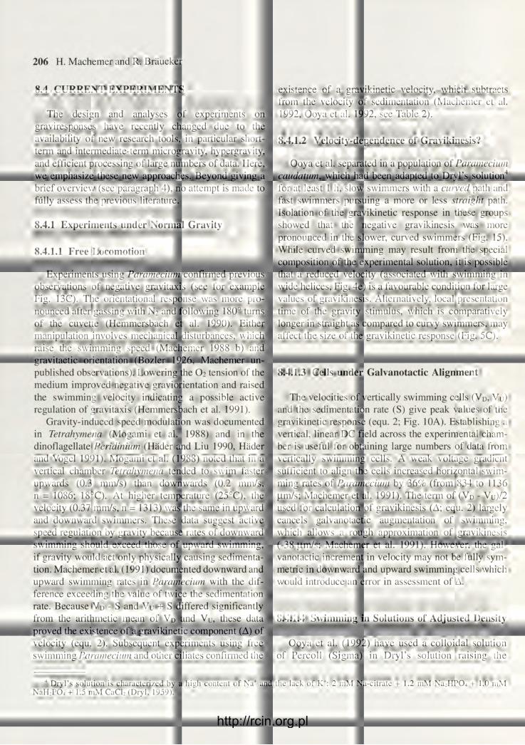
206 H. Machemer and R. Braucker
8.4 CURRENT EXPERIMENTS
The design and analyses of experiments on graviresponses have recently changed due to the availability of new research tools, in particular short-term and intermediate-term microgravity, hypergravity, and efficient processing of large numbers of data. Here, we emphasize these new approaches. Beyond giving a brief overview (see paragraph 4), no attempt is made to fully assess the previous literature.
8.4.1 Experiments under Normal Gravity
8.4.1.1 Free Locomotion
Experiments using Paramecium confirmed previous observations of negative gravitaxis (see for example Fig. 13C). The orientational response was more pro-nounced after gassing with N2 and following 180° turns of the cuvette (Hemmersbach et al. 1990). Either manipulation involves mechanical disturbances, which raise the swimming speed (Machemer 1988 b) and gravitactic orientation (Bozler 1926, Machemer un-published observations). Lowering the O2 tension of the medium improved negative graviorientation and raised the swimming velocity indicating a possible active regulation of gravitaxis (Hemmersbach et al. 1991).
Gravity-induced speed modulation was documented in Tetrahymena (Mogami et al. 1988) and in the dinoflagellate Peridinium (Hader and Liu 1990, Hader and Vogel 1991). Mogami et al. (1988) noted that in a vertical chamber Tetrahymena tended to swim faster upwards (0.3 mm/s) than downwards (0.2 mm/s, n = 1086; 18°C). At higher temperature (25°C), the velocity (0.37 mm/s, n = 1313) was the same in upward and downward swimmers. These data suggest active speed regulation by gravity because rates of downward swimming should exceed those of upward swimming, if gravity would act only physically causing sedimenta-tion. Machemer et al. (1991) documented downward and upward swimming rates in Paramecium with the dif-ference exceeding the value of twice the sedimentation rate. Because VD - S and Vu + S differed significantly from the arithmetic mean of VD and Vu, these data proved the existence of a gravikinetic component (A) of velocity (equ. 2). Subsequent experiments using free swimming Paramecium and other ciliates confirmed the
existence of a gravikinetic velocity, which subtracts from the velocity of sedimentation (Machemer et al. 1992, Ooya et al. 1992, see Table 2).
8.4.1.2 Velocity-dependence of Gravikinesis?
Ooya et al. separated in a population of Paramecium caudatum, which had been adapted to Dryl's solution4
for at least 1 h, slow swimmers with a curved path and fast swimmers pursuing a more or less straight path. Isolation of the gravikinetic response in these groups showed that the negative gravikinesis was more pronounced in the slower, curved swimmers (Fig. 15). While curved swimming may result from the special composition of the experimental solution, it is possible that a reduced velocity (associated with swimming in wide helices, Fig. 4e) is a favourable condition for large values of gravikinesis. Alternatively, local presentation time of the gravity stimulus, which is comparatively longer in straight as compared to curvy swimmers, may affect the size of the gravikinetic response (Fig. 5C).
8.4.1.3 Cells under Galvanotactic Alignment
The velocities of vertically swimming cells (VD, Vu) and the sedimentation rate (S) give peak values of the gravikinetic response (equ. 2; Fig. 10A). Establishing a vertical, linear DC field across the experimental cham-ber is useful for obtaining large numbers of data from vertically swimming cells. A weak voltage gradient sufficient to align the cells increased horizontal swim-ming rates of Paramecium by 36% (from 834 to 1136 |im/s; Machemer et al. 1991). The term of (VD - Vu)/2 used for calculation of gravikinesis (A; equ. 2) largely cancels galvanotactic augmentation of swimming, which allows a rough approximation of gravikinesis (-38 |nm/s; Machemer et al. 1991). However, the gal-vanotactic increment in velocity may not be fully sym-metric in downward and upward swimming cells which would introduce an error in assessment of A.
8.4.1.4 Swimming in Solutions of Adjusted Density
Ooya et al. (1992) have used a colloidal solution of Percoll (Sigma) in Dryl's solution raising the
4 Dryl's solution is characterized by a high content of Na+ and the lack of K+: 2 mM Na-citrate + 1.2 mM NaiHPO-i + 1.0 mM NaH:P0 4 + 1.5 mM CaCL (Dryl, 1959).
http://rcin.org.pl

Gravireception and Graviresponses in Ciliates 207
1.0
0.5 o o <1) >
down h or. Swimming Direction
up
Fig. 15. Average swimming velocities of two populations of Paramecium caudatum specified by curvature of the observed swimming course (filled circles: fast and straight swimmers, cur-vature < 0.10/mm; open circles: slow and curved swimmers, cur-vature > 0.85/mm). The lines give the calculated velocities5 at 6 different angles between vertical downward and vertical upward on the assumption that only the invariant velocity and sedimentation determine the observed velocity (continuous line: of straight swim-mers; broken line: of curved swimmers. It is seen that (1) the offset from the "no-gravikinesis" line is larger in curved swimmers ap-proximating full cancelling of the sedimentation rate, (2) gravikinesis acts to reduce downward swimming and to raise upward swimming rates, (3) the size of the gravikinetic component of velocity decreases from vertical toward horizontal orientation. Modified after Ooya et al. (1992), with permission
density to 1.033 g/cnr (viscosity relative to water, 1.12; 24.5°C), which reduces the density decrement between Paramecium cytoplasm and freshwater from 0.04 to 0.007 g/cm3, that is, to less than 20% of its original value (see 2.1). According to the statocyst hypothesis, reduction in gravity-induced pressure dif-ference attenuates the gravikinetic response. Figure 16 shows that the slope of gravity-induced change in invariant propulsion per angular reorientation was reduced in the Percoll-cells, straight as well as curved swimmers. From this, Ooya et al. (1992) have con-cluded that in Paramecium, the cell membrane is the primary candidate for gravireceptor, and pressure difference across the membrane rather than an ab-solute hydrostatic pressure increment is the crucial signal for gravireception (see 4.2). Further neutral-buoyancy experiments are now awaited in ciliates which may even more substantiate this important conclusion.
8.4.1.5 Nonswimming Locomotion
The "gliding" of Loxodes along substrate surfaces is a preferred type of locomotion under steady-state con-ditions, and its orientation and velocity can be adequate-ly measured (Braucker et al. 1992). For investigations of a possible gravikinesis of gliding cells the sedimen-tation rate is difficult to determine because wall effects (Wu 1977) may reduce the rate of a sedimenting specimen. Braucker et al. (1992) observed that the gliding rate in Loxodes is independent of the cell's orientation in space suggesting complete compensation of the sedimentation rate whatever its size (see, in addition, 8.4.3). The sedimentation rate during gliding locomotion of Loxodes remains to be established during step transition of horizontally moving cells from normal
Gravity
Fig. 16. Evidence for external-membrane based gravireceptor in Paramecium. Isolation of active propulsion from swimming velocity and sedimentation rate for 6 different orientations of curved swim-mers (between vertical down and vertical up) gives a linear slope, which rises with gravity increasing from 1 g to 5 g (inset). This slope was plotted as a function of gravity (g) for curved swimmers (filled symbols) and straight swimmers (open symbols) in Dryl's solution (circles) and Percoll solved in Dryl's solution (1.033 g/cm'; triangles). It is seen that the increase in gravikinetic response was proportional to gravity, and was depressed in the Percoll solution, in particular in the curved swimmers. The residual gravikinetic sensitivity in the Percoll solution is due to the remaining 18% of normal gravitational pressure difference across the lower membrane, or results from an additional unknown mechanism of gravireception. Modified after Ooya et al. (1992), with permission
5 If, and only if, the swimming rate of a ciliate is assumed to include no gravity-induced kinetic component (A), the propulsion as unaffected by gravity (= invariant velocity, P) results from the arithmetic mean of the vertical downward and upward swimming velocities (VD, Vu). An observed velocity in arbitrary direction is then determined by
V = V p 2 - S 2 s i n : e - S cos 9 , (Ooya et al. 1992) where 9 is the inclination angle of V (see Fig. 9). Assuming that a gravikinetic component of velocity (A) adds to P, this equation is equivalent to equation 4.
http://rcin.org.pl
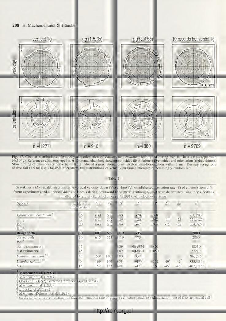
208 H. Machemer and R. Braucker
vertical 1g M9 (1.5-3s) pg (3-4.5S) 10 records horizontal 1g
cz o ce
03 o
'(Xc \i \ \ V v
n =1277 n = 946 n = 860 n = 9789
Fig. 17. Circular distributions of velocity and orientation of Paramecium caudatum before and during free fall in a 4.6 s-drop tower (4xl04 g). Reference cells were kept in the horizontal chamber, showing non-sided distributions of velocities and orientation (right column). Slow turning of chamber to the vertical at 1 g induced a gravitational bias in velocity and orientation within 1 min. During two periods of free fall (1.5 to 3 s; 3 to 4.5 s after start), the distributions of velocity and orientation were increasingly randomized
Table 2
Gravikinesis (A) as calculated using the vertical velocity down (VD) and up (Vu), and the sedimentation rate (S) of ciliates (from dif-ferent experiments and authors). Values of kinesis during downward and upward motion (AD, AU) were determined using the velocity as
unaffected by gravity (P; Machemer et al. 1991). All velocities in pm/s
Species ± Degree VD Vu S A P Ad Au n from vertical
Paramecium caudatum ' 15 639 500 89 -20 568 63, 203 45 641 502 89 -20 568 -14 -25 221, 487
P.c. 2
P.c. 3
straight path
90 843 808 84 -67 834 -75 -58 364, 674 P.c. 2
P.c. 3
straight path 30 807 639 119 -35 2000 curved path P.c.4
norm, swimmers
30 615 523 119 -73 2000 curved path P.c.4
norm, swimmers 45 89 0 to -70 850 18310 fast swimmers 45 89 0 to -90 1850 27279 Didinium nasutum 1 45 1504 1401 90 -39 86, 286 Loxodes striatus 5 0 169 169 497 -497 169 -497 -497 835, 816 T 6 L.s. 15 159 153 487 -477 156 -457 -45 7 2402, 2152
' Machemer et al. (1992) " Machemer et al. (1991) 4 Ooya et al. (1992, extracted from their Figs. 3 and 4)
Machemer et al. (1993) 6 Bràucker et al. (1992)
Machemer-Rôhnisch et al. (1993) on the so far unproven assumption that sedimentation rate of gliding cell corresponds to sedimentation rate of free suspended cell
600 [TV/s
http://rcin.org.pl

Gravireception and Graviresponses in Ciliates 209
swimmina rate fum/sl
vertical 1 g pg (1.5-3 s) pg (3-4.5 s) horizontal 1 g n - 708 503 836 6179
Fig. 18. Quantitative changes in gravity-induced swimming velocity of Paramecium (data shown in Fig. 17). At 1 g, the rates of upward swimming were significantly smaller than the rates of downward swimming (medians, 95% confidence range). Following step-tran-sition to weightlessness, these differences gradually subsided in the course of 4.5 s, and the velocities were inseparable from those observed during horizontal orientation at 1 g of the same cell population. The same 90°-sectors (inset) of the observed field were used for evaluations throughout. Machemer at al. (1992) with permission
gravity to weightlessness. Populations of Loxodes were found to occur in equal proportions moving vertically upward or downward. This orientational response, together with neutralization of sedimentation, was termed a "neutral gravitaxis" (Machemer-Rohnisch et al. 1993).
8.4.2 Microgravity Experiments
A preliminary exploration of the behaviour of Paramecium during a 35 m free fall (10 2 g; Mogami et al. 1988) showed reduction in velocity of upward swimming single specimens after transition to 2 s of weightlessness. Using a drop tower of 110 m free fall at 10 Pa (10 4 g) and populations of about 100 cells, the gravity bias of velocity and orientation relaxed in the course of 4.5 s (Fig. 17). The direction coefficient (r) dropped from 0.200 (r0 = r) at 1 g to 0.136 at the end of weightlessness (Machemer et al. 1992) indicat-ing that full random orientation of Paramecium was not achieved within a few seconds of weightlessness. With weightlessness continuing over several minutes in a sounding rocket experiment (xl0~fl g; Fig. 19B), r-values centered about 0.08 (extremes: 0.2 and 0.02; Hemmersbach et al. 1993).
Quantitative evaluation of velocities in Paramecium before and during transition to weight-lessness confirmed the difference between upward and
downward swimming rates at 1 g (Fig. 18, Machemer et al. 1992). The difference did not immediately disappear with the step-type removal of the gravity bias, but attenuated in the course of 4.5 s. This suggests a slow relaxation time constant of the gravikinetic response. Because electromotor coupling events involve fractions of a second for minor shifts in membrane potential (Machemer 1975), the ob-served slow relaxation of gravikinesis may be a cue of the nature of the receptor mechanism. The final free-fall values of previously upward and downward swimming rates coincide suggesting that Paramecium showed the value of velocity (P) as unaffected by gravity. This P-value was the same as the median rate of swimming in horizontally oriented cells at normal gravity (Fig. 18). The data suggest that the velocity in well equilibrated, horizontally swimming cells is a good measure of the important value of invariant propulsion (P; see 8.1.1).
8.4.3 Hypergravity Experiments
Raising gravity by linear acceleration or centrifuga-tion is quite useful for investigating gravireception. Behavioural consequences of gravity, which are subtle and hard to document under natural gravity, may be potentiated during hypergravity. The sedimentation rate of free floating immobilized cells rises in propor-tion to gravity according to Stoke's law, but the unstirred layer of .water near the walls of the ex-perimental chamber (8.3.3) may interfere with the sedimentation-gravity relationship. Ooya et al. (1992) confirmed exact proportionality up to 5 g for Paramecium. These authors documented an increase in gravikinesis with gravity, which was most pronounced in slow cells swimming in arcs (Fig. 16). At 1 g sedimentation was 119 |im/s and mean gravikinesis was -73 (im/s (Table 2); at 5 g and 5-fold increase in sedimentation rate, their plots (Ooya et al. 1992, Fig. 4) indicate a gravikinetic rate of -456 |um/s corresponding to a rise in gravikinesis by a factor of 6.2.
Application of hypergravity in Loxodes revealed a difference in gravikinetic responses of vertically upward and downward gliding cells. Upward velocities were maintained up to 5.4 g, whereas downward velocities steadily rose with the gravitational force (Machemer-Rohnisch et. al. 1993). These data indicate that the value of gravikinesis and, implicitly, the signal generated by
http://rcin.org.pl

210 H. Machemer and R. Bräucker
1 '
90' 270' 90*
Fig. 19. Orientation and movement of Paramecium (aurelia group) at normal gravity and during free fall inside a sounding rocket rising to 250 km altitude. A. On ground 5 min before launch; n = 883. B. 5 min of weightlessness ( 4x10" g); n = 863. C. 45 min after landing; n = 2509. D. Swimming traces on ground. E. Swimming traces during weightlessness. Hemmersbach et al. (1993), with permission
the gravireceptor, differs during upward and downward locomotion under artificially raised gravity, even though it may be the same in all directions at normal gravity (Braucker et al. 1992, Table 2). Hypergravity induced the neutral gravitactic response of Loxodes (8.4.1.5) to shift from the line of resultant acceleration vector by a counterclockwise angle (5.4 g: 60°). This error in graviorientation was reproducible with rising and decreasing hypergravity (Machemer-Rohnisch et. al. 1993). Observations of hypergravity-induced angular offset have also been reported from Paramecium (Hem-mersbach-Krause unpublished). Running hypergravity experiments in forward and backward sequence (1 g—>hyper-g; hyper-g—> 1 g) did not affect the observed velocities and orientations of Loxodes (Machemer-Rohnisch et. al. 1993) suggesting that the graviresponse does not adapt on short term in agreement with findings in Paramecium (Ooya et al. 1992).
9. CONCLUSION AND PERSPECTIVE
An overview of the current literature on gravireception and graviresponses in ciliates leads to a clear result: the statocyst hypothesis, in electrophysiological reinterpretation (Fig. 20), gives the theoretical framework, into which experimental data can be fitted with ease, and using this paradigm, new experiments may be designed which ask precise questions. So far, no alternative model of gravirecep-tion in the free swimming cell is available with similar predictive value. At the same time, physiological and exclusively physical mechanisms of graviresponses are likely to coexist in ciliates. Evidence is available suggesting that various strategies are being used by ciliates to guide their ways in a world, where "Up" and "Down" are crucial
http://rcin.org.pl

Gravireception and Graviresponses in Ciliates 211
ant.
A B
Fig. 20. Simplified model of gravitransduction and kinetic response in the ciliate organism. A cell is thought to swim upward (A) or downward (B) in arbitrary direction (ant., anterior cell end). Cytoplasmic density generates differential pressure across the actual lower membrane activating, by deformation of a sensitive intramembranous structure, mechanically sensitive membrane channels. A. During upward locomotion the posterior membrane is deformed; here gravireceptor K-channels prevail leading to hyperpolarization and augmentation of the ciliary beating rate. The invariant rate of propulsion (P) grows by a gravity-induced component (A) and combines with the rate of sedimentation (S) to produce the observed direction and velocity of the cell (V). B. In the downward swimming cell gravity deforms the anterior membrane activating anteriorly prevailing gravireceptor Ca-channels. The resulting depolarization slows down steady ciliary propulsion, and the swimming rate (V) results after subtraction of A from P with S maintained. The sign and size of the gravikinetic response (A) determine, together with sedimentation and possible gravity-induced mechanisms of reorientation, the net locomotion of cells with respect to the gravity vector
cues for survival. The complexity of interactions between gravity and the cell, and the subtleness of the stimulus have been a challenge for generations of researchers, and there is reason to expect that this will continue in the foreseeable future. Modern methods and advanced tools of data evaluation sup-port the analysis of graviresponses but, at the same time, highly sensitive instruments have also raised the experimenter 's awareness of the conditions, which affect the behaviour of the organism.
A major goal of this review is practical: fostering the understanding of the ciliate organism as a model for investigations of cellular gravireception and graviresponses, and calling attention to possible physical environments of gravistimulation. Knowing and gaining command on such conditions, or neglect-ing them, may decide on the size of experimental "noise" or, eventually, isolation or masking of a graviresponse. Much methodical work is still to be
done. While an exhaustive description of the signall-ing pathway of gravity is the ultimate goal, direct measurements of receptor currents and potential responses to gravity are demanding tasks, and suc-cess may be still far ahead. We have pointed out that gravity is a weak stimulus and difficult to handle. Only few types of ciliates have so far been accessible to intracellular electrophysiology, and those few may not al together qual ify for investigations of gravireception. Individual variability in responsive-ness to the gravity stimulus is a restriction to con-clusions drawn from experimental work on single cells. Experiments on large numbers of cells with the potential to produce relevant biophysical in-formation must be designed and developed to maturity. In the near future, a host of increasingly sophisticated behavioural analyses of graviresponses may have a pacemaking function for the under-standing of gravitransduction in ciliates.
http://rcin.org.pl

212 H. Machemer and R. Braucker
Acknowledgments. Financial support provided by the German Space Agency (DARA), grant 50QV9957-5, the Ministerium für Wissenschaft und Forschung NRW, and the Deutsche Forschungsgemeinschaft, Forschergruppe Konzell, is gratefully ac-knowledged. We are indebted to D.P. Hader, W. Haupt, A. Sievers and U. Thurm for criticism and suggestions.
REFERENCES
Audus L. J. (1979) Plant geosensors. J. Exp. Bot. 30: 1051-1073. Baba S. A. (1991) A new hypothesis concerning graviperception of
single cells, and supporting simulated experiments. Biol. Sei. Space 5: 290-291 (in Japanese).
Baba S. A., Inomata S., Ooya M„ Mogami Y., Izumi-Kurotani A. (1991) Three-dimensional recording and measurement of swim-ming paths of micro-organisms with two synchronized monochrome cameras. Rev. Sei. Instrum. 62: 540-541.
Batschelet E. (1981) Circular statistics in Biology. In: Mathematics in Biology (Eds. R. Sibson, J. E. Cohen). Academic Press, Lon-don, New York.
Bean B. (1984) Microbial geotaxis. In: Membranes and Sensory Transduction (Eds. G. Colombetti,F. Lenci), Plenum Publishing Corporation, New York, 163-198.
Björkman T. (1988) Perception of gravity by plants. Adv. Bot. Res. 15: 1-41.
Bräucker R., Machemer-Röhnisch S., Machemer H., Murakami A. (1992) Gravity-controlled gliding velocity in Loxodes. Europ. J. Protistol. 28: 238-245.
Colombetti G. (1990) New trends in photobiology: photomotile responses in ciliated protozoa. J. Photochem. Photobiol. B 4: 243-259.
Crenshaw H. C. (1989) The kinematics of the helical motion of microorganisms capable of motion with four degrees of freedom. Biophys. J. 56: 1029-1035.
Crenshaw H. C. (1990) Helical orientation - a novel mechanism for the orientation of microorganisms. Lect. Notes Biomath. 89: 361-386.
Czapek F. (1895) Untersuchungen über Geotropismus. Jb. Wiss. Bot. 27: 243-339.
Davenport C. B. (1897) Experimental morphology. Macmillan Publ. Co., New York, 1-509.
Dembowski J. (1924) Über die Bewegungen von Paramecium caudatum. Arch. Protistenkd. 47: 25-54.
Diehn B., Feinleib M., Haupt W., Hildebrand E., Lenci F., Nultsch W. (1977) Terminology of behavioral responses of motile microorganisms. Photochem. Photobiol. 26: 559-560.
Dryl S. (1959) Antigenic transformation in Paramecium aurelia after homologous antisera treatment during autogamy and conjugation. J. Protozool. 6: 25 (Suppl.).
Eckert R., Randall D. (1983) Animal Physiology. Mechanisms and Adaptations, 2nd ed., Freeman and Company, San Francisco.
Eggersdorfer B„ Häder D. P. (1991) Phototaxis, gravitaxis and verti-cal migrations in the marine dinoflagellates, Peridiniumfaeroense and Amphidinium caterea. Acta Protozool. 30: 63-71.
Falke L. C„ Edwards K. L„ Pickard B. G., Misler S. (1988) A stretch-activated anion channel in tobacco protoplasts. FEBS Lett. 237: 141-144.
Fenchel T., Finlay B. J. (1984) Geotaxis in the ciliated protozoon Loxodes. J. Exp. Biol. 110: 17-33.
Fenchel T., Finlay B. J. (1986a) The structure and function of Müller vesicles in loxodid ciliates. J. Protozool. 33: 69-76.
Fenchel T., Finlay B. J. (1986b): Photobehavior of the ciliated protozoon Loxodes: taxic, transient, and kinetic responses in the presence and absence of oxygen. J. Protozool. 33: 139-145.
Finlay B. J., Fenchel T. (1986) Photosensitivity in the ciliated protozoon Loxodes: pigment granules, absorption and action spectra, blue light perception, and ecological significance. J. Protozool. 33: 534-542.
Finlay B. J., Fenchel T., Gardener S. (1986) Oxygen perception and O2 toxicity in the freshwater ciliated protozoon Loxodes. J. Protozool. 33: 157-165.
Fetter D. (1926) Determination of the protoplasmic viscosity of Paramecium by the centrifuge method. J. Exp. Zool. 44: 279-283.
Fraenkel G. S., Gunn D. L. (1940) The orientation of animals. Oxford University Press. 2nd edition: (1961) Dover Publ. Inc., New York.
Fukui K., Asai H. (1980) The most probable mechanism of the negative geotaxis of Paramecium caudatum. Proc. Japan. Acad. 56(B): 172-177.
Fukui K., Asai H. (1985) Negative geotactic behavior of Paramecium caudatum is completely described by the mechanism of buoyan-cy-oriented upward swimming. Biophys. J. 47: 479-483.
Gelei J. von (1935) Ni-Infusion im Dienste der Forschung und des Unterrichtes. Biol. Zbl. 55: 57-74.
Gr^becki A., Nowakowska G. (1977) On the mechanism of orienta-tion of Paramecium caudatum in the gravity field. I. Influence of ciliary reversal and of external Ca deficiency on the geotactic behaviour. Acta Protozool. 16: 351-358.
Häder D.P., Griebenow K. (1988) Orientation of the green flagellate, Euglena gracilis, in a vertical column of water. FEMS Microbiol. Ecol. 53: 159-167.
Häder D. P., Liu S. M. (1990) Effects of artificial and solar UV-B radiation on the gravitactic orientation of the dinoflagellate, Peridinium gatunense. FEMS Microbiol. Ecol. 73: 331-338.
Häder D. P., Lebert M. (1985) Real time computer-controlled tracking of motile microorganisms. Photochem. Photobiol. 42: 509-514.
Häder D. P., Vogel K. (1991) Simultaneous tracking of flagellates in real time by image analysis. J. Math. Biol. 30: 63-72.
Happel J., Brenner H. (1973) Low Reynolds number hydrodynamics. 2nd ed., Noordhoff Int., Groningen, Leyden.
Haupt W. (1962) Geotaxis. In: Handbuch der Pflanzenphysiologie, (Ed. W.Ruhland), Springer Verlag, Berlin, XVII/2: 390-395 /
Hejnowicz Z., Buchen B., Sievers A. (1985) The endogenous difference in the rates of acropetal and basipetal cytoplasmic streaming in Chara rhizoids is enhanced by gravity. Protoplasma 125: 219-229.
Hemmersbach-Krause R., Häder D. P. (1990) Negative gravitaxis (geotaxis) of Paramecium - demonstrated by image analysis. Applied Microgravity Tech. 2: 221-223.
Hemmersbach-Krause R„ Briegleb W„ Häder D. P. (1991) Depend-ence of gravitaxis in Paramecium on oxygen. Eur. J. Protistol. 27: 278-282.
Hemmersbach-Krause R., Briegleb W., Häder D. P., Vogel K., Grothe G., Meyer I. (1993) Orientation of Paramecium under the condi-tions of weightlessness. J. Protozool. (in press)
Hubert G„ Rieder N.. Schmitt G„ Send W. (1975) Bariuman-reicherung in den Müllerschen Körperchen der Loxodidae (Ciliata, Holotricha). Z. Naturforschg. 30c: 422.
Hudspeth A. J. (1985) The cellular basis of hearing: the biophysics of hair cells. Science 230: 745-752.
Inoue S. (1986) Video microscopy. Plenum Press, New York, Lon-don, 1-584.
Inoue T., Nakaoka Y. (1990) Cold-sensitive responses in the Paramecium membrane. Cell Struct. Funct. 15: 107-112.
Iwatsuki K., Naitoh Y. (1982) Photoresponses in colourless Paramecium. Experientia 38: 1453-1454.
Iwatsuki K., Naitoh Y. (1983) Behavioral responses in Paramecium multimicronucleatum to visible light. Photochem. Photobiol. 37: 415-419.
Jensen P. (1893) Über den Geotropismus niederer Organismen. Pflügers Arch. Ges. Physiol. 53: 428-480.
Keller S. R., Wu T. Y. (1977) A porous prolate spheriodal model for ciliated microorganisms. J. Fluid Mech. 80: 259-278.
Kennedy J. S. (1945a) Classification and nomenclature of animal behaviour. Nature 155: 178-179.
Kennedy J. S. (1945b) Classification and nomenclature of animal behaviour. Nature 156: 754.
Koehler O. (1921) Über die Geotaxis von Paramecium. Verh. Dtsch. Zool. Ges. 26: 69-71.
Koehler O. (1922) Über die Geotaxis von Paramecium. Arch. Protis-tenkd. 45: 1-94.
http://rcin.org.pl

Gravireception and Graviresponses in Ciliates 213
Koehler O. (1930) Über die Geotaxis von Paramecium. II. Arch. Protistenkd. 70: 279-306.
Koehler 0 . (1931) Die Orientierung von Pflanze und Tier im Räume. II. Zoologischer Teil. Biol. Zentralbl. 51: 36-58.
Kuroda K., Kamiya N. (1989) Propulsive force of Paramecium as revealed by the video centrifuge microscope. Exp. Cell Res. 184: 268-272.
Kuznicki L. (1968) Behavior of Paramecium in gravity fields. I, Sinking of immobilized specimens. Acta Protozool. 6: 109-117.
Lapidus I. R., Levandowsky M. (1981) Mathematical models of behavioral responses to sensory stimuli by Protozoa. In: Biochemistry and Physiology of Protozoa, (Eds. M. Levan-dowsky, S. H. Hutner), Academic Press, New York, 4: 235-260.
Larsen J., Satir P. (1991) Analysis of Ni2+-induced arrest of Paramecium axonemes. J. Cell Sei. 99: 33 -40.
Loewenstein W. R. (1960) Mechanisms of nerve impulse initiation in a pressure receptor (Lorenzinian ampulla). Nature 188: 1034-1035.
Loewenstein W. R. (1965) Facets of a transducer process. Cold Spring Harb. Symp. quant. Biol. 30: 29-43.
Lyon E. P. (1905) On the theory of geotropism in Paramecium. Am. J.Physiol. 14: 421-432.
Machemer H. (1972a) Properties of polarized ciliary beat in Paramecium. Acta Protozool 11: 295-300.
Machemer H. (1972b) Temperature influences on ciliary beat and metachronal coordination in Paramecium. J. Mechanochem. Cell Motil. 1: 57-66.
Machemer H. (1974) Frequency and directional responses of cilia to membrane potential changes in Paramecium. J. Comp. Physiol. 92: 293-316.
Machemer H. (1975) Modification of ciliary activity by the rate of membrane potential changes in Paramecium. J. Comp. Physiol. 101: 343-356.
Machemer H. (1986) Electromotor coupling in cilia. In: Membrane control of cellular activity (Ed. H.C.Lüttgau). Fortschr. Zool. 33: 205-250.
Machemer H. (1988a) Electrophysiology. In: Paramecium (Ed, H. - D. Görtz). Springer Verlag, Berlin, 185-215.
Machemer H. (1988b) Motor control of cilia. In: Paramecium (Ed. H.- D. Görtz). Springer Verlag, Berlin, 216-235.
Machemer H. (1989a) Cellular behaviour modulated by ions: electrophysiological implications. J. Protozool. 36: 463-487.
Machemer H. (1989b) A quantitative approach toward isolation of a gravity-induced motor response in Paramecium I. Theory. Biol Sei. Space 3: 286 (Abstract).
Machemer H., Deitmer J. W. (1985) Mechanoreception in ciliates. Progress in Sensory Physiology, Springer Verlag, Berlin, 5: 81-118.
Machemer H„ De Peyer J. E. (1977) Swimming sensory cells: Electri-cal membrane parameters, receptor properties and motor control in ciliated protozoa. Verh. Dtsch. Zool. Ges. 1977: 86-110.
Machemer H., Machemer-Röhnisch S. (1984) Mechanical and electric correlates of mechanoreceptor activation of the ciliated tail in Paramecium. J. Comp. Physiol. A 154: 273-278.
Machemer H., Sugino K. (1989) Electrophysiological control of ciliary beating: A basis of motile behaviour in ciliated protozoa. Comp. Biochem. Physiol. 94A: 365-374.
Machemer H., Teunis P.F.M. (1993) Sensorimotor coupling and motor responses. In: Ciliates: cells as organisms (Eds. K. Haus-mann, P. C. Bradbury). Gustav Fischer Verlag, Stuttgart (in press).
Machemer H., Bräucker R., Murakami A., Yoshimura K. (1992) Graviperception in unicellular organisms: a comparative be-havioural study under short-term microgravity. Microgravity Science and Technology (in press)
Machemer H., Machemer-Röhnisch S., Bräucker R. (1993) Velocity and graviresponses in Paramecium during adaptation and varied oxygen concentration. Arch. Protistenkd. 143: (in press)
Machemer H., Machemer-Röhnisch S., Bräucker R., Takahashi K. (1991) Gravikinesis in Paramecium: Theory and isolation of a physiological response to the natural gravity vector. J. Comp. Physiol. A 168: 1-12.
Machemer-Rohnisch S., Machemer H. (1989) A Ca paradox: electric and behavioural responses of Paramecium to changes in cation concentration of the medium. Europ. J. Protistol. 25: 45-59.
Machemer-Rohnisch S., Brâucker R„ Machemer H. (1993) Neutral gravitaxis of gliding Loxodes exposed to normal and raised gravity. J. Comp. Physiol. A (in press)
Mark] H. ( 1974) The perception of gravity and of angular acceleration in invertebrates. In: Handbook of Sensory Physiology, (Ed. H.H. Kornhuber), Springer Verlag, Berlin, VI/1: 17-74.
Martinac B., Machemer H. (1984) Effects of varied culturing and experimental temperature on electrical membrane properties in Paramecium. J. Exp. Biol. 108: 179-194.
Martinac B., Buechner M., Delcour A. H., Adler J„ Kung C. (1987) Pressure-sensitive ion channel in Escherichia coli. Proc. Ntl. Acad. Sci. USA 84: 2297-2301.
Matsuoka K,, Nakaoka Y. (1988) Photoreceptor potential causing phototaxis in Paramecium bursaria. J. Exp. Biol. 137: 477-485.
Matsuoka T., Watanabe Y., Kuriu T., AritaT., Taneda K., Ishida M., Suzaki T., Shigenaka Y. (1991) Cell models of Blepharisma: Ca2+-dependent modification of ciliary movement and cell elon-gation. Europ, J. Protistol. 27: 371-374.
Methfessel C., Witzemann V., Takahashi T., Mishina M., Numa S., Sakmann B. (1986) Patch clamp measurements onXenopus laevis oocytes: currents through endogenous channels and implanted acetocholine receptor and sodium channels. Pfliigers Arch. 407: 577-588.
Mogami Y., Machemer H. (1991) In-vivo activation of cirral move-ment in Stylonychia by calcium. J. Comp. Physiol. A 168: 687-695.
Mogami Y., Pernberg sr. J., Machemer H. (1990) Messenger role of calcium in ciliary electromotor coupling: a reassessment. Cell Calcium 11: 665-673.
Mogami Y., KimuraT., Okuno M„ YamashitaM., BabaS. A. (1988) Free fall experiments on swimming behavior of ciliates. Proceed. 16. Intern. Symp. Space Technol. Sci., 2351-2354.
Mogami Y., Oobayashi C., Yamaguchi T., Ogiso Y., Baba S. A. (1988) Negative geotaxis in sea urchin larvae: a possible role of mechanoreception in the late stages of development. J. Exp. Biol. 137: 141-156.
Naitoh Y., Sugino K. (1984) Ciliary movement and its control in Paramecium. J. Protozool. 31: 31-40.
Nakaoka Y., Tanaka H., Oosawa F. (1984) Ca2+-dependent regula-tion of beat frequency of cilia in Paramecium. J. Cell Sci. 65: 223-231.
Nakaoka Y., Kurotani T., Itoh H. (1987) Ionic mechanism of thermoreception in Paramecium. J. Exp. Biol. 127: 95-103.
Noll F. (1900) Ûber Geotropismus. Jb. Wiss. Bot. 34: 457-506. Nowakowska G., Grçbecki A. (1977) On the mechanism of orienta-
tion of Paramecium caudatum in the gravity field. II. Contribu-tions to a hydrodynamic model of geotaxis. Acta Protozool. 16: 359-376.
Oka T., Nakaoka Y., Oosawa F. (1986) Changes in membrane poten-tial during adaptation to external potassium ions in Paramecium caudatum. J. Exp. Biol. 126: 111-117.
OoyaM., Mogami Y., Izumi-Kurotani A., BabaS. A. (1992) Gravity-induced changes in propulsion of Paramecium caudatum: a pos-sible role of gravireception in protozoan behaviour. J. Exp. Biol. 163:153-167.
Otter T., Salmon E. D. (1985) Pressure-induced changes in Ca2+-channel excitability in Paramecium. J. Exp. Biol. 117: 29-43.
Pfeffer W. (1904) Pflanzenphysiologie, 2. AufL, W. Engelmann, Leipzig.
Pickard B. G., Thimann K. V. (1966) Geotropic response of wheat coleoptiles in absence of amyloplast starch. J. Gen. Physiol. 49: 1065-1086.
Reisser W-., Hàder D. P. (1984) Role of endosymbiotic algae in photokinesis and photophobic responses of ciliates. Photochem. photobiol. 39: 673-678.
Rieder N., Ott H. A., Pfundstein P., Schoch R. (1982) X-ray microanalysis of the mineral contents of some protozoa. J. Protozool. 29: 15-18.
http://rcin.org.pl

214 H. Machemer and R. Braucker
Roberts A. M. (1970) Geotaxis in motile micro-organisms. J. Exp. Biol. 53: 687-699.
Roberts A. M. (1981) Hydrodynamics of protozoan swimming. In: Biochemistry and Physiology of Protozoa, (Eds. M. Levan-dowsky, S. H. Hutner), Academic Press, New York, 4: 5-66.
Sack F. D. (1991) Plant gravity sensing. Int. Rev. Cytol. 127:193-252. Schaefer G. (1922) Studien über den Geotropismus von Paramecium
aurelia. Pflügers Arch. Ges. Physiol. 195: 227-244. Schröter K., Läuchli A., Sievers A. (1975) Mikroanalytische Iden-
tifikation von Bariumsulfat-Kristallen in den Statolithen der Rhizoide von Charafragilis. Planta 122: 213-225.
Sievers A., Schmitz M. (1982) Röntgen-Mikroanalyse von Barium, Schwefel und Strontium in Statolithen-Kompartimenten von Chara-Rhizoiden. Ber. Deutsch. Bot. Ges. 95: 353-360.
Sievers A., Schnepf E. (1981) Morphogenesis and polarity of tubular cells with tip growth. In: Cytomorphogenesis in plants (Ed. O. Kiermayer), Springer Verlag, Wien, 265-299.
Sievers A., Volkmann D. (1979) Gravitropism in single cells. In: Physiology of movements, Encyclopedia of Plant Physiology, (Eds. W.Haupt, M.E.Feinleib), Springer Verlag, Berlin, NS 7: 567-572.
Sievers A., Buchen B., Volkmann D., Hejnowicz Z. (1991) Role of the cytoskeleton in gravity perception. In: The Cytoskeletal Basis of Plant Growth and Form (Ed. C. W. Lloyd), Academic Press, London,169-182.
Taneda K. (1987) Geotactic behavior in Paramecium caudatum. I. Geotaxis assay of individual specimen. Zool. Science 4: 781-788.
Taneda K., Miyata S., Shiota A. (1987) Geotactic behavior in Paramecium caudatum. II. Geotaxis assay in a population of the specimens. Zool. Science 4: 789-795.
Thurm U. (1982) Biophysik der Mechanorezeption. In: Mechanoelektrische Transduktion (Eds. W. Hoppe, W. Lohmann, H. Markl, H. Ziegler). Springer Verlag, Berlin, 691-696.
Thurm U., Erler G„ Gödde L, Rastrup H., Keil T., Völker W., Vohwinkel B. (1983). Cilia specialized for mechanoreception. J. Submicrosc. Cytol. 15: 151-155.
Tominaga T., Naitoh Y. (1992) Membrane potential responses to thermal stimulation and the control of thermoaccumulation in Paramecium caudatum. I. Exp. Biol. 164: 39-53.
Van Houten J. (1992) Chemosensory transduction in eukaryotic microorganisms. Annu. Rev. Physiol. 54: 639-663.
Verworn M. (1889) Psychophysiologische Protistenstudien. Gustav Fischer, lena, 1-219.
Volkmann D. (1974) Amyloplasten und Endomembranen: Das Geoperzeptionssystem der Primärwurzel. Protoplasma 79: 159-183.
Volkmann D., Sievers A. (1979) Graviperception in multicellular organs. In: Physiology of Movements, Encyclopedia of Plant Physiology, (Eds. W. Haupt, M. E. Feinleib), Springer Verlag, Berlin, NS 7: 573-600.
VolkmannD., Sievers A. (1992) Forschung unter reduzierter Schwer-kraft. Teil I: Grundlagen der Gravitationsbiologie. Naturwissen-schaften 79: 68-74.
Wager H. (1911) The effect of gravity upon the movements and aggregation of Euglena viridis Ehrbg., and other micro-or-ganisms. Phil. Trans. Roy. Soc. B 201: 333-388.
Winet H., Jahn T. L. (1974) Geotaxis in protozoa. I. A propulsion gravity model for Tetrahymena (Ciliata). J. Theor. Biol. 46: 449-465.
Wu Y. T. (1977) Hydrodynamics of swimming at low Reynolds numbers. In: Physiology of movement - biomechanics (Ed. W. Nachtigall). Progr. Zool. 24: 149-169.
Received on 18th September, 1992
Correction
1. The citation "Baba (1991) is incomplete; the correct writing is "Baba et al. (1991). Correspondingly, in the References should be: Baba S.A., Tatematsu R., Mogami Y. (1991) A new hypothesis concerning graviperception of single cells, and supporting simulated experiments. Biol.Sci.Space 5: 290-291 (in Japanese).
2. The citation od Pfeffer (1904) is to be deleted from the text and in the References.
http://rcin.org.pl

ACTA
PROTOZOOLOGICA
Acta Protozoologica (1992) 31: 215 - 219
Evidence for Growth Factors which Control Cell Multiplication in Tetrahymena thermophila
S0ren T. CHRISTENSEN and Leif RASMUSSEN
Institute of Medical Biology, Department of Anatomy and Cytology, University of Odense, Odense , Denmark
Summary. The ciliate Tetrahymena thermophila was grown in a standard synthetic nutrient medium that supports doubling times of about 2 h. Cell multiplication was investigated as a function of initial cell density and addition of either certain chemical compounds, or cell-free extracellular fluid. In cultures inoculated at 750 or more initial cells per ml the cells initially thrived and multiplied but below this density the cells soon died. Addition of extracellular fluid or either hemin, phospholipids or bovine serum albumin, egg albumin, trypsin, or soy bean trypsin inhibitor, however, saved these cells from dying and supported cell proliferation. We have also investigated the cloning efficiency of the cells as a function of culture volume of the synthetic medium. In 1 ml portions of standard synthetic medium the cloning efficiency was 0%, but in volumes of 1 |oi it was 90%. We suggest that Tetrahymena thermophila produces and releases growth factors that stimulate the cells to leave the lag phase and start cell multiplication. Certain compounds like hemin, phospholipids and certain proteins can substitute for these growth factors.
Key words. Synthetic nutrient medium, cell multiplication, cell proliferation, initial cell density, growth factors, hemin, phospholipids, proteins, Tetrahymena.
INTRODUCTION
The first recipe for synthetic nutrient medium for Tetrahymena thermophila is more than 40 years old (Kidder and Dewey 1951). This type of medium has been used for gaining insight into nutrient requirements (Holz 1973), metabolic pathways (Kidder 1967), uptake mechanisms (Dunham and Kropp 1973) and reactions between inorganic and organic molecules in the medium (Hutner 1972). In spite of the fact that it was possible to maintain doubling times and final cell densities comparable to those obtained in the best complex
Address for correspondence: L. Rasmussen, Institute of Medi-cal Biology, Department of Anatomy and Cytology, University of Odense, 5230 Odense, Denmark.
medium, the synthetic medium are deficient in at least one respect: single cells in 1 ml cultures die shortly after being transferred to such a medium (Christensen et al. 1992).
We have recently shown that T. thermophila grown under nutritional stress situations need various com-pounds not required at initial densities of more than 1,000 cells per ml. The stress conditions consist of growth at low population density (Schousboe et al. 1992) or growth at low ambient nutrient concentrations (Chris-tensen et al. 1992). These results agree with the notion that the cells produce and release compounds with effects like those of growth factors (Ghiladi et al. 1992). Here we present further detailed evidence for these views.
http://rcin.org.pl

216 S. T. Christensen and L. Rasmussen
MATERIAL AND METHODS
Cells: The following cell lines of Tetrahymena thermophila were used: wildtype inbred strain B 1868-III (Orias and Bruns 1976); the mutant strains MS-1, secreting lysosomal enzymes at low rates (Hiinseler et al. 1987); II8G, defective in food vacuole formation (Tiedtke et al. 1988); and SB 281, releasing no mucocysts (Orias et al. 1983).
Nutrient medium: Cells were grown either in a complex medium, PPYS, or in a standard synthetic nutrient medium, SSM. PPYS is a solution of 0.75% proteose peptone (Difco Laboratories, Detroit, Michigan, USA) enriched with 0.75% yeast extract (Difco) and-salts. SSM consists of 19 amino acids, 4 nucleosides, glucose, 7 vitamins, salts, and citrate (Szablewski et al. 1991). In both cloning and mass culture experiments SSM was diluted in the ratio of 1:1 with TRIS/HC1 buffer (pH 7.5). When testing the presence of stimulatory compounds in extracellular fluid, we replaced the buffer with ex-tracellular fluid, keeping the concentrations of the nutrients in the medium constant. Cells requiring special conditions for growth and multiplication were grown in media modified to fulfill these condi-tions: mutant strain II8G was grown in medium enriched with iron and copper salts and folic acid as prescribed for this mutant (Tiedtke et al. 1988). Chemicals were from Sigma Chemical Co., St Louis, Missouri.
Cultures: Stock cultures were grown in 2 ml portions of standard synthetic medium, SSM. Experimental cells were grown in 10 ml portions of SSM in conical flasks for 20 h, transferred to 10 mM TRIS/HC1 buffer (pH 7.5), centrifuged for 3 min at 800xg and resuspended in the buffer. This procedure was repeated three times and resulted in more than a 104-fold dilution of the extracellular fluid. The cells were then divided into three batches and used as follows: i) for preparation of extracellular fluid, ii) for multiplication analysis in 10 ml portions of SSM where effects of initial cell density and presence of hemin, phospholipids, proteins or extracellular fluid were studied, and iii) for cloning analysis in various volumes of either PPYS or SSM or SSM enriched with either hemin, phospholipids or proteins. The cells were grown at 37°C.
Preparation of cell-free extracellular fluid: Cells were starved in 10 ml TRIS/HC1 buffer (pH 7.5) in conical flasks for 0-5 min and 5 h at a density of 50,000 cells per ml. After that, a sample of a culture was transferred to a centrifuge tube and placed on top of 2 ml of a solution of 10% Ficoll and precipitated for 10 min at 3000xg. The Ficoll solution was sterilized by filtration and used at 4°C. The cell-free supernatant, containing no cells, was removed and used in the experiments. The Ficoll has a high specific weight and high viscosity and prevents the cells from swimming back up. To certify that no cells were left in the extracellular fluid a sample was placed in a small Petri dish and checked under a stereo microscope.
Cloning procedures: After centrifugation and resuspension in the buffer single cells were transferred to two different sizes of cultures, 1 ml or 10"3 ml (l |il). One ml cultures: single cells were transferred to either PPYS, SSM or SSM enriched with either hemin, phospholipids or proteins with a fine pipette (see Schousboe et al. 1992). One p/ cultures: single cells were suspended in either SSM or PPYS in test tubes at a density of 1,000 cells per ml. By means of a 10 |il Hamilton pipette droplets of 1 pi from the test tube were placed on the top of paraffin oil placed in a glass Petri dish. The droplets sank to the bottom of the dish and the paraffin oil prevented evaporation of the medium. The dish had a diameter of 7 cm, the paraffin oil was
autoclaved at 121°C for 45 min and reached a height of 2 mm in it. Only droplets containing a single initial cell were kept under observa-tion.
Cell densities in test tubes and droplets were recorded every day. In most cases many cells (1,000) were present already after the first 24 h. In all cases droplets and tubes were kept under observation until day 4.
Compounds added to SSM: Hemin and asolectin, a crude preparation of phospholipids, were used at final concentrations of 7.5 pM and 50 pg per ml, respectively (see Schousboe et al. 1992 and Christensen etal. 1992). Bovine serum albumin, egg albumin, trypsin and soy bean trypsin inhibitor were used as sources of proteins. They were dissolved in redistilled water, sterilized by filtration and used at final concentrations of 50 pg per ml.
Cell counting: Culture samples were enumerated in an electronic particle counter (see Christensen et al. 1992).
All experiments were repeated more than 10 times with similar results.
RESULTS
Mass cultures
Initial cell density is an important parameter in cell multiplication of Tetrahymena in synthetic medium. Figure 1 shows the number of cell doubling of T. ther-mophila, wildtype, as a function of time and number of cells transferred into SSM. In cultures having 750 or
1 2 -
0 10 2G 3 0 40
h
Fig. 1. The number of population doubling of Tetrahymena ther-mophila, wildtype, as a function of time and initial population density in standard synthetic medium, SSM; • : 250 and 500 cells per ml, O : 750 cells per ml, • : 1000 cells per ml, A : 2500 cells per ml. All cultures were observed for the whole duration of the experiment. Final cell densities were in all cases about 500,000 cells per ml. Differences in the number of cell doubling are due to differences in initial cell densities. The cells died, if they had not initiated multiplication after about 20 h
http://rcin.org.pl

Growth Factors in Tetrachymena thermophilia 217
1 2 n
1 0
4 -
2 -
0 - no a d d i t i o n
1 2 - i
1 0
6 -
4 -
0 -
10 20 30 40 1 0 20 30 40
Fig. 2. The number of population doubling of Tetrahymena ther-mophilci, wildtype, as a function of time and percentage of extracel-lular fluid in standard synthetic medium, SSM. All cultures were inoculated at 250 cells per ml. The cells died, if they had not initiated multiplication after about 20 h
more initial cells per ml the cells thrive, whereas 250 and 500 cells per ml die within 20 h. One thousand or more cells per ml have a short lag phase (about 1 h); 750 cells per ml have a lag phase lasting about 10 h.
Extracellular fluid is also an important parameter for cell proliferation in synthetic medium. Figure 2 shows the number of cell doubling of T. thermophila, wildtype, as a function of time and concentration of added extracellular medium where cells were starved for 5 h. In all cases the initial cell densities were 250 cells per ml. Without any addition of extracellular fluid the cells do not multiply. If extracellular fluid is added in the ratio of 1 portion of SSM to 0.02 portion of extracellular fluid
Table 1
Cloning efficiency in 1 pi or 1 ml of complex medium, PPYS, standard synthetic medium, SSM and SSM supplemented with either hemin (H), Phospholipids (PL), bovine serum albumin (BSA), egg albumin (EA), soy bean trypsin inhibitor (SBTI), or trypsin. As an example, the expression "29/32" indicates that 29 out of 32 single cells formed clones. Hemin was used at 7.5 pM and phospholipids and proteins at 50 pg per ml. The dashes indicate conditions which were not tested since the cloning efficiency was high in 1 ml portions
Culture SSM supplemented with volume SSM PPYS H PL BSA EA Trypsin SBTI
1 ml 0/30 29/30 14/15 15/15 12/15 11/15 19/20 16/20
1 pi 29/32 15/15 - - -
Fig. 3. The number of population doubling of Tetrahymena ther-mophila, wildtype, as a function of time and added chemical compound in standard synthetic medium, SSM. All cultures were inoculated at 250 cells per ml; A : SSM + phospholipids (50 pg per ml); O : SSM + hemin (7.5 pM); and • : SSM + bovine serum albumin, BSA, (50 pg per ml)
(to achieve 2% of extracellular fluid in the medium) the cells multiply with a lag phase of about 15 h. If the extracellular fluid amounts to 10% of the volume, the cells have a lag phase lasting about 10 h before multi-plying, and where extracellular fluid is 50% of the media's volume the cells have a short lag phase, repeat-ing the growth curve representing 2,500 cells per ml.
Hemin or phospholipids or proteins also initiate cell proliferation. Figure 3 shows the effects of these com-pounds on cell multiplication in the assay system used for testing effects of extracellular fluid. Cells in SSM enriched with these compounds multiply after a short lag phase (0-1 h), repeating the growth curve repre-senting 2,500 cells per ml.
It should be noted, that if the cells survive and start multiplication then they multiply at the same doubling times (about 2 h) and reach the same final cell densities irrespective of the duration of the lag phase.
Cloning
Results of cloning experiments in various volumes of PPYS, SSM and SSM enriched with either hemin, phos-pholipids or proteins, are listed in Table 1. T. ther-mophila, wildtype, in 1 ml portions do not form clones in SSM unless it is supplemented with hemin, phos-
http://rcin.org.pl

218 S. T. Christensen and L. Rasmussen
pholipids or the proteins bovine serum albumin, egg albumin, soy bean trypsin inhibitor or trypsin or grown in PPYS. In 1 (al portions of SSM, however, the cells have a high probability to form clones. Thus the cloning efficiencies of T. thermophila in SSM are culture volume dependent. The presence of paraffin oil in test tubes with one ml of either SSM or PPYS does not affect the cloning efficiencies (data not shown).
DISCUSSION
Many workers have shown that a wide variety of compounds are released from Tetrahymena. These in-clude hydrolytic enzymes (Tiedtke et al. 1992), mating pheromones (Adair et al. 1978), transcription inhibiting factors (Andersen et al. 1980), mucocysts and accom-panying compounds (Maihle and Satir 1986) and proteins without ascribed functions (Suhr-Jessen 1987). Our results suggest that Tetrahymena also release un-known compounds with effects on cell proliferation. Also known groups of compounds (tetrapyrroles, phos-pholipids and proteins) have similar effects on cell proliferation (Schousboe et al. 1992 and Christensen et al. 1992). The cell-produced factors appear to be dif-ferent from those added by us in that they are active at lower concentrations.
Our experiments show that cell-free media samples of a TRIS/HC1 buffer, in which cells have been starved, improve cell proliferation and reduce lag phases in a concentration dependent fashion, Figure 2. As controls, we have also made experiments with cell-free media samples, where cells have been exposed to starvation conditions for a period of 0 to 5 min. In these cases we did not see any stimulatory effect on cell multiplication. Furthermore, we have shown that single cells in 1 jul of standard synthetic medium (corresponding to an initial cell density of 1,000 cells per ml) form clones with high probability. These observations indicate that com-ponents from dead cells - or physical contacts between the cells - are not responsible for the stimulation. Therefore, it seems that a small volume of growth medium allows the single cell to build up sufficient concentrations of one or more molecules stimulating its own growth and multiplication. In larger volumes these molecules may become too diluted to be effective.
The ideas presented here may explain why initial cell densities are decisive for the fate of T. thermophila in standard synthetic medium. The amounts of growth stimulating factors released at low initial cell densities may be insufficient for stimulation of the cells. However,
at higher cell densities the amounts of these molecules may be sufficient to support cell proliferation. This idea may also explain why cells exposed to starvation con-ditions for 4 h can multiply when inoculated at 250 cells per ml in synthetic medium as previously reported (Christensen etal. 1992): the inoculation volume needed to transfer cells to the growth medium may contain sufficient amounts of growth stimulating compounds which had been released during the period of starvation.
We have previously shown that certain porphyrins, including hemin, as well as phospholipids, improve proliferation under nutritional stress and conditions of expected low cloning efficiencies in standard synthetic medium (Schousboe et al. 1992 and Christensen et al 1992). Here we show that these compounds, in addition to certain other proteins, stimulate cell growth and multiplication in mass culture experiments where the cells are inoculated immediately after the extracellular medium had been diluted more than 104-fold by centrifugation and resuspension in a TRIS/HC1 buffer. These data, combined with results obtained with ex-tracellular fluid and "micro-cloning", seem to indicate: i) that Tetrahymena thermophila produce and release growth stimulating compounds into the growth medium; ii) that the compounds are responsible for cell prolifera-tion in standard synthetic nutrient medium; iii) at low initial cell densities we can see that exogenous com-pounds as hemin, phospholipids and certain proteins can substitute for cell-produced stimulatory compounds which may be present, but at ineffective concentrations; iv) stimulation is expressed neither in changes of dou-bling times, nor in attainment of higher final cell densities, but in the rate with which the cells leave the lag phase and start growth and multiplication. We choose to name these stimulatory compounds growth factors, and point out that they appear to represent a group of compounds different from the well-known nutritionally required components.
PPYS supports cell proliferation at low initial cell densities. This may mean that the complex medium contains growth factors or growth factor-substituting molecules, e.g. proteins. In SSM these compounds are missing and the cells are forced to adapt or "condition" the medium by releasing their own growth factors. Thus, survival, growth and multiplication are dependent on cell-produced growth factors in synthetic medium.
There are several possible mechanisms behind release of compounds from Tetrahymena. Molecules leave the cells either via lysosomes (Tiedtke et al. 1992), mucocysts (Maihle and Satir 1986), or egestion of food
http://rcin.org.pl

Growth Factors in Tetrachymena thermophilic! 219
vacuoles to mention a few. To investigate possible connections between some of these mechanisms and the release of growth factors, we have made experiments with the mutant cell strains II8G (lacking food vacuoles), MS-1 (releasing lysosomal enzymes at low rates) and SB 281 (releasing no mucocysts). The observed patterns of growth and multiplication in both cloning and mass culture experiments were similar to those presented in Figures 1, 2 and 3 and Table 1. This seems to exclude the possibility that growth factors are released together with lysosomal enzymes and compounds from mucocysts. Furthermore, food vacuoles appear to play no part in either utilization or release of the growth factors.
Tanabe et al. (1990) have reported on a growth factor from a mutant Paramecium tetraurelia. When this factor is added to the mutant, it will recover the multiplication rates characteristic of the wildtype. In our case the growth factor is so far unknown, but it is apparently secreted by wildtype cells. Cells in synthetic medium seem to require the presence of a growth factor.
Previously, Kidder and Dewey (1951) reported that it was necessary to inoculate Tetrahymena cultures in synthetic medium with not less than 1% of the new culture volume. This concurs with the idea that the cells somehow change their medium making it fit for cell survival and multiplication. Lilly (1967) and Lilly and Stillwell (1965) have reported on the effects of growth factors on multiplication of T. pyriformis in conditioned medium. Their results indicate that such compounds may be present, but the reported difference between control and experimental cultures was small. In view of our results it is obvious, that their initial cell densities (20,000 cells per ml) were too high to reveal any significant effect.
The experiments presented here indicate that a variety of compounds, including hemin, phospholipids etc., can initiate cell multiplication in synthetic medium. These compounds may act as signals for cell multiplication. They may do so by acting on the cell surface, in the cytoplasm or inside the cell nucleus. Furthermore, the cells themselves appear to release these or other factors, by what looks like an autocrin process, to stimulate their own cell division.
Acknowledgements. We thank the NOVO Foundation, Copen-hagen, Denmark, for support; Peter Schousboe for stimulating dis-cussions; Helle Eis and Esther J. Laursen for the results obtained with the strains II8G, SB 281 and MS-1; and Lene Jorgensen and Ruth Lonstrup for technical assistance. Flemming Bryde Hansen advised us in establishing micro-cloning procedures.
REFERENCES
Adair W. S., Barker R„ Turner R. S., Jr., Wolfe J. (1978) Demonstra-tion of a cell-free factor involved in cell interactions during mating in Tetrahymena. Nature 274: 54-55.
Andersen H. A., Lykkesfeldt A. E„ Nielsen S. J. (1980) On the role of small peptides in the regulation of RNA synthesis in Tetrahymena pyriformis. J. Cell Sci. 45: 31-39.
Christensen S. T., Schousboe P., Ghiladi M., Rasmussen L. (1992) Nutritional stress in Tetrahymena relieved by addition of hemin and phospholipids. J. Comp. Physiol. B 162: 107-110.
Dunham P. B., Kropp D. L. (1973) Regulation of solutes and water in Tetrahymena. In: Biology of Tetrahymena. (Ed. A. M. Elliott), Dowden, Hutchinson & Ross, Inc., Stroudsburg, PA., 165-198.
Ghiladi M., Christensen S. T„ Schousboe P., Rasmussen L. (1992) Compounds stimulating growth and multiplication in ciliates. Do also free-living cells release growth factors? Naturwissenschaften 79:234-235.
Holz G. G., Jr. (1973) The nutrition of Tetrahymena: Essential nutrients, feeding and digestion. In: Biology of Tetrahymena. (Ed. A. M. Elliott), Dowden, Hutchinson & Ross Inc., Stroudsburg, PA., 89-98.
Hiinseler P., Scheidgen-Kleyboldt G., Tiedtke A. (1987) Isolation and characterization of a mutant of Tetrahymena thermophila blocked in secretion of lysosomal enzymes. J. Cell Sci. 88: 47-55.
Hutner S. H. (1972) Inorganic nutrition. Ann. Rev. Microbiol., 313-346.
Kidder G. W. (1967) Nitrogen: Distribution, nutrition, and metabo-lism. In: Chemical Zoology, Vol. I, Protozoa. (Ed. G. W. Kidder), Academic Press, London, New York, 93-160.
Kidder G. W., Dewey V. C. (1951) The biochemistry of ciliates in pure cultures. In: Biochemistry and Physiology of Ciliates. (Ed. A. Lwoff), Academic Press, New York, 323-400.
Lilly D. M. (1967) Growth factors in protozoa. In: Chemical Zoology, Vol. I, Protozoa.(Ed. G. W. Kidder), Academic Press, London, New York, 275-308.
Lilly D. M., Stillwell R. H. (1965) Probiotics: Growth-promoting factors produced by microorganisms. Science 147: 747-748.
Maihle N. J., Satir B. H. (1986) Protein secretion in Tetrahymena thermophila. Characterization of the major proteinaceous secretory proteins. J. Biol. Chem. 261: 7566-7570.
Orias E., Bruns P. J. (1976) Induction and isolation of mutants in Tetrahymena. In: Methods in Cell Biology, 13 (Ed. D. M. Pres-cott), Academic Press, New York, 247-282.
Orias E., Flacks M., Satir B. H. (1983) Isolation and ultrastructural characterization of secretory mutants of Tetrahymena ther-mophila. J. Cell Sci. 64: 49-67.
Schousboe P., Christensen S. T., Ghiladi M., Rasmussen L. (1992) Porphyrin rings and phospholipids: stimulators of cloning ef-ficiency in certain species of Tetrahymena. J. Protozool. 39: 343-345.
Suhr-Jessen P. (1987) Constitutive and induced excretion of polypep-tides in Tetrahymena thermophila. J. Protozool. 34: 54-57.
Szablewski L., Andreassen P. H., Tiedtke A., Florin-Christensen J., Florin-Christensen M., Rasmussen L. (1991) Tetrahymena ther-mophila: Growth in the synthetic nutrient medium in the presence and absence of glucose. J. Protozool. 38: 62-65.
Tanabe H„ Nishi N., Takagi Y„ Wada F„ Akamatsu I., Kaji K. (1990) Purification and identification of a growth factor produced by Paramecium tetraurelia. Biochem. Biophys. Res. Comm. 170: 786-792.
Tiedtke A., Hiinseler P., Rasmussen L. (1988) Growth requirements of a new food vacuole-less mutant of Tetrahymena. Eur. J. Protis-tol. 23: 350-353.
Tiedtke A., Kiy T., Vosskiihler C., Rasmussen L. (1992) Pathways of lysosomal enzyme secretion in Tetrahymena. In: Membrane Traffic in Protozoa. (Ed. H. Plattner), JAI Press Inc., Greenwich (Conn.), USA.
Received on 6th July, 1992; accepted on 21st September, 1992
http://rcin.org.pl

http://rcin.org.pl

ACTA
PROTOZOOLOGICA
Acta Protozoologica (1992) 31: 221 - 23
L'Ultrastructure du Cilié Myxophyllum steenstrupi (Stein, 1861) Parasite de Mollusques Pulmonés et la Famille des Thigmophryidae Chatton et Lwoff, 1923
Ultrastructure of Ciliates Myxophyllum steenstrupi (Stein, 1861) Parasite of Molluscs and Family of Thigmophryidae Chatton et Lwoff, 1923
Pierre de PUYTORAC*, Claude-Alain GROLIERE*, Inacio D. Da SILVA NETO**
Zoologie, UFR Sciences, Aubière, France; SETOR de Microscopia Electrônica. Inst. de Microbiologia - C.C.S. Universidade Fédéral do Rio de Janeiro, Brasil
Résumé. Une comparaison est faite entre les ultrastructures corticale et buccale des ciliés Myxophyllum (endocommensal de Pulmonés), Myxophthirus (endocommensal de Bivalves marins) d'une part, Conchophthirus (endocommensal de Bivalves d'eau douce), Thigmotriches (endocommensaux surtout de Mollusques marins), d'autre part.
Chez tous est différencié un cortex locomoteur et un cortex thigmotactique. Le cortex locomoteur est formé de cinéties à monocinétides, sauf chez Myxophthirus où les cinétosomes y sont, le plus souvent, appariés et cilifères. Chez Myxophyllum un fin cordon microfibrillaire à noeuds longe la droite des cinétosomes, alors que chez Myxophthirus y sont présents 2-3 microtubules longitudinaux parallèles à chaque cinétie, comme chez les Thigmotriches (Proboveria) et que chez les Conchophthirus il y a des rideaux de microtubules sous-cinétiens.
Les monocinétides des cinéties corticales locomotrices ont les dérivés fibrillaires tangentiels des Scuticociliés, dont un tractus transverse avec le rideau des microtubules transverses.
Le cortex thigmotactique est toujours constitué de dicinétides dont le cinétosome postérieur porte les mêmes systèmes fibrillaires tangentiels que ceux associés aux cinétosomes isolés, le cinétosome antérieur étant pourvu de quelques microtubules transverses et d'un tractus transverse. Alors que chez Myxophyllum, Myxophthirus, Conchopthirus les 2 cinétosomes des dicinétides sont décalés l'un par rapport à l'autre et accompagnés chacun d'un sac parasomal, chez les Proboveria les cinétosomes sont groupés en file, avec un seul sac parasomal médian à droite par groupe et le cinétosome moyen porte une fibre cinétodesmale comme le cinétosome postérieur. Un thigmoplasme à base de saccules et de vésicules est toujours différencié au niveau de la zone thigmotactique. L'emplacement de la ligne antérieure de suture des cinéties délimitant les 2 faces principales est toujours renforcée d'un cytosquelette de microtubules inclus dans une matrice. Chez Myxophyllum et Myxophthirus il y a de nombreuses mitochondries éparses dans l'endoplasme alors que le chondriome de Conchopthirus et des Thigmotriches {Ancistrum, Proboveria, Boveria) est essentiellement formé de gros et longs chondriocontes inclus dans les crêtes ectoplasmiques. Dans tous les cas l'organelle paroral est une stichodyade associée à un réseau microfibrillaire à noeuds. Chez Myxophyllum et Myxophthirus l'organelle adorai est unique et formé de rangées de cinétosomes dont certains portent des fibres microtubulaires postciliaires. Les 3 organelles adoraux du plafond de la cavité buccale de Conchopthirus sont différents de ceux des Thigmotriches formés de la juxtaposition de 2-3 rangées de cinétosomes dont tous portent des microtubules postciliaires.
Address for correspondence: P. de Puytorac, Zoologie, UFR Sciences, 63177 Aubiere, France.
http://rcin.org.pl

222 P. de Puytorac et al.
Il parait convenable de réunir les Myxophyllum et Myxophthirus avec Thigmophrya dans la famille des Thigmophryidae incluse dans les Scuticociliés Philasterina, de considérer les Conchopthiridae comme une famille distincte des Scuticociliés Pleuronematina et, en l'absence de données ultrastructurales sur l'ensemble Pleuronema-Histiobalantium d'accepter un sous-ordre Thigmotrichina.
Mots clés. Cilié, ultrastructure, cortex.
Summary. In this work, we compared the cortical as well as the buccal ultrastructure of the ciliated protozoans Myxophyllum (endocommensal of the Pulmonate) and Myxophthirus (endocommensal of marine Bivalves) to those observed in other ciliates such as Conchophthirus (endocommensal of freshwater Bivalves) and Thigmotricha species (endocommensal of marine Molluscs). All the species studied show a locomotory cortex and a thigmotactic cortex. The locomotory cortex consists of kineties with monokinetids except Myxophthirus which most often shows paired and ciliated kinetosomes. Myxophyllum presents a thin microfibrillar rope long the right side of kinetosomes whereas Myxophthirus has 2-3 longitudinal microtubules parallel to each kinetie as in Thigmotrichs (Proboveria). Ribbon of sub-kinetial microtubules are clearly seen in the cortex in Conchophtirus. The monokinetids of the cortical locomotory kineties have the Scuticociliate-fibrillar tangential derivates, notably the transverse tract with the ribbon of transverse microtubules. The thigmotactic cortex always consists of dikinetids. The posterior kinetosome of the latter supports the fibrillary tangential systems similar to those associated with isolated kinetosomes. The anterior kinetosome shows some transverse microtubules and a transverse tract. The two kinetosomes of each dikinetid in Myxophyllum, Myxophthirus and Conchopthirus species are displaced and each kinetosome is associated to parasomal sac. In Proboveria the kinetosomes of each cluster are in a straight line and the middle kinetosome bears a kinetodesmal fibre similarly to the posterior kinetosome. A thigmoplasm formed by vesicles and saccules is always differentiated in the thigmotactic zone. The location of the anterior suture line of kineties which delimits both principal faces is always reinforced by a cytoskeleton consisting of microtubules into a matrix. Both Myxophyllum and Myxophthirus endoplasm show numerous thick mitochondria whereas the chondrioma of either Conchopthirus or that of the Thigmotrichs (Ancistrum, Proboveria and Boveria) consists chiefly of long and big chondrioconts included in the ectoplasmic crests. Overall, the paroral organelle is a stichodyade associated with a microfibrillar network. Myxophyllum and Myxophthirus species have a single adorai organelle formed by several rows of kinetosomes. Among those, some support postciliary microtubular fibres. The three adorai organelles of the buccal cavity-upper limit of Conchopthirus differ from those observed within the Thigmotrichs. In this group indeed, the adorai organelles develop through a juxtaposition of 2-3 rows of kinetosomes-bearing postciliary microtubules.
Lastly, it seems suitable to join together both Myxophyllum, Myxophthirus from one hand and Thigmophrya species from the other hand within the Thigmophryidae group which is ranged in the Scuticociliated Philasterina. In addition, we propose that the Conchopthiridae should be distinguished into the Scuticociliates Pleuronematina. Since we lack ultrastructural data on the whole Pleuronema-Histiobalantium group, a new sub-order Thigmotrichina should thus be accepted.
Key words. Ciliates, ultrastructure, cortex.
INTRODUCTION
Raabe crée, en 1934, le genre Myxophyllum, distinct du genre Conchophthirus Stein, 1861, pour un Cilié présent dans le mucus et la cavité palléale de divers Pulmonés terrestres et il l'inclut, en 1963, dans la famille des Thigmophryidae Chatton et Lwoff, 1923. Si Myxophyllum steenstrupi a fait l'objet de nombreuses observations morphologiques (Rossolimo et Jakimovitsch 1929, Penn 1958, Beers 1962 a, b, Kazubski, 1973) et écologiques (Beers 1959, Kazubski 1964, Antipa et Small 1971), son ultrastructure n'a été que brièvement signalée (de Puytorac et al. 1983). II était intéressant d'en reprendre l'étude, conjointement à
celle d'un autre Cilié, Myxophthirus anomalocardiae de la cavité palléale du Lamellibranche Anomalocardia et décrit par l'un de nous (Silva Neto 1992).
MATERIEL ET METHODES
Myxophyllum steenstrupi a été récolté en été, dans le manteau du Mollusque Succinea putris (L.) du Massif Central français.
Les Ciliés ont été fixés pour la technique d'imprégnation au protargol selon la technique de Bodian modifiée par Grolière (1980). Pour la microscopie électronique, les Ciliés ont été fixés au glutaraldéhyde à 2% dans du tampon cacodylate 0.05 M (pH = 7,2) pendant 15 minutes puis rincés et postfixés pendant 0 h 30 avec du tétroxyde d'osmium à 1% dans le même tampon.
http://rcin.org.pl

Ultrastructure du Cilié Myxophyllum steenstrupi 223
Figs 1 -4. Imprégnations au Protargol de Myxophyllum ( 1,3) et de Myxophthirus (2,4) montrant. 1 - vue générale de Myxophyllum avec la bouche (B) en position postérieure, et l'appareil macronucléaire (MA) (X 1000). 2- la position équatoriale de la bouche (B) de Myxophthirus (X 1700). 3- la ligne antérieure (LS) de suture de Myxophyllum avec l'aire thigmotactique (ZT) bien délimitée (flèches), aux doubles rangées de cinétosomes (X 3200). 4- la ligne de suture (LS) antérieure de Myxophthirus et l'aire thigmotactique (ZT) (X 1600)
http://rcin.org.pl

224 P. de Puytorac et al.
RESULTATS
Morphologie. Ovoïde, aplati latéralement, le corps atteint 100-150 tm de long pour 80-100 |im de large, avec une épaisseur de l'ordre de 30 (im. La face latérale gauche est concave, thigmotactique (Fig. 3), la face latérale droite convexe. Les cinéties sont nombreuses (125), serrées, dessinant par leurs extrémités antérieures une ligne de suture séparant les 2 faces latérales de la cellule et à leurs extrémités postérieures une ligne de suture ventrale. Située dans le 1/3 postérieur du corps (Fig. 1), profondément invaginée, la cavité buccale est précédée d'un vestibule dont l'ouverture forme une ligne oblique et dont le bord droit est abrupt alors que le bord gauche est, au contraire, en pente douce, recouvert des prolongements de cinéties somatiques invaginées. L'infraciliature adorale (Fig. 5) est représentée par une plage de cinétosomes dont l'ensemble figure un triangle à base postérieure, et dans laquelle il est impossible de reconnaître les éventuelles limites de plusieurs organel-les. L'organelle paroral est court et son extrémité antérieure est au niveau du milieu du champ adorai. Quelques cinétosomes isolés pourraient représenter le scuticus.
Plusieurs macronoyaux (5-7) ont une disposition en fer à cheval (Fig. 1), à l'intérieur duquel est placé un micronoyau unique.
ULTRASTRUCTURE
Ultrastructure corticale. La couche alvéolaire est généralement bien marquée, l'épiplasme mince (Fig. 7).
Les cinéties de la face latérale droite (cortex locomoteur), implantées dans des sillons délimités par de hautes crêtes ectoplasmiques (Fig. 7) dont le sommet porte un microtubule longitudinal, sont formées de monocinétides (Fig. 6).
Chaque cinétosome, bordé de part et d'autre, par des sacs parasomaux (une rangée intercalée entre les cinéties) est associé aux systèmes fibrillaires suivants:
a) une fibre cinétodesmale courte, liée au triplet 7, d'abord transversalement ou obliquement orientée avant de se diriger vers l'avant; b) un rideau de 5-6 microtubules postciliaires lié au triplet 9; c) un rideau de 6-7 microtubules transverses dans le secteur du triplet 5; d) un tractus trans verse (Fig. 6) lié au secteur des triplets 4-5.
Un fin cordon microfibrillaire à noeuds, court latéralement à la droite des cinétosomes; à leur base part un faisceau microfibrillaire à structure périodique orienté transversalement à la cinétie et d'un côté, comme de l'autre, de cette dernière.
Au niveau de la zone thigmotactique de la face latérale gauche, les cinéties sont constituées de dicinétides (Fig. 9), bordées à droite comme à gauche de sacs parasomaux, de telle sorte qu'entre chaque cinétie il y a 2 rangées de sacs parasomaux (Fig. 11). Les 2 cinétosomes d'une dicinétide sont cilifères et le cinétosome postérieur est légèrement décalé sur la droite de la cinétie par rapport au cinétosome antérieur auquel il est relié par une desmose oblique bien marquée. On retrouve au niveau du cinétosome postérieur les même systèmes fibrillaires tangentiels que ceux qui sont associés aux cinétosomes isolés. Au niveau du cinétosome antérieur, un tractus transverse et 1-2 microtubules transverses peuvent être présents. A la base des cinétosomes postérieurs court parallèlement à la cinétie un ruban microfibriliaire à structure périodique de bandes épaisses contrastées, alternant avec des inter-bandes plus étroites (Fig. 10). De ces mêmes cinétosomes partent de courts faisceaux microfibrillaires transversaux dirigés vers la droite jusqu'au niveau de la cinétie voisine (Fig. 10).
Des chondriocontes sont présents au niveau des crêtes ectoplasmiques mais de nombreuses mitochondries dont certaines cupuliformes, enfermant une Bactérie non envacuolée, sont aussi dispersées dans l'endoplasme (Fig. 7). Ce dernier est très riche en reticulum endoplas-mique et en Bactéries envacuolées, en vacuoles alimen-taires. Des saccules et vésicules de type Golgien sont abondants dans le cytoplasme du cortex thigmotactique,
Fig. 5. Imprégnation au Protargol de Myxophyllum montrant l'aire buccale avec les cinéties vestibulaires (CV), l'organelle paroral (PA) et l'infraciliature adorale (OA) (X 1150)
Figs 6-8. Electronographies de Myxophyllum (6,7) et de Myxophthirus (8). 6- Coupe tangentielle du cortex locomoteur de Myxophyllum montrant les monocinétides avec fibre cinétodesmale (KD), rideau de microtubules postciliaires (PC), tractus transverse (TT), rideau de microtubules transverses (T), sacs parasomaux (SP), épiplasme (EP), mitochondries (M) (X 152000). 7- Coupe transversale de Myxophyllum au niveau du cortex locomoteur montrant les cinétosomes implantés dans les sillons délimités par les crêtes ectoplasmiques (CR), les alvéoles corticales (AL), l'épiplasme (EP), les mitochondries (M) corticales et endoplasmiques, des amas de vésicules golgiennes (SG), des Bactéries (BA), des tractus microfibrillaires, à structure périodique (RF) (X 20000). 8- Coupe tangentielle du cortex locomoteur de Myxophthirus montrant les dicinétides avec, au niveau du cinétosome postérieur: fibre cinétodesmale (KD), rideau de microtubules postciliaires (PC), tractus transverse (TT), microtubules (MT) courant parallèlement aux cinéties, sacs parasomaux (SP), couche alvéolaire (AL) mitochondrie (M)(X 18000)
http://rcin.org.pl

Ultrastructure du Cilié Myxophyllum steenstrupi 225
http://rcin.org.pl

226 P. de Puytorac et al.
http://rcin.org.pl

Ultrastructure du Cilié Myxophyllum steenstrupi 227
participant à la constitution du thigmoplasme (Fig. 11). Des vésicules à contenu granuleux, filamenteux ou tubulaires sont nombreuses dans la partie postérieure de la cellule (Fig. 15). La ligne antérieure de suture est occupée par un amas dense de microtubules parallèlement alignés, dans une matrice (Fig. 13).
Ultrastructure buccale. L'organelle paroral (Figs. 5, 15, 17) est une stychodyade dont les cinétosomes ciiiés de la rangée externe sont bordés de sacs parasomaux et dont les cinétosomes nus de la rangée interne sont pourvus d'un rideau de microtubules postciliaires. Un réseau microfibrillaire à noeuds de condensation est sous-jacent à la parorale en s'étalant dans la zone des crêtes orales (Figs. 16, 17). Chaque crête supporte 2 rangées inégales de microtubules (7 + 3).
Le champ adorai est formé de rangées de cinétosomes dont certains portent des fibres microtubulaires postciliaires (Fig. 16).
DISCUSSION
Comparaison avec l'ultrastructure de Myxophthirus anomalocardiae Da Silva Neto, 1992.
Rappelons que chez ce Cilié d'un Lamellibranche du Brésil (Anomalocardia brasiliana), la région antérieure de la face gauche est déprimée en ventouse (Fig. 4) et que la cavité buccale, précédée d'un vestibule, est immédiatement sous-équatoriale (Fig. 2). L'infracilia-ture buccale est formée d'une parorale courte et d'un champ adorai d'une dizaine de rangées de cinétosomes.
L'ultrastructure de Myxophthirus est du même type que celle de Myxophyllum tant au niveau cortical que buccal. Cependant, au niveau du cortex locomoteur de Myxophthirus (Fig. 8), les cinéties sont, le plus souvent, formées de dicinétides, aux deux cinétosomes cilifères et chez Myxophyllum sont absents les 2-3 microtubules longitudinaux parallèles à chaque cinétie présents à la base des cinétosomes et à leur droite chez Myxophthirus
(Fig. 8); ils sont remplacés chez Myxophyllum par un fin cordon microfibrillaire à noeud. Au niveau du cortex thigmotactique (Figs. 12, 14), les cinéties sont à dicinétides avec les mêmes dérivés fibrillaires tangen-tiels que chez Myxophyllum. Cependant, dans le cortex thigmotactique de Myxophthirus une fibre périodique squelettique, accolée aux extrémités proximales des cinétosomes (Fig. 14), longeant la gauche de chaque cinétie, renforce la ventouse (Fig. 12).
Tant chez Myxophyllum que Myxophthirus, l'organelle paroral en stichodyade est associé à un réseau microfibrillaire à noeuds et certains cinétosomes de l'organelle adorai portent de longs rideaux de fibres postciliaires.
Il est donc justifié de réunir les genres Myxophyllum et Myxophthirus dans une même famille avec Thig-mophrya Chatton et Lwoff, 1923, genre de Ciliés parasites de la cavité palléale de Bivalves marins et dont la cavité buccale est postérieure, précédée d'un vestibule à parois dissymétriques et dont l'infraciliature adorale serait aussi sous forme d'un champ cinétosomien (Fenchel 1964).
La rétrogradation buccale est moins avancée chez Myxophthirus que chez Myxophyllum et Thigmophrya. L'inclusion dans cette famille des Thigmophryidae du genre Conchophyllum créé par Raabe (1936) pour l'espèce C. caryoclada Kidder, 1933, commensal du Bivalve Siliqua patula à cavité buccale située dans le 1/4 postérieur de la cellule, à ciliature buccale inconnue, est moins évidente. Il en est de même pour le genre Cochliophilus Kozloff, 1945 commensal de Pulmonés (Phytia) dont la cavité buccale postérieure n'est pas précédée d'un vestibule et dont l'infraciliature buccale est inconnue et pour le genre Cochliodomus Raabe, 1971 dont l'espèce C. oncomelaniae (Tchang et al. 1957) de la cavité palléale de Gastropodes est totalement à redécrire.
Raabe place aussi dans la famille des Thig-mophryidae, outre la sous-famille des Thigmophryinae, les sous-familles des Conchopththirinae et des Peniculistomatidae.
Figs 9-14. Electronographies de cortex de Myxophyllum (9, 10, 11, 13) et de Myxophthirus (12, 14). 9, 10, 11- Coupes tangentielles du cortex thigmotactique de Myxophyllum montrant les dicinétides, avec sur le cinétosome postérieur la fibre cinétodesmale (KD), le rideau de microtubules postciliaires (PC), le tractus transverse (TT); les sacs parasomaux (SP), les tractus microfilamenteux (RF) à structure périodique sous-cinétiens, les saccules de type golgien (SG) du thigmoplasme 1' épiplasme (EP) (9: X 30000; 10: X 24000; 11: X 40000). 12- Coupe tangentielle du cortex thigmotactique de Myxophthirus montrant les dicinétides avec, sur le cinétosome postérieur, le rideau de microtubules postciliaires (PC), des rideaux de fibres transverses (T); les sacs parasomaux (SP), la fibre squelettique sous-cinétienne (FP) longeant la gauche des cinéties, l'épiplasme (EP) localement épaissi une mitochondrie (M) (X 29000). 13- Coupe de Myxophyllum passant par la ligne de suture antérieure (LS) avec la masse sous-jacente des microtubules (MT) et les cortex locomoteur (CL) à monocinétides et thigmotactique (CT) à dicinétides (X 16000). 14- Coupe passant par la ligne de suture antérieure (LS) de Myxophthirus avec les microtubules (MT), le cortex locomoteur (CL) et thigmotactique (CT), (X 45000), la position de la fibre squelettique (FP) sous-cinétienne (X 45000)
http://rcin.org.pl

228 P. de Puytorac et al.
Comparaison de Pultrastructure des genres Myxophyllum, Myxophthirus avec celle du genre Con-chophthirus Stein, 1861 dont les espèces sont parasites de Mollusques Bivalves d'eau douce (Unio, Anodonta) (Kidder 1933 a, b, c, 1934; Penn 1958; Beers 1959; Raabe 1936, 1947, 1963).
L'ultrastructure corticale des Thigmophryidae a des similitude avec celle de Conchophthirus (Antipa 1971); même type de cortex locomoteur à monocinétides (chez Myxophyllum), de cortex thigmotactique à dicinétides; mêmes types de dérivés fibrillaires tangentiels. Cepen-dant, des microtubules sous-cinétosomiens ne sont présents que chez Conchophthirus. Quelques microtubules longent la base des cinétosomes de Myxophthirus. Ils manquent chez Myxophyllum. En outre, le chondriome de Conchophthirus est essentielle-ment à base de longs chondriocontes inclus dans les crêtes ectoplasmiques, alors que dans les 2 autres genres, les mitochondries, de formes variées, dispersées dans le cytoplasme sont une fraction plus importante que les mitochondries périphériques et il n'y a pas de cristal-locystes, mais des mucocystes et/ou des inclusions de types divers. Dans les 3 genres, la zone de la ligne de suture antérieure est renforcée par un cytosquelette de microtubules noyés dans une matrice plus ou moins dense.
L'organisation buccale de Conchophthirus est très différente de celle des Thigmophryidae. L'appareil buc-cal de Conchophthirus est situé dans le 1/4 antérieur du corps et ce sont quelques cinéties droites qui représentent des cinéties vestibulaires alors que chez les Thig-mophryidae, ce sont des cinéties gauches qui s'invaginent dans le vestibule.
En outre, chez Conchophthirus, il y a 3 organelles adoraux au plafond de la cavité buccale, parallèlement alignés en se chevauchant un peu, formé chacun de 3 rangées de cinétosomes, au lieu d'un champ adorai des Thigmophryidae. Enfin, l'organelle paroral est long, chez Conchophthirus, doublé par une scuticocinétie alors qu'il est court et très réduit chez les Thig-mophryidae. Il semble donc justifié de distinguer une famille des Conchophthiridae Kahl in Doflein et
Reichenow, 1929 et une famille des Thigmophryidae Chatton et Lwoff, 1923.
La première est considérée par Corliss (1979), Small et Lynn (1985) comme appartenant aux Scuticociliés Pleuronematina, la deuxième aux Scuticociliés Philasterina. Chez ces derniers: a) les primordiums oraux dérivent de champs cinétosomiens développés les uns à partir de la parorale, les autres à partir du scuticus, et cela selon des types différents (Grolière 1974, 1980) correspondant aux différences de potentialités morphogénétiques respectives de la parorale et du scuticus; b) la structure buccale parentale se dédifférencie peu, au cours de la division cellulaire. Chez les Pleuronematina: a) les primordiums oraux dérivent de la seule parorale (Pleuronema, Grolière et Detcheva, 1974; Histiobalantium, Peniculostoma, Mytilophilus Dolan et Antipa, 1965) ou dans le cas de Conchophthirus (Antipa et Hatzidimitriou, 1981) comme de Cyclidium (Grolière, 1980); Ancitrum (Hat-zidimitriou et Berger, 1971) de la parorale et d'un scuticus; b) la structure buccale parentale subit une importante dédifférenciation puis redifférenciation.
En l'absence de données convenables sur la stomatogénèse des Thigmophryidae, l'éloignement de cette famille avec celle des Conchophthiridae peut être maintenue.
Comparaison de Pultrastructure des genres Myxophyllum, Myxophthirus avec celle des Thig-motriches.
Chez Ancistrum, Boveria (Lom et ail., 1968), Probo-veria (de Puytorac et coll., 1978) comme chez les Thigmophryidae, la couche alvéolaire est bien marquée et l'épiplasme est présent, mais ce sont là des caractères généraux aux Scuticociliés, Péniculiens, Péritriches. Les zones ciliaires locomotrices sont à monocinétides, les zones thigmotactiques à di- ou tricinétides mais alors que chez Myxophyllum et Myxophthirus (comme chez Conchophthirus), les 2 cinétosomes des dicinétides sont décalés l'un par rapport à l'autre et accompagné chacun d'un sac parasomal, chez Proboveria les cinétosomes
Figs 15-18: Electronographies d'aires buccales de Myxophyllum (15-17) et de Myxophthirus (18). 15- Coupe au niveau de la cavité buccale, au fond de l'infundibulum (INF) montrant la gouttière contenant la parorale (PA), les crêtes orales (CR), la concavité contenant l'infraciliature adorale (AD), des vésicules de contenu fibreux (VF) (X 20000). 16- Coupe tangentielle de l'infraciliature adorale (AD) montrant des rideaux de fibres postciliaires (PC) portés par des cinétosomes de différentes rangées et un réticulum microfilamentaire à noeuds (RF) à la base des cinétosomes (X 32000). 17- Coupe passant par l'organelle paroral (PA) montrant la rangée externe de cinétosomes ciliés bordée par les sacs parasomaux (SP), la rangée interne de cinétosomes nus porteurs de microtubules postcilaires (PC), le réseau à noeuds de condensation (RF), les crêtes orales (CR) (X 37000). 18- Coupe de Myxophthirus au niveau de l'organelle paroral (PA) inclus dans une gouttière délimitée par des crêtes (CR) avec couche alvéolaire (AL) et de l'infraciliature adorale. Microtubules postciliaires (PC). (X 22000)
http://rcin.org.pl

Ultrastructure du Cilié Myxophyllum steenstrupi 229
http://rcin.org.pl

230 P. de Puytorac et al.
groupés sont en file, avec un seul sac parasomal médian, à droite.
Chez Proboveria, comme chez Myxophyllum, Myxophthirus (et Conchophthirus) au niveau des cinétosomes isolés ou au niveau du cinétosome postérieur (dans les groupes de 2-3 cinétosomes), un tractus transverse est présent, outre le rideau des microtubules transverses, mais c'est aussi le cas pour les Scuticociliés et les Tétrahyméniens. Chez Proboveria, par contre, une fibre cinétodesmale courte est portée par le cinétosome moyen et/ou antérieur, s'ajoutant à la fibre cinétodesmale un peu plus longue du cinétosome postérieur. Cet état ne se retrouve pas chez les Thig-mophryidae et les Scuticociliés. Des microtubules lon-gitudinaux accompagnent les cinéties, à la base ou au côté des cinétosomes tant chez Proboveria que Myxoph-thirus (et Conchophthirus).
Un thigmoplasme à base de saccules et de vésicules est toujours différencié au niveau de la zone thigmotac-tique. Chez Proboveria, comme chez les Thig-mophryidae (et Conchophthirus), l'emplacement de la ligne antérieure de suture des cinéties est garni de microtubules parallèlement alignés. Le chondriome à chondriocontes intercinétiens des Ancistrum, Boveria, Proboveria est plus proche de celui de Conchophthirus que du chondriome à mitochondries éparses des Thig-mophryidae.
Dans tous les cas, 1'organelle paroral est une stychodyade comme chez les Scuticociliés, Tétrahyméniens, Péniculiens, Péritriches. Les 3 organel-les adoraux d 'Ancistrum, Boveria, Proboveria sont constitués de la juxtaposition de 2 ou 3 rangées de cinétosomes dont chacune porte des rideaux de microtubules postciliaires, ce qui n'est pas le cas pour 1'organelle adorai des Thigmophryidae, où ne se retrouvent pas, en outre, les desmoses épaisses reliant les bases cinétosomiennes comme dans certains membranoïdes des Scuticociliés (C. Bouland et coll., 1986-87).
Bien que partageant avec les Thigmotriches des caractères ultrastructuraux communs aux Scuticociliés, les Thigmophryidae ont donc des caractéristiques différentes de celles des Thigmotriches.
Pour ces derniers, Corliss (1979) puis Small et Lynn (1985), de Puytorac et al. (1987) maintiennent dans les Scuticociliatida Small, 1967 un sous-ordre des Thig-motrichina Chatton et Lwoff, 1922, avec les familles des Ancistridae Issel, 1903; Hemispeiridae Kônig, 1894; Hysterocinetidae Diesing, 1866; Protoanoplophryidae
Miyashita, 1929; Nucleocorbulidae Santhakumari et Nair, 1970.
Mais depuis les observations de Ngassam et col. (inédit), on sait que les Hysterocinetidae, tant par leur ultrastructure que leurs processus stomato-génétiques sont bien distincts des Thigmotriches.
Chez Ancistrum mytili, le scuticus est formé de 2 segments de cinétosomes nus, alignés parallèlement à la partie postérieure de la parorale et au dessous d'elle, ce qui n'est pas sans évoquer la scuticocinétie de Con-chophthirus.
Par la stomatogénèse, les Ancistridae diffèrent nette-ment des Peniculostomatidae et Histiobalantiidae. On peu alors se demander si les Thigmotrichina, même épuré des Hysterocinetidae, représente un ensemble monophylétique et si ce sous-ordre doit être maintenu tel quel avec celui des actuels Pleuronematina. En l'absence de données ultrastructurales sur l'ensemble Pleuronema-Histiobalantium, on peut accepter un sous-ordre des Thigmotrichina, avec un sous-ordre des Pleuronematina, les Thigmophryidae appartenant en tous cas au sous-ordre des Philasterina.
BIBLIOGRAPHIE
Antipa G. A., (1971) Structural differentiation in the somatic cortex of a ciliated Protozoan, Conchophthirus curtus Engelmann, 1862. Protistologica 7: 471 -501.
Antipa G. A., Hatzidimitriou G. ( 1981 ) Morphogenesis in Conchoph-thirus curtus: the study of the morphological events associated with binary fission. J. Protozool. 28: 206-214.
Antipa G. A., Small E. B. (1971a) The occurence of thigmotrichous ciliated protozoa inhabiting the mantle cavity of unionid mollucs of Illinois. Trans. Amer. Microsc. Soc. 90: 463-472.
Antipa G. A., Small E. B. (1971b) A redescription of Conchophthirus curtus Engelmann, 1862 (Protozoa, Ciliatea). J. Protozool. 18: 491-503.
Beers C. D. (1959) Some observations on the auto-ecology of the ciliate Conchophthirus mytili. J. Elisha Mitchell. Scient. Soc. 75: 3-10.
Beers C. D. (1962a) The chemical nature und fonction of the en-doplasmic granules of the ciliate Conchophthirus curtus. J. Protozool. 9: 364-370.
Beers C. D. (1962b) A comparaison of two methods of chromatin extrusion from the macronucleus of Conchophthirus curtus Engl. Trans. Am. Microsc. Soc. 82: 131-137.
Bouland C., Puytorac P. de, Bricourt E. (1986-87) Orchitophrya stellarum, Cilié prétendu astome, est un scuticocilié. Ann. Sc. Nat. Zool. 13ème série 8: 249-257.
Chatton E., Lwoff A. (1923) Sur l'évolution des Infusoires des Lamellibranches. Les formes primitives du phylum des Thig-motriches. Le genre Thigmophrya. C. R. Ac. Sc. Paris 177: 81-84.
Corliss J. O. (1979) The ciliated protozoa. Characterization, clas-sification and guide to the literature. 2nd ed. N. Y. Pergamon Press
Dolan J., Antipa A. G. (1985) Comparative stomatogenesis of two endocommensal scuticociliates, Peniculostoma mytili and Mytilophilus pacificae from marine mytilid mussels. Protis-tologica 21: 323-332.
http://rcin.org.pl

Ultrastructure du Cilié Myxophyllum steenstrupi 231
Fenchel T. (1964) On the morphology, morphogenesis and sys-tematics of Thigmophrya Ch. et Lw. (Ciliata, Thigmotrichida) with a description of T. saxicavae sp. n. Acta Protozool 2: 113-1 2 1 .
Grolière C. (1974) Etude comparée de la stomatogénèse chez quelques Ciliés Hyménostomes des genres Paralembus Kahl, 1933, Philaster Fabre-Domergue, 1985, Parauronema Thompson, 1967, Tetrahymena Furgason, 1940. Protistologica 10: 319-331.
Grolière C. (1980) Morphologie et stomatogénèse chez deux Ciliés Scuticociliatida des genres Philasterides Kahl, 1926 et Cyclidium 0 . F. Müller, 1786. Acta Protozool. 19: 195-206.
Grolière C., Detcheva R. (1974) Description et stomatogénèse de Pleuronema puytoraci n. sp. (Ciliata, Holotricha). Protistologica 10:91-99.
Hatzidimitriou G., Berger J. (1971) Morphology and morphogenesis of Ancistrum mytili (Scuticociliatida: Thigmotrichina), a com-mensal ciliate of mytilid pelecypods. Protistologica 13: 477-495.
Kazubski S. L. ( 1964) New hosts for Myxophyllum steenstrupi (Stein) and Trichodinopsis paradoxa Claparède et Lachmann, in Caucasus USSR and Greece. J. Protozool 11 (suppl.), 100.
Kazubski S. L. (1973) Buccal apparatus of Myxophyllum steenstrupi (Stein, 1861) Raabe, 1934 (Thigmotricha). Acta Protozool. 12: 169-173.
Kidder G. W. (1933a) Conchophthirus caryoclada sp. nov. (Protozoa, Ciliata). Biol. Bull. 65: 175-178.
Kidder G. W. (1933b) Studies on Conchophthirus mytili De Morgan. 1. Morphology and division. Arch. Protistenk. 79: 1-24.
Kidder G. W. (1933c) Studies on Conchophthirus mytili De Morgan. II. Conjugation and nuclear reorganisation. Arch. Protistenk. 79: 25-49.
Kidder G. W. (1934) Studies on the ciliated from fresh-water mussels. I. The structure and neuromotor system of Conchophthirus anodontae Stein, C.curtusEngl., and C. magna sp. nov. Biol. Bull. 66: 69-90. II. The nuclei of Conchophthirus anodontae Stein, C. curtus Engl., and C. magna Kidder, during binary fission. Biol. Bull. 67: 286-303.
Kozloff E. (1945) Cochliophilus depressus gen. nov., sp. nov. and Cochliophilus minor sp. nov., holotrichous ciliates from the mantle cavity of Phyta setifer (Cooper). Biol. Bull. 89: 95-102.
Lom J.. Corliss J. O., Noirot-Timothee C. (1968). The ultrastructure of the buccal apparatus in thigmotrich ciliates and its bearing on thigmotrich-peritrich affinities. J. Protozool. 15: 824-871.
Penn J. H. (1958) Studies on ciliates from mollusks of Iowa. Proc. Iowa Acad. 65:517-534.
Puytorac P. de, Grain J., Mignot J. P. (1987) Précis de Protistologie. Boubée ed. 581 pp.
Puytorac P. de, Detcheva R., Kazubski S. L. (1983) Observations sur l'ultrastructure du Cilié Myxophyllum steenstrupi, parasite de Mollusques Pulmonés. J. Protozool. ABSTR. 29, 3, 189.
Puytorac P. de, Grain J., Grolière C., Lopez-Ochoterena E. (1978). Sur l'ultrastructure du Cilié Proboveria rangiae sp. nov., en-docommensal du Lameiiibranche Rangia cuneata. Protistologica 14: 503-512.
Raabe Z. (1934) Weitere Untersuchungen an einigen arten des Genus Conchophthirus Stein. Mém. Acad. Pol. Sci. Lettr., Sér. B Sci. Nat. 221-235.
Raabe Z. (1936) Weitere Untersuchungen an parasitischen Ciliaten aus den polnischen Teil der Ostsee. I. Ciliata Thigmotrichida aus den Familien: Thigmophryidae, Conchophthiridae und An-cistrumidae. Ann. Mus. Zool. pol. 11: 419-442.
Raabe Z. (1947) Recherches sur les Ciliés Thigmotriches (Thig-motricha Ch. Lw.). I. Sur un genre nouveau de la famille Con-chpthiridae Kahl. Ann. Univ. M. Curie Sklod. C, 1: 61-70.
Raabe Z. (1963) La stomatogénèse chez Conchophthirus (Ciliata, Thigmotricha). Acta Protozool 1: 139-146.
Raabe Z. (1971) Ordo Thigmotricha (Ciliata - Holotricha). IV. Familia Thigmophryidae. Acta Protozool. 9: 121-170.
Rossolimo L., Jakimovitsch K. (1929) Die Kernteilung bei Con-chophthirus steenstrupi Stein. Zool. Anz. 84: 323-335.
Silva Neto I. da (1992) Observations sur la structure et l'ultrastructure du Cilié Myxophthirus anomalocardiae gen. nov., sp. nov. (Scuticociliatida, Thigmophryidae), parasite du Bivalve Anomalocardia brasiliana Gmelin, 1791. Europ. J. Protistol. (sous presse).
Small E. B., Lynn D. H. (1985) Phylum Ciliophora. In: Illustrated guide to the Protozoa (Eds. J. L. Lee, S. Hutner, E. C. Boree), Lawrence Allen Press, 395-575.
Tcahng T., Tang C., Wang S. (1957) Une nouvelle espèce du genre Cochliophilus, C. oncomelaniae. Acta Zool. sin. 9: 345-350.
Uiyemura M. (1935) Ueber drei in der süsswasser-Muscheln (Anodonta lauta v. Martens) lebende Ciliaten (Conchophthirus). Sci. Rep. Tokyo Beinrika Dargaku, B, 2: 89-100.
Received on 10th February, 1992; accepted on 15thJune, 1992
http://rcin.org.pl

http://rcin.org.pl

ACTA
PROTOZOOLOGICA
Acta Protozoologica (1992) 31: 233 - 239
The Morphology and Ultrastructure of the Antarctic Ciliate, Cymatocylis convallaria (Tintinnina)
Anna WASIK and Ewa MIKOŁAJCZYK
Department of Cell Biology, Nencki Institute of Experimental Biology, Warszawa, Poland
Summary. The morphology and ultrastructure of the tintinnid ciliate Cymatocylis convallaria, a planktonie species restricted to Antarctic waters, were examined. The structures common for all ciliates, as well as those characteristic of tintinnids, are described. The ultrastructure of the lorica changes from trilaminar at the home, through bilaminar in the middle part, to monolaminar near the collar. The capsules are typical intracellular elements for all tintinnids, however, fibrillar connections between them and the affiliated caps as well as between the caps and plasmalemma are described for the first time. The structures called "morulas", formed by loosely and closely packed osmophilic globules, are noticed. The simultaneous presence of differently compact "morulas" was not previously reported. Although the ultrastructure of C. convallaria is typical for Tintinnina in general, some features seem to be characteristic for this species.
Key words, lorica, ciliature, perilemma, intracellular organelles.
INTRODUCTION
The cells belonging to suborder Tintinnina are an abundant group among the Antarctic marine protozooplankton (Boltovskoy et al. 1989, Brandini and Kutner 1987, Garrison and Buck 1989). Studies on morphology and physiology of the cells belonging to this suborder began at the end of the ninetheenth century (Entz 1885, Daday 1887, Hofker 1931). After-wards precise descriptions of some genera were made by Campbell (1926) and Biernacka (1952).
The studies of the ultrastructure of tintinnids were initiated by Laval (1971) and have been continued
Address for correspondence: A. Wasik, Department of Cell Biol-ogy, Nencki Institute of Experimental Biology, 3 Pasteur Str., 02-093 Warszawa, Poland.
until the present (Hedin 1975, 1976b, Laval 1972, Laval-Peuto 1975, 1980, Laval-Peuto and Brownlee 1986, Laval-Peuto et al. 1979, Sokolova and Geras-simova 1984, Sokolova et al. 1986, Wasik and Mikołajczyk 1991). Additional information about the infraciliature has been obtained by protargol-silver staining (Brownlee 1977, Choi et al. 1992, Foissner and Wilbert 1979, Hedin 1976a, Laval-Peuto and Brownlee 1986).
In spite of the expansion of studies on tintinnids very little is known about the ultrastructure of the species restricted to Antarctic waters. Therefore, the purpose of our study was to describe the ultrastructure of Cymatocylis convallaria, which is one of the most abundant species in this region (Alder and Boltovskoy 1991, Boltovskoy et al. 1989, Wasik and Mikołajczyk 1990).
http://rcin.org.pl

234 A. Wasik and E. Mikołajczyk
MATERIAL AND METHODS
Cymatocylis convallaria (Laackmann, 1909) cells were collected in Admiralty Bay of the King George Island and near the South Shetland Islands in lanuary 1989 during an Antarctic expedition organized by the Institute of Ecology the Polish Academy of Scien-ces. The cells were fixed at 0-3°C immediately after collection in: precooled 4% formalin buffered with calcium carbonate, 1 % osmium tetroxide with sublimate (1:1) or 2.5% glutaraldehyde. The fixed cells were transported to the laboratory at the Nencki Institute of Experimental Biology where they were further processed and ex-amined.
For scanning and transmission electron microscopy cells were prepared as previously described (Wasik and Mikołajczyk 1991).
The protargol-silver staining was carried out according to Hedin (1976 a) with minor modifications: impregnation in protargol (Ro-ques) was for 30 min. at 60°C.
The cells were viewed on a JEOL 1200 EX electron microscope equipped with an ASID 19 scanning attachment operating at 40 kV and on a IEOL 1200 EX transmission electron microscope operating at 80 kV. The material was also examined under light microscope (Leitz and lenalumar, Carl Zeiss, lena).
RESULTS
Cymatocylis convallaria is protected by a bell shaped, smooth, hyaline lorica ending in a small, blunt horn (Fig. 1). The total length of the lorica is about 110 jam, while the oral diameter is 90 |um. The collar is well developed, with a striated oral rim and a denticulated inner erected crest (Fig. 1). The ultrastructure of the lorica is rather complex. It is trilaminar in the horn zone (Fig. 2); the external and internal layers have the same thickness (ca 100 nm) and are divided into almost rectangular compartments; the middle layer, which is formed by netted structures, can reach a thickness of 500 nm and sometimes even more. Towards the oral margin, the middle layer gradually declines and the lorica becomes bilaminar (Fig. 3) turning finally into monolaminar near the collar. The monolaminar part of the lorica periodically narrows, forming knots (Fig. 4). The collar is constructed of an irregular net (Fig. 5).
The cell body length is about 70-90 |J.m. It is measured as a distance from the base of the adoral membranelles to the aboral end, which is attached to the lorica by an adhesive disc. The oral diameter is about 40 (am. The cell body length can change since the cell is able to contract rapidly. The contractile zone is located in the C. convallaria aboral end (Fig. 6). In the con-tracted stage, this zone is darker and therefore easily distinguished from the rest of the cytoplasm. In this region there are a lot of deep surface invaginations
-
Fig. 1. Overview of Cymatocylis convallaria. SEM. Bar = 100 |im Figs. 2-5. Ultrastructure of the lorica. TEM. 2. Trilaminar lorica. a - external, b - middle, c - internal layers, x 20,000. 3. Transition region from trilaminar to bilaminar lorica (arrowheads), x 10,000. 4. Monolaminar lorica (full arrowhead) with knots (open ar-rowheads). x 12,000. 5. The collar, x 10,000
(Fig. 6). In the state of relaxation these invaginations flatten and the aboral end becomes elongated into a slender peduncle.
The cytostome-cytopharynx complex extends posteriorly and almost reaches the contractile zone. The peristome is surrounded by 19 adoral membranelles (AZM) comprising three rows of almost equal 20-30 (am long cilia (Fig. 7). The proximal ends of the membranel-les form a sinistral spiral. The kinetosomes of the membranelles are linked by a filamentous net (Fig. 8). The kinetosomes in each column are connected by transversal filaments, while the kinetosomes in the rows are linked by anterior, median and posterior connections. Additionally, diagonal fibrillar connections link the kinetosomes of the neighbouring columns. From the rows of the marginal kinetosomes, lateral microtubules extend; transversal microtubules have not been dis-cerned.
Three striae, bulges of the membrane, are connected to each adoral membranelle (Fig. 9). Usually two of
http://rcin.org.pl

Morphology and ultrastructure of Cymatocylis convallaria 235
Fig. 6. Ultrastructure of the aboral end with the contractile zone (CZ). M - mitochondria, arrowhead - surface invagination. TEM. x 5,000
them are located on the internal, while one on the external side of the membranelle. Between the membranelles, small cytoplasmic extensions, called ac-cessory combs, are located (Fig. 10). The paroral membrane is composed of a single row of cilia (Fig. 11), whose kinetosomes are connected to each other by triple fibrillar bridges. At the distal ends of the adoral and paroral cilia, swellings of the ciliary membrane are visible (Fig. 10). These modified cilia are termed "pad-dle cilia" or "discocilia", and have been previously described in C. convallaria by Wasik and Mikołajczyk (1991).
The somatic ciliature in C. convallaria, as in many tintinnids, is reduced. It consists of one ventral (Fig. 12) and one dorsal kinety, which begin near the AZM and run posteriorly to the aboral end. The somatic ciliature is made up of left and right ciliary fields. A posterior kinety, composed of widely spaced kinetosomes, is located on the posterior part of the cell body. Single cilia are irregulary dispersed on the cell surface. The cell body of C. convallaria is covered by plasmalemma and additionaly by perilemma (Figs. 9, 13). Both of them surround the entire cell body including invaginations located in the contractile zone, adoral and paroral membranelles (with their blebs,
accessory combs and striae) as well as the somatic cilia.
Very interesting structures called capsules are ob-served within the striae (Fig. 9) and accessory combs (Fig. 10), and also in the cytoplasm of the posterior part of the cell. They are usually positioned close to the plasmalemma (Fig. 13). The capsules are slightly oval (ca 230 x 210 nm), and covered by a unit membrane. Inside the capsule, an electron-dense truncated cone, formed by laminated layers, is visible (Fig. 13). This structure is recognized on the cross section as a ring. Beneath the cone there is a lighter striated region (Fig. 13). The anterior pole of the capsule can be distinguished from the posterior one by the presence of a cap, situated over the capsule. This cap is connected with the capsule by fibrilles. The cap of each capsule adjacent to the plasmalemma is also connected to it by fibrilles. Very often the capsules are accompanied by mucocyst-like structures.
The structures called "morulas", which are sur-rounded by the unit membrane, are spherical or slightly ovoid. They can vary in size, from very small to large (4 (Xm). They are formed by osmophilic globules from loosely to closely packed (Fig. 14). Their localization depends on the stage of the cell life cycle. In the interphase they are observed in the posterior part of the cell body. During division they move toward the anterior end of the cell, to be finally located in the daughter cell, where they are believed to take part in the process of lorica formation.
The nuclear apparatus in C. convallaria is composed of two macro- and two micronuclei. Inside the macro-nucleus numerous nucleoli are located.
In the cytoplasm numerous spherical mitochondria with tubular cristae are observed (Fig. 6). Most of them are located near the anterior and posterior parts of the cell. Close to the mitochondria, smooth and rough endoplasmic reticulum are present.
DISCUSSION
Three features are basis to the individualization of the suborder Tintinnina from the phylum Ciliophora: (1) the presence of the lorica, (2) the presence of the adoral membranelles [AZM] in an anterior crown, (3) the reduction of the somatic cilia.
For many years the morphology of the lorica was the main taxonomic distinction. However, as it was shown by some authors (Laval-Peuto 1981, Laval-Peuto and
http://rcin.org.pl

236 A. Wasik and E. Mikołajczyk
Fig. 7. Adorai membranelies (AM). SEM. Bar = 100 pm.
Fig. 8. Kinetosomes of adorai membraneile, white arrow - diagonal connections, white arrowhead - lateral microtubules, black arrow -anterior, median and posterior connections, black arrowhead - transversal connections. ТЕМ. х 20,000. Fig. 9. Stria on the adorai membranelle (AM). CS - capsules, PL -plasmalemma, PR - perilemma. ТЕМ. x 20,000. Fig. 10. Adorai membranelies (AM) with accompanied structures. L - lorica, AC - accessory comb, S - ciliary swelling, CS - capsules. ТЕМ. x 7,000 Fig. 11. Paroral membranelle (PM). ТЕМ. x 20,000
Brownlee 1986), this system has led to erroneous and artificial decisions. It should be emphasized that even within the same species variability in the lorica shape and size is observed (Boltovskoy et al. 1990, Laval-Peuto 1976, 1977). It has appeared that also the ultrastructure of the lorica is different depending on the species (Laval-Peuto 1980). For example, the lorica is monolaminar in Parafavella gigantea and Favella eh-renbergii (Hedin 1975), while is trilaminar in Petalotricha ampulla (Laval 1972). In C. convallaria the ultrastructure of the lorica is different to that of other tintinnids, because it changes from threelaminar at the horn, through bilaminar in the middle part, to monolaminar near the collar.
The somatic ciliature of C. convallaria, like in other tintinnids, is reduced and no longer participates in swimming. The locomotion functions are taken over by the oral ciliature exclusively. It has been suggested (Brownlee 1977) that the somatic cilia may be involved in lorica formation, in the positioning and maintenance of the cell body within the lorica and in removing waste from the lorica. According to Laval-Peuto and Brownlee (1986) the degree of the reduction of the somatic ciliature and the degree of their specialization points to their stage of evolution. The Tintinnina with many long kineties per cell circumference, low specialization of somatic cilia, and the strictly anterior position of the oral apparatus would be considered the
http://rcin.org.pl

Morphology and ultrastructure of Cymatocylis convallaria 237
— 12 Fig. 12. Protargol impregnation of C. convallaria. arrowhead -ventral kinety. LF. Bar = 10 |im
most primitive, while the decreasing in number and length of the kineties as well as a dislocation of the mouth towards the ventral side of the cell suggest a more derived cell morphology.
The layer external to the plasmalemma, called by Faure-Fremiet and Ganier (1970) perilemma, is char-acteristic for all cells from the suborder Tintinnina (order Choreotrichida) (Laval-Peuto 1975); however, it can also be found in some species belonging to the order Oligotrichida (Bardele 1981). In addition, this layer is present in some genera of the order Stichotrichida (Bardele 1981). It is interesting to note that in tintinnids this layer covers the entire cell body, along with the oral and somatic cilia, while in others ciliates it may be broken at the level of cilia (Laval-Peuto 1975). According to Montagnes and Lynn (1991), the presence or absence of this layer may give important information for phylogenetic relationships. They suggested that choreotrichs and oligotrichs form a monophyletic group with a possible stichotrich ancestor.
Fig. 13. Capsules located in the stria, black C - cap, white C -truncated cone, SR - striated region, PL - plasmalemma, PR -perilemma. TEM. x 60,000 Fig. 14. "Morulas", a - loosely packed, b - transitional form, c - closely packed. TEM. x 15,000
Other structures characteristic for the Tintinnina are capsules. Their morphology and size may be used as an additional feature for species identification (Laval 1971). The individual variability in their number depends on the stage in the cell life cycle (Laval 1971). Capsules were described previously as bacteria (Entz 1909), then as trichocysts (Campbell 1926) or trichocyst-like structures (Hedin 1975). Laval (1971) showed their similarity to microtoxicysts. It was also suggested that capsules could play a sensory role or might function as statocysts to maintain balance during fast swimming (Laval 1971). Laval-Peuto et al. (1979) distinguished 3 types of capsules according to their shape and ultrastructure. The capsules in C. convallaria seems to belong to the type characterized as being "grossly spherical, with straight conical anterior pool". Laval-Peuto et al. (1979) described the polarization of capsules in relation to the plasmalemma. Our observa-tions confirm capsule polarization for C. convallaria, the more so that we observed the presence of fibrillar
http://rcin.org.pl

238 A. Wasik and E. Mikołajczyk
structures connecting not only the cap to the capsules but also the cap to the plasmalemma. The function of capsules remains unknown.
The presence of the striae on the adoral membranelles appears to be characteristic of many, but not all, tintin-nids (Laval-Peuto et al. 1979). The number of striae per membranelle varies in different species from 2 in 77/?-tinnopsis parva (Laval-Peuto et al. 1979), 3 in C. convallaria to 10 or more in genera Petalotricha and Cyttarocylis (Laval 1971, 1972).
The term "morula" is used for a group of osmophilic globules surrounding by the unit membrane (Laval 1972, Laval-Peuto 1975). Such structures are present only in the suborder Tintinnina, but not in all species. In C. convallaria the globules forming "morulas" can be loosely to closely packed. The simultaneous presence of differently compacted "morulas" has not previously been reported. Their localization is not constant and changes with the stages of the cell life cycle. In the interphase they are located in the posterior portion of the cell. During division they move towards the anterior part, and thus, after division they are present in the proter. It is suggested that "morulas" are involved in lorica formation (Biernacka 1952, 1965, Hedin 1975, Laval 1972). In tintinnids without "morulas", like 77/?-tinnopsis par\'a or Parafavella gigantea (Hedin 1975), this function is fulfilled by lorica-building granules (Laval-Peuto et al. 1979).
In conclusion, although the ultrastructure of C. con-vallaria is typical for the suborder Tintinnina in general, some features seem to be characteristic for this species.
Acknowledgments. The authors are grateful to Prof. M. Peuto-Moreau and Dr. R. Leakey for helpful discussion and their critical reading of the manuscript. The authors are also thankful to Małgorzata Gołębiowska for her technical assistance. The electron-micrographs were taken in the Laboratory of Electron-microscopy at the Nencki Institute of Experimental Biology.
REFERENCES
Alder V. A.. Boltovskoy D. (1991) Microplanktonic distributional pattern west of the Antarctic Peninsula, with special emphasis on the tintinnids. Polar Biol. 11: 103-112.
Bardele C. F. (1981) Functional and phylogenetic aspects of the ciliary membrane: A comparative freeze-fracture study. Bio Sys-tems 14: 403-421.
Biernacka I. (1952) Studies on the reproduction of some species of the genus Tintinnopsis Stain. Ann. Univ. Mariae Curie-Sklodowska 6: 211-247 [in Polish],
Biernacka I. (1965) Ausscheidung gehausebildender Substanzen durch reife Formen gewisser Arten der Gattung Tintinnopsis Stein. Acta Protozool. 3: 265-268.
Boltovskoy D.. Alder V. A., Spinelli F. (1989) Summer Weddell Sea microplankton: assemblage structure, distribution and abundance, with special emphasis on the Tintinnina. Polar Biol. 9: 447-456.
Boltovskoy D„ Dinofrio E. 0 . , Alder V. A. (1990) Infraspeeific variability in Antarctic tintinnids: the Cymatocylisaffmis/conval-laria species group. J. Plankton Res. 12: 403-413.
Brandini F. P., Kutner M. B. B. (1987) Phytoplankton and nutrient distribution of the northern South Shetland Islands (summer 1984 - BIOMASS/SIBEX). La Mer 25: 93-103.
Brownlee D. C. ( 1977) The significance of cytological characteristics as revealed by protargol silver staining in evaluating the systrmatics of the ciliate suborder Tintinnina. Master Thesis, Dep. Zool., Univ. Maryland.
Campbell A. S. (1926) On Tintinnus neriticus, sp. nov., from San Francisco Bay. Univ. Calif. Pubis. Zool. 29: 237-239.
Choi J. K., Coats D. W„ Brownlee D. C„ Small E. B. (1992) Morphology and infraciliature of three species of Eutintinnus (Ciliophora, Tintinnina) with guidelines for interpreting protar-gol-stained Tintinnine ciliates. J. Protozool. 39: 80-92.
Entz G. Jr. ( 1885) Zur näheren Kenntnis der Tintinnoden. Mitt. Zool. Sta. Neapel 6: 185-216.
Entz G. Jr. (1909) Studien über Organisation und Biologie der Tin-tinniden. Arch. Protistenk. 15: 93-226.
Daday E. von ( 1887) Monographie der Familie der Tintinnoden. Mitt. Zool. Sta. Neapel. 7: 473-591.
Fauré-Fremiet E., Ganier M. C. ( 1970) Structure fine du Strombidium sulcatum Cl. et L. (Ciliata, Oligotrichida). Protistologica 6: 207-223.
Foissner W., Wilbert N. (1979) Morphologie, Infraciliatur und Ökologie der limnischen Tintinnina: Tintinnidiumßuviatile Stein, Tintinnidium pusillum Entz, Tintinnopsis cylindrata und Codonella cratera (Leidy) (Ciliophora, Polyhymenophora). J. Protozool. 26: 90-103.
Garrison D. L„ Buck K. R. (1989) Protozooplankton in the Weddell Sea, Antarctica: abundance and distribution in the ice-edge zone. Polar Biol. 3: 341-351.
Hedin H. (1975) On the ultrastructure of Favella ehrenbergii (Claparede & Laackmann) and Parafavella gigantea (Brandt), Protozoa, Ciliata, Tintinnida. Zoon 3: 11-18.
Hedin H. (1976 a) Examination of the tintinnid ciliate Parafavella denticulata (Ehrenberg) by scanning electron microscopy and the Bodian protargol technique. Acta Zool. (Stockholm) 57: 113-1 18.
Hedin H. (1976 b) Microtubules and microfilaments in the tintinnid ciliate Ptychocylis minor Jorgensen. Zoon 4: 3-10.
Hofker J. (1931) Studien über Tintinnoidea. Arch. Protistenk. 75: 315-402.
Laval M. (1971) Mise en évidence par la microscopie électronique dun organite dun type nouvean chez les Ciliés Tintinnides. Protistologie, C. R. Acad. Sc., Paris 273: 1383-1386.
Laval M. (1972) Ultrastructure de Petalotricha ampulla (Fol) com-parison avec d'autres tintinnides et avec les autres de Ciliés. Protozoologica 8: 369-386.
Laval-Peuto M. ( 1975) Cortex, périlemme et reticulum vésiculeux de Cyttarocylis brandti (Cilié, Tintinnide). Les Ciliés a perilemme. Protistologica 11: 83-93.
Laval-Peuto M. (1976) Intervention des deux tomites du tintinnide Favella ehrenbergii (Ciliata, Tintinnina) dans l'édification de sa lorica. Protistologie, C. R. Acad. Sc.. Paris 282: 2187-2190.
Laval-Peuto M. ( 1977) Reconstruction d une lorica de forme Coxliel-la per le trophonte nu de Favella ehrenbergii (Ciliata, Tintinnina). Protistologie, C. R. Acad. Sc., Paris 284: 547-550.
Laval-Peuto M. (1980) Ultrastructure of the lorica-wall of Tintinnina (Ciliata, Spirotricha). J. Protozool. 27: 83 A.
Laval-Peuto M. ( 1981 ) Construction of the lorica in ciliate Tintinnina. In vivo study of Favella ehrenbergii: variability of the phenotypes during the cycle, biology, statistics, biometry. Protistologica 17: 249-272.
Laval-Peuto M. and Brownlee D. C. (1986) Identification and sys-tematics of the Tintinnina (Ciliophora): evaluation and sugges-tions for improvement. Ann. Inst, oceanogr., Paris 62: 69-84.
http://rcin.org.pl

Morphology and ultrastructure of Cymatocylis convallaria 239
Laval-Peuto M„ Gold K. and Storm E. R. (1979) The structure of Tintinnopsisparva. Trans. Amer. Micr. Soc. 98: 204-212.
Montagnes D. J. S., Lynn D. H. (1991) Taxonomy of Choreotrichs, the major marine planktonic ciliates, with emphasis on the alori-cate forms. Mar. Microb. Food Webs 5: 59-74.
Sokolova Y. Y., Gerassimova Z. P. (1984) The ultrastructure of the ciliates Parafavella denticulata Ehrenberg, 1840. Citologia 26: 1237-1245 [in Russian],
Sokolova Y. Y., Seravin L. N., Gerassimova Z. P. ( 1986) Membrane structures and their supposed involvement in the digestive events with the ciliate Parafavella denticulata Ehrenberg, 1890 (Tintin-nina). Citologia 28: 220-224 [in Russian],
Wąsik A., Mikołajczyk E. (1990) Tintinnids near pack-ice between South Shetland and the South Orkney Islands (26 Dec. 1988-18 Jan. 1989). Acta Protozool. 29: 229-244.
Wąsik A., Mikołajczyk E. (1991) Discocilia (paddle cilia) in the marine ciliate Cymatocylis convallaria (Tintinnina). Cell Biol. Inter. Rep. 15: 485-491.
Received on 1st October, 1992; accepted on 15th October, 1992
http://rcin.org.pl

http://rcin.org.pl

ACTA
PROTOZOOLOGICA
Acta Protozoologica (1992) 31: 241 - 246
Effect of Bacitracin on Tetrahymena
Peter KOVACS and Gyorgy CSABA
Department of Biology, Semmelwe i s University of Medicine, Budapest, Hungary
Summary. The lysosomal protein (insulin) degradation blocker antibiotic, bacitracin inhibited the insulin imprinting of Tetrahymena and direct insulin binding alike. It diminished the glucose activated growth capacity. At the same time the exocytosis of lysosomal enzymes (acid phosphatase, glucosidase and glucosaminidase) increased. The sugar pattern of the plasma membrane changed under the effect of bacitracin treatment, demonstrated by lectin binding. The experiments call the attention to the role of insulin degradation in the insulin imprinting of Tetrahymena.
Key words. Tetrahymena pyriformis, hormonal imprinting, insulin, bacitracin.
INTRODUCTION
It is essential for the free living unicellulars to feel changes in the environment and to react on them the proper way. To investigate the development of the hormone receptors we use this phenomenon in our experiments, too. Under experimental conditions, molecules suitable for rising signals e.g. hormones, which are present in the surroundings of the ciliate Tetrahymena are able to produce changes effecting the further progeny generations. At repeated treatment the changes are detectable in the hormone binding capacity of the membrane (Kovacs and Csaba 1990a) and other physiological responses like the phagocytosis (Kovacs and Csaba 1990b), growth rate, amount of PAS+ sub-stances (Kovacs and Csaba 1989), and the level of the second messenger molecules (Kovacs et al. 1989). This
Address for correspondence: G. Csaba, Department of Biology, Semmelweis University of Medicine, 1445 Budapest, Nagyvärad ter4. P.O.B. 370, Hungary.
phenomenon is the hormonal imprinting. The phenomenon of hormonal imprinting is well established at phylogenetic levels (Csaba 1980, 1985, 1986), but the mechanism of its development is not clarified yet.
From research of the pathways of insulin action the possibility arose that for some effects it is not the intact insulin but the product of degradation, developed after the internalization (Steiner 1977). On this basis it is supposed that during the development of the hormonal imprinting itself the fragments of insulin molecule can play important roles, too. An other possibility is, that after the ligand degradation (?) the recycling of receptors is needed for the development of reactions characteristic of the hormonal imprinting. To prove these suppositions the following experiments were made. According to the literature the polypeptide antibiotic molecule bacitracin can inhibit degradation of insulin while there is no decrease in the quantity of the cell bound insulin; in certain cases it causes an increase (DeVries et al. 1990). In accordance with the results above we inhibited the degradation of insulin with bacitracin (Plas and Desbu-
http://rcin.org.pl

242 P. Kovacs and G. Csaba
quois 1982). In control and bacitracin treated cells the insulin imprinting was studied. We tested the bacitracin effects on the phagocytosis and growth as it was sup-posed that this agent inhibits not only degradation of insulin but also the lysosomal degradation of proteins and thus influences the normal nutritive processes of cells (the inhibition of the lysosomal function could influence the nutritive processes, too). It is not only the increased pH value - used to explain the effect of lysosomotropic agents - that might be responsible for the inhibition of the activity of endosomal compartments and lysosomes, but also the modification of the synthesis, signalization and sorting of essential enzymes of these compartments. On the basis of the facts mentioned above we investigated whether the bacitracin inhibited the intracellular activity of characteristic lysosomal enzymes of Tetrahymena and if it provoked a more intensive secretion of these enzymes into the culture medium. This type of alteration might refer to the altered sig-nalization of these newly synthesized peptides resulted by the changed saccharide signal components. We tested if there is any effect of bacitracin on the lectin binding capacity of the Tetrahymena membrane, if it can change the distribution of the surface sugar components. It was done because in prokaryote the bacitracin has an effect on the glycosilation mediated by lipids, and presumably is able to influence all processes where polyisoprenoid pyrophosphate molecules are involved (Parodi et al. 1982, Siewert and Strominger 1967) and can result a modified signalization in eukaryotes.
MATERIAL AND METHODS
The Tetrahymena pyriformis GL strain was used in the experi-ments in the logarithmic phase of growth. The cells were grown at 28°C on 1% Tryptone medium (Difco, Michigan, USA) containing 0.1 % yeast extract.
[A] Investigation of the hormonal imprinting
10 ml of culture at density of 3x 10⁵ cell/ml were treated for 1 hour: (a) treatment with 10 6 M insulin (Semilente MC, Novo, Copen-hagen, Denmark), (b) treatment with 2mM bacitracin (Sigma Chemical Co., St Louis, USA). Beside these groups combined treat-ments were also applied, in which insulin treatment was running in the presence of bacitracin. We had absolute control groups (without any treatment), too. After 1 hour treatment the cells were centrifuged and washed by normal medium. 0.5 ml of cell suspension (3xl05 cell/ml) was inoculated into 10 ml normal medium.
After 1 day the cells were fixed in 4% neutral formalin dissolved in PBS for 5 minutes. The samples were washed trice with PBS. For one hour the samples were incubated at room temperature with FITC
(fluorescein isothiocyanate, isomer I.; BDH Chemicals Ltd., Poole, England) labelled insulin (FITC/protein ratio was 0.37; concentration of protein was 0.05 mg/ml). After incubation the cells were washed with PBS three times and dropped onto microscopic slides. The intensity of fluorescence of the cells was measured by means of a Zeiss Fluoval cytofluorimeter. The analogue signals were converted into digital ones by means of an analogue digital processor. A Hewlett Packard HP 41CX microcomputer registered the signals. Its mathematical program provided the means of the individual groups, the standard deviations and the significance values (Student t-test) between the individual groups.
[B] Measurement of the phagocytotic activity
Populations of Tetrahymena cells were separated from the medium by centrifugation and then washed in Losina-Losinsky (L-L) solution. The cells were inoculated into L-L solution for 4 hours. Then the Tetrahymena cells were treated with 2 mM bacitracin for 10 minutes. After this incubation Indian ink suspension was added (Tetrahymena suspension: bacitracin solution: Indian ink = v : v : v) for 10 minutes. Cells were fixed with formalin and washed in L-L solution. Then cells were dropped onto microscopic slides. The number of the food particles were counted in 100 cells of each group.
[C] Investigations on the rate of multiplication
0.5 ml of cell cultures (4xl05 cell/ml) were added to 9.5 ml of culture medium. This way the density of samples was O^xlO^ cell/ml. In addition to the control groups there were others treated by: (a) glucose (25mM), (b) bacitracin (2 mM) and (c) glucose + bacitracin. The sampling was at the time points shown on the figures. The cell number of the samples was determined in Fuchs-Rosenthal chamber.
|D] Measurement of the lysosomal enzyme activity
Activity of acid-phosphatase, glucosidase and glucose-aminidase enzymes was determined according to Suzuki's method with sub-strates containing methyl-umbelliferyl (Suzuki 1978). Prior to the determinations the cells were harvested from the culture medium by centrifugation and washed with normal medium. Then the cells were
• • C o n t r o l • I n s u l i n L Z L B a c i t r a c i n E B B l n s * B a c
Fig. 1. Binding of FITC-insulin to Tetrahymena one day after insulin, bacitracin or combined treatments S = p < 0.01
http://rcin.org.pl

Effect of Bacitracin on Tetrachymena 243
P . c . ( C - 1 . 0 0 )
H i C o n t r o l I B a c i t r a c i n
Fig. 2. Phagocytosis coefficient (P.c.) after 10 min bacitracin treat-ment, related to the control as 100%. S = p < 0.01
placed into normal medium. This cell suspension (density of cells: 5x10s cell/ml) was divided into two parts/groups: a) control, b) 2mM bacitracin treated. The cells were then incubated for 1 hour at 28°C. The cells and culture medium were separated by centrifugation and the enzyme activity was determined in both the cells and the medium. The following substrates were applied: (a) 4 methyl-umbelliferyl-phos-phate (to acid phosphatase); 4-methyl-umbelliferyl-2-acetamido-2-deoxy-fi-D - glucopyranoside (for N - acetyl-D - glucoseaminidase); 4-methyl-umbelliferyl- (3-D-glucopyraranoside (for (3-D-glucosidase) (Koch-Light Laboratories Ltd., Colnbrook, Bucks, England) Sub-strates were applied in 5 mM concentration, pH 4.2 in citrate buffer. The intensity of fluorescence of methyl-umbelliferon liberated by the enzyme reactions was determined by Clinifluor 85-M fluorimeter (Izinta, Budapest) with 365nm filter for the excitation and 448nm filter for emission range.
[E] Assay of lectin binding of Tetrahymena
The following lectins were applied to assay the sugar components of the surface membrane: (a) Concanavalin A (Con A) (Serva Fein-biochemica, Heidelberg, Germany) Sugar specificity: a-D-Man; a-D-Glc (FITC: protein ratio 1.4), (b) Helix pomatia (purified ac-cording to Hammarstrom and Kabat (1969) in our laboratory) Sugar specificity: D-GalNac (FITC: protein ratio 1.55), (c) Phaseolus vul-garis (purified according to Itoh et al.( 1980) in our laboratory). Sugar specificity: oligosaccharides - mainly of NacGal content. (FITC: protein ratio 1.69), (d) Datura stramonium (purified according to Kilpatrick and Yeoman (1978) in our laboratory). Sugar specificity: D-GlcNac dimers (FITC: protein ratio 2.07), (e) Pisum sativum (purified according to Entlicher et al. 1970, in our laboratory). Sugar specificity: D-Man. (FITC: protein ratio 1.77). The Tetrahymena cultures were divided into two parts in the logarithmic phase of growth: one part served as the control group the other was treated with 2mM bacitracin for two hours. Following the treatment the cells were fixed then incubated with the FITC labelled lectins at room temperature for 1 hour. The protein concentration of each FITC labelled lectins was 0.1 mg/ml. After incubation the cells were washed in PBS three times, then dropped onto microscopic slides.
n o f c e l l s / ml x 1 0 . 0 0 0
time (h)
C o n t r o l G l u c o s e B a c i t r a c i n - = - G i u c . * B a c .
Fig. 3. Growth activity of control, glucose or bacitracin treated and jointly treated Tetrahymena plotted against the time
The intensity of fluorescence was determined. Along with the lectins the FITC labelled insulin binding was also determined.
RESULTS
On the following day of insulin imprinting the binding of FITC-insulin increased significantly above the level of the untreated, control group (Fig. I). After one day of the treatment with bacitracin - about 6 new generations were developed - we could register a significant decrease of the insulin binding. In the presence of bacitracin the insulin did not result an increased binding which is characteristic of imprinting. We measured a significantly decreased capacity of insulin binding immediately after the bacitracin treatment for two hours (Fig. 5). The activity of phagocytosis was reduced very significantly by bacitracin (Fig. 2). The growth activity of the Tetrahymena cultures was stimulated by the 25 mM glucose. Bacitracin (from the beginning) significantly diminished the growth activity but the presence of glucose in the culture medium could compensate this effect, mainly after the 20th hour of the experiment (Fig. 3). By the applied methylumbelliferyl substrates we could detect an increased acid phosphatase activity following the bacitracin treatment (p < 0.01) (Fig. 4a). On the other side there was no considerable change in the cells after treatment. The bacitracin results in a significant increase of glucosidase and glucosaminidase levels in the culture medium (p < 0.01) (Fig. 4b and c). The treatment with bacitracin decreased binding of insulin significantly compared to the control cells (Fig. 5). The Con A and PHA binding is increased significantly (p < 0.01); while this increase is moderate
http://rcin.org.pl

244 P. Kovacs and G. Csaba
but significant in the case of Datura lectin (p<0 .01) after the bacitracin treatment. There was a significant drop in the binding of Helix lectin (p < 0.01).
DISCUSSION
Bacitracin is an inhibitor of protein degradation. This way it inhibits the degradation of insulin, too, but there is no inhibition on the binding sites of insulin or internalization of the hormone (DeVries et al. 1990, Plas and Desbuquois 1982, Yagil et al. 1988). Presumab-ly the bacitracin has an effect also on the lipid-dependent glycosylation (Siewert and Strominger 1967) not only in prokaryote but in unicellular eukaryotes (where the dolichol-phosphate - like in the higher eukaryotes - ser-ves as an oligosaccharide donor). It also influences inhibition the glycosilation of lysosomal enzymes (though the mechanism of glycosilation differs from the higher eukaryotes, somehow (Parodi et al. 1982). The acid phosphatase enzyme contains 10% (referred to weight) carbohydrate (Banno and Nozawa 1984).
In mammalian cells the most lysosomal enzymes are glycoproteins (Hasilik and Neufeld 1980). The inhibitory effect of bacitracin could stop the initial steps of the insulin degradation and drive the intact insulin molecules or the early products of degradation to the "retroen-docytotic pathway" in cells of kidney epithelial cell-line (Dahl et al. 1990). All these results suggest that the degradation of insulin started on a prelysosomal way. Presumably it runs already in those endosomes where the pH is not decreased and the insulin is not dissociated from the insulin receptor (Hamel et al. 1991). In a cell-free system the bacitracin inhibits also the enzymatic activity of isolated endolysosomes (Doherty et al. 1990). So we have to suppose that not only the inhibition of the lysosomal enzyme synthesis but the inhibition of enzyme activity is responsible for the effects of bacitracin. Though there are also data that bacitracin does not inhibit the lysosomal enzyme activity (Plas and Desbuquois 1982). Our results derived from Tetrahymena experiments do not agree with the results mentioned above in many respects. The 2 hour bacitracin treatment decreased significantly the binding of insulin (Fig. 5). This fall in binding was significant following 1 day of the one hour treatment. Insulin could not develop imprinting in the presence of bacitracin (Fig. 1). The changes in the composition of the plasma membrane are characterized by the great increase of the lectin binding capacity of the plasma membrane (Con A; PHA
acid phosphatase U/ml x 10
glucosidase U/ml x K>
glucosaminidase U/rol x 10
• M C o n l . (Cul t .m. ) (Cul l .m. ) C o n U C t l l ) QUI] • • « . ( o t l l )
Fig. 4. Lysosomal enzymes in control and bacitracin treated cells and in the culture media
and Datura in a moderate manner), only the GalNac specific binding of the Helix lectin decreased below the control level (Fig. 5). Presumably this altered composi-tion of the membrane is also responsible for low phagocytotic activity measured in the presence of bacitracin (Fig. 2). The reduced phagocytotic acthity after bacitracin treatment could be evoked theoretically by different factors: the bacitracin can influence the
http://rcin.org.pl

Effect of Bacitracin on Tetrachymena 245
function of lysosomes directly (thought there are more disproving data of literature e.g. (Plas and Desbuquois 1982); can inhibit the synthesis of the lysosomal enzymes and the transport to the lysosomal compartments; by the altered glycoproteins of the surface there is an inhibition of the first step of endocytosis the recognition of the food molecules by the membrane. Presumably bacitracin is able to develop such kind of effects which are characteristic for the starvation by its capacity to inhibit formation of food vacuoles. Our previous ex-periments demonstrated that insulin evoked imprinting did not develop in Tetrahymena cells during starvation and the insulin binding capacity was also decreased in these conditions (Csaba et al. 1992). The measured alterations were similar to the bacitracin induced ones. In the synthesis of the lysosomal enzymes the car-bohydrate molecule components have an important role as signals in the transport of these molecules to the lysosomes (Hasilik and Neufeld 1980). If bacitracin interferes with the glycosilation of these molecules (Siewert and Strominger 1967) we can explain the increased secretion of lysosomal enzymes to the culture medium in the presence of bacitracin. The changed binding of lectins refers also to the qualitative alteration of glycoproteins built into the plasma membrane. The disturbance of the signalization might lead to a changed sorting of glycoproteins in the Golgi complex resulting that certain glycoproteins will be placed to an inadequate site. Moreover the Golgi apparatus can influence the membrane recycling in bacitracin treated cells. The inhibition of glycosilation can inhibit the normal process. It is possible that following the insulin binding on the surface membrane and internalization the structures possessing a special binding capacity can not recycle. On one hand this can inhibit the binding of insulin further on, on the other hand - because of the reduced uptake of food - this will lead to a decreased multi-plication, too. Bacitracin reduced the multiplication rates of Tetrahymena (Fig. 3b), but in the presence of glucose there was an increased multiplication in the bacitracin treated group. Although this can be explained by one more or less doubling of the participating cells, it was consistent in the whole period of measurements. Ac-cording to this it is presumable that bacitracin does not inhibit the glucose utilization. On the basis of our results we can conclude - as we have already supposed accord-ing to our previous experiments - that the disturbance of the system which is responsible for binding of hormone and transmission of effect towards the effector systems do not make the development of hormonal
I n t . o f F l u o . ( 0 - 1 0 0 % )
2 0 0 -
H i I n s u l i n ! C o n A E H P H A EIF5B H e l i x D a t u r a
Fig. 5. Binding of FITC-insulin and FITC-lectins to Tetrahymena treated with bacitracin for two hours, related to the control as 100%. (Con A - concanavalin A; PHA - phytohaemagglutinin)
imprinting possible. This way the alteration of surface proteins, the surface receptor-pool (fall in insulin binding capacity) did not let hormonal imprinting develop. Our present and other experiments lead to the conclusion that the inhibition of insulin degradation has also a role in the absence of hormonal imprinting. Lidocaine can decrease the insulin degradation in hepatocyte (Terns et al. 1979) and in the presence of it the insulin imprinting failed (Nozawa et al. 1985). Also cycloheximide can inhibit the degradation of lysosomal proteins (Hopgood et al. 1981) and it did not make possible to develop hormonal imprinting neither in Tetrahymena (Kohidai et al. 1985). This is the situation in the case of chloro-quine, too (Kovacs and Csaba 1988). Obviously the mentioned materials affects not only degradation but if we study these results together there is no reason to preclude the possibility that the inhibition of degradation is responsible for the failed imprinting, too. In the literature there is a theory that points to that: there are certain characteristic effects of insulin which are produced mainly by degradation components associated with the membrane (Plas and Desbuquois 1982).
REFERENCES
Banno Y., Nozawa Y. (1984) Purification and characterization of extracellular acid phosphatase of Tetrahymena pyriformis. Biochem. Biophys. Acta 799: 20-28.
Csaba G. (1980) Phylogeny and ontogeny of hormone receptors: the selection theory of receptor formation and hormonal imprinting. Biol. Rev. 47-63.
Csaba G. (1985) The unicellular Tetrahymena as a model cell for receptor research. Int. Rev. Cytol. 95: 327-377.
Csaba G. (1986) Receptor ontogeny and hormonal imprinting. Ex-perientia 42: 750-759.
http://rcin.org.pl

246 P. Kovacs and G. Csaba
Csaba G., Kovacs P., Klein I. (1992) Impact of starvation on hormone binding and hormonal imprinting in Tetrahymena. Cytobios. 69: 7-13.
Dahl D. C., TsaoT„ Duckworth W. C„ Frank B. H„ Rabkin R. (1990) Effect of bacitracin on retroendocytosis and degradation of insulin in cultured kidney epithelial cell line. Diabetes 39: 1339-1346.
De Vries C. P., Van Haeften T. W„ Rao B. R„ Van der Veen E. A. (1990) Effect of the insulin degradation inhibitors bacitracin and chloroquine on insulin binding data to H35 rat hepatoma cells. Diabetes Metab. 16: 70-76.
Doherty J. J., Kay D. G„ Lai W. H„ Posner B. I., Bergeron J. J. (1990) Selective degradation of insulin within rat liver endosomes. J. Cell Biol. 110: 35-42.
EntlicherG., Kostir J. V., Kocourek J. (1970) Studies on phytohemag-glutinins. III. Isolation and characterization of hemagglutinins from the pea (Pisum sativum). Biochim. Biophys. Acta 221: 272-281.
Hamel F. G., Mahoney M. J., Duckworth W. C. (1991) Degradation of intraendosomal insulin by insulin-degrading enzyme without acidification. Diabetes 40: 436-443.
Hammarstrom S., Kabat E. A. (1969) Purification and charac-terization of a blood-group A reactive hemagglutininfrom the snail Helix pomatia and a study of its combining site. Biochemistry 8: 2696-2705.
Hasilik A., Neufeld E. F. (1980) Biosynthesis of lysosomal enzymes in fibroblasts. Phosphorylation of mannose residues. J. Biol. Chem. 255: 4946-4950.
Hopgood M. F., Clark M. G., Ballard F. J. (1981) Stimulation byglucocorticoids of protein degradation in hepatocyte monolayers. Biochem. J. 196: 33-40.
Itoh M., Kondo K., Komada H., Izutsu K., Shimabayashi Y., Takahashi T. (1980) Purification and characterization of a lectin from Phaseolus vulgaris seed. Agric. Biol. Chem. 44: 125-133.
Kilpatrick D. C„ Yeoman M. M. (1978) Purification of the lectin from Datura stramonium. Biochem. J. 175: 1151-1153.
Kovacs P., Csaba G. (1988) Effect of inhibition of endocytosis, recycling and lysosomal activity on the insulin binding; capacity and imprintability of Tetrahymena. Acta Physiol. Hung 71: 315-322.
Kovacs P., Csaba G. (1989) Binding and functional studies of hor-monal imprinting in different taxa of Tetrahymena. Comp. Biochem. Physiol. 94 A: 627-631.
Kovacs P., Csaba G. (1990a) Evidence of receptor nature of the binding sites induced in Tetrahymena by insulin treatment. A quantitative cytofluorimetric technique for the study of binding kinetics. Cell. Biochem. Funct. 8: 49-56.
KovMacs P., Csaba G. (1990b) Study of insulin provoked en-docytosis in different taxa of Tetrahymena. Acta Protozool. 29: 205-212.
Kovacs P., Csaba G., Nagao S., Nozawa Y. (1989) The regulatory role of calmodulin-dependent guanylate cyclase in association with hormonal imprinting in Tetrahymena. Microbios 59: 123-128.
Kohidai L., Kovacs P., Csaba G. (1985) Effect of inhibitors of protein synthesis and endocytosis on hormonal imprinting in the Tetrahymena. Acta Protozool. 24: 259-264.
Nozawa Y., Kovacs P., Csaba G. (1985) The effects of membrane perturbants, local anesthetics and phenothiazines on hormonal imprinting in Tetrahymena pyriformis. Cell. Mol. Biol. 31: 223-227.
Parodi A. J., Cazzulo J. J., Allue L. Q. (1982) The mechanism of synthesis of glycoproteins in Trypanosoma cruzi. Abstract 179. 34th Annual meeting of the Society of Protozoologists. 27-30. December, 1981. Dallas, Texas. J. Protozool. 29: 329.
Plas C., Desbuquois B. (1982) Receptor-mediated insulin degradation and insulin-stimulated glycogenesis in cultured foetal hepatocyte. Biochem. J. 202: 333-341.
Siewert G., Strominger J. L. (1967) Bacitracin: an inhibitor of the dephosphorylation of lipid pyrophosphate, an intermediate in biosynthesis of the peptidoglycan of bacterial cell walls. Proc. Natl. Acad. Sci. USA 57: 767.
Steiner D. F. (1977) Insulin today. (The Banting memorial lecture 1976) Diabetes 26: 322-340.
Suzuki K. (1978) Enzymic diagnosis of sphingolipidoses. Methods in Enzymology 50: 456-488.
Terris S., Hofmann C., Steiner D. F. (1979) Mode of uptake and degradation of l2^I-labelled insulin by isolated hepatocyte and H 4 hepatoma cells. Can. J. Biochem. 57: 459-468.
Yagil C., Frank B. H., Rabkin R. (1988) Effect of bacitracin on binding and processing of insulin by established renal cell line. Diabetes 37: 800-805.
Received on 11th March, 1992; accepted on 19th October, 1992
http://rcin.org.pl

Acknowledgement
The editors wish to acknowledge the help of the following colleagues who have served as reviewers for one or more manuscripts submitted for publication.
Christian F. Bardele Elio Hildebrand Barton Berquiste Kwang W. Jeon Tamara Beyer Aleksander Koj Guy Brugerolle Armend M. Kuris Ulrich Buitkamp Francesco Lenci Jean Cohen Jiri Lom Rita R. Colwell Pierangelo Luporini John 0 . Corliss Denis H. Lynn Gyorgy Csaba Jean-Pierre Mignot Colin R. Curds Stephen F. Ng Maria Dziadosz Muriel J. Ord Tom Fenchel Igor B. Raikov Wilhelm Foissner Leif Rasmussen Karl T. Friedhoff Jerzy Sikora Hans-Dieter Görtz Michael Sleight Andrzej Grębecki Pill-Soon Song Donat-Peter Häder Arno Tiedtke Carmel Harkins Jiri Vavra
http://rcin.org.pl

Instruction to Authors
ACTA PROTOZOOLOGICA publishes original papers embodying the results of experimental or theoretical research in all fields of protistology with the exception of faunistic notices of local character and purely clinical reports. Short (rapid) communications are acceptable but also long review articles. The papers should be as concise as possible, be written in English, French or German, however the English language is preferred to keep printing costs lower. Submission of a manuscript to ACTA PROTOZOOLOGI-CA implies that it has not been submitted for publication elsewhere and that it contains unpublished, new information. There are no page charges. Authors should submit papers to:
Miss Malgorzata Woronowicz Managing Editor of ACTA PROTOZOOLOGICA Nencki Institute of Experimental Biology, ul. Pasteura 3 02-093 Warszawa, Poland
Organization of Manuscripts
Submissions
Please enclose three copies of the text, one set of original illustrations and three sets of copy Figures.
The ACTA PROTOZOOLOGICA can use the author's word-processor disks (5.25" and 3.5" format IBM or IBM com-patible) of the manuscripts instead of rekeying articles. If available, please send a copy of the disk with your manuscript. Disks will be returned. Please observe the following instructions:
1. Label the disk with: your name: the word processor/computer used, e.g. IBM; the printer used, e.g. LaserWriter; the name of the program, e.g. Wordperfect 5.1; and any special characters used, and how you obtained them (i.e. dedicated key pressed or printer control codes used directly).
2. Send the manuscript as a single file; do not split it into smaller files.
3. Give the file a name which is no longer than 8 characters. 4. Create and/or edit your manuscript, using the document mode
(or equivalent) in the word-processor program. 5. If necessary, use only italic, bold, underline, subscript and
superscript. Multiple font, style or ruler changes, or graphics inserted the text, reduce the usefulness of the disc.
6. Do not right-justify text. 7. Avoid the use of footnotes. 8. Use paragraph indents. 9. Leave a blank line before and after all headings.
10. Distinguish the numerals 0 and 1 from the letters O and 1.
Text
The text must be typewritten, doublespaced. with numbered pages. The manuscript should be organized into Summary, In-troduction, Materials and Methods, Results, Discussion, Acknow-ledgments, References, Tables and Figure Legends. The Title Page should include the full title of the article, first name(s) in full and surname(s) of author(s), the address(es) where the work was carried out, page heading of up to 40 characters, and up to
6 Key Words. The present address for correspondence, the phone and FAX numbers should also be given.
The Titles. Summary and Key Words of the manuscripts submit-ted in French and German should be translated into English according to the demands of Current Contents. Each table must be on a separate page. Figure legends must be in a single series at the end of the manuscript. References must be listed alphabetically, ab-breviated according to the World List of Scientific Periodicals, 4th ed. (1963). Nomenclature of genera and species names must agree with the International Code of Zoological Nomenclature, third edition, London (1985) or International Code of Botanical Nomenc-lature, adopted by XIV International Botanical Congress, Berlin. 1987. It is preferable to use SI units. References should be cited in the text by name and date in parentheses.
Examples for bibliographic arrangement of references:
Journals:
Ehret C. F.. Powers E. L. (1959) The cell surface of Paramecium. Int. Rec. Cytol. 8: 97-133.
Books:
Wichterman R. (1986) The Biology of Paramecium. 2 ed. Plenum Press, New York.
Articles from books:
Allen R. D. (1988) Cytology. In: Paramecium, (Ed. H.-D. Gortz). Springer-Verlag, Berlin, Heidelberg, 4-40.
Illustrations
All line drawings and photographs should be labeled, with the first author's name written on the back. The figures should be numbered in the text as arabic numerals (e.g. Fig. 1). Illustrations must fit within either one column (86 x 231 mm) or the full width and length of the page (177 x 231 mm). Figures and legends should fit on the same page.
Line drawings
Line drawings should be made suitable for reproduction in the form of well-defined line drawings and should have a white background. Computer printouts of laser printer quality may be accepted. Avoid fine stippling or shading. Line drawings must be no more than twice final size.
Photographs
Photographs at final size should be sharp, with a glossy finish, bromide prints, and mounted on firm board. Photographs grouped as plates must be trimmed at right angles very accurately mounted with edges touching. The engraver will then cut a fine line of separation between figures. Magnification should be indicated.
Proof sheets and offprints
Authors will receive one set of page proofs for correction and return to the Editor. Fifty reprints will be furnished free of charge.
http://rcin.org.pl

i
Index of Authors: Acta Protozoologica 31 (1, 2, 3, 4)
AMOJI S.D. and KORI S.S.: Revision of the Genus Odonaticola Sarkar et Haider, 1981 (Apicomplexa: Sporozoasida: Gregarinasina) 169
BAUTISTA L.M. see ESTEBAN G. 129
BRAUCKER R. see MACHEMER H. 185
BREUNIG A. see RADEK R. 93
CHRISTENSEN S. and RASMUSSEN L.: Evidence for Growth Factors which Control Cell Multiplication in Tetrahymena thermophila 215
CONSTANTINI M. see DELMONTE CORRADO M.U. 69
CORLISS J.O.: The Interface Between Taxonomy and Ecology in Modern Studies on the Protists 1
CRIPPA FRANCESCHI T. see DELMONTE COR-RADO M.U. 69
CSABA G. see KOVACS P. 241
CZAPIK A., FY DA J.: Contribution to the Knowledge of Psammophilic Ciliates from the Baltic Sea 109
DARWISH A.I. see GOLEMANSKY V.G. 173
DELMONTE CORRADO M.U. see RAMOINO P. 49
DELMONTE CORRADO M.U., RAMOINO P., MAR-GALLO E.: Differential Responses to Neutral Red Uptake of Mating Type I and Mating Type II Cells in Paramecium primaurelia. II. Change in the Macro-nuclear DNA Content 55
DELMONTE CORRADO M.U., CONSTANTINI M., CRIPPA FRANCESCHI T.: Macronuclear DNA and Total Protein Contents in Differently Cultured Cell Lines of Paramecium primaurelia Mating Types 69
ESTEBAN G., TELLEZ C„ BAUTISTA L.M.: The Indicator Value Tetrahymena thermophila Populations in the Activated-sludge Process 129
FUJISHIMA M. see HORI M. 151
FYDA J. see CZAPIK A. 109
GHOSH S., SAHA K.: Experimental Application of Nosema sp. on Lophocateres pusillus (K.) (Coleoptera: Curculionidae) 181
GOLEMANSKY V.G., DARWISH A.I.: The Coccidian Parasites (Apicomplexa: Coccidia) of the Herbivorous Mole-rat Spalax (Microspalax) ehrenbergi Nehring (Rodentia: Spalacidae) from Syria 173
GOLIKOVA M. N., SELIVANOVA G. V., NILOVA V. K.: DNA Amounts in the Nuclei of Vegetative Cells and Cysts and Nucleic Acid Synthesis in Hibernating Specimens of the Ciliate Nyctotherus cordiformis Stein 75
GROLIERE C.-A. see PUYTORAC P. DE 221
H ABERLEIN A., HADER D.-P.: UV Effects of Photosyn-thetic Oxygen Production and Chromoprotein Composi-tion in a Freshwater Flagellate Cryptomonas 85
HADER D.-P. see HABERLEIN A. 85
HADER D.-P. see VORNLOCHER H.-P. 159
HAUSMANN K. see RADEK R. 93
HORI M„ FUJISHIMA M.: Identification of Ciliary Membrane Proteins Closely Related with the Mating Reactivity of Paramecium caudatum 151
JANARDANAN K.P. see RAJENDRAN K.V. 177
JARENO M.A.: Macronuclear Behavior During Deve-lopment of Doublets in Onychodromus acuminatus (Ciliophora, Hypotrichia) 143
JERKA-DZIADOSZ M. see FRONTCZAK-BANIE-WICZ M. 59
KALININA L.V.and PAGE F.C.: Culture and Preserva-tion of Naked Amoebae 115
http://rcin.org.pl

ii
FRONTCZAK - BANIEWICZ M., JERKA - DZIA-DOSZ M.: Conjugation-specific Cortical Reorganiza-tion During Zygocyst Formation in the Hypotrich Ciliate Paraurostyla weissei 59
KOVALEVA V.G., RAIKOV I.B.: Ultrastructural Peculiarities of the Meiotic Prophasein the Ciliate Tracheloraphis totevi (Karyorelictida): Synaptonemal Complexes and Sheaths around Bivalents 133
MACHEMER H., BRÀUCKER R.: Gravireception and Graviresponses in Ciliates 185
MARGALLO E. see DELMONTE CORRADO M.U. 55
MIKOŁAJCZYK E. see WASIK A. 233
NILOVA V.K. see GOLIKOVA M.N. 75
OLESZCZAK B., SZABLEWSKI L.: Morpho-physiological Characteristics of Tetrahymena rostrata (Ciliata). I. Changes During Establishment of Labo-ratory Strains 39
OLESZCZAK B. see SZABLEWSKI L. 43
PAGE F.C. see KALININA L.V. 115
PUYTORAC P. DE, GROLIERE C.-A., SILVA NETO I.D. DA: Ultrastructure of Ciliates Myxophyllum steenstrupi (Stein, 1861) Parasite of Molluscs and Fami-ly of Thigmophryidae Chatton et Lwoff, 1923 221
RADEK R„ HAUSMANN K„ BREUNIG A.: Ec-tobiotic and Endocytobiotic Bacteria Associated with the Termite Flagellate Joenia annectens 93
RAIKOV V.G. see KOVALEVA V.G. 133
RAJENDRAN K. V., JANARDANAN K. P. : Cerato-myxa etroplusi sp. n. (Protozoa: Myxozoa) from the Gall-bladder of the Brackishwater Fish, Etroplus maculatus (Bloch) 177
RAMOINO P., DELMONTE CORRADO M.U.: Dif-ferential Responses to Neutral Red Uptake of Mating Type I and Mating Type II Cells in Paramecium primaurelia. I. Cell Size Variation 49
KORI S.S. see AMOJI S.D. 169
KOVÄCS P., CSABA G.: Effect of Bacitracin on Tetrahymena 241
RAMOINO P. see DELMONTE CORRADO M.U. 55
RASMUSSEN L. see CHRISTENSEN S. 215
RICCI N.: Etho-ecology of Ciliates: a Reappraisal of Their Adaptative Biology, an Insight in Their Environ-mental Constraints 19
SAHA K. see GHOSH S. 181
SCHÖNBORN W.: The Role of Protozoan Communities in Freshwater and Soil Ecosystems 11
SELIVANOVA G.V. see GOLIKOVA M.N. 75
SILVA NETO I.D. DA see PUYTORAC P. DE 221
STRÜDER M., WILBERT N.: Contribution to the Taxonomy of the Family Paranophryidae Jankowski in Small & Lynn, 1985 33
SZABLEWSKI L. see OLESZCZAK B. 39
SZABLEWSKI L„ OLESZCZAK B.: Morpho-physiological Characteristics of Tetrahymena rostrata (Ciliata). II. Effects of Long-term Incubation in Axenic Medium 43
TELLEZ C. see ESTEBAN G. 129
VORNLOCHER H.-P, HÄDER D.-P.: Biochemical and Spectroscopic Investigations of the Presumed Photoreceptor for Phototaxis in Amoebae of the Cellular Slime Mold, Dictyostelium discoideum 159
WASIK A., MIKOŁAJCZYK E.: The Morphology and Ultrastructure of the Antarctic Ciliate, Cymatocylis con-vallaria (Tintinnina) 233
WILBERT N. see STRÜDER M. 33
http://rcin.org.pl

i i i
Index of Subjects: Acta Protozoologica 31 (1, 2, 3, 4)
activated-sludge processes, indicator value of Tetra-hymena thermophile populations 129
adaptive biology of Ciliates 19
Amoeba(e) - naked, culture and preservation 115 - photoreceptor for phototaxis 159
Antarctic ciliate, Cymatocylis convallaria (Tintinnina) morphology and ultrastructure 233
Apicomplexa - of the herbivorous Mole-rat Spalax (Microspalax)
ehrenbergi (Rodentia) from Syria 173 - parasite of insects 169
axenic medium 43
bacitracin effect on Tetrahymena pyriformis 241
bacteria (ectobiotic and endocytobiotic), associated with the termite flagellate Joenia annectens 93
Baltic sea, psammophilic Ciliates 109
biochemical and spectroscopic investigations of the presumed photoreceptor for phototaxis in amoebae of the cellular slime mold, Dictyostelium discoideum 159
cell - lines of Paramecium primaurelia 69 - multiplication in Tetrahymena thermophila, growth
factors 215 - size variation of Paramecium primaurelia, mating
type I and II, responses to neutral red uptake 49
Ceratomyxa etroplusi sp. n. (Protozoa: Myxozoa) from the gall-bladder of the brackishwater fish, Etroplus maculatus 177
chromoprotein composition in a freshwater flagellate Cryptomonas, UV effects 85
ciliary membrane proteins closely related with the mating reactivity of Paramecium caudatum 151
- gravireception and graviresponses 185 - (psammophilic) from the Baltic sea 109 - ultrastructural peculiarities of the meiotic prophase
in Tracheloraphis totevi: synaptonemal com-plexes and sheaths around bivalents 133
coccidian parasites of the herbivorous Mole-rat Spalax (Microspalax) ehrenbergi (Rodentia) from Syria 173
conjugation-specific cortical reorganization during zygocyst formation in Paraurostyla weissei 59
cortical reorganization during zygocyst formation in Paraurostyla weissei 59
Cryptomonas, UV effects of photosynthetic oxygen production and chromoprotein composition 85
culture and preservation of naked Amoebae 115
Cymatocylis convallaria (Tintinnina), morphology and ultrastructure 233
cysts of Nyctotherus cordiformis, DNA amounts and nucleic acid synthesis in hibernating specimens 75
development of doublets in Onychodromus acuminatus, macronuclear behavior 143
diagnosis of the family Parcinophryidae 33
Dictyostelium discoideum, biochemical and spectro-scopic investigations of the presumed photoreceptor for phototaxis in amoebae 159
differential responses to neutral red uptake of Para-mecium primaurelia, mating type I and II 49, 55
DNA - amounts in the nuclei of vegetative cells and cysts
in hibernating specimens Nyctotherus cordiformis 75
- in differently cultured cell lines of Paramecium primaurelia mating types I and II 69
- (macronuclear) content in Paramecium primaurelia, mating type I and II, responses to neutral red uptake 55
http://rcin.org.pl

i v
ciliate(s) - conjugation-specific cortical reorganization during
zygocyst formation in Paraurostyla weissei 59 - etho-ecology 19
ecology in modern studies on the Protists 1
effect(s) of - bacitracin on Tetrahymena 24 - long-term incubation of Tetrahymena rostrata in
axenic medium 43 - neutral red uptake on Paramecium primaurelia,
mating type I and II 49, 55 - UV on photosynthetic oxygen production and
chromoprotein composition in Cryptomonas 85
Eimeria - celebii 173 - oytuni 173 - microspalacis sp. nov.173
- spalacis 173
environmental constrains of Ciliates 19
etho-ecology of Ciliates 19 experimental infection of Lophocateres pusillus 0Coleoptera) with Nosema sp. 181
fauna of the psammophilic Ciliates from the Baltic sea 109
fish parasite Ceratomyxa etroplusi from the gall-bladder of Etroplus maculatus 177
freshwater ecosystem, role of protozoan communities 11
gravireception in Ciliates 185
graviresponses in Ciliates 185
growth factors which control cell multiplication in Tetrahymena thermophila 215
hibernating Nyctotherus cordiformis, DNA amounts in the nuclei of vegetative cells and cysts and nucleic acid synthesis in 75
doublets in Onychodromus acuminatus, macronuclear behavior 143
Karyorelictida, ultrastructural peculiarities of the meiotic prophase in Tracheloraphis totevi: synaptonemal complexes and sheaths around bivalents 133
laboratory strains of Tetrahymena rostrata, changes during establishment 39
macronuclear - behavior during development of doublets in
Onychodromus acuminatus (Ciliophora, Hypotrichia) 143
- DNA - - content in Paramecium primaurelia, mating type
I and II, responses to neutral red uptake 55 - - in differently cultured cell lines of Paramecium
primaurelia mating types I and II 69
mating - reactivity of Paramecium caudatum 151 - type I and II in Paramecium primaurelia 49 - types in Paramecium primaurelia 69
medium axenic, effect of long-term incubation of Tetrahymena rostrata 43
modern studies on the Protists 1
morpho-physiological characteristics of Tetrahymena rostrata 39, 43
morphology of the Antarctic ciliate, Cymatocylis con-vallaria (Tintinnina) 233
Myxophyllum steenstrupi, ultrastructure 221
Myxosporidian parasite from the gall-bladder of the brackishwater fish, Etroplus maculatus 177
naked Amoebae, culture and preservation 115
neutral red, differential responses to 49, 55
http://rcin.org.pl

V
indicator value of Tetrahymena thermophila populations in the activated-sludge process 129
infection of Lophocateres pusillus (Coleoptera: Cur-culionidae) with Nosema sp. 181
Isospora anatolicum 173
Joenia annectens, ectobiotic and endocytobiotic bacteria associated with the flagellate 93
new species - Ceratomyxa etroplusi 177 - Eimeria microspalacis 173
Nosema sp., experimental infection of Lophocateres pusillus (Coleoptera) 181
nucleic acid synthesis in hibernating specimens of Nyc-totherus cordiformis 75
Nyctotherus cordiformis, DNA amounts in the nuclei of vegetative cells and cysts and nucleic acid synthesis in hibernating specimens 75
obituary notice, Bronisław M. Honigberg 127
Odonaticola - (Apicomplexa: Sporozoasida: Gregarinasina),
revision of the genus 169 - gracilis nov. comb, for Hoplorhynchus gracilis 169 - hoshidei nov. nom. for Hoplorhynchus orthetri 169 - magnus nov. comb, for Hoplorhynchus magnus 169 - orthetri nov. comb, for Hoplorhynchus orthetri 169 - polyhamatgus nov. comb, for Hoplorhynchus
polyhamatgus 169
Onychodromus acuminatus, macronuclear behavior during development of doublets 143
oxygen production in a freshwater flagellate Cryp-tomonas, UV effects 85
Paramecium - caudatum, identification of ciliary membrane
proteins closely related with the mating reactivity 151
new combinations - Odonaticola - - gracilis for Hoplorhynchus gracilis 169 - - hoshidei for Hoplorhynchus orthetri 169 - - magnus for Hoplorhynchus magnus 169 - - orthetri for Hoplorhynchus orthetri 169 - - polyhamatgus for Hoplorhynchus polyhamatgus
169
new name - Odonaticola hoshidei for Hoplorhynchus orthetri 169
Paraurostyla weissei, conjugation-specific cortical reor-ganization during zygocyst formation 59
photoreceptor for phototaxis in amoebae of the cellular slime mold, Dictyostelium discoideum, biochemical and spectroscopic investigations 159
photosynthetic oxygen production in a freshwater flagel-late Cryptomonas, UV effects 85
preservation of naked Amoebae 115
protein (s) - contents in differently cultured cell lines of
Paramecium primaurelia mating types I and II 69 - related with the mating reactivity of Paramecium
caudatum 151
Protists, modern studies 1
protozoan communities in freshwater and soil ecosys-tems 11
psammophilic Ciliates from the Baltic sea 109
revision of the genus Odonaticola (.Apicomplexa: Sporozoasida: Gregarinasina) 169
soil ecosystems, role of protozoan communities 11
spectroscopic investigations of the presumed photoreceptor for phototaxis in amoebae of the cellular slime mold, Dictyostelium discoideum 159
synaptonemal complexes and sheaths around bivalents meiotic prophase, ultrastructural peculiarities of Tracheloraphis totevi 133
http://rcin.org.pl

vi
- primaurelia - - cell size variation 49 - - change in the macronuclear DNA content 55 - - differential responses to neutral red uptake 49,
55 - - macronuclear DNA and total protein contents in
differently cultured cell lines of mating types I and II 69
Paranophryidae, taxonomy of the family 33
parasite of - fish, Etroplus maculatus 177 - herbivorous Mole-rat Spalax (Microspalax) ehren-
bergi (Rodentia) from Syria 173 - insects 169, 181 - mollusc 221 - thermophila - - growth factors which control cell multiplication
215 - - indicator value of population in activated-sludge
processes 129
Tintinnina, Cymatocylis convallaria, morphology and ultrastructure 233
Tracheloraphis totevi, ultras true tural peculiarities of the meiotic prophase: synaptonemal complexes and sheaths around bivalents 133
ultrastructure of - Antarctic ciliate, Cymatocylis convallaria (Tintin-
nina) 233
taxonomy - family Paranophryidae 33 - in modern studies on the Protists 1
termite flagellate Joenia annectens, ectobiotic and en-docytobiotic bacteria 93
Tetrahymena - pyriformis, effect of bacitracin 241 - rostrata - - changes during establishment of laboratory strains
39 - - effect of long-term incubation in axenic medium
43 - - morpho-physiological characteristics 39, 43 - ciliate Myxophyllum steenstrupi 221 - ciliate Tracheloraphis totevi, peculiarities the
meiotic prophase: synaptonemal complexes and sheaths around bivalents 133
UV effects of photosynthetic oxygen production and chromoprotein composition in a freshwater flagellate Cryptomonas 85
vegetative cells of Nyctotherus cordiformis, DNA amounts and nucleic acid synthesis in hibernating specimens 75
zygocyst formation in Paraurostyla weissei 59
http://rcin.org.pl

NENCKI INSTITUTE OF EXPERIMENTAL BIOLOGY 1 M M H V O L U M E 31
W A R S A W . POLAND 1 J J L ISSN 0065-1583
http://rcin.org.pl

P o l i s h A c a d e m y of S c i e n c e s N e n c k i I n s t i t u t e of E x p e r i m e n t a l B i o l o g y
ACTA PROTOZOOLOGICA
International Journal on Protistology
Editor in Chief Stanisław L. KAZUBSKI
Editors Jerzy SIKORA and Anna WASIK
Managing Editor Małgorzata WORONOWICZ
Editorial Board
Andre ADOUTTE, Paris Christian F. BARDELE, Tübingen Magdolna Cs. BERECZKY, Göd Jacques BERGER, Toronto Y.-Z. CHEN, Beijing Jean COHEN, Gif-Sur-Yvette John O.CORLISS, Albuquerque Gyorgy CSABA, Budapest Isabelle DESPORTES-LIVAGE, Paris Stanisław DRYL, Warszawa Tom FENCHEL, Helsing0r Wilhelm FOISSNER, Salsburg Vassil GOLEMANSKY, Sofia Andrzej GRĘBECKI, Warszawa, Vice-Chairman Lucyna GRĘBECKA, Warszawa Donat-Peter HÄDER, Erlangen
, Amherst Janina KACZANOWSKA, Warszawa Witold KASPRZAK, Poznań
Stanisław L. KAZUBSKI, Warszawa Leszek KUŹNICKI, Warszawa, Chairman John J. LEE, New York Jiri LOM, Ćeske Budejovice Pierangelo LUPORINI, Camerino Hans MACHEMER, Bochum Jean-Pierre MIGNOT, Aubiere Yutaka NAITOH, Tsukuba Eduardo ORIAS, Santa Barbara Dimitrii V. OSSIPOV, St. Petersburg George I. POLIANSKY, St. Petersburg Igor B. RAIKOV, St. Petersburg Leif RASMUSSEN, Odense Jerzy SIKORA, Warszawa Michael SLEIGHT, Southampton Ksenia M. SUKHANOVA, St. Petersburg Jiri VAVRA, Praha Anna WASIK, Warszawa
Bronisław HONIGBERG
ACTA PROTOZOOLOGICA appears quarterly. Indexed in Current Contents in CABS and in Protozoological Abstracts.
Front cover: Thigmocoma acuminata Kazubski. Acta Protozool. 1963, Vol. 1 fasc. 25 p. 239, Fig. 1
http://rcin.org.pl

ix
Contents of Acta Protozoologica
Volume 31, Number 1
CORLISS J.O.: The Interface Between Taxonomy and Ecology in Modern Studies on the Protists 1
SCHONBORN W.: The Role of Protozoan Communities in Freshwater and Soil Ecosystems 11
RICCI N.: Etho-ecology of Ciliates: a Reappraisal of Their Adaptative Biology, an Insight in Their Environmental Constraints 19
STRUDER M., WILBERT N.: Contribution on the Taxonomy of the Family Paranophryidae lankowski in Small & Lynn, 1985 33
OLESZCZAK B., SZABLEWSKI L.: Morpho-physiological Characteristics of Tetrahymena rostrata (Ciliata). I. Changes During Establishment of Laboratory Strains 39
SZABLEWSKI L., OLESZCZAK B.: Morpho-physiological Characteristics of Tetrahymena rostrata (Ciliata). II. Effects of Long-term Incubation in Axenic Medium 43
RAMOINO P., DELMONTE CORRADO M.U.: Differential Responses to Neutral Red Uptake of Mating Type I and Mating Type II Cells in Paramecium primaurelia. I. Cell Size Variation 49
DELMONTE CORRADO M.U., RAMOINO P., MARGALLO E.: Differential Responses to Neutral Red Uptake of Mating Type I and Mating Type II Cells in Paramecium primaurelia. II. Change in the Macronuclear DNA Content 55
Number 2
FRONTCZAK-BANIEWICZ M., JERKA-DZIADOSZ M.: Conjugation-specific Cortical Reorganization During Zygocyst Formation in the Hypotrich Ciliate Paraurostyla weissei 59
DELMONTE CORRADO M.U., CONSTANTINI M., CRIPPA FRANCESCHI T : Macronuclear DNA and Total Protein Contents in Differently Cultured Cell Lines of Paramecium primaurelia Mating Types 69
GOLIKOVA M.N., SELIVANOVA G.V., NILOVA V.K.: DNA Amounts in the Nuclei of Vegetative Cells and Cysts and Nucleic Acid Synthesis in Hibernating Specimens of the Ciliate Nyctotherus cordiformis Stein 75
HABERLEIN A., HADER D.-P.: UV Effects of Photosynthetic Oxygen Production and Chromoprotein Com-position in a Freshwater Flagellate Cryptomonas 85
RADEK R., HAUSMANN K., BREUNIG A.: Ectobiotic and Endocytobiotic Bacteria Associated with the Termite Flagellate Joenia annectens 93
CZAPIK A., FYDA J.: Contribution to the Knowledge of Psammophilic Ciliates from the Baltic Sea 109
KALININA L.V., PAGE F.C.: Culture and Preservation of Naked Amoebae 115
http://rcin.org.pl

X
Number 3
Bronisław M.Honigberg - obituary notice 127
ESTEBAN G., TELLEZ C., BAUTISTA L.M.: The Indicator Value Tetrahymena thermophila Populations in the Activated-sludge Process 129
KOVALEVA V.G., RAIKOV I.B.: Ultrastructural Peculiarities of the Meiotic Prophase in the Ciliate Tracheloraphis totevi (Karyorelictida): Synaptonemal Complexes and Sheaths around Bivalents 133
JAREŃO M.A.: Macronuclear Behavior During Development of Doublets in Onychodromus acuminatus (Ciliophora, Hypotrichia) 143
HORI M., FUJISHIMA M.: Identification of Ciliary Membrane Proteins Closely Related with the Mating Reactivity of Paramecium caudatum 151
VORNLOCHER H.-P., HADER D.-P.: Biochemical and Spectroscopic Investigations of the Presumed Photoreceptor for Phototaxis in Amoebae of the Cellular Slime Mold, Dictyostelium discoideum 159
AMOJI S.D., KORI S.S.: Revision of the Genus Odonaticola Sarkar et Haider, 1981 (Apicomplexa: Sporozoasida: Gregarinasina) 169
GOLEMANSKY V. G., DARWISH A.I.: The Coccidian Parasites (Apicomplexa: Coccidia) of the Herbivorous Mole-rat Spalax (Microspalax) ehrenhergi Nehring (Rodentia: Spalacidae) from Syria 173
RAJENDRAN K. V., JANARDANAN K.P.: Ceratomyxa etroplusi sp. n. (Protozoa: Myxozoa) from the Gall-bladder of the Brackishwater Fish, Etroplus maculatus (Bloch) 177
GHOSH S., SAHA K.: Experimental Application of Nosema sp. on Lophocateres pusillus (K.) (Coleoptera: Curculionidae) 181
Number 4
Review article
MACHEMER H. and BRAUCKER R.: Gravireception and Graviresponses in Ciliates 185
Original articles
CHRISTENSEN S., RASMUSSEN L.: Evidence for Growth Factors which Control Cell Multiplication in Tetrahymena thermophila 215
PUYTORAC P. DE, GROLIERE C.-A. and SILVA NETO I.D. DA: Ultrastructure of Ciliates Myxophyllum steenstrupi (Stein, 1861) Parasite of Molluscs and Family of Thigmophryidae Chatton et Lwoff, 1923 221
WĄSIK A., MIKOŁAJCZYK E.: The Morphology and Ultrastructure of the Antarctic Ciliate, Cymatocylis convallaria (Tintinnina) 233
KOVACS P., CSABA G.: Effect of Bacitracin on Tetrahymena 241
http://rcin.org.pl

SUBSCRIPTION and BACK VOLUMES
ACTA PROTOZOOLOGICA may be ordered directly from the Nencki Institute of Experimental Biology, ul. Pasteura 3, 02-093 Warszawa, Poland (account No. 370044-3522 at Państwowy Bank Kredytowy, XIII Oddział,, Warszawa) or from agents. Annual subscription rate for 1992 will be US $ 70,- for individual subscribers, US $ 80.- for Instituton and Library, and additionaly: surface mail US $ 10, or air mail for European and overseascountries US $ 40 and 60 respectively.
The Nencki Institute is in possesion of a limited number of back volumes of ACTA PROTOZOOLOGICA available at reduced rates. Please contact the Managing Editor for prices and conditions of delivery.
Subscription may be ordered also at ARS POLONA, 00-950 Warszawa, Krakowskie Przedmieście 7, Poland.
The Nencki Institute of Experimental Biology is publishing also quarterly journal ACTA NEUROBIOLOGIAE EXPERIMENTAL^,
Printed by "MARBIS" 60 Kombatantów St., 05-070 Sulejówek, Poland
http://rcin.org.pl

REVIEW ARTICLE
PROTOZOOLOGICA
H. Machemer and R. Brâucker: Gravireception and Graviresponses in Ciliate 185
ORIGINAL ARTICLES
S. Christensen and L. Rasmussen: Evidence for Growth Factors which Control Cell Multiplication in Tetrahymena thermophila 215
P. de Puytorac, C. -A. Groliere and I.D. da Silva Neto: L'ultrastructure du cilié Myxophyllum steenstrupi (Stein, 1861) parasite de mollusques pulmonés et la famille des Thigmophryidae Chatton et Lwoff, 1923 [Ultrastructure of Ciliates Myxophyllum steenstrupi (Stein, 1861) Parasite of Molluscs and Family of Thigmophryidae Chatton et Lwoff, 1923] 221
A. Wąsik and E. Mikołajczyk: The Morphology and Ultrastructure of the Antarctic Ciliate, Cymatocylis convallaria (Tintinnina) 233
P. Kovâcs and G. Csaba: Effect of Bacitracin on Tetrahymena 241
http://rcin.org.pl