190513 Proefschrift W.G. Wieringa.pdf - Nederlands ...
160
-
Upload
khangminh22 -
Category
Documents
-
view
1 -
download
0
Transcript of 190513 Proefschrift W.G. Wieringa.pdf - Nederlands ...


Diagnostic and therapeutic challenges in inflammatory eye diseases
Wietse G. Wieringa

ColophonCopyright © 2019 W.G. WieringaAll rights reserved. No parts of this publication may be reproduced, stored in a retrieval system, or transmitted in any form or by any means without permission of the author and the publisher holding the copyright of the published articles
Graphic design: www.studioanne-marijn.comPrinted by: Gildeprint, Enschede
ISBN printed version: 9789463236218ISBN digital version: 9789463236225
Printing of this thesis was financially supported by the University Medical Center Groningen, Prof. Mulder Stichting and Medical Workshop Groningen

Diagnostic and therapeutic challenges in inflammatory eye diseases
Proefschrift
ter verkrijging van de graad van doctor aan de Rijksuniversiteit Groningen
op gezag van de rector magnificus prof. dr. E. Sterken
en volgens besluit van het College voor Promoties.
De openbare verdediging zal plaatsvinden op
maandag 13 mei 2019 om 14.30 uur
door
Wietse Grieco Wieringa
geboren op 26 april 1972 te Leens

Promotor Prof. dr. J.M.M. Hooymans
Copromotor Dr. L.I. Los
Beoordelingscommissie Prof. dr. H. Bootsma
Prof. dr. A. Rothova
Prof. dr. N.M. Wulffraat
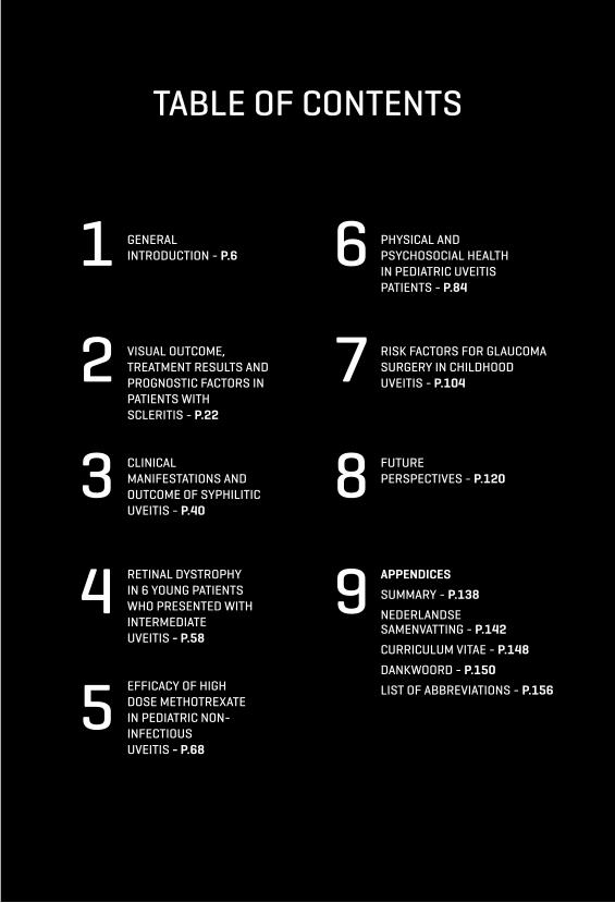
TABLE OF CONTENTS


GENERALINTRODUCTION1
1 —
8
GENERAL INTRODUCTION
Scleritis and uveitis are inflammatory eye-diseases which can threaten vision. In general, inflammatory eye-diseases can be triggered by auto-immune disease, infections, masquerade syndromes presenting as inflammatory eye-disease, medication, trauma and repeated ocular surgery 1 - 3. Scleritis and uveitis can occur at any age so the burden of visual loss, the uncertain prognosis of the eye-disease and its complications and the side-effects of treatment on daily life are profound 4 - 7.
Epidemiology of scleritisPublished epidemiologic data about the incidence and prevalence of scleritis in adults is scarce 8. The estimated reported annual incidence of scleritis is between 4 to 6 per 100,000 person-years 8, 9. This scarcity of epidemiologic data confirms that scleritis is a rare condition. Studies on scleritis are hampered by disease severity, its rarity and the intense pain reported by most patients suffering from scleritis 2, 8. Scleritis as an expression of underlying auto-immune disease such as rheumatoid artritis or granulomatosis with polyangiitis is the most common 2, 8. Loss of vision is more common in eyes with posterior or necrotizing scleritis and a loss of 2 or more lines Snellen visual acuity despite optimal treatment has been noted in 30% of patients 2. Patients with severe disease often have multiple causes for loss of visual function, such as corneal involvement, cataract, glaucoma, maculopathy, papilledema or retinal detachment 2, 8, 10. Information about the incidence of scleritis in children is even less available. One study reported that 1.2% of all scleritis cases are found in children 11 and others reported a female preponderance 12, 13. Among subtypes, posterior scleritis is relatively common in children 11. Although there is no literature supporting this, outcome and disease development in pediatric scleritis are probably worse than outcome and disease development in adults. It seems likely that children with scleritis have a greater risk of visual loss due to the higher reported incidence of posterior scleritis and a greater risk of ocular complications related to longer life expectancy and disease duration in this chronic disease. Pharmacological developments in the treatment of auto-immune diseases such as rheumatoid arthritis are promising. Hopefully, patients with scleritis can benefit from this.
Epidemiology of uveitisThe overall reported annual incidence of uveitis is between 17 and 52 per 100,000 person-years and the prevalence is 38 to 714 cases per 100,000 persons 14. The variation in reported incidences and prevalences between publications is due to variations worldwide in several predisposing factors such as genetic, geographic, social and environmental factors 14, 15. It has been estimated that uveitis accounts for about 10% of the visual handicap in the Western world, and up to 35% of all uveitis patients have been reported to suffer significant visual impairment or
1 — 9
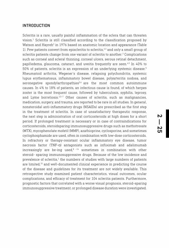
Anatomic location uveitis
Anterior uveitis Intermediate uveitis Posterior uveitis Pan uveitis
Anterior scleritis
Diffuse Nodular Necrotizing Scleromalacia
Posterior scleritis (incl SINSa) Posterior Surgery induced (SINS)
Panscleritis (anterior + posterior) aSINS = surgically-induced necrotizing scleritis
legal blindness 16. More recent publications on long-term clinical outcome in adults show more favorable visual outcomes due to improved treatment options 17. Uveitis in children is relatively uncommon and accounts for 5 to 10% of the total uveitis population 14, 18. The reported annual incidence is 4 per 100,000 population and the prevalence 28 per 100,000 population 18. It is estimated that in the western world 17-28% of the children with uveitis become legally blind in one eye 19, 20. Uveitis in childhood offers specific challenges when compared to uveitis in adults 21. The risk of poorer visual outcome is possibly greater in children when compared to adults 21. In most cases of uveitis in childhood the uveitis is related to juvenile idiopathic arthritis (JIA) 20. The onset is insidious in most cases of JIA-uveitis and diagnosis is often delayed resulting in deterioration of the visual prognosis 22. Ocular complications such as cataract, glaucoma, band keratopathy and amblyopia may silently develop and are reported in up to 50% of children with uveitis 20, 21.
Diagnosis of inflammatory eye disease.Early diagnosis of inflammatory eye disease and start of adequate therapy are the most important factors improving visual outcome. Diagnosis of scleritis is usually suspected from the clinical history with severe pain as a hallmark, and is confirmed by its characteristic clinical signs 2, 8. Scleritis is classified by its anatomic location and clinical appearance (table 1) 23.In case of posterior scleritis clinical signs may be less obvious and evaluation by ultrasonography or other imaging techniques are necessary 2.The main differential diagnosis of scleritis is episcleritis. Episcleritis is usually a mild non-vision threatening form of inflammation of the superficial episcleral tissue, for which no treatment is required in most cases 2, 8. The diagnosis in uveitis is more difficult. There are various etiologies and the systemic associations of uveitis differ between adults and children 14, 18, 24. In general, the differential diagnosis of uveitis is based upon the anatomical location of the inflammation (Table 2) 25, the recognition of specific ophthalmic clinical signs and the outcome of the different serological tests and – when necessary – outcome of analysis of intra-ocular fluid.
Table 1. Classification of scleritis 23 Table 2. Classification of uveitis 25
1 —
10
Treatment in generalThe treatment of inflammatory eye diseases depends on the etiology and possible underlying disease. In many cases, the uveitis or scleritis are part of an autoimmune process. The treatment is aimed at suppressing the inflammatory response and limiting the resulting damage. For scleritis, local therapy is insufficient and systemic therapy is required, although in some cases of non-infectious anterior scleritis a subconjunctival injection with corticosteroids can be given 26. In general, nonsteroidal anti-inflammatory drugs (NSAIDs) are prescribed as the first step in the treatment of scleritis. In case of unsatisfactory therapeutic response, the next step is administration of oral corticosteroids at high doses for a short period of time. If prolonged treatment is necessary or in case of contraindications for corticosteroids, steroidsparing immunosuppressive drugs such as methotrexate (MTX), mycophenolate mofetil (MMF), azathioprine, cyclosporine, and sometimes cyclophosphamide are used, often in combination with low-dose corticosteroids. In refractory or therapy resistant ocular inflammatory eye disease, tumor necrosis factor (TNF- α) antagonists such as infliximab and adalimumab or chimeric monoclonal antibodys targeted on B lymphocytes like rituximab, are increasingly being used, 27 - 31 sometimes in combination with other steroid- sparing immunosuppressive drugs.
For the treatment of uveitis, the first step in treatment are topical corticosteroids. If these are insufficient, local corticosteroid injections can be considered. Systemic corticosteroids are started in the case of severe uveitis or in case of failure of topical therapy. In case of chronic uveitis or underlying systemic disease, steroid-sparing immunosuppressive medication is required to maintain disease remission and to avoid the side effects of prolonged oral corticosteroids. Methotrexate (MTX) is the steroid sparing immunosuppressive agent of first choice in almost all cases of non-infectious uveitis 32 - 34. If MTX is ineffective or side effects occur, a switch towards another steroid sparing immunosuppressive agent such as mycophenolate mofetil (MMF), azathioprine or cyclosporine can be made. In persistent active uveitis despite treatment, tumor necrosis factor (TNF-α) antagonists such as infliximab and adalimumab and others are increasingly being used 20, 35. When the scleritis or uveitis has developed as a result of an infectious process, the primary treatment is aimed at the infectious pathogens. When the treatment against the infectious process starts, systemic immune suppression may additionally be necessary to reduce the inflammatory response - and thus reduce the resulting damage .
OutcomeInflammatory eye diseases are still a leading cause of visual impairment 36, 37. The main goal of the treatment of inflammatory eye diseases is to maintain visual function by reducing the inflammation and by the timely treatment of complications such as glaucoma, macular edema, and cataract 14 ,35. Visual

1 — 11
outcome is measured as visual acuity. In case of posterior and panuveitis or secondary glaucoma, visual outcome can be impaired by visual field loss through loss of function in the affected tissues by the inflammation itself or by damage to the optic nerve as a result of high intra-ocular pressure. Loss of vision and side effects of systemic treatment are related to loss of health-related quality of life (HR QoL) in children and adults with uveitis 4, 38 - 41. It has been suggested that the effects of uveitis on HR QoL in children are similar to those of children with other chronic conditions 42 and the disease burden of uveitis can affect quality of life even when there is no loss of vision 42.
Aims and outline of this thesisThe aim of this thesis is to improve the care for patients with inflammatory eye disease on a number of aspects. This thesis consists of 2 parts and describes studies on both the diagnostic and therapeutic challenges in the treatment and counseling of patients with inflammatory eye disease. In the first part the focus is on scleritis and uveitis in the adult population, the second part concerns uveitis in childhood. The first 3 chapters are about improving the diagnostic and therapeutic process in adult patients with rare inflammatory eye diseases such as scleritis, syphilitic uveitis and retinal dystrophies masquerading as intermediate uveitis. In the 3 chapters of the second part, efficacy and outcomes of different dosages of methotrexate (MTX) in non-infectious pediatric uveitis are evaluated, physical and psychosocial outcomes in pediatric uveitis are analyzed and risk factors for the development of secondary glaucoma in childhood uveitis are addressed.
Scleritis As mentioned before, scleritis is a rare disease. Because of this and the prompt need for treatment, there is a paucity in the literature regarding studies predicting disease-course and visual outcome, and offering guidelines for treatment. Therefore, chapter 2 describes patient characteristics, visual outcome, ocular complications and treatment results in a cohort of 104 patients with scleritis from 2 tertiary uveitis centers in the Netherlands. Also, predictors for a worse visual outcome, the need for steroid-sparing immunosuppressive treatment and a longer period of active disease were analyzed.
Ocular syphilisOcular syphilis can mimic a wide range of ocular disorders 43, 44 and is a rare sexually transmitted infection (STI) nowadays accounting for 1% to 2% of all uveitis patients 45 -47. In the pre-antibiotic era, syphilis was more common 46. Due to the improved screening and treatment programs it almost disappeared in the western world. Data on the epidemiology of STI needs to be interpreted carefully because they are influenced by multiple factors 47. The incidence and prevalence of the infection are affected by biological factors, such as transmission probability, infection duration and loss of protective immunity such as in HIV-positive
1 —
12
patients. Also, changes in sexual attitudes and behaviors and developments in service provision, treatment, interventions, diagnostic technologies and surveillance affect incidence and prevalence 47. Ocular syphilis is a treatable disease and because of the changes in epidemiology and unpredictability of the anatomical presentation of the uveitis 43, 44, 46, 47 ocular syphilis should always be considered in the differential diagnosis of uveitis. In the current guidelines, the recommended treatment for syphilitic uveitis is intravenous benzylpenicillin which is identical to the treatment for neurosyphilis 48, 49. Next to adequate treatment for the syphilis infection, the use of oral corticosteroids as systemic immune suppression are recommended to prevent a Jarisch-Herxheimer reaction 48, 49. Which is a reaction on the endotoxin-like products released by the death of harmful microorganisms within the body during antibiotic treatment and most commonly characterized by acute febrile illness with headache, myalgia, chills and rigors, resolving within 24 h 48. It is unclear if systemic immunosuppression – next to anti-syphilitic treatment - improves visual outcome in syphilitic uveitis. Favorable visual outcome is related to early diagnosis and treatment 50, 51. The clinical presentation of ocular syphilis has been described in many publications with relatively small numbers of patients. Due to the variability in clinical presentation, the sometimes confusing interpretation of serological tests and the debatable optimal treatment of a syphilis infection, the results from a large cohort of patients with serologically proven ocular syphilis are presented in chapter 3. More specifically, we report on the clinical manifestations and outcome of syphilitic uveitis in 85 patients with serologically proven syphilitic uveitis from 5 different tertiary uveitis centers in The Netherlands. The factors that correlate with a worse visual prognosis or a chronic disease course and the visual outcome of the different types of treatment are reported.
Masquerade uveitisRetinital dystrophies (RD) are a rare group of progressive hereditary retinal degenerative diseases characterized by progressive degeneration of retinal photoreceptors leading to profound visual loss and blindness in middle or later life 52. Worldwide, the prevalence of RD is approximately 1 in 3,000 individuals 53,
54. The diagnosis is made by recognition of the typical clinical picture, complaints of nyctalopia, a family history of retinal degenerative disease, visual field testing and a full-field electroretinogram (ERG). In most cases of advanced RD a progressively deteriorating ERG pattern is found, characterized by undetectable rod response and reduced cone response. In uveitis, the ERG response depends on the anatomical location of the uveitis. Most frequently, reduced amplitudes of a and b waves with long implicit times are found. In some cases, the ERG response normalizes with treatment, whereas in others it stays permanently abnormal 55. A retinal dystrophy can present itself with intraocular inflammation and cystoid macular edema masquerading as intermediate uveitis 56. Ongoing research suggests that in CRB1-linked retinal dystrophy masquerading as

1 — 13
intraocular inflammation, the disease is accompanied by molecular activation of inflammatory cytokine pathways and immune cells in the blood 56 - 58. These results on the role of inflammation in RD will hopefully provide insight in and possibilities for the treatment of RD and its complications in the future. At present, there are no treatment options besides corticosteroids and acetazolamide for macula edema and counseling of the patient. Nevertheless, patients can benefit from an early diagnosis which may result in more adequate counseling of the patient, and avoidance of prolonged treatment with high doses of immunosuppressive medication for a supposed uveitis. In chapter 4 the diagnostic process, clinical characteristics and outcome of 6 patients from 3 different tertiary uveitis centers in The Netherlands with retinal dystrophy presenting as intermediate uveitis are reported. This study intends to improve the diagnostic process and to provide insight into the specific characteristics and clinical signs in this patient group.
Methotrexate in pediatric non-infectious uveitisMethotrexate (MTX), due to its effectiveness, long track record 59 and good safety profile, is the steroid-sparing agent of first choice in almost all cases of non-infectious inflammatory eye diseases 32-34. MTX is effective in about 70% of patients 32-34 and it is usually given orally or subcutaneously. The bioavailability of oral MTX varies per patient and appears to decrease at higher doses due to limits in absorption in the gastrointestinal tract 60 - 62. Several studies in rheumatoid arthritis (RA) indicate that MTX exerts its effect by influencing multiple inflammatory pathways 63 - 65. Firstly, MTX undergoes polyglutamation within the cells, after that MTX and its polyglutamates inhibit purine and pyrimidine synthesis, reduce antigen-dependent T-cell proliferation, and promote release of adenosine which in turn activates receptors on macrophages and neutrophils to decrease the release of proinflammatory cytokines and elevate the secretion of anti-inflammatory molecules. It is unclear if these mechanisms of action of MTX in RA are similar to uveitis 66. But, due to its known 32-34 efficacy in ocular inflammation it is likely that the extraocular effects of MTX on the immune system provide the primary therapeutic mechanism by which systemically administered MTX affects ocular inflammation 34. Systemic administration of MTX leads to detectable intraocular MTX levels 67, 68 and the efficacy of intra-ocular MTX on uveitis and cystoid macular edema has been described in the literature 69, 70. However, the current evidence about dosage, duration of treatment and best route of administration for MTX in ocular inflammation is limited 32
- 34. Also, there are concerns in the treatment of RA that since the introduction and advent of TNF inhibitors MTX is less aggressively dosed, duration of use is shorter and a more rapid escalation to biologicals is made 62, 71, 72. In chapter 5 we present the results of our study on the efficacy of high dose in comparison to low dose MTX in 42 pediatric patients with non-infectious uveitis. Outcome measures are time to disease remission, steroid-sparing effect and side effects.
1 —
14
Physical and psychosocial health in pediatric uveitis patientsPatients with auto-immune diseases are more physically inactive compared to the general population 73. Also, aerobic fitness in children with different types of chronic conditions is reduced and they report more fatigue and lower health related quality of life (HR QoL) 74--77. In the developed countries the majority (41.5%) of the pediatric uveitis cases are related to juvenile idiopathic arthritis (JIA) 18, 78. Systemic immunosuppressive treatment in children with idiopathic uveitis who do not respond sufficiently to topical therapy is comparable to that used in the treatment of JIA. In JIA, children are found to be less physically active and have reduced physical fitness levels 79 which does not restore after remission has been reached 80, 81. The causes of these persistent impairments of physical fitness and physical activity are not known, but it has been suggested that a combination of disease-related factors, treatment (e.g., medication), hypo-activity, and deconditioning could be involved 82 - 84. Hypoactive children are often at greater risk of preventable health problems, such as obesity and cardio-metabolic diseases 82, 85.This higher risk of cardiovascular diseases is increased by the inflammation itself, circulating cytokines and the use of systemic immunosuppressive medication 83, 84, 86, 87. Cardiovascular health in children can be improved by sufficient physical activity (PA) and physical fitness 88, whereas PA also has a beneficial effect on HR-QoL73. The use of systemic immunomodulatory treatment or the presence of co-morbidity other than uveitis, did negatively influence general HR QoL scores in adult uveitis patients 4, 6. Also, in adolescents with non-infectious uveitis despite quiescence of disease and good visual function, certain factors, such as a high number of recurrences, chronicity of the uveitis and fear of blindness were correlated with a decreased HR QoL 39, 40. Fatigue is also highly present in patients with JIA and is related to many factors including PA, physical fitness and HR QoL of which cause and effect are not exactly known 89. In the literature, there are no publications about the physical fitness in children with uveitis and the information on the psychosocial health of children with uveitis is scarce 7, 41, 90, 91. To add to a better understanding and treatment of the effects of a chronic disease - like uveitis - on a child’s life, we present the results of our study on physical fitness, physical activity and psychosocial health in 23 children with uveitis in chapter 6.
Secondary glaucoma in pediatric uveitisChildhood uveitis has an inherent predisposition to develop secondary glaucoma, with a prevalence of 5-13.5% 92. Secondary glaucoma occurs when uveitis is associated with raised intraocular pressure (IOP) and optic nerve damage, resulting in irreversible visual field loss and possible visual impairment 93. The damage to the trabecular system by the inflammation, but also the use of topical steroids as treatment of uveitis can increase the IOP. Secondary glaucoma in childhood uveitis has an unpredictable course, with large IOP fluctuations, varying responses to eye-pressure lowering medication and a frequent steroid-
1 — 15
response 94. Increased IOP is initially treated pharmacologically by using topical anti-glaucoma medication. If pharmacological treatment of IOP is insufficient, glaucoma surgery is required. Only small studies have investigated the risk factors of developing secondary glaucoma in childhood uveitis. Two studies reported a female preponderance, JIA as the most common etiology and anterior uveitis as the predictive anatomical site in the glaucoma group 92, 95. Another small study compared the need of glaucoma surgery in children with uveitis who developed secondary glaucoma. Both mean age and the average number of previous intraocular surgeries in the surgery group were significantly higher than in the control group 96. To obtain the best long-term visual outcome, it is important to identify children with refractory glaucoma at an early stage and to treat them by glaucoma surgery before irreversible damage has occurred 97. In chapter 7 the results of our study on the possible risk factors for the development of secondary glaucoma needing glaucoma surgery are reported. The study was conducted in a large cohort of 196 children with uveitis from 2 tertiary uveitis centers in the Netherlands.
1 —
16
REFERENCES
1. Tuft SJ, Watson PG. Progression of scleral disease. Ophthalmology 1991;98:467-71.
2. Okhravi N, Odufuwa B, McCluskey P, Lightman S. Scleritis. Surv Ophthalmol 2005;50:351-63.
3. Cobo M. Inflammation of the sclera. Int Ophthalmol Clin 1983;23:159-71.
4. Schiffman RM, Jacobsen G, Whitcup SM. Visual functioning and general health status in patients with uveitis. Arch Oph-thalmol. 2001 Jun;119(6):841-9
5. Hoeksema L, Los LI. Vision-related qual-ity of life in herpetic anterior uveitis pa-tients. PLoS One. 2014 Jan 2;9(1). eCol-lection 2014.
6. Haasnoot AJW, Sint Jago NFM, Tekstra J, de Boer JH. Impact of Uveitis on Quality of Life in Adult Patients With Juvenile Id-iopathic Arthritis. Arthritis Care Res (Ho-boken). 2017 dec;69(12):1895-1902
7. Angeles-Han ST. Quality-of-life metrics in pediatric uveitis. Int Ophthalmol Clin. 2015;55(2):93-101
8. Lagina A, Ramphul K. Scleritis. Source; StatPearls [Internet]. [cited 15-07-2018]Treasure Island (FL): StatPearls Publish-ing; 2018-.2018 Apr 23.
9. Gelareh Homayounfar, Natalie Nardone, Durga S. Borkar, Vivien M. Tham, Travis C. Porco, Wayne T.A. Enanoria, John V. Park-er, Aleli C. Vinoya, Aileen Uchida, Nisha R. Acharya.Incidence of Scleritis and Epis-cleritis: Results From the Pacific Ocular Inflammation Study. American Journal of Ophthalmology, Volume 156, Issue 4, 2013, pp. 752-758.e3
10. McCluskey P, Wakefield D. Prediction of response to treatment in patients with scleritis using a standardised scoring sys-tem. Aust N Z J Ophthalmol 1991;19:211-5.
11. Majumder PD, Ali S, George A, Ganesh S, Biswas J. Clinical Profile of Scleritis in Children. Ocul Immunol Inflamm. 2018 Jan 25:1-5.
12. Sainz De La Maza M, Molina N, Gonza-lez-Gonzalez LA,Doctor PP, Tauber J, Foster CS Clinical characteristics of a large cohort of patients with scleritis and episcleri-tis. Ophthalmology. 2012;119(1):43–50.
13. Akpek EK, Thorne JE, Qazi FA, Do DV, Jabs DA Evaluation of patients with scleritis for systemic disease. Ophthal-mology.2004;111(3):501–506.
14. Wakefield D, Chang HC. Epidemiology of uveitis. International Ophthalmology clinics. 45(2):1-13, apr 2005.
15. Chan NS, Choi J, Cheung CMG. Pediatric Uveitis. Asia Pac J Ophthalmol (Phila). 2018 May-Jun;7(3):192-199
16. Durrani OM, Tehrani NN, Marr JE, Moradi P, Stavrou P, Murray PI. Degree, duration, and causes of visual loss in uveitis. Br J Ophthalmol. 2004 Sep;88(9):1159-62.
17. Tomkins-Netzer O, Talat L, Bar A, Lula A, Taylor SR, Joshi L, Lightman S. Long-term clinical outcome and causes of vision loss in patients with uveitis. Oph-thalmology. 2014. Dec; 121(12): 2387-92.
18. Päivönsalo-Hietanen T, Tuominen J, Saari KM. Uveitis in children: population-based study in Finland. Acta Ophthalmol Scand. 2000 Feb;78(1):84-8.
19. Zierhut M, Michels H, Stübiger N, Besch D, Deuter C, Heiligenhaus A. Uveitis in children. Int Ophthalmol Clin. 2005 Spring;45(2):135-56.
20. Angeles-Han ST, Rabinovich CE. Uveitis in children. Curr Opin Rheumatol. 2016 Sep;28(5): 544-9.
21. Wentworth BA, Freitas-Neto CA, Foster CS. Management of pediatric uveitis. F1000Prime Rep. 2014;6:41-41. eCollec-tion 2014.
22. BenEzra D, Cohen E, Maftzir G. Uveitis in children and adolescents. Br J Ophthal-mol. 2005 Apr;89(4):444-8.
23. Watson PG, Hayreh SS. Scleritis and epis-cleritis. Br J Ophthalmol 1976;60:163-91
24. Smith JA, Mackensen F, Sen HN, et al. Epidemiology and course of disease in childhood uveitis. Ophthalmology. 2009;116(8):1544-51, 1551.e1.
1 — 17
25. Jabs DA, Nussenblatt RB, Rosenbaum JT, Standardization of Uveitis Nomenclature (SUN) Working Group. Standardization of uveitis nomenclature for reporting clinical data. results of the first inter-national workshop. Am J Ophthalmol. 2005;140(3):509-516.
26. Sohn EH, Wang R, Read R, Roufas A, Teo L, Moorthy R, Albini T, Vasconcelos-San-tos DV, Dustin LD, Zamir E, Chee SP, Mc-Cluskey P, Smith R, Rao N. Long-term, multicenter evaluation of subconjuncti-val injection of triamcinolone for non-ne-crotizing, noninfectious anterior scleritis. Ophthalmology. 2011 Oct;118(10):1932-7.
27. Chauhan S, Kamal A, Thompson RN, et al. Rituximab for treatment of scleritis as-sociated with rheumatoid arthritis. Br J Ophthalmol 2009;93:984 –5.
28. Iaccheri B, Androudi S, Bocci EB, et al. Rituximab treatment for persis-tent scleritis associated with rheuma-toid arthritis. Ocul Immunol Inflamm 2010;18:223–5.
29. Kurz PA, Suhler EB, Choi D, Rosenbaum JT. Rituximab for treatment of ocular inflammatory disease: a series of four cases. Br J Ophthalmol 2009;93:546–8.
30. Restrepo JP, Molina MP. Successful treat-ment of severe nodular scleritis with adal-imumab. Clin Rheumatol 2010;29:559 – 61
31. Sen HN, Sangave A, Hammel K, et al. In-fliximab for the treatment of active scleri-tis. Can J Ophthalmol 2009;44:e9–e12.
32. Simonini G, Paudyal P, Jones GT, Cimaz R, Macfarlane GJ. Current evidence of methotrexate efficacy in childhood chronic uveitis: A systematic review and meta-analysis approach. Rheumatology (Oxford). 2013;52(5):825-831.
33. Ali A, Rosenbaum JT. Use of metho-trexate in patients with uveitis. Clin exp rheumatol 2010 sep-oct;28(5 Suppl 61):S145-50
34. Gangaputra Sapna et al. Methotrexate for Ocular Inflammatory Diseases. Oph-thalmology. 2009; 116:2188-2198
35. Dick AD, Rosenbaum JT, Al-Dhibi HA, Belfort R Jr, Brézin AP, Chee SP, Davis JL, Ramanan AV, Sonoda KH, Carreño E, Nas-cimento H, Salah S, Salek S, Siak J, Stee-ples L; Guidance on Noncorticosteroid Systemic Immunomodulatory Therapy in Noninfectious Uveitis: Fundamentals Of Care for UveitiS (FOCUS) Initiative. Oph-thalmology. 2018 May;125(5):757-773.
36. de Smet MD, Taylor SR, Bodaghi B, et al. Understanding uveitis: the impact of re-search on visual outcomes. Prog Retin Eye Res. 2011;30:452–470.
37. Miserocchi E, Fogliato G, Modorati G, et al. Review on the worldwide epidemiology of uveitis. Eur J Ophthalmol. 2013;23:705–717.
38. Miserocchi E, Modorati G, Mosconi P, Colucci A, Bandello F. Quality of Life in Patients with Uveitis on Chronic System-ic Immunosuppressive Treatment. Ocul Immunol Inflamm. 2010;18(4):297-304.
39. Maca SM, Amirian A, Prause C, Gruber K, Mejdoubi L, Barisani-Asenbauer T. Under-standing the Impact of Uveitis on Health-re-lated Quality of Life in Adolescents. Acta Ophthalmol. 2013;91(3):219-224.
40. Petrina Tan, Yan Tong Koh, Poh Ying Wong & Stephen C. Teoh. Evaluation of the Impact of Uveitis on Visual-related Quality of Life. Ocular Immunology and Inflammation. 2012;20(6):453-459
41. Angeles-Han ST, Griffin KW, Lehman TJ, et al. The importance of visual function in the quality of life of children with uveitis. J AAPOS. 2010; 14(2):163–168. [PubMed: 20236847]
42. Parker DM, Angeles-Han ST, Stanton AL, Holland GN. Chronic Anterior Uveitis in Children: Psychosocial Challenges for Patients and Their Families. Am J Oph-thalmol. 2018 Jul;191:xvi-xxiv.
43. Davis JL. Ocular syphilis. Curr Opin Oph-thalmol. 2014 Nov;25(6):513-8.
44. Amaratunge BC, Camuglia JE, Hall AJ. Syphilitic uveitis: A review of clinical manifestations and treatment outcomes of syphilitic uveitis in human immuno-deficiency virus-positive and negative patients. Clin Experiment Ophthalmol. 2010;38(1):68-74.

1 —
18
45. Schlaegel TF,Jr, O'Connor GR. Metastatic nonsuppurative uveitis. Int Ophthalmol Clin. 1977;17(3):87-108.
46. Fenton KA, Breban R, Vardavas R, et al. Infectious syphilis in high-income set-tings in the 21st century. Lancet Infect Dis.2008;8:244-253.
47. Hughes G, Field N. The epidemiology of sexually transmitted infections in the UK: impact of behavior, services and interven-tions. Future Microbiol. 2015;10:35-51.
48. Janier M, Hegyi V, Dupin N, et al. 2014 European guideline on the manage-ment of syphilis. J Eur Acad Dermatol Venereol. 2014.
49. Workowski KA, Bolan GA, Centers for Disease Control and Prevention. Sex-ually transmitted diseases treatment guidelines,2015. MMWR Recomm Rep. 2015;64:1-137.
50. Balaskas K, Sergentanis TN, Giulieri S, et al. Analysis of significant factors influ-encing visual acuity in ocular syphilis. Br J Ophthalmol. 2011;95:1568–1572.
51. Moradi A, Salek S, Daniel E, et al. Clinical features and incidence rates of ocular com-plications in patients with ocular syphilis. Am J Ophthalmol. 2015;159:334–343.e1.
52. Francesco Parmeggiani. Clinics, Epidemi-ology and Genetics of Retinitis Pigmentosa. Curr Genomics. 2011 Jun; 12(4): 236–237.
53. Bessant DA, Ali RR, Bhattacharya SS. Molecular genetics and prospects for therapy of the inherited retinal dys-trophies. Curr Opin Genet Dev. 2001 Jun;11(3):307-16.
54. Hartong DT, Berson EL, Dryja TP. Retinitis pigmentosa. Lancet. 2006;368:1795–809
55. Sevgi DD, Davoudi S, Comander J, Sobrin L. Retinal pigmentary changes in chronic uveitis mimicking retinitis pigmentosa. Graefes Arch Clin Exp Ophthalmol. 2017 Sep;255(9):1801-1810
56. Yoshida N, Ikeda Y, Notomi S, et al. Clini-cal evidence of sustained chronic inflam-matory reaction in retinitis pigmentosa. Ophthalmology. 2013;120(1):100–105
57. Tamm S, Whitcup SM, Gery I, et al. Im-mune response to retinal antigens in patients with gyrate atrophy and other hereditaryretinal dystrophies. Ocul Im-munol Inflamm. 2001;9(2):75–84.
58. Stunkel M, Bhattarai S, Kemerley A, et al. Vitritis in pediatric genetic retinal disorders. Ophthalmology. 2015;122(1):192–199.
59. Wong VG. Methotrexate treatment of uveal disease. Am J Med Sci. 1966;251(2):239-241.
60. Herman RA, Veng-Pedersen P, Hoffman J, Koehnke R, Furst DE. Pharmacokinet-ics of low-dose methotrexate in rheu-matoid arthritis patients. J Pharm Sci. 1989;78(2):165–171
61. van Roon EN, van de Laar MA. Methotrex-ate bioavailability. Clin Exp Rheumatol 2010;28(Suppl):27-32.
62. Bello AE, Perkins EL, Jay R, Efthimiou P. Recommendations for optimizing meth-otrexate treatment for patients with rheumatoid arthritis. Open Access Rheu-matol. 2017 Mar 31;9:67-79.
63. Chan ES, Cronstein BN. Molecular action of methotrexate in inflammatory diseas-es. Arthritis Res. 2002;4(4):266–273.
64. Milne GR, Palmer TM. Anti-inflamma-tory and immunosuppressive effects of the A2A adenosine receptor. Scienti-ficWorldJournal.2011;11:320–339.
65. Tian H, Cronstein BN. Understanding the mechanisms of action of methotrexate: implications for the treatment of rheu-matoid arthritis. Bull NYU Hosp Jt Dis. 2007;65(3):168–173.
66. Hashkes PJ, Becker ML, Cabral DA, Laxer RM, Paller AS, Rabinovich CE, Turner D, Zulian F. Methotrexate: new uses for an old drug. J Pediatr. 2014 Feb;164(2):231-6
67. Puchta J, Hattenbach LO, Baatz H. In-traocular levels of methotrexate after oral low-dose treatment in chronic uvei-tis. Ophthalmologica. 2005; 219:54–5.
68. de Smet MD, Stark-Vancs V, Kohler DR, et al. Intraocular levels of methotrexate after intravenous administration. Am J Ophthalmol. 1996; 121:442–4.
1 — 19
69. Taylor SR, Banker A, Schlaen A, Couto C, Matthe E, Joshi L, Menezo V, Nguyen E, Tomkins-Netzer O, Bar A, Morarji J, Mc-Cluskey P, Lightman S. Intraocular meth-otrexate can induce extended remission in some patients in noninfectious uveitis. Retina. 2013 Nov-Dec;33(10):2149-54.
70. Taylor SR, Habot-Wilner Z, Pacheco P, Lightman SL. Intraocular methotrexate in the treatment of uveitis and uveitic cystoid macular edema. Ophthalmology. 2009 Apr;116(4):797-801. doi: 10.1016/j.ophtha.2008.10.033.
71. Pincus T, Gibson KA, Castrejón I. Update on methotrexate as the anchor drug for rheumatoid arthritis. Bull Hosp Jt Dis. 2013;71(Suppl 1):S9–S19.
72. Rohr MK, Mikuls TR, Cohen SB, Thorne CJ, O’Dell JR. The underuse of methotrexate in the treatment of RA: a national analysis of prescribing practices in the U.S. Arthritis Care Res (Hoboken). Epub 2016 Nov 18.
73. 4. Sharif K,Watad A, Bragazzi N.L, Licht-broun M, Amital H, Shoenfeld Y. Physical activity and autoimmune diseases: Get moving and manage the disease. Auto-immun Rev. 2018; 17( 1), 53-72.
74. Takken T, Bongers BC, van Brussel M, Haapa-la EA, Hulzebos EHJ. Cardiopulmonary Ex-ercise Testing in Pediatrics. Ann Am Thorac Soc. 2017; Supplement 1, S123-S128.
75. van Brussel M, van der Net J, Hulzebos E, Helders PJ, Takken T. The Utrecht ap-proach to exercise in chronic childhood conditions: the decade in review. Pediatr Phys Ther. 2011; 23, (1): 2-14
76. Gualano B, Bonfa E, Pereira RMR, Silva CA. Physical activity for paediatric rheumatic diseases: standing up against old paradigms. Nat Rev Rheumatol. 2017;13, (6): 368-379.
77. Barthel D, Ravens-Sieberer U, Nolte S, Thyen U, Klein M, Walter O, Meyrose AK, Rose M, Otto C. Predictors of health-re-lated quality of life in chronically ill chil-dren and adolescents over time. J Psy-chosom Res. 2018 Jun;109:63-70.
78. Mehta PJ, Alexander JL, Sen HN. Pediatric uveitis: New and future treatments. Curr Opin Ophthalmol. 2013;24(5):453-462.
79. Lelieveld OT, Armbrust W, van Leeuwen M a, et al. Physical Activity in Adoles-cents with Juvenile Idiopathic Arthritis. Arthritis Rheum. 2008;59(10):1379-1384
80. van Brussel M, Lelieveld OTHM, van der Net J, Engelbert RHH, Helders PJM, Takken T. Aerobic and Anaerobic Exer-cise Capacity in Children with Juvenile Idiopathic Arthritis. Arthritis Rheum. 2007;57(6):891-897.
81. Ploeger HE, Takken T, Wilk B, et al. Exer-cise Capacity in Pediatric Patients with Inflammatory Bowel Disease. J Pediatr. 2011;158(5):814-819.
82. Takken T, Bongers BC, van Brussel M, Haapala EA, Hulzebos EHJ. Cardiopul-monary Exercise Testing in Pediatrics. Ann Am Thorac Soc. 2017; Supplement 1, S123-S128.
83. Roubenoff R. Exercise and Inflammatory Disease. Arthritis Care Res (Hoboken). 2003;49(2): 263.
84. Gupta Y, Gupta A. Glucocorticoid-induced Myopathy: Pathophysiology, Diagnosis, and Treatment. Indian J Endocrinol Me-tab. 2013;17(5):913-916.
85. Zoico E, Roubenoff R. The Role of Cy-tokines in Regulating Protein Metab-olism and Muscle Function. Nutr Rev. 2002;60(2):39-51.
86. Carnethon M, Gidding S, Nehgme R, Sid-ney S, Jacobs D, Liu K. Cardiorespiratory Fitness in Young Adulthood and the Devel-opment of Cardiovascular Diseases Risk Factors. JAMA. 2003;290(23):3092-3100
87. Steene-Johannessen J, Anderssen S a, Kolle E, Andersen LB. Low muscle fit-ness is associated with metabolic risk in youth. Med Sci Sports Exerc. 2009 Jul;41(7):1361–7.
88. Strong WB, Malina RM, Blimkie CJR, et al. Evidence Based Physical Activ-ity for School-age Youth. J Pediatr. 2005;146(6):732-737.
1 —
20
89. Armbrust W, Lelieveld OH, Tuinstra J, Wulffraat NM, Bos GJ, Cappon J, van Ros-sum MA, Sauer PJ, Hagedoorn M. Fatigue in patients with Juvenile Idiopathic Ar-thritis: relationship to perceived health, physical health, self-efficacy, and partici-pation. Pediatr Rheumatol Online J. 2016 Dec 6;14(1):65.
90. Angeles-Han ST, Griffin KW, Harrison MJ, Lehman TJ, Leong T, Robb RR, Shain-berg M, Ponder L, Lenhart P, Hutchinson A, Srivastava SK, Prahalad S, Lambert SR, Drews-Botsch C. Development of a vision-related quality of life instrument for children ages 8-18 years for use in juvenile idiopathic arthritis-associated uveitis. Arthritis Care.Res.(Hoboken), 2011;63(9)1254-1261
91. Angeles-Han ST, Yeh S, McCracken C, Jenkins K, Stryker D, Myoung E, Vogler LB, Rouster-Stevens K, Lambert SR, Har-rison MJ, Prahalad S, Drews-Botsch C. Using the Effects of Youngsters' Eyesight on Quality of Life Questionnaire to Meas-ure Visual Outcomes in Children With Uveitis. Arthritis Care Res (Hoboken). 2015 Nov;67(11):1513-20.
92. Gautam Seth N, Yangzes S, Thattaruthody F, et al. Glaucoma Secondary to Uveitis in Children in a Tertiary Care Referral Center. Ocular Immunology and Inflam-mation. Published February 2, 2018.
93. Baneke AJ, Lim KS, Stanford M. The Patho-genesis of Raised Intraocular Pressure in Uveitis. Curr Eye Res. 2016;41(2):137-149.
94. Muñoz-Negrete FJ, Moreno-Montañés J, Hernández-Martínez P, Rebolleda G. Cur-rent Approach in the Diagnosis and Man-agement of Uveitic Glaucoma. Biomed Res Int. 2015;2015:1-13.
95. Heinz C, Koch JM, Zurek-Imhoff B, Heili-genhaus A. Prevalence of uveitic second-ary glaucoma and success of nonsurgi-cal treatment in adults and children in a tertiary referral center. Ocul Immunol Inflamm. 2009;17(4):243-248.
96. Kalinina Ayuso V, Scheerlinck LM, de Boer JH. The effect of an Ahmed glauco-ma valve implant on corneal endothelial cell density in children with glaucoma secondary to uveitis. Am J Ophthalmol. 2013 Mar;155(3):530-5.
97. Abu Samra K, Maghsoudlou A, Roohipoor R, Valdes-Navarro M, Lee S, Foster CS. Current Treatment Modalities of JIA-as-sociated Uveitis and its Complications: Literature Review. Ocul Immunol In-flamm. 2016;24(4):431-439.

1 — 21

VISUAL OUTCOME, TREATMENT RESULTS AND PROGNOSTIC FACTORS IN PATIENTS WITH SCLERITISAuthors:Wietse G. Wieringa1,
Jaap E. Wieringa2, Ninette H. ten Dam-van Loon3, 4,
Leonoor I. Los1, 5
1 Department of Ophthalmology, University Medical Center Groningen, University of Groningen, P. O. Box 30001, 9700 RB Groningen, The Netherlands2 Department of Marketing, Faculty of Economics and Business, University of Groningen, P.O. Box 800, 9700 AV Groningen, The Netherlands3 Department of Ophthalmology, University Medical Center Utrecht, University of Utrecht, P.O. Box 85500, 3508 GA Utrecht, The Netherlands4 Eijkman Graduate School for Immunology and Infectious Diseases, University of Utrecht, The Netherlands5 W.J. Kolff institute, Graduate School of Medical Sciences, University of Groningen, The Netherlands
Ophthalmology. 2013 Feb;120(2):379-86
2

2 —
24
ABSTRACT
Purpose: To analyze the visual outcome, systemic associations, effec-tiveness of treatment and predicting features of 104 scleritis patients. Design: Retrospective case series.Participants: 104 patients treated for scleritis at the University Medical Centers of Groningen and Utrecht.Methods: The clinical records of 104 patients diagnosed with scleritis between 1992 and 2011 at the University Medical Centers of Groningen, (n= 64) and Utrecht (n=40) were retrospectively analyzed.Main outcome measures: Loss of visual acuity, ocular complications, related systemic disease, type of treatment, time to treatment success and predictive features.Results: Mean age was 51.5 (standard deviation[SD], ±13.6) years, 63 (60.6 %) patients were female. Mean follow up was 38.2 (SD± 33.8) months. A loss of more than two lines of Snellen acuity was observed in 23 patients, 3 of whom had a final visual acuity of no light perception (NLP). In general, patients with necrotizing scleritis (n=15) had a poorer outcome. Ocular complications were observed in 88 (84.6%) patients. Underlying systemic disease was identified in 34 (32.7 %) patients. Steroid-sparing immunosuppressive medication was used in 47 patients, 36 of these were treated with methotrexate (MTX). This led was successful in 17 (47.2%) patients over the course of a mean ± SD 103.7 ± 83.7 weeks. Mycophenolate mofetil (MMF) was the treatment in 10 patients, and in 5 patients treatment success was achieved in a mean ± SD 65.3 ± 37.4 weeks. Treatment with tumor necrosis factor-alpha (TNF-α) antagonists led to treatment success in a mean ± SD 32.6 ± 21.8 weeks in 5 of the 11 treated patients. Patients with loss of visual acuity or those treated with oral steroid-sparing immunosuppressive drugs had more often an underlying associated disease, a bilateral scleritis and a longer period of symptoms at presentation.Conclusions: Scleritis is a severe ocular inflammation often associated with ocular complications. In this population roughly half of the patients were treated with systemic immunosuppressive medication. MMF and TNF-α antagonists can be used in case of MTX-failure. TNF-α antagonists seemed to be more effective than MTX. Within this group, an underlying associated disease, a bilateral scleritis and a longer period of symptoms at presentation were predictive features for a more severe disease course.
2 — 25
INTRODUCTION
Scleritis is a rare, usually painful inflammation of the sclera that can threaten vision.1 Scleritis is still classified according to the classification proposed by Watson and Hayreh2 in 1976 based on anatomic location and appearance (Table 1). Few patients convert from episcleritis to scleritis,2,3 and only a small group of scleritis patients change from one variant of scleritis to another.3 Complications such as corneal and scleral thinning, corneal ulcers, serous retinal detachment, papilledema, glaucoma, cataract, and uveitis frequently are seen.4,5 In 40% to 50% of patients, scleritis is an expression of an underlying systemic disease.5 Rheumatoid arthritis, Wegener’s disease, relapsing polychondritis, systemic lupus erythematosus, inflammatory bowel disease, polyarteritis nodosa, and seronegative spondylarthropathies5,6 are the most common autoimmune causes. In 4% to 18% of patients, an infectious cause is found, of which herpes zoster is the most frequent cause, followed by tuberculosis, syphilis, leprosy, and Lyme borreliosis.3,5–7 Other causes of scleritis, such as malignancies, medication, surgery, and trauma, are reported to be rare in all studies. In general, nonsteroidal anti-inflammatory drugs (NSAIDs) are prescribed as the first step in the treatment of scleritis. In case of unsatisfactory therapeutic response, the next step is administration of oral corticosteroids at high doses for a short period. If prolonged treatment is necessary or in case of contraindications for corticosteroids, steroidsparing immunosuppressive drugs such as methotrexate (MTX), mycophenolate mofetil (MMF), azathioprine, cyclosporine, and sometimes cyclophosphamide are used, often in combination with low-dose corticosteroids. In refractory or therapy-resistant ocular inflammatory eye disease, tumor necrosis factor (TNF-α) antagonists such as infliximab and adalimumab increasingly are be-ing used,8 –14 sometimes in combination with other steroid- sparing immunosuppressive drugs. Because of the low incidence and prevalence of scleritis,1 the numbers of studies with large numbers of patients are limited,15 and well-documented clinical experience in predicting the course of the disease and guidelines for its treatment are not widely available. This retrospective study examined patient characteristics, visual outcomes, ocular complications, and efficacy of treatment for 104 scleritis patients. Furthermore, prognostic factors that correlated with a worse visual prognosis, steroid-sparing immunosuppressive treatment, or prolonged disease duration were investigated.
2 —
26
METHODS AND PATIENTS
One hundred four patients diagnosed with scleritis between 1992 and 2011 at the University Medical Centers of Groningen, The Netherlands (n = 64), and of Utrecht, The Netherlands (n = 40), were analyzed. The Medical Ethical Committee of the University Medical Center of Groningen ruled that approval was not required for this study. Patients were identified by searching on the diagnosis code ‘scleritis’ in the digital uveitis databases of both centers. If in doubt about the diagnosis of scleritis, the opinion of an academic uveitis specialist based on the patient’s file was decisive. Only patients with a follow-up of more than 3 months were included. The Watson and Hayreh classification was used for the type of scleritis, with the diagnosis of posterior scleritis or panscleritis confirmed by ultrasound. Necrotizing scleritis was classified as necrotizing (with inflammation) or scleromalacia perforans (without inflammation).
The decimal equivalent of the Snellen visual acuity of both eyes at presentation and at last follow-up and the maximum visual acuity were recorded. This visual acuity was converted to logarithm of the minimum angle of resolution units and, after computation of mean and standard deviation, was calculated back to Snellen decimal acuity. Loss of visual acuity was defined as a decrease of more than 2 lines on the Snellen chart. No light perception in the affected eye was defined as blindness. Patients whose loss of visual acuity was not a result of the scleritis were excluded from this analysis. Corneal complications were characterized as ulcerative or peripheral thinning. Uveitis was diagnosed when cells could be observed in the anterior chamber or in the vitreous and was classified as anterior uveitis, intermediate uveitis, posterior uveitis, or panuveitis. The lens was graded as clear, having cataract, pseudophakic, or having posterior capsule opacification. The presence of cystoid macular edema was noted only if confirmed by fluorescein angiography or optical coherence tomography. Serous retinal detachments were diagnosed by funduscopy or ultrasound. Ocular hypertension was defined as an intraocular pressure of more than 21 mmHg, and the given treatment was recorded.
All 104 patients underwent screening for underlying systemic disease, and 88 patients underwent screening in accordance to the guidelines of the Dutch Ophthalmologic Society: (http://www.oogheelkunde.org/uploads/fl/ve/flvem3mKxt8ThFFYVhn8GQ/Richtlijn- voor-diagnostiek-en-behandeling-van-uveitis-15-mei-2007-1.pdf; accessed January 23, 2012). The other 16 patients were screened by a tailored approach or screening was not performed when scleritis was considered a manifestation of a known systemic disease. Laboratory testing included blood and urine tests, chest radiography, and tuberculin skin testing. Serologic and general laboratory tests included complete blood count, white cell differential, inflammatory parameters (C-reactive protein
2 — 27
and erythrocyte sedimentation rate), liver and kidney function tests, antinuclear antibody analysis, antineutrophil cytoplasmic antibody (ANCA) analysis, and rheumatoid factor analysis. Other tests, such as Treponema pallidum antibody titers, Lyme antibody titers, angiotensin-converting enzyme, and human leukocyte antigen (HLA)-B27, were not obtained routinely, but were based on history and physical examination. In case of an underlying systemic disease, the patient was diagnosed by a specialist in that area. Associated systemic diseases were classified as infectious or autoimmune. The most common autoimmune and infectious causes were recorded. The rare causes were listed per patient. When known, the smoking status was included in the analysis.
In these patients, treatment was administered mainly according to a stepladder approach: In infectious causes, the cause of the infection was treated. In autoimmune nonnecrotizing scleritis, NSAIDs were given as a first choice, and in case of NSAID failure, high-dose corticosteroids were given. In case corticosteroids could not be reduced to a dosage of less than 10 mg daily, a corticosteroid-sparing immunosuppressive drug was considered, which was usually MTX. Methotrexate was started orally in a dosage between 7.5 and 15 mg weekly and was increased according to clinical response to a maximum of 25 mg weekly via subcutaneous injection or 30 mg weekly orally. In most patients, this was carried out in at least 3 steps each with an interval of at least 2 months. In case of MTX failure, MMF was started, and in case this failed as well, a TNF-α antagonist was introduced. In case of necrotizing scleritis, corticosteroids and corticosteroid-sparing medication were started immediately.
Treatment success was defined as a subjective and an observable inactive disease for longer than 3 months using less than 10 mg daily oral prednisone alone or in combination with corticosteroidsparing drugs. A relapse was defined as a recurrence of the scleritis after a quiet episode described in the patient file. The total followup time (disease duration including treatment of secondary complications) and time to treatment success were documented. In case of a multiple medication regimen, a stepwise approach was used and the time to control of the inflammation was related to the last added systemic immunosuppressive drug. In patients who were already receiving systemic medication for a systemic disease at presentation, the change in medication or dosage responsible for treatment success was recorded.
How clinical characteristics, visual outcome, ocular complications, and differences in treatment affected outcome was analyzed using SPSS software version 18 (SPSS, Inc, Chicago, IL) based on 3 end points: loss of visual acuity, treatment with steroid-sparing immunosuppressive drugs, or longer disease duration. A P value of 0.05 or less was considered to be statistically significant. To assess the value and weight of the prognostic factors, the chi-square test
2 —
28
for categorical variables, the Student t test for comparing independent groups with a continuous variable, and the Spearman bivariate correlation coefficient for analysis of a correlation between 2 continuous variables were used. These findings were verified and confirmed by logistic and linear regression models. Two patients were identified by SPSS analysis (boxplot) as extreme outliers (more than 3.0 times the interquartile range above the third quartile) and therefore were excluded from the analysis. Kaplan-Meier curves were used to display graphically the type of treatment related to time to disease remission.
RESULTS
Patients’ characteristics are summarized in Table 1. Of the 104 scleritis patients, 63 (60.6%) were female. Mean follow-up was 38.2 months (range, 3–154 months). Mean age was 51.5 years (range, 18–91 years). The 6 patients with necrotizing disease were the oldest; the 4 patients with posterior disease were the youngest. The latter were all female. Most patients (n = 64) had unilateral disease. Diffuse anterior scleritis was the most common type of scleritis, followed by panscleritis.
Table 2 summarizes ocular complications. Complications were observed in 88 (84.6%) patients. The largest percentage of complications were seen in necrotizing scleritis patients. Uveitis was the most common complication (n = 47). Cataract formation was documented in 30 patients, whereas 6 patients were pseudophakic at presenta-tion. Posterior scleral swelling as shown by ultrasound was found in 31 patients, and 2 patients had posterior scleral thickening related to severe anterior scleritis.
Table 3 shows the loss of visual acuity related to type of scleritis and severity. A loss of more than 2 lines of Snellen acuity occurred in 23 patients (Table 3), 3 of whom became blind (no light perception visual acuity) because of scleritis. All 3 of the latter patients had necrotizing scleritis. In 2 of these patients, there was an association with Wegener’s disease, and the third patient had a scleromalacia perforans without known underlying systemic disease.
The 23 patients with a decrease in visual acuity had an average Snellen visual acuity standard deviation (SD) at presentation of 0.9 ± 0.34, and their final average Snellen visual acuity ±SD was 0.66±0.38. The remaining 81 patients showed, on average, an increase in visual acuity of 0.17 (range, 0.007–1.0).
Of the 43 patients (Table 2) with an intraocular pressure to more than 21 mmHg, 26 patients were diagnosed as steroid responders because of the use of local or systemic corticosteroids. Because of the elevation in intraocular pressure, 20 patients were administered antiglaucoma medication, and glaucoma surgery was undertaken in 6 patients.

2 — 29
Feature Anterior Diffuse
Anterior nodular
Anterior necrotizing
Sclero- malacia
Posterior Surgery Induced
Pan- scleritis
N (%)
N total 36 20 6 9 4 3 26 104 Bilateral 18 5 3 3 1 0 10 40 (38.5%) Ocular complications
31 11 6 7 4 3 26 88 (84.6%)
Corneal Thinning
2 2 2 2 0 0 1 9 (8.7%)
Corneal ulcerative 8 2 4 2 1 0 2 19 (18.3%) Uveitis 20 1 4 4 2 1 15 47 (45.6%) Anterior uveitis 18 1 2 3 2 1 10 37 (35.9%) Intermediate uveitis 2 0 0 1 0 0 1 4 (3.9%) Panuveitis 0 0 2 0 0 0 4 6 (5.8%) Cataract 10 2 4 5 1 0 8 30 (28.8%) CME 5 1 3 1 1 0 11 22 (21.4%) Exudative retinal detachment
0 0 2 1 0 0 6 9 (8.7%)
T-Sign (US) 0 1 1 0 4 0 25 31 (30.1%) VA-loss* 10 3 2 4 0 0 4 23 (22.1%) Ocular hypertension§ 13 8 3 3 0 2 14 43 (41.7%) Steroidresponder 9 4 2 2 0 2 7 26 (25.2%)
CME = cystoid macular edema; US = ultrasound; VA = visual acuity. *Decrease in visual acuity of 2 Snellen lines or more (worse of the 2 eyes) at the end of the follow up. § Intra-ocular pressure higher than 21 mmHg
Table 2. Ocular complications
Table 3. Vision loss related to scleritis
Diagnosis/ type
Anterior Diffuse
Anterior nodular
Anterior necrotizing
Sclero- malacia
Posterior Surgery Induced
Pan- scleritis
N (%)
N total 36 20 6 9 4 3 26 104 Loss of ≥ 2
Snellen lines* 7 1 1 1 10 (9.6%)
Severe loss > 3 Snellen lines§
3 2 2 3 10 (9.6%)
NLP 2 1 3 (2.9%)
NLP = No light perception. * Decrease in visual acuity of ≥ 2 Snellen lines at the end of the follow up period. § Decrease in visual acuity of > 3 Snellen lines at the end of the follow up period
Diagnosis/type N Mean age (range) Male N (%) Female N (%) Bilateral N (%) Scleritis total 104 51.5 (18.6 - 91.8) 41 (39.8%) 63 (60.6 %) 40 (38.5%) Anterior scleritis 71 51.9 (25.4 – 91.8) 29 (40.8%) 42 (59.2%) 29 (40.8%)
Diffuse 36 51.4 (25.4 – 79.5) 13 (36.1%) 23 (63.9%) 18 (50%) Nodular 20 50.1 (30.5 – 67) 7 (35%) 13 (65%) 5 (25%) Necrotizing 6 62.5 (41.3 – 91.8) 3 (50%) 3 (50%) 3 (50%) Scleromalacia 9 50.9 (38.6 – 71.6) 6 (66.7%) 3 (33.3%) 3 (33.3%)
Posterior scleritis (incl SINS) 7 47.9 (18.6 – 73.1) 2 (28.6%) 5 (71.4%) 1 (14.3%) Posterior 4 39.4 (18.6 – 56.5) 0 4 1 (25%) Surgery induced (SINS) 3 59.2 (39.8 – 73.1) 2 (66.7%) 1 (33.3%) 0
Panscleritis (anterior + posterior) 26 51.4 (25.6 – 69.2) 10 (38.5%) 16 (61.5%) 10 (38.5%)
Table 1. Patient characteristics

2 —
30
Underlying systemic diseases are summarized in Table 4. In 34 (32.7%) patients, an underlying cause was found. Within the noninfectious group, rheumatoid arthritis (RA) was the most frequently identified underlying disease (n = 14), followed by Wegener’s disease in 7 patients. In most of the patients (n = 26) with an underlying noninfectious cause, the disease was already diagnosed before the first episode of scleritis. In 2 patients, Wegener’s disease was found by screening, and in 1 patient, RA was found by screening, and in another it manifested during follow-up. In 3patients, a likely infectious cause of the scleritis was found by screening (Table 4).
Screening according to the guidelines of the Dutch Ophthalmologic Society (http://www.oogheelkunde.org/uploads/fl/ve/flvem3mKxt8ThFFYVhn8GQ/Richtlijn-voor-diagnostiek-en-behandeling-van-uveitis-15-mei-2007-1.pdf; accessed January 23, 2012) was performed in 88 patients. The other 16 patients were screened by a tailored approach.
Inflammatory parameters (CRP and ESR) were raised in 52 of the 93 tested patients. In 4 of the 46 tested patients, HLA-B27 positivity was found. Lues serologic level was tested in 62 patients, and in 1 patient, Treponema pallidum antibodies were found. Elevated antinuclear antibody titers were found in 22 of the 79 patients tested. Seven of these patients had an autoimmune disease. Rheumatoid factors were demonstrated in 11 of 73tested patients; 5 of these patients had RA. P or c-ANCA autoantibodies were found in 12 of the 80 patients tested. In 5 of 7 patients with Wegener’s disease, an increased c-ANCA titer was found. In 64 patients, the angiotensin converting enzyme (ACE) level was determined, 1 of which was out of normal range. In 82 patients, chest radiography was performed. In 7 of them, abnormalities were found, and in 3 cases, there was a probable association between the findings and scleritis.
Table 4. Underlying systemic disease
Systemic disease N (%) Present before scleritis
Diagnosis through screening
Diagnosis during follow up
Total n (%) 34 (32.7%) 26 (25%) 6 (5.8%) 2 (1.9%) Infectious 3 (2.9%) 3 (2.9%)
Herpes zoster 2 2 Lues/syfilis 1 1
Non-infectious 31 (29.8%) 26 (25%) 3 (2.9%) 2 (1.9%) Rheumatoid arthritis 14 12 1 1 Wegener’s granulomatosis 7 5 2 Inflammatory bowel disease 3 3 Behçet’s disease 2 2 Myastenia Gravis 1 1 Polyarteritis Nodosa 1 1 Relapsing Polychondritis 2 1 1 Psoriatic Arthritis 1 1
2 — 31
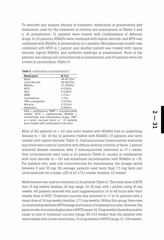
To describe and analyze efficacy of treatment, medication at presentation and medication used for the treatment of scleritis are summarized in Tables 5 and 6. At presentation, 31 patients were treated with combinations of different drugs. In 23 patients, NSAIDs were combined with topical steroids, and MTX was combined with NSAIDs at presentation in 6 patients. Mycophenolate mofetil was combined with MTX in 1 patient, and another patient was treated with topical steroids, topical NSAIDs, and antibiotic eyedrops at presentation. None of the patients was taking oral corticosteroids at presentation, and 49 patients were not treated at presentation (Table 5).
Most of the patients (n = 42) who were treated with NSAIDs had no underlying disease (n = 32). Of the 42 patients treated with NSAIDs, 33 patients also were treated with topical steroids (Table 5). Subconjunctival triamcinolone acetonide injections were used in 3 patients with diffuse anterior scleritis; of these, 1 patient achieved disease remission after 3 subconjunctival injections in 47.1 weeks. Oral corticosteroids were used in 64 patients (Table 6), usually in combination with local steroids (n = 52) and sometimes incombination with NSAIDs (n = 8). For patients who used oral corticosteroids for maintenance, the dosage varied between 5 and 20 mg. On average, patients used more than 7.5 mg daily oral corticosteroids for a mean ±SD of 65 ±72.4 weeks (median, 42 weeks).
Methotrexate was used as treatment in 36 patients (Table 6). The mean dose of MTX was 19 mg weekly (median, 20 mg; range, 10–30 mg), with 1 patient using 30 mg weekly. All patients received folic acid supplementation 24 to 48 hours after their weekly dose of MTX. Treatment success was achieved in 17 of 36 patients with a mean dose of 18 mg weekly (median, 17.5 mg weekly). Within this group, there was no relationship between MTX dosage and chance of treatment success. However, the patients who received a higher dose of MTX (mean, 26.3 mg weekly) showed a smaller range in time to treatment success (range, 80–215 weeks) than the patients who were treated with a lower dose (mean, 15.6 mg weekly) of MTX (range, 21–336 weeks).
Table 5. medication at presentation*
Medication N (%) None 49 (47.5%) Local steroids 46 (44.2%) NSAIDs 31 (29.8%) MTX 6 (5.8%) AZA 4 (3.8%) MMF 1 (1%) Cyclosporine 1 (%) TNF-α antagonist 3 (2.9%) Antiviral 2 (2.9%) Antibiotics 5 (4.8%) AZA = azathioprine; MMF = mycophenolate mofetil: MTX = methotrexate; NSAIDs = nonsteroidal anti-inflammatory drugs; TNF- α = tumor necrosis factor α. * 31 patients were treated with combinations (see text)
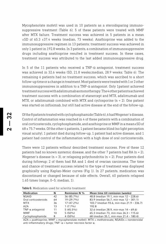
2 —
32
Mycophenolate mofetil was used in 10 patients as a steroidsparing immuno-suppressive treatment (Table 6); 5 of these patients were treated with MMF after MTX failure. Treatment success was achieved in 5 patients in a mean ±SD of 65.3 ±37.4 weeks (median, 73 weeks). Azathioprine was added to the immunosuppressive regimen in 13 patients; treatment success was achieved in only 1 patient in 192.8 weeks. In 3 patients, a combination of immunosuppressive drugs including azathioprine resulted in treatment success. In these cases, treatment success was attributed to the last added immunosuppressive drug.
In 5 of the 11 patients who received a TNF-α antagonist, treatment success was achieved in 32.6 weeks (SD, 21.8 weeks;median, 28.9 weeks; Table 6). The remaining 6 patients had no treatment success, which was ascribed to a short follow-up time or a change in treatment. Most patients were treated with 1 or 2 other immunosuppressives in addition to a TNF-α antagonist. Only 1patient achieved treatment success with adalimumab as monotherapy. The 4 other patients achieved treatment success with a combination of etanercept and MTX, adalimumab and MTX, or adalimumab combined with MTX and cyclosporine (n = 2). One patient was started on infliximab, but still had active disease at the end of the follow-up.
Of the 8 patients treated with cyclophosphamide (Table 6), 6 had Wegener’s disease. Control of inflammation was reached in 4 of these patients with a combination of oral corticosteroids, cyclophosphamide, and azathioprine or MMF in a mean±SD of 68 ± 75.7 weeks. Of the other 4 patients, 1 patient became blind (no light perception visual acuity), 1 patient died during follow-up, 1 patient had active disease, and 1 patient had control of the inflammation with a high dose of oral corticosteroids.
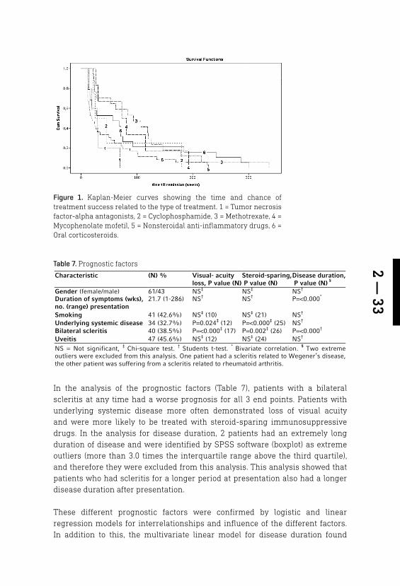
There were 12 patients without described treatment success. Five of these 12 patients had no known systemic disease, and the other 7 patients had RA (n = 2), Wegener’s disease (n = 3), or relapsing polychondritis (n = 2). Four patients died during followup; 2 of them had RA and 1 died of ovarian carcinoma. The time and chance of treatment success related to the type of treatment was displayed graphically using Kaplan-Meier curves (Fig 1). In 27 patients, medication was discontinued or changed because of side effects. Overall, 60 patients relapsed 1.65 times (range, 0–5; median, 1).
Medication N Remission N % Mean time till remission (weeks) NSAID 42 36 (85.7%) 48.8 (median 19.1, min-max 12 – 228.6) Oral corticosteroids 64 19 (29.7%) 83.9 (median 56.7, min-max 12 – 301.1) MTX 36 17 (47.2%) 103.7 (median 93.6, min-max 21.9 – 336.7) AZA 13 1 (7.7%) 192.8 TNF-α antagonist 11 5 (45.5%) 32.6 (median 28.9, min-max 14 – 69.6) MMF 10 5 (50%) 65.3 (median 73, min-max 26.3 – 115.6) Cyclophosphamide 8 4 (50%) 68 (median 35.1, min-max 21.4 – 180.4) AZA = azathioprine; MMF = mycophenolate mofetil: MTX = methotrexate; NSAIDs = nonsteroidal anti-inflammatory drugs; TNF- α = tumor necrosis factor α.
Table 6. Medication used for scleritis treatment
2 — 33
In the analysis of the prognostic factors (Table 7), patients with a bilateral scleritis at any time had a worse prognosis for all 3 end points. Patients with underlying systemic disease more often demonstrated loss of visual acuity and were more likely to be treated with steroid-sparing immunosuppressive drugs. In the analysis for disease duration, 2 patients had an extremely long duration of disease and were identified by SPSS software (boxplot) as extreme outliers (more than 3.0 times the interquartile range above the third quartile), and therefore they were excluded from this analysis. This analysis showed that patients who had scleritis for a longer period at presentation also had a longer disease duration after presentation.
These different prognostic factors were confirmed by logistic and linear regression models for interrelationships and influence of the different factors. In addition to this, the multivariate linear model for disease duration found
Figure 1. Kaplan-Meier curves showing the time and chance of treatment success related to the type of treatment. 1 = Tumor necrosis factor-alpha antagonists, 2 = Cyclophosphamide, 3 = Methotrexate, 4 = Mycophenolate mofetil, 5 = Nonsteroidal anti-inflammatory drugs, 6 = Oral corticosteroids.
Table 7. Prognostic factors
Characteristic (N) % Visual- acuity loss, P value (N)
Steroid-sparing, P value (N)
Disease duration, P value (N) §
Gender (female/male) 61/43 NS‡ NS‡ NS†
Duration of symptoms (wks), no. (range) presentation
21.7 (1-286) NS† NS† P=<0.000*
Smoking 41 (42.6%) NS‡ (10) NS‡ (21) NS†
Underlying systemic disease 34 (32.7%) P=0.024‡ (12) P=<0.000‡ (25) NS†
Bilateral scleritis 40 (38.5%) P=<0.000‡ (17) P=0.002‡ (26) P=<0.000†
Uveitis 47 (45.6%) NS‡ (12) NS‡ (24) NS†
NS = Not significant, ‡ Chi-square test. † Students t-test. * Bivariate correlation. § Two extreme outliers were excluded from this analysis. One patient had a scleritis related to Wegener’s disease, the other patient was suffering from a scleritis related to rheumatoid arthritis.
2 —
34
that men—although fewer—had a longer disease duration. To investigate the effect of a longer disease duration on the risk of loss of visual acuity, a separate multivariate inear model was used. In this model, a longer disease duration was not predictive of loss of visual acuity.
This analysis suggests that the strongest predictor for a worse prognosis is bilateral disease at any time. Patients with bilateral disease lost significantly more visual acuity, were treated significantly more often with steroid-sparing immunosuppressive drugs, and had a significantly longer disease duration.
DISCUSSION
Within this cohort, 23 patients (22.1 %) lost more than two lines of visual acuity on the Snellen-chart. This in contrast to the 81 patients (77.9 %) who gained visual acuity or lost less than 2 lines on the Snellen-chart. Most patients with a loss of visual acuity in our group (n=10) had a diffuse anterior scleritis. Necrotizing scleritis was the most threatening variant of scleritis. Of the 15 patients with necrotizing scleritis, 3 had a final visual acuity of no light perception and 2 lost more than 3 Snellen lines of visual acuity. In contrast to some other studies, 5, 16
panscleritis had a good prognosis and no higher association with an underlying disease. Although these findings regarding visual acuities should be interpreted cautiously in a retrospective study 17, 18, they are in concordance with the literature where loss of visual acuity is found in 15.9 to 37 % of the patients with scleritis 1, 16,19, 20.
Screening of scleritis patients for an underlying systemic disease should be aimed at the high-impact diseases such as RA and Wegener’s disease. The same holds true for infectious causes: They should be identified early on because they need a different therapeutic approach. In most of the current patients, systemic disease had already been diagnosed before scleritis onset. in few of them, this was newly identified by screening, and in even fewer patients systemic disease became manifest during follow-up. Screening for HLA-B27 positivity seems questionable, because an occurrence of HLA-B27 positivity equal to that in the normal population (8%) was found and because HLA-association with scleritis is rarely described in the literature 21 - 23. Also, the use of screening for sarcoidosis is not evident, since this is considered a rare cause of scleritis 5, 24, and in this study only one patient had elevated ACE-levels, without systemic manifestations of sarcoidosis. These findings suggest that customizing the screening for each patient seems an approach by which more useful clinical information can be obtained at lower costs.
2 — 35
In the scleritis patients, MTX was the most frequently used primary steroid-sparing immunosuppressive drug. The dosage of MTX did not influence the treatment success rate, but it had an effect on the range in time span to reach treatment success, with a lower range in de maximally treated group. The time to treatment success of MTX in our study was long, but it is comparable to that in other reports on scleritis 25. However, the time to success of MTX treatment in this study was considerably longer than the reported time to treatment success of MTX in uveitis eyes 26.
Reduction in time to treatment success of MTX could be attempted by introducing a quicker dose escalation scheme including a faster switch to subcutaneous administration. The latter will result in a better bioavailability of the drug, particularly in higher MTX doses 27 - 30.
In rheumatic disorders it is recommended to start with 10-15 mg weekly, with an escalation of 5 mg every 2-4 weeks up to 20-30 mg weekly, depending on clinical response and tolerance, whereas subcutaneous administration should be considered in case of an inadequate response or intolerance 29. Such schemes are currently being used in the treatment of RA-patients and have resulted in a reduction of the time to treatment success and a better steroid-sparing effect 27 -
29. It has been shown that subcutaneous administration of MTX is equally as well tolerated as oral administration 28.
Reducing time to reach the maximum MTX dose will probably lead to a reduction in time to treatment success in scleritis patients. Also, MTX failure will be sooner evident, so that therapy can be switched at an earlier point in time. A reduction in time to treatment success will probably also reduce ocular complications, which mainly are the result of active disease, steroid use, or a combination of both. Whether more patients can be successfully treated with monotherapy is an issue beyond the scope of this study and one that could be studied in a comparative study.
Mycophenolate mofetil seems a viable option after MTX-failure because it can induce treatment success in an additional 50 % of the patients. It is open to discussion whether MMF in selected patients is preferable as primary therapy based on underlying systemic disease or susceptibility to side effects. However, the availability of this option also depends on local healthcare policies.
In case both options fail, TNF-α antagonists can induce control of inflammation in a further half of the patients. In case of TNF-α antagonists, time to treatment success seems to be much shorter than that needed for MTX and MMF. This suggests a more effective mechanism of action compared to MTX and MMF. Whether TNF-α antagonists can be administered as monotherapy, cannot be concluded from this study because only 1 patient received monotherapy. With
2 —
36
regard to TNF-α antagonists, it is not known presently which drug is preferable in the treatment of scleritis, and long-term effectiveness needs to be established as well. Most reports are on infliximab, a humanized chimeric monoclonal antibody, which was the first TNF-α antagonist introduced 8, 10, 13, 14. Several small case-reports describe that rituximab, a genetically engineered chimeric monoclonal antibody that recognizes CD20 on mature B lymphocytes can be successful in severe recalcitrant forms of scleritis 9, 11, 12. This potential effectiveness is supported by 1 study 19 of a small number of eyes enucleated because of severe necrotizing auto-immune scleritis that showed CD20 positive cells along with plasma cells as major components of the inflammatory infiltrate. Finally, a case-report of a patient with nodular scleritis illustrates that adalimumab, a humanized monoclonal antibody against soluble and membrane-bound TNF-α, may be effective as well 13.
Within the present cohort, scleritis seems to be divided into 2 variants. A mild form which is responding well to NSAIDs and a more severe or recalcitrant variant that required other types of treatment. Low-dose corticosteroids as monotherapy seemed to be effective in only a minority of these patients. Because most patients in the severe group needed steroid-sparing immunosuppressive drugs, the threshold to start these should be low. Globally, for each steroid-sparing immunosuppressive drug, treatment success was achieved approximately half of the patients. Azathioprine seems to be an exception because this drug was much less effective in the present study.
Assessing the severity of scleritis at an early stage is important for an adequate choice of treatment regimen. Within this patient group necrotizing scleritis, male gender, a longer period of complaints at presentation, systemic disease, and bilateral disease at any time indicated a worse prognosis. By multivariate regression analysis, bilateral disease was the strongest predictor of worse prognosis. Patients with these characteristics at presentation had more loss of visual acuity, longer disease duration and were more often treated with steroid sparing immunosuppressive medication. Risk factors for visual loss or prolonged treatment in the literature include necrotizing or posterior scleritis 31, underlying systemic disease 15, corneal involvement 32, positive results for c-ANCA 20, a combination of anterior and posterior scleritis 16 and a posterior scleritis at an older age than 50 years 16. With regard to necrotizing scleritis and systemic disease, the present results were in agreement with dose reported in the literature. Most of these factors are easy to observ at presentation or during the course of the disease and contribute to an early recognition of a more severe form of scleritis. In contrast, the scleritis scoring system proposed by McCluskey and Wakefield 4 did not contribute to estimating the severity of scleritis in the current patients.

2 — 37
However, the results of the current study are limited by the fact that the study was retrospective, the numbers of patients were small in some subgroups, there was a large variability in follow up and the inclusion period was relatively long 17,
18. Also, the Snellen visual acuities were not obtained according to a standardized protocol and our study was conducted in 2 subspecialty clinics at university hospitals and therefore this population does not represent the total spectrum of scleritis. Regarding treatment; there is a bias towards personal experience and preferences of the ophthalmologists of the two university hospital centers and there is an unknown influence on treatment of the health insurance politics in the Netherlands. Despite this, the authors believe that they can make contributions and recommendations for the improvement of care for scleritis patients by sharing our treatment experiences, indicating prognostic factors and advising on steps to optimize treatment regimens.
In conclusion, these data shows that scleritis often is a severe, vision threatening, and chronic eye disease. Patients with a mild form of scleritis in most cases are treated adequately with oral NSAIDs. Patients with more severe or recalcitrant forms of scleritis can benefit from a more aggressive treatment strategy. Clinical features indicating a more severe form of scleritis make a fast recognition of the disease possible. Adequate treatment based on the severity of the scleritis and a timely switch to steroid-sparing immunosuppressive drugs can reduce total treatment time. Using an appropriate MTX dose-escalation scheme and an earlier switch to subcutaneous administration seems advisable in patients with non-infectious recalcitrant scleritis. Azathioprine should be avoided in patients with scleritis. After MTX failure, MMF is a good option as a secondary steroid-sparing immunosuppressive drug. Tumor necrosis factor-α antagonists may be a viable option for patients with noninfectious, recalcitrant scleritis who are not responding to MTX or MMF.
2 —
38
REFERENCES
1. Watson PG, Hazleman B, Pavésio C, Green RW. The Sclera and Systemic Dis-orders. Second edition ed. London:But-terworth-Heineman; 2004:64-7.
2. Watson PG, Hayreh SS. Scleritis and epis-cleritis. Br J Ophthalmol 1976;60:163-91.
3. Tuft SJ, Watson PG. Progression of scleral disease. Ophthalmology 1991;98:467-71.
4. McCluskey P, Wakefield D. Prediction of response to treatment in patients with scleritis using a standardised scoring sys-tem. Aust N Z J Ophthalmol 1991;19:211-5.
5. Okhravi N, Odufuwa B, McCluskey P, Lightman S. Scleritis. Surv Ophthalmol 2005;50:351-63.
6. Smith JR, Mackensen F, Rosenbaum JT. Therapy insight: scleritis and its relation-ship to systemic autoimmune disease. Nat Clin Pract Rheumatol 2007;3:219-26.
7. Cobo M. Inflammation of the sclera. Int Ophthalmol Clin 1983;23:159-71.
8. Murphy CC, Ayliffe WH, Booth A, et al. Tumor necrosis factor alpha blockade with infliximab for refractory uveitis and scleritis. Ophthalmology 2004;111:352-6.
9. Chauhan S, Kamal A, Thompson RN, et al. Rituximab for treatment of scleritis as-sociated with rheumatoid arthritis. Br J Ophthalmol 2009;93:984-5.
10. Doctor P, Sultan A, Syed S, et al. Infliximab for the treatment of refractory scleri-tis. Br J Ophthalmol 2010;94:579-83.
11. Iaccheri B, Androudi S, Bocci EB, et al. Rituximab treatment for persistent scleri-tis associated with rheumatoid arthritis. Ocul Immunol Inflamm 2010;18:223-5.
12. Kurz PA, Suhler EB, Choi D, Rosenbaum JT. Rituximab for treatment of ocular inflammatory disease: a series of four cases. Br J Ophthalmol 2009;93:546-8.
13. Restrepo JP, Molina MP. Successful treat-ment of severe nodular scleritis with adal-imumab. Clin Rheumatol 2010;29:559-61.
14. Sen HN, Sangave A, Hammel K, et al. Inflix-imab for the treatment of active scleritis. Can J Ophthalmol 2009;44:e9-e12. Avail-able at: http://download.journals.elsevi-erhealth.com/pdfs/journals/0008-4182/PIIS0008418209802263.pdf. Accessed January 23, 2012.
15. Werkgroep uveitis, Nederlands Oogheel-kundig Gezelschap. Richtlijn diagnostiek en behandeling van uveitis. 2007. Availa-ble at http://www.oogheelkunde.org/up-loads/fl/ve/flvem3mKxt8ThFFYVhn8GQ/Richtlijn-voor-diagnostiek-en-behande-ling-van-uveitis-15-mei-2007-1.pdf. Ac-cessed January 23, 2012.
16. McCluskey PJ, Watson PG, Lightman S, et al. Posterior scleritis: clinical features, systemic associations, and outcome in a large series of patients. Ophthalmology 1999;106:2380-6.
17. DiLoreto DA,Jr, Bressler NM, Bressler SB, Schachat AP. Use of best and fi-nal visual acuity outcomes in ophthal-mological research. Arch Ophthalmol 2003;11:1586-90.
18. Kempen JH. Appropriate use and re-porting of uncontrolled case series in the medical literature. Am J Ophthalmol 2011;1:7-10.
19. Usui Y, Parikh J, Goto H, Rao NA. Immu-nopathology of necrotising scleritis. Br J Ophthalmol 2008;92:417-9.
20. Akpek EK, Thorne JE, Qazi FA, et al. Evaluation of patients with scleritis for systemic disease. Ophthalmology 2004;111:501-6.
21. Joysey VC, Roger JH, Ashworth F, et al. Parallel studies of HLA antigens in patients with rheumatic heart disease and scleritis: comparisons with three control popula-tions. J Rheumatol 1977;3(Suppl):84-88.
22. Anshu A, Chee SP. Posterior scleritis and its association with HLA B27 haplotype. Ophthalmologica 2007;221:275-8.
23. van der Horst-Bruinsma IE, Lems WF, Dijkmans BA. A systematic compari-son of rheumatoid arthritis and anky-losing spondylitis. Clin Exp Rheumatol 2009;27(Suppl):43-9.
2 — 39
24. Pavesio CE, Meier FM. Systemic disorders associated with episcleritis and scleritis. Curr Opin Ophthalmol 2001;12:471-8.
25. Jachens A, Chu DS. Retrospective re-view of methotrexate therapy in the treatment of chronic, noninfectious, non-necrotizing scleritis. Am J Ophthalmol 2008;145:487-92.
26. Gangaputra S, Newcomb CW, Liese-gang TL, et al. Methotrexate for ocular inflammatory diseases. Ophthalmology 2009;116:2188-98.
27. Braun J. Optimal administration and dosage of methotrexate. Clin Exp Rheu-matol 2010;28(Suppl):46-51.
28. Braun J, Kastner P, Flaxenberg P, et al. Comparison of the clinical efficacy and safety of subcutaneous versus oral ad-ministration of methotrexate in patients with active rheumatoid arthritis: results of a six-month, multicenter, randomized, double-blind, controlled, phase IV trial. Arthritis Rheum 2008;58:73-81.
29. Visser K, Katchamart W, Loza E, et al. Multinational evidence-based recom-mendations for the use of methotrexate in rheumatic disorders with a focus on rheumatoid arthritis: integrating sys-tematic literature research and expert opinion of a broad international panel of rheumatologists in the 3E Initiative. Ann Rheum Dis 2009;68:1086-93.
30. van Roon EN, van de Laar MA. Methotrex-ate bioavailability. Clin Exp Rheumatol 2010;28(Suppl):27-32.
31. Jabs DA, Mudun A, Dunn JP, Marsh MJ. Episcleritis and scleritis: clinical fea-tures and treatment results. Am J Oph-thalmol 2000;130:469-76.
32. Carrasco MA, Cohen EJ, Rapuano CJ, Laibson PR. Therapeutic decision in anterior scleritis: our experience at a tertiary care eye center. J Fr Ophtalmol 2005;28:1065-9.

CLINICAL MANIFESTATIONS AND OUTCOME OF SYPHILITIC UVEITISAuthors:Jan G. Bollemeijer MD MSc1, 2 *, Wietse G. Wieringa BSc MPA3,4 *,Tom O.A.R. Missotten MD1, Ina Meenken MD PhD5, Ninette H. ten Dam-van Loon MD6, Aniki Rothova MD PhD6, 7, Leonoor I. Los MD PhD3, 4
1 Rotterdam Eye Hospital, Rotterdam, the Netherlands2 Department of Ophthalmology, Leiden University Medical Center, Leiden, the Netherlands3 Department of Ophthalmology, University Medical Center Groningen, Groningen, the Netherlands4 W.J. Kolff Institute, Graduate School of Medical Sciences, University of Groningen, Groningen, the Netherlands 5 Department of Ophthalmology, VU University Medical Center, Amsterdam, the Netherlands6 Department of Ophthalmology, University Medical Center Utrecht, Utrecht, the Netherlands7 Department of Ophthalmology, Erasmus University Medical Center, Rotterdam, the Netherlands
* These authors share first authorship
Invest Ophthalmol Vis Sci. 2016 Feb;57(2):404-11
3
3 —
42
ABSTRACT
Purpose: To analyze visual outcome, effectiveness of various modes of antibiotic treatment and prognostic factors in patients with serologically proven syphilitic uveitis.
Methods: The clinical records of 85 patients (139 eyes) diagnosed with syphilitic uveitis between 1984 and 2013 at tertiary centers in the Netherlands were retrospectively analyzed.
Results: Mean age was 47 years (range 27 – 73), 82.4% were male. HIV positivity was found in 28 (35.9 %) patients, 13 were newly diagnosed. Most patients had pan (45.9%) or posterior (31.8%) uveitis. On average, LogMAR VA improved significantly from 0.55 at the start of syphilis treatment to 0.34 at 1 month and to 0.27 at 6 months follow up. Most patients (86.7%) reached disease remission. No differences in efficacy between the various treatment regimens were found. A high LogMAR VA at the start of syphilis treatment and a treatment delay of more than 12 weeks were prognostic for a high LogMAR VA at six months follow up. Chronicity was not related to any form of treatment, HIV status or VDRL test outcome.
Conclusion: In this large cohort of 85 patients with syphilitic uveitis, visual outcomes were favorable in the majority of cases. Visual outcome was dependent on VA at the start of syphilis treatment and treatment delay.
3 — 43
INTRODUCTION
Syphilis is caused by an infection with the bacterium Treponema pallidum (T. pallidum) and classified as acquired or congenital. As with other spirochete infections, the clinical course of acquired untreated syphilis can be divided into four different stages depending on the clinical manifestations1. Syphilitic uveitis can occur in all stages except in the primary stage.
Different tests are available for the diagnosis and staging of syphilis. These include the so-called nonspecific tests like the VDRL (Venereal Disease Research Laboratory) and RPR (Rapid Plasma Reagin), which quantify the amount of serum anticardiolipin antibodies by flocculation and T. pallidum specific tests like the FTA-ABS (Fluorescent treponemal antibody absorption test), TPPA (Treponema pallidum particle agglutination) and TPHA (Treponema pallidum haemagglutination assay), which measure the amount of serum antibodies specifically directed against treponemal antigens1. As was shown by Grange et al2, newer PCR-based techniques have very low sensitivity to detect syphilis in blood, and thus cannot replace the above-mentioned serological tests.
The clinical presentation of ocular syphilis has been described in many publications. It has been dubbed “The Great Imitator” as it can mimic a wide range of ocular disorders. The most common presentation of ocular syphilis is uveitis. Before 1940, syphilis was the second cause of uveitis after tuberculosis. With the introduction of penicillin and improved diagnostics, syphilitic uveitis is a rare disease nowadays, accounting for 1 to 2% of all uveitis patients3. However, the outcomes of the different serologic tests for syphilis can be confusing, the optimal treatment of syphilitic uveitis is debatable and it is unknown which factors will determine visual prognosis.
To contribute to clarification of these aspects, we retrospectively evaluated visual outcomes in 85 patients with syphilitic uveitis. Specifically, factors that correlated with a worse visual prognosis or a chronic disease course were investigated.
PATIENTS AND METHODS
Patients with a confirmed diagnosis of syphilitic uveitis between 1984 and 2013 at the University Medical Centers of Leiden (n=12), Groningen (n=19), Utrecht (n=33), the VU University Medical Center Amsterdam (n=3) and the Rotterdam Eye Hospital (n=18) were included. The diagnosis of syphilitic uveitis was made in uveitis patients with positive results for specific anti T. pallidum serologic tests (i.e. a positive TPPA or TPHA and/or a positive FTA-ABS test) and agreement on the diagnosis syphilitic uveitis between ophthalmologist, dermatologist,
3 —
44
infectious disease specialist and neurologist. In the above centers, serologic testing for syphilis is part of the work-up in uveitis of unknown cause4. This work-up depends on the clinical presentation of the uveitis and may include blood tests (e.g. ESR, CRP, Hb, Ht, erythrocyte, thrombocyte and leukocyte counts, leukocyte differential, kreatinin, sodium, potassium, calcium, albumin, liver transaminases, angiotensin converting enzyme, auto-antibodies, tests for tuberculosis and chest X-rays. Additional tests may be ordered in special situations (e.g. anterior chamber fluid tests for infectious uveitis). (Uveitis Guidelines Dutch Opthalmic Society, 2007)4. However, since all these centers are tertiary referral centers, patients are often referred by ophthalmologists working in general practices. A systematic work-up for uveitis has not always been performed prior to referral. Medical records were retrospectively analyzed. The study was conducted in accordance with the Declaration of Helsinki and the study design was evaluated by the Medical Ethical Committee of the University Medical Center of Groningen who ruled that approval was not required for this study.
All participating centers collect data on uveitis patients in a database. However, the inclusion of patients started at different time points at the different centers. Therefore, the inclusion period varied per center (years are given between brackets): Leiden University Medical Center (1985 to 2008), University Medical Center Groningen (2001 to 2013), University Medical Center Utrecht (1991 to 2011), VU University Medical Center Amsterdam (2009 to 2012) and the Rotterdam Eye Hospital (1996 to 2010).
The following data were recorded on an anonymized standard entry form: sex, age at the start of syphilis treatment, race, affected eye(s), interval between the date of onset of uveitis symptoms and the date of final diagnosis of syphilitic uveitis and initiation of anti-syphilis treatment, laboratory data including HIV status and the results of various serologic tests for syphilis, the results of cerebrospinal fluid analyses, classification of the uveitis based upon standardization of uveitis nomenclature (SUN) criteria5, various clinical features, treatment modalities, visual acuity in logarithm of the minimal angle of resolution units (LogMAR) at the start of syphilis treatment, and at one and six months follow-up. The Snellen VA was converted to LogMAR VA for calculations. Visual acuity at one and six months was analyzed in relation to type of uveitis, treatment before syphilis treatment, interval between uveitis and syphilis treatment, administration route of syphilis treatment, HIV-status and immunosuppressive treatment during syphilis treatment.
Uveitis was classified as anterior, intermediate, posterior or panuveitis. The presence of cystoid macular edema (CME) was confirmed by fluorescein angiography (FA) or optical coherence tomography (OCT). Retinitis, retinal ischemia and papillitis were diagnosed by FA. Serous retinal detachments were
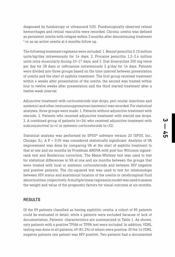
3 — 45
diagnosed by fundoscopy or ultrasound (US). Fundoscopically observed retinal hemorrhages and retinal vasculitis were recorded. Chronic uveitis was defined as persistent uveitis with relapse within 3 months after discontinuing treatment 5 or as an active uveitis at 6 months follow up.
The following treatment regimens were included: 1. Benzyl penicillin 0.15 million units/kg/day intravenously for 14 days, 2. Procaine penicillin 1.2–2.4 million units intra-muscularly during 10–17 days, and 3. Oral doxycycline 200 mg twice per day for 28 days or ceftriaxone intravenously 2 g/day for 14 days. Patients were divided into three groups based on the time interval between presentation of uveitis and the start of syphilis treatment. The first group received treatment within 4 weeks after presentation of the uveitis, the second was treated within four to twelve weeks after presentation and the third started treatment after a twelve week interval.
Adjunctive treatment with corticosteroids (eye drops, peri-ocular injections and systemic) and other immunosuppressives (systemic) was recorded. For statistical analyses, three groups were made. 1. Patients without adjunctive treatment with steroids. 2. Patients who received adjunctive treatment with steroid eye drops. 3. A combined group of patients (n=36) who received adjunctive treatment with subconjunctival (n=4) or systemic corticosteroids (n=32).
Statistical analysis was performed by SPSS® software version 20 (SPSS, Inc., Chicago, IL). A P < 0.05 was considered statistically significant. Analysis of VA improvement was done by comparing VA at the start of syphilis treatment to that at one and six months by Friedman ANOVA with post hoc Wilcoxon signed-rank test and Bonferroni correction. The Mann-Whitney test was used to test for statistical differences in VA at one and six months between the groups that were treated with local or systemic corticosteroids and between HIV negative and positive patients. The chi-squared test was used to test for relationships between HIV status and anatomical location of the uveitis or cerebrospinal fluid abnormalities, respectively. A multiple linear regression model was used to assess the weight and value of the prognostic factors for visual outcome at six months.
RESULTS
Of the 89 patients classified as having syphilitic uveitis, a cohort of 85 patients could be evaluated in detail, while 4 patients were excluded because of lack of documentation. Patients’ characteristics are summarized in Table 1. As shown, only patients with a positive TPHA or TPPA test were included. In addition, VDRL testing was done in all patients, 69 (81.2%) of whom were positive. Of the 16 VDRL negative patients one patient was HIV positive. Two patients had a documented
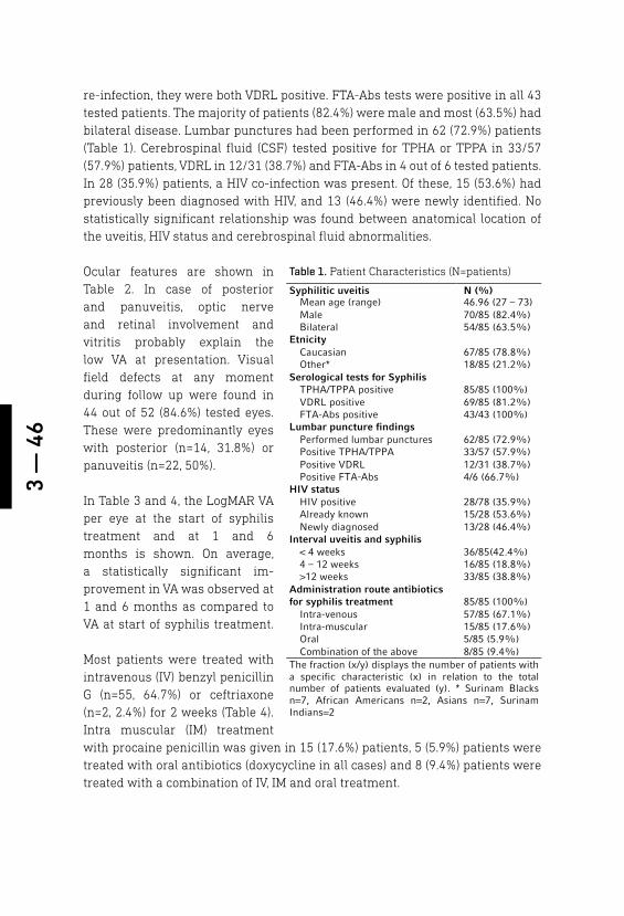
3 —
46
re-infection, they were both VDRL positive. FTA-Abs tests were positive in all 43 tested patients. The majority of patients (82.4%) were male and most (63.5%) had bilateral disease. Lumbar punctures had been performed in 62 (72.9%) patients (Table 1). Cerebrospinal fluid (CSF) tested positive for TPHA or TPPA in 33/57 (57.9%) patients, VDRL in 12/31 (38.7%) and FTA-Abs in 4 out of 6 tested patients. In 28 (35.9%) patients, a HIV co-infection was present. Of these, 15 (53.6%) had previously been diagnosed with HIV, and 13 (46.4%) were newly identified. No statistically significant relationship was found between anatomical location of the uveitis, HIV status and cerebrospinal fluid abnormalities.
Ocular features are shown in Table 2. In case of posterior and panuveitis, optic nerve and retinal involvement and vitritis probably explain the low VA at presentation. Visual field defects at any moment during follow up were found in 44 out of 52 (84.6%) tested eyes. These were predominantly eyes with posterior (n=14, 31.8%) or panuveitis (n=22, 50%).
In Table 3 and 4, the LogMAR VA per eye at the start of syphilis treatment and at 1 and 6 months is shown. On average, a statistically significant im-provement in VA was observed at 1 and 6 months as compared to VA at start of syphilis treatment.
Most patients were treated with intravenous (IV) benzyl penicillin G (n=55, 64.7%) or ceftriaxone (n=2, 2.4%) for 2 weeks (Table 4). Intra muscular (IM) treatment with procaine penicillin was given in 15 (17.6%) patients, 5 (5.9%) patients were treated with oral antibiotics (doxycycline in all cases) and 8 (9.4%) patients were treated with a combination of IV, IM and oral treatment.
Table 1. Patient Characteristics (N=patients)
Syphilitic uveitis N (%) Mean age (range) 46.96 (27 – 73) Male 70/85 (82.4%) Bilateral 54/85 (63.5%)
Etnicity Caucasian 67/85 (78.8%) Other* 18/85 (21.2%)
Serological tests for Syphilis TPHA/TPPA positive 85/85 (100%) VDRL positive 69/85 (81.2%) FTA-Abs positive 43/43 (100%)
Lumbar puncture findings Performed lumbar punctures 62/85 (72.9%) Positive TPHA/TPPA 33/57 (57.9%) Positive VDRL 12/31 (38.7%) Positive FTA-Abs 4/6 (66.7%)
HIV status HIV positive 28/78 (35.9%) Already known 15/28 (53.6%) Newly diagnosed 13/28 (46.4%)
Interval uveitis and syphilis treatment
< 4 weeks 36/85(42.4%) 4 – 12 weeks 16/85 (18.8%) >12 weeks 33/85 (38.8%)
Administration route antibiotics for syphilis treatment 85/85 (100%)
Intra-venous 57/85 (67.1%) Intra-muscular 15/85 (17.6%) Oral 5/85 (5.9%) Combination of the above 8/85 (9.4%)
The fraction (x/y) displays the number of patients with a specific characteristic (x) in relation to the total number of patients evaluated (y). * Surinam Blacks n=7, African Americans n=2, Asians n=7, Surinam Indians=2
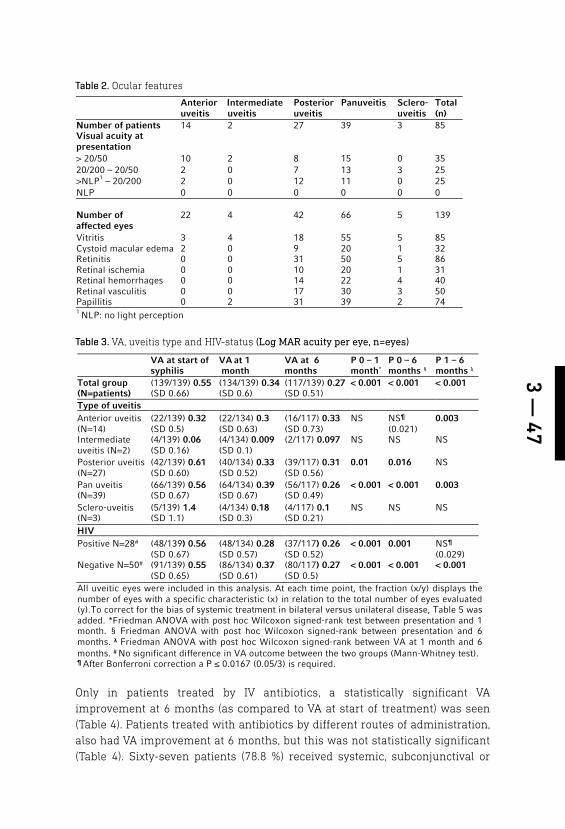
3 — 47
Table 2. Ocular features
Anterior uveitis
Intermediate uveitis
Posterior uveitis
Panuveitis Sclero-uveitis
Total (n)
Number of patients 14 2 27 39 3 85 Visual acuity at presentation
> 20/50 10 2 8 15 0 35 20/200 – 20/50 2 0 7 13 3 25 >NLP1 – 20/200 2 0 12 11 0 25 NLP 0 0 0 0 0 0 Number of affected eyes
22 4 42 66 5 139
Vitritis 3 4 18 55 5 85 Cystoid macular edema 2 0 9 20 1 32 Retinitis 0 0 31 50 5 86 Retinal ischemia 0 0 10 20 1 31 Retinal hemorrhages 0 0 14 22 4 40 Retinal vasculitis 0 0 17 30 3 50 Papillitis 0 2 31 39 2 74 1 NLP: no light perception
Table 3. VA, uveitis type and HIV-status (Log MAR acuity per eye, n=eyes)
VA at start of syphilis treatment
VA at 1 month
VA at 6 months
P 0 – 1 month*
P 0 – 6 months §
P 1 – 6 months λ
Total group (N=patients)
(139/139) 0.55 (SD 0.66)
(134/139) 0.34 (SD 0.6)
(117/139) 0.27 (SD 0.51)
< 0.001 < 0.001 < 0.001
Type of uveitis Anterior uveitis (N=14)
(22/139) 0.32 (SD 0.5)
(22/134) 0.3 (SD 0.63)
(16/117) 0.33 (SD 0.73)
NS
NS¶ (0.021)
0.003
Intermediate uveitis (N=2)
(4/139) 0.06 (SD 0.16)
(4/134) 0.009 (SD 0.1)
(2/117) 0.097 NS NS NS
Posterior uveitis (N=27)
(42/139) 0.61 (SD 0.60)
(40/134) 0.33 (SD 0.52)
(39/117) 0.31 (SD 0.56)
0.01 0.016 NS
Pan uveitis (N=39)
(66/139) 0.56 (SD 0.67)
(64/134) 0.39 (SD 0.67)
(56/117) 0.26 (SD 0.49)
< 0.001 < 0.001 0.003
Sclero-uveitis (N=3)
(5/139) 1.4 (SD 1.1)
(4/134) 0.18 (SD 0.3)
(4/117) 0.1 (SD 0.21)
NS NS NS
HIV Positive N=28#
(48/139) 0.56 (SD 0.67)
(48/134) 0.28 (SD 0.57)
(37/117) 0.26 (SD 0.52)
< 0.001
0.001
NS¶ (0.029)
Negative N=50# (91/139) 0.55 (SD 0.65)
(86/134) 0.37 (SD 0.61)
(80/117) 0.27 (SD 0.5)
< 0.001 < 0.001 < 0.001
All uveitic eyes were included in this analysis. At each time point, the fraction (x/y) displays the number of eyes with a specific characteristic (x) in relation to the total number of eyes evaluated (y).To correct for the bias of systemic treatment in bilateral versus unilateral disease, Table 5 was added. *Friedman ANOVA with post hoc Wilcoxon signed-rank test between presentation and 1 month. § Friedman ANOVA with post hoc Wilcoxon signed-rank between presentation and 6 months. λ Friedman ANOVA with post hoc Wilcoxon signed-rank between VA at 1 month and 6 months. # No significant difference in VA outcome between the two groups (Mann-Whitney test). ¶ After Bonferroni correction a P ≤ 0.0167 (0.05/3) is required.
Only in patients treated by IV antibiotics, a statistically significant VA improvement at 6 months (as compared to VA at start of treatment) was seen (Table 4). Patients treated with antibiotics by different routes of administration, also had VA improvement at 6 months, but this was not statistically significant (Table 4). Sixty-seven patients (78.8 %) received systemic, subconjunctival or
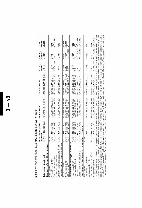
3 —
48
Tabl
e 4.
VA
an
d tr
eatm
ent (
Log
MA
R a
cuit
y pe
r ey
e, n
=eye
s)
VA
at
star
t of
syp
hili
s tr
eatm
ent
V
A at
1 m
onth
V
A a
t 6
mon
ths
P
0 –
1
mon
th*
P 0
– 6
m
onth
s §
P 1
– 6
m
onth
s λ
Tot
al g
roup
(N
=pat
ien
ts)
(139
/139
) 0.
55 (
SD
0.6
6)
(134
/139
) 0.
34 (
SD
0.6
)
(117
/139
) 0.
27 (
SD
0.5
1)
< 0.
001
< 0.
001
< 0.
001
Tre
atm
ent
befo
re s
yph
iliti
c tr
eatm
ent
Mis
sing
dat
a 11
/139
11
/134
9/
117
N
o tr
eatm
ent
(N=4
1)
(69/
128)
0.5
1 (S
D 0
.6)
(67/
123)
0. 2
9 (S
D 0
.52)
(6
2/10
8) 0
.29
(SD
0.6
1)
< 0.
001
0.00
2 0.
010
Cor
tico
ster
oids
and
oth
er
imm
unos
uppr
essi
ves
N=1
4 (2
4/12
8) 0
.51
(SD
0.6
4)
(22/
123)
0.4
2 (S
D 0
.78)
(2
0/10
8) 0
.28
(SD
0.4
7)
NS
¶ (
0.03
6)
0.01
1§ N
S¶ (
0.04
3)
Oth
er t
reat
men
t (n
ot a
dequ
ate)
N=2
3 (3
5/12
8) 0
.69
(SD
0.7
6)
(34/
123)
0.4
2 (S
D 0
.68)
(2
6/10
8) 0
.20
(SD
0.3
8)
0.0
06)
<0.0
01
0.00
2 In
terv
al u
veit
is a
nd
syph
ilis
trea
tmen
t <
4 w
eeks
N=3
6 (5
6/13
9) 0
.59
(SD
0.7
3)
(55/
134)
0.3
4 (S
D 0
.68)
(4
3/11
7) 0
.20
(SD
0.4
2)
< 0.
001
< 0.
001
< 0.
001
4 –
12 w
eeks
N=1
6 (2
9/13
9) 0
.45
(SD
0.5
8)
(25/
134)
0.2
2 (S
D 0
.39)
(
22/1
17)
0.09
(S
D 0
.22)
0.
012
< 0.
001
< 0.
001
>12
wee
ks N
=33
(54/
139)
0.5
7 (S
D 0
.63)
(5
4/13
4) 0
.39
(SD
0.6
)
(52/
117)
0.4
0 (S
D 0
.62)
0.
008
NS
¶ (
0.02
2)
NS
A
dmin
istr
atio
n r
oute
syp
hili
s tr
eatm
ent
Intr
a-ve
nous
N=5
7 (9
9/13
9) 0
.53
(SD
0.6
1)
(96/
134)
0.3
0 (S
D 0
.59)
(8
6/11
7) 0
.25
(SD
0.5
2)
< 0.
001
< 0.
001
0.00
2 In
tra-
mus
cula
r N
=15
(20/
139)
0.5
1 (S
D 0
.81)
(1
8/13
4) 0
.4 (
SD
0.6
3)
(16/
117)
0.3
8 (S
D 0
.64)
N
S
NS
N
S¶
O
ral N
=5
(7/1
39)
0.52
(S
D 0
.74)
(7
/134
) 0.
51 (
SD
0.7
4)
(7/1
17)
0.18
(S
D 0
.28)
N
S
NS
¶ (
0.04
3)
NS
¶ (
0.02
8)
Com
bina
tion
of
the
abov
e N
=8
(13/
139)
0.7
8 (S
D 0
.75)
(1
3/13
4)0.
49 (
SD
0.5
5)
(8/1
17)
0.43
(S
D 0
.68)
N
S
NS
¶ (
0.01
8)
NS
¶ (
0.04
6)
Imm
unos
uppr
essi
ve t
reat
men
t du
rin
g sy
phili
s tr
eatm
ent
Mis
sing
dat
a 3/
139
1/13
4 1/
117
O
ral o
r su
bcon
junc
tiva
l
cort
icos
tero
ids# N
=36
(63/
136)
0.6
5 (S
D0.
65)
(60/
133)
0.3
3 (S
D 0
.58)
(5
8/11
6) 0
.28
(SD
0.5
6)
< 0.
001
< 0.
001
0.00
3
Cor
tico
ster
oid
eye
drop
s# N
=31
(46/
136)
0.4
4 (S
D 0
.58)
(4
6/13
3) 0
.41
(SD
0.7
2)
(37/
116)
0.2
8 (S
D 0
.55)
N
S
0.0
01
0.00
8 N
o co
rtic
oste
roid
s N
=16
(27/
136)
0.4
8 (S
D0.
7)
(27/
133)
0.2
4 (S
D 0
.38)
(2
1/11
6) 0
.25
(SD
0.4
5)
NS
¶ (
0.03
7)
NS
¶ (
0.03
5)
NS
¶ (
0.06
2)
All
uvei
tic
eyes
wer
e in
clud
ed i
n th
is a
naly
sis.
At
each
tim
e po
int,
the
fra
ctio
n (x
/y)
disp
lays
the
num
ber
of e
yes
wit
h a
spec
ific
cha
ract
eris
tic
(x)
in r
elat
ion
to t
he
tota
l nu
mbe
r of
eye
s ev
alua
ted
(y).
To
corr
ect
for
the
bias
of
syst
emic
tre
atm
ent
in b
ilate
ral
vers
us u
nila
tera
l di
seas
e, T
able
5 w
as a
dded
. *F
ried
man
AN
OV
A w
ith
post
hoc
Wilc
oxon
sig
ned-
rank
tes
t be
twee
n pr
esen
tati
on a
nd 1
mon
th.
§ Fr
iedm
an A
NO
VA
wit
h po
st h
oc W
ilcox
on s
igne
d-ra
nk b
etw
een
pres
enta
tion
and
6
mon
ths.
λ Fri
edm
an A
NO
VA
wit
h po
st h
oc W
ilcox
on s
igne
d-ra
nk b
etw
een
VA
at
1 m
onth
and
6 m
onth
s. #
No
sign
ific
ant
diff
eren
ce i
n V
A o
utco
me
betw
een
the
two
grou
ps (
Man
n-W
hitn
ey t
est)
. ¶ A
fter
Bon
ferr
oni c
orre
ctio
n a
P ≤
0.0
167
(0.0
5/3)
is r
equi
red.
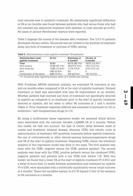
3 — 49
local steroids next to antibiotic treatment. No statistically significant difference in VA at six months was found between patients who had versus those who had not received any adjunctive treatment with systemic or local steroids (p=0.691). No cases of Jarisch-Herxheimer reaction were reported.
Table 5 displays the course of the disease after treatment. Ten (13.5 %) patients developed chronic uveitis. Chronicity was not related to the duration of treatment delay, any form of treatment or outcome of VDRL testing.
With Friedman ANOVA statistical analysis, we evaluated VA outcomes at one and six months when compared to VA at the start of syphilis treatment. Delayed treatment in itself was associated with less VA improvement at six months. Whether patients had received any form of treatment not specifically directed at syphilis as compared to no treatment prior to the start of specific treatment directed at syphilis, did not seem to affect VA outcomes at 1 and 6 months (Table 4). Prior treatment regimens differed and consisted of antivirals (n=10) or antibiotics / anti-toxoplasmosis drugs (n=13).
By using a multivariate linear regression model, we analyzed which factors were associated with the outcome variable LogMAR VA at 6 months. Within this model, we took into account: the type of uveitis, delay between onset of uveitis and treatment, bilateral disease, ethnicity, VDRL test results, route of administration of treatment, HIV-positivity, treatment before syphilis treatment, the use of corticosteroids or other steroid-sparing immunosuppressive drugs and VA at the start of syphilis treatment. Regarding VDRL status, the statistical analysis in this regression model was done in two ways. The first analysis was done with the VDRL negative versus the VDRL positive patients. The second analysis was done with the VDRL positive patients versus the combined VDRL negative patients and patients with a low VDRL-titer (below 1:8). With this model, we found that a lower VA at the start of syphilis treatment (P<0.001) and a delay of more than 12 weeks between presentation and treatment for syphilis (P=0.038), were associated with a statistically significantly worse visual outcome at 6 months. These two variables explain 34.6% (R Square 0.346) of the variance in VA outcomes at 6 months.
Table 5. Administration route syphilis treatment (N=patients)
Administration route syphilis treatment
N (%) Remission at 6 months#
Chronic at 6 months
Total 85/85 65/75 (86.7%) 10/75 (13.3%) Intra-venous 57/85 (67.1%) 44/57(77.2%) 7/57 (12.2 %) Intra-muscular 15/85 (17.6%) 11/15 (73.3) 2/15 (13.3 %) Oral 5/85 (5.9%) 5/5 0 Combination of the above 8/85 (9.4%) 5/8 (62.5%) 1/8 (12.5%) # In 10 patients data regarding disease status at 6 months was missing.

3 —
50
Tabl
e 7.
Ch
arac
teri
stic
s of
6 p
atie
nts
(10
eyes
) wit
h v
isu
al lo
ss a
t 6 m
onth
s.
Pat
ien
t N
o ey
es
Typ
e of
uve
itis
V
A a
t
pres
enta
tion
V
A a
t 1
m
onth
V
A a
t 6
m
onth
s H
IV
Cau
se o
f
visu
al lo
ss
Pre
viou
s
imm
unos
upre
ssiv
es
Tre
atm
ent
w
ith
in 4
wee
ks
Adm
inis
trat
ion
ro
ute
1 2
post
erio
r 0.
2 N
LP
NLP
-
Is
chem
ic o
ptic
ne
urop
athy
O
ral c
orti
cost
eroi
ds
No
IV
0.
2 N
LP
NLP
2 2
post
erio
r 1.
2 0.
8 0.
3 -
O
ptic
neu
ropa
thy
No
No
IV
FC
H
M
HM
S
ub r
etin
al f
ibro
sis
3 2
pan
0.8
HM
0.
1 +
R
etin
al d
etac
hmen
t O
ral c
orti
cost
eroi
ds
and
cycl
ospo
rine
N
o N
o IV
HM
H
M
0.1
glau
com
a
4 1
post
erio
r 1.
2 1.
2 1.
2 -
No
No
IV
0.
1 FC
FC
S
ub r
etin
al f
ibro
sis
5 1
scle
ro-u
veit
is
0.5
0.8
0.8
unkn
own
Opt
ic n
euro
path
y N
o Y
es
IV
H
M
NLP
N
LP
Sub
ret
inal
fib
rosi
s
6 2
ante
rior
0.
8 0.
25
0.25
-
No
No
IM
0.4
HM
H
M
hypo
pyon
T
en e
yes
of 6
pat
ient
s w
ith
bila
tera
l uve
itis
lost
VA
dur
ing
follo
w-u
p. (
The
dat
a of
the
tw
o ey
es t
hat
impr
oved
are
giv
en in
ital
ics)
. VA
= V
isua
l acu
ity
(Sne
llen)
, IV
= I
ntra
ven
ous,
IM
= I
ntra
mus
cula
r, F
C =
Fin
ger
coun
ting
, HM
= H
and
mot
ion,
NLP
= N
o lig
ht p
erce
ptio
n.
Tabl
e 6.
Bes
t eye
an
alys
is1
(Log
MA
R a
cuit
y pe
r ey
e)
Ocu
lar
syph
ilis
V
A a
t pr
esen
tati
on
VA
at
1 m
onth
V
A a
t 6
mon
ths
V
A d
iffe
ren
ce
1 an
d 6
mon
ths
‡ A
ll ey
es (
n=13
9)
(1
39/1
39)
0.55
(S
D 0
.66)
(13
4/13
9) 0
.34
(SD
0.6
) P
=0.0
01*
(117
/139
) 0.
27 (
SD
0.5
1) P
=<0.
001§
P
=<0.
001
Uni
late
ral e
yes
(n=3
1)
(31/
31)
0.71
(S
D 0
.77)
(3
1/31
) 0.
4 (S
D 0
.55)
P=0
.003
* (2
6/31
) 0.
25 (
SD
0.4
) P
=<0.
001§
P
=0.0
02
Bila
tera
l bes
t ey
e an
d un
ilate
ral
eyes
(n=
85)
(85/
85)
0.39
(S
D 0
.58)
(8
3/85
) 0.
22 (
SD
0.4
7) P
=<0.
001*
72
/85
0.15
(S
D 0
.33)
P=<
0.00
1§
P=<
0.00
1 T
o co
rrec
t fo
r th
e bi
as o
f sy
stem
ic t
hera
py i
n bi
late
ral
vers
us u
nila
tera
l di
seas
e, w
e pe
rfor
med
VA
ana
lyse
s at
the
var
ious
tim
e po
ints
for
all
eyes
, un
ilate
ral
eyes
onl
y, a
nd b
ilate
ral
best
eye
s pl
us u
nila
tera
l ey
es.
At
each
tim
e po
int,
the
fra
ctio
n (x
/y)
disp
lays
the
num
ber
of e
yes
wit
h a
spec
ific
ch
arac
teri
stic
(x)
in
rela
tion
to
the
tota
l nu
mbe
r of
eva
luat
ed e
yes
(y).
The
res
ults
of
all
anal
yses
sho
w a
sig
nifi
cant
im
prov
emen
t of
VA
bet
wee
n al
l-ti
me
poin
ts a
nd i
n al
l an
alyz
ed g
roup
s. *
Frie
dman
AN
OV
A w
ith
post
hoc
Wilc
oxon
sig
ned-
rank
tes
t be
twee
n pr
esen
tati
on a
nd 1
mon
th.
§ Fr
iedm
an
AN
OV
A w
ith
post
hoc
Wilc
oxon
sig
ned-
rank
bet
wee
n pr
esen
tati
on a
nd 6
mon
ths.
‡ F
ried
man
AN
OV
A w
ith
post
hoc
Wilc
oxon
sig
ned-
rank
bet
wee
n V
A a
t 1
mon
th a
nd 6
mon
ths.

3 — 51
To correct for the possible bias of systemic treatment in a mixed population of bilateral and unilateral disease we took bilateral disease into account in the regression model and we added Table 6. Bilateral disease did not influence the visual outcome at 6 months (P=0.216).
Table 7 shows the characteristics of the 6 patients (10 eyes) with visual loss at 6 months. Posterior uveitis was the predominant type of uveitis associated with visual loss. Optic neuropathy, subretinal fibrosis, retinal detachment and glaucoma were the main causes of severely reduced VA in these patients. In one patient with hand motion VA, the posterior segment could not be evaluated due to severe persistent anterior segment inflammation (hypopyon) (This patient refused intensive local treatment).
DISCUSSION
Within this cohort of 85 patients with serologically proven syphilitic uveitis , the overall visual prognosis was good if timely and adequate therapy was given. High LogMAR VA (low Snellen VA) outcomes were associated with a treatment delay of more than 12 weeks, and high LogMAR VA at the start of syphilis treatment. A statistically significant improvement of VA at 1 and 6 months as compared to that at the start of syphilis treatment, was observed in patients treated with IV antibiotics, and in those that received adjunct corticosteroids. Absolute values of LogMAR VA at 6 months were not associated with the route of administration of antibiotics, treatment with corticosteroids and HIV status. The majority of patients had one uveitis episode, but chronic uveitis developed in 13.5%. None of the evaluated factors was associated with a chronic course.
Overall, a good visual prognosis in our study is supported by the finding that the 117 eyes included in the per eye analysis showed a statistically significant improvement at one and six months follow up. Also, 16 of the 22 eyes (72.7%) with missing data at 6 months had a Snellen VA above 20/32 (LogMAR 0.2) at 1 month follow up. Therefore, we may assume an even more favorable prognosis than that presented in our tables. An overall good visual prognosis in syphilitic uveitis such as found in this large cohort, confirms previous studies 6-11. In our study, a higher LogMAR VA at the start of syphilis treatment and a delay of more than 12 weeks between the first presentation of uveitis and treatment for syphilis, were associated with a statistically significantly higher LogMAR VA at 6 months. Possible reasons for diagnostic delay include patients’ and doctors’ delay. It is hard to reduce the former, whereas the latter can be minimized by following the general advice to test for syphilis in patients with uveitis of unknown origin 1, 12, 13.

3 —
52
Treatment outcomeProbably, delayed treatment is associated with irreversible structural damage 1, 7, 14, 15. This is supported by the association between high LogMAR VA at the start of syphilis treatment and at 6 months. Worsening of VA during follow up occurred in ten eyes and was associated with structural damage to the optic disc and retina. In our study, the majority of patients were treated with intravenous penicillin (Table 4). Smaller numbers of patients were treated with intramuscular or oral antibiotics. These different treatment modalities were not prognostic for a higher LogMAR VA at 6 months.
Previous studies on prognostic factors in ocular syphilis 6, 16, 17 showed no difference in visual outcome when comparing the “classic” regimen of intravenous penicillin with other antibiotics. But, these studies 6, 16, 17 were all in small groups, with different kind of antibiotics and therefore results should be interpreted cautiously.
CorticosteroidsAnother finding in our study is that patients who received local, subconjunctival or systemic corticosteroids next to antibiotic treatment for syphilis had on average a statistically significant improvement in VA at 1 and 6 months, when compared to VA at the start of syphilis treatment. The absence of a statistically significant difference between patients treated with additional oral steroids or steroid injections versus those not treated in this way, seems to indicate that additional steroids may be ineffective. However, the fact that mean LogMAR VA at the start of syphilis treatment was higher in patients receiving additional oral steroids or steroid injections than in patients not receiving this (Table 4), indicates that adjunct corticosteroid treatment may have been preferably given to the more severe cases. Since LogMAR VA outcomes in these possibly more severe cases are similar to outcomes in the probably less severe groups (Table 4), a beneficial effect of adjunct corticosteroid treatment cannot be excluded. Some authors have reported on their clinical experience with corticosteroids in ocular syphilis 18, 19 but on a smaller scale and not at set time points. Previous studies advised local corticosteroids in case of interstitial keratitis or anterior uveitis 14,
20 and systemic corticosteroids in case of profound visual loss, posterior uveitis 21, scleritis, and optic neuritis 14, 20. Because of our results and recommendations in the literature, we suggest considering adding corticosteroids to antibiotic treatment in all cases of syphilitic uveitis. The use of oral corticosteroids is also considered beneficial in preventing a Jarisch–Herxheimer reaction 20, 22. The use of corticosteroids without antibiotic treatment, though not associated with worse VA outcomes at 6 months in our study, may aggravate syphilitic uveitis. Zamani and Garfinkel 23 published a case report on a patient who developed yellow placoid chorioretinal lesions during treatment with oral corticosteroids, which disappeared after their discontinuation.
3 — 53
HIVIn our study, HIV positivity was found in 28 patients, 13 of whom were newly diagnosed. This re-emphasizes the risk of co-infection with other sexually transmitted diseases in this patient group, and the desirability to test for HIV in case of ocular syphilis. Previously, HIV-positivity has been associated with a worse visual outcome in syphilitic uveitis, a finding we and other recent studies 9-11, 24,
25 could not confirm. Also, previous studies described that HIV-positive patients tended to have a higher proportion of posterior and panuveitis and neurosyphilis than HIV-negative patients. In contrast, we did not find statistically significant associations between HIV- positivity, anatomical location of the uveitis and CSF abnormalities. Differences between our outcomes and those in previous studies may be due to an improved immune-status of HIV-positive patients because of highly active antiretroviral therapy (HAART) 26, 27. In line with this, current IUSTI guidelines 1, 12, 13 state that HIV co-infected syphilitic patients should be treated as immunocompetent patients, except for those who have CD4+ cell counts of less than or equal to 350/μl.
Clinical presentation Our study confirms previous reports 6, 7, 16, 28, 29 that ocular syphilis occurs predominantly in men. Further, it confirms that syphilitic uveitis is a variable condition with a high diversity of clinical features. It can be uni- or bilateral, all anatomical locations may be affected, and it may run an acute or chronic course. In our study, bilateral uveitis was seen in 63.5%, whereas posterior (n=27, 31.8%)) and panuveitis (n=39, 45.9%) were far more often present than anterior uveitis (n=14, 16.5%). This is in line with recent papers 6, 7, 16, 24, but it differs from some older studies that observed uveitis to be located mainly anteriorly 30, 31.
VDRL test outcomeVDRL test results give some information on the duration and activity of the infection and they can be used to monitor the response to treatment. The interpretation of the VDRL test is sometimes difficult and debatable. A VDRL test becomes positive 4 to 5 weeks after infection, but it can sometimes be negative due to the prozone phenomenon1. Next to that, in 20 - 30% of the patients the test becomes negative over time 32. Therefore, a negative VDRL test result does not rule out the diagnosis of syphilitic uveitis10. A previous study reported that HIV positivity may be associated with higher than expected VDRL serologic titers, false-negative serologic results and delayed appearance of sero-reactivity 32. These findings were not confirmed within our study. Also, we did not observe an effect of VDRL test outcome on VA at 6 months in the multivariate linear regression model. Neither did we find a difference in chronicity at 6 months between VDRL-negative or positive patients.
3 —
54
Antibiotic TreatmentAccording to the European guidelines, the gold standard for the treatment of syphilitic uveitis is intravenous (IV) benzyl penicillin 12–24 million units daily, given in 3–4 million units doses every four hours for 10–21 days 1, 12, 13. In special situations (pregnancy, allergy or refusal of intravenous treatment), oral or intramuscular treatment can be considered. Improvement in VA at 1 and 6 months was seen in all groups of patients independent of the route of administration of antibiotics. Based upon the multiple linear regression model , the group of patients treated with IV penicillin, showed a tendency towards a somewhat better VA result. The absence of a statistically significant difference in this model may probably be explained by the modest sizes of the non IV treated groups. A similar argument may apply to the absence of an effect of route of administration on the development of chronic uveitis. At present, the results of our study support the current guidelines on treatment for ocular syphilis.
Strengths and limitations of the studyThe strengths of this study are its relatively large study population, the systematic way in which data were collected, and its adherence to the SUN classification system and guidelines for publications. The limitations of this study are its retrospective nature, and its long inclusion period. The latter may theoretically have influenced treatment strategies. However, the mainstay of syphilis treatment is penicillin, and this has not changed over the past decades. Some statistically significant relations may have been missed because of small numbers of patients in some subgroups. Also, the study was conducted in tertiary uveitis centers, and therefore, this population may not represent the total spectrum of syphilitic uveitis. Personal experience and preferences of ophthalmologists may have influenced their choice of treatment. Despite this, we feel that the study results can contribute to optimum care for patients with syphilitic uveitis.
CONCLUSION
Overall, VA outcomes in syphilitic uveitis are good. A low VA at the start of syphilis treatment and treatment delay of more than 12 weeks results in a less favorable visual prognosis. To shorten this delay, low threshold testing for syphilis should be done in uveitis of unknown cause. Intravenous benzyl penicillin is an effective treatment for syphilitic uveitis. It is not clear whether adjunct steroid treatment is beneficial. Structural damage to the optic nerve and retina are the main causes of permanent visual loss.

3 — 55
REFERENCES
1. Janier M, Hegyi V, Dupin N, et al. 2014 Eu-ropean guideline on the management of syphilis. J Eur Acad Dermatol Venereol 2014Nov.13;19(45):20957
2. Grange PA, Gressier L, Dion PL, et al. Evaluation of a PCR test for detection of treponema pallidum in swabs and blood. J Clin Microbiol 2012;50(3):546-52.
3. Schlaegel TF,Jr, O'Connor GR. Metastatic nonsuppurative uveitis. Int Ophthalmol Clin 1977;17(3):87-108.
4. Werkgroep Uveitis, Nederlands Oo-gheelkundig Gezelschap. Richtlijn diag-nostiek en behandeling van uveitis. 2007. Available at: http://www.oogheelkunde.org/uploads/fl/ve/flvem3mKxt8ThFFYVh-n8GQ/Richtlijn-voor-diagnostiek-en-be-handeling-van-uveitis-15-mei-2007-1.pdf. Accessed November 27, 2015.
5. Jabs DA, Nussenblatt RB, Rosenbaum JT, et al. Standardization of uveitis nomen-clature for reporting clinical data. results of the first international workshop. Am J Ophthalmol 2005;140(3):509-16.
6. Yang P, Zhang N, Li F, et al. Ocular mani-festations of syphilitic uveitis in Chinese patients. Retina 2012;32(9):1906-14.
7. Puech C, Gennai S, Pavese P, et al. Ocular manifestations of syphilis: Recent cases over a 2.5-year period. Graefes Arch Clin Exp Ophthalmol 2010;248(11):1623-9.
8. Jager S, Meier FM. Syphilis-associated uveitis in Switzerland. Klin Monbl Augen-heilkd 2011;228(4):330-3.
9. Mathew RG, Goh BT, Westcott MC. Brit-ish ocular syphilis study (BOSS): 2-year national surveillance study of intraoc-ular inflammation secondary to ocu-lar syphilis. Invest Ophthalmol Vis Sci 2014;55(8):5394-400.
10. Moradi A, Salek S, Daniel E, et al. Clinical features and incidence rates of ocular com-plications in patients with ocular syphilis. Am J Ophthalmol 2015;159(2):334,43.e1.
11. Northey LC, Skalicky SE, Gurbaxani A, et al. Syphilitic uveitis and optic neuritis in Sydney, Australia. Br J Ophthalmol 2015; 2015;99(9) 1215-9.
12. French P, Gomberg M, Janier M, et al. IUSTI: 2008 European guidelines on the management of syphilis. Int J STD AIDS 2009;20(5):300-9.
13. Pastuszczak M, Wojas-Pelc A. Current standards for diagnosis and treatment of syphilis: Selection of some practical is-sues, based on the European (IUSTI) and U.S. (CDC) guidelines. Postepy Dermatol Alergol 2013;30(4):203-10.
14. Kiss S, Damico FM, Young LH. Ocular manifestations and treatment of syphi-lis. Semin Ophthalmol 2005;20(3):161-7.
15. Villanueva AV, Sahouri MJ, Ormerod LD, et al. Posterior uveitis in patients with positive serology for syphilis. Clin Infect Dis 2000;30(3):479-85.
16. Balaskas K, Sergentanis TN, Giulieri S, et al. Analysis of significant factors influ-encing visual acuity in ocular syphilis. Br J Ophthalmol 2011;95(11):1568-72.
17. Thomas S, Wiselka M, Dhar J, et al. Syphilis presenting as acute multifocal retino-cho-roiditis. J R Soc Med 2006;99(7):371-2.
18. Chiquet C, Khayi H, Puech C, et al. Ocular syphilis. J Fr Ophtalmol 2014;37(4):329-36.
19. Solebo AL, Westcott M. Corticoster-oids in ocular syphilis. Ophthalmology 2007;114(8):1593.
20. Aldave AJ, King JA, Cunningham ET,Jr. Ocular syphilis. Curr Opin Ophthalmol 2001;12(6):433-41.
21. Doris JP, Saha K, Jones NP, et al. Ocular syphilis: The new epidemic. Eye (Lond) 2006;20(6):703-5.
22. Danesh-Meyer H, Kubis KC, Sergott RC. Not so slowly progressive visual loss. Surv Ophthalmol 1999;44(3):247-52.
23. Zamani M, Garfinkel RA. Corticoster-oid-induced modulation of acute syphi-litic posterior placoid chorioretinitis. Am J Ophthalmol 2003;135(6):891-4.
3 —
56
24. Amaratunge BC, Camuglia JE, Hall AJ. Syphilitic uveitis: A review of clinical manifestations and treatment outcomes of syphilitic uveitis in human immuno-deficiency virus-positive and negative patients. Clin Experiment Ophthalmol 2010;38(1):68-74.
25. Tucker JD, Li JZ, Robbins GK, et al. Ocular syphilis among HIV-infected patients: A systematic analysis of the literature. Sex Transm Infect 2011;87(1):4-8.
26. Shalaby IA, Dunn JP, Semba RD, et al. Syphilitic uveitis in human immunodefi-ciency virus-infected patients. Arch Oph-thalmol 1997;115(4):469-73.
27. Balba GP, Kumar PN, James AN, et al. Ocu-lar syphilis in HIV-positive patients receiv-ing highly active antiretroviral therapy. Am J Med 2006;119(5):448.e21,448.e25.
28. Browning DJ. Posterior segment man-ifestations of active ocular syphilis, their response to a neurosyphilis regi-men of penicillin therapy, and the influ-ence of human immunodeficiency virus status on response. Ophthalmology 2000;107(11):2015-23.
29. Wickremasinghe S, Ling C, Stawell R, et al. Syphilitic punctate inner retinitis in immunocompetent gay men. Ophthal-mology 2009;116(6):1195-200.
30. Barile GR, Flynn TE. Syphilis exposure in patients with uveitis. Ophthalmology 1997;104(10):1605-9.
31. Tamesis RR, Foster CS. Ocular syphilis. Ophthalmology 1990;97(10):1281-7.
32. Nayak S, Acharjya B. VDRL test and its interpretation. Indian J Dermatol 2012;57(1):3-8.
3 — 57

RETINAL DYSTROPHY IN 6 YOUNG PATIENTS WHO PRESENTED WITH INTERMEDIATE UVEITISAuthors:Y.M. Hettinga, M.D.1,2, M.M. van Genderen2, W. Wieringa3, J. Ossewaarde-van Norel1, J.H. de Boer1
1 Department of Ophthalmology, University Medical Center Utrecht, Heidelberglaan 100, 3584 CX Utrecht, The Netherlands2 Diagnostic Center Bartiméus Institute for the Visually Impaired, Van Renesselaan 30a, Zeist, The Netherlands3 Department of Ophthalmology, University Medical Center Groningen, Hanzeplein 1, 9700 RB Groningen, The Netherlands
Ophthalmology. 2016 Sep;123(9):2043-6.
4

4 —
60
4 — 61
Inherited retinal dystrophies (RD) comprise a clinically and genetically heterogeneous group of inherited diseases characterized by progressive rod and cone dysfunction and degeneration. Intermediate uveitis (IU) is a clinical diagnosis characterized by bilateral intraocular inflammation located primarily in the vitreous and pars plana. IU is the second most common uveitis entity among children. Most young patients with IU do not present with any apparent underlying disease.1 IU is frequently complicated by cystoid macular edema (CME), which may also occur in RD.
We describe six patients initially diagnosed with IU who visited the outpatient uveitis clinics of three University Medical Centers at Utrecht, Groningen and Amsterdam between 2006 and 2015. During the course of the disease, these patients were diagnosed with RD.
At presentation, age of the patients ranged from 5 to 22 years. Five patients had a negative family history for consanguinity or retinal or immunological disease and for one patient (patient 3, Table 1) it was unknown. They all had subnormal vision (range: 20/25 – 20/50 Snellen equivalent) and one patient complained of nyctalopia. Slit-lamp examination revealed minimal or no anterior segment inflammation and 1+ to 3+ cells with mild to moderate haze in the vitreous. Funduscopy showed CME in all patients and several white peripheral lesions in one patient and some pigment deposits in another patient. None of the six patients had waxy disc pallor or the typically pigment changes seen in RD.
Visual field (VF) testing was performed in all patients at different time points due to referral delay by general ophthalmologists. VF of all patients showed various degrees of relative (mid-) peripheral defects (n= 3) or ring scotoma (n=3) 1 to 7 years after onset of complaints. Optical coherence tomography (OCT) showed CME on OCT in all patients with foveal thickness ranging from 206 µm (but cysts present) to 651 µm (Table 1). All patients showed some attenuation of the outer nuclear layer of the central retina, which can be a sign of RD but also of macular atrophy in longstanding inflammatory CME (Figure 1). Fluorescein angiography (FA) was done in four patients showing optic disc hyperfluorescence in two of them (Figure 1; patient 5 and 6); and capillary leakage also in two patients (Figure 1; patient 2 and 6). One patient showed a remarkable discrepancy between severe bilateral macular edema on OCT and absence of leakage in the macula on FA. In contrast, another patient showed both severe bilateral macular edema on OCT and severe leakage on FA (Figure 1; patient 1 and 2). In two patients, a full field electroretinogram (ffERG) was performed shortly after the onset of symptoms to rule out a RD. In these cases amplitudes were scotopic and photopic just at or below threshold and latency times were normal in one patient and prolonged in another patient (patient 1 and 5, Table 1). These subnormal ffERG scores were considered to be secondary to uveitis activity as previously has been described.2

4 —
62
Figure 1. Fundus photographs (left column), optical coherence tomography (middle column), and fluorescein angiography (FA, right column) in six patients with retinal dystrophy; these six patients were initially diagnosed with intermediate uveitis. The patient numbers correspond to the patient numbers shown in Table 1. FA images were not available for patients 3 and 4.
Based on a combination of clinical characteristics and additional diagnostics uveitis specialists diagnosed IU. The diagnosis was subsequently classified as idiopathic IU following an extensive diagnostic workup by a (pediatric) rheumatologist/immunologist.

4 — 63
All patients received periocular and/or intraocular corticosteroids with temporarily improvement of CME in 3 patients but CME recurred in 4 to 8 weeks.
Four patients were treated with acetazolamide for at least 3 months but CME did not respond to this treatment. Multiple immunomodulating therapies were initiated in five patients, and biologicals were used in four of them. However, prolonged and combined systemic immunosuppressive therapies failed to reduce CME or inflammation permanently in all patients and did not improve visual acuity.
During follow-up, which ranged from 1 to 8 years, CME of varying degrees persisted and visual acuity deteriorated in all patients (Table 1). Three patients started complaining about nyctalopia. Four patients showed progression of VF defects. Full field electroretinogram recordings were obtained according to the standards of the International Society for Clinical Electrophysiology of Vision (ISCEV) (Table 1). All patients had severely abnormal scotopic and photopic ffERG responses (Figure 2). In the two patients that had an ffERG shortly after the onset of symptoms, the second ffERG, at 4 and 6 years follow up, showed that responses had deteriorated significantly. The dark adapted threshold proved to be elevated by >2 log in four patients tested. In all patients the results of psychophysical (VF and dark adaptation) testing and ffERG findings led to a diagnosis of RD. Patient genomic DNA was isolated from peripheral blood samples using standard procedures. The DNA was tested for the presence of mutations using Micro-Array Analysis and all sequences were confirmed using DNA sequence analysis confirming the diagnosis RD. Three patients proved to have a mutation in CRB1 ( one identical novel mutation in two patients with potential pathogenicity), one in RP1, one in USH2A, and one in two dominant RD genes (Table 1). The last patient had lost contact with her family, so possible dominant inheritance could not be verified. However, in the past, her mother used to complain about nyctalopia.
Here, we report on six young patients with genetically proven RD who initially presented with refractory IU with vitreous inflammation and severe CME. In RD, the vitreous may show cells or debris resembling inflammatory conditions such as uveitis.3 Macular abnormalities resembling CME and retinal deposits have been described in RD, especially in CRB1 retinal dystrophy.4,5 Therefore, in young patients who present with indolent mild intermediate uveitis without pars planitis but accompanied by CME on OCT, an early stage isolated case of RD should be included in the differential diagnosis. In these patients, we recommend direct questioning of nyctalopia and family history for retinal diseases, appropriate VF testing with assessment of the mid and far periphery, measurement of ffERG, and, if necessary, consultation of an ophthalmologist with expertise in the field of retinal dystrophies. An early diagnosis of retinal dystrophy may result in more adequate treatment with avoidance of high dose immunosuppression, which may have significant side effects.
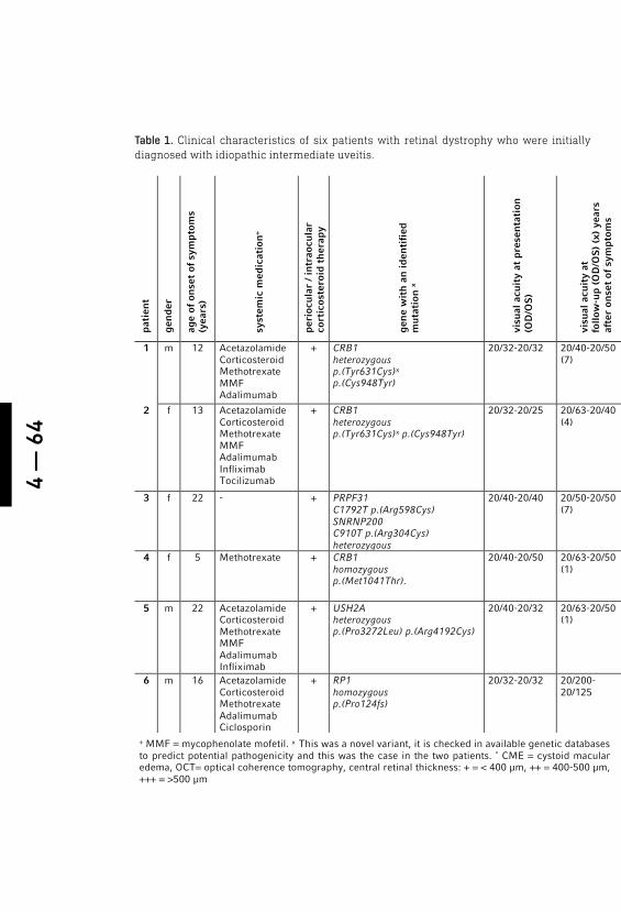
4 —
64
Table 1. Clinical characteristics of six patients with retinal dystrophy who were initially diagnosed with idiopathic intermediate uveitis.
pati
ent
gen
der
age
of o
nse
t of
sym
ptom
s (y
ears
)
syst
emic
med
icat
ion
+
peri
ocul
ar /
intr
aocu
lar
cort
icos
tero
id t
her
apy
gen
e w
ith
an
iden
tifi
ed
mut
atio
n x
visu
al a
cuit
y at
pre
sen
tati
on
(OD
/OS
)
visu
al a
cuit
y at
fo
llow
-up
(OD
/OS
) (x
) ye
ars
afte
r on
set
of s
ympt
oms
CM
E o
n O
CT
(pe
rfor
med
aft
er
(x)
year
s)
Fir
st v
isua
l fie
ld (
perf
orm
ed
afte
r x
year
s)
Ele
ctro
reti
nog
ram
(pe
rfor
med
af
ter
(x)
year
s)
follo
w-u
p ye
ars
befo
re
diag
nos
is r
etin
al d
ystr
oph
y w
as m
ade
1 m 12 Acetazolamide Corticosteroid Methotrexate MMF Adalimumab
+ CRB1 heterozygous p.(Tyr631Cys)x p.(Cys948Tyr)
20/32-20/32
20/40-20/50 (7)
+++ (1)
Peritest Some mid-peripheral defects. (1)
scotopic; reduced and delayed photopic; reduced and delayed (7)
7
2 f 13 Acetazolamide Corticosteroid Methotrexate MMF Adalimumab Infliximab Tocilizumab
+ CRB1 heterozygous p.(Tyr631Cys)x p.(Cys948Tyr)
20/32-20/25 20/63-20/40 (4)
+++ (1)
Humphrey 24-2 Some peripheral defects (3)
scotopic; reduced and delayed photopic; reduced and delayed (4)
4
3 f 22 - + PRPF31 C1792T p.(Arg598Cys) SNRNP200 C910T p.(Arg304Cys) heterozygous
20/40-20/40 20/50-20/50 (7)
+ (7)
Goldmann Severe relatively concentric limited (7)
scotopic; absent photopic; reduced and delayed (8)
8
4 f 5 Methotrexate
+ CRB1 homozygous p.(Met1041Thr).
20/40-20/50 20/63-20/50 (1)
++ (1)
Goldmann Severe relatively concentric limited (1)
scotopic; absent photopic; only at the highest intensity a very small response (1)
1
5 m 22 Acetazolamide Corticosteroid Methotrexate MMF Adalimumab Infliximab
+ USH2A heterozygous p.(Pro3272Leu) p.(Arg4192Cys)
20/40-20/32 20/63-20/50 (1)
++ (1)
Humphrey 30-2 Severe relatively concentric limited (1)
scotopic; just below the lower limit of normal photopic; just below the lower limit of normal and prolonged latency (7)
7
6 m 16 Acetazolamide Corticosteroid Methotrexate Adalimumab Ciclosporin
+ RP1 homozygous p.(Pro124fs)
20/32-20/32 20/200-20/125
+ (1.5)
Humphrey 24-2 Mild peripheral defects (3)
scotopic; absent photopic; reduced and delayed (5)
5
+ MMF = mycophenolate mofetil. x This was a novel variant, it is checked in available genetic databases to predict potential pathogenicity and this was the case in the two patients. * CME = cystoid macular edema, OCT= optical coherence tomography, central retinal thickness: + = < 400 µm, ++ = 400-500 µm, +++ = >500 µm
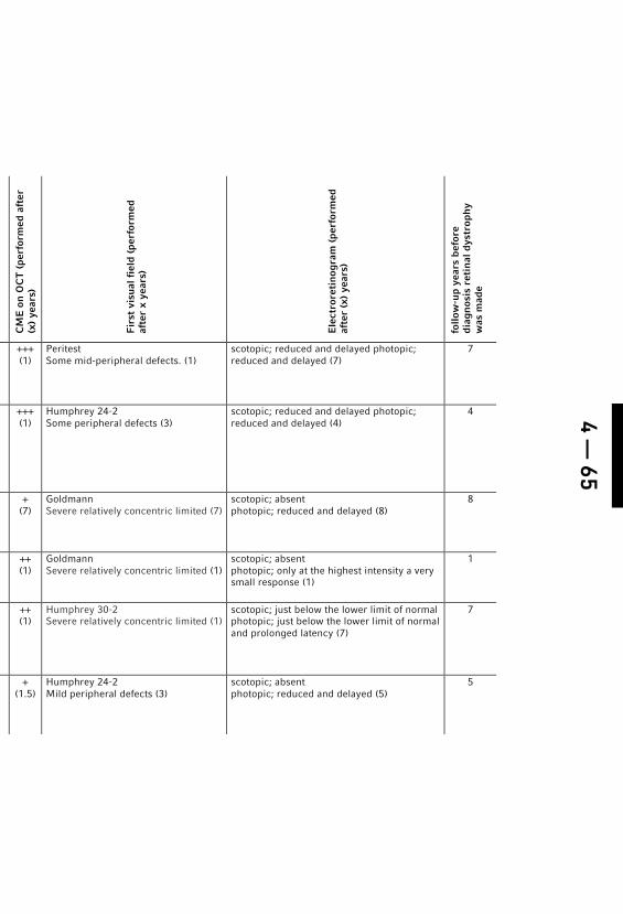
4 — 65
pati
ent
gen
der
age
of o
nse
t of
sym
ptom
s (y
ears
)
syst
emic
med
icat
ion
+
peri
ocul
ar /
intr
aocu
lar
cort
icos
tero
id t
her
apy
gen
e w
ith
an
iden
tifi
ed
mut
atio
n x
visu
al a
cuit
y at
pre
sen
tati
on
(OD
/OS
)
visu
al a
cuit
y at
fo
llow
-up
(OD
/OS
) (x
) ye
ars
afte
r on
set
of s
ympt
oms
CM
E o
n O
CT
(pe
rfor
med
aft
er
(x)
year
s)
Fir
st v
isua
l fie
ld (
perf
orm
ed
afte
r x
year
s)
Ele
ctro
reti
nog
ram
(pe
rfor
med
af
ter
(x)
year
s)
follo
w-u
p ye
ars
befo
re
diag
nos
is r
etin
al d
ystr
oph
y w
as m
ade
1 m 12 Acetazolamide Corticosteroid Methotrexate MMF Adalimumab
+ CRB1 heterozygous p.(Tyr631Cys)x p.(Cys948Tyr)
20/32-20/32
20/40-20/50 (7)
+++ (1)
Peritest Some mid-peripheral defects. (1)
scotopic; reduced and delayed photopic; reduced and delayed (7)
7
2 f 13 Acetazolamide Corticosteroid Methotrexate MMF Adalimumab Infliximab Tocilizumab
+ CRB1 heterozygous p.(Tyr631Cys)x p.(Cys948Tyr)
20/32-20/25 20/63-20/40 (4)
+++ (1)
Humphrey 24-2 Some peripheral defects (3)
scotopic; reduced and delayed photopic; reduced and delayed (4)
4
3 f 22 - + PRPF31 C1792T p.(Arg598Cys) SNRNP200 C910T p.(Arg304Cys) heterozygous
20/40-20/40 20/50-20/50 (7)
+ (7)
Goldmann Severe relatively concentric limited (7)
scotopic; absent photopic; reduced and delayed (8)
8
4 f 5 Methotrexate
+ CRB1 homozygous p.(Met1041Thr).
20/40-20/50 20/63-20/50 (1)
++ (1)
Goldmann Severe relatively concentric limited (1)
scotopic; absent photopic; only at the highest intensity a very small response (1)
1
5 m 22 Acetazolamide Corticosteroid Methotrexate MMF Adalimumab Infliximab
+ USH2A heterozygous p.(Pro3272Leu) p.(Arg4192Cys)
20/40-20/32 20/63-20/50 (1)
++ (1)
Humphrey 30-2 Severe relatively concentric limited (1)
scotopic; just below the lower limit of normal photopic; just below the lower limit of normal and prolonged latency (7)
7
6 m 16 Acetazolamide Corticosteroid Methotrexate Adalimumab Ciclosporin
+ RP1 homozygous p.(Pro124fs)
20/32-20/32 20/200-20/125
+ (1.5)
Humphrey 24-2 Mild peripheral defects (3)
scotopic; absent photopic; reduced and delayed (5)
5
+ MMF = mycophenolate mofetil. x This was a novel variant, it is checked in available genetic databases to predict potential pathogenicity and this was the case in the two patients. * CME = cystoid macular edema, OCT= optical coherence tomography, central retinal thickness: + = < 400 µm, ++ = 400-500 µm, +++ = >500 µm
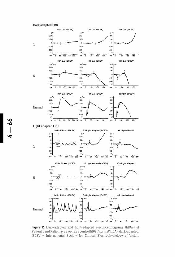
4 —
66
Figure 2. Dark-adapted and light-adapted electroretinograms (ERGs) of Patient 1 and Patient 6, as well as a control ERG (“normal”). DA = dark-adapted. ISCEV = International Society for Clinical Electrophysiology of Vision.
Dark adapted ERG
Normal
Normal
6
6
1
1
Light adapted ERG

4 — 67
REFERENCES
1. Boer JD, Berendschot TTJM, Does P, Ro-thova A. Long-term follow-up of inter-mediate uveitis in children. Am J Oph-thalmol. 2006;141(4):116-21.
2. Moschos MM, Gouliopoulos NS, Kalo-geropoulos C. Electrophysiological ex-amination in uveitis: A review of the liter-ature. Clin Ophthalmol. 2014;8:199-214.
3. Stunkel M, Bhattarai S, Kemerley A, et al. Vitritis in pediatric genetic retinal disor-ders. Ophthalmology. 2015;122(1):192-19.
4. Den Hollander AI, Heckenlively JR, van den Born LI, et al. Leber congenital amaurosis and retinitis pigmentosa with Coats-like exudative vasculopathy are associated with mutations in the crumbs homologue 1 (CRB1) gene. Am J Hum Genet. 2001;69(1):198-203.
5. Wolfson Y, Applegate CD, Strauss RW, Han IC, Scholl HP. CRB1-Related Maculopathy With Cystoid Macular Edema. JAMA Oph-thalmol. 2015 Nov 1;133(11):1357-60.

EFFICACY OF HIGH DOSE METHOTREXATE IN PEDIATRIC NON-INFECTIOUS UVEITISAuthors:Wietse G. Wieringa, BSc, MPA1, Wineke Armbrust, MD, PhD2, G. Elizabeth Legger, MD2, Leonoor I. Los, MD, PhD1,3
Affiliations of authors: University Medical Center Groningen, University of Groningen, Department of Ophthalmology1 and -Beatrix children’s hospital department of children’s rheumatology and immunology2, P. O. Box 30001, 9700 RB Groningen, the Netherlands.W.J. Kolff Institute3, Graduate School of Medical Sciences, University of Groningen, the Netherlands
Ocul Immunol Inflamm. 2018 Oct 22:1-9. [Epub ahead of print]
5
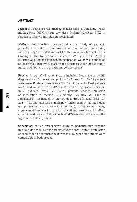
5 —
70
ABSTRACT
Purpose: To analyze the efficacy of high dose (≥ 15mg/m2/week) methotrexate (MTX) versus low dose (<15mg/m2/week) MTX in relation to time to remission on medication.
Methods: Retrospective observational cohort study of pediatric patients with auto-immune uveitis with or without underlying systemic disease treated with MTX at the University Medical Center Groningen (the Netherlands) between 1990 and 2014. Primary outcome was time to remission on medication, which was defined as an observable inactive disease in the affected eye for longer than 3 months without the use of systemic corticosteroids.
Results: A total of 42 patients were included. Mean age at uveitis diagnosis was 6.5 years (range 1.7 – 14.4), and 22 (52.4%) patients were male. Bilateral disease was found in 33 patients. Most patients (n=25) had anterior uveitis. JIA was the underlying systemic disease in 21 patients. Overall, 28 (66.7%) patients reached remission on medication in (median) 22.5 months (IQR 10.4- 45). Time to remission on medication in the low dose group (median 35.2, IQR 20.5 – 72.1 months) was significantly longer than in the high dose group (median 16.6, IQR 7.8 – 22.5 months) (p= 0.01). No statistically significant differences in ocular complications, steroid-sparing effect, cumulative dosage and side effects of MTX were found between the high and low dose groups.
Conclusion: In this retrospective study on pediatric auto-immune uveitis, high dose MTX was associated with a shorter time to remission on medication as compared to low dose MTX, while side effects were comparable in both groups.

5 — 71
INTRODUCTION
Uveitis is an inflammatory disorder of the eye, involving the uveal tract. It is classified as anterior, intermediate, posterior or panuveitis, depending on the part of the eye affected by the inflammatory process. Uveitis can be associated with a systemic auto-immune disease, can be caused by an infection and it can occur as an isolated ocular condition. In the developed countries, 87 – 89 % of the pediatric uveitis cases are non-infectious and the majority (41.5%) are related to juvenile idiopathic arthritis (JIA)1.
Pediatric uveitis is a potentially blinding disorder and accounts for 3.2 –15.2% of all cases of legal blindness in the affected eye(s) in the United States2,3. Many children with uveitis do not report any symptoms 4. This may lead to a delay in diagnosis and treatment, resulting in complications such as band keratopathy, posterior synechiae, cataract, glaucoma and amblyopia, which give a guarded prognosis 5. Early detection and aggressive treatment of uveitis can prevent visual loss and ocular complications 6.
The first line of treatment are local corticosteroids. If these are insufficient, local injections with corticosteroids can be considered. Systemic prednisone is started in case of severe uveitis or in case of failure of the local therapy. In case of chronic uveitis, steroid sparing immunosuppressive therapy may be indicated. Because of its long track record and good safety profile, methotrexate (MTX) is the steroid sparing immunosuppressive agent of first choice in almost all cases of non-infectious pediatric uveitis 7.
MTX is an efficacious drug, since remission on medication is reached in about 70% of pediatric non-infectious uveitis cases 8. However, MTX also has side-effects such as gastro-intestinal discomfort (nausea and vomiting), which are frequently reported, and the less common hepatic toxicity and bone marrow suppression 9,10. Also, anticipatory nausea and needle phobia in case of subcutaneous administration of MTX are common.
In pediatric uveitis patients, evidence regarding optimal dosage of MTX is scarce 8,11. Frequently used medication regimens start with low dose MTX, with increasing doses at 2-6 monthly intervals in case of insufficient effectiveness. In the treatment of JIA there is evidence on the effectiveness of higher starting doses and faster dose-escalation schemes 11-13 . Therefore, it would be relevant to evaluate whether such schemes would also be more efficient in the treatment of pediatric uveitis.
Optimizing the treatment of pediatric uveitis patients would be relevant because vision threatening complications are directly related to uveitis activity 6,14.

5 —
72
Shortening the time to remission on medication will probably reduce or postpone long-term ocular complications and may improve visual prognosis. Also, it seems likely that a higher steroid sparing effect will be achieved with less side effects and complications of systemic corticosteroids. The present retrospective study aims primarily to evaluate the effectiveness of high dose (≥ 15mg/m2/week) and that of low dose MTX (<15mg/m2/week) in relation to time to remission on medication. Secondarily, the steroid sparing effect, cumulative dosage of MTX, side effects of MTX treatment, ocular complications and visual acuity are evaluated.
PATIENTS AND METHODS
We performed a retrospective observational cohort study on pediatric patients treated with methotrexate (MTX) for uveitis between 1990 and 2014 at the University Medical Center of Groningen, The Netherlands. This study reflects the daily practice in a tertiary center. The Medical Ethical Committee of the University Medical Center of Groningen ruled that approval was not required for this study. Patients were identified from the digital uveitis database of the University Medical Center of Groningen. All patients who were younger than 18 years of age at the start of their uveitis and who were treated with MTX for longer than 6 months were included. MTX treatment was classified as high (≥ 15mg/m2/week, maximum of 25mg/week/sc) or low (<15mg/m2/week) dose, based on the MTX dose given before remission on medication was reached or medication was switched. Based on this classification, patients were divided into a high (≥ 15mg/m2/week) or low (<15mg/m2/week) -dose group. Before 2007, most patients received low dose MTX, and hereafter most patients were treated with high dose MTX. This reflects evolving treatment strategies. Data collection was done from the pediatric and ophthalmological medical records. The diagnosis of uveitis was made by an ophthalmologist specialized in uveitis and dedicated to this patient group. During the follow up period two other ophthalmologists were occasionally involved in the ophthalmological care for these patients. Classification of uveitis was done according to the Standardization of Uveitis Nomenclature (SUN) criteria 15 and was based on the available information in the ophthalmological medical record. Children were evaluated for the presence of an underlying systemic disease by a pediatric rheumatologist. When JIA was diagnosed, it was classified according to the ILAR (International League of the Association for Rheumatology) criteria 16.
General descriptivesFor each patient the following descriptives were recorded: age, gender, ethnicity, date of first diagnosis of uveitis (further referred to as: uveitis onset), type of uveitis, laterality, date of onset of arthritis, diagnosis and
5 — 73
subtype of underlying systemic disease, weight and length (at several time points during follow-up), anti-nuclear antibody (ANA) serologic status, HLA B27 status and ophthalmological findings at presentation. Prognostic signs for a worse outcome (young age, male gender, severity of uveitis at presentation, vitreous involvement and oligo arthritis) were recorded 5.
Uveitis diagnosis and classificationUveitis was diagnosed when cells could be observed in the anterior chamber or in the vitreous. Activity of anterior chamber (AC) inflammation (cells) evaluated by standard slit-lamp examination was recorded according to the recommendations of the SUN working group 15. Cells in vitreous humor were scored as being present or not. The diagnosis of posterior and panuveitis was made by fundoscopy and in some cases fluorescein angiography (FA) was performed.
TreatmentMTX dosage and route of administration at the start was recorded as mg/m2/week/orally or subcutaneously. Indications (uveitis, arthritis or both) and date for MTX dosage changes were documented. The MTX dosage was related to body surface area (BSA) at the moments of dosage change. Body surface area was calculated by the Mosteller formula [BSA (m2) = ( Height(cm) x Weight(kg) / 3600 )½] 17. Measurements of length and weight were performed at the start of the treatment and during follow up. Length and weight values were plotted routinely in the growth curves corrected for age, sex and race. When a value was missing the growth line was plotted between the 2 existing values. Cumulative dosages of MTX were calculated by multiplying the time (weeks) to remission on medication by the dose in mg/m2 of MTX. Route of administration (oral or subcutaneous) and – in case the route of administration was switched - the indications for switch were noted. Side effects and indications to stop MTX were recorded. Initially, liver enzyme testing is done after four weeks, and thereafter every 3 months. In case of elevated liver enzymes, testing is more frequently performed. The steroid sparing effect of MTX was evaluated by calculating/counting the number of weeks in which patients were treated with oral corticosteroids in a dosage of more than 0.5 mg/kg/day.
In case of cataract or glaucoma surgery (Baerveldt glaucoma implant), patients were given intravenous corticosteroids during surgery followed by a tapering dosage of oral corticosteroids in the period thereafter. In case of glaucoma surgery MTX was stopped for 2 months prior to surgery and re-introduced after the Baerveldt implant was functional. During this period, patients were treated with oral corticosteroids.
5 —
74
Remission on medication Remission on medication was defined as an observable inactive disease in the affected eye for longer than 3 months without the use of systemic corticosteroids or local steroid injections (subtenon or subconjunctival). During this period local steroid medication such as eye drops or ointment were allowed in a maintenance dosage of less than 4 drops per eye daily. With this treatment regimen, sufficient compliance-adherence was expected and it was regarded as being compatible with daily activities 18. Patients were advised to use the eye drops during mealtimes and – when necessary- before sleeping. A relapse was defined as a recurrence of the uveitis after a quiet episode described in the patient file. The total follow up time, time to remission on medication, time between dose adjustments and time to cataract and glaucoma surgery were documented.
Visual acuityThe decimal equivalent of the Snellen visual acuity (VA) of the affected eyes was recorded at presentation, six, twelve and twenty-four months and at last follow-up. Snellen VA was converted to logarithm of the minimum angle of resolution units (LogMAR) VA for calculations.
Ocular complicationsThe following ocular complications were scored per eye; band keratopathy, posterior synechiae, cataract and amblyopia. Ocular hypertension was defined as an intra-ocular pressure above 21 mmHg without treatment 15. Glaucoma was defined as glaucomatous changes to the optic nerve or visual field 15. Surgery for medically uncontrollable intra-ocular pressure was separately scored.
StatisticsStatistical analysis was performed by SPSS® software version 22 (SPSS, Inc., Chicago, IL) .A P < 0.05 was considered statistically significant. Descriptive statistics were used to present mean and standard deviation (SD) in normally distributed data or median and range if data were abnormally distributed. For the differences between the nominal data in the high and low dose groups we used the Chi-square test. In case of non-normally distributed linked samples, the Wilcoxon test for paired samples and the Mann-Whitney U test for independent samples were used. Analysis of VA at presentation compared to that at six, twelve and twenty-four months and at the end of follow up was done by the independent samples T-test. A Kaplan-Meier survival analysis with a log rank test was used to analyze survival curves and to compare the two treatment groups. Finally, a multiple regression model was used to assess the weight and influence of treatment groups, age, gender, underlying disease and anatomic location of the uveitis on the time to remission on medication.
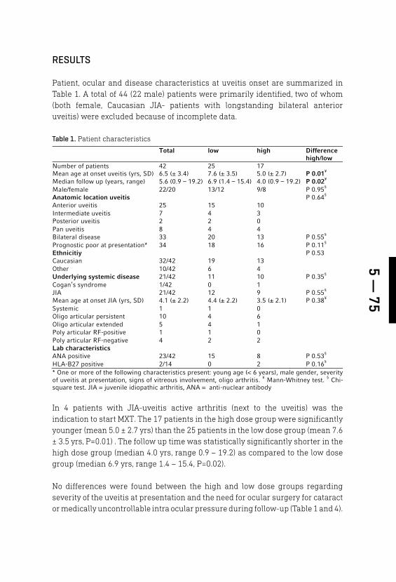
5 — 75
RESULTS
Patient, ocular and disease characteristics at uveitis onset are summarized in Table 1. A total of 44 (22 male) patients were primarily identified, two of whom (both female, Caucasian JIA- patients with longstanding bilateral anterior uveitis) were excluded because of incomplete data.
In 4 patients with JIA-uveitis active arthritis (next to the uveitis) was the indication to start MXT. The 17 patients in the high dose group were significantly younger (mean 5.0 ± 2.7 yrs) than the 25 patients in the low dose group (mean 7.6 ± 3.5 yrs, P=0.01) . The follow up time was statistically significantly shorter in the high dose group (median 4.0 yrs, range 0.9 – 19.2) as compared to the low dose group (median 6.9 yrs, range 1.4 – 15.4, P=0.02).
No differences were found between the high and low dose groups regarding severity of the uveitis at presentation and the need for ocular surgery for cataract or medically uncontrollable intra ocular pressure during follow-up (Table 1 and 4).
Table 1. Patient characteristics
Total low high Difference high/low
Number of patients 42 25 17 Mean age at onset uveitis (yrs, SD) 6.5 (± 3.4) 7.6 (± 3.5) 5.0 (± 2.7) P 0.01¥ Median follow up (years, range) 5.6 (0.9 – 19.2) 6.9 (1.4 – 15.4) 4.0 (0.9 – 19.2) P 0.02¥ Male/female 22/20 13/12 9/8 P 0.95§ Anatomic location uveitis P 0.64§ Anterior uveitis 25 15 10 Intermediate uveitis 7 4 3 Posterior uveitis 2 2 0 Pan uveitis 8 4 4 Bilateral disease 33 20 13 P 0.55§ Prognostic poor at presentation* 34 18 16 P 0.11§ Ethnicitiy P 0.53 Caucasian 32/42 19 13 Other 10/42 6 4 Underlying systemic disease 21/42 11 10 P 0.35§ Cogan’s syndrome 1/42 0 1 JIA 21/42 12 9 P 0.55§ Mean age at onset JIA (yrs, SD) 4.1 (± 2.2) 4.4 (± 2.2) 3.5 (± 2.1) P 0.38¥ Systemic 1 1 0 Oligo articular persistent 10 4 6 Oligo articular extended 5 4 1 Poly articular RF-positive 1 1 0 Poly articular RF-negative 4 2 2 Lab characteristics ANA positive 23/42 15 8 P 0.53§ HLA-B27 positive 2/14 0 2 P 0.16§ * One or more of the following characteristics present: young age (< 6 years), male gender, severity of uveitis at presentation, signs of vitreous involvement, oligo arthritis. ¥ Mann-Whitney test. § Chi-square test. JIA = juvenile idiopathic arthritis, ANA = anti-nuclear antibody
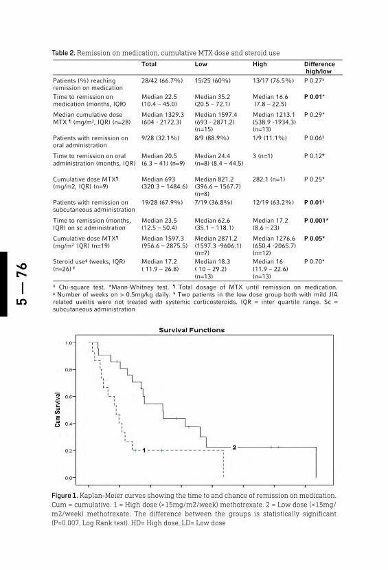
5 —
76
Figure 1. Kaplan-Meier curves showing the time to and chance of remission on medication. Cum = cumulative. 1 = High dose (>15mg/m2/week) methotrexate. 2 = Low dose (<15mg/m2/week) methotrexate. The difference between the groups is statistically significant (P=0.007, Log Rank test). HD= High dose, LD= Low dose
Table 2. Remission on medication, cumulative MTX dose and steroid use
Total Low High Difference high/low
Patients (%) reaching remission on medication
28/42 (66.7%)
15/25 (60%)
13/17 (76.5%)
P 0.27§
Time to remission on medication (months, IQR)
Median 22.5 (10.4 – 45.0)
Median 35.2 (20.5 – 72.1)
Median 16.6 (7.8 – 22.5)
P 0.01*
Median cumulative dose MTX ¶ (mg/m2, IQR) (n=28)
Median 1329.3 (604 - 2172.3)
Median 1597.4 (693 - 2871.2) (n=15)
Median 1213.1 (538.9 -1934.3) (n=13)
P 0.29*
Patients with remission on oral administration
9/28 (32.1%) 8/9 (88.9%) 1/9 (11.1%) P 0.06§
Time to remission on oral administration (months, IQR)
Median 20.5 (6.3 – 41) (n=9)
Median 24.4 (n=8) (8.4 – 44.5)
3 (n=1)
P 0.12*
Cumulative dose MTX¶ (mg/m2, IQR) (n=9)
Median 693 (320.3 – 1484.6)
Median 821.2 (396.6 – 1567.7) (n=8)
282.1 (n=1) P 0.25*
Patients with remission on subcutaneous administration
19/28 (67.9%) 7/19 (36.8%) 12/19 (63.2%) P 0.01§
Time to remission (months, IQR) on sc administration
Median 23.5 (12.5 – 50.4)
Median 62.6 (35.1 – 118.1)
Median 17.2 (8.6 – 23)
P 0.001*
Cumulative dose MTX¶ (mg/m2 IQR) (n=19)
Median 1597.3 (956.6 – 2875.5)
Median 2871.2 (1597.3 -9606.1) (n=7)
Median 1276.6 (650.4 -2065.7) (n=12)
P 0.05*
Steroid use‡ (weeks, IQR) (n=26) ¥
Median 17.2 ( 11.9 – 26.8)
Median 18.3 ( 10 – 29.2) (n=13)
Median 16 (11.9 – 22.6) (n=13)
P 0.70*
§ Chi-square test. *Mann-Whitney test. ¶ Total dosage of MTX until remission on medication. ‡ Number of weeks on > 0.5mg/kg daily. ¥ Two patients in the low dose group both with mild JIA related uveitis were not treated with systemic corticosteroids. IQR = inter quartile range. Sc = subcutaneous administration
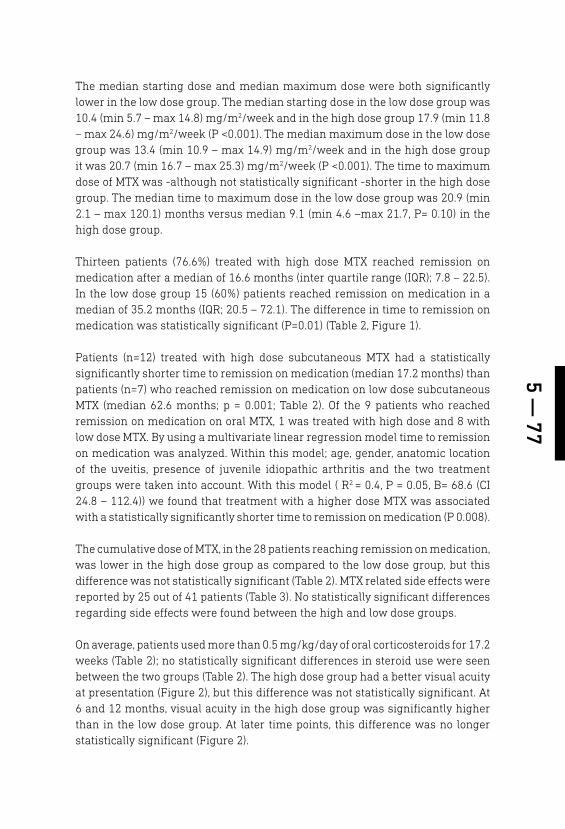
5 — 77
The median starting dose and median maximum dose were both significantly lower in the low dose group. The median starting dose in the low dose group was 10.4 (min 5.7 – max 14.8) mg/m2/week and in the high dose group 17.9 (min 11.8 – max 24.6) mg/m2/week (P <0.001). The median maximum dose in the low dose group was 13.4 (min 10.9 – max 14.9) mg/m2/week and in the high dose group it was 20.7 (min 16.7 – max 25.3) mg/m2/week (P <0.001). The time to maximum dose of MTX was -although not statistically significant -shorter in the high dose group. The median time to maximum dose in the low dose group was 20.9 (min 2.1 – max 120.1) months versus median 9.1 (min 4.6 –max 21.7, P= 0.10) in the high dose group.
Thirteen patients (76.6%) treated with high dose MTX reached remission on medication after a median of 16.6 months (inter quartile range (IQR); 7.8 – 22.5). In the low dose group 15 (60%) patients reached remission on medication in a median of 35.2 months (IQR; 20.5 – 72.1). The difference in time to remission on medication was statistically significant (P=0.01) (Table 2, Figure 1).
Patients (n=12) treated with high dose subcutaneous MTX had a statistically significantly shorter time to remission on medication (median 17.2 months) than patients (n=7) who reached remission on medication on low dose subcutaneous MTX (median 62.6 months; p = 0.001; Table 2). Of the 9 patients who reached remission on medication on oral MTX, 1 was treated with high dose and 8 with low dose MTX. By using a multivariate linear regression model time to remission on medication was analyzed. Within this model; age, gender, anatomic location of the uveitis, presence of juvenile idiopathic arthritis and the two treatment groups were taken into account. With this model ( R2 = 0.4, P = 0.05, B= 68.6 (CI 24.8 – 112.4)) we found that treatment with a higher dose MTX was associated with a statistically significantly shorter time to remission on medication (P 0.008).
The cumulative dose of MTX, in the 28 patients reaching remission on medication, was lower in the high dose group as compared to the low dose group, but this difference was not statistically significant (Table 2). MTX related side effects were reported by 25 out of 41 patients (Table 3). No statistically significant differences regarding side effects were found between the high and low dose groups.
On average, patients used more than 0.5 mg/kg/day of oral corticosteroids for 17.2 weeks (Table 2); no statistically significant differences in steroid use were seen between the two groups (Table 2). The high dose group had a better visual acuity at presentation (Figure 2), but this difference was not statistically significant. At 6 and 12 months, visual acuity in the high dose group was significantly higher than in the low dose group. At later time points, this difference was no longer statistically significant (Figure 2).
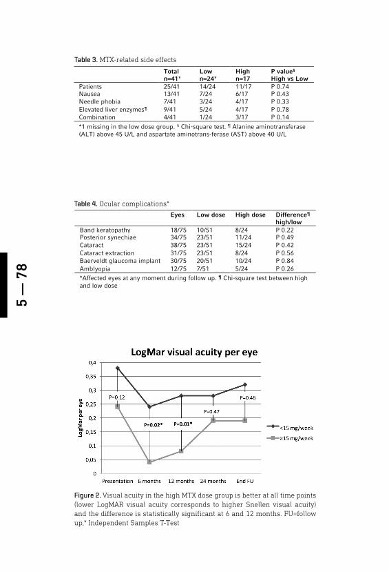
5 —
78
Table 4. Ocular complications*
Eyes
Low dose
High dose
Difference¶
high/low Band keratopathy 18/75 10/51 8/24 P 0.22 Posterior synechiae 34/75 23/51 11/24 P 0.49 Cataract 38/75 23/51 15/24 P 0.42 Cataract extraction 31/75 23/51 8/24 P 0.56 Baerveldt glaucoma implant 30/75 20/51 10/24 P 0.84 Amblyopia 12/75 7/51 5/24 P 0.26 *Affected eyes at any moment during follow up. ¶ Chi-square test between high and low dose
Figure 2. Visual acuity in the high MTX dose group is better at all time points (lower LogMAR visual acuity corresponds to higher Snellen visual acuity) and the difference is statistically significant at 6 and 12 months. FU=follow up,* Independent Samples T-Test
Table 3. MTX-related side effects
Total n=41*
Low n=24*
High n=17
P value§ High vs Low
Patients 25/41 14/24 11/17 P 0.74 Nausea 13/41 7/24 6/17 P 0.43 Needle phobia 7/41 3/24 4/17 P 0.33 Elevated liver enzymes¶ 9/41 5/24 4/17 P 0.78 Combination 4/41 1/24 3/17 P 0.14
*1 missing in the low dose group. § Chi-square test. ¶ Alanine aminotransferase (ALT) above 45 U/L and aspartate aminotrans-ferase (AST) above 40 U/L
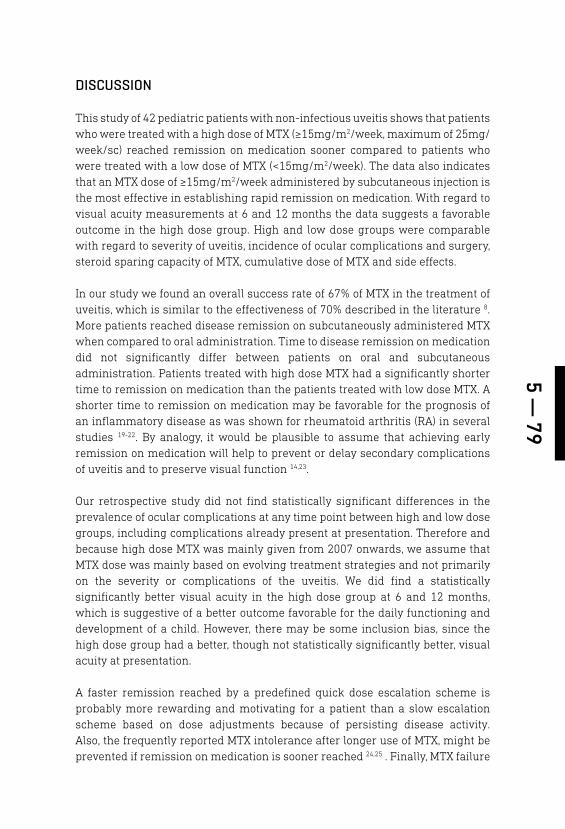
5 — 79
DISCUSSION
This study of 42 pediatric patients with non-infectious uveitis shows that patients who were treated with a high dose of MTX (≥15mg/m2/week, maximum of 25mg/week/sc) reached remission on medication sooner compared to patients who were treated with a low dose of MTX (<15mg/m2/week). The data also indicates that an MTX dose of ≥15mg/m2/week administered by subcutaneous injection is the most effective in establishing rapid remission on medication. With regard to visual acuity measurements at 6 and 12 months the data suggests a favorable outcome in the high dose group. High and low dose groups were comparable with regard to severity of uveitis, incidence of ocular complications and surgery, steroid sparing capacity of MTX, cumulative dose of MTX and side effects.
In our study we found an overall success rate of 67% of MTX in the treatment of uveitis, which is similar to the effectiveness of 70% described in the literature 8. More patients reached disease remission on subcutaneously administered MTX when compared to oral administration. Time to disease remission on medication did not significantly differ between patients on oral and subcutaneous administration. Patients treated with high dose MTX had a significantly shorter time to remission on medication than the patients treated with low dose MTX. A shorter time to remission on medication may be favorable for the prognosis of an inflammatory disease as was shown for rheumatoid arthritis (RA) in several studies 19-22. By analogy, it would be plausible to assume that achieving early remission on medication will help to prevent or delay secondary complications of uveitis and to preserve visual function 14,23.
Our retrospective study did not find statistically significant differences in the prevalence of ocular complications at any time point between high and low dose groups, including complications already present at presentation. Therefore and because high dose MTX was mainly given from 2007 onwards, we assume that MTX dose was mainly based on evolving treatment strategies and not primarily on the severity or complications of the uveitis. We did find a statistically significantly better visual acuity in the high dose group at 6 and 12 months, which is suggestive of a better outcome favorable for the daily functioning and development of a child. However, there may be some inclusion bias, since the high dose group had a better, though not statistically significantly better, visual acuity at presentation.
A faster remission reached by a predefined quick dose escalation scheme is probably more rewarding and motivating for a patient than a slow escalation scheme based on dose adjustments because of persisting disease activity. Also, the frequently reported MTX intolerance after longer use of MTX, might be prevented if remission on medication is sooner reached 24,25 . Finally, MTX failure
5 —
80
will be apparent after a shorter time interval when a faster dose escalation scheme is used, thus enabling an earlier switch in therapy. Our high dose group is comparable to the intermediate MTX dose group of Ruperto et al 12 who evaluated the effectiveness and side effects of MTX in JIA. They found a better effect of an intermediate (15 mg/m2/week) MTX dose as compared to a low MTX dose (10 mg/m2/week). In addition, they observed that increasing the MTX dose to 30mg/m2/week (with a maximum of 40mg/week by intra muscular or subcutaneous administration) was not associated with any therapeutic benefit and resulted in more adverse events 12. In line with that study, our maximum MTX dose is 25 mg/week by subcutaneous administration. Our findings indicating a positive effect of higher MTX dosages in pediatric non-infectious uveitis are in line with the results of a systematic review 8 that showed that the proportion of children responding to MTX is the highest in the studies with an MTX dosage of ≥15mg/m2/week 26-29.
No significant differences were found in steroid sparing effect, cumulative MTX dosage until remission on medication and side effects of MTX between our two study groups. The first is possibly explained by our reluctance to use systemic steroids in children, since they were mainly given in case of severe uveitis at presentation and peri-operatively. The second reflects that remission on medication is reached sooner in the high dose group as compared to the low dose group. And the latter may be explained by the lower cumulative MTX dosage in the high dose group.
The results of the current study are limited by the fact that the study is retrospective, the numbers of patients are small and there is a large variability in follow up time. The better outcome in the high dose group is possibly influenced by positive developments in treatment options and improved screening programs for JIA uveitis. The reporting of side-effects is influenced in an uncertain way because of the retrospective study design and variability in follow up time. All patients were included from a tertiary center and two patients had to be excluded because of missing data, therefore this study does not represent the total spectrum of pediatric non-infectious uveitis. Also, personal experience or preferences of ophthalmologists and pediatric rheumatologist may have influenced the choice of treatment. The strengths of this study are the systematic way in which data were collected, its adherence to the SUN classification system and guidelines for publications and the dose adjustment for body surface area.
Based on our findings, we would recommend an MTX starting dose of ≥15mg/m2/week with a maximum of 25 mg/week by subcutaneous administration in the treatment of pediatric non-infectious uveitis. After reaching remission on medication a lower (10-15 mg) – possibly oral – maintenance dosage can be considered to maintain remission. Earlier publications about the efficacy of
5 — 81
low dose MTX in rheumatoid arthritis are supporting this 30-33. Because of the lower and varying bioavailability of oral MTX when compared to subcutaneous administration 34 -36 the effect of switching from subcutaneous to oral administration is difficult to predict. Ayuso et al 29 described a higher relapse rate after withdrawal of MTX in pediatric non-infectious uveitis. Their results indicate that the period of inactivity before withdrawal should be preferably longer than 2 years 29. They do not describe a dose reduction after remission on medication is reached. By sharing our treatment experiences and advising on steps to optimize treatment regimens, we hope to make a contribution to the improvement of care for children with non-infectious uveitis.
In conclusion, children with non-infectious uveitis can benefit from early treatment with high dose MTX (≥15mg/m2/week, maximum 25mg/week/sc) preferably by subcutaneous administration. Such a strategy may lead to a shorter time to remission on medication, a higher rate of remission on MTX and similar rates of side effects as in low dose MTX treatment strategies. Future studies, most preferably randomized controlled trials, are needed to confirm these findings.

5 —
82
REFERENCES
1. Mehta PJ, Alexander JL, Sen HN. Pediatric uveitis: New and future treatments. Curr Opin Ophthalmol. 2013;24(5):453-462.
2. Sauberan DP. Pediatric uveitis. Int Oph-thalmol Clin. 2010;50(4):73-85.
3. de Boer J, Wulffraat N, Rothova A. Visual loss in uveitis of childhood. Br J Ophthal-mol. 2003;87(7):879-884.
4. Wentworth BA, Freitas-Neto CA, Foster CS. Management of pediatric uveitis. F1000Prime Rep. 2014;6:41-41. eCollec-tion 2014.
5. Rosenberg KD, Feuer WJ, Davis JL. Oc-ular complications of pediatric uveitis. Ophthalmology. 2004;111(12):2299-2306.
6. Gregory AC,2nd, Kempen JH, Daniel E, et al. Risk factors for loss of visual acuity among patients with uveitis associat-ed with juvenile idiopathic arthritis: The systemic immunosuppressive therapy for eye diseases study. Ophthalmology. 2013;120(1):186-192.
7. Wong VG. Methotrexate treatment of uveal disease. Am J Med Sci. 1966;251(2):239-241.
8. Simonini G, Paudyal P, Jones GT, Cimaz R, Macfarlane GJ. Current evidence of methotrexate efficacy in childhood chronic uveitis: A systematic review and meta-analysis approach. Rheumatology (Oxford). 2013;52(5):825-831.
9. Visser K, van der Heijde D. Optimal dosage and route of administration of methotrexate in rheumatoid arthritis: A systematic review of the literature. Ann Rheum Dis. 2009;68(7):1094-1099.
10. Chan ES, Cronstein BN. Methotrex-ate--how does it really work? Nat Rev Rheumatol. 2010;6(3):175-178.
11. Hashkes PJ, Becker ML, Cabral DA, et al. Methotrexate: New uses for an old drug. J Pediatr. 2014;164(2):231-236.
12. Ruperto N, Murray KJ, Gerloni V, et al. A randomized trial of parenteral metho-trexate comparing an intermediate dose with a higher dose in children with juve-nile idiopathic arthritis who failed to re-spond to standard doses of methotrexate. Arthritis Rheum. 2004;50(7):2191-2201.
13. Visser K, Katchamart W, Loza E, et al. Multinational evidence-based recom-mendations for the use of methotrexate in rheumatic disorders with a focus on rheumatoid arthritis: Integrating sys-tematic literature research and expert opinion of a broad international panel of rheumatologists in the 3E initiative. Ann Rheum Dis. 2009;68(7):1086-1093.
14. Smith JA, Mackensen F, Sen HN, et al. Epidemiology and course of disease in childhood uveitis. Ophthalmology. 2009;116(8):1544-51, 1551.e1.
15. Jabs DA, Nussenblatt RB, Rosenbaum JT, Standardization of Uveitis Nomen-clature (SUN) Working Group. Stand-ardization of uveitis nomenclature for reporting clinical data. results of the first international workshop. Am J Ophthal-mol. 2005;140(3):509-516.
16. Petty RE, Southwood TR, Manners P, et al. International league of associa-tions for rheumatology classification of juvenile idiopathic arthritis: Second revision, edmonton, 2001. J Rheumatol. 2004;31(2):390-392.
17. El Edelbi R, Lindemalm S, Eksborg S. Estimation of body surface area in various childhood ages--validation of the mosteller formula. Acta Paediatr. 2012;101(5):540-544.
18. Sharma SM1, Dick AD, Ramanan AV. Non-infectious pediatric uveitis: an up-date on immunomodulatory manage-ment. Paediatr Drugs. 2009;11(4):229-41.
19. van Nies JA, Tsonaka R, Gaujoux-Viala C, Fautrel B, van der Helm-van Mil AH. Eval-uating relationships between symptom duration and persistence of rheumatoid arthritis: Does a window of opportunity exist? Results on the Leiden early arthri-tis clinic and ESPOIR cohorts. Ann Rheum Dis. 2015;74(5):806-812.

5 — 83
20. van Nies JA, Krabben A, Schoones JW, Huizinga TW, Kloppenburg M, van der Helm-van Mil AH. What is the evidence for the presence of a therapeutic window of opportunity in rheumatoid arthritis? A systematic literature review. Ann Rheum Dis. 2014;73(5):861-870.
21. Wallace CA, Giannini EH, Spalding SJ, et al. Trial of early aggressive therapy in polyarticular juvenile idiopathic arthritis. Arthritis Rheum. 2012;64(6):2012-2021.
22. Magnani A, Pistorio A, Magni-Manzoni S, et al. Achievement of a state of inactive disease atl east once in the first 5 years predicts better outcome of patients with polyarticular juvenile idiopathic arthritis. J Rheumatol 2009;36;628-634
23. Wolf MD, Lichter PR, Ragsdale CG. Prog-nostic factors in the uveitis of juvenile rheumatoid arthritis. Ophthalmology. 1987;94(10):1242-1248.
24. van Dijkhuizen EH, Wulffraat NM. Predic-tion of methotrexate efficacy and adverse events in patients with juvenile idiopathic arthritis: A systematic literature review. Pediatr Rheumatol Online J. 2014;12:51-0096-12-51. eCollection 2014.
25. van Dijkhuizen EH, Bulatovic Calasan M, Pluijm SM, et al. Prediction of metho-trexate intolerance in juvenile idiopathic arthritis: A prospective, observational cohort study. Pediatr Rheumatol Online J. 2015;13:5-015-0002-3. eCollection 2015.
26. Malik AR, Pavesio C. The use of low dose methotrexate in children with chronic anterior and intermediate uveitis. Br J Ophthalmol. 2005;89(7):806-808.
27. Foeldvari I, Wierk A. Methotrexate is an effective treatment for chronic uveitis associated with juvenile idiopathic ar-thritis. J Rheumatol. 2005;32(2):362-365.
28. Heiligenhaus A, Mingels A, Heinz C, Gan-ser G. Methotrexate for uveitis associ-ated with juvenile idiopathic arthritis: Value and requirement for additional an-ti-inflammatory medication. Eur J Oph-thalmol. 2007;17(5):743-748.
29. Kalinina Ayuso V, van de Winkel EL, Ro-thova A, de Boer JH. Relapse rate of uve-itis post-methotrexate treatment in juve-nile idiopathic arthritis. Am J Ophthalmol. 2011;151(2):217-222.
30. Hoffmeister RT (1972) Methotrexate in rheumatoid arthritis (abstract) Arthritis. Rheumatol. 1972;15, 114.
31. Hoffmeister RT (1983) Methotrexate therapy in rheumatoid arthritis: 15 years’ experience. Am J Med. 1983;75,69–73.
32. Willkens RF, Watson MA, Paxson CS (1980) Low dose pulse methotrexate therapy in rheumatoid arthritis. J Rheu-matol . 1980;7, 501 –5.
33. Willkens RF, Watson MA (1982) Meth-otrexate: a perspective of its use in the treatment of rheumatic diseases. J Lab Clin Med. 1982;100, 314–21.
34. Tuková J, Chládek J, Nemcová D, Chlád-ková J, Dolezalová P. Methotrexate bio-availability after oral and subcutaneous dministration in children with juvenile idiopathic arthritis. Clin Exp Rheumatol. 2009 Nov-Dec;27(6):1047-53.
35. Braun J. Optimal administration and dosage of methotrexate. Clin Exp Rheu-matol. 2010 Sep-Oct;28(5 Suppl 61):S46-51. Epub 2010 Oct 28.
36. Bello AE, Perkins EL, Jay R, Efthimiou P. Recommendations for optimizing meth-otrexate treatment for patients with rheumatoid arthritis. Open Access Rheu-matol. 2017 Mar 31;9:67-79


PHYSICAL AND PSYCHOSOCIAL HEALTH IN PEDIATRIC UVEITIS PATIENTSAuthors:Wietse G. Wieringa1, Rosanne van Berkel2, Leonoor I. Los1,3, Otto T.H.M. Lelieveld4, Wineke Armbrust2
Affiliations of authors: University Medical Center Groningen, University of Groningen, Department of Ophthalmology1 and -Beatrix children’s hospital department of children’s rheumatology and immunology2, P. O. Box 30001, 9700 RB Groningen, the Netherlands.W.J. Kolff Institute3, Graduate School of Medical Sciences, University of Groningen, Antonius Deusinglaan 1, 9713 AV Groningen, the NetherlandsCenter of Rehabilitation4, University of Groningen, University Medical Center Groningen
Submitted
6

6 —
86
ABSTRACT
Background: To investigate the possible associations between childhood uveitis and cardio-respiratory fitness, physical activity, health related quality of life and fatigue.
Methods: Cross-sectional analysis of 23 patients with non-infectious uveitis, aged 8-18 years. BMI, exercise capacity, muscle strength and physical activity were measured. Health-related quality of life and fatigue were assessed.
Results: Twenty-three patients were included. Children with uveitis had a higher bodyweight and body mass index when compared to healthy children. Patients with juvenile idiopathic arthritis (JIA) -associated uveitis had a significantly higher BMI than patients with idiopathic uveitis. Children with uveitis had lower cardio-respiratory fitness and they were less physically active when compared to their healthy peers, but they experienced a normal quality of life and normal fatigue. Parents of children with uveitis reported a lower quality of life and more fatigue for their children than parents of healthy children.
Conclusion: Our study indicates that children with non-infectious uveitis are at risk of developing lower physical and psychosocial health. We recommend that investigation and treatment of these aspects should be part of a multidisciplinary treatment approach in children with non-infectious uveitis.
6 — 87
BACKGROUND
Uveitis is an inflammatory disorder of the eye, involving the uveal tract. In the western world the prevalence of pediatric uveitis is 30/100,000 and children account for 5-10% of the total uveitis population 1. Uveitis may be caused by an infection, may be associated with a systemic auto-immune disease or may occur as an isolated auto-immune reaction without a known underlying cause 2. In the developed countries, 87 – 89 % of the pediatric uveitis cases are non-infectious and the majority (41.5%) is related to juvenile idiopathic arthritis (JIA)1, 3.
Patients with auto-immune diseases are more physically inactive compared to the general population 4. Also, aerobic fitness in children with different types of chronic conditions is reduced and they report more fatigue 5 -7. In juvenile idiopathic arthritis (JIA), children are also found to be less physically active and have reduced physical fitness levels8 which does not restore after remission has been reached 9, 10. The causes of these persistent impairments of physical fitness and physical activity are not known, but it has been suggested that a combination of disease-related factors, treatment (e.g., medication), hypo-activity, and deconditioning could be involved 5, 11, 12.
The pathophysiology of non-infectious uveitis has not exactly been revealed13. It is not clear whether the inflammation in this “isolated uveitis” is really limited to the eye or may extend itself systemically 14 – 20. A number of biomarkers have been identified in JIA-uveitis 20 and in auto-immune uveitis 13. In oligoarticular and polyarticular rheumatoid factor negative JIA, an elevated erythrocyte sedimentation rate has been confirmed as a predictor of uveitis 21, 22. Also, in JIA-uveitis a lower level of cytokine IL-29 in aqueous humor (AqH) has been identified as a potential biomarker for uveitis 20. In children with autoimmune uveitis, an increase in two pro-inflammatory S100 protein subtypes (S100A8/A9 and S100A12) levels in both serum and AqH has been reported 23. And, in both idiopathic and JIA-uveitis a number of genetic predispositions have been found 13, 24.
Systemic treatment in children with idiopathic uveitis who do not respond sufficiently to topical therapy is comparable to that used in JIA. The first line of treatment in pediatric uveitis are local corticosteroids. If local corticosteroids are insufficient, a switch towards steroid sparing immunosuppressive therapy will be made in most cases. Sometimes local injections with corticosteroids can be considered. Systemic prednisone is started in case of severe uveitis and is given peri-operatively in case of intraocular surgery.
Because systemic inflammation can contribute to arteriosclerosis 15 – 17, there is concern that children with inflammatory disease are at higher risk for cardiovascular diseases. In addition to the inflammation itself, systemic

6 —
88
corticosteroids have a negative impact on the cardiovascular risk profile. Well-known side effects are increased bodyweight, hypertension, and accelerated arteriosclerosis 25.
In the literature, information on the physical and psychosocial health of children with uveitis is scarce 26 – 28. A recent study on the quality of life (QoL) in children with JIA showed that children with uveitis had poorer vision-related QoL and function when compared to those without uveitis 29. Some studies on health related (HR) QoL in adult uveitis patients found a decreased HR-QoL compared to the general population30, 31, whereas one study observed that HR-QoL in adults with JIA-uveitis was not decreased 32. However, the use of systemic immunomodulatory treatment or the presence of co-morbidity other than uveitis, did negatively influence general HR-QoL scores in adult uveitis patients 32, 33. Also, in adolescents with non-infectious uveitis despite quiescence of disease and good visual function, certain factors, such as a high number of recurrences, chronicity of the uveitis and fear of blindness were correlated with a decreased HR-QoL 34, 35.
In adults, fatigue has been shown to be a barrier for being physically active 36. Fatigue is highly present in patients with JIA and is related to many factors including physical activity, physical fitness and HR-QoL of which cause and effect are not exactly known 37. Also, in our clinical experience, fatigue is often reported by children with uveitis or by their parents. Therefore, uveitis may have a large impact on a child’s life and can alter their QoL 26, 27, 34.
To optimize treatment for children with uveitis it is of great importance to get insight in risk factors that have a negative impact on physical and psychosocial health. Regarding the possible negative effects of uveitis, we therefore studied levels of cardio-respiratory fitness, physical activity, muscle strength, health-related quality of life and fatigue in pediatric non-infectious uveitis patients.
PATIENTS AND METHODS
The Medical Ethical Committee of the University Medical Center of Groningen (UMCG) approved the conduction of the study. Patients were included from the departments of children’s rheumatology and ophthalmology of the UMCG (the Netherlands) from July till December 2014. Patients aged 8-18 years, known with idiopathic or JIA-associated uveitis were eligible for this study. Patients with infectious uveitis were not included. Patients with co-morbidities, not related to the uveitis, that could influence the outcome of the exercise test, like pulmonary or cardiac diseases, were excluded from the study. All investigations were carried out at one moment following the regular visit. Informed consent was obtained from the parents and from the child if the child was ≥ 12 years old.
6 — 89
Patient characteristicsInformation regarding patient characteristics (gender, age), disease characteristics (location of the uveitis, etiology, time since diagnosis, disease status), current treatment (medication, dose, route of administration), complications, and surgery was retrospectively gathered by consulting the medical charts of the patients. Median duration of active disease was recorded. Active disease was defined as observed cells in the anterior chamber or in the vitreous38. The diagnosis of posterior and panuveitis was made by fundoscopy and in some cases fluorescein angiography (FA) was performed.
Remission on medication was defined as an observable inactive disease in the affected eye for longer than 3 months without the use of systemic corticosteroids or local steroid injections (subtenon or subconjunctival) 38. During this period, local steroid medication such as eye drops or ointment were allowed in a maintenance dosage of maximum of 4 times a day.
Patients were examined by an ophthalmologist to determine the activity of the uveitis. The visual acuity was measured with a Snellen chart and was converted to LogMAR-acuity for calculation and statistical purposes 38, 39. Blindness was defined as a visual acuity less than 0.01 (or LogMAR > 1.3) or a visual field ≤ 10º 40. Visual impairment was defined as a visual acuity ≥ 0.05 (LogMAR ≤ 1.30) and < 0.3 (LogMAR > 0.50)40.
Disease activity of JIA was scored on a 0-10 Physician Global Assessment (PGA) scale by a physician. Height and bodyweight were measured and body mass index (BMI = bodyweight(kg)/height2 (m))was calculated. These measurements were compared with the reference values of Dutch children 41. Overweight was defined as ≥ 1SD above the mean reference BMI and obesity as >2SD above the mean reference BMI 42.
Physical fitnessPhysical fitness was assessed by measuring exercise capacity and muscle strength. Exercise capacity was measured with a cardiopulmonary exercise test using an electronically braked cycle ergometer, and was expressed by peak oxygen consumption (VO
2peak) and peak work rate (W
peak). We used a ramp
version of the Godfrey protocol 43 in which the work rate increased gradually over time with 10, 15 or 20 Watt/min depending on the body height of the patient, as described by Bongers et al 44. All patients were verbally encouraged to cycle until exhaustion. Maximal exertion was defined as a heart rate of > 180 beats per minute and a respiratory exchange ratio of more than 1.044. The absolute values obtained during the test were compared with the reference values of healthy Dutch children 44. VO
2peak - and W
peak per kg bodyweight were calculated
and these relative values were also compared with the reference values 44.
6 —
90
General muscle function was assessed by manual muscle testing using the scale of the Medical Research Council (MRC). This scale ranges from 0 till 5, in which 0 means no muscle contraction and 5 means normal muscle power 45. Isometric muscle strength of four muscle groups was assessed bilaterally by hand-held dynamometry (HHD): the biceps, triceps, iliopsoas, and quadriceps muscles. The assessed values were converted to a total z-score of the four muscle groups and compared with the reference values of healthy children 46.
Physical activityPhysical activity (PA) was subsequently measured by an accelerometer (Actical, Philips respironics). The accelerometer was given on the day of the regular visit and research measurements. The Actical measures accelerations in any plane of movement which are translated into activity counts as a reflection of physical activity. Counts were summed in 1-minute periods. Cut-off points were used to categorize activities as sedentary, light physical activity (LPA), and moderate-to-vigorous physical activity (MVPA)47. Patients were instructed to wear the accelerometer during 7 days, for all hours except during sleep and wet-activities (showering, swimming). Patients were also asked to record their physical activities in a diary during the same 7 days as they were wearing the accelerometer. In the diary, patients scored their dominant activity of each 15 minute period of every 24 hours of the day The parents were allowed to help the child with filling out the diary 48. Patients were asked to register in the diary at which moment they put the accelerometer on and off. Because non-wearing time of the accelerometer can be mistakenly categorized into sedentary activity, we corrected non-wearing time with the information provided in the activity diary. Patients were included in the analysis if they had minimally 4 valid days of wearing the accelerometer. A valid day was defined as a wearing time of minimally 8 hours on a weekday or minimally 6 hours on a weekend day. Mean daily counts were determined by the sum of the total daily counts divided by the number of valid days. The mean amount of time spent in the four different categories of physical activity per day was compared to the values of healthy Canadian youth 49.
Functional ability Functional ability was assessed by using the Child Health Assessment Questionnaire (CHAQ38). Functional ability was expressed in the disability index (DI) which was calculated as the mean of the maximum scores of all domains. A higher score suggests more disability. The DI of the patients was compared to the DI of healthy Dutch children 50, 51.
Health related quality of lifeHealth related quality of life (HR-QoL) was evaluated with the Pediatric Quality of Life Inventatory (PedsQL 4.0). The PedsQL measures HR-QoL in four domains:
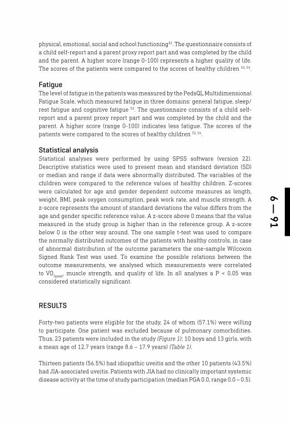
6 — 91
physical, emotional, social and school functioning52. The questionnaire consists of a child self-report and a parent proxy report part and was completed by the child and the parent. A higher score (range 0-100) represents a higher quality of life. The scores of the patients were compared to the scores of healthy children 52, 53.
Fatigue The level of fatigue in the patients was measured by the PedsQL Multidimensional Fatigue Scale, which measured fatigue in three domains: general fatigue, sleep/rest fatigue and cognitive fatigue 53. The questionnaire consists of a child self-report and a parent proxy report part and was completed by the child and the parent. A higher score (range 0-100) indicates less fatigue. The scores of the patients were compared to the scores of healthy children 52, 54.
Statistical analysisStatistical analyses were performed by using SPSS software (version 22). Descriptive statistics were used to present mean and standard deviation (SD) or median and range if data were abnormally distributed. The variables of the children were compared to the reference values of healthy children. Z-scores were calculated for age and gender dependent outcome measures as length, weight, BMI, peak oxygen consumption, peak work rate, and muscle strength. A z-score represents the amount of standard deviations the value differs from the age and gender specific reference value. A z-score above 0 means that the value measured in the study group is higher than in the reference group. A z-score below 0 is the other way around. The one sample t-test was used to compare the normally distributed outcomes of the patients with healthy controls, in case of abnormal distribution of the outcome parameters the one-sample Wilcoxon Signed Rank Test was used. To examine the possible relations between the outcome measurements, we analysed which measurements were correlated to VO
2peak, muscle strength, and quality of life. In all analyses a P < 0.05 was
considered statistically significant.
RESULTS
Forty-two patients were eligible for the study, 24 of whom (57.1%) were willing to participate. One patient was excluded because of pulmonary comorbidities. Thus, 23 patients were included in the study (Figure 1); 10 boys and 13 girls, with a mean age of 12.7 years (range 8.6 – 17.9 years) (Table 1).
Thirteen patients (56.5%) had idiopathic uveitis and the other 10 patients (43.5%) had JIA-associated uveitis. Patients with JIA had no clinically important systemic disease activity at the time of study participation (median PGA 0.0, range 0.0 – 0.5).
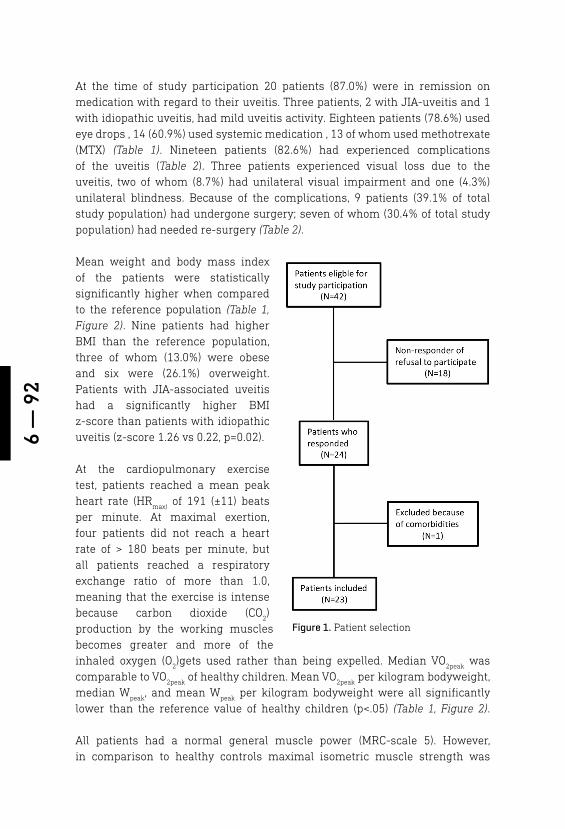
6 —
92
At the time of study participation 20 patients (87.0%) were in remission on medication with regard to their uveitis. Three patients, 2 with JIA-uveitis and 1 with idiopathic uveitis, had mild uveitis activity. Eighteen patients (78.6%) used eye drops , 14 (60.9%) used systemic medication , 13 of whom used methotrexate (MTX) (Table 1). Nineteen patients (82.6%) had experienced complications of the uveitis (Table 2). Three patients experienced visual loss due to the uveitis, two of whom (8.7%) had unilateral visual impairment and one (4.3%) unilateral blindness. Because of the complications, 9 patients (39.1% of total study population) had undergone surgery; seven of whom (30.4% of total study population) had needed re-surgery (Table 2).
Mean weight and body mass index of the patients were statistically significantly higher when compared to the reference population (Table 1, Figure 2). Nine patients had higher BMI than the reference population, three of whom (13.0%) were obese and six were (26.1%) overweight. Patients with JIA-associated uveitis had a significantly higher BMI z-score than patients with idiopathic uveitis (z-score 1.26 vs 0.22, p=0.02).
At the cardiopulmonary exercise test, patients reached a mean peak heart rate (HR
max) of 191 (±11) beats
per minute. At maximal exertion, four patients did not reach a heart rate of > 180 beats per minute, but all patients reached a respiratory exchange ratio of more than 1.0, meaning that the exercise is intense because carbon dioxide (CO
2)
production by the working muscles becomes greater and more of the inhaled oxygen (O
2)gets used rather than being expelled. Median VO
2peak was
comparable to VO2peak
of healthy children. Mean VO2peak
per kilogram bodyweight, median W
peak, and mean W
peak per kilogram bodyweight were all significantly
lower than the reference value of healthy children (p<.05) (Table 1, Figure 2).
All patients had a normal general muscle power (MRC-scale 5). However, in comparison to healthy controls maximal isometric muscle strength was
Figure 1. Patient selection
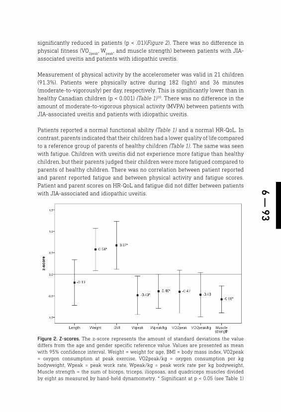
6 — 93
significantly reduced in patients (p < .01)(Figure 2). There was no difference in physical fitness (VO
2peak, W
peak, and muscle strength) between patients with JIA-
associated uveitis and patients with idiopathic uveitis.
Measurement of physical activity by the accelerometer was valid in 21 children (91.3%). Patients were physically active during 182 (light) and 36 minutes (moderate-to-vigorously) per day, respectively. This is significantly lower than in healthy Canadian children (p < 0.001) (Table 1)30. There was no difference in the amount of moderate-to-vigorous physical activity (MVPA) between patients with JIA-associated uveitis and patients with idiopathic uveitis.
Patients reported a normal functional ability (Table 1) and a normal HR-QoL. In contrast, parents indicated that their children had a lower quality of life compared to a reference group of parents of healthy children (Table 1). The same was seen with fatigue. Children with uveitis did not experience more fatigue than healthy children, but their parents judged their children were more fatigued compared to parents of healthy children. There was no correlation between patient reported and parent reported fatigue and between physical activity and fatigue scores. Patient and parent scores on HR-QoL and fatigue did not differ between patients with JIA-associated and idiopathic uveitis.
Figure 2. Z-scores. The z-score represents the amount of standard deviations the value differs from the age and gender specific reference value. Values are presented as mean with 95% confidence interval. Weight = weight for age, BMI = body mass index, VO2peak = oxygen consumption at peak exercise, VO2peak/kg = oxygen consumption per kg bodyweight, Wpeak = peak work rate, Wpeak/kg = peak work rate per kg bodyweight, Muscle strength = the sum of biceps, triceps, iliopsoas, and quadriceps muscles divided by eight as measured by hand-held dynamometry. * Significant at p < 0.05 (see Table 1)
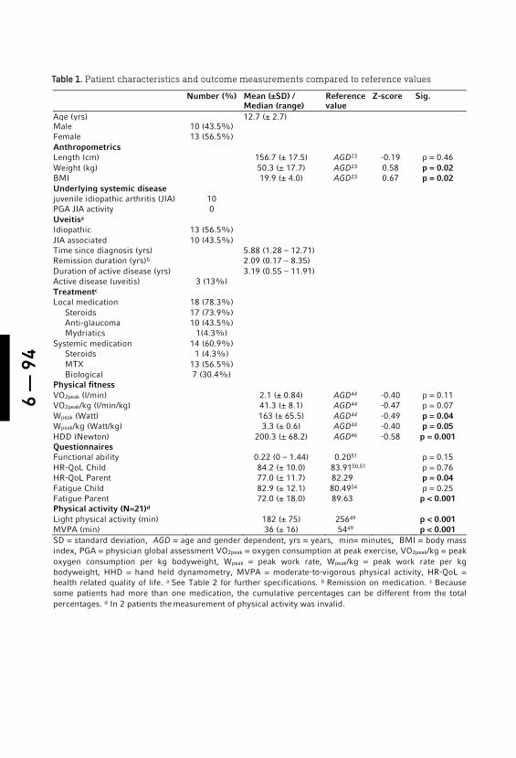
6 —
94
Table 1. Patient characteristics and outcome measurements compared to reference values
Number (%) Mean (±SD) / Median (range)
Reference value
Z-score Sig.
Age (yrs) 12.7 (± 2.7) Male 10 (43.5%) Female 13 (56.5%) Anthropometrics Length (cm) 156.7 (± 17.5) AGD23 -0.19 p = 0.46 Weight (kg) 50.3 (± 17.7) AGD23 0.58 p = 0.02 BMI 19.9 (± 4.0) AGD23 0.67 p = 0.02 Underlying systemic disease juvenile idiopathic arthritis (JIA) 10 PGA JIA activity 0 Uveitisa Idiopathic 13 (56.5%) JIA associated 10 (43.5%) Time since diagnosis (yrs) 5.88 (1.28 – 12.71) Remission duration (yrs)b 2.09 (0.17 – 8.35) Duration of active disease (yrs) 3.19 (0.55 – 11.91) Active disease (uveitis) 3 (13%) Treatmentc Local medication 18 (78.3%) Steroids 17 (73.9%) Anti-glaucoma 10 (43.5%) Mydriatics 1(4.3%) Systemic medication 14 (60.9%) Steroids 1 (4.3%) MTX 13 (56.5%) Biological 7 (30.4%) Physical fitness VO2peak (l/min) 2.1 (± 0.84) AGD44 -0.40 p = 0.11 VO2peak/kg (l/min/kg) 41.3 (± 8.1) AGD44 -0.47 p = 0.07 Wpeak (Watt) 163 (± 65.5) AGD44 -0.49 p = 0.04 Wpeak/kg (Watt/kg) 3.3 (± 0.6) AGD44 -0.40 p = 0.05 HDD (Newton) 200.3 (± 68.2) AGD46 -0.58 p = 0.001 Questionnaires Functional ability 0.22 (0 – 1.44) 0.2051 p = 0.15 HR-QoL Child 84.2 (± 10.0) 83.9150,51 p = 0.76 HR-QoL Parent 77.0 (± 11.7) 82.29 p = 0.04 Fatigue Child 82.9 (± 12.1) 80.4954 p = 0.25 Fatigue Parent 72.0 (± 18.0) 89.63 p < 0.001 Physical activity (N=21)d Light physical activity (min) 182 (± 75) 25649 p < 0.001 MVPA (min) 36 (± 16) 5449 p < 0.001 SD = standard deviation, AGD = age and gender dependent, yrs = years, min= minutes, BMI = body mass index, PGA = physician global assessment VO2peak = oxygen consumption at peak exercise, VO2peak/kg = peak oxygen consumption per kg bodyweight, Wpeak = peak work rate, Wpeak/kg = peak work rate per kg bodyweight, HHD = hand held dynamometry, MVPA = moderate-to-vigorous physical activity, HR-QoL = health related quality of life. a See Table 2 for further specifications. b Remission on medication. c Because some patients had more than one medication, the cumulative percentages can be different from the total percentages. d In 2 patients the measurement of physical activity was invalid.

6 — 95
VO2peak* HDD* HR-QoL child HR-QoL parent P p r p r p r p Gender -0.15 0.51 -0.13 0.55 -0.25 0.25 -0.06 0.79 Age 0.37 0.09 0.51 0.01 0.40 0.06 0.30 0.17 Duration of active disease -0.28 0.19 -0.12 0.59 -0.16 0.47 -0.63 0.001 systemic medication -0.28 0.20 -0.05 0.81 -0.07 0.75 -0.21 0.33 BMI* 0.30 0.17 0.69 < 0.001 0.38 0.08 -0.11 0.63 HDD* 0.53 0.01 0.55 0.01 0.19 0.38 VO2peak* 0.53 0.01 0.35 0.10 0.26 0.24 MVPA 0.05 0.85 -0.08 0.74 0.04 0.86 0.24 0.32 DI -0.64 0.001 -0.10 0.64 HR-QoL child 0.35 0.10 0.06 0.78 HR-QoL parent 0.26 0.24 0.06 0.78 Fatigue child 0.36 0.09 0.46 0.03 0.83 < 0.001 -0.21 0.34 Fatigue parent 0.33 0.12 0.21 0.33 -0.04 0.85 0.73 < 0.001 Values presented as Spearman correlation (P) or Pearson correlation (r) and statistical significance (p). * Z-scores. Abbreviations: BMI = body mass index, HDD = hand-held dynamometry measurements for muscle strength, VO2peak= peak oxygen consumption (l/min), Wpeak = peak work rate (Watt), MVPA = moderate-to-vigorous physical activity, DI = disability index, HR-QoL = health related quality of life.
Table 2. Ocular features
Number of Patients (N=23)
Percentage Median Range
Uveitis localization Anterior 15 65.2 % Intermediate 4 17.4 % Posterior 0 – Panuveitis 4 17.4 % Bilateral disease 16 69.6% Visual acuity (LogMAR*) Worse eye 0.05 -0.08 – 2.48 Better eye 0.00 -0.08 – 0.22 Unilateral impairment** 2 8.7% Unilateral blindness** 1 4.3% Complications** 19 82.6% Cataract 13 56.5% Glaucoma 13 56.5% Posterior synechiae 10 43.5% Band keratopathy 5 21.7% Amblyopia 1 4.3% Surgery*** 9 39.1% Cataract extraction 8 34.8% Baerveldt-implantation (anti-glaucoma treatment)
8 34.8%
Re-surgery 7 30.4% LogMAR= Logarithm of the Minimum Angle of Resolution. * A lower LogMAR visual acuity score corresponds to higher Snellen visual acuity and vice versa. ** Visual impairment was defined as a visual acuity ≥ 0.05 (LogMAR ≤ 1.30) and < 0.3 (LogMAR > 0.50), blindness was defined as a visual acuity less than 0.01 (or LogMAR > 1.3) or a visual field ≤ 10º 40. *** Because some patients had more than one complication, surgery or medication, the cumulative percentages can be different from the total percentages.
Table 3. Correlations
6 —
96
Correlations are shown in Table 3.The correlation-coefficient between VO2peak
and W
peak was 0.94 (P = < 0.001), VO
2peak was therefore used and interpreted as a
measure for exercise capacity. Muscle strength (HDD) was correlated with higher VO
2peak. Older age and higher BMI were correlated with higher muscle strength.
Higher child reported HR-QoL was correlated with higher muscle strength and less fatigue (higher score means less fatigue). Higher disability was correlated with lower HR-QoL. Longer duration of active disease was correlated with lower HR-QoL reported by the parents about their child. Less fatigue was associated with a higher HR-QoL reported by the parents about their children.
DISCUSSION
Patients with uveitis have higher BMI compared to healthy children, they are at risk for reduced physical fitness levels as indicated by a lower aerobic exercise capacity and reduced muscle strength when compared to the healthy pediatric population. Also, children with uveitis are less physically active (PA), and their parents report a lower quality of life (HR-QoL) and more fatigue for their children when compared to parents of healthy children. In contrast, the children themselves report a normal HR-QoL and fatigue. The children with JIA-uveitis have a statistically significantly higher BMI than the children with idiopathic uveitis. No differences are found between JIA and idiopathic uveitis patients in physical fitness levels.
We found a significantly higher percentage of overweight (26%) and obese (14%) patients compared to the Dutch population, 13-15% and 2.2%, respectively. In patients with JIA-uveitis BMI was significantly higher compared to non JIA uveitis. Corticosteroids are a well-known cause of weight gain12, however in our study only one patient used low dose (5 mg) systemic corticosteroids and most of the patients had not used systemic steroids for a long period of time. In JIA, contradictory results concerning obesity have been found and the cause has not been revealed yet 55, 56. A possible explanation is a more sedentary lifestyle which we also found in this study. There are indications that obesity in JIA can result in higher inflammatory markers and an increased risk of atherosclerosis11, 12, 57. It is reasonable to assume that this risk is comparable in patients with uveitis, so healthcare professionals and carers should be aware of weight gain in patients with uveitis.
Children with uveitis have lower aerobic exercise capacity levels than their healthy peers, but relatively well preserved levels when compared to children with other chronic conditions 5, 6. Interestingly, we found no differences in aerobic exercise capacity between JIA and idiopathic uveitis patients. The arthritis of the ten patients with JIA uveitis was in remission. It is known that the aerobic exercise capacity in patients with JIA does not restore after remission has been
6 — 97
reached9. We assume that comparable underlying mechanisms could play a causative role in uveitis but their nature has not yet been revealed . The general assumption is that reduced levels of aerobic fitness are caused by a combination of disease-related pathophysiology, treatment (e.g., medication), hypo-activity, and deconditioning5, 11, 12.
Patients with uveitis have decreased muscle strength that is possibly caused by the same combination of mechanisms that are responsible for the reduced exercise capacity. From the literature it is known that low exercise capacity, decreased muscle strength, the inflammation itself, circulating cytokines and the use of systemic corticosteroids are correlated with an increased risk of cardiovascular diseases11, 12, 58, 59. In children with uveitis, these factors are present. Therefore, physicians should be alert and try to eliminate extra cardiovascular risk factors.
Our patients report 32 minutes of moderate-to-vigorous physical activity (MVPA) per day which is considerably less than the 60 minutes of daily MVPA as recommended by the WHO and the MVPA of the reference group49, 60. Similar results have been found for adolescents with JIA8, 9. Hypoactive children are often at greater risk of preventable health problems, such as obesity and cardio-metabolic diseases 5 , 57. Cardiovascular health in children can be improved by sufficient physical activity (PA) and physical fitness 61, whereas PA also has a beneficial effect on HR-QoL4. In several auto-immune diseases, PA has been shown to be safe, to improve HR-QoL and to reduce fatigue 4.
The parents of our patients score a lower quality of life and higher levels of fatigue for their children than parents of healthy children, whereas the children themselves report outcomes comparable to those of their healthy peers on both questionnaires. This difference is probably due the proxy-problem, a known variation in patient and parent-report 62. In the measurement of quality of life, parents tend to score a lower quality of life for their chronically ill children than the children themselves. This is possibly due to the differences in adaptation to a chronic disease in child and parent. Parents are possibly more aware of the health risks and have a broader perspective than children 62, 63. Also, it is likely that the parent- reported HR-QoL and fatigue are influenced by their frequent visits to the hospital and their efforts associated with the medical treatment of their child.
The positive correlations in our study between exercise capacity, muscle strength and BMI are not supported in the literature 65. Also, the reported loss of HR-QoL 52, 66 and increase in fatigue in children with overweight is not found in our results. We cannot explain these findings. Perhaps the significantly lower PA combined with adaptation in coping strategies by the children are responsible for these contradictory results. We did not investigate body composition, so we
6 —
98
cannot comment on the influence of differences between muscle and fat mass on measured BMI in relation the muscle strength and exercise capacity. The negative correlations between lower HR-QoL (reported by children) and loss of functional ability and between lower HR-QoL (reported by parents) and longer disease duration are in line with the literature 33, 51 – 54, 67.
Limitations of the studyWe performed this study as a pilot with a small number of patients. Furthermore, this study is cross sectional and most patients were in disease remission. Patients in other phases of the disease may have different results. Also, there is an unknown selection bias, because not all eligible patients participated. Another possible bias is that this study was conducted in a tertiary center. In the Netherlands, children with uveitis who require systemic therapy are treated and managed in tertiary (in most cases university) centers. The results thus do not represent the total spectrum of pediatric non-infectious uveitis and the results and conclusions should be interpreted in this way.
Clinical implicationsWe recommend that clinicians discuss the importance of sufficient levels of physical fitness and PA during outpatient visits with patients and their parents. Also, close monitoring of body weight should be performed and the prevention of overweight should be a treatment goal.
CONCLUSION
This pilot-study investigated the physical and psychosocial consequences of uveitis in childhood. We showed that patients with non-infectious uveitis are at risk of developing cardiovascular risk factors early in life. Children with uveitis have a higher BMI, lower cardio-respiratory fitness and are less physically active when compared to healthy peers. Furthermore, their parents report a lower quality of life and more fatigue for their children compared to the parents of healthy children. It remains undecided whether this can be attributed to the systemic treatment or the inflammatory disease, since children with idiopathic non-infectious uveitis had similar test results as children with JIA-uveitis. Clinicians should discuss the importance of sufficient levels of physical fitness and PA with patients and their parents during outpatient visits. With the current knowledge and the results of our study we believe we can contribute to the optimisation of the treatment for children with uveitis. Treatment of paediatric uveitis should be aimed at improving the physical and psychosocial health and reducing cardiovascular risk factors in this vulnerable group of patients in addition to maintaining and preserving vision.
6 — 99
REFERENCES
1. Päivönsalo-Hietanen T, Tuominen J, Saari KM. Uveitis in Children: Popula-tion-based Study in Finland. Acta Oph-thalmol Scand. 2000;78(1):84-88.
2. Zierhut M, Doycheva D, Biester S, Stü-biger N, Kümmerle-Deschner J, Deuter C. Therapy of Uveitis in Children. Int Oph-thalmol Clin. 2008;48(3):131-152.
3. Mehta PJ, Alexander JL, Sen HN. Pediatric uveitis: New and future treatments. Curr Opin Ophthalmol. 2013;24(5):453-462.
4. Sharif K,Watad A, Bragazzi N.L, Licht-broun M, Amital H, Shoenfeld Y. Physical activity and autoimmune diseases: Get moving and manage the disease. Auto-immun Rev. 2018; 17( 1), 53-72.
5. Takken T, Bongers BC, van Brussel M, Haapala EA, Hulzebos EHJ. Cardiopul-monary Exercise Testing in Pediatrics. Ann Am Thorac Soc. 2017; Supplement 1, S123-S128.
6. van Brussel M, van der Net J, Hulzebos E, Helders PJ, Takken T. The Utrecht ap-proach to exercise in chronic childhood conditions: the decade in review. Pediatr Phys Ther. 2011; 23, (1): 2-14.
7. Gualano B, Bonfa E, Pereira RMR, Silva CA. Physical activity for paediatric rheu-matic diseases: standing up against old paradigms. Nat Rev Rheumatol. 2017;13, (6): 368-379.
8. Lelieveld OT, Armbrust W, van Leeuwen M a, et al. Physical Activity in Adolescents with Juvenile Idiopathic Arthritis. Arthri-tis Rheum. 2008;59(10):1379-1384.
9. van Brussel M, Lelieveld OTHM, van der Net J, Engelbert RHH, Helders PJM, Takken T. Aerobic and Anaerobic Exer-cise Capacity in Children with Juvenile Idiopathic Arthritis. Arthritis Rheum. 2007;57(6):891-897.
10. Ploeger HE, Takken T, Wilk B, et al. Exer-cise Capacity in Pediatric Patients with Inflammatory Bowel Disease. J Pediatr. 2011;158(5):814-819.
11. Roubenoff R. Exercise and Inflammatory Disease. Arthritis Care Res (Hoboken). 2003;49(2):263-.
12. Gupta Y, Gupta A. Glucocorticoid-induced Myopathy: Pathophysiology, Diagnosis, and Treatment. Indian J Endocrinol Me-tab. 2013;17(5):913-916.
13. Angeles-Han ST, Rabinovich CE. Uveitis in children. Curr Opin Rheumatol. 2016 Sep;28(5): 544-9.
14. Lee RW, Nicholson LB, Sen HN, et al. Autoimmune and Auto-inflammatory Mechanisms in Uveitis. Semin Immuno-pathol. 2014;36(5):581-594.
15. Coulson EJ, Ng W-F, Goff I, Foster HE. Cardi-ovascular Risk in Juvenile Idiopathic Arthri-tis. Rheumatology. 2013;52(7):1163-1171.
16. Barsalou J, Bradley TJ, Silverman ED. Cardiovascular Risk in Pediatric-onset Rheumatological Diseases. Arthritis Res Ther. 2013;15:212.
17. Libby P. Role of Inflammation in Atheroscle-rosis Associated with Rheumatoid Arthritis. Am J Med. 2008;121(10 Suppl 1):S21-S31.
18. Dana MR, Merayo-Lloves J, Schaum-berg DA, Foster CS. Visual outcomes prognosticators in juvenile rheumatoid arthritis-associated uveitis. Ophthalmol-ogy.1997;104(2):236-244.
19. Kalinina Ayuso V, Makhotkina N, van Tent-Hoeve M, de Groot-Mijnes JD, Wulf-fraat NM, Rothova A, de Boer JH. Patho-genesis of juvenile idiopathic arthritis as-sociated uveitis: the known and unknown. Surv.Ophthalmol. 2014;59(5):517-531.
20. Haasnoot AM, Kuiper JJ, Hiddingh S, Schell-ekens PA, de Jager W, Imhof SM, Radstake TR, de Boer JH. Ocular Fluid Analysis in Children Reveals Interleukin-29/Interfer-on-lambda1 as a Biomarker for Juvenile Idiopathic Arthritis-Associated Uveitis. Ar-thritis Rheumatol. 2016;68(7):1769-1779.
21. Haasnoot AJ, van Tent-Hoeve M, Wulffraat NM, Schalij-Delfos NE, Los LI, Armbrust W, Zuithoff NP, de Boer JH. Erythrocyte sedi-mentation rate as baseline predictor for the development of uveitis in children with ju-venile idiopathic arthritis. Am J Ophthalmol. 2015 Feb;159(2):372-7.

6 —
100
22. Pelegrin L, Casaroli-Marano R, Anton J, Garcia de Vicuna MC, Molina-Prat N, Ignacio Arostegui J, Yague J, Rios J, Adan A. Predictive value of selected bi-omarkers, polymorphisms, and clinical features for oligoarticular juvenile idio-pathic arthritis-associated uveitis. Ocul Immunol Inflamm. 2014;22(3):208-212.
23. Walscheid K, Heiligenhaus A, Holzinger D, Roth J, Heinz C, Tappeiner C, Kasper M, Foell D. Elevated S100A8/A9 and S100A12 Serum Levels Reflect Intraoc-ular Inflammation in Juvenile Idiopath-ic Arthritis-Associated Uveitis: Results From a Pilot Study. Invest Ophthalmol Vis Sci. 2015 Dec;56(13):7653-6.
24. Haasnoot AJW, Schilham MW, Kamphu-is S, Hissink Muller PCE, Heiligenhaus A, Foell D, Minden K, Ophoff RA, Radstake TRDJ, Den Hollander AI, Reinards THCM, Hiddingh S, Schalij-Delfos NE, Hoppenreijs EPAH, van Rossum MAJ, Wouters C, Sau-renmann RK, van den Berg JM, Wulffraat NM; ICON-JIA Study Group, Ten Cate R, de Boer JH, Pulit SL, Kuiper JJW. Identification of an Amino Acid Motif in HLA-DRβ1 That Distinguishes Uveitis in Patients With Ju-venile Idiopathic Arthritis. Arthritis Rheu-matol. 2018 Jul;70(7):1155-1165.
25. Gedalia A, Shetty AK. Chronic Steroid and Immunosuppressant Therapy in Chil-dren. Pediatr Rev. 2004;25(12):425-434.
26. Angeles-Han S, Griffin K, Lehman T, Ru-tledge J, Lyman S, Nguyen J, et al. The importance of visual function in the qual-ity of life of children with uveitis. J Am Assoc Pediatr Ophthalmol Strabismus. 2010;12(2):163–8.
27. Angeles-Han ST. Quality-of-life metrics in pediatric uveitis. Int Ophthalmol Clin. 2015;55(2):93-101
28. Angeles-Han ST, Griffin KW, Harrison MJ, Lehman TJ, Leong T, Robb RR, Shain-berg M, Ponder L, Lenhart P, Hutchinson A, Srivastava SK, Prahalad S, Lambert SR, Drews-Botsch C. Development of a vision-related quality of life instrument for children ages 8-18 years for use in juvenile idiopathic arthritis-associated uveitis. Arthritis Care.Res.(Hoboken), 2011;63(9)1254-1261.
29. Angeles-Han ST, McCracken C, Yeh S, Jenkins K, Stryker D, Rouster-Stevens K, Vogler LB, Lambert SR, Drews-Botsch C, Prahalad S. Characteristics of a cohort of children with Juvenile Idiopathic Arthri-tis and JIA-associated Uveitis. Pediatr Rheumatol Online J. 2015 Jun 2;13:19.
30. Schiffman RM, Jacobsen G, Whitcup SM.Visual functioning and general health status in patients with uveitis. Arch Oph-thalmol. 2001 Jun;119(6):841-9
31. Hoeksema L, Los LI. Vision-related qual-ity of life in herpetic anterior uveitis pa-tients. PLoS One. 2014 Jan 2;9(1)
32. Haasnoot AJW, Sint Jago NFM, Tekstra J, de Boer JH. Impact of Uveitis on Quality of Life in Adult Patients With Juvenile Id-iopathic Arthritis. Arthritis Care Res (Ho-boken). 2017 dec;69(12):1895-1902
33. Miserocchi E, Modorati G, Mosconi P, Colucci A, Bandello F. Quality of Life in Patients with Uveitis on Chronic System-ic Immunosuppressive Treatment. Ocul Immunol Inflamm. 2010;18(4):297-304.
34. Maca SM, Amirian A, Prause C, Gruber K, Mejdoubi L, Barisani-Asenbauer T. Under-standing the Impact of Uveitis on Health-re-lated Quality of Life in Adolescents. Acta Ophthalmol. 2013;91(3):219-224.
35. Petrina Tan, Yan Tong Koh, Poh Ying Wong & Stephen C. Teoh. Evaluation of the Impact of Uveitis on Visual-related Quality of Life. Ocular Immunology and Inflammation. 2012;20(6):453-459.
36. Reichert FF, Barros AJD, Domingues MR, Hallal PC. The Role of Perceived Personal Barriers to Engagement in Leisure-time Physical Activity. Am J Public Health. 2007;97(3):515-519.
37. Armbrust W, Lelieveld OH, Tuinstra J, Wulffraat NM, Bos GJ, Cappon J, van Ros-sum MA, Sauer PJ, Hagedoorn M. Fatigue in patients with Juvenile Idiopathic Ar-thritis: relationship to perceived health, physical health, self-efficacy, and partici-pation. Pediatr Rheumatol Online J. 2016 Dec 6;14(1):65.
6 — 101
38. Jabs DA, Nussenblatt RB, Rosenbaum JT; Standardization of Uveitis Nomenclature (SUN) Working Group. Standardization of uveitis nomenclature for reporting clinical data. Results of the First Interna-tional Workshop. Am J Ophthalmol. 2005 Sep;140(3):509-16.
39. Holladay JT. Proper method for calculat-ing average visual acuity. J Refract Surg. 1997 Jul-Aug;13(4):388-91.
40. World Health Organization. Universal eye health: a global action plan 2014–2019. [Internet].p 7-8.[cited 2018 July
8th] Available from: http://www.who.int/blindness/AP2014_19_English.pdf?ua=1
41. TNO. Vijfde Landelijke Groeistudie: Groe-idiagrammen [Internet]. 2010. [cited 2018
july 8th] Oct 20] Available from: https://www.tno.nl/nl/aandachtsgebieden/ge-zond-leven/prevention-work-health/lang-gezond-en-actief-leven/pdf-groe-idiagrammen/
42. Cole TJ, Lobstein T. Extended Interna-tional (IOTF) Body Mass Index Cut-offs for Thinness, Overweight and Obesity. Pediatr Obes. 2012;7(4):284-294.
43. Godfrey S. Methods of measuring the re-sponse to exercise in children. Exercise testing in children: applications in health and disease. London: W.B. Saunders Company Ltd; 1974. p. 12–41.
44. Bongers BC, Hulzebos EHJ, van Brussel M, Takken T. Pediatric Norms for Cardio-pulmonary Exercise Testing: In Relation to Gender and Age. ’s-Hertogenbosch Uitg BOXPRESS. 2012:129.
45. Compston A. From the archive: Aids to the Investigation of Peripheral Nerve In-juries: Medical Research Council 1942. Brain. 2010 Sep 29;133(10):2838–44).
46. Beenakker E a, van der Hoeven JH, Fock JM, Maurits NM. Reference values of maximum isometric muscle force ob-tained in 270 children aged 4-16 years by hand-held dynamometry. Neuromus-cul Disord. 2001 Jul;11(5):441–6.
47. Puyau MR, Adolph AL, Vohra FA, Zakeri I, Butte NF. Prediction of Activity En-ergy Expenditure Using Accelerome-ters in Children. Med Sci Sports Exerc. 2004;36(9):1625-1631.
48. Bratteby L, Sandhagen B, Fan H, Sam-uelson G. A 7-day activity diary for as-sessment of daily energy expenditure validated by the doubly labelled water method in adolescents. Eur J Clin Nutr. 1997;51:585–91.
49. Colley RC, Garriguet D, Janssen I, Craig CL, Clarke J, Tremblay MS. Physical ac-tivity of Canadian Children and Youth: Accelerometer results from the 2007 to 2009 Canadian Health Measures Survey. Heal Reports. 2011;22(1):1-9.
[cited 2018 july 8th]. http://www.stat-can.gc.ca/pub/82-003-x/2011001/arti-cle/11396-eng.htm
50. Wulffraat N, Net JJ Van Der, Ruperto N, et al. The Dutch Version of the Child-hood Health Assessment Questionnaire (CHAQ) and the Child Health Ques-tionnaire (CHQ). Clin Exp Rheumatol. 2001;19(Suppl 23):S111-S115.
51. Ouwerkerk JW, van Pelt PA, Takken T, Helders PJ, Net Jv. Evaluating score dis-tributions in the revised Dutch version of the Childhood Health Assessment Ques-tionnaire. Pediatr Rheumatol Online J. 2008 Sep 11;6:14.
52. Varni JW, Burwinkle TM, Seid M. The PedsQLTM 4.0 as a Pediatric Popula-tion Health Measure: Feasibility, Re-liability, and Validity. Ambul Pediatr. 2003;3(6):329-341.
53. Varni JW, Burwinkle TM, Szer IS. The PedsQL TM Multidimensional Fatigue Scale in pediatric rheumatology: re-liability and validity. J Rheumatol. 2004;31(12):2494–500.
54. Gordijn M, Cremers EM, Kaspers GJ, Gemke RJ. Fatigue in children: reliability and validity of the Dutch PedsQL™ Multi-dimensional Fatigue Scale. Qual Life Res. 2011 Sep;20(7):1103-8.
6 —
102
55. Pelajo CF, Lopez-Benitez JM, Miller LC. Obesity and disease activity in juvenile idiopathic arthritis. Pediatr Rheumatol Online J 2012;10:3.
56. Schenck S1, Niewerth M, Sengler C, Trauzeddel R, Thon A, Minden K, Klotsche J. Prevalence of overweight in children and adolescents with juvenile idiopathic arthri-tis. Scand J Rheumatol. 2015;44(4):288-95.
57. Zoico E, Roubenoff R. The Role of Cy-tokines in Regulating Protein Metab-olism and Muscle Function. Nutr Rev. 2002;60(2):39-51.
58. Carnethon M, Gidding S, Nehgme R, Sid-ney S, Jacobs D, Liu K. Cardiorespiratory Fitness in Young Adulthood and the Devel-opment of Cardiovascular Diseases Risk Factors. JAMA. 2003;290(23):3092-3100
59. Steene-Johannessen J, Anderssen S a, Kolle E, Andersen LB. Low muscle fitness is associated with metabolic risk in youth. Med Sci Sports Exerc. 2009 Jul;41(7):1361–7.
60. WorldHealthOrganization. Global Rec-ommendations on Physical Activity for
Health.; 2010. [cited 2018 july 8th]. Availa-ble at: http://www.who.int/dietphysicalac-tivity/publications/9789241599979/en/.
61. Strong WB, Malina RM, Blimkie CJR, et al. Evidence Based Physical Activ-ity for School-age Youth. J Pediatr. 2005;146(6):732-737.
62. Sattoe JNT, van Staa A, Moll HA. The Proxy Problem Anatomized: Child-Par-ent Disagreement in Health Related Quality of Life Reports of Chronically Ill Adolescents. Health Qual Life Outcomes. 2012;10(1):10.
63. Jardine J, Glinianaia S V, McConachie H, Embleton ND, Rankin J. Self-Report-ed Quality of Life of Young Children With Conditions From Early Infan-cy: A Systematic Review. Pediatrics. 2014;134(4):e1129-e1148.
64. Schönbeck Y, Talma H, van Domme-len P, et al. Increase in prevalence of overweight in Dutch children and ado-lescents: a comparison of nationwide growth studies in 1980, 1997 and 2009. PLoS One. 2011;6(11):e27608.
65. Rauner A, Mess F, Woll A. The Relation-ship Between Physical Activity, Physical Fitness and Overweight in Adolescents: a Systematic Review of Studies Pub-lished in or after 2000. BMC Pediatr. 2013;13(1):19.
66. Keating CL, Moodie ML, Swinburn B a. The health-related quality of life of over-weight and obese adolescents: a study measuring body mass index and adoles-cent-reported perceptions. Int J Pediatr Obes. 2011 Oct;6(5-6):434–41.
67. Haverman L, Grootenhuis MA, van den Berg JM, van Veenendaal M, Dolman KM, Swart JF, Kuijpers TW, van Rossum MA. Predictors of health-related quality of life in children and adolescents with juvenile idiopathic arthritis: results from a Web-based survey. Arthritis Care Res (Hoboken). 2012 May;64(5):694-703
6 — 103


RISK FACTORS FOR GLAUCOMA SURGERY IN CHILDHOOD UVEITISAuthors:Wietse G. Wieringa Bsc MPA1*, Charlotte L.L.I. van Meerwijk MD1*, Joke H. de Boer MD PhD2, Nomdo M. Jansonius MD PhD1, Leonoor I Los MD PhD1, 3
Affiliations of authors: 1University Medical Center Groningen, University of Groningen, Department of Ophthalmology, Groningen, The Netherlands. 2University Medical Center Utrecht, University of Utrecht, Department of Ophthalmology2, Utrecht, The Netherlands. 3W.J. Kolff Institute, Graduate School of Medical Sciences, University of Groningen, Groningen, The Netherlands
*These authors share first authorship
To be submitted
7
7 —
106
ABSTRACT
Purpose: To identify risk factors for medically uncontrollable high intraocular pressure (IOP) secondary to uveitis in children.
Methods: Patients diagnosed with uveitis before their 18th birthday and with a minimal follow-up of one year were included from the ophthalmology departments of the University Medical Center Groningen and the University Medical Center Utrecht in a retrospective case-control study.
Results: A total of 196 patients were included, 85 of whom had undergone glaucoma surgery (cases). Compared to those without glaucoma surgery (controls), cases were younger (median age 6 versus 8 years, P=0.008), uveitis was more often located anteriorly (78% versus 62%, P=0.02) and was predominantly associated with juvenile idiopathic arthritis (JIA) (62% versus 35%, P<0.001). During follow-up, cases underwent cataract surgery more often (80% versus 31%, P<0.001), had higher maximum IOPs (median IOP 37 mmHg versus 27 mmHg, P<0.001), and more often used > 2 types of glaucoma medication (83% versus 24%, P<0.001). Of those needing > 2 types of glaucoma medication 68% underwent glaucoma surgery within one year. Gender, bilaterality, visual acuity and ocular complications at diagnosis, ANA positivity, use of systemic immune-suppression, and cataract surgery before glaucoma surgery were not significantly different between the two groups. Cox survival analysis showed that anterior uveitis (P = 0.04) and increased IOP at presentation (P = 0.02) were predictive of increased risk of needing glaucoma surgery.
Conclusion: Anterior location of the uveitis and higher IOP at presentation are associated with an increased risk of glaucoma surgery. Patients who need > 2 types of glaucoma medication are likely to need glaucoma surgery, usually within 1 year after increasing the number of medications.
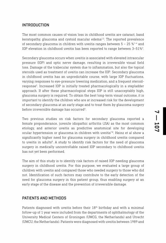
7 — 107
INTRODUCTION
The most common causes of vision loss in childhood uveitis are cataract, band keratopathy, glaucoma and cystoid macular edema1,2. The reported prevalence of secondary glaucoma in children with uveitis ranges between 5 – 25 %1–4 and IOP elevation in childhood uveitis has been reported to range between 3–51%3.
Secondary glaucoma occurs when uveitis is associated with elevated intraocular pressure (IOP) and optic nerve damage, resulting in irreversible visual field loss. Damage of the trabecular system due to inflammation, but also the topical steroids used as treatment of uveitis can increase the IOP. Secondary glaucoma in childhood uveitis has an unpredictable course, with large IOP fluctuations, varying responses to eye-pressure lowering medication, and a frequent steroid-response5. Increased IOP is initially treated pharmacologically in a stepladder approach. If after these pharmacological steps IOP is still unacceptably high, glaucoma surgery is required. To obtain the best long-term visual outcome, it is important to identify the children who are at increased risk for the development of secondary glaucoma at an early stage and to treat them by glaucoma surgery before irreversible damage has occurred6.
Two previous studies on risk factors for secondary glaucoma reported a female preponderance, juvenile idiopathic arthritis (JIA) as the most common etiology, and anterior uveitis as predictive anatomical site for developing ocular hypertension or glaucoma in children with uveitis7,8. Heinz et al show a significantly higher need for glaucoma surgery in childhood uveitis compared to uveitis in adults8. A study to identify risk factors for the need of glaucoma surgery in medically uncontrollable raised IOP secondary to childhood uveitis has not yet been performed.
The aim of this study is to identify risk factors of raised IOP needing glaucoma surgery in childhood uveitis. For this purpose, we evaluated a large group of children with uveitis and compared those who needed surgery to those who did not. Identification of such factors may contribute to the early detection of the need for glaucoma surgery in this patient group, thus enabling surgery at an early stage of the disease and the prevention of irreversible damage.
PATIENTS AND METHODS
Patients diagnosed with uveitis before their 18th birthday and with a minimal follow-up of 1 year were included from the departments of ophthalmology of the University Medical Centers of Groningen (UMCG, the Netherlands) and Utrecht (UMCU, the Netherlands). Patients were diagnosed with uveitis between 1989 and
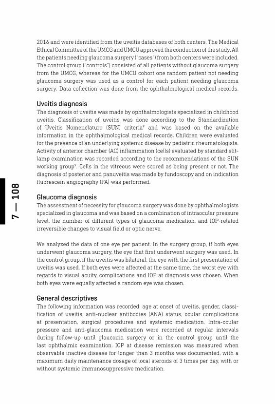
7 —
108
2016 and were identified from the uveitis databases of both centers. The Medical Ethical Committee of the UMCG and UMCU approved the conduction of the study. All the patients needing glaucoma surgery (“cases”) from both centers were included. The control group (“controls”) consisted of all patients without glaucoma surgery from the UMCG, whereas for the UMCU cohort one random patient not needing glaucoma surgery was used as a control for each patient needing glaucoma surgery. Data collection was done from the ophthalmological medical records.
Uveitis diagnosisThe diagnosis of uveitis was made by ophthalmologists specialized in childhood uveitis. Classification of uveitis was done according to the Standardization of Uveitis Nomenclature (SUN) criteria9 and was based on the available information in the ophthalmological medical records. Children were evaluated for the presence of an underlying systemic disease by pediatric rheumatologists. Activity of anterior chamber (AC) inflammation (cells) evaluated by standard slit-lamp examination was recorded according to the recommendations of the SUN working group9. Cells in the vitreous were scored as being present or not. The diagnosis of posterior and panuveitis was made by fundoscopy and on indication fluorescein angiography (FA) was performed.
Glaucoma diagnosisThe assessment of necessity for glaucoma surgery was done by ophthalmologists specialized in glaucoma and was based on a combination of intraocular pressure level, the number of different types of glaucoma medication, and IOP-related irreversible changes to visual field or optic nerve.
We analyzed the data of one eye per patient. In the surgery group, if both eyes underwent glaucoma surgery, the eye that first underwent surgery was used. In the control group, if the uveitis was bilateral, the eye with the first presentation of uveitis was used. If both eyes were affected at the same time, the worst eye with regards to visual acuity, complications and IOP at diagnosis was chosen. When both eyes were equally affected a random eye was chosen.
General descriptivesThe following information was recorded: age at onset of uveitis, gender, classi-fication of uveitis, anti-nuclear antibodies (ANA) status, ocular complications at presentation, surgical procedures and systemic medication. Intra-ocular pressure and anti-glaucoma medication were recorded at regular intervals during follow-up until glaucoma surgery or in the control group until the last ophthalmic examination. IOP at disease remission was measured when observable inactive disease for longer than 3 months was documented, with a maximum daily maintenance dosage of local steroids of 3 times per day, with or without systemic immunosuppressive medication.

7 — 109
Ocular complicationsComplications were scored as anterior when band keratopathy, cataract, or posterior synechiae were present. Posterior complications were scored when macular edema or papillitis were present. All patients needing cataract surgery during follow up were recorded. Additionally, as possible risk factor for glaucoma surgery, cataract surgery > 3 months prior to glaucoma surgery was recorded.
Visual acuityThe decimal equivalent of the Snellen visual acuity (VA) of the affected eyes was recorded at presentation. The Snellen VA was converted to logarithm of the minimum angle of resolution units (LogMAR) VA for calculations.
Data analysisData were statistically analyzed with SPSS 23.0.0 (SPSS Inc, Chicago, Illinois, USA). A P<0.05 was considered statistically significant. Bonferroni correction was applied where needed. Descriptive statistics were used to present mean and standard deviation (SD) in normally distributed data or median and inter quartile range (IQR) in non-normally distributed data. In case of non-normally distributed linked samples, the Wilcoxon test for paired samples and the Mann-Whitney U test for independent samples were used. For the differences between the nominal data groups we used the Chi-square test. A Cox survival analysis was performed. All categorical variables were dichotomized for the purpose of this analysis. The need for glaucoma surgery was defined as the event. Covariates present at baseline with P≤0.2 were analyzed as predictors in a backward stepwise conditional method. To correct for differences in data between the two centers, we added the center as a covariate to the multivariable model. The survival curve was graphically displayed as mean value for all covariates. In one covariate, 20% of the data-points were missing. Missing data patterns were analyzed and the data was classified as missing at random (MAR) based upon Little's MCAR test (supplementary data: Table 2). Missing data was compensated for by imputing mean values (supplementary data: Table 2). Next to that, analyses were repeated with missing data compensated by multiple imputation and outcomes were compared to original data and data with imputed values (supplementary data: Table 2, 3)10,11.
RESULTS
Patients characteristics are summarized in Table 1. In total 196 patients (85 female) were included in the study. In the univariable comparisons, cases were younger than the controls (median age 6 versus 8 years, P=0.008).
7 —
110
Total
(n=196) Glaucoma surgery (cases, n=85)
No glaucoma surgery (controls, n=111)
P-value
Number of patients 196 85 111 Median age at uveitis onset (yrs), ( IQR)b 7 (4-10) 6 (4-9) 8 (5-11) 0.008a Center 1c 97 (49%) 34 63 Center 2d 99 (51%) 51 48 Gender – female 85 (43%) 53 32 0.51e Bilateral disease 134 (68%) 57 77 0.73e Anatomic location uveitis (n (% of total)) 0.02e Anterior 135 (69%) 66 69 Intermediate 21 (11%) 6 15 Posterior 8 (4%) 0 8 Pan uveitis 32 (16%) 13 19 Median LogMar visual acuity at diagnosis (IQR)
0.22 (0.11 to 0.60)
0.22 (0.03 to 0.60)
0.22 (0.01 to 0.70)
0.61a
Ocular complicatons (n (% of total))f 0.11c No complications 56 (29%) 24 32 Anterior complicationsg 66 (34%) 36 30 Posterior complicatonsh 36(18%) 11 25 Anterior and posterior complications 36(18%) 14 22 Etiology (n (% of total)) < 0.001c E.c.i 81 (41%) 28 53 JIAi 92 (47%) 53 39 HLA-B27 6 (3%) 3 3 infectious 11 (6%) 1 10 Other auto-immune 6 (3%) 0 6 ANAj positive 104 (57%)i 53 (62%) 51 (46%) 0.07c IOP l measurements (mmHg), (median, (IQR))
First IOP measurement 16 (13 - 20) 16 (14 - 22) 16 (13 - 19) 0.11a IOP disease remission 19 (15 - 23) 21 (17 - 27) 18 (15 - 21) 0.002a Highest IOP during FUm 32 (24.5 - 38) 37 (34 - 42) 27 (20 - 32) < 0.001a Time measurements (months, (median, (IQR))
Time to disease remissionn 6 (3 - 12) 5 (3 - 10) 6 (3 - 14) 0.23a Time to highest IOP during FUo 17 (6 - 41.5) 28 (10 - 52) 13 (3 - 36) 0.004a Time to start glaucoma medication 6 (1 - 21) 5 (1-21) 9 (1-25) 0.33a Time to glaucoma surgery N/Ap 31 (12-54) N/Ap Median FU 86 (43 - 144) 114 (71 - 180) 66 (37 - 115) < 0.001a Additional treatment (n (% of total))
Systemic immune- suppression At start uveitisq 71 (36%) 24 47 0.04c During follow upr 160 (82%) 71 89 0.47c Cataract surgery 102 (52%) 68 34 < 0.001c Cataract surgery before glaucoma surgerys 65 (33%) 31 34 0.39c Maximum glaucoma medication used (n) 5 4 (4 - 5) 1 (0 - 2) < 0.001a > 2 types of glaucoma medication n (% of total))
107 (54%) 83 24 < 0.001c
a Mann-Whitney, b interquartile range,c Center 1 = University medical center Groningen. From this center, all cases and controls were included. d Center 2= University medical center Utrecht. All cases were included and 48 randomly chosen controls were included. e Pearson chi-square, fmissing n=2, ganterior complications: Band keratopathy, cataract, posterior synechiae, h posterior complications : Macular-edema, papillitis, iJuvenile idiopathic arthritis, jAnti nuclear antibodies, k missing n=12, l IOP = intra ocular pressure, m FU = follow up, nmissing n=42, omissing n= 2, p N/A = not applicable, qmissing n=1, r missing n=2,s cataract surgery longer than 3 months before glaucoma surgery
Table 1. Characteristics complete cohort
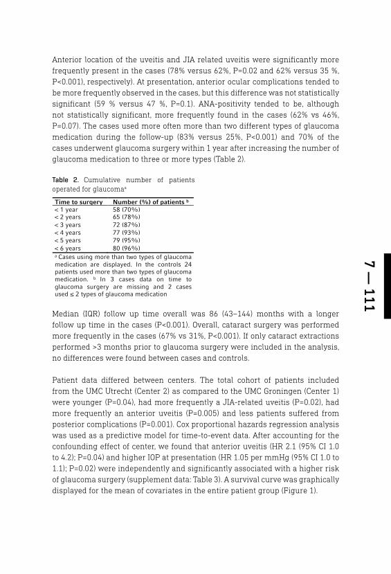
7 — 111
Anterior location of the uveitis and JIA related uveitis were significantly more frequently present in the cases (78% versus 62%, P=0.02 and 62% versus 35 %, P<0.001), respectively). At presentation, anterior ocular complications tended to be more frequently observed in the cases, but this difference was not statistically significant (59 % versus 47 %, P=0.1). ANA-positivity tended to be, although not statistically significant, more frequently found in the cases (62% vs 46%, P=0.07). The cases used more often more than two different types of glaucoma medication during the follow-up (83% versus 25%, P<0.001) and 70% of the cases underwent glaucoma surgery within 1 year after increasing the number of glaucoma medication to three or more types (Table 2).
Median (IQR) follow up time overall was 86 (43–144) months with a longer follow up time in the cases (P<0.001). Overall, cataract surgery was performed more frequently in the cases (67% vs 31%, P<0.001). If only cataract extractions performed >3 months prior to glaucoma surgery were included in the analysis, no differences were found between cases and controls.
Patient data differed between centers. The total cohort of patients included from the UMC Utrecht (Center 2) as compared to the UMC Groningen (Center 1) were younger (P=0.04), had more frequently a JIA-related uveitis (P=0.02), had more frequently an anterior uveitis (P=0.005) and less patients suffered from posterior complications (P=0.001). Cox proportional hazards regression analysis was used as a predictive model for time-to-event data. After accounting for the confounding effect of center, we found that anterior uveitis (HR 2.1 (95% CI 1.0 to 4.2); P=0.04) and higher IOP at presentation (HR 1.05 per mmHg (95% CI 1.0 to 1.1); P=0.02) were independently and significantly associated with a higher risk of glaucoma surgery (supplement data: Table 3). A survival curve was graphically displayed for the mean of covariates in the entire patient group (Figure 1).
Time to surgery Number (%) of patients b < 1 year 58 (70%) < 2 years 65 (78%) < 3 years 72 (87%) < 4 years 77 (93%) < 5 years 79 (95%) < 6 years 80 (96%) a Cases using more than two types of glaucoma medication are displayed. In the controls 24 patients used more than two types of glaucoma medication. b In 3 cases data on time to glaucoma surgery are missing and 2 cases used ≤ 2 types of glaucoma medication
Table 2. Cumulative number of patients operated for glaucomaa

7 —
112
DISCUSSION
In this study with 196 pediatric uveitis patients, we show that the risk of glaucoma surgery is highest in children with anterior uveitis. Our data also indicates that in the majority of the cases glaucoma surgery is required within a year after a third type of glaucoma medication is prescribed. In patients with uveitis onset at younger age, JIA-related uveitis and patients with higher IOP during follow up, statistically significantly more glaucoma surgery is performed. Known risk factors from the literature such as ANA positivity, female predominance and anterior complications12, are not confirmed in our study.
In our multivariate Cox survival analysis, after adjusting for other covariates, only anterior uveitis and IOP at diagnosis are independently and significantly associated with a higher risk of glaucoma surgery. Anterior uveitis is a known risk factor for the development of ocular hypertension and glaucoma7,8,12–15. Also, for the treating ophthalmologist, anterior uveitis is a clearly identifiable risk factor, whereas IOP at presentation is much less clinically relevant, since median IOP (16 mmHg) did not differ between the cases and controls. Although anterior uveitis is most frequently found in the cases, the presence of anterior complications at diagnosis such as band keratopathy, cataract, or posterior
Figure 1. Survival-curve at mean covariates for the entire patient group. The relationship between the influence of multiple risk factors on the probability of glaucoma surgery is displayed for the entire patient group. The horizontal axis shows the time to glaucoma surgery, the vertical axis shows the probability of survival. The shape of the survival function is estimated from all observed subjects.
7 — 113
synechiae are not related to a higher risk of glaucoma surgery in our study (P=0.1), which contradicts the literature7,8,12–15.
During follow-up, additional risk factors were identified by univariable analyses (Table 1). Of these, requirement of a third type of glaucoma medication can be considered as a tipping point, since it indicates the need of glaucoma surgery within a year in the majority of the cases. (Table 2). More frequent monitoring of IOP and compliance to glaucoma medication are recommended for these children. Also, early referral to an ophthalmologist specialized in glaucoma-surgery is advised to prevent a delay in surgical treatment of glaucoma.
In our study, patients who require glaucoma surgery are younger at the time of uveitis diagnosis. This may indicate a more severe disease and inherent complications due to prolonged disease and treatment with topical steroids13,16. There is no consensus in the literature regarding this aspect, since some previous studies agree8,13, but others report older age at onset of the uveitis to be related to ocular hypertension or secondary glaucoma12.
More patients with JIA-uveitis were found among the cases when compared to the controls (53 (58%) vs 39 (42%), P=<0.001). In the literature, the prevalence of glaucoma in JIA-associated uveitis has been reported to range from 14 -40%12. JIA-related uveitis is most commonly anteriorly located, chronic and most patients are treated in a uveitis expertise center. We included patients from two tertiary centers, of which the UMCU is an academic expertise center for patients with JIA. This possibly explains the relatively higher need of glaucoma surgery in the JIA subgroup when compared to the literature7,13.
In our study, topical, subconjunctival as well as systemic anti-inflammatory medication are widely used mostly during the whole follow-up. Increasing IOP due to steroid-response induced by topical steroids may therefore play an important role, despite the careful prescription of local steroids and a maximum daily maintenance dose of 3 times per day. In children, the ocular-hypertensive response to topical steroids occurs more frequently, severely and rapidly than in adults1,17,18. Elevation of IOP can be found in most patients as early as the first or second week19,20. In our study, the highest IOP during the follow-up is measured after 28 months in the surgery group and 13 months in the control group with a large variation in time. This relatively late increase in IOP in both groups is a combined result of changes in the trabecular system, the ocular-hypertensive response to topical steroids, and adaptations in glaucoma medication during follow-up. Thus reflecting the multifactorial pathophysiology of increasing IOP in uveitis.

7 —
114
In some papers, previous cataract surgery is related to the severity of the uveitis and a higher risk of uveitic glaucoma12,16. In our data, the proportion of cases that underwent cataract surgery before glaucoma surgery was not statistically significantly different from that of controls. During the entire follow-up, more cataract surgery is performed in the cases than in the controls (68% versus 34%, P<0.001). The development of cataract is probably based on the impact of glaucoma surgery and the long-term use of medication to control the uveitis19,21.
To analyze the differences between the two groups at diagnosis and in the course of the disease, we followed the patients as long as possible. The follow-up is shorter in the controls when compared to the cases, partly because of a loss to follow-up due to a stable course and therefore a return to the referring ophthalmologist. With a different follow-up duration in cases versus controls of 9 years versus 6 years, it is likely that there are patients in the control-group who will need glaucoma surgery in the future.
Various surgical techniques are possible for treating glaucoma in childhood22,23. Glaucoma surgery is still a challenge for the ophthalmologist and far-reaching for the child. In this patient group, it is essential to perform surgery on time, but choosing the optimal moment is difficult. Weighing the chance of irreversible visual loss due to glaucomatous damage against that from complications induced by glaucoma surgery are difficult considerations22. The changes in eyesight, uncertainty about the disease course, necessary changes in medication and frequent school absence due to monitoring visits to the ophthalmologist may have a strong impact on the quality of life of a young patient and their parents24. Finding a balance between prevention of irreversible loss of vision due to glaucoma and reducing the iatrogenic impact on a child’s quality of life is a challenge encountered in all cases.
In our study, we combined data from the UMCG and the UMCU. Our cox-regression analysis showed a significant differences between the two centers (supplement data: Table 3). This differences are meanly based on differences in patient population, due to the specialized health care in treating JIA patients at the UMCU and the responsibility for specialized and more general care of uveitis patients during the study period at the UMCG.
The results of the current study are limited by the fact that the study is retrospective, there is a large variability in follow up time and data-imputation was performed for variables with missing data. All patients were included from two tertiary centers in the Netherlands and therefore do not represent the total spectrum of pediatric uveitis. Also, personal experience or preferences of ophthalmologists and pediatric rheumatologist may have influenced the choice and course of treatment. The strengths of this study are its cohort size,
7 — 115
the systematic way in which data were collected, its adherence to the SUN classification system and guidelines for publications9–11 and the sharing of expertise in a difficult and challenging patient population.
CONCLUSION
This study on the risk factors for developing medically uncontrollable high intra-ocular pressure in pediatric uveitis is one of the largest currently available in the literature. The authors emphasize the importance of careful treatment and monitoring in pediatric uveitis patients with anterior uveitis, JIA-related uveitis and in patients who are already treated with more than two types of glaucoma medication. Adequate monitoring, risk assessment and early referral to a glaucoma specialist experienced in this patient group is recommended.
7 —
116
REFERENCES
1. Kaur S, Kaushik S, Singh Pandav S. Pedi-atric Uveitic Glaucoma. J Curr glaucoma Pract. 2013;7(3):115-117.
2. De Boer J, Wulffraat N. Visual Loss in Uve-itis of Childhood. Vol 87.; 2003. http://bjo.bmj.com/.
3. Kothari S, Foster CS, Pistilli M, et al. The risk of intraocular pressure elevation in pediatric noninfectious uveitis. Ophthal-mology. 2015.
4. Paroli MP, Speranza S, Marino M, Pirra-glia MP, Pivetti-Pezzi P. Prognosis of ju-venile rheumatoid arthritis-associated uveitis. Eur J Ophthalmol. 2003.
5. Muñoz-Negrete FJ, Moreno-Montañés J, Hernández-Martínez P, Rebolleda G. Cur-rent Approach in the Diagnosis and Man-agement of Uveitic Glaucoma. Biomed Res Int. 2015;2015:1-13.
6. Abu Samra K, Maghsoudlou A, Roohipoor R, Valdes-Navarro M, Lee S, Foster CS. Current Treatment Modalities of JIA-as-sociated Uveitis and its Complications: Literature Review. Ocul Immunol In-flamm. 2016;24(4):431-439.
7. Gautam Seth N, Yangzes S, That-taruthody F, et al. Glaucoma Secondary to Uveitis in Children in a Tertiary Care Referral Center. Ocular Immunology and Inflammation. https://www.tandfonline.com/doi/full/10.1080/09273948.2017.1411517. Published February 2, 2018. Ac-cessed June 13, 2018.
8. Heinz C, Koch JM, Zurek-Imhoff B, Heili-genhaus A. Prevalence of uveitic second-ary glaucoma and success of nonsurgi-cal treatment in adults and children in a tertiary referral center. Ocul Immunol Inflamm. 2009;17(4):243-248.
9. Jabs DA, Nussenblatt RB, Rosenbaum JT, et al. Standardization of uveitis nomen-clature for reporting clinical data. Re-sults of the first international workshop. Am J Ophthalmol. 2005;140(3):509-516.
10. Hayati Rezvan P, Lee KJ, Simpson JA. The rise of multiple imputation: A review of the reporting and implementation of the method in medical research Data collec-tion, quality, and reporting. BMC Med Res Methodol. 2015.
11. Karahalios A, Baglietto L, Carlin JB, English DR, Simpson JA. A review of the reporting and handling of missing data in cohort stud-ies with repeated assessment of exposure measures. BMC Med Res Methodol. 2012.
12. Stroh IG, Moradi A, Burkholder BM, Horn-beak DM, Leung TG, Thorne JE. Occurrence of and Risk Factors for Ocular Hyperten-sion and Secondary Glaucoma in Juvenile Idiopathic Arthritis-associated Uveitis. Ocul Immunol Inflamm. 2017;25(4):503-512.
13. Sijssens KM, Rothova A, Berendschot TTJM, de Boer JH. Ocular Hypertension and Sec-ondary Glaucoma in Children with Uveitis. Ophthalmology. 2006;113(5):853-859.e2.
14. BenEzra D, Cohen E, Maftzir G. Uveitis in children and adolescents. Br J Ophthal-mol. 2005.
15. Tugal-Tutkun I, Havrlikova K, Power WJ, Foster CS. Changing patterns in uveitis of childhood. Ophthalmology. 1996.
16. Hwang D-K, Chou Y-J, Pu C-Y, Chou P. Risk factors for developing glaucoma among patients with uveitis: a nationwide study in Taiwan. J Glaucoma. 2015;24(3):219-224.
17. Ng JSK, Fan DSP, Young AL, et al. Ocular hypertensive response to topical dexa-methasone in children: A dose-depend-ent phenomenon. Ophthalmology. 2000.
18. Al Hanaineh AT, Hassanein DH, Abdelbaky SH, El Zawahry OM. Steroid-induced ocu-lar hypertension in the pediatric age group. Eur J Ophthalmol. 2018;28(4):372-377.
19. Carnahan MC, Goldstein DA. Ocular com-plications of topical, peri-ocular, and sys-temic corticosteroids. Curr Opin Ophthal-mol. 2000.
20. Becker B, Mills DW, Louis S. Corticoster-oids and Intraocular Pressure. Arch Oph-thalmol. 1963 Oct;70:500-7.
7 — 117
21. Blum-Hareuveni T, Seguin-Greenstein S, Kramer M, et al. Risk Factors for the Development of Cataract in Children with Uveitis. Am J Ophthalmol. 2017.
22. Papadopoulos M, Edmunds B, Fenerty C, Khaw PT. Childhood glaucoma surgery in the 21st Century. Eye. 2014.
23. Wu Z, Wu J, Tan Q, Jiang J, Song W, Xia X. Therapeutic effect analysis on the treat-ment of congenital glaucoma through modified combined trabeculotomy-tra-beculectomy. Int J Ophthalmol. 2016.
24. Gothwal VK, Seelam B, Mandal AK. Quali-ty of life following surgery for congenital glaucoma: findings of the LVPEI congeni-tal glaucoma registry. Eye. 2018.

7 —
118
SUPPLEMENTARY DATA
Mis
sin
g
(n(%
of
tota
l)
Tot
al
Cen
ter
1 C
ente
r 2
P-v
alue
a C
ases
C
ente
r 1
Cen
ter
2 P
-val
uea
Con
trol
s C
ente
r 1
Cen
ter
2 P
-val
uea
Firs
t IO
P
mea
sure
men
t 40
(20
%)
23
17
0.2
18
7 11
0.
9 22
16
6
0.09
IOP
dis
ease
re
mis
sion
39
(20
%)
22
17
0.3
22
8 14
0.
6 17
14
3
0.02
Tim
e to
dis
ease
re
mis
sion
42
(21
%)
24
18
0.2
23
10
13
0.7
19
14
5 0.
1
a Pea
rson
chi
-squ
are
Supp
lem
ent d
ata:
Tab
le 1
. Var
iabl
es w
ith
≥ 1
0% m
issi
ng
data
O
rigi
nal
a 1b
2 b
3 b
4 b
5 b
6 b
7 b
8 b
9 b
10 b
11
b
12 b
13
b
14 b
15
b
16 b
17
b
18 b
19
b
20 b
21
d
Var
iabl
esc
Cen
ter
0.04
0.
04
0.04
0.0
2 0.
02 0
.06
0.03
0.0
4 0.
05 0
.03
0.02
0.0
4 0.
06
0.02
0.0
2 0.
04 0
.03
0.03
0.0
2 0.
04 0
.02
0.02
A
ge a
t on
set
uvei
tis
(yrs
)
0.
09 0
.06
0.06
0.
06
0.06
0.06
0.09
0.0
7 0.
05
0.06
A
nte
rior
loca
tion
uve
itis
0.
02
0.06
0.
08
0.
09
0.
06
0.05
0.0
8
0.06
0.0
8
0.05
0.
06
0.04
JI
A
AN
A
An
teri
or c
ompl
icat
ion
s
F
irst
IO
P m
easu
rem
ent
(mm
Hg)
0.0
2 0.
005
0.07
0.0
05 0
.05
0.00
3 0.
02 0
.003
0.0
6 0.
01 0
.05
0.05
0.0
05 0
.02
0.00
2
0.02
0.
07 0
.01
0.09
0.0
2
a O
rigi
nal d
atab
ase,
b I
mpu
ted
data
set,
c V
aria
bles
wit
h si
gnif
ican
ce le
vel P
< 0
.1 a
re p
rese
nted
, d M
ean
impu
ted.
Supp
lem
ent d
ata:
Tab
le 3
. Ou
tcom
e C
ox-r
egre
ssio
n a
nal
ysis
in o
rigi
nal
an
d im
pute
d da
ta. P
-val
ue
< 0.
1c
Supp
lem
ent d
ata:
Tab
le 2
. Im
pute
d da
taa
Var
iabl
es
Ori
gin
al d
ata
( m
ean
, SD
) P
oole
d da
ta (
mea
n, S
EM
) Fi
rst
IOP
mea
sure
men
t (m
mH
g)
16.9
(±
6.1)
16
.9 (
0.5)
IO
P d
isea
se r
emis
sion
(m
mH
g)
19.9
(±
7.7)
20
(0.
6)
Tim
e to
dis
ease
rem
issi
on (
mon
ths)
9
(± 9
) 9.
5 (0
.7)
a
Mul
tipl
e im
puta
tion
by
Line
ar R
egre
ssio
n. M
issi
ng d
ata
was
mis
sing
at
rand
om (
MA
R)
(Lit
tle'
s M
CA
R t
est:
P =
0.00
6).
Thi
rtee
n ca
ses
wit
h m
issi
ng d
ata
on a
ll th
ree
vari
able
s w
ere
excl
uded
fr
om d
ata-
impu
tati
on.
Var
iabl
es w
ith
mis
sing
dat
a on
< 3
var
iabl
es w
ere
used
as
pred
icto
rs f
or
impu
tati
on.
Con
stra
ints
wer
e se
t ba
sed
upon
obs
erve
d m
inim
um a
nd m
axim
um v
alue
s fo
und
wit
hin
the
vari
able
s. N
umbe
r of
impu
tati
ons:
20.

7 — 119


FUTURE PERSPECTIVES8

8 —
122
CAN VISUAL OUTCOME AND TREATMENT IN SCLERITIS BE IMPROVED?
Recent papers on the outcome of patients with scleritis 1, 2 do not report better visual outcomes than the results presented in our study. Improvement of visual outcome depends on early recognition, adequate assessment of the severity and tailored treatment of the scleritis and its complications. Early recognition can sometimes be difficult in posterior scleritis causing delay in diagnosis and treatment 3, 4. For diagnosing posterior scleritis, ultrasound is necessary. Performing and interpreting ultrasound is not a standard competence of every ophthalmologist. Recent developments in easy and accessible imaging such as enhanced depth OCT may improve diagnostics in posterior scleritis 5, 6. Studies on the immunopathology of necrotizing scleritis provide insight in the disease mechanism and perhaps a chance of more effective treatment 7, 8. Studies on histopathological specimens, usually with chronic and severe end stage disease have revealed four types of scleritis, each with different disease associations, involved cell types, immune complexes and cytokines 7 - 10. New treatments should be based upon the improved understanding of the immuno-pathogenesis and should ideally be targeted at specific mediators and cells of the immune system and be as local as possible. Still, almost all cases of scleritis need systemic treatment, although the temporally positive effect of subconjunctival injections with local steroids has been described 11, 12. The deliberate use of financial resources in health care should also be considered in treating patients with scleritis. The optimal use of older proven medications such as methotrexate is of benefit for patients because effect and side-effects are well-known 13, 14 and in many cases these medications are cheaper than newly developed drugs. A number of attempts have been made to develop and validate a clinically applicable grading system for the severity of scleritis 15, 16. In our study, these grading systems could not be validated. Recent, another simplified grading system was proposed but not validated 17. In the busy clinical ophthalmology practice, a clinical assessment should be practical and quick. The clinical picture, the severity of the patients complaints, the presence or absence of an underlying systemic disease and the necessary additional investigations should ideally guide diagnosis, treatment and thus prognosis. Questions such as how long treatment should be continued before dosage is tapered or treatment can be stopped still remain unanswered.
8 — 123
THE GREAT MASQUERADER STRIKES AGAIN. REMAINING QUESTIONS REGARDING OCULAR SYPHILIS.
Does HIV positivity have an impact on presentation, outcome and prognosis of ocular syphilis?In earlier publications on ocular syphilis, HIV positivity has been associated with a more posteriorly located uveitis, neurosyphilis and a worse visual outcome 18 - 21. These findings could not be confirmed by us and other recent studies 22 - 25. This is probably due to the improved treatment and immune status of HIV-positive patients in which they react and respond similar to infection and treatment as HIV negative patients. This is in line with current IUSTI guidelines which state that HIV co-infected syphilitic patients should be treated as immunocompetent patients, except for those who have CD4+ cell counts of ≤ 350/µL 26.
What is the relationship between ocular syphilis and neurosyphilis?There is an ongoing debate as to whether ocular syphilis should be classified as neurosyphilis. In particular in isolated anterior uveitis, with involvement of structures that are embryonically not derived from the neuroepithelium 27. Some suggest that structures derived from the neuroepithelium should be regarded as part of the brain and therefore retinitis and optic neuritis should be classified as neurosyphilis 24, 28. Others suggest that involvement of any eye structure, irrespective of its embryogenesis, should be managed identically to neurosyphilis 24, 28. This advice is adopted by the current guidelines on the treatment of ocular syphilis 26 wherein- regardless of the anatomical location of the uveitis - a treatment regimen identical to that of neurosyphilis is advised. The diagnosis of neurosyphilis depends on a combination of positive serologic test results, neurologic signs and symptoms and cerebrospinal fluid (CSF) abnormalities 26, 27. Up to 60% of patients with ocular syphilis will have cerebrospinal fluid (CSF) abnormalities 27 and there is no definite evidence that anterior uveitis is associated with a decreased risk of having abnormal CSF compared with posterior uveitis 24. CSF examination can be helpful in the differential diagnosis by excluding other pathologies and if found to be abnormal and consistent with neurosyphilis, appropriate follow-up to ensure all markers return to acceptable levels is required 26.
Can syphilis screening and confirmatory tests be improved?For syphilis screening, serologic tests are used. If a screening test is found to be positive, a confirmatory test, in most cases an enzyme immunoassay (EIA), chemiluminescence immunoassay (CIA) or immunoblot is used 26. Different tests are available for the diagnosis and staging of syphilis. Untreated syphilis is divided into four stages, ocular syphilis may occur in all stages, except in the primary stage. Serologic screening tests are divided into nontreponemal and treponemal tests. Nontreponemal are not specific for treponemal infection and are generally used to monitor responses to treatment or to indicate new infections in patients
8 —
124
with possible syphilis re-infection. False-positive nontreponemal tests have been associated with multiple conditions 29 and nontreponemal test results might be falsely negative in longstanding latent infection 30. Treponemal tests, which are based on antigens derived from T. pallidum, have higher sensitivity and specificity than nontreponemal tests. However, because treponemal antibodies may survive a lifetime after infection, they cannot distinguish between current infection and past infection and they cannot be used for evaluation of therapeutic effect 29. Analysis of ocular fluid for treponemal DNA has been reported to be helpful for diagnosis in some case reports 31-34. However it is not well-validated for aqueous and vitreous humor and neither sensitivity nor specificity are clear 31. Ongoing research is aimed at developing new generations of immunotests with advanced diagnostic capabilities which will hopefully be able to detect immunoreactivity in different syphilis stages and a decreasing immune response after the infection regresses 35.
DEVELOPMENTS IN UNDERSTANDING OF THE PATHOGENESIS AND POSSIBLE THERAPEUTIC APPROACHES IN RETINAL DYSTROPHIES
Retinal dystrophies are a rare group of retinal diseases and a major cause of incurable blindness in the western world. Retinal dystrophies have remained largely untreatable due to the challenges posed by their genetic heterogeneity and due to lacunae in the understanding of the mechanisms of these diseases 36. Recent developments in research have improved knowledge of the pathogenesis and mutations in over 200 genes are now known to be involved in the pathogenesis of this group of diseases 36. Several pathways of disease are likely to be involved in retinal dystrophies depending on the genes involved, and may require different therapeutic approaches for genetically different groups of patients 37. Therapeutic approaches that are being explored in clinical trials include dietary supplements of carotenoids and related compounds to promote retinal function 38, 39 administration of neurotrophic factors 40 - 42 gene replacement therapy 43 - 47, and the use of prosthetic devices 48, 49. Some of these trials have so far indicated safety and efficacy in humans of the treatments tested 36, 38 - 49. These results are promising and future challenges in research and treatment are focused on further unraveling of the heterogenic disease mechanisms and safety and efficacy of its possible treatments.
THE ROLE OF MTX IN THE ERA OF EXPANDING TREATMENT OPTIONS IN PEDIATRIC NON-INFECTIOUS UVEITIS
For decades, MTX monotherapy has been the cornerstone of systemic treatment for auto-immune ocular inflammatory disease (OID) 13, 14, 50. This is mainly based upon its well-known safety profile and its effectiveness in about 70 % of patients with OID 14, 50. Treatment options for patients suffering from auto-immune OID

8 — 125
disease have expanded profoundly over the last decades and have been proven safe and effective 51-53. In the treatment of adult rheumatoid arthritis (RA) patients there are concerns that since the introduction and advent of TNF inhibitors MTX is less aggressively dosed, duration of use is shorter and a more rapid escalation to biologicals is made 54, 55. This was confirmed by a large study performed in adult RA patients 56. In this study, a large part of the patients switched to other, more expensive treatments with less well known efficacy and long term safety. In children with non-infectious uveitis, ineffectiveness or side effects are common reasons for switching to other forms or treatment. If side effects such as nausea, needle phobia or elevated liver enzymes can be managed, MTX treatment can be continued. The frequency and consequences of MTX-induced nausea has probably the greatest impact in clinical practice and frequently leads to non-adherence or discontinuation of MTX 57 -59. Gastro intestinal (GI) related symptoms in children with JIA and treated with MTX can be evaluated with the Methotrexate Intolerance Severity Score (MISS) 58 or the Gastrointestinal Symptom Scale for Kids (GISSK) 60. In some cases, switching to oral or subcutaneous administration solves the GI symptoms. In others patients, co-medication with anti-emetics or behavioral interventions for MTX-induced anticipatory nausea can be tried. In case of ineffectiveness, a switch to another medication is inevitable, although this can sometimes be combined with a lower dose of MTX in combination with another route of administration. This concomitant use of MTX during treatment with certain TNF-α inhibitors has been demonstrated to decrease the formation of antidrug antibodies (immunogenicity) 61. These anti-drug antibodies can be functionally neutralizing and thereby directly affect treatment efficacy. Prevention or reduction of immunogenicity, results in higher systemic exposure and enhanced clinical efficacy 62-64. Next to that, combination therapy may enable dose reductions of individual agents, thereby decreasing toxicity and improving tolerability and compliance 61. MTX remains the anchor DMARD (disease modifying anti rheumatic drug) for OID, it is effective, well-tolerated, economical and universally recommended by all treatment guidelines and it can optimize treatment with TNF-α inhibitors50-52, 56,61, 65, 66.
A HOLISTIC APPROACH IN THE TREATMENT OF PEDIATRIC UVEITIS
Patients with chronic diseases are suffering from the direct and indirect consequences of their disease 67. Physical and psychosocial consequences not directly related to the disease are of importance for assessment and comparison of the level at which a patient is functioning despite their illness. Treatment goals in chronic disease should therefore include patient reported outcomes with regard to physical and psychosocial functioning next to satisfactory medical outcome. Questionnaires used for testing quality of life (QoL) should incorporate questions addressing visual function for testing vision related QoL and these questionnaires
8 —
126
should be suitable and validated for use in children with uveitis 68, 69. In roughly 40 % of the children, the uveitis is related to JIA. From the literature, we know that patients with JIA and other chronic diseases are physically less active and have reduced physical fitness levels 70, 71. Also, lower health-related quality of life (HR QoL) and more fatigue is reported for adult and pediatric patients with uveitis and other auto-immune diseases 68, 69, 72 -82. Further, it is known that in auto-immune disease physical activity performed in the appropriate way is safe, improves QoL, decreases fatigue and has a number of positive effects on the immune system 83. Further research focused on the pathophysiology of non-infectious uveitis is needed to assess whether the inflammation in uveitis is really limited to the eye or may extend itself systemically and on what aspects JIA-patients with uveitis are different from JIA-patients without uveitis 79, 84. Finally, children with uveitis are treated in a multidisciplinary approach. Patients and their parents benefit from optimal communication between all involved physicians 65 Next to that, creating awareness for a healthy lifestyle, encouraging hobbies or sports activities and being a role model are recommended for every involved physician.
DEVELOPMENTS IN PEDIATRIC UVEITIC GLAUCOMA
In uveitic glaucoma, IOP’s are generally unacceptable high on maximal medication and the only solution to prevent irreversible visual loss or blindness is glaucoma surgery. Anatomical and biochemical changes in the anterior part of the eye related to the inflammation and its treatment are responsible for the rise in IOP. An important factor is the ocular-hypertensive response to topical steroids. This response is well documented in children and is known to occur more frequently, severely and rapidly than reported in adults 85, 86. Unfortunately, avoidance of topical steroids is in most cases no option because alternative eye drops with equal effectiveness are currently not available 87. Other, more experimental, local treatment alternatives such as MTX, infliximab and sirolimus should be administered by frequent intravitreal injection. 88 - 92. This route of administration is much more invasive and too little is known about efficacy and safety. This in contrast to systemic immune suppression wherein safety and efficacy have been shown extensively 93, 94. In one study, a delay in time to necessary cataract extraction with 3.5 years is reported in patients treated early with systemic MTX 95. But, evidence supporting starting or increasing systemic immune suppression in an attempt to reduce topical steroids and thus reducing or preventing the ocular-hypertensive response to topical steroids is lacking in the current literature. As shown in our study and by others, children with JIA-uveitis 96,97 are more prone to develop secondary glaucoma. Recent studies suggest that neuro-inflammation is a contributing factor for glaucomatous neurodegeneration 98, 99. It is suggested that IOP elevation can activate inflammatory responses and production of cytokines and chemokines especially by microglia 98, 99.
8 — 127
Microglial activation is reported to be one of the first events in glaucomatous neural damage occurring prior to retinal ganglion cell loss 100,101. This neuro-inflammatory reaction shows overlap and similarities with reported neuro-inflammation in autoimmune conditions102. These findings support the theory that neuro-inflammation increases the occurrence of glaucoma in patients with JIA-uveitis. Further research is necessary to unravel these disease pathways and possible treatment options. A number of different surgical techniques are used in the surgical management of medically uncontrollable high IOP. The traditional procedure of first choice is a trabeculectomy 103. If trabeculectomy fails or is not possible, aqueous shunts such as Ahmed, Baerveldt or Molteno implants can be used. In the literature, slightly lower IOP and lower complication rates are reported for the Baerveldt implant when compared to trabeculectomy and Molteno and Ahmed implants 103 - 105. Recent publications in small groups of uveitis patients report positive results from angle surgery procedures like goniotomy and trabeculotomy 106 - 108. The latter have the advantage that in case of ineffectiveness or complications they can be followed by implant surgery. Next to that, in angle surgery systemic immune suppressives can be continued. In contrast, in our clinic, patients who are planned for glaucoma implant surgery are advised to stop MTX two months prior to surgery, because MTX gives a higher chance of hypotonia due to less marked encapsulation of the implant. This procedure is based upon our own clinical experience of postoperative hypotonia and on the results of in vitro studies showing that MTX inhibits the proliferation of fibroblasts and induces their apoptosis 109 - 116. Developments and insights in disease mechanisms, pharmacological and surgical treatments in pediatric uveitis glaucoma are promising. But, the disease course and its treatment remain complex and challenging for the clinician, patients and their parents.
In conclusion, the results of the research presented in this thesis emphasize the need for a tailored and multidisciplinary treatment approach in inflammatory eye diseases. Ideally, treatment should be based upon disease mechanisms, location of the inflammation, necessary treatment of ocular complications, presence of underlying systemic disease, effectiveness and side-effects of medication, effects on general well-being and functioning, judicious use of available financial resources and individual patient characteristics.

8 —
128
REFERENCES
1. Tanaka R, Kaburaki T, Ohtomo K, Taka-moto M, Komae K, Numaga J, Fujino Y, Aihara M. Clinical characteristics and ocular complications of patients with scleritis in Japanese. Jpn J Ophthalmol. 2018 Jul;62(4):517-524.
2. Caimmi C, Crowson CS, Smith WM, Mat-teson EL, Makol A. Clinical Correlates, Outcomes, and Predictors of Inflam-matory Ocular Disease Associated with Rheumatoid Arthritis in the Biologic Era. J Rheumatol. 2018 May;45(5):595-603.
3. Lavric A, Gonzalez-Lopez JJ, Majumder PD, Bansal N, Biswas J, Pavesio C, Agraw-al R. Posterior Scleritis: Analysis of Epi-demiology, Clinical Factors, and Risk of Recurrence in a Cohort of 114 Patients. Ocul Immunol Inflamm. 2016;24(1):6-15.
4. Gonzalez-Gonzalez LA1, Molina-Prat N, Doctor P, Tauber J, Sainz de la Maza M, Foster CS. Clinical features and pres-entation of posterior scleritis: a report of 31 cases. Ocul Immunol Inflamm. 2014 Jun;22(3):203-7.
5. Uchihori H, Nakai K, Ikuno Y, Gomi F, Hashi-da N, Jo Y, Nishida K. Choroidal observa-tions in posterior scleritis using high-pen-etration optical coherence tomography. Int Ophthalmol. 2014 Aug;34(4):937-43.
6. Hirukawa K, Keino H, Watanabe T, Okada AA. Enhanced depth imaging optical coherence tomography of the choroid in new-onset acute posterior scleritis. Graefes Arch Clin Exp Ophthalmol. 2013 Sep;251(9):2273-5.
7. Wakefield D, Di Girolamo N, Thurau S, Wild-ner G, McCluskey P. Scleritis: challenges in immunopathogenesis and treatment. Dis-cov Med. 2013 Oct;16(88):153-7. Review.
8. Usui Y1, Parikh J, Goto H, Rao NA. Immu-nopathology of necrotising scleritis. Br J Ophthalmol. 2008 Mar;92(3):417-9.
9. Fong, L.P., Sainz de la Maza, M., Rice, B.A., Kupferman, A.E., Foster, C.S., 1991. Immu-nopathology of scleritis. Ophthalmology 98, 472e479.
10. Rao, N.A., Marak, G.E., Hidayat, A.A., 1985. Necrotizing scleritis. A clinico-patholog-ic study of 41 cases. Ophthalmology 92, 1542e1549.
11. Sohn EH, Wang R, Read R, Roufas A, Teo L, Moorthy R, Albini T, Vasconcelos-San-tos DV, Dustin LD, Zamir E, Chee SP, Mc-Cluskey P, Smith R, Rao N. Long-term, multicenter evaluation of subconjuncti-val injection of triamcinolone for non-ne-crotizing, noninfectious anterior scleritis. Ophthalmology. 2011 Oct;118(10):1932-7.
12. Nascimento H, França M, García LG, Muc-cioli C, Belfort R Jr. Subconjunctival dex-amethasone implant for non-necrotizing scleritis. J Ophthalmic Inflamm Infect. 2013 Jan 7;3(1):7.
13. Wong VG. Methotrexate treatment of uveal disease. Am J Med Sci. 1966;251(2):239-241.
14. Gangaputra Sapna et al. Methotrexate for Ocular Inflammatory Diseases. Oph-thalmology 2009;116:2188-2198
15. McCluskey P, Wakefield D. Prediction of re-sponse to treatment in patients with scleritis using a standardised scoring system. Aust N Z J Ophthalmol. 1991 Aug;19(3):211-5.
16. Sen HN, Sangave AA, Goldstein DA, Suhler EB, Cunningham D, Vitale S, Nussenblatt RB. A standardized grading system for scleritis. Ophthalmology. 2011 Apr;118(4):768-71.
17. Aoki H, Hiraoka M, Hashimoto M, Ohguro H. Systemic Cyclosporine Therapy for Scleritis: A Proposal of a Novel System to Assess the Activity of Scleritis. Case Rep Ophthalmol. 2015 May 5;6(2):149-57.
18. Browning DJ. Posterior segment mani-festations of active ocular syphilis, their response to a neurosyphilis regimen of penicillin therapy, and the influence of hu-man immunodeficiency virus status on re-sponse. Ophthalmology 2000;107:2015e23.
19. Becerra LI, Ksiazek SM, Savino PJ et al. Syphilitic uveitis in human immunodefi-ciency virus-infected and noninfected pa-tients. Ophthalmology 1989; 96: 1727–30.
20. Shalaby IA, Dunn JP, Semba RD, et al. Syphilitic uveitis in human immunodefi-ciency virus-infected patients. Arch Oph-thalmol 1997;115:469e73.
8 — 129
21. Tran TH, Cassoux N, Bodaghi B, et al. Syphilitic uveitis in patients infect-ed with human immunodeficiency vi-rus. Graefes Arch Clin Exp Ophthalmol 2005;243:863e9.
22. Mathew RG, Goh BT, Westcott MC. Brit-ish Ocular Syphilis Study (BOSS): 2-year national surveillance study of intraoc-ular inflammation secondary to ocu-lar syphilis. Invest Ophthalmol Vis Sci. 2014;55:5394–5400.
23. Northey LC, Skalicky SE, Gurbaxani A, et al. Syphilitic uveitis and optic neuritis in Sydney, Australia. Br J Ophthalmol 2015; 2015;99(9) 1215-9.
24. Amaratunge BC, Camuglia JE, Hall AJ. Syphilitic uveitis: a review of clinical manifestations and treatment outcomes of syphilitic uveitis in human immuno-deficiency virus-positive and negative patients. Clin Experiment Ophthalmol. 2010;38: 68–74
25. Tucker JD, Li JZ, Robbins GK, et al. Ocular syphilis among HIVinfected patients: A systematic analysis of the literature. Sex Transm Infect. 2011;87:4–8.
26. Janier M, Hegyi V, Dupin N, Unemo M, Ti-plica GS, Potočnik M, French P, Patel R. 2014 European guideline on the man-agement of syphilis. J Eur Acad Derma-tol Venereol. 2014 Dec;28(12):1581-93.
27. Tuddenham S, Ghanem KG. Ocular syph-ilis: Opportunities to address important unanswered questions. Sex Transm In-fect 2016; 92:563–565.
28. Margo CE, Hamed LM. Ocular syphilis. Surv Ophthalmol. 1992; 37:203–20.
29. Zhiyan L, Meiling W, Ping L, Jinhua D, Zhen-lin Y, Zhenru F. Consistency Between Tre-ponema pallidum Particle Agglutination Assay and Architect Chemiluminescent Microparticle Immunoassay and Char-acterization of Inconsistent Samples. J Clin Lab Anal. 2015 Jul;29(4):281-4.
30. Larsen SA, Steiner BM, Rudolph AH. Laboratory diagnosis and interpretation of tests for syphilis. Clin Microbiol Rev 1995;8:1–21.
31. Troutbeck R, Chhabra R, Jones NP. Poly-merase chain reaction testing of vitreous in atypical ocular syphilis. Ocul Immunol Inflamm. 2013 Jun;21(3):227-30.
32. Müller M, Ewert I, Hansmann F, Tiemann C, Hagedorn HJ, Solbach W, Roider J, Nölle B, Laqua H, Hoerauf H. Detec-tion of Treponema pallidum in the vit-reous by PCR. Br J Ophthalmol. 2007 May;91(5):592-5.
33. Silpa-Archa S, Preble JM, Foster CS. Vitreous treponemal antibody as a supplementary test for the confirmation of syphilitic chori-oretinitis. Retin Cases Brief Rep. 2017 Nov 22.
34. Pierre-Loïc Cornut, MD, Chantal Roure Sobas, MD, Laurent Perard, MD, Flore De Bats, MD, Hélène Salord, MD, Hélène Janin Manificat, MD, Philippe Denis, MD, PhD, and Carole Burillon, MD, PhD. De-tection of Treponema pallidum in Aque-ous Humor by Real-time Polymerase Chain Reaction. Ocular Immunology & Inflammation, 19(2), 127–128, 2011.
35. Kubanov A, Runina A, Deryabin D. Novel Treponema pallidum Recombinant An-tigens for Syphilis Diagnostics: Current Status and Future Prospects. Biomed Res Int. 2017;2017:1436080.
36. Kannabiran C, Mariappan I. Therapeutic av-enues for hereditary forms of retinal blind-ness. J Genet. 2018 Mar;97(1):341-352.
37. Isha Akhtar‐Schäfer, Luping Wang, Tim U Krohne, Heping Xu, Thomas Langmann. Modulation of three key innate immune pathways for the most common retinal degenerative diseases. EMBO Mol Med. 2018 Oct; 10(10): e8259. Published on-line 2018 Sep 17.
38. Rotenstreich Y., Belkin M., Sadetzki S., Chetrit A., Ferman-Attar G., Sher I. et al. 2013 Treatment with 9-cis β-carotener-ich powder in patients with retinitis pig-mentosa: a randomized crossover trial. JAMA Ophthalmol. 131, 985–992.
39. Rotenstreich Y., Harats D., Shaish A., Pras E. and Belkin M. 2010 Treatment of a retinal dystrophy, fundus albipuncta-tus, with oral 9-cis-{beta}-carotene. Br. J. Ophthalmol. 94, 616–621.
8 —
130
40. Sieving P. A., Caruso R. C., TaoW., Cole-man H. R., Thompson, D. J., Fullmer K. R. and Bush R. A. 2006 Ciliary neurotrophic factor (CNTF) for human retinal degener-ation: phase I trial of CNTF delivered by encapsulated cell intraocular implants. Proc. Natl. Acad. Sci. USA 103, 3896–3901
41. Zein W. M., Jeffrey B. G.,WileyH. E., Tur-rif A. E., Tumminia S.J., TaoW. et al. 2014 CNGB3-achromatopsia clinical trial with CNTF: diminished rod pathway respons-es with no evidence of improvement in cone function. Invest. Opthalmol. Vis. Sci. 55, 6301–6308.
42. Birch D. G., Weleber R. G., Duncan J. L., Jaffe G. J. and Tao W. 2013 Ciliary Neu-rotrophic Factor Retinitis Pigmentosa Study Groups.Randomized trial of ciliary neurotrophic factor delivered by en-capsulated cell intraocular implants for retinitis pigmentosa. Am. J. Ophthalmol. 156, 283–292.
43. Bainbridge J.W., Smith A. J., Barker S. S., Robbie S., Henderson R., Balaggan K. et al. 2008 Effect of gene therapy on visual function in Leber’s congenital amauro-sis. N. Engl. J. Med. 22, 2231–2239.
44. Bainbridge J. W. B., Mehat M. S., Sunda-ram V., Robbie S. J.,Barker C., Ripamonti A. et al. 2015 Long term effect of gene therapy on Leber’s congenital amauro-sis. N. Engl. J.Med. 372, 1887–1897.
45. Hauswirth W. W., Aleman T. S., Kaushal S., Cideciyan A. V.,Schwartz S. B.,Wang L. et al. 2008 Treatment of leber congenital amau-rosis due to RPE65 mutations by ocular subretinal injection of adeno-associated virus gene vector: short-term results of a phase I trial. Hum. Gene Ther. 19, 979–990.
46. Maguire A. M., Simonelli F., Pierce E. A., Pugh Jr E. N., Mingozzi F., Bennicelli J. et al. 2008 Safety and efficacy of gene transfer for Leber’s congenital amauro-sis. N. Engl. J. Med. 22, 2240–2248.
47. Vasireddy V., Mills J. A., Gaddameedi R., Basner-Tschakarjan E., Kohnke M., Black A. D. et al. 2013 AAV-mediated gene thera-py for choroideremia: preclinical studies in personalized models. PLoS One 8, e61396.
48. Humayun M. S., Dorn J. D., da Cruz L., Dagnelie G., Sahel. J. A. et al. 2012 Inter-im results from the international trial of second sight’s visual prosthesis. Oph-thalmology 119, 779–788.
49. Stronks H.C, Dagnelie G. 2014 The function-al performance of the Argus II retinal pros-thesis. Expert Rev. Med. Devices 11, 23–30.
50. Simonini G, Paudyal P, Jones GT, Cimaz R, Macfarlane GJ. Current evidence of methotrexate efficacy in childhood chronic uveitis: A systematic review and meta-analysis approach. Rheumatology (Oxford). 2013;52(5):825-831.
51. Jabs DA. Immunosuppression for the Uveitides. Ophthalmology. 2018 Feb;125(2):193-202.
52. Dick AD, Rosenbaum JT, Al-Dhibi HA, Belfort R Jr, Brézin AP, Chee SP, Davis JL, Ramanan AV, Sonoda KH, Carreño E, Nas-cimento H, Salah S, Salek S, Siak J, Stee-ples L; Guidance on Noncorticosteroid Systemic Immunomodulatory Therapy in Noninfectious Uveitis: Fundamentals Of Care for UveitiS (FOCUS) Initiative. Oph-thalmology. 2018 May;125(5):757-773.
53. Kempen JH, Daniel E, Dunn JP, et al. Overall and cancer related mortality among patients with ocular inflamma-tion treated with immunosuppressive drugs: retrospective cohort study. BMJ. 2009;339:b2480.
54. Bello AE, Perkins EL, Jay R, Efthimiou P. Recommendations for optimizing meth-otrexate treatment for patients with rheumatoid arthritis. Open Access Rheu-matol. 2017 Mar 31;9:67-79.
55. Pincus T, Gibson KA, Castrejón I. Update on methotrexate as the anchor drug for rheumatoid arthritis. Bull Hosp Jt Dis. 2013;71(Suppl 1):S9–S19.
56. Rohr MK, Mikuls TR, Cohen SB, Thorne CJ, O’Dell JR. The underuse of metho-trexate in the treatment of RA: a national analysis of prescribing practices in the U.S. Arthritis Care Res (Hoboken). Epub 2016 Nov 18.
8 — 131
57. Patil P, Parker RA, Rawcliffe C, Olaleye A, Moore S, Daly N, et al. Methotrexate-in-duced nausea and vomiting in adoles-cent and young adult patients. Clin Rheu-matol.2014;33(3):403–7.
58. Bulatovic M, Heijstek MW, Verkaaik M, van Dijkhuizen EH, Armbrust W, Hoppen-reijs EP, et al. High prevalence of metho-trexate intolerance in juvenile idiopathic arthritis: development and validation of a methotrexate intolerance severity score. Arthritis Rheum. 2011;63(7):2007–13.
59. Sonja Falvey, Lauren Shipman, Norman Ilowite, and Timothy Beukelman. Metho-trexate-induced nausea in the treatment of juvenile idiopathic arthritis. Pediatr Rheumatol Online J. 2017; 15: 52. Pub-lished online 2017 Jun 19.
60. Brunner HI, Johnson AL, Barron AC, Passo MH, Griffin TA, Graham TB, et al. Gastrointestinal symptoms and their as-sociation with health-related quality of life of children with juvenile rheumatoid arthritis: validation of a gastrointestinal symptom questionnaire. J Clin Rheuma-tol. 2005;11(4):194–204
61. Busard C, Zweegers J, Limpens J, Lan-gendam M, Spuls PI. Combined use of systemic agents for psoriasis: a sys-tematic review. JAMA Dermatol. 2014; 150(11):1213–20.
62. Bartelds GM, Wijbrandts CA, Nurmo-hamed MT, Stapel S, Lems WF, Aarden L,et al. Clinical response to adalimum-ab: relationship to anti-adalimumab antibodies and serum adalimumab con-centrations in rheumatoid arthritis. Ann Rheum Dis. 2007;66(7):921–6.
63. Zhuang Y, Xu Z, Frederick B, de Vries DE, Ford JA, Keen M, et al. Golimumab phar-macokinetics after repeated subcutane-ous and intravenous administrations in patients with rheumatoid arthritis and the effect of concomitant methotrexate: an open-label, randomized study. Clin Ther. 2012;34(1):77–90.
64. Weisman MH, Moreland LW, Furst DE, Weinblatt ME, Keystone EC, Paulus HE, et al. Efficacy, pharmacokinetic, and safety assessment of adalimumab, a fully human anti-tumor necrosis fac-tor-alpha monoclonal antibody, in adults with rheumatoid arthritis receiving con-comitant methotrexate: a pilot study. Clin Ther. 2003;25(6):1700–21.
65. Tamas Constantin, Ivan Foeldvari, Jordi An-ton, Joke de Boer, Severine Czitrom-Guil-laume, Clive Edelsten, Raz Gepstein, Arnd Heiligenhaus, Clarissa A Pilkington, Ga-briele Simonini, Yosef Uziel, Sebastian J Vastert, Nico M Wulffraat, Anne-Mieke Haasnoot, Karoline Walscheid, Annamária Pálinkás, Reshma Pattani, Zoltán Györ-gyi, Richárd Kozma, Victor Boom, Andrea Ponyi, Angelo Ravelli, Athimalaipet V Ra-manan. Consensus-based recommenda-tions for the management of uveitis as-sociated with juvenile idiopathic arthritis: the SHARE initiative. Ann Rheum Dis. 2018 Aug; 77(8): 1107–1117. Published online 2018 Mar 28.
66. Visser K, Katchamart W, Loza E, et al. Multinational evidence-based recom-mendations for the use of methotrexate in rheumatic disorders with a focus on rheumatoid arthritis: Integrating sys-tematic literature research and expert opinion of a broad international panel of rheumatologists in the 3E initiative. Ann Rheum Dis. 2009;68(7):1086-1093.
67. Ge L, Ong R, Yap CW, Heng BH. Effects of chronic diseases on health-related qual-ity of life and self-rated health among three adult age groups. Nurs Health Sci. 2018 Dec 10.
68. Angeles-Han S, Griffin K, Lehman T, Ru-tledge J, Lyman S, Nguyen J, et al. The importance of visual function in the qual-ity of life of children with uveitis. J Am Assoc Pediatr Ophthalmol Strabismus. 2010;12(2):163–8.
8 —
132
69. Angeles-Han ST, Griffin KW, Harrison MJ, Lehman TJ, Leong T, Robb RR, Shain-berg M, Ponder L, Lenhart P, Hutchinson A, Srivastava SK, Prahalad S, Lambert SR, Drews-Botsch C. Development of a vision-related quality of life instrument for children ages 8-18 years for use in juvenile idiopathic arthritis-associated uveitis. Arthritis Care.Res.(Hoboken), 2011;63(9)1254-1261
70. Lelieveld OT, Armbrust W, van Leeuwen M a, et al. Physical Activity in Adolescents with Juvenile Idiopathic Arthritis. Arthri-tis Rheum. 2008;59(10):1379-1384.
71. van Brussel M, van der Net J, Hulzebos E, Helders PJ, Takken T. The Utrecht ap-proach to exercise in chronic childhood conditions: the decade in review. Pediatr Phys Ther. 2011; 23, (1): 2-14
72. Petrina Tan, Yan Tong Koh, Poh Ying Wong & Stephen C. Teoh. Evaluation of the Impact of Uveitis on Visual-related Quality of Life. Ocular Immunology and Inflammation. 2012;20(6):453-459.
73. Maca SM, Amirian A, Prause C, Gruber K, Mejdoubi L, Barisani-Asenbauer T. Under-standing the Impact of Uveitis on Health-re-lated Quality of Life in Adolescents. Acta Ophthalmol. 2013;91(3):219-224.
74. Miserocchi E, Modorati G, Mosconi P, Colucci A, Bandello F. Quality of Life in Patients with Uveitis on Chronic System-ic Immunosuppressive Treatment. Ocul Immunol Inflamm. 2010;18(4):297-304.
75. Haasnoot AJW, Sint Jago NFM, Tekstra J, de Boer JH. Impact of Uveitis on Quality of Life in Adult Patients With Juvenile Id-iopathic Arthritis. Arthritis Care Res (Ho-boken). 2017 dec;69(12):1895-1902
76. Schiffman RM, Jacobsen G, Whitcup SM.Visual functioning and general health status in patients with uveitis. Arch Oph-thalmol. 2001 Jun;119(6):841
77. Hoeksema L, Los LI. Vision-related qual-ity of life in herpetic anterior uveitis pa-tients. PLoS One. 2014 Jan 2;9(1)
78. Angeles-Han ST. Quality-of-life metrics in pediatric uveitis. Int Ophthalmol Clin. 2015;55(2):93-101
79. Angeles-Han ST, McCracken C, Yeh S, Jenkins K, Stryker D, Rouster-Stevens K, Vogler LB, Lambert SR, Drews-Botsch C, Prahalad S. Characteristics of a cohort of children with Juvenile Idiopathic Arthri-tis and JIA-associated Uveitis. Pediatr Rheumatol Online J. 2015 Jun 2;13:19.
80. Angeles-Han ST, Rabinovich CE. Uveitis in children. Curr Opin Rheumatol. 2016 Sep;28(5): 544-9
81. Reichert FF, Barros AJD, Domingues MR, Hallal PC. The Role of Perceived Personal Barriers to Engagement in Leisure-time Physical Activity. Am J Public Health. 2007;97(3):515-519
82. Armbrust W, Lelieveld OH, Tuinstra J, Wulf-fraat NM, Bos GJ, Cappon J, van Rossum MA, Sauer PJ, Hagedoorn M. Fatigue in patients with Juvenile Idiopathic Arthritis: relationship to perceived health, physical health, self-efficacy, and participation. Pedi-atr Rheumatol Online J. 2016 Dec 6;14(1):65
83. Sharif K,Watad A, Bragazzi N.L, Licht-broun M, Amital H, Shoenfeld Y. Physical activity and autoimmune diseases: Get moving and manage the disease. Auto-immun Rev. 2018; 17( 1), 53-72.
84. Haasnoot AJW, Schilham MW, Kamphuis S, Hissink Muller PCE, Heiligenhaus A, Foell D, Minden K, Ophoff RA, Radstake TRDJ, Den Hollander AI, Reinards THCM, Hid-dingh S, Schalij-Delfos NE, Hoppenreijs EPAH, van Rossum MAJ, Wouters C, Sau-renmann RK, van den Berg JM, Wulffraat NM; ICON-JIA Study Group, Ten Cate R, de Boer JH, Pulit SL, Kuiper JJW. Identifica-tion of an Amino Acid Motif in HLA-DRβ1 That Distinguishes Uveitis in Patients With Juvenile Idiopathic Arthritis. Arthri-tis Rheumatol. 2018 Jul;70(7):1155-1165.
85. Ng JSK, Fan DSP, Young AL, et al. Ocular hypertensive response to topical dexa-methasone in children: A dose-depend-ent phenomenon. Ophthalmology. 2000.
86. Kaur S, Dhiman I, Kaushik S, Raj S, Pan-dav SS. Outcome of ocular steroid hy-pertensive response in children. J Glau-coma. 2016;25(4):343-347.

8 — 133
87. De Majumdar S, Subinya M, Korward J, Pettigrew A, Scherer D, Xu H. A Low Concentration of Tacrolimus/Semifluor-inated Alkane (SFA) Eyedrop Suppress-es Intraocular Inflammation in Experi-mental Models of Uveitis. Curr Mol Med. 2017;17(3):211-220.
88. Khalil HE, El Gendy HA, Youssef HA, et al. The effectiveness of intraocular meth-otrexate in the treatment of posterior uveitis in Behçet’s disease patients com-pared to retrobulbar steroids injection. J Ophthalmol. 2016;2016:1678495.
89. Hamza MM, Macky TA, Sidky MK, et al. In-travitreal infliximab in refractory uveitis in Behçet’s disease: a safety and efficacy clinical study. Retina. 2016;36:2399-2408.
90. Hamam RN, Barikian AW, Antonios RS, et al. Intravitreal adalimumab in active noninfectious uveitis: a pilot study. Ocul Immunol Inflamm. 2016;24:319-326.
91. Giganti M, Beer PM, Lemanski N, et al. Adverse events after intravitreal inflixi-mab (Remicade). Retina. 2010;30:71-80.
92. Nguyen QD, Merrill PT, Clark WL, et al. Intravitreal sirolimus for noninfectious uveitis: a Phase III Sirolimus Study Assessing Double-masKed Uveitis TReAtment (SAKURA). Ophthalmology. 2016;123:2413-2423.
93. Jane S. Kim, BS, Jared E. Knickelbe-in, MD, PhD, Robert B. Nussenblatt, MD, MPH, H. Nida Sen, MD, MHS. Clinical Tri-als in Noninfectious Uveitis. Int Ophthal-mol Clin. 2015 Summer; 55(3): 79–110.
94. Gregory AC 2nd, Kempen JH, Daniel E, et al. Risk factors for loss of visual acuity among patients with uveitis associated with juve-nile idiopathic arthritis: the systemic im-munosuppressive therapy for eye diseases study.Ophthalmology. 2013;120(1):186–192.
95. Sijssens KM, Rothova A, Van De Vijver DA, et al. Risk factors for the development of cataract requiring surgery in uveitis as-sociated with juvenile idiopathic arthri-tis. Am J Ophthalmol. 2007;144:574
96. Gautam Seth N, Yangzes S, Thattaruthody F, et al. Glaucoma Secondary to Uveitis in Children in a Tertiary Care Referral Center. Ocular Immunology and Inflammation. https://www.tandfonline.com/doi/full/10.1080/09273948.2017.1411517.
97. Heinz C, Koch JM, Zurek-Imhoff B, Heili-genhaus A. Prevalence of uveitic second-ary glaucoma and success of nonsurgi-cal treatment in adults and children in a tertiary referral center. Ocul Immunol Inflamm. 2009;17(4):243-248.
98. Wei X, Cho KS, Thee EF, Jager MJ, Chen DF. Neuroinflammation and microglia in glaucoma: time for a paradigm shift. J Neurosci Res. 2019 Jan;97(1):70-76. doi: 10.1002/jnr.24256.
99. Chen H, Cho KS, Vu THK, et al. Commen-sal microflora-induced T cell responses mediate progressive neurodegeneration in glaucoma. Nat Commun. 2018.
100. Bosco A, Romero CO, Breen KT, Chagov-etz AA, Steele MR, Ambati BK, Vetter ML. (2015). Neurodegeneration severity can be predicted from early microglia altera-tions monitored in vivo in a mouse model of chronic glaucoma. Disease Models & Mechanisms, 8 (5), 443–455.
101. Ramirez A., de Hoz, R. O., Salobrar-Garcia, E. L., Salazar, J. J., Rojas, B.L., Ajoy, D. A., Ramírez, J. M. (2017). The role of microglia in retinal neurodegeneration: Alzheimer’s Disease, Parkinson, and Glaucoma. Fron-tiers in Aging Neuroscience, 9, 214.
102. Fuggle NR, Howe FA, Allen RL, Sofat N. New insights into the impact of neu-ro-inflammation in rheumatoid arthritis.Front Neurosci. 2014 Nov 6;8:357. doi: 10.3389/fnins.2014.00357. eCollection 2014.
103. Tseng VL, Coleman AL, Chang MY, Caprioli J. Aqueous shunts for glau-coma. Cochrane Database Syst Rev. 2017;7:CD004918. Published 2017 Jul 28.
104. Wang YW, Wang PB, Zeng C & Xia XB (2015): Comparison of the glaucoma valve with the Baerveldt glaucoma implant: a meta-analysis. BMC Ophthalmol 15:132.
8 —
134
105. Chow A, Burkemper B, Varma R, Rodg-er DC2, Rao N, Richter GM.Comparison of surgical outcomes of trabeculectomy, Ahmed shunt, and Baerveldt shunt in uveitic glaucoma. J Ophthalmic Inflamm Infect. 2018 Jun 18;8(1):9.
106. Papadopoulos M, Edmunds B, Fenerty C, Khaw PT. Childhood glaucoma sur-gery in the 21st century.Eye (Lond). 2014 Aug;28(8):931-43.
107. Freedman SF, Rodriguez-Rosa RE, Rojas MC, Enyedi LB. Goniotomy for glaucoma secondary to chronic childhood uveitis. Am J Ophthalmol 2002 May;133(5):617-621.
108. Bohnsack BL, Freedman SF. Surgical out-comes in childhood uveitic glaucoma Am J Ophthalmol. 2013 Jan;155(1):134-42.
109. Pountos I, Giannoudis PV. Effect of metho-trexate on bone and wound healing.Expert Opin Drug Saf. 2017 May;16(5):535-545.
110. Nabai L, Kilani RT, Aminuddin F, et al. Methotrexate modulates the expres-sion of MMP-1 and type 1 collagen in dermal fibroblast. Mol Cell Biochem. 2015;409:213–224.
111. Kastratović T, Arsenijević S, Matović Z, et al. Methotrexate and myotrexate induce apoptosis in human myoma fibroblasts (ThES cell line) via mitochondrial path-way. Acta Pol Pharm. 2015;72:455–464.
112. Xu K, Cai YS, Lu SM, et al. Autophagy in-duction contributes to the resistance to methotrexate treatment in rheumatoid arthritis fibroblast-like synovial cells through high mobility group box chro-mosomal protein 1. Arthritis Res Ther. 2015;17:374.
113. Katula KS, Heinloth AN, Paules RS. Folate deficiency in normal human fi-broblasts leads to altered expression of genes primarily linked to cell signaling, the cytoskeleton and extracellular ma-trix. J Nutr Biochem. 2007;18:541–552.
114. Van Den Hoogen FH, Van Der Kraan PM, Boerbooms AM, et al. Effects of metho-trexate on glycosaminoglycan produc-tion by scleroderma fibroblasts in cul-ture. Ann Rheum Dis. 1993;52:758–761.
115. Kinsella AR, Haran MS. Decreasing sen-sitivity to cytotoxic agents parallels in-creasing tumorigenicity in human fibro-blasts. Cancer Res. 1991;51:1855–1859.
116. Meyer FA, Yaron I, Mashiah V, et al. Meth-otrexate inhibits proliferation but not interleukin 1 stimulated secretory activ-ities of cultured human synovial fibro-blasts. J Rheumatol. 1993;20:238–242.
8 — 135


9

9 —
138
SUMMARY In the six studies presented in this thesis, several challenges on both the diagnostic and therapeutic aspects in the treatment and counseling of patients with inflammatory eye disease are addressed. The first part of this thesis consists of three studies aimed at improving the diagnostic and therapeutic process in adult patients with inflammatory eye diseases of different etiology, prognosis and treatment. In the second part, three studies are presented that investigate some challenges that are encountered in the treatment of inflammatory eye disease and its complications in children. In this closing chapter the results of the different studies are summarized and future perspectives are discussed.
In chapter 2 the outcome, treatment results and prognosis in a cohort of 104 patients with scleritis are presented. This study is - just as the five other studies in this thesis - retrospective in nature. This approach gives the second lowest level of scientific evidence and is often the only methodology available and ethically allowed for rare and threatening diseases. In our study, the visual outcome of patients with scleritis was generally good with the exception of necrotizing and posterior scleritis. Treatment was administered by a stepladder approach based upon etiology, severity of the scleritis and the presence of an underlying systemic disease. In 47 patients, treatment with steroid-sparing immunosuppressive medication was started, in most cases MTX followed by MMF. In 11 patients, after failure of MTX or MMF, treatment with tumor necrosis factor α (TNF-α) antagonists was started. Globally in our study, for each steroid-sparing immunosuppressive drug, treatment success was achieved in approximately half of the patients. Azathioprine seemed to be an exception, because this drug seemed less effective in our study. Within this patient group, necrotizing scleritis, male gender, a longer duration of symptoms at presentation, systemic disease, and bilateral disease at any time indicated a worse prognosis. In multivariate analysis, bilateral disease at any time was the strongest independent predictor associated with a more severe disease course.
In chapter 3 we report on the visual outcome, effectiveness of various modes of antibiotic treatment, and prognostic factors in 85 patients with serologically proven syphilitic uveitis treated in 5 different tertiary uveitis centers in The Netherlands. In this study, the overall visual prognosis was good if timely and adequate therapy was given, although 6 patients with structural damage to the optic nerve and retina suffered from severe visual loss. Improvement in visual acuity 6 months after initiation of antibiotic treatment was found in all treatment modalities. Some non-intravenous treatment subgroups had modest size, but no statistically significant differences were found in visual acuity at 6 months
9 — 139
follow up in multivariable analysis. In multivariable analysis, patients treated with intravenous penicillin showed a tendency towards a better visual acuity at 6 months follow up. We could not comment on the beneficial or adverse effects of additional oral steroids or steroid injections because corticosteroids were likely to have been preferably given to the more severe cases. Prognostic factors were analyzed in relation to visual outcome at 6 months. In this analysis, a low visual acuity at presentation and a treatment delay of more than 12 weeks were both independently associated with a less favorable visual prognosis.
In chapter 4 the diagnostic process, clinical characteristics and outcome of 6 patients from 3 different tertiary uveitis centers in The Netherlands with genetically confirmed retinal dystrophy presenting as intermediate uveitis (IU) with cystoid macular edema (CME) are reported. All 6 patients presented themselves with suboptimal visual acuity and intermediate uveitis with CME. Their IU was subsequently classified as idiopathic based upon a negative diagnostic workup by a rheumatologist or immunologist. In none of the six patients obvious clinical signs of retinitis pigmentosa were found on presentation or during follow up. Treatment with immune - suppressives was commenced in 5 patients. Additional testing of visual field was performed, which showed varying degrees of peripheral visual field loss. During follow up CME persisted, peripheral visual field loss progressed and 3 patients reported nyctalopia. Also, in all patients full field electroretinograms (ERG) were performed and showed reduced scotopic and photopic responses. Systemic immune suppressive treatment did not reduce the CME or inflammation and did not improve visual acuity. The disease course led to the reconsideration of diagnosis and retinal dystrophy was suspected. Diagnosis of retinal dystrophy was confirmed in all patients by DNA screening for known DNA mutations related to retinal dystrophies. A mutation in the CRB1 gene was found in 3 patients, 1 patient had a mutation in the RP1 gene, 1 patient had a mutation in the USH2A gene, and 1 patient had 2 dominant RD genes. Considering the outcome in these 6 patients, retinal dystrophy should be included in the differential diagnosis of patients with therapy resistant intermediate uveitis with CME on OCT. Furthermore, in these patients we advise direct questioning of nyctalopia and family history of retinal disease, testing of peripheral visual field, measuring of full-field ERG and counseling by an ophthalmologist with expertise in the field of retinal dystrophies. Timely recognition and thus early diagnosis prevents unnecessary treatment with immune – suppressives and allows adequate counseling in this patient group.
Chapter 5 is the first chapter of the second part of this thesis. In this second part the results of the studies in children are reported. In chapter 5, the study on the comparison of efficacy of high and low dose methotrexate in 42 children with non-infectious uveitis is presented. Outcome measures are time to disease
9 —
140
remission, steroid-sparing effect and side effects. This study shows that patients who are treated with a high dose of MTX (≥15mg/m2/week) reach remission on medication sooner when compared to patients who are treated with a low dose of MTX (<15mg/m2/week). The data also indicates that an MTX dose of ≥15mg/m2/week administered by subcutaneous injection is the most effective in establishing rapid remission on medication. The sample size in the patients treated with highly dosed oral MTX is small and is therefore not commented on. Visual acuity measurements at 6 and 12 months show a better outcome in the group treated with high dose MTX. But, later in the follow up visual outcome between high and low dose groups is comparable. High and low dose groups are equal to each other with regards to severity of uveitis, incidence of ocular complications and surgery, steroid sparing capacity of MTX, cumulative dose of MTX and side effects. Based upon our results, the best available evidence from the treatment of rheumatoid arthritis and our own clinical experience, an MTX starting dose of ≥15mg/m2/week by subcutaneous administration with a maximum of 25 mg/week is recommended in the treatment of pediatric non-infectious uveitis. After reaching remission on medication a lower (10-15 mg) – possibly oral – maintenance dosage can be considered to maintain remission. This treatment strategy hopefully leads to shorter disease duration, prevention of visual loss and ocular complications and to rational use of the different treatment options in these patients.
In chapter 6 we present the results of our pilot study on physical fitness, physical activity and psychosocial health in 23 children treated for uveitis at our center. Initially, 42 patients were eligible for the study, after exclusion of 1 patient, 23 patients were willing to participate. In these 23 patients, levels of cardio-respiratory fitness, physical activity, muscle strength, health-related quality of life and fatigue were studied. Mean weight and body mass index (BMI) of the patients were statistically significantly higher when compared to the reference population. The 10 patients with juvenile idiopathic arthritis (JIA) -associated uveitis had a significantly higher BMI than the 13 patients with idiopathic uveitis. Physical fitness was measured by exercise capacity and muscle strength. Exercise capacity was, although not statistically significant, lower than the reference values of healthy children. Muscle strength was in comparison to healthy controls, statistically significantly reduced in our group of patients. No differences in physical fitness between JIA-related and idiopathic uveitis were found. All children with uveitis were statistically significantly less physically active when compared to their healthy peers and no differences were found between JIA and idiopathic uveitis patients. Health related quality of life (HR QoL) and fatigue were addressed by questionnaires which consisted of a child self-report and a parent proxy report part. Children themselves reported normal HR QoL and fatigue. Parents reported that their children have a statistically significantly lower HR QoL and more fatigue when compared to parents of healthy children. In

9 — 141
conclusion, in this study we show that patients with non-infectious uveitis are at risk of developing cardiovascular risk factors early in life. Children with uveitis have a higher BMI, lower cardio-respiratory fitness and are less physically active when compared to healthy peers. Furthermore, their parents report a lower quality of life and more fatigue for their children compared to the parents of healthy children. Children with idiopathic non-infectious uveitis had similar test results as children with JIA-uveitis, so it remains undecided whether these results can be attributed to the systemic treatment or the inflammatory disease. Treatment of pediatric uveitis should be aimed at improving the physical and psychosocial health and reducing cardiovascular risk factors in this vulnerable group of patients in addition to maintaining and preserving vision.
In chapter 7 the results of our study on the risk factors for the development of secondary glaucoma needing glaucoma surgery are reported. The study was conducted in a cohort of 196 pediatric uveitis patients from 2 tertiary uveitis centers in the Netherlands. The aim of this study was to identify risk factors of raised intra ocular pressure (IOP) needing glaucoma surgery in a large group of children with uveitis. In this study, 85 patients underwent glaucoma surgery. At uveitis onset, patients in the surgery group as compared to the non-surgery group were younger (median age 6 versus 8 years, P=0.008), uveitis was more often located anteriorly (78% versus 62%, P=0.02), and was predominantly associated with JIA (62% versus 35%, P <0.001). During follow-up, patients in the surgery group underwent cataract surgery more often (80% versus 31%, P < 0.001), had higher maximum intra ocular pressures (IOP) (IOP 37 mmHg versus 27 mmHg, P <0.001) and more often used more than 2 types of glaucoma medication (83% versus 24%, P <0.001). To identify possible risk factors, covariates present at presentation were included in a multivariable Cox survival analysis. We found that anterior uveitis and a higher IOP at presentation were independently and significantly associated with a higher risk of glaucoma surgery. Next to that, patients who needed more than 2 types of glaucoma medication were likely to need glaucoma surgery in the nearby future. Early referral to a glaucoma specialist experienced in pediatric glaucoma surgery may contribute to the early detection of the need for glaucoma surgery in this patient group, thus enabling surgery at an early stage of the disease and the prevention of irreversible damage. Careful treatment and monitoring in pediatric uveitis patients with anterior uveitis, JIA-related uveitis and in patients who are already treated with more than two types of glaucoma medication is recommended.
9 —
142
NEDERLANDSE SAMENVATTING In de onderzoeken die in dit proefschrift worden gepresenteerd, komen verschil-lende uitdagingen op het gebied van diagnostische en therapeutische aspecten in de behandeling van patiënten met inflammatoire oogziekten aan de orde. Het eerste deel van dit proefschrift beslaat drie onderzoeken gericht op verbetering van het diagnostisch en het therapeutisch proces bij volwassen patiënten met inflammatoire oogziekten van verschillende etiologie, prognose en behandeling. In het tweede deel worden drie studies gepresenteerd aangaande de behandeling van inflammatoire oogziekten en bijkomende complicaties bij kinderen.
In hoofdstuk 2 worden de uitkomsten, behandelresultaten en prognose gepresenteerd van het onderzoek wat verricht werd onder een groep van 104 patiënten met scleritis. Deze studie is – net als 4 andere studies in dit proefschrift – retrospectief opgezet. Deze opzet geeft het op één na laagste niveau van wetenschappelijk bewijs, maar is vaak de enige methode beschikbaar die medisch ethisch verantwoord is voor zeldzame en bedreigende aandoeningen. In onze studie was de visuele uitkomst over het algemeen goed, met de uitzondering van necrotiserende en posterieure scleritis. De behandeling werd in oplopende zwaarte gegeven op basis van etiologie, ernst van de scleritis en de aanwezigheid van een onderliggende systemische ziekte. In totaal werden 47 patiënten behandeld met steroïdsparende medicatie. In de meeste gevallen betrof dit Methotrexaat (MTX), gevolgd door Mycofenolaat Mofetil (MMF). Bij 11 patiënten werd, na het falen van MTX of MMF, behandeling met TNF-alfa-remmers gestart. Globaal werd ongeveer bij de helft van de patiënten bij ieder steroidsparend middel behandelsucces bereikt. Hoewel Azathioprine hierop een uitzondering lijkt, omdat dit middel in onze studie niet effectief bleek bij patiënten met scleritis. In onze studie bleken necrotiserende scleritis, mannelijk geslacht, een langere duur van de symptomen bij presentatie, een onderliggende systemische ziekte en bilaterale scleritis indicatief voor een slechtere prognose. In multivariate analyse bleek het hebben van een bilaterale scleritis op enig moment tijdens het ziektebeloop de sterkste onafhankelijke voorspeller die geassocieerd was met een ernstiger ziektebeloop.
In hoofdstuk 3 presenteren we de resultaten van onze studie bij 85 patiënten met serologisch bewezen syfilitische uveitis die behandeld werden in 5 verschillende tertiaire uveitis centra in Nederland. Uitkomstmaten van deze studie waren; visuele uitkomst, de effectiviteit van verschillende vormen van antibioticabehandeling en prognostische factoren. In deze studie bleek de algehele visuele prognose goed mits er tijdig adequate therapie werd gegeven. Alhoewel er bij 6 patiënten ernstig visueel verlies optrad door structurele
9 — 143
schade aan de oogzenuw en het netvlies. Verbetering van de gezichtsscherpte 6 maanden na start van de antibioticabehandeling werd gevonden in alle behandelingsmodaliteiten. Sommige niet-intraveneuze behandelgroepen waren klein qua aantal patiënten. Er werden geen statistisch significante verschillen gevonden in de gezichtsscherpte na 6 maanden follow-up in multivariabele analyse tussen de verschillende behandelmodaliteiten. Wel vertoonden de patiënten die werden behandeld met intraveneuze penicilline (hoewel niet statistisch significant) een tendens tot een betere gezichtsscherpte na 6 maanden follow-up. Er werden geen gunstige of nadelige effecten van extra orale steroïden of steroïde-injecties gevonden. Dit komt waarschijnlijk doordat corticosteroïden (oraal of per subconjunctivale injectie) aan de ernstigere gevallen waren toegediend. Voorspellende factoren werden geanalyseerd met betrekking tot de gezichtsscherpte na 6 maanden. In deze analyse waren een lage gezichtsscherpte bij presentatie en een vertraging in behandeling van meer dan 12 weken, allebei onafhankelijk geassocieerd met een minder gunstige visuele uitkomst na 6 maanden.
In hoofdstuk 4 worden het diagnostisch proces, de klinische kenmerken en de uitkomst van 6 patiënten met een genetisch bevestigde retinale dystrofie, zich presenterend als intermediaire uveitis (IU) met cystoïd macula-oedeem (CME), uit 3 verschillende tertiaire uveitiscentra in Nederland gerapporteerd. Alle 6 patiënten presenteerden zich met een suboptimale gezichtsscherpte en IU met CME. De IU werd geclassificeerd als idiopathisch op basis van het uitsluiten van onderliggende verklarende oorzaken door reumatoloog of immunoloog. Bij presentatie of tijdens follow up werd bij geen van de 6 patiënten klinische tekenen van retinitis pigmentosa gevonden. Behandeling met immuun suppressie werd bij 5 patiënten gestart. Tijdens de follow-up werden bij gezichtsveldonderzoek verschillende mate van perifeer gezichtsveldverlies gevonden. Tijdens het beloop bleef, ondanks de behandeling, de CME aanhouden, nam het perifere gezichtsveldverlies toe en 3 patiënten rapporteerden in toenemende mate slechter zien in het donker. Bij alle patiënten werden elektroretinogrammen (ERG) verricht. Hierop werden verminderde scotopische en fotopische responsen gevonden. Daarnaast verminderde onder de systemische immuun suppressie de CME of ontsteking niet en bleef de gezichtsscherpte verlaagd. Het ziektebeloop en uitkomsten van aanvullende onderzoeken leidden tot de heroverweging van de diagnose en tot de verdenking op een retinale dystrofie. De diagnose van retinale dystrofie werd bij alle patiënten bevestigd door DNA-screening op bekende DNA-mutaties die verband houden met retinale dystrofieën. Een mutatie in het CRB1-gen werd gevonden bij 3 patiënten, 1 patiënt had een mutatie in het RP1-gen, 1 patiënt had een mutatie in het USH2A-gen en 1 patiënt had 2 dominante RD-genen. Gezien de resultaten bij deze 6 patiënten, zou retinale dystrofie moeten worden opgenomen in de differentiële diagnose van patiënten met therapieresistente, IU met CME op OCT. Het verdient aanbeveling in deze
9 —
144
patiëntengroep om bij de anamnese aandacht te hebben voor nachtblindheid en een eventuele familiegeschiedenis van retinale aandoeningen. Daarnaast draagt het onderzoek van het perifere gezichtsveld, het meten van het ERG en het tijdig betrekken van een oogarts met expertise op het gebied van retinale dystrofieën, bij aan het herkennen en diagnosticeren van een retinale dystrofie. Tijdige herkenning en dus vroege diagnose maakt adequate begeleiding mogelijk en voorkomt onnodige behandeling met immuun suppressie in deze patiëntengroep.
Hoofdstuk 5 is het eerste hoofdstuk van het tweede deel van dit proefschrift. In dit tweede deel worden de resultaten van de studies bij kinderen gerapporteerd. In hoofdstuk 5 wordt het onderzoek naar de vergelijking in effectiviteit van een hoge en lage dosering methotrexaat bij 42 kinderen met niet-infectieuze uveitis gepresenteerd. Uitkomstmaten zijn: tijd tot ziekteremissie met medicatie, steroïdsparend effect en bijwerkingen. Deze studie toont aan dat patiënten die worden behandeld met een hogere dosering MTX (≥ 15 mg/m2/week) eerder ziekteremissie bereiken dan patiënten die worden behandeld met een lagere dosering MTX (<15 mg/m2/week). Onze resultaten laten tevens zien dat een hogere dosering MTX ( ≥ 15 mg/m2/week) toegediend via subcutane injectie het meest effectief is bij het bereiken van ziekteremissie met medicatie. De steekproefomvang bij de patiënten die worden behandeld met hogere dosering orale MTX is te klein om te interpreteren en om conclusies aan te verbinden. Metingen van de gezichtsscherpte na 6 en 12 maanden laten een beter resultaat zien in de groep die werd behandeld met hogere dosering MTX. Later in de follow-up is de visuele uitkomst tussen groepen met hoge en lage dosis vergelijkbaar. De hoog - en laag gedoseerde groepen zijn gelijk aan elkaar met betrekking tot de ernst van uveitis, incidentie van oculaire complicaties, de benodigde oogheelkundige chirurgische ingrepen, steroïdsparend vermogen van MTX, cumulatieve dosis MTX en gevonden bijwerkingen. Op basis van onze resultaten, het beste beschikbare bewijs uit de behandeling van reumatoïde artritis en onze eigen klinische ervaring, wordt een MTX-aanvangsdosis van ≥ 15 mg/m2/week per subcutane toediening met een maximum van 25 mg/week aanbevolen voor de behandeling van niet-infectieuze uveitis bij kinderen. Na het bereiken van remissie kan een lagere (10-15 mg) - mogelijk orale - onderhoudsdosis worden overwogen om remissie te handhaven. Deze behandelingsstrategie leidt hopelijk tot kortere ziekteduur, preventie van visueel verlies en oculaire complicaties en tot rationeel gebruik van de verschillende behandelingsopties bij deze patiënten.
In hoofdstuk 6 presenteren we de resultaten van onze pilotstudie over fysieke fitheid, fysieke activiteit en psychosociale gezondheid bij 23 kinderen met uveitis. In totaal kwamen 42 patiënten in aanmerking voor het onderzoek, na exclusie van 1 patiënt, waren 23 patiënten bereid om deel te nemen aan het onderzoek. Deze 23 patiënten ondergingen algemeen lichamelijk - en oogheelkundig onderzoek. Daarnaast werden fysieke fitheid, fysieke activiteit, spierkracht en
9 — 145
gezondheid gerelateerde kwaliteit van leven (HR QoL) en vermoeidheid gemeten. Het gemiddelde gewicht en de Body Mass Index (BMI) van de kinderen met uveitis waren statistisch significant hoger in vergelijking met gezonde leeftijdsgenootjes. De 10 patiënten met juveniele idiopathische artritis (JIA)-gerelateerde uveitis hadden een statistisch significant hogere BMI dan de 13 patiënten met idiopathische uveitis. De fysieke fitheid werd gemeten aan de hand van het aeroob inspanningsvermogen en spierkracht. Het aeroob inspanningsvermogen was, hoewel net niet statistisch significant, lager dan de referentiewaarden van gezonde kinderen. De spierkracht was statistisch significant verminderd in onze groep patiënten in vergelijking met controles bij gezonde kinderen. Er werden geen verschillen in fysieke fitheid tussen JIA-gerelateerde en idiopathische uveitis gevonden. De kinderen met uveitis waren statistisch significant minder fysiek actief in vergelijking met gezonde leeftijdsgenoten en er werden geen verschillen gevonden tussen kinderen met JIA-uveitis en idiopathische uveitis. Voor de HR QoL en vermoeidheid werden vragenlijsten gebruikt die ingevuld werden door zowel ouder en kind. De kinderen zelf rapporteerden een normale HR QoL en vermoeidheid. Ouders rapporteerden over hun kinderen een statistisch significant lagere HR QoL en meer vermoeidheid in vergelijking met ouders van gezonde kinderen. Concluderend laten we in dit onderzoek zien dat patiënten met een niet-infectieuze uveitis risico lopen om vroeg in hun leven cardiovasculaire risicofactoren te ontwikkelen. Kinderen met uveitis hebben een hogere BMI, lagere fysieke fitheid en zijn minder fysiek actief in vergelijking met hun gezonde leeftijdsgenoten. Bovendien rapporteren ouders een lagere kwaliteit van leven en meer vermoeidheid over hun kinderen, vergeleken met de ouders van gezonde kinderen. Daarnaast vinden we geen verschillen tussen kinderen met idiopathische niet-infectieuze uveitis en kinderen met JIA-gerelateerde uveitis. Op basis van ons onderzoek is geen onderscheid te maken of de door ons gevonden effecten ontstaan door de systemische medicamenteuze behandeling, onderliggende ziekte of mogelijke systemische inflammatie bij uveitis. Het is belangrijk om bij de behandeling van uveitis op kinderleeftijd, naast de adequate oogheelkunde behandeling, aandacht te hebben voor de fysieke en psychosociale gezondheid en het risico op het ontwikkelen van cardiovasculaire risicofactoren in deze jonge patiëntengroep.
In hoofdstuk 7 worden de resultaten van ons onderzoek naar de risicofactoren voor de ontwikkeling van medicamenteus oncontroleerbaar hoge oogdruk gerapporteerd. De studie werd uitgevoerd in een cohort van 196 kinderen met uveitis die behandeld werden in 2 tertiaire centra in Nederland. In 85 patiënten bleek glaucoomchirurgie nodig, de resterende 111 patiënten hadden op dat moment geen glaucoomchirurgie nodig en werden gebruikt als controlegroep. De chirurgiepatiënten waren bij eerste presentatie van de uveitis jonger (mediane leeftijd 6 versus 8 jaar, P = 0,008), hadden vaker uveitis anterior (78% versus 62%, P = 0,02) en vaker een JIA-gerelateerde uveitis (62% versus 35%, P
9 —
146
<0,001). De chirurgiepatiënten ondergingen vaker cataractchirurgie (80% versus 31%, P <0,001), hadden een hogere maximale intra-oculaire druk (IOP) (IOP 37 mmHg versus 27 mmHg, P <0,001) en gebruikten vaker meer dan 2 soorten glaucoommedicatie (83% versus 24%, P <0,001). Om mogelijke risicofactoren te identificeren, werden kenmerken die aanwezig waren bij presentatie opgenomen in een multivariabele Cox-survivalanalyse. Uit deze analyse bleek dat uveitis anterior en een hogere IOP bij presentatie onafhankelijk en significant geassocieerd waren met een hoger risico op glaucoomchirurgie. Daarnaast bleek dat 70% van de patiënten die meer dan 2 soorten glaucoommedicatie gebruikten glaucoomchirurgie nodig hadden binnen 1 jaar. Vroege verwijzing naar een glaucoomspecialist met ervaring in deze patiëntengroep kan bijdragen aan het tijdig herkennen van de noodzaak voor glaucoomchirurgie. Hierdoor kan glaucoomchirurgie vroeger in de ziekte plaatsvinden en onherstelbare schade voorkomen worden. Frequente controle bij kinderen met uveitis anterior, kinderen die met meer dan twee soorten glaucoommedicatie worden behandeld en kinderen met JIA- gerelateerde uveitis wordt daarom aanbevolen.

9 — 147
9 —
148
CURRICULUM VITAEEDUCATION
Wietse Grieco Wieringa was born in Leens on the 26th of April 1972. After secondary school he started combining work and learning during his education to become an optician, optometrist and physician assistant ophthalmology. In 1995 he graduated as an optician (Hoofddorp), in 1999 he graduated as an optometrist ( University of Applied Sciences, Utrecht), between 2004 and 2007 he followed modules of the MSc clinical optometry (City University, London) and in 2007 he started his training as a physician assistant (PA) ophthalmology at the University Medical Center Groningen, for which he graduated cum laude in 2010 (Hanze University of Applied Sciences, Groningen). In 2012 he got the opportunity to start his PhD project under supervision of Prof. Dr. J.M.M. Hooymans and Dr. L.I. Los while working at the outpatient ophthalmology departments of the UMC Groningen, OZG Delfzijl and Scheper Ziekenhuis Emmen.
WORK EXPERIENCE
From 1991 Wietse started his working career by working in a number of optics shops (Marree Optiek Assen, Cocon Oogmode Leeuwarden and Groningen), a refraction surgery clinic (Hanzevision Groningen), and several ophthalmology departments (UMC Groningen, OZG Delfzijl and Scheper Ziekenhuis Emmen). In 1999 and 2005 he got the opportunity to travel to Nouadhibou (Mauritania) to participate in an eye care project initiated by the foundation Help Mauritania. From 2007 and onwards he has been involved in educational and teaching activities for several professions in ophthalmology. From 2008 until 2014 he was a member of the optometry disciplinary court. After becoming a physician assistant ophthalmology in 2010, he founded in 2014 together with ophthalmic PA colleagues, the ophthalmology section of the Dutch PA association (NAPA), of which he is chairman since then. Currently, Wietse is working at the ophthalmology department of the University Medical Center Groningen as physician assistant and is involved in the ophthalmological care for patients with uveitis, glaucoma and macular disease.
9 — 149
LEISURE
In his spare time Wietse likes to read and practice endurance sports such as cycling, mountain biking and running. But he is basically interested in all sports from martial arts to table tennis. Next to work and leisure he is and has been socially involved in the boards of organizations that are committed to preserving cultural-historical heritage in the province of Groningen and in the village of Winsum (Gr).
Wietse is married to Gerda Brokelman, together they raise their three children; Lars (2003), Susan (2005) en Ewout (2007). They live – together with their dog and two cats - in Winsum (Gr).
9 —
150
DANKWOORD Het afronden van dit proefschrift is gelukt dankzij de inspanning, kennis en ervaring van een grote groep mensen. Mijn naam is weliswaar verbonden aan dit boekje, maar het kan niet genoeg benadrukt worden dat zonder de hulp en ondersteuning van de hieronder genoemde personen dit proefschrift niet tot stand gekomen was.
Na het afronden van de opleiding tot physician assistant mondde het afstudeeronderzoek uit in een publicatie in Ophthalmology. Hieruit vloeide het promotietraject voort wat leidde tot dit proefschrift. Mogelijkheden, kansen en het kunnen benutten van beiden zijn onlosmakelijk verbonden met de mensen waar ik het vertrouwen van kreeg om deze stappen te maken. Daarom allereerst een woord van dank aan mijn promotoren, gevolgd door de mensen die gedurende mijn jaren in het AZG/UMCG een belangrijke rol gespeeld hebben in mijn loopbaan en ontwikkeling.
Prof. dr. Hooymans, beste Anneke. In 2002 was je als afdelingshoofd er mede voor verantwoordelijk dat ik werd aangenomen als optometrist op de afdeling oogheelkunde van het toenmalige AZG. Naderhand kreeg ik de kans om de opleiding tot physician assistant te volgen. De afronding van deze opleiding was min of meer de start van het promotietraject met jou als promotor. Jouw begeleiding, hulp bij het proefschrift, de altijd motiverende en inspirerende voortgangsgesprekken en het behouden van overzicht en vertrouwen op het bereiken van het einddoel hebben mij enorm geholpen. Veel dank hiervoor.
Dr. L.I. Los, beste Leonie. Als mijn dagelijkse begeleider hebben we de afgelopen jaren nauw samengewerkt in de kliniek en aan de diverse onderzoeksprojecten. Ik heb enorm veel van je geleerd. Jouw kennis, schrijfkwaliteiten, positief kritische grondhouding en hoge werktempo hebben zeer positief bijgedragen aan het tot stand komen van dit proefschrift. Je was (en bent) altijd benaderbaar voor vragen en overleg. Naast de vele gesprekken en overlegmomenten omtrent de lopende onderzoeken en patiëntenzorg was er eigenlijk altijd ruimte – ondanks de altijd drukke werkzaamheden – om te socializen. Dit laatste heeft ook zeer bijgedragen aan een prettige sfeer en goed werkklimaat. Als laatste wil ik je zeer bedanken voor de kans en het vertrouwen wat ik van je kreeg om als niet-dokter te promoveren.
Joke van Enk. Beste Joke. Jij hebt als manager zorg en bedrijfsvoering van de afdeling oogheelkunde een grote hand gehad in de taakherschikking en positionering van de vele ondersteunende beroepen in de oogheelkunde. Voor

9 — 151
mij persoonlijk heeft dat betekend dat er perspectief was en ik me verder kon ontwikkelen. Daarnaast ben je op een aantal belangrijke momenten opgekomen voor mijn belangen en heb je daarmee gezorgd voor rust, stabiliteit en continuïteit. Ik wil je bedanken voor het goede en constructieve contact door de jaren heen en de kansen en mogelijkheden die ik kreeg. Ik waardeer het bijzonder dat jij mijn promotie aanwezig bent.
Drs. B.A.E. van der Pol, beste Bert. Jij was als chef de clinique, oogarts en als opleider tijdens de physician assistant opleiding mijn eerste aanspreekpunt tijdens de dagelijkse gang van zaken. Ik heb veel aan je te danken. We hebben bijzondere en erg leuke dingen samen gedaan. Ik denk dan vooral terug aan Mauritanie, tal van onderwijsmomenten, congresbezoek en het vele samenwerken in de kliniek, Wat ik opgeschreven en gedeeld heb met je bij jouw afscheid in 2015 is onverminderd van kracht. Ik kijk met bijzonder goed gevoel terug op de lange periode van samenwerking en wil je danken voor je inzet, wijsheid, vertrouwen, kennis en humor.
Prof. Dr. N.M. Jansonius. Beste Nomdo, veel dank voor de ruimte en kansen die ik kreeg -ondanks de turbulente tijd waar we als ziekenhuis en afdeling soms zaten - om dit proefschrift tot een goed einde te brengen. Dank ook voor je scherpe blik, glasheldere analyses en zeer bruikbare adviezen in algemene zin en rondom het laatste hoofdstuk.
Prof. Dr. J. de Boer. Beste Joke, veel dank voor de fijne constructieve samenwerking, hulp en goede adviezen op meerdere momenten.
Een woord van dank aan de leden van de beoordelingscommissie. Geachte Prof. dr. H. Bootsma, Prof. dr. A. Rothova en Prof. dr. N.M. Wulffraat. Dank voor jullie bereidheid om plaats te nemen in de beoordelingscommissie en te opponeren. Ik heb het contact met jullie als prettig ervaren.
Drs. N.H. ten Dam – van Loon. Beste Ninette, het scleritisartikel kreeg mede zijn inhoud en omvang dankzij jouw bereidheid en hulp. Het contact door de jaren heen is bijzonder prettig en hartelijk. Ik ben je daar zeer erkentelijk voor.
Drs. J.G. Bollemeijer. Beste Jan Geert, dank voor de kans die ik kreeg om met je samen te werken aan het Luesartikel. Ik ben onder de indruk van je kennis en jouw bereidheid die te delen.
Charlotte van Meerwijk. Nadat jij betrokken raakte bij het glaucoomproject ging alles plots veel sneller en gemakkelijker. Dankzij jouw gedrevenheid, harde werken en vasthoudendheid is het gelukt om het 1e deel van het glaucoomproject toe te voegen aan dit proefschrift. Ik volg met belangstelling de verdere stappen

9 —
152
in jouw carrière. Dank dat je bij mijn kan promotie zijn en veel dank voor de fijne samenwerking, ik kijk uit naar het vervolg.
Lisette Hoeksema. Jij startte op het (toenmalige) LEO als onderzoekster toen ik daar nog zat als net afgestudeerde physician assistant en beginnend onderzoeker. We deelden veel ervaringen, schreven samen het uveitis onderzoeksprotocol, bezochten het IOIS congres (Valencia) en waren deelgenoot van bijzondere momenten in elkaars leven. Jij combineert een nuchtere constructieve kijk op zaken met een hoog werktempo en een mooi gevoel voor humor. Veel dank voor je luisterend oor, adviezen, vele gezellige momenten en de altijd bruikbare korting en bespaartips.
Bart Wullink. Samen zijn wij toch een beetje de LEO-veteranen geworden. Ik heb respect voor je onverwoestbare optimisme en relativeringsvermogen. Onze regelmatige gesprekken over ons enorme lijden in deze laatste afrondende fase hebben me er door getrokken. Veel dank voor je luisterend oor, wijze adviezen en humor. Binnenkort mag jij.
Drs. F. Hoogslag-Bienfait. Beste Francine, dank voor je altijd oprechte interesse in mijn proefschrift en je advies daarbij. Dank ook voor de fijne samenwerking binnen de uveitisgroep.
Dr. W. Armbrust. Beste Wineke, jij bent de drijvende kracht achter de samenwerking binnen ons ziekenhuis(en daar buiten) rondom de zorg voor kinderen met uveitis. Ik waardeer enorm de mogelijkheden die via en dankzij jou ontstonden en uitmonden in 2 hoofdstukken in dit proefschrift. Dank voor al je hulp, delen van kennis, adviezen, gezellige koffiemomenten en de echt heel erg leuke sportieve activiteiten waar ik via jou aan mee kon doen.
Dr. O.T.H.M. Lelieveld. Beste Otto, veel dank voor je bruikbare adviezen, delen van kennis en hulp rondom hoofdstuk 6.
Rosanne van Berkel. Beste Rosanne, veel dank voor al het door jouw verzette werk en de fijne samenwerking rondom hoofdstuk 6.
Secretariaat oogheelkunde; Diana, Ella, Fenna en Stella; Lieve dames van het secretariaat dank voor jullie hulp, luisterend oor en inzet.
(Oud) onderzoekers LEO (lab experimentele oogheelkunde). Ik ben ondertussen 1 van de langst zittende personen geweest op het LEO. Daardoor had ik het genoegen veel mensen te leren kennen en in meer of mindere mate met hen samen te werken. Daarom (niet in volgorde van voorkeur): Else, Michael, Kim, Sao Chung, Marielle, Esther, Francisco, Doety, Tim, Lisanne, Bernadette, Ronald ,

9 — 153
Danna, Christiaan, Margriet, Bart, Nancy, Marleen, Casper, Thom; dank voor alle hulp, gezelligheid en humor.
Stafleden van de afdeling oogheelkunde van het UMCG. Hartelijk dank voor de goede samenwerking, het in mij gestelde vertrouwen, interesse en aanmoedigingen door de jaren heen.
AOIS afdeling oogheelkunde UMCG; dank voor de fijne samenwerking in de kliniek, de goede sfeer, samenwerking en de met regelmaat getoonde interesse in mijn proefschrift
Dagelijks bestuur, management en coördinatoren polikliniek oogheelkunde UMCG; Beste Janneke, Richard, Rogier en Ruben. Veel dank voor jullie hulp, flexibiliteit en creativiteit wanneer ik weer eens aanklopte met een verzoek.
Leden van het multidisciplinair (MDO-uveitis) uveitis team. Beste Elizabeth, Ina, Janny, Wineke en Bram. Dank voor de goede samenwerking en jullie interesse in mijn vorderingen qua onderzoek.
MMA afdeling oogheelkunde UMCG. Dank voor jullie ondersteuning en hulp. In het bijzonder een woord van dank aan Chantal. Veel dank voor jouw hulp rondom opzoeken en zorgvuldig bewaren van allerlei gegevens.
Paramedici (TOA, orthoptisten, optometristen, verpleegkundigen en polikliniekassistenten) afdeling oogheelkunde UMCG. Dank voor jullie interesse in de diverse onderzoeksprojecten en fijne samenwerking door de jaren heen
Een woord van dank aan de mede auteurs van de diverse artikelen. Dr. Y Hettinga, beste Ymkje dank voor het beschikbaar stellen en de samenwerking rondom hoofdstuk 4. Drs. E. Legger, beste Elizabeth dank voor je bijdrage aan hoofdstuk 5. Drs. O.A.R. Misotten en Dr. I. Meenken dank voor jullie bedragen aan hoofdstuk 3.
Collega’s oogheelkunde UMC Utrecht. Beste Anne-Mieke, Fleurieke en Kamil, dank voor jullie hulp en gastvrijheid.
Wim Berghuis. Beste Wim, hoewel je niet meer werkt op de afdeling oogheelkunde wil ik je danken voor je financieel inzicht en bruikbare adviezen.
NAPA vakgroepbestuursleden oogheelkunde; Beste Chantal, Gerlineke, Dave, Rini en Hilke, wat hebben we samen ondertussen veel bereikt! Het samenwerken met jullie is bijzonder prettig en geeft energie. Veel dank voor jullie belangstelling en aanmoedigingen door de jaren heen.

9 —
154
Het selecte groepje leden van The Pink Panthers Cycling team. Beste Hero, Ruben en Peter, veel dank voor jullie vriendschap, het delen van lief en leed en de tal van mooie tochten die we samen reden. Ik hoop dat we dit vaak samen kunnen blijven doen en kijk uit naar de komende rit in de Pyreneeën.
Mijn goede vrienden Erik, Jan-Willem, Pieter en Wolter. Ik voel me rijk met vrienden zoals jullie. Ik waardeer het bijzonder dat jullie bij mijn promotie aanwezig zijn en ik dank jullie voor de lange vriendschap en loyaliteit in tijden van minder contact door drukke levensfasen.
Mijn lieve schoonfamilie. Ik voel me enorm thuis bij jullie en geniet van de vele gezellige en leuke, feestjes, vakanties en uitstapjes. Veel dank voor alle hulp, belangstelling en support. Jullie zijn echt en jullie zijn er ook echt voor elkaar. Niet alleen als alles crescendo gaat maar vooral ook als het leven soms onvermijdelijk tegen zit.
Mijn moeder. Lieve mam, dank voor je onvoorwaardelijke steun en liefde. Ik vind het knap van je hoe je de draad weer opgepakt hebt na een moeilijk jaar met grote veranderingen.
Mijn broer. Beste Jaap, dank voor je hulp en adviezen rondom hoofdstuk 2 (en echt mooi dat dit resulteerde in een 2e auteurschap) en op een aantal andere belangrijke momenten. Ik ben oprecht blij dat jij paranimf bent op deze voor mij memorabele dag. Het afgelopen decennium was voor ons beiden - naast werk en privé - een periode van veel verhuizingen en mantelzorg. Ik hoop dat de toekomst rustiger wordt met meer tijd & ruimte voor normalere en leukere gezamenlijke activiteiten.
Mijn zus. Lieve Florieke. Ondanks de onzekerheid en onrust in jouw thuissituatie, maak je tijd voor onze gezamenlijke taak er te zijn voor de ouderen in onze familie. Ik waardeer dat enorm en hoop dat er daarnaast ruimte is voor het ondernemen van leuke activiteiten met elkaar. Veel dank voor de oprechte gesprekken, je luisterend oor en bruikbare adviezen met name in de laatste periode. Ik ben heel blij en trots dat jij mij terzijde staat als paranimf.
Als laatste het thuisfront; Lieve Lars, Susan en Ewout. Ik ben zo enorm blij met en trots op jullie. Wat doen jullie het goed! Ik kijk uit naar jullie verdere stappen en ontwikkeling. Een belangrijk deel van mijn inspiratie en motivatie ontstaat dankzij jullie. Bedankt om wie jullie zijn, ik hou van jullie!
De slotwoorden van dit dankwoord kunnen maar voor één persoon zijn; lieve Gerda, dank voor je onvoorwaardelijke liefde, steun en wijze raad. Ik hou van je, jij bent mijn thuis, waar we ook zijn.
9 — 155
9 —
156
LIST OF ABBREVIATIONS AGD = age and gender dependentANA = antinuclear antibody ANCA = antineutrophil cytoplasmic antibody AqH = aqueous humor AZA = azathioprine BMI = body mass index CSF = cerebrospinal fluidCHAQ = child Health assessment questionnaire CI = confidence interval CIA = chemiluminescence immunoassay CME = cystoid macular edema CO
2 = carbon dioxide
CsA = cyclosporine CTX = cyclophosphamide DI = disability indexDNA = Deoxyribonucleic acidEIA = enzyme immunoassay ERG = electroretinogram FA = fluorescein angiography FTA-ABS = Fluorescent treponemal antibody absorption testFU = follow upGI = gastro intestinal HAART = highly active antiretroviral therapy HHD = hand-held dynamometryHIV = human immunodeficiency virus HLA = human leukocyte antigenHR = Hazard ratioHRmax = peak heart rate HR-QoL = health related quality of lifeIM = Intra muscularIOP = intraocular pressure IQR = inter quartile rangeISCEV = International Society for Clinical Electrophysiology of Vision IU = Intermediate uveitisIUSTI = international union against sexually transmitted infectionsIV = intravenousJIA = juvenile idiopathic arthritisKg = kilogramLogMAR = logarithm of the minimum angle of resolution
9 — 157
LPA = light physical activity MAR = missing at random MMF = mycophenolate mofetilMRC = Medical research council MTX = methotrexate MVPA = moderate-to-vigorous physical activity NLP = no light perception NSAIDs = nonsteroidal anti-inflammatory drugsOCT = optical coherence tomographyOID = ocular inflammatory disease PA = physical activity PCR = Polymerase chain reactionPedsQL = pediatric quality of life inventoryPGA = physician global assessment QoL = quality of life RA = rheumatoid arthritis RD = Retinal dystrophies RPR = Rapid Plasma ReaginSC = sub cutaneous SD = standard deviationSPSS = statistical package for the social sciencesSTI = sexually transmitted infection SUN = Standardization of uveitis nomenclatureTNF - α = tumor necrosis factor - αTPHA = Treponema pallidum haemagglutination assayTPPA = Treponema pallidum particle agglutination assayUMCG = university medical center of Groningen UMCU = university medical center of UtrechtUS = ultrasonography VA = visual acuityVDRL = Venereal Disease Research LaboratoryVF = Visual field VO
2peak = peak oxygen consumption
WHO = World Health OrganizationW
peak = peak work rate