12 November 2016 - Haematologica
112
-
Upload
khangminh22 -
Category
Documents
-
view
4 -
download
0
Transcript of 12 November 2016 - Haematologica

PROCEEDINGS and ABSTRACT BOOK
2nd MEGMA CONFERENCEon Thalassaemia & other Haemoglobinopathies
Amman, Jordan • 11-12 November 2016
ISSN 0390-6078
Volume 102A P R I L
2017|s1
Journal of the European Hematology AssociationPublished by the Ferrata Storti Foundation
Journal of the European Hematology Association
haematologica

Editor-in-ChiefJan Cools (Leuven)
Deputy EditorLuca Malcovati (Pavia)
Managing DirectorAntonio Majocchi (Pavia)
Associate EditorsHélène Cavé (Paris), Ross Levine (New York), Claire Harrison (London), Pavan Reddy (Ann Arbor), AndreasRosenwald (Wuerzburg), Juerg Schwaller (Basel), Monika Engelhardt (Freiburg), Wyndham Wilson (Bethesda), PaulKyrle (Vienna), Paolo Ghia (Milan), Swee Lay Thein (Bethesda), Pieter Sonneveld (Rotterdam)
Assistant EditorsAnne Freckleton (English Editor), Cristiana Pascutto (Statistical Consultant), Rachel Stenner (English Editor), Kate O’Donohoe (English Editor), Ziggy Kennell (English Editor)
Editorial BoardOmar I. Abdel-Wahab (New York); Jeremy Abramson (Boston); Paolo Arosio (Brescia); Raphael Bejar (San Diego); ErikBerntorp (Malmö); Dominique Bonnet (London); Jean-Pierre Bourquin (Zurich); Suzanne Cannegieter (Leiden);Francisco Cervantes (Barcelona); Nicholas Chiorazzi (Manhasset); Oliver Cornely (Köln); Michel Delforge (Leuven);Ruud Delwel (Rotterdam); Meletios A. Dimopoulos (Athens); Inderjeet Dokal (London); Hervé Dombret (Paris); PeterDreger (Hamburg); Martin Dreyling (München); Kieron Dunleavy (Bethesda); Dimitar Efremov (Rome); SabineEichinger (Vienna); Jean Feuillard (Limoges); Carlo Gambacorti-Passerini (Monza); Guillermo Garcia Manero(Houston); Christian Geisler (Copenhagen); Piero Giordano (Leiden); Christian Gisselbrecht (Paris); AndreasGreinacher (Greifswals); Hildegard Greinix (Vienna); Paolo Gresele (Perugia); Thomas M. Habermann (Rochester);Claudia Haferlach (München); Oliver Hantschel (Lausanne); Christine Harrison (Southampton); Brian Huntly(Cambridge); Ulrich Jaeger (Vienna); Elaine Jaffe (Bethesda); Arnon Kater (Amsterdam); Gregory Kato (Pittsburg);Christoph Klein (Munich); Steven Knapper (Cardiff); Seiji Kojima (Nagoya); John Koreth (Boston); Robert Kralovics(Vienna); Ralf Küppers (Essen); Ola Landgren (New York); Peter Lenting (Le Kremlin-Bicetre); Per Ljungman(Stockholm); Francesco Lo Coco (Rome); Henk M. Lokhorst (Utrecht); John Mascarenhas (New York); Maria-VictoriaMateos (Salamanca); Simon Mendez-Ferrer (Madrid); Giampaolo Merlini (Pavia); Anna Rita Migliaccio (New York);Mohamad Mohty (Nantes); Martina Muckenthaler (Heidelberg); Ann Mullally (Boston); Stephen Mulligan (Sydney);German Ott (Stuttgart); Jakob Passweg (Basel); Melanie Percy (Ireland); Rob Pieters (Utrecht); Stefano Pileri (Milan);Miguel Piris (Madrid); Andreas Reiter (Mannheim); Jose-Maria Ribera (Barcelona); Stefano Rivella (New York);Francesco Rodeghiero (Vicenza); Richard Rosenquist (Uppsala); Simon Rule (Plymouth); Claudia Scholl (Heidelberg);Martin Schrappe (Kiel); Radek C. Skoda (Basel); Gérard Socié (Paris); Kostas Stamatopoulos (Thessaloniki); David P.Steensma (Rochester); Martin H. Steinberg (Boston); Ali Taher (Beirut); Evangelos Terpos (Athens); Takanori Teshima(Sapporo); Pieter Van Vlierberghe (Gent); Alessandro M. Vannucchi (Firenze); George Vassiliou (Cambridge); EdoVellenga (Groningen); Umberto Vitolo (Torino); Guenter Weiss (Innsbruck).
Editorial OfficeSimona Giri (Production & Marketing Manager), Lorella Ripari (Peer Review Manager), Paola Cariati (Senior GraphicDesigner), Igor Ebuli Poletti (Senior Graphic Designer), Marta Fossati (Peer Review), Diana Serena Ravera (Peer Review)
Affiliated Scientific SocietiesSIE (Italian Society of Hematology, www.siematologia.it)SIES (Italian Society of Experimental Hematology, www.siesonline.it)
haematologicaJournal of the European Hematology Association
Published by the Ferrata Storti Foundation


Information for readers, authors and subscribers
Haematologica (print edition, pISSN 0390-6078, eISSN 1592-8721) publishes peer-reviewed papers on all areas of experi-mental and clinical hematology. The journal is owned by a non-profit organization, the Ferrata Storti Foundation, andserves the scientific community following the recommendations of the World Association of Medical Editors(www.wame.org) and the International Committee of Medical Journal Editors (www.icmje.org).
Haematologica publishes editorials, research articles, review articles, guideline articles and letters. Manuscripts should beprepared according to our guidelines (www.haematologica.org/information-for-authors), and the Uniform Requirementsfor Manuscripts Submitted to Biomedical Journals, prepared by the International Committee of Medical Journal Editors(www.icmje.org).
Manuscripts should be submitted online at http://www.haematologica.org/.
Conflict of interests. According to the International Committee of Medical Journal Editors (http://www.icmje.org/#conflicts),“Public trust in the peer review process and the credibility of published articles depend in part on how well conflict ofinterest is handled during writing, peer review, and editorial decision making”. The ad hoc journal’s policy is reported indetail online (www.haematologica.org/content/policies).
Transfer of Copyright and Permission to Reproduce Parts of Published Papers. Authors will grant copyright of their articles to theFerrata Storti Foundation. No formal permission will be required to reproduce parts (tables or illustrations) of publishedpapers, provided the source is quoted appropriately and reproduction has no commercial intent. Reproductions with com-mercial intent will require written permission and payment of royalties.
Detailed information about subscriptions is available online at www.haematologica.org. Haematologica is an open accessjournal. Access to the online journal is free. Use of the Haematologica App (available on the App Store and on GooglePlay) is free.For subscriptions to the printed issue of the journal, please contact: Haematologica Office, via Giuseppe Belli 4, 27100Pavia, Italy (phone +39.0382.27129, fax +39.0382.394705, E-mail: [email protected]).
Rates of the International edition for the year 2017 are as following: Institutional PersonalPrint edition Euro 500 Euro 150
Advertisements. Contact the Advertising Manager, Haematologica Office, via Giuseppe Belli 4, 27100 Pavia, Italy (phone+39.0382.27129, fax +39.0382.394705, e-mail: [email protected]).
Disclaimer. Whilst every effort is made by the publishers and the editorial board to see that no inaccurate or misleadingdata, opinion or statement appears in this journal, they wish to make it clear that the data and opinions appearing in thearticles or advertisements herein are the responsibility of the contributor or advisor concerned. Accordingly, the publisher,the editorial board and their respective employees, officers and agents accept no liability whatsoever for the consequencesof any inaccurate or misleading data, opinion or statement. Whilst all due care is taken to ensure that drug doses and otherquantities are presented accurately, readers are advised that new methods and techniques involving drug usage, anddescribed within this journal, should only be followed in conjunction with the drug manufacturer’s own published liter-ature.
Direttore responsabile: Prof. Edoardo Ascari; Autorizzazione del Tribunale di Pavia n. 63 del 5 marzo 1955.
haematologicaJournal of the European Hematology Association
Published by the Ferrata Storti Foundation


haematologicaJournal of the European Hematology Association
Published by the Ferrata Storti Foundation
ProceedingsSession 1: Prevention and diagnosis globally and in the region1 National prevention strategies for the control of haemoglobin disorders. A special reference to the countries of the Middle East
Michael Angastiniotis, Androulla Eleftheriou
6 Diagnostic dilemmas in the detection of complex haemoglobinopathiesErol Baysal
Session 2: How I treat and monitor12 How I treat and monitor sickle cell disease
Wasil Jastaniah
20 How I treat and monitor non-transfusion-dependent thalassaemia Abdul-Hamid A. Bazarbachi, Hassan M. Moukhadder, Rayan I. Bou Fakhredin, Joseph E. Roumi, Bachar F. Chaya, Ali T. Taher
Session 3: Addressing emergency problems28 Thalassaemia major emergency cases
Perla Eleftheriou
30 Challenges in the management of silent cerebral infarct in sickle cell diseaseWasil Jastaniah
Keynote presentation32 Thalassaemia and other haemoglobinopathies in the WHO Eastern Mediterranean region
Asmus Hammerich, Slim Slama, Karima Gholbzouri
Session 4: Multidisciplinary care35 Endocrine complications in thalassaemia syndromes
Farrukh T. Shah
39 Cardiac complications in haemoglobinopathiesDimitrios Farmakis
41 Liver complications in thalassaemia syndromesFarrukh T. Shah
Session 5: Reference centres and networking. Learning fron UE standards44 The use of e-Registries in building and upgrading services
Michael Angastiniotis, Zinonas Antoniou, E.C. Schiza, Costas P. Pattichis, Christos N. Schizas
49 Experience of thalassaemia care in Saudi ArabiaZakaria M. Al Hawsawi, Mohammed A. Alaidros, Ahmed M. Tarawah, Waheed A. Turkistani, Mohamed A. Zolaly, Abdulhadi M. Habib
51 Reference centres and networking. Experiences from the region: PakistanMohammed Adil Akhter
Session 6: Emerging issues in patient care52 Patient’s rights: legislation globally and in the region
Mahmoud Hadipour Dehshal, Mehdi Tabrizi Namini
Volume 102, s1: April 2017
Table of Contents
Haematologica 2017; vol. 102 s1 - April 2017http://www.haematologica.org/

Haematologica 2017; vol. 102 s1 - April 2017http://www.haematologica.org/
55 Challenges of migrations in the Middle East regionWalid Slim, Galev Aleksandar
Session 7: New approaches and advances for care and cure59 Clinical outcomes of gene therapy with BB305 lentiviral vector for sickle cell disease and β-thalassaemia
Jean-Antoine Ribeil, Marina Cavazzana, Fabien Touzot, Emmanuel Payen, Bénédicte Neven, Francois Lefrere, Felipe Suarez, Elisa Magrin, Yves Beuzard,Resy Cavallesco, Stany Chretien, Philippe Bourget, Fabrice Monpoux, Corinne Pondarré, Pablo Bartolucci, Manfred Schmidt, Christof von Kalle, Frans A.Kuypers, Laura Sandler, Sandeep Soni, Michaela Semeraro, Wassim El Nemer, Olivier Hermine, Mariane De Montalembert, Stéphane Blanche, SalimaHacein-Bey-Abina, Philippe Leboulch
Session 8: Ongoing concerns globally and in the region60 Blood and blood products: adequacy and safety
Hassan Abolghasemi, Vahid Abolghasemi
67 Approach to haemovigilance in transfusion dependent patientsSalwa Hindawi, Maha Badawi
70 Medicinal products; accessibility and availability; experience of EgyptAmal El-Beshlawy
73 The use of safe and effective medicinal products in the regionMahmoud Hadipour Dehshal
PostersBlood transfusion - Adequacy and safety75 Assessment of a cohort of b-thalassaemia patients: new challenges
Mohamed Bradai, Nouria Benmouaffek
Bone Disease75 Evaluation of bone mineral density in patients with haemoglobin H disease
Mehran Karimi,Tahereh Zarei, Sezaneh Haghpanah, Shirin Parand, Hossein Moravej, Mohammad Hossein Dabbaghmanesh, Gholamhossein RanjbarOmrani, Zohre Zahedi
76 Which pamidronate protocol is the best for treating osteoporosis in b-thalassaemia major? Mandana Zafari, Mehrnoush Kowsariyan
76 Osteoporosis among β-thalassaemia patients in the West Bank of PalestineBashar Karmi, Akram Kharroubi, Walaa Shamasna, Elias Saba
Diagnostic and Monitoring Techniques76 Evaluation of renal iron deposition using T2* magnetic resonance imaging in thalassaemia (Large Cohort Study)
Shirkavand Afshan, Mokhtari Hesari Parisa, Akhlaghpoor Shahram, Mansoori Bahar, Azarkeivan Azita, Hashemieh Mozhgan
77 Serum ferritin level as a surrogate to liver iron concentration and cardiac T2* for determination of hepatic and cardiac iron overload inpatients with thalassaemia majorHassan M. Moukhadder, Rayan Bou Fakhredin, Joseph Roumi, Hassan Beydoun, Therese Abou Nasr, Suzanne Koussa, Ali T. Taher
77 Determination of cardiac function by serum ferritin level, cardiac T2*, and liver iron concentration in patients with thalassaemia majorHassan M. Moukhadder, Joseph Roumi, Rayan Bou Fakhredin, Hassan Beydoun, Therese Abou Nasr, Suzanne Koussa, Ali T. Taher
Endocrine Complications78 The Saudi experience on the effects of endocrine complications and its morbidity in thalassaemia
Soad Khalil Al Jaouni
haematologicaJournal of the European Hematology Association
Published by the Ferrata Storti Foundation

Haematologica 2017; vol. 102 s1 - April 2017http://www.haematologica.org/
78 The association of pancreatic MRI R2* with fasting and 2-hour postprandial blood glucose among thalassaemia major patients in IndonesiaPustika Amalia Wahidiyat, Siti Ayu Putriasih, Nitish Basant Adnani
Epidemiology and Prevention78 Carrier frequency of a-thalassaemia mutations among newborns in Northern Iran
Mohammad Reza Mahdavi, Mernoush Kosaryan, Hossein Karami, Merhad Mahdavi, Hossein Jaladi, Payam Roshan
79 Non-invasive prenatal diagnosis of β-thalassaemia by detection of the cell free fetal DNA in maternal circulation; a systematic reviewstudy and meta-analysisMandana Zafari, Mehrnoush Kowsaryan, Pooria Gill, Abbass Alipour, Mohammad Reza Shiran, Hossein Jalalli, Ali Banihashemi, Fatemeh Fatahi
79 High resolution melting analysis for non-invasive prenatal diagnosis of IVS-II-1 (G-A) fetal DNA in minor b-thalassaemia mothersMandana Zafari, Pooria Gill, Mehrnoush Kowsaryan, Abbass Alipour, Ali Banihashemi
79 National Programme for Blood Genetic Disorders – RevisitedMohsen A.F.El-Hazmi
80 Prenatal genetic implantation as a possible alternative to elective abortion: the Lebanese experienceRayan Bou Fakhredin, Hassan M. Moukhadder, Joseph Roumi, Rita Aoun, Therese Abou Nasr, Ali T. Taher
80 Impact of prenatal diagnosis of thalassaemia on disease prevention: a single-center experience from LebanonMichele Abi Saad, Hassan M. Moukhadder, Rita Aoun, Joseph Roumi, Rayan Bou Fakhredin, Sanaa Aoun, Ali T. Taher
80 Insight into the incidence and inheritance pattern of b-thalassaemia in LebanonJoseph Roumi, Hassan M. Moukhadder, Rayan Bou Fakhredin, Rita Aoun, Ali T. Taher
81 Epidemiology of b-thalassaemia major in the Rabat Reference CenterMohammed Khattab*, Zineb Isfaoun, Maria EL Kababri, Amina Kili, Mohammed El Khorassani, Laila Hessissen
Fertility and Pregnancy81 Evaluation of marriage and childbirth in patients with non-transfusion dependent b-thalassaemia major at Thalassemia Research Center
of Sari, IranMandana Zafari, Mehrnoush Kowsariyan
Gene Regulation and Therapy81 β-globin gene cluster haplotypes of Hb D-Los Angeles in Mazandaran Province, Iran
Mohammad Reza Mahdavi, Hossein Jalali, Mehrnoush Kosaryan, Payam Roshan, Mehrad Mahdavi
82 b globin gene haplotypes associated with haemoglobin D-Punjab in Northern IranJalali Hossein, Mahdavi Mohammad-Reza, Kosaryan Mehrnohsh, Karami Hossein, Roshan Payam, Maddahian Fatemeh
Heart and Vascular Abnormalities82 Prevalence of pulmonary hypertension in patients with thalassaemia intermedia; a single center experience
Zahra Badiei*, Hassan Mottaghi Moghaddam, Hamid Farhangi, Reza Shakeri
82 Kidney injury molecule-1 and heart-type fatty acid binding protein as novel early markers of proximal and distal renal tubular dysfunctionin children with β-thalassaemia majorLaila M Sherief,Osama R El-Safy, Amal Zidan, Doaa M Youssef, Manar El-sayed
83 The relationship between serum vascular endothelial growth factor and b-thalassaemia major Farzane Farokhi, Javad Razaviyan, Mehrnoush Kosaryan, Mostafa Roudbari, Samira Esmaeili Reykande, Aily Aliasghariyan, Maryam Dehghani
83 Digital thermography and vascular involvement in thalassaemia intermediaMarwan Refaat, Patrick Zakka, Mostafa Hotait, Samir Arnaout, Hussein Isma’eel, Ali Taher
Hepatological Complications83 Decrease of hepatitis C burden in patients with transfusion dependent b-thalassaemia major, Thalassemia Research Center, 1995-2014
Mehrnoush Kosaryan, Aily Aliasgharian
haematologicaJournal of the European Hematology Association
Published by the Ferrata Storti Foundation

Haematologica 2017; vol. 102 s1 - April 2017http://www.haematologica.org/
84 Iron liver toxicity presenting as hepatic nodule in major b-thalassaemia patient: a case reportAnna Mira Lubis
Haematopoietic stem cells transplantation84 A retrospective long-term study from allogeneic haematopoietic stem cells transplantation in major b-thalassaemia: the Algerian experience
Malek Benakli, Redhouane Ahmed Nacer, Rachida Belhadj, Farih Mehdid, Farida Tensaout, Nadia Rahmoune, Dina Ait Ouali, Mounira Baazizi, HananeBouarab, Sara Zerkout, Nadia Boukhenfouf, Rose-Marie Hamladji
Iron overload and management85 Safety of deferasirox in b-thalassaemia patients with serum ferritin less than 1000
Mehran Karimi, Zahra Barati Shourijeh, Sezaneh Haghpanah, Zohre Zahedi
85 Efficacy of oral deferasirox by twice-daily dosing in patients with transfusion-dependent b-thalassaemia Ebrahim Salehifar, Hosein Karami, Mehrnoosh Kosaryan, Hosein Masoudi, Aily Aliasgharian, Masoumeh Mousavi, Razieh Avan
85 In vivo iron-chelating activity and phenolic profiles of the Angel’s Wings mushroom, Pleurotus porrigens (Higher Basidiomycetes)Masoumeh Khalili, Mohammad Ali Ebrahimzadeh, Mehrnoush Kosaryan
86 Iron chelation and liver disease healing activity of edible mushroom (Cantharellus cibarius), in vitro and in vivo assaysMasoumeh Khalili, Mohammad Ali Ebrahimzadeh, Mehrnoush Kosaryan, Ali Abbasid, Mohammad Azadbakhta
86 Efficacy of three iron-chelating agents in thalassaemia patients from our practiceJoseph Roumi, Rayan Bou Fakhredin, Hassan M. Moukhadder, Hassan Beydoun, Therese Abou Nasr, Suzanne Koussa, Ali T. Taher
86 Urinary iron examination to evaluate iron overload in children with thalassaemia majorPustika Amalia Wahidiyat, Ellen Wijaya
87 Combination of deferoxamine/deferiprone in improving cardiac, liver and pancreatic T2* in twins with b-thalassaemia majorand severe iron overload: a case reportLudi Dhyani Rahmartani, Pustika Amalia Wahidiyat, Teny Tjitra Sari
87 Is there a difference in neutrophil phagocytosis among different iron chelators?T.T. Sari, A.A.P. Akib, D. Gatot, S. Bardosono, S.R.H. Hadinegoro, A.R. Harahap, P. Idjradinata
Miscellaneous87 Refractive errors and ocular biometry components in thalassaemia major patients
Samira Heydarian, Reza Jafari, Hosein Karami
88 Ocular abnormalities in multi-transfused b-thalassaemia patients Reza Jafari, Samira Heydarian, Hosein Karami, Mohammad Momeni Shektaei, Kiumars Noruzpour Dailami, Ahmad Ahmadzadeh Amiri, Majid RezaSheikh Rezaee, Asad Allah Farrokh Far
88 Does b-thalassaemia increase the incidence of bells palsy?Shahrazad S. Al Jebori
88 Thalassaemia, new challenges/PalestineBashar Karmi
New Developments in Thalassaemia89 Potential effects of silymarin and its flavonolignan components in patients with β-thalassaemia major: a comprehensive review in 2015
Hadi Darvishi Khezri, Ebrahim Salehifar, Mehrnoush Kosaryan, Aily Aliasgharian, Hossein Jalali, Arash Hadian Amree
89 Luspatercept decreases transfusion burden and liver iron concentration in regularly transfused adults with b-thalassaemiaAntonio G. Piga, Silverio Perrotta, Angela Melpignano, Caterina Borgna-Pignatti, M. Rita Gamberini, Ersi Voskaridou, Vincenzo Caruso, Paolo Ricchi,Antonello Pietrangelo, Xiaosha Zhang, Dawn M. Wilson, Ashley Bellevue, Abderrahmane Laadem, Matthew L. Sherman, Kenneth M. Attie
90 Luspatercept increases haemoglobin and improves quality of life in non-transfusion dependent adults with b-thalassaemiaAntonio G. Piga, Silverio Perrotta, Angela Melpignano, Caterina Borgna-Pignatti, M. Rita Gamberini, Ersi Voskaridou, Vincenzo Caruso, Paolo Ricchi,Antonello Pietrangelo, Xiaosha Zhang, Dawn M. Wilson, Ashley Bellevue, Abderrahmane Laadem, Matthew L. Sherman, Kenneth M. Attie
haematologicaJournal of the European Hematology Association
Published by the Ferrata Storti Foundation

Haematologica 2017; vol. 102 s1 - April 2017http://www.haematologica.org/
Non transfusion-dependent thalassaemia90 Utility of serum ferritin levels between 300 and 800 ng/mL in clinical decision making in non transfusion-dependent thalassaemia when
magnetic resonance imaging for liver iron concentration measurement is not availableAntoine N. Saliba, Khaled M. Musallam, Maria D. Cappellini, Vip Viprakasit, Shahina Daar, Hassan M. Moukhadder, Ali T. Taher
91 ARAP-536 (murine analog of ace-536/luspatercept) inhibits smad2/3 signaling and promotes erythroid differentiationby restoring gata-1 function in a murine model of b-thalassaemiaPedro Martinez, Manoj Bhasin, Robert Li, Scott Pearsall, Ravindra Kumar, Rajasekhar Suragani
91 Qualitative research on the obstacles in marriage and reproductive process of people with transfusion dependent thalassaemiaSachiko Hosoya, Gholam Reza Jamali, Mohsen Shahriari
91 Knowledge attitude and practices among patients on treatment of thalassaemia in MaldivesFathmath Jeehan Saleem
92 Attributional styles in adolescents with transfusion-dependent thalassaemiaLerlugsn Suwanthol, Tuangrath Sangpaypan, Phetcharat Naknum, Kleebsabai Sanpakit
Quality Care for Quality of Life92 Multifaceted approach to thalassaemia care and management – Our experience and lessons learned
Shashikant Patil, Rajendra Kulkarni, Vaishali Kale, Snehal Patil, Nandkishor Tated, Atul Jain
93 Presentation of thalassaemia ward in East medical special center in Tehran and clinical challenges in management of thalassaemicpatients in all fieldsM. Sabzechian, R. Fallah, M. Gholami
93 Effect of deferasirox use on the educational status of patients with thalassaemia: a 20-year, single-center experienceRayan Bou Fakhredin, Joseph Roumi, Josephine Ajouz, Hassan M. Moukhadder, Rita Aoun, Suzanne Koussa, Ali T. Taher
93 Development of patient-reported outcomes symptom measure for patients with non-transfusion-dependent thalassaemia (NTDT-PRO©)Ali Taher1, Antoine Saliba, Antonis Kattamis, Richard Ward, Gale Harding, Xiaohan Hu, Dalia Mahmoud
Sickle Cell Disease94 Clinical and haematological profiles among Bahraini children with sickle cell disease
Husain AlMukharraq
94 Association of methylenetetrahydrofolate reductase A1298C and C677T polymorphisms with vaso-occlusive crisis in sickle cell diseaseAhmed A. Raouf, Mona M. Hamdy, Ahmed M. Badr, Osama F. Shalaan, Moustafa A. Sakr, Abdel Rahman A. Abdel Rahman
95 Clinical presentation of childhood sickle cell disease and its correlation with disease prognosis: a single centre studyAbdullah A. Bukhari, Abdulmalik A. Mulla, Abdullah M. Sonbol, Anas M. Makhdoum, Assem A. Alblowi, Badr Z. Mahmoud, Zakaria Alhawsawi,Abeer Abd Elmoneim
95 The outcome of preoperative transfusion guideline on sickle cell disease patients at King Fahd Hospital-JeddahSameera M.R. Felemban, Rekha Bajoria, Amani Alsawaf, Ratna Chatterjee, Abdulelah I. Qadi
95 Demands and challenges for patients with sickle cell disease requiring haematopoietic stem cell transplantation in Saudi ArabiaAbdulrahman Alsultan, Wasil Jastaniah, Sameera Al Afghani, Muneer H. Al Bagshi, Zaki Nasserullah, Ahmed M. Al-Suliman, Mohammed K Alabdulaali
96 Hydroxyurea, an effective therapy to avoid splenectomy in sickle cell disease and thalassaemia: single institute experience in Saudi ArabiaSoad Khalil Al Jaouni
96 My personal journey through hydroxyurea therapy in sickle cell disease during pregnancy and lactationAishath Shifneez Shakir
haematologicaJournal of the European Hematology Association
Published by the Ferrata Storti Foundation

Haematologica 2017; vol. 102 s1 - April 2017http://www.haematologica.org/
haematologicaJournal of the European Hematology Association
Published by the Ferrata Storti Foundation
Organized by the
Thalassaemia International Federation
In collaboration with
Jordanian Thalassemia & Hemophilia Society
Ministry of Health – Jordan
TIF Regional Collaborating Office
Under the auspices of
Proceedings Book supported by
International Network of Hematology

haematologica | 2017; 102(s1) 1
©2017 Ferrata Storti FoundationMaterial published in Haematologica is cov-ered by copyright. All rights reserved to theFerrata Storti Foundation. Copies of articlesare allowed for personal or internal use.Permission in writing from the publisher isrequired for any other use.
Correspondence: [email protected]
National prevention strategies for the controlof haemoglobin disorders. A special referenceto the countries of the Middle EastMichael Angastiniotis, Androulla Eleftheriou
Thalassaemia International Federation, Nicosia, Cyprus
Ferrata StortiFoundation
EUROPEANHEMATOLOGYASSOCIATION
Haematologica 2017Volume 102(s1):1-5
ARTICLESession 1: Prevention and diagnosis globally and in the region
ABSTRACT
IntroductionHaemoglobin disorders are a group of hereditary anaemias, which without opti-
mum care, are lethal often in early childhood. In developing economies optimumcare is often not provided and the result is premature death of patients, in theirteens or early adulthood, who have lived the life of a chronic invalid. Such a situ-ation is an additional burden to the health economy, since all resources offered tothe patients are lost before the patient can achieve any quality of life and be ableto work as an independent member of society. Optimum care on the other handhas been shown to allow for a long and productive life with patients being able tointegrate and contribute to society. While developing patient care services, a pro-gramme of prevention is also essential since increasing numbers of affected birthswill soon overwhelm the resources required for optimum care.The importance of prevention has been recognized as early as the 1950s, when
Silvestroni and Bianco, in Italy, recommended to the High Commission forHygiene and Health in 1955 the provision of free medical care for patients and theestablishment of large scale screening and preventive counselling programs [1] andto achieve this a centre for the study of ‘microcytaemia’ was established. Adoptingsuch a program in an effective way was not initially possible since warning of theconsequences of carrier marriages in this autosomal recessive condition throughcounselling, was the only means of prevention. One of the first attempts at nation-al scale prevention was adopted by Cyprus from 1972 [2, 3]. The eventual successof this and other national programs implemented in the Mediterranean countries,have served as an inspiration for other high prevalence countries, to formulatetheir own programmes.The World Health Organization (WHO), in its resolution EB118.R1 of May
2006, urges member states to “design, implement, and reinforce in a systematic andeffective manner, comprehensive national, integrated programs for prevention and manage-ment of thalassaemia, including information and screening…Such programs being tailoredto specificsocioeconomic and cultural contexts aimed at reducing incidence, morbidity andmortality”. In a previous document WHO issued guidelines on prevention [4].It is in the implementation of such programs that many member states have
either stumbled on difficulties, or have not provided the necessary planning. On aworldwide scale few countries have actually successfully implemented the WHOresolutions.Genetic prevention touches various sensitive issues such as marriage practices
and choices in reproduction, and so it is important to have a clear understandingof the reasons for embarking upon such a programme as well as the need for aplanned and regulated approach to prevention. Families have to face the death ofaffected children as well as the burden of chronic disease for which often theyhave little or no support. They experience an unbearable psychosocial burden,
This is a review of the practices that have been applied for the pre-vention of haemoglobin disorders, mainly of thalassaemia, inseveral countries. It includes the motives for embarking on a pro-
gramme of prevention and the need for national policies and centralcontrol to assure quality and best practices. The current situation in thecountries of the Eastern Mediterranean Region of the WHO is referredto, with the conclusion that improvements are needed in many coun-tries of the region and that collaborations and networking betweencountries and experts will help to promote such services.

including very often social isolation. Added to all this isthe financial burden which is unbearable if the supportfrom the health services is not fully comprehensive.
Prevention is a cost-effective alternative to lifelongtreatmentCost-effectiveness has been demonstrated in various
locations, mainly by comparing the cost of patient carewith the cost a comprehensive prevention program. InCyprus the cost of prevention over treatment was esti-mated at 0.15 i.e. the cost of prevention for one year wasequal to the cost of treating patients for 8 weeks in 1984[5]. This degree of cost effectiveness has increased consid-erably over the years with increasing survival of patientsand the introduction of other modalities of treatment andmonitoring. Over the years several studies have agreedthat prevention is cost effective [6,7,8,9]. Despite thesereports few governments have adopted policies for pre-vention, leaving to individual families the cost of bothpatient care and prevention, which results in the mosteducated and the most affluent of the community beingable to benefit.
The prevention programmeA comprehensive prevention programme consists of
the following elements [4,5]:• Public education and awareness: the public must beaware of the disease and why it is asked to cooperatein a prevention programme.
• Screening the population to identify carriers.• Providing carriers and at risk couples with responsible
genetic counseling.• The provision of services for prenatal diagnosis orpre-implantation diagnosis.
Such comprehensive prevention services are a publichealth concern, and are successful if they are part of anational plan with central coordination, budgetary sup-port and with ethical control of practices. Programmes,should be centrally monitored and outcomes recorded.In the Middle East several countries have now imple-
mented prevention services and have all experiencedvarying degrees of reduction of affected births.
Observations from ME prevention programmesThe characteristic of most prevention programmes in the
ME is that they have focused on a policy of obligatory pre-marital screening aiming at marriage cancellation. This isbecause of cultural and religious considerations relating tomarriage customs as well as to attitudes concerning termi-nation of pregnancy. With these considerations many MEcountries have adopted a policy of mandatory premaritalscreening which aims to avoid the choice of prenatal diag-nosis and termination of pregnancy. Whether such a policycan be effective in prevention, since it presupposes anacceptance of marriage cancellation, is a question that canonly be answered by following the outcomes over time[10, 11]. The results so far are that no programme hasachieved a 65% at-risk marriage cancellation [11].
AwarenessOne of the most significant weaknesses in these pro-
grammes is the poor awareness of both public and profes-sionals. Despite high prevalence in some countries, genet-ic diseases and birth defects are not given priority byhealth authorities and even doctors. This is partlybecause health policy makers have not been aware of theimmense global toll of birth defects, including the thalas-
saemias, because of high infant mortality from infections,masking the lethal diseases of infancy [12]. There is alsoa widespread misconception that little can be done forthese disorders and that expensive, high – technology isneeded for their effective control and management [13].In a few countries only, have good epidemiological databeen recorded and few countries maintain registries.Because of this, the real burden of disease has not beenquantified. Other priorities for funding, such as the infec-tious diseases, are thought to be more urgent.Recognizing that lack of information and understandingis a major cause of poor services, the World HealthOrganization passed a resolution urging member states toraise awareness and set priorities for Birth defects [14].Raising awareness among the public is also a vital
aspect since preventing a genetic disorder, like thalas-saemia, requires decisions from the community membersconcerning screening, marriage choices and reproductivechoices, all of which are personal and sensitive issues[15]. Without preparing the public, screening pro-grammes, especially premarital screening may not yieldthe results that are expected from the cost-effective pub-lic health point of view [16, 17]. Awareness and educationbecomes even more difficult in multi-ethnic, multi-cultur-al and multi-lingual societies, especially where marriagepractices differ among the various groups, consan-guineous marriage being a striking example [18, 19].The processes for promoting knowledge, which should
involve primary care services [20] become even more dif-ficult when barriers such as language and cultural differ-ences require special preparation of professionals and per-haps employment of additional staff derived from thecommunities.
ScreeningScreening to identify thalassaemia carriers is the next
step following the public education programme. The avail-ability of competent services should be ensured. Screeninglaboratories should be specialized and follow standard pro-cedures with recognized algorithms for quality assurance[21]. It is important to have a reference laboratory, whichhas the capacity to elucidate difficult diagnosis cases. Thevarious techniques for screening have been described andreviewed several times but the recent short guide for labo-ratories published by the Thalassaemia InternationalFederation (TIF) may be a useful tool to guide laboratoriesand to adopting strategies [22].Developing a strategy for targeted screening is also
important. Most countries of the Middle East have adopt-ed premarital screening [11]. This in some cultures carriesthe risk of stigmatization and reducing the chances ofbeing accepted as partner [23]. Such prejudice can only bereduced through increased awareness and knowledgewithin the community. Other alternatives include familyscreening which would yield many carriers, especially incommunities where consanguineous marriage is custom-ary [24]. In communities where the carrier rate is low andethnic groups are difficult to reach, screening in earlypregnancy has been the policy adopted, despite the factthat this limits the couples’ choices [25]. Screening schoolpupils and other groups (for example army recruits orblood donors), which are easy to reach, is a strategy thatis used but this also raises questions of parental and indi-vidual consent and whether the individual will under-stand the implications of being a carrier. Single peoplemay not remember or are reluctant to disclose their carri-er status although in one study in Italy, the time lapse of15 years between school screening and eventual pregnan-cy, the information was “well conserved” [26].Another important question is whether screening
M. Angastiniotis and A. Eleftheriou
2 haematologica | 2017; 102(s1)

should be voluntary or mandated [10,11,15]. In manycountries of the Middle East, the policy of mandatorypremarital screening has ben adopted. The reasons foradopting this approach are first, that awareness of thedisease and of the availability of carrier detection is verylimited, so that making a premarital certification manda-tory will inform couples, at a time when choices are morethan if already married or already pregnant. Another fac-tor is the fact that prenatal diagnosis and termination ofpregnancy is not acceptable culturally and by religion inmany cultures, so that early warning is expected toincrease the choices concerning marriage and reproduc-tion. It should be noted, that although screening ismandatory, the couple is free to make choices after coun-selling in all these programmes, although family pressurescannot be excluded [27]. Mandatory screening is notacceptable in the West but the level of education andknowledge, as well as culture must be considered [28, 29].
Genetic counsellingThis is one of the most difficult aspects of prevention,
particularly since trained counsellors are not clearly defined,or appointed, in most countries. It is assumed that coun-selling is left to the doctor, who is involved with the thalas-saemia patients or the scientist involved in thalassaemiascreening. Genetic counselling requires detailed knowledgeof the disease in question and the solutions and choices,which can be offered for each at-risk couple. Howevercounselling also requires communication skills as well asthe adherence to principles such as not directing the coupletowards solutions that the counselor favors and other ethi-cal issues [30, 31]. One striking example is the issue of con-sanguinity, which many counselors in the Middle Eastactively discourage, in the belief that it increases the chanceof bearing a thalassaemic child [32]. Added to these issuesare others, such as language barriers and religious attitudes.Counseling therefore cannot be undertaken without train-ing if couples are going to benefit and not be misinformed.Such training however, is non-existent in most countries ofthe region. Choosing counsellors to train is a matter thatmust be part of the whole planning process in a preventionprogramme. Specialized geneticists are few in many coun-tries but they can be trainers of doctors, nurses, social work-ers or other disciplines, who can then be certified asapproved counselors. Counselling should result in informedchoice, which can only be based on accurate knowledge.
Prenatal diagnosis and termination of affectedpregnanciesPrenatal diagnosis proved a solution for many couples
detected in prevention programmes in the Mediterraneanbasin, where a marriage prevention approach was notaccepted by the population. Cyprus is one examplewhere any interference with the choice of partner, wasrejected as a choice by 97% of the couples counselled[33]. The termination of affected pregnancies is a painfulalternative and has been an issue discussed both from thelegal point of view, since abortion is illegal in most coun-tries, and the religious point of view. It was this issue thatprompted the Church of Cyprus to mandate premaritalscreening, aiming to bring couples to counselling beforethe first pregnancy [34]. In the Islamic world, the issuewas extensively discussed and a fatwa (a religious verdict)was pronounced by Islamic authorities in severalCountries of the Middle East. The decision from variouscouncils (but not all in Islam) was that for serious congen-ital conditions, termination in early pregnancy, before thefirst 100-120 days of gestation, is acceptable [35]. This hasled to a several countries allowing the practice of prenataldiagnosis and termination of early pregnancy. Such coun-
tries are Iran [36], Pakistan [37], Tunisia [38], Bahrain [39],Egypt [40], Iraqi Kurdistan [41] UAE [42].
Pre-implantation genetic diagnosisAn alternative prenatal diagnosis is pre-implantation
genetic diagnosis (PGD). This is based on assisted repro-duction technology (ART), and in particular in-vitro fertil-ization (IVF). In this technology ova are fertilized in vitro,prior to implantation in the womb. In PGD single cell sam-ples from the early embryos are used for genetic analysisand embryos are selected for implantation which are freeof the disease. This technology has been used for haemo-globinopathy prevention since the early 1990s [43]. Eventhough this is acceptable to many couples, including inIslamic countries, there are several ethical and religiousconsiderations, which have been extensively discussed [44,45]. One of the main issues is that the technology leavesmany embryos unused and their fate is a major concern. InRoman Catholic and Eastern Orthodox Churches, an earlyembryo is still regarded as loss life but others have com-pared the fate of the embryo with the birth of a child suf-fering from a serious genetic disorder. Other considerationsinclude the high cost of the procedure, that it is burden-some to the potential mother especially since implantationoften fails requiring the repetition of the whole procedure.The use of PGD has for these reasons limitations, but itremains a legitimate choice of many couples. It is availablein many Middle East countries but is minor contributor tothe prevention process (Table 1).
DiscussionPrevention of a genetic disease is a complex process,
with essential components, which cannot be left out, if theprogramme is going to succeed. Success is usually meas-ured as a reduction of affected births, but the need to saveresources, which will benefit existing patients must not beforgotten. The need for voluntary blood donors mayincrease beyond the capacity of any system to satisfy, if thenumber of patients increases annually. Such donations arealready inadequate in many countries of the region leavingexisting patients under-transfused. The same is also true ofessential drugs (mainly iron chelating agents) which ‘bur-den’ the pharmaceutical budget and already provision islimited in many countries.The need for prevention, especially in high prevalence
communities, is clear. However planning is essential, con-sidering the need for the community to accept and collab-orate as an informed partner in the process. The elementsof prevention, discussed in detail in this review, need notbe repeated. Convincing health authorities to adopt anational policy and not to leave the initiative to individuallaboratories or other enterprises, is the duty of those whoare already stakeholders in the haemoglobin disorders.These include professionals and academics in areas suchas paediatrics, hematology and public health. Added sup-port from non-governmental organizations, such as tha-lassaemia associations, is also very important both bylobbying health authorities, sensitizing the public andsupporting high risk families and patients.Prevention is a major component of a comprehensive
programme for the control of these hereditary conditions,which will ultimately contribute to the best possible out-comes of patients.From the information gathered so far by the Thalassaemia
International Federation (TIF) much still needs to be done inmany countries, within and without the EasternMediterranean Region. International partnerships and net-working between countries, transfer of technology and stafftraining are actions that could achieve better results.
National prevention strategies for the control of haemoglobin disorders
haematologica | 2017; 102(s1) 3

M. Angastiniotis and A. Eleftheriou
4 haematologica | 2017; 102(s1)
Table 1. National control programmes in the countries of the WHO Eastern Mediterranean Region, according to the TIF database.Country Expect Expect SCD Public awareness Screening Neonat. PND PGD Nat reg thal birhts births program program screen
Afghanistan 283 0 no no no no no noAlgeria 61 100 limited no no no no plannedBahrain 4 61 yes yes yes yes ? yesEgypt 1423 166 limited limited no limited avail noIran 486 257 yes yes no yes avail yesIraq 616 183 yes yes no limited no noJordan 38 40 limited yes no limited ? ?KSA 138 455 limited yes no no avail noKuwait 7 18 yes yes yes no ? yesLebanon 9 21 yes limited no yes yes yesLibya 9 40 no no no no no noMorocco 51 164 limited no no no no noOman 19 96 limited yes yes no no yesPakistan 5844 455 limited no yes yes no noPalestine Gaza 26 14 no yes no no no noPalestine WB 33 31 yes yes no limited no noQatar 25 37 no limited yes no no noSyria 338 71 - - - - - -Tunisia 23 55 limited yes no yes no noUAE 124 105 yes yes no limited ? noYemen 391 489 - - - - - -Total 9948 2858 33% yes 52% yes 81% no 48% 19% 19% 38% limited 19% limit avail avail avail
References1. Silvestroni E, Bianco I. Screening for micro-cytemia in Italy: analysis of data collectionin the past 30 years. Am J Hum Genet.1975, 27: 198-212.
2. Ashiotis Th, ZachariadIs Z, SophroniadouK, Loukopoulos D, StamatoyannopoulosG. Thalassaemia in Cyprus. BMJ. 1973. 2:38-41.
3. Angastiniotis MA, Hadjiminas MG.Prevention of thalassaemia in Cyprus.Lancet. 1981 Feb 14;1 (8216):369-71. PMID:6109998.
4. WHO; Geneva: 1994. Guidelines for thecontrol of haemoglobin disorders. WHO/HDP/HB/GL/94.1.
5. Angastiniotis M, Kyriakidou S, HadjiminasM. The Cyprus thalassaemia control pro-gram. Birth Defects: Original ArticlesSeries, 23(5B): 417-432.
6. Ostrowsky JT, Lippman A, Scriver CR.Cost-benefit analysis of a thalassemia dis-ease prevention program. Am J PublicHealth. 1985, 75(7): 732-6.
7. Ginsberg G, Tulchinsky T, Filon D,Goldfarb A, Abramov L, Rachmilevitz EA.Cost-benefit analysis of a national thalas-saemia prevention programme in Israel. JMed Screen. 1998; 5(3): 120-6.
8. Koren A, Profeta L, Zalman L, Palmor H,Levin C, Zamir RB, Shalev S, BlondheimO. Prevention of � thalassemia in NorthernIsrael – a cost benefit analysis. Med JHematol Infect Dis. 2014; 6(1): e20140122.
9. Leung KY, Lee CP, Tang MH, Lau ET, NgLK, Lee YP, Chan HY, Ma ES, Chan V.Cost-effectiveness of prenatal screening for
thalassaemia in Hong Kong. Prenat Diagn.2004; 24(11): 899-907.
10. Memish ZA, Saeedi MY. Six-year outcomeof the national premarital screening andgenetic counselling program for sickle celldisease and �-thalassaemia in Saoudi Arabia.Ann Saoudi Med. 2011; 31(3): 229-35.
11. Saffi M, Howard N. Exploring the effec-tiveness of mandatory premarital screeningand genetic counselling programmes for �-thalassaemia in the Middle East: a scopingreview. Public Health Genomics. 2015;18:193-203.
12. Christianson A, Howson CP, Modell B.March of Dimes global report on birthdefects. March of Dimes Birth DefectsFoundation. 2006.
13. Nacul LC, Stewart A, Alberg C,Chowdhoury S, Darlison MW, Grollman C,Hall A, Modell B, Sagoo GS, Burton H. Atoolkit to assess health needs for congenitaldisorders in low- and middle income coun-tries: an instrument for public health action.J Public Health. 2013; 36(2): 243-250.
14. World Health Assembly. 63rd world HealthAssembly. Birth Defects. WHA63.17.
15. Old J, Angastiniotis M, Eleftheriou A,Galanello R, Harteveld CL, Petrou M,Traeger-Synodinos J. Prevention of thalas-saemias and other haemoglobin disorders.Vol 1: principles. Thalassaemia InternationalFederation publication 18. 2013.
16. Al-Azri MH, Al-Belushi R, Al-Mamari M,Davidson R, Mathew AC. Knowledge andhealth beliefs regarding sickle cell diseaseamong Omanis in a primary healthcare set-ting: cross-sectional study. Sultan QaboosUniv Med J. 2016; 16(4): e437-e444.
17. Okyay RA, Celenk O, Nazlican E, AlbabaM. Haemoglobinopathy awareness amongyoung students in Turkey: outcomes of acity-wide survey. Plos One. 2016; 11(7):e0159816.
18. Darr A, Small N, Ahmad WIU, Atkin K,Corry P, Modell B. Addressing the issues inconsanguinity-related risk of autosomalrecessive disorders in consanguineouscommunities: lessons from a qualitativestudy of British Pakistanis. J CommunityGenet. 2016; 7: 65-79.
19. Anwar WA, Khyatti M, Hemminki K.Consanguinity and genetic diseases inNorth Africa and immigrants to Europe. EurJ Public Health. 2014; 24(suppl 1): 57-63
20. Knottnerus JA. Community genetics andcommunity medicine. Family Practice2003; 20(5): 601-6.
21. Traeger-Synodinos J, Harteveld CL, OldJM, Petrou M, Galanello R, Giordano P,Angastiniotis M, De la Salle B, HendersonS, May A on behalf of contributors to theEMQN haemoglobinopathies best practicemeeting. EMQN Best Practice Guidelinesfor molecular and haematology methodsfor carrier identification and prenatal diag-nosis of the haemoglobinopathies. Eur JHuman Genetics. 2015; 23, 560.
22. Old J. Prevention and diagnosis of haemo-globinopathies: a short guide for healthprofessionals and laboratory scientists.Thalassaemia International publication 21.2016.
23. Alkhaldi SM, Khatatbeh MM, BerggrenVE, Taha HA. Knowledge and attitudestoward mandatory premarital screeningamong university students in North

Jordan. Hemoglobin. 2016; 40(2): 118-24.24. Ansari SH, Baig N, Shamsi TS, Saif-ur-
Rehman, Ansari ZH, Behar Z, Perveen K,Erum S, Bukhari ZR, Khan MT, Akbar M.Screening immediate family members forcarrier identification and counselling: acost-effective and practical approach. J PakMed Assoc. 2012; 62(12): 1314-7.
25. Stirling B, Dormandy E, Roberts T, Ades A,Barton P, Juarez-Garcia A, Andronis L,Karnon J, Marteau TM. Screening for sicklecell and thalassaemia in primary care: acost effectiveness study. Brit J Gen Practice.2011; doi: 10.3399/bjgp11X601325.
26. Lena-Russo D, Badens C, Aubinaud M,Merono F, Paolasso C, Martini N, Mattei JF.Outcome of a school screening programmefor cariers of haemoglobin disorders. J MedScreen. 2002; 9:67-69.
27. Alswaidi FM, Memish ZA, O’Brian SJ, Al-Hamdan NA, Al-Enzy FM, Alhayani OA,Al-Wadey AM. At-risk marriages aftercompulsory premarital testing and coun-selling for �-thalassaemia and sickle cell dis-ease in Saudi Arabia 2005-2006. J GenetCouns. 2012; 21(2): 243-55.
28. Cousens NE, Gaff CL, Metcalf SA,Delatycki MB. Carrier screening for betathalassaemia: a review of international prac-tice. Eur J Hum Genet. 2010; 18:1077-1083.
29. Cowan RC. Moving up the slippery slope:mandated genetic screening on Cyprus.Am J Med Genet. C Semin Med Genet.2009; 151c(1): 95-103.
30. Muthuswamy V. Ethical issues in geneticcounselling with special reference tohaemoglobinopathies. Indian J Med Res.2011; 134(4): 547-551.
31. Reeder R, Veach PM, MacFarlane IM,LeRoy BS. Characterizing clinical geneticcounselors’ countertransference experi-ences: an exploratory study. J Genet Couns.2017; doi: 10.1007/s10897-016-0063-6.
32. Alnaqeb D, Hamamy H, Youssef AM, Al-
Rubeaan K. Assessment of knowledge,attitude and practice towards consan-guineous marriages mong a cohort of mul-tiethnic health care providers in SaudiArabia. J Biosoc Sci. 2016:1-18. doi:10.1017/S0021932016000675.
33. Angastiniotis M, Modell B, Englezos P,Boulyjenkov V. Prevention and control ofhaemoglobinopathies. Bull World HealthOrgan. 1995; 73(3):375-86.
34. Kalokairinou EM. The experience of �-tha-lassaemia and its prevention in Cyprus.Med Law. 2008; 27: 825-842.
35. Al-Matari A, Ali J. Controversies and con-siderations regarding the termination ofpregnancy for foetal anomalies in Islam.MBC Med Ethics. 2014; 15:10 doi:10.1186/1472-6939-15-10.
36. Deshlal MH, Namini MT, Ahmadvand A,Manshadi, M, Varnosfaderani FS,Abolghasemi H. Evaluation of the NationalPrevention programme with reflections onthe path ahead. Hemoglobin. 2014; doi:10.3109/03630269.2014.893530.
37. Bozdar M, Ahmed S, Jamy OH, Bin Haif T,Ali N, Khan Khattak SA. Role of geneticcounselling in prenatal diagnosis of �-tha-lassaemia in Pakistan. J Coll Phys Surg Pak.2013; 23(8): 553-7.
38. Quali F, Siala H, Bibi A, Hadj Fredj S,Dakhlaoui B, Othmani R, Ouenniche F,Zaouri, Bouguerra B, Rezigua H, FattoumS, Messaoud T. Prenatal diagnosis oofhemoglobinopathies in Tunisia: an 18years experience. Int J Lab Hematol. 2016;38(3): 223-32.
39. Almutawa F, Alqamish J. Outcome of pre-marital counselling of haemoglobinopathycarrier couples attending premarital servic-es in Bahrain. J Bahrain Med Soc. 2009; 21:217-220.
40. El-Beshlawy A(1), El-Shekha A, MomtazM, Said F, Hamdy M, Osman O, MeshaalS, Gafaar T, Petrou M. Prenatal diagnosis
for thalassaemia in Egypt: what changedparents' attitude? Prenat Diagn. 2012;32(8):777-82. doi: 10.1002/pd.3901.
41. Al-Allawi NA1, Al-Doski AA, Markous RS,Mohamad Amin KA, Eissa AA, Badi AI,Asmaro RR, Hamamy H. Premaritalscreening for hemoglobinopathies: experi-ence of a single center in Kurdistan, Iraq.Public Health Genomics. 2015; 18(2):97-103. doi: 10.1159/000368960.
42. Belhoul KM(1), Abdulrahman M, Alraei RF.Hemoglobinopathy carrier prevalence inthe United Arab Emirates: first analysis ofthe Dubai Health Authority premaritalscreening program results. Hemoglobin.2013; 37(4):359-68. doi: 10.3109/03630269.2013.791627.
43. Kuliev A(1), Rechitsky S, Verlinsky O,Ivakhnenko V, Cieslak J, Evsikov S, WolfG, Angastiniotis M, Kalakoutis G, StromC, Verlinsky Y. Birth of healthy childrenafter preimplantation diagnosis of tha-lassemias. J Assist Reprod Genet. 1999;16(4):207-11.
44. Harper J, Geraedts J, Borry P, Cornel MC,Dondorp WJ, Gianaroli L, Harton G,Milachich T, Kääriäinen H, Liebaers I,Morris M, Sequeiros J, Sermon K,Shenfield F, Skirton H, Soini S, Spits C,Veiga A, Vermeesch JR, Viville S, de WertG, Macek M Jr; ESHG, ESHRE andEuroGentest2. Current issues in medicallyassisted reproduction and genetics inEurope: research, clinical practice, ethics,legal issues and policy. Hum Reprod. 2014;29(8):1603-9.
45. De Wert G, Dondorp W, Shenfield F,Devroey P, Tarlatzis B, Barri P, Diedrich K,Provoost V, Pennings G. ESHRE task forceon ethics and Law22: preimplantationgenetic diagnosis. Hum Reprod. 2014;29(8):1610-7.
National prevention strategies for the control of haemoglobin disorders
haematologica | 2017; 102(s1) 5

6 haematologica | 2017; 102(s1)
©2017 Ferrata Storti FoundationMaterial published in Haematologica is cov-ered by copyright. All rights reserved to theFerrata Storti Foundation. Copies of articlesare allowed for personal or internal use.Permission in writing from the publisher isrequired for any other use.
Correspondence: [email protected] ; [email protected]
Ferrata StortiFoundation
EUROPEANHEMATOLOGYASSOCIATION
Haematologica 2017Volume 102(s1):6-11
ARTICLE Session 1: Prevention and diagnosis globally and in the region
Diagnostic dilemmas in the detectionof complex haemoglobinopathiesErol Baysal
Professor of Genomics, Consultant & Head of Molecular Genetics, Dubai Genetic andThalassemia Center, Pathology and Genetics Dept, Dubai Health Authority, Dubai, UAE
ABSTRACT
IntroductionThe United Arab Emirates (UAE) is a federation of seven emirates situated on
the Eastern Arabian Peninsula bordering Oman, Saudi Arabia and Qatar. Iran andthe Arabian Gulf are in the north. The population of the UAE is diverse and, likethe other Gulf countries, is made up of immigrants from the Middle East, Africa,India, Pakistan, Iran, Southeast Asia and Europe. In the last two decades, the pop-ulation of the UAE swelled significantly from 600,000 in 1985, to 2.5 million in2015, and boasts slightly over 9 million people today (estimated at 9.35 million inJan 2016). Life expectancy is one of the highest in the world; 72.7 years for malesand 77.9 for females (1).Dubai is the most populous emirate in the UAE with an estimated 2.5 million
inhabitants (ca. 2015). The oil revenues in the late 60s led to a rapid economic
We have conducted molecular studies on globin genes for morethan 4000 UAE nationals of all ages. The youngest case wasseveral days old and the oldest was over 80. The prevalence
of β-globin gene defects encompassing β-thal. and abnormal hemoglo-bins (Hb) was 8.5%. The latter included common as well as rare Hbvariants e.g., Hb S, Hb D-Punjab, Hb O-Arab, Hb C and Hb E. The sick-le gene (�S or Hb S) contributed significantly to the molecular epidemi-ology of the hemoglobinopathies in the UAE. Our molecular studiesdepicted that the majority of the β-thal mutations in the UAE are verysevere. Though known as a β+ mutation, the most common IVS-I-5(G>C) allele exhibits severe β-thal phenotypes in both homozygousand in double heterozygous states. The high frequency of moderate orsevere β-thal mutations have implications in the wide spectrum of clin-ical manifestations seen in patients whose phenotypes vary from β-Thal Intermedia to severe, transfusion-dependent β-Thal Major.Prenatal diagnosis (PND) of hemoglobinopathies in the UAE was immi-nent following the establishment of mandatory premarital screeningprogram in 2005. Direct detection of mutant genes has enabled manycouples to seek DNA diagnostic services in the first trimester of preg-nancy. The procedure empowers couples to consider options underinformed consent. In the UAE, PND has been employed since 2005 asa principal diagnostic line of prevention through determination of fetalDNA status for β-thalassemia. For over a decade, PND has been avail-able for pregnancies at risk for virtually all inherited hemoglobin disor-ders in the UAE. Hitherto, nearly 200 couples have been tested usingthe CVS and subsequent DNA analyses involving PCR and DNASequencing. The couples were predominantly from the UAE. Otherswere from Bahrain, Kuwait, India, Pakistan and other countries in theregion. The couples with affected fetuses were counselled and givenappropriate available options. Accurate molecular detection and coun-selling of at-risk couples is a promising way to prevent β-thalassemia incountries where it is prevalent.
development that transformed Dubai into a modernmetropolis. Today, Dubai is the undisputed commercialand business hub in the entire Gulf Region. The UAEnationals make up approximately 15% of the total popu-lation, while Asians (Indians and Pakistanis) make up50%, Arabs and Iranians 23%, and Westerners andSoutheast Asians account for 8%. Around 85% of thepopulation is comprised of expatriates.
β-Thalassemia (β-thal) constitutes a major public healthproblem in the UAE. Not much was known about thespectrum of β-thal mutations in the UAE until the mid-1990s. Preliminary surveys by White et al. (2,3) based onhematological data showed that β-thal and Hb S/β-thaland other abnormal hemoglobins (Hbs) existed in theUAE at high frequencies. Owing to the unavailability ofDNA methods, the findings were limited to hematologi-cal evaluations such as microcytosis, hypochromia, ironstatus, and Hb A2 values. The majority of Emirati familiestraditionally had many children, sometimes severalafflicted with a hemoglobinopathy.Previous hematology-based surveys showed that the
UAE exhibited one of the highest carrier frequencies of β-thal in the Gulf region (2-6). The first molecular study onthe distribution of β-thal in the UAE was reported byQuaife et al. (6) who showed seven β-thal alleles in 50 car-riers with the most common allele being the IVS-I-5(G>C) substitution. It was suggested that this mutationwas introduced to the UAE by population migration fromthe Baluchistan province of Pakistan, which neighboursIran and Afghanistan.The Dubai Genetic and Thalassemia Center was inau-
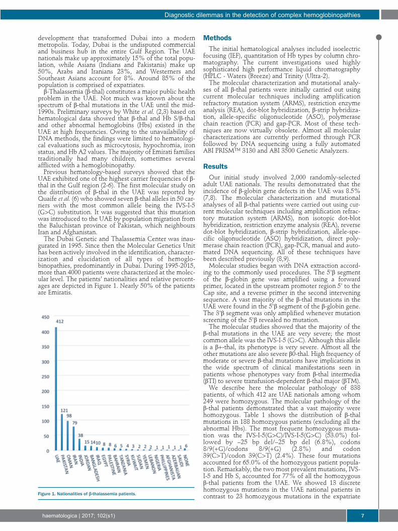
gurated in 1995. Since then the Molecular Genetics Unithas been actively involved in the identification, character-ization and elucidation of all types of hemoglo-binopathies, predominantly in Dubai. During 1995-2015,more than 4000 patients were characterized at the molec-ular level. The patients’ nationalities and relative percent-ages are depicted in Figure 1. Nearly 50% of the patientsare Emiratis.
MethodsThe initial hematological analyses included isoelectric
focusing (IEF), quantitation of Hb types by column chro-matography. The current investigations used highlysophisticated high performance liquid chromatography(HPLC - Waters (Breeze) and Trinity (Ultra-2).The molecular characterization and mutational analy-
ses of all β-thal patients were initially carried out usingcurrent molecular techniques including amplificationrefractory mutation system (ARMS), restriction enzymeanalysis (REA), dot-blot hybridization, β-strip hybridiza-tion, allele-specific oligonucleotide (ASO), polymerasechain reaction (PCR) and gap-PCR. Most of these tech-niques are now virtually obsolete. Almost all molecularcharacterizations are currently performed through PCRfollowed by DNA sequencing using a fully automatedABI PRISM™ 3130 and ABI 3500 Genetic Analyzers.
ResultsOur initial study involved 2,000 randomly-selected
adult UAE nationals. The results demonstrated that theincidence of β-globin gene defects in the UAE was 8.5%(7,8). The molecular characterization and mutationalanalyses of all β-thal patients were carried out using cur-rent molecular techniques including amplification refrac-tory mutation system (ARMS), non isotopic dot-blothybridization, restriction enzyme analysis (REA), reversedot-blot hybridization, β-strip hybridization, allele-spe-cific oligonucleotide (ASO) hybridization, direct poly-merase chain reaction (PCR), gap-PCR, manual and auto-mated DNA sequencing. All of these techniques havebeen described previously (8,9). Molecular studies began with DNA extraction accord-
ing to the commonly used procedures. The 5'β segmentof the β-globin gene was amplified using a forwardprimer, located in the upstream promoter region 5' to theCap site, and a reverse primer in the second interveningsequence. A vast majority of the β-thal mutations in theUAE were found in the 5'β segment of the β-globin gene.The 3'β segment was only amplified whenever mutationscreening of the 5'β revealed no mutation.The molecular studies showed that the majority of the
β-thal mutations in the UAE are very severe; the mostcommon allele was the IVS-I-5 (G>C). Although this alleleis a β+-thal, its phenotype is very severe. Almost all theother mutations are also severe β0-thal. High frequency ofmoderate or severe β-thal mutations have implications inthe wide spectrum of clinical manifestations seen inpatients whose phenotypes vary from β-thal intermedia(βTI) to severe transfusion-dependent β-thal major (βTM).We describe here the molecular pathology of 838
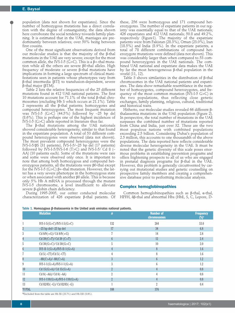
patients, of which 412 are UAE nationals among whom249 were homozygous. The molecular pathology of theβ-thal patients demonstrated that a vast majority werehomozygous. Table 1 shows the distribution of β-thalmutations in 188 homozygous patients (excluding all theabnormal Hbs). The most frequent homozygous muta-tion was the IVS-I-5(G>C)/IVS-I-5(G>C) (53.0%) fol-lowed by –25 bp del/–25 bp del (6.8%), codons8/9(+G)/codons 8/9(+G) (2.8%) and codon39(C>T)/codon 39(C>T) (2.4%). These four mutationsaccounted for 65.0% of the homozygous patient popula-tion. Remarkably, the two most prevalent mutations, IVS-I-5 and Hb S, accounted for 77% of all the homozygousβ-thal patients from the UAE. We showed 13 discretehomozygous mutations in the UAE national patients incontrast to 23 homozygous mutations in the expatriate
Diagnostic dilemmas in the detection of complex hemoglobinopathies
haematologica | 2017; 102(s1) 7
Figure 1. Nationalities of β-thalassemia patients.
population (data not shown for expatriates). Since thenumber of homozygous mutations has a direct correla-tion with the degree of consanguinity, the data shownhere corroborate the social tendency towards family plan-ning. It is estimated that in the UAE, marriages are pre-dominantly between relatives; over 50% being betweenfirst cousins.One of the most significant observations derived from
our molecular studies is that the majority of the β-thalmutations in the UAE are very severe; except for the mostcommon allele, the IVS-I-5 (G>C). This is a β+-thal muta-tion while all the others are severe β0-thal alleles. Highfrequency of moderate or severe β-thal mutations haveimplications in forming a large spectrum of clinical mani-festations seen in patients whose phenotypes vary fromβ-thal intermedia (βTI) to transfusion-dependent, severeβ-thal major (βTM).Table 2 lists the relative frequencies of the 25 different
mutations found in 412 UAE national patients. The first10 mutations account for 71.1% of the total β-thal chro-mosomes (excluding Hb S which occurs at 21.1%). Table2 represents all the β-thal patients; homozygotes andcompound heterozygotes. The most frequent mutationwas IVS-I-5 (G>C) (44.5%) followed by –25 bp del(8.6%). This is perhaps one of the highest incidences ofIVS-I-5 (G>C) allele reported in literature thus far.The β-thal mutations among the UAE nationals
showed considerable heterogeneity, similar to that foundin the expatriate population. A total of 53 different com-pound heterozygotes were observed (data not shown).The most prevalent compound heterozygotes were theIVS-I-5/βS (31 patients), IVS-I-5/–25 bp del (17 patients)followed by IVS-I-5/IVS-I-6 (T>C) and IVS-I-5/ Cd 8 (–AA) (10 patients each). Some of the mutations were rareand some were observed only once. It is important tonote that among both homozygous and compound het-erozygous patients, all the mutations were β0-thal exceptfor the IVS-I-5 (G>C) β+-thal mutation. However, the lat-ter has a very severe phenotype in the homozygous stateor when associated with another β0 allele. This is becauseonly 5% Hb A mRNA is processed through the mutantIVS-I-5 chromosome, a level insufficient to alleviatesevere β-globin chain deficiency.During 1995-2005, our center conducted molecular
characterization of 426 expatriate β-thal patients. Of
these, 256 were homozygous and 171 compound het-erozygotes. The number of expatriate patients in our reg-istry was essentially equal to that of the UAE nationals;426 expatriates and 412 UAE nationals; 50.8 and 49.2%,respectively (Figure1). The majority of the expatriatepatients were from Pakistan (28.3%), Oman (23.0%), Iran(18.5%) and India (8.9%). In the expatriate patients, atotal of 78 different combinations of compound het-erozygote mutations were defined (data not shown). Thiswas considerably larger than when compared to 53 com-pound heterozygotes in the UAE nationals. The com-bined UAE national and expatriate data makes the UAEby far the most heterogeneous β-thal population in theworld (11, 12).Table 3 shows similarities in the distribution of β-thal
chromosomes in the UAE national patients and expatri-ates. The data show remarkable resemblance in the num-ber of homozygotes, compound heterozygotes, and fre-quency of the most common mutation (IVS-I-5 G>C) inthe two populations, thus reflecting close geneticexchanges, family planning, religious, cultural, traditionaland historical traits.Hitherto, our molecular studies revealed 66 different β-
thalassemia mutations in the UAE population (Figure 2).In perspective, the total number of mutations in the UAEsurpasses the combined number of mutations reportedfrom China and India, just over 32. These are the twomost populous nations with combined populationsexceeding 2.5 billion. Considering Dubai’s population of2.5 million, this accounts to one thousandth of the abovepopulations. The data reported here reflects considerablediverse molecular heterogeneity in the UAE. It must benoted that the genetic diversity of this scale poses enor-mous problems in establishing prevention programs andoffers frightening prospects to all of us who are engagedin prenatal diagnosis programs for β-thal in the UAE.However, this problem is generally circumvented by car-rying out mutational studies and genetic counselling onprospective family members and creating a comprehen-sive database prior to performing molecular analysis.
Complex hemoglobinopathiesCommon hemoglobinopathies such as β-thal, α-thal,
HPFH, δβ-thal and abnormal Hbs (HbE, S, C, Lepore, D-
E. Baysal
8 haematologica | 2017; 102(s1)
Table 1. Homozygous β-thalassemia in the United arab emirates national patients. Mutation n Number of Frequency chromosomes (%)a
1 IVS-I-5(G>C)/IVS-I-5(G>C) 132 264 53.02 –25 bp del/–25 bp del 17 34 6.83 Cd 8/9(+G)/ Cd 8/9(+G) 7 14 2.84 Cd 39(C>T)/ Cd 39 (C>T) 6 12 2.45 Cd 30(G>C)/ Cd 30(G>C) 5 10 2.06 IVS-II-1(G>A)/IVS-II-1(G>A) 4 8 1.67 Cd 5(–CT)/Cd 5(–CT) 4 8 1.68 –88(C>A)/–88(C>A) 3 6 1.29 IVS-I-1(G>A)/IVS-I-1(G>A) 3 6 1.210 Cd 15(G>A)/ Cd 15(G>A) 2 4 0.811 Cd 8(–AA)/ Cd 8(–AA) 2 4 0.812 IVS-I-110(G>A)/IVS-I-110(G>A) 2 4 0.813 Cd 82/83(–G)/ Cd 82/83(–G) 1 2 0.4TOTAL 188 376aExcluded from the table are Hb SS (23.7%) and Hb DD (0.8%).

Punjab, O-Arab) are found in the UAE and they aredetected easily. Their accurate identification can providevaluable diagnostic and prognostic options for cliniciansand for couples who may consider family planning.The diagnosis of hemoglobinopathies has become an
increasing challenge in the multinational countries suchas Australia, USA, Canada as well as in Europe and theGulf Countries. This is well exemplified by Dubai whichis home to around 230 different nationalities (2015 cen-sus), hence the propensity for significantly enriched tha-lassemia gene pool coupled with high degree of consan-guinity. In an attempt to curb the hemoglobinopathyproblem, the National Premarital Screening Program(PMS) became mandatory in Dubai in 2006 for all nation-alities and the Prenatal Diagnosis Program (PND) hasbeen underway successfully since 2005. These preventionprograms were rendered mandatory, as hemoglo-binopathies are a major public health concern in the UAE. Dubai is arguably the most heterogeneous hemoglo-
binopathy nation in the world with 66 β-globin genedefects reported to date. It is anticipated that various com-plex hemoglobinopathies with extensive heterogeneity ingenotype and variable phenotype will emerge from suchadmixture of genes in a small nation where first cousinmarriage among the indigenous population is a norm andnot an exception. In addition, α-thal and β-thal interactionsoccur due to relatively high frequencies of α globin and βglobin gene defects; 50% and 8.3%, respectively (11, 12).This makes clinical diagnosis and laboratory evaluationmuch more challenging in a young nation where 26% ofthe population is below age 15 and 71% is between 15-64. The laboratory diagnoses of hemoglobinopathies are
often made with certain assumptions:(a) Variation in the phenotype could be a reflection of
interplay between different abnormal globin genes (α,β, γ, δ).
(b) Same genotypes may have different phenotypes indifferent geographical areas thus denoting the role ofenvironment as a modulator.
(c) Complex genotypes occur rarely so no concrete con-clusions must be drawn from only a few examples.The complex hemoglobinopathies provide valuable
information and guidance to clinicians, counsellors andhealthcare providers. Extra care and vigilance is requiredto identify the causative mutations in these cases. Theserare cases also highlight and emphasize the importance ofaccurate laboratory assessment and interpretation espe-cially with reference to different normal ranges for MCV,MCH, HbA2 quantitation.
Prenatal diagnosisPrenatal diagnosis (PND) of hemoglobinopathies in the
UAE was imminent following the establishment of
mandatory premarital screening program in 2005 and theadvances made in chorionic villus sampling (CVS) and inDNA-based diagnostics. Advances in CVS aspirationhave rendered the first trimester PND a standard practice.Direct detection of mutant genes has enabled many cou-ples to seek DNA diagnostic services in the first trimesterof pregnancy. The procedure empowers couples to con-sider options through proper genetic counselling coupledwith informed consent. In the UAE, PND has beenemployed since 2005 as a principal diagnostic means todetermine fetal DNA status for β-thal. The ability to detect mutant globin genes in CVS has
provided a rapid, safe, accurate, reliable and affordable
Diagnostic dilemmas in the detection of complex hemoglobinopathies
haematologica | 2017; 102(s1) 9
Table 2. β-thalassemia Gene Frequency among the Emirati patients(n=412). Mutation No. of Gene frequency chromosomes (%)a
1 IVS-I-5 (G>C) 367 44.52 –25 bp deletion 71 8.63 Codons 8/9 (+G) 25 3.04 IVS-II-1 (G>A) 23 2.85 Codon 39 (C>T) 18 2.26 Codon 8 (–AA) 18 2.27 Hb D-Punjab (GAA>CAA) 18 2.28 Codon 30 (G>C) 17 2.19 Codon 5 (–CT) 17 2.110 IVS-I-6 (T>C) 12 1.511 –88 (C>A) 9 1.112 Codons 82/83 (–G) 8 1.013 IVS-I-110 (G>A) 8 1.014 IVS-I-5 (G>T) 7 0.915 Codon 15 (G>A) 7 0.916 Codon 44 (–C) 6 0.717 Codon 110 (T>C) 3 0.418 IVS-II-848 (C>A) 3 0.419 Poly A site (AATAAA>AATAAG) 3 0.420 –101 (C>T) 2 0.221 Hb Knossos (codon 27, G>T) 1 0.122 Codon 37 (G>A) 1 0.123 Codons 36/37 (–T) 1 0.124 Hb E (codon 26, G>A) 1 0.125 δβ deletion 1 0.1aExcluded from the table is Hb S (21.1%).
Table 3. Similarities in β-thalassemia alleles between emirati and expatriate patients.Patientsa UAE Expatriates n: 412 n: 426
Homozygotes 249 60.0% 264 62.0%Compound heterozygotes 163 40.0% 162 38.0%IVS-I-5(G>C)/IVS-I-5(G>C) (in all homozygotes) 132 54.0% 130 49.2%IVS-I-5(G>C)/IVS-I-5(G>C) (in all patients) 132 32.0% 130 31.0%IVS-I-5 (G>C) chromosomes 367 44.5% 336 39.4%Homozygous mutations 15 23Compound heterozygous mutations 53 78aHb S patients are excluded.

methodology for the early detection of many fatalgenetic diseases. The genetic information enables thecouples to reach a decision compatible with their beliefsand family planning criteria.In the Dubai Genetics Center, we employ the most
advanced diagnostic tools including PCR and DNASequencing to diagnose many hemoglobinopathiesencompassing thalassemias (α & β-thal), sickle cell dis-ease (SCD) and abnormal hemoglobins. The DNA isextracted from CVS using Qiagen kits and is amplified byPCR using specific primers. The amplicons are sequencedon an ABI Genetic Analyzer 3130 and 3500. The resultsare reported within 24 hours. Without exception, thematernal contamination is excluded for each sample withSTR Cofiler®.Our results demonstrate that since 2005, PND has been
available for pregnancies at risk for virtually all inheritedhemoglobin disorders in the UAE. Nearly 200 coupleshave been tested using the CVS and subsequent DNAanalyses involving PCR and DNA Sequencing. The cou-ples were predominantly from the UAE. Others werefrom Bahrain, Kuwait, India, Pakistan and other countriesin the region. The couples with affected fetuses werecounselled and given appropriate available options. OurPND data will be not presented here.
Summary• The IVS-I-5 (G-C) allele is the most prevalent mutationamong the Emiratis and the expatriates with a frequen-cy of >50%.
• Sickle cell gene (β-sickle) is the second most prevalentallele accounting for >20% of the chromosomes.
• Significantly high incidence of homozygous mutationsdemonstrate the degree of consanguinity among theUAE nationals.
• Frequency of β gene defects that encompass β-thal, β-sickle and β-variants is 8.3%.
• Frequency of various α-thal genotypes is around 50%• UAE is the most heterogeneous population with 66alleles at present surpassing the most populated nationslike China and India put together.
• Most of the mutations identified in the UAE populationare β0/+ leading to very severe phenotype.
• Premarital Screening (PMS) is mandatory since 2006.• Prenatal diagnosis (PND) was implemented in 2005 andresulted in decreasing the β-thal births dramatically. Atpresent there is significantly high demand for thisrapid, sensitive, accurate, affordable procedure acrossall factions of society.
ConclusionsIn conclusion, accurate detection and counselling of at-
risk couples is a promising way to reduce the mortalityand morbidity from β-thalassemia in countries where it isprevalent.
AcknowledgmentsThe author would like to thank the technical staff at the
Molecular Genetics Unit and all the colleagues at the DubaiThalassemia Center.
E. Baysal
10 haematologica | 2017; 102(s1)
Figure 2. Heterogeneity of β-thalassemia mutations worldwide as measured by the total number of alleles in a given country or region.

References1. Al Hosani H. Health for all in the UnitedArab Emirates. East Mediterr Health J.2000;6(4):838-840.
2. White JM, Byrne M, Richards R, et al.Thalassaemia genes in Peninsular Arabs. BrJ Haematol. 1985;60(2):269-278.
3. White JM, Byrne M, Richards R, BuchananT, Katsoulis E, Weerasingh K. Red cellgenetic abnormalities in Peninsular Arabs:sickle haemoglobin, G6PD deficiency, andα and β thalassaemia. J Med Genet. 1986;23(3):245-251.
4. Al-Gazali LI, Bener A, Abdulrazzaq YM,Micallef R, al-Khayat AI, Gaber T.Consanguineous marriages in the United
Arab Emirates. J Biosoc Sci. 1997;29(4):491-497.
5. El Hazmi MA, Al-Swailem AR, Warsy AS.Molecular defects in β thalassaemias in thepopulation of Saudi Arabia. Hum Hered.1995;45(5):278-285.
6. Quaife R, al Gazali L, Abbas S, et al. Thespectrum of β-thalassemia mutations inthe UAE national population. J Med Genet.1994;31(1):59-61.
7. Baysal E. Advances in HemoglobinopathiesResearch – Hemoglobinopathies in theUnited Arab Emirates. Hemoglobin.2001;25(2):247-253.
8. Baysal E. Molecular heterogeneity of β-tha-lassemia in the United Arab Emirates.Community Genet. 2005;8(1):35-39.
9. El Kalla S, Baysal E. Genotype-phenotypecorrelation of sickle cell disease in theUnited Arab Emirates. Pediatr HematolOncol. 1998;15(3):237-242.
10. Baysal E, Yousef I, Zeinali M, et al.Molecular basis of hemoglobinopathies inthe UAE: implications for prenatal diagno-sis and prevention programs. EmiratesMed J. 2007;25(1):7-21.
11. Baysal, E., Molecular basis of α-tha-lassemia in the United Arab EmiratesHemoglobin; 35 (5-6):574-80, 2011. PMID:22074123.
12. Baysal, E., Molecular basis of β-tha-lassemia in the United Arab Emirates.Hemoglobin; 35 (5-6):581-88, 2011. PMID:22074124.
Diagnostic dilemmas in the detection of complex hemoglobinopathies
haematologica | 2017; 102(s1) 11

12 haematologica | 2017; 102(s1)
©2017 Ferrata Storti FoundationMaterial published in Haematologica is cov-ered by copyright. All rights reserved to theFerrata Storti Foundation. Copies of articlesare allowed for personal or internal use.Permission in writing from the publisher isrequired for any other use.
Correspondence: [email protected]
Ferrata StortiFoundation
EUROPEANHEMATOLOGYASSOCIATION
Haematologica 2017Volume 102(s1):12-19
ARTICLE Session 2: How I treat and monitor
How I treat and monitor sickle cell diseaseWasil Jastaniah1,2
1Department of Pediatrics, Faculty of Medicine, Umm AlQura University, Makkah, SaudiArabia; 2Princess Noorah Oncology Center, King Abdulaziz Medical City, Jeddah, SaudiArabia
ABSTRACT
IntroductionSickle cell disease (SCD) is an autosomal recessive disorder that results from a
point mutation leading to a single nucleotide substitution (A to T) in the codonfor the 6th amino acid of the beta-globin chain and, as a result, substitutes thehydrophobic amino acid valine for the normal hydrophilic amino acid glutamicacid [1]. This results in hemoglobin polymerization and precipitation leading tored cell membrane damage and deformity into sickle shaped cells. This leads toincreased red cell destruction (hemolysis) and red cell clumping in vessels (vaso-occlusion). The hemolytic and vasco-occlusive phenotype lead to the spectrumof clinical manifestations observed in patients with SCD from severe anemia, legulcers,, and pulmonary hypertension to recurrent pain crisis, acute chest syn-drome, splenic infarction, stroke, and avascular necrosis on the other end of thespectrum [2]. The clinical severity of these manifestations are influenced andmodified by genetic and environmental factors that result in the variabilityobserved between patients with SCD [1,2].SCD is a global health problem affecting approximately 2-3% of the world
population with variable geographic distribution [3]. The prevalence of sicklecell trait reaches up to 40% in some regions such as central India, the Easternprovince of Saudi Arabia, and Sub-Saharan Africa and, worldwide, approximate-ly 275,000 are born with SCD annually and need early diagnosis, monitoring,and treatment [3,4,5,6]. In developed countries, early newborn screening, peni-cillin prophylaxis, comprehensive care programs, institution of disease modify-ing therapies such as hydroxyurea, blood transfusion, and chelation therapy, inaddition to, hematopoietic stem cell transplant (HSCT) as curative therapy haveimproved the survival and quality of life of patients with SCD and the majorityof children are expected to survive well into adulthood [7]. In contrast, 90% ofchildren in resource-poor countries do not reach adulthood [3,4,5].In this review, the importance of early screening, penicillin prophylaxis,
immunization, monitoring and screening, institution of a comprehensive careprogram, and transcranial Doppler (TCD) screening and stroke prevention willbe reviewed. In addition, the review will highlight the indications of hydrox-yurea, transfusion therapy, and HSCT in SCD. Finally, the review will focus onthe current challenges faced in developing countries when dealing with patients
Sickle cell disease is the most common genetic hemoglobin disor-der worldwide with thousands of children born yearly with thisdisease. Advances in our understanding of sickle cell disease man-
agement has improved over the years and it is now expected that, withearly screening, monitoring, prevention, and intervention, childrenwith sickle cell disease can live well into adulthood. However, the bur-den of the disease is shifting to adulthood in developed and resource-rich countries. In contrast, due to the limited access to comprehensivecare programs for children with sickle cell disease in resource-poorcountries, high morbidity and early mortality of patients with sicklecell disease remains a major global problem. Therefore, this review willdiscuss the strategy and recommendations we use in monitoring andtreating patients with sickle cell disease in our center. The review willalso highlight some of the challenges faced in less developed countries.

with SCD. The recommendations made are based onthe presented evidence and the standard practice used inthe authors’ center.
Early screening and monitoringEarly newborn screening, initiation of penicillin pro-
phylaxis in early infancy, and early education have beenshown to reduce the risk of mortality from infections ininfants and young children with SCD [8]. However, uni-versal newborn screening is not implemented in allcountries around the globe and reinforcing the impor-tance of including SCD in newborn screening programsparticularly in areas with high prevalence of the diseaseshould be a priority to be able to initiate early preventa-tive and screening measures to reduce the morbidity andmortality associated with SCD. This includes the earlyinitiation of penicillin prophylaxis, immunizations, andproviding access to a comprehensive SCD program.
Penicillin prophylaxisInfection has been the leading cause of death in chil-
dren with SCD under the age of 5 years. The early initi-ation of penicillin prophylaxis has been shown, in therandomized trial on prophylaxis with oral penicillin inchildren with sickle cell anemia (PROPS I) study, toreduce the risk of developing serious pneumococcalinfection in SCD children under the age of 3 years by84% and abolish mortality from this infection [9].Subsequently, the PROPS II study showed that, in chil-dren without history of severe pneumococcal infectionor surgical splenectomy, discontinuing penicillin pro-phylaxis at the age of 5 years may be safe [10].Therefore, early initiation of penicillin prophylaxisbetween the age of 2 to 4 months at a dose of 125 mgtwice daily until the age of 3 years, and 250 mg twicedaily until the age of 5 years is recommended.Discontinuing penicillin prophylaxis after the age of 5years in children with SCD who have completed theirimmunization schedule, had no prior invasive pneumo-coccal infection, or surgical splenectomy is safe.However, it is important to educate the family regardingthe importance of seeking medical intervention in caseof fever. Therefore, assessment of social circumstancesthat may lead to delayed access to care needs to be con-sidered before discontinuing prophylactic therapy.
ImmunizationsThe delivery of routine immunizations to children is
considered one the most important and cost-effectivepreventive measures that has saved many childhoodlives over the years. Patients with SCD are at increasedrisk of invasive bacterial infection from encapsulatedorganisms [11]. Therefore, in addition to the routinechildhood immunizations, we give the 23-valent pneu-mococcal polysaccharide vaccine (PPSV23) at 2 yearsand a second repeated dose at 5 years. In addition, werecommend giving the meningococcal (MenACWY) vac-cine 3 years after completing the primary vaccines and abooster repeated every 5 years thereafter. Furthermore,in our practice we recommend to give children abovethe age of 6 months the seasonal influenza vaccineannually. Patients planned for an elective splenectomyshould also be planned to receive all vaccines in additionto vaccination against the pneumoccal (PCV13) andhemophillus influenza b (Hib) vaccine (if not previouslyreceived), the PPSV23, and the MenACWY with the lastvaccine given at least 2 weeks prior to surgical splenec-tomy. These patients should continue on lifelong peni-cillin prophylaxis.
Monitoring and treatment of SCDThe first visit of a patient and his/her family with SCD
is an important visit to conduct proper education andexplain the natural history of SCD with its potential com-plications. The visit should include explanation regardingemergency signs that should alert the family to seekimmediate medical attention such as high fever above38.5 degrees Celsius, pain and swelling in the extremities,respiratory symptoms, abdominal pain and distension,neurological symptoms, increased pallor or fatigue, pri-apism, or vomiting and diarrhea. Teaching the familyhow to perform a spleen exam is also important andneeds to be reviewed on subsequent visits. The frequencyand goals of follow up and monitoring should beexplained. The family should be able to understand thechronic nature of the disease and the importance of earlyprevention to reduce the short- and long-term morbidityand mortality associated with this disease. The monitor-ing methods and potential need for transfusion should beexplained. Based on the age of the child, anticipatoryguidance to reinforce the importance of screening andprevention is important. As the child grows to school age,communication with the school via a medical report andinstructions regarding hydration and symptoms thatwould require immediate medical attention should behighlighted. Advice regarding screening of family mem-bers and newborns, if not routinely available, should berecommended during the first visit as well.
Comprehensive care programDuring the first visit, we usually show the plan of
monitoring and introduce the family to the concept ofcomprehensive care upfront to explain the rational forthe different screening modalities as summarized inTable 1.As shown in Table 1, the frequency of visits is once
every 2 months in the first 6 months, followed by onceevery 3 months until 18 months of age, and then 6monthly thereafter. During each visit, we review historyof sickle cell crisis, admissions, transfusion history,home medication use, daily activity, and sleeping diffi-culties including snoring. In addition, monitoring vitalsigns including blood pressure, pain score, and growthparameters are important to record baseline values.Documentation of the spleen size, if palpable, hip andshoulder range of movement, respiratory, and neurolog-ical exam. These, together with the laboratory andimaging surveillance, can help in stratifying the severityof the SCD phenotype to adjust the frequency of visitsand implement timely preventative measures. As thepatient approaches 10 years of age, it is our practice toperform a cardiac evaluation, if not previously done, toassess for prolonged QTc, echocardiogram to assess tri-cuspid jet velocity to screen for pulmonary hyperten-sion, and a pulmonary function test to screen for under-lying lung disease. Although, the evidence to supportroutine screening in asymptomatic children is notstrong, we believe that these baseline exams can helppredict and guide in risk stratification of the severity ofthe sickle cell phenotype and the need for more frequentmonitoring, in addition to delivering early interventionsas appropriate. Similarly, we screen asymptomaticpatients in our center for silent infarcts by brainMRI/MRA when the patient is old enough to get it donewithout any sedation. As the patient graduates to adult-hood, we ensure that a full assessment including oph-thalmology retinal exam, screening for avascular necro-sis by x-ray or MRI based on symptoms, and urinalysisfor microalbuminuria is performed.
How I treat and monitor sickle cell disease
haematologica | 2017; 102(s1) 13

TCD and stroke preventionOver the past 20 years, our understanding and man-
agement of neurological complications in SCD hasimproved. This is reflected by the reduction in risk ofovert stroke in countries utilizing Trans cranial Doppler(TCD) ultrasound screening for central nervous system(CNS) vasculopathy. Adams and colleagues have shownthat starting patients with abnormally high TCD veloc-ities (above 200 cm/second) on a strict chronic transfu-sion program that targets reducing the hemoglobin Slevel to less than 30%, can reduce the risk of develop-ment of a first stroke [12,13]. The STOP 1 and STOP 2trial confirmed the benefit of continued chronic transfu-sion in children between the ages of 2 and 16 years withSCD in primary stroke prevention [12,13]. In fact, theroutine use of TCD screening together with regularblood transfusion therapy has reduced the prevalence ofovert stroke in developed countries from approximately11% to 1% [14]. Recently, the TWiTCH trial has shownthat bridging patients, who convert to normal TCDvelocities on chronic transfusion therapy, using hydrox-yurea until maximum tolerated dose is achieved thenswitching them to hydroxyurea alone was not inferiorto continuation of chronic transfusion for primary stokeprevention [15]. This latter study, together with others,suggests a beneficial role of hydroxyurea therapy in pri-mary stoke prevention [16,17,18]. However, optimalpatient selection and monitoring is required.In contrast, overt stroke remains a challenging and dev-
astating problem. Chronic transfusion therapy for sec-ondary stroke prevention can reduce the recurrence rateof overt stroke by 90% [18,19,20]. However, approxi-mately half of patients with overt stroke on chronic trans-fusion continue to develop progressive vasculopathy sug-gesting the need for alternative approaches to therapy[21]. Switching patients with overt stroke from chronictransfusion to hydroxyurea has not been proven to beeffective based on the SWiTCH trial which was terminat-ed early [22]. However, hydroxyurea is superior to notherapy in preventing stroke recurrence [23].
Silent cerebral infarct is the most frequent neurologicalcomplication reported in patients with SCD [14,24]. Thecumulative prevalence of silent cerebral infarct increaseswith age and it is estimated that approximately 53.3% ofpatients suffer from a silent cerebral infarct by the 4thdecade of life [14]. Patients with silent cerebral infarcthave an increased risk of developing overt stroke com-pared to SCD patients without silent cerebral infarct[14,24,25]. The SIT trial studied the benefit of transfusiontherapy compared to standard care and showed thattransfusions resulted in a relative risk reduction of 58% inthe occurrence of all neurological events [25]. Based onthe results of the SIT trial, patients with silent cerebralinfarct may be offered transfusion therapy to prevent pro-gression of neurological events [25]. However, as the ben-efit of transfusion to prevent progression was incomplete,further modalities to address the optimal management ofpatients with silent cerebral infarct are needed. The use ofhydroxyurea or HSCT as alternative therapies to bloodtransfusions in patients with silent cerebral infarctrequires further studies.
HydroxyureaHydroxyurea (or hydroxycarbamide) is a ribonu-
cleotide reductase inhibitor that has been used inpatients with SCD for more than 20 years now [26-28].Many experts believe that hydroxyurea has changed thelives of patients with SCD. Hydroxyurea exerts itseffects by inducing fetal hemoglobin, improving labora-tory parameters through its effects on hematopoiesisand thus reducing the inflammatory state, reducinghemolysis, and improving nitric oxide bioavailability[27]. Overwhelming studies have shown the effective-ness and safety of hydroxyurea in both adults and chil-dren in ameliorating the clinical complications andorgan dysfunction, reducing the need for blood transfu-sions, decreasing the rate of hospitalizations, and atten-uating mortality [26,28,29]. Based on the available evi-dence, the most recent published National Institute of
W. Jastaniah
14 haematologica | 2017; 102(s1)
Table 1. Sickle cell disease comprehensive screening and monitoring.
Age History/Exam CBC HPLC RBC Chemistry U/A Pulse O2% CXR ECG, CNS GB U/S Ophth phenotype Echo, PFT Consult

Health Evidence Based Expert Panel Report onManagement of SCD recommended that every childwith SCD should be offered hydroxyurea from the ageof 9 months even if asymptomatic [30]. In our practice,we offer to the family hydroxyurea with the firstepisode of sickle cell-related crisis if the child is 9months of age or older. The indications, dose, and mon-itoring of hydroxyurea based on our practice is summa-rized in Table 2.
TransfusionRed blood cell transfusion therapy is the first therapy
used to target the pathophysiology of SCD. The indica-tions of transfusion therapy have expanded over theyears and more children are now being transfusedchronically to prevent stroke and other complications ofSCD. There are two methods of transfusion in patientswith SCD: intermittent/simple transfusions andexchange transfusions. In contrast to transfusion thera-py in patients with thalassemia, the goal of transfusionin SCD is to improve the anemia and reduce the sicklehemoglobin (Hb S) level to reduce the hemolysis andviscosity associated with SCD. However, due to thepresence of sickle cells in the blood, the viscosity of theblood in patients with SCD is increased. Therefore, cau-tion should be practiced when prescribing blood trans-fusion therapy to patients with SCD to avoid hypervis-cosity and its deleterious effects on patients with sicklecell. The hemoglobin level should not exceed 11 gm/dl,and that is why exchange transfusion may be the onlymethod to transfuse patients with SCD presenting witha hemoglobin levels above 9 gm/dl. The indications forred cell transfusions in SCD are outlined in Table 3.
The long-term use of blood transfusion has its conse-quences and patients with SCD receiving multiple orchronic transfusions develop iron overload that requiresmonitoring and chelation therapy. Chelation therapy inpatients with SCD should be initiated when the totalnumber of life time transfusions reaches 20 simple trans-fuisons (120 ml/kg) or when the liver iron concentration,as assessed by a quantitiative measurement (MRI meas-urement), exceeds 7 mg Fe/g liver dry weight [30,31].Defiraxirox therapy has been reported to be effectiveand safe in treating iron overload in patients with SCDeven if hydroxyurea is concomitantly used [32,33].Other complications of transfusion therapy in patientswith SCD include: alloimmunization, venous access,compliance, cost, and infection. These complications areincreased in patients receiving exchange transfusion dueto the increased exposure to blood with each exchangetransfusion compared to simple transfusions. Therefore,efforts are needed to reduce the risk of these complica-tions and further research is required to find alternativesto transfusion therapy.
Indication for HSCTHSCT is the only available curative therapy in
patients with SCD to date. Human leukocyte antigen(HLA)-identical related donor HSCT for children withSCD has a long-term overall survival rate of 94-97%, anevent-free survival rate of 84-86%, and a graft rejectionrate of approximately 10% as reported by the NorthAmerican and European studies [34,35]. Despite thishigh success rate, HSCT remains limited by optimalpatient selection criteria, infertility risk with currentconditioning regimens, availability of donors, and cost.
How I treat and monitor sickle cell disease
haematologica | 2017; 102(s1) 15
Table 2. How I use hydroxyurea in sickle cell disease.Indications
1. Dactylitis or pain crisis 2. Acute chest syndrome 3. Hemolytic crisis requiring transfusion therapy 4. Conditional TCDI 5. Parent preference in patient with abnormal TCDI or after TCDI normalization with chronic transfusion 6. Parent preference in patient with Silent cerebral infarct (hydroxyurea vs. chronic transfusion program) 7. Secondary stroke prevention if transfusion not possible or in combination with transfusion if progressive vasculopathy or increased frequency of exchange transfusion 8. Parent request (age 9 months or older)Dose
Start at 15 mg/kg/day and increase to maximum tolerated dose (maximum dose 30-35 mg/kg/day)Monitoring
1. Before starting perform baseline CBC with differential, reticulocyte count, hemoglobin electrophoresis (HPLC), Creatinine, and LFT (ALT, bilirubin) 2. Follow up 1 month after starting or escalating the dose to assess for toxicity with CBC and differential (hold if ANC <1x10⁹/L or platelet count <100 and reduce dose if ANC=1 -1.5x10⁹/L or platelet count <150. Restart at lower dose once recovered) 3. Escalate dose monthly, by 5 mg/kg/dose, to maximum dose or maximum tolerated dose, whichever occurs first 4. Once maximum/maximum tolerated dose reached, follow the patient every 3-4 months and assess for compliance and toxicity (CBC with differential, MCV, creatinine, ALT, reticulocyte count) 5. Assess for efficacy by reviewing history (crisis, admissions, and blood transfusions), MCV, WBC, reticulocyte count, bilirubin, LDH, and measure Hb electrophoresis twice yearly until HbF level stable then measure as needed

The indication of HSCT in patients with SCD is summa-rized in Table 4. These indications are based on theselection criteria used in published clinical trials [36].However, the timing of HSCT is important as delayingtransplant after significant morbidity like stroke mayaffect the quality of life of such patients. Other criteriathat may be considered, as indication for HSCT, isabnormal high-risk TCD velocities on screening for pri-mary stroke prevention. However, the recent resultsfrom the TWiTCH trial showing the efficacy of hydrox-yurea therapy as an alterantive to chronic blood transfu-sion may challenge this indication [15]. Further studiesare therefore needed to study the best timing, patientselection, conditioning regimen, and donor source inpatients with SCD.
Challenges in less developed countriesThe majority of children with SCD are born in less
developed countries. It is reported that 90% of childrenliving in resource-poor countries do not survive to adult-hood and more than half of children die before theirfifth birthday [37-39]. Therefore, there is a high demandto improve early screening and intervention strategies inless developed nations. The following are some of thechallenges that need to be addressed globally:
Newborn screening programs in developing countriesNewborn screening for sickle cell is not universally
available at the global level. Advocating for and initiat-ing early newborn screening will allow implementationof early antibiotic prophylaxis, optimize immuniza-tions, and early education to prevent early death frominfections. Alternatives to newborn screening such aspremarital screening programs to reduce at-risk mar-riages, preimplantation genetic diagnosis (PGD), andschool educational programs may help [40,41].However, these programs are either associated withhigh cost or have shown little, if any, benefit in reducingearly mortality rates from SCD, or in reducing the inci-dence of SCD in countries with high prevalence rates[40,41]. Therefore, there is a need to establish newbornand early screening and intervention programs in devel-oping countries.
Standard clinical care pathways and guidelinesIn developing countries, there is variation in resources
and in the clinical phenotype even within the samecountry, in addition, there is lack of targeted guidelines.This results in lack of standardized care delivery sys-tems and timely prevention, which impacts the diseaseburden and the utilization of resources. The variabilityin phenotype can result in a different spectrum of dis-
W. Jastaniah
16 haematologica | 2017; 102(s1)
Table 3. Indications for red blood cell transfusion in sickle cell disease. Acute/episodic transfusion Chronic (long-term) transfusion
Acute stroke Prevention of recurrent stroke Primary stroke prevention Prevention of recurrent SCI ACS Splenic sequestration Previous splenic sequestration (≤2–3 years) Pre-operatively (select cases) Heart failure/renal failure/chronic PHT Severe hemolytic anemia Chronic pain in HU non-responders Severe or long-lasting aplastic crises Short program: pregnancy Acute multiorgan-failure syndrome
Table 4. Indicaitons for hematopoietic stem cell transplant in patients with sickle cell disease. [36] Children Adult
Stroke or CNS event lasting >24 hours Age 15 to 40 years and any of the indications below: Stroke or CNS event lasting >24 hours Impaired neuropsychological function with abnormal MRI/MRA Regular RBC transfusion therapy to prevent vaso-occlusive clinical complications (i.e., pain, stroke, and acute chest syndrome) Recurrent acute chest syndrome Recurrent ACS in the 2-year period preceding HCT despite supportive care measures (i.e., asthma therapy and/or HU) Sickle lung disease 3 VOC per year in the 2-year period preceding enrollment despite Recurrent VOC or recurrent priapism supportive care measures (i.e., pain management and/or treatment Sickle nephropathy (GFR 30-50% of predicted normal) with HU) Alloimunization ECHO TRV Jet velocity >2.7 m/s

ease. For instance, the Arab-Indian haplotype has beenlabeled as benign for many years but patients with thisphenotype present with different patterns of morbidity[6,42]. This phenotype is predominant in certain regionsof the globe such as the Eastern province of SaudiArabia, Kuwait, Oman, and India. Patients with thishaplotype suffer from more splenic complications andavascular necrosis in childhood [6,42]. In addition, aspatients with this haplotype enter into adulthood, theysuffer from severe sickle cell related complicationsincluding acute chest syndrome, recurrent pain, stroke,and mortality peaking in the second and third decade oflife [43,44]. This suggests that regions with high preva-lence of the Arab-Indian haplotype may require compre-hensive care and intervention programs tailored to thenatural history of the specific phenotypic spectrum ofthe disease. Further studies in these regions are warrant-ed to deliver cost-effective interventions that wouldimprove the quality of care of these patients.Developing structured and standard guidelines based onphenotype predominance and resources is required tohelp improve the care in different regions around theglobe. This would require further clinical trials in identi-fying alternative and cost-effective therapeutic and pre-ventative approaches.
Demands and resources in less developed countriesAs the majority of patients with SCD live in less
developed countries, the availability of resources includ-ing access to care, transfusion and chelation therapy,cost of screening programs, or availability of sufficientresources to meet the high demand in specific countriesremains a big challenge [45]. This is even true inresource-rich countries. For example, a recent studyfrom Saudi Arabia (a resource-rich developing country)estimated the number of patients that were candidatesfor curative HSCT in the pediatric and adult populationusing strict HSCT indications and found that with thecurrent high demand, due to the high prevalence of thedisease, the numbers of transplant facilities were limitedto match the current demand [46]. Therefore, efforts inimproving preventative measures and delivery of dis-ease modifying therapies to improve the quality of lifeand reduce the morbidity and mortality of this diseaseremains a priority in developing nations.
Challenges in low-and middle-income countriesThe variability in the resource settings in low- and
middle-income countries have a major influence on thesurvival and quality of life of patients with SCD living inthese countries. This includes access to health care,access to medications, and access to blood transfusions.In fact, death from infection remains the major cause ofmortality in these countries [37-39,]. Therefore, focus onearly diagnosis and antimicrobial prophylaxis to preventinfection should be the main priority. Screening andaccess to healthcare may also be a challenge, therefore,consideration of disease modifying therapies such ashydroxyurea for all children 9 months of age or older ata fixed dose may be appropriate in these countries, thisis particularly true as blood transfusion therapy in some
countries are not routinely available. These challengeshighlight the need for alternative cost-effective primaryand secondary preventative strategies. Therefore, fur-ther research is needed to identify resourceful strategiesin treating and monitoring patients with SCD in thesecountries.
Transitioning to adulthoodThis is a common challenge all over the globe but may
be more pronounced in developing countries. Despite asteady decline in mortality rates among children withSCD in developed countries, the mortality rates inadults with SCD continues to increase [47]. The mostvulnerable age group at risk of morbidity and mortalityin patients with SCD in developed countries is nowshifted to the 18-30 year age group [7,48]. There aremultiple factors that increase the risk of morbidity andmortality in patients transitioning to adulthood [47,48].SCD is a chronic disease with a natural progressive his-tory, if not prevented or treated, can lead to increasedmorbidity and mortality as patient’s grow older and thedisease progresses. In addition, patient-related factorssuch as compliance to therapy during teenage years andthe psychosocial factors upon transitioning to adulthoodalso contribute to the vulnerability. Furthermore,resource and practice-related factors including the bur-den of the disease on resources, the limited adult SCDprograms/centers, limited access to specialized multidis-ciplinary sickle cell care in adult facilities, and the limit-ed research in prevention and management in adultpatients with SCD. Therefore, as the care and survivalof children with SCD improves globally the burden ofcare will shift to adulthood and this should be a focus ofcare at the global level.
ConclusionsSignificant advances have occurred over the past 20
years in caring for patients with SCD. The advances inprevention and early intervention have lead to improvedsurvival and quality of life of children with SCD. Theseinterventions involved the institution of simple meas-ures such as early newborn screening and early initia-tion of antimicrobial prophylaxis in addition to immu-nizations. Early monitoring and screening has allowedfor targeted preventative interventions such as hydrox-yurea and transfusion therapy. These comprehensivecare approaches in addition to curative therapies such asHSCT have contributed to the success in care made thusfar in SCD. It is now expected that children with SCDwill survive well into adulthood. However, challengesare now shifting to the adult age group and global chal-lenges continue to exist due to the limited resources anddifficulties in access to care in less developed countries.Therefore, establishing newborn screening and compre-hensive care programs that are cost-effective, increasingeducation, developing resource-specific standard of careguidelines to allow delivery of a structured approach tocare, and improving access to care needs to be priori-tized at the global level.
How I treat and monitor sickle cell disease
haematologica | 2017; 102(s1) 17

References1. Habara A, Steinberg MH. Minireview:Genetic basis of heterogeneity and severi-ty in sickle cell disease. Exp Biol Med(Maywood). 2016;241:689-96.
2. Gladwin MT, Vichinsky E. Pulmonarycomplications of sickle cell disease. NEngl J Med. 2008;359:2254-65.
3. Modell B, Darlison M. Global epidemiolo-gy of haemoglobin disorders and derivedservice indicators. Bull World HealthOrgan. 2008;86:480-7.
4. Serjeant GR. The Natural History ofSickle Cell Disease. Cold Spring HarborPerspectives in Medicine 2013;3: a011783.
5. Colah RB, Mukherjee MB, Martin S,Ghosh K. Sickle cell disease in tribal pop-ulations in India. Indian Journal ofMedical Research 2015;141: 509-515.
6. Jastaniah W. Epidemiology of sickle celldisease in Saudi Arabia. Ann Saudi Med.2011 May-Jun; 31(3): 289-293.
7. Quinn CT, Rogers ZR, McCavit TL,Buchanan GR. Improved survival of chil-dren and adolescents with sickle cell dis-ease. Blood. 2010;115(17):3447-3452.
8. Vichinsky E, Hurst D, Earles A, Kleman K,Lubin B. Newborn screening for sickle celldisease: effect on mortality. Pediatrics.1988;81:749-55.
9. Gaston MH, Verter JI, Woods G, PegelowC, Kelleher J, Presbury G, et al.Prophylaxis with oral penicillin in chil-dren with sickle cell anemia. A random-ized trial. N Engl J Med. 1986;314:1593-9.
10. Falletta JM, Woods GM, Verter JI,Buchanan GR, Pegelow CH, Iyer RV, et al.Discontinuing penicillin prophylaxis inchildren with sickle cell anemia.Prophylactic Penicillin Study II. J Pediatr.1995;127:685-90.
11. Ramakrishnan M, Moïsi JC, Klugman KP,Iglesias JMF, Grant LR, Mpoudi-Etame M,et al. Increased risk of invasive bacterialinfections in African people with sickle-cell disease: a systematic review andmeta-analysis. Lancet Infect Dis. 2010;10:329-37.
12. Adams RJ, McKie VC, Hsu L, Files B,Vichinsky E, Pegelow C, Abboud M,Gallagher D, Kutlar A, Nichols FT, BondsDR, Brambilla D. Prevention of a firststroke by transfusions in children withsickle cell anemia and abnormal results ontranscranial Doppler ultrasonography. NEngl J Med. 1998;339:5-11.
13. Adams RJ, Brambilla D; OptimizingPrimary Stroke Prevention in Sickle CellAnemia (STOP 2) Trial Investigators.Discontinuing prophylactic transfusionsused to prevent stroke in sickle cell dis-ease. N Engl J Med. 2005;353:2769-78.
14. Kassim AA, Pruthi S, Day M, RodeghierM, Gindville MC, Brodsky MA, DeBaunMR, Jordan LC. Silent cerebral infarctsand cerebral aneurysms are prevalent inadults with sickle cell anemia. Blood.2016;127:2038-2040.
15. Ware R et al. Hydroxycarbamine versuschronic transfusion for maintenance oftranscranial doppler flow velocities in chl-dren with sickle cell anaemia-TCD WithTransfusion Changing to Hydroxyurea(TWiTCH): a multicneter, open-label,phase 3, non-inferiority trial. The Lancet2016:387:661-670.
16. Hankins JS, McCarville MB, Rankine-Mullings A, Reid ME, Lobo CL, MouraPG, Ali S, Soares DP, Aldred K, Jay DW,Aygun B, Bennett J, Kang G, GoldsmithJC, Smeltzer MP, Boyett JM, Ware RE.
Prevention of conversion to abnormaltranscranial Doppler with hydroxyurea insickle cell anemia: A Phase III internation-al randomized clinical trial. Am JHematol. 2015;90:1099-105.
17. Bernaudin F, et al. Long-term treatmentfollow-up of children with sickle cell dis-ease monitored with abnormal transcra-nial Doppler velocities. Blood 2016;127:1814-1822.
18. Platt OS. Prevention and management ofstroke in sickle cell anemia. HematologyAm Soc Hematol Educ Program. 2006:54-57.
19. Powars D, Wilson B, Imbus C, Pegelow C,Allen J. The natural history of stroke insickle cell disease. Am J Med. 1978;65:461-71.
20. Pegelow CH, Adams RJ, McKie V,Abboud M, Berman B, Miller ST, OlivieriN, Vichinsky E, Wang W, Brambilla D.Risk of recurrent stroke in patients withsickle cell disease treated with erythro-cyte transfusions. J Pediatr. 1995;126:896-9.
21. Hulbert M L et al. Silent cerebral infarctsoccur despite regular blood transfusiontherapy after first strokes in children witsickle cell disease. Blood 2011;117:772-779.
22. Ware RE, Helms RW; SWiTCHInvestigators. Stroke With TransfusionsChanging to Hydroxyurea (SWiTCH).Blood 2012;119:3925-3932.
23. Kassim AA, Galadanci NA, Pruthi S,DeBaun MR. How I treat and managestrokes in sickle cell disease. Blood2015;125:3401-3410.
24. DeBaun MR, Kirkham FJ. Central nervoussystem complications and management insickle cell disease. Blood 2016;127:829-838.
25. DeBaun MR, Gordon M, McKinstry RC,Noetzel MJ, White DA, Sarnaik SA, et al.Controlled trial of transfusions for silentcerebral infarcts in sickle cell anemia. NEngl J Med. 2014;371:699-710.
26. Voskaridou E, Christoulas D, Bilalis A,Plata E, Varvagiannis K, Stamatopoulos G,Sinopoulou K, Balassopoulou A,Loukopoulos D, Terpos E. The effect ofprolonged administration of hydroxyureaon morbidity and mortality in adultpatients with sickle cell syndromes:results of a 17-year, single-center trial(LaSHS). Blood. 2010;115:2354-63.
27. Charache S, Terrin ML, Moore RD, DoverGJ, Barton FB, Eckert SV, McMahon RP,Bonds DR. Effect of hydroxyurea on thefrequency of painful crises in sickle cellanemia. Investigators of the MulticenterStudy of Hydroxyurea in Sickle CellAnemia. N Engl J Med. 1995;332:1317-1322.
28. Ware RE. How I use hydroxyurea to treatyoung patients with sickle cell anemia.Blood. 2010;115: 5300-11.
29. Strouse JJ, Heeney MM. Hydroxyurea forthe treatment of sickle cell disease: effica-cy, barriers, toxicity, and management inchildren. Pediatr Blood Cancer 2012;59:365-371.
30. Yawn BP, Buchanan GR, Afenyi-AnnanAN, Ballas SK, Hassell KL, James AH,Jordan L, Lanzkron SM, Lottenberg R,Savage WJ, Tanabe PJ, Ware RE, MuradMH, Goldsmith JC, Ortiz E, Fulwood R,Horton A, John-Sowah J. Management ofsickle cell disease: summary of the 2014evidence-based report by expert panelmembers. JAMA. 2014;312:1033-48.
31. Standards for the clinical care of adultswith sickle cell disease in the UK. SickleCell Society; 2008. Available from:h t t p : / / s i c k l e c e l l s o c i e t y. o r g /wp -content/uploads/2016/02/Standards-for-the-Clinical-Care-of-Adults-with-Sickle-Cell-Disease-in-the-UK.pdf
32. Vichinsky E, Bernaudin F, Forni GL,Gardner R, Hassell K, Heeney MM, InusaB, Kutlar A, Lane P, Mathias L, Porter J,Tebbi C, Wilson F, Griffel L, Deng W,Giannone V, Coates T. Long-term safetyand efficacy of deferasirox (Exjade) for upto 5 years in transfusional iron-over-loaded patients with sickle cell disease. BrJ Haematol. 2011;154:387-97.
33. Vichinsky E, Torres M, Glass J, et al. ARandomized Phase II Study Evaluating theEfficacy and Safety of Deferasirox VersusDeferoxamine in Patients with Sickle CellDisease (SCD): 2-Year Results IncludingPharmacokinetics (PK) and Safety ofDeferasirox with ConcomitantHydroxyurea Therapy. Blood 2011 118:1082.
34. Walters MC. Stem cell therapy for sicklecell disease: transplantation and genetherapy. Hematology Am Soc HematolEduc Program. 2005:66-73.
35. Bernaudin F, Socie G, Kuentz M, ChevretS, Duval M, Bertrand Y, Vannier JP,Yakouben K, Thuret I, Bordigoni P, FischerA, Lutz P, Stephan JL, Dhedin N, PlouvierE, Margueritte G, Bories D, Verlhac S,Esperou H, Coic L, Vernant JP, GluckmanE; SFGM-TC. Long-term results of relatedmyeloablative stem-cell transplantation tocure sickle cell disease. Blood. 2007;110:2749-56.
36. Walters MC, De Castro LM, Sullivan KM,Krishnamurti L, Kamani N, Bredeson C,Neuberg D, Hassell KL, Farnia S,Campbell A, Petersdorf E. Indications andResults of HLA-Identical SiblingHematopoietic Cell Transplantation forSickle Cell Disease. Biol Blood MarrowTransplant. 2016;22:207-11.
37. Grosse SD, Odame I, Atrash HK,Amendah DD, Piel FB, Williams TN.Sickle Cell Disease in Africa: A NeglectedCause of Early Childhood Mortality.American Journal of Preventive Medicine2011;41: S398-405.
38. Tewari S, Rees D. Morbidity pattern ofsickle cell disease in India: A single centreperspective. Indian Journal of MedicalResearch 2013;138: 288-290.
39. Piel FB, Hay SI, Gupta S, Weatherall DJ,Williams TN. Global Burden of Sickle CellAnaemia in Children under Five, 2010-2050: Modelling Based on Demographics,Excess Mortality, and Interventions. PLOS2013;10: e1001484.
40. Memish ZA and Saeedi MY. Six-year out-come of the national premarital screeningand genetic counseling program for sicklecell disease and β-thalassemia in SaudiArabia. Ann Saudi Med. 2011:31:229-235.
41. Mason K, Gibson F, Gardner R, Serjeant B,Serjeant GR. Prevention of sickle cell dis-ease: observations on females with thesickle cell trait from the Manchester proj-ect, Jamaica. J Community Genet. 2016; 7:127-132.
42. Serjeant GR. The geography of sickle celldisease: Opportunities for understandingits diversity. Ann Saudi Med. 1994;14:237-46.
43. Alsultan A, Alabdulaali MK, Griffin PJ,Alsuliman AM, Ghabbour HA, Sebastiani
W. Jastaniah
18 haematologica | 2017; 102(s1)
P, Albuali WH, Al-Ali AK, Chui DH,Steinberg MH. Sickle cell disease in SaudiArabia: the phenotype in adults with theArab-Indian haplotype is not benign. Br JHaematol. 2014;164:597-604.
44. Al-Suliman A, Elsarraf NA, Baqishi M,Homrany H, Bousbiah J, Farouk E.Patterns of mortality in adult sickle celldisease in the Al-Hasa region of SaudiArabia. Ann Saudi Med. 2006;26:487-8.
45. Ansong D, Akoto AO, Ocloo D, Ohene-Frempong K. Sickle cell disease: manage-ment options and challenges in develop-ing countries. Mediterr J Hematol InfectDis. 2013;5:e2013062.
46. Alsultan A, Jastaniah W, Al Afghani S, AlBagshi MH, Nasserullah Z, Al-SulimanAM, Alabdulaali MK. Demands and chal-lenges for patients with sickle-cell diseaserequiring hematopoietic stem cell trans-
plantation in Saudi Arabia. PediatrTransplant. 2016;20:831-5.
47. Lanzkron S, Carroll CP, Haywood C Jr.Mortality rates and age at death fromsickle cell disease: U.S., 1979-2005. PublicHealth Rep. 2013;128:110-6.
48. Brousseau DC, Owens PL, Mosso AL,Panepinto JA, Steiner CA. Acute care uti-lization and rehospitalizations for sicklecell disease. JAMA. 2010;303:1288-94.
How I treat and monitor sickle cell disease
haematologica | 2017; 102(s1) 19

20 haematologica | 2017; 102(s1)
©2017 Ferrata Storti FoundationMaterial published in Haematologica is cov-ered by copyright. All rights reserved to theFerrata Storti Foundation. Copies of articlesare allowed for personal or internal use.Permission in writing from the publisher isrequired for any other use.
Correspondence: [email protected]
Ferrata StortiFoundation
EUROPEANHEMATOLOGYASSOCIATION
Haematologica 2017Volume 102(s1):20-27
ARTICLE Session 2: How I treat and monitor
How I treat and monitornon-transfusion-dependent thalassaemiaAbdul-Hamid A. Bazarbachi,1 Hassan M. Moukhadder,2 Rayan I. Bou Fakhredin,2
Joseph E. Roumi,2 Bachar F. Chaya,1 Ali T. Taher2
1Faculty of Medicine, American University of Beirut Medical Center, Beirut, Lebanon;2Division of Hematology/Oncology, Department of Internal Medicine, American Universityof Beirut Medical Center, Beirut, Lebanon
ABSTRACT
IntroductionThalassemia is a group of autosomal recessive disorders of hemoglobin that
includes a myriad of phenotypes stemming from defective α- and/or β-globinchain synthesis (α-thalassemia and/or β-thalassemia, respectively).1 Transfusiondependence is the single most important determining factor in classifying the tha-lassemia syndromes according to clinical severity, spanning patients who areasymptomatic carriers (α- or β-thalassemia minor) to those who have transfusion-dependent thalassemia (TDT), which encompasses β-thalassemia major (TM),Bart’s hemoglobin, and severe forms of HbE/β-thalassemia. A group of non-trans-fusion-dependent thalassemia (NTDT) patients exists in between two extremeends. Patients in this group, which includes individuals with β-thalassemia inter-media (TI), HbH disease, and mild and moderate forms of HbE/ β-thalassemia, arenot transfusion-dependent but may require transfusions during periods of stress.2
EpidemiologyThalassemias are especially prevalent in low- to middle-income countries of the
tropical belt, partly due to the high prevalence of consanguineous marriages inthese regions. Approximately 80% of thalassemia cases worldwide are observedin the area extending from sub-Saharan Africa to the Mediterranean basin, theMiddle East, and South and Southeast Asia. However, thalassemia has recently
Non-transfusion-dependent thalassemia (NTDT) is a term thathas been conceived to describe a group of thalassemia syn-dromes that fall in the gray zone of disease severity between
thalassemia trait and transfusion-dependent thalassemia (TDT).Although patients with NTDT do not require regular blood transfu-sions for survival, the natural history of this disease entity is often com-plicated by a host of morbidities directly or indirectly attributable toineffective erythropoiesis, chronic hemolytic anemia, and iron over-load. Due to the seriousness of these complications, timely initiation ofadequate treatment strategies is key in managing NTDT patients,while emphasizing the personalization of therapeutic plans in regardsto the individual patient’s symptoms, thalassemia-related and non-tha-lassemia-related morbidities, disease progression, and quality of life.Several strategies are at the disposal of treating clinicians whereby themainstay of medical care includes iron chelation therapy and fetalhemoglobin induction, in addition to transfusions in the context of spe-cific morbidities only and splenectomy in select cases. Promising treat-ment modalities that are currently on the rise include Janus kinase 2(JAK2) inhibitors, hepcidin mimetics, apo-transferrin therapy, activinreceptor chimeric proteins, stem cell transplantation, and gene therapy.Our ever-evolving understanding of the molecular pathophysiology ofNTDT is the bedrock of uncovering yet other therapeutic targets in theNTDT realm in the future.

evolved as an “emerging minority disease” in the West, inlight of the increasing migration patterns in the past twodecades from regions historically associated with tha-lassemia toward North America and Europe.α-Thalassemia remains to be the most common tha-lassemia, with a 5% worldwide carrier rate, in contradis-tinction to β-thalassemia which has a global carrier inci-dence of 1.5%. It remains important to stress that anaccurate epidemiological registry on the incidence andprevalence of thalassemia is lacking, so any presentednumbers in this regard ought to be interpreted with cau-tion as they are not necessarily reflective of the true bur-den of the disease.1
PathophysiologyThe pathophysiology of the NTDT syndromes, similar to
that of the other thalassemias, is believed to stem fromhemoglobin chain imbalance and secondary oxidative dam-age.3 The complications associated with NTDT are clus-tered under the classic triad of ineffective erythropoiesis(and resultant extramedullary hematopoiesis), chronic ane-mia and hemolysis, and iron overload (Figure 1).4 In fact,chronic hemolytic anemia and related hypoxia lead to acompensatory increase in erythropoietin levels anddecrease in serum hepcidin, which synergistically con-tribute to a state of increased iron burden.3,5 Iron balance,in general, depends on intake and losses, and iron overloadresults from an overly positive iron balance. Hepcidin,which is the main regulator of iron balance, decreases ironabsorption from the gut and iron release from the reticu-loendothelial system.6 In NTDT, where hepcidin levels areabnormally low, excess iron is absorbed into the systemand thereafter released into the circulation, depletingmacrophage iron and leading to preferential portal andhepatocyte iron loading.7 This culminates in an inevitable
increase in free iron in the bloodstream, which results inend-organ damage. Growth differentiating factor-15(GDF-15), a member of the transforming growth factor-β(TGF-β) family which is increased with cellular stress, isnormally inhibited by effective erythropoiesis.8 It isbelieved that in light of ineffective erythropoiesis at theheart of NTDT progression, GDF-15 is disinhibited, thusadding to the iron overload in these patients by decreasingthe levels of hepcidin in the circulation.8 In fact, GDF-15levels were found to have a positive linear correlation withthe morbidities classically associated with NTDT, suggest-ing the role of this molecular player in NTDT pathogene-sis.9 On the other hand, the main culprit of iron overloadin TDT is chronic transfusional iron accumulation, whichcontributes to a much lesser extent to the development ofiron overload in NTDT.10
ComplicationsNTDT is associated with high morbidity rates as sug-
gested by the results of the OPTIMAL CARE study, withan age-related increase in the severity of the NTDT-asso-ciated complications, manifesting as early as the age of 10(Figure 2).11 In fact, chronic anemia was found to be inde-pendently associated with morbidity in NTDT, where allpatients with a hemoglobin level below 7 g/dL sufferedfrom thalassemia-related complications.12
I. Hematological and cardiovascular
A. Hypercoagulable StateThalassemia-attributable thromboembolic complica-
tions are 4.38 times more common in TI than in TM.13This multifactorial complication is due to the additiveeffects of the procoagulant activity of hemolyzed redblood cells (RBCs) and the concurrently increased activa-
Non-transfusion-dependent thalassemia
haematologica | 2017; 102(s1) 21
Figure 1. Pathophysiological pillars of non-transfusion-dependent thalassemia and their associated complications.NTDT: non-transfusion-dependent thalassemia.

tion of circulating platelets, in addition to coagulation fac-tor defects, antithrombotic factor depletion, and endothe-lial injury secondary to iron toxicity.2 Furthermore, theblood transfusions NTDT patients might require can leadto the production of RBCs with an abnormally increasedcontent of cell surface phosphatidylserine, which isthought to enhance hypercoagulability by rendering theplasma membrane negatively charged, rigid, and moreprone to aggregation.2 Splenectomy also contributes to anadded risk in the occurrence of thromboembolic compli-cations in this patient population, considering the func-tion of the normal spleen in sequestering one-third of thetotal platelet burden. It was determined in a study byTaher et al. that splenectomized TI patients who experi-enced thromboembolic events secondary to a hypercoag-ulable state had characteristically high counts of nucleat-ed RBCs (≥300×106/L) and platelets (≥500×109/L).14Nucleated RBCs, in particular, express adhesion mole-cules which are speculated to further contribute to thehypercoagulable state pervasive among patients with tha-lassemia.15
B. Pulmonary HypertensionPulmonary hypertension (PHTN) is 5 times more preva-
lent in NTDT than in TM.16 The diagnosis of PHTN isestablished when tricuspid valve regurgitant jet velocityexceeds 2.5-2.8 m/s, the equivalent of a pulmonary arte-rial systolic pressure of 30-35 mmHg.1 The physiologicalassociation between thalassemia and PHTN is still notvery clear, but it is hypothesized that excessive hemolysisalong with nitric oxide depletion and enhanced plateletactivation are at the very core of the vasculopathy con-tributing to the development of PHTN in thalassemiapatients, with a possible role for nucleated red blood cellsin the progression of this complication.17-19 Interestingly,transfusions were observed to reduce the incidence ofPHTN in patients with TI, but a more extensive risk-to-
benefit evaluation of this intervention’s utility is yet to beconducted.11
C. Iron overload cardiomyopathyIron-induced injury to cardiac myocytes causes dilata-
tion of the left ventricular chamber and subsequentdecrease of left ventricular ejection fraction, resulting in asubtype of dilated cardiomyopathy called iron overloadcardiomyopathy (IOC). However, this complication isclassically associated with TDT, and despite some evi-dence pointing towards a similar phenomenon in NTDT,the occurrence of this entity in the non-transfusion-dependent subpopulation of thalassemics remains to bespeculative.20
D. Extramedullary hematopoietic pseudotumorsCharacteristic ineffective erythropoiesis with ensuing
bone marrow insufficiency is the driving force forextramedullary hematopoiesis in patients with tha-lassemia. This compensatory mechanism, which is physi-ological in fetal organs during gestation, is especially prob-lematic in thalassemia patients as it can occur anywhere inthe body.6 It most commonly involves the liver and spleen,hence the hepatosplenomegaly frequently associated withthalassemia. While usually causing mild compressionsymptoms, some extramedullary hematopoietic lesions arepresent as pseudotumors that can cause a multitude ofneurological symptoms due to spinal compression.4 Tonote, extramedullary haematopoiesis is by far more com-monly associated with NTDT than TDT (20% vs 1%,respectively), which is expected considering the chronichemolytic anemia and absence of regular transfusions inthe NTDT subtype.4
E. Leg ulcersReduced tissue oxygenation secondary to the combina-
tion of anemia, hypercoagulability, and ineffective erythro-poiesis leads to tissue fragility and eventual trauma-inducedulceration.1 This risk increases with age and can affect up toone-third of poorly managed NTDT patients.21,22
II. HepatobiliaryHemolysis in NTDT leads to the formation of pig-
mented gallstones which greatly increase the risk ofcomplicated cholecystitis, a potentially fatal morbidityin splenectomized patients.21 Furthermore, iron accumu-lation in the liver parenchyma can result in liver damagewhich might progress to fibrosis and eventually cirrho-sis, thereby greatly increasing the risk of development ofhepatocellular carcinoma (HCC).23 Transfusion-trans-mitted viral hepatitis is another concern in thesepatients, further adding to their risk of progression tohepatic carcinogenesis.23
III. Endocrine and bone diseaseAlthough more common in TDT, endocrine gland dys-
function resulting from iron toxicity is fairly prevalent inNTDT.2 While skeletal deformities and growth delay aremore commonly encountered in TDT patients, NTDT ismore frequently complicated by hypothyroidism,hypoparathyroidism, adrenal insufficiency, diabetes melli-tus, and hypogonadism, yet still rare overall.11,24 Althoughpubertal delay is not uncommon in patients with NTDT,these patients usually have normal sexual developmentand are generally fertile.25 Interestingly, iron chelation wasfound to be the single most important intervention in pre-venting endocrinopathy in NTDT patients.11
IV. Renal complicationsPersistent hypoxia, anemia, and severe iron overload in
NTDT result in both tubulointerstitial and glomerular
A.-H.A. Bazarbachi et al.
22 haematologica | 2017; 102(s1)
Figure 2. Complications associated with non-transfusion-dependent thalassemia.IUGR: intrauterine growth restriction; NTDT: non-transfusion-dependent tha-lassemia.

dysfunction manifested as glomerular hyperfiltration andproteinuria.26,27 In addition, iron-chelating drugs might beassociated with renal toxicity, hence predisposingpatients to an increased risk of renal disease.27
V. Pregnancy-related complicationsNTDT is associated with a host of complications in
pregnancy, ranging from preterm delivery to intrauterinegrowth restriction (IUGR) and spontaneous abortion.28In a case series by Nassar et al., up to 57.1% of pregnan-cies in NTDT females were found to be complicated byIUGR.29 Data from non-thalassemic cohorts suggeststhat hemoglobin levels above 10 g/dL during gestationare recommended for optimal fetal growth and preclu-sion of preterm delivery.30 However, targeting the 10g/dL cutoff proved to be of clinical benefit to only 78%of the pregnant NTDT patients and their fetuses in anItalian case series, where the fetuses of the other 22%suffered from IUGR.28 This suggests that, apart from theabsolute hemoglobin concentration, transfusion therapyin pregnant women with NTDT should be tailored tothe cardiac function and general condition of the motherand the growth status of the fetus.28 However, routinetransfusions are not recommended in pregnant NTDTfemales because blood transfusions can increase the riskof developing alloiummune antibodies that would exac-erbate any preexisting hemolytic anemia, such as tha-lassemia.Another pregnancy-related morbidity in NTDT
patients is the possible limitation of uterine enlargementby hepatosplenomegaly, sometimes requiring splenecto-my during gestation or postpartum.29
VI. MalignanciesThalassemia is associated with an increased risk of
developing several malignancies, most commonly HCCand hematological malignancies.31 Interestingly, HCCseems to be more common in patients with NTDT thanTDT, possibly because NTDT patients usually have
improved survival compared with patients who haveTDT, which enables them to live long enough to developHCC.23 Iron overload, transfusion-transmitted viruses,and transfusion-related immunomodulation are suggest-ed to be incriminated in the pathophysiology of neoplas-tic transformation in thalassemia patients.31
ManagementTable 1 summarizes the treatment modalities applied to
the major NTDT-associated complications.
I. SplenectomySplenomegaly is a common sequel in NTDT patients as
a result of the additive effect of extramedullaryhematopoiesis and chronic passive splenic congestion.2The spleen is normally responsible for the sequestrationof one-third of the platelets that are produced by the bonemarrow and the removal of abnormal circulating RBCs.Therefore, splenic dysfunction in NTDT patients mani-fests in ways that range from worsening of anemia toneutropenia and thrombocytopenia (and their associatedcomplications of bacterial infections and bleeding, respec-tively).2,32 If extensive, splenomegaly can cause significantleft upper quadrant pain and imminent splenic rupture.2,32Splenectomy, which aims at ameliorating these adverseoccurrences, leads to a 1-2 g/dL increase in the totalhemoglobin concentration.2,32 Splenectomy, however, isnot without complications. The OPTIMAL CARE studyelucidated a high rate of long-term morbidity in relationto splenectomy, mostly attributable to ensuing hyperco-agulability and increased susceptibility to infection withencapsulated bacteria.11 More specifically, splenectomywas found to be associated with an increased incidence ofvenous thromboembolic events, silent cerebral infarcts,PHTN, cholelithiasis, hypothyroidism, osteoporosis, andleg ulcers, in addition to fatal overwhelming post-spelenc-tomy sepsis and recurrent infections.11Therefore, splenectomy is reserved to select NTDT
patients in whom the benefits of the intervention out-
Non-transfusion-dependent thalassemia
haematologica | 2017; 102(s1) 23
Table 1. Morbidity-directed treatment in NTDT.Complication Management
Hyper-coagulable state Aspirin68
Pulmonary hypertension Phosphodiesterase-5 inhibitors Endothelin receptor antagonists Prostacyclin analoguesIron overload cardiomyopathy Iron chelation therapyExtramedullary hematopoietic pseudotumors Fetal hemoglobin inducers Novel agents (see Table 2)Leg ulcers Routine skin inspection69
Keeping the legs and feet raised above the level of the heart for 1-2 hours per day69
Occlusive dressing69
Topical antibiotic69
Liver cirrhosis Liver function and hepatitis serology testing in patients on chronic transfusion70
MRI-based LIC measurement Biannual liver ultrasound in high-risk patients23
Cholelithiasis Cholecystectomy if symptomaticEndocrine and bone Iron chelation therapyRenal complications Renal function monitoring Avoidance of nephrotoxic drugs (expert opinion)Pregnancy-related complications Keeping hemoglobin levels above threshold ensuring adequate fetal growth and maternal well being Splenectomy in case of splenomegaly limiting uterine enlargementLIC: liver iron concentration; MRI: magnetic resonance imaging; NTDT: non-transfusion-dependent thalassemia.

weigh its potential complications. Examples of such situ-ations include:1• Hypersplenism resulting in symptomatic anemia,thrombocytopenia causing hemorrhages, and/orleukopenia causing recurrent bacterial infections.
• Early satiety due to gastric displacement by the spleen,or a palpable left upper quadrant abdominal mass (>20cm in the largest diameter) that may or may not bepainful but is at risk of rupture.
• Unavailability or contraindication of transfusion thera-py and/or iron-chelating medicines.
• Poor growth or failure to thrive.
II. Transfusion therapyAlthough NTDT patients are by definition transfusion-
independent, they seldom require blood transfusions incertain clinical scenarios. The hemoglobin level per se isnot an indication for blood transfusion, and transfusiontherapy in patients with NTDT is guided by clinicalnecessity.33 Transfusions in this subpopulation can bedivided into occasional in acute settings, frequent for pro-longed defined durations, and preventive in high-riskindividuals.1,32Occasional transfusions are warranted in serious infec-
tions, in surgical settings in anticipation of acute bloodloss, and in pregnancy to decrease the burden of anemiaof NTDT already accentuated by the physiologic anemiaof pregnancy.1,32Frequent transfusions, although not as frequent as in
TM, are given for defined durations and are only indicat-ed for children with poor growth and adults being treatedfor (or to prevent) specific complications.1,32Preventive transfusions are given for high-risk patients,
such as those at risk of thrombotic events or cerebrovas-cular disease, PHTN, extramedullary hematopoiesis (par-ticularly paraspinal pseudotumors), cholelithiasis, and legulcers, where transfusions were shown to greatlyimprove the prognosis of these complications.11,32Conversely, blood transfusions were observed to increasethe risk of endocrine disease, especially hypogonadismand osteoporosis, and worsen the overly positive ironbalance, limiting their use to situations where they areabsolutely indicated.11 Another potential complication ofblood transfusions is alloimmunization, which is morecommonly observed in splenectomized patients, mini-mally or newly transfused patients of old age, andpatients receiving transfusates harboring allogeneic whiteblood cells.34 This complication, however, can be avoidedby using matched leukoreduced blood and supplement-ing erythropoietin, iron, and folic acid.34
III. PharmacologicalA. Iron chelation therapyAs previously discussed, iron overload is associated
with a host of complications in patients with NTDT,which calls for closely monitoring these patients forincreased iron burden. Liver iron overload, in particular,has been found to proportionally correlate with anincreased incidence of several morbities in both TDTand NTDT.35,36 Since liver iron concentration (LIC) andtotal body iron are linearly related,37 yearly non-invasiveLIC quantification with R2 or R2* MRI is currently thecardinal test for estimation of total body iron in all tha-lassemia patients. Yet, the serum ferritin assay, which isan easy and inexpessive method compared to LIC meas-urement, remains to be heavily relied on in resource-poor areas where MRI technology is not available.38Although this marker is reflective of iron stores inpatients with TDT,39 serum ferritin level underestimates
the iron burden in NTDT,40 which can be explained bythe fact that hyperabsorbed iron in NTDT is accumulat-ed in hepatocytes, leading to usually lower serum fer-ritin levels.40,41Iron chelation therapy in NTDT is only indicated if iron
concentrations reach levels associated with increasediron-related complications, which are LIC ≥5 mg Fe/g dryweight or serum ferritin level ≥800 ng/mL in patientsolder than 10 years (or 15 years in hemoglobin H disease)when MRI technology is not available.42 Iron-chelatingdrugs include deferoxamine, which is given parenterally,and deferiprone and deferasirox, which are administeredorally.43 In TDT, both deferoxamine and deferasirox arefirst line in patients >2 years old.1 Deferiprone is notlicensed for use in children younger than 6 years, and it isindicated in children >6 years old and adults only if otherchelators are not tolerated or ineffective.1 In NTDT, how-ever, deferasirox remains to be the only chelator to havereceived Food and Drug Administration (FDA) andEuropean Medicines Agency (EMA) approval based onfindings from the THALASSA Trial.2,44 One-yeardeferasirox treatment in NTDT patients older than 10years was found to decrease LIC by a mean of 2.33±0.70and 4.18±0.69 mg Fe/g dry weight at a daily dose of 5mg/kg and 10 mg/kg, respectively, compared to placebo.44The overall incidence of adverse effects was comparablebetween the deferasirox and placebo arms, and the mainside effects were nausea, gastrointestinal discomfort, andheadache. These side effects, which were mild to moder-ate in severity, resolved spontaneously without discontin-uation of the drug. The THETIS study, a phase IV, multi-center efficacy and safety study of deferasirox targeting alarger population of patients with NTDT, showed at 1-year analysis of the results that deferasirox is effective inreducing iron overload in NTDT at a starting dose of 10mg/kg/day, with dose escalations starting at week 4 up to30 mg/kg/day according to the LIC response.45 In addi-tion, the THESIS study provided more evidence about thesatisfactory safety profile of the drug and reported oncases of pancreatitis and ocular toxicity possibly relatedto treatment with deferasirox.45
B. Fetal hemoglobin inducersIncreasing γ-globin chain synthesis in β-thalassemia
decreases the level of free α-chains, improves both α/β-chain imbalance and erythropoiesis, and leads to the pro-duction of fetal hemoglobin (HbF).46 Low levels of HbF inpatients with the same thalassemia mutation are associ-ated with a higher incidence of TM.47 The extent towhich HbF contributes to the clinical variation seen inNTDT was not known before the work of Musallam et al.in 2012, where it was demonstrated that higher bloodlevels of HbF are associated with a milder disease coursein patients with NTDT.48Among the HbF inducers, 5-azacytidine, a DNA
methylation inhibitor, was found to be associated with amarked hematologic response.46,49 However, its unfavor-able safety profile has limited its use.42 Another drug inthis family is decitabine, which was shown to increasethe hemoglobin level by an average of 1 g/dL.50 The safetyprofile of this medicine is still under investigation, how-ever, especially that the studies investigating its utilitywere done on small patient populations.51 On the otherhand, hydroxyurea, an antineoplastic cytotoxic agentcommonly employed as an HbF inducer in sickle cell dis-ease, has been used in NTDT where it is considered to beclinically effective and have satisfactory long-term safe-ty.52 However only small non-randomized control trialshave been done to date on the effectiveness of this drug.42Studies have demonstrated that hydroxyurea increases γ-
A.-H.A. Bazarbachi et al.
24 haematologica | 2017; 102(s1)

chain mRNA expression by up to 9 folds in patients withβ-thalassemia.53 Importantly, hydroxyurea was shown todecrease complications such as leg ulcers, andextramedullary hematopoietic tumors and improve qual-ity-of-life measure in patients with NTDT.11The amino acid butyrate was found to be associated
with increased levels of HbF in NTDT patients, withgood tolerability and minimal side effects overall.49 OtherHbF-stimulating agents that are used in hydroxyurea-refractory patients with NTDT include thalidomide,which achieved a decent hematological response and waswell tolerated in the limited studies done on its utilizationfor HbF induction in this patient population.54
C. Novel therapeuticsTable 2 outlines the most prominent novel agents in the
thalassemia realm.
1. Janus Kinase 2 (JAK2) inhibitorsJAK 2 is a non-receptor tyrosine kinase that, when
phosphorylated, plays a role in the growth and differenti-ation of erythroid progenitor cells in response to erythro-poietin.2 Thalassemia patients were shown to demon-strate increased expression of phosphorylated JAK2, lead-ing to excessive proliferation and decreased differentia-tion of erythroid progenitors.25,55 Thus, JAK2 inhibitorsmight be effective in thalassemia.6 Ruxolitinib, a JAK2kinase inhibitor already used in myeloproliferative dis-eases characterized by aberrant JAK2 kinase activationsuch as myelofibrosis and polycythemia vera, was shownto decrease the spleen size in murine models with tha-lassemia.56 The TRUTH study, a phase IIa study inspiredby this preclinical observation, elucidated up to a 26.8%decrease of spleen volume during the 30-week study peri-od, in addition to slightly improved pre-transfusionhemoglobin levels, with a benign safety profile overall.56Although all study subjects had TM, the results of thisclinical trial might be promising for NTDT patients withenlarged spleens.
2. Hepcidin mimeticsAs discussed above, low hepcidin levels are among the
most significant contributors to iron overload in NTDT.57Minihepcidins are hepcidin analogues that increase the lev-els of hepcidin, therefore decreasing iron absorption from
the gastrointestinal tract, increasing the redistribution ofiron to macrophages, and limiting end-organ toxicity.58 Inaddition, experiments on mice revealed that minihepcidintherapy increases hemoglobin concentrations, decreasesreticulocyte counts, and reduces spleen size.58,59
3. Apo-transferrin therapyTransferrin, the body’s main iron transporter, delivers
iron to different tissues by receptor-mediated endocyto-sis.10 The low hepcidin state in NTDT causes saturationof blood transferrin with circulating iron, resulting in theaccumulation of toxic non-transferrin bound iron.46Transferrin mainly circulates in the blood in three majorforms depending on iron levels: monoferric transferrin,dimeric transferrin, and apo-transferrin.60 In experimentson thalassemic mice, daily apo-transferrin injectionsincreased hemoglobin levels, decreased apoptosis of ery-throid precursors and improved their maturation, anddecreased the size of the spleen.60 These findings havepromising clinical implications in NTDT patients.46
4. Activin receptor fusion proteinsSotatercept (ACE-011) is an activin type IIA receptor
(ActRIIA) fusion protein that blocks the activity ofnumerous TGF-β cytokines.61 It acts mainly on late-stageerythropoiesis leading to increased hemoglobin produc-tion. Phase I clinical data have confirmed that sotatercepttherapy increases RBC counts and hemoglobin concentra-tions.62 In NTDT murine models, sotatercept therapycaused a decrease in ineffective erythropoiesis and biliru-bin levels and markedly improved anemia.61 A phase IIastudy by Porter et al. concluded that the subcutaneousadministration of ACE-011 every three weeks mightimprove anemia in NTDT patients with a good safetyprofile.61 Interim results from this study showed expo-sure- (or dose-) related mean hemoglobin increases forNTDT patients and reduced transfusion burden for TDTpatients, with a favorable safety profile in general.63 Otherfusion proteins of the same family include luspatercept(ACE-536), which is currently undergoing extensiveresearch, especially in TDT.51
5. Stem cell transplantationStem cell transplantation remains to be the only cura-
tive therapy in thalassemia, both TDT and NTDT.64 A ret-
Non-transfusion-dependent thalassemia
haematologica | 2017; 102(s1) 25
Table 2. Novel therapeutic modalities in NTDT treatment.Modality Mechanism of action Treatment outcomes
JAK2 inhibitors Inhibition of JAK2, a signaling molecule that regulates Reduction of splenomegaly and avoidance of splenectomy erythropoietin-mediated proliferation and (and associated sequelae) due to ineffective differentiation of erythrocyte progenitors erythropoiesisHepcidin mimetics (e.g., minihepcidins) Behaving as agonists at the hepcidin receptor, Limitation of intestinal absorption of iron, increase of iron therefore regulating iron homeostasis sequestration by macrophages, and suppression of ineffective and extramedullary hematopoiesisApo-transferrin therapy Transporting iron between sites of uptake, storage, Improvement of anemia and ineffective erythropoiesis and utilization (and corresponding reversal of splenomegaly), reduction of iron overload, and increase of hepcidin expressionActRIIB/IgG1 Fc recombinant protein Trapping of TGF-β superfamily ligands and inhibition Treatment of anemia due to ineffective erythropoiesis of Smad2/3 signaling through promotion of red blood cell precursor differentiationMolecules targeting BCL11A, MYB, KLF1 Induction of fetal hemoglobin through inactivation Improvement of anemia and possibly other NTDT of gene products or interference with epigenetic complications mechanisms, ultimately improving the α/β globin chain imbalanceNTDT: non-transfusion-dependent thalassemia.

rospective study on the natural history of thalassemiapatients who received allogeneic hematopoietic stem celltransplantation (HSCT) showed a 2-year overall survivalrate of 88±1% and a 2-year event-free survival incidenceof 81±1%.64 Patients who received a transplant from anHLA-identical sibling had the best results, with 2-yearoverall and event-free survival rates of 91±1% and83±1%, respectively. However, these data were frompatients with TDT, and the literature remains to be lack-ing in reports of HSCT in NTDT.
6. Gene therapyRecent studies described long-term correction of murine
models of human β-thalassemia and sickle cell anemia bylentivirus-mediated gene transfer.65,66 Evidence of high genetransfer and expression in transduced hematopoietic cellsin humans has also been noted. Although promising, thistherapeutic modality was applied on knock-out models ofthalassemia that might not necessarily reflect the pheno-typic variation of the disease in humans.66 No solid datafrom systematic studies on gene therapy in human tha-lassemia patients are available to date.
ConclusionsThe pathophysiology of NTDT and its complications
can be attributed to the well-known triad of ineffectivehematopoiesis, chronic anemia, and iron overload.57While previously considered a disorder of the developingworld, thalassemia is increasingly establishing itself in theWestern world as an epidemiologically expanding diseasethat one might speak of a “thalassemia pandemic” in thenext few years.Classically, thalassemia patients have been grossly cate-
gorized into TDT and NTDT and have received treatmentaccordingly. Because quite often the fine line betweenTDT and NTDT might be blurry, a disease severity scoringsystem specific for NTDT could be of great clinical utility,directly influencing the management schemes of this sub-population of thalassemia patients, enhancing their sur-vival, and bettering their quality of life. The scoring systemdeveloped by Cappellini et al. is one example that yetawaits validation.67 Such scoring systems can help draw anassociation between the ascribed score and elements oftreatment, allowing for individualized management planstailored to the individual patient’s clinical status. Finally, itremains important to underline that multicenter studiesinvestigating into the clinical utility of the novel drugs dis-cussed above is an absolute need, provided the very prom-ising preclinical and early-phase clinical data on these med-icines/therapies.
A.-H.A. Bazarbachi et al.
26 haematologica | 2017; 102(s1)
References1. Taher A, Vichinsky E, Musallam K,Cappellini MD, Viprakasit V. Guidelines forthe management of non transfusiondependent thalassaemia (NTDT):Thalassaemia International Federation,Nicosia, Cyprus; 2013.
2. Musallam KM, Rivella S, Vichinsky E,Rachmilewitz EA. Non-transfusion-depen-dent thalassemias. haematologica 2013;98:833-44.
3. Olivieri NF. The β-thalassemias. NewEngland journal of medicine 1999;341:99-109.
4. Haddad A, Tyan P, Radwan A, Mallat N,Taher A. β-Thalassemia Intermedia: ABird’s-Eye View. Turkish Journal ofHematology 2014;31.
5. Musallam KM, Cappellini MD, Taher AT.Iron overload in beta-thalassemia interme-dia: an emerging concern. Curr OpinHematol 2013;20:187-92.
6. Rivella S. The role of ineffective erythro-poiesis in non-transfusion-dependent tha-lassemia. Blood reviews 2012;26:S12-S5.
7. Nemeth E. Hepcidin in β�thalassemia.Annals of the New York Academy ofSciences 2010;1202:31-5.
8. Tanno T, Noel P, Miller JL. Growth differen-tiation factor 15 in erythroid health and dis-ease. Curr Opin Hematol 2010;17:184-90.
9. Musallam KM, Taher AT, Duca L, CesarettiC, Halawi R, Cappellini MD. Levels ofgrowth differentiation factor-15 are highand correlate with clinical severity in trans-fusion-independent patients with beta tha-lassemia intermedia. Blood Cells Mol Dis2011;47:232-4.
10. Ginzburg Y, Rivella S. β-thalassemia: amodel for elucidating the dynamic regula-tion of ineffective erythropoiesis and ironmetabolism. Blood 2011;118:4321-30.
11. Taher AT, Musallam KM, Karimi M, et al.Overview on practices in thalassemiaintermedia management aiming for lower-ing complication rates across a region ofendemicity: the OPTIMAL CARE study.Blood 2010;115:1886-92.
12. Taher AT, Musallam KM, Saliba AN,Graziadei G, Cappellini MD. Hemoglobinlevel and morbidity in non-transfusion-dependent thalassemia. Blood cells, mole-cules & diseases 2015;55:108.
13. Taher A, Isma’eel H, Mehio G, et al.Prevalence of thromboembolic eventsamong 8,860 patients with thalassaemiamajor and intermedia in the Mediterraneanarea and Iran. Thromb Haemost 2006;96:488-91.
14. Taher AT, Musallam KM, Karimi M, et al.Splenectomy and thrombosis: the case ofthalassemia intermedia. J ThrombHaemost 2010;8:2152-8.
15. Garozzo G, Distefano R, Miceli A,Bonomo P. Detection of ICAM-1, ICAM-2,ICAM-3, PECAM-1 and VCAM-1, evalua-tion of hypercoagulable state and plateletaggregation in hemoglobinopathy patientswith erythroblasts. Haematologica 2001;86:778-9.
16. Derchi G, Galanello R, Bina P, et al.Prevalence and risk factors for pulmonaryarterial hypertension in a large group ofbeta-thalassemia patients using right heartcatheterization: a Webthal study.Circulation 2014;129:338-45.
17. Gladwin MT, Lancaster JR, Jr., FreemanBA, Schechter AN. Nitric oxide’s reactionswith hemoglobin: a view through theSNO-storm. Nat Med 2003;9:496-500.
18. Gladwin MT, Vichinsky E. Pulmonarycomplications of sickle cell disease. N EnglJ Med 2008;359:2254-65.
19. Phrommintikul A, Sukonthasarn A,Kanjanavanit R, Nawarawong W.Splenectomy: a strong risk factor for pul-monary hypertension in patients with tha-lassaemia. Heart 2006;92:1467-72.
20. Pennell DJ PJ, Piga A, Lai Y, El-Beshlawy A,Belhoul KM, Elalfy M, Yesilipek A, KilinçY, Lawniczek T, Habr D, Weisskopf M,Zhang Y, Aydinok Y; CORDELIA studyinvestigators. A 1-year randomized con-trolled trial of deferasirox vs deferoxaminefor myocardial iron removal in β-tha-lassemia major (CORDELIA). Blood 2014;123:1447-54.
21. Musallam KM, Taher AT, RachmilewitzEA. beta-thalassemia intermedia: a clinicalperspective. Cold Spring Harb PerspectMed 2012;2:a013482.
22. Olivieri NF, Muraca GM, O’Donnell A,Premawardhena A, Fisher C, WeatherallDJ. Studies in haemoglobin E beta-thalas-saemia. Br J Haematol 2008;141:388-97.
23. Moukhadder HM, Halawi R, CappelliniMD, Taher AT. Hepatocellular carcinomaas an emerging morbidity in the tha-lassemia syndromes: A comprehensivereview. Cancer 2016.
24. Kurtoglu AU, Kurtoglu E, Temizkan AK.Effect of iron overload on endocrinopathiesin patients with beta-thalassaemia majorand intermedia. Endokrynol Pol 2012;63:260-3.
25. Taher A, Isma’eel H, Cappellini MD.Thalassemia intermedia: revisited. BloodCells Mol Dis 2006;37:12-20.
26. Quinn CT, Johnson VL, Kim HY, et al.Renal dysfunction in patients with thalas-saemia. Br J Haematol 2011;153:111-7.
27. Mallat NS, Mallat SG, Musallam KM,Taher AT. Potential mechanisms for renaldamage in beta-thalassemia. J Nephrol2013;26:821-8.
28. Origa R, Piga A, Quarta G, et al. Pregnancyand beta-thalassemia: an Italian multicenterexperience. Haematologica 2010;95: 376-81.
29. Nassar AH, Usta IM, Rechdan JB, Koussa S,Inati A, Taher AT. Pregnancy in patientswith beta-thalassemia intermedia: out-come of mothers and newborns. Am JHematol 2006;81:499-502.
30. Levy A, Fraser D, Katz M, Mazor M,Sheiner E. Maternal anemia during preg-nancy is an independent risk factor for lowbirthweight and preterm delivery.European Journal of Obstetrics &Gynecology and Reproductive Biology2005;122:182-6.
31. Zanella S, Garani MC, Borgna�Pignatti C.Malignancies and thalassemia: a review ofthe literature. Annals of the New YorkAcademy of Sciences 2016.
32. Taher AT, Musallam KM, Cappellini MD,Weatherall DJ. Optimal management of

beta thalassaemia intermedia. Br JHaematol 2011;152:512-23.
33. Allen A, Fisher C, Premawardhena A, et al.Adaptation to anemia in hemoglobin E-ssthalassemia. Blood 2010;116:5368-70.
34. Chou ST, Liem RI, Thompson AA.Challenges of alloimmunization in patientswith haemoglobinopathies. Br J Haematol2012;159:394-404.
35. Musallam KM, Cappellini MD, Wood JC, etal. Elevated liver iron concentration is amarker of increased morbidity in patientswith β thalassemia intermedia. Haemato-logica 2011:haematol. 2011.047852.
36. Borgna-Pignatti C, Marsella M. Iron chela-tion in thalassemia major. Clinical thera-peutics 2015;37:2866-77.
37. Angelucci E, Brittenham GM, Mclaren CE,et al. Hepatic iron concentration and totalbody iron stores in thalassemia major.New England Journal of Medicine 2000;343:327-31.
38. Puliyel M, Sposto R, Berdoukas VA, et al.Ferritin trends do not predict changes intotal body iron in patients with transfu-sional iron overload. American journal ofhematology 2014;89:391-4.
39. Fischer R, Harmatz PR. Non-invasiveassessment of tissue iron overload. ASHEducation Program Book 2009;2009:215-21.
40. Taher A, El Rassi F, Isma’eel H, Koussa S,Inati A, Cappellini MD. Correlation of liveriron concentration determined by R2 mag-netic resonance imaging with serum fer-ritin in patients with thalassemia interme-dia. Haematologica 2008;93:1584-6.
41. Pakbaz Z, Fischer R, Fung E, Nielsen P,Harmatz P, Vichinsky E. Serum ferritinunderestimates liver iron concentration intransfusion independent thalassemiapatients as compared to regularly trans-fused thalassemia and sickle cell patients.Pediatric blood & cancer 2007;49:329-32.
42. Vichinsky E. Non-transfusion-dependentthalassemia and thalassemia intermedia:epidemiology, complications, and manage-ment. Current medical research and opin-ion 2016;32:191-204.
43. Cappellini MD, Cohen A, Piga A, et al. Aphase 3 study of deferasirox (ICL670), aonce-daily oral iron chelator, in patients withβ-thalassemia. Blood 2006;107:3455-62.
44. Taher AT, Porter JB, Viprakasit V, et al.Deferasirox effectively reduces iron over-load in non-transfusion-dependent tha-lassemia (NTDT) patients: 1-year exten-sion results from the THALASSA study.Annals of hematology 2013;92:1485-93.
45. Taher AT, Cappellini MD, Aydinok Y, et al.Optimising iron chelation therapy withdeferasirox for non-transfusion-dependentthalassaemia patients: 1-year results fromthe THETIS study. Blood Cells, Molecules,and Diseases 2016;57:23-9.
46. Musallam KM, Taher AT, Cappellini MD,Sankaran VG. Clinical experience withfetal hemoglobin induction therapy inpatients with β-thalassemia. Blood 2013;121:2199-212.
47. Galanello R, Sanna S, Perseu L, et al.Amelioration of Sardinian β0 thalassemiaby genetic modifiers. Blood 2009;114:3935-7.
48. Musallam KM, Sankaran VG, CappelliniMD, Duca L, Nathan DG, Taher AT. Fetalhemoglobin levels and morbidity inuntransfused patients with β-thalassemiaintermedia. Blood 2012;119:364-7.
49. Perrine SP, Ginder GD, Faller DV, et al. Ashort-term trial of butyrate to stimulatefetal-globin-gene expression in the β-glo-bin disorders. New England Journal ofMedicine 1993;328:81-6.
50. Olivieri NF, Saunthararajah Y,Thayalasuthan V, et al. A pilot study ofsubcutaneous decitabine in β-thalassemiaintermedia. Blood 2011;118:2708-11.
51. Taher AT, Cappellini MD. Management ofnon-transfusion-dependent thalassemia: apractical guide. Drugs 2014;74:1719-29.
52. Fung EB, Kwiatkowski JL, Huang JN,Gildengorin G, King JC, Vichinsky EP. Zincsupplementation improves bone density inpatients with thalassemia: a double-blind,randomized, placebo-controlled trial. TheAmerican journal of clinical nutrition2013;98:960-71.
53. Italia KY, Jijina FJ, Merchant R, et al.Response to hydroxyurea in β thalassemiamajor and intermedia: experience in west-ern India. Clinica Chimica Acta 2009;407:10-5.
54. Aerbajinai W, Zhu J, Gao Z, Chin K,Rodgers GP. Thalidomide induces γ-globingene expression through increased reactiveoxygen species–mediated p38 MAPK sig-naling and histone H4 acetylation in adulterythropoiesis. Blood 2007;110:2864-71.
55. Libani IV, Guy EC, Melchiori L, et al.Decreased differentiation of erythroid cellsexacerbates ineffective erythropoiesis in β-thalassemia. Blood 2008;112:875-85.
56. Aydinok Y, Karakas Z, Cassinerio E, et al.Efficacy and Safety of Ruxolitinib inRegularly Transfused Patients withThalassemia: Results from Single-Arm,Multicenter, Phase 2a Truth Study. Am SocHematology; 2016.
57. Bazarbachi A-Ha, Chaya BF, MoukhadderHM, Taher AT. Non-transfusion-depen-dent thalassaemia: a panoramic surveyfrom pathophysiology to treatment. EMJ2016;1:53-61.
58. Gardenghi S, Ramos P, Roy CN, et al.Hepcidin as a Therapeutic Tool to LimitIron Overload and Improve Anemia In β-Thalassemia. Blood 2010;116:1009-.
59. Ramos E, Ruchala P, Goodnough JB, et al.Minihepcidins prevent iron overload in a
hepcidin-deficient mouse model of severehemochromatosis. Blood 2012;120:3829-36.
60. Li H, Rybicki AC, Suzuka SM, et al.Transferrin therapy ameliorates disease in[beta]-thalassemic mice. Nature medicine2010;16:177-82.
61. Porter J, Origa R, Forni GL, et al. A phase2a, open-label, dose-finding study to deter-mine the safety and tolerability of sotater-cept (ACE-011) in adults with beta (β)-tha-lassemia: interim results. Blood 2013;122:3448-.
62. Ruckle J, Jacobs M, Kramer W, et al.Single�dose, randomized, double�blind,placebo�controlled study of ACE�011(ActRIIA�IgG1) in postmenopausal women.Journal of Bone and Mineral Research2009;24:744-52.
63. Cappellini M, Porter J, Origa R, et al.Interim results from a phase 2a, open-label,dose-finding study of sotatercept (ACE-011) in adult patients (PTS) with Beta-tha-lassemia. Haematologica; 2015: FerrataStorti Foundation Via Giuseppe Belli 4,27100 Pavia, Italy. p. 17-8.
64. Baronciani D, Angelucci E, Potschger U, etal. Hemopoietic stem cell transplantationin thalassemia: a report from the EuropeanSociety for Blood and Bone MarrowTransplantation HemoglobinopathyRegistry, 2000–2010. Bone marrow trans-plantation 2016;51:536-41.
65. Bank A, Dorazio R, Leboulch P. A Phase I/IIClinical Trial of β�Globin Gene Therapy forβ�Thalassemia. Annals of the New YorkAcademy of Sciences 2005;1054:308-16.
66. Raja J, Rachchh M, Gokani R. Recentadvances in gene therapy for thalassemia.Journal of pharmacy & bioallied sciences2012;4:194.
67. Cappellini MD, Porter JB, Musallam KM,et al. Development of a new disease sever-ity scoring system for patients with non-transfusion-dependent thalassemia. Eur JIntern Med 2016;28:91-6.
68. Succar J, Musallam KM, Taher AT.Thalassemia and venous thromboem-bolism. Mediterranean journal of hematol-ogy and infectious diseases 2011;3:2011025.
69. Taher A, Vichinsky E, Musallam K,Cappellini M-D, Viprakasit V. Guidelinesfor the management of non transfusiondependent thalassaemia (NTDT):Thalassaemia International Federation,Nicosia, Cyprus; 2013.
70. AL-Sudani RS, Liu J, Wu J. Estimation ofCo-Integration of the Relationshipbetween Blood Transfusion and IronDeposits in Thalassemia Patients andStudy of the Effects of Some PhysiologicalFactors. International MathematicalForum; 2016. p. 1089-102.
Non-transfusion-dependent thalassemia
haematologica | 2017; 102(s1) 27

28 haematologica | 2017; 102(s1)
©2017 Ferrata Storti FoundationMaterial published in Haematologica is cov-ered by copyright. All rights reserved to theFerrata Storti Foundation. Copies of articlesare allowed for personal or internal use.Permission in writing from the publisher isrequired for any other use.
Correspondence: [email protected]
Ferrata StortiFoundation
EUROPEANHEMATOLOGYASSOCIATION
Haematologica 2017Volume 102(s1):28-29
ARTICLE Session 3: Addressing emergency problems
Thalassaemia major emergency casesPerla Eleftheriou
Consultant Haematologist, University College London Hospital, UK
Case #145yrs male with Thalassaemia intermedia on regular blood transfusions for last 15years and very poor adherence to iron chelation, presents with a cardiac T2* 3.9ms and severe hepatic siderosis. Admitted to the hospital and switched Exjade tointensive intravenous Desferrioxamine and oral Deferiprone. Cardiac T2*increased to 6.4 ms after 5 months on combination chelation, but patient was re-hospitalised acutely unwell after contracting pneumonia. Despite best supportivetreatment, he developed septiciaemia, multiorgan failure and decompensated car-diac failure as a consequence and died.Cardiac iron accumulation is the single greatest risk factor for cardiac dysfunctionin thalassaemia. Continuous careful and meticulous clinical monitoring of ironoverload and chelation is vital.
AimIn the presence of severe myocardial iron (T2*<10 ms) to de- iron promptly to pre-vent cardiac failure. The urgency of escalation of chelation was under recognisedfor long at the previous non specialist treating centre. Patient’s non adherence wasa huge limiting factor. Despite improvement in cardiac T2* with acute intensivechelation , high risk of cardiac function decompensation in the presence of triggerfactors persists.Intensive rescue chelation (continuous iv) best to initiate as inpa-tient and can lead to lengthy stay. Very close outpatient follow up after dischargeneeded.
Case #230yrs, male. Significant Iron overload- very suboptimal adherence to iron chela-tion. Also, history of Ulcerative colitis (bowel resection), previous portacaththrombosis (right atrium)- anti coagulated with Warfarin , however self-discontin-ued 18 months prior to presentation. Currently presents with several month his-tory of progressive bilateral arm swelling and mild ankle oedema.Venogram at presentation showed complete occlusive thrombus of the left distalbrachiocephalic vein and non-occlusive thrombus of the right and left subclavianveins. Warfarin was restarted with a target INR of 3-4 , significance of full adher-ence emphasised. Compliance remained suboptimal,therefore following a repeatDEXA scan which showed improved BMD in the osteopenia range, warfarin wasswitched to LMWH (Dalteparin 5000 U bd sc).After 12 months od LMWH treatment, clinical signs of thrombosis were resolvingand upper arms no longer swollen. Review on admission for sub acute bowelobstruction (required dilatation of his anal rectal pouch) revealed he had discon-tinued therapeutic LMWH for about 1 year, arms swollen again but no objectivedetection of acute thrombosis on the CT venogram. LMWH restarted (Fragmin12500 U od sc) with the view to consider switching to Rivaroxaban. Hyper coag-ulable state in thalassaemia is considered multifactorial and the risk is recognisedto be increased in NTDT compared to TDT patients. Splenectomy and transfusionnaivety are increasingly highlighted as important risk factors for VTE in NTDTand in TDT patients the risk is significantly increased in the presence of indwellingcatheters used for intravenous chelation for example. An individualised approachis recommended to establish an optimal strategy for preventing the occurrence ofthis complication and the role of DOACs such as Rivaroxaban in patients withTDT and VTE and on chelation needs to be further explored.
Case #319yrs, female presented with 24hr history of pyrexia, rigors, headache, diarrhoea,vomiting and abdominal pain. She had been on DFO 2.5 g 5x/week ,was splenec-tomised in childhood and was on regular penicillin. She had previous history ofunexplained sepsis and has been on erythomycin for 24hrs prior to presentation.At presentation the antibiotics were changed to amoxicillin for treatment of pre-sumed sinusitis but symptoms did not resolve and the diarrhoea worsened .Ciprofloxacin was added and a few days later gram negative rods were identifiedin her blood cultures. She was switched to Amikacin and Azlocillin and the USabdomen was suggestive of mesenteric adenitis. The pyrexia and diarrhoea

improved. The identity of the Gram negative rods in theblood was revealed to be Yersinia enterocolitica. Thepatient was sent home on Septrin. 3 months later, report-ed diarrhoea at the end of each 5 days of DFO usage andalso intermittent abdominal colicky pains. Stool cultureswere positive for Yersinia. Yersinia antibody titres 1/320.Was treated with oral septrin bd for 1 month andimproved. 8 months later, abdominal pain and diarrhoearecurred. Again, Yersinia was cultured in stool. Wasrecommenced on Septrin and treated for 2 months, sincethen no further relapse.
Yersinia enterocolitica can present with localised infec-tion (terminal ileitis, mesenteric adenitis, gastroenteritis,peritonitis) and is usually benign and self limiting or thepresentation can be generalised with septicaemia. Thelatter is associated with high mortality if not diagnosedand treated promptly. Predisposing factors: raw pork,cir-rhosis, maligancy, diabetes, immunosuppression, ironoverload, DFO use. Yersinia does not make its ownsiderophore but has a high requirement for iron andtherefore uses siderophores of other microorganisms. (e.g.DFO).
Thalassaemia major emergency cases
haematologica | 2017; 102(s1) 29

30 haematologica | 2017; 102(s1)
©2017 Ferrata Storti FoundationMaterial published in Haematologica is cov-ered by copyright. All rights reserved to theFerrata Storti Foundation. Copies of articlesare allowed for personal or internal use.Permission in writing from the publisher isrequired for any other use.
Correspondence: [email protected]
Ferrata StortiFoundation
EUROPEANHEMATOLOGYASSOCIATION
Haematologica 2017Volume 102(s1):30-31
ARTICLE Session 3: Addressing emergency problems
Challenges in the management of silentcerebral infarct in sickle cell diseaseWasil Jastaniah1,2
1Department of Pediatrics, College of Medicine, Umm AlQura University, Makkah, SaudiArabia; 2Princess Noorah Oncology Center, King Abdulaziz Medical City, Jeddah, SaudiArabia
ABSTRACT
IntroductionSilent cerebral infarct (SCI) is the most common permanent and progressive brain
injury reported in patients with sickle cell anemia [1,2]. SCI is defined both radiolog-ically and clinically by the presence of a three-millimeter or greater signal abnormalityon T2-weighted brain magnetic resonance images (MRI) and seen in two imageplanes in the absence of clinical neurological impairment [3]. The distribution of SCIon brain MRI correlates with small vessel disease in watershed areas [4]. This sug-gests that the pathogenesis of SCI may be related to defective regulation of cerebralperfusion pressure, which could be aggravated by low baseline hemoglobin levelsand high baseline systolic blood pressure measurements [4,5]. The cumulative preva-lence of SCI is reported with increasing frequency by age with no evidence of aplateau at a certain age group [1,2,6]. In fact, SCI is common in infants and preschoolchildren with an approximate cumulative incidence of 25% before 6-years and 39%by 18-years of age [1,2,6]. By the 4th decade of life, it is estimated that approximatelyhalf of patients with sickle cell disease suffer from SCI [1]. SCI is associated with anapproximate 15-fold increased risk of developing ischemic overt stroke compared tosickle cell diseased patients without SCI [1,2,7]. In addition, SCI is associated withdecline in cognitive function, work memory, and processing [1,2,7,8]. Despite theabove, there are no clear recommendations to support routine screening of SCI inpatients with sickle cell anemia and the best preventative and management approachremains to be defined. Therefore, the present case study presents the challenges inthe management of SCI in patients with sickle cell anemia and describes the treat-ment approach utilized to manage this common and progressive complication.
Case presentation and DiscussionA 12-year old Saudi girl known case of sickle cell anemia presents to our practice
at seven years of age with past history of osteomyelitis of the left arm at the age ofthree years and history of hospitalization three times for pain crisis. Her transfusionhistory was positive for receiving red blood cell transfusion once during her episodeof osteomyelitis. Her physical exam was unremarkable and she was pain free.Laboratory workup revealed hemoglobin of 8.2 g/dL, mean corpuscular volume(MCV) of 80, and hemoglobin electrophoresis measured by HPLC showed hemo-globin S level of 90%, hemoglobin F of 8%, and hemoglobin A2 of 2% consistentwith hemoglobin SS disease. Based on her history, hydroxyurea therapy was startedto help reduce the recurrent pain crisis. One year later she was pain-free with no his-tory of hospitalizations or pain crisis. Her systemic review was unremarkable andher clinical exam was normal. Laboratory investigations showed hemoglobin of 9g/dL, MCV 98, and normal liver and renal profile. However, transcranial Doppler
This report presents a case of a child with sickle cell disease who,during the course of her disease, was found to have magnetic res-onance imaging (MRI) changes consistent with silent cerebral
infarct (SCI). Despite chronic transfusion therapy, she continued todevelop progressive cerebral vasculopathy. This case study discussesthe challenges in the management of SCI in patients with sickle cell dis-ease and presents the management approach used in treating thispatient. This report highlights the need for further studies to identifyeffective preventative and curative therapies for this common compli-cation in patients with sickle cell disease.

ultrasound image (TCDI) showed a maximum velocity of180 cm/sec in the right middle cerebral artery. Therefore, arepeat TCDI was requested and an MRI/MRA study of thebrain was also requested to rule out stenosis as the patienthad a conditional TCDI despite being on hydroxyurea.The child’s compliance to hydroxyurea was reviewed andthe dose was increased to 30 mg per kilogram per day. Therepeated TCDI was normal, however, the MRI/MRA brainimage showed signal abnormalities in the right fronto-pari-etal and left frontal areas consistent with SCI. Her neuro-logical examination was intact with no evidence of neuro-logical deficit. The child and family were counseled regard-ing the diagnosis of SCI. The risk of developing overtstroke, cognitive dysfunction, and progression wasexplained [1,2,7,8]. The management options available forSCI were explained [7]. The SIT trial evaluated the benefitof transfusion therapy compared to standard care andshowed that transfusions resulted in a relative risk reduc-tion of 58% in the occurrence of all neurological events [7].Based on these results, patients with SCI may benefit fromtransfusion therapy to prevent progression of neurologicalevents [7]. The child developed SCI while on hydroxyurea,however, she had no previous MRI images and was startedon hydroxyurea therapy only one year before identifyingSCI on brain MRI images and thus may have had thechanges before commencing hydroxyurea therapy.Whether continuing hydroxyurea therapy at maximumtolerated dose would prevent progression of SCI or not isunclear but was deemed unlikely in this patient. Thisreflects the lack of routine screening and prevention guide-lines for SCI. It may therefore be reasonable to screen allpatients early or at least before implementing disease-mod-ifying therapies such as hydroxyurea or transfusion thera-py for any sickle cell related complications. No studieshave been conducted to evaluate the effectiveness ofhydroxyurea therapy in preventing progression of SCIcompared to transfusion therapy. Therefore, hydroxyureatherapy was discontinued and the child was started on achronic transfusion program to prevent progression of herSCI. Three years later, while on chronic transfusion thera-py, she presented at the age of 11-years with an episode ofheadache and transient loss of vision. An urgent MRI/MRAwith diffusion-weighted images (DWI) of the brain wasrequested to rule out ischemic stroke and she was plannedfor an exchange transfusion to reduce the hemoglobin Slevel to less than 30%. The results of the MRI/DWIshowed no evidence of ischemic infarcts and stable appear-ance of the SCI compared to previous images. Based onthese findings she was continued on a strict bloodexchange transfusion program to keep her hemoglobin Slevel less than 30% and a repeat MRI/MRA study in one-year time was requested.
One year later, at 12-years of age, while on strict chronicexchange transfusion, she had no history of repeatedheadaches or neurological deficit. However, the repeatMRI/MRA images showed increased signal abnormalities in
watershed areas involving the right fronto-parietal, left fron-to-parietal, and occipital areas of the brain, in addition to evi-dence of right middle cerebral artery stenosis on MRAimages. Therefore, despite strict transfusion therapy, shedeveloped progressive cerebral vasculopathy. Although, theSIT trial showed benefit of transfusion therapy in reducingthe risk of neurological events, this benefit was incompletein preventing further progression [7]. This highlights theneed to identify other modalities of preventative and cura-tive therapy to optimize the management of patients withSCI. The use of hydroxyurea or hematopoietic stem celltransplant (HSCT) as alternative therapies to blood transfu-sions requires further studies. Nevertheless, HSCT wasconsidered the best option for our patient as she developedSCI while on hydroxyurea and had evidence of progressivecerebral vasculpathy despite transfusion therapy. HSCT isconsidered the only curative therapy for sickle cell diseaseand SCI with cognitive deficit is one of the indications forHSCT in patients with sickle cell anemia [9]. However, oneof the challenges in providing this therapy is the availabilityof a suitable related donor. Unfortunately, no HLA-matchedrelated donor was identified for our patient. Therefore, theoption of alternative donor HSCT was discussed with thefamily. However, the use of alternative donor HSCT in sick-le cell disease should only be offered under a clinical trial asthe benefits and risks from this form of therapy still needsto be addressed. The family was offered to be transferred toa center with an active clinical trial utilizing alternativedonor HSCT but they did not consent for this form of ther-apy. Based on this, the child was started on hydroxyurea incombination with chronic transfusion therapy in anattempt to reduce the rate of progressive cerebral vascu-lopathy. The use of hydroxyurea and transfusion as com-bined therapy has not been formerly studied. However,reports have shown that this combination is safe [10]. Infact, a recent report on seven children with progressivecerebral vasculopathy received combination therapy withno observed toxicity suggesting that this approach may bereasonable in the absence of other options [10]. However,further studies are warranted.
ConclusionsSCI is the most common neurological complication
observed in patients with sickle cell disease and is associat-ed with a progressive natural history. Therefore, there isneed for optimizing early screening and prevention meas-ures. Although transfusion therapy may reduce the risk ofprogressive neurological events, the benefit of transfusiontherapy for SCI is incomplete. Therefore, alternative thera-pies such as HSCT or the combined use of hydroxyureawith blood transfusion may help halt the development ofprogressive cerebral vasculopathy in these patients. Thepresent case study highlights the challenges in the manage-ment of sickle cell disease patients with SCI and addressesthe need for further studies.
Running Title
haematologica | 2017; 102(s1) 31
References1. Kassim AA, Pruthi S, Day M, Rodeghier M,Gindville MC, Brodsky MA, DeBaun MR,Jordan LC. Silent cerebral infarcts and cere-bral aneurysms are prevalent in adults withsickle cell anemia. Blood. 2016;127:2038-2040.
2. DeBaun MR, Kirkham FJ. Central nervoussystem complications and management insickle cell disease. Blood 2016;127:829-838.
3. Casella JF, King AA, Barton B et al. Design ofthe silent cerebral infarct transfusion (SIT)trial. Pediatr. Hematol. Oncol. 2010;27:69-89.
4. DeBaun MR, Armstrong D, McKinstry RC,Ware RE, Vichinsky E, Kirkham FJ. Silentcerebral infarcts: a review on a prevalent and
progressive cause of neurologic injury in sick-le cell anemia. Blood. 2012;119:4587-4596.
5. Dowling MM, Quinn CT, Rogers ZR,Buchanan GR. Acute silent cerebral infarc-tion in children with sickle cell anemia.Pediatr. Blood Cancer. 2010;54:461-464.
6. Kassim AA, Galadanci NA, Pruthi S,DeBaun MR. How I treat and managestrokes in sickle cell disease. Blood2015;125:3401-3410.
7. DeBaun MR, Gordon M, McKinstry RC,Noetzel MJ, White DA, Sarnaik SA, et al.Controlled trial of transfusions for silentcerebral infarcts in sickle cell anemia. N EnglJ Med. 2014;371:699-710.
8. Vichinsky EP, Neumayr LD, Gold JI et al.;Neuropsychological Dysfunction and
Neuroimaging Adult Sickle Cell AnemiaStudy Group. Neuropsychological dysfunc-tion and neuroimaging abnormali- ties inneurologically intact adults with sickle cellanemia. JAMA. 2010;303:1823-1831.
9. Walters MC, De Castro LM, Sullivan KM,Krishnamurti L, Kamani N, Bredeson C,Neuberg D, Hassell KL, Farnia S, CampbellA, Petersdorf E. Indications and Results ofHLA-Identical Sibling Hematopoietic CellTransplantation for Sickle Cell Disease. BiolBlood Marrow Transplant. 2016;22:207-11.
10. Brousse V, Gandhi S, de Montalembert M, etal.Combined blood transfusion and hydrox-ycarbamide in children with sickle cellanaemia. British Journal of Haematology.2013;160:255-272.

32 haematologica | 2017; 102(s1)
©2017 Ferrata Storti FoundationMaterial published in Haematologica is cov-ered by copyright. All rights reserved to theFerrata Storti Foundation. Copies of articlesare allowed for personal or internal use.Permission in writing from the publisher isrequired for any other use.
Correspondence: [email protected]
Ferrata StortiFoundation
EUROPEANHEMATOLOGYASSOCIATION
Haematologica 2017Volume 102(s1):32-34
ARTICLE Keynote presentation
Thalassaemia and other haemoglobinopathiesin the WHO Eastern Mediterranean regionAsmus Hammerich,1 Slim Slama,2 Karima Gholbzouri3
1Acting Director, Department of Noncommunicable Diseases and Mental Health (NMH),WHO EMRO, Cairo/Egypt; 2Medical Officer, NCD Management Unit, Department ofNoncommunicable Diseases & Mental Health (NMH), WHO EMRO, Cairo/Egypt; 3MedicalOfficer, Women’s and Reproductive Health Unit, Department of Health Promotion andProtection (DHP), WHO EMRO, Cairo/Egypt
ABSTRACT
Introduction: Thalassemia and other haemoglobinopathies constitute aserious health problem and a major cause of death and disabilityaround the world and particularly in the Eastern Mediterranean Region
(EMR), placing a high burden on individuals and their families as well ason our health systems. In the Region, these conditions, categorized asautosomal recessive single gene disorders, remain among the most com-mon congenital and genetic disorders. Burden of thalassaemia and otherhaemoglobinopathies: Haemoglobinopathies population carrier rates in theEMR range from: 2-50% for α-thalassaemia; 2-7% for β-thalassaemia;0.3-30% for sickle cell and 2.5 to 27% for G6PD deficiency. The globalresponse: Acknowledging the high burden that thalassaemia and otherhaemoglobinopathies pose on families and health systems, WHO hassince the early 1970s, and with two endorsed global resolutions in 2006and 2010 (WHA59.20 on sickle cell disease and resolution WHA63.17 onbirth defects), advocated and promoted the development and implemen-tation of comprehensive national, integrated programs for the preventionand management of those disorders, including surveillance, dissemina-tion of information, awareness-raising and screening, tailored to specificsocioeconomics and cultural contexts, with the aim to reduce the relatedincidence, morbidity and mortality. Main challenges are: •Consanguineous marriage account for 20-50% of all marriages, with firstcousin unions amounting to 20-30% of all marriages in the EMR; • Youngage of marriage and low educational level bringing high fertility; •Religious and social reservations regarding interventions during pregnan-cy; • The fact that haemoglobinopathies are not always recognized aspriorities in national health plan; • Limited preventive programs and lackof accurate data; • Lack of public awareness about genetic risks and thepossibilities of prevention; • Poor genetic education of health profession-als both at undergraduate and postgraduate levels; • Political instability &related humanitarian crises affecting health system delivery in severalcountries of the Region; and • Poor data and surveillance system to mon-itor and assess health outcomes and health system performance. WHOEMR initiatives: There have been important WHO EMRO initiatives inresponse to congenital and genetic disorders (CGD) including thalas-saemia and other haemoglobinopathies such as promoting an inclusivepreconception care (PCC) core package for the Region recommendinginterventions to reduce burden of CGD at the preconception/premaritalstage. Conclusions: Evidence suggests that successful programs wherethose that combined the following elements: 1) Public awareness raisingand education to improve genetic literacy and address common miscon-ceptions which represent key barriers to the successful implementationof community genetic programs; 2) Adequate dialogue with legal, cultur-

IntroductionThalassemia and other haemoglobinopathies constitute
a serious health problem and a major cause of death anddisability around the world and particularly in the EasternMediterranean Region (EMR), placing a high burden onindividuals and their families as well as on our health sys-tems. In the Region, these conditions, categorized asautosomal recessive single gene disorders, remain amongthe most common congenital and genetic disorders.Haemoglobinopathies population carrier rates in the EMRrange from: 2-50% for α-thalassaemia; 2-7% for β-thalas-saemia; 0.3-30% for sickle cell and 2.5 to 27% for G6PDdeficiency.Leading causes of thalassaemia and other haemoglo-
binopathies are well known with the main one being con-sanguinity (20-50% in the EMR). Increasing demand forservices required to prevent, early detect and managethem, require more systemic and integrated approachesto reduce the burden of these conditions. Many EMRcountries face major challenges in providing comprehen-sive, high-quality and up-to-date health care includinggenetic services. Costs related to the treatment of severeforms of haemoglobinopathies remain high and qualityservices may be inaccessible.
Methodology and hypothesisAvailable epidemiological data on haemoglo-
binopathies published in the Region point to the highpopulation carrier rates of these disorders. The disordersoccur in EMR countries in rates similar to or exceedingthose in industrialized countries. This phenomenonmight be explained by the following factors:First, previous presence of malaria in the Region has
conferred a selective advantage to carriers against falci-parum malaria allowing them to live longer and to givebirth to more children presenting these defects.The second critical factor is cultural and relates to high
consanguinity rates, especially among first cousins,which, coupled with large family size, represents one ofthe predictors for the expression of autosomal recessivegenetic disorders and haemoglobinopathies, includingthalassemia and sickle-cell disease. Despite some recentchanges, consanguinity rates in the Region remain thehighest in the world, reaching 20-50% in most EMRcountries. Social, cultural, political and economic factorsall play a role in favoring consanguineous marriagesamong the young generation just as much as they didamong earlier generations, particularly in rural areas.Finally, the Region like many parts of the world is
undergoing an important epidemiological and demo-graphic transition. As neonatal and childhood mortalityrates decline due to improved social and public healthconditions, infants who would have previously died fromthese conditions before being diagnosed, are now surviv-ing to present for diagnosis and treatment.
Results and responseHaemoglobinopathies, such as the most severe forms
of thalassemia, were in the past a synonym of an earlydeath. Bone marrow transplantation, the only radical cur-ative treatment for the most severe homozygote forms,poses challenges in terms of donor compatibility andremains out of reach for most affected individuals in ourRegion, due to the lack of infrastructure, and the cost andcapacity required performing this complex intervention.However, improved access to blood transfusions and
iron chelation therapy mean that patients with tha-lassemia can today expect to live longer, provided theycomply with their therapy. As survival improves, theseconditions become chronic, thus requiring regular followup including the management of recurrent exacerbationsand psychological support.The frequency of haemoglobinopathic disorders in the
Region and the increasing demand for services that arerequired to prevent, early detect and manage them, requiremore systemic and integrated approaches to progresstowards reducing the burden of these conditions.Acknowledging the high burden that thalassaemia and
other haemoglobinopathies pose on families and healthsystems, WHO has since the early 1970s, and with twoendorsed global resolutions in 2006 and 2010 (WHA59.20on sickle cell disease and resolution WHA63.17 on birthdefects), advocated and promoted the development andimplementation of comprehensive national, integratedprograms for the prevention and management of thosedisorders, including surveillance, dissemination of infor-mation, awareness-raising and screening, tailored to spe-cific socioeconomics and cultural contexts, with the aimto reduce the related incidence, morbidity and mortality.Despite some success stories, such as premarital screen-
ing programs in Northern Iraq, the carrier screening pro-gram in Iran or extensive community awareness cam-paigns on sickle cell anemia implemented in Bahrain overthe past 20 years, national programs and services offeredin our Region remain patchy, selective and ultimatelyinadequate for providing the range of preventive and cur-ative services needed. As it stands, most EMR countriesstill face major challenges in providing comprehensive,quality health services including genetic services requiredto address these disorders. For instance, recently published reviews of the efficacy
of mandatory premarital screening and genetic coun-selling programs in selected countries of the EMR demon-strated mixed results, showing that these programs wereoften unsuccessful in discouraging at-risk marriages, pri-marily due to poor timing of the screening, a lack ofknowledge regarding inherited diseases, and socioculturaland religious concerns. By contrast, success stories in the Region and globally
were recorded when programs adopted a more compre-hensive “life cycle” approach to prevention, covering thepreconception period, pregnancy and interventions afterbirth such as newborn screening.
DiscussionThe following challenges remain:
• Consanguineous marriage account for 20-50% of all
Thalassaemia and other haemoglobinopathies in the WHO Eastern Mediterranean region
haematologica | 2017; 102(s1) 33
al and religious leaders in order to establish acceptability of policies according to the specific context ofthe country and its population; 3) Availability of population screening programs with provision of geneticcounselling for individuals and at-risk couples, including at the primary health care level; and 4) The avail-ability of choices for prevention, including pre-natal diagnosis and pre-implantation genetic diagnosis,linked with legal therapeutic abortion.

marriages, with first cousin unions amounting to 20-30% of all marriages in the EMR.
• Young age of marriage and low educational level bring-ing high fertility.
• Religious and social reservations regarding interven-tions during pregnancy.
• The fact that haemoglobinopathies are not always rec-ognized as priorities in national health plan.
• Limited preventive programs and lack of accurate data.• Lack of public awareness about genetic risks and thepossibilities of prevention.
• Poor genetic education of health professionals both atundergraduate and postgraduate levels.
• Political instability and related humanitarian crisesaffecting health system delivery in several countries.
• Poor data and surveillance system to monitor and assesshealth outcomes and health system performance.At the same time there have been important WHO
EMRO initiatives in response to CGD including thalas-saemia and other haemoglobinopathies, such as promot-ing an inclusive preconception care (PCC) core packagefor the Region recommending interventions to reduceburden of CGDs at the preconception/premarital stage.Moreover, a WHO EMRO landmark publication on com-
munity-based control of genetic and congenital disorders of1997 (WHO EMRO Technical Publications Series 24) is cur-rently revised. It includes recommendations on premaritalprenatal, newborn and school screening, genetic coun-selling, community education, genetic laboratories, andcongenital abnormalities and hereditary diseases registries.On the global level, world leaders have last year agreed
on a new, bold development roadmap with the adoption ofthe Sustainable Developments Goals (SDGs) and thepledge “to leave no one behind”. Health occupies a promi-nent place on this new agenda as SDG 3, notably with theUniversal Health Coverage (UHC) framework attached.Achieving UHC to reach the new health-related SDG
will require addressing a broader range of health chal-lenges including a growing burden of noncommunicablediseases (esp. cardiovascular and chronic lung diseases,diabetes, cancer and mental health) as well as the unfin-ished agenda related to maternal and child health andcommunicable diseases. The UHC framework along itsthree dimensions, namely access to quality essentialhealth services, coverage of services, and financial protec-tion, offer an opportunity for health systems and pro-grams to revisit the set of population and individualhealth interventions to be prioritized.Revisiting past work accomplished in the field of con-
genital disorders, WHO EMRO has over the last twoyears undertaken a situation analysis of congenital disor-ders in the Region. This work has informed a recentexpert consultation which, in line with the UHC impera-
tive, has started to identify a core package of evidence-based high impact interventions to be included as part ofan essential set of community genetic services. Recommendations were made for the inclusion of spe-
cific interventions related to thalassemia and otherhaemoglobinopathies, such as population carrier screen-ing, prenatal diagnosis with termination of pregnancy, inaccordance with the country’s specific health system, cul-tural, religious and legal context. This core package of high impact interventions needs
to be tailored also to country specific epidemiology andhealth system capacity. Given the phenotypic diversity ofhaemoglobin disorders, country specific profiles combin-ing reliable data on the magnitude and characteristics ofthese disorders as well as an assessment of the health sys-tem requirements (e.g. reference genetic laboratory serv-ices) to implement this core package of interventions, willbe required. The WHO EMR Office is currently finalizing core inter-
ventions along the continuum of care with guiding pro-grammatic steps to support our Member States in review-ing their epidemiological status, implementing preventiveand management measures to reduce the burden of con-genital and genetic disorders, and particularly thalassemiaand other haemoglobinopathies, as well as monitoringthe effectiveness and progress made.
ConclusionsEvidence suggests that successful programs where
those that combined the following elements:1) Public awareness raising and education to improve
genetic literacy and address common misconceptionswhich represent key barriers to the successful imple-mentation of community genetic programs;
2) Adequate dialogue with legal, cultural and religiousleaders in order to establish acceptability of policiesaccording to the specific context of the country and itspopulation;
3) Availability of population screening programs withprovision of genetic counselling for individuals and at-risk couples, including at the primary health care level;and
4) The availability of choices for prevention, includingpre-natal diagnosis and pre-implantation genetic diag-nosis, linked with legal therapeutic abortion.There is hope that the described renewed interest and
effort in the field of congenital and genetic disorders willcontribute to raising the profile and further strengthencountry capacity in preventing and managing the mostprevalent haemoglobinopathies in the EM Region.This is work in progress, where the continuous support
of a wide range of concerned partners is very muchrequired and appreciated.
A. Hammerich et al.
34 haematologica | 2017; 102(s1)
ReferencesWHO HQ: World Health Statistics, 2014.WHO EMRO: Technical publications Series 24
– Community control of genetic and con-genital disorders, 1997.

haematologica | 2017; 102(s1) 35
©2017 Ferrata Storti FoundationMaterial published in Haematologica is cov-ered by copyright. All rights reserved to theFerrata Storti Foundation. Copies of articlesare allowed for personal or internal use.Permission in writing from the publisher isrequired for any other use.
Correspondence: [email protected]
Endocrine complications in thalassaemiasyndromesFarrukh T. Shah
Consultant haematologist, King Khalid University Hospital, Riyadh, Saudi Arabia;Whittington hospital, London
Ferrata StortiFoundation
EUROPEANHEMATOLOGYASSOCIATION
Haematologica 2017Volume 102(s1):35-38
ARTICLESession 4: Multidisciplinary care
ABSTRACT
Prevalence of endocrinopathies Endocrine organs are particularly susceptible to iron related damage and certain-
ly pituitary damage and its direct impact on transition to puberty is known tooccur early in childhood. Data from cohort studies show that in some populationsendocrinopathies remain highly prevalent especially in those patients who havehad poor chelation either due to lack of adherence or insufficient access to chela-tion due to either financial factors or clinician factors. More recently MRI assess-ments undertaken of target organs such as the pituitary, pancreas etc.. are addingto our understanding of the pathophysiology iron related endocrinopathies inyoung children.
HypogonadismHypogonadism is the most prevalent of all iron overload related
endocrinopathies and is defined as patients having failure of initiation and devel-opment of secondary sexual characteristics by the age of 13 in females or 14 yearsin males or the presence of primary or secondary amenorrhea in females over theage of 16 or the need for testosterone replacement.Registry data from centres shows that hypogonadism is the most prevalent
endocrinopathy in patients with transfusion dependant thalassaemia and ranges from32-50% (1-7). What is striking in these publications is that even in centres where thepopulation is relatively young there is a high prevalence of hypogonadism. If hypogonadism is diagnosed, then initiation of the appropriate hormone ther-
apy to help correct growth potential in a timely fashion is critical to ensure growthpotential is achieved. In situations where the epiphysis have fused and growth isnot the issue it remains important to ensure adequate and appropriate hormonereplacement therapy to ensure development of secondary sexual characteristicsand prevent osteoporosis. The psychosocial burden of being visibly different totheir peers is significant for patients and all efforts should be made to prevent this.
Short statureShort stature is defined as height which is two standard deviations below the
mean height for age and sex or below the 3rd percentile or more than two standarddeviations below the mid parental height. Short stature is relatively common inthalassaemia patients with incidences ranging from 20-40% (8) (9). The reasons
The outcomes for patients with thalassaemia syndromes hasimproved significantly in the last 4 decades with improvements inblood transfusion, chelation therapy and monitoring. As a conse-
quence of this success, patients are now expected to live a nearly normallife expectancy and therefore aspire to lead as normal a life as possible.Endocrine complications and in particular their impact on quality of lifeand fertility are of paramount concern for patients and the cliniciansinvolved in their care. Endocrinopathies are preventable provided thatiron burden is well controlled from an early age and maintained at safelevels lifelong. Recent data suggests that endocrinopathies may bereversed in some patients with high intensity chelation regimes and verylow target serum ferritins, but there remain significant concerns of thefeasibility of this in routine patients, many of whom already strugglewith maintaining even reasonable levels of iron burden.

for short stature are multifactorial in thalassaemiapatients. Under transfusion can result in short statureespecially if the anaemia is significant. Hypogonadism byvirtue of failure to transition through puberty may resultin short stature particularly if the induction of puberty isdelayed after the closure of epiphysis. Short stature mayalso be due to the impact of high doses of desferrioxam-ine given to children which resulted in the classical shorttruncal height seen in many of the older patients withthalassaemia (10) (11). Aside from all of the thalassemiaspecific issues nutritional issues should also be reviewed.In patients with short stature all the above issues should
be excluded and although growth hormone deficiency therole of Growth hormone deficiency as confirmed by a lowIgF 1Beta and failure to increase levels following a growthhormone stimulation test. Growth hormone replacementtherapy can correct the deficiency state. More commonlyin adults some degree of subclinical/ clinical degrees ofGH deficiency are being diagnosed (9).
HypothyroidismHypothyroidism is a relatively common side effect gen-
erally ranging from 5 to 7% with a higher prevalence inolder patients and those with multiple endocrinopathies(9, 12) Predominantly this occurs due to the thyroid glandfailing to respond to the pituitary signal. It is relativelyuncommon for hypothyroidism to be the onlyendocrinopathy is a thalassaemia patient and is seenmore often in patients with multiple endocrinopathiesoften a reflection of long standing poor iron chelation.
HypoparathyroidismHypoparathyroidism is again rarely seen as an individual
endocrinopathy and more frequently seen in patients withmultiple endocrinopathies. The prevalence of hypoparathy-roidism ranges between 1 and 8% in various studies and isa relatively rare endocrinopathy and criteria used to confirma diagnosis of hypoparathyroidism in the various studies isnot uniform. It is important when diagnosing hypoparathy-roidism that the Ca/Vitamin D levels are as optimised aspossible before confirming low PTH levels.
Diabetes Mellitus/Impaired glucose toleranceThe frequency of diabetes is quite variable with rates
fluctuating between 5-25% across the studies. Howeverthe frequency of diabetes is relatively high in the older
thalassaemia major patients where rates are around 40%(9, 12, 13). This is probably because most older patientsstarted chelation therapy at an older age and were morelikely to have developed significant iron overload. Theredoes appear to be a relationship between cardiac T2* andthe risk of developing diabetes and this is most likely dueto periods of poor chelation. Monitoring for impaired glucose tolerance should start
in patients from around the age of 10-15 years.
Adrenal insufficiencyIt is difficult to be clear about the precise frequency of
adrenal deficiency in the thalassaemia population as thiswas not previously well studied but recent publicationsare suggesting rates of adrenocortical insufficiency at 2-60% (14-16). Many patients received and still do receivehydrocortisone as part of the blood transfusion to reducereactions and where possible this should be limited asmuch as possible to avoid iatrogenic
The role of multiple endocrinopathiesAlthough multiple endocrinopathies are more prevalent
in older patients, even in young patients can develop mul-tiple endocrinopathies as this reflects the impact of poorchelation (12, 17, 18) .
Reversibility of endocrinopathiesRecent publications have suggested that
endocrinopathies can be reversed (19, 20). These studiesraise questions about what threshold of iron burden isneeded to reverse endocrinopathies as both studiesrecorded improvement in endocrinopathies only in thosepatients who had very low levels of liver iron and ferritin(Poggi: Median ferritin 185 ug/l (160-649ug/l) andFarmaki: mean serum ferritin 103±60 ug/l.) However,both studies has very small numbers of patients whowere considered to have reversed an endocrinopathy andcertainly in Poggi’s study patients developed newendocrinopathies despite very low ferritins. In Farmakis study the major endocrinopathy that
improved was diabetes with improvement in 1 patientwith NIDDM to an impaired glucose tolerance and theimprovement of 2 patients with impaired GTT to normalGTT. Reversibility of impaired glucose tolerance has beenpreviously described (18, 21, 22). The Poggi’s study did define what ferritin was associat-
F.T. Shah
36 haematologica | 2017; 102(s1)
Table 1.Measurement Frequency
Assessments of weight and height – sitting and standing 6 monthly from diagnosis until final adult heightAssessment of puberty, using Tanner staging Yearly from age 10 yearsPlain X ray of left wrist 1-2 yearly from age 10 or if concern about fall in height velocityGrowth hormone stimulation test If declining growth velocity from age 8Oral glucose tolerance test Yearly from puberty, or from age 10 yearsFull annual diabetes review, including glycaemic control, cardiovascular Yearly if a diagnosis of diabetes is establishedrisk factors, diabetic complications and sexual healthTSH, fT4 Yearly from age 12Ca, PO4, ALP, PTH 6 monthly from age 12 If calcium level is lowVitamin D level At least every year from age 2Morning cortisol level Yearly from age 14LH/FSH and oestradiol If menstrual disturbance developstestosterone level Yearly from age 14LH/FSH and sex hormone binding globulin level In men if morning testosterone level is low

ed with an increased risk of developing an endocrinopa-thy in patients who did not previously have one. In addi-tion, the analysis suggests for reversal of an endocrinopa-thy a target ferritin of below 200ug/l was needed.
Diagnostic work upSee Table 1.
Management and prevention of endocrinopathiesLiterature from times when good chelation was not
readily available and the modern era confirm thatendocrinopathies are inextricably linked to iron overloadin thalassaemia patients. Therefore, the key principle thatunderpins management of these disorders is prevention.Appropriate screening of patients to identify abnormali-ties at an early stage where they may be reversible byincreasing the intensity of chelation is critical. In addition,the optimisation of patient concordance with therapy isneeded. Management of heavily iron overloaded patients’needs a multidisciplinary approach withEndocrinologists, haematologists, and clinical psycholo-gists all playing a critical role. Once an endocrinopathyhas been established optimised management of the rele-vant endocrinopathy as well as iron overload is essentialto prevent the development of further complications.
Specific management issues in Hypogonadal malepatientsIt is essential that patients are managed by a centre
with expertise in male factor infertility but also haveactive involvement of the thalassaemia specialist. In orderto have a good response to spermatogenesis iron burdenmust be well controlled and kept at optimal levels for theduration of spermatogenesis induction. In addition anyco-existing endocrinopathies should be optimally man-aged, in particular diabetes should be optimised as muchas possible and the HbA1c kept well within the targetlimit as per national guidance for Insulin dependent dia-betes. The clinical work up for hypogonadism should beoverseen by the fertility team. By and large for patientswho have not completed puberty spontaneously the tes-ticular volumes will frequently be small and they willrequire spermatogenesis induction. Conversely inpatients who completed puberty with simple testos-terone priming and did not require testosterone supple-mentation long term or those who completed pubertynaturally will mostly have normal testicular volumes and
spermatogenesis; provided they have not at a later postpubertal stage developed hypogonadism. The goal shouldtherefore be for all male patients to complete puberty nat-urally or with minimal testosterone priming. There arenewer studies which suggest that induction of puberty inhypogonadal adolescents can be successfully undertakenwith human chorionic gonadotropin and recombinantFSH (23) which would then hopefully reduce the need forspermatogenesis induction in the adult patients.Spermatogenesis induction should be undertaken in a
stepwise manner in patients who require it. In our centrethe practice is to stop testosterone supplementation andinitiate.Therapy with HCG (750-2000iu sc 3 times a week). In
patients with testicular volumes greater than 8mls it islikely to be all that is needed. This is generally given for 6months with monthly to 3 monthly sperm samples. Inthalassaemia patients this might be needed for longer ifthere is some evidence of improvement in sperm countsor endogenous testosterone levels. In patients who initially had lower testicular volumes or
those who are not showing improvement in sperm countswith HCG alone the addition of FSH (human menopausalgonadotrophin) generally at a dose of 75 iu sc on alternatedays is recommended. This combination therapy is contin-ued until adequate sperm is produced for sperm banking orconception occurs. We do not recommend that treatmentis continued much beyond the completion of the 3rd year.Patients should continue on the normal chelation regimeuntil there is adequate spermatogenesis and once the countis sufficient for spontaneous conception they should beswitched over to desferrioxamine for 2 to 3 months priorto planned conception. At this time point it is our practiceto undertake sperm banking for future pregnancies. Inpatients who fail spermatogenesis induction they shouldthen proceed to testicular biopsy and if possibleIntracytoplasmic Sperm injection (ICSI).In our centre we have had 16 long term adult
Thalassaemia major patients with children, 3 neededinduction of puberty and 13 went through puberty spon-taneously. 2 patients who had completed puberty andwere not on testosterone supplementation required sper-matogenesis induction. Of the 5 who required spermato-genesis induction the duration of therapy was between 3months to 3 years and with one patient failed to have anyspermatogenesis. The reasons for the spermatogenesisfailure remain unclear. So far in our 19 patients 27 chil-dren have been born (range: 0-5, average 1.7 children) 7children in 4 patients with spermatogenesis induction. Inthe 4 patients post spermatogenesis induction one has 3children and the another 2 children without further treat-ment, one has IVF with banked sperm for subsequentchildren, one no further children by choice.Patients who have undergone spontaneous puberty
generally have few problems with fertility however it isimportant to discuss plans for initiation of a family withthe patients so that the treatment can be optimised toensure optimal sperm production and reduce any poten-tial of teratogenicity from chelation therapy (Figure 1).
ConclusionsPrevention of endocrine complications is critical to
improving long term outcomes in thalassaemia patients.This is only possible by good monitoring to identifyproblems early in childhood and adolescence in order toensure optimal growth, prevention of hypogonadism andother endocrinopathies. It appears from recent data thattarget ferritins should be considerably lower than previ-
Endocrine complications in thalassaemia syndromes
haematologica | 2017; 102(s1) 37
Figure 1. Testosterone levels in a patient undergoing spermatogenesis induction.Graph of patient undergoing spermatogenesis induction showing endogenousserum testosterone levels improving on HCG very slowly and then more signifi-cant improvement on HMG.

ously acceptable values for prevention of newendocrinopathies (<1300ug/l). The data on reversibility ofendocrine complications remains sparse and requiresmore clarity from larger multicentre studies. All patients
with endocrinopathies should be managed by multidisci-plinary teams with endocrinologists playing a key role inpreventing long term sequelae of poorly managedendocrine complications.
F.T. Shah
38 haematologica | 2017; 102(s1)
References1. Cunningham MJ, Macklin EA, Neufeld EJ,Cohen AR. Complications of beta-tha-lassemia major in North America. Blood.2004;104(1):34-9.
2. Jensen CE, Tuck SM, Old J, Morris RW,Yardumian A, De Sanctis V, et al. Incidenceof endocrine complications and clinical dis-ease severity related to genotype analysisand iron overload in patients with beta-thalassaemia. Eur J Haematol. 1997;59(2):76-81.
3. Kattamis CA, Kattamis AC. Managementof thalassemias: growth and development,hormone substitution, vitamin supplemen-tation, and vaccination. Seminars in hema-tology. 1995;32(4):269-79.
4. Toumba M, Sergis A, Kanaris C, Skordis N.Endocrine complications in patients withThalassaemia Major. Pediatric endocrinol-ogy reviews : PER. 2007;5(2):642-8.
5. De Sanctis V, Eleftheriou A, MalaventuraC. Prevalence of endocrine complicationsand short stature in patients with thalas-saemia major: a multicenter study by theThalassaemia International Federation(TIF). Pediatric endocrinology reviews :PER. 2004;2 Suppl 2:249-55.
6. Mula-Abed WA, Al Hashmi H, Al MuslahiM, Al Muslahi H, Al Lamki M. Prevalenceof endocrinopathies in patients with Beta-thalassaemia major - a cross-sectionalstudy in oman. Oman medical journal.2008;23(4):257-62.
7. Belhoul KM, Bakir ML, Kadhim AM,Dewedar HE, Eldin MS, Alkhaja FA.Prevalence of iron overload complicationsamong patients with b-thalassemia majortreated at Dubai Thalassemia Centre.Annals of Saudi medicine. 2013;33(1):18-21.
8. Altincik A, Akin M. Prevalence ofEndocrinopathies in Turkish ChildrenWith beta-Thalassemia Major: A Single-Center Study. Journal of pediatric hematol-ogy/oncology. 2016;38(5):389-93.
9. De Sanctis V, Elsedfy H, Soliman AT,Elhakim IZ, Kattamis C, Soliman NA, et al.Clinical and Biochemical Data of AdultThalassemia Major patients (TM) with
Multiple Endocrine Complications (MEC)versus TM Patients with NormalEndocrine Functions: A long-termRetrospective Study (40 years) in a TertiaryCare Center in Italy. Mediterr J HematolInfect Dis. 2016;8(1):e2016022.
10. De Sanctis V, Soliman AT, Elsedfy H,Skordis N, Kattamis C, Angastiniotis M, etal. Growth and endocrine disorders in tha-lassemia: The international network onendocrine complications in thalassemia (I-CET) position statement and guidelines.Indian journal of endocrinology andmetabolism. 2013;17(1):8-18.
11. De Sanctis V, Pinamonti A, Di Palma A,Sprocati M, Atti G, Gamberini MR, et al.Growth and development in thalassaemiamajor patients with severe bone lesionsdue to desferrioxamine. Eur J Pediatr.1996;155(5):368-72.
12. Ang AL, Tzoulis P, Prescott E, Davis BA,Barnard M, Shah FT. History of myocardialiron loading is a strong risk factor for dia-betes mellitus and hypogonadism in adultswith beta thalassemia major. Eur JHaematol. 2014;92(3):229-36.
13. De Sanctis V, Soliman AT, Elsedfy H, PepeA, Kattamis C, El Kholy M, et al. Diabetesand Glucose Metabolism in ThalassemiaMajor: An Update. Expert review of hema-tology. 2016:1-8.
14. Huang KE, Mittelman SD, Coates TD,Geffner ME, Wood JC. A significant pro-portion of thalassemia major patients haveadrenal insufficiency detectable onprovocative testing. Journal of pediatrichematology/oncology. 2015;37(1):54-9.
15. Matin S, Jahromi MG, Karemizadeh Z,Haghpanah S, De Sanctis V, Soliman A, etal. The Frequency of Adrenal Insufficiencyin Adolescents and Young Adults withThalassemia Major versus ThalassemiaIntermedia in Iran. Mediterr J HematolInfect Dis. 2015;7(1):e2015005.
16. Ucar A, Oner N, Ozek G, CetincakmakMG, Abuhandan M, Yildirim A, et al.Evaluation of the glucocorticoid, mineralo-corticoid, and adrenal androgen secretiondynamics in a large cohort of patients aged6-18 years with transfusion-dependent
beta-thalassemia major, with an emphasison the impact of cardiac iron load.Endocrine. 2016;53(1):240-8.
17. Sharma R, Seth A, Chandra J, Gohain S,Kapoor S, Singh P, et al. Endocrinopathiesin adolescents with thalassaemia majorreceiving oral iron chelation therapy.Paediatrics and international child health.2016;36(1):22-7.
18. Gamberini MR, De Sanctis V, Gilli G.Hypogonadism, diabetes mellitus, hypothy-roidism, hypoparathyroidism: incidenceand prevalence related to iron overload andchelation therapy in patients with thalas-saemia major followed from 1980 to 2007 inthe Ferrara Centre. Pediatric endocrinologyreviews : PER. 2008;6 Suppl 1:158-69.
19. Poggi M, Sorrentino F, Pugliese P, SmacchiaMP, Daniele C, Equitani F, et al.Longitudinal changes of endocrine andbone disease in adults with beta-tha-lassemia major receiving different ironchelators over 5 years. Ann Hematol.2016;95(5):757-63.
20. Farmaki K, Tzoumari I, Pappa C. Oral chela-tors in transfusion-dependent thalassemiamajor patients may prevent or reverse ironoverload complications. Blood cells, mole-cules & diseases. 2011;47 (1):33-40.
21. Farmaki K, Angelopoulos N,Anagnostopoulos G, Gotsis E,Rombopoulos G, Tolis G. Effect ofenhanced iron chelation therapy on glucosemetabolism in patients with beta-thalas-saemia major. British journal of haematol-ogy. 2006;134(4):438-44.
22. Christoforidis A, Perifanis V, Tsatra I,Vlachaki E, Athanassiou-Metaxa M.Evolution of OGTT in patients with beta-thalassaemia major in relation to chelationtherapy. Diabetes research and clinicalpractice. 2007;76(1):6-11.
23. Rohayem J, Hauffa BP, Zacharin M, KlieschS, Zitzmann M. Testicular growth andspermatogenesis: new goals for pubertalhormone replacement in boys with hypog-onadotropic hypogonadism? -a multicen-tre prospective study of hCG/rFSH treat-ment outcomes during adolescence.Clinical endocrinology. 2017;86(1):75-87.
haematologica | 2017; 102(s1) 39
©2017 Ferrata Storti FoundationMaterial published in Haematologica is cov-ered by copyright. All rights reserved to theFerrata Storti Foundation. Copies of articlesare allowed for personal or internal use.Permission in writing from the publisher isrequired for any other use.
Correspondence: [email protected]
Ferrata StortiFoundation
EUROPEANHEMATOLOGYASSOCIATION
Haematologica 2017Volume 102(s1):39-40
ARTICLESession 4: Multidisciplinary care
Cardiac complications represent a leading cause of morbidity and mortality inpatients with inherited hemoglobinopathies, including �-thalassemia and sickle celldisease (SCD). In the current era, given the global distribution of hemoglo-binopathies due to the immigration of populations with high prevalence of thedisorders on one hand and the prolonged survival of patients by virtue of moderntherapy on the other, both the cardiovascular spectrum and the need for long-termcardiac follow-up and care of patients are continuously growing. Therefore, thereis a need for increased awareness and better understanding of the cardiovascularcomplications in hemoglobinopathies so as healthcare professionals are able todiagnose and treat those complications in a proper and timely manner. Chronic hemolysis is the key pathogenetic mechanism underlying the hemoglo-
binopathies. Chronic hemolysis results in anemia that in turn leads to compensa-tory elevation of cardiac output and, when severe, a high cardiac output state. Onthe other hand, anemia treatment with repetitive blood transfusions, unless com-bined with proper iron chelation therapy, results in myocardial iron overload.Chronic hemolysis has further been associated with a complex vasculopathy,caused by NO depletion, endothelial dysfunction and elastic tissue abnormalities,while in the case of SCD, the abnormal hemoglobin S may cause recurrentepisodes of microvascular obstruction that are followed by ischemia and reperfu-sion injury. The above mechanisms, which have a differential expression in thedifferent hemoglobinopathies, may ultimately lead to left ventricular (LV) dysfunc-tion, pulmonary hypertension (PH) and/or right ventricular (RV) dysfunction. The main phenotypes of cardiac disease seen in patients with hemoglo-
binopathies include:• Iron overload cardiomyopathy seen mainly in regularly transfused patients withthalassemia major who are not properly treated with iron chelation regimens.Left ventricular diastolic dysfunction is the early-stage condition. In advancedstages, two distinct cardiomyopathy phenotypes are recognized, the most fre-quent dilated cardiomyopathy phenotype, characterized by LV dilatation andreduced contractility, and the less frequent restrictive cardiomyopathy pheno-type with restrictive LV filling, preserved systolic function and secondary pul-monary hypertension.
• Pulmonary hypertension is the main cause of heart disease and failure in non-regularly transfused patients with thalassemia intermedia, while it also seen inone third of patients with SCD, in whom it carries an adverse prognosis.
• High output failure represented the main cause of death in thalassemia majorpatients before the introduction of regular transfusion therapy and it may alsobe seen in patients with severe thalassemia intermedia not receiving bloodtransfusions.
• Acute pericarditis used to be a frequent complication in poorly treated tha-lassemia major patients, but only sporadic cases are seen nowadays in regularlytransfused and chelated subjects. Acute myocarditis has been considered apotential cause of heart failure in thalassemia major, but it is also sporadicallyencountered today.
• Myocardial ischemia, mainly in the form of stress-induced myocardial perfusionabnormalities, has been encountered in patients with SCD.
• Arrhythmias, including a wide spectrum of supraventricular and ventriculararrhythmias and conduction abnormalities, are usually seen in patients withcoexistent significant cardiac dysfunction, regardless of the underlying cause. Inaddition, myocardial iron overload has been recognized as a risk factor for thedevelopment of arrhythmias. The basic cardiac workup of patients with haemoglobinopathies includes his-
tory taking, physical examination, resting electrocardiogram, chest radiogram andresting echocardiography. This workup should be performed regularly on an annu-al basis when no cardiac abnormalities are present, in shorter intervals in the pres-ence of cardiac complications or when new symptoms suggestive of heart diseasedevelop. In addition to basic workup, cardiac magnetic resonance imaging withdetermination of myocardial T2* relaxation time represents the gold standard toassess myocardial iron overload and guide iron chelation therapy in patients onregular transfusion therapy. Additional modalities including ambulatory electro-
Cardiac complications in haemoglobinopathiesDimitrios Farmakis
Department of Internal Medicine, “Laiko” University Hospital, National and KapodistrianUniversity of Athens, Athens, Greece

cardiography, stress testing and cardiac catheterizationmay be required in selected cases. The prevention and treatment of cardiac complica-
tions in patients with hemoglobinopathies lies primarilyon the proper management of the main disease itself withhemoglobinopathy-specific modalities that include regu-lar blood transfusions and iron chelation in the case of �-thalassemia and hydroxyurea or exchange transfusions in
SCD. Cardioactive medications or other cardiac modali-ties are required in the presence of specific forms of heartdisease. When heart failure with reduced LV ejection frac-tion develops, treatment with neurohormonal inhibitors,including angiotensin enzyme inhibitors or angiotensinreceptor blockers, beta blockers and mineralocorticoidreceptor antagonists, is required in combination with theintensification of the hemoglobinopathy-specific therapy.
D. Farmakis
40 haematologica | 2017; 102(s1)
References
1. Kremastinos DT, Farmakis D, Aessopos A,Hahalis G, Hamodraka E, Tsiapras D,Keren A. Beta-thalassemia cardiomyopa-thy: history, present considerations, andfuture perspectives. Circ Heart Fail 2010;3:451-8.
2. Kremastinos DT, Farmakis D. Iron over-load cardiomyopathy in clinical practice.Circulation 2011; 124:2253-63.
3. Farmakis D, Aessopos A. Pulmonaryhypertension associated with hemoglo-binopathies: Prevalent but overlooked.Circulation 2011; 123:1227-32.
4. Gladwin MT, Sachdev V, Jison ML, et al.Pulmonary hypertension as a risk factor fordeath in patients with sickle cell disease. NEngl J Med 2004; 350:886-95.
5. Gladwin MT, Vichinsky E. Pulmonarycomplications of sickle cell disease. N EnglJ Med 2008; 359:2254-65.
6. Aessopos A, Farmakis D, Tsironi M, et al.
Endothelial function and arterial stiffness insickle-thalassemia patients. Atherosclerosis2006; 191:427-32.
7. Aessopos A, Farmakis D, Karagiorga M, etal. Cardiac Involvement in ThalassemiaIntermedia: A Multicenter Study. Blood2001; 97:3411-6.
8. Aessopos A, Farmakis D, Deftereos S, et al.Thalassemia heart disease: a comparativeevaluation of thalassemia major and tha-lassemia intermedia. Chest 2005;127:1523-30.
9. Aessopos A, Kati M, Farmakis D. Heartdisease in thalassemia intermedia: a reviewof the underlying pathophysiology.Haematologica 2007; 92:658-65.
10. Aessopos A, Farmakis D, Trompoukis C, etal. Cardiac involvement in sickle beta-tha-lassemia. Ann Hematol 2009; 88:557-64.
11. Anderson LJ, Holden S, Davis B, et al.Cardiovascular T2-star (T2*) magnetic res-onance for the early diagnosis of myocar-
dial iron overload. Eur Heart J 2001;22:2171-9.
12. Aessopos A, Tsironi M, Vassiliadis I, et al.Exercise-induced myocardial perfusionabnormalities in sickle beta-thalassemia:Tc-99m tetrofosmin gated SPECT imagingstudy. Am J Med 2001; 111:355-60.
13. Kremastinos DT, Tiniakos G, TheodorakisGN, Katritsis DG, Toutouzas PK.Myocarditis in β-Thalassemia Major: ACause of Heart Failure. Circulation 1995;91:66-71.
14. Engle MA, Erlandson M, Smith CH. Latecardiac complications of chronic, refracto-ry anemia with hemochromatosis.Circulation 1964; 30:698–705.
15. Kirk P, Roughton M, Porter JB, et al.Cardiac T2* magnetic resonance for pre-diction of cardiac complications in tha-lassemia major. Circulation 2009;120:1961-8.

haematologica | 2017; 102(s1) 41
©2017 Ferrata Storti FoundationMaterial published in Haematologica is cov-ered by copyright. All rights reserved to theFerrata Storti Foundation. Copies of articlesare allowed for personal or internal use.Permission in writing from the publisher isrequired for any other use.
Correspondence: [email protected]
Ferrata StortiFoundation
EUROPEANHEMATOLOGYASSOCIATION
Haematologica 2017Volume 102(s1):41-43
ARTICLESession 4: Multidisciplinary care
IntroductionIn the last few years there have been increasing reports of malignancies in in tha-
lassaemia major (TDT) and non-transfusion dependant thalassaemia (NTDT), themajority of which have been liver tumours and this has prompted a renewed inter-est in iron overload and its impact on cancer risk (1-3). Liver disease is multifacto-rial in thalassaemia syndromes with iron overload, concurrent infection or pastinfection with Hepatitis C (HCV) , Hepatitis B (HBV), metabolic syndrome,lifestyle issues such as alcohol use and the possible immune modulation by bloodtransfusion potentially all playing a role (4-7).Studies suggest an incidence of around 2% for development of Hepatocellular
Carcinoma (HCC) in prospective studies in patients with thalassaemia (3) howev-er, these are in the Italian population where there is a high prevalence of HCV;therefore the true risk of developing HCC is still unclear, particularly in the popu-lation without co-infection.
The role of iron in health and diseaseIron is an essential component of cytochrome enzymes, myoglobin and haemo-
globin (8). Its role as an electron transporter is critical for cell homeostasis and ithas an essential role in cell proliferation (9). Excess iron has a pro-oxidant effectthat has been implicated in the development of liver cirrhosis, heart disease anddiabetes (10, 11). More recent publications have shown a potential relationshipbetween breast cancer prognosis and iron overload (12). In TDT iron overloaddevelops rapidly as a consequence of blood transfusions, this iron is predominant-ly stored in the reticuloendothelial system (RE) and as iron overload worsens mayspill into the hepatocytes. Recent histological studies have shown iron to also bedistributed according to a periportal-to-pericentral pattern with a decreasing gra-dient in a large population of TDT patients suggesting that either there is an ele-ment of increased gastrointestinal iron absorption due to insufficient suppressionof endogenous erythropoiesis or there is some degree of iron redistribution withchelation therapy (13). Conversely in NTDT, iron overload occurs more slowlyand is predominantly via increased iron absorption through the gastrointestinaltract. The resultant iron is predominantly stored in the hepatocytes (14). This dif-ferential in both the rate and route of iron loading results in significant differencesin the distribution of iron in end organs and ultimately affects the type of compli-cations seen in patients with thalassaemia syndromes. In TDT endocrine and car-diac complications occur as a consequence of the iron overload at a relativelyyoung age and long term liver cirrhosis is less commonly seen, presumably as aconsequence of the predominantly RE site of iron storage which prevents moresignificant damage to the hepatocytes. In NTDT, cirrhosis is more commonly seenas a consequence of iron overload as well as HCC in a similar manner and frequen-cy to Hereditary haemochromatosis (HH) (14).
The role of iron in liver damage and carcinogenesisThe liver is the main site for storage iron. In health, iron is transported around
the body bound to transferrin and stored in the body either as hemosiderin or fer-ritin. Iron is present in the serum as transferrin bound ferric iron, however oncetransferrin becomes fully saturated then free iron is found in the form of non-transferrin bound iron (NTBI/LPI). Intracellular NTBI species can cause oxidativedamage by generation of free radicals by the Haber- Weiss reaction. Reactive oxy-gen species (ROS) such as superoxide and hydroxyl radicals can be derived fromelectron leakage from the mitochondrial electron transport chain (15). The gener-ation of ROS in the presence of iron overload is thought to be the primary way bywhich iron is likely to be carcinogenic. There are a number of mechanisms by which iron may promote carcinogenesis
including up-regulation of oxidative and inflammatory responses and potentiallyacting at either the promotion or progression stages of the carcinogenic process(10). Iron via ROS is therefore thought to have both an apoptotic and an anti-apop-totic effect in carcinogenesis.
Liver complications in thalassaemia syndromesFarrukh T. Shah
Consultant haematologist, King Khalid University Hospital, Riyadh, Saudi Arabia;Whittington hospital, London

ROS once generated can result in genomic instabilitybecause it is thought to be the primary cause of ironinduced base oxidation (16) and is known to alter themethylation pattern of a number of genes and cause avariety of DNA lesions (17). In particular iron is known tocause a higher frequency of G:C to T:A transversions atcodon 249 of the p53 gene in liver of patients with HH(18). 8-Hydroxy-guanine (8-OHdG) is one of the mostcommon lesions resulting from ROS and is increased inhealthy individuals with higher iron stores (19) and alsoincreased in amount in hepatocytes when ironloaded(20). Apoptotic effects are primarily thought to be due to
ROS induced lipid peroxidation and the formation of veryreactive �, � unsaturated aldehydes (21, 22), which causefurther oxidative damage by protein alkylation, mito-chondrial dysfunction, altered nuclear signalling, inhibi-tion of membrane transporters and further generation ofROS (23). This results in organelle damage, lysosomalleakage and ultimately cell death. The anti-apoptoticeffects of ROS are thought to be partly due to its role inupregulating the NF-�B signalling pathway, resulting inincreased expression of cytokines, chemokines, cellularadhesion molecules, inflammatory enzymes and ROSinduced DNA damage.
Liver complications in thalassaemia syndromes
Liver iron overload and cancerIn thalassaemia patients liver fibrosis is known to occur
at liver iron values above 15 mg/g/dw,(24). The presenceof fibrosis appears to be related to the degree of liverinjury as measured by the transaminase levels and thepresence of iron overload as documented by the ferritinand was found to be unrelated to HCV co-infection inmultivariate analysis (13). However, the same studyfound using a mathematical model that progression to cir-rhosis was more rapid in HCV positive patients (49 years)compared to non-infected patients (67 years). A laterstudy confirmed that more severe fibrosis is seen inpatients with HCV infection (25). Liver iron therefore alone, can promote fibrosis and
cirrhosis but the co-infection with HCV results in amore rapid progression to cirrhosis. Data from theItalian registry has shown an increasing incidence ofliver cancer in thalassaemia patients; however of the 62patients reported in the registry data only 4 patients, allof whom were NTDT, had never had an infection withHCV or hepatitis B. In addition, the median age at diag-nosis of HCC was 48 years of age which suggested thatHCC occurs in thalassaemia patients at a younger agethen the general population rate of HCC (75 and 79years for males and females, respectively). However, theItalian rate of HCC in HCV infected populations peaksin the 34-64 age cohort (26) and therefore in a similarpopulation, the risk of HCC developing is probably inthe correct age range and not occurring earlier thanexpected. The exact mechanism by which HCV causes
HCC is not clear, but there is a known association withalcohol use, smoking and obesity. In addition, mostHCC is known to occur in the presence of cirrhosis (26).Therefore, in TDT patients many years of iron overloadresulting in significant transaminitis, would result inROS mediated damage and development of fibrosiswhich may then may act as fertile ground for the pro-gression to carcinogenesis, however the incidence ofHCC is still not clearly defined in the HCV negativeTDT population.In NTDT the incidence of HCC is increased and from
the Italian registry suggests a rate of around 1.75% com-pared to the general population rate of 0.1% (10/100,000);even if all HCV and HBV co-infected patients NTDT areexcluded the rate remains high at 0.25% (4/1607) (1).
Hepatitis CInfection with HCV appears to be a major risk factor in
the development of HCC in TDT and NTDT. Manypatients who were infected have not been treated forHCV or failed HCV eradication using interferon and rib-avirin. The predominant genotype in Europe is genotype1, middle east genotype 4 and in South Asia genotype 3(27). The prevalence for HCV infection globally is 2% butthere are wide variations according to regions with 31countries accounting for around 80% of the HCV infec-tions globally. The incidence in the high risk populationsis therefore significantly higher and ranges between 4.4-87% (1, 28, 29) Recent EASL guidelines recommend eradication of
HCV in all patients with compensated or non-compensat-ed chronic liver disease whether they have previouslybeen treated or not (30).
MonitoringGood monitoring is critical in identifying complications
early and ensuring better outcomes.In particular liver iron levels should be monitored regu-
larly in both NTDT and TDT. Patients who are known tobe cirrhotic should be regularly reviewed by a hepatolo-gist and have liver ultrasound or screening by MRI or CTevery 6 months along with alpha fetoprotein levels. Theyshould also undergo surveillance for oesophageal varicesat the time of diagnosis of cirrhosis and then every coupleof years (31).
ConclusionsHepatic iron overload is a serious complication associ-
ated with increased risk of fibrosis and liver cirrhosis. Inthe presence of HCV or HBV there is an increased associ-ation with HCC. In all patients the liver iron should bekept at the lowest possible levels (with normal transami-nases) to reduce the risk of progression to fibrosis or cir-rhosis. Patients should be advised about lifestyle factorsthat impact on progression to cirrhosis such as alcohol,obesity and smoking. All patients with HCV, HBV, docu-mented fibrosis or cirrhosis should be reviewed regularlyby a hepatologist.
F.T. Shah
42 haematologica | 2017; 102(s1)

References
1. Borgna-Pignatti C, Garani MC, Forni GL,Cappellini MD, Cassinerio E, Fidone C, et al.Hepatocellular carcinoma in thalassaemia:an update of the Italian Registry. British jour-nal of haematology. 2014;167(1):121-6.
2. Voskaridou E, Ladis V, Kattamis A,Hassapopoulou E, Economou M, KourakliA, et al. A national registry of haemoglo-binopathies in Greece: deducted demo-graphics, trends in mortality and affectedbirths. Ann Hematol. 2012;91(9):1451-8.
3. Mancuso A, Sciarrino E, Renda MC,Maggio A. A prospective study of hepato-cellular carcinoma incidence in tha-lassemia. Hemoglobin. 2006;30(1):119-24.
4. Burrows L, Tartter P, Aufses A. Increasedrecurrence rates in perioperatively trans-fused colorectal malignancy patients.Cancer detection and prevention. 1987;10(5-6):361-9.
5. Aquina CT, Blumberg N, Becerra AZ,Boscoe FP, Schymura MJ, Noyes K, et al.Association Among Blood Transfusion,Sepsis, and Decreased Long-term SurvivalAfter Colon Cancer Resection. Annals ofsurgery. 2016.
6. Lannan KL, Sahler J, Spinelli SL, Phipps RP,Blumberg N. Transfusion immunomodula-tion--the case for leukoreduced and (per-haps) washed transfusions. Blood cells,molecules & diseases. 2013;50(1):61-8.
7. Kew MC. Hepatocellular carcinoma: epi-demiology and risk factors. Journal ofhepatocellular carcinoma. 2014;1:115-25.
8. Andrews NC. Iron metabolism: iron defi-ciency and iron overload. Annual review ofgenomics and human genetics. 2000;1:75-98.
9. Andrews NC. Disorders of iron metabo-lism. The New England journal of medi-cine. 1999;341(26):1986-95.
10. Okada S. Iron-induced tissue damage andcancer: the role of reactive oxygen species-free radicals. Pathology international.1996;46(5):311-32.
11. Hanson EH, Imperatore G, Burke W. HFEgene and hereditary hemochromatosis: aHuGE review. Human GenomeEpidemiology. American journal of epi-demiology. 2001;154(3):193-206.
12. Pinnix ZK, Miller LD, Wang W, D'Agostino
R, Jr., Kute T, Willingham MC, et al.Ferroportin and iron regulation in breastcancer progression and prognosis. Sciencetranslational medicine. 2010;2(43):43ra56.
13. Prati D, Maggioni M, Milani S, Cerino M,Cianciulli P, Coggi G, et al. Clinical and his-tological characterization of liver disease inpatients with transfusion-dependent beta-thalassemia. A multicenter study of 117cases. Haematologica. 2004;89(10):1179-86.
14. Origa R, Galanello R, Ganz T, Giagu N,Maccioni L, Faa G, et al. Liver iron concen-trations and urinary hepcidin in beta-tha-lassemia. Haematologica. 2007;92(5):583-8.
15. Halliwell B, Gutteridge JM. Role of freeradicals and catalytic metal ions in humandisease: an overview. Methods Enzymol.1990;186:1-85.
16. Puppo A, Halliwell B. Formation ofhydroxyl radicals from hydrogen peroxidein the presence of iron. Is haemoglobin abiological Fenton reagent? TheBiochemical journal. 1988;249(1):185-90.
17. Pra D, Franke SI, Henriques JA, Fenech M.Iron and genome stability: an update.Mutation research. 2012;733(1-2):92-9.
18. Hussain SP, Raja K, Amstad PA, Sawyer M,Trudel LJ, Wogan GN, et al. Increased p53mutation load in nontumorous humanliver of wilson disease and hemochromato-sis: oxyradical overload diseases.Proceedings of the National Academy ofSciences of the United States of America.2000;97(23):12770-5.
19. Nakano M, Kawanishi Y, Kamohara S,Uchida Y, Shiota M, Inatomi Y, et al.Oxidative DNA damage (8-hydroxy-deoxyguanosine) and body iron status: astudy on 2507 healthy people. Free radicalbiology & medicine. 2003;35(7):826-32.
20. Morel I, Hamon-Bouer C, Abalea V, CillardP, Cillard J. Comparison of oxidative dam-age of DNA and lipids in normal andtumor rat hepatocyte cultures treated withferric nitrilotriacetate. Cancer letters.1997;119(1):31-6.
21. Halliwell B, Chirico S. Lipid peroxidation:its mechanism, measurement, and signifi-cance. Am J Clin Nutr. 1993;57(5Suppl):715S-24S; discussion 24S-25S.
22. Picklo SMJ, Montine TJ, Amarnath V, Neely
MD. Carbonyl Toxicology and Alzheimer'sDisease. Toxicology and AppliedPharmacology. 2002;184(3):187-97.
23. Del Rio D, Stewart AJ, Pellegrini N. Areview of recent studies on malondialde-hyde as toxic molecule and biologicalmarker of oxidative stress. Nutrition,Metabolism and Cardiovascular Diseases.2005;15(4):316-28.
24. Angelucci E, Muretto P, Nicolucci A,Baronciani D, Erer B, Gaziev J, et al. Effectsof iron overload and hepatitis C virus pos-itivity in determining progression of liverfibrosis in thalassemia following bone mar-row transplantation. Blood. 2002;100(1):17-21.
25. Di Marco V, Capra M, Gagliardotto F,Borsellino Z, Cabibi D, Barbaria F, et al.Liver disease in chelated transfusion-dependent thalassemics: the role of ironoverload and chronic hepatitis C.Haematologica. 2008;93(8):1243-6.
26. Bosetti C, Turati F, La Vecchia C.Hepatocellular carcinoma epidemiology.Best practice & research Clinical gastroen-terology. 2014;28(5):753-70.
27. Gower E, Estes C, Blach S, Razavi-ShearerK, Razavi H. Global epidemiology andgenotype distribution of the hepatitis Cvirus infection. Journal of hepatology.2014;61(1 Suppl):S45-57.
28. Mirmomen S, Alavian SM, Hajarizadeh B,Kafaee J, Yektaparast B, Zahedi MJ, et al.Epidemiology of hepatitis B, hepatitis C,and human immunodeficiency virus infe-cions in patients with beta-thalassemia inIran: a multicenter study. Archives ofIranian medicine. 2006;9(4):319-23.
29. Wonke B, Hoffbrand AV, Brown D,Dusheiko G. Antibody to hepatitis C virusin multiply transfused patients with thalas-saemia major. Journal of clinical pathology.1990;43(8):638-40.
30. EASL Recommendations on Treatment ofHepatitis C 2016. Journal of hepatology.2017;66(1):153-94.
31. Yardumian A, Telfer PT, Shah F, Ryan K,Darlison M, Miller E, et al. Standards forthe clinical care of children and adultswith thalassaemia in th UK. 3rd edition ed:UKTS; 2016 2016.
Liver complications in thalassaemia syndromes
haematologica | 2017; 102(s1) 43

44 haematologica | 2017; 102(s1)
©2017 Ferrata Storti FoundationMaterial published in Haematologica is cov-ered by copyright. All rights reserved to theFerrata Storti Foundation. Copies of articlesare allowed for personal or internal use.Permission in writing from the publisher isrequired for any other use.
Correspondence: [email protected]
Ferrata StortiFoundation
EUROPEANHEMATOLOGYASSOCIATION
Haematologica 2017Volume 102(s1):44-48
ARTICLE Session 5: Reference centres and networking. Learning from UE standards
The use of e-Registries in building andupgrading servicesMichael Angastiniotis,1 Zinonas Antoniou,2 E.C. Schiza,2 Costas P. Pattichis,2
Christos N. Schizas2
1Thalassaemia International Federation; 2University of Cyprus, Department of InformationTechnology, Cyprus
Introduction
Thalassaemia and Sickle cell disorders are complex lifelong haematological dis-orders which are complicated over time with multi-organ involvement. Effectivemanagement requires many resources which the health services of each countryare called upon to provide, if patient survival and wellbeing are to be safeguarded.The elements of management include services such as those listed in Table 1.The need for complex services make it essential to understand the real burden
of disease and this is an important issue for budgetary and public health planning[1,2]. Registries, health records and other databases are essential tools for gather-ing information, which help to define the epidemiology, clinical outcomes and thenatural history of these rare conditions. Such information will help to improvequality of care and to plan services, as well as to assist in research projects includ-ing clinical trials and the recruitment of volunteer patients. Policies concerning ref-erence centres, networking and cross-border health, make the development of reg-istries at healthcare facility, national and international level, necessary tools tofacilitate the creation and implementation of these policies. The Thalassaemia International Federation, through a European project with the
title eEnerca, has developed a rare anaemias registry. In its development, EU direc-tives were studied, including the current state of the art for standards and legalissues which concern data ownership and confidentiality, incorporating the neces-sary safeguards for privacy in the electronic registry. It’s important to mention that Health, Demographic Change and Wellbeing is
under the Horizon 2020 ambitious Work Programme and focus on the improve-ment of healthcare for the benefit of the citizen. This approach relies on the phi-losophy that citizens are the owner of their own medical records. Thus eHealthvision relies on the patient-centred philosophy, meaning the Electronic HealthRecord (EHR) of the citizen needs to be interoperable with the eRegistry and othersystems. An integrated and structured EHR environment yields many benefits,such as better management of resources, improved care coordination, chronic dis-ease management, national and worldwide access of medical data and the resolu-tion of interoperability issues, elimination of medical errors and delays, reducedoperational costs, personalized prescription, and patient involvement in theirtreatment. Interoperability is a very important functionality that an EHR manage-ment system should offer to the healthcare providers and the citizen. Hence guar-
ABSTRACT
This paper presents an electronic registry system for the purposesof the rare congenital conditions that require lifelong follow upand treatment. The main objective of the eRegistry focusses on
the prevention of major rare anaemias (RAs) by facilitating the access,at a European level, to the best genetic counselling, diagnosis and clin-ical management of the patients with RA independently of their coun-try of origin. This can be achieved by promoting an extension of thefull Electronic Health Record system and specifically the electronic reg-istries for RAs, across Europe for the purposes stated hence promotingservice development for the benefit of patients. The eRegistry willserve as an epidemiological tool to improve the management of patientservices and ultimately improve patient care.

antee instantaneous updating of the content of EHR andeRegistries when a change takes place. This will securethat medical professionals will have always readily avail-able the latest data concerning the citizen under examina-tion. The eRegistries need to rely on eHealth EUInteroperability Framework (EIF) and this will helpexpand the market for healthcare [3].Having an interoperable eRegistry system, a doctor can
share information in a harmonized way with colleaguesand other experts in red cell haematology in Europe andother countries. This will contribute to the national orEuropean registry and have access to relevant clinicalinformation in a timely manner.
eRegistries: a by-product of the electronichealth recordA registry is “an organised system that collects and
stores information in the form of unified data, to evaluate
specified outcomes for a population defined by a particu-lar disease”. A registry is different from an index or a simple list, since
it contains more extensive information. However, it doesnot collect all clinical information, and so it is not an elec-tronic health record. An electronic registry, compared topaper based documents, allows complex data to beanalysed and shared by several institutions. Its qualitydepends on the quality of data collected. Such eRegistriescan be very easily derived from an eHealth repository orbuilt independently if eHealth environments are not fullydeveloped in a country or region as can be seen in Figure 1.A registry should also follow legal and ethical require-
ments, which must be adopted by each national agency.Such requirements include the respect for privacy andconfidentiality of patient information. EU Directives96/9/EC and 91/250/EEC state the legal requirements forbuilding and operating medical databases.In short, a registry is understood to be a collection of
The use of e-Registries in building and upgrading services
haematologica | 2017; 102(s1) 45
Table 1. Services required for patient care in red blood disorders.
Blood adequacy and safety which involves the blood transfusion services.Essential drugs.Technology is essential (e.g. MRI to assess iron overload) and its development and implementation requires health technology assessment (HTA) to guidepolicy decisions. Day care units for blood transfusions and outpatient clinics. Multidisciplinary care and Psychosocial support.Reference centres and networking are policies still under development and eHealth support for networking is still not offered in many centres.There is need for educational activities such as ePlatforms, publications and workshops which need financial support.Epidemiological studies (geomapping, micromapping and, disease surveillance) are still not used to support policy and service development.
Figure 1. eRegistry system communication in an eHealth environment.

M. Angastiniotis et al.
46 haematologica | 2017; 102(s1)
standardised data and information concerning patientswith a common disorder [2].
Uses of patient registries in public health• The first need is to know the number of patients affect-
ed by a particular disorder. The number of patientsrecorded should be as near to the total number in exis-tence in a particular population, allowing the collectionof the most accurate data possible. Such data may leadto the creation of a broad geomap, country by country.However, in order to serve need for service develop-ment, finer locating of patients, recording distance fromthe nearest reference centre, will eventually assist indeveloping both centres of excellence and a networklinking reference and secondary centres, as described inthe Enerca White Book [1].
• A registry should monitor incidence over time as wellas prevalence.
• Outcomes such as death rates, complication frequen-cies, and survival figures, should also be derived fromany registry designed for epidemiological purposes.Other examples of data analysis, include the age dis-tribution of the patient population (or cohort), andpatient reported outcomes.
• The support for networking between centres, is par-ticularly important in the case of rare diseases forwhich multi-centre collaborations for clinical researchbut also for diagnostic and clinical support by experts,is essential because of the often reduced experience ofperipheral centres in such diseases and are essentialfor quality care. A disease is defined as rare accordingto the European definition is one that occurs in 5 indi-viduals in 10000 of the population. There are manysuch conditions and in many countries, the haemoglo-bin disorders fall into this category. For the purposesof providing optimum care and adopting preventionpolicies, electronic registries are essential tools forpublic health services.
• Facilitate teleconsultation, network of experts opera-tion, intelligent systems applications (diagnosis, prog-nosis, monitoring, and prevention).
• Can be easily adopted by countries that have no elec-tronic systems for patient care implemented.
• When properly anonymised the data can be integratedin a cohort and used for research purposes and demo-graphic analyses. Such data are important not only for academic record-
ing but also for health planning, such as manpower plan-ning, locating services and budgeting. Registries can also be designed to support auditing a
service, for clinical trials and other research. In thisrespect patient sub-groups can be identified. In the case of haemoglobin disorders for which com-
plex services are required (see Table 1), the organisationof data electronically is a major tool in making sense ofthe clinical data collected over many years.
Developing a registry for rare diseasesFor the purpose of networking, it is useful for registries
to be designed in a manner that facilitates communicationbetween centres. This requires standardisation, whichhas been studied by various agencies in Europe and inNorth America. There has been agreement on the generalcontent of a rare disease registry. In a joint declaration by
rare disease organisations, EURORDIS-NORD-CORD,10 key principles for the development of rare disease reg-istries have been defined, and in developing the Enerca-TIF registry, it was decided to adopt and to include theseprinciples in the design:1.The registry is a global priority for ‘increasing knowl-edge on rare diseases, pooling data for epidemiologi-cal research and clinical research’. This means thatenough information must be gathered to satisfy theserequirements.
2.The registry should encompass the widest geographicscope as possible. This is recognised to go beyond theboundaries of any single country, as expressed in theInternational Rare Disease Research Consortium(IRDiRC), which advocates worldwide sharing ofinformation, data and samples by robust and har-monised registries. The registry could, therefore, beoffered to as many countries possible, especially tothose who wish to network.
3.The registry is centred on a group of diseases – therare congenital anaemias, including thalassaemia andsickle cell disease, will benefit from the implementa-tion of a national as well as local registries.
4. Interoperability is a basic principle. To facilitate thisprocess, interoperability standards recognised interna-tionally are being utilised.
5.A minimum set of Common Data Elements of demo-graphic information: These elements should be con-sistently used in all rare disease patient registries, sothat patient identification and location can beshared. There are several examples of common dataelements, developed independently or in collabora-tion by various agencies and projects. One datasetagreed in the European setting can be found in thefollowing document: “Guidelines on patient summa-ry set of data for electronic exchange under thecross-border directive” - This list of data is based onthe minimal dataset recommended by the EuropeanHealth Ministerial Conference which was held inDublin in 2013 with similar data adopted by the EPI-RARE project [4, 5].
6. Linking with a corresponding biobank data.Linking with a biobank will not be a difficult under-taking but following international standards requiresexpertise and practical experience.
7. Patient reported data.Patient reports on the experiences and quality of lifeare important components of registry. NORD(National Organization of Rare Diseases) in the US isdeveloping a system for patients and their families toenter their own data.
8. Public-private partnerships to ensure sustainability,which is a major concern for any registry which hopesto be used over many years.
9. Patients as stakeholders in the governance of the reg-istry: In the West, the patient is the owner of his/herhealth data, which can only be used with his consent.
10. Registries must also be instruments to empowerpatient communities.
The data included in the registryfor the anaemiasFor the registry design and development, the current
standards and legal issues which concern data ownershipand confidentiality have been taken into consideration,

incorporating the necessary safeguards in the electronicregistry. As a result, a registry was developed, primarily to aim
of epidemiological surveillance and the secondary to facil-itate clinical research. The registry includes the followingfields, arranged in a modular manner:• A minimal dataset based on the needs of networking
(cross border health) as well as the needs of collectingepidemiological data on rare anaemias. This includesmostly demographic data,
• An emphasis on the diagnosis and the accuracy of thediagnosis. The diagnosis is coded using an autocom-plete mechanism for both ICD10 code and theOrphacode.
• Annual clinical summaries with patient data frompatients with transfusion dependent and milderanaemias and from patients with sickle cell syn-dromes are collected separately because of the differ-ent manifestations and complications.
• Patient outcome measures are encountered in order toassess the effectiveness of the service,
• Aggregate data to support overall European or interna-tional epidemiological information,
• Statistical assistance for data analysis. This modulefacilitates the research, both epidemiological and clin-ical including an extensible set of queries.The principles of interoperability and extendibility
were considered in the design. In order to gather data from several centres, nationally
and internationally if required, the registry is designedwith respect to interoperability guidelines. This meansfacilitating each local registry to share their data withother centres, if of course they so wish. This would servethe primary purpose of the registry, which is epidemio-logical mapping of the rare anaemias across countries.This makes it necessary to use a generalized approach inorder to represent every conceivable kind of data struc-ture in a consistent way. The Institute of Electrical andElectronics Engineers defines interoperability as the “abil-ity of two or more components to exchange informationand to use the information that has been exchanged” [6].Interoperability for health information systems requiresaccurate and consistent data exchange and use of theinformation that has been exchanged. This includes syn-tactic interoperability as the ability to exchange data andsemantic interoperability as the ability to understandexchanged data. These are the core constructs of interop-erability and must be present in order for registries toshare data successfully [7, 8]. Additionally, care must betaken to ensure that integration efforts comply with legaland regulatory requirements for the protection of patientprivacy.The reason for making the registry extendible by
means of adding modules, especially to capture clinicaldata, is to increase the possibility of the registry beingused for research purposes and for recruitment for clinicaltrials. A simple registry may be expanded by adopting amodular architecture so that it may be further developed,even to become a full electronic patient clinical recordFunctionality of the registry takes into consideration
the primary aims (epidemiology, service development)but also offers the option of adding data collection on
clinical aspects for secondary aims. Basic clinical outcomeinformation is part of epidemiology, but by expansion, itis understood to collect longitudinal data on the progressof the condition in individual patients. It is observed that in many cases, data entry may be dif-
ficult because of physician reluctance or legal objectionsto data sharing. This is a common difficulty encounteredby all registries.
Ethical and legal considerations in creatingthe databaseMedical information exchange has always been a sensi-
tive subject due to the highly confidential nature of healthinformation. Besides having means to identify a patient,facilities to identify a health professional or health careprovider organisation is also a requisite. There is a needfor maintaining confidentiality of medical informationand protecting the patients’ privacy. Information shouldbe exchanged in a secure manner when shared betweenhealth professionals and health care provider organisa-tions. This is achieved by creating a patient, but also ahealth professional/health care provider organisationidentifier, which is coupled to a digital identity, and isissued by a certified authority. This identifier provides abase to create a trust circle between health professionals/health care provider organisations and is also a precondi-tion for electronic signing by the health professional/health care provider organisation. Regarding the role ofeach user of the eRegistry, different authenticationaccesses is provided, having different functionalities.According to the British Medical Association: “The
physician must maintain secrecy on all he knows. In thisgeneral principle, however, there are a few exceptions,releasing the doctor from the duty of confidentiality.These are:• when the patient gives his consent,• when it is in the interest of patients, it overrides the
doctor’s duty to secrecy, • for research purposes but only approval by the Ethics
Committee for Clinical Research and • the information required for legal procedures” [9, 10].Violations of this right can destroy the trust that is a
fundamental condition between doctor and patient.The patients should be well informed on the purpose of
their registration and give written consent for their inclu-sion. Also they should be informed of any additional usesof the registry, if they change from the original use.
ConclusionsA registry of patients suffering from a rare disease, is an
essential tool in knowing and understanding the burdenof disease. This in turn allows a knowledge based plan-ning for patient care, such as locating services and man-power development, based on facts. Registries aredesigned for a specific purpose, such as epidemiology, butcan be modified if required. Another important use is thenetworking and collaboration between centres so thatinteroperability is aspect. Ethical and legal principles should be respected, mainly
with respect to privacy, and safeguards should be incor-porated in the design of the registry.
The use of e-Registries in building and upgrading services
haematologica | 2017; 102(s1) 47

References1. ENERCA recommendations for centres ofexpertise in rare anaemias: a White book.Published by the Enerca project. 2014.
2. Gliklich R, Leavy MB. Patient registries forunderstanding the natural history of dis-eases. iHealth Connections, 2011;1(1):12-15.
3. Schiza EC, Neokleous KC, Petkov N,Schizas CN. A patient centered electronichealth: eHealth system development.Technology and Health Care. TechnolHealth Care. 2015;23(4):509-22. doi:10.3233/THC-150919.
4. www.epsos.eu5. Taruscio D, Mollo E, Gainotti S, PosadaMP, Bianchi F. The EPIRARE proposal of aset of indicators and common data ele-ments for the European platform for raredisease registration. Archives of PublHealth. 2014; 72:35.
6. Institute of Electrical and ElectronicsEngineers (IEEE). IEEE Standard ComputerDictionary: A compilation of IEEEStandard Computer Glossaries. New York,NY, 1990.
7. Vittozzi L, Gainotti S, Mollo E, Donati C,Taruscio D. A model for the European plat-form for rare disease registries. Publ HealthGenomics. 2013; 16(6): 299-304.
8. Santoro M, Coi A, Lipucci Di Paola M,Bianucci AM, Gainotti S, et al. Rare diseaseregistries classification and characterisa-tion: a data mining approach. Publ HealthGenomics. 2015; 18(2): 113-22.
9. Moskop JC, Marco CA, Larkin GL, et al.:From hippocrates to HIPAA: privacy andconfidentiality in emergency medicine:Part I. Conceptual, moral and legal founda-tions. Ann Emerg Med 2005, 45:53-9.
10. Geiderman JM, Moskop JC, Derse AR:Privacy and confidentiality in emergencymedicine: obligations and challenges. EmergMed Clin North Am 2006, 24:633-56.
Further reading1. Glicklich RE, Dreyer NA, Leavy MB.Registries for evaluating patient outcomes:a user’s guide. AHRQ Publication no. 07-EHC001-1 (www.effectivehealthcare.ahrq.gov)
2. Richesson RL, Shereff D, Andrews JE.Standardisation of questions in rare diseaseregistries: the PRISM library. Interact J MedRes. 2012; 1(2): e10.
3. Thompson R, Johnston L, Taruscio D,Monaco L, Beroud C et al. RD-Connect: anintegrated platform connecting databases,registries, biobanks and clinical bioinfor-matics for rare disease research. J GenIntern Med. 2014; suppl 3: s780-7.
4 Muschol M, Lablans M, Wagner OFT,Uckert F. OSSE – open source registry soft-ware solution. Orphanet J Rare Dis. 2014;9(suppl 1): 09.
5. EUCERD Minimum data set for rare diseaseregistries January 2015 www.eucerd.eu
6. Coding of rare diseases in health informa-tion systems with Orphacodes. JointResearch Centre & EUCERD Joint ActionWorkshop Report. Ispra, 1-2 October 2014www.eucerd.eu
7. Summar ML. Registries for Rare Diseases:involve the patient. www.medscape.com/viewarticle/837851
M. Angastiniotis et al.
48 haematologica | 2017; 102(s1)

haematologica | 2017; 102(s1) 49
©2017 Ferrata Storti FoundationMaterial published in Haematologica is cov-ered by copyright. All rights reserved to theFerrata Storti Foundation. Copies of articlesare allowed for personal or internal use.Permission in writing from the publisher isrequired for any other use.
Correspondence: [email protected]
Ferrata StortiFoundation
EUROPEANHEMATOLOGYASSOCIATION
Haematologica 2017Volume 102(s1):49-50
ARTICLESession 5: Reference centres and networking. Learning from UE standards
IntroductionThalassemia is a hereditary blood disease characterized by the absence or reduc-
tion in synthesis of �-globins chains of the normal hemoglobin resulting in animbalance between alpha and beta chains and consequent ineffective erythro-poiesis and hemolysis. The molecular basis of �-thalassemia in various Arab countries reveals the pres-
ence of 52 mutations (1) , which are mostly of Mediterranean and Asian origin. InSaudi Arabia 14 mutations were identified and the commonest of which is IVS-1-110 (G-A), condons 39 (C-T) and IVS11-1 (G-A) (2)Al Madinah Thalassemia Center was established in 1992 (3) at Madina
Maternity and Children’s Hospital. Madinah Almunawara is a holy city at KSA with estimated population of 1.5
million with estimated gene frequency of �-thalassemia of 0.1% (4) . The centerprovides comprehensive Thalassemia care to all Thalassemia and other transfu-sion dependent hemoglobin disorders in the region with age limit up to 20 years. The aim of this study is to demonstrate our experience in managing
Thalassemia patients and to describe the outcome of comprehensive Thalassemiacare in improving adherence of patients to iron chelation therapy.
MethodsThe medical records of 135 transfusion dependent patients with Thalassemia
and sickle cell disease followed at Madinah Thalassemia Center were retrospec-tively reviewed from 2008 to 2013, 110 Thalassemia patients and 25 patients withsickle cell disease were enrolled in the study, the age range was 6 months to 20years. The comprehensive care includes regular blood transfusion at 2-4 weeksintervals with the use of desferrioxamine, deferiprone and defrasirox, it alsoincludes multidisciplinary care through joint clinic with endocrinologist and otherspecialties. In 2007 general survey for all patients was done through questionnaire to eval-
uate patients medical knowledge and their adherence to iron chelation therapy. Steps for improvement of adherence to iron chelation therapy was started on
2008 which includes: 1. Patients and parents education 2. To provide iron chelator to all patients3. To minimize drugs adverse reaction 4. Social support
Results135 patients were included in the study, 80% of the patients had severe iron
overload with serum ferritin level more than 3000 along with very poor adherenceto iron chelator, also the medical knowledge about the disease was very poor at2007. Result of implementation of the program from 2008 to 2013 showed 65% of
patients had serum ferritin less than 2500 with improvement in adherence to ironchelator therapy.
DiscussionOur previous study in the Madinah region which reviewed the care over 10
years from 1992 to 2002 showed poor Thalassemia care with lack of comprehen-
Experience of thalassaemia care in Saudi ArabiaZakaria M. Al Hawsawi,1 Mohammed A. Alaidros,2 Ahmed M. Tarawah,3
Waheed A. Turkistani,3 Mohamed A. Zolaly,4 Abdulhadi M. Habib5
1Consultant Pediatric Haematology Oncology, Clinical Assistant Professor, College ofMedicine, Taiba University, Madina Munawara, Kingdom of Saudi Arabia; 2PediatricHaematology Oncology Registrar, Pediatric Department, Madina Maternity andChildren’s Hospital, AlMadina AlMunawara, Kingdom of Saudi Arabia; 3ConsultantPediatric Haematology Oncology, Pediatric Department, Madina Maternity andChildren's Hospital, AlMadina AlMunawara, Kingdom of Saudi Arabia; 4ConsultantPediatric Haematology Oncology, Associate Professor, College of Medicine, TaibaUniversity, AlMadina AlMunawara, Kingdom of Saudi Arabia; 5Consultant PediatricEndocrinology, National Guard Hospital, AlMadina AlMunawara, Kingdom of SaudiArabia

sive Thalassemia and poor adherence to iron chelationtherapy. Our study demonstrated the importance ofpatients education and social support in improving adher-ence to iron chelation therapy. With implementation of improved adherence program,
significant improvement of patients compliance occursand patients with serum ferritin less than 2500 increasefrom 30% to 65%. Also this study highlight our experience with combined
hematology and endocrine clinic indicated the, thismodel of care has potential benefits. Patients were assessed by endocrinologist, hematolo-
gist, Gynecologist and social worker on the same day inthe environment where they receive their regular blood
transfusion, from this joint clinic we were able to evalu-ate endocrine complication among our patients (5) . Detail improvement adherence program for iron chela-
tion and its outcome will be discussed in the conferencepresentation.
ConclusionsImprovement of adherence to iron chelation therapy
can be achieved with:1. Comprehensive thalassemia care2. Multidisciplinary care through joint clinic3. Continuous patients and parents education4. Continuous social support.
Z. Al Hawsawi et al.
50 haematologica | 2017; 102(s1)
References1. Zahed L. The spectrum of �-thalassemiamutation in Arab population. J BiomedBiotech 2001; 1: 129-132
2. Hasounah FH, Sejency SA, Omer JA, OldJM, Oliver RWA. Spectrum of �-tha-
lassemia mutation in the population ofSaudi Arabia. Hum hered 1995; 45:
3. Alhawawi ZM, Ismail GA, Al Harbi HA,Alsubhi ZR. �-thalassemia major . Saudimedj 2003; 24:1027 – 1029
4. Elhazmi MAF. Hemoglobin disorders, apattern for thalassemia and hemoglo-
binopathie in Saudi Arabia. Acta Hematol1982; 68: 43-51.
5. Habib AM, Alhawsawi ZM, Marsy MM,Alharbi AM, Osilan AS, Almagamsi MS,Zolaly MA. Endocrinopathies in betaThalassemia major. Saudi medj 2013; 34:67-73.

haematologica | 2017; 102(s1) 51
©2017 Ferrata Storti FoundationMaterial published in Haematologica is cov-ered by copyright. All rights reserved to theFerrata Storti Foundation. Copies of articlesare allowed for personal or internal use.Permission in writing from the publisher isrequired for any other use.
Correspondence: [email protected]
Ferrata StortiFoundation
EUROPEANHEMATOLOGYASSOCIATION
Haematologica 2017Volume 102(s1):51
ARTICLESession 5: Reference centres and networking. Learning from UE standards
The history of thalassaemia care before the 1990’s in Pakistan was mainly based ontransfusing blood and not much else. True holistic care in Pakistan started in thePediatric department of Sir Ganga Ram Hospital, Lahore under the supervision of Prof.Dr. Jovaria Mannan in 1991. As thalassaemics flocked to this department, it was soonrealized that they would need a center where they can feel safe and get the multi-dis-ciplinary care they deserve. In 1994 along with Prof. Dr. Yasmin Raashid, a gynecolo-gist, the Thalassaemia Society of Pakistan was formed.With a daunting population of over 200 million and a gene frequency of Beta-thalas-
saemia of 5-8%, the enormity of the healthcare burden was too large to even imagine.Multiple satellite thalassaemia care centers, individually catering for these patient’sblood transfusion requirements was less than ideal. But the Thalassaemia Society ofPakistan networked with Sir Ganga Ram Hospital, Lahore was able to provide qualitymulti-disciplinary care to them.As years went by and thalassaemics were able to scale to the base camp of K2, at a
height of 5650meters/18,645feet, it was realized that optimal care for thalassaemics inPakistan can be possible, especially in the form of centers associated with tertiary careteaching hospitals across the country.As it was soon found, there are many satellite Non-Governmental Organizations
(NGO’s) catering for thalassaemia across the country with varying quality of care. Theneed to consolidate care and bring these NGO’s under one umbrella was felt to beessential as this would ensure that children with thalassaemia across the country wouldget quality care no matter where they lived. This led to the formation of theThalassaemia Federation of Pakistan http://tfp.org.pk in 2005, a platform of now morethan 43 NGO’s across Pakistan.The Thalassaemia Federation of Pakistan acted as a catalyst of change in the mindset
of healthcare providers towards thalassaemics. Awareness was created, networks wereformed with private NGO’s as well as Government Healthcare centers. These partner-ships across the country led to NGO’s creating a voice for thalassaemic patients, health-care providers and parents on one platform.Prevention programs started to bloom in different provinces because of the voice of
the Thalassaemia Federation of Pakistan and its provincial chapters. This led to the for-mation of the largest prevention program in Asia as the Punjab ThalassaemiaPrevention Programme http://ptpp.punjab.gov.pk/.The Sir Ganga Ram Hospital, Lahore is currently the main reference center for the
Management and Prevention of Thalassaemia in Punjab. This center in coordinationwith the Thalassaemia Federation of Pakistan has conducted numerous training ses-sions across the country. It houses the Punjab Thalassaemia Prevention Programme andthus offers management and preventative services under one roof. It coordinates activ-ities across the province and the country. The Federation did not stop there, it further developed networks with the Safe Blood
Transfusion Programme, Government of Pakistan http://stbp.gov.pk. This would helpensure that even thalassaemics in the rural parts of the country would have access tosafe blood.Healthcare in Pakistan is primarily consistent with a pay out of pocket model. Over
the years the Federation has approached the governments to provide iron chelationmedication free of cost to patients across the country. Although some Governmenthospitals do provide the drugs, the number of patients surpasses the capability of thegovernment to cater for a majority of thalassaemic patients.When the Pakistan Bait-ul-Mal http://pbm.gov.pk , an autonomous institiution set up
to provide social protection to the poor marginalized segments of the society, planneda thalassaemia center, they approached the Federation. Experts from Lahore, Peshawar,and Rawalpindi advised them to follow the Sir Ganga Ram Hospital, Lahore –Thalassaemia Society of Pakistan model. This would ensure optimal care for thalas-saemics as well as help develop a multi-disciplinary team of experts to cater for thesepatients.Over all in Pakistan this is an ongoing process of developing reference centers across
the country and further networking between thalassaemics, parents, healthcareproviders, NGO’s, government institutions, blood transfusion authorities, health regu-latory authorities and autonomous institutions to improve the lives of thalassaemics.
Reference centres and networking.Experiences from the region: PakistanMohammed Adil Akhter
Member, Medical Advisory Board, Thalassaemia Federation of Pakistan, ExecutiveMember, Pakistan Pediatric Association, Punjab, Associate Professor of Pediatrics,Fatima Jinnah Medical University, Sir Ganga Ram Hospital, Lahore, Punjab, Pakistan

52 haematologica | 2017; 102(s1)
©2017 Ferrata Storti FoundationMaterial published in Haematologica is cov-ered by copyright. All rights reserved to theFerrata Storti Foundation. Copies of articlesare allowed for personal or internal use.Permission in writing from the publisher isrequired for any other use.
Correspondence: [email protected]
Ferrata StortiFoundation
EUROPEANHEMATOLOGYASSOCIATION
Haematologica 2017Volume 102(s1):52-54
ARTICLE Session 6: Emerging issues in patient care
Patient’s rights: legislation globally andin the regionMahmoud Hadipour Dehshal, Mehdi Tabrizi Namini
Blood Transfusion research center, High Institute for Education and Research inTransfusion Medicine, Tehran, Iran
ABSTRACT
IntroductionThe human rights declaration ratified in 1948 emphasizes on equality of all
mankind.1 The concept of human rights has developed much since then affectingthe rights of the more vulnerable walks of people including patients. The under-standing and perception of the patient rights differs across countries and culturesstrongly intertwined with social norms.2 Whatever the perceptions are, the modelsof patient rights stress on the effective relations between physicians and healthcare providers. The best model to ensure patients’ rights is the one which providespatients with adequate and complete information about their condition and treat-ment options and ensure their rights in decision making. In this model, the patientis considered to be the consumer of the health services that deserves most to knowabout what they should. The patients’ rights are not limited to the decision mak-ing faculty; it entitles the patient with the rights of privacy and the treatmentrefusal. The consensus on the patients’ rights paves the ground for moral and legalwise treatment whereby the human virtue of patients is recognized.3Knowledgeabout the variations and delicacies in different cultures can lead to the adoption ofconstructive decisions in the improvement of patients’ rights.
Overview of patients’ rightsThe general notion of patient rights is depicted at Figure 1.
The step-one essentials of the patient rightsA) The patient shall be entitled the access to correct and complete information on
their condition and health record;B) The patient in case finding a part of health information or the application of a
certain means of treatment partially ambiguous shall be entitled to questionaccordingly;
C) The patient shall have the right to inform caregivers on the treatment denial;D) The patient shall sign the informed consent in which the type of care, other
alternative options, the positive treatment outcomes, and the care side effectsare emphasized;
E) Care providers shall respect the patients’ rights.4
The step-two essentials of the patient rightsA) The health condition of the patient shall be assessed and the full treatment be
provided;B) The patient shall be entitled the supportive care including the adequate pain
relief if deemed appropriate;C) The care process shall abide by the scientific based standards;
Formulated in 1948, the human rights declaration insists on the equityof all mankind. The concept has been developed into different areassuch as patient rights. The concept of patient rights cannot be unan-
imously understood in different cultures. “Patient rights” is attachedimportance to in all countries located in the Middle East. However, thereare reports showing the fact that respecting patient rights is not accept-able in the region. In the article we aim to have an overlook at the conceptof patient rights and find a way towards the concept through sharia law.

haematologica | 2017; 102(s1) 53
Patient’s rights: legislation globally and in the region
D) During the course of treatment, the communicationbetween the care givers and patients shall be in plainand clear language;
E) During the course of treatment, any changes in thecare process shall be communicated with the patientswith their subsequent consent;
F) The patient is entitled to know about the care expens-es.5
The step-three essentials of the patient rightsA) The patient information shall be kept confidential and
not to be disclosed without authorization;B) The access of other care givers to the patient informa-
tion for better care management is authorized afterthe patient notification;
C) The communication of the patient information isauthorized just among the care givers involved intreatment;
D) The use of the patient information for research pur-poses is not allowed unless authorized otherwise bythe patient.6
The step-four essentials of the patient rightsA) Care should not be aborted incomplete for non-med-
ical reasons;B) The end of the care period shall not differ from the
previously informed time limit;C) Before the care attempt, all care necessities shall be
available;D) The patient is entitled to refuse the medical treatment
at any time during the process;E) The medical practitioner is obliged to follow up
patients medically appropriately long enough after theend of procedures like surgeries;
F) The physician feeling unable to continue their practiceshall relegate the treating position to a new substituteor to refer the patient to an eligible alternative doctor.7
The patients’ rights from the perspective of theIslamic lawsThe Islamic Sharia law emphasizes on the right to
properties and persons.8 Based on the sharia law, man hasthe freedom of choice to decide on their own care whichis rendered null and void in the absence of access to ade-quate information. In Islam context of law, the right todecision making about the care is assigned to the relativesjust in case of immature persons or the ones sufferingfrom mental incompetency or retardedness or the alteredmental status and the ones lacking discretion like thealcoholic addicts or mentally damaged patients. This listof exclusion is similar to the ones ratified in the westerncountries.9Confidentiality has been firmly emphasized onin Islam religion. Imam Ali has denounced the disclosureof people secrets as disloyalty and has condemned it10;just in rare cases and based on the law mandate or thepatient consent could it be allowed and again it is not dif-ferent from the laws and statutes in the free world.11The only exception in the Sharia law that is prohibitive
pertains to suicide that is haram (forbidden by Islamiclaw).12 The main question is if the patient at risk of deathor serious harm has the right to care refusal. This is a sub-tle issue that requires more judicious scrutiny.
The status of the patient rights in theMiddle-East and the North Africa “Patient rights” is attached importance to in almost all
the countries in the Middle-East. The law of the patientrights and liabilities has been formulated in Iran aiming toprovide appropriate and accessible care.13 In 2012, thenew Iraqi patient rights charter was drafted.14 What hap-pens in Jordan in regard to patient rights is not dissimilarto the current practice in the western countries.15 TheLebanon hospitals have also approved of a set of laws andregulations for the protection of patient rights.16Egypt,Saudi Arabia, Tunisia, Kuwait, Bahrain, Qatar, UAE, andOman follow a set of laws for patient rights that is akinto that in the west, though it seems to be a translation ofthe universal declaration of the patient rights.However,the study conducted in one of the hospitals in SaudiArabia estimates the patients’ awareness about theirrights to be very low. Another study in Iran emphasizeson the immediacy of change in the process of obtainingthe informed consent prior to surgeries.
Chart 1. The general sketch of the patients’ rights.

ConclusionsThe patient rights are of prime importance in then con-
temporary world. The abidance by the patient rights canensure access to more qualified care. Since “the patientrights” in different cultures is perceived differently, itshould be addressed contingent on the relevant culturalnorms and criteria. David Land in the prelude of his book“The Wealth and Poverty of Nations” emphasizes that
the Third World countries should seek their way of devel-opment in conformity with their own social realities.17 Itis then imperative that Islamic countries by scrutiny inthe Sharia laws strive to elicit and formulate their appro-priately tailored patient rights. More importantly, it is therole of education in raising the awareness of patients andcare providers about the patient rights without which theprotocols and laws would be issued but to no avail.
M. Hadipour Dehshal
54 haematologica | 2017; 102(s1)
References1. Universal Declaration of Human Rights,united nations, available from http://www.un.org/en/universal-declaration-human-rights/
2. Patients' rights, World Health Organization,available fromhttp://www.who.int/genomics/public/patientrights/en/
3. Advancing Human Rights in Patient Care:Practitioner Guides, open society publica-tion, available from https://www.openso-cietyfoundations.org/publications/advanc-ing-human-rights-patient-care-practition-er-guides
4. Appelbaum PS. Assessment of patient’scompetence to consent to treatment. NewEngland Journal of Medicine. 2007; 357:1834-1840
5. Fong Ha. J, Longnecker. N, Doctor-PatientCommunication: A Review, Ochsner J.2010 Spring; 10(1): 38–43
6. CONFIDENTIALITY, PATIENT RIGHTS &RESPONSIBILITIES, the university ofArizona, available from https://www.health.arizona.edu/confidentiality-patient-rights-responsibilities
7. The Right to Treatment, USlegal, availablefrom https://healthcare. uslegal.com/patientrights/the-right-to-treatment/
8. Allama Majlisi. Bihar Al-Anwar (Sea ofLights). 1694-1698. 722/2, Ketabolelm . Vol33. Hadith (narration) 7: “People dominateover their own lives and properties”.
9. Habiba MA (2000) Examining consentwithin the patient-doctor-relationship.Journal of Medical Ethics 26:183-87
10. Allama Majlisi. Bihar Al-Anwar (Sea ofLights). 1694-1698. Vol 74. Hadith (narra-tion): “Do not divulge the other one’ssecrets even though they have done so anddo not breach their trust even though theyhave betrayed you”.
11. Merideth, P. The Five C's ofConfidentiality and How to DEAL withThem, Psychiatry (Edgmont). 2007 Feb;4(2): 28–29.
12. Maede Surah, Verse 32: ” that if anyonekilled a person not in retaliation of murder,or (and) to spread mischief in the land - itwould be as if he killed all mankind, and ifanyone saved a life, it would be as if hesaved the life of all mankind”.
13. Parsapoor A, Bagheri A, Larijani B.
Patient's rights charter in Iran, Acta MedIran. 2014; 52(1):24-8.
14. New Iraqi Patients’ Rights Charter Is MajorStep for Human Rights, available fromhttp://www.msiworldwide.com/2012/05/new-iraqi-patients-rights-charter-is-major-step-for-human-rights
15. Patient Rights, The Hashemite Kingdom ofJordan, The Official Site of the Jordanian e-Government, http://www.jordan.gov.jo/wps/portal
16. Your Rights and Responsibilities, http://www.hopital-libanais. com/hospitaliza-tions/your_rights_and_responsibilities
17. Almoajel. A.M, Hospitalized PatientsAwareness of Their Rights in SaudiGovernmental Hospital, Middle-EastJournal of Scientific Research 11 (3): 329-335, 2012
18. Nasiriany, KH , Salemi, S, Salman Yazdy,N, Hosseini, F, HOW NURSES RESPECTPATIENT’S RIGHTS, Iran Journal ofNursing, Volume 15, Number 32 and 33(2002)
19. Lands. D, The Wealth and Poverty ofNations, (ISBN 0-393-04017-8), publishedin 1998.

Correspondence: [email protected]
Session 6: Emerging issues in patient care
IntroductionThe International Organization for Migration (IOM) thanks the Thalassemia
International Federation (TIF) for the invitation to, and is delighted to contributeto the discussion on health challenges related to the migration and displacementin the region, to the 2nd Middle East (ME), Gulf (G), Maghreb (M) and African (A)Conference on Thalassemia and Other Hemoglobinopathies, organized by the TIFin Amman, Jordan in November 2016.The TIF raised valid concerns regarding the delivery of, and access to health care
services to refugees, migrants, and internally displaced persons affected by theSyrian war.IOM’s Migration Health Division (MHD) advocates for equitable access to
health care for all migrants and displaced populations globally; MHD will bring tothe audience of the scientific program of the MEGMA Conference the followingpoints:• Briefing on the ongoing humanitarian crisis in the Middle East,• The dynamics, trends and challenges of migration taking place in the context ofcrisis: including irregular migration, human smuggling and trafficking, and regu-lated migration,
• Introduction of the United Nations’ cluster approach for the coordination of thehumanitarian crisis response,
• Presentation of IOM’s health services provided to the affected population andhosting communities in the Middle East region,
• Presentation of some of the preliminary results of the IOM’s work on the HealthProfile of the
• Syrian refugees in the Middle East region, examined by the IOM for resettle-ment/migration.
Context of the crisis in the middle eastThe humanitarian situation in the Middle East continues to worsen as the
Syrian crisis reaches its 5th year and intensified violence. While the Syrian crisisis considered one the greatest refugee crisis witnessed in generations (1), there areother countries in the region where forced migration, internal displacement andborder crossings are taking place, mainly in Yemen and Iraq. Other countries inthe region like Jordan, Lebanon and Turkey are affected by the influx of migrantsand refugees fleeing the violence. As the conflict intensifies across the region,refugees and internally displaced persons (IDP) are increasingly exposed to risksto their physical safety, mental health and wellbeing due to the lack and inade-quate access to essential health and essential social and public services.Furthermore, they are increasingly exposed to greater risk of trafficking and abuseduring their hopeless trip to safer places, including countries in the region, as wellas the European countries.The total number of Syrian population in need of humanitarian assistance is
estimated to 13.5 million persons, including 6.5 million of internally displaced per-sons and minimum of 4.7 million of refugees (1).In Iraq, the humanitarian situation continues to deteriorate as the ongoing mili-
tary operations are intensifying. The total number of affected population is esti-mated to 10 million including 3.3 million of internally displaced persons, 250,000Syrian refugees and millions of affected host communities (2).One year of conflict in Yemen has resulted an estimated total number of 22.5
million civilians who urgently need some form of humanitarian assistance. Thisincludes 14.4 million people unable to meet their food needs, 19.4 million wholack clean water and sanitation, 14.1 million without adequate healthcare, and atleast 2.7 million who have fled their homes within Yemen or sought asylum in theneighboring countries (3).Health facilities in war-torn countries are becoming a regular and deliberate tar-
get, as stated by the international humanitarian groups. In 2015, there were 94 aer-ial and shelling attacks on 63 Doctors Without Borders (MSF) supported facilitiesin Syria, resulting in the total destruction of 12 of them; and the death or injury of
Challenges of migrations in the Middle EastregionWalid Slim,1 Galev Aleksandar2
1Migration Health Officer, IOM Iraq; 2Regional Migration Health Coordinator, IOM Jordan
haematologica | 2017; 102(s1) 55
©2017 Ferrata Storti FoundationMaterial published in Haematologica is cov-ered by copyright. All rights reserved to theFerrata Storti Foundation. Copies of articlesare allowed for personal or internal use.Permission in writing from the publisher isrequired for any other use.
Ferrata StortiFoundation
EUROPEANHEMATOLOGYASSOCIATION
Haematologica 2017Volume 102(s1):55-58
ARTICLE

56 haematologica | 2017; 102(s1)
81 MSF supported medical staff. Also, medical facilities inYemen were target to Airstrikes bombing toward the endof 2015 (7).
Dynamics and challenges of migrationin the middle east regionThere are multiple patterns of migration taking place in
the Middle East region: regular and irregular labor migra-tion; forced migration; and mixed migration flows.Forced migration to, from and through the region is theresult of people fleeing conflict. Forced migration consti-tutes the second predominant migration pattern in theregion. Movements are large scale and result in temporaryor protracted displacement of migrants outside or withincountries of origin. Secondary displacement or onwardmigration are also common. (4)
1. Mixed migration flowsMixed migration flows in the Middle East region are
motivated by:• flight from conflict,• generalized violence,• persecution,• breakdowns in public order,• famine or drought,• the desire to join family members abroad; and• escape from economic hardship or the search for a bet-ter livelihood and lifestyle opportunities.Mixed migration is therefore commonly defined as
complex population movements including refugees anddisplaced people, asylum-seekers, migrant workers andother migrants. (4) Migrants and refugees in mixed migration are using
same routes and sharing transport means to their destina-tions, where human traffickers and migrant smugglersoften play an important role. This involves persons with-out the requisite documentation who cross borders andarrive at their destination in an unauthorized manner.Such a journey renders the migrants and refugees vulner-able under the full power and abuse of smugglers andtraffickers. Mixed migration flows, migrants smugglingand human trafficking lead to limited possibilities of thehumanitarian community and the governmental authori-ties of origin, transit and destination countries to managethe flows of migrants and provide adequate protectionand health services.In the context of mixed migration flows in crises, the
humanitarian community and local authorities in destina-tion countries work in partnership to achieve equitableaccess to treatment and preventive care for the refugee,IDPs, and other displaced populations. This is achievablethrough opening existing health-care services to displacedpopulations (the case of urban migrants) or by setting upparallel health facilities (the case of camps or transit cen-ters). It is often extremely difficult to impossible to pro-vide such services before and during the movement ofmigrants undertaking irregular journeys.
2. Regular migration and refugee resettlementprogramsThe governance of international migration has been
defined as all “policies and programmes of individual coun-tries, inter-State discussions and agreements [at bilateral,regional and international level], multilateral forums andconsultative processes, the activities of international organ-izations, as well as relevant laws and norms” that affect
migration directly or indirectly (4). Some traditional migra-tion-receiving nations have developed health componentswithin their immigration regulatory processes; while manycountries who do not have long histories of immigrationoften lack immigration health policies and practices.The modern day immigration health regulations include
pre-departure and post-arrival health assessment for trans-missible and non-transmissible diseases. These programsprovide pre-immigration screening, treatment and immu-nization services aiming to protect the public health andthe migrants by identifying their medical needs.
The United Nation’s cluster system forhumanitarian responseFollowing an independent review of the Humanitarian
Response System, the United Nations’ Inter- AgencyStanding Committee (IASC) adopted the Cluster approachin 2005, to address gaps and to increase the effectiveness ofhumanitarian response through implementation of animproved inter- agency coordination mechanism. TheCluster System ensures that international responses tohumanitarian emergencies are predictable and account-able, by defining clear leadership and division of roles andresponsibilities of organizations in eleven different areas(5): the 11 Clusters (Figure 1). The 11 Clusters are:1. Camp management and camp coordination - led by the
United Nations High Commissioner for Refugees(UNHCR) and the IOM, depending on the type of crisis.
2. Early recovery - led by the United NationsDevelopment Programme (UNDP).
3. Education - led by the United Nations InternationalChildren’s Emergency Fund (UNICEF) and the Save theChildren organization.
4. Emergency communications - led by the UnitedNations World Food Programme (WFP).
5 Food security - led by the United Nations World FoodProgramme (WFP) and the Food and AgricultureOrganization of the United Nations (FAO).
6. Health - led by the World Health Organization (WHO).7. Logistics - led by the United Nations World Food
Programme (WFP).8. Nutrition - led by the United Nations International
Children’s Emergency Fund (UNICEF).
W. Slim and G. Aleksandar
Figure 1.

haematologica | 2017; 102(s1) 57
9. Protection - led by the UNHCR.10. Shelter led by the UNHCR and the InternationalFederation of Red Cross and Red Crescent Societies(IFRC).
11. Water, sanitation and hygiene - led by the UNICEF.In response to humanitarian crisis some or all of the
above clusters can be activated to coordinate the responseof all UN and non-UN agencies in coordination and lead-ership of the country’s government authorities. The clus-ter system have comprehensive mechanism of activationand deactivation of clusters based on the assessment ofthe needs.The Cluster has six core functions at country level (5):
1. To support service delivery: the cluster provides aplatform of coordination that ensures service deliveryis driven by the Humanitarian Response Plan andstrategic priorities, and eliminates duplication of serv-ice delivery.
2. To assist in strategic decision making by preparingneeds assessments and analysis of gaps (across andwithin clusters), and setting of priorities.
3. To plan and implement cluster strategies by developingsectoral plans, objectives and indicators of the overallresponse’s strategic objectives; adhering to commonstandards; and clarifying funding requirements.
4. To monitor and evaluate performance by monitoringand reporting on activities and needs; measuringprogress against the cluster strategy and agreed results;and recommending corrective action where necessary.
5. To build national capacity in preparedness and contin-gency planning.
6. To support advocacy by identifying concerns, andcontributing key information and messages; andundertaking advocacy on behalf of the cluster, clustermembers, and affected people.
IOM’s health servicesSince its establishment in 1951, the International
Organization for Migration has been committed to theprinciple that humane and orderly migration benefitsmigrants and society. As an intergovernmental organiza-tion, IOM acts with its partners in the international com-munity to:• assist in meeting the operational challenges of migra-tion,
• advance understanding of migration issues,• encourage social and economic development throughmigration, and
• uphold the human dignity and well-being of migrants.According to the IOM’s vision on Migration Health,
migrants and mobile populations benefit from animproved standard of physical, mental and social wellbe-ing, which enables them to substantially contributetowards the social and economic development of theirhome communities and host societies.The Migration Health Division (MHD) of IOM is
implementing all IOM’s health activities and services.The role of IOM’s Migration Health Division in emer-gency response is driven by the dual objective to:• advance the public healthcare agenda aimed to supportlocal health systems and
• establish a competent operational approach to providedirect interventions to crisis-affected populations,while identifying gaps related to health and humanmobility.Prioritizing health-care support leads to the reduction
of morbidity and mortality among vulnerable crisis-affected migrant populations. (6)
IOM’s MHD is implementing activities in three pro-grammatic areas:- Health assessment and travel health assistance (throughthe refugee resettlement and regular immigration pro-grams in partnership with member state Governments),
- Health promotion for migrants: activities in this pro-gramme area promote the equitable access to qualityhealth services for migrants and mobile populations,including migrants in irregular situations such as traf-ficked persons and stranded migrants, as well as labormigrants and migrant hosting communities. Advancingthe understanding through relevant health indicatorsmonitoring for migrant populations is done throughthis service area as well,
- Health services for crisis affected populations is a serv-ice area which provides direct health care services tomigrants, refugees and displaced persons affected withhumanitarian crisis and natural deserters, including pri-mary health care services, tuberculosis detection andmanagement, and mental health and psychosocial sup-port programmes, amongst others.
Syrian refugee’s health profile study (preview)IOM conducted a descriptive study on the health pro-
file of the Syrian refugees in MENA region. It tries todescribe the health profile of Syrian refugees based on theanalysis of secondary data of the refugee health assess-ment programs conducted in Jordan, Lebanon, Turkey,Iraq, Egypt, and few other countries from 1st of January2015 to 30th of June 2016. Disease groups and specificconditions were encoded according to the InternationalClassification of Diseases and Related Health Problems,10th Revision (ICD-10). Data was entered into IOM’sMigration Management Operational Systems Application(MiMOSA) database. Medical conditions reported byrefugees to IOM Physicians during clinical examinationwere recorded, and relevant medical records examined.The study population consisted of 67,026 Syrian refugeeslocated in Jordan (42.3%), Lebanon (49.2%), Iraq (1.4%),Turkey (3.6%), Egypt (3.1%) and other countries (0.5%).The mean age was 37.4 years; 48.4% were female; 16%examined were children below 5 years, and 45% wereunder 15 years or age.The overall prevalence rate of morbidity among the
study population was 321/1000, and about 21% of theassessed refugees have one morbid condition, or more. Incomparison with other refugee groups assessed by IOM,about 29% of Iraqi refugees, 27.4% of Myanmarrefugees, about 31% of Somali refugees, and 22.6% ofEritrean refugees were diagnosed with one or more mor-bid conditions.The top eight groups of diseases that have the highest
prevalence were:1. diseases of the circulatory system,2. endocrine, nutritional and metabolic diseases,3. diseases of the musculoskeletal system and connec-
tive tissue,4. mental and behavioral disorders,5. diseases of the eye and adnexa,6. diseases of the respiratory system,7. diseases of the digestive system, and8. diseases of the nervous system.Diseases of the blood and blood-forming organs and
certain disorders involving the immune mechanismaccording to ICD-10 coding blocks rank 14th in preva-lence among the morbidity groups of diseases with aprevalence rate of 7.5 per 1000 of the study population.Hemolytic and nutritional anemias formed more than
Challenges of migrations in the Middle East region

58 haematologica | 2017; 102(s1)
three quarters of the reported cases of blood diseaseswith a prevalence rates of 2.9/1000 and 2.7/1000 respec-tively. The prevalence of blood diseases among femaleswas higher than among males (8.7/1000 compared to6.3/1000). The two age groups mostly affected were; 25-34 and 15-24 (prevalence rates of 9.1and 8.2/1000 respec-tively). By combining the age groups, the prevalence washighest in the age 15-44 with a rate of 8.5/1000 andbecomes lowest when the age grow longer, the ratebecomes 4.1/1000 for the age group 45 and more, and itis in between for under 15 years old children(rate of7.4/1000). This probably can be interpreted on the basisthat most of these diseases are anemias and since womenin the child bearing age lie in this age group (15-44) andknowing that anemias specially the nutritional one are socommon among women in the fertile age in this region.The prevalence of all types of thalassemia combined is1.5 per 1000 (Table 1).
W. Slim and G. Aleksandar
Table 1. Prevalence rate/1000 of thalassemia.Variables Frequency Percent
GenderMales 53 43.8Females 50 56.2Age0 - 4 20 13.25 - 14 46 3215 - 24 14 14.225 - 34 14 19.435 - 44 9 14.645 - 54 1 4.8Location countryLebanon 24 23%Jordan 58 56%Turkey 8 8%Egypt 4 4%Iraq 9ICD-10 classificationThalassaemia D56 39 38%Beta thalassaemia D56.1 22 21%Thalassaemia trait D56.3 15 15%Other thalassaemias D56.8 3 3%Thalassaemia, unspecified D56.9 24 23%Total 103 100%
References1. Overview: 2016 syria humanitarian responseplan and 2016-2017 regional refugee andresilience plan; London 4th February 2016.https://www.humanitarianresponse.info/en/operations/whole-of-syria/document/overview-2016-syria-humanitarian-response-plan-2016-2017-regional
2. IRAQ: Humanitarian Dashboard, May toAugust 2016. United Nations Office for theCoordination of Humanitarian Affairs;
12th October 2016. https://www.humani-tarianresponse.info/en/operations/iraq/infographic/iraq-humanitarian-dashboard-may-august-2016.
3. 2016 Humanitarian Response plan, January– December 2016; Yemen. https://www.humanitar ianresponse. info/en/pro-gramme-cycle/space/document/2016-yemen- humanitarian-response-plan
4. 2015 Situation Report on InternationalMigration: Migration, Displacement andDevelopment in a Changing Arab Region;
International Organization for Migration andUnited Nations Economic and SocialCommission for Western Asia. http://publica-tions.iom.int/system/files/pdf/sit_rep_en.pdf
5. Reference module for cluster coordinationat country level; revision July 2015; IASCWorking Group.
6. Migration health annual review 2014;International Organization for Migration. 7)International activity report, second edition2015. Doctors Without Borders (MSFInternational).

haematologica | 2017; 102(s1) 59
©2017 Ferrata Storti FoundationMaterial published in Haematologica is cov-ered by copyright. All rights reserved to theFerrata Storti Foundation. Copies of articlesare allowed for personal or internal use.Permission in writing from the publisher isrequired for any other use.
Correspondence: [email protected]
Ferrata StortiFoundation
EUROPEANHEMATOLOGYASSOCIATION
ARTICLE
Haematologica 2017Volume 102(s1):59
Session 7: New approaches and advances for care and cure
Purpose of the studyIn patients with hemoglobinopathies, transduction of hematopoietic stem cell
(HSC) with β-globin gene may induce production of functional β-globin, potentiallyreducing or eliminating the symptoms of disease. LentiGlobin BB305 Drug Product(DP), an investigational gene therapy product for the treatment of sickle cell disease(SCD) and β-thalassemia (βT), consists of autologous CD34+ cells transduced withthe BB305 lentiviral vector. BB305 is a replication defective, self-inactivating vectorusing erythroid-specific globin gene regulatory elements. The vector carries an HBBgene with an anti-sickling amino acid substitution found in γ globin (T87Q) that alsoallows for HPLC monitoring of transgene globin levels in the subjects’ blood.
Materials and methodsHGB-205 is a phase 1/2 clinical study of safety and efficacy of LentiGlobin BB305
DP in severe SCD and transfusion-dependent βT. Subjects undergo HSC collectionvia bone marrow harvest (SCD) or mobilization and apheresis (βT). CD34+ cells areselected and transduced with BB305 vector to produce the DP. Subjects undergo mye-loablation with IV busulfan followed by DP infusion. As of 10 November 2015, 5subjects had been treated: 1 with SCD and 4 with βT. No replication competentlentivirus has been detected and integration site analysis shows polyclonal reconsti-tution without clonal dominance at all time points to date (follow-up between 2 and23 months). No subjects have experienced drug product-related adverse events; safe-ty observations to date are consistent with myeloablative conditioning.
ResultsThe subject with SCD received 5.6 x 10e6 CD34+ cells/per kg from two DP lots
with VCN of 1.0 and 1.2. He achieved neutrophil engraftment on Day +37 andplatelet engraftment on Day +91. Prior to study treatment, he had been on prophy-lactic transfusions to manage severe SCD symptoms including multiple vaso-occlu-sive crises. He stopped transfusions at Day +88 and at 12 months post DP infusionhad a total Hb of 11.7 g/dL, of which approximately 49% is anti-sickling hemoglobin(47% HbAT87Q, 2% HbF). This subject has had no post-infusion pain crises or SCD-related hospitalizations. For the βT subjects the cell dose infused was 8.8 to 13.6 x10e6 CD34+ cells/per kg, with VCN between 0.8 and 2.1, with neutrophil engraft-ment at Day +13 to +28 and platelet engraftment at Day +17 to +24. The first twoβT subjects treated had genotype β0/βE. They have been transfusion independentsince ~2 weeks after DP infusion, with consistent levels of total Hb (>10g/dL) andHBAT87Q (7-8 g/dL and 9-10 g/dL). The third βT subject is homozygous for the IVS1nt 110 G>A mutation. This subject at 4.5 months of follow-up had total Hb 8.0 g/dLand had gone ~2 months without transfusion. The fourth βT subject (β0/βE) has lessthan 2 months follow-up.
ConclusionsThese interim data suggest that gene therapy with LentiGlobin DP is a promising
potential treatment for severe SCD and transfusion-dependent βT.
Clinical outcomes of gene therapy with BB305lentiviral vector for sickle cell disease andβ-thalassaemiaJean-Antoine Ribeil,1 Marina Cavazzana,1 Fabien Touzot,1 Emmanuel Payen,2
Bénédicte Neven,1 Francois Lefrere,1 Felipe Suarez,3 Elisa Magrin,1
Yves Beuzard,2 Resy Cavallesco,4 Stany Chretien,3 Philippe Bourget,1
Fabrice Monpoux,5 Corinne Pondarré,6 Pablo Bartolucci,6 Manfred Schmidt,7
Christof von Kalle,7 Frans A. Kuypers,8 Laura Sandler,9 Sandeep Soni,9
Michaela Semeraro,1 Wassim El Nemer,10 Olivier Hermine,1
Mariane De Montalembert,1 Stéphane Blanche,1 Salima Hacein-Bey-Abina,1
Philippe Leboulch2,4,11
1Hôpital Universitaire Necker-Enfants Malades, Paris, France; 2CEA (iMETI) and Universityof Paris-Sud, Fontenay-aux-Roses, France; 3Paris Descartes Sorbonne Paris CiteUniversity, Paris, France; 4Brigham & Women’s Hospital, Boston, MA; 5Centre Hospitalierde Nice Sophia-Antipolis, Nice, France; 6Université Paris Est Créteil, Créteil, France;7National Center for Tumor Diseases, Heidelberg, Germany; 8Children's Hospital OaklandResearch Institute, Oakland, CA; 9Bluebird bio, Cambridge, MA; 10INSERM, Paris, France;11Universitaire Paris-Sud, Paris, France

60 haematologica | 2017; 102(s1)
©2017 Ferrata Storti FoundationMaterial published in Haematologica is cov-ered by copyright. All rights reserved to theFerrata Storti Foundation. Copies of articlesare allowed for personal or internal use.Permission in writing from the publisher isrequired for any other use.
Ferrata StortiFoundation
EUROPEANHEMATOLOGYASSOCIATION
Haematologica 2017Volume 102(s1):60-66
ARTICLE
Correspondence: [email protected]
Session 8: Ongoing concerns globally and in the region
Blood and blood products: adequacy and safetyHassan Abolghasemi,1 Vahid Abolghasemi2
1Pediatric Congenital Hematologic Disordes, Research Center, Mofid children hospital,Shahid Beheshti University of Medical Sciences, 2Baqiatallah University of MedicalScience, Teheran, Iran
ABSTRACT
Regular blood transfusions have been a crucial aspect of the treat-ment of thalassemia since the 1960s.thalassemic patients are thelargest community of lifelong users of donated bloods and between
6000,000-12000,000 transfusions are needed each year for around500,000 patients with beta thalassemia[1]. Ensuring the safety and avail-ability of blood and blood components is an essential public healthresponsibility. Measures to ensure blood safety play a major role in pre-venting the transmission of HIV, hepatitis B and C viruses and otheremerging blood borne pathogens in health care system. The WorldHealth Organization (WHO) supports that providing safe and adequateblood should be an integral part of every country’s national health carepolicy and infrastructure. According to WHO report specific legislationcovering the safety and quality of blood transfusion are only implement-ed in 62% of countries worldwide (of which 81% are high-income coun-tries, 60% are middle-income countries and 44% are low-income coun-tries). Of the 112.5 million blood donations collected globally, approxi-mately half of these are collected in the high-income countries, with 19%of the world’s population. There is a marked difference in the level ofaccess to blood between low- and high-income countries. The wholeblood donation rate is an indicator for the general availability of blood ina country. The median blood donation rate in high-income countries is33.1 donations per 1000 people. This compares with 11.7 donations per1000 people in middle-income countries, and 4.6 donations per 1000 peo-ple in low-income countries.70 countries report collecting fewer than 10donations per 1000 people. Of these, 38 countries are in WHO’s AfricanRegion, 6 in the Americas, 6 in the Eastern Mediterranean, 5 in Europe, 6in South-Eastern Asia and 9 in the Western Pacific. All are low- or middle-income countries. Thus there is a significant difference in the level ofaccess to safe blood between low- and high-income countries, especiallyconsidering that up to 65% of blood transfusions are given to childrenunder five years of age in low-income countries. To safeguard the healthof the transfusion recipient, including patients with thalassaemia, bloodshould be obtain from carefully selected regular voluntary, non-remuner-ated donors and should be collected, tested processed, stored and distrib-uted, in the context of dedicated, quality assured national blood transfu-sion centers. Blood screening should be performed according to the qual-ity system requirements. According to WHO report16 countries are notable to screen all donated blood for 1 or more of the above infections [2].WHO recommends that all blood donations should be screened for trans-fusion transmitted infections prior to use. Irregular supply of test kits isone of the most commonly reported barriers to screening.Haemovigilance is required to identify and prevent occurrence or recur-rence of transfusion related adverse effect, to increase the safety, efficacyand efficiency of blood transfusion. in this review we discuss someaspects of blood safety from vein to vein with focus on information pro-vided from countries in the region.

haematologica | 2017; 102(s1) 61
Introduction
Blood transfusion saves lives and improves health insignificant numbers of people specially patients with con-genital blood disorders, but many patients requiringtransfusion do not have timely access to safe and ade-quate blood. Providing safe and adequate blood andblood products should be an integral part of every coun-try’s national health care policy and infrastructure.Regular blood transfusions have been a crucial aspect ofthe treatment of thalassemia since the 1960s.thalassemicpatients are the largest community of lifelong users ofdonated bloods and between 6000,000 – 12000,000 trans-fusions are needed each year for around 500,000 patientswith beta thalassemia[1].WHO recommends that all activities related to blood
collection, testing, processing, storage and distribution becoordinated at the national level through effective organ-ization and integrated blood supply networks. Thenational blood system should be governed by nationalblood policy and legislative framework to promote uni-form implementation of standards and consistency in thequality and safety of blood and blood products. Thecapacity to provide patients with the different bloodcomponents they require is still limited in low-incomecountries: 43% of the blood collected in low-incomecountries is separated into components, 78% in middle-income countries and 96% in high-income countries(Figure 1).[2]In 2013, 73%, or 122 out of 167 countries, had a nation-
al blood policy. Overall, 65%, or 108 out of 167 countries,have specific legislation covering the safety and quality ofblood transfusion, including:79% of high-income coun-tries; 64% of middle-income countries; and 41% of low-income countries. About 112.5 million blood donations
are collected worldwide. More than half of these are col-lected in high-income countries, with 19% of the world’spopulation. About 13 000 blood centres in 176 countriesreport collecting a total of 110 million donations.Collections at blood centres vary according to incomegroup. The median annual donations per blood centre is5400 in the low- and middle-income countries, as com-pared to 16 000 in the high-income countries. Based on these data from WHO blood centers are more
efficient in developed country compared with developingcountries. so there is a marked difference in the level ofaccess to blood between low- and high-income countries.The whole blood donation rate is an indicator for the gen-eral availability of blood in a country. The median blooddonation rate in high-income countries is 33.1 donationsper 1000 people. This compares with 11.7 donations per1000 people in middle-income countries, and 4.6 dona-tions per 1000 people in low-income countries. 70 coun-tries report collecting fewer than 10 donations per 1000people. Of these, 38 countries are in WHO’s AfricanRegion, 6 in the Americas, 6 in the EasternMediterranean, 5 in Europe, 6 in South-Eastern Asia and9 in the Western Pacific. All are low- or middle-incomecountries.[2]Data about the gender profile of blood donors show
that globally 28% of blood donations are given bywomen, although this ranges widely. In 16 of the 119reporting countries, less than 10% of donations are givenby female donors. The study by Khadir and coworkers7in 2001 on 12,121 females in eight provinces of iranshowed that the more common reasons for not donatingblood are concern for contacting infectious diseases orbecoming anemic (40.4%). A lack of information aboutthe importance of donated blood (37.7%), misconcep-tions about the consequences of blood donation (31.3%),
Blood and blood products: adequacy and safety
Figure 1. Percentage of voluntary nonremunerated blood donations, 2011.

62 haematologica | 2017; 102(s1)
and finally a fear of the needle were other reasons men-tioned[10].The age profile of blood donors shows that, propor-
tionally, more young people donate blood in low- andmiddle-income countries than in high-income countries.Demographic information of blood donors is importantfor formulating and monitoring recruitment strategies. allblood center should provide a complete demographic fea-ture of blood donors in order to find the best altruismapproach for recruiting and retention more blood donor
Types of blood donorsvoluntary unpaid,family/replacement and paid are 3
types of blood donors.Two studies in the late 1959showed a higher incidence of post-transfusion hepatitis inpaid and professional donors. In the early 1970s, allorganizations participating in blood transfusion proce-dures in United States issued position statements sup-porting the concept of voluntary donation. In 1978, theFDA requested that all blood and blood products to belabeled as “paid” or “volunteer.” In accordance with thistrend, voluntary blood donation was promoted inEuropean countries too. This kind of donation has beenaccepted for over 50 years in France, Luxembourg,Northern Ireland, and also it was introduced in Spain inthe 1980s, Italy, Portugal, Greece, and Estonia in the1990s.An adequate and reliable supply of safe blood can be
assured by a stable base of regular, voluntary, unpaidblood donors. These donors are also the safest group ofdonors as the prevalence of blood borne infections is low-est among this group. World Health Assembly resolutionWHA63.12 urges all Member States to develop nationalblood systems based on voluntary unpaid donations andto work towards the goal of self-sufficiency. Donor selec-tion is a critical process to identify high risk volunteersand defer them from donating blood. Despite viralscreening test on all donated blood, one cannot rely onscreening tests alone to ensure a safe blood supply.Monitoring and assessment of the deferral procedure is ofutmost importance to balance blood availability and safe-ty. A study by razjou and etal compared the prevalence ofHIV, HCV, and HBV markers between deferred donorsand accepted blood donors in order to evaluate the effec-tiveness of the current donor selection process in Iran.Theprevalence of HIV, Hepatitis B, and Hepatitis C was 120(CI 95%; 90-150), 1280 (CI 95%; 1170-1390), and 580 (CI95%; 510-650) in 100,000 deferred donors respectively. A
significant increase exists in the prevalence of HBV (1.7times), HIV (24 times) and HCV (15 times) in deferreddonors as compared to accepted blood donors[3].Another study was done by amini ethal showed the fre-quency of HBV infection entering the blood supply hasdecreased as a result of improvement in donor recruit-ment and selection, usage of software in transfusion serv-ices and possibly decreasing HBV infection prevalence ingeneral population[5].In a retrospectively designed study, yenicisu ethal eval-
uated the influence of the permanence and qualificationsof health-care professionals on blood disposal rates due tohepatitis seropositivity. They observed a decrease of44.2% in the number of blood units being rejected due tothe donor’s hepatitis B seropositivity in the second studyperiod in which self-exclusion forms and where blooddonation candidates were evaluated by a family physi-cian[7]. in another study by cokac N ethal in turkey thedecreasing trends observed in TTI from the 17-year peri-od studied indicated the value of safety measures taken,in particular the implementation of donor screening pro-cedures in 1997[8].Data reported to WHO shows significant increases of
voluntary unpaid blood donations in low- and middle-income countries: • An increase of 10.7 million blood donations from vol-untary unpaid donors from 2008 to 2013 has beenreported by 159 countries. The highest increase of vol-untary unpaid blood donations is in the African (85%)and South-East Asian (74%) Regions. The maximumincrease in absolute numbers was reported the South-East Asia region (5.3 million donations), followed bythe Western Pacific Region (2.8 million donations).
• 74 countries collect more than 90% of their blood sup-ply from voluntary unpaid blood donations (39 high-income countries, 26 middle-income countries and 9low-income countries). This includes 62 countries with100% (or more than 99%) of their blood supply fromvoluntary unpaid blood donors.
• In 72 countries, more than 50% of the blood supply isstill dependent on family/replacement and paid blooddonors (11 high-income countries, 45 middle-incomecountries and 16 low-income countries).
• 24 countries still report collecting paid donations in2013, around 1 650 000 donations in total (Figure 2). [2]
Quality systems for blood safetyBlood transfusion is a multi-step chain with risk of error
H. Abolghasemi and V. Abolghasemi
Figure 2. Prevalence of positive HBS Ag, HCV and in blood donors in IRAN, 2007.

haematologica | 2017; 102(s1) 63
in each process from donor selection, blood collectionand processing, testing of donor and patient samples,issue of compatible blood, to transfusing the patient. Aneffective quality system provides a framework withinwhich activities are established, performed in a quality-focused way and continuously monitored to improveoutcomes. The risk associated with blood transfusion riskcan be dramatically reduced by the introduction of quali-ty systems, external quality control and education andtraining of staff.A quality system should cover all aspects of its activi-
ties from vein to vein and ensure traceability, from therecruitment and selection of blood donors to the transfu-sion of blood and blood products to patients. It shouldalso reflect the structure, needs and capabilities of theblood transfusion service, as well as the needs of the hos-pitals and patients that it serves. Organizational manage-ment; Standards; Documentation; Training andAssessment are key elements of a quality system.Management commitment and support are essential forthe development, implementation and monitoring of anational quality system in order to ensure continuousquality improvement. All staff should understand theimportance of quality and the consequences of failure inthe quality system[2].
Blood screeningWHO recommends that all blood donations should be
screened for transfusion transmited infections prior touse. Screening of donated blood should be mandatory forHIV, hepatitis B, hepatitis C and syphilis. other testsshould be added based on epidemiologic data which isavailable in health care system for instance malaria orwest Nile virus. Blood screening should be performedaccording to the quality system requirements. based onWHO report16 countries are not able to screen all donat-ed blood for 1 or more of the above infections. Irregularsupply of test kits is one of the most commonly reportedbarriers to screening. 81% blood screening laboratories inhigh-income countries are monitored through externalquality assessment schemes, as compared to 55% in mid-dle-income countries and 34% in low-income countries.The prevalence of transfusion-transmissible infections(TTI) in blood donations in high-income countries is con-siderably lower than in low- and middle-income coun-tries[2] (Table 1).These differences reflects the variation in prevalence of
TTI among general population and population who areeligible to donate blood, the type of donors (such as vol-untary unpaid blood donors from lower risk populations)and the effectiveness of the system of educating andselecting donors. There are a lot of subject need to be dis-cussed in this regards, including the sensitivity and speci-ficity of the tests, using rapid test, nucleic acid amplifica-tion test (NAT) and automation system and all of theseelements have impact on affordability of screening test
and consequently the level of blood safety in differentcountries.
Supply of plasma-derived medicinal productsPlasma contains several therapeutically important pro-
teins, the most important of them are factor 8 and 9,gamma globulin and albumin. Currently more than 25 ofthem are commercially available to treat life-threateningdiseases such as hemophilia, immune deficiency disor-ders and sever kidney and liver diseases. Some of thesemedicines already included in the WHO Model List ofEssential Medicines indicating their importance from aglobal perspective. However, unfortunately due to veryhigh cost of these medicines, they are not available for amajority of the patients living in low income countries.The capacity to provide patients with the different bloodcomponents they require is still limited in low-incomecountries: 43% of the blood collected in low-incomecountries is separated into components, 78% in middle-income countries and 96% in high-income countries[2]There are some options available for securing accessi-
bility to these medicines. These include local production,importation and contract fractionation of locally pro-duced plasma. Although local production of plasma-derived medicines and/or importation of these medicinesmight be a practical approach to respond to the needs forthese medicines, in recent years several countries in theworld and some countries in the region have used con-tract fractionation of locally produced plasma as a veryeffective approach for improving availability and afford-ability of plasma-derived medicines in their national mar-ket [11].World Health Assembly resolution WHA63.12 urges
Member States to establish, implement and supportnationally-coordinated, efficiently-managed and sustain-able blood and plasma programs according to the avail-ability of resources, with the aim of achieving self-suffi-ciency. It is the responsibility of all governments to ensuresufficient supply of plasma-derived medicinal productsnamely immunoglobulin and coagulation factors, whichare needed to prevent and treat a variety of serious dis-eases. 43 countries (26 high-income, 16 middle-income, 1low-income) of the 175 reporting countries reported pro-ducing all or part of the PDMP through the fractionation(for example, domestic or/and contract fractionation) ofplasma collected in the country.106 countries report thatall PDMP are imported: 18 countries report that no PDMPwere used during the reporting period; 8 countries reportthat plasma collected in the country was sold to the man-ufacturers of plasma-derived medicinal products andproducts purchased from PMDP suppliers in the market.Around 14.3 million liters of plasma from 43 reportingcountries (22 high-income countries, 12 middle-incomecountries and 1 low-income countries, covering a popula-tion of 2.76 billion) was fractionated for the production of
Blood and blood products: adequacy and safety
Table 1. Prevalence of TTIs in blood donations (Median, Interquartile range (IQR)), by income groups. HIV HBV HCV
High-income countries 0.003% 0.030% 0.020% (0.001%-0.040%) (0.008%-0.180%) (0.003%-0.160%)Middle-income countries 0.120% 0.910% 0.320% (0.020%-0.340%) (0.280%-2.460%) (0.090%-0.690%)Low-income countries 1.080% 3.700% 1.030% (0.560%-2.690%) (3.340%-8.470%) (0.670%-1.800%)
64 haematologica | 2017; 102(s1)
plasma derived medicines during the year. This includesaround 50% plasma recovered from the whole blooddonations [11,6].
HaemovigilanceHaemovigilance is required to identify and prevent
occurrence or recurrence of transfusion related unwantedevents, to increase the safety, efficacy and efficiency ofblood transfusion. haemovigilance covering all activitiesof the transfusion chain from vein to vein. The systemshould include monitoring, identification, reporting,investigation and analysis of adverse events near-missesand reactions related to transfusion and manufacturing.The pioneering work on hemovigilance started in Francein 1994 with the setup of monitoring systems by BloodTransfusion Committees and establishing a nationalhemovigilance system. Later, in 1995 with an objective toimprove public confidence in safe blood supply, theEuropean Council published a resolution.The data provided from the investigations and analyses
facilitate corrective and preventive actions to be taken tominimize the potential risks associated with safety andquality in blood processing and transfusion for donors,recipient and medical personel. Such information is alsocrusial to introduce required changes in the applicablepolicies, improve standards, systems and processes, assistin the formulation of guidelines, and increase the safetyand quality of the entire process from donation to trans-fusion.The haemovigilance system should involve all relevant
stakeholders, and should be coordinated between theblood transfusion service, hospital clinical staff and trans-fusion laboratories, hospital transfusion committees, reg-ulatory agency and national health authorities. Extensionof the haemovigilance system to regional and global shar-ing of information will further enhance the process oflearning for improvement. The WHO draft guidelines on adverse event reporting
and learning systems: from information to action empha-size the fundamental role of reporting systems in enhanc-ing patient safety by learning from failures of health caresystems. Effectiveness of such systems should be meas-ured not only by data reporting and analysis but by theuse of such systems to improve patient safety. Amongcountries around Persian Gulf a few countries such asSaudi Arabia and iran reported their activity aroundhaemovigilance. In one center in Saudi Arabia with pop-ulation of 23,132 donors, One hundred and forty-eightdonor reactions were reported, resulting in a rate of 0.6%.Eighty-four transfusion reactions were reported and mostwere allergic reactions (79.7%). Errors or incidents werereported with approximately 0.3% of the total number ofsubmitted samples/request forms
Status of blood safety in the regionThe Eastern Mediterranean region consists of a hetero-
geneous group of countries with various levels of devel-opment. In the 1990s, blood transfusions was reported tobe one of the main causes of HIV contamination in theMiddle East. A cumulative total of 3,745 AIDS cases werereported from the countries of the region through the endof 1995. Information about the mode of transmission wasavailable in 3,461 cases (92.4%). Of that percentage, 368(10.6%) cases were due to the receipt of blood or bloodproducts, the largest numbers of which were reported inIran, Morocco, Saudi Arabia, Egypt, and Iraq. However, itwas later proven that the majority of the HIV cases in
Iranian patients who received plasma products wereattributed to imported concentrated coagulation factorsfrom the France[4]. Nonetheless, owing to improvementsin transfusion safety in the region, blood transfusion is nolonger the main route of HIV transmission and even hep-atitis .Although there is confirmed data that the prevalence of
transfusion transmited infections are lowest amongunpaid blood donors who give blood voluntarily, severalcountries in the region still rely heavily on paid donorsand/or replacement donors. Despite the presence of acentralized blood transfusion service in some countries ofthe region, such as Iran, several countries have fragment-ed blood transfusion services.[9]A limited number published data is available that
describes blood safety and availability statuses in theeastern Mediterranean region. This type of data is evenscarcer with regard to Arab countries of the Persian Gulfcoasts. According to a published survey from 17 Arabcountries, 14 had thus far formulated a national policybut only nine had national regulation. In 2004, a total of2,400,000 units of blood were collected in these 17 coun-tries. The proportion of blood collected on the basis ofvoluntary blood donation in these countries varies drasti-cally and is reported to be between 3% and 100%.Donation rates vary from 0.2% (Syria) to 2.7% (Kuwait).In Saudi blood banks, 47% of donated blood is from therelatives, friends, or colleagues of patients, and voluntarydonation constitutes 40% [12] of donated blood.However, all this donated blood is tested against hepatitisB and C and HIV in these countries. Multi-transfusedpatients, including thalassemia patients, are among themost vulnerable to TTIs. Therefore, the prevalence ofinfectious agents in this group of patients could be con-sidered an indirect index of blood safety. The prevalenceof HCV infection in thalassemia patients in the EasternMediterranean region has been recently reviewed.Although up to 80% of adult thalassemia patients are
infected with HCV in the world, there is substantial vari-ation among the countries in the region. For example, inIran, HCV infection rates range from 2% to 32% and inSaudi Arabia, from 33% to 93%. In Kuwait and Jordan,all the HCV-infected thalassemia cases were transfusedbefore the onset of blood donor screening. However, inthis regard, there is no data from most of the countries inthe region. In Egypt, sexual contact is the main cause(71%) of HIV transmission. However, cumulatively anduntil 2009, blood and blood products were identified asthe cause of infection in 5% of the cases. To prevent HIVtransmission through blood and blood products, a bloodsafety policy applies to all blood banks in Egypt, whichincludes the screening of donated blood for HBV, HCV,HIV, and syphilis. From 2008 and 2009, 52 HIV-positiveblood bags were detected and discarded. In 2009, a totalnumber of 1,280,000 blood units were screened and 44HIV-positive blood bags were detected and discarded. Ananalysis of data from 211,772 blood samples collectedfrom 2000 to 2008 in Egypt has shown that the overallHBsAg and anti-HCV prevalence were 1.65% and 9.02%,respectively. Anti-HCV and HBsAg prevalence hasdropped from 11.06% (in 2000) to 6.3% (in 2008) andfrom 1.24% (in 2000) to 1.17% (in 2008), respectively.The prevalence of anti-HCV and HBsAg was significantlyhigher among males donors (10% and 1.74%, respective-ly) compared with females (3% and 1.07%, respectively).It was also reported that there was a significantly higheroccurrence of both anti-HCV and HBsAg prevalenceamong donors from rural area (11.3% and 2.27%, respec-tively) compared with urban donors. Kuwait’s central
H. Abolghasemi and V. Abolghasemi

haematologica | 2017; 102(s1) 65
blood bank is the only public supplier of blood and bloodcomponents in Kuwait that performs blood and bloodcomponent collection, processing, testing, distribution,and transfusion services for all governmental and privatehospitals in the country. As a reflection of the country’smultinational residences, over the past decades only 40-43% of blood donors in Kuwait were Kuwaiti nationals.On the basis of a study conducted in 2002 among 26,874blood donors to Kuwait’s central blood bank, 51.2% wereKuwaiti nationals and 48.8% were non-Kuwaiti Arabs.However, only 15.3% of donations were volunteers,while 84.7% were replacement donors.The prevalence of anti-HCV among Kuwaiti national
and non-Kuwaiti Arab first-time donors was 0.8 and5.4%, respectively, whereas the prevalence of HBsAg was1.1 and 3.5%, respectively. Among the first-time donors,13.7 percent were positive for the presence of anti-HBs.These figures show that the heterogeneity of the popula-tion living in Kuwait, and the primary reliance on replace-ment blood donors, might have a significant impact onthe prevalence of hepatitis infection among blood donors. The prevalence of HBsAg has been evaluated in blood
donations in Iran over the period from 1998-2007. A totalof 14,599,783 donations were collected during these 10years. The overall HBsAg prevalence rates declined from1.79% in 1998 to 0.41% in 2007 as a result of improve-ments in donor recruitment and selection, implementa-tion of automation in transfusion services, and a possiblydecreasing HBV infection prevalence in the general popu-lation. A separate report has also examined the resultsfrom the viral screening of 6,499,851 donations from 2004through 2007 for HBV, HCV, HIV, and syphilis. The over-all prevalence of these viral markers was 0.56% for HBV,0.004% for HIV, and 0.13% for HCV. There was a signif-icant decrease in HBsAg prevalence from 0.73% in 2004to 0.41% in 2007. Despite an increasing HIV prevalencein the general population of Iran, the prevalence of HIVdecreased from 0.005% in 2004 to 0.004% in 2007. HCVprevalence showed a slight decline in blood donationsfrom 0.14% in 2005 to 0.12% in 2007.The situation in Pakistan with regard to blood transfu-
sion has remained far from satisfactory over the years.There is extreme fragmentation and rampant commer-cialism with questionable transfusion practice quality inthe majority of blood establishments throughout thecountry. The blood transfusion services in Pakistan aremostly hospital-based, and majority of blood donors areusually first-time replacement donors or paid and direct-ed donors. About 75% of blood donations in Pakistancome from replacement donors, and around 15% of theblood is still donated by professional donors. Only 10%of blood donors in Pakistan make voluntary unpaid dona-tions. It seems the cultural and socioeconomic factors areassociated with a reluctance to donate blood at all, espe-cially without reward. The annual estimated requirementof blood is approximately 1.5 million units, with 40% ofthe demand being met by the public sector. Althoughthere is about a 40% shortage of blood and blood compo-nents in this country, about 85% of blood used as wholeblood products.In Pakistan, crude low-sensitivity kits, available at very
low prices, are being used for screening. Commercialblood donors with infections like HIV, HCV, and HBV arenot stringently screened before a blood donation.Published reports indicate discouraging trends in appro-priate screenings of potential donors for viral infections,unsafe sexual behavior, and drug abuse. Data on the sero-logic testing of blood donors for HBV, HCV, HIV, andsyphilis in a 10-year period (1996-2005) in Lahore has
been reviewed. The frequency of serologic markersranged from 1.46-2.99% for HBV, with a downwardtrend over time: 3.01-4.99% for HCV, 0-0.06% for HIV,and 0.19-0.57% for syphilis. However, volunteer donors(6.98% of all the donors) had the lowest seroprevalencefor the diseases.The statuses of blood transfusion services in
Afghanistan have also been reviewed recently. Owing tothe lack of appropriate infrastructure and resources, thenational blood supply system is damaged and heavily dis-rupted. Afghanistan, with an estimated population of 30million, needs at least 300,000 units of blood. Presently,more than 95% of blood donation in Afghanistan comesfrom family replacements, which are often paid by thepatient’s family, and less than 5%are from voluntarydonation [11].
IRAN experienceIranian Blood Transfusion Organization (IBTO) is the
only nationally accredited organization in Iran that per-forms blood transfusion procedures ranging from blooddonor recruitment as well as blood distribution. IBTOwas established in May 1974. This government-basedorganization provides its services free of charge. Beforeits establishment, blood services were provided throughhospital-based systems. IBTO is managed by theSupreme Council, which consists of five experts in hema-tology and related fields appointed by the Minister ofHealth. The Managing Director of IBTO, elected by theSupreme Council, ensures proper implementation of thedecisions adopted. The financial resources of IBTO arecovered by a government-approved budget.[9]The mission of IBTO is to provide and ensure a safe
and adequate blood supply in Iran. IBTO fulfils its goalsthrough 30 regional blood centers, which are located in30 different provinces with more than 200 blood dona-tion sites throughout the country to meet the demands ofthe Iranian community for blood. It also aims at promot-ing transfusion medicine in Iran. The trend in yearlyblood donations has significantly increased from1,183,630 blood units in 1998 to 1,735,008 by the end of2007. During this period the overall growth rate was59.8% (p <0.005). Yearly blood donation in the northwestregion of the country was 13 per 1000 in contrast to 39per 1000 in the central region. There was a significantdecrease in the number of donations during the monthsof April, September, and January whereas in May,August, and the religious month of Muharram a signifi-cant increase was noted. Voluntary donations increasedfrom 77% in 1998 to 100% by the end of 2007.Continuous donor recruitment efforts in Iran have
resulted in a significant increase in blood donation ratesduring the past decade as well as achieving 100% volun-tary, nonremunerated blood donors [9]. Neverthelesssome provinces will have to put more effort into donorrecruitment and retention so as to ensure self sufficiencyin their blood supply.. Since 2004, IBTO has initiated aprogramme to enter into a contract fractionation agree-ment for the surplus of recovered plasma produced in itsblood collecting centres. Although IBTO has used thisproject as a mean to improve national transfusion systemthrough upgrading its quality assurance systems, IBTOfractionation project has played a major role in improvingavailability of plasma-derived medicines in Iran. During2006-2007, this project furnished the Iran market with44% and 14% of its needs to the intravenousimmunoglobulin and albumin, respectively. Iranian expe-rience showed that contract fractionation of plasma in
Blood and blood products: adequacy and safety

66 haematologica | 2017; 102(s1)
countries with organized centralized transfusion system,which lack national plasma fractionation facility, in addi-tion to substantial saving on national health resource andenhancing availability of plasma-derived medicines,could serve as a useful means to improve national bloodsafety profile[6].The prevalence of HBsAg has been evaluated in blood
donations in Iran over the period from 1998-2007. Atotal of 14,599,783 donations were collected duringthese 10 years (Figure 3). The overall HBsAg prevalencerates declined from 1.79% in 1998 to 0.41% in 2007 asa result of improvements in donor recruitment andselection, implementation of automation in transfusionservices, and a possibly decreasing HBV infection preva-lence in the general population. A separate report hasalso examined the results from the viral screening of
6,499,851 donations from 2004 through 2007 for HBV,HCV, HIV, and syphilis. The overall prevalence of theseviral markers was 0.56% for HBV, 0.004% for HIV, and0.13% for HCV. There was a significant decrease inHBsAg prevalence from 0.73% in 2004 to 0.41% in2007. Despite an increasing HIV prevalence in the gen-eral population of Iran, the prevalence of HIV decreasedfrom 0.005% in 2004 to 0.004% in 2007. HCV preva-lence showed a slight decline in blood donations from0.14% in 2005 to 0.12% in 2007[5].
AcknowledgeI would like to acknowledge professor cheraghali who sup-
ported this article with providing required information about“sta-tus of blood safety in the region”and “Supply of plasma-derivedmedicinal products”.
H. Abolghasemi and V. Abolghasemi
Figure 3. Prevalence of HBV, HCV and HIV in Blood Donation, year 2004-2014.
References1. http://www.thalassaemia.org.cy/policy-issues/policy-issues/blood-safty-donation
2. Blood safety and availability WHO Factsheet Updated July 2016.
3. Razjou F1, Maghsudlu M, Nasizadeh S,Zadsar; M,Transfus Apher Sci. 2012Aug;47(1):13-6. doi:10.1016/j.transci.2012.03.001. Epub 2012Apr 20. The impact of donor selection onblood safety in Iran.
4. Cheraghali AM1, Eshghi P, AbolghasemiH; East Mediterr Health J. 2011Jun;17(6):552-6. Social consequences ofinfected haemophilia cases in the IslamicRepublic of Iran.
5. Kafi-abad SA1, Rezvan H, AbolghasemiH.; Transfus Med. 2009 Aug; 19(4):189-94.doi: 10.1111/j.1365-3148.2009.00935.x.Trends in prevalence of hepatitis B virusinfection among Iranian blood donors,1998-2007.
6. Cheraghali AM1, Abolghasemi H.;
Haemophilia. 2009 Mar; 15(2):487-93.Plasma fractionation, a useful means toimprove national transfusion system andblood safety: Iran experience.
7. Yenicesu I1, Dilsiz G, Oztürk G. J Int MedRes. 2004 Nov-Dec;32(6):671-5. TropDoct. 2008 Apr;38(2):112-3. doi:10.1258/td.2007.060187. Reduction ofhepatitis B seroprevalence in blood bank-ing units by combined utilization of self-exclusion forms and clinical evaluation ofblood donation candidates in Turkey.
8. Hepgul S, Ozbayburtlu S, Altunay H,Ozsoy MF, Kosan E, Aksu Y, Yilmaz G,Pahsa A. J Int Med Res. 2004 Nov-Dec;32(6):671-5. Trends in major transfu-sion-transmissible infections among blooddonors over 17 years in Istanbul, Turkey 1.
9. Mahtab Maghsudlu, Soheila Nasizadeh,Hassan Abolghasemi, and ShimaAhmadyar; Transfusion. 2009 Nov;49(11):2346-51. doi: 10.1111/j.1537-2995.2009.02309.x. Epub 2009 Jul 14.Blood donationand donor recruitment in Iran from 1998
through 2007: ten years’ experience_0230923462351.
10. Khadir M, Maghsudlu M, Gharehbaghian A.Evaluation of the attitude of Iranian womentowards blood donation in eight provinces.Vox San 2004;87 Suppl 3:s93-145.
11. Hepat Mon. 2011 Jun 1; 11(6): 422–426.Published online 2011 Jun 1. PMCID:PMC3212795 Blood safety concerns in theEastern Mediterranean region Abdol MajidCheraghali
12. Hindawi SI1, Badawi MA, Raj ET, GholamKA, Al-Weail SO, Azher F, Saudi Med J.2016 May;37(5):538-43. doi: 10.15537/smj.2016.5.15084. The use of transfusion qual-ity indicators as a tool for hemovigilancesystem implementation at a tertiary carecenter in Saudi Arabia.
13. Saudi Arabian Blood Services (2011) Arewe on the right track? Available: https:http://www.transfusionmedicine.ca/sites/transfusionmedicine/files/events/Saudi%20Arabian%20Blood%20Serv ice .pdf .Accessed 10 October 2014.

haematologica | 2017; 102(s1) 67
©2017 Ferrata Storti FoundationMaterial published in Haematologica is cov-ered by copyright. All rights reserved to theFerrata Storti Foundation. Copies of articlesare allowed for personal or internal use.Permission in writing from the publisher isrequired for any other use.
Ferrata StortiFoundation
EUROPEANHEMATOLOGYASSOCIATION
Haematologica 2017Volume 102(s1):67-69
ARTICLE
Correspondence: [email protected]
Approach to haemovigilance in transfusiondependent patientsSalwa Hindawi,1 Maha Badawi2
1Associate Professor in Haematology and Transfusion Medicine, King Abdulaziz University,Jeddah, Saudi Arabia; 2Assistant Professor in Haematology and Transfusion Medicine, KingAbdulaziz University, Jeddah, Saudi Arabia
Session 8: Ongoing concerns globally and in the region
ABSTRACT
IntroductionTransfusion of blood and blood components is an integral part of healthcare
services to patients with many haematologic conditions, such as thalassemiamajor (TM), sickle cell anaemia (SCA), and bone marrow failure syndromes. Whileblood transfusion is considered a lifesaving intervention in these patients, it isassociated with certain risks. The risk of transfusion-transmitted infections hasdecreased significantly in many parts of the world given improved donor screen-ing and introduction of pathogen reduction technologies. However, the non-infec-tious complications of transfusion are still a cause of concern around the world.These include errors and near-misses, in addition to acute or delayed transfusionreactions. These potential complications carry significant risk that affects the safe-ty and well-being of transfusion dependent patients (TDP). Given how integralblood transfusion is to the outcomes of these patients, it is essential to follow goodtransfusion practices to:- reduce the risk of alloimmunization to red blood cell (RBC) antigens.- ensure maximum possible survival of transfused red cells.- ensure the presence of optimal number of RBCs per unit transfused.
Haemovigilance (HV) is a relatively recent development in transfusion safety.Haemovigilance is now universally recognized as an essential safety and qualityprocess along the blood transfusion chain. According to the definition by the
Transfusion of blood and blood components is a critical elementfor the delivery of healthcare services to patients. Tools to helpimprove the safety of the blood supply of a country include:
i)Clinical Transfusion Guidelines to improve transfusion practices.ii)An Audit System to monitor adherence to the guidelines as well asthe effects of adjustments to the guidelines.
iii) A Haemovigilance Program, which monitors the entire blood supplychain, develops measures and solutions to problems that mightthreaten the safety of blood component recipients, and monitors theimplementation of these corrective actions.Transfusion dependent patients are those who require frequent and
long-term transfusion support to sustain life. Most of these patientshave been diagnosed with one of the following conditions:√ Thalassemia syndromes √ Sickle cell anaemia√ Chronic hemolytic anaemias√ Bone marrow failure syndrome, aplastic anaemia, and myelodys-plastic syndrome.In additions to the potential complications of red blood cell (RBC)
transfusion common to all recipients, there are special problems thatare unique in transfusion dependent patients who are on chronic trans-fusion support. The most common of these complications are alloim-munization and iron overload. Specific measures must be considered todecrease the burden of these complications on patients’ outcomes.

68 haematologica | 2017; 102(s1)
International Haemovigilance Network, Haemovigilanceis a set of surveillance procedures covering the wholetransfusion chain (from the collection of the blood and itscomponents to the follow up of recipients), intended tocollect and assess information on unexpected or undesir-able effects resulting from the therapeutic use of labileproducts, and to prevent their occurrence or recurrence.The concept of Haemovigilance within the blood transfu-sion services encompasses many activities such as settingstandards of quality and safety for the collection, testing,processing, storage, and distribution of human blood andblood components, in addition to surveillance of adversetransfusion events.Health authorities are expected to develop and imple-
ment a Haemovigilance program to monitor and trackadverse events, which can include processes to improvesafety of transfusion, such as:• Development of clinical transfusion guidelines, in addi-tion to development of inter-professional educationalactivities to ensure competency of staff in use of bloodand blood components.
• Appropriate labelling for accurate identification of sam-ples and blood components.
• Implantation of hospital standards, clinical guidelinesand protocols for safe blood transfusion, investigationand reporting of adverse events.
• Maintain traceability and documentation of transfusedblood products.
• Notification and response to product recalls and look-backs.
• Active participation in a hospital transfusion committee• Integration of Haemovigilance in the hospital qualitysystem and implementation of processes to take correc-tive and preventive actions and for monitoring.
• Training and assessment of staff involved in all steps ofclinical transfusion, blood sampling, laboratory prac-tice, handling of blood units in clinical areas, bedsideadministration of transfusion and patient monitoring.
• Regular audits of clinical practices.• Implementation of effective communication strategiesbetween blood transfusion services and clinical depart-ments within the hospital, and blood transfusion serv-ices within other hospitals.
Haemovigilance Approach to TDPAll patients who receive transfusion are at risk of devel-
oping transfusion reactions and are also at risk of transfu-sion-transmitted infections. TDP are at higher risk ofdevelopment of an additional number of complications,including alloimmunization and iron overload. To minimize the risks of such complications,
Haemovigilance processes should be followed by all healthcare professionals involved in the transfusion chain in all hos-pitals. Assigning a Hemovigilance officer to support the edu-cation of staff involved in transfusion chain is encouraged.Hemovigilance processes may involve:
• Promoting the appropriate use of blood and bloodproducts.
• Provision and organization of education and trainingactivities relevant to staff involved in transfusionprocess.
• Coordination of efforts toward collection and reportingof serious adverse reaction and events related to bloodtransfusion.
• Tracing and recall of blood component transfusion asneeded.
• Participation in an active hospital transfusion committee.• Reviewing quality indicators and audits.
In TDP, developing a Haemovigilance system (HV)aims to improve their quality of life through developingmeasures and solutions to problems that might threatenthe safety of these patients, and monitor the implementa-tion of these corrective actions. An effective HV in TDP encompasses early application
of preventive measures starting at the time of diagnosis,in addition to appropriate medical care and close followup to minimize adverse events related to blood transfu-sion therapy. It is crucial to monitor iron overloadthrough documenting units of packed red blood celltransfused, and through assessment of ferritin level andtransferrin saturation. Such data must be recorded in apatient’s transfusion history and made accessible to treat-ing physicians. To reduce the risk of alloimmunization,TDP should ideally receive antigen matched RBC units.This is facilitated by wide-scale phenotyping (and/or)genotyping. In addition, it is advisable that blood compo-nents be leukodepleted. They can also can be washed andirradiated if needed. Reducing donor exposure and improving availability of
blood from rare group donors can be achieved throughthe use of apheresis procedures to collect blood form spe-cific donors and also support treatment of complicatedcases.It is highly recommended to have national patient reg-
istries and patient-specific identification cards. In addi-tion, planning and implementation of a national donorregistry to maintain a list of volunteer phenotyped blooddonors must be considered. Communication and collabo-ration between hospitals is an essential step towardsimprovement of patient care.
Iron OverloadRegular blood transfusion can lead to iron overload,
which is a serious complication in TDP. Significant morbid-ity and mortality may result from iron overload in inade-quately chelated patients. Chelation should be consideredafter one to two years of transfusion therapy, when theserum ferritin is greater than 1000 ng/dL, or when thehepatic iron is approximately 7mg/gm dry weight. Theoldest available therapy is deferoxamine, a safe and effec-tive treatment. However; it has to be administered par-enterally which results in poor compliance. Oral chelatingagents are also available, which are safe, effective agentswith a positive benefit to risk ratio. Determination of liveriron by biopsy is recommended prior to initiation of ironchelation therapy as well as every 12 to 24 months (or asclinically indicated). Through not routinely available, liveriron can be also determined by superconducting quantuminterference device (SQUID) or T2-star magnetic resonanceimaging (MRI). Evaluation of ferritin level quarterly is anavailable option for follow up in most hospitals.
AlloimmunizationDevelopment of antibodies against red cell antigens
that the person lacks is called alloimmunization. A num-ber of factors contribute to increasing the risk of alloim-munization, such as the RBC antigenic differencesbetween the blood donor and the recipient, the recipient’simmune status and the immunmodulatory effect of theallogeneic blood transfusion on the recipient’s immunesystem. The transfusion dependent patient is at signifi-cantly high risk of alloimmunization, although the ratesvary in the literature. In SCA, the rates range from 18 to47%. Locally the rate is around 13%, while 11.5% of tha-lassemia patients have alloantibodies.
S. Hindawi

haematologica | 2017; 102(s1) 69
Patients who develop alloantibodies must receivegroup compatible packed red blood cell units that are neg-ative for the antigen towards which the patient has anantibody and compatible on serologic cross-match.Alloimmunization in general may be mitigated by a
number of methods, including transfusion of phenotypi-cally matched RBCs units, and universal leukodepletion.
ConclusionsHaemovigilance in TDP can lead to improvements in
quality and safety of medical care provided to patients.This is facilitated by implementation of specific clinical
guidelines for management of TDP and to have a nationalpolicy for extended red blood cell typing of patientsincluding typing for C, E, e, c and K antigens before thefirst transfusion. Screening for new antibodies andmatching for Rh including C, E, c, e and K antigens mustbe done for new patients. Using leukoreduced blood withpre-storage filtration, reporting of any adverse reaction,and follow up of corrective and preventive actions areessential. To improve survival in TDP, special attentionmust be paid for prevention, early diagnosis, and treat-ment of iron overload. The combination of transfusionand chelation therapy has dramatically extended the lifeexpectancy of these patients.
Approach to haemovegilance in transfusion dependant patients
ReferencesAide-memoire, National Haemovigilance
System. World Health Organization. 2015.Alboog AS, Tayeb TM, Alsager MO, et al. Red
blood cell alloimmunization in sickle celldisease patients in Jeddah, Saudi Arabia: apilot study. JKAU Med Sci. 2015;22(3):17-22.
Ameen R, Al-Shemmari S, Al-Humood S,Chowdhury R, Al-Eyaadi O, Al-Bashir A.RBC alloimmunization and autoimmuniza-tion among transfusion-dependent Arabthalassemia patients. Transfusion. 2003;43(11):1604-1610. doi:10.1046/j.1537-2995.2003.00549.x.
Brittenham G, Griffith P, Nienhuis A et al.
Efficacy of Deferoxamine in PreventingComplications of Iron Overload in Patientswith Thalassemia Major. New EnglandJournal of Medicine. 1994;331(9):567-573.doi:10.1056/nejm199409013310902.
Faber J. Worldwide overview of existinghaemovigilance systems. Transfusion andApheresis Science. 2004;31(2):99-110. doi:10.1016/j.transci.2004.07.004.
Gader A, Al Ghumlas A., Al-Momen A.Transfusion medicine in a developingcountry-alloantibodies to red blood cells inmulti-transfused patients in Saudi Arabia.Transfus and Apher Sci. 2008; 39:199–204.
Hendrickson J, Tormey C, Shaz B. Red BloodCell Alloimmunization MitigationStrategies. Transfusion Medicine Reviews.
2014;28(3):137-144. doi:10.1016/j.tmrv.2014.04.008.
Langhi D, Jr., Ubiali EM, Marques JF, Jr., etal.Guidelines on Beta-thalassemia major -regular blood transfusion therapy:Associacao Brasileira de Hematologia,Hemoterapia e Terapia Celular: projectguidelines: Associacao Medica Brasileira -2016. Rev Bras Hematol Hemoter. 2016;38(4):341-5.
Telen M, Afenyi-Annan A, Garrett M, CombsM, Orringer E, Ashley-Koch A.Alloimmunization in sickle cell disease:changing antibody specificities and associa-tion with chronic pain and decreased sur-vival. Transfusion. 2014;55(6pt2):1378-1387.doi:10.1111/trf.12940.

70 haematologica | 2017; 102(s1)
©2017 Ferrata Storti FoundationMaterial published in Haematologica is cov-ered by copyright. All rights reserved to theFerrata Storti Foundation. Copies of articlesare allowed for personal or internal use.Permission in writing from the publisher isrequired for any other use.
Ferrata StortiFoundation
EUROPEANHEMATOLOGYASSOCIATION
Haematologica 2017Volume 102(s1):70-72
ARTICLE
Correspondence: [email protected]
Session 8: Ongoing concerns globally and in the region
In Egypt B-Thalassemia has been described since 1944. It is the most commonhereditary chronic hemolytic anemia with a high carrier rate (5.5 to >9%). Asthere is no national prevention program, the number of patients are increasing(1000/1.5 million live birth) The registered cases in the big thalassemia centers in2013 was more than nine thousands (Figures 1, 2 and 3).1
How we treat?The corner stone of management is blood transfusion and iron chelation thera-
py. Supportive treatment Vit. D. folic acid, Hb F inducers L-Carnitine(Phenylbutyric acid derivative) and Hydroxycarbamide (Hydroxyurea) are com-mon medications offered to our patients.
Chelation therapy in Egypt:It was mainly Desferrioxamine (DFO), though it has a well-established impact
on the survival of our patients its inconvenient mode of administration renders thecompliance to be difficult among our patients with the increase of morbidity andmortality from iron overload.2
The era of oral iron chelatorsDeferriprone (DFP) was the first oral iron chelator (1994) Followed by
Deferasirox (DFX) in 2005. The compliance of our patients to chelation remark-ably improved after the oral therapy. In a Thalassemia International Federation(TIF) survey in 2001 the compliance to DFO was 48% in the Egyptian patients.Our studies reported 80% of the patients are compliant to oral iron chelators.3
Availability of oral iron chelatorsDFO& oral iron chelators (DFP & DFX) are available in Egypt with the license
of the Ministry of Health. Egypt was a partner in the international studies for thesafety and efficacy of oral iron chelators.4The accessibility of the chelation therapy to the patients is through the tha-
lassemia centers in the university hospitals, school medical insurance, theEgyptian Thalassemia Association (ETA), and the ministry of Health specializedcommittees (Figure 4).
ChallengesThe high cost of oral iron chelators limit its availability to all patients and moti-
vated the school insurance to limit its use to patients below the age of 10 years.Patient advocacy groups supported by the concerned physicians, ETA and TIF letthe governmental institutes and the school medical insurance to support the avail-ability of the oral iron chelators to all the thalassemic patients. This increases theexpenditure for DFX from 19,000,000 in 2013 to 30,284,875 EP in 2016 (Figure 5).
Supportive medicationsVit. D and L-Carnitine are medications which our patients receive regularly from
the time of the disease confirmed diagnosis.L-Carnitine is a butyric acid derivative essential in myocardial energy produc-
tion, it stabilize the red cell membrane, antioxidants and HbF inducer.5,6The studies on L-Carnitine in our thalassemia patients showed improvement in
cardiac function7,8 Growth parameters and Physical fitness9-11.L-Carnitine being a butyric acid derivative our study reported an increase in the
fetal hemoglobin production.12 Previous studies reported the potential positiveeffect of L-Carnitine in thalassemia patients.11Hydroxycarbamide (Hydroxyurea): It is the main medications for the treatment
of thalassemia intermedia in Egypt based on our study and different international
Medicinal products; accessibility and availability;experience of EgyptAmal El-Beshlawy
Cairo University, Egypt

haematologica | 2017; 102(s1) 71
published studies conforming the positive effect in adultand pediatric patients. It can decrease or stop the need forblood transfusion with the concomitant decrease of ironoverload and the blood born viral transmission.13-14All supportive medications are available and accessible to
most of our patients through the caring centers (Figure 6).
Medicinal products; accessibility and availability; experience of Egypt
Figure 1. Registered cases of homozygous beta thalassemia in big centers ofEgypt in 2013 (n=9258).
Figure 2. Number of thalassemia patients in the pediatric hematology cinic-Cairo university from 1982 to 2013.
Figure 3. Follow up cases in hematology clinic of pediatric hospital of CU(n=10968).
Figure 4. Health insurance and universities expenditure on iron chelation.
Figure 5. Ministry of health (Schhool Insurance) yearly expenditure on chelatingagents.
Figure 6. At 24 months, myocardial T2* improved significantly from baselinein patients with baseline T2* >5–<10 and 10–<20 ms (P<0.001).(15)

72 haematologica | 2017; 102(s1)
References1. El-Beshlawy A1, Youssry I. Prevention ofhemoglobinopathies in Egypt. Hemoglobin.2009;33 Suppl 1:S14-20.
2. John B Porter, Michael Evangeli and AmalEl-Beshalwy: The challenges of adherenceand persistence with iron chelation therapy.International Journal of Hematology 2011,Nov. 94(5): 453-60. Epub 2011 Oct. 13.
3. El Beshlawy A . The Egyptian experiencewith oral iron chelators. Hematology.2005;10 Suppl 1:174-5.
4. Ali Taher, Amal El-Beshlawy, Mohsen SElalfy, Kusai Al Zir, Shahina Daar, DanyHabr, Ulrike Kriemler-Krahn, Abdel Hmissi,Abdullah Al Jefri Efficacy and safety ofdeferasirox, an oral iron chelator, in heavilyiron-overloaded patients with beta-thalas-saemia: the ESCALATOR study. Europeanjournal of haematology. 2009 Jun. 82 (6):458-465.
5. Palmieri L, Ronca F, Malengo S, Bertelli A:Protection of beta-thalassaemic erythro-cytes from oxidative stress by propionyl car-
nitine. Int J Tissue React. 1994;16(3):121-9.6. Perrine SP et al. A short-term trial ofbutyrate to stimulate fetal-globin-geneexpression in the beta-globin disorders. NEngl J Med. 1993 Jan 14;328(2):81-6.
7. A El-Beshlawy, L. Ragab, A. Abd El Fatah,E. Youssry, A. Makhloof and A. Taher. Im-provement of Cardiac Function in Thalas-semia Major Treated with L-Carnitine.Acta Haematologica 2004; 111:143-148.
8. A. El Beshlawy, E. Abdel Raouf, F. Mostafa,M. Talaat, H. Ismaeel, E. Aoun, A. V.Hoffbrand and A. Taher: DiastolicDysfunction and Pulmonary Hypertensionin Sickle Cell Anemia: Is There a Role for L-Carnitine Treatment? Acta Haematologica2005; 557.
9. A El Beshlawy, SMA El Dayem, F ElMougy: Screening of growth hormonedeficiency in short thalassaemic patientsand effect of L-carnitine treatment- ArchMed Sci, 2010
10. Ye�ilipek M.A. Hazar V. Yegin O :L-Carnitine Treatment in Beta Thalassemia
Major. Acta Haematol 1998;100:162-163.11. El-Beshlawy A, El Accaoui R, Abd El-Sattar
M,Gamal El-Deen M H, Youssry I, ShaheenN, Hamdy M, El-Ghamrawy M :Effect of L-carnitine on the physical Fitness of tha-lassemic patients. Ann Hematology 2006;86:31-34.
12. Amal El-Beshlawy (2), Mona Hamdy, MonaEl Ghamrawy: Fetal globin induction inbeta-thalassemia. Hemoglobin. 2009; 33Suppl 1:S197-203. (ISSN: 0363-0269, IF:1.304, Total citation: 7).
13. A El-Beshlawy, M El-Ghamrawy, MA EL-Ela, F Said Response to hydroxycarbamidein pediatric β-thalassemia intermedia: 8years’ follow-up in Egypt: Annals ofHematology December 2014, Volume 93,Issue 12, pp 2045.2050.
14. Karimi M, Musallam KM, Cappellini MD,Daar S, El-Beshlawy A, et al: Risk factors forpulmonary hypertension in patients with �thalassemia intermedia: Eur J Intern Med.2011 Dec;22(6):607-10. doi: 10.1016/j.ejim.2011.05.013. Epub 2011 Jun 29.
A. El-Beshlawy

haematologica | 2017; 102(s1) 73
©2017 Ferrata Storti FoundationMaterial published in Haematologica is cov-ered by copyright. All rights reserved to theFerrata Storti Foundation. Copies of articlesare allowed for personal or internal use.Permission in writing from the publisher isrequired for any other use.
Ferrata StortiFoundation
EUROPEANHEMATOLOGYASSOCIATION
Haematologica 2017Volume 102(s1):73-74
ARTICLE
Correspondence: [email protected]
Session 8: Ongoing concerns globally and in the region
IntroductionGenerics and Bio-similars are options to increase the access to treatment and
balance the price of medicine in the contemporary market. They are copies of pio-neer medicines that should be of the same efficacy and safety.1 Health care profes-sional and consumers ought to be assured about the quality of the copied drugsand their similarity to original ones by the national health authority. In 1984 twosenators of the United States proposed an act called Hatch-Waxman based onwhich the distribution of generic drugs become easier and generic drugs producersjust need to prove their products enjoy the similar bioequivalence of originalbrand-names.2 According to the United States Food and Drug Administration(FDA) nearly 8 in 10 prescriptions in America are for generic drugs.3In 2010 FDA imposed a guideline to allow the distribution of bio-similars (fol-
low-on biologics) into the market. According to FDA it is mandatory for bio-sim-ilars’ producers to conduct clinical trials to support the demonstration of similarityto original drug.4The Middle East and North Africa (MENA) region is the next growth engine of
global pharmaceutical sales. Medical awareness has caused growing demands formedicines and medical devices.5 Recently the production of generics and bio-sim-ilars either manufactured by local companies or imported by multinational com-panieshas played a more effective role in the regional health system6 with moreaccess to necessary drugs. However, the lack of regulation, surveillance, and track-ing over drugs production and distributions has increased consumer concerns overthe quality of the drugs.
Drug qualityAccording to World Health Organization (WHO) “Quality assurance is a wide-
ranging concept covering all matters that individually or collectively influence thequality of a product.”7 Thus, the goal of medicine quality assurance is both toaccess safe and effective medicines through the structured and valid methods andto maintain quality of medicine through the appropriate storage, distribution,monitoring, and use. To assure consumers about quality of drugs, the nationalauthority have to develop norms and standards and enforce the organizations tocontinuously evaluate products’ quality. WHO is considering the development ofan international pharmacopeia in order to secure equal access to effective and safecare throughout the world. In addition medical professionals and consumers areable to play an effective role in securing quality of drugs because it is unquestion-ably accepted that part of the process of quality assurance needs to happen inpost-marketing study.
Drug quality in MENAMENA is an English acronym referring to the Middle East and North Africa
region which extends from Iran to Morocco. The population of the region is esti-mated to be roughly about 381 million people about 6 percent of the total worldpopulation.8 Multinational pharmaceutical companies have an eye on the attrac-tive market while local pharmaceutical companies such as Hikma from Jordan andSPIMACO from Saudi Arabia aim to be the leaders in the region.9 A study wasconducted on Egypt pharmaceutical market as an example of MENA market, bywhich Quality and Safety control, Pharmaceutical legislation, GMP, andPharmacological were proven to be lower than what it should have been in a stan-dard system. Therefore, it can be inferred that a political movement is inevitableto push fresh legislation aimed at dealing especially with the companies whichproduce substandard medicines. In addition, there is an ambiguous area betweenproducers and regulators in MENA market which ought to be modified and theindependent regulator should restore consumers’ confidence about quality ofdrugs. Regarding GMP, all countries located on MENA should revise their legisla-tion and impose GMP standards on domestic companies and active pharmaceuti-
The use of safe and effective medicinalproducts in the regionMahmoud Hadipour Dehshal
Blood Transfusion Research Centre, High Institute for Research and Education inTransfusion Medicine, Tehran, Iran

74 haematologica | 2017; 102(s1)
cal ingredient producers. In this case receiving advicefrom and making close collaboration with internationalorganizations such as WHO can be an efffectivestrategy.Our findings show that some domestic producers haveswitched from approved active ingredients into cheaperand more inferior ones. To prevent such devastating strat-egy, inhibiting penalties should be imposed on the phar-maceutical producers which easily break the law.Furthermore, disagreement over the information in localproducts inserts versus brand-name inserts is anotherconcern about pharmaceutical companies in the region.The national authority should implement appropriatemeasures to remove misleading data from pharmaceuticalproducts and make consumers ensured that they canreceive useful information about their prescribed medi-cine. Since MENA lies into growing markets, competitionfor penetrating into it would make deviation from theethical pharmacy; hence, self-regulation and national reg-
ulation should be implemented so that domestic and for-eign companies market their products based on the phar-maceutical ethical codes. To fulfill the goal, NGOs shouldstart with them and accordingly push governments andproducers to impose ethical codes.
ConclusionsNowadays MENA is an attractive market for pharma-
ceutical companies especially in the field of thalassaemia.Due to the lack of financial resources, decision makerssupport generic and domestic products with the strategyaddressing the concern of consumers about the qualityand safety of the products. To make societies confidentabout the standardization of medicine, the better legisla-tion and well-organized pharmaceutical authority isrequired.
M. Hadipour Dehshal
References1. Understanding Generic Drug, availablefromhttp://www.fda.gov/Drugs/ResourcesForYou/Consumers/BuyingUsingMedicineSafely/UnderstandingGenericDrugs
2. Fact Sheet: What’s Involved in Reviewingand Approving Generic Drug Applications?available fromhttp://www.fda.gov/Drugs/ResourcesForYou/Consumers/BuyingUsingMedicineSafely/UnderstandingGenericDrugs/ucm506040.htm
3. Fact about Generic Drugs. Available from:http://www.fda.gov/Drugs/ResourcesForYou/Consumers/BuyingUsingMedicineSafel
y/UnderstandingGenericDrugs/ucm167991.htm
4. Information on Biosimilars. Available fromhttp://www.fda.gov/Drugs/DevelopmentApprovalProcess/HowDrugsareDevelopedandApproved/ApprovalApplications/TherapeuticBiologicApplications/Biosimilars/
5. Report from: Middle East and North Africa.Jill E. Sackman. Pharmaceutical Technology.Oct 02, 2013
6. Industry Trend Analysis - Advances ToMedicine Dispensing Will Improve MarketAttractiveness - NOV 2016. Available fromhttp://www.pharmaceuticalsinsight.com/industry-trend-analysis-advances-medi-
cine-dispensing-will-improve-market-attractiveness-nov-2016
7. Medicines quality assurance. Available fromhttp://www.who.int/medicines/areas/qual-ity_safety/quality_assurance/en/
8. Middle East and North Africa. Availble fromhttp://www.worldbank.org/en/region/mena
9. The Outlook for Pharmaceuticals in theMiddle East and North Africa. Availablefrom http://www.reportlinker.com/p0570364/The-Outlook-for-Pharmaceuticals-in-the-Middle-East-and-North-Africa.html#utm_source=prnewswire&utm_medi-um=pr&utm_campaign=Managed_care
haematologica | 2017; 102(s1) 75
Scientific program - Poster Abstracts
BLOOD TRANSFUSIONADEQUACY AND SAFETY
ASSESSMENT OF A COHORT OF β-THALASSAEMIAPATIENTS: NEW CHALLENGES Mohamed Bradai1, Nouria Benmouaffek21Hématology Department; 2Pediatric Department, CHU of AlgiersAlgeria
Background: The combination of transfusion and chelation ther-apy has dramatically extended the life expectancy of Beta-tha-lassemic patients who can now survive into their fourth andfifth decades of life. The goal of long-term transfusional supportis to maintain the patient’s hemoglobin level at 9-10g/dL.However, complications are still frequent and affect the patients’quality of life, particularly in countries with limited resources.The main objective of this study is to determine the prevalenceof prominent complications in a cohort of Algerian thalassemiapatients. Materials and Methods: This study was conducted toassess 81 polytransfused Beta-thalassemic patients, mean age 16years (6-31), sex ratio: 37M/44G: We assess blood transfusionregime, pre-transfusion hemoglobin, iron overload, by serumferritin level, hepatic iron concentration. Bone mineral density(BMD) measurements at lumbar and femoral regions have beendone using dual x-ray absorptiometry. We assess Growth andpubertal maturation (Taner stage), and dosing gonadic hormonedosage, calcium, vitamin D, Parathyroid, and thyroid hormone.Results: The pretransfusion hemoglobin level was maintainedbetween 6 and 9 g/dL in 90% of patients. Additionally, 5% hadlevels less than 6 g/dL, 5% from 9 to 11 g/dL. Fifty-five percentof patients had undergone splenectomy at a median age of 9years (range, 1-31 years). Ferritin levels ranged from 147 to13500 ng/mL (median, 4992 ng/mL). Among 44 patientsassessed, 77% had values of 15 mg/g dry weight or higher, 50%LCI>43 mg/gdw, 20% had moderate LCI 15 to 7 mg/gdw, andonly 1 patient <7mg/gdw. Hepatic biopsy, performed in 26patients (assessed for bone morrow transplantation), all hadhepatic fibrosis moderate to severe (Metavir score). BMD per-formed in 49 patients, prevalence of lumbar osteoporosis andosteopenia were 38% and 62%. Dosage of vitamin D performedin 34 patients noted: 5 <10ng/ml, 17 [10-20ng/ml], 8 [20-30ngml], 4 normal >30ng/ml. Twelve patients receive Biphosphonatetreatment. Short stature was seen in 65% of our patients .Hypogonadism was diagnosed in 22,9% of 74 patients who hadreached pubertal age: 50% of hypogonadic females and maleswere receiving hormonal substitution. No patient hadHypoparathyroidism and primary hypothyroidism Three casesof primary amenorrhea, and 2 diabetes aged 17 and 31 years.Despite high ferritine serum no patients had heart diseaserequiring medication. Discussion: The goal of long-term hyper-transfusional support is to maintain the patient’s hemoglobinlevel at 9-10 g/d. This threefold is rarely reached in our patients,for mixture of reasons (compliance, limited blood pack). Iron
chelation has been formerly limited, reason of the high preva-lence of hemochromatosis in the majority of our patients.Intensification of iron therapy is now assessed to obtain reversaltissues lesions. Well treated 90% of thalassemics patientsreached normal puberty; in contrast, in our group of patients,only 20% achieved normal pubertal status after 16 years. Poorpubertal growth and impaired sexual maturation, and endocrineabnormalities in children, adolescents and young adults havebeen observed because of conventional treatment deficiency,substitutive hormonal therapy is indicated. BMD is a good indexof bone status in patients with Thalassemia and should be donein these patients annual. Summary: High prevalence of complica-tions among our thalassemics signifies the importance of moredetailed studies along with therapeutic interventions. In conclu-sion, the survival of patients with thalassemia major improving,but the prevalence of severe complications is still high.
BONE DISEASEEVALUATION OF BONE MINERAL DENSITY IN PATIENTSWITH HAEMOGLOBIN H DISEASEMehran Karimi1,Tahereh Zarei,1 Sezaneh Haghpanah,1Shirin Parand1, Hossein Moravej2,Mohammad Hossein Dabbaghmanesh3,Gholamhossein Ranjbar Omrani,3 Zohre Zahedi1*1Hematology research center, Shiraz University of medical science,Shiraz, Iran; 2Department of pediatrics, School of Medicine, ShirazUniversity of medical science, Shiraz, Iran; 3Endocrine andMetabolism Research Center,Nemazee Hospital, Shiraz University ofMedical Sciences, Shiraz, IR Iran
Background: This study was conducted to assess bone mineraldensity (BMD) and bone mineral content (BMC) of patients withhemoglobin H (HbH) disease. Methods: In this cross sectionalstudy, we evaluated BMD and BMC in 21 patients over age 10out of 37 Iranian patients with HbH disease, from November2014 to August 2015. BMD and BMC were measured by dualenergy X-ray absorptiometry of lumbar spines and femur neck.Also, association of BMD with sex, age, hemoglobin, calcium,phosphorus and serum ferritin level was evaluated. All patientswere non-transfusion dependent and took folic acid 5mg/kg oral-ly once a day. An informed written consent form was filled bythose who accepted to take part in the study. Results: Mean ageof the patients was 20.23±9.87 years and ranged from 11-56years old. Prevalence of patients with BMD in the lumbar spinebelow the expected range for age was 33.3%. Also, 14.3% of ourpatients suffered from BMD with below the expected range forage in the femur neck region. There was no significant associa-tion in any of the evaluated variables with BMD in the lumbarand femur neck (P value>0.05). A total of 5 (13.5%) patients weresplenectomized.Bone mineral density showed no significantassociation with age, sex and splenectomy (P value>0.05).Conclusions: Data regarding bone density in HbH disease is limit-ed, osteoporosis as a common complication of thalassemia inter-
2nd MEGMA Conference on Thalassaemia & other HaemoglobinopathiesAmman - Jordan, 11-12 November 2016
SCIENTIFIC PROGRAM - POSTER ABSTRACTS

mediate syndrome should be considered even in HbH whichshows its prevalence is less than beta thalassemia intermedia.Keywords: Evaluation, Hemoglobin H, Bone mineral density.
WHICH PAMIDRONATE PROTOCOL IS THE BEST FORTREATING OSTEOPOROSIS INβ-THALASSAEMIA MAJOR? Mandana Zafari1, Mehrnoush Kowsariyan2*1Thalassemia Research Center, Hemoglobinopathy Institute,Mazandaran University of Medical Sciences, Sari, Iran;2Professor of Pediatrics, Thalassemia Research Center,Hemoglobinopathy Institute, Mazandaran University of MedicalSciences, Sari, Iran
Background and Purpose: One of the most common complica-tions in thalassemia major patients is osteopenia and osteo-porosis. In this study, we compare the therapeutic effect oftwo treatment protocols involving infusion of 45 mg ofPamidronate injection every 6 weeks (P45) and 90 mgPamidronate infusion every 4 weeks (P90). Methods: Bonemineral density was measured using dual energy X-rayabsorptiometry (DEXA). Z-score of lumbar vertebra (as Ltotal) and the femoral head (as F total) were compared beforeand after administration of two protocols. Bone densitybetween the two groups was compared by Student t test andby the paired t test before and after the intervention. Datawere analyzed using SPSS (18). Results: Ninety-one patientswere treated with Pamidronate 45 mg (P45), and 36 patientswere treated with Pamidronate 90 mg (P90). Ninety-one and36 patients received P45 and P90 protocol, respectively.Mean age was 29.4±8.1 and 30.9±8.0 years old in P45 andP90 groups, respectively. Sixty-two and 58% of P45 and P90group were female. The means of F total were −1.73±1.11and −1.47±0.92 before and after treatment in patients P45(P=0.01) and were −1.83±0.75 and −1.57±0.99 in group P90(P=0.005), respectively. Before treatment, the means of Ltotal were −2.95±0.81 and −2.92±0.66 (P=0.8) and after treat-ment were −2.53±1.13 and 2.81±0.98 (P=0.1) in P45 and P90groups, respectively. In P45, between the mean of L total wasstatistically significant difference before and after treatment(effectiveness of both protocols). Conclusions: As the medica-tion is expensive and should be administrated parenterally,we recommend P45 protocol which is less expensive withfewer injections. Keywords: Beta (β)-thalassemia major,Osteopenia, Osteoporosis, Bone mineral density,Pamidronate, DXA.
OSTEOPOROSIS AMONG β-THALASSAEMIA PATIENTS INTHE WEST BANK OF PALESTINEBashar Karmi1, Akram Kharroubi2, Walaa Shamasna1,Elias Saba31Medicare, Ramallah; 2Al-Quds University, Faculty of HealthProfessions, Abu Deis; 3Palestinian Osteoporosis PreventionSociety [POPS], Bethlehem, Palestine
Purpose: This study was carried out to evaluate the bone min-eral density (BMD) in thalassemia major by DEXA and calcu-late the hemoglobin threshold value in patients with tha-lassemia that develop osteoporosis. Background:Hemoglobinopathy in thalassemia patients is associated withbone disorders like deformities, bone pain, fractures, osteope-
nia and osteoporosis. Apart from disease process per se, high-dose iron chelating therapy with desferrioxaminemay alsocontribute to osteopenia and osteoporosis, which is a signifi-cant cause of morbidity in these patients. It is characterized bylow BMD and disruption of bone architecture, resulting inreduced bone strength and increased risk of fractures. Methods:Thalassemia patients from different governorates of the WestBank of Palestine. From a total of 551 thalassemia patients inthe West Bank, 135 agreed to participate and singed a consentform. BMD was estimated as well as hemoglobin levels (114of them were below the age of 30 years). Subjects were clas-sified as having osteoporosis if the z score was less than -2 atany of the three sites (lumber spine, femoral neck and totalhip). Results: In thalassemia patients, there was no differencein mean age, BMI and hemoglobin levels of males andfemales, BMD of femoral neck and total hip was statisticallysignificantly lower in females compared to males. However,lumber spine BMD was not statistically different. Chi squareanalysis showed no significant difference between males andfemales in the number of subjects having osteoporosis at thethree sites or any site. Osteoporosis subjects have statisticallylower weight but have no difference in BMI. Hemoglobin wasstatistically significantly lower mean values. Pearson correla-tion showed significant positive correlation between hemo-globin and BMD, but the correlation coefficient was higherbetween hemoglobin and lumber spine BMD (r=0.444) com-pared to femoral neck (r=0.291) and total hip (r=0.224).Weight was also positively correlated with BMD at the threesites. Analysis of the ROC curves for hemoglobin using BMDas a reference standard at any of the three sites showed thathemoglobin threshold for thalassemia subjects to have osteo-porosis was ≤9.3 g/dL with area under the ROC curve (AUC)equals 0.699 and the percentage of osteoporosis among tha-lassemia subjects of the study was 67.4%. The sensitivity andspecificity were 87.9% and 47.7%, respectively. Only ROCcurve for hemoglobin using lumber spine BMD as a referencestandard was valid (or using osteoporosis at any of the threesites). When lumber spine was used to identify subjects hav-ing osteoporosis, the AUC was 0.727, the sensitivity andspecificity were 82.8% and 54.4%, respectively. The AUC ofhemoglobin using BMD for total hip and femoral neck werenot significant from random (0.50). Conclusions andRecommendations: Two-third of thalassemia patients had osteo-porosis which is close to the international prevalence of 50%.The ROC curve analysis revealed that the threshold of hemo-globin associated with osteoporosis was ≤9.3 g/dL. It is rec-ommended that hemoglobin levels be kept above 9.3 g/dL forthe purpose of bone health.
DIAGNOSTIC ANDMONITORING TECHNIQUES
EVALUATION OF RENAL IRON DEPOSITION USING T2*MAGNETIC RESONANCE IMAGING IN THALASSAEMIA(LARGE COHORT STUDY)Shirkavand Afshan1*, Mokhtari Hesari Parisa2,Akhlaghpoor Shahram3*, Mansoori Bahar4,Azarkeivan Azita5, Hashemieh Mozhgan61PhD, Medical physicist, Pardis Noor Medical Imaging center,Tehran, Iran; 2Biostatistician, Cancer Quality of Life Dept., Breast
2nd MEGMA Conference on Thalassaemia & other Haemoglobinopathies Amman - Jordan, 11-12 November 2016
76 haematologica | 2017; 102(s1)

Cancer Research center, ACECR, Tehran, Iran; 3Professor ofRadiology, Tehran University of Medical Sciences, Pardis NoorMedical Imaging center, Tehran, Iran; 4Radiology resident.Radiology department, University Hospitals Case Medical center.Cleveland. Ohio, USA; 5Associate professor of PediatricsHematology Oncology, Blood Transfusion Research center, Institutefor Research and Education in Transfusion Medicine, ThalassemiaClinic, Tehran, Iran; 6Associate professor of Pediatrics HematologyOncology, Imam Hossein Medical center, Shahid BeheshtiUniversity of Medical Sciences, Tehran, Iran
Background: Patients with thalassemia require chronic bloodtransfusion for survival. Multi-organ hemosiderosis is aknown complication.T2-Star (T2*)MRI has been introducedas a non-invasive tool for detecting iron overload in liver andheart in these patients. Objectives: The purpose of this studyis twofold: 1) Determining of kidneyT2* MRI values andprevalence of iron overload in Iranian thalassemia patients.2) Investigating the correlation between serum ferritin andrenal, myocardial and hepatic hemosiderosis. Methods: FromJuly 2012 to June 2014, 821transfusion dependent Major andintermedia tethalassaemia patients (age range 10-50 years)were enrolled and underwent MRI. Iron values were calcu-lated and averaged in different region of interest ROIs usingfast-gradient-echo multi-echo T2*sequences. The resultswere analyzed statistically. Results: 19.6% patients withmean age (27.98.6years) had pathological renal iron contentless than 36 ms while mean kidney T2* of the total popula-tion was 50.26ms. There wasa weakrelationship betweenage and kidney T2*relaxation time (r=0.14, p-value=0.005).A weaknegative correlation between kidney T2*relaxationtime and serum ferritin (r=-0.40, p-value<0.0001) was noted.For liver and heart, T2*relaxation time correlated weakly byrenal T2*relaxation time (r=0.31, 0.36 respectively, p-value<0.0001). Conclusions: Renal hemosiderosis was seen insome of the thalassemia patients. A weak correlationbetween hemosiderosis of liver, heart and, serum ferritinlevel with kidney T2* was noted. Monitoring renal iron over-load in thalasseamic patients using MRI T2* method couldbe beneficial for optimizing iron chelating regimen ofpatients and preventing its toxicity effects in kidney. Keywords: Iron overload, Thalassaemia, Kidney, MRI T2*.
SERUM FERRITIN LEVEL AS A SURROGATE TO LIVERIRON CONCENTRATION AND CARDIAC T2* FORDETERMINATION OF HEPATIC AND CARDIAC IRONOVERLOAD IN PATIENTS WITH THALASSAEMIA MAJORHassan M. Moukhadder1,2, Rayan Bou Fakhredin1,Joseph Roumi1,2, Hassan Beydoun1, Therese Abou Nasr2,Suzanne Koussa2, Ali T. Taher1,21American University of Beirut Medical Center, Department ofInternal Medicine, Beirut, Lebanon; 2Chronic Care Center,Hazmieh, Lebanon
Background: It is very well established that iron toxicity is thehallmark of life-threatening complications in thalassemia.The utility of serum ferritin level, a marker of total body ironstores, as a less costly and more readily available tool in placeof liver iron concentration (LIC) and cardiac T2* has beeninvestigated in the literature. Our goal is to study the associ-ation between serum ferritin levels and LIC on one hand andserum ferritin level and cardiac T2* on the other hand in tha-lassemia major (TM) patients who are chelated with defer-
sirox, deferoxamine, or deferiprone. Methods: A total of 70patients with TM, aged 12 to 51 years, were included andclassified into 3 subgroups according to the chelating agentused. Serum ferritin level, LIC, and cardiac T2* were deter-mined for each patient. SPSS was used for determination ofstatistical significance of the correlations noted in each sub-group using the Pearson correlation coefficient. Results: In thetotal 70 patients, LIC and serum ferritin level were found tobe positively correlated (r=0.429, p=0.002). Only in the sub-group of patients receiving deferasirox therapy, however,LIC and serum ferritin level were also found to positivelycorrelate (r=0.324, p=0.034). No other statistically significantassociations were determined. Conclusions: Our findings echothose reported in the literature, where serum ferritin levelwas found to be positively correlated with LIC. While allchelating drugs decrease systemic iron burden, deferasirox isable to specifically target liver iron, as reflected by our analy-sis. This is particularly relevant in our practice wheredeferasirox is the most commonly prescribed iron chelator inTM. Other well-established associations such as the negativerelationship between serum ferritin level and cardiac T2*,especially in deferiprone users, could have been reported hadthe corresponding sample size been larger.
DETERMINATION OF CARDIAC FUNCTION BY SERUMFERRITIN LEVEL, CARDIAC T2*, AND LIVER IRONCONCENTRATION IN PATIENTS WITH THALASSAEMIAMAJORHassan M. Moukhadder1,2, Joseph Roumi1,2,Rayan Bou Fakhredin1, Hassan Beydoun1,Therese Abou Nasr2, Suzanne Koussa2, Ali T. Taher1,21American University of Beirut Medical Center, Department ofInternal Medicine, Beirut, Lebanon; 2Chronic Care Center,Hazmieh, Lebanon
Background: Cardiotoxicity due to iron overload remains tobe the leading cause of death in patients with thalassemiamajor (TM). While serum ferritin levels mirror systemic ironoverload, iron burden in the heart and liver is measuredusing a non-invasive MRI technology that detects myocar-dial and hepatic iron concentrations, respectively. The aim ofthis study is to determine the correlation between left ven-tricular ejection fraction (LVEF) and serum ferritin level, car-diac T2*, and liver iron concentration (LIC) in patients withTM. Methods: A total of 70 TM patients, 38 males and 32females aged 12 to 51 years, were studied. All patients werereceiving iron chelation therapy with deferasirox, deferox-amine, or deferiprone. Serum ferritin level was classified as(500-1000 ng/mL), (1000-1500 ng/mL), or (>1500 ng/mL).LIC was categorized as (<3 mg/g dry weight [dw]), (3-7 mg/gdw), or (>7 mg/g dw). Finally, cardiac T2* was stratified as(>20 ms), (10-20 ms), or (<10 ms). IBM SPSS was used forstatistical analysis and for determination of potential correla-tions between the investigated variables. Results: LIC wasfound to negatively correlate with LVEF (r=�0.477, p=0.025)in patients with LIC >7 mg/g dw, while cardiac T2* wasfound to positively correlate with LVEF (r=0.999, p=0.024) inpatients with T2* <10 ms. No other statistically significantassociations were found. Conclusions: Our results echo syn-onymous data in the literature, which advocates the pre-dictability of cardiac function deterioration by severemyocardial siderosis. However, the negative correlation
Scientific program - Poster Abstracts
haematologica | 2017; 102(s1) 77

between severe hepatic iron overload and cardiac function inour analysis does not mirror analogous findings in the litera-ture, which speak against a significant association betweenthe aforementioned variables. This might be accounted forby our small population size (2 patients), which is one of ourprincipal study limitations.
ENDOCRINE COMPLICATIONSTHE SAUDI EXPERIENCE ON THE EFFECTS OFENDOCRINE COMPLICATIONS AND ITS MORBIDITY INTHALASSAEMIASoad Khalil Al JaouniDepartment of Hematology, King Abdulaziz University Hospital,Faculty of Medicine, King Abdulaziz University, Jeddah, Kingdomof Saudi Arabia
Background: Endocrine dysfunction in thalassemia is a com-mon complication due to excessive iron overload and sub-optimal chelation. Disturbances in growth pubertal develop-ment, abnormal gonadal functions, impaired thyroid,parathyroid, adrenal functions, diabetes. Bone complicationare commonly encountered. Methods: Summary of the tworetrospective studies. 1990-2004: Survival and disease com-plication of thalassemia major (TM) in single institute at KingAbdulaziz University Hospital (KAUH), Jeddah, SaudiArabia. Results: A total of 278 patients were enrolled, theendocrine complication were increased with age and ironoverload. Four patient (1%) died due to endocrine dysfunc-tion. 2006-2010: Endocrinopathies in children and adoles-cents with β thalassemia major at KAUH total of 143 patientsenrolled in the study. Results: Vitamin D3 deficiency was thecommonest (56%) endocrinopathy in both children and ado-lescents with β-TM, followed by puberal delay (29.37%) andhypothroidism (21%); 7.6% of the patients had noendocrinopathhies, and 45.5% had 3 or moreendocrinopathies. Growth hormone deficiency wasobserved in 12.58% of the patients. The overall mean andSD serum ferritin levels were 3400.86 and 3067.43 ng/mL,respectively. Iron overload worsened as the children grewolder; the mean and SD serum ferritin levels were 2893 and1919 ng/mL, respectively for pre-adolescents and 4299 and4276 ng/mL, respectively for adolescents (p=0.0368).Conclusions: Comprehensive care with early detection andrecognition of disease complication and appropriate transfu-sion regimen and chelation therapy are the keys to manag-ing, preventing disease complication improve quality of lifeon thalassemia.
THE ASSOCIATION OF PANCREATIC MRI R2* WITHFASTING AND 2-HOUR POSTPRANDIAL BLOOD GLUCOSEAMONG THALASSAEMIA MAJOR PATIENTS IN INDONESIAPustika Amalia Wahidiyat, Siti Ayu Putriasih,Nitish Basant AdnaniPediatric Hematology-Oncology Division, Department of ChildHealth, Faculty of Medicine Universitas Indonesia, Dr. CiptoMangunkusumo Hospital, Jakarta, Indonesia
Background: Iron overload assessment is of pertinent impor-tance among thalassemia major (TM) patients. While the useof MRI to estimate hepatic and cardiac iron stores has largely
replaced other conventional methods, there are limited studiesthat evaluate pancreatic siderosis and its correlation withblood glucose results in TM patients. Objectives: This studyaims to identify the relationship between pancreatic MRI R2*and blood glucose results among TM patients in Indonesia.Methods: This study evaluated TM patients with pancreaticMRI R2* and fasting and 2-hour postprandial blood glucoseexaminations results, who were grouped into normal (pancre-atic R2* <30 Hz), mild (pancreatic R2* 30-100 Hz), or moder-ate (pancreatic R2* >100 Hz) pancreatic siderosis. The fastingand 2-hour postprandial blood glucose results for the threegroups were then compared. Results: Sixty-three TM subjectswere included, with an age range of 7-18 years old. Amongthem 35 (55.5%) subjects were categorized as normal, while25 (39.7%) had mild, and 3 (4.8%) had moderate pancreaticsiderosis. One patient had an impaired fasting glucose (127mg/dL), and 2 others had an impaired glucose tolerance (141mg/dL and 147 mg/dL). Data analysis revealed no significantdifferences in the fasting and 2-hour postprandial blood glu-cose levels between the three groups. Conclusions: The resultsof this study is in accordance with previous studies whichdemonstrate that while pancreatic R2* may be sensitive forglucose dysregulation, most patients may still have normalblood glucose results. Blood glucose testing and MRI are rec-ommended for TM patients after age 10. However, in thisstudy, pancreatic siderosis was already seen in 44.5% of thepatients, the youngest being 7 years old. Further rigorous trialsare warranted to identify whether pancreatic R2* should beconducted at an earlier age for TM patients. Keywords: MRIR2*, pancreatic siderosis, blood glucose, thalassemia.
EPIDEMIOLOGY AND PREVENTION
CARRIER FREQUENCY OF a-THALASSAEMIA MUTATIONSAMONG NEWBORNS IN NORTHERN IRAN Mohammad Reza Mahdavi1, Mernoush Kosaryan2,Hossein Karami3, Merhad Mahdavi4, Hossein Jaladi5,Payam Roshan6*1Department of Laboratory Medicine, Thalassemia ResearchCenter, Hemoglobinopathy Institute, Mazandaran University ofMedical Sciences, Sari, Iran; 2Department of Pediatrics,Thalassemia Research Center, Hemoglobinopathy Institute,Mazandaran University of Medical Sciences, Sari, Iran;3Pediatrics Hematology and Oncology, Thalassemia ResearchCenter, Hemoglobinopathy Institute, Mazandaran University ofMedical Sciences, Sari, Iran; 4Veterinary Medicine, Sina MehrResearch Center, Sari Iran; 5Research in Thalassemia,Thalassemia Research Center, Hemoglobinopathy Institute,Mazandaran University of Medical Sciences, Sari, Iran;6Immunology, Sina Mehr Research Center, Sari, Iran
Background and Purpose: Alpha Thalassemia is one of the mostprevalent hemaglobinophaties worldwide. Alpha thalasseimapatients may represent wide spectrum of symptoms rangingfrom asymptomatic to severe life threatening anemia. Thisstudy was done to assess the carrier frequency of alpha globingene mutations among newborns in north of Iran. Methods: Inthis descriptive study, 412 cord blood samples of neonate fromAmir Mazandari Hospitali were randomly selected during2012. Genomic DNA was extracted using phenol-chloroformmethod. Multiplex Gap- PCR and PCR-RFLP methods wereapplied in order to detect three common gene deletions, one
2nd MEGMA Conference on Thalassaemia & other Haemoglobinopathies Amman - Jordan, 11-12 November 2016
78 haematologica | 2017; 102(s1)

triplication and one point mutation. Results: Total allelic fre-quency of investigated mutations was 0.0825. The -α3.7 dele-tion with allelic frequency of 0.0485 was the most prevalentmutation among 412 neonates. Allelic frequencies of -α4.2,αααanti3.7 triplication and α-5nt mutations were 0.0206,0.0109 and 0.0024; respectively and -Med double gene dele-tion was not detected. Conclusions: Most mutated cases hadsingle gene deletion that is asymptomatic while -Med doublegene deletion was not detected among the neonates.Therefore, there is low probability of a child birth with Hb Hdisorder in the region. Keywords: Alpha Thalassemia, Alphaglobin, Gene Mutation, Newborn, Iran.
NON-INVASIVE PRENATAL DIAGNOSIS OFβ-THALASSAEMIA BY DETECTION OF THE CELL FREEFETAL DNA IN MATERNAL CIRCULATION; A SYSTEMATICREVIEW STUDY AND META-ANALYSISMandana Zafari1, Mehrnoush Kowsaryan1*, Pooria Gill1,Abbass Alipour1, Mohammad Reza Shiran2, Hossein Jalalli1,Ali Banihashemi3, Fatemeh Fatahi41Thalassemia Research Center, Hemoglobinopathy Institute,Mazandaran University of Medical Sciences, Sari, Iran;2Department of Physiology & Pharmacology, School of Medicine,Mazandaran University of Medical Sciences; 3Amir Kola Geneticlaboratory, Babol University of Medical Sciences, Sari, Iran;4Mazandaran University of Medical Sciences, Sari, Iran
Introduction: The discovery of cell free fetal DNA (cffetal-DNA) in mother circulation has opened up new opportuni-ties in non- invasive prenatal diagnosis. Since 2002 manystudies have been examined the sensitivity and specificity ofthis method for NIPD of Thalassemia in couples at risk ofhaving affected baby. Here we report a review and meta-analysis of the published papers to evaluate the use of thismethod for early NIPD of this disease. Methods: We applieda sensitive search of multiple databases including; PubMed,SID, ProQuest, Springer, Ovid, Biomedical Center, ClinicalKey, Global Health, JAMA, and Science Direct. Results: 9studies, including 250 pregnancies and 250 conventional pre-natal diagnosis results met our inclusion criteria. Overallmean sensitivity was 99% (95% confidence interval 69% to100) and mean specificity was 99% (95% confidence interval89% to 100). Conclusions: Based on this review and meta-analysis we concluded that prenatal diagnosis ofThalassemia, in suitable cases can be determined with highlevel of accuracy by analyzing cffDNA. Using cffDNA in pre-natal diagnosis to replace existing conventional and invasivemethods can remove or reduce the risk of miscarriages. Keywords: Non invasive prenatal diagnosis, Thalassemia,cffDNA, maternal plasma.
HIGH RESOLUTION MELTING ANALYSIS FORNON-INVASIVE PRENATAL DIAGNOSIS OF IVS-II-1 (G-A)FETAL DNA IN MINOR β-THALASSAEMIA MOTHERSMandana Zafari1, Pooria Gill1,2*, Mehrnoush Kowsaryan1,Abbass Alipour1,2, Ali Banihashemi31- Thalassemia Research Center, Hemoglobinopathy Institute,Mazandaran University of Medical Sciences, Sari, Iran;2Immunogenetics Research Center, Mazandaran University ofMedical Sciences, Sari, Iran; 3Amir Kola Genetic Laboratory,Babol University of Medical Sciences, Sari, Iran
Background and Purpose: β- thaslassemia carrier couples are atrisk of having affected baby; prenatal diagnosis (PND) playsan essential role in obstetrical care of them. Routine prenataldiagnosis such as; chorionic villus sampling (CVS) andamniocentesis are invasive methods. Detection of cell freefetal DNA in maternal plasma is a non invasive method fordetermination of paternally inherited mutation in fetus. Theaim of this study is determine the sensitivity and specificityof this method. Methods: Genomic DNA from 35 β-tha-lassemia minor couples whose pregnancy was at risk forhomozygouse β- Thalassemia was used. We focused exclu-sively on samples in which the father was carrier for IVS II-IG→A mutation and the mother had been genotyped to carryanother β-globin gene mutation. Ten milliliters of maternalblood from each pregnant woman were collected, plasmawas separated, and it stored at -80°C until analysis. We weremasked as to the identity the samples with numerical codingsystem, so, these were examined in a blinded manner.Extracted DNA analyzed by HRM-Real time PCR for detec-tion of paternally inherited mutation. The CVS sample wasobtained Trans abdominal puncture with a 23-gauge needleunder ultrasound guidance. The samples were taken fromCVS analyzed by reveres dot blot analysis. Results: The sen-sitivity and specificity of this method were 92.6%, 82.6%respectively. The positive and negative predict value were86.2%, 90.47% respectively. Conclusions: Non invasive pre-natal diagnosis is a sensitive and specific method for deter-mination of paternally inherited mutation in fetus at risk ofthalassemia major. Key words: Non Invasive PrenatalDiagnosis, High Resolution Real Time PCR, IVS II-I, ΒThalassemia.
NATIONAL PROGRAMME FOR BLOOD GENETICDISORDERS – REVISITEDMohsen A.F. El-HazmiAsh Shura Council, Riyadh, Saudi Arabia
The first indication for the presence of Blood GeneticDisorders in Saudi Arabia was the finding of Sickle Cellgene in an applicant for employment to Aramco, the SaudiOil Company in the Eastern Province. Consequently, severalstudies, including a National Screening Programme of vari-ous regions of the Kingdom were carried out. As a result, thepresence of Sickle Cell and the Thalassaemia genes weredocumented in all regions, but at a variable frequency.Therefore, Community and official health care CommitteeMembers worked toward a comprehensive prevention andcare programmes, where clinics and specialised centreswere established. The Charity societies played a central rolein these efforts, including awareness, counseling and com-munity acceptance of pre-marital prgramme. Currently,there exists six societies located in the major regions.Several regions of the vast country lack the availability ofthe professional and hence the services. To ensure moreappropriate service coverage of various regions of the coun-try, this paper present a proposal for extending the serviceumbrella of the charity societies and the National pro-gramme to all regions of the country. The proposal was pre-sented and discussed in the societies meetings. It is hopedthat excutive plan will be drawn for the implementation ofthe programme in due course.
Scientific program - Poster Abstracts
haematologica | 2017; 102(s1) 79
PRENATAL GENETIC IMPLANTATION AS A POSSIBLEALTERNATIVE TO ELECTIVE ABORTION: THE LEBANESEEXPERIENCERayan Bou Fakhredin1, Hassan M. Moukhadder1,2,Joseph Roumi1,2, Rita Aoun2, Therese Abou Nasr2,Ali T. Taher1,21American University of Beirut Medical Center, Department ofInternal Medicine, Beirut, Lebanon; 2Chronic Care Center,Hazmieh, Lebanon
Background: Preimplantation genetic diagnosis (PGD) hasrecently been employed as a means of identifying embryosobtained by in vitro fertilization (IVF) which are afflicted withautosomal recessive disorders. In a country like Lebanonwhere elective abortion remains to be illegal, PGD has estab-lished itself as an ethically acceptable alternative to such pre-natal diagnostic procedures as chorionic villus sampling(CVS) and amniocentesis for those who advocate against ter-mination of pregnancy. The aim of this report is to highlightthe increasing role of PGD in detecting cases of β-tha-lassemia born to couples seeking prenatal genetic counselingat the Chronic Care Center, a hemoglobinopathy center inLebanon. Methods: Retrospective data was collected from thegeneticist at our center, which included all PGD proceduresperformed during the period extending from 2011 till 2015for autosomal recessive heritable diseases. Our primary end-point was tracking the cases of β-thalassemia detected byPGD during the aforementioned timeframe. Results: A totalof 15 PGD procedures were performed from 2011 to 2015, 5of which were targeted at preimplantation detection of β-thalassemia: 1 case was detected in 2013, 2 cases in 2014,and 2 cases in 2015. Conclusions: Compared to otherapproaches for prenatal diagnosis of β-thalassemia, PGD is aconvenient tool that is able to identify the disease prior togestation. This serves to reduce the rate of elective abortionand the moral dilemma associated with it. Endorsement ofPGD by governmental agencies as a legal obligation for at-risk couples can serve as a preventive measure to electiveabortion in a country where the latter is still illegal.However, this remains to be limited by both financial andreligious obstacles.
IMPACT OF PRENATAL DIAGNOSIS OF THALASSAEMIA ONDISEASE PREVENTION: A SINGLE-CENTER EXPERIENCEFROM LEBANONMichele Abi Saad1, Hassan M. Moukhadder1,2, Rita Aoun1,Joseph Roumi1,2, Rayan Bou Fakhredin2, Sanaa Aoun1,Ali T. Taher1,21Chronic Care Center, Hazmieh, Lebanon; 2American Universityof Beirut Medical Center, Department of Internal Medicine,Beirut, Lebanon
Background: The Chronic Care Center is the only center inLebanon specialized in the treatment and prevention of tha-lassemia. In 1994, the center launched a national thalassemiaprevention program, and in 1999, prenatal diagnosis for tha-lassemia was introduced at the Genetics Laboratory of thecenter. The aim of this report is to highlight the impact ofprenatal diagnosis on decreasing the incidence of tha-lassemia in Lebanon. Methods: Data on the yearly count ofprenatal diagnostic procedures performed from 1999 to 2015was obtained from the Genetics Laboratory at the Chronic
Care Center. Registered data also included the prevalence ofcarrier, normal, and affected cases in each year. Results: From1999 till 2015, 262 prenatal diagnostic procedures targeted atthe detection of thalassemia were performed. 67 fetuseswere diagnosed with thalassemia, corresponding to 25.57%and therefore echoing the mode of inheritance of the diseaseas an autosomal recessive disorder. On another level, 14cases of thalassemia could be prevented yearly from 2000 till2012 (20 being the expected number of cases per year inaccordance with 8-year data before 1994, and 6 being theactual average annual incidence), which denotes a 70%reduction rate. 46 thalassemia cases were detected duringthe aforementioned period, translating into an average num-ber of 3.5 cases per year (17.5% of the prevented cases). Infact, the percentage of cases prevented due to prenatal diag-nosis was 28% in the last 5 years during the same time peri-od, i.e., from 2008 till 2012, reflecting an annual decrease of5.6 cases by prenatal diagnosis. Conclusions: Our analysisclearly demonstrates that the demand for prenatal diagnosisfor the detection of thalassemia before birth is on the rise inLebanon and that prenatal diagnosis has been playing animportant role in decreasing the incidence of the disease inthe country.
INSIGHT INTO THE INCIDENCE AND INHERITANCEPATTERN OF β-THALASSAEMIA IN LEBANONJoseph Roumi1,2, Hassan M. Moukhadder1,2,Rayan Bou Fakhredin1, Rita Aoun2, Ali T. Taher1,21American University of Beirut Medical Center, Department ofInternal Medicine, Beirut, Lebanon; 2Chronic Care Center,Hazmieh, Lebanon
Background: Thalassemia is prevalent in Lebanon, with acarrier rate of 2.3% in the general population and 4-41% inhigh-risk groups. The incidence of β-thalassemia hasdropped due to the implementation of prenatal screeningand diagnostic tools. It is far well known that the inheri-tance of β-thalassemia follows simple Mendelian geneticswith an autosomal recessive mode of inheritance.However, some mutations in β-thalassemia behave in adominant negative fashion whereby the mutated allele candecrease the production of normal β-globin encoded by thenormal allele, thereby variably modifying the mode ofinheritance of the disease. The aim of this study is toreflect on the incidence of β-thalassemia major over a 6-year period (2010-2015) in Lebanon. Methods: Data on 94heterozygous couples was retrieved from our center’s reg-istry on cases of β-thalassemia detected by prenatal diag-nosis (chorionic villus sampling or amniocentesis) from2010 to 2015. The annual incidence of β-thalassemia wasthen calculated and compared to the rates expected basedon standard Mendelian laws. Results: The incidence of β-thalassemia major was found to be 34.3%, which corre-sponds to 6 cases per year, with a pronounced year-to-yearvariegation and absence of a well-defined decreasing pat-tern over the years. 4 cases per year are implied by simpleMendelian genetics (an incidence of 25%). Conclusions: Theincidence of β-thalassemia did not follow a steady decreas-ing pattern over the studied 6-year period, which mightinfer the presence of pitfalls in implementation of and/orcompliance with the premarital law in Lebanon, or reflectconstant socio-demographic changes in the Lebanese pop-
2nd MEGMA Conference on Thalassaemia & other Haemoglobinopathies Amman - Jordan, 11-12 November 2016
80 haematologica | 2017; 102(s1)

ulation. In addition, the higher than expected incidence ofβ-thalassemia in the Lebanese population could beexplained by the presence of genetic modifiers whichdeserve to be further investigated. Our relatively smallsample size, however, might be a major limitation to mak-ing such a conclusion.
EPIDEMIOLOGY OF β-THALASSAEMIA MAJOR IN THERABAT REFERENCE CENTERMohammed Khattab*, Zineb Isfaoun, Maria El Kababri,Amina Kili, Mohammed El Khorassani, Laila HessissenUniversity Children’s Hospital of Rabat, Morocco
Background: Since the thalassemia national program waslaunched by the Ministry of health in 2011, Blood transfu-sion and iron chelation are offered for free of charge to allpatients without health insurance (85%). Rabat center is themain reference center for hemoglobin disorders wherepatients have been followed for 25 years. The aim of thisstudy is to evaluate our experience. Methods: All patientswith major thalassemia followed regularly in the Rabat cen-ter were examined and their medical records reviewed andupdated during 2015. Results: With a median follow-up of 15years (1-25), 105 patients with major thalassemia are current-ly treated in our center. Sex-ratio was 1.1 (56 males and 49females). The median age was 12 years (9m-31y). The con-sanguinity was very high at 50%. The mean age of onset oftransfusion was 2y (1m-16y), and of iron chelation started atthe mean age of 6y (1-20). Facial abnormalities wereobserved in only 15%. Splenomegaly and hepatomegalywere present in 63% and 18% respectively. Splenectomywas done in 19%. The median pré-transfusionnel Hb was9.8g/dl (6.6-11.3). The median serum ferritin was 5106ng/ml(225-22.300). Alloimmunization and autoimmunisationwere observed in 8% and 5% respectively. Short stature wasobserved in 50%, puberty delay in 21% and diabetes melli-tus in 6%. Conclusions: The Moroccan thalassemia programhad a positive impact on the main aspects of outcome thisdisease. We are working on implementation an efficientmonitoring tests for evaluation of iron chelation and recruit-ment of specialists taking in charge complications of majorthalassemia.
FERTILITY AND PREGNANCY
EVALUATION OF MARRIAGE AND CHILDBIRTH INPATIENTS WITH NON-TRANSFUSION DEPENDENTβ-THALASSAEMIA MAJOR AT THALASSEMIA RESEARCHCENTER OF SARI, IRANMandana Zafari1, Mehrnoush Kowsariyan2*1Thalassemia Research Center, Hemoglobinopathy Institute,Mazandaran University of Medical Sciences, Sari, Iran;2Professor of Pediatrics, Thalassemia Research Center,Hemoglobinopathy Institute, Mazandaran University of MedicalSciences, Sari, Iran
Background and Purpose: Patients with non-transfusion-dependent beta thalassemia major (NTDTM) could reachold age, marry and have children with appropriate care.This study aimed to review the marital status and mater-nal-fetal outcomes of NTDTM patients at Thalassemia
Research Center (TRC) of Sari, Iran. Methods: In this study,medical records of patients with β-thalassemia major werereviewed from July 2014 to December 2014. All thepatients were interviewed, and a questionnaire was desig-nated by research methodology experts. Reliability of thequestionnaires was measured through a pilot study on 12patients using the test-retest method (r=0.9). In addition,epidemiological characteristics and pregnancy outcomes ofthe patients were recorded. Data analysis was performedusing descriptive statistics in SPSS. Results: In total, 419records were reviewed, and 74 cases (17.6%) had NTDTM.During a 25-year marriage period, 23 pregnancies werereported with 18 childbirths. Low birth weight wasobserved in three neonates (23.1%), and there was oneassisted pregnancy. In addition, one female NTDTMpatient was married to a β-thalassemia carrier and had twoabortions (one after prenatal diagnosis). In this study, 24(32.4%) and 14 (58.3%) male NTDTM patients were mar-ried, and only one case had a child. Mean age of marriagein male NTDTM patients was 25.3±4.2 years. Conclusions:According to the results of this study, proper managementof NTDTM patients will help them reach the reproductiveage. It also seems that fertility is higher among femaleNTDTM patients. Keywords: Childbirth, Fertility, Infertility,Intermedia β-thalassemia, Marriage, Non-transfusion-dependent thalassemia major, Pregnancy.
GENE REGULATION AND THERAPY
β-GLOBIN GENE CLUSTER HAPLOTYPES OF HB D-LOSANGELES IN MAZANDARAN PROVINCE, IRAN Mohammad Reza Mahdavi1, Hossein Jalali1,Mehrnoush Kosaryan1, Payam Roshan2, Mehrad Mahdavi2*1Thalassemia Research Center, Hemoglobinopathy Institute,Mazandaran University of Medical Sciences, Sari, Iran; 2SinaMehr Research Center, Sari, Iran
Background and Purpose: Several types of hemoglobin D (HbD) are distinguishable by DNA analysis, and the aim of thisstudy was to identify the types of Hb D variant and β-glo-bin gene haplotypes linked to Hb D in MazandaranProvince, northern Iran. Methods: Fifty five individuals wereidentified as Hb D carriers, and PCR-RFLP analysis revealedthat all 55 had the Hb D-Los Angeles type. To identify hap-lotypes associated with the βD allele, family linkage analy-sis, using the PCR-RFLP method for seven polymorphismsin the β-globin gene cluster, was carried out on families of23 of these 55 individuals. Results:We observed three differ-ent haplotypes in association with Hb D-Los Angeles. Inmost cases (91.4%) βD alleles were linked to haplotype I [+– – – – + +]. Haplotype II [– + + – + + +] and an atypical hap-lotype [– + + – – + –] were each in association with the βDallele in only one case (4.3%). This is the first report world-wide of the [– + + – – + –] haplotype in association with HbD-Los Angeles. Conclusions: We conclude that more than90% of the evaluated Hb D-Los Angeles alleles inMazandaran has the same origin, and the two rare haplo-types may represent different genetic origins and/or othermolecular events, such as gene conversion or recombina-tion, in the region. Keywords: β-Globin, Hb D-Los Angeles,Mazandaran, Iran.
Scientific program - Poster Abstracts
haematologica | 2017; 102(s1) 81

β-GLOBIN GENE HAPLOTYPES ASSOCIATED WITHHAEMOGLOBIN D-PUNJAB IN NORTHERN IRANHossein Jalali1, Mohammad Reza Mahdavi2,Mehrnohsh Kosaryan3*, Hossein Karami4, Payam Roshan5,Fatemeh Maddahian61Research Student in Thalassemia, Thalassemia Research Center,Hemoglobinopathy Institute, Mazandaran University of MedicalSciences, Sari, Iran; 2Department of Laboratory Medicine,Thalassemia Research Center, Hemoglobinopathy Institute,Mazandaran University of Medical Sciences, Sari, Iran;3Department of Pediatrics, Thalassemia Research Center,Hemoglobinopathy Institute, Mazandaran University of MedicalSciences, Sari, Iran; 4Hematology and Oncology, ThalassemiaResearch Center, Hemoglobinopathy Institute, MazandaranUniversity of Medical Sciences, Sari, Iran; 5Immunology, SinaMehr Research Center, Sari, Iran; 6Genetic, Sina Mehr ResearchCenter, Sari, Iran
Background and Purpose: Hemoglobin D-Punjab is one of thevariant of hemoglobin caused by a mutation on position 121of beta globin gene which is frequent in India, Pakistan andIran. Heterozygote form of this variant is mainly asympto-matic while in combination with hemoglobin S, severe formof anemia occurs. This study was carried out to determinethe beta globin gene haplotypes associated with hemoglobinD-Punjab in Northern Iran. Methods: This descriptive studywas carried out on families of 18 individuals whom were car-riers of hemoglobin D-Punjab in Sari in Northern Iran.Genomic DNA was extracted from peripheral blood samplesusing Phenol-chloroform standard protocol. In order to iden-tify different haplotypes associated with hemoglobin D-Punjab, PCR-RFLP method and family linkage analysis wereused. Results: In 17 subjects hemoglobin D-Punjab was linkedto [+ - - - - + +] haplotype and in one case association with [-+ + - + + +] haplotype was observed. Conclusions: The hemo-globin D-Punjab alleles have mainly unicentric origin and [-+ + - + + +] rare haplotype may have different genetic originor is created as a result of gene recombination. Keywords:Hemoglobin D-Punjab, Haplotype, PCR-RFLP, Iran.
HEART AND VASCULAR ABNORMALITIES
PREVALENCE OF PULMONARY HYPERTENSION INPATIENTS WITH THALASSAEMIA INTERMEDIA; A SINGLECENTER EXPERIENCEZahra Badiei*, Hassan Mottaghi Moghaddam,Hamid Farhangi, Reza ShakeriPediatric Hematology & Oncology, Faculty of Medicine, MashhadUniversity of Medical Sciences, Mashhad, Iran
Background: One of the most significant complications inpatients with thalassaemia intermedia is pulmonary arterialhypertension (PAH) syndrome. PAH is more common in TIthan major form, and it may cause cardiac complications inpatients who are older than 30y. The aim of this study wasto estimate the prevalence of PAH in TI patients so that theycan be referred expeditiously for treatment, thereby prevent-ing the complications that occur later. Methods: Forty-onethalassaemia intermedia patients were examined at theHemophilia-Thalassaemia center of Mashhad (Sarvar Clinic).Electrocardiography, chest-X Ray, and echocardiographytests were performed for all of the patients by the same pedi-atric cardiologist. The data were processed by SPSS soft-
ware, version 11.5, and the results were analyzed using chi-squared, Student’s t, and Mann-Whitney tests. Results: Themean age of the patients was 21.93±8.34 y. They had beenunder pediatric heart specialists’ constant examination andtreatment since their childhood when they were diagnosedwith TI, and continue to receive regular follow-up care. Theprevalence of pulmonary hypertension was 24% in ourstudy population. In patients with thalassaemia intermedia,the left ventricular (LV) mass indices were about 3-5 timeshigher than would be expected in a normal population.Patients with higher LV mass indices have a greater risk ofdeveloping pulmonary hypertension, and those with serumferritin levels below 1000 ng/ml are less susceptible to dias-tolic dysfunction. Conclusions: Pulmonary hypertension iscommon in patients with thalassaemia intermedia. Irregularchelation therapy or absence of this treatment might lead todiastolic dysfunction, and serum ferritin levels below 1000ng/ml could be an important factor in preventing the devel-opment of diastolic dysfunction or slowing down its pro-gression. Keywords: Thalassaemia intermedia, pulmonaryhypertension, echocardiography, ferritin.
KIDNEY INJURY MOLECULE-1 AND HEART-TYPE FATTYACID BINDING PROTEIN AS NOVEL EARLY MARKERS OFPROXIMAL AND DISTAL RENAL TUBULAR DYSFUNCTIONIN CHILDREN WITH β-THALASSAEMIA MAJORLaila M. Sherief1,Osama R. El-Safy1, Amal Zidan2,Doaa M. Youssef1, Manar El-Sayed1Departments of Pediatrics; 2Clinical Pathology, Faculty ofMedicine, Zagazig University, Egypt
Background: Survival of the patients with B-thalassemia hasimproved recently. This success has allowed previously unrec-ognized complication to emerge, including renal abnormali-ties. Aim: Was to investigate urinary kidney injury molecule-1(KIM-1) and urinary heart type fatty acid binding protein (H-FABP) as novel early markers of renal tubular dysfunction inchildren with β-thalassemia major. Patients and Methods: Thiscross-sectional study, case control study included a total of 124children; as sixty-two children with beta thalassemia major(B-TM) aged 5-18 years, on regular blood transfusion regimenrepresented the patient group and 62 healthy children, withcomparable age and gender, were assigned as control group.All participants were subjected to history taking, thoroughclinical examination and laboratory investigations including;complete blood count, liver and kidney function tests, serumferritin, Hepatitis markers including HBs Ag and HCV anti-body and tubular markers, urinary (KIM-1), urinary (H-FABP)determined by ELISA immunoassay. Results: Our B-TMpatients have been transfusion-dependent for as long as9.51±2.96 years with significantly higher serum ferritin levels,liver enzymes and bilirubin when compared to controls. Onthe contrary, hemoglobin and glomerular filtration rate(eGFR), were significantly lowered in patients versus normalpeers. Urinary KIM-1 and H-FABP were significantly increasedfor patients when compared with healthy controls. Of partic-ular interest is the positive correlation between urinary kIM-1and H-FABP and, age, duration of transfusion, blood urea,serum creatinine, and serum ferritin levels. Conclusions: Weconcluded that renal dysfunction is not rare in children withbeta thalassemia major and that renal tubular dysfunctionmay not be detected by routine tests so the use of early mark-
2nd MEGMA Conference on Thalassaemia & other Haemoglobinopathies Amman - Jordan, 11-12 November 2016
82 haematologica | 2017; 102(s1)

ers KIM -1 and H-FABP is recommended in children with betathalassemia.
THE RELATIONSHIP BETWEEN SERUM VASCULARENDOTHELIAL GROWTH FACTOR ANDβ-THALASSAEMIA MAJOR Farzane Farokhi1*, Javad Razaviyan2,3, Mehrnoush Kosaryan4,Mostafa Roudbari5, Samira Esmaeili Reykande3,6,Aily Aliasghariyan7, Maryam Dehghani81Department of Biology, Azad University of Sari Branch,Mazandaran, Iran; 2Clinical Biochemistry, Medical School,Tehran University of Medical Sciences, Tehran, Iran; 3ScientificResearch Center, School of Allied Medicine, Tehran University ofMedical Sciences, Tehran, Iran; 4Pediatrics, Thalassemia ResearchCenter, Hemoglobinopathy Institute, Mazandaran University ofMedical Sciences, Sari, Iran; 5Department of LaboratoryMedicine, Faculty of Allied Medicine, Mazandaran University ofMedical Sciences, Mazandaran, Iran; 6Department ofHematology, Allied Medical School, Tehran University of MedicalSciences, Tehran, Iran; 7Medical Microbiology, ThalassemiaResearch center, Hemoglobinopathy Institute, MazandaranUniversity of medical sciences, Sari, Iran; 8Department ofEpidemiology and Biostatistics, School of Public Health, TehranUniversity of Medical Sciences, Tehran, Iran
Background and Purpose: Beta thalassemia is an inherited disor-der characterized by absent or reduced amounts of beta globinchains. Vascular Endothelial Growth Factor (VEGF) is a signif-icant regulator of hemangioblast differentiation. This studywas aimed to assess serum VEGF levels in patients with betathalassemia major in comparison with control group. Methods:This historical cohort study was conducted on 36 patientswith β-thalassemia major who had received regular bloodtransfusion and 26 healthy people which were referred forcheckup in a general hospital, Sari, north of Iran, during Marchto May 2015. Demographic characterization and laboratorytests such as Complete Blood Count (CBC), and evaluation oflevels of serum ferritin, serum VEGF, hepatitis B virus anti-body and hepatitis C virus antibody were carried out for ourpatients. The statistical analyses were performed by SPSS (16)software. The Pearson correlation coefficient test was used totest the significant correlations for quantitative parameters. Avalue of P<0.05 was considered statistically significant. Results:Mean serum VEGF level in case and control groups was153.8±77.5 and 120.2±45.4 pg/ml, respectively. Serum VEGFlevel was higher in beta thalassemia major (p=0.037). SerumVEGF level was significantly higher in splenectomizedpatients (P=0.006). There was not any significant correlationbetween serum VEGF levels and Hemoglobin, WBC andplatelet count and neither was with serum ferritin level(p>0.05). Conclusions: Serum VEGF level was higher in tha-lassemic patients. Splenectomized patients had higher serumVEGF levels than others. Keywords: Thalassemia major,Vascular Endothelial Growth Factor, Angiogenesis,Splenectomy.
DIGITAL THERMOGRAPHY AND VASCULAR INVOLVEMENTIN THALASSAEMIA INTERMEDIAMarwan Refaat1, Patrick Zakka2, Mostafa Hotait3,Samir Arnaout1, Hussein Isma’eel1, Ali Taher41American University of Beirut Faculty of Medicine, Division ofCardiology; 2American University of Beirut Faculty of Medicine,
Department of Internal Medicine; 3American University of BeirutFaculty of Medicine, Division of Neurology; 4American Universityof Beirut Faculty of Medicine, Division of Hematology/Oncology
Background: Physical properties of thalassemic cells as beingdeformed, rigid and aggregable, and increased levels ofendothelial adhesion proteins in these patients all contribute tovascular complications. Digital thermal monitoring (DTM) is anovel non-invasive tool that evaluates vascular function basedon changes in fingertip temperature during and after cuffocclusion. In this study, we applied this test on ThalassemiaIntermedia (TI) patients. Methods: 15 patients with TI wererecruited from a thalassemia chronic care facility. Exclusion cri-teria included any factors that are known to be associated withvascular damage. Patients underwent DTM and results wereextracted as vascular reactivity index (VRI), a measure of howwell the circulatory system responds to physiologic and phar-macologic stimuli that require adjustments of blood flow. TheDTM machine used defines 3 categories: poor (VRI <1), inter-mediate (1<VRI<2) and good (VRI>2). Results: 15 patients wererecruited: 9 females (age 32.44±11.26) and 6 males (age30.67±8.43). The average VRI in males was 2.05±0.41 and infemales 2.32±0.45, but the difference was not significant(p=0.250). The patient’s age (R2=0.017, p=0.458) and BMI(R2=0.003, p=0.460) showed poor correlation with vascularreactivity. Ferritin levels were not shown to be correlated withVRI (R2=0.011, p=0.478). 33.3% (5/15) of patients VRI’s fell inthe intermediate zone of vascular reactivity. Conclusions: A sig-nificant proportion of patients had vascular reactivity indicesin the intermediate zone, suggesting that thalassemia interme-dia in itself may be associated with a decrease in VRI. Thissample should be compared to a control group to look for anysignificant differences in VRI. If any differences are found,early detection of VRI changes in these patients may help inearlier intervention to prevent or delay incidence of vasculardisease-related symptoms.
HEPATOLOGICAL COMPLICATIONS
DECREASE OF HEPATITIS C BURDEN IN PATIENTS WITHTRANSFUSION DEPENDENT β-THALASSAEMIA MAJOR,THALASSEMIA RESEARCH CENTER, 1995-2014Mehrnoush Kosaryan, Aily Aliasgharian*
Professor of Pediatrics, Thalassemia Research Center,Hemoglobinopathy Institute, Mazandaran University of MedicalSciences, Sari, Iran
Background and Purpose: Chronic hepatitis C infection used tobe one of the most important burdens on patients with trans-fusion-dependent beta thalassemia major (TDTM). Chronicactive hepatitis reduces quality of life, and liver cirrhosis andcancer shorten life expectancy in many cases. We comparedthe characteristics of our patients at the ThalassemiaResearch Center (TRC) regarding hepatitis C infection attwo time points. Methods: A review was conducted in acohort of 390 TDTM patients with a history of at least oneblood transfusion in 2014. Type of treatment protocol forhepatitis C virus (HCV) and the number of courses weredefined. Descriptive statistics were performed using SPSSsoftware (V16). Results: Screening for HCV started in 1995 atthe TRC. Seventy-seven (15%) patients were antibody-posi-tive in 1995. Tests for virus detection were not available at
Scientific program - Poster Abstracts
haematologica | 2017; 102(s1) 83

the time. Patients have been examined using serum AST,ALT, bilirubin, PT, PTT, and liver biopsy, and 45 were treatedusing alpha interferon alone. A second liver biopsy was per-formed at the end of treatment for 21 patients, and a blindedpathologist compared the histology according to the Knodellscore. According to normalization of liver enzymes, thetreatment was successful (McNemar test, P <0.02). Based onthe Knodell score, 54%, 31%, and 11% had complete, par-tial, and no response, respectively. A quantitative test forviremia became available thereafter. Thirteen patients whowere resistant to alpha interferon have been treated using“Pegasys”™±ribavirin. Ten patients responded; however,three have been resistant and are still viremic. Twenty-sevenpatients received no treatment. Twenty-two (81.4%) hadnegative PCR tests. Five viremic patients refused treatment.A second screening test for HCV antibody was introduced in2001, and, since then, annual screening for HCV antibodyhas been performed for all patients. No new case has beenfound since 2001. During the follow-up period, two deathshave been recorded in the cohort; none was a direct conse-quence of liver disease. Both patients had negative PCR testsfor viremia. In 2014, there were 72 patients (52% men) withpositive antibody tests, with a mean age of 30.5±5.7 years.They mean age at the first blood transfusion was 2.8±2.5years. At the time of publishing, 15 patients (3.8%; 95% con-fidence interval 2-5.6) had viremia. Five patients had docu-mented liver cirrhosis. Conclusions: The prevalence of hepati-tis C virus has decreased dramatically owing to primary pre-vention (donor blood screening and discarding infectedblood) and antiviral treatment of affected patients. Betterclinical management with iron chelating agents and support-ive therapy for cirrhotic patients is also in place. Keywords:Hepatitis C Virus; Treatment; Iran.
IRON LIVER TOXICITY PRESENTING AS HEPATIC NODULEIN MAJOR β-THALASSAEMIA PATIENT:A CASE REPORTAnna Mira LubisHematology and Medical Oncology Division, Department ofInternal Medicine, Faculty of Medicine, Universitas Indonesia
Iron overload is still the biggest issue in major thalassemiapatients. Not only viral hepatitis, liver iron overload second-ary to long life blood transfusion support lead to Kuppferiron loading and considerably elevated liver iron concentra-tion promoting liver fibrogenesis. A 20 years old was femalediagnosed with major Beta Thalassemia since she was 8months old. She has been having regular blood transfusionsince she was 1 year old. She also has been given deferipronein combination with deferioxamine as iron chelator agent.Her average ferritin level in the last 2 years was 8000 ng/dL.The recorded iron toxicity found was endocrinopathy mani-fested as hypothyroidism. She has chronic liver disease withportal hypertension assessed by ultrasound. There was nohepatitis B nor hepatitis C infection and her liver dan cardiac2015 MRI T2*was 3,98 ms and 18,72 ms respectively. In thelast 1 year she had been having complaints of abdominalenlargement. She had recurrent transudate type ascites andumbilical hernia. A repetitive ultrasound evaluation was per-formed and a hepatic nodule was found. A multiphaseabdominal Computed Tomography revealed
hepatosplenomegaly, a heterogenous mass (3,3x2,7x2,95 cm)with prominent contras enhancement at vein and delayedphase, in the third segmen of left hepatic lobus and promi-nent Portal Vein and Lienalis Vein suggesting portal hyper-tension. A Liver MRI T2* was significantly worsening (1,26ms) with normal liver function tests and Alfa Fetoprotein(AFP) value. An ultrasound- guided biopsy was performed onher hepatic nodule and the histopathology result revealedchronic hepatitis with iron accumulation grade 4, consistentwith Metavir Grade 2-3, stage F2, without malignant cell.She received an intensive iron chelation ever since with closemonitoring of liver MRI T2*. This case ilustrates the liveriron toxicity in iron overloaded major thalassemia patient,even under iron chelation. Although the impact in quality oflife is devastating, it is a predictable and preventable compli-cation. Recognition and monitoring of iron toxicity are criti-cal to optimize iron chelation therapy to improve survivaland quality of life.
HAEMATOPOIETIC STEM CELLSTRANSPLANTATION
A RETROSPECTIVE LONG-TERM STUDY FROM ALLOGENEICHAEMATOPOIETIC STEM CELLS TRANSPLANTATION INMAJOR β-THALASSAEMIA: THE ALGERIAN EXPERIENCEMalek Benakli, Ahmed Nacer Redhouane, Rachida Belhadj,Farih Mehdid, Farida Tensaout, Nadia Rahmoune,Dina Ait Ouali, Mounira Baazizi, Hanane Bouarab,Sara Zerkout, Nadia Boukhenfouf, Rose-Marie HamladjiHematology and BMT Department, Pierre and Marie CurieCenter, Algiers, Algeria
Introduction: Allogeneic hematopoietic stem cell transplanta-tion (Allo-HSCT) remains the only curative approach for chil-dren with major β-thalassemia. HLA-matched related donorallo-HSCT should be performed as early as possible if donoris available. This retrospective study analyzes the outcome of47 consecutive pediatric patients (pts) with β-thalassemiawho underwent Allo-HSCT with HLA-identical donors in aour center. Materials and Methods: From June 1999 until May2015, 47 consecutive pts affected by transfusion-dependentmajor β-thalassemia underwent allo-HSCT from matchedrelated donors in our institution (Sibling HLA identical: 38,pheno-identical: 6, cord blood: 3). The median age: 7,6 years(3-15) within 28 (59,7%) older than 7 years. Sex-ratio: 1,47.The median red blood cell transfusions was 69,5 units/pt (20-177). Irregular iron chelation in 39 pts (82,9%) and medianserum ferritin at transplant was 2238 ng/ml (250-7424). Liverbiopsy was performed in all pts and Risk class Pesaro wasdetermined in all pts: 6 (13%) were assigned to risk class 1, 19(41,3%) to class 2 and 22 (45,7%) to class 3. Nineteen pts(41,3%) had splenectomy. The median time diagnosis-graftwas 80,7 months (27-174). The preparative regimen consiston Misulban 500 mg/m2 per os or Busilvex doses adapted,Ciclophosphamide 200 mg/kg and Thymoglobuline 10mg/kg(Pesaro 1-2); Misulban 14 mg/kg or Busilvex dose adapted,Ciclophosphamide 120 mg/kg and Thymoglobuline 10mg/kg (Pesaro 3); Busilvex dose adapted, Thiotepa 10mg/kgand Fludarabine 160 mg/m2 (Cord blood). Prevention ofGVHD consisted of the association Ciclosporine andMethotrexate short cycle (Seattle) or Ciclosporine alone (cord
2nd MEGMA Conference on Thalassaemia & other Haemoglobinopathies Amman - Jordan, 11-12 November 2016
84 haematologica | 2017; 102(s1)

blood). The grafts used are peripheral blood stem cells in 41pts (84%) with an average rate of CD34+ cell: 10,3x10⁶/kg(4,47-37,90), bone marrow in 4 pts with an average rate ofNuclear Cells (NC): 5.12 108/kg and cord blood in 3 pts witha rate of NC: 4.8 107/kg. At November 2015, the minimal fol-low-up was 7 months and the maximal 198 months. Results:Median time of aplasia, observed in all pts, was 17 days (6-71). Neutrophil engraftment was observed at day 16 (9-68).Transfusions were required in all pts with an average of redblood cells: 5,4 units/pt (2-15) and Platelets concentrates: 5,3units/pt (0-34). Seven pts (5 with Pesaro score 3) presented anearly rejection and received boost without any benefit.Veinous occlusive disease observed in 4 pts (8,7%). AcuteGVHD grade II-IV was seen in 13 cases (28%) and extensivechronic GVHD in 4 pts (8,7%). Thirty-five pts (74,4%) arealive with a median follow up of 71 months (7-185) within 29pts (68%) with total donor chimerism. Twelve pts (25,5%)died (graft rejection: 4, earlier infection: 2, VOD: 1, GVH: 4,hydrocephalus: 1). Overall survival (OS) and event-free sur-vival (EFS) at 16 years are 75,7% and 66,8% respectively.According to Pesaro score, OS and EFS are 76,7% and 70,5%(Pesaro 1-2) and 74,6% and 62,8% (Pesaro 3). Conclusions: Inour series, most of pts have higher Pesaro score and are olderthan 7 years. Allo-HSCT should be performed early to have abest results.
IRON OVERLOAD AND MANAGEMENT
SAFETY OF DEFERASIROX IN β-THALASSAEMIAPATIENTS WITH SERUM FERRITIN LESS THAN 1000Mehran Karimi1*, Zahra Barati Shourijeh2,Sezaneh Haghpanah1, Zohre Zahedi11Hematology Research Center, Shiraz University of MedicalSciences, Shiraz, Iran; 2Department of Biochemistry, Islamic AzadUniversity, Fars Science and Research Branch, Shiraz, Iran
Background: In patients suffering from anemia with bloodtransfusion, organ dysfunction caused by iron overload isthe most important cause of mortality in both pediatricsand adolescent. The aim of this study was to survey thesafety and efficacy of deferosirox (Exjade) in patients withserum ferritin level below 1000 ng/ml. Study design andMethods: This was a historical-cohort study of 53 beta tha-lassemia major and intermedia patients who had lowerserum ferritin level and have been on deferosirox 10-38mg/Kg/day for up to 12 months. Serum ferritin level wascategorized into three groups: �300ng/ml, 300-500 ng/mland 500-1000 ng/ml. It was checked at 3, 6 and 12 monthsafter treatment. Any adverse effect was evaluated every 3months. Results: Fifty-three patients participated in thisstudy. A reduction in serum ferritin levels was observedoverall, however, it was significant only in patients whosebaseline ferritin was 500-1000 mg/dl. Some mild drugdependent adverse effects were seen during the studywhich were not serious enough to be the cause of discon-tinuing therapy. Conclusions: This study confirmed thatdeferosirox (Exjade) is an effective drug with no significantadverse effects in both beta thalassemia major and interme-diate patients even when serum ferritin is less than 1000ng/ml. Key words: Deferosirox, Ferritin, beta thalassemia,adverse effects.
EFFICACY OF ORAL DEFERASIROX BY TWICE-DAILYDOSING IN PATIENTS WITH TRANSFUSION-DEPENDENTβ-THALASSAEMIAEbrahim Salehifar1, Hosein Karami2, Mehrnoosh Kosaryan3,Hosein Masoudi4, Aily Aliasgharian5, Masoumeh Mousavi6,Razieh Avan71Department of Clinical Pharmacy, Thalassemia Research Center,Hemoglobinopathy Institute, Faculty of Pharmacy, MazandaranUniversity of Medical Sciences, Sari, Iran; 2Department ofPediatrics, Thalassemia Research Center, HemoglobinopathyInstitute, Faculty of Medicine, Mazandaran University of MedicalSciences, Sari, Iran; 3Department of Pediatrics, ThalassemiaResearch Center, Hemoglobinopathy Institute, Faculty of Medicine,Mazandaran University of Medical Sciences, Sari, Iran;4StudentResearch Committee, Faculty of Pharmacy, Mazandaran Universityof Medical Sciences, Sari, Iran; 5Medical Microbiology,Thalassemia Research Center, Hemoglobinopathy Institute, Facultyof Medicine, Mazandaran University of Medical Sciences, Sari,Iran; 6Thalassemia Research Center, Hemoglobinopathy Institute,Mazandaran University of Medical Sciences, Sari, Iran; 7Residentin Clinical Pharmacy, Student Research Committee, Faculty ofPharmacy, Mazandaran University of Medical Sciences, Sari, Iran
Background and Purpose: Patients with Beta thalassemia majorneed consistent blood transfusion from early years of life.Deferasirox is used as an oral chelating agent (once daily) toexcrete excess iron. This study aimed to compare the effica-cy of deferasirox twice daily and the usual once daily dosing.Materials and Methods: This before after clinical trial was per-formed in 2013-2014 in patients who were at least 2 years ofage and received only deferasirox as the chelating agent. Allpatients had received deferasirox for at least six month oncedaily. The last ferritin before entering the study and themean deferasirox daily dose during the previous six monthswere considered as baseline ferritin and deferasirox dose,respectively. Laboratory tests were performed includingCBC-diff, serum ferritin, Creatinine (Cr), AspartateAminotransferase (AST) and Alanine Aminotransferase(ALT) to evaluate the efficacy and safety of deferasirox.Results: A total of 21 transfusion-dependent patients (meanage: 21±6 years old) were included of whom 67% weremale. The mean ferritin level decreased significantly from1814±922 ng/ml to 1472±907 ng/ml (P=0.02). There were noany significant changes in AST, ALT and Cr levels comparedto baseline values. Conclusions: Twice daily dosing ofdeferasirox was associated with more decrease in ferritinlevel compared to baseline single daily dose values withoutany hepatic or renal adverse effects. Keywords: Beta tha-lassemia major, deferasirox, ferritin, twice-daily, efficacy.
IN VIVO IRON-CHELATING ACTIVITY AND PHENOLICPROFILES OF THE ANGEL’S WINGS MUSHROOM,PLEUROTUS PORRIGENS (HIGHER BASIDIOMYCETES)Masoumeh Khalili1, Mohammad Ali Ebrahimzadeh2,Mehrnoush Kosaryan31Pharmaceutical Sciences Research Center, HemoglobinopathyInstitute, Mazandaran University of Medical Sciences, Sari, Iran;Student Research Committee, School of Pharmacy, MazandaranUniversity of Medical Sciences, Sari, Iran; 2PharmaceuticalSciences Research Center, Hemoglobinopathy Institute,Mazandaran University of Medical Sciences, Sari, Iran;3Professor of Pediatrics, Thalassemia Research Center,Hemoglobinopathy Institute, Mazandaran University of MedicalSciences, Sari, Iran
Scientific program - Poster Abstracts
haematologica | 2017; 102(s1) 85

Background and Purpose: Pleurotus porrigens is a culinary-med-icinal mushroom. It is locally called sadafi and is found in thenorthern regions of Iran, especially in Mazandaran. Thismushroom is used to prepare a variety of local and specialtyfoods. Because of the phenol and flavonoid contents and thestrong iron-chelating activity of this mushroom, it was select-ed for an assay of in vivo iron-chelating activity. Methods:Methanolic extract was administered intraperitoneally to iron-overloaded mice at two dosages (200 and 400 mg/kg/24hours) for a total of 20 days, with a frequency of 5 times aweek for 4 successive weeks. The total iron content was deter-mined by atomic absorption spectroscopy. Plasma Fe3+ contentwas determined using a kit. Liver sections were stained byhematoxylin and eosin and Perls stain. Results: A significantdecrease in the plasma concentration of iron was observed inmice treated with extracts (P <0.001). The animals showed adramatic decrease in plasma Fe3+ content when comparedwith the control group (P <0.001). Also, Perls stain improvedthe smaller amount of deposited iron in the liver of iron-over-loaded mice treated with the extract. Liver sections revealed amarked reduction in the extent of necrotic hepatocytes,fibrous tissues, and pseudo-lobules. A high-performance liq-uid chromatography method was developed to simultaneous-ly separate 7 phenolic acids in extract. Rutin (1.784±0.052 mgg−1 of extract) and p-coumaric acid (1.026±0.043 mg g−1 ofextract) were detected as the main flavonoid and phenolicacids in extract, respectively. Conclusions: The extract exhibitedsatisfactory potency to chelate excessive iron in mice, poten-tially offering new natural alternatives to treat patients withiron overload. More studies are needed to determine whichcompounds are responsible for these biological activities.Keywords: Medicinal mushrooms, Hematoxylin, Eosin, Iron-Chelating, P-Coumaric Acid, Pleurotus, Porrigens, Rutin.
IRON CHELATION AND LIVER DISEASE HEALING ACTIVITYOF EDIBLE MUSHROOM (CANTHARELLUS CIBARIUS),IN VITRO AND IN VIVO ASSAYSMasoumeh Khalili1,2, Mohammad Ali Ebrahimzadeh1*,Mehrnoush Kosaryan3, Ali Abbasid4,Mohammad Azadbakhta51Department of Medicinal Chemistry, Pharmaceutical SciencesResearch Center, School of Pharmacy, Mazandaran University ofMedical Sciences, Sari, Iran; 2Student Research Committee, Schoolof Pharmacy, Mazandaran University of Medical Sciences, Sari,Iran; 3Thalassemia Research Centre, Hemoglobinopathy Institute,Mazandaran University of Medical Sciences, Sari, Iran; 4LegalMedicine Research Center, Legal Medicine Organization, Tehran,Iran
Abstract: Cantharellus cibarius, an edible mushroom, has beenreported to display a wide variety of biological properties,including iron-chelation activity. In the current study weevaluated the chelating capacity of ethyl acetate andmethanolic extracts of Cantharellus cibarius on iron-over-loaded mice. Extracts and defroxamine were injected for afrequency of 5 times a week for 4 weeks. Total iron and Fe3+
content of plasma was determined by atomic absorptionspectroscopy and kit respectively. Liver sections werestained by haematoxylin and eosin and Perls’ stain. Iron-overloaded animals treated with the extract, showed a dra-matic decrease in plasma iron content when compared withthe control group. The highest activity was observed in the
methanolic extract. High-performance liquid chromatogra-phy was performed to simultaneously separate 5 phenolicacids and 2 flavonoids in extracts. p-Coumaric acid and fer-ulic acid were discovered to be major phenolic acids in ethylacetate extract and methanol extract, respectively. Bothmethanolic and ethyl acetate derived extracts of mushroomCantharellus cibarius exhibit satisfactory potency to chelateexcessive iron in mice. Keywords: Iron chelation, liver dis-ease, mushroom, Cantharellus cibarius, in vitro, in vivo.
EFFICACY OF THREE IRON-CHELATING AGENTS INTHALASSAEMIA PATIENTS FROM OUR PRACTICEJoseph Roumi1,2, Rayan Bou Fakhredin1,Hassan M. Moukhadder1,2, Hassan Beydoun1,Therese Abou Nasr2, Suzanne Koussa2, Ali T. Taher1,21American University of Beirut Medical Center, Department ofInternal Medicine, Beirut, Lebanon; 2Chronic Care Center,Hazmieh, Lebanon
Background: Iron chelation therapy (ICT) remains one of thepillars in the treatment of thalassemia. Regular blood trans-fusions in transfusion-dependent patients and increasediron absorption in the gut eventually lead to iron depositionin multiple organs, including the liver and the heart. Theaim of this study is to determine the efficacy of three iron-chelating drugs – deferasirox, deferoxamine, anddeferiprone – in terms of serum ferritin level, liver iron con-centration (LIC), and cardiac T2*. Methods: Using SPSS, wecalculated the mean serum ferritin level, mean LIC, andmean cardiac T2* in patients taking deferasirox, deferoxam-ine, or deferiprone. We then compared the aforementionedmeans across the three drug categories for any statisticallysignificant differences. Results: The mean serum ferritinlevel in patients chelated with deferasirox, deferoxamine,and deferiprone, was 1,518.1860, 4,326.1818, and1,490.9375 ng/mL, respectively. The mean LIC in the threepatient populations was 5.4026, 7.6182, and 8.2175 mg/gdry weight, respectively, and the mean cardiac T2* was28.8047, 24.8000, and 27.3688 ms, respectively. The superi-ority of deferasirox in lowering LIC was statistically signif-icant (p=0.023). However, the supremacy of deferiprone inchelating cardiac tissue, reflected by the highest mean car-diac T2*, was not statistically significant (p=0.6).Conclusions: Deferasirox is the iron-chelating drug of choicefor reducing liver iron overload in patients with tha-lassemia. This finding is commensurate with data previous-ly published in the literature, which also advocates theadvantage of deferasirox over other iron chelators in ame-liorating the burden of iron overload in the liver.
URINARY IRON EXAMINATION TO EVALUATE IRONOVERLOAD IN CHILDREN WITH THALASSAEMIA MAJORPustika Amalia Wahidiyat1, Ellen Wijaya21Pediatric Hematology-Oncology Division, Department of ChildHealth, Faculty of Medicine, Universitas Indonesia, Dr. CiptoMangunkusumo Hospital, Jakarta, Indonesia; 2Department ofChild Health, Faculty of Medicine Universitas Indonesia,Dr. Cipto Mangunkusumo Hospital, Jakarta, Indonesia
Background: Iron overload evaluation is particularly importantin thalassemia major (TM) patients. The current gold standard
2nd MEGMA Conference on Thalassaemia & other Haemoglobinopathies Amman - Jordan, 11-12 November 2016
86 haematologica | 2017; 102(s1)

examination is MRI T2*; however, it is only available in onecentre in Jakarta. Serum ferritin (SF) is widely available butmay not reflect the true amount of free iron. Urinary ironexamination shows a promising prospect as an efficient alter-native measure of iron overload. Objectives: To evaluate theappropriateness of urinary iron examination as an indicator ofiron overload. Methods: A preliminary cohort study was con-ducted at the Thalassemia Centre of Dr. CiptoMangunkusumo Hospital, Jakarta, Indonesia, with TMpatients aged 7-17 receiving deferiprone therapy as the sub-jects. A 24-hour urinary iron examination was serially per-formed using inductively coupled plasma – mass spectrometry(ICP-MS) on the day prior to transfusion, the day of transfu-sion, the 1st and 7th day following transfusion, and the dayprior to the next transfusion. These were then correlated tothe time of transfusion, transferrin saturation (TS), SF, andMRI T2* results. Results: Among the 20 subjects, 15 showedhigher urinary iron excretion prior to transfusion (mean:16,142.0 ng/mL, SD: 14,312.2) compared to that followingtransfusion (mean: 12,116.6 ng/mL, SD: 6,957.0). An increasewas also noted prior to the next transfusion (mean: 15,560.9ng/mL, SD: 11,599.0). Urinary iron was found to significantlycorrelate to TS (r=0.55, p=0.012), but not to SF (r=0.145,p=0.542), heart MRI T2* (r=0.228, p=0.333), or liver MRI T2*(r=-0.38, p=0.875). The heart MRI T2* showed normal ironstores on all subjects (100%), while liver MRI T2* showedmild, moderate, and severe iron overload in 10 (50%), 5(25%), and 5 (25%) subjects respectively. Conclusions: Urinaryiron is an appropriate measure to evaluate iron overload.Increasing the dose of deferiprone or using a combinationtherapy may resolve liver iron overload. Keywords: Urinaryiron examination, thalassemia, iron overload.
COMBINATION OF DEFEROXAMINE/DEFERIPRONE INIMPROVING CARDIAC, LIVER AND PANCREATIC T2* INTWINS WITH β-THALASSAEMIA MAJOR AND SEVEREIRON OVERLOAD: A CASE REPORTLudi Dhyani Rahmartani, Pustika Amalia Wahidiyat,Teny Tjitra SariThalassemia Centre, Department of Child Health, Faculty ofMedicine Universitas Indonesia / Cipto Mangunkusumo Hospital,Jakarta, Indonesia
Background: Adequate chelating agent is important to man-age iron overload in thalassemia patients. The combinationof deferoxamine/deferiprone (DFO/DFP) as iron chelatoragents for thalassemia patients has been reported since manyyears, particularly in patients who do not respond adequate-ly to a single chelating agent. The aim of this case report isto investigate the efficacy and safety of combined DFO/DFPin Thalassemia patients with severe iron overload. Case:These twins were diagnosed beta thalassemia major since 20years ago. They were diagnosed cardiomyopathy due to lackof efficacy in removing liver/cardiac iron with monotherapy,diabetes mellitus and hepatitis C. They received combina-tion therapy with DFO (30 mg/kg/day) and DFP (100mg/kg/day). After 1 year, the improvement in cardiac andliver T2* value was detected. Twin #1: cardiac T2* from 9,43ms to 10,83 ms, liver T2* from 1,18 ms to 1,70 ms. Twin #2:cardiac T2* from 13,45 ms to 15,89 ms; liver T2* from 1,57ms to 2,19 ms. The pancreatic T2* #1 was 7,32 ms and 9,53in #2. The ferritin level was also reduced, from 5,992 mg/dL
to 2,310 mg/dL in #1 and 6,039 mg/dL to 2639 mg/dL in #2.No adverse effect was found in these twins until now.Conclusions: Combined DFO/DFP can be considered whenmonotherapy fails to remove iron overload in cardiac andliver tissues. Keywords: Combination therapy; deferoxamine;deferiprone; cardiac liver iron overload; thalassemia.
IS THERE A DIFFERENCE IN NEUTROPHIL PHAGOCYTOSISAMONG DIFFERENT IRON CHELATORS?T.T. Sari, A.A.P. Akib, D. Gatot, S. Bardosono,S.R.H. Hadinegoro, A.R. Harahap, P. IdjradinataThalassaemia Centre, Department of Child Health, Dr. CiptoMangunkusumo Hospital, Faculty of Medicine UniversitasIndonesia
Background: Iron overload in thalassemia patients may leadto the occurrence of many complications, one of which isan impaired immune response. Neutrophil phagocytosisremains as a controversy among thalassemia patients,which can be mainly attributed to difference in transfusionrequirements, splenectomy status, iron overload, or ironchelator agents used. Iron chelators are primarily used todecrease the burden of iron overload and improve immuneresponse. This study aims to evaluate neutrophil phagocy-tosis following the administration of different iron chela-tors. Methods: This cross sectional study was conducted atDr. Cipto Mangunkusumo Hospital. Subjects were healthythalassemia major patients above 12 years old who wereseronegative-HIV and receiving iron chelation therapy forat least 1 year. Neutrophil phagocytosis was measuredusing the Phagotest® kit, with a reference value of 96.8-99.6%. Results: Among the 116 subjects, 68 (58.6%) werefemale and 48 (41.4%) were male. The median age of sub-jects was 21 years old (range 12-38). Fifty-percent of all sub-jects had undergone splenectomy. The iron chelators usedincluded deferiprone (55%), desferoxamine (22%),deferasirox (14%), and combination therapy (9%). Themean ferritin level was 5,256.50 ng/mL (range 645-21,835).Mean percentage of neutrophil phagocytosis (%) for thesubjects receiving desferoxamine, deferiprone, deferasirox,and combination therapy were 46.75 (±29.77), 43.21(±26.0), 48.10 (±30.67), and 39.58 (±24.43) respectively(p=0.868). Conclusions: There were no significant differ-ences observed in neutrophil phagocytosis receiving vari-ous iron chelators. Neutrophil phagocytosis in thalassemiapatients was comparatively lower to that in healthy indi-viduals. It needs other strategy may be implemented toimprove neutrophil phagocytosis. Keywords: Neutrophilphagocytosis, thalassemia, iron chelator.
MISCELLANEOUS
REFRACTIVE ERRORS AND OCULAR BIOMETRYCOMPONENTS IN THALASSAEMIA MAJOR PATIENTSSamira Heydarian, Reza Jafari, Hosein Karami
Background and Purpose: The aim of this study is to determineand compare biometric and refractive characteristics of tha-lassemia major patients and normal individuals. Methods: Inthis cross-sectional study, 54 thalassemia major patientswere selected randomly as case group, and 54 age- and sex-
Scientific program - Poster Abstracts
haematologica | 2017; 102(s1) 87

matched healthy subjects were regarded as control group.Refractive errors, corneal curvature and ocular componentswere measured by autokerato-refractometery and A-scanultrasonography, respectively. Results: Mean spherical equiv-alent was -0.0093±0.86 D in thalassemia patients and -0.22±1.33 D in the normal group. The prevalence of myopia,Hyperopia, and emmetropia among thalassemia patientswas 16.7, 19.4, and 63.9%, respectively. While in the controlgroup, 26.9% were myopic, 25% were hyperopic, and48.1% were emmetropic. The prevalence of astigmatism incase group was 22.2%, which was not significantly differentfrom that in control group, (27.8%, p=0.346). Mean axiallength in thalassemia patients was 22.89±0.70 which wassignificantly lower than that in normal group (23.37±0.91,p=0.000). The flattest meridian of the cornea (R1) was signif-icantly steeper in thalassemia patients (7.77±0.24) in compar-ison to normal individuals (7.85±0.28). Although thalassemicpatients had significantly smaller axial length and vitreouschamber depth in comparison to normal group, which couldbe due to their abnormal physical growth, there was no sig-nificant difference between the mean of spherical equivalentamong two groups. Conclusions: This can be due to theirsteeper corneal curvature that overcomes the refractive dis-advantage of their shorter axial length. Keywords:Thalassemia major, Ocular biometry, Refractive error,Corneal curvature, Axial length In.
OCULAR ABNORMALITIES IN MULTI-TRANSFUSEDβ-THALASSAEMIA PATIENTSReza Jafari, Samira Heydarian, Hosein Karami, Mohammad Momeni Shektaei,Kiumars Noruzpour Dailami, Ahmad Ahmadzadeh Amiri,Majid Reza Sheikh Rezaee, Asad Allah Farrokh Far
Background and Purpose: The aim of this study was to assessocular changes in thalassemia patients who have receivedmultiple transfusions and chelate binding therapy in orderto avoid iron accumulation. Methods: A cross-sectionalstudy. Subjects and Methods: A total of 54 thalassemiamajor patients were selected as case group, and 54 age- andsex-matched healthy subjects were regarded as a controlgroup. Ocular examination included visual acuity, refrac-tion testing, slit lamp examination, funduscopy, tonometry,perimetry, tear break-up time test, and color vision testingwere performed for all the participants. We computed thefrequency and duration of blood transfusion, the meanserum ferritin level, pre-transfusion hemoglobin concentra-tion, and type, duration, and daily dose of chelation thera-py for thalassemia patients based on their records.Statistical Analysis Used: All data analysis was performedusing SPSS, version 19. Results: All the thalassemic patientswere asymptomatic, but abnormal ocular findings (dry eye(33.3%), cataract (10.2%), retinal pigment epitheliumdegeneration (16.7%), color vision deficiency (3.7%), andvisual field defects (33.7%)) were seen in 68.5% of tha-lassemic group. The prevalence of ocular abnormalities innormal group was 19.4%, which was significantly lowerthan that in thalassemia patients (P=0.000). No significantcorrelation was found between ocular abnormalities andmean serum ferritin level (P=0.627) and mean hemoglobinconcentration (P=0.143). Correlation of number of bloodtransfusion with the presence of ocular abnormalities was
found to be statistically significant (P=0.005). Conclusions:As life expectancy for beta-thalassemia patients extends,regular ophthalmological evaluation to detect early changesin their ocular system is recommended. Key words:Beta-thalassemia major, Blood transfusion, Chelation ther-apy, Ferritin, Ocular abnormality.
DOES β-THALASSAEMIA INCREASE THE INCIDENCE OFBELLS PALSY?Shahrazad S. Al JeboriFICMS, Kerbala Medical College Department of Family andCommunity Medicine, Iraq
Background: A five year old Iraqi patient with beta tha-lassemia major (βTM) was presented with recurrent attack ofBell’s palsy (BP) on 2 successive years being a milder one inthe second year. This is a rare occurrence in βTM patientsand needs to be further explored. This case report is present-ed to find out whether βTM patients are at higher risk fordeveloping BP. Methods: This is a case report of a nine yearold Iraqi child who is the only child born to parents of sec-ondary consanguinity relatives from Babil Province wasdiagnosed as βTM on the age of 6 months on regular bloodtransfusion and iron chelation therapy of deferasirox tablets,his medical follow up takes place at Babil Thalassemia centerin Babil Maternity and Child Hospital. On the age of 5 years,He was presented with a sudden onset of right sided BPwithin 24 hours of swimming in a cool swimming pool at hishome, July 2011. A neurosurgeon examined him, no investi-gations were done, the diagnosis was purely clinical, steroid& neurotonics were recommended, antiviral agents were notprescribed , lacrimation & taste were not affected, there wasa slow improvement, 2 weeks later physiotherapy sessionsincluding facial massage and electric stimulation were rec-ommended after which the child improved. The next year,another attach of BP developed at a similar time preceded bythe same sequel of events, this time the attach was milderwith residual effects seen in the eyes & mouth. Results: Thiscoincidence of βTM & BP can be further studied by observa-tional epidemiologic studies to prove or disprove thehypothesis that βTM patients especially children are at high-er risk of developing BP than healthy children. Conclusions: β-thalassemia major patients may be at higher risk of develop-ing BP. Key words: Beta Thalassemia major, Bell’s Palsy, BabilThalassemia center.
THALASSAEMIA, NEW CHALLENGES/PALESTINEBashar KarmiThalassemia Patients’ Friends Society (TPFS), Palestine
Background: Mediterranean region is considered an endemicarea for Thalassemia. A lot of efforts were made since 1990to handle thalassemia disease. These works included bothprevention and improving health services. The breakingpoint was establishing Thalassemia patients’ FriendsSociety on 1996. Through the last twenty years TPFSefforts were directed to achieve these two goals, preventingnew thalassemia cases and improving the health situationand welfare of thalassemia patients and their families. Thefirst challenge was dealing with some foreign word likethalassemia in the Palestinian community. Finding our way
2nd MEGMA Conference on Thalassaemia & other Haemoglobinopathies Amman - Jordan, 11-12 November 2016
88 haematologica | 2017; 102(s1)
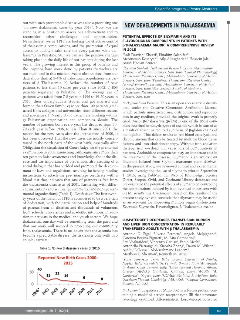
out with such preventable disease was also a promising one“no new thalassemia cases by year 2013”. Now, we arestanding in a position to assess our achievement and toreconsider other challenges and opportunities.Nevertheless, we at TPFS are looking for effective controlof thalassemia complications, and the promotion of equalaccess to quality health care for every patient with tha-lassemia in Palestine. Still we can see the positive changestaking place in the daily life of our patients during the lastyears. The growing interest in this group of patients andthe inspiring hard work done by patients themselves areour main tool in this mission. Major observations from ourdata show that: a) 3-4% of Palestinian populations are car-riers of β Thalassemia. b) Reduce the number of newpatients to less than 10 cases per year since 2002. c) 863patients registered in Palestine. d) The average age ofpatients was raised from 7-8 years in 1996 to 19-20 years in2015, their undergraduate studies and got married andformed their Owen family. e) More than 100 patients grad-uated from colleges and universities from different sectorsand specialties. f) Nearly 30-35 patient are working within.g) Palestinian organization and companies. Results: Thenumber of patients born every year has dropped from 50-70 each year before 1996, to less. Than 10 since 2001, thereason for the new cases after the instructions of 2000, ithas been observed That the new cases are mostly concen-trated in the north parts of the west bank, especially afterObligation the circulation of Court Judge for the premaritalexamination in 2000. Launching campaigns since more thanten years to Raise awareness and knowledge about the dis-ease and the importance of prevention, also creating of asocial dialogue that has yielded and promoted the develop-ment of laws and regulations, resulting in: issuing bindinginstructions to attach the pre- marriage certificate with ablood test that indicates that one of partners is free fromthe thalassemia disease as of 2001. Partnering with differ-ent institutions and sectors (governmental and non- govern-mental organizations) (Table 1). Conclusions: The first twen-ty years of the march of TPFS is considered to be a very richof dedication, with the participation and help of hundredsof parents from all districts and thousands of volunteersfrom schools, universities and academic intuitions, in addi-tion to activists in the medical and youth sectors. We hopethalassemia one day will be something from the past, andthat our work will succeed in protecting our communityfrom thalassemia. There is no doubt that thalassemia hasbecome a predictable disease, the risk exists only with twocouple- carriers.
Table 1. No new thalassemia cases of 2015.
NEW DEVELOPMENTS IN THALASSAEMIA
POTENTIAL EFFECTS OF SILYMARIN AND ITSFLAVONOLIGNAN COMPONENTS IN PATIENTS WITHβ-THALASSAEMIA MAJOR: A COMPREHENSIVE REVIEWIN 2015Hadi Darvishi Khezri1, Ebrahim Salehifar2*,Mehrnoush Kosaryan3, Aily Aliasgharian4, Hossein Jalali1,Arash Hadian Amree11Research Student, Thalassemia Research Center, MazandaranUniversity of Medical Sciences, Sari, Iran; 2Clinical Pharmacology,Thalassemia Research Center, Mazandaran University of MedicalSciences, Sari, Iran; 3Pediatrics, Thalassemia Research Center,Hemoglobinopathy Institute, Mazandaran University of MedicalSciences, Sari, Iran; 4Microbiology, Faculty of Medicine,Thalassemia Research Center, Mazandaran University of MedicalSciences, Sari, Iran
Background and Purpose: This is an open access article distrib-uted under the Creative Commons Attribution License,which permits unrestricted use, distribution, and reproduc-tion in any medium, provided the original work is properlycited. Major β-thalassemia (β-TM) is one of the most com-mon inherited hemolytic types of anemia which is caused asa result of absent or reduced synthesis of β-globin chains ofhemoglobin. This defect results in red blood cells lysis andchronic anemia that can be treated by multiple blood trans-fusions and iron chelation therapy. Without iron chelationtherapy, iron overload will cause lots of complications inpatients. Antioxidant components play an important role inthe treatment of the disease. Silymarin is an antioxidantflavonoid isolated from Silybum marianum plant. Methods:In the present study, we reviewed clinical and experimentalstudies investigating the use of silymarin prior to September1, 2015, using PubMed, ISI Web of Knowledge, ScienceDirect, Scopus, Ovid, and Cochrane Library databases andwe evaluated the potential effects of silymarin on controllingthe complications induced by iron overload in patients withβ-TM. Results and Conclusions: Based on the results of thepresent study, we can conclude that silymarin may be usefulas an adjuvant for improving multiple organ dysfunctions.Keywords: Silymarin, Flavonolignan, β-Thalassemia Major.
LUSPATERCEPT DECREASES TRANSFUSION BURDENAND LIVER IRON CONCENTRATION IN REGULARLYTRANSFUSED ADULTS WITH β-THALASSAEMIAAntonio G. Piga1, Silverio Perrotta2, Angela Melpignano3,Caterina Borgna-Pignatti4, M. Rita Gamberini4,Ersi Voskaridou5, Vincenzo Caruso6, Paolo Ricchi7,Antonello Pietrangelo8, Xiaosha Zhang9, Dawn M. Wilson9,Ashley Bellevue9, Abderrahmane Laadem10,Matthew L. Sherman9, Kenneth M. Attie91Turin University, Turin, Italy; 2Second University of Naples,Naples, Italy; 3Ospedale “A. Perrino”, Brindisi, Italy; 4ArcispedaleS. Anna, Cona, Ferrara, Italy; 5Laiko General Hospital, Athens,Greece; 6ARNAS Garibaldi, Catania, Italy; 7AORN “A.Cardarelli“, Naples, Italy; 8CEMEF, Medicina 2, Modena, Italy;9Acceleron Pharma, Cambridge, MA, USA; 10Celgene Corporation,Summit, NJ, USA
Background: Luspatercept (ACE-536) is a fusion protein con-taining a modified activin receptor type IIB that promoteslate-stage erythroid differentiation. Luspatercept corrected
Scientific program - Poster Abstracts
haematologica | 2017; 102(s1) 89

the effects of ineffective erythropoiesis in a thalassemiamouse model and was well tolerated and increased hemo-globin in a phase 1 clinical study. Methods: In this phase 2open-label study, luspatercept was administered to adultswith β-thalassemia SC every 3 wks for 12 weeks in the com-pleted base study and an additional 2 years in the ongoinglong-term extension study. Six cohorts (n=35) were treated atdose levels from 0.2-1.25 mg/kg. Patients in the expansioncohort (n=29) and extension study (n=51) were treated at 0.8mg/kg with escalation up to 1.25 mg/kg. Results: 30 regularlytransfused pts (≥4 units RBC/8 wks) completed the basestudy and 24 enrolled in the extension study (data as of 11March 2016). Data summarized are for the long-term exten-sion study; results from the base study will also be present-ed. Median age was 38 yr (range 22-55 yr), 67% had priorsplenectomy. Median baseline transfusion burden was 8units/12 wks (range 4-15 units). Mean (±SD) LIC was 5.1±5.3mg/g dw. 16/24 (67%) pts achieved a ≥33% and ≥50%decrease in transfusion burden over any 12-weeks comparedto baseline, respectively. Duration of response ranged from12-48+ weeks. 2/3 (67%) pts with baseline LIC ≥5 mg/g dwhad a decrease in LIC ≥2 mg/g dw after at least 6 months oftreatment. Luspatercept was generally well tolerated, withno related SAEs. AEs were mostly mild-moderate; the mostfrequent related AEs (≥10%) were bone pain, myalgia,arthralgia, headache, asthenia, and musculoskeletal pain.Conclusions: Luspatercept treatment was well-tolerated andled to decreased RBC transfusion requirements and LIC inTD β-thalassemia patients. A Phase 3 study of luspatercept inregularly transfused adults with β-thalassemia is ongoing(NCT02604433).
LUSPATERCEPT INCREASES HAEMOGLOBIN ANDIMPROVES QUALITY OF LIFE IN NON-TRANSFUSIONDEPENDENT ADULTS WITH β-THALASSAEMIAAntonio G. Piga1, Silverio Perrotta2, Angela Melpignano3,Caterina Borgna-Pignatti4, M. Rita Gamberini4,Ersi Voskaridou5, Vincenzo Caruso6, Paolo Ricchi7,Antonello Pietrangelo8, Xiaosha Zhang9, Dawn M. Wilson9,Ashley Bellevue9, Abderrahmane Laadem10,Matthew L. Sherman9, Kenneth M. Attie91Turin University, Turin, Italy; 2Second University of Naples,Naples, Italy; 3Ospedale “A. Perrino”, Brindisi, Italy; 4ArcispedaleS. Anna, Cona, Ferrara, Italy; 5Laiko General Hospital, Athens,Greece; 6ARNAS Garibaldi, Catania, Italy; 7AORN “A.Cardarelli“, Naples, Italy; 8CEMEF, Medicina 2, Modena, Italy;9Acceleron Pharma, Cambridge, MA, USA; 10Celgene Corporation,Summit, NJ, USA
Background: Luspatercept (ACE-536) is a fusion protein con-taining a modified activin receptor type IIB that promoteslate-stage erythroid differentiation. Luspatercept correctedthe effects of ineffective erythropoiesis in a thalassemiamouse model and was well tolerated and increased hemo-globin in a phase 1 clinical study. Methods: In this phase 2open-label study, luspatercept was administered to adultswith β-thalassemia SC every 3 wks for 12 weeks in the com-pleted base study and an additional 2 years in the ongoinglong-term extension study. Six cohorts (n=35) were treated atdose levels from 0.2-1.25 mg/kg. Patients in the expansioncohort (n=29) and extension study (n=51) were treated at 0.8mg/kg with escalation up to 1.25 mg/kg. Results: 34 non-
transfusion dependent pts (<4 units RBC/8 wks) completedthe base study and 27 enrolled in the extension study (dataas of 11 March 2016). Data summarized are for the long-term extension study; results from the base study will also bepresented. Median age was 37 yr (range 23-62 yr); 67% hadprior splenectomy. Median baseline Hgb was 8.7 g/dL (range7.6-9.8 g/dL). Mean (±SD) LIC was 4.9±3.4 mg/g dw. 21/27(78%) and 15/27 (56%) pts achieved ≥1.0 g/dL and ≥1.5 g/dLincrease in mean Hgb over any 12-weeks, respectively.Duration of response ranged from 113-505+ days. Increasesin mean hemoglobin over a 12-week period correlated withincreases in patient reported quality of life questionnaire,FACIT-F (r=0.67, p=0.001). Decreases in measures of ironoverload were also observed in some patients. Luspaterceptwas generally well tolerated, with no related SAEs. AEs weremostly mild-moderate; the most frequent related AEs(≥10%) were bone pain, headache, musculoskeletal pain,and arthralgia. Conclusions: Luspatercept treatment was well-tolerated and led to increased hemoglobin levels, decreasedLIC, and improved quality of life in NTD adults with β-tha-lassemia.
NON TRANSFUSION-DEPENDENTTHALASSAEMIA
UTILITY OF SERUM FERRITIN LEVELS BETWEEN 300AND 800 NG/ML IN CLINICAL DECISION MAKING IN NONTRANSFUSION-DEPENDENT THALASSAEMIA WHENMAGNETIC RESONANCE IMAGING FOR LIVER IRONCONCENTRATION MEASUREMENT IS NOT AVAILABLEAntoine N. Saliba1, Khaled M. Musallam2,Maria D. Cappellini3, Vip Viprakasit4, Shahina Daar5,Hassan M. Moukhadder2,6, Ali T. Taher2,61Indiana University School of Medicine, Department of Medicine,Indianapolis, Indiana, United States; 2American University ofBeirut Medical Center, Department of Internal Medicine, Beirut,Lebanon; 3University of Milan, IRCCS Ca’ Granda FoundationMaggiore Policlinico Hospital, Department of Clinical Sciencesand Community, Milan, Italy; 4Mahidol University, SirirajHospital, Department of Pediatrics and Internal Medicine,Bangkok, Thailand; 5Sultan Qaboos University, College ofMedicine & Health Sciences, Department of Hematology, Muscat,Oman; 6Chronic Care Center, Hazmieh, Lebanon
Background: The assessment of liver iron concentration (LIC)by magnetic resonance imaging (MRI) remains to be the rec-ommended means of quantifying iron overload in patientswith non-transfusion-dependent thalassemia (NTDT) toguide iron chelation therapy (ICT). On the other hand,serum ferritin measurement, a simple and more affordabletool, has a significantly positive correlation with LIC andtherefore may be utilized in lieu of the latter in countries orcenters where MRI is not available or affordable, in light ofthe high prevalence of NTDT in developing countries. Whileserum ferritin levels of >800 ng/mL were shown in previousstudies to most certainly predict an LIC ≥5 mg/g dry weight(dw), which corresponds to the concentration heralding iron-related morbidity and therefore prompts initiation of ICT,almost 50% of patients with levels <800 ng/mL weredemonstrated to have LIC levels ≥5 mg/g dw. To this pur-pose, we evaluated the utility of serum ferritin in predicting
2nd MEGMA Conference on Thalassaemia & other Haemoglobinopathies Amman - Jordan, 11-12 November 2016
90 haematologica | 2017; 102(s1)

the need for ICT for levels ranging between 300 and 800ng/mL, 300 being the upper limit of normal serum ferritinlevel. Methods:We looked at 71 patients (mean age 33.3±13.9years) with serum ferritin levels ranging between 300 and800 ng/mL attending centers in Italy, Lebanon, Oman, andThailand. 53.5% were female, and 45.1% were splenec-tomized. Results:Using a logistic regression analysis with LIC≥5 mg/g dw as the desired outcome, we were able to con-struct a probability curve for serum ferritin level in predictingan LIC ≥5 mg/g dw. Conclusions: Instead of categoricallyassuming patients with serum ferritin levels <800 ng/mL notto be eligible for ICT, our probability curve helps assign apercentage risk of having an LIC ≥5 mg/g dw to suchpatients, which, in tandem with the patients’ general clinicalpicture, can better guide decision making towards imple-mentation of ICT.
ARAP-536 (MURINE ANALOG OF ACE-536/LUSPATER-CEPT) INHIBITS SMAD2/3 SIGNALING AND PROMOTESERYTHROID DIFFERENTIATION BY RESTORING GATA-1FUNCTION IN A MURINE MODEL OF β-THALASSAEMIAPedro Martinez1*, Manoj Bhasin2, Robert Li1, Scott Pearsall1,Ravindra Kumar1, Rajasekhar Suragani11Preclinical Department, Acceleron Pharma, Cambridge;2Genomics and Proteomics Center, Beth Israel Deaconess MedicalCenter, Harvard Medical School, Boston, United States
Background: Luspatercept (modified ActRIIB receptor-Fcfusion protein), a Smad 2/3 ligand trap, has demonstratedefficacy in correcting ineffective erythropoiesis (IE) and ane-mia in murine models of MDS and β-thalassemia. Methods:β-thalassemic mice (Hbbth3/+) were administered a singlebolus of vehicle (VEH) or RAP-536. Splenic basophilic ery-throblasts were sorted for genome wide transcriptome pro-filing. Also, mouse erythroid leukemic (MEL) cells, primaryfetal liver erythroid and β-thalassemic erythroid precursorswere treated with GDF11 in the presence or absence of ACE-536. Results: Transcriptome analysis of β-thalassemic ery-throblasts identified 74 genes that were differentiallyexpressed in RAP-536 treated samples vs. VEH. Gene setenrichment analysis (GSEA) depicted significant upregula-tion of 158 activated genes downstream of GATA-1 (ery-throid master regulator) such as those involved in hemebiosynthesis and terminal erythroid differentiation. ACE-536binds and inhibits Smad2/3 ligands such as GDF8, GDF11and Activin B. Treatment of MEL and fetal liver erythroidcells with GDF11 induced Smad2/3 phosphorylation andACE-536 co-treatment inhibited the increase in pSmad2/3.In differentiating erythroid cells, GDF11 treatment displayedreduced nuclear GATA-1 protein levels. Reactive oxygenspecies (ROS) and Caspase 3/7 were elevated in MEL andprimary fetal liver cells following GDF11 treatment.Importantly, treatment of erythroid cells with ACE-536 andGDF11 decreased ROS, and restored GATA-1 to control lev-els. Conclusions: These data provide a potential mechanisticrole for luspatercept as a novel treatment of β-thalassemia.By inhibiting pSmad2/3 signaling, RAP-536 treatmentdecreases ROS, caspase3/7 activation and GATA-1 cleavage.Thus by restoring GATA-1 availability and functional activi-ty, RAP-536 treatment causes upregulation of genes involvedin promoting terminal erythroid maturation, and conse-quently corrects anemia in β-thalassemia. Luspatercept has
initiated phase 3 studies in patients with MDS and β-tha-lassemia.
QUALITATIVE RESEARCH ON THE OBSTACLES INMARRIAGE AND REPRODUCTIVE PROCESS OF PEOPLEWITH TRANSFUSION DEPENDENT THALASSAEMIASachiko Hosoya1*, Gholam Reza Jamali2, Mohsen Shahriari31Research Fellow, Research Institute of Language and Cultures inAsia and Africa, Tokyo University of Foreign Studies; 2Nursing,School of Nursing and Midwifery, Isfahan University of MedicalSciences; 3School of Nursing and Midwifery, Isfahan University ofMedical Sciences
Background: In Iran, the largest population age of the peoplewith transfusion dependent Thalassemia (PTDT) now hasreached late teens to 20’s. In other words, most of the PTDTare in their reproductive age. However, many PTDT havedifficulties going through the marriage and reproductiveprocess. Considering the recent increase of life expectancy ofPTDT in Iran, it is an urgent task now to create a better sup-port program for PTDT’s marriage/reproductive life from thenursing care perspective, and a basic study for this purpose isneeded. Methods: Semi-structured interview with 60 mar-ried/single PTDT who came to the Thalassemia Center inSeyed-al-Shoahad hospital in Isfahan, and qualitative analy-sis were conducted in 2016. Results: There are some differ-ences between the difficulties which male and female PTDTexperience. For male PTDT, unemployment were recognizedas the hardest obstacles, while female PTDT found that thefertility problems are more difficult matters. Prejudicetowards PTDT is a serious problem for both men andwomen. Many of them had a thought to choose a PTDT asa marriage partner at least once, but only some of them havemarried with PTDT, because of their families’ disagreement.The negative episodes such as unsuccessful marriage propos-al, quarrel with their family over their marriage choice, orpressure to have a baby, tend to result in depression andinappropriate self-care. Conclusions: Pre-marital educationalprograms for PTDT about the fertility should be createdbased on this research, as well as the personal counselingprograms for their own family and future partner/familyabout the PTDT’s capabilities and health situations. To solvetheir unemployment problem, which could be recognized asa health issue equally as a social issue, the staff inThalassemia word can approach through collaborative pro-grams with local community.
KNOWLEDGE ATTITUDE AND PRACTICES AMONGPATIENTS ON TREATMENT OF THALASSAEMIA INMALDIVESFathmath Jeehan SaleemMaldivian Thalassaemia Society, Republic of Maldives
Background: Maldives has the highest prevalence of betaThalassaemia in the world with 18% of its population beingcarriers for � Thalassaemia (SHE 1992). To date over 800patients have been registered. Adherence to the manage-ment protocol followed by patients is inadequate resulting inpremature mortality among the young adults. Aims andObjectives: The aim of the study was to assess awareness andknowledge among young patients on standard treatment
Scientific program - Poster Abstracts
haematologica | 2017; 102(s1) 91
protocols that they require and if the knowledge translatesinto practices on factors that make them susceptible to andprevent them from chronic organ damage and other compli-cations. It further explores availability and access to stan-dardised treatment in the country. Methods: A questionnairebased KAP survey among Thalassaemia patients (above 12years) registered at National Thalassaemia centre(NTC/MBS). Survey was conducted betweeen July 2014 toOctober 2014. Questionannaire was designed to gatherinformation on transfusion regime, chelation therapy andgrowth and development, treatment facilties and sevices.Results: Results of the survey show that standard treatmentwith blood transfusion and iron chelation is the primaryfocus of treatment for patients despite their age. Patients areknowledgeable about transfusion, but not so much on chela-tion and less aware and knowledgeable on aspects related togrowth and development. 100% of the patients strictlyadhered to transfusion regime while with chelation allpatients have had lapses at some point of their life. The aver-age serum ferritin levels of patients are over 4000 µg/l. Over90% patients are aware excess iron in their body will havecardiac, hepatic and endocrine problems which can be lifethreatening. However patients are not fully concerned thatiron overload could have an immediate impact on their ownhealth. Lack of motivation to adhere to treatment has beenreported as the main reason for poor compliance. Majoritypatients reported they are blamed for low compliance ratherthen encouraged or provided support toward establishing aroutine chelating regime. Patient reported that their inputs,discussion into decisions reagrding their health and wellbe-ing are not encouraged. Treatment regime has a meticulousfocus on transfusion and adult patients are still being treatedand managed with teatment adequate for children. Allpatients recognised the importance of professional supportas they face daily challenges growing up with a chronic con-dition. Psychosocial issues are a very important yet a neglect-ed area to integrate into standard treatment protocols in thecountry. Conclusions: Improved patient knowledge and theirpositive attitude is very important in shaping up their com-pliance and healthy progression to adulthood. Both medicalcommunity and patients need to reshape their thinking andimprove knowledge. Information from the study needs totranslate to targeted awareness programs, identify and fillgaps in treatment protocols, which will have behavioralmodifications to prevent complications and potentially sav-ing young lives. A patient centred approach in managementand treatment is required to ensure patients an equal chancein life. Key words: Transfusion, Chelation, Thalassaemia,Growth and development, Compliance.
ATTRIBUTIONAL STYLES IN ADOLESCENTS WITHTRANSFUSION-DEPENDENT THALASSAEMIALerlugsn Suwanthol, Tuangrath Sangpaypan, Phetcharat Naknum, Kleebsabai SanpakitDepartment of Pediatrics, Faculty of Medicine Siriraj Hospital,Mahidol University, Bangkok ,Thailand
Background: Adolescents with Transfusion- dependent tha-lassemia must cope with the ongoing anxiety and negativeattributional styles due to treatment management, but fewmeasured broad categories of causal explanations for this
population. Objectives: To examine empirically the relationbetween anxiety symptoms and three dimensions of attribu-tions : internal-external, stable-unstable, and global-specificin Adolescents with Transfusion- dependent thalassemia.Methods: With cross-sectional research design and consistedof 102 adolescents (48 males, 54 females) diagnosed withTransfusion- dependent thalassemia (more than 50 times forblood transfusions) completed measure of AttributionalStyles and Anxiety Questionnaires. For correlations in thepredicted directions among variables were examined withPearson product- moment correlation coefficients, t-test andOne way ANOVA were conducted to ascertain significantbetween group differences on attributional factors and levelsof anxiety symptoms. Results: Adolescent samples with high-er levels of anxiety revealed statistically significant relation-ship among three negative attributional dimensions (overallcomposite F=4.5, p<.05; negative composite F=4.99 , p<.01;negative internality F=4.50, p<.01; negative stability F=3.42,p<.05 and negative globality F=3.77, p<.05). In addition, sig-nificant age- group differences were found for the total neg-ative globality (t=-2.05, p<.05) and negative globality (t=-2.22, p<.05). Conclusions: This work reveals consistent sup-port for the reformulated learned helplessness theory ofdepression. In finding that individuals who attribute nega-tive life events to internal, stable, and global causes will bemore vulnerable to anxiety than those who make external,unstable, and specific attributions. Moreover, those adoles-cents more than 17 years evidenced a more negative global-ity attributional style than their groups less than 16 years,and this pattern may be influenced by female adolescents.Finally, examining adolescent’s attributions as a risk factorfor poor adjustment will help pediatric professionals identifyadolescent at risk for mental health problems, while investi-gating the effect of negative globality attributional style willhelp clarify whether cognitive interventions should be targeton early, middle or late adolescence. Keywords: Attributionalstyles, anxiety, adolescents with transfusion- dependent tha-lassemia.
QUALITY CARE FOR QUALITY OF LIFE
MULTIFACETED APPROACH TO THALASSAEMIA CAREAND MANAGEMENT – OUR EXPERIENCE AND LESSONSLEARNEDShashikant Patil, Rajendra Kulkarni, Vaishali Kale,Snehal Patil, Nandkishor Tated, Atul JainArpan Thalassemia Society, Arpan Blood Bank, Nashik,Maharashtra, India
Background: If managed properly with adequate overall med-ical care and good quality & safe blood transfusion support,the quality of life of thalassemia major patients can beimproved significantly. Aim: To implement a comprehensive,multifaceted thalassemia care and management programmeunder one roof. Methods: Thalassemia major patients withinthe expanse of north Maharashtra were registered with ourorganisation. A programme for medical management includ-ing iron chelation therapy, haematologic, cardiologic, paedi-atric, orthopedic, pathological, radiological evaluation wasdefined. A monitoring programme was formulated usingguidelines given by Indian Academy of Paediatrics. All
2nd MEGMA Conference on Thalassaemia & other Haemoglobinopathies Amman - Jordan, 11-12 November 2016
92 haematologica | 2017; 102(s1)

patients were offered ID-NAT tested, inline-leucodepletedblood free of cost with the help of corporate support. A ded-icated donor programme was implemented for the transfu-sion support of the study group. All the patients were fol-lowed up for febrile non-hemolytic reactions, sero-conver-sion for TTI, alloimmunization, growth-disorders, iron over-load for a period of 3 years. Results: Total 181 patients withage ranging from 2 years to 30 years were enrolled in thisprogramme. 4662 transfusions have been given over theperiod of 3 years to 181 patients. 3 patients (1.6%) havedeveloped alloimunization. 1 patient (0.5%) had experiencedfebrile non-hemolytic transfusion reaction. Severe iron over-load (according to T2* MRI) was seen in 2 out of 12 patients(16.66%). Growth disorders/stunted growth have beenobserved in 25 (13.81%) patients. None of the above patientswere found to have sero-conversion for HIV, HBV or HCV.Conclusions: Implementing a comprehensive transfusion ther-apy and health monitoring programme for thalassemiapatients under one roof is feasible with support from corpo-rate & society. Careful periodic monitoring of these patients& implementation of NAT tested, leucodepleted & pheno-type matched blood transfusion can go a long way toimprove the quality of life and overall life expectancy of tha-lassemia major patients.
PRESENTATION OF THALASSAEMIA WARD IN EASTMEDICAL SPECIAL CENTER IN TEHRAN AND CLINICALCHALLENGES IN MANAGEMENT OF THALASSAEMICPATIENTS IN ALL FIELDSM. Sabzechian1, R. Fallah2, M. Gholami3 1Pediatric Hematologist Oncologist in East Medical Special Center;2General Physician in East Medical Special Center; 3Neurse ofThalassemic Patients in East Medical Special Center, Iran
East Medical Centre with the most advanced and specializedmedical facilities based on international standards have beenestablished by the Charity Foundation for Special Diseases.The East medical special center is located in the south east ofTehran and ready to deliver health care in different parts ofthe public tariff. East Medical Centre by the CharityFoundation for Special Diseases and assistance of the peopleand the central government set up a fourth center in thecountry launched by the Charity Foundation for SpecialDiseases. This center was started its activity on 2005.Thalssemia ward is very active in last 5 years. About 60 tha-lassemic patient confer in this center regularly. During thelast 3-4 years the number of patients referred with regard tothe facilities of this center is to this sector increased by 50%.At this time about 60 thalassemic patients is in health carecoverage. Health care provided to patients with thalassemiainclude: regular visits to patients by medical professionals,complete tests of hematology, kidney, liver, virology,endocrinology (every 6 months in own laboratory), psychi-atric counseling, family counseling, marriage counseling isdone, regular visit by cardiologist and endocrinologist, bloodtransfusion, intra venuos desferal therapy, intravenouszolendronic acid in osteoporotic thalassemic patients, intra-venous anti biotic therapy for splenectomized patients, andother services. In this center Ab screening is done in labora-tory. Thalassemia patents refer for liver and heart T2 and R2star MRI, bone mineral density assay, abdominal sonogra-
phy, opyometry, oudiology, echocardiography annually.Patient information and results is recorded in file for eachpatient. The services provided patient satisfaction was verygood during recent years . At this time 50 cases of major tha-lassemia, 6case of intermedia thalassemia, one case ofsicklecelldisease, one case of alpha thalassemia, one case of leporthalassemia is confer in this center. Two cases of major tha-lassemia refer for bone marrow transplantation ward. Onecase of two patient that refer for BMT have very good result.
EFFECT OF DEFERASIROX USE ON THE EDUCATIONALSTATUS OF PATIENTS WITH THALASSAEMIA: A 20-YEAR,SINGLE-CENTER EXPERIENCERayan Bou Fakhredin1, Joseph Roumi1,2, Josephine Ajouz2,Hassan M. Moukhadder1,2, Rita Aoun2, Suzanne Koussa2,Ali T. Taher1,21American University of Beirut Medical Center, Department ofInternal Medicine, Beirut, Lebanon; 2Chronic Care Center,Hazmieh, Lebanon
Background: Very few reports in the literature have focused onthe psychosocial status of patients with thalassemia. Indeed,the multitude of medical morbidities that have been histori-cally associated with thalassemia, namely those stemmingfrom iron overload and its complications, used to get in theway of patients wanting to seek education. However, animproved health-related quality of life following the move-ment towards deferasirox use in the Chronic Care Center inLebanon in 2006, enabled many patients to actualize them-selves through the attainment of higher education levels. Theaim of this study is to evaluate the impact of deferasirox useon the educational status of patients with thalassemia fromour practice. Methods: A total of 60 thalassemia patients (30males and 30 females) were included in this study. Patientswere divided into 3 age groups: 12-18, 19-25, and 26-40. Theeducational status of the patients in each group was com-pared between two time periods – 1996-2005 and 2006-2016.Educational status was categorized as university degree, highschool degree, or less than high school degree. Results: In the12-18 age group, the number of patients achieving a highschool degree increased from 30% before the implementa-tion of deferasirox use to 90% after it. Similarly, the numberof patients achieving a university degree increased in both 18-25 and 25-40 age groups after the introduction of deferasiroxtreatment, from 20% to 50% in the former and from 30% to70% in the latter. Conclusions: Individuals with thalassemiaare now able to seek and achieve higher levels of educationafter the introduction of deferasirox use for iron chelation,which has translated into a health-related quality of life morepermissive of their quest to better educate themselves. Thismight be very helpful in creating effective psychosocial plansfor thalassemia patients.
DEVELOPMENT OF PATIENT-REPORTED OUTCOMESSYMPTOM MEASURE FOR PATIENTS WITH NON-TRANSFUSION-DEPENDENT THALASSAEMIA (NTDT-PRO©)Ali Taher1, Antoine Saliba1, Antonis Kattamis2,Richard Ward3, Gale Harding4, Xiaohan Hu5,Dalia Mahmoud51American University Beirut, Beirut, Lebanon; 2Aghia SophiaChildren’s Hospital, Athens, Greece; 3University Health Network,
Scientific program - Poster Abstracts
haematologica | 2017; 102(s1) 93
Toronto, Canada; 4Evidera, Inc., Bethesda, USA; 5Celgene Corp.,Summit, USA
Background: Currently, there is no disease-specific Patient-Reported Outcomes (PRO) measure available to assess keysymptoms experienced by patients with non-transfusion-dependent thalassemia (NTDT). Aim: To develop a symptommeasure for NTDT patients for use in clinical trials. Methods:A qualitative study was conducted to develop the NTDT-PRO© symptom measure consistent with regulatory require-ments. The instrument was developed based on concept elic-itation interviews, input from clinical experts, and refinedthrough a process of cognitive interviews. Concept elicitationinterviews were conducted among a total of 25 NTDTpatients recruited from Lebanon, Greece, and Canada.Findings from these interviews were used to develop the draftmeasure. Cognitive interviews were subsequently conductedamong subjects in Greece and Lebanon (N=21) to examine therelevance and understanding of the items among the targetpopulation. All interviews were conducted in person in thelocal language with written informed consent obtained.Results: Based on findings from concept elicitation interviews,a total of nine symptoms were included in the NTDT-PRO©
Version 1. An 11-point numeric rating scale was used for theresponse options and a daily recall period was selected basedon the day-to-day variability of symptoms. Findings from thecognitive interviews indicate that subjects understood theitems as intended, with a few minor exceptions. Four itemswere deleted as they were not considered core symptoms, andone item was added to address the symptom both with andwithout physical activity. The NTDT-PRO© Version 2 includessix items that address three key symptoms of tiredness, weak-ness, and shortness of breath both during physical activity andat rest. Conclusions: The NTDT-PRO© Version 2 consists of sixitems to assess the key symptoms of NTDT using a 24-hourrecall period, with plans to further evaluate the measures in anongoing observational study and for eventual inclusion into arandomized phase 3 study.
SICKLE CELL DISEASE
CLINICAL AND HAEMATOLOGICAL PROFILES AMONGBAHRAINI CHILDREN WITH SICKLE CELL DISEASEHusain AlMukharraqConsultant Pediatrician-Hematologist & Oncologist, Manama,Bahrain
Aim: To determines the clinical and hematological parameterand manifestation of children with sickle cell disease inBahrain. Patients and Methods: Total number of admissionover one year in the pediatric department were 4552, sicklecell disease patients were 552 (12.1%) percentage. Results: Atotal of 552 files reviewed and 52 were excluded and theremaining 500 enrolled in the study. The study populationdivided into 2 group according to age: Group I: 1-7 years andgroup II: 8-15 years. The male is predominating in bothgroups 68% and 52% respectively, there is no difference inthe mean hemoglobin. Although the MCV and MCH islower in both groups yet it is lower in group I. The reticscounts is higher in group II (4.9% compared 3.9%). Themean WBC and platelets counts were both high in group I.The HBS is higher in group II (78.1% VS 65.8%), the HBF
higher in group I (25.9% VS 17.7%). The G6PD deficiencywas higher in group I (42.1% VS 35.2%). The jaundice andsplenomegaly were higher in group II (84% and 56% respec-tively). Vaso-occlusive (painful) crises represent 69.3%, fever11.7%, hyper hemolysis/severe anemia 9.1%, acutechest/pneumonia 6.5%, gastroenteritis 2.7%, osteomyelitis2.2%. The cerebrovascular accident, sequestration and handfood syndrome were each less than 1%. Conclusions: The lowMCV and MCH in both group could be explained by the co-inheritance of alpha thalassemia or iron deficiency anemia;HbS, jaundice and splenomegaly is higher in older age chil-dren; the G6PD deficiency is a common inherent amongpatients with sickle cell disease. The Vaso-occlusive crisis isthe commonest manifestation of sickle cell disease whereother complications were relatively less common in compar-ison to other SCD haplotypes.
ASSOCIATION OF METHYLENETETRAHYDROFOLATEREDUCTASE A1298C AND C677T POLYMORPHISMSWITH VASO-OCCLUSIVE CRISIS IN SICKLE CELL DISEASEAhmed A. Raouf1, Mona M. Hamdy2, Ahmed M. Badr2,Osama F. Shalaan3, Moustafa A. Sakr3,Abdel Rahman A. Abdel Rahman31Clinical Biochemistry Department, Liver Institute , MenoufiaUniversity, Egypt; 2Pediatrics Department, Faculty of Medicine,Cairo University, Egypt; 3Molecular Diagnostics and TherapeuticsDepartment, Genetic Engineering and Biotechnology ResearchInstitute, University Of Sadat City, Egypt
Background: Vaso-occlusion is a determinant for most signsand symptoms of sickle-cell anemia (SCA). ElevatedHomocysteine concentration contribute to thrombosis, a fre-quent event in sickle cell anemia. Methylenetetrahydrofolatereductase (MTHFR) is an important enzyme, which regulateshomocysteine metabolism, and some polymorphisms ofgene encoding this enzyme are associated with a decreasedactivity of the enzyme. The aim of the study was to assessan association between the C677T and A1298C polymor-phisms and the frequency of vasoocclusive crisis. Methods: Acase- control study was carried over a period of one yearfrom January to December 2014 including 50 sickle cell dis-ease patients collected together with 30 age, and sexmatched apparently healthy controls. Venous blood samplewere aspirated from both groups to estimate serum homo-cysteine, folic acid, C677T and A1298C polymorphism iden-tification through tetra primer ARMS PCR. Statistical analy-sis was done, using the student T-test, Pearson correlationanalysis and χ2test. Results: Homocysteine level was signifi-cantly higher in the patients group compared with controlgroup with P value >0.01. Moreover, a strong positive corre-lation between homocysteine level and the frequency ofvaso-occlusive crisis was found (×2 4.836 and P value 0.04).Association between vaso-occlusive events and polymor-phism frequency showed no significant difference for boththe C677T and the A1298C polymorphisms (χ2=0.206;p=0.9020) and (χ2=1. 720; p=0.4231) respectively.Conclusions: We concluded that hyperhomocysteinaemia ispositively correlated with the frequency of vaso-occlusivecrisis and neither the presence of C677T nor A1298CMTHFR gene polymorphism are risk factors for vaso-occlu-sive crisis in the SCD patients evaluated. Keywords: Vaso-occlusive crisis. Hyperhomocysteinemia, C677T, A1298C.
2nd MEGMA Conference on Thalassaemia & other Haemoglobinopathies Amman - Jordan, 11-12 November 2016
94 haematologica | 2017; 102(s1)

CLINICAL PRESENTATION OF CHILDHOOD SICKLE CELLDISEASE AND ITS CORRELATION WITH DISEASEPROGNOSIS: A SINGLE CENTRE STUDYAbdullah A. Bukhari1, Abdulmalik A. Mulla1,Abdullah M. Sonbol1, Anas M. Makhdoum1,Assem A. Alblowi1, Badr Z. Mahmoud1, Zakaria Alhawsawi2,Abeer Abd Elmoneim1,3
1Pediatric Department, Taibah University, AlmadinahAlmounourah, Saudi Arabia; 2Hematology/oncology center,Maternity and Children Hospital (MCH), AlmadinahAlmounourah, Saudi Arabia; 3Pediatric Department, SohagUniversity, Sohag, Egypt
Background and Aim: Sickle cell disease (SCD) is an autosomalrecessive disorder characterized by production of abnormalhemoglobin S and is associated with high morbidity andmortality. The prevalence of SCD in Saudi Arabia varies sig-nificantly in different parts of the country, with the highestprevalence is in the eastern province, followed by the south-western provinces. In Madinah, the estimated prevalence ofsickle cell homozygosity (Hb SS) is 0.01, and that of the car-rier state (Hb AS) is 0.087. In this study we aim to evaluatethe clinical presentation of SCD children in MadinahGovernorate, Saudi Arabia and its correlation to the diseaseprognosis. Patients and Methods: In a retrospective analysisthe study included all SCD children admitted to Maternityand Children hospital (MCH) in Almadinah Almounourah,Saudi Arabia from year 2000 to 2015. Descriptive analysisand chi-square test were used to identify the main factorsusing SPSS (Version 20, USA). Backward logistic regressionwas used to find the association between factors. Results:SCD is commonly diagnosed in infants aged 6-11 months,with hand-foot swelling and jaundice being the commonestsymptoms at presentation. Anaemic and vaso-occlusivecrises were seen more common in children aged 1-5 years.The over-all morbidity pattern is the same in both sexes withdiseases such as bronchopneumonia, malaria, osteomyelitis,urinary tract infections, septicaemia and septic arthritis beingcommon. Age and presence of complications such as acutechest syndrome and strock at first presentation have beenfound to influence morbidity pattern in our studied patients.Conclusions: There is need for early diagnosis and counseling,so that mothers or caregivers will be able to assist in promptidentification of these morbidities and to seek for promptand appropriate treatment in the health facilities.
THE OUTCOME OF PREOPERATIVE TRANSFUSIONGUIDELINE ON SICKLE CELL DISEASE PATIENTS AT KINGFAHD HOSPITAL-JEDDAHSameera M.R. Felemban1,2, Rekha Bajoria2, Amani Alsawaf1,Ratna Chatterjee2, Abdulelah I. Qadi11King Fahd Hospital, Jeddah, Saudi Arabia; 2Institute For Women’sHealth, UCL, UK
Background: We developed a local hospital preoperativetransfusion guideline for sickle cell disease (SCD) patients toreduce the perioperative and the postoperative complica-tions. This study was conducted to evaluate the outcome ofclinical practice on SCD patients undergoing surgeries in ourinstitution. Methods: A retrospective review of 75 SCDpatients undergoing surgery at King Fahd Hospital, Jeddah,Saudi Arabia was conducted between April 2005 and May
2010. The medical records were reviewed to define the peri-operative risks and the postoperative complications in rela-tion to the type of transfusion modality selected. Results: Themedical records of 75 SCD patients who underwent surger-ies were reviewed. Preoperatively, 25.3% had completeexchange transfusion (CETX), 17.3% had partial exchangetransfusion (PETX), 26.7% had simple top up transfusion(STX) and 30.7% did not require transfusion (NTX). Thepostoperative complications included vaso-occlusive crises(VOC) in 20%, acute chest syndrome (ACS) in 2.7%, andfever in 16% cases. 33.3% patients required the prolongedperiod of the hospital stay. In the patients of our study, post-operative fever, VOC, ACS, and the length of hospital staydid not show any difference regardless of types of transfu-sion modalities. However, the correlation was highly signif-icant between the pre-operative haemoglobin (Hb) level andpostoperative fever (P <0.01) and VOC (P <0.01).Interestingly, SCD patients who received hydroxyurea hadless postoperative complications such as fever (P <0.05) andvaso-occlusive crises (P <0.05), while those who receivedprophylactic heparin in the postoperative period had areduced length of hospital stay (P <0.01) and vaso-occlusivecrises (P <0.01). Conclusions: The guidelines for preoperativetransfusion in SCD patients were effective in reducing thepostoperative morbidity and mortality. Moreover, thisguideline emphasises the surgical situations where preopera-tive transfusion is needed and optimum regimen is requiredfor different surgical sub-types.
DEMANDS AND CHALLENGES FOR PATIENTS WITHSICKLE CELL DISEASE REQUIRING HAEMATOPOIETICSTEM CELL TRANSPLANTATION IN SAUDI ARABIAAbdulrahman Alsultan1,2, Wasil Jastaniah3,4,Sameera Al Afghani5, Muneer H. Al Bagshi6,Zaki Nasserullah7, Ahmed M. Al-Suliman8,Mohammed K. Alabdulaali81Department of Pediatrics and Sickle Cell Disease ResearchCenter, College of Medicine, King Saud University, Riyadh,Saudi Arabia; 2Department of Pediatric Hematology/Oncology,King Abdullah Specialist Children’s Hospital, Riyadh, SaudiArabia; 3King Abdulaziz Medical City, Jeddah, Saudi Arabia;4Department of Pediatrics, College of Medicine, Umm alQuraUniversity, Makkah; 5King Fahad Specialist Hospital, Dammam,Saudi Arabia; 6Maternity and Children’s Hospital, Hofuf, SaudiArabia; 7Maternity & Children’s Hospital, Dammam, SaudiArabia; 8King Fahad Hospital, Hofuf, Saudi Arabia
Background: Allogeneic hematopoietic stem cell transplanta-tion (HSCT) is the only curative treatment for sickle cell dis-ease (SCD). Methods: In this study, we estimated the numberof Saudi SCD patients who are candidates for HSCT. Weused presence of overt stroke, recurrent acute chest syn-drome (ACS), and frequent severe pain crisis as indicationsfor HSCT. Results: We enrolled 376 SCD patients. A total of250 patients were from the Southwestern (SW) provinces;the mean age of this group was 20 years old (range, 4-56years old) and 48% of these subjects were female. The totalnumber of potential transplant candidates from the SW was59 (23.6%). Among individuals from the Eastern (E)province, 126 participated in the study. These subjects had amean age of 25 years old (range, 4-55 years old) and 58%were females. There were a total of 22 (17.4%) transplantcandidates from E province. Based on the reported preva-
Scientific program - Poster Abstracts
haematologica | 2017; 102(s1) 95

lence of SCD in Saudi Arabia and the size of the Saudi pop-ulation in 2010, it is estimated that nearly 61,000 SCDpatients reside in Saudi Arabia. Of these, approximately18,000 are children under 14 years of age. The projectednumber of Saudi SCD patients who are candidates forHSCT is 10,536 patients. Of those, 2,148 are children.Conclusions: The burden of SCD on HSCT centers in SaudiArabia is substantial and is difficult currently to meet thedemand. We recommend recruiting/training more trans-plant physicians and nurses, expand current capacity of cen-ters if feasible, and open new transplant centers to makeHSCT a practical therapeutic option for patients with severeSCD in Saudi Arabia.
HYDROXYUREA, AN EFFECTIVE THERAPY TO AVOIDSPLENECTOMY IN SICKLE CELL DISEASE ANDTHALASSAEMIA: SINGLE INSTITUTE EXPERIENCE INSAUDI ARABIASoad Khalil Al JaouniHematology Department, King Abdulaziz University Hospital,Faculty of Medicine, King Abdulaziz University, Jeddah, SaudiArabia
Background and Purpose: Spleen plays an important part in theimmune system which helps the body fight infection andhave a major role in children. Splenectomy have been declinein hemoglobin disorders in recent years. To assess the role ofhydroxyurea if can eliminate the risk of splenectomy in sick-le cell disease (SCD) and thalassemia in our medical center.Methods: Total of 54 patients enrolled in the study between2004 to 2016 at King Abdulaziz University Hospital, Facultyof Medicine, King Abdulaziz University, Jeddah, Kingdom ofSaudi Arabia, 25 sickle cell disease (SCD), 17 thalassemiamajor (TM) were suboptimal transfusion dependents andhypersplenism, 12 thalassemia inter media (TI). Results: Riskof splenectomy have been avoided in 24/25 SCD, 15/17 inTM and 12/12 in TI. Recent studies and meta analysisshowed that hydroxyurea is safe and non- carcinogenic.Conclusions: Hydroxyurea is an effective and safe therapy can
eliminate risk of splenectomy in SCD and thalassemia.Keywords: Hydroxyurea, SCD, Thalassemia, splenectomy.
MY PERSONAL JOURNEY THROUGH HYDROXYUREATHERAPY IN SICKLE CELL DISEASE DURING PREGNANCYAND LACTATIONAishath Shifneez ShakirMaldivian Thalassaemia Society, Maldives
Hydroxyurea (HU), a drug that is known to reduce the fre-quency of sickle crisis episodes and hospitalizations in SCDpatients. However, it is believed that if HU is used duringpregnancy or if the patient becomes pregnant while takingthis drug, the patient should be told of the potential harm tothe fetus as animal testing shows that the drug crosses to theplacenta and causes embryo toxicity, fetal malformations,growth retardation. Therefore, patients are advised to avoidbecoming pregnant while taking HU. This leaves pregnantsickle women with a real dilemma: Take a drug withunknown effects on fetal development or give up a helpful,necessary medication. I am Shifneez, 33 yrs old fromMaldives. As a sickle beta-thalassaemia patient, I dedicate myefforts to create sickle cell awareness throughout my country.Past 11 years, I have had 3 pregnancies each experience dif-ferent with regard to HU therapy. Today I am a mother of 2.I had the courage to go through my last pregnancy while tak-ing HU. I believe that my daughter is the hope everyone islooking for. Many women are in fear, when it comes to preg-nancy and raising a family, since we are reminded of howrisky it is to become a mother. In my opinion, doctors whoare unaware that HU can actually improve the health of sicklecell patients during pregnancy is taking a huge risk by stop-ping it since it would mean the recurrence of sickle complica-tions that could be life threatening for mother and baby. It’sreally important that doctors find it in themselves to empow-er patients and encourage them to be the best they can be. Ihope the story of my personal journey can open up the pos-sibility for others like me to have their dreams come true.
2nd MEGMA Conference on Thalassaemia & other Haemoglobinopathies Amman - Jordan, 11-12 November 2016
96 haematologica | 2017; 102(s1)

haematologica | 2017; 102(s1) 97
AUTHORS INDEX
AAbbasid, A. 86Abd Elmoneim, A. 95Abdel Rahman, A.R.A. 94Abi Saad, M. 80Abolghasemi, H. 60Abolghasemi, V. 60Abou Nasr, T. 77;80;86Adnani, N.B. 78Afshan, S. 76Ahmadzadeh Amiri, A. 88Ait Ouali, D. 84Ajouz, J. 93Akhter, M.A. 51Akib, A.A.P. 87Al Afghani, S. 95Al Bagshi, M.H. 95Al Hawsawi, Z.M. 49Al Jaouni, S.K. 78;96Al Jebori, S.S. 88Al-Suliman, A.M. 95Alabdulaali, M.K. 95Alaidros, M.A. 49Alblowi, A.A. 95Aleksandar, G. 55Alhawsawi, Z. 95Aliasgharian, A. 83;85;89Aliasghariyan, A. 83Alipour, A. 79AlMukharraq, H. 94Alsawaf, A. 95Alsultan, A. 95Angastiniotis, M. 1;44Antoniou, Z. 44Aoun, R. 80;93Aoun, S. 80Arnaout, S. 83Attie, K.M. 89;90Avan, R. 85Azadbakhta, M. 86Azita, A. 76
BBaazizi, M. 84Badawi, M. 67Badiei, Z. 82Badr, A.M. 94Bahar, M. 76Bajoria, R. 95Banihashemi, A. 79Barati Shourijeh, Z. 85Bardosono, S. 87Bartolucci, P. 59Baysal, E. 6Bazarbachi, A.-H.A. 20Belhadj, R. 84Bellevue, A. 89;90Benakli, M. 84Benmouaffek, N. 75Beuzard, Y. 59Beydoun, H. 77;86Bhasin, M. 91
Blanche, S. 59Borgna-Pignatti, C. 89;90Bou Fakhredin, R. 77;80;86;93Bou Fakhredin, R.I. 20Bouarab, H. 84Boukhenfouf, N. 84Bourget, P. 59Bradai, M. 75Bukhari, A.A. 95
CCappellini, M.D. 90Caruso, V. 89;90Cavallesco, R. 59Cavazzana, M. 59Chatterjee, R. 95Chaya, B.F. 20Chretien, S. 59
DDaar, S. 90Dabbaghmanesh, M.H. 75Dailami, K.N. 88Darvishi Khezri, H. 89De Montalembert, M. 59Dehghani, M. 83
EEbrahimzadeh, M.A. 85;86El Kababri, M. 81El Khorassani, M. 81El Nemer, W. 59El-Beshlawy, A. 70El-Hazmi, M.A.F. 79El-Safy, O.R. 82El-Sayed, M. 82Eleftheriou, A. 1Eleftheriou, P. 28Esmaeili Reykande, S. 83
FFallah, R. 93Farhangi, H. 82Farmakis, D. 39Farokhi, F. 83Farrokh Far, A.A. 88Fatahi, F. 79Felemban, S.M.R. 95
GGamberini, M.R. 89;90Gatot, D. 87Gholami, M. 93Gholbzouri, K. 32Gill, P. 79
HHabib, A.M. 49Hacein-Bey-Abina, S. 59Hadian Amree, A. 89Hadinegoro, S.R.H. 87

Hadipour Dehshal, M. 52;73Haghpanah, S. 75;85Hamdy, M.M. 94Hamladji, R.-M. 84Hammerich, A. 32Harahap, A.R. 87Harding, G. 93Hermine, O. 59Hessissen, L. 81Heydarian, S. 87;88Hindawi, S. 67Hosoya, S. 91Hossein K. 78Hotait, M. 83Hu, X. 93
IIdjradinata, P. 87Isfaoun, Z. 81Isma’eel, H. 83
JJafari, R. 87;88Jain, A. 92Jaladi, H. 78Jalali, H. 81;82;89Jalalli, H. 79Jamali, G.R. 91Jastaniah, W. 12;30;95
KKale, V. 92Karami, H. 85;87;88Karami, H. 82Karimi, M. 75;85Karmi, B. 76;88Kattamis, A. 93Khalili, M. 85;86Kharroubi, A. 76Khattab, M. 81Kili, A. 81Kosaryan, M. 82Kosaryan, M. 85Kosaryan, M. 81;83;85;86;89Koussa, S. 77;86;93Kowsariyan, M. 76;81Kowsaryan, M. 79Kulkarni, R. 92Kumar, R. 91Kuypers, F.A. 59
LLaadem, A. 89;90Leboulch, P. 59Lefrere, F. 59Li, R. 91Lubis, A.M. 84
MMaddahian, F. 82Magrin, E. 59
Mahdavi, M. 81Mahdavi, M. 78Mahdavi, M.R. 78;81;82Mahmoud, B.Z. 95Mahmoud, D. 93Makhdoum, A.M. 95Martinez, P. 91Masoudi, H. 85Mehdid, F. 84Melpignano, A. 89;90Mernoush K. 78Monpoux, F. 59Moravej, H. 75Mottaghi Moghaddam, H. 82Moukhadder, H.M. 20;77;80;86;90;93Mousavi, M. 85Mozhgan, H. 76Mulla, A.A. 95Musallam, K.M. 90
NNaknum, P. 92Nasserullah, Z. 95Neven, B. 59
OOmrani, G.R. 75
PParand, S. 75Parisa, M.H. 76Patil, S. 92Patil, S. 92Pattichis, C.P. 44Payen, E. 59Pearsall, S. 91Perrotta, S. 89;90Pietrangelo, A. 89;90Piga, An.G. 89;90Pondarré, C. 59Putriasih, S.A. 78
QQadi, A.I. 95
RRahmartani, L.D. 87Rahmoune, N. 84Raouf, A.A. 94Razaviyan, J. 83Redhouane, A.N. 84Refaat, M. 83Ribeil, J.-A. 59Ricchi, P. 89;90Roshan, P. 78;81;82Roudbari, M. 83Roumi, J. 77;80;86;93Roumi, J.E. 20
SSaba, E. 76Sabzechian, M. 93Sakr, M.A. 94
Authors Index
98 haematologica | 2017; 102(s1)

Saleem, F.J. 91Salehifar, E. 85;89Saliba, A. 93Saliba, A.N. 90Sandler, L. 59Sangpaypan, T. 92Sanpakit, K. 92Sari, T.T. 87Sari, T.T. 87Schiza, E.C. 44Schizas, C.N. 44Schmidt, M. 59Semeraro, M. 59Shah, F.T. 35;41Shahram, A. 76Shahriari, M. 91Shakeri, R. 82Shalaan, O.F. 94Shamasna, W. 76Sheikh Rezaee, M.R. 88Shektaei, M.M. 88Sherief, L.M. 82Sherman, M.L. 89;90Shifneez Shakir, A. 96Shiran, M.R. 79Slama, S. 32Slim, W. 55Sonbol, A.M. 95Soni, S. 59Suarez, F. 59Suragani, R. 91Suwanthol, L. 92
TTabrizi Namini, M. 52Taher, A. 83;93Taher, A.T. 20;77;80;86;90;93Tarawah, A.M. 49Tated, N. 92Tensaout, F. 84Touzot, F. 59Turkistani, W.A. 49
VViprakasit, V. 90von Kalle, C. 59Voskaridou, E. 89;90
WWahidiyat, P.A. 78;86;87Ward, R. 93Wijaya, E. 86Wilson, D.M. 89;90
YYoussef, D.M. 82
ZZafari, M. 76;79;81Zahedi, Z. 75;85Zakka, P. 83Zarei, T. 75Zerkout, S. 84Zhang, X. 89;90Zidan, A. 82Zolaly, M.A. 49
Authors Index
haematologica | 2017; 102(s1) 99