BLOCK - Fakultas Kedokteran Udayana | Just another ... · Web viewContoh orang imunokompromais...
74
Study Guide The Respiratory System and Disorders TABLE OF CONTENTS Page Table of Contens 1 Introduction 2 Curriculum 3 Planner team & Lecturers 4 Facilitators 6 General Time Table 7 Important Informations 8 Meeting of the students’ representative 8 Self Assessment 8 Assessment Method 8 Time Table (Regular Class) 9 Time Table (English Class) 14 Learning Programs 19 Curriculum Map 50 Faculty of Medicine Udayana University,DME 1
Transcript of BLOCK - Fakultas Kedokteran Udayana | Just another ... · Web viewContoh orang imunokompromais...
Study Guide The Respiratory System and Disorders
TABLE OF CONTENTS
Page
Table of Contens 1
Introduction 2
Curriculum 3
Planner team & Lecturers 4
Facilitators 6
General Time Table 7
Important Informations 8
Meeting of the students’ representative 8
Self Assessment 8
Assessment Method 8
Time Table (Regular Class) 9
Time Table (English Class) 14
Learning Programs 19
Curriculum Map 50
Faculty of Medicine Udayana University,DME 1
Study Guide The Respiratory System and Disorders
INTRODUCTION
The medical curriculum has become increasingly vertically integrated, with
stronger basic concept and support by clinical examples and cases to help in the
understanding of the relevance of the underlying basic science. Basic science
concepts may help in the understanding of the pathophysiology and treatment of
diseases. Respiratory system and disorders block has been written to take account
of this trend, and to integrate core aspects of basic science, pathophysiology and
treatment into a single, easy to use revision aid.
The respiratory system consists of a pair of lungs within the thoracic cage. Its
main function is gas exchange, but other roles include speech, filtration of
microthrombin arriving from systemic veins and metabolic activities such as
conversion of angiotensin I to angiotensin II and removal or deactivation of
serotonin, bradykinin, norepinephrine, acetylcholine and drugs such as propranolol
and chlorpromazine. So this block will discuss about anatomy, histology, symptom
and signs of lung disease and its pathophysiology, major upper respiratory
diseases, major lung diseases, major pediatric lung disease, and basic principle
concept to education, prevention, treatment and rehabilitation in respiratory system
disorder in patient, family and community.
The learning process will be carried out for 6 weeks (27 working days) starts from
17th of March 2014 as shown in the time table. The final examination will be
conducted on 28th of April 2014 in the form of MCQ. The learning situation include
lecture, individual learning, small group discussion, plenary session, practice, and
clinical skill.
Most of the learning material should be learned independently and discuss in
SGD by the students with the help of facilitator. Lecture is given to emphasize the
most important thing of the material. In small group discussion, the students gave
learning task to lead their discussion.
This simple study guide need more revision in the future, so that the planners
kindly invite readers to give any comments and critics for its completion. Thank you.
Planners
Faculty of Medicine Udayana University,DME 2
Study Guide The Respiratory System and Disorders
CURRICULUMRESPIRATORY SYSTEM AND DISORDER
Aims :
Comprehend the structure, physiologic, and pathologic of the respiratory system. Interpret the laboratory and imaging examination of the respiratory system
disorders Diagnose and treat the patient with common respiratory system disorders Plan education, prevention, management and rehabilitation of respiratory system
disorders to patient, family and community.
Learning outcomes: Concern about the size of problem and diversity of respiratory disease in the
community Able to describe the structure and function of the respiratory system Able to interpret the result of examination (physical, laboratory, function test,
blood gas analysis and chest imaging) Able to explore patients with respiratory problem (runny nose, cough, dyspnea,
non cardiac chest pain, hemoptysis) Able to manage major upper respiratory diseases (tonsillitis, rhinitis, sinusitis) Able to manage major lung diseases (TBC, asthma, COPD, lung cancer,
pneumonia, occupational lung disease, pleural disease) on patient, family and community
Able to manage major pediatric lung disease (bronchiolitis, TB, asthma) Able to implement DOTS program against TB Able to implement the strategy of smoking cessation, especially in patient with
respiratory disease
Curriculum contents: Structural and function of the respiratory system Physiology of lung in related with oxygen consumption and acid base balance Symptoms and signs of lung disease Pathophysiology of respiratory system disorders Basic physical, laboratory and imaging examination Interpretation of examination results. Drugs that commonly used in respiratory system disorders (decongestant, anti-
asthma & bronchodilators, antitussive, expectorant Basic principle concept to education, prevention, treatment and rehabilitation in
respiratory system disorders in patient, family and community.
Faculty of Medicine Udayana University,DME 3
Study Guide The Respiratory System and Disorders
PLANNER TEAM
LECTURERS
No Name Department Phone1 Prof. Dr.dr.IB Ngr Rai Sp.P (K) Pulmonology 08123804579
2 dr.I GN Sri Wiryawan,M.Repro Histology 08123925104
3 dr.Gede Wardana, M.Biomed Anatomy 0361-7864957
4 dr.Dsk Made Wihandani, M.Kes Biochemistry 081338776244
5 dr.Ida Bagus Subanada, Sp.A Paediatric Dept. 0812399533
6 dr.Dewa Artika, Sp.P Pulmonology 08123875075
7 dr.Ida Bagus Suta, Sp.P Pulmonology 08123990362
8 dr. Made Bagiada, Sp.PD-KP Pulmonology 081236078748543948
9 Prof.dr I Gst.Md.Aman,Sp.FK Pharmacology 081338770650
10 Dr. dr.Muliarta, M.Kes Physiology 081338505350
11 dr. IGN Bagus Artana, Sp.PD Pulmonology 08123994203
12 dr.Ketut Putu Yasa, Sp.BTKV Thorax surgery 08123843260
13 dr.Elysanti Martadiani,SpRad Radiology 08123807313
14 dr. Winarti, Sp.PA Pathology Anatomy 08123997328
15 Prof.Dr.dr. M.Wiryana,Sp.AnKIC Anaesthesiology 0811392171
16 dr.Putu Siadi Purniti,Sp.A Paediatric 08123812106
17 dr.DGA Eka Putra,Sp.THT Otorhinolaryngology 0813387826317
18 dr. Luh Made Ratnawati, Sp.THT(KL)
Otorhinolaryngology 08123806108
Faculty of Medicine Udayana University,DME 4
Study Guide The Respiratory System and Disorders
18 dr. Putu Andrika, Sp.PD-KIC Pulmonology 08123989192
19 dr. Gede Ketut Sajinadiyasa, Sp.PD
Pulmonology 085237068670
20 dr. Winarti, Sp.PA Pathology Anatomi 087862457438
21 Prof. Suardana, Sp.THT Otorhinolaryngology 0811385299
Faculty of Medicine Udayana University,DME 5

Study Guide The Respiratory System and Disorders
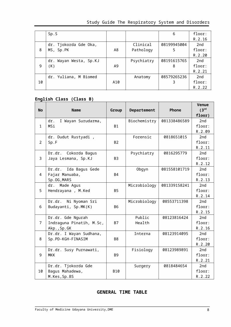
~ FACILITATORS ~Regular Class (Class A)
No Name Group Departement Phone Venue (2rd floor)
1 dr. Firman Parulian Sitanggang, Sp.Rad(K)RI A1 Radiology 081337165566 2nd floor:
R.2.09
2dr. Tjok. Istri Anom Saturti, Sp.PD A2
Interna 081916253777 2nd floor: R.2.11
3dr. I Wayan Gede Sutadarma, M Gizi A3
Biochemistry 082144071268 2nd floor: R.2.12
4dr. Yenny Kandarini, Sp.PD-KGH-FINASIM A4
Interna 08123805344 2nd floor: R.2.13
5dr. I Gusti Ngurah Pramesemara, S.Ked A5
Andrology 081338605087 2nd floor: R.2.14
6dr. I Wayan Weta, MS
A6Public Health 081337005360 2nd floor:
R.2.15
7dr. I A. Sri Indrayani, Sp.S
A7Neurology 081246751536 2nd floor:
R.2.16
8dr. Tjokorda Gde Oka, MS, Sp.PK A8
Clinical Pathology
081999450045 2nd floor: R.2.20
9dr. Wayan Westa, Sp.KJ (K)
A9Psychiatry 081916157658 2nd floor:
R.2.21
10 dr. Yuliana, M Biomed A10 Anatomy 085792652363 2nd floor: R.2.22
English Class (Class B)
No Name Group Departement Phone Venue (3rd floor)
1 dr. I Wayan Surudarma, MSi B1 Biochemistry 081338486589 2nd floor: R.2.09
2dr. Dudut Rustyadi , Sp.F
B2Forensic 0818651015 2nd floor:
R.2.11
3Dr.dr. Cokorda Bagus Jaya Lesmana, Sp.KJ B3
Psychiatry 0816295779 2nd floor: R.2.12
4Dr.dr. Ida Bagus Gede Fajar Manuaba, Sp.OG,MARS B4
Obgyn 081558101719 2nd floor: R.2.13
5dr. Made Agus Hendrayana , M.Ked B5
Microbiology 081339158241 2nd floor: R.2.14
6Dr.dr. Ni Nyoman Sri Budayanti, Sp.MK(K) B6
Microbiology 08553711398 2nd floor: R.2.15
7Dr.dr. Gde Ngurah Indraguna Pinatih, M.Sc, Akp.,Sp.GK B7
Public Health 08123816424 2nd floor: R.2.16
8Dr.dr. I Wayan Sudhana, Sp.PD-KGH-FINASIM B8
Interna 08123914095 2nd floor: R.2.20
9Dr.dr. Susy Purnawati, MKK
B9Fisiology 08123989891 2nd floor:
R.2.21
10Dr.dr. Tjokorda Gde Bagus Mahadewa, M.Kes,Sp.BS B10
Surgery 0818484654 2nd floor: R.2.22
Faculty of Medicine Udayana University,DME 6
Study Guide The Respiratory System and Disorders
GENERAL TIME TABLEFOR A AND B CLESSES
CLASS A CLASS B
TIME ACTIVITIES TIME ACTIVITIES08.00-09.00 Lecture 09.00-10.00 Lecture
09.00-10.30 Independent learning 10.00-11.30 Student project
10.30-12.00 SGD 11.30-12.00 Break
12.00-12.30 Break 12.00-13.30 Independent learning
12.30-14.00 Student project 13.30-15.00 SGD
14.00-15.00 Plenary session 15.00-16.00 Plenary session
There are several types of learning activity: Lecture
independent learning based on the lecture’s topic
Small group discussion to solve the learning task
Practice
Student project
Clinical skill and demonstration
Self assessment at the end of every topic
Plenary session
Lecture will be held at room 402, while discussion rooms available at 3rd floor (room 3.09-3.17&3.19)
Faculty of Medicine Udayana University,DME 7
Study Guide The Respiratory System and Disorders
IMPORTANT INFORMATIONS
Meeting of the students’ representativeIn the middle of block schedule, a meeting is designed among the student
representatives of every small group discussions, facilitators, and resource persons. The
meeting will discuss the ongoing teaching learning process, quality of lecturers and
facilitators as a feedback to improve the next process. The meeting will be taken based on
schedule from Medical Education Unit.
SELF ASSESSMENT
Self assessment of each lecture will be given after each lecture session, and will be
marked. This mark can determine whether the student pass this block or not. Any final mark
between 65 to 69 will be reconsidered with self assessment’s mark to see the student’s
status. Any student with self assessment’s mark more than 70 will pass this block. And for
the lower one will have to attend the remedial examination. It is important to do this self
assessment cautiously, because this activity may be your ticket to pass this block.
ASSESSMENT METHODAssessment in this theme consists of:SGD : 5%
Final Exam : 80%
Student Project : 15%
Final mark more than 70 considered to pass this block. Certain conditions applied for those
with final mark between 65 – 69. These students will be analyzed using their self
assessment’s mark. Students with final mark 65 – 69 and self assessment’s mark more than
70 will also considered pass this block.
Faculty of Medicine Udayana University,DME 8

Study Guide The Respiratory System and Disorders
TIME TABLEREGULAR CLASS
DAY/DATE TIME ACTIVITY VENUE PIC
1MondayFeb 16,
2015
08.00-08.15 Introduction Class room Prof.I.B. Rai08.15-09.00 Lecture 1
Anatomy of Respiratory System
Class room dr.Wardana
09.00-10.30 Independent learning10.30-12.00 SGD Disc room Facilitator 12.00-12.30 Break 12.30-14.00 Student project14.00-15.00 Plenary session Class room dr.Wardana
2TuesdayFeb 17,
2015
08.00-09.00 Lecture2 Histology of Respiratory System
Class room dr. Sri Wiryawan
09.00-10.30 Independent learning10.30-12.00 SGD Disc room Facilitator12.00-12.30 Break 12.30-14.00 Student project14.00-15.00 Plenary session Class room dr. Sri Wiryawan
3Wednesday
Feb 18,2015
08.00-09.00 Lecture 3 Physiology of Respiratory System: Ventilation
Class room dr. Muliarta
09.00-10.30 Independent learning10.30-12.00 SGD Disc room Facilitator12.00-12.30 Break 12.30-14.00 Student project14.00-15.00 Plenary session Class room dr. Muliarta
4Friday
Feb 20, 2015
08.00-09.00 Lecture 4 Physiology of Respiratory System: Gas Exchange, diving, altitude
Class room dr. Muliarta
09.00-15.00 Independent learningPractice : Anatomy, Anatomy: dr. Wardana
Faculty of Medicine Udayana University,DME 9
REG
ULA
R CL
ASS
Study Guide The Respiratory System and Disorders
Histology1st floorHistology: 4th floor
dr. Sri Wiryawan
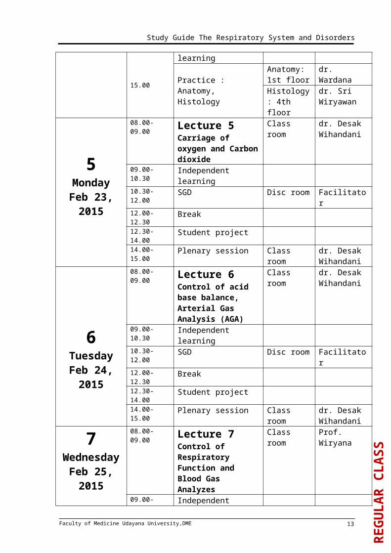
5Monday
Feb 23, 2015
08.00-09.00 Lecture 5Carriage of oxygen and Carbon dioxide
Class room dr. Desak Wihandani
09.00-10.30 Independent learning10.30-12.00 SGD Disc room Facilitator12.00-12.30 Break 12.30-14.00 Student project14.00-15.00 Plenary session Class room dr. Desak
Wihandani
6Tuesday
Feb 24, 2015
08.00-09.00 Lecture 6Control of acid base balance, Arterial Gas Analysis (AGA)
Class room dr. Desak Wihandani
09.00-10.30 Independent learning10.30-12.00 SGD Disc room Facilitator12.00-12.30 Break 12.30-14.00 Student project14.00-15.00 Plenary session Class room dr. Desak
Wihandani
7WednesdayFeb 25, 2015
08.00-09.00 Lecture 7Control of Respiratory Function and Blood Gas Analyzes
Class room Prof. Wiryana
09.00-10.30 Independent learning10.30-12.00 SGD Disc room Facilitator12.00-12.30 Break 12.30-14.00 Student project14.00-15.00 Plenary session Class room Prof. Wiryana
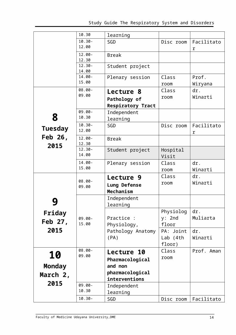
8TuesdayFeb 26,
2015
08.00-09.00 Lecture 8Pathology of Respiratory Tract
Class room dr. Winarti
09.00-10.30 Independent learning10.30-12.00 SGD Disc room Facilitator12.00-12.30 Break 12.30-14.00 Student project Hospital
Visit
Faculty of Medicine Udayana University,DME 10
REG
ULA
R CL
ASS

Study Guide The Respiratory System and Disorders
14.00-15.00 Plenary session Class room dr. Winarti
9FridayFeb 27,
2015
08.00-09.00Lecture 9Lung Defense Mechanism
Class room dr. Winarti
09.00-15.00
Independent learning
Practice : Physiology, Pathology Anatomy (PA)
Physiology: 2nd floor
dr. Muliarta
PA: Joint Lab (4th floor)
dr. Winarti
10Monday March 2,
2015
08.00-09.00 Lecture 10Pharmacological and non pharmacological interventions
Class room Prof. Aman
09.00-10.30 Independent learning10.30-12.00 SGD Disc room Facilitator12.00-12.30 Break 12.30-14.00 Student project14.00-15.00 Plenary session Class room Prof. Aman
11TuesdayMarch 3,
2015
08.00-09.00 Lecture 11Pharmacological and non pharmacological interventions
Class room Prof. Aman
09.00-10.30 Independent learning10.30-12.00 SGD Disc room Facilitator12.00-12.30 Break 12.30-14.00 Student project Hospital
Visit14.00-15.00 Plenary session Class room Prof. Aman
12Wednesday
March 4,2015
08.00-09.00 Lecture 12Respiratory Imaging
Class room dr. Elysanti
09.00-10.30 Independent learning10.30-12.00 SGD Disc room Facilitator12.00-12.30 Break 12.30-14.00 Student project14.00-15.00 Plenary session Class room dr. Elysanti
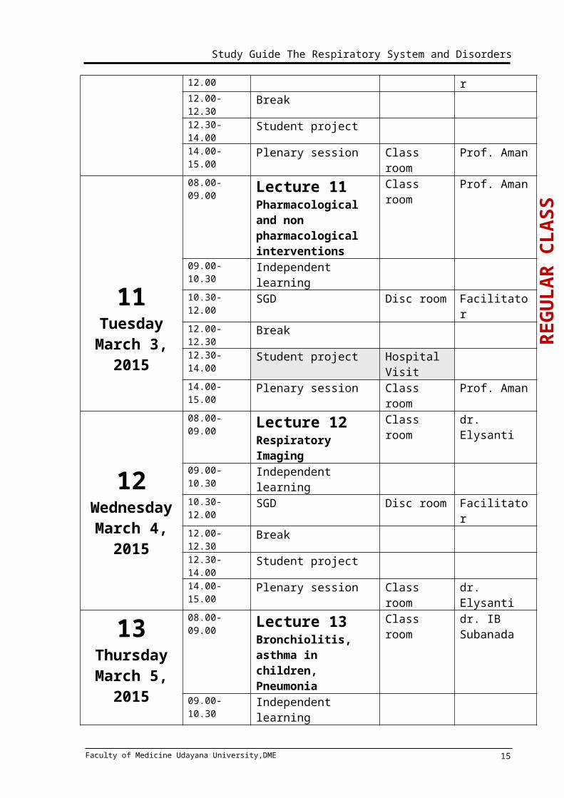
13 08.00-09.00 Lecture 13Bronchiolitis, asthma in children,
Class room dr. IB Subanada
Faculty of Medicine Udayana University,DME 11
REG
ULA
R CL
ASS

Study Guide The Respiratory System and Disorders
ThursdayMarch 5,
2015
Pneumonia09.00-10.30 Independent learning10.30-12.00 SGD Disc room Facilitator12.00-12.30 Break 12.30-14.00 Student project Hospital
Visit14.00-15.00 Plenary session Class room dr. IB
Subanada
14Friday
March 6,2015
08.00-09.00 Lecture 14TB in children, Difteri, Pertusis
Class room dr. Siadi Purniti
09.00-10.30 Independent learning10.30-12.00 SGD Disc room Facilitator12.00-12.30 Break 12.30-14.00 Student project14.00-15.00 Plenary session Class room dr. Siadi
Purniti
15MondayMarch 9,
2015
08.00-09.00 Lecture 15Pulmonary TB and Extrapulmonary TB, TB in the Immunocompromised Host, Abses TB
Class room dr. Sutha,
dr. Bagiada
09.00-10.30 Independent learning10.30-12.00 SGD Disc room Facilitator12.00-12.30 Break 12.30-14.00 Student project Hospital
Visit14.00-15.00 Plenary session Class room dr. Sutha,
dr. Bagiada
16TuesdayMarch 10,
2015
08.00-09.00 Lecture 16Asthma, COPD
Class roomProf. IB Rai, dr. Artana
09.00-10.30 Independent learning
10.30-12.00 SGD Disc room Facilitator12.00-12.30 Break
Faculty of Medicine Udayana University,DME 12
REG
ULA
R CL
ASS
Study Guide The Respiratory System and Disorders
12.30-14.00 Student project14.00-15.00 Plenary session Class room Prof. IB Rai,
dr. Artana
17WednesdayMarch 11,
2015
08.00-09.00 Lecture 17Pleural effusion, Pneumothorax, Hematothorax
Class roomdr. Andrika, dr, Yasa
09.00-10.30 Independent learning10.30-12.00 SGD Disc room Facilitator12.00-12.30 Break 12.30-14.00 Student project Hospital
Visit14.00-15.00 Plenary session Class room dr. Andrika,
dr, Yasa
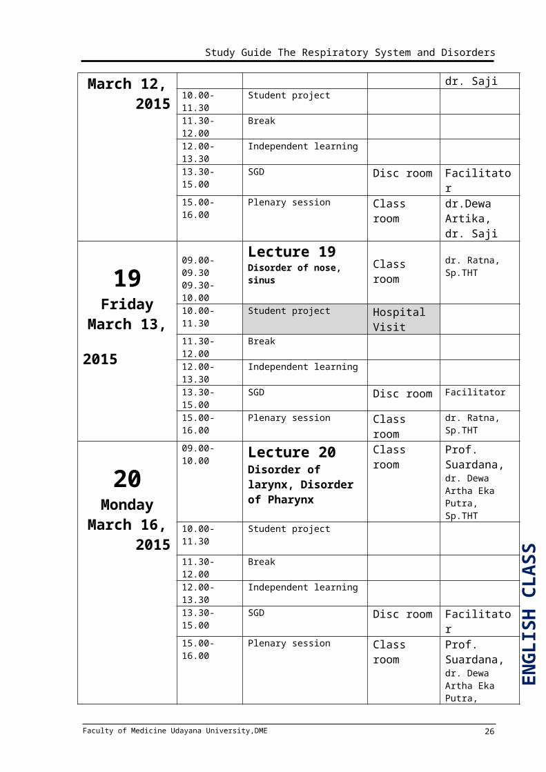
18ThursdayMarch 12,
2015
08.00-09.00 Lecture 18Bronchitis and Bronchiectasis, Lung Ca and Smoking Cessation
Class room
dr.Dewa Artika,dr. Saji
09.00-10.30 Independent learning10.30-12.00 SGD Disc room Facilitator12.00-12.30 Break 12.30-14.00 Student project14.00-15.00 Plenary session Class room dr.Dewa
Artika, dr. Saji
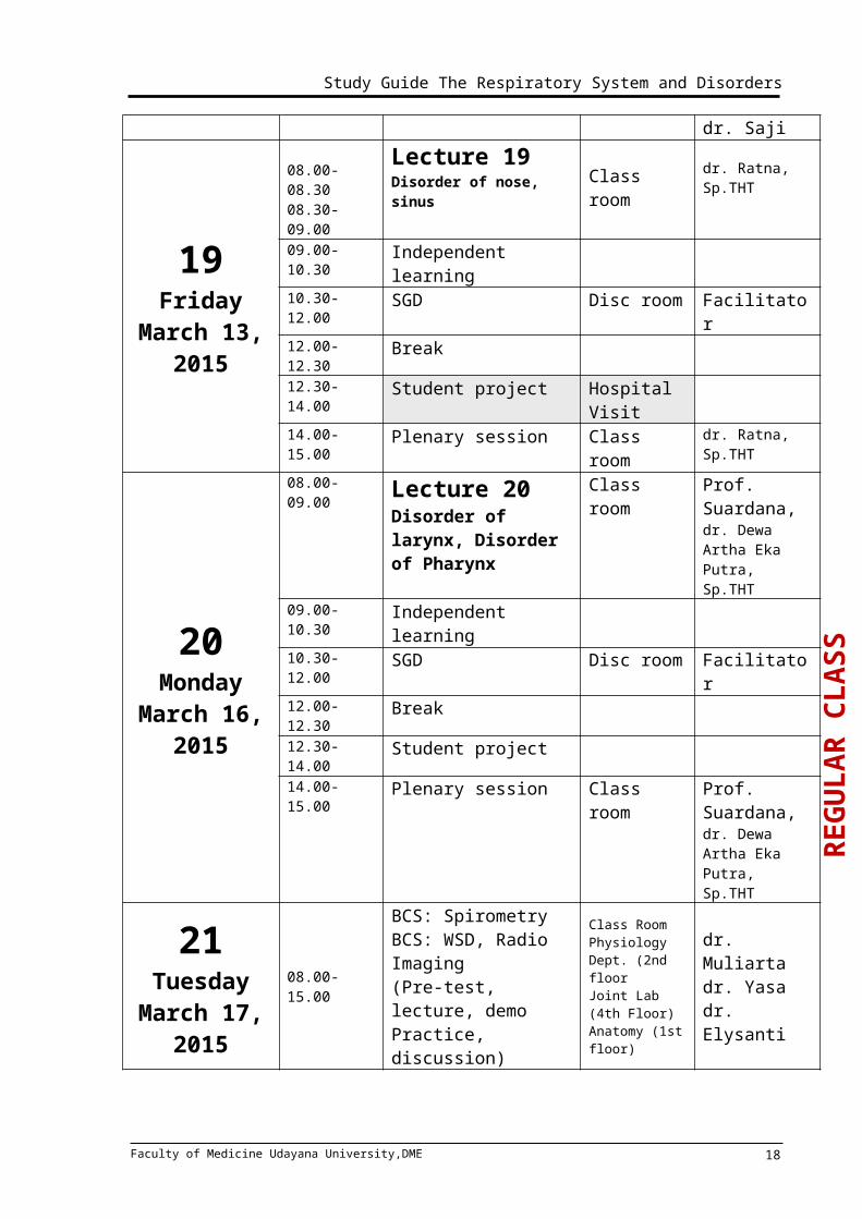
19Friday
March 13,2015
08.00-08.3008.30-09.00
Lecture 19Disorder of nose, sinus Class room dr. Ratna,
Sp.THT09.00-10.30 Independent learning10.30-12.00 SGD Disc room Facilitator12.00-12.30 Break 12.30-14.00 Student project Hospital
Visit14.00-15.00 Plenary session Class room dr. Ratna,
Sp.THT
20Monday
08.00-09.00 Lecture 20Disorder of larynx, Disorder of Pharynx
Class room Prof. Suardana, dr. Dewa Artha Eka Putra, Sp.THT
Faculty of Medicine Udayana University,DME 13
Study Guide The Respiratory System and Disorders
March 16,2015
09.00-10.30 Independent learning
10.30-12.00 SGD Disc room Facilitator12.00-12.30 Break 12.30-14.00 Student project14.00-15.00 Plenary session Class room Prof.
Suardana, dr. Dewa Artha Eka Putra, Sp.THT
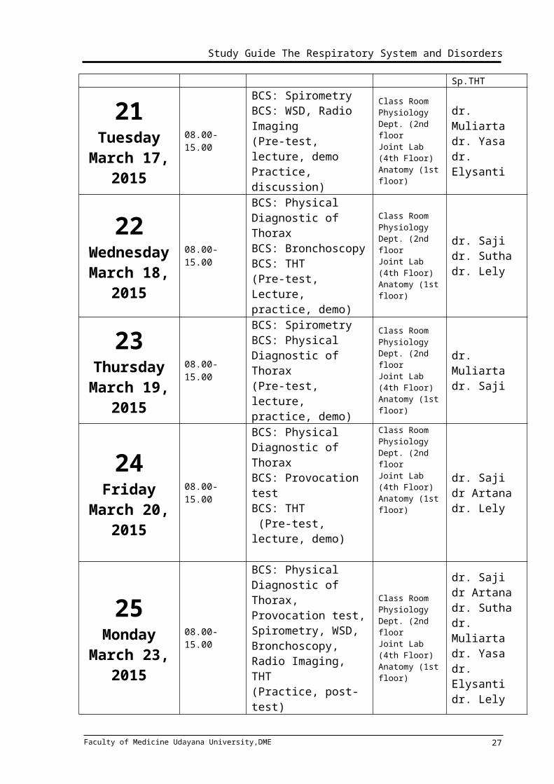
21TuesdayMarch 17,
2015
08.00-15.00
BCS: SpirometryBCS: WSD, Radio Imaging(Pre-test, lecture, demo Practice, discussion)
Class RoomPhysiology Dept. (2nd floorJoint Lab (4th Floor)Anatomy (1st floor)
dr. Muliartadr. Yasa dr. Elysanti
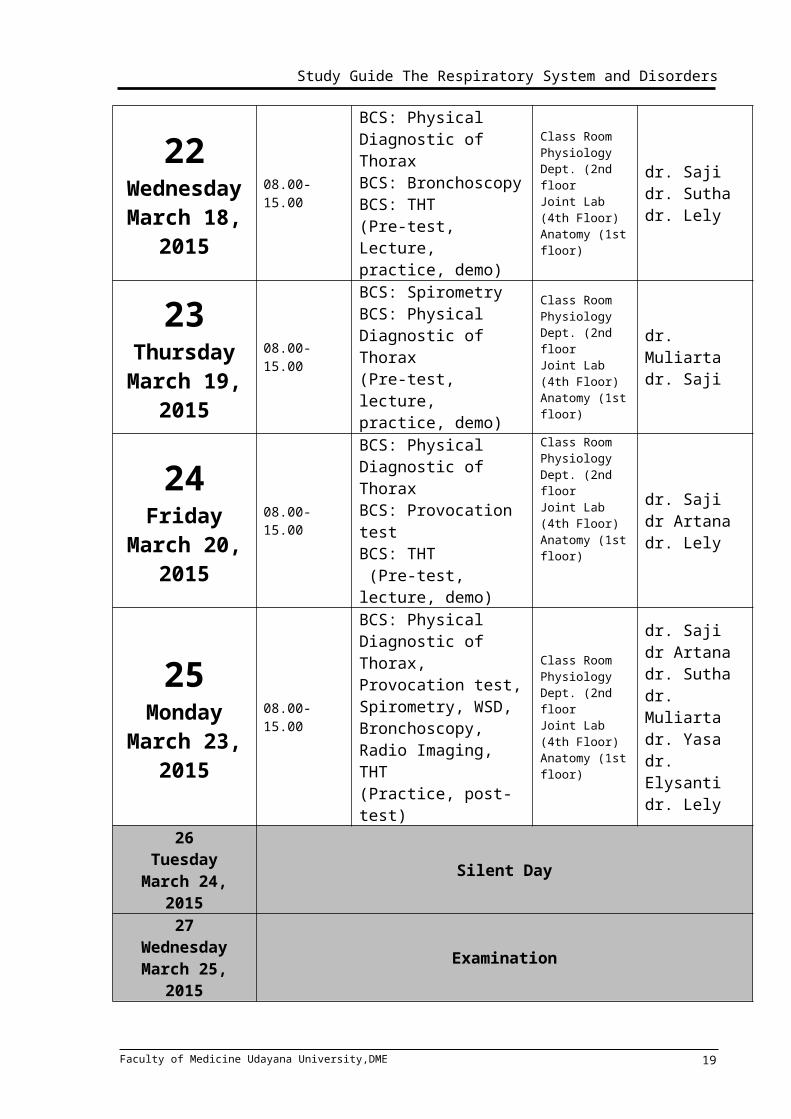
22WednesdayMarch 18,
2015
08.00-15.00
BCS: Physical Diagnostic of ThoraxBCS: BronchoscopyBCS: THT(Pre-test, Lecture, practice, demo)
Class RoomPhysiology Dept. (2nd floorJoint Lab (4th Floor)Anatomy (1st floor)
dr. Sajidr. Suthadr. Lely
23ThursdayMarch 19,
2015
08.00-15.00
BCS: SpirometryBCS: Physical Diagnostic of Thorax (Pre-test, lecture, practice, demo)
Class RoomPhysiology Dept. (2nd floorJoint Lab (4th Floor)Anatomy (1st floor)
dr. Muliartadr. Saji
24Friday
March 20,2015
08.00-15.00
BCS: Physical Diagnostic of Thorax BCS: Provocation test BCS: THT (Pre-test, lecture, demo)
Class RoomPhysiology Dept. (2nd floorJoint Lab (4th Floor)Anatomy (1st floor)
dr. Saji dr Artanadr. Lely
25Monday
08.00-15.00 BCS: Physical Diagnostic of Thorax, Provocation test, Spirometry, WSD,
Class RoomPhysiology Dept. (2nd floorJoint Lab (4th Floor)Anatomy (1st
dr. Saji dr Artana dr. Suthadr. Muliarta
Faculty of Medicine Udayana University,DME 14
REG
ULA
R CL
ASS
Study Guide The Respiratory System and Disorders
March 23,2015
Bronchoscopy, Radio Imaging, THT(Practice, post-test)
floor)dr. Yasadr. Elysantidr. Lely
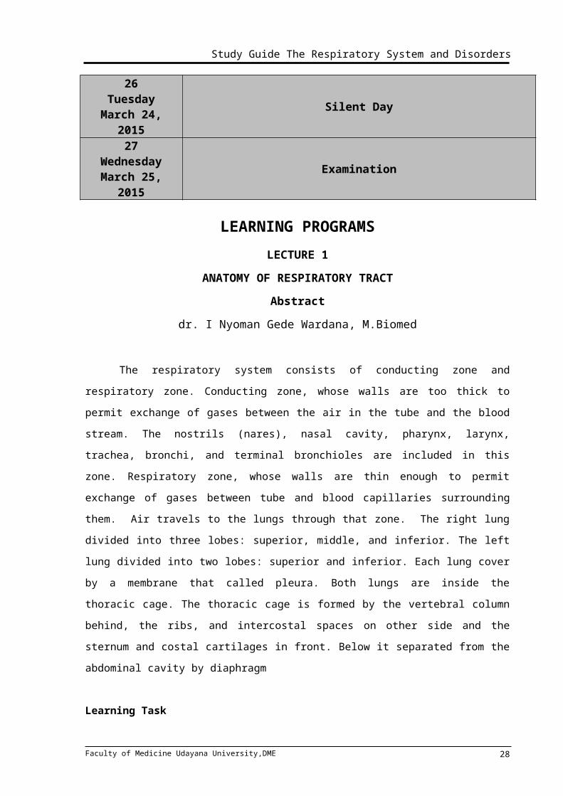
26TuesdayMarch 24,
2015Silent Day
27Wednesday
March 25, 2015Examination
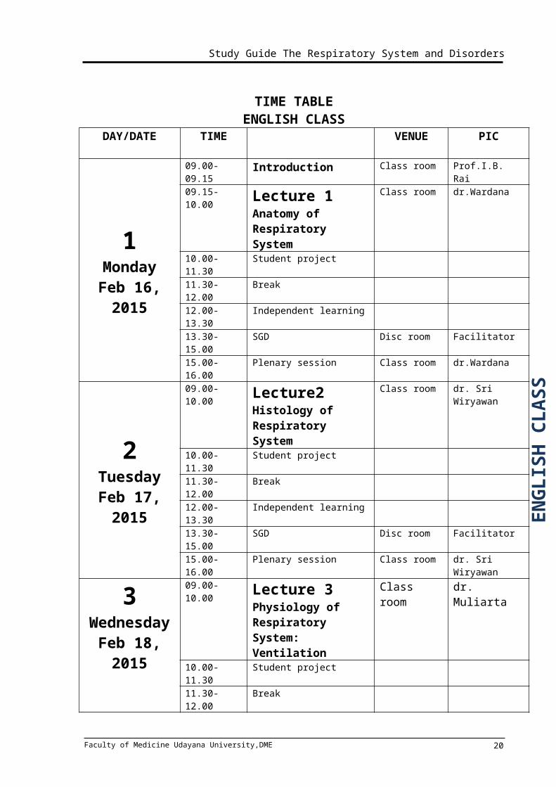
TIME TABLEENGLISH CLASS
DAY/DATE TIME VENUE PIC
1MondayFeb 16,
2015
09.00-09.15 Introduction Class room Prof.I.B. Rai09.15-10.00 Lecture 1
Anatomy of Respiratory System
Class room dr.Wardana
10.00-11.30 Student project 11.30-12.00 Break 12.00-13.30 Independent learning13.30-15.00 SGD Disc room Facilitator 15.00-16.00 Plenary session Class room dr.Wardana
2TuesdayFeb 17,
2015
09.00-10.00 Lecture2 Histology of Respiratory System
Class room dr. Sri Wiryawan
10.00-11.30 Student project 11.30-12.00 Break 12.00-13.30 Independent learning13.30-15.00 SGD Disc room Facilitator 15.00-16.00 Plenary session Class room dr. Sri Wiryawan
3Wednesday
09.00-10.00 Lecture 3 Physiology of Respiratory System: Ventilation
Class room dr. Muliarta
Faculty of Medicine Udayana University,DME 15
ENG
LISH
CL
ASS

Study Guide The Respiratory System and Disorders
Feb 18,2015
10.00-11.30 Student project 11.30-12.00 Break 12.00-13.30 Independent learning13.30-15.00 SGD Disc room Facilitator15.00-16.00 Plenary session Class room dr. Muliarta
4Friday
Feb 20, 2015
09.00-10.00 Lecture 4 Physiology of Respiratory System: Gas Exchange, diving, altitude
Class room dr. Muliarta
10.00-16.00
Independent learning
Practice : Anatomy, Histology
Anatomy: 1st floor
dr. Wardana
Histology: 4th floor
dr. Sri Wiryawan
5Monday
Feb 23, 2015
09.00-10.00 Lecture 5Carriage of oxygen and Carbon dioxide
Class room dr. Desak Wihandani
10.00-11.30 Student project 11.30-12.00 Break 12.00-13.30 Independent learning13.30-15.00 SGD Disc room Facilitator15.00-16.00 Plenary session Class room dr. Desak
Wihandani
6Tuesday
Feb 24, 2015
09.00-10.00 Lecture 6Control of acid base balance, Arterial Gas Analysis (AGA)
Class room dr. Desak Wihandani
10.00-11.30 Student project 11.30-12.00 Break 12.00-13.30 Independent learning13.30-15.00 SGD Disc room Facilitator15.00-16.00 Plenary session Class room dr. Desak
Wihandani
7WednesdayFeb 25, 2015
09.00-10.00 Lecture 7Control of Respiratory Function and Blood Gas Analyzes
Class room Prof. Wiryana
10.00-11.30 Student project
Faculty of Medicine Udayana University,DME 16
ENG
LISH
CL
ASS

Study Guide The Respiratory System and Disorders
11.30-12.00 Break 12.00-13.30 Independent learning13.30-15.00 SGD Disc room Facilitator15.00-16.00 Plenary session Class room Prof. Wiryana
8ThursdayFeb 26,
2015
09.00-10.00 Lecture 8Pathology of Respiratory Tract
Class room dr. Winarti
10.00-11.30 Student project Hospital Visit
11.30-12.00 Break 12.00-13.30 Independent learning13.30-15.00 SGD Disc room Facilitator15.00-16.00 Plenary session Class room dr. Winarti
9FridayFeb 27,
2015
09.00-10.00
Lecture 9Lung Defense Mechanism
Class room dr. Winarti
10.00-16.00
Independent learning
Practice : Physiology, Pathology Anatomy (PA)
Physiology: 2nd floor
dr. Muliarta
PA: Joint Lab (4th floor)
dr. Winarti
10Monday March 2,
2015
09.00-10.00 Lecture 10Pharmacological and non pharmacological interventions
Class room Prof. Aman
10.00-11.30 Student project 11.30-12.00 Break 12.00-13.30 Independent learning13.30-15.00 SGD Disc room Facilitator15.00-16.00 Plenary session Class room Prof. Aman
11Tuesday
09.00-10.00 Lecture 11Pharmacological and non pharmacological interventions
Class room Prof. Aman
10.00-11.30 Student project Hospital Visit
11.30-12.00 Break 12.00-13.30 Independent learning13.30-15.00 SGD Disc room Facilitator
Faculty of Medicine Udayana University,DME 17
Study Guide The Respiratory System and Disorders
March 3, 2015
15.00-16.00 Plenary session Class room Prof. Aman
12Wednesday
March 4, 2015
09.00-10.00 Lecture 12Respiratory Imaging
Class room dr. Elysanti
10.00-11.30 Student project 11.30-12.00 Break 12.00-13.30 Independent learning13.30-15.00 SGD Disc room Facilitator15.00-16.00 Plenary session Class room dr. Elysanti
13ThursdayMarch 5,
2015
09.00-10.00 Lecture 13Bronchiolitis, asthma in children
Class room dr. IB Subanada
10.00-11.30 Student project Hospital Visit
11.30-12.00 Break 12.00-13.30 Independent learning13.30-15.00 SGD Disc room Facilitator15.00-16.00 Plenary session Class room dr. IB
Subanada
14Friday
March 6, 2015
09.00-10.00 Lecture 14TB in children
Class room dr. Siadi Purniti
10.00-11.30 Student project 11.30-12.00 Break 12.00-13.30 Independent learning13.30-15.00 SGD Disc room Facilitator15.00-16.00 Plenary session Class room dr. Siadi
Purniti
15MondayMarch 9,
2015
09.00-10.00 Lecture 15Pulmonary TB and Extrapulmonary TB, TB in the Immunocompromised Host
Class room dr. Sutha,
dr. Bagiada
10.00-11.30 Student project Hospital Visit
11.30-12.00 Break
Faculty of Medicine Udayana University,DME 18
ENG
LISH
CL
ASS
Study Guide The Respiratory System and Disorders
12.00-13.30 Independent learning13.30-15.00 SGD Disc room Facilitator15.00-16.00 Plenary session Class room dr. Sutha,
dr. Bagiada
16TuesdayMarch 10,
2015
09.00-10.00 Lecture 16Asthma, COPD
Class roomProf. IB Rai, dr. Artana
10.00-11.30 Student project 11.30-12.00 Break 12.00-13.30 Independent learning13.30-15.00 SGD Disc room Facilitator
15.00-16.00 Plenary session Class room Prof. IB Rai, dr. Artana
17WednesdayMarch 11,
2015
09.00-09.00 Lecture 17Pleural effusion, Pneumothorax
Class roomdr. Andrika, dr, Yasa
10.00-11.30 Student project Hospital Visit
11.30-12.00 Break 12.00-13.30 Independent learning13.30-15.00 SGD Disc room Facilitator15.00-16.00 Plenary session Class room dr. Andrika,
dr, Yasa
18ThursdayMarch 12,
2015
08.00-09.00 Lecture 18Bronchitis and Bronchiectasis, Lung Ca and Smoking Cessation
Class room
dr.Dewa Artika,
dr. Saji 10.00-11.30 Student project 11.30-12.00 Break 12.00-13.30 Independent learning13.30-15.00 SGD Disc room Facilitator15.00-16.00 Plenary session Class room dr.Dewa
Artika, dr. Saji
09.00-09.30 Lecture 19 dr. Ratna,
Faculty of Medicine Udayana University,DME 19
ENG
LISH
CL
ASS
Study Guide The Respiratory System and Disorders
19Friday
March 13, 2015
09.30-10.00 Disorder of nose, sinus Class room Sp.THT10.00-11.30 Student project Hospital
Visit11.30-12.00 Break 12.00-13.30 Independent learning13.30-15.00 SGD Disc room Facilitator15.00-16.00 Plenary session Class room dr. Ratna,
Sp.THT
20Monday
March 16, 2015
09.00-10.00 Lecture 20Disorder of larynx, Disorder of Pharynx
Class room Prof. Suardana, dr. Dewa Artha Eka Putra, Sp.THT
10.00-11.30 Student project
11.30-12.00 Break 12.00-13.30 Independent learning13.30-15.00 SGD Disc room Facilitator15.00-16.00 Plenary session Class room Prof.
Suardana, dr. Dewa Artha Eka Putra, Sp.THT
21TuesdayMarch 17,
2015
08.00-15.00
BCS: SpirometryBCS: WSD, Radio Imaging(Pre-test, lecture, demo Practice, discussion)
Class RoomPhysiology Dept. (2nd floorJoint Lab (4th Floor)Anatomy (1st floor)
dr. Muliartadr. Yasa dr. Elysanti
22WednesdayMarch 18,
2015
08.00-15.00
BCS: Physical Diagnostic of ThoraxBCS: BronchoscopyBCS: THT(Pre-test, Lecture, practice, demo)
Class RoomPhysiology Dept. (2nd floorJoint Lab (4th Floor)Anatomy (1st floor)
dr. Sajidr. Suthadr. Lely
23ThursdayMarch 19,
2015
08.00-15.00
BCS: SpirometryBCS: Physical Diagnostic of Thorax (Pre-test, lecture, practice, demo)
Class RoomPhysiology Dept. (2nd floorJoint Lab (4th Floor)Anatomy (1st floor)
dr. Muliartadr. Saji
Faculty of Medicine Udayana University,DME 20
ENG
LISH
CL
ASS

Study Guide The Respiratory System and Disorders
24Friday
March 20,2015
08.00-15.00
BCS: Physical Diagnostic of Thorax BCS: Provocation test BCS: THT (Pre-test, lecture, demo)
Class RoomPhysiology Dept. (2nd floorJoint Lab (4th Floor)Anatomy (1st floor)
dr. Saji dr Artanadr. Lely
25Monday
March 23,2015
08.00-15.00
BCS: Physical Diagnostic of Thorax, Provocation test, Spirometry, WSD, Bronchoscopy, Radio Imaging, THT(Practice, post-test)
Class RoomPhysiology Dept. (2nd floorJoint Lab (4th Floor)Anatomy (1st floor)
dr. Saji dr Artana dr. Suthadr. Muliartadr. Yasadr. Elysantidr. Lely
26TuesdayMarch 24,
2015Silent Day
27Wednesday
March 25, 2015Examination
LEARNING PROGRAMSLECTURE 1
ANATOMY OF RESPIRATORY TRACTAbstract
dr. I Nyoman Gede Wardana, M.Biomed
The respiratory system consists of conducting zone and respiratory zone.
Conducting zone, whose walls are too thick to permit exchange of gases between the air in
the tube and the blood stream. The nostrils (nares), nasal cavity, pharynx, larynx, trachea,
bronchi, and terminal bronchioles are included in this zone. Respiratory zone, whose walls
are thin enough to permit exchange of gases between tube and blood capillaries
surrounding them. Air travels to the lungs through that zone. The right lung divided into
three lobes: superior, middle, and inferior. The left lung divided into two lobes: superior and
inferior. Each lung cover by a membrane that called pleura. Both lungs are inside the
thoracic cage. The thoracic cage is formed by the vertebral column behind, the ribs, and
intercostal spaces on other side and the sternum and costal cartilages in front. Below it
separated from the abdominal cavity by diaphragm
Learning Task
Faculty of Medicine Udayana University,DME 21
Study Guide The Respiratory System and Disorders
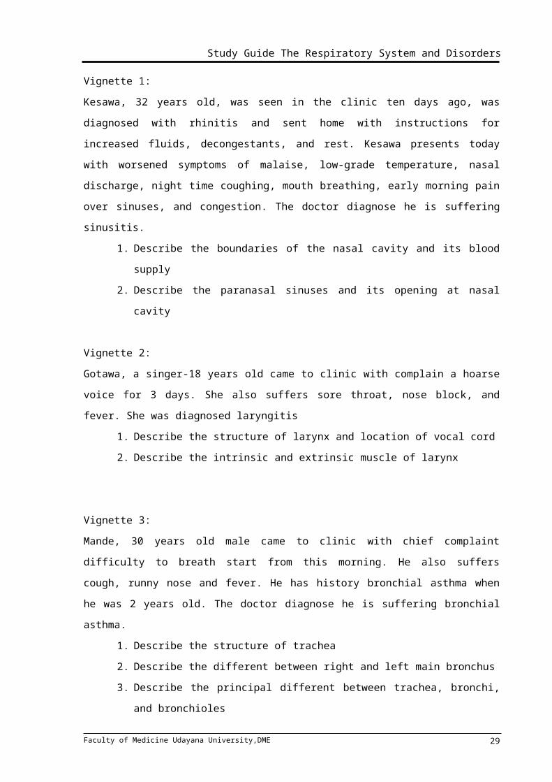
Vignette 1:
Kesawa, 32 years old, was seen in the clinic ten days ago, was diagnosed with rhinitis and
sent home with instructions for increased fluids, decongestants, and rest. Kesawa presents
today with worsened symptoms of malaise, low-grade temperature, nasal discharge, night
time coughing, mouth breathing, early morning pain over sinuses, and congestion. The
doctor diagnose he is suffering sinusitis.
1. Describe the boundaries of the nasal cavity and its blood supply
2. Describe the paranasal sinuses and its opening at nasal cavity
Vignette 2:
Gotawa, a singer-18 years old came to clinic with complain a hoarse voice for 3 days. She
also suffers sore throat, nose block, and fever. She was diagnosed laryngitis
1. Describe the structure of larynx and location of vocal cord
2. Describe the intrinsic and extrinsic muscle of larynx
Vignette 3:
Mande, 30 years old male came to clinic with chief complaint difficulty to breath start from
this morning. He also suffers cough, runny nose and fever. He has history bronchial asthma
when he was 2 years old. The doctor diagnose he is suffering bronchial asthma.
1. Describe the structure of trachea
2. Describe the different between right and left main bronchus
3. Describe the principal different between trachea, bronchi, and bronchioles
Vignette 4:
A 57-year-old male is admitted to the hospital with a chief complaint of shortness of breath
for 2 weeks. The radiology examination shows a large left-side pleural effusion.
1. Describe the different between right lung and left lung
2. Describe the structure of pleura
3. Describe the structure of thoracic wall
LECTURE 2HISTOLOGY OF RESPIRATORY TRACT
dr. Sri Wiryawan, MRepro
Faculty of Medicine Udayana University,DME 22
Study Guide The Respiratory System and Disorders
AbstractThe lower respiratory tract consists of : the lower part of the trachea, the two main
bronchi, lobar, segmental, and smaller bronchi, bronchioles and terminal bronchioles, and
last but not least is the end respiratory unit. These structure make up the tracheobronchial
tree. As for the structure distal to the main bronchi along with a tissue known as the lung
parenchyma.
There are several structure we should also understand, when talking about lower
respiratory tract. Several structures such as thorax, mediastinum, pleurae and pleural
cavity, and lung. Thorax especially thoracic cavity and thoracic wall protect our lung and
mediastinum and also play an important role in respiratory process. The mediastinum,
which has a role in protecting our heart , located between the two lungs, and contains the
heart and great vessels, trachea and esophagus, phrenic and vagus nerves, and lymph
nodes.
The pleurae covers the external surface of the lung, and is then reflected to cover
the inner surface of thoracic cavity. Pleurae divided into the visceral (lines the surface of the
lung) and parietal (lines the thoracic wall and diaphragm) one. The space between these
two pleurae called as pleural cavity which contains a thin film fluid to allow the pleurae to
slip over each other during breathing.
The lungs are placed within the thoracic cavity. The lungs contain airways structure,
vessels, lymphatic and lymph nodes, nerves, and supportive connective tissue. The trachea
divides and form the left and right primary bronchi, which in turn divide to form lobar bronchi.
Each lobar bronchi divide again to give segmental bronchi to supply air to
bronchopulmonary segments. The tracheobronchial tree can also be classified into two
functional zones: the conducting zone (proximal to the respiratory bronchioles) which
involved in air movement, and the respiratory zone (distal to the terminal bronchioles) which
involved in gaseous exchange.
The other term to show functional structure of the lower respiratory tract is the
acinus. The acinus defined as the part of the airway that is involved in gaseous exchange.
The acinus consist of respiratory bronchioles, alveolar ducts, and alveoli as the smallest
functional structure of the lung. The areas of lung containing groups of between three to five
acini surrounded by parenchimal tissue are called lung lobules.
The alveolus is an blind-ending terminal sac of respiratory tract. Most gaseous
exchange occurs in the alveoli. The alveoli are lined with type I (structural) and type II
(produce surfactant) of pneumocytes cell. The understanding about histological pattern of
these functional structures of the lung is important in pathophysiology of lung problems.
Learning Tasks
Faculty of Medicine Udayana University,DME 23
Study Guide The Respiratory System and Disorders
I. Structure of The Upper Respiratory tract
Krishna, a man, 25 years old came to doctor Arjuna clinic with fever, sore throat,
sneezing, runny nose and sometimes blocked nose. He also cannot smell well. The
doctor diagnoses Krishna with acut Rhinopharingitis.
1. Describe the histological structure of the upper respiratory tracts
are involved ?
2. Describe the histological structure and function of epiglottis !
3. Compare the histological structure and function between vestibular fold and
vocal fold !
II. Structure of The Lower Respiratory tract
Radha, a 17 years old beautiful girl, came to doctor Laksmi clinic with shortness of
breath, wheezing and cough with phlegm. The doctor diagnoses Radha with
Asthma.
1.Describe the histological structure of the lower respiratory tracts are involved ?
2.Compare the histological structure and function between terminal bronchioles and
respiratory bronchioles !
3.Describe the histological structure of the interalveolar septum !
4.Describe the histological structure of blood-air barrier ?
5.Describe about the pulmonary surfactant ?
LECTURE 3PHYSIOLOGY OF RESPIRATORY SYSTEM: VENTILATION
dr. I Made Muliarta, MKes
Abstract In living cells aerobic metabolism consumes oxygen and produces carbon dioxide. Gas
exchange requires a large , thin, moist exchange surface, a pump to move air circulatory
system to transport gases to cells. The primary function system are:
Exchange the gases between atmosphere and the blood.
Homeostatic regulation of body pH .
Protection from inhaled pathogens and irritation substance
Vocalization.
Faculty of Medicine Udayana University,DME 24
Study Guide The Respiratory System and Disorders
In addition to serving these function, the respiratory system also source of significant
losses of water and heat from the lung.
A single respiratory cycle consists of an inspiration and expiration. Relation with
ventilation had to know about compliance, surfactant, lung volume and capacities
Respiratory control resides in a central pattern generator, a net work of neurons in the
pons and medulla oblongata.
Faculty of Medicine Udayana University,DME 25
Study Guide The Respiratory System and Disorders
LEARNING TASK dr. Muliarta, MKes1. What is the sequence of event during quiet inspiration (muscle involvement,
pressure changes (intrapulmonary and intrapleura), volume changes)
2. What is pulmonary ventilation and alveolar ventilation means?
3. Andi, male, 30 years old, has a puncture wound due to car accident in his right chest
and penetrate his pleural cavity. The patient has complained shortness of breathing and
doctor determine that his lung is collapsed.
a. What is this condition called?
b. Describe the mechanism of the lung collapse!
c. What kind respiratory system compensation to anticipate this condition (lung
collapse)
d. How can he still be alive in this condition?
4. Describe the Boyle’s Law!
LECTURE 4PHYSIOLOGY OF RESPIRATORY SYSTEM: GAS EXCHANGE, DIVING,
ALTITUDEdr. I Made Muliarta, MKes
Faculty of Medicine Udayana University,DME 26
Study Guide The Respiratory System and Disorders
AbstractGas exchange during external respiration occurs in respiratory membrane. Several
factors may influence gas exchange. Dalton’s law and Henry’s law may apply during gas
exchange.
Some physiologic responses on respiratory system at high altitude and during diving.
Some illnesses/injuries related pressure change may occurs at high altitude and during
diving.
LEARNING TASK dr. Muliarta, MKes1. Describe the Dalton’s Law!
2. Describe the factors that influence oxygen diffusion from alveoli into the blood!
3. Predict the response of the pulmonary arterioles and bronchioles when PO2 increase
and PCO2 decrease!
4. Describe some illnesses/injuries due to high altitude
5. Describe some illnesses/injuries due to diving
LECTURE 5CARRIAGE OF OXYGEN AND CARBON DIOXIDE
dr. Desak Wihandani
AbstractGas Transport
The supply of oxygen to the tissues is our most immediate physical need. We take in about
250 ml of oxygen gas per minute and this is our most pressing physical need. If our oxygen
supply is interrupted for more than a few minutes, irreversible damage is done to some
tissues, notably the brain. Oxygen is abundantly available in the air around us but cannot
diffuse into our tissues at sufficient rate to meet our needs. It must be transported from the
lung, the specialized organ for gas exchange, by the blood to all the other tissue.
While oxygen has to be transported from lungs to tissues, carbon dioxide must be
transported from the tissues for excretion by the lungs. Carbon dioxide has physicochemical
properties that make its transport less difficult then transport of oxygen. Carbon dioxide can
be transported in the blood in three ways: in simple solution, by reversible conversion to
bicarbonate and by reversible combination with haemoglobin to form carbamino
haemoglobin.
Faculty of Medicine Udayana University,DME 27
Study Guide The Respiratory System and Disorders
LEARNING TASK:1. Describe the structure and function of hemoglobin
2. Describe the mechanism of oxygen binding to hemoglobin
3. Describe the differences between hemoglobin and myoglobin
4. Describe the mechanism of oxygen binding to myoglobin
5. Describe conformational differences between deoxygenated and oxygenated Hb!
6. Summarize the processes by which carbondioxide is transported from peripheral
tissues to the lungs
LECTURE 6CONTROL OF ACID BASE BALANCE, ARTERIAL GAS ANALYSIS (AGA)
dr. Desak Wihandani
AbstractAcid-Base BalanceThere is large daily flux of oxygen, carbon dioxide and hydrogen ion through the human body. Carbon dioxide generated in tissues dissolves in H2O to form carbonic acid, which in turn dissociates releasing hydrogen ion. The blood concentration of hydrogen ion is constant, it remains between 36 and 46 nmol/L (pH 7,36-7,46). Changes in pH will affect the activity of many enzyme and tissue oxygenation. Problems with gas exchange and acid-base balance underlie many diseases of respiratory system.
Blood GasesBlood gas measurement is an important first-line investigation performed whenever there is a suspicion of respiratory failure or acid-base disorders. In respiratory failure, the results of such measurements are also an essential guide to oxygen therapy and assisted ventilation. The key clinically used parameters are pH, pCO2 and pO2, the bicarbonate concentration is calculated from pH and pCO2 values.
Learning Task:1. Describe organs in our body involved in acid-base balance, and how they work2. Describe acid-base balance disorders! What is mean by : a. Respiratory
alkalosis, b. metabolic alkalosis, c.respiratory acidosis, and d. metabolic acidosis
3. In which condition respiratory acidosis and respiratory alkalosis occurs ?
Faculty of Medicine Udayana University,DME 28
Study Guide The Respiratory System and Disorders
4. What is the importance of blood gas measurement. To perform measurement where are the blood sample taken from? What kind of measurement are done?
LECTURE 7CONTROL OF RESPIRATORY FUNCTION
Prof. Dr. dr. Wiryana, SpAnAbstract
When considering contol of breathing, the main control variable is PaCO2 (we try to
control this value near to 40 mmHg). This can be carried out by adjusting the respiratory
rate, the tidal volume, or both. By controlling PaCO2 we are effectively controlling alveolar
ventilation (see Ch.3) and thus PACO2. Although PaCO2 is the main control variable, PaO2
is also controlled, but normally to a much lesser extent than PaCO2. However, the PaO2
control system can take over and become the main controlling system when the PaO2 drops
below 50 mmHg.
Control can seem to be brought about by :
1. Metabolic demands of the body (metabolic control)-tissue oxygen demand and acid-
base balance.
2. Behavioural demands of the body (behavioral control) – singing, coughing, laughing
(i.e.control is voluntary).
These are essentially feedback and feed-forward control systems, respectively. The
behavioural control of breathing overalys the metabolic control.
Its control is derived from higher centres of the brain. The axons of neurons whose cell
bodies are situated in the cerebral cortex bypass the respiratory centres in the brainstem
and synapse directly with lower motor neurons that control respiratory muscles. This system
will not be dealt with in this next;we shall deal only with the the metabolic control of
respiration.
Learning Tasks1. Discuss the central control of breathing with reference to the pontine respiratory
group and the dorsal-ventral respiratory groups of medulla spinalis
2. List the different types of receptors involved in controlling the respiratory system
3. Describe factors that stimulate central and peripheral chemoreceptor
Faculty of Medicine Udayana University,DME 29
Study Guide The Respiratory System and Disorders
4. outline the response of the respiratory system to change in carbon dioxide
concentration, oxygen concentration and pH.
5. discuss the mechanism thought to influence the control of ventilation in exercise
6. discuss the changes that occur in response to high altitude
LECTURE 8PATHOLOGY OF UPPER AND LOWER URINARY TRACT
dr. Ni Wayan Winarti, SpPA
ABSTRACT
The term upper airways is used here to include the nose, pharynx, and larynx and their
related parts. Disorders of these structures are among the most common afflictions of
humans, but fortunately the overwhelming majority are more nuisances than threats.
Inflammatory diseases are the most common disorders of the upper respiratory tract, i.e.
rhinitis, sinusitis, pharyngitis, tonsillitis and laryngitis. It may occur as the sole manifestation
of allergic, viral, bacterial or chemical insult. Although most infections are self-limited, they
may at times be serious, especially laryngitis in infancy or childhood, when mucosal
congestion, exudation, or edema may cause laryngeal obstruction. Tumors in these
locations are infrequent but include the entire category of mesenchymal and epithelial
neoplasms. Some distinctive types are nasopharyngeal angiofibroma, Sinonasal
(Scheiderian) Papilloma, Olfactory Neuroblastoma and Nasopharyngeal Carcinoma.
Classification of lower respiratory tract (lung) diseases can be made based on the result of
lung function test, although some authors prefer etiology and pathogenesis background.
Some important diseases are obstructive lung disease (asthma, COPD, bronchiectasis) and
restrictive lung disease (ARDS), and also infections, diseases of vascular origin and tumors.
Pleura as protective structure of the lungs, are sometimes involved as secondary
complication of some underlying disease, but in rare case, can be primary.
Because of the complexity of respiratory disease, it is important to understand their
pathogenesis, supported by recognizing their morphologic changes.
LEARNING TASKCase 1A male patient, 16 year old, came to a doctor with chief complaint difficulties in breathing. It
has occurred since 1 month ago. This patient suffers from rhinitis alergica since he was 3
year old. On physical examination, a pedunculated nodule in right nasal cavity was found. It
was whitish in color, 1.5 cm in diameter occluding the nasal cavity.
Faculty of Medicine Udayana University,DME 30

Study Guide The Respiratory System and Disorders
1. Based on clinical finding, what is the most possible diagnosis?
2. What are the DDs?
3. Describe the morphological appearance (macroscopy and microscopy) that
supposed to be found to confirm your diagnosis!
4. Explain the pathogenesis of this diasease!
Case 2A male patient, 65 year old, has suffered from dyspnea and productive cough since 1 year
ago. Lung function test showed increased of FEV1 with normal FVC (confirm an obstructive
lung disease). He is a heavy smoker since he was 25 year old. No history of atopy. No
evidence of cardiac disorders.
A. Mention 4 diseases including in the spectrum of obstructive lung disease!
B. Explain their pathogenesis!
C. Distinguish their morphology!
Case 3A female patient, 50 year old, has suffered from tumor of right lung with pleural effusion. As
the first step to confirm the diagnosis, doctor asked the patient to do cytology test.
A. Mention some cytology test can be choose for this patient!
B. Among the test mention above (A), which one is the most simple and non-invasive?
And, discuss how to collect the specimen
LECTURE 9LUNG DEFENCE MECHANISM
dr. Ni Wayan Winarti, SpPA
AbstractRespiratory tract is an organ that constantly exposed by contaminated air. It is there
fore a small miracle that the normal lung parenchyma remains sterile. Fortunately, a
plethora of immune and non immune defense mechanisms exist in the respiratory system,
extending from the nasopharynx all the way into alveolar airspaces.
The major categories of defense mechanisms to be discussed include : (1)physical
or anatomic factors related to deposition and clearance of inhaled materials, (2)antimicrobial
peptides, (3) phagocytic and inflammatory cells that interact with inhaled materials,
(4)adaptive immune response, which depends on prior exposure to recognize the foreign
materials. Each components appears to have a distinct role, but a tremendous degree of
redundancy and interaction exists among different components.
Faculty of Medicine Udayana University,DME 31

Study Guide The Respiratory System and Disorders
Any condition breaks down the lung defense mechanism may result in lung injury
and respiratory tract infections
Learning Tasks1. Defense mechanism of the lung and respiratory tract ca be divided into four
major categories. Mention them, their components and explain how each of them
acts against foreign materials.
2. Explain about diseases or conditions that break the lung defense mechanism
down which result in increase susceptibility to respiratory tract infections
LECTURE 10PHARMACOLOGICAL AND NON PHARMACOLOGICAL INTERVENSION I
Prof. dr. GM Aman
AbstractDrugs for cough, rhinitis, asthma bronchiale
Cough is a protective reflex mechanism that removes foreign material and secretions from the bronchi and bronchioles. It can be inappropriately stimulated by inflammation in the respiratory system or by neoplasia. In these cases, antitussive (cough suppressant) drugs are sometimes used. It should be understood that these drugs merely suppress the symptom without influencing the underlying condition. In cough associated with bronchiectasis or chronic bronchitis, antitussive drugs can cause harmful sputum thickening and retention. They should not be for the cough associated with asthma.
Most drugs used in rhinitis are effectively relief the symptom of rhinitis, not affect the underlying disease. No drug can relief symptom completely. Drugs are more effective for allergic rhinitis than non allergic rhinitis, and acute form of allergy respond more favorable than chronic form of allergy. The most common drugs used for rhinitis are antihistamine, nasal disodium cromoglycate, nasal decongestant, anticholinergic, intranasal corticosteroid.
Bronchial Asthma is a disease characterized by airway inflammation, edema and reversible bronchospasm. Bronchodilator and anti-inflammatory are the most useful drugs used in asthma. B2 selective agonists, muscarinic antagonists, aminophylline and leucotriene receptor blockers are the most effective bronchodilator. Anti-inflamatory drugs such as corticosteroid, mast cell stabilizers, leucotriene antagonists, and an anti IgE antibody are widely used. Short acting B2 agonist are the most widely used for acute asthma attack, by relaxing airway smooth muscle. Theophylline, aminophylline and antimuscarinic agent are also used for acute asthma attack. Long term control can be achieved with an anti-inflammatory agent such as corticosteroid (systemic or inhaled), with
Faculty of Medicine Udayana University,DME 32

Study Guide The Respiratory System and Disorders
leucotriene antagonist, mast cell stabilizers (cromolyn or nedocromil). Long acting B2 agonists such as Salmeterol and Formeterol, are effectively in improving asthma control, when taken regularly.
Learning Tasks Day 10
The patient complained about a sore throat and a nasty cough. It started two weeks ago with a cold. The cold was over within a week, but he continued coughing, especially at night. He is a heavy smoker. After physical examination you diagnosed a dry, tickling cough.
Task 11. Differentiate between Antitussive, Expectorant, Mucolytic2. Differentiate the effects of Codeine, Dextromethorphan and Diphenhydramine3. List the side effects of Codeine4. In this patient, what kind of anti cough you give best.
Task 2If the patient also has sneezing, rhinorrhea and congested nose and then you diagnosed as rhinitis.1. List the group of drugs used for Rhinitis2. List the drugs used as oral nasal decongestant, and describe the important side effects.3. List the side effects of intranasal decongestant4. what is the drug of choice for patient suffer from Rhinitis Medicamentosa
LECTURE 11PHARMACOLOGICAL AND NON PHARMACOLOGICAL INTERVENSION II
Prof. dr. GM Aman
Task Day 11If the patient come with cough, breathless, and in your examination, you found wheezing. After physical examination you diagnosed Acute attack of bronchial asthma.1. Chose the drug of first choice for this patient2. List the side effects of this drug3. Compare the effect of this drug with Salmeterol4. Theophyllin is a bronchodilator, but has a narrow safety margin. List the side
effects & toxic effect of Theophyllin.5. Ipratropium not as effective as Salbutamol in treating bronchial asthma. What is
the main use of Ipratropium6. Cromolyn and Nedocromil are often used for Asthma bronchial. Describe the
mechanism of action of Cromolyn (Disodium Cromoglycate)7. To decrease the side effet of Corticosteroid in asthma patient, Corticosteroid
often use as inhaled Corticosteroid. What are the side effect of inhaled Corticosteroid
1. List the anticough that are contraindicated in acute asthma attack.2. If you need anticough, what drug you give best
Faculty of Medicine Udayana University,DME 33
Study Guide The Respiratory System and Disorders
LECTURE 12RESPIRATORY IMAGING
dr. Elysanti, Sp.Rad
AbstractThe imaging investigations of the chest may be considered under the following heading:
1. Simple X- Ray.(conventional X-ray)
2. Chest screening.
3. Tomography.
4. Bronchography.
5. Pulmonary angiography.
6. Isotope scanning.
7. Computed tomography(CT-scan)
8. MRI.
9. Needle biopsy.
The conventional Chest X Ray has to diagnose the anatomical disorders of the chest for
example:
1. Lungs disease-----pneumonia, mass, atelectasis etc.
2. Pleural disease----pleural effuse, pneumothorax etc
3. Cardiac disease----cardiomegali
4. Bone disorders ----fracture
5. Soft tissue disease—emphysema cutis.
Sometimes conventional X-ray diagnostic can not enough for diagnostic of the chest
disorders, for this the CT scan, MRI, bronchography and arteriography can be help.
Learning TasksA male patient, 68 years old, with chronic cough and hemoptoe.
What is the imaging choice for establish the diagnosis ?
What kind of diagnosis you will consider if the imaging revealed some consolidation at
the apex of the right lung accompanied by rib destruction?
A 1- month old female patient is suffered from fever and dyspneu
What kind of abnormality you hope to see on the chect X ray film?
What do you thing about the diagnosis of the disease?
Faculty of Medicine Udayana University,DME 34
Study Guide The Respiratory System and Disorders
LECTURE 13BRONCHIOLITIS AND
ASTHMA IN CHILDDr. IB Subanada, SpA
AbstractBronchiolitis is an acute inflammatory disease of the lower respiratory tract
(bronchioles) caused predominantly by respiratory syncytial virus (RSV). The inflammation
response characterized by bronchiolar epithelial necrosis, bronchiolar occlusion, and
peribronchiolar collection of lymphocytes. Bronchiolus become edematous and obstructed
with mucus and celluler debris, which may lead to partial or complete collapse of the
bronchioles. By the age 2 years nearly all children have been infected, with severe disease
more common among infants aged 1-3 months.
The clinical manifestation, initially upper respiratory signs and symptoms and
followed by obstructed bronchioles signs and symptoms.
The white blood cell and differential counts are usually normal. Chest x-ray reveals
hyperinflation, peribronchial cuffing, and atelectasis.
The mainstay of therapy is supplemented oxygen with close monitoring and supportive care.
There are higher incidence of wheezing and asthma in children with history of
bronchiolitis. Pooled hyperimmune RSV intravenous immunoglobulin (RSV-IVIG) and
palivizumab intramuscular are effective to preventing severe RSV disease in high risk
infants. The case fatality rate is less than 1%.
Learning TasksA 6-months old male infant came to Outpatient Clinic, Department of Child Health,
Medical School, Udayana University, Sanglah Hospital, Denpasar with the chief complaint
of difficult to breath since yesterday. According to his mother, three days before, he suffered
from coryza, cough, and low grade fever. On physical examination, fast breathing, wheezing
and a prolonged expiratory phase were found.
Please discuss his mother the disease of the infant!
Learning Tasks1. explain the pathological concept of asthma in child
2. explain the clinical manifestations of asthma in child
3. explain the diagnosis principles of asthma in child
4. determine the severity of asthma and the degree of asthma attack in child
Faculty of Medicine Udayana University,DME 35
Study Guide The Respiratory System and Disorders
5. construct management plans for asthma attack in child (reliever) and determine the
need for controller management
6. abl to identify the need for referral
LECTURE 14TB IN CHILD
dr. Ni Putu Siadi Purniti, SpAAbstract
Tuberculosis (TB) is systemic infection cause by Mycobacterium tuberculosis
complex : M tuberculosis, M. Bovis, M. africanum, M. microti, and M. canetti. Tuberculosis
infection occurs after inhalation of infective droplet nuclei containing M. tuberculosis. A
reactive tuberculin skin test and the absence of clinical and radiographic manifestations are
the hallmark of this stage. Tuberculosis disease occurs when sign and symptoms or
radiographic changes becaome apparent. In the year 2001 prevalens rate of TB is
5,6/100.000 population, of these, 931 (6 % ) cases occurred in children < 15 year of age
(rate 1,5/100.000 population). Transmission of M tuberculosis is person to person, usually
by airborne mucus droplet nuclei, particles 1-5 µm in diameter that contain M tuberculosis.
In the United States, most children are infected with M. tuberculosis in their home by adult
patient tuberculosis close to them. The tubercle bacilli multiply initially within alveoli and
alveolar duct. Most of bacilli are killed, but some survive within nonactivated macrophages,
which carry them through lymphatic vessels to the regional lymph nodes. When the primary
infection is the lung, the hilar lymph nodes ussualy are involved. The primary complex of
tuberculosis includes local infection at the portal of entry ( primary focus) and the regional
lymph nodes that drain the area. During the development of the primary complex, tubercle
bacilli are carried to most tissues of the the body through the blood and lymphatic
vessels.Pulmonary tuberculosis that occurs more than a year4 after the primary infection is
usually caused by endogenous regrowth of bacilli persisting in partially encapsulated
lesions. The majority of children with tuberculosis infection develop no signs or symptoms at
any time. Occasionally, infection is marked by low grade fever and mild cough, and rarely by
high fever, cough, malaise, and flu like symptoms. Several drugs are used to effect a
relatively rapid cure and prevent the emergence of secondary drug resistance during
therapy. The standard therapy of intrathoracic tuberculosis (pulmonary disease and/or hilar
lymphadenopathy) in children, recommended by the CDC and AAP, is 6 month regiment of
isoniazid (INH), rifampin (RIF) supplemented in the first 2 month of treatment by
pyrazinamide (PZA).
Faculty of Medicine Udayana University,DME 36

Study Guide The Respiratory System and Disorders
Learning Tasks In Outpatient Clinic Department of Pediatric, the baby 10 month of age carried by the
mother with the chief complaint is loss of weight since 3 month, suffered low grade fever,
chronic cough, malaise and flu like symptoms. The grandfather whom was diagnosed
pulmonary tuberculosis and she has been in recent closed contact. In physical examination
found that there were enlargement of neck lymph nodes.
Learning ResourcesNelson Textbook of Pediatrics Ed. 17 th 2004: pp 958-972
LECTURE 15PULMONARY TB AND EXTRAPULMONARY TB
TB IN THE IMMUNOCOMPROMISED HOSTdr. IB Sutha, SpP and dr. Bagiada, SpPD
PULMONARY TB AND EXTRAPULMONARY TBdr. IB Sutha, SpP
AbstractWHO estimates that about 9.27 million new cases in 2007 compared with 2.24
million cases in 2006, with 44% or 4.1 million cases of the infectious cases (sputum
smear new cases with positive). TB problem in Indonesia is a national problem, the case
is increasing and increasingly concerned with the increasing HIV infection and AIDS are
rapidly growing emergence of multi-drug resistance TB problem.
Tuberculosis is an infectious disease directly caused by the bacteria Mycobacterium
tuberculosis that primarily attacks the lungs. TB bacteria are rod-shaped, aerobic with a
complex cell wall structure, it was mainly composed of fatty acids that are acid resistant
and can survive in a dormant form.
Faculty of Medicine Udayana University,DME 37
Study Guide The Respiratory System and Disorders
TB germs enter through inhalation of the bacteria will reach the alveoli and catched
by alveolar macrophages, the bacteria will die. If the germs stay alive it will proliferate to
form primary apex (Primer Apex) and will limphogen or hematogenous spread. Primary
apex surround by limphogen spreading form the "primary complex of Ghon" and formed
specific cellular immunity is characterized by a positive tuberculin test. If the immunity is
low, complex primary complications, the patient became ill and the symptoms and
clinical signs of disease. M. tuberculosis may attack any organ of the body and most
importantly the lungs.
Clinical symptoms involve respiratory symptoms and prodromal symptoms, whereas
clinical signs obtained at once with the examination depends on the type and extent of
lesions in the lungs and surrounding organs. Radiological examination of the thorax will
get the infiltrates, fibrosis and kaverna. Bacteriological examination by smear and
culture of sputum smear examination.
TB treatment follow national treatment program. Tuberculosis control which refers to
the eradication of TB WHO guideline.
Objectives
1. Knowing the microbiology, epidemiology and pathogenesis of tuberculosis
2. Knowing the clinical symptoms, clinical and radiological signs of pulmonary TB and
extra-pulmonary TB
3. Able to clasify Tuberculosis
4. able to explain treatment program of tuberculosis and side effect
5. Able to describe the prevention of tuberculosis and MDR TB
Triger
A male patient aged 25 years came to a health center with complaints of bloody cough
every time since one month ago. That was not originally phlegm but since two weeks
ago a yellowish productive cough. The coughing did not disappear with anti-cough
medicine. Shortness of breath and chest pain is absent. Patients feel the slightly fever
and night sweating and also weakness, no appetite. Patients had never been sick
before, enough food, smoking and family sometimes there is no similar illness. Physical
examination has been found: look thin, alert state, blood pressure 110/70 mmHg; pulse
rate 108 x/mnt; Respiration rate 24 breaths/mnt, T.aksila 370C. Lymph nodes
enlargement on the right neck. On chest examination: symmetrical right-left chest,
normal heart, vesicular breath sounds in the chest and rhales on the third upright.
Faculty of Medicine Udayana University,DME 38
Study Guide The Respiratory System and Disorders
Learning Tasks:1. What should you do to ensure the diagnosis of this patient?
2. What should you do for this patient with enlargement of gland in the neck?
3. If the sputum smear examination results - / +2 / -, what is diagnosis?
4. Explain the treatment program appropriate to this patient!
5. Explain about patient monitoring and Communication-Information-and Education
for this patient and his family?
TB IN THE IMMUNOCOMPROMISED HOSTdr. Made Bagiada, SpPD-KP
Sebagai seorang dokter yang bekerja di tingkat pelayanan primer, pemahaman tentang
diagnosis dan penatalaksanaan TB pada imunokompromais sangatlah penting. Kejadian TB
lebih tinggi pada imunokompromais dibanding dengan non-imunokompromais. Penyakit
infeksi kronik ini bila tidak ditangani dengan baik menyebabkan morbiditas dan mortalitas
yang tinggi. Di Indonesia dengan beban TB tinggi (nomor 5 di dunia) akan lebih tinggi lagi
dengan meningkatnya prevalensi penderita HIV/AIDS.
TB adalah penyakit infeksi kronis yang disebabkan oleh M.tuberculosis. Tempat masuk dan
target organ terbanyak adalah paru. Orang yang terinfeksi M.tuberculosis hanya sebagian
kecil yang menjadi sakit TB dan sebagian besar tidak menjadi sakit (latensi). Orang yang
tidak sakit (latensi) akan menjadi sakit (reaktivasi) atau TB aktif bila terjadi penurunan daya
tahan tubuh atau imunitas (imunokompromais). Secara umum klinis TB ditandai dengan
batuk-batuk produktif lebih dari 2 – 3 minggu disertai dengan gejala-gejala respiratorik
lainnya dan gejala non-respiratorik. Namun, manifestasi klinis dari TB pada individu
imunokompromais terletak pada derajat beratnya penurunan imunitas. Sering tanda dan
gejala TB atipikal, sering terjadi kesalahan diagnosis, sehingga prognosis menjadi lebih buruk.
Imunokompromais adalah suatu kondisi dimana sistem kekebalan tubuh seseorang melemah atau
tidak ada. Individu yang imunokompromais kurang mampu melawan atau memerangi infeksi karena
respon imun yang berfungsi tidak benar. Contoh orang imunokompromais adalah mereka yang
terinfeksi HIV atau AIDS, wanita hamil, atau sedang menjalani kemoterapi atau terapi radiasi untuk
kanker. Kondisi lain dengan imunokompromais, seperti kanker tertentu dan kelainan genetik,
diabetes mellitus, dan penderita yang mendapatkan terapi TNF-α. Individu
immunocompromised kadang-kadang lebih rentan terhadap infeksi serius dan /atau komplikasi
Faculty of Medicine Udayana University,DME 39
Study Guide The Respiratory System and Disorders
dibanding orang sehat. Mereka juga lebih rentan untuk mendapatkan infeksi oportunistik, yaitu
infeksi yang biasanya tidak mengenai orang yang sehat.
Dalam keadaan penderita dengan imunokompromais, seorang dokter harus dapat mengenali penyakit
TB aktif. Diagnosis TB pada imunokompromais adalah dengan menemukan kuman BTA pada
sputum baik dengan pemeriksaan langsung BTA maupun kultur. Pengobatan TB penderita
imunokompromais sama dengan pada non-imunokompromais dan pengobatan TB-nya
diutamakan. Dokter harus mampu mengidentifikasi penderita TB pada imunokompromais
yang tidak respon (resisten) dengan obat TB, sehingga dapat melakukan tindakan lebih dini
untuk menurunkan perburukan prognosis (kematian).
Objektif1. Mampu menjelaskan penegakan diagnosis TB pada imunokompromais
2. Mampu menyusun program pengobatan jangka panjang penderita TB pada
imunokompromais
3. Mampu mengidentifikasi kemungkinan gagal respon pengobatan (resisten) penderita
TB pada imunokompromais
4. Mampu menyusun pengobatan utama pada penderita TB dengan imunokompromais
5. Mampu mengidentifikasi penderita TB dengan imunokompromais yang perlu rujukan
lebih lanjut.
TriggerAnda sebagai seorang dokter yang bekerja di sebuah Puskemas, datang seorang pasien
laki-laki, usia 28 tahun. Dia mengeluhkan panas badan sejak lebih kurang 2 minggu.
Demam tidak begitu tinggi dan tidak sampai menggigil. Disamping demam juga ada batuk-
batuk ringan tanpa disertai dahak yang dialami lebih dari 1 minggu. Penderita sudah
minum obat penurun panas dan obat batuk yang dibeli di warung tapi tidak ada
kesembuhan. Berat badan penderita dirasakan menurun drastis belakangan ini. Napsu
makan berkurang sehingga badan penderita dirasakan semakin kurus. Penderita adalah
seorang sopir pengangkut barang jawa – bali, sudah menikah dan mempunyai anak wanita
usia 4 tahun. Sesekali penderita minum bir. Penderita mempunyai tattoo di badannya yang
dibuat sewaktu penderita klas 1 SMA.
TugasDiskusikan!
1. Jelaskan bagaimana Sdr memastikan bahwa pasien tersebut memang menderita TB
dan imunokompromais!
Faculty of Medicine Udayana University,DME 40

Study Guide The Respiratory System and Disorders
2. Mengapa TB laten menjadi reaktivasi (TB aktif)?
3. Bagaimana Sdr mengenali pasien TB imunokompromais mengalami Immune
Reconstitution Inflammatory Syndrome (IRIS)?
4. Jika ternyata pasien tersebut menderita TB dengan imunokompromais bagaimana
cara menyusun pengobatan penderita?
5. Bagaimana cara menilai respon pengobatan TB pada pasien dengan
imunokompromais?
6. Jelaskan kriteria TB pada imunokompromais!
LECTURE 16ASTHMA
Prof. IB RaiAbstract
Airway hyper responsiveness is known as the denominator underlying all
form of asthma. The basis of this abnormal bronchial response is not fully
understood. Most current evidence suggests that bronchial inflammation is the
substrate for this hyper responsiveness, manifested by the presence of inflammatory
cells and by damage of bronchial epithelium. In extrinsic (allergic) asthma, bronchial
inflammation is caused by type I hypersensitivity reactions, but in intrinsic asthma,
the cause is less clear. Incriminated in such cases are viral infections of the
respiratory tract and inhaled air pollutant such as sulfur dioxide, ozone and nitrogen
dioxide.
Objektif:1. Mampu menjelaskan penegakan diagnosis asma
2. Mampu menyusun program pengobatan jangka panjang asma
3. Mampu mengidentifikasi pasien dengan serangan asma akut.
Faculty of Medicine Udayana University,DME 41
Study Guide The Respiratory System and Disorders
4. Mampu memberikan pengobatan awal pasien dengan serangan asma akut.
5. Mampu mengidentifikasi pasien asma akut yang perlu perawatan inap di
rumah sakit, dan merujuknya
Triger:Anda sebagai seorang dokter yang bekerja di sebuah Puskesmas kota, datang
seorang pasien wanita, usia 36 tahun. Dia menyampaikan bahwa telah menderita
asma sejak usia remaja. Dalam 3 bulan terakhir ini, dia mengalami serangan asma
hampir setiap 3 hari , termasuk serangan di malam hari. Untungnya, kata pasien,
serangan asmanya dapat diatasi dengan obat semprot yang dia miliki. Pasien
menginginkan agar terbebas dari penyakitnya ini.
Tugas:Diskusikan!
1. Jelaskan bagaimana Sdr. memastikan bahwa pasien tersebut memang
menderita asma!
2. Apakah asma pasien tersebut dalam keadaan terkontrol? Jelaskan!
3. Apakah inhaler yang dipergunakan oleh pasien tersebut termasuk ke dalam
kelompok pelega (reliever)? Jelaskan perbedaan fungsi antara reliever dan
controller, dan sebutkan obat-obat dari kedua kelompok tersebut!
4. Susun rencana penatalaksanaan jangka panjang pasien tersebut!
5. Apabila suatu saat pasien tersebut mengalami suatu serangan asma akut,
terapi apa yang akan Sdr. berikan?
6. Jelaskan kreteria serangan asma akut berat!
LECTURE 16CHRONIC OBSTRUCTIVE PULMONARY DISEASE
dr. IGN Bagus Artana, SpPD
Chronic Obstructive Pulmonary Disease (COPD) is a disease state characterized by airflow
limitation that is not fully reversible. COPD is the fourth leading cause of death in the world
and the number of patients is projected to increase worldwide in the future. Tobacco
accounts for an estimate of 90% to the risk of developing COPD. Patient with COPD first
Faculty of Medicine Udayana University,DME 42

Study Guide The Respiratory System and Disorders
complaining chronic cough with sputum and followed by dyspnea. This condition worsening
progressively until the patient unable to do his daily activities.
Treatment aim for COPD is to decrease symptom, without stopping the progression
of this disease. Prevention is more important in this condition, such as by smoking cessation
program.
Objektif:1. Mampu menjelaskan penegakan diagnosis PPOK serta penilaian kombinasi pasien
2. Mampu menyusun rencana pengobatan pada kasus PPOK stabil
3. Mampu menangani factor risiko pasien PPOK
4. Mampu menentukan eksaserbasi akut dari PPOK
5. Mampu menjelaskan manajemen gawat darurat pasien dengan PPOK
eksaserbasiakut
Kasus:Seorang pasien laki-laki usia 70 tahun datang bersama anaknya kepoliklinik paru Rumah
Sakit Daerah tempat anda bertugas dengan mengeluh sesak nafas. Sesak nafas dirasakan
sangat berat, berpakaian pun pasien mengaku sesak. Sebelumnya pasien memang
merokok sejak usia 20 tahun sebanyak 2 pak sehari. Pasien juga mengatakan sering
opname di rumah sakit karena serangan sesak nafas yang sangat berat. Pasien dan
keluarganya ingin mengetahui dengan pasti mengenai penyakitnya serta tindak lanjut
penanganannya.
Tugas:Diskusikanlah mengenai:
1. Jelaskan bagaimana penegakan diagnosis pasien tersebut
2. Bagaimanakan kombinasi penilaian pasien ini? Data apa saja yang saudara
perlukan untuk melengkapi kombinasi penilaian tersebut
3. Sebutkan dan jelaskan obat-obat yang dapat digunakan untuk menangani kasus
PPOK stabil
4. Bagaimana anda menyusun rencana penatalaksanaan pasien ini secara
komprehensif?
5. Bagaimana penatalaksanaan pasien ini apabila mengalaami PPOK eksaserbasi
akut?
LECTURE 17PLEURAL EFFUSION
dr. Putu Andrika, SpPD-KICPNEUMOTHORAX
Faculty of Medicine Udayana University,DME 43
Study Guide The Respiratory System and Disorders
dr. Yasa, SpBTKV
PLEURAL EFFUSIONdr. Putu Andrika, SpPD-KIC
Membran tipis pleura terdiri dari dua lapisan yaitu pleura visceralis dan pleura
parietalis. Penumpukan cairan melebihi jumlah fisiologis 10-20 ml disebut efusi pleura,
akibat dari peningkatan produksi yaang melebihi kemampuan absorpsi.
Penting untuk menegakkan diagnosis berdasarkan anamnesis yang baik dan pemeriksaan
fisik yang teliti, pemeriksaan radiologi torak serta melakukan pungsi pleura. Analisis cairan
pleura akan sangat berguna untuk menuntun kearah penyebab efusi pleura. Dibedakan
cairan efusi yang transudat dan eksudat.
Volume efusi pleura yang banyak akan menimbulkan gangguan fungsi respirasi yang
memerlukan pengeluaran cairan efusi melalui aspirasi cairan pleura (torako sentesis) atau
melalui pemasangan chest cube (Water Seal Drainage).
Dalam mengelola pasien dengan efusi selain menangani keluhan akibat menumpuknya
cairan efusi juga harus menangani penyebab terjadinya efusi tersebut.
Objektif:1. Mampu menjelaskan penegakan diagnosis efusi pleura
2. Mampu menilai analisis cairan pleura
3. Mampu merencanakan pemeriksaan penunjang untuk mendapatkan penyebab
terjadinya efusi pleura.
4. Mampu mengidentifikasi kasus yang memerlukan penanganan segara dan
kasus yang harus dirujuk ke rumah sakit.
Triger:Seorang wanita muda datang dengan keluhan sesak nafas yang semakin memberat sejak
seminggu. Pada pemeriksaan fisik didapatkan frekwensi nafas 24x/mnt, suhu tubuh 37,5 o
C, pemeriksaan torak asimetris, kanan tertinggal, perkusi redup dan suara nafas melemah
di bagian kanan bawah. Penderita juga mengeluh batuk batuk sejak 3 bulan yang lalu dan
pernah batuk berisi darah segar sedikit, juga nampak semakin kurus.
Tugas:Diskusikan
1. Apakah kemungkinan penyebab keluhan pasien tersebut?
2. Pemeriksaan penunjang apa yang diperlukan?
Faculty of Medicine Udayana University,DME 44
Study Guide The Respiratory System and Disorders
3. Perlukah melakukan parasentesis? (jelaskan)
4. Perlukah pemasangan WSD, apa alasannya?
PNEUMOTORAKSdr. Yasa, SpBTKVPneumotoraks merupakan salah satu kegawatdaruratan di bidang paru yang berarti
terisinya rongga pleura oleh udara. Pneumotoraks ini perlu mendapatkan perhatian serius,
karena dengan penanganan yang cepat dan tepat akan sangat mengurangi angka
kematiannya. Sebagai seorang dokter yang ada di fasilitas kesehatan primer, sangat
diperlukan pengetahuan mengenai keadaan ini.
Diagnosis pneumotoraks dapat ditegakkan dari anamnesis, pemeriksaan fisik dan foto
polos dada. Pneumotoraks dapat dibagi berdasarkan berbagai kriteria, tetapi yang paling
sering adalah dibagi menurut terjadinya (pneumotoraks artifisial, traumatic, serta spontan)
serta berdasarkan jenis fistelnya (pneumotoraks terbuka, tertutup, dan ventil).
Beberapa kondisi pneumotoraks akan sangat mengancam nyawa, sehingga memerlukan
penanganan yang tepat dan segera. Penatalaksanaan pneumotoraks pada prinsipnya
adalah mengeluarkan udara yang ada di rongga pleura tersebut, terapi penyebabnya, serta
edukasi untuk mencegah berulangnya pneumotoraks pada pasien yang memiliki risiko.
Objektif:1. Mampu menjelaskan penegakan diagnosis pneumotoraks
2. Mampu menyebutkan beberapa penyebab pneumotoraks yang sering dijumpai
3. Mampu menjelaskan beberapa pembagian jenis pneumotoraks
4. Mampu menyusun rencana penatalaksanaan pasien dengan pneumotoraks
Kasus:Seorang pasien laki-laki usia 30 tahun datang kePuskesmas tempat anda bertugas dengan
mengeluh sesak nafas tiba-tiba dan sangat berat. Pasien sebelumnya dengan riwayat
menderita penyakit TB paru dan sudah berobat dengan lengkap. Sebelumnya pasien
sempat terbatuk-batuk, kemudian tiba-tiba sesak nafas. Pasien ini tampak sesak dan
sianosis.
Tugas:Diskusikan mengenai
1. Jelaskan temuan fisik dan foto polos dada yang kemungkinan ditemukan pada
pasien pneumotoraks tersebut
2. Sebutkan beberapa penyebab pneumotoraks yang anda ketahui
Faculty of Medicine Udayana University,DME 45

Study Guide The Respiratory System and Disorders
3. Bagaimana penatalaksanaan kasus dengan pneumotoraks tersebut ?
LECTURE 18BRONCHITIS AND BRONCHIECTASIS
dr.Dewa Artika, SpP
LUNG CA AND SMOKING CESSATIONdr. Gede Ketut Sajinadiyasa, SpPD
BRONKITIS dan BRONKIEKTASISdr. Dewa Made Artika, SpPUntuk menentukan suatu Bronkitis dan Bronkiektasis tidaklah terlalu sulit, tapi diperlukan
suatu pemahaman untuk mendiagnosis dan penatalaksanaan Bronkitis dan Bronkiektasis
dengan baik dan benar. Disamping prevalensinya cukup tinggi, penyakit ini bila tidak
ditangani dengan baik, akan berlanjut menjadi lebih parah.
Bronkitis adalah inflamasi saluran napas sentral yang mengenai mukosa ditandai oleh batuk
dengan dahak, sering disertai dengan panas dan sesak.Bronkiektasis adalah kelainan pada
dinding bronkus besar dan sedang berupa kelemahan otot sehingga terjadi pelebaran
lumen, karena proses infeksi transmural dan pelepasan mediator.
Diagnosis Bronkitis berdasarkan pada anamnesa, pemeriksaan fisik dan foto toraks,
sedang bronkiektasis ditegakkan dengan anamnesa, pemeriksaan fisik, foto toraks, CT
Scan, dan kultur sputum.
Prinsip penatalaksanaan Bronkitis dan Bronkiektasis adalah dengan menghilangkan batuk
dan produksi dahak. Bila disertai tanda infeksi dapat ditambahkan antibiotika. Pada
Bronkiektasis perlu dilakukan Chest Fisioterapi atau bronkoskopi untuk mempermudah
pengeluaran sputum. Pada keadaan eksaserbasi sering disebabkan oleh infeksi apakah
viral atau bakteri.
Obyektif1. Mampu menjelaskan penegakan diagnosis bronkitis dan bronkiektasis
2. Mampu menyususn program pengobatan jangka panjang
3. Mampu mengidentifikasi pasien dengan keadaan eksaserbasi
4. Mampu memberikan pengobatan awal pasien dengan serangan akut
5. Mampu mengidentifikasi pasien eksaserbasi yang perlu rawat inap dan merujuknya.
KasusSeorang penderita laki umur 35 th datang dengan keluhan : batuk berdahak sejak 3 bulan
dan memberat sejak 5 hari yang lalu dan disertai dengan panas badan. Bila diperhatikan
Faculty of Medicine Udayana University,DME 46
Study Guide The Respiratory System and Disorders
dahaknya ada 3 lapis yaitu dari atas sampai bawah mulai dari yang bening sampai keruh
dan batuknya terutama pagi hari. Dikatakan pula setahun lalu pernah menderita sakit
seperti ini dan kadang disertai sesak napas, bila dahaknya sulit dikeluarkan.
TugasDiskusikan
1. Jelaskan bagaimana sdr. Memastikan bahwa pasien tsb. menderita bronkitis
2. Bagaimana sdr membedakan dengan bronkiektasis.
3. Apakah penderita tsb dalam keadaan eksaserbasi, jelaskan
4. Jelaskan prinsip pengobatan pasien dg bronkitis dan bronkiektasis
5. Obat-obat apa saja yang diperlukan pada pasien tsb diatas
6. Apa yang dikerjakan bila sputum pasien tsb diatas sulit dikeluarkan
KANKER PARU (LUNG CANCER)dr. Gede Ketut Sajinadiyasa, SpPDKanker Paru merupakan penyebab kematian tersering diantara kematian oleh karena
kanker di seluruh dunia baik pada laki-laki ataupun perempuan. Insiden kanker paru di
dunia diperkirakan 1,3 juta kasus per tahunnya. Kanker paru terjadi sebagai akibat proses
yang komplek antara paparan karsinogen dan kerentanan genetik. Faktor kebiasaan dan
lingkungan berhubungan dengan terjadinya kanker paru dan merokok merupakan faktor
risiko utama. Jenis histologi kanker paru sebagian besar adalah Small Cell Lung Cancer
(SCLC) dan Non Small Cell Lung Cancer(NSCLC) . NSCLC terdiri atas squamus cell
carcinoma, adeno carcinoma dan large cell carcinoma. Manifestasi klinis dari kanker paru
dapat asimtomatik pada stadium awal dan baru bergejal pada stadium lanjut. Pasien
biasanya datang dengan keluhan batuk, batuk darah, sesak, nyeri dada dan suar serak.
Sering juga dijumpai tanda-tanda syndrome paraneoplastik dan gejala umum seperti
anoreksia, asthenia dan berat badan yang menurun.
Diagnosis kanker paru dapat ditegaknya dengan anamnesis, pemeriksaan fisik dan
pemeriksaan penunjang. Pemeriksaan penunjang yang umum dikerjakan seperti sitologi
sputum, rontgen dada, ct scan toraks, Biopsi(FNAB/TTB), bronkoskopi, PET scan dan
lainnya. Setelah diagnosis ditegakkan dan sebelum memulai pengobatan ditentukan
stadium penyakit dan status performan. Dengan diketahuinya jenis histology dan stadium
penyakit kemudian ditentukan modalitas terapi. Modalitas terapi pada pasien kanker paru
diantaranya adalah pembedahan, kemoterapi, radiasi dan target terapi
Objektif1. Mengetahui pathogenesis , faktor risiko dan usaha preventif kanker paru
2. Dapat mengetahui klasifikasi kanker paru
Faculty of Medicine Udayana University,DME 47
Study Guide The Respiratory System and Disorders
3. Mengetahui proses penegakan diagnosis dan stadium kanker paru
4. Mengetahui modalitas penunjang dalam penegakan diagnosis
5. Mengetahui modalitas terapi kanker paru dan merujuk
TrigerSeorang pasien laki-laki umur 65 tahun datang ketempat pratek saudara sendirian dengan
keluhan batuk berdarah. Satu minggu yang lalu pasien sempat menjalani cek up didapatkan
pada foto rontgen dada, tumor dengan ukuran diameter 2,5 cm pada hilus kiri menempel di
pinggang jantung kiri. Pada pemeriksaan USG abdomen didapatkan tumor multiple ukuran
diameter sekitar 1-1,5 cm pada hati, sedang pemeriksaan yang lain dalam batas normal.
Pasien memiliki kebiasaan merokok sejak umur 20 tahun dengan jumlah 1-2 bungkus per
harinya.
Tugas1. Apa yang saudara lakukan untuk memastikan diagnosis pasien ini?
2. Kalau diperlukan tindakan invasive, prioritas tindakan yang saudara usulkan?
Jelaskan alasannya!
3. Bila ini kanker paru, apa kemungkinan klasifikasi histologinya?
4. Tentukan stadium pasien ini dan status performannya serta alasannya!
5. Tentukan modalitas terapinya!
LECTURE 19DISORDERS OF NOSE AND SINUS
dr. Ratna, SpTHTAbstract
The anatomy of the larynx consist of cart.Haginous framework bound together by
ligaments and covered with muscle and mucous membrane. The most important cartilage is
the arytenoid cartilages which is can rotate and slide on the cricoid cartilage and thus play
an important role in the movement of the vocal cords. The epiglottis is a leaf-shape cartilage
of the larynx which is attached to the base of the tongue by the glossoepiglottic ligament
and inner part of thyroid cartilage. The thyroid cartilage is that which makes the prominence
upon the front of the neck known as ‘Adam’s apple, particularly visible in man. Interior of the
larynx can looking down by laryngoscopy indirect or direct. The function of the larynx
includes protection of lower respiratory tract and phonation. The protection of respiratory
tract acting by the epiglottis, sensory nerve supply which is produce cough and vocal cords.
Faculty of Medicine Udayana University,DME 48
Study Guide The Respiratory System and Disorders
Voices or phonation is produce by vocal cords function consist adduction and abduction
movement and vibration of the vocal cords.
Throat
Normal Vocal cord and disordersThe symptoms of laryngeal disorders are hoarseness, dysphonia and stridor.
Hoarseness is caused by an abnormal flow of air past the vocal cords. The voice is harsh when turbulence is created by the irregularity of the vocal cords. The irregularity of the vocal cord caused by vocal nodule, edema of the vocal cord and laryngitis. Dysphonia is weakness of the voice caused by paresis or paralysis of the vocal cords. And aphonia is loss of voice. Stridor is a high pitch sound, is produce by lesion that narrowing the airway. If narrowing of the airway upper the vocal cord produce inspiratory stidor, and if narrowing the airway below the vocal cord will produce inspiratory and expiratory stridor.
Some lesion will be discussed are vocal cord nodule, vocal cord paralysis, laryngeal palillomas and gastrolaryngopharyngeal reflux disease. Vocal nodule or Singer’s nodes is benign lesion in the vocal cord particularly at the site of the junction of the anterior third and posterior two-thirds of the cord (halfway along the membranous cord). This condition is caused by misuse of the voice or overuse as well as singers, teachers, priest, actors who have not undergone formal voice training. Misuse of the voice also happen in the schoolchildren, sometime call by screamer’s node.
Vocal cord paralysis causes of dysphonia symptom, define as weakness or even though temporary loss of the voice (aphonia). A vocal cord may paralysed by mechanical fixation of the arytenoids or vocalis muscle or by nerve paralysis. Paralysis may be unilateral or bilateral and the cords paralysed in abduction or adduction. Abduction paralysis causes loss of the voice because the cord can not move to the midline position and adduction paralysis, the cords can not move to the lateral position and cause severe stridor.
Faculty of Medicine Udayana University,DME 49

Study Guide The Respiratory System and Disorders
Larengeal papilloma is a benign lesion single or multiple, non keratinizing papilloma in characteristic is due by infection of human papilloma virus type 6 and 11. Papillomatosis present more frequently in children than in adult, the peak incidence occurring between 2 and 5 years of age, and very common of high recurrent. Relaps or recurrent may be precipitated by trauma or immunosuppressive condition.
Gastrolaryngeal reflux is very common condition to causes hoarseness. The pathology of gastro-esophageal-laryngeal reflux disease may be a result of direct effect of gastric acid, bile salts or enzymes on mucosa of the larynx.
Learning Tasks1. Describe and discuss of specific symptoms of the larynx disease & disorders.2. Describe and discuss etiology and patophysiology of hoarseness, dysphonia and
stridor with its clinical implication3. Manage and provide initial management or refer patient with certain larynx
disease and disordes
References1. Textbook Diseases of the Ear, Nose and Throat edited by Martin Burton
CHURCHILL LIVINGSTQNE 15TH ED 2000: Section 5 The Larynx, Pharynx and Oesophagus. Pp 165-206
2. Textbook Current Medical Diagnosis & Treatment Edited by Lawrence M.Tierney,Jr. Stephen J.Mc Phee, Maxine A.Papadakis 45 Ed 2006: Diseases of the Larynx p209-213
LECTURE 20DISORDERS OF PHARYNX AND LARYNX
Prof. Suardana, SpTHT, dr. Dewa Artha Eka Putra, SpTHT
AbstractThe Adenoids (pharyngeal tonsils) are a triangular mass of lymphoid tissue located
on the posterior aspect of the boxlike nasopharynx. The nasopharynx serves as a conduit
for Inspired air and Sinonasal Sections that drain from the nasal cavity into the oropharynx.
a resonance box for for speech and a drainage area for the Eustachian tube — middle ear
mastoid complex.
Adenoid have three types of Surface epithelium ciliated pseudostratified squamous, and
transitional.
The Adenoids and tonsils, like all lymphoid tissue, enlarge when infected. Although
lymphoid tissue does act to fight infection. Some time bacteria and viruses can lodge within
Faculty of Medicine Udayana University,DME 50
Study Guide The Respiratory System and Disorders
it and survive. Group A B—hemolytic streptococcus (GABHS) is classically described as the
only bacterium implicated frequently in acute Adenoiditis or tonsilitis.
Chronic infection, either viral or bacterial, can keep the pad of adenoids enlarged for
years, even into adulthood. Some viruses, Such as the Epstein Barr virus, can cause
dramatic enlargement of lymphoid tissue.
Clinical classification of the adenoid : Acute adenoiditis, recurrent Acute Adenoiditis,
chronic adenoiditis and obstructive Adenoid Hyperplasia. Clinical classification of the tonsils:
acute tonsillitis, recurrent acute tonsillitis, chronic tonsillitis, and obstructive tonsilar
hyperplasia.
The main symptoms of adenoid diseases is Rhinorhea, chronic nasal obstruction
(associated with Snoring and obligate mouth breathing), malodorous, cough, post nasal
drip, sinusitis, otitis media and a hyponasal voice. The main symptomsof tonsils diseases
are: sore throat, dysphagia, fever, halithosis, muffled voices, snoring, and other symptomsof
sleep disturbance and tender cervical adenopathy.
Adenoiditis is best diagnosed by clinical history, physical examination.
nasopharyngoscopy and Radiography. The physical examination should include both
anterior and posterior rhinoscopy. A lateral neck Radiograph and Sinus Radiography taken
to show soft tissue density, can show the adenoids and sinus. Tonsilitis is diagnosed by
clinical history, physical examination, throat culture, and flexible laryngoscope.
Management of diseases of the adenoids and tonsils: antimicrobial, intranasal
steroids and adenoidectomy. Indications for tonsillectomy and adenoidectomy are
obstruction, infection and Neoplasia.
Learning Tasks1. Describe and discuss of etiology of adenoid diseases.
2. Explain pathogenesis of adenoid diseases.
3. Describe and discuss of clinical classification of diseases in the adenoids
4. Describe clinical evaluation to support diagnosis of the adenoid diseases.
5. Manage and provide initial management or refer patient with certain adenoid
diseases.
6. Explain indications for adenoidectomy.
7. Describe complications of adenoid diseases and adenoidectomv.
Learning Resources1. Linda Brodsky. Christhopher Poje. Tonsilitis, Tonsillectomy and Adenoidectomy. In BaiIe
BJ Editor. Head and Neck Surgery-Otolaryngologv 3 ed. Philadelphia Lippincort
Williams and Willkins; 2001 p 979— 991.
Faculty of Medicine Udayana University,DME 51

Study Guide The Respiratory System and Disorders
~ CURRICULUM MAP ~Smstr Program or curriculum blocks
10 Senior Clerkship
9 Senior Clerkship
8 Senior clerksh ip
7
Medical Emergency(3 weeks)
BCS (1 weeks)
Special Topic:-Travel medicine(2 weeks)
Elective Study III(6 weeks)
Clinic Orientation (Clerkship)(6 weeks)
6
The Respiratory System and Disorders(4 weeks)
BCS (1 weeks)
The Cardiovascular System and Disorders(4 weeks)
BCS (1 weeks)
The Urinary System and Disorders(3 weeks)
BCS (1 weeks)
The Reproductive System and Disorders (3 weeks)
BCS (1 weeks)
5Elective Study II
Alimentary & hepato-
The Endocrine System,
Clinical Nutrition and Disorders
Special Topic : - Palliative
Elective Study II
Faculty of Medicine Udayana University,DME 52
Study Guide The Respiratory System and Disorders
(1 weeks) biliary systems& disorders(4 Weeks)
BCS (1 weeks)
Metabolism and Disorders(4 weeks)
BCS (1 weeks)
(2 weeks)
BCS (1 weeks)
medicine-Complementary & Alternative Medicine - Forensic(3 weeks)
(1 weeks)
4
Musculoskeletal system &connectivetissue disorders(4 weeks)
BCS (1 weeks)
Neuroscienceandneurologicaldisorders(4 weeks)
BCS (1 weeks)
Behavior Changeand disorders(4 weeks)
BCS(1 weeks)
The Visualsystem &disorders(2 weeks)
BCS(1 weeks)
3
Hematologicsystem & disor-ders & clinical oncology(4 weeks)
BCS (1 weeks)
Immune system &disorders(2 weeks)
BCS(1 weeks)
Infection & infectiousdiseases(5 weeks)
BCS (1 weeks)
The skin & hearing system& disorders(3 weeks)
BCS(1 weeks)
2
Medical Professionalism(2 weeks)
BCS (1 weeks)
Evidence-based Medical Practice(2 weeks)
Health System-based Practice(3 weeks)
BCS (1 weeks)
Community-based practice(4 weeks)
Special Topic- Ergonomi- Geriatri(2 weeks)
Elective Study I(2 weeks)
1
StudiumGenerale and Humaniora(3 weeks)
Medicalcommunication(3 weeks)
BCS (1 weeks)
The cellas bioche-mical machinery(3 weeks)
BCS(1 weeks)
Growth&development(4 weeks)
BCS: (1 weeks)
Pendidikan Pancasila & Kewarganegaraan (3 weeks)
Faculty of Medicine Udayana University,DME 53

![Kemoterapi Bep[1]](https://static.fdokumen.com/doc/165x107/55cf8a8655034654898b6860/kemoterapi-bep1.jpg)

![KEMOTERAPI [Compatibility Mode]](https://static.fdokumen.com/doc/165x107/5571fd704979599169991974/kemoterapi-compatibility-mode.jpg)