236-390-1-PB
12
RICKETTSIAL DISEASES: RISK FOR INDONESIA Allen L. Richards*, Eko Rahardjo**, Djoko W. Soeatmadji*** ABSTRAK PENYAKIT RICKETTSU: RISIKO UNTUK INDONESIA Penyakit Rickettsia bersifat endemik hampir di seluruh bagian dunia, dun begitu juga di Indonesia. Termasuk dalam penyakit-penyakit rickettsia adalah tifus epidemik, tifus murine, 'kcrub typhus," dun 'kpotted fever." Tifus epidemik, yang ditularkan kepada manusia melalui tuma pada tubuh manusia, dun dapat menyebabkan sakit berat dun kematian. Tifus murine (tifus endemik), bersumber pada pinjal hewan, merupakan penyakit vang mirip tifus epidemik, tetapi dengan gejala-gejala yang lebih ringan dun jarang menyebabkan kentatian. "Scrub typhus", merupakan penyakit yang dapat ringan sampai berat dun dapat nlembahayakan hidup, ditularkan kepada manusia melalui gigitan tungau yang belum dewasa yang dikenal sebagai "chigger". "Spotted fever: (demam yang disertai dengan bintik-bentik pada kulit), disebabkan karena terinfeksi oleh salah satu dari berbagai spesies rickettsia dari kelompok "spotted fever", dun ditularkan kepada manusia oleh pejamu (hospes) vertebrata melalui gigitan capluk (tick) yang terinfeksi. penyakif yang disebabkan oleh organisma yang menyerupai rickettsia (rickettsia-like organism) adalah: "Q fever", yaitu penyakit yang akut atau kronis yang diduga ditularkan secara alamiah akibat terhirup oleh partikel udara yang terinfeksi Coxiella burnetti sejenis bakteri yang sangat resisten terhadap upaya menonaktifiannya secara kimiawi dun jsik. Bartonellosis atau penyakit Carridn, ditemukan pada daerah dengan ketinggian sedang di Andes, Amerika Selatan. Penyakit ini ditularkan oleh lalat pasir (sand pies). "Trench feverf', mirip dengan tifus epidemik, ditularkan kepada nlanusia oleh tuma; penyakit ini sembuh sendiri. Penyakit garutan kucing (Cat-scratch disease), disebabkan oleh infeksi Bartonella henselae di tempat gigitan atau garutan kucing rumah yang merupakan hospes. Demam sennetsu, merupakan penyakit yang dapat sembuh sendiri dun hanya diternukan d i Jepang dun Malaysia. Pengobatan dengan tetrasiklin atau kloramfenikol untuk penyakit Rickettsia dav penyakit yang menyerupai Rickettsia, serta monositik dun granulositik ehrlichiosis pada manusia, menunjukkan hasil yang baik. Ehrlichiosis pada manusia merupakan penyakit baru yang tidak diketahur penyebarannya di seluruh dunia, sangat mungkin ditularkan oleh "tickf: Walaupun umumnya dapat sembuh sendiri, angka kematian ehrlichiosis dilaporkan mencapai 2-10%. Sebagian besar penyakit rickettsia dun penyakit yang rnenyerupai rickettsia tersebut di atas belurn dikaji secara intensf di Asia Tenggara. Walaupun nrasih terbatas, di Indonesia sudah dilakukan penelitian-penelitian tentang "scrub typhus" dun "murine typhus." Sedangkan penyakit-penyakit yang ada hublingan denpan kutu penyebar Rickettsia seperti "tick typhusff atau "spottedfever': "trenchfever': bartonellosis, "Q fever", dun ehrlichiosis masih terabaikan di Indonesia. Tinjauan ini bertujuan untuk memperkenalkan hasil-hasil penelitian tentang penyakit-penyakit rickettsia di Indonesia, dun nlemperlihatkan berberapa penyakit yang penyebarannya sekarang telah diketahui disebnbkan oleh arthroioda. * Rickettsia1 Diseases Program, Department of Immunology, Naval Medical Research Unit No. 2, Jakarta, Indonesia ** Pusat Penelitian Penyakit Menular, Badan Litbangkes, Jakarta, Indonesia *** Fakultas Kedokteran, Universitas Brawijaya, Malang, Indonesia. Bul. Penelit. Kesehat. 23 (3) 1995
-
Upload
likha-alayya -
Category
Documents
-
view
16 -
download
1
Transcript of 236-390-1-PB
RICKETTSIAL DISEASES: RISK FOR INDONESIA
Allen L. Richards*, Eko Rahardjo**, Djoko W. Soeatmadji***
ABSTRAK
PENYAKIT RICKETTSU: RISIKO UNTUK INDONESIA
Penyakit Rickettsia bersifat endemik hampir di seluruh bagian dunia, dun begitu juga di Indonesia. Termasuk dalam penyakit-penyakit rickettsia adalah tifus epidemik, tifus murine, 'kcrub typhus," dun 'kpotted fever." Tifus epidemik, yang ditularkan kepada manusia melalui tuma pada tubuh manusia, dun dapat menyebabkan sakit berat dun kematian. Tifus murine (tifus endemik), bersumber pada pinjal hewan, merupakan penyakit vang mirip tifus epidemik, tetapi dengan gejala-gejala yang lebih ringan dun jarang menyebabkan kentatian. "Scrub typhus", merupakan penyakit yang dapat ringan sampai berat dun dapat nlembahayakan hidup, ditularkan kepada manusia melalui gigitan tungau yang belum dewasa yang dikenal sebagai "chigger". "Spotted fever: (demam yang disertai dengan bintik-bentik pada kulit), disebabkan karena terinfeksi oleh salah satu dari berbagai spesies rickettsia dari kelompok "spotted fever", dun ditularkan kepada manusia oleh pejamu (hospes) vertebrata melalui gigitan capluk (tick) yang terinfeksi. penyakif yang disebabkan oleh organisma yang menyerupai rickettsia (rickettsia-like organism) adalah: "Q fever", yaitu penyakit yang akut atau kronis yang diduga ditularkan secara alamiah akibat terhirup oleh partikel udara yang terinfeksi Coxiella burnetti sejenis bakteri yang sangat resisten terhadap upaya menonaktifiannya secara kimiawi dun jsik. Bartonellosis atau penyakit Carridn, ditemukan pada daerah dengan ketinggian sedang di Andes, Amerika Selatan. Penyakit ini ditularkan oleh lalat pasir (sand pies). "Trench feverf', mirip dengan tifus epidemik, ditularkan kepada nlanusia oleh tuma; penyakit ini sembuh sendiri. Penyakit garutan kucing (Cat-scratch disease), disebabkan oleh infeksi Bartonella henselae di tempat gigitan atau garutan kucing rumah yang merupakan hospes. Demam sennetsu, merupakan penyakit yang dapat sembuh sendiri dun hanya diternukan d i Jepang dun Malaysia. Pengobatan dengan tetrasiklin atau kloramfenikol untuk penyakit Rickettsia dav penyakit yang menyerupai Rickettsia, serta monositik dun granulositik ehrlichiosis pada manusia, menunjukkan hasil yang baik. Ehrlichiosis pada manusia merupakan penyakit baru yang tidak diketahur penyebarannya di seluruh dunia, sangat mungkin ditularkan oleh "tickf: Walaupun umumnya dapat sembuh sendiri, angka kematian ehrlichiosis dilaporkan mencapai 2-10%. Sebagian besar penyakit rickettsia dun penyakit yang rnenyerupai rickettsia tersebut di atas belurn dikaji secara intensf di Asia Tenggara. Walaupun nrasih terbatas, di Indonesia sudah dilakukan penelitian-penelitian tentang "scrub typhus" dun "murine typhus." Sedangkan penyakit-penyakit yang ada hublingan denpan kutu penyebar Rickettsia seperti "tick typhusff atau "spottedfever': "trench fever': bartonellosis, "Q fever", dun ehrlichiosis masih terabaikan di Indonesia. Tinjauan ini bertujuan untuk memperkenalkan hasil-hasil penelitian tentang penyakit-penyakit rickettsia di Indonesia, dun nlemperlihatkan berberapa penyakit yang penyebarannya sekarang telah diketahui disebnbkan oleh arthroioda.
* Rickettsia1 Diseases Program, Department of Immunology, Naval Medical Research Unit No. 2, Jakarta, Indonesia
** Pusat Penelitian Penyakit Menular, Badan Litbangkes, Jakarta, Indonesia
*** Fakultas Kedokteran, Universitas Brawijaya, Malang, Indonesia.
Bul. Penelit. Kesehat. 23 (3) 1995
Rickettsial diseases: Risk for ............... Allen L. Richards et al
INTRODUCTION
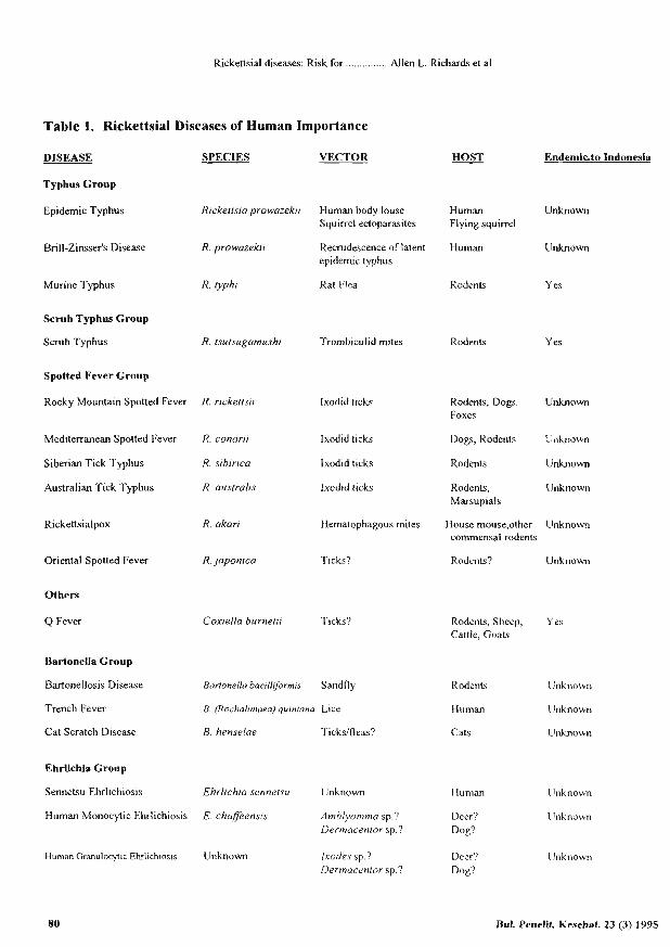
Rickettsiae are gram-negative, obligate intracellular bacteria which are responsible for a large number of arthropod transmitted diseases in man. These diseases include: epidemic (R. prowazekii), and murine (R. @phi) typhus; Rocky Mountain spotted fever (R. rickettsii), Mediterranean spotted fever (R. conorii), Siberian tick typhus (R. sibirica), rickettsialpox (R. akari), and Australian tick typhus (R. australis); and 3) scrub typhus (R. tsutsugamushi). In addition, other related "rickettsial diseases" associated with man include: 1) trench fever (Bartonella (Rochalirnaea) quinfana); 2) Q fever (Coxiella burnetii); ehrlichiosis (Ehrlichia sennetsu, 6 chafleensis). A list of these diseases and some of their properties are presented in Table 1.
Rickettsial diseases and diseases due to rickettsial-like bacteria (accept for trench fever and sennetsu ehrlichiosis) are ever present zoonoses which exhibit periodic fluctuations in attack rates due to environmental influences on the vectors and hosts and variations in human (accidental host) a~tivities'.~. The resulting disease is associated with the infection of the host's endothelial cells or cells associated with the endotheliun~, or blood cells',2. The result of rickettsial infection of the endothelium leads to an intense vasculitis in the affected organs (including brain, lungs, kidneys, heart, and liver), which leads to headache, fever. malaise, rnvalgia and in many cases a macular, maculo-papular or papular rash (rarely in Q fever)'-4. Infections involving the cells of the blood system result in bactercmia with fever and hcadach~ for infections with bartonellae, and sudden o~~cc t offevcr chills, headache. myalgia, arthralgia and anorcsca for chrlichiaez. The illnesses are oftcn self-limiting (approximately 15 days of fever). houcv:r, depending on !hc agcnt involvcd the rnortnlii!, rate may bc as high
as 60% without proper treatment. The intensity and duration of the maladies are sigruficantly decreased by intervention with antibiotic therapy (e.g., tetracycline or chloramphenicol). Untreated illness may be a source of significant morbidity and mortality". There are currently no commercial vaccines available for any of these diseases and indeed specific diagnosis is accomplished only by a few specialized laboratories around the world because of the hazardous nature and fastidious growth characteristics of the organisms involved. Diagnosis is generally by symptomatology and case history with laboratory confirmation done rarely"'. Thus, the base of information on rickettsiosis especially in Indonesia, as an important contributor to acute human febrile disease, is limited',4.
RICKETTSIAL DISEASES
Rickettsiosis as shown in Table 1 has been divided into three groups based upon the antigenic relatedness of the bacteria causing the diseases. The following is a detailed description of these rickettsial groups as well as diseases due to infection with bacteria related to rickettsiae.
TYPHUS GROUP
Within the typhus group is epidemic typhus also known as louse-borne or classic t-yphus. Thc etiologic agent is Rickettsia prowazekii which is passed among man by the human body louse (Pediculus hunranus huntanus), or among flying squirrels and to man by unknown squirrel ectopara~itesl.~,~. The bacteria are passed to man via the arthropod vector fxes rubbcd into skin abrasions or the conj~~ncti+.e. or bj. inhalation of dried infectious feces. Ep~demic typhus has world wide
Rul. Penelit. Keschat. 23 (3) 1995 79
Rickettsial diseases: Risk for ............... Allen L. Richards et al
Table 1. Rickettsial Diseases of Human Importance
DISEASE ' SPECIES VECTOR HOST Endemicto Indonesia
Typhus Group
Epidemic Typhus Rickettsia prowazekri Human body louse Human Unknown Squirrel ectoparasites Flying squirrel
Brill-Zinsser's Disease R. prowazekri Recrudescence of latent Human Unknown epidemic tyrhus
Murine Typhus R. typhi Rat Flea Rodents Yes
Scrub Typhus Group
Scrub Typhus
Spotted Fever Group
R. fsutsugamushr Trombiculid mites Rodents
Rocky Mountain Spotted Fever R. rrckettsir lxodid ticks
Yes
R~detlts, Dogs, Unknown Foxes
Mediterranean Spotted Fever R. conorrr lxodid ticks Dogs, Rodents U~~known
Siberian Tick Typhus R. srbrrrca Ixodid ticks Rodents Unknown
Australian Tick Typhus R. ausfrabs lxodid ticks Rodents, Unknown Marsupials
Rickettsialpox R. akarr Hematophagous mites House mouse,other Unknown commensal rodents
Oriental Spotted Fever R. japonica Ticks? Rodents? Unknown
Others
Q Fever Coxrella burnebr Ticks?
BartoneUa Group
Bartonellosis Disease Barfonella banllfonnrs Sandfly
Trench Fever B. (Rochahmaea) qulnfana Lice
Cat Scratch Disease B. henselae Ticksitleas?
Rodents, Sheep, Yes Cattle, Goats
Rodents Unknown
Human Unknown
Cats Unknown
Ehrlichia Group
Sennetsu Ehrlichiosis Ehrlrchia sennetsu Unknown Human Unknown
Human Monocytic Ehrlichiosis E. chaffeensrs Amblyomma sp.? Deer? Dermacentor sp.? Dog?
Unknown
Human Granulocytic Ehrl~chiosls Unknown Ixodes sp.? Deer? Ilnknown Dermacentor sp.? Dog?
80 Rul. Penelit Kesehat. 23 (3) 1995
Rickettsia1 diseases: Risk for ............... Allen L. Richards et a1
distribution and has been involved in determining man's destiny especially during times of war'. The symptoms follow an incubat~on period of 1-2 weeks. They include an abrupt onset of fever, chills, headache and myalgia. Macular rash of upper trunk and axillary folds occurs around day five. The maculopapular rash spreads over the entire body except for the face, palms and soles. Death may occur in the third week, with stupor, peripheral vascular collapse and renal failure. Diagnosis is usually based on clinical suspicion plus reaction to the Proteus vulgaris antigens' OX19 + OX2 v and no reaction to the P. rnirabilis Kingsbury strain OXK antigen in the nonspecific Weil- Felix serological t e ~ t ~ . ~ . There are also some rickettsial antigen specific noncommercial enzyme immuno- assays (EIA) that are used in a few reference laboratories. In even fewer laboratories there are the capabilities to culture clinical specimens (blood) for rickettsial isolation4. Recently, the polymerase chain reaction has allowed the identification of bacteria in blood of acute ill patientss. This procedure is very sensitive, but is also technically difficult. The treatment for epidemic typhus is tetracycline (doxycycline) or chl~ramphenicol '~~~~.
Brill-Zinsser disease has been determined to be a recrudescence of epidemic typhus. The reemerging agent, R. prowazekii, produces a similar but much milder illness then during the primary disease','. Diagnosis is usually based upon a history of epidemic typhus. The Weil-Felix reaction and EIA results are usually unreliable. Culture of R. prowazekii is possible and PCR has been proven successful with specific primerse. Treatment involves use of either tetracycline or chloramphenicol. Brill-Zinsser diseabe is also distributed world-wide','.
Murine typhus, also known as endemic, flea-borne, shop, or urban typhus, 1s endemic
throughout the world9.10. The disease is due to an infection with R. @phi (R. mooseri). This agent is passed to man by the rat flea (Xenopsylla cheopis) or possibly other arthropod vectors. The infection is started when either the flea feces is rubbed into bite or other wound, or the host inhales dried f ece~ ' .~ .~ , ' ~ . Symptoms are similar to epidemic typhus, but milder. There is a gradual onset of disease with non-productive cough, fever, headache, myalgia, nausea, vomiting and in about 50% of the cases a macular or maculopapular rash4. The illness maybe debilitating, especially with neurologic or nephrotic changes, fatalities are rare". For diagnosis the nonspecific Weil-Felix reaction generally gives the following results: OX19 + OX2 v OXK -2,4. If a reference laboratory is available serologies, in which a four fold rise in titer is diagnostic, and PCR with specific primers, can be performed'.'.".''. Clinical specimens can be cultured for R. typhi, but this is difficult and hazardous to laboratory personnel. Treatment as with other rickettsial diseases incorporates use of one of the tetracyclines or chl~ramphenicol '~~~' ' .
In Indonesia, endemicity of epidemic typhus and Brill-Zinsser diseases is unknown4, whereas, it is known that murine typhus is endemic throughout the archipelago 4,13-'7.
Nevertheless, murine typhus is rarely diagnosed or is misdiagnosed during acute illness, due to its mild, nonspecific clinical manifestations9-". Therefore, little is known of the actual prevalence of this disease, but only of the prevalence of antibodies to R. @phi4 "-I7. In Java, R. @phi has been isolated from rodent hosts and arthropod vectors" and the presence of rickettsial nucleic acid in X. cheopis fleas has been detected by PCR (Rusidy AF, Richards AL, Soeatmadji DW, Church, et al unpublished observ?tion) utilizing a procedure describe previcu;lyi2. Enteric fever, is also endemic to Indonesia .and often presents with clinical manifestations similar to those of rickettsiosis,
BuL Penelit. Kesehat. 23 (3) 1995 81
Rickettsia1 diseases: Risk for ............... Allen L. Richards et a1
i.e., fever, headache, malaise and in 30-50% of cases of typhoid fever, rose s p ~ t s ' ~ . Unfortunately, many enteric fevers, like rickettsial fevers, are difficult to diagnose by laboratory methods available in Ind~nesia'~. The commonly used Weil-Felix and Widal serological tests are nonspecific tests for rickettsial diseases and typhoid fcbcr. respectively, and require assessing both acute and convalescent serum samples far
In addition, the isolation and culture of the causative agents are often difficult and require specialized equipment which are not routinely found in the clinical laboratories of Indonesia19. Thus, illresses characterized by fever and headache in Indonesia are often clinically diagnosed as "tifus," which may include many enteric fevers as well as other infectious diseases including rickettsial fevers. "Tifus" is one of the leading reported causes of hospital admission in Jakarta2'.
SCRUB TYPHUS GROUP
Scrub typhus is a zoonosis that causes human disease comrnonly seen in ~ ~ i ~ ~ , z , d , 5 . ~ 5.16.22-24 . The etiologic agent, R. (Orientia) tsutsugamushi, the only species in the SCRUB TYPHUS GROUP, is acquired following the feeding of an infected chigger, the six legged larval stage of trombiculid mites, on skin tissue fluids of a human host. The subsequent rickettsial infection is characterized by first a local then a systemic vasculitis. A cutaneous lesion or eschar begins at the site of the chigger bite, and the vascular inflammation spreads throughout the blood vessels of the body involving various o r g a ~ i s ' ~ ~ ~ . Though scrub typhus has been known since the late 1800's its notoriety did not emerge until World War 11, where in the Pacific theater, i t was feared by military personnel more than malaria22.2'. This was due in part to the high nlortality rate, where
up to 50% of patients died because of lack of a known treatment at that timez2. Today, the treatment of choice for scrub typhus is tetracycline or chloramphenicol which generally produces a rapid clinical improvement in the patient with defervescence of clinical syniptoms usually seen within two d a y ~ l . ~ , ~ ~ . However, there have been recent reports of drug resistant scrub tvphrls described both in Thailand and 1~di~26,Ruphanath D personal commmcabon
Scrub typhus is considered a rural, tropical disease niost commonly found afflicting individuals traversing through or working in terrain encompassing secondary vegetation resulting from previously cleared forest or abandoned agricultural a:casl.22-24 . However. there have been reports of individuals acquiring scrub typhus in urban settirigs of Hong Kong, J a~an '~ .~ ' and while gardening in suburban and urban areas of South Korea2". Scrub tqphus cases havc been reported from the southern section of Jakarta by Gispen ct al.". R. tsut.vugamushi has been isolated from trombiculid mites and three different species of rats collected in North J;akarta'4. Recently. a case of scrub typhus in an individual residing in central Jakarta has been identified3'.
The species, H. tsutsugattiu.~h:hi, is made up of many distinct strains that vary in virulence32 and antibiotic rc~istance~~. Immunity to hornologous strains of s cn~b typhus rickettsiae may continue for years. however. immunity to the many heterologous strains of R. ts~tsugamushi may onlv persist for a few months3'. Conseaoently, individuals can become infected more than once with H. tstrtsugatn~shi"~'~. This lack of cross immunity and the previously successful treatment with antibiotics dinlinishcd the drive for and the successful completion of the development of an effective vaccine for scrub typlit~s'~.
82 RoI. Penelit. Kesehat. 23 (3) 1995
Rickettsia1 diseases: Risk for ....
SPOTTED FEVER GROUP
The spotted fever group is composed of more than 20 antigenically related rickettsia1 species'. Described here are several human diseases associated with the spotted fever group rickettsiae. Rocky Mountain spotted fever (R. rickettsii) is found only in the Western Hemisphere. The vectors are the ixodid ticks and the normal hosts include rodents, dogs and foxes. Mediterranean Spotted Fever also known as Boutonneuse fever, Kenya tick typhus, Indian tick typhus and South African tick typhus (R. conorii) is also vectored by ixodid ticks. Hosts include dogs and rodents. Mediterranean Spotted Fever distribution includes the Mediterranean, Black and Caspian Sea areas, and in the Middle East, India and Africa. Siberian tick typhus or North Asian tick typhus (R. sibirica) is found in Siberia, Armenia, Mongolia, Central Asia & Europe. Ixodid ticks are the vectors and rodents are the usual hosts. Australian or Queensland tick typhus (R. australis) was discovered in Queensland, Australia, but has now been reported in other areas of A~stralia'~. The vectors are ixodid ticks and the hosts are rodents and marsupials. Rickettsialpox (R. akari) has been located mainly in urban areas of North America and Russia, but has also been found in Southern Africa and Korea'.'. The vector is the hematophagous mite (A/lodermanyssus sanguineus). Hosts include the house mouse and other commensal rodents.
Infection with spotted fever group rickettsiae usually involves the bacteria entering the human body through openings or abrasions io the skin due to the tick or mite bite, or by the infection of the conjunctiva by fingers contaminated after crushing the arthropod vectors. The organisms multiply locally at the site of invasion and spread to the endothelial cells of blood vessels. An inflammatory
........... Allen L. Richards et al
response develops, causing vasculitis, and the triad of fever, head ache and rash (rash may only occur in 50% of the patients and is more difficult to see in darker pigmented people). The disease often affects multiple organs with manifestations involving the skin, skeletal muscles, central nervous system, myocardium, lungs, liver and kidneys'". Diagnosis often depends on the history and clinical presentation. The Weil-Felix agglutination reaction is variable for both the OX19 and OX2 antigens and negative for the OXK. A four fold or greater rise in titer for EIA or IFA followed by ;onfirmatory Western blot analysis will provide laboratory diagnosis for spotted fever di~ease".'~. However, for species identification the method of choice is culture of the agent in laboratory animals or tissue culture. In addition, PCR has been shown to detect rickettsial nucleic acid from blot clots of individuals with acute cases of Rocky Mountain spotted fever)9, and species identification can be ascertained utilizing PCR and restriction fragment length polymorphisms (RFLP) ana ly~ i s~~ .~ ' . Spotted fever disease may be fatal, but when identified and treated early with tetracycline or chloramphenicol mortality can be reduced ~ignificantly'.'.~~.
Members of the spotted fever group of rickettsiae have not been reported in Indonesia though they are endemic in other Asia countries and Australial.2,4,5,?2.36 . There has been a lack of seroreactivity found in Indonesian populations that were surveyed utilizing an EIA with R. conorii antigen preparation".
DISEASES DUE TO RICKETTSIA-LIm ORGANISMS
Q fever (C. burnetii) is found worldwide and is associated with domestic animals and rodents. Though C. burnetii has been found in
Rickettsia1 diseases: Risk for ............... Allen L. Richards et al
several tick species and other arthropod vectors, it is thought to be maintained in nature primarily by aerosol transmission. Humans are believed to be infected by inhalation of the airborne organisms. C. burnetii, unlike the rickettsiae, are very resistant to inactivation by chemical and physical treatments. C. burnetii is known to survive on wool and other fomites for up to one year2. Symptoms of acute Q fever are nonspecific and characterized by fever, headache, chills, myalgia, and malaise. Pneumonitis may occur, however, rash rarely does. Q fever is usually self limiting, though chronic Q fever with hepatitis or endocarditis is not un~ommon'.~. Diagnosis by reference laboratory usually involves performance of a noncommercial enzyme-linked immunoassay. Recently, PCR with species specific primers have become available. Culture of C. burnetii, is risky and so is only performed in a few laboratories world wide. Treatment usually is with one of the tetracyclines, though there maybe resistance2, or chloramphenicol.
Q fever is most likely endemic in Indonesia as it is believed to have world wide distrib~tion'.~. In addition, a serosurvey conducted by Van Peenen, et a]., reported that 25% of human serum tested from throughout the archipelago had evidence of Phase I1 antibodies against C. burnetii4). The prevalence of hospital admitted cases of Q fever in Indonesia is currently unknown.
Very recently, the Rochalirnaea genus has been combined with that of Bartonella because of sinlilarity in 16s rRNA, and DNA relatedness. The genus name Bartonella was retained because of its nomenclature priority2. B. hacillfornlis was identified in 1909 as the erythrocyte-adherent agent of bartonellosis or Carrion's disease. Bartonellosis is found only in the intermediate altitudes of the South American Andes. This is believed to be due to
the limited distribution of the sandfly vector (Lutzomyia verrucurum).
Trench fever, discovered among the troops fighting in the trenches during World War I, was originally named Rickettsia quintana. However, when it was found to be able to grow on blood agar plates and therefore determined not to be an obligate intracellular parasite (it attaches to the outer membrane of eukaryotic cells44) as are the other rickettsiae, it was separated into its own genus R~chal imaea' .~ . Bartonella (Rochalimaea) quintana is the only agent described in this review that does not have a host other than humans. The vector, the human body louse (Pediculus humanus humanus), transmits the disease from man to man. Humans have been known to be infectious for more. than ten years2. Distribution is associated with humans, generally living in poor social economic conditions, worldwide.
Identification of the agent associated with cat scratch disease had eluded scientists for some time. It is believed now to be B. (Rochalimaea) h e n ~ e l a e ~ ~ . ~ ~ . The natural vector for B. henselae is unknown, but may be ticks andlor fleas. This agent is found worldwide and its major host is the domestic cat4'.
Infections for bartonellae are usually via openings or abrasions in the skin of the human body due to the bite of sandfly, louse, tick or flea, by infection of the conjunctiva by fingers contaminated after crushing vectors or as in cat scratch disease the bite or scratch of the natural host-cat. The organislris multiply locally and spread through the blood vessels (bacteremia). The im~tiediate response to bacteremia includes fever and headache. In Carrion's bartonellosis there are also anemia, adenopathy, thrombo- cytopenia, severe myalgias and arthralgias, and possible mental status changes. Without antibiotic treatment, fatality rates for
Rul. Penelit. Kesehat 23 (3) 1995
Rickettsia1 diseases: Risk for ............... Allen L. Richards et al
bartonellosis maybe high2. Vermga pemna may be a common late-stage manifestation of Carri6n's disease. Bacteremia due to B. quintana and B. henselae are rarely fatal though the illness maybe more severe in the immunocompromised host, e.g., AIDS patient$'. Cat scratch disease is commonly associated with a cutaneous papule or pustule at the site of the bite or scratch and regional lymphadenopathy. Fever may also accompany this self-limiting disease. Diagnosis of Bartonella species by culture of clinical specimens is difficult and thus the laboratory must be instructed about the presumptive diagnosis before submitting the specimen. Warthin-Stany silver technique is used for staining of tissue samples. Fourfold or greater rise in titer for EIA- and FA-IgG or the presence of species specific IgM is diagnostic. PCR is the most sensitive method for Bartonella species identification. Treatment with tetracyclines, erythromycin or chloramphenicol is effective'.
EHRLICHIA GROUP is made up of related bacteria species in the genus Ehrlichia and the tribe Ehrlichieae in the family of Rickettsiaceae4'. Several species have been related to animal disease and two to human diseases. Sennetsu ehrlichiosis, also known as sennetsu fever, sennetsu rickettsiosis (E. sennetsu) was described in Southwest Japan in the late 1800's. However, the agent was not identified until 1953. The vector is currently unknown as is the natural host. The disease has only been identified in Japan and Malaysiaz. The symptoms include a sudden onset of fever, chills, headache and myalgia, insomnia, diaphoresis, sore throat, and anorexia. General lymphadenopathy is common, hepatomegaly and splenomegaly occur in approximately a third of the cases. The disease is self-limiting, and no serious complications or fatalities have been reported2. Successful treatment with
oxytetracycline, doxycycline and chloramphe- nicol have been reported2.
Human ehrlichiosis is a recently described disease whose importance as an arthropod borne-illness continues to g r ~ d ~ ~ . ~ ~ " ~ . Recently this disease has been divided into two similar illnesses that have different causative agents. The original human ehrlichiosis is now known as human monocytic ehrlichiosis (E. chaffeensis). The second, is known as human granulocytic ehrlichiosis, the etiological agent is unknown but is very closely related to E. ,~hagocytophila and E. egui5'. The tick vectors for the two diseases are believed to be Ambylyomma and Dermacentor species for human monocytic ehrlichiosis and Ixodes and Dermacentor species for human granulocytic ehrlichiosis. The natural hosts may be deer, dogs or other mammalss2. Distribution in the United States is wide spread. Other locations throughout the world have not been extensively studied5. Though different agents are involved in human monocytic and granulocytic ehrlichiosis, symptoms are similar and include a sudden onset of fever, chills, headache, myalgia, arthralgia, anorexia, nausea, vomiting, pneumonia and rarely rashsz. The diseases are self-limiting. However, fatality rates of 2-10% have been reported2.". Laboratory diagnosis is by polymerase chain reaction to identi@ the agent during the acute phase and by serological assays (EIAIIFAI Western blots) during c o n v a l e ~ c e n c e ~ ~ ~ ~ ~ ~ ~ ~ . Attempts of isolation from blood are usually unsuc~essful~~. Recommended treatment is with the tetracyclines (doxycycline). Chloramphe- nicol may not be effective5'.
Ehrlichiae are responsible for many animal and human disease^^,^^. They can be divided into two groups based upon whether they infect monocytes or granulocytes. E. sennetsu; E. chafleensis; E. canis canine ehrlichiosis,
Rul. Penelit. Kesehat 23 (3) 1995 85
Rickettsial diseases: Risk for ............... Allen L. Richards et al
tropical canine pancytopenia; and E. risticii equine monocytic ehrlichiosis, Potomac horse fever; cause monocytic ehrlichiosis. E. phagocytophila tick-borne fever of sheep, goat, cows, bison and deer; E. equi equine ehrlichiosis; and E. ewingii canine granulocytic ehrlichiosis are granulocytic ehrlichiosis. Currently little is known of the presence of any of these agents in Indonesia.
SUMMARY STATEMENT
Rickettsia1 diseases and diseases due to rickettsial-like bacteria are endemic throughout the world. This includes Indonesia as well. Scrub typhus and murine typhus have been investigated in Indonesia, however, diseases such as those related to the spotted fever group of rickettsiae, trench fever, bartonellosis, Q fever and ehrlichiosis have been almost completely overlooked. Thus the aim of this review was to promote awareness of diseases associated with rickettsiae and related organisms, and to stimulate rickettsia1 disease research in Indonesia.
DISCLOSURE
REFERENCES
1. Dasch GA, Weiss E. (1992). The Genera Rickettsia, Rochalimaea, Ehrlichia, Cowdria, and i\'eorickettsia. In: Balows A, ed. The prokaryotes. A handbook on the biology of bacteria. Vol III. New York: Springer Vcrlag. p:2407-2470.
2. Ferraro MJ, Gilligan PH. (1995). Chlamydia, Rickettsia, and Related Bacteria, in Manual of Clirzical ~i4icrobiology, 6th edit. Murray PK, Baron EJ, Pfaller MA, Tenover FC, Yolken RH, eds. ASM Press, Washington DC Ch.VI:663-695.
3. Walker DH. (1995). Rocky Mountain spotted fever: A seasonal alert. Clin Inf Dis, 20:1 1 1 1-1 117.
4. Cross JH. (1984). Rickettsia] infections in Southeast Asia. Medical Progress, 1 I : 19-26.
5. World Health Organization Memoranda/ Memorandums. (1 993). Global surveillance of rickettsia1 diseases: Memorandum from a WHO meeting. Bull WHO, 71:293-296.
6. Regnery RL, Fu ZY, Spruill CL. (1986). Flying squirrel-associated Rickettsia prowazekii (epidemic typhus rickettsiae) characterized by a specific DNA fragment produced by restriction endonuclease digestion. J Clin Mcrobiol, 23: 189-1 91.
The opinions and assertions contained 7. Arnano K-I, Mizushiri S, Fuji S, Fukushi K,
herein are the private ones of the authors and Suto T. ( 1 990). Immunological characterization of lipplysaccharides from Proteus strains used
are not to be construed as ofiicial or as in Weil-Felix test and reactivity with patient reflecting the views of the U.S. Navy or the sera of tsutsugamushi diseases. Microbial Department of Defense, or Departemen Imrnunol, 34:135-145. Kesehatan RI.
8. Carl M, Tibbs CW, Dobson ME, Paparello S, Dasch GA. (1990). Diagnosis of acute typhus
ACKNOWLEDGMENTS infection using the polymerase chain reaction. J Inf Dis, 16 b:791-793.
The authors would like to thank Dr. F. 9. Azad AF. (1990). Epidemiology of ~nurine Stephen Wignall for reviewing this manuscript. typhus. h u Rev Entomol, 35553-569,
86 Rul. Penelit. Kesehat 23 (3) 1995
Rickettsial diseases: Risk for ............... Allen L. Richards et al
10. Traub R, Wisseman CLJr, Farhang-Azad A. (1 978). The ecology of murine typhus-A critical review. Trop Dis Bull, 75237-3 17.
11. Taylor JP, Beh TG, Rawlings JA. (1986). Epidemiology of murine typhus in Texas. 1980 through 1984. JAMA, 255:2 173-2 176.
12. Webb L, Carl M, Malloy DC, Dasch GA, Azad AF. (1990). Detection of murine typhus infection in fleas by using the polymerase chain reaction. J Clin Microbial, 28530-534.
13. Van Peenen PFD. (1977). Antibodies against murine typhus in sera from Indonesians. Trans R Soc Trop Med Hyg, 71:297-299.
14. Dennis DT, Hadi TR, Brown RJ, Sukaeri S, Leksana B, Cholid R. (1981). A survey of scrub and murine typhus in the Ancol section of Jakarta, Indonesia. Southeast Asian J Trop Med Pub1 Hlth, 12:574-580.
15. Corwin A, Sie A, Church C, Graham R, Kelly D, Dasch G, Richards A. A serological survey for evidence of infection with pathogenic rickettsiae in humans and animals of Indonesia. American Society for Microbiology 93rd Annual Meeting. Las Vegas, Abst V-6, pg 61 8.
Oxford University Press, Inc. New York, NY. 1991.
19. Juwono R. (1981). Diagnostic and therapeutic problems of typhoid fever in Indonesia. KOPAPDI, 5:702-7 16.
20. Farmer S.G. (1985). Immunology of bacterial infections. In Manual of Clinical Microbiology, Fourth Edition, Lennette E H , Balows A, Hausler WJJr, Shadomy HJ, eds. American Society for Microbiology, Washington DC, pp.898-904.
21. McGladdery S, Silitonga N, Sukri N, Rogayah I, Budiyanto, Pulungsih S, Lesmana M, Rifajati A, Rafli K, Punjabi N, O'Hanley P, and the Jakarta Typhoid Fever Group. Descriptive epidemiology and clinical spectrum of typhoid and paratyphoid fever in North Jakarta, from 1984-1991. Submitted for publication. 1994.
22. Rapmund G. (1984). Rickettsial diseases of the far east: New perspectives. J Inf Diseases. 1984;149:330-8.
23. Liat LB, Hadi TR. (1986). Status of scrub typhus research in Indonesia. Bul Penelit Kesehat 14:37-49.
16. Rahardjo E, Soeatmadji DW, Widodo MA, 24. Wisseman CLJr. (1991). Scrub typhus. In:
Sardjono TW, Yanuwiadi B, Hernowati TE, Strickland GT, ed. Hunter's tropical medicine.
Baskoro AD, Roeiyoso, Hakim L, Soendoro M, 7th ed. Philadelphia: WB Saunders, 279-282.
Putxi MP* J, Strickman D7 25. Moe JB, Pedersen CEJr (1980). The impact of Olson JG, Church CJ, Convin AL, Richards AL. rickettsia] diseases on military operations. Mil (1995). Rickettsiosis Di Jawa Timur. Seminar Med, 145: Biologi XIV dan Kongres Nasional Biologi XI. 24-26 Juli 1995. ~bs t rac t page 56
Nuraini N, Raharjo E, Komalarini S, Nasution C, Hendrawanto, Strickman D, Dasch G, Corwin A, Lesmana M, Punjabi NH, Richards AL. (1 995). Etiologi demam diantara pasien-pasien yang dirawat di dua rumah s k i t di Jakarta. Seminar Biologi X7V dan Kongres Nasional Biologi XI. 24-26 Juli 1995. Abstract page 53.
Wilson ME. (1991). hi A U'orld Guide to Infectious Diseases, DistriSution and Diagnosis.
26. Strickman D, Bodhidatta D, Kelly D, Dasch G, Chouriyagune C, Watt G. Antibiotic susceptibility of Rickettsia tsutsugamushi from patients with severe scrub typhus in northern Thailand. Annual Meeting of the American Society of Tropical Medicine and Hygiene Atlanta, GA. Abst No. 243.
27. Bassett DCJ, Ho AKC, Tam JSL, Lam LY, Cheng AFB. (1992). The laboratory diagnosis of rickettsial diseases in Hong Kong. J Trop Med Hyg, 95:327-330.
BuL Penelit. Kesehat 23 (3) 1995
Rickettsia1 diseases: Risk for ....... ........ Allen L. Richards et a1
28. 'Urban scrub typhus in Japan'. (1992). Presentation at the Annual Asian Pacific Military Medicine Conference, No. 11. Bangkok, 'Thailand.
29. Arthur JD, Graham RR, Cheong TW, Hwang YJ, Yi KS. (1993). Urban scrub typhus in South Korea. Presentation at the Annual Asian Pacific
Military Medicine Conference, No. Ill. Jakarta.
30 Gispen et al., (1949). Epidemic scrub typhus in Batavia. Docum Neerl Indones Morb Trop. 1:135-138.
3 1 Richards AL, Rahardjo E, Wignall FS. (1 995). A case of scrub typhus ir Jakarta, Indonesia. In preparation.
32. Robinson DM, Gan E, Chan TC, Huxsoll DL. (1 977). Patterns of rickettsemia and antibody response in silvered leaf monkeys (Presby6s cristatus) after inoculation with virulent and avirulent strains of Rickettsia tsutsugamushi. J Inf Dis, 135:664-668.
33. Smadel E, Ley WL, Diercks FH, Trar~b R. (1950). Immunity in scrub typhus: Resistance to induced reinfection. Arch Path, 50:847-86 1.
34. Bourgeois AL, Olson JG, RCY Fang, Eluang J, Wang CL, Chow L, Bechthold D, Dennis DT, Coolbaugh JC, Weiss E. (1982). Humoral and cellular responses in scrub typhus patients reflecting primary infection and reinfection with Rickettsia tsutsugamushi. Am J Trop Med Hyg, 3 1:532-540.
35. Rights RL, Smadel JE, Jackson EB. (1948). Studies on scrub typhus (Tsutsugamushi Disease) D. Heterogenicity of strains of R. tsutsugarnushi as demonstrated by cross-vaccination studies. J Exp Med, 87:339-35 1.
36. Hudson BJ, McPetrie R, Kitchener-Smith J, Eccles J. (1994). Vesicular rash associated with infection due to Rickettsia australis. Clin Inf Dis. 18:118-119.
37. Clements ML, Durnler JS, Fiset P, Wisseman CLJr, Snyder MJ, Levine MM. (1983). Serodiagnosis of Rocky Mountain spotted fever: comparison of IgM and IgG enzyme-linked imrnunosorbent assays and indirect flunrescent antibody test. J Inf Dis, 148:876-880.
38. Sanchez L, Candler WH, Fishbein DB, Greene CR, Cote TR, Kelly DJ, Driggers DP, Johnson BJR. (1992). A cluster of tick-borne infections; association with military training and asymptomatic infections due to Rickettsia n'ckerrsii. Trans R Soc Trop Med Hyg, 8632 1-325.
39. -Tiianabos T, Anderson RE, McDade JE. (i989). Detection of Rickettsia n'ckettsii DNA in clinical specimens by using polymerase chain reaction technology. J Clin Miirobiol, 2866-2868.
40. Regnery RL, Spruill CI., Plikaytis BD. (1991). Genotypic identification of rickettsiae and estimation of intraspecies sequence divergence for portions of two rickettsia1 genes. J Bacteriol, 17311576-1589.
41. Dupont HT, Comet J-P, Raoult D. (1994). Identification of rickettsiae from ticks collected in the Central African Republic using the polymerase chain reaction. Am J Trop Med Hyg, 50:37?-380.
42. Woodward TE. (1984). Rocky Mountain spotted fever: epidemiology and early clinical signs are keys to treatment and reduced mortality. J Inf Dis, 150:465468.
43. Van Peenen PF,D, Gundelfinger BF, Koesharjono C, See R. (1978). Seroepidemiological evidence for occupational exposure to Q fever in Indonesia. J Occup Med, 20:488489.
44. Merrell BR, Weiss E, Dasch GA. (1978). Morphological and cell association characteristics of Rochalimaea quintatla: comparison of the vole and Fuller strains J Bacteriol, 135:633640.
Rickettsia1 diseases: Risk for ............... Allen L. Richards e: al
45. Regnery RL, Olson JG, Perkins BA, Bibb W. (1 992). Serological response to "Rochalimaea henselae" antigen in suspected cat-scratch disease. Lancet, 339: 1443-1445.
46. Zangwill KM, Hamilton DH, Perkins BA, Regnery RL, Plikaytis BD, Hadler K, Cartter ML, Wenger JD. (1993). Cat scratch disease in Connecticut. Epidemiology, risk factors, and evaluation of a new diagnostic test. N Engl J Med, 32923-1 3.
47. Koehler JE, Glaser CA, Tappero JW. (1994). Rochalimaea henselae infection. A new Zoonosis with the domestic cat as reservoir. JAMA, 271:531-535.
48. Rikihisa Y. (1991). The tribe Ehrlichieae and ehrlichial diseases. Clin Microbiol Rev, 4:286-308.
49. Dawson JE, Anderson BE, Fishbein DB, Sanchez JL, Goldsmith CS, Wilson KH, Duntley CW. (1991). Isolation and characterization of an Ehrlichia sp. from a patient diagnosed with human ehrlichiosis. J Clin Microbiol, 29:274 1-2745.
50. Anderson BE, Dawson JE, Jones DC, Wilson KH. (1991). Ehrlichia chafleensis, a new species associated with human ehrlichiosis. J Clin Microbiol, 29:2838-2842.
51. Chen S-M, Dumler JS, Bakke JS, Walker DH. (1994). Identification of a granulocytotropic Ehrlichia species as the etiologic agent of human disease. J Clin Microbiol, 32:589-595.
52. Dumler JS, Bakken JS. (1995). Ehrlichial diseases of humans: emerging tick-borne infections. Clin Infect Dis, 20:1102-1110.