01 Patofisiologi Penyakit Infeksi
22
Patofisiologi Penyakit infeksi dr. Al Munawir, M.Kes., Ph.D. Fakultas Kedokteran Universitas Jember, Jl. Kalimantan 37 Jember 68121 Telp. (0331) 324446, Fax (0331) 324445
-
Upload
rizkyimansari -
Category
Documents
-
view
501 -
download
43
description
17
Transcript of 01 Patofisiologi Penyakit Infeksi
Patofisiologi Penyakit infeksi
dr. Al Munawir, M.Kes., Ph.D.Fakultas Kedokteran Universitas
Jember, Jl. Kalimantan 37 Jember 68121Telp. (0331) 324446, Fax (0331) 324445
Tujuan Belajar:• Masalah penyakit infeksi.• Macam-macam penyebab infeksi.• Penyebaran dan penularan infeksi.• Patogenesis penyakit infeksi.• Diagnosis penyakit infeksi.• Respons inflamasi terhadap infeksi.• Infeksi oleh kuman.• Infesi oleh virus.• Infeksi oleh jamur.• Infeksi oleh parasit.
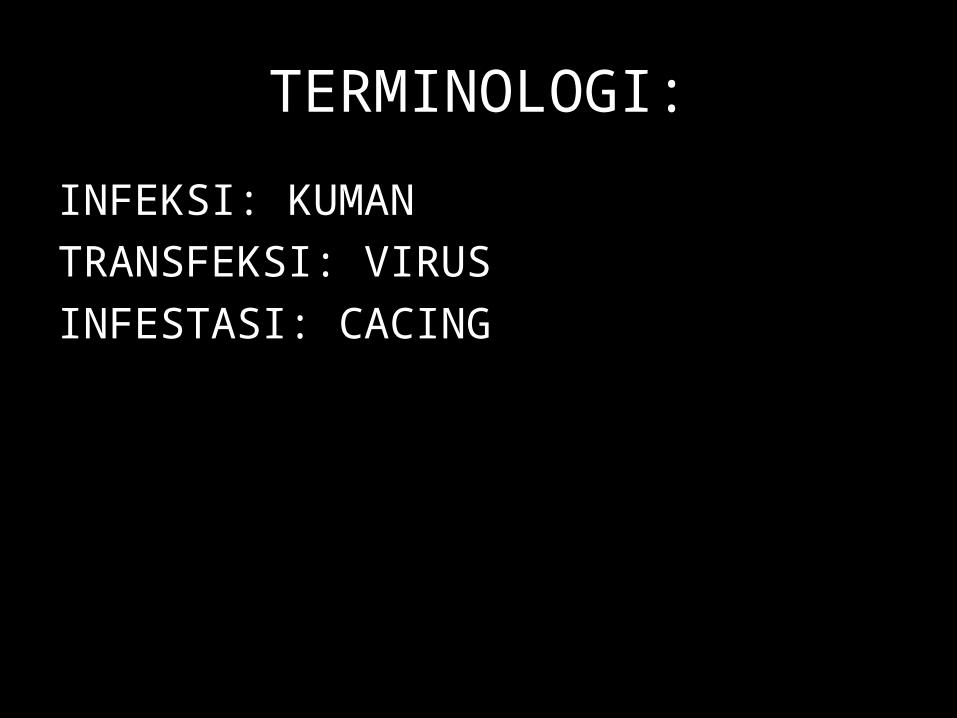
TERMINOLOGI:
INFEKSI: KUMANTRANSFEKSI: VIRUSINFESTASI: CACING
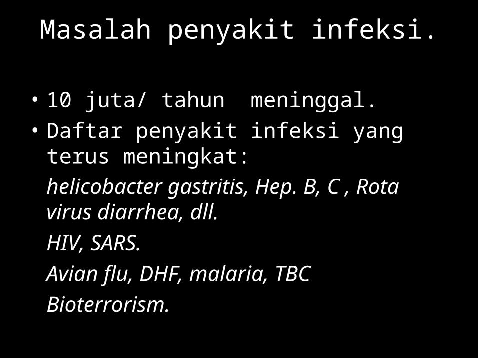
Masalah penyakit infeksi.
• 10 juta/ tahun meninggal.• Daftar penyakit infeksi yang terus meningkat:
helicobacter gastritis, Hep. B, C , Rota virus diarrhea, dll.HIV, SARS.Avian flu, DHF, malaria, TBCBioterrorism.
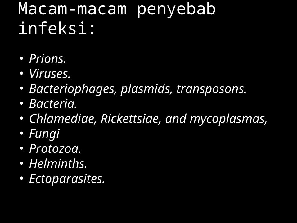
Macam-macam penyebab infeksi:
• Prions.• Viruses.• Bacteriophages, plasmids, transposons.• Bacteria.• Chlamediae, Rickettsiae, and mycoplasmas,• Fungi• Protozoa.• Helminths.• Ectoparasites.
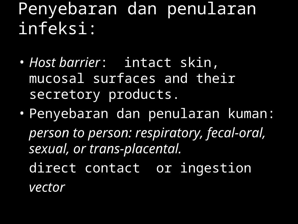
Penyebaran dan penularan infeksi:
• Host barrier: intact skin, mucosal surfaces and their secretory products.
• Penyebaran dan penularan kuman: person to person: respiratory, fecal-oral, sexual, or trans-placental.direct contact or ingestionvector
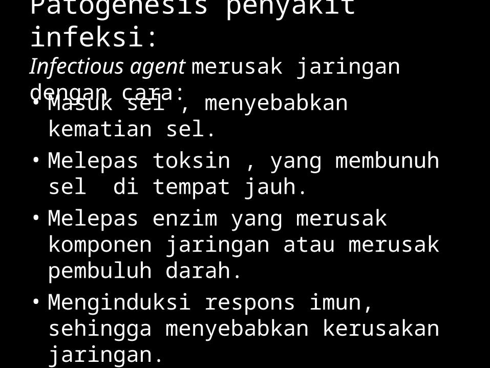
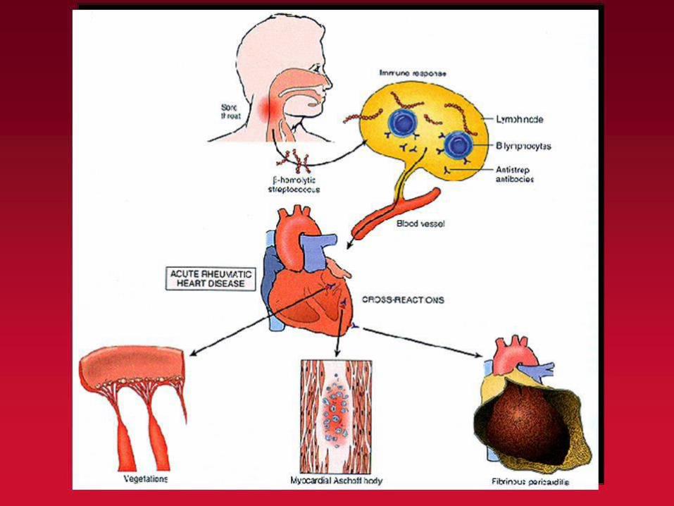
Patogenesis penyakit infeksi:Infectious agent merusak jaringan dengan cara:
• Masuk sel , menyebabkan kematian sel.• Melepas toksin , yang membunuh sel di
tempat jauh.• Melepas enzim yang merusak komponen
jaringan atau merusak pembuluh darah.• Menginduksi respons imun, sehingga
menyebabkan kerusakan jaringan.
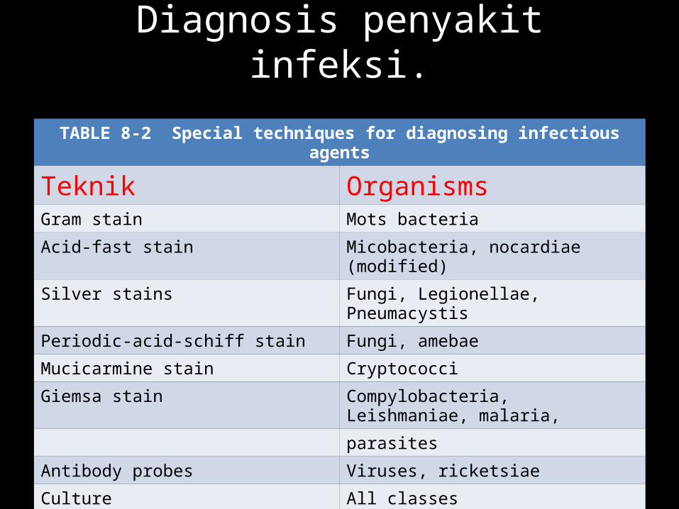
Diagnosis penyakit infeksi.
TABLE 8-2 Special techniques for diagnosing infectious agents
Teknik OrganismsGram stain Mots bacteria
Acid-fast stain Micobacteria, nocardiae (modified)
Silver stains Fungi, Legionellae, Pneumacystis
Periodic-acid-schiff stain Fungi, amebae
Mucicarmine stain Cryptococci
Giemsa stain Compylobacteria, Leishmaniae, malaria,
parasites
Antibody probes Viruses, ricketsiae
Culture All classes
DNA probes Viruses, bacteria, protozoa
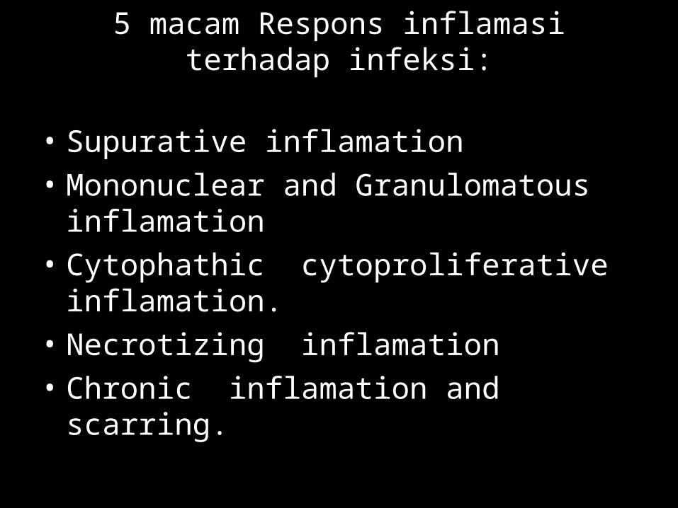
5 macam Respons inflamasi terhadap infeksi:
• Supurative inflamation• Mononuclear and Granulomatous inflamation• Cytophathic cytoproliferative inflamation.• Necrotizing inflamation• Chronic inflamation and scarring.
Supurative inflamation
• Disebabkan oleh pyogenic bacteria.• Adanya Bacterial chemoatractant.• Lesi bervariasi dari kecil (microabses ) sampai
besar (difuse).
Mononuclear and Granulomatous inflamation:
• Disebabkan oleh virus, intracellular bacteria,spirochetes, intracellular parasites, dan helminths.
• Type cells inflamasi : plasma cell, lymphocytes, macrophage.
• Activated macrophage.
Cytophathic cytoproliferative inflamation:
• Disebabkan oleh virus, yang ditandai necrosis dan proliferasi sel.
• Morfologi: Inclusion body, polykaryons, blisters.
Necrotizing inflamation
• Disebabkan oleh virus (HBV), sekresi toxin kuman (Clostridium perferingent), protozoa cytolysis (E. Hystolitica).
• Pada kasus yang berat tidak ditemukan inflamasi.
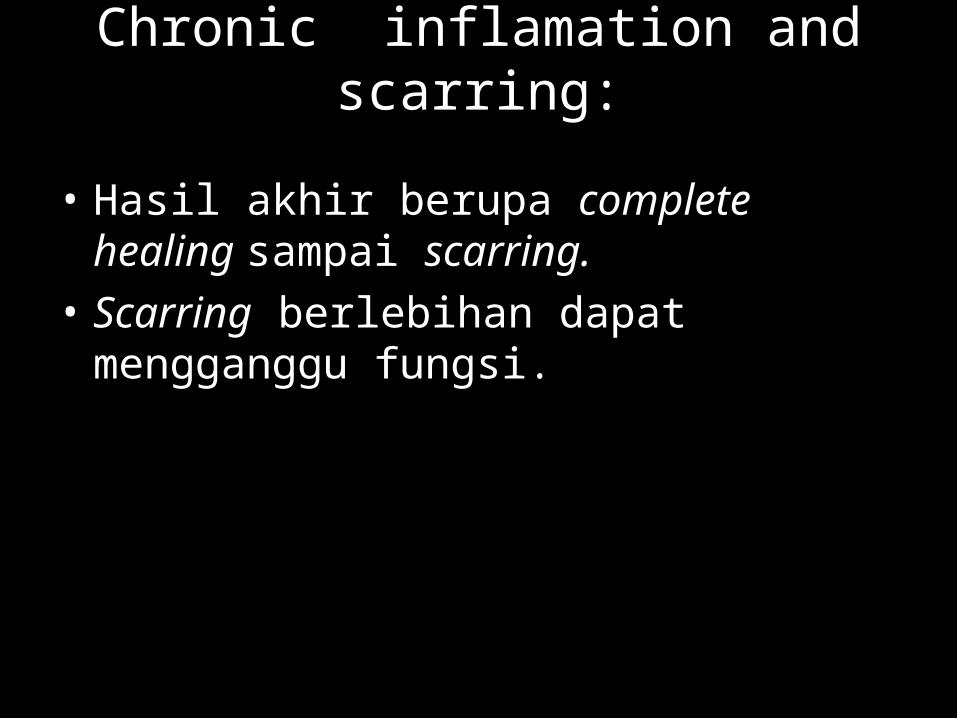
Chronic inflamation and scarring:
• Hasil akhir berupa complete healing sampai scarring.
• Scarring berlebihan dapat mengganggu fungsi.
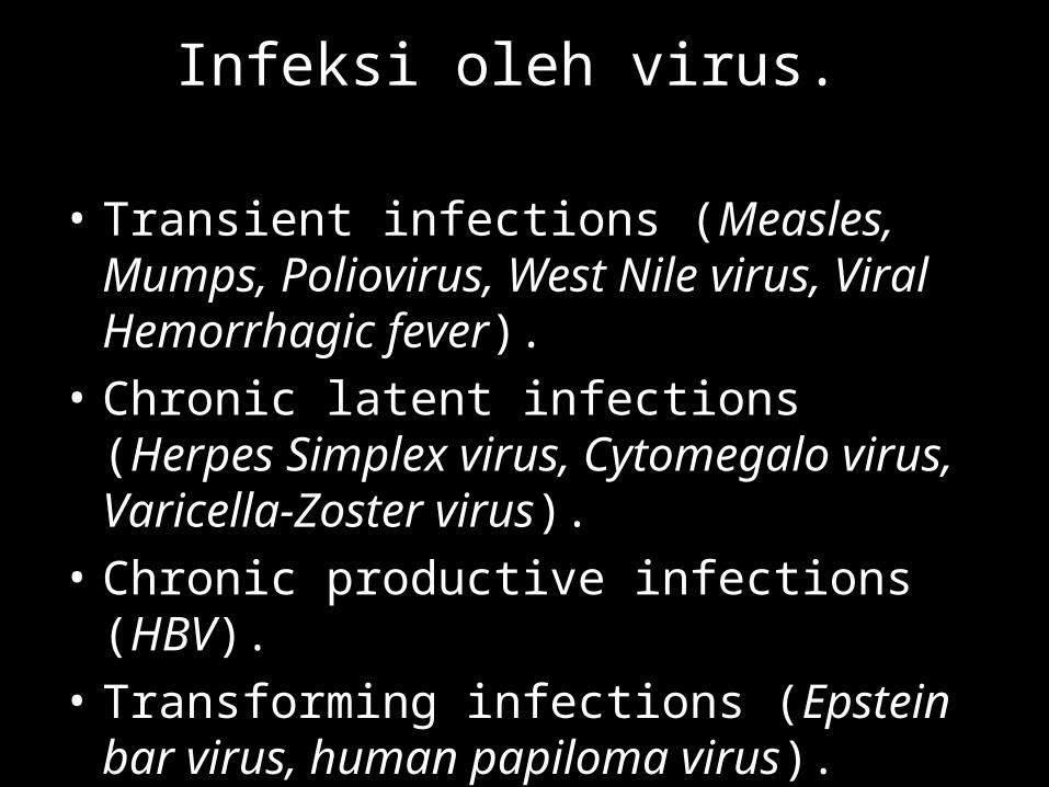
Infeksi oleh virus.
• Transient infections (Measles, Mumps, Poliovirus, West Nile virus, Viral Hemorrhagic fever).
• Chronic latent infections (Herpes Simplex virus, Cytomegalo virus, Varicella-Zoster virus).
• Chronic productive infections (HBV).• Transforming infections (Epstein bar virus,
human papiloma virus).
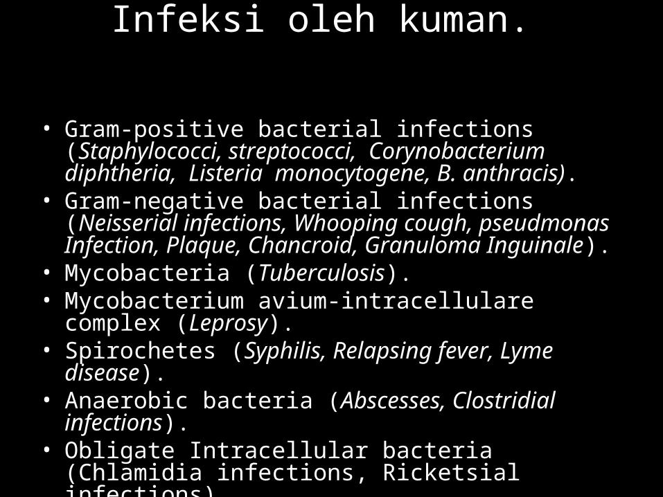
Infeksi oleh kuman.
• Gram-positive bacterial infections (Staphylococci, streptococci, Corynobacterium diphtheria, Listeria monocytogene, B. anthracis).
• Gram-negative bacterial infections (Neisserial infections, Whooping cough, pseudmonas Infection, Plaque, Chancroid, Granuloma Inguinale).
• Mycobacteria (Tuberculosis).• Mycobacterium avium-intracellulare complex (Leprosy).• Spirochetes (Syphilis, Relapsing fever, Lyme disease).• Anaerobic bacteria (Abscesses, Clostridial infections).• Obligate Intracellular bacteria (Chlamidia infections,
Ricketsial infections).
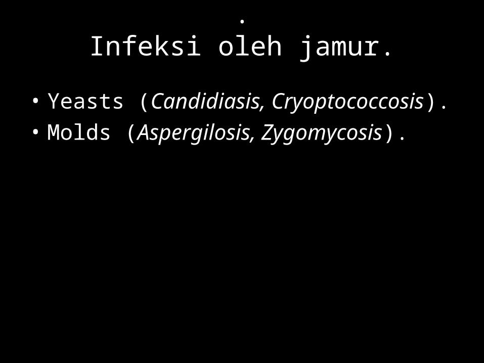
.Infeksi oleh jamur.
• Yeasts (Candidiasis, Cryoptococcosis).• Molds (Aspergilosis, Zygomycosis).
Infeksi oleh parasit.
• Protozoa (Malaria, babesiosis, Leishmaniasis, African Trypanosomiasis, Chagas disease).
• Metazoa (Strongyloidiasis, Cysticercosis and Hydatid disease, Trichinosis, Schistosomiasis, Lymphatic filariasis, Onchocerciasis).
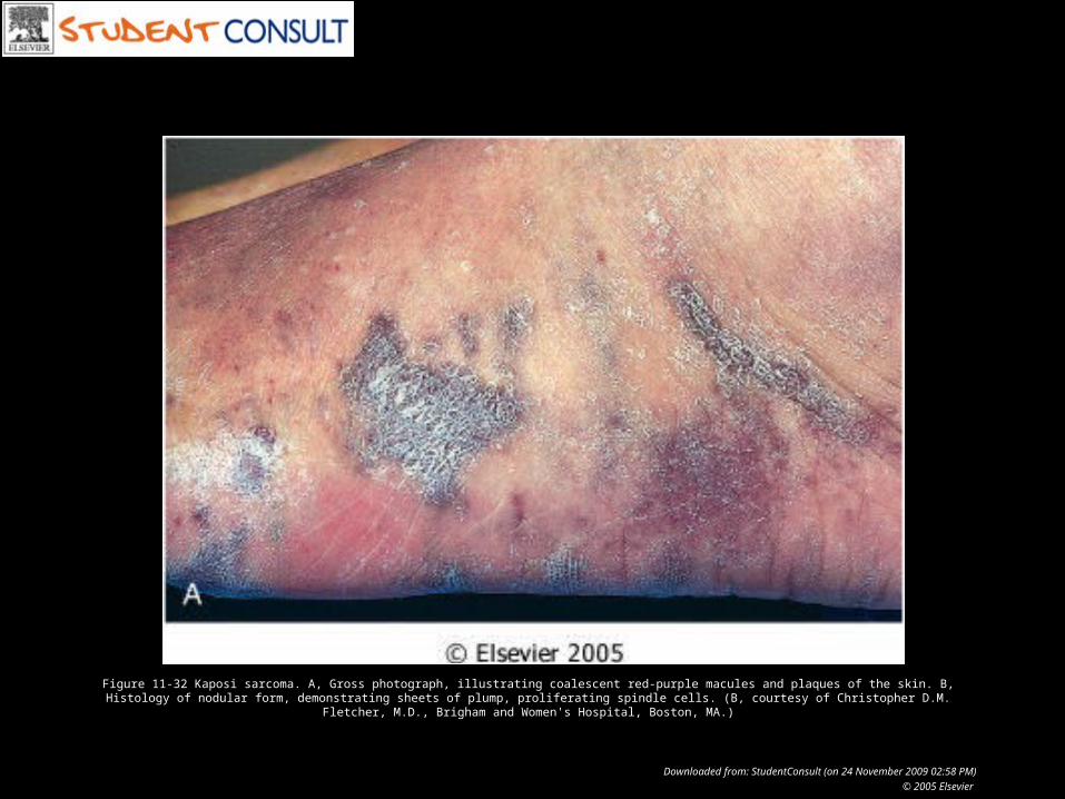
Figure 11-32 Kaposi sarcoma. A, Gross photograph, illustrating coalescent red-purple macules and plaques of the skin. B, Histology of nodular form, demonstrating sheets of plump, proliferating spindle cells. (B, courtesy of Christopher D.M. Fletcher, M.D., Brigham and Women's Hospital, Boston, MA.)
Downloaded from: StudentConsult (on 24 November 2009 02:58 PM)
© 2005 Elsevier
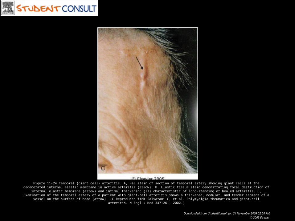
Figure 11-24 Temporal (giant cell) arteritis. A, H&E stain of section of temporal artery showing giant cells at the degenerated internal elastic membrane in active arteritis (arrow). B, Elastic tissue stain demonstrating focal destruction of internal elastic membrane (arrow) and intimal thickening (IT) characteristic of long-standing or healed arteritis. C, Examination of the temporal artery of a patient with giant-cell arteritis shows a thickened, nodular, and tender segment of a vessel on the surface of head
(arrow). (C Reproduced from Salvarani C, et al. Polymyalgia rheumatica and giant-cell arteritis. N Engl J Med 347:261, 2002.)
Downloaded from: StudentConsult (on 24 November 2009 02:58 PM)
© 2005 Elsevier

Pathology Laboratory