


Bahasa
Halaman
Hukum

©20
11 N
atu
re A
mer
ica,
Inc.
All
rig
hts
res
erve
d.
nature CHeMICaL BIOLOGY | advance online publication | www.nature.com/naturechemicalbiology 1
articlepuBLIsHed OnLIne: 30 januarY 2011 | dOI: 10.1038/nCHeMBIO.522
Natural products have played an important role in the discov-ery and development of drugs1. In recent years, they have also become important molecular probes for studying different
cellular processes by virtue of their ability to bind to specific protein targets and interfere with their cellular functions. For example, the identification of calcineurin as the target of the immunosuppressive drugs cyclosporine A and FK506 (ref. 2) and of TOR as the target of rapamycin3 opened the gateways to the subsequent studies of calcium-calcineurin and TOR signaling pathways, respectively. More recently, we and others identified the type 2 methionine aminopeptidase as the target for the potent angiogenesis inhibitors fumagillin and ovalicin4 and the eukaryotic translation initiation factor 4A as the target for the marine sponge–derived antitumor natural product pateamine A5. These studies have led to the exploitation of MetAP2 and eIF4A1 as new molecular targets for discovering and developing new antiangio-genic and anticancer drugs, respectively. Thus, natural products can serve as bridges between chemistry, biology and medicine, constitut-ing a major tool set for chemical biologists today. Elucidation of the mechanisms of action of natural products not only offers new insights into the cellular functions of their protein targets but also facilitates the ensuing use of natural products as leads in drug development.
Triptolide6 is a diterpene triepoxide purified from Tripterygium wilfordii Hook F, commonly known as lei gong teng or thunder god vine, a medicinal plant whose extracts have been used in tradi-tional Chinese medicine for treating a wide variety of diseases from inflammation to arthritis for centuries7. It is structurally distinct in that it contains three epoxide groups next to each other (Fig. 1a). It also possesses a unique profile of biological activities. Triptolide has been shown to have potent antiproliferative and immunosup-pressive activities. Preclinical studies have revealed that triptolide is effective against cancer, collagen-induced arthritis, skin allograft rejection and bone marrow transplantation in animal models8–10. Triptolide and derivatives have entered human clinical trials for cancer, among other diseases11.
Extensive scrutiny of its mechanism of action in the past few decades has yielded important insights. At the cellular level, triptolide shows strong antiproliferative activity, inhibiting the proliferation of all 60 US National Cancer Institute cancer cell lines with half-maximal inhibitory concentration (IC50) values in the low nanomolar range (average IC50 = 12 nM). It also induces apoptosis in a number of cancer cell lines. At the molecular level, triptolide was shown to interfere with a number of transcription factors including NF-κB, p53, NF-AT and HSF-1 (refs. 12–14). An interesting common feature of the effects of triptolide on all those transcription factors is that it seems to block their trans-activation activity without affecting DNA binding. More recently, it was shown that triptolide inhibits de novo RNA synthesis, which was suggested to be because of indirect inhibition of tran-scription mediated by RNA polymerases I and II (refs. 15–18). Attempts to isolate the molecular target of triptolide have led to the identification of a calcium channel polycystin-2 and detec-tion of a 90-kDa nuclear protein as potential molecular targets of triptolide15,19. However, polycystin-2 cannot account for most of the aforementioned biological activities of triptolide, and the identity of the 90-kDa putative nuclear triptolide-binding pro-tein has remained unknown.
In this study, we took a ‘top-down’ approach to identifying the molecular target of triptolide. Taking advantage of the extensive prior knowledge of eukaryotic transcription initiation, we system-atically examined the effects of triptolide on different steps and players involved and eventually identified the molecular target of triptolide as the XPB subunit of the general transcription factor TFIIH. Inhibition of XPB by triptolide offers a unified molecular mechanism for the diverse biological activities of triptolide. Our preliminary evidence also revealed a previously unknown activity of triptolide: inhibition of nucleotide excision repair, which has important implications in the application of triptolide for the treatment of cancer.
1department of pharmacology and Molecular Sciences, the Johns Hopkins university School of Medicine, baltimore, Maryland, uSa. 2department of chemistry and biochemistry, university of colorado, boulder, colorado, uSa. 3department of biochemistry and Molecular biology, bloomberg School of public Health, Johns Hopkins university, baltimore, Maryland, uSa. 4charles a. dana Research institute for Scientists emeriti, drew university, Madison, new Jersey, uSa. 5department of oncology, the Johns Hopkins university School of Medicine, baltimore, Maryland, uSa. 6present: department of pharmaceutical Sciences, college of pharmacy, St. John’s university, Queens, new York, uSa (W.-K.l.). *e-mail: [email protected]
XpB, a subunit of tFIIH, is a target of the natural product triptolidedenis V titov1, Benjamin Gilman2, Qing-Li He1, shridhar Bhat1, Woon-Kai Low1,6, Yongjun dang1, Michael smeaton3, arnold L demain4, paul s Miller3, jennifer F Kugel2, james a Goodrich2 & jun O Liu1,5*
Triptolide (1) is a structurally unique diterpene triepoxide isolated from a traditional Chinese medicinal plant with anti- inflammatory, immunosuppressive, contraceptive and antitumor activities. Its molecular mechanism of action, however, has remained largely elusive to date. We report that triptolide covalently binds to human XPB (also known as ERCC3), a subunit of the transcription factor TFIIH, and inhibits its DNA-dependent ATPase activity, which leads to the inhibition of RNA polymerase II–mediated transcription and likely nucleotide excision repair. The identification of XPB as the target of triptolide accounts for the majority of the known biological activities of triptolide. These findings also suggest that triptolide can serve as a new molecular probe for studying transcription and, potentially, as a new type of anticancer agent through inhibition of the ATPase activity of XPB.
©20
11 N
atu
re A
mer
ica,
Inc.
All
rig
hts
res
erve
d.
2 nature CHeMICaL BIOLOGY | advance online publication | www.nature.com/naturechemicalbiology
article NATuRE CHEmICAl BIology dOI: 10.1038/nCHeMBIO.522
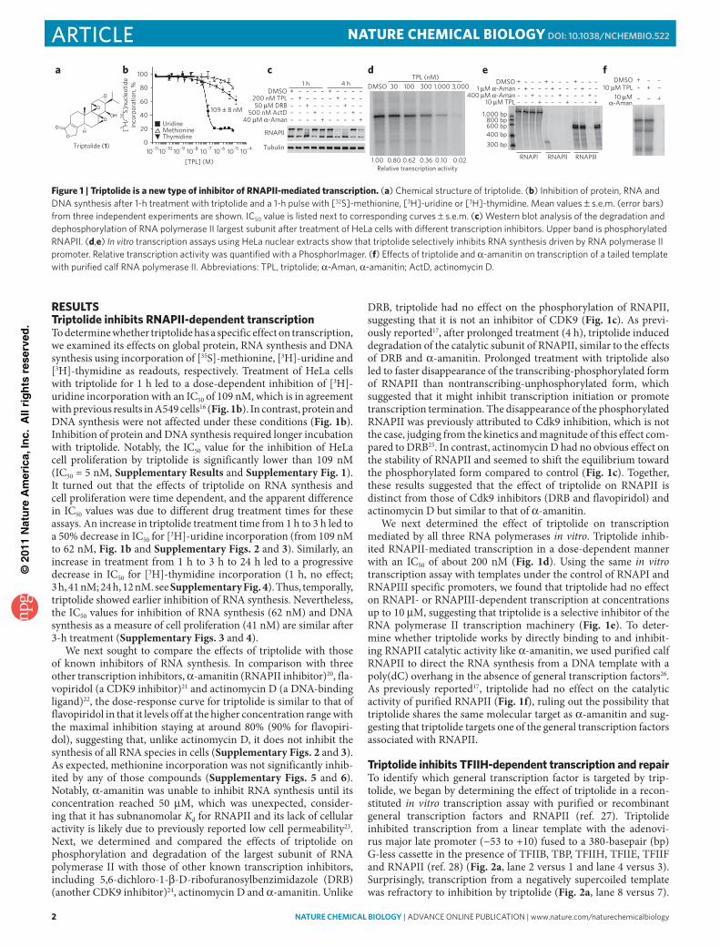
RESulTSTriptolide inhibits RNAPII-dependent transcriptionTo determine whether triptolide has a specific effect on transcription, we examined its effects on global protein, RNA synthesis and DNA synthesis using incorporation of [35S]-methionine, [3H]- uridine and [3H]-thymidine as readouts, respectively. Treatment of HeLa cells with triptolide for 1 h led to a dose-dependent inhibition of [3H]-uridine incorporation with an IC50 of 109 nM, which is in agreement with previous results in A549 cells16 (Fig. 1b). In contrast, protein and DNA synthesis were not affected under these conditions (Fig. 1b). Inhibition of protein and DNA synthesis required longer incubation with triptolide. Notably, the IC50 value for the inhibition of HeLa cell proliferation by triptolide is significantly lower than 109 nM (IC50 = 5 nM, Supplementary Results and Supplementary Fig. 1). It turned out that the effects of triptolide on RNA synthesis and cell proliferation were time dependent, and the apparent difference in IC50 values was due to different drug treatment times for these assays. An increase in triptolide treatment time from 1 h to 3 h led to a 50% decrease in IC50 for [3H]-uridine incorporation (from 109 nM to 62 nM, Fig. 1b and Supplementary Figs. 2 and 3). Similarly, an increase in treatment from 1 h to 3 h to 24 h led to a progressive decrease in IC50 for [3H]-thymidine incorporation (1 h, no effect; 3 h, 41 nM; 24 h, 12 nM. see Supplementary Fig. 4). Thus, temporally, triptolide showed earlier inhibition of RNA synthesis. Nevertheless, the IC50 values for inhibition of RNA synthesis (62 nM) and DNA synthesis as a measure of cell proliferation (41 nM) are similar after 3-h treatment (Supplementary Figs. 3 and 4).
We next sought to compare the effects of triptolide with those of known inhibitors of RNA synthesis. In comparison with three other transcription inhibitors, α-amanitin (RNAPII inhibitor)20, fla-vopiridol (a CDK9 inhibitor)21 and actinomycin D (a DNA-binding ligand)22, the dose-response curve for triptolide is similar to that of flavopiridol in that it levels off at the higher concentration range with the maximal inhibition staying at around 80% (90% for flavopiri-dol), suggesting that, unlike actinomycin D, it does not inhibit the synthesis of all RNA species in cells (Supplementary Figs. 2 and 3). As expected, methionine incorporation was not significantly inhib-ited by any of those compounds (Supplementary Figs. 5 and 6). Notably, α-amanitin was unable to inhibit RNA synthesis until its concentration reached 50 μM, which was unexpected, consider-ing that it has subnanomolar Kd for RNAPII and its lack of cellular activity is likely due to previously reported low cell permeability23. Next, we determined and compared the effects of triptolide on phosphorylation and degradation of the largest subunit of RNA polymerase II with those of other known transcription inhibitors, including 5,6-dichloro-1-β-D-ribofuranosylbenzimidazole (DRB) (another CDK9 inhibitor)24, actinomycin D and α-amanitin. Unlike
DRB, triptolide had no effect on the phosphorylation of RNAPII, suggesting that it is not an inhibitor of CDK9 (Fig. 1c). As previ-ously reported17, after prolonged treatment (4 h), triptolide induced degradation of the catalytic subunit of RNAPII, similar to the effects of DRB and α-amanitin. Prolonged treatment with triptolide also led to faster disappearance of the transcribing-phosphorylated form of RNAPII than nontranscribing-unphosphorylated form, which suggested that it might inhibit transcription initiation or promote transcription termination. The disappearance of the phosphorylated RNAPII was previously attributed to Cdk9 inhibition, which is not the case, judging from the kinetics and magnitude of this effect com-pared to DRB25. In contrast, actinomycin D had no obvious effect on the stability of RNAPII and seemed to shift the equilibrium toward the phosphorylated form compared to control (Fig. 1c). Together, these results suggested that the effect of triptolide on RNAPII is distinct from those of Cdk9 inhibitors (DRB and flavopiridol) and actinomycin D but similar to that of α-amanitin.
We next determined the effect of triptolide on transcription mediated by all three RNA polymerases in vitro. Triptolide inhib-ited RNAPII-mediated transcription in a dose-dependent manner with an IC50 of about 200 nM (Fig. 1d). Using the same in vitro transcription assay with templates under the control of RNAPI and RNAPIII specific promoters, we found that triptolide had no effect on RNAPI- or RNAPIII-dependent transcription at concentrations up to 10 μM, suggesting that triptolide is a selective inhibitor of the RNA polymerase II transcription machinery (Fig. 1e). To deter-mine whether triptolide works by directly binding to and inhibit-ing RNAPII catalytic activity like α-amanitin, we used purified calf RNAPII to direct the RNA synthesis from a DNA template with a poly(dC) overhang in the absence of general transcription factors26. As previously reported17, triptolide had no effect on the catalytic activity of purified RNAPII (Fig. 1f), ruling out the possibility that triptolide shares the same molecular target as α-amanitin and sug-gesting that triptolide targets one of the general transcription factors associated with RNAPII.
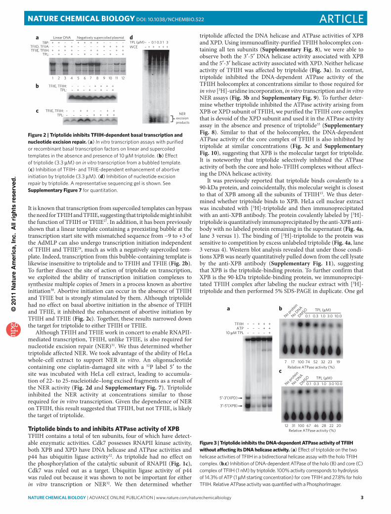
Triptolide inhibits TFIIH-dependent transcription and repairTo identify which general transcription factor is targeted by trip-tolide, we began by determining the effect of triptolide in a recon-stituted in vitro transcription assay with purified or recombinant general transcription factors and RNAPII (ref. 27). Triptolide inhibited transcription from a linear template with the adenovi-rus major late promoter (−53 to +10) fused to a 380-basepair (bp) G-less cassette in the presence of TFIIB, TBP, TFIIH, TFIIE, TFIIF and RNAPII (ref. 28) (Fig. 2a, lane 2 versus 1 and lane 4 versus 3). Surprisingly, transcription from a negatively supercoiled template was refractory to inhibition by triptolide (Fig. 2a, lane 8 versus 7).
100
80
60
[3 H/35
S]nu
cleo
tide
inco
rpor
atio
n, %
Triptolide (1)
40
20
0
Uridine
109 ± 8 nM
DMSODMSO DMSO
200 nM TPL
500 nM ActD50 µM DRB
40 µM α-Aman
400 µM α-Aman10 µM TPL
10 µM TPL
1,000 bp
300 bp400 bp600 bp800 bp
1 µM α-Aman10 µM
α-Aman
Thymidine
10–11
[TPL] (M)10–410–510–610–710–810–910–10
Methionine
Tubulin
RNAPll
RNAPlllRNAPl RNAPll
––––
– – – – – – – ––––––––
––– –
–– – – – –
–– –
–– – – – – – –
––––––
– –
––
– –– – – – –––– – – – –
–––
–
––– –
–––––– – – – –++
++
+ ++
++
+
++
++ +
++
+
++
+
++
++
4 h DMSO 30 100TPL (nM)
1,000 3,000300
1.00 0.80 0.62Relative transcription activity
0.020.100.36
1 h
ba c d e f
O
OH
O
O
O
OH
Figure 1 | Triptolide is a new type of inhibitor of RNAPII-mediated transcription. (a) chemical structure of triptolide. (b) inhibition of protein, Rna and dna synthesis after 1-h treatment with triptolide and a 1-h pulse with [32S]-methionine, [3H]-uridine or [3H]-thymidine. Mean values ± s.e.m. (error bars) from three independent experiments are shown. ic50 value is listed next to corresponding curves ± s.e.m. (c) Western blot analysis of the degradation and dephosphorylation of Rna polymerase ii largest subunit after treatment of Hela cells with different transcription inhibitors. upper band is phosphorylated Rnapii. (d,e) In vitro transcription assays using Hela nuclear extracts show that triptolide selectively inhibits Rna synthesis driven by Rna polymerase ii promoter. Relative transcription activity was quantified with a phosphorimager. (f) effects of triptolide and α-amanitin on transcription of a tailed template with purified calf Rna polymerase ii. abbreviations: tpl, triptolide; α-aman, α-amanitin; actd, actinomycin d.
©20
11 N
atu
re A
mer
ica,
Inc.
All
rig
hts
res
erve
d.
nature CHeMICaL BIOLOGY | advance online publication | www.nature.com/naturechemicalbiology 3
articleNATuRE CHEmICAl BIology dOI: 10.1038/nCHeMBIO.522
It is known that transcription from supercoiled templates can bypass the need for TFIIH and TFIIE, suggesting that triptolide might inhibit the function of TFIIH or TFIIE27. In addition, it has been previously shown that a linear template containing a preexisting bubble at the transcription start site with mismatched sequence from −9 to +3 of the AdMLP can also undergo transcription initiation independent of TFIIH and TFIIE29, much as with a negatively supercoiled tem-plate. Indeed, transcription from this bubble- containing template is likewise insensitive to triptolide and to TFIIH and TFIIE (Fig. 2b). To further dissect the site of action of triptolide on transcription, we exploited the ability of transcription initiation complexes to synthesize multiple copies of 3mers in a process known as abortive initiation30. Abortive initiation can occur in the absence of TFIIH and TFIIE but is strongly stimulated by them. Although triptolide had no effect on basal abortive initiation in the absence of TFIIH and TFIIE, it inhibited the enhancement of abortive initiation by TFIIH and TFIIE (Fig. 2c). Together, these results narrowed down the target for triptolide to either TFIIH or TFIIE.
Although TFIIH and TFIIE work in concert to enable RNAPII-mediated transcription, TFIIH, unlike TFIIE, is also required for nucleotide excision repair (NER)31. We thus determined whether triptolide affected NER. We took advantage of the ability of HeLa whole-cell extract to support NER in vitro. An oligonucleotide containing one cisplatin-damaged site with a 32P label 5′ to the site was incubated with HeLa cell extract, leading to accumula-tion of 22- to 25-nucleiotide–long excised fragments as a result of the NER activity (Fig. 2d and Supplementary Fig. 7). Triptolide inhibited the NER activity at concentrations similar to those required for in vitro transcription. Given the dependence of NER on TFIIH, this result suggested that TFIIH, but not TFIIE, is likely the target of triptolide.
Triptolide binds to and inhibits ATPase activity of XPBTFIIH contains a total of ten subunits, four of which have detect-able enzymatic activities. Cdk7 possesses RNAPII kinase activity, both XPB and XPD have DNA helicase and ATPase activities and p44 has ubiquitin ligase activity32. As triptolide had no effect on the phosphorylation of the catalytic subunit of RNAPII (Fig. 1c), Cdk7 was ruled out as a target. Ubiquitin ligase activity of p44 was ruled out because it was shown to not be important for either in vitro transcription or NER32. We then determined whether
triptolide affected the DNA helicase and ATPase activities of XPB and XPD. Using immunoaffinity-purified TFIIH holocomplex con-taining all ten subunits (Supplementary Fig. 8), we were able to observe both the 3′-5′ DNA helicase activity associated with XPB and the 5′-3′ helicase activity associated with XPD. Neither helicase activity of TFIIH was affected by triptolide (Fig. 3a). In contrast, triptolide inhibited the DNA-dependent ATPase activity of the TFIIH holocomplex at concentrations similar to those required for in vivo [3H]-uridine incorporation, in vitro transcription and in vitro NER assays (Fig. 3b and Supplementary Fig. 9). To further deter-mine whether triptolide inhibited the ATPase activity arising from XPB or XPD subunit of TFIIH, we purified the TFIIH core complex that is devoid of the XPD subunit and used it in the ATPase activity assay in the absence and presence of triptolide33 (Supplementary Fig. 8). Similar to that of the holocomplex, the DNA-dependent ATPase activity of the core complex of TFIIH is also inhibited by triptolide at similar concentrations (Fig. 3c and Supplementary Fig. 10), suggesting that XPB is the molecular target for triptolide. It is noteworthy that triptolide selectively inhibited the ATPase activity of both the core and holo-TFIIH complexes without affect-ing the DNA helicase activity.
It was previously reported that triptolide binds covalently to a 90-kDa protein, and coincidentally, this molecular weight is closest to that of XPB among all the subunits of TFIIH15. We thus deter-mined whether triptolide binds to XPB. HeLa cell nuclear extract was incubated with [3H]-triptolide and then immunoprecipitated with an anti-XPB antibody. The protein covalently labeled by [3H]-triptolide is quantitatively immunoprecipitated by the anti-XPB anti-body with no labeled protein remaining in the supernatant (Fig. 4a, lane 3 versus 1). The binding of [3H]-triptolide to the protein was sensitive to competition by excess unlabeled triptolide (Fig. 4a, lane 3 versus 4). Western blot analysis revealed that under those condi-tions XPB was nearly quantitatively pulled down from the cell lysate by the anti-XPB antibody (Supplementary Fig. 11), suggesting that XPB is the triptolide-binding protein. To further confirm that XPB is the 90-kDa triptolide-binding protein, we immunoprecipi-tated TFIIH complex after labeling the nuclear extract with [3H]-triptolide and then performed 5% SDS-PAGE in duplicate. One gel
TBP:a
b
c
dTFIID, TFIIA:TFIIE, TFIIH:
TFIIE, TFIIH:
TPL:
TPL (µM)WCE
TPL:
+ + + + + ++ + + +
0.1 0.3 1 3+ + + + +
+++++++
+ ++ +
++– –
– –– – –
–
–– –
–
– ––
– – ––
––Negatively supercoiled plasmidLinear DNA
1 2 3 4 5 6 7 8 9 10 11 12
––––
+ + ++
+++ +– – – –
– – – –
TFIIE, TFIIH:TPL:
++ +
++++– – –
+–
– – – –
+ + +
NERexcision
products
Figure 2 | Triptolide inhibits TFIIH-dependent basal transcription and nucleotide excision repair. (a) In vitro transcription assays with purified or recombinant basal transcription factors on linear and supercoiled templates in the absence and presence of 10 μM triptolide. (b) effect of triptolide (3.3 μM) on in vitro transcription from a bubbled template. (c) inhibition of tFiiH- and tFiie-dependent enhancement of abortive initiation by triptolide (3.3 μM). (d) inhibition of nucleotide excision repair by triptolide. a representative sequencing gel is shown. See Supplementary Figure 7 for quantitation.
TFIIH
a b
c
10 µM TPL
––– – – –
––– + +
No protein
No DNA
DMSO
++
TPL (µM)1.0 10.03.00.30.1
No protein
No DNA
DMSO TPL (µM)
1.0 10.03.00.30.1
5′-3′(XPD)
3′-5′(XPB)
12 31 100 2022284667
7 19Relative ATPase activity (%)
Relative ATPase activity (%)
2332527410017
++
ATP
Figure 3 | Triptolide inhibits the DNA-dependent ATPase activity of TFIIH without affecting its DNA helicase activity. (a) effect of triptolide on the two helicase activities of tFiiH in a bidirectional helicase assay with the holo tFiiH complex. (b,c) inhibition of dna-dependent atpase of the holo (b) and core (c) complex of tFiiH (1 nM) by triptolide. 100% activity corresponds to hydrolysis of 14.3% of atp (1 μM starting concentration) for core tFiiH and 27.8% for holo tFiiH. Relative atpase activity was quantified with a phosphorimager.
©20
11 N
atu
re A
mer
ica,
Inc.
All
rig
hts
res
erve
d.
4 nature CHeMICaL BIOLOGY | advance online publication | www.nature.com/naturechemicalbiology
article NATuRE CHEmICAl BIology dOI: 10.1038/nCHeMBIO.522
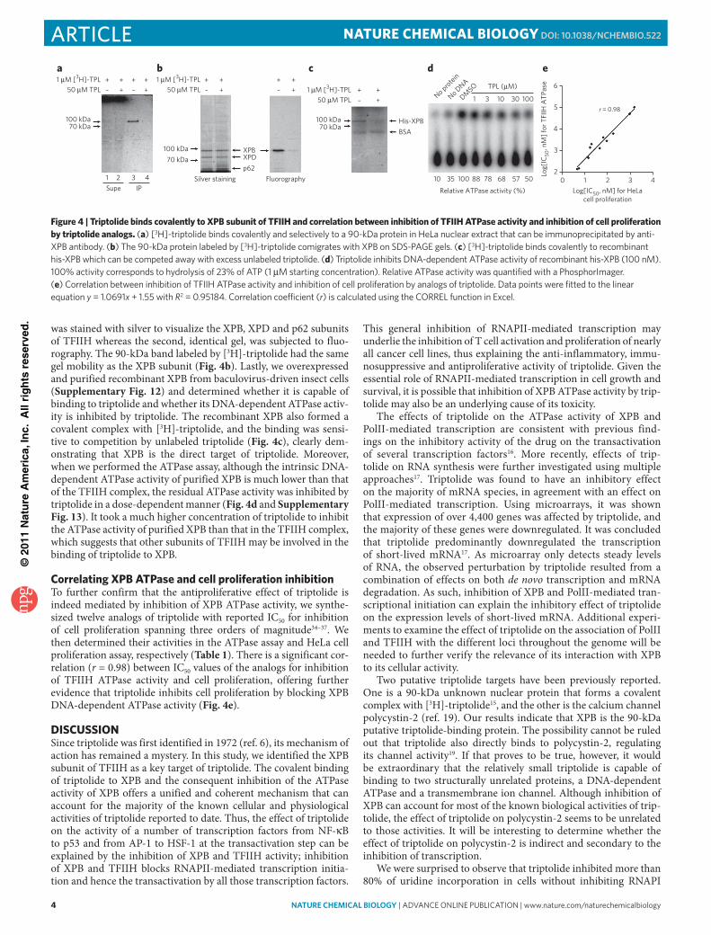
was stained with silver to visualize the XPB, XPD and p62 subunits of TFIIH whereas the second, identical gel, was subjected to fluo-rography. The 90-kDa band labeled by [3H]-triptolide had the same gel mobility as the XPB subunit (Fig. 4b). Lastly, we overexpressed and purified recombinant XPB from baculovirus-driven insect cells (Supplementary Fig. 12) and determined whether it is capable of binding to triptolide and whether its DNA-dependent ATPase activ-ity is inhibited by triptolide. The recombinant XPB also formed a covalent complex with [3H]-triptolide, and the binding was sensi-tive to competition by unlabeled triptolide (Fig. 4c), clearly dem-onstrating that XPB is the direct target of triptolide. Moreover, when we performed the ATPase assay, although the intrinsic DNA-dependent ATPase activity of purified XPB is much lower than that of the TFIIH complex, the residual ATPase activity was inhibited by triptolide in a dose-dependent manner (Fig. 4d and Supplementary Fig. 13). It took a much higher concentration of triptolide to inhibit the ATPase activity of purified XPB than that in the TFIIH complex, which suggests that other subunits of TFIIH may be involved in the binding of triptolide to XPB.
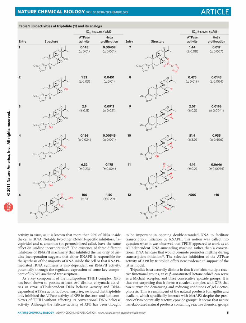
Correlating XPB ATPase and cell proliferation inhibitionTo further confirm that the antiproliferative effect of triptolide is indeed mediated by inhibition of XPB ATPase activity, we synthe-sized twelve analogs of triptolide with reported IC50 for inhibition of cell proliferation spanning three orders of magnitude34–37. We then determined their activities in the ATPase assay and HeLa cell proliferation assay, respectively (Table 1). There is a significant cor-relation (r = 0.98) between IC50 values of the analogs for inhibition of TFIIH ATPase activity and cell proliferation, offering further evidence that triptolide inhibits cell proliferation by blocking XPB DNA-dependent ATPase activity (Fig. 4e).
DISCuSSIoNSince triptolide was first identified in 1972 (ref. 6), its mechanism of action has remained a mystery. In this study, we identified the XPB subunit of TFIIH as a key target of triptolide. The covalent binding of triptolide to XPB and the consequent inhibition of the ATPase activity of XPB offers a unified and coherent mechanism that can account for the majority of the known cellular and physiological activities of triptolide reported to date. Thus, the effect of triptolide on the activity of a number of transcription factors from NF-κB to p53 and from AP-1 to HSF-1 at the transactivation step can be explained by the inhibition of XPB and TFIIH activity; inhibition of XPB and TFIIH blocks RNAPII-mediated transcription initia-tion and hence the transactivation by all those transcription factors.
This general inhibition of RNAPII-mediated transcription may underlie the inhibition of T cell activation and proliferation of nearly all cancer cell lines, thus explaining the anti-inflammatory, immu-nosuppressive and antiproliferative activity of triptolide. Given the essential role of RNAPII-mediated transcription in cell growth and survival, it is possible that inhibition of XPB ATPase activity by trip-tolide may also be an underlying cause of its toxicity.
The effects of triptolide on the ATPase activity of XPB and PolII-mediated transcription are consistent with previous find-ings on the inhibitory activity of the drug on the transactivation of several transcription factors16. More recently, effects of trip-tolide on RNA synthesis were further investigated using multiple approaches17. Triptolide was found to have an inhibitory effect on the majority of mRNA species, in agreement with an effect on PolII-mediated transcription. Using microarrays, it was shown that expression of over 4,400 genes was affected by triptolide, and the majority of these genes were downregulated. It was concluded that triptolide predominantly downregulated the transcription of short-lived mRNA17. As microarray only detects steady levels of RNA, the observed perturbation by triptolide resulted from a combination of effects on both de novo transcription and mRNA degradation. As such, inhibition of XPB and PolII-mediated tran-scriptional initiation can explain the inhibitory effect of triptolide on the expression levels of short-lived mRNA. Additional experi-ments to examine the effect of triptolide on the association of PolII and TFIIH with the different loci throughout the genome will be needed to further verify the relevance of its interaction with XPB to its cellular activity.
Two putative triptolide targets have been previously reported. One is a 90-kDa unknown nuclear protein that forms a covalent complex with [3H]-triptolide15, and the other is the calcium channel polycystin-2 (ref. 19). Our results indicate that XPB is the 90-kDa putative triptolide-binding protein. The possibility cannot be ruled out that triptolide also directly binds to polycystin-2, regulating its channel activity19. If that proves to be true, however, it would be extra ordinary that the relatively small triptolide is capable of binding to two structurally unrelated proteins, a DNA-dependent ATPase and a transmembrane ion channel. Although inhibition of XPB can account for most of the known biological activities of trip-tolide, the effect of triptolide on polycystin-2 seems to be unrelated to those activities. It will be interesting to determine whether the effect of triptolide on polycystin-2 is indirect and secondary to the inhibition of transcription.
We were surprised to observe that triptolide inhibited more than 80% of uridine incorporation in cells without inhibiting RNAPI
a b c d e1 µM [3H]-TPL
50 µM TPL1 µM [3H]-TPL
50 µM TPL
100 kDa70 kDa
100 kDa70 kDa
100 kDa70 kDa
+ + + +++ –
+ ++– 1 µM [3H]-TPL
50 µM TPL+ +
+–
+ ++––
1 2 3 4Supe IP
Silver staining
XPBXPD
His-XPBBSA
p62Fluorography
No protein
No DNA
DMSO
1 3 10 30 100
TPL (µM)
10 35 100 88 78 68 57 50
Relative ATPase activity (%)
6
5
4
3
20 1 2 3 4
r = 0.98
Log[
IC50
, nM
] for
TFI
IH A
TPas
e
Log[IC50, nM] for HeLacell proliferation
Figure 4 | Triptolide binds covalently to XPB subunit of TFIIH and correlation between inhibition of TFIIH ATPase activity and inhibition of cell proliferation by triptolide analogs. (a) [3H]-triptolide binds covalently and selectively to a 90-kda protein in Hela nuclear extract that can be immunoprecipitated by anti-Xpb antibody. (b) the 90-kda protein labeled by [3H]-triptolide comigrates with Xpb on SdS-paGe gels. (c) [3H]-triptolide binds covalently to recombinant his-Xpb which can be competed away with excess unlabeled triptolide. (d) triptolide inhibits dna-dependent atpase activity of recombinant his-Xpb (100 nM). 100% activity corresponds to hydrolysis of 23% of atp (1 μM starting concentration). Relative atpase activity was quantified with a phosphorimager. (e) correlation between inhibition of tFiiH atpase activity and inhibition of cell proliferation by analogs of triptolide. data points were fitted to the linear equation y = 1.0691x + 1.55 with R2 = 0.95184. correlation coefficient (r) is calculated using the coRRel function in excel.
©20
11 N
atu
re A
mer
ica,
Inc.
All
rig
hts
res
erve
d.
nature CHeMICaL BIOLOGY | advance online publication | www.nature.com/naturechemicalbiology 5
articleNATuRE CHEmICAl BIology dOI: 10.1038/nCHeMBIO.522
activity in vitro, as it is known that more than 90% of RNA inside the cell is rRNA. Notably, two other RNAPII-specific inhibitors, fla-vopiridol and α-amanitin (in permeabilized cells), have the same effect on uridine incorporation23. The existence of three different inhibitors of RNAPII machinery that inhibited the majority of uri-dine incorporation suggests that either RNAPII is responsible for the synthesis of the majority of RNA inside the cell or that RNAPI-mediated rRNA synthesis is also dependent on RNAPII activity, potentially through the regulated expression of some key compo-nent of RNAPI-mediated transcription.
As a key component of the multiprotein TFIIH complex, XPB has been shown to possess at least two distinct enzymatic activi-ties in vitro: ATP-dependent DNA helicase activity and DNA-dependent ATPase activity. To our surprise, we found that triptolide only inhibited the ATPase activity of XPB in the core- and holocom-plexes of TFIIH without affecting its conventional DNA helicase activity. Although the helicase activity of XPB has been thought
to be important in opening double-stranded DNA to facilitate transcription initiation by RNAPII, this notion was called into question when it was observed that TFIIH appeared to work as an ATP-dependent DNA-unwinding machine rather than a conven-tional DNA helicase that would promote promoter melting during transcription initiation38. The selective inhibition of the ATPase activity of XPB by triptolide offers new evidence in support of the latter model.
Triptolide is structurally distinct in that it contains multiple reac-tive functional groups, an α, β-unsaturated lactone, which can serve as a Michael acceptor, and three consecutive epoxide groups. It is thus not surprising that it forms a covalent complex with XPB that can survive the denaturing and reducing conditions of gel electro-phoresis. This is reminiscent of the natural products fumagillin and ovalicin, which specifically interact with MetAP2 despite the pres-ence of two potentially reactive epoxide groups4. It seems that nature has elaborated natural products containing reactive chemical groups
Table 1 | Bioactivities of triptolide (1) and its analogs
entry structure
IC50 ± s.e.m. (mM)
entry structure
IC50 ± s.e.m. (mM)
atpase activity
HeLa proliferation
atpase activity
HeLa proliferation
1
O
OH
O
O
O
OH
0.145 (± 0.01)
0.00459 (± 0.001)
7
O
OH
O
O
O
O
S N
N
1.44 (± 0.08)
0.017 (± 0.007)
2
O
OH
O
O
O
OH
1.32 (± 0.03)
0.0451 (± 0.01)
8
O
OH
O
O
O
F
0.475 (± 0.019)
0.0143 (± 0.004)
3
O
OOH
O
O
O
OH
2.9 (± 0.11)
0.0913 (± 0.021)
9
O
OH
O
O
O
O
2.07 (± 0.2)
0.0196 (± 0.0041)
4
O
OH
O
O
O
O
0.156 (± 0.024)
0.00545 (± 0.001)
10
O
OOH
O
O
O
O
51.4 (± 3.0)
0.935 (± 0.406)
5
O
OOH
O
O
O
O
6.32 (± 0.23)
0.175 (± 0.024)
11
O
OH
O
O
OH
O
Cl 4.19 (± 0.2)
0.0646 (± 0.0094)
6
O
OH
O
O
O
O
OH106
(± 8)1.50
(± 0.29)12
O
OH
OH
O
O
OH
>500 >10
©20
11 N
atu
re A
mer
ica,
Inc.
All
rig
hts
res
erve
d.
6 nature CHeMICaL BIOLOGY | advance online publication | www.nature.com/naturechemicalbiology
article NATuRE CHEmICAl BIology dOI: 10.1038/nCHeMBIO.522
in such a way that their presence does not compromise the binding specificity of natural products. In a model reaction with propanethiol in vitro, it was previously shown that the 9,11-epoxide was attacked by the thiol, assisted by the neighboring 14β-hydroxyl group, to form a covalent adduct39. It remains to be seen whether a cysteine resi-due in XPB is involved in the formation of the triptolide-XPB cova-lent complex. The presence of multiple epoxides in triptolide also raised the possibility that it may have nonspecific interactions with cysteine-containing proteins in general. However, only a single cova-lent complex was detected when the nuclear lysate was exposed to [3H]-triptolide, suggesting that triptolide is highly specific for XPB. In support of XPB as a physiologically relevant target of triptolide, we found a significant correlation between the antiproliferative activities and the potencies for inhibition of XPB ATPase activity by a num-ber of triptolide analogs with a wide range of activities. Moreover, we found that overexpression of XPB in 293T cells, but not XPD, conferred resistance to triptolide (Supplementary Fig. 14). Despite these lines of supportive evidence, however, we cannot completely rule out the possibility that triptolide may have additional targets with lower abundance but higher affinity that might have eluded detection under the current experimental conditions.
The potent inhibition of XPB and the accompanying RNAPII-mediated transcription makes triptolide a unique and useful molec-ular tool among the existing inhibitors of transcription: it has higher potency and greater cell permeability than α-amanitin and greater specificity than either actinomycin D or DRB. A deeper understand-ing of the interaction between triptolide and XPB will facilitate the design of new inhibitors of XPB and other homologous DNA heli-cases as anticancer and antiproliferative drug leads. Moreover, our preliminary evidence also suggested a previously unknown activity for triptolide: inhibition of nucleotide excision repair. Although the effect of triptolide on nucleotide excision repair remains to be dem-onstrated in vivo, it is expected to be found, as the ATPase activity has been shown to be crucial for that activity of TFIIH. The abil-ity of triptolide to block DNA repair has important implications, as its impairment has been shown to be responsible for resistance of cancer cells to certain classes of anticancer drugs. For example, it has been shown that triptolide can enhance the anticancer activity of such DNA-damaging drugs as cisplatin and topoisomerase I inhibitors40,41. It is tempting to speculate that this unique inhibi-tory effect of triptolide on nucleotide excision repair in addition to transcription may be further exploited for the development of new anticancer modalities.
mETHoDSIn vitro transcription assay. The in vitro transcription assay was conducted as previously described42. HeLa cell nuclear extract was prepared as previously described43. Cytomegalovirus (CMV) promoter from Positive Control DNA (Promega), human rDNA promoter from pETS-RB digested with XhoI44 and adenovirus VAI promoter from pVATK digested with SacI45 were used for RNA polymerase II–, RNA polymerase I– and RNA polymerase III–directed transcrip-tion, respectively. pETS-RB and pVATK were gifts from B. Sollner-Webb.
RNA polymerase II activity on oligo(dC) tailed template. The tailed template assay for RNA polymerase II catalytic activity was performed as previously described26. Purified bovine RNA polymerase II was a gift from A. Gnatt.
In vitro transcription assay with purified or recombinant transcription factors. Recombinant (TBP, TFIIA, TFIIB, TFIIE and TFIIF) and native (TFIID, TFIIH and Pol II) human transcription factors were prepared as previously described46. In vitro transcription on the linear and negatively supercoiled templates containing the adenovirus major late promoter (−53 to +10) fused to a 380-bp G-less cassette was performed as previously described28,46. The heteroduplex template consisted of the adenovirus major late promoter with a mismatched region from −9 to +3 (ref. 29).
Abortive initiation assay. Abortive initiation was performed as previously described30.
In vitro nucleotide excision repair assay. A site-specific 1,3 GTG Pt lesion was placed in the center of a 150mer substrate as previously described47.
The preparation of HeLa whole-cell extracts and the NER excision assay were carried out as previously outlined in detail48.
ATPase assay. The DNA-dependent ATPase assay was performed as previously described49. Briefly, a 10-μl reaction mixture contained 20 mM Tris (pH 7.9), 4 mM MgCl2, 1 μM ATP, 1 μCi ATP (3,000 Ci mmol−1), 100 μg ml−1 BSA, 50 ng RNA polymerase II promoter Positive Control DNA (Promega), 1–2 nM TFIIH or 100 nM his-XPB and indicated concentration of drugs. The reactions were started by either addition of TFIIH/his-XPB or ATP and incubated at 37 °C for 2 h. The reactions were stopped by addition of 2 μl 0.5 M EDTA and dilution in up to 100 μl with TE buffer. An aliquot of 1 μl reaction mixture was spotted on PEI-cellulose, and the chromatogram was developed with 0.5 M LiCl and 1 M HCOOH. The percent of ATP hydrolysis was quantified using a PhosphorImager.
TFIIH helicase assay. The helicase assay was performed as previously described50. Briefly, a 10-μl reaction mixture contained 20 mM Tris (pH 7.9), 4 mM MgCl2, 4 mM ATP, 100 μg ml−1 BSA, 0.12 nM of M13mp18-based bidirectional helicase substrate and 0.4 nM TFIIH. The reactions were started by either addition of TFIIH/his-XPB or ATP and incubated at 37 °C for 2 h. The reactions were stopped by addition of 5 μl of a quenching buffer (60 mM EDTA, 50% (v/v) glycerol, 0.75% (w/v) SDS). An aliq-uot of 10 μl of the mixture was loaded on a 10% nondenaturing polyacrylamide gel containing 0.1% SDS and run at 200 V in 0.5x TBE buffer with 0.1% (w/v) SDS. The gel was dried and subjected to quantification using a PhosphorImager.
[3H]-triptolide binding to XPB in HeLa nuclear extract. HeLa nuclear extract ( containing 400 μg of total proteins) was incubated with 1 μM [3H]-triptolide with or without 50 μM unlabeled triptolide in 50 μl of 10 mM HEPES (pH 8.0), 50 mM KCl, 5 mM MgCl2, 10% (v/v) glycerol, 0.1 mM EDTA, 0.5 mM DTT and 50 μM PMSF for 1 h at 30 °C. Three micrograms of affinity-purified rabbit anti-XPB antibody (A301-337A, Bethyl Laboratories) was added, and the mixture was incubated for an additional 30 min at 25 °C. The mixture was added to 50 μl of Dynabeads Protein A (100.01D, Invitrogen, storage solution removed) and further mixed by rotation for 10 min at 25 °C. The supernatant was aspirated, and the beads were washed three times with PBS. The beads were resuspended in 40 μl of sample buffer, boiled for 5 min and subjected to SDS-PAGE. After electrophoresis, the gel was soaked in En3hance solution (PerkinElmer) according to the manufacturer’s instructions and exposed to preflashed X-ray film for 2 weeks before autoradiography.
[3H]-triptolide binding to recombinant his-XPB. His-XPB (300 ng) was incubated with 1 μM [3H]-triptolide with or without 50 μM cold triptolide in 40 μl binding buffer (20 mM Tris (pH 7.9), 4 mM MgCl2, 1 μM ATP, 100 μg ml−1 BSA) and 500 ng RNA polymerase II promoter Positive Control DNA (Promega) for 1 h at 30 °C. Samples were boiled in sample buffer and subjected to 12% SDS-PAGE. After electrophoresis, the gel was soaked in En3hance solution (PerkinElmer) according to manufacturer instructions and exposed to preflashed X-ray film for 2 weeks before development.
Other methods. For the remaining experimental procedures and a more detailed description of the above procedures, see Supplementary Methods.
received 25 august 2010; accepted 6 January 2011;published online 30 January 2011
references1. Koehn, F.E. & Carter, G.T. The evolving role of natural products in drug
discovery. Nat. Rev. Drug Discov. 4, 206–220 (2005).2. Liu, J. et al. Calcineurin is a common target of cyclophilin-cyclosporin A and
FKBP-FK506 complexes. Cell 66, 807–815 (1991).3. Heitman, J., Movva, N.R. & Hall, M.N. Targets for cell cycle arrest by the
immunosuppressant rapamycin in yeast. Science 253, 905–909 (1991).4. Griffith, E.C. et al. Methionine aminopeptidase (type 2) is the common target
for angiogenesis inhibitors AGM-1470 and ovalicin. Chem. Biol. 4, 461–471 (1997).
5. Low, W.K. et al. Inhibition of eukaryotic translation initiation by the marine natural product pateamine A. Mol. Cell 20, 709–722 (2005).
6. Kupchan, S.M., Court, W.A., Dailey, R.G. Jr., Gilmore, C.J. & Bryan, R.F. Triptolide and tripdiolide, novel antileukemic diterpenoid triepoxides from Tripterygium wilfordii. J. Am. Chem. Soc. 94, 7194–7195 (1972).
7. Zhao, X.-M. Supplement to Materia Medica (Zhang’s Jie Xing Tang Publishing House, 1765).
8. Shamon, L.A. et al. Evaluation of the mutagenic, cytotoxic, and antitumor potential of triptolide, a highly oxygenated diterpene isolated from Tripterygium wilfordii. Cancer Lett. 112, 113–117 (1997).
9. Gu, W.Z. & Brandwein, S.R. Inhibition of type II collagen-induced arthritis in rats by triptolide. Int. J. Immunopharmacol. 20, 389–400 (1998).
10. Yang, S.X., Gao, H.L., Xie, S.S., Zhang, W.R. & Long, Z.Z. Immunosuppression of triptolide and its effect on skin allograft survival. Int. J. Immunopharmacol. 14, 963–969 (1992).
©20
11 N
atu
re A
mer
ica,
Inc.
All
rig
hts
res
erve
d.
nature CHeMICaL BIOLOGY | advance online publication | www.nature.com/naturechemicalbiology 7
articleNATuRE CHEmICAl BIology dOI: 10.1038/nCHeMBIO.522
11. Kitzen, J.J. et al. Phase I dose-escalation study of F60008, a novel apoptosis inducer, in patients with advanced solid tumours. Eur. J. Cancer 45, 1764–1772 (2009).
12. Qiu, D. et al. Immunosuppressant PG490 (triptolide) inhibits T-cell interleukin-2 expression at the level of purine-box/nuclear factor of activated T-cells and NF-kappaB transcriptional activation. J. Biol. Chem. 274, 13443–13450 (1999).
13. Chang, W.T. et al. Triptolide and chemotherapy cooperate in tumor cell apoptosis. A role for the p53 pathway. J. Biol. Chem. 276, 2221–2227 (2001).
14. Westerheide, S.D., Kawahara, T.L., Orton, K. & Morimoto, R.I. Triptolide, an inhibitor of the human heat shock response that enhances stress-induced cell death. J. Biol. Chem. 281, 9616–9622 (2006).
15. McCallum, C. et al. Triptolide binds covalently to a 90 kDa nuclear protein. Role of epoxides in binding and activity. Immunobiology 212, 549–556 (2007).
16. McCallum, C. et al. In vitro versus in vivo effects of triptolide: the role of transcriptional inhibition. Therapy 2, 261–273 (2005).
17. Vispé, S. et al. Triptolide is an inhibitor of RNA polymerase I and II-dependent transcription leading predominantly to down-regulation of short-lived mRNA. Mol. Cancer Ther. 8, 2780–2790 (2009).
18. Pan, J. RNA polymerase—an important molecular target of triptolide in cancer cells. Cancer Lett. 292, 149–152 (2010).
19. Leuenroth, S.J. et al. Triptolide is a traditional Chinese medicine-derived inhibitor of polycystic kidney disease. Proc. Natl. Acad. Sci. USA 104, 4389–4394 (2007).
20. Lindell, T.J., Weinberg, F., Morris, P.W., Roeder, R.G. & Rutter, W.J. Specific inhibition of nuclear RNA polymerase II by alpha-amanitin. Science 170, 447–449 (1970).
21. Chao, S.H. et al. Flavopiridol inhibits P-TEFb and blocks HIV-1 replication. J. Biol. Chem. 275, 28345–28348 (2000).
22. Reich, E., Franklin, R.M., Shatkin, A.J. & Tatum, E.L. Effect of actinomycin D on cellular nucleic acid synthesis and virus production. Science 134, 556–557 (1961).
23. Alonso, M.A. & Carrasco, L. Action of membrane-active compounds on mammalian cells. Permeabilization of human cells by ionophores to inhibitors of translation and transcription. Eur. J. Biochem. 109, 535–540 (1980).
24. Zandomeni, R., Zandomeni, M.C., Shugar, D. & Weinmann, R. Casein kinase type II is involved in the inhibition by 5,6-dichloro-1-beta-D-ribofuranosylbenzimidazole of specific RNA polymerase II transcription. J. Biol. Chem. 261, 3414–3419 (1986).
25. Leuenroth, S.J. & Crews, C.M. Triptolide-induced transcriptional arrest is associated with changes in nuclear substructure. Cancer Res. 68, 5257–5266 (2008).
26. Kadesch, T.R. & Chamberlin, M.J. Studies of in vitro transcription by calf thymus RNA polymerase II using a novel duplex DNA template. J. Biol. Chem. 257, 5286–5295 (1982).
27. Goodrich, J.A. & Tjian, R. Transcription factors IIE and IIH and ATP hydrolysis direct promoter clearance by RNA polymerase II. Cell 77, 145–156 (1994).
28. Hieb, A.R., Baran, S., Goodrich, J.A. & Kugel, J.F. An 8 nt RNA triggers a rate-limiting shift of RNA polymerase II complexes into elongation. EMBO J. 25, 3100–3109 (2006).
29. Gilman, B., Drullinger, L.F., Kugel, J.F. & Goodrich, J.A. TATA-binding protein and transcription factor IIB induce transcript slipping during early transcription by RNA polymerase II. J. Biol. Chem. 284, 9093–9098 (2009).
30. Kugel, J.F. & Goodrich, J.A. Translocation after synthesis of a four-nucleotide RNA commits RNA polymerase II to promoter escape. Mol. Cell. Biol. 22, 762–773 (2002).
31. Schaeffer, L. et al. DNA repair helicase: a component of BTF2 (TFIIH) basic transcription factor. Science 260, 58–63 (1993).
32. Takagi, Y. et al. Ubiquitin ligase activity of TFIIH and the transcriptional response to DNA damage. Mol. Cell 18, 237–243 (2005).
33. LeRoy, G., Drapkin, R., Weis, L. & Reinberg, D. Immunoaffinity purification of the human multisubunit transcription factor IIH. J. Biol. Chem. 273, 7134–7140 (1998).
34. Aoyagi, Y. et al. Semisynthesis of C-ring modified triptolide analogues and their cytotoxic activities. Bioorg. Med. Chem. Lett. 16, 1947–1949 (2006).
35. Aoyagi, Y. et al. Fluorination of triptolide and its analogues and their cytotoxicity. Bioorg. Med. Chem. Lett. 18, 2459–2463 (2008).
36. Ning, L. et al. Cytotoxic biotransformed products from triptonide by Aspergillus niger. Planta Med. 69, 804–808 (2003).
37. Li, Z. et al. Design and synthesis of novel C14-hydroxyl substituted triptolide derivatives as potential selective antitumor agents. J. Med. Chem. 52, 5115–5123 (2009).
38. Kim, T.K., Ebright, R.H. & Reinberg, D. Mechanism of ATP-dependent promoter melting by transcription factor IIH. Science 288, 1418–1422 (2000).
39. Kupchan, S.M. & Schubert, R.M. Selective alkylation: a biomimetic reaction of the antileukemic triptolides? Science 185, 791–793 (1974).
40. Matsui, Y. et al. Cancer-specific enhancement of cisplatin-induced cytotoxicity with triptolide through an interaction of inactivated glycogen synthase kinase-3beta with p53. Oncogene 27, 4603–4614 (2008).
41. Fidler, J.M. et al. PG490–88, a derivative of triptolide, causes tumor regression and sensitizes tumors to chemotherapy. Mol. Cancer Ther. 2, 855–862 (2003).
42. Dignam, J.D., Lebovitz, R.M. & Roeder, R.G. Accurate transcription initiation by RNA polymerase II in a soluble extract from isolated mammalian nuclei. Nucleic Acids Res. 11, 1475–1489 (1983).
43. Pugh, B.F. Preparation of HeLa nuclear extracts. Methods Mol. Biol. 37, 349–357 (1995).
44. Miesfeld, R. & Arnheim, N. Identification of the in vivo and in vitro origin of transcription in human rDNA. Nucleic Acids Res. 10, 3933–3949 (1982).
45. Sisodia, S.S., Sollner-Webb, B. & Cleveland, D.W. Specificity of RNA maturation pathways: RNAs transcribed by RNA polymerase III are not substrates for splicing or polyadenylation. Mol. Cell. Biol. 7, 3602–3612 (1987).
46. Yakovchuk, P., Gilman, B., Goodrich, J.A. & Kugel, J.F. RNA polymerase II and TAFs undergo a slow isomerization after the polymerase is recruited to promoter-bound TFIID. J. Mol. Biol. 397, 57–68 (2010).
47. Mason, T.M., Smeaton, M.B., Cheung, J.C., Hanakahi, L.A. & Miller, P.S. End modification of a linear DNA duplex enhances NER-mediated excision of an internal Pt(II)-lesion. Bioconjug. Chem. 19, 1064–1070 (2008).
48. Smeaton, M.B., Miller, P.S., Ketner, G. & Hanakahi, L.A. Small-scale extracts for the study of nucleotide excision repair and non-homologous end joining. Nucleic Acids Res. 35, e152 (2007).
49. Conaway, R.C. & Conaway, J.W. An RNA polymerase II transcription factor has an associated DNA-dependent ATPase (dATPase) activity strongly stimulated by the TATA region of promoters. Proc. Natl. Acad. Sci. USA 86, 7356–7360 (1989).
50. Schaeffer, L. et al. The ERCC2/DNA repair protein is associated with the class II BTF2/TFIIH transcription factor. EMBO J. 13, 2388–2392 (1994).
acknowledgmentsThis work was supported by discretionary funds (J.O.L.). We are grateful to A. Gnatt (University of Maryland) for a kind gift of purified RNAPII and B. Sollner-Webb (Johns Hopkins University) for plasmids. We thank D. Yang for earlier support of this project. We thank P. Cole, J. Corden, J. Stivers and members of the Liu lab for helpful suggestions.
author contributionsD.V.T., J.A.G., P.S.M. and J.O.L. designed the experiments. D.V.T., B.G., Q.-L.H., S.B., W.-K.L. and M.S. performed the experiments. W.-K.L., A.L.D., P.S.M., J.F.K., Y.D. and J.A.G. contributed reagents. D.V.T. and J.O.L. wrote the manuscript.
Competing financial interestsThe authors declare no competing financial interests.
additional informationSupplementary information and chemical compound information is available online at http://www.nature.com/naturechemicalbiology/. Reprints and permissions information is available online at http://npg.nature.com/reprintsandpermissions/. Correspondence and requests for materials should be addressed to J.O.L.
Top Related
Copyright © 2022 FDOKUMEN

