


Bahasa
Halaman
Hukum

Parasites & VectorsParasites & Vectors
This Provisional PDF corresponds to the article as it appeared upon acceptance. Fully formattedPDF and full text (HTML) versions will be made available soon.
Whole genome phylogenetic investigation of a West Nile virus strain isolated from atick sampled from livestock in north eastern Kenya
Parasites & Vectors 2014, 7:542 doi:10.1186/s13071-014-0542-2
Olivia Wesula Lwande ([email protected])Marietjie Venter ([email protected])Joel Lutomiah ([email protected])
George Michuki ([email protected])Cecilia Rumberia ([email protected])
Francis Gakuya ([email protected])Vincent Obanda ([email protected])
Caroline Tigoi ([email protected])Collins Odhiambo ([email protected])
Fredrick Nindo ([email protected])Samwel Symekher ([email protected])
Rosemary Sang ([email protected])
Sample
ISSN 1756-3305
Article type Research
Submission date 27 August 2014
Acceptance date 15 November 2014
Article URL http://www.parasitesandvectors.com/content/7/1/542
Like all articles in BMC journals, this peer-reviewed article can be downloaded, printed and distributedfreely for any purposes (see copyright notice below).
Articles in BMC journals are listed in PubMed and archived at PubMed Central.
For information about publishing your research in BMC journals or any BioMed Central journal, go tohttp://www.biomedcentral.com/info/authors/
© Lwande et al.; licensee BioMed CentralThis is an Open Access article distributed under the terms of the Creative Commons Attribution License (http://creativecommons.org/licenses/by/4.0), whichpermits unrestricted use, distribution, and reproduction in any medium, provided the original work is properly credited. The Creative Commons Public Domain
Dedication waiver (http://creativecommons.org/publicdomain/zero/1.0/) applies to the data made available in this article, unless otherwise stated.
Whole genome phylogenetic investigation of a West
Nile virus strain isolated from a tick sampled from
livestock in north eastern Kenya
Olivia Wesula Lwande1,2,*
Email: [email protected]
Marietjie Venter2,7
Email: [email protected]
Joel Lutomiah3
Email: [email protected]
George Michuki5
Email: [email protected]
Cecilia Rumberia5
Email: [email protected]
Francis Gakuya4
Email: [email protected]
Vincent Obanda4
Email: [email protected]
Caroline Tigoi1
Email: [email protected]
Collins Odhiambo1
Email: [email protected]
Fredrick Nindo6
Email: [email protected]
Samwel Symekher3
Email: [email protected]
Rosemary Sang3
Email: [email protected]
1 International Centre of Insect Physiology and Ecology, Nairobi, Kenya
2 Department of Medical Virology, University of Pretoria, Pretoria, South Africa
3 Kenya Medical Research Institute, Nairobi, Kenya
4 Kenya Wildlife Service, Nairobi, Kenya
5 International Livestock Research Institute, Nairobi, Kenya
6 Computational Biology Group, Institute of Infectious Diseases and Molecular
Medicine, (IIDMM) University of Cape Town, Cape Town, South Africa
7 Global Disease Detection, United States-Centers for Disease Control, Cape
Town, South Africa
* Corresponding author. International Centre of Insect Physiology and Ecology,
Nairobi, Kenya
Abstract
Background
West Nile virus (WNV) has a wide geographical distribution and has been associated to cause
neurological disease in humans and horses. Mosquitoes are the traditional vectors for WNV;
however, the virus has also been isolated from some tick species in North Africa and Europe
which could be a means of introduction and spread of the virus over long distances through
migratory birds. Although WNV has been isolated in mosquitoes in Kenya, paucity of genetic
and pathogenicity data exists. We previously reported the isolation of WNV from ticks
collected from livestock and wildlife in Ijara District of Kenya, a hotspot for arbovirus
activity. Here we report the full genome sequence and phylogenetic investigation of their
origin and relation to strains from other regions in Africa and beyond.
Methods
A total of 10,488 ticks were sampled from restrained animal hosts, classified to species and
processed in pools of up to eight ticks per pool. Virus screening was performed by cell
culture, RT-PCR and sequencing. Phylogenetic analysis was carried out to determine the
evolutionary relationships of our isolate.
Results
Among other viruses, WNV was isolated from a pool of Rhipicephalus pulchellus sampled
from cattle, sequenced and submitted to GenBank (Accession number: KC243146).
Comparative analysis with 27 different strains from other regions revealed that our isolate
belongs to lineage 1 and clustered relatively closely to isolates from North Africa and
Europe, Russia and the United States. Overall, Bayesian analysis based on nucleotide
sequences showed that lineage 1 strains including the Kenyan strain had diverged 200 years
ago from lineage 2 strains of southern Africa. Ijara strain collected from a tick sampled on
livestock was closest to another Kenyan strain and had diverged 20 years ago from strains
detected in Morocco and Europe and 30 years ago from strains identified in the USA.
Conclusion
To the best of our knowledge, this is the first characterized WNV strain isolated from R.
pulchellus. The epidemiological role of this tick in WNV transmission and dissemination
remains equivocal but presents tick verses mosquito virus transmission that over the years has
been neglected. Genetic data of this strain suggest that lineage 1 strains from Africa could be
dispersed through tick vectors by wild migratory birds to Europe and beyond.
Keywords
West Nile virus, Tick, Kenya, Livestock, Wildlife
Background
West Nile virus (WNV) is classified in the family Flaviviridae, genus Flavivirus which is
closely related to viruses such as Japanese encephalitis, Saint Louis encephalitis, Usutu,
Kunjin, Kokobera, Stratford, Alfuy and Murray Valley encephalitis that belong to the
Japanese encephalitis serocomplex [1]. WNV has a wide geographical distribution mainly in
Africa, Europe, Russia, the Middle East, India, Australia and North and South America and
the Caribbean [2-4]. Recent outbreaks of WNV have been reported in Israel, France, Italy,
Greece, South Africa, Hungary, southeast Romania and the USA [5-10].
WNV is known to be transmitted primarily by mosquitoes of the genus Culex (principally C.
univittatus and C. pipiens) [11-14] although evidence of tick-borne transmission has also
been documented [15,16]. Vector competence studies performed mainly on soft ticks such as
Argas persicus, A. hermanni [17] and Ornithodoros moubata [15] indicate the potential role
of ticks in WNV transmission. Although vector competence studies indicate a potential role
for ticks in WNV transmission, results of these studies indicate that ticks are likely to be far
less efficient vectors of WNV than mosquitoes [15]. The ability of WNV to replicate in
mosquitoes, mammals and birds provides an opportunity for this virus to amplify across a
wider geographical coverage enabling the circulation of different strains across continents.
WNV causes mainly mild febrile illness but can result in meningo-encephalitis, acute
paralysis and death in severe cases in humans and horses [7,18-22]. In humans, the
proportion of elderly patients presenting with severe neurologic illness due to WNV has been
reported to be high in the USA [23,24].
WNV genome is composed of an 11 kb positive RNA fragment that is translated to form a
polyprotein. This polyprotein consists of three structural (C, prM/M and E) and seven non-
structural proteins (NS1, NS2A, NS2B, NS3, NS4A, NS4B and NS5) [25]. Phylogenetically,
WNV belongs to two major lineages, Lineage 1 and 2. Lineage 1 is composed of three sub-
lineages a, b and c. Lineage 1a is distributed in Africa, the Middle East, Europe and America.
Lineage 1b, which is also known as Kunjin virus (KUNV), is found in Australia and Linegae
1c mainly circulates in India. WNV Lineage 2 comprises strains isolated in sub-Saharan
Africa [26]. Other proposed WNV lineages include Lineage 3, also known as Rabensberg
virus, that occurs in southern Moravia and Czech Republic, Lineage 4 from Russia, Lineage 5
from India and Lineage 6 that is also known as Koutango virus [27-29].
Previous studies indicate that WNV circulates in Kenya, having been isolated from diverse
mosquito species [30-33]. Evidence of natural isolation of WNV from Rh. pulchellus ticks
sampled from cattle and warthog has been documented in Kenya [34], although there is
scanty genetic and pathogenicity data on this virus since it has not been characterized in
Kenya. In addition, the genetic profile and phylogeny of the circulating strains remains
unresolved. Therefore the objectives of this study were (i) to determine the role of ticks in the
maintenance and circulation of arboviruses in Kenya and (ii) to genetically analyze the
isolated strains. This study was carried out during 2010–2012 in Ijara district, a semi-arid
pastoral region which periodically experiences RVFV outbreaks that tend to co-circulate with
other mosquito-borne arboviruses like WNV.
This study aimed to investigate circulation, transmission and diversity of WNV in Ijara
District and test the hypothesis of its spread through migratory birds and tick vectors across
the continent. This study will also aid in assessing the risk of disease, monitoring emerging
tick-borne viral diseases and providing a better understanding of the patterns and processes of
evolution.
Methods
This was a field-based descriptive cross-sectional and laboratory-based study conducted
between 2010 and 2012.
Ethics statement
This study was approved by the Kenya Medical Research Institute Ethical Review Committee
protocol number SSC 2050. The Committee of the Department of Veterinary and Capture
Services (DVCS) of the Kenya Wildlife Service (KWS) approved the study including animal
immobilization capturing protocols. KWS guidelines on Wildlife Veterinary Practice-2006
were followed. All KWS veterinarians were guided by the Veterinary Surgeons Act Cap 366
Laws of Kenya that regulates veterinary practices in Kenya. For purposes of livestock use,
permission was obtained from the owners involved. We worked in collaboration with the
department of veterinary services and veterinarians mandated by the government to do
livestock sampling and research. The above terms were stipulated well in an agreement
between the farmers and the International Centre of Insect Physiology and Ecology (icipe),
the hosting institution for the Arbovirus Incidence and Diversity (AVID) Project Consortium.
Study area
This study was carried out in Ijara District, North Eastern Province of Kenya. The district lies
between latitude 1°7′S and 2°3′S and longitude 40°4′E and 41°32′E. This is a low altitude
(ranging between 0 and 90 meters above sea level) arid and semi-arid region where 90% of
the people practice nomadic pastoralism, keeping indigenous cattle, goats, sheep, donkeys
and camels. Approximately one-quarter of the district is covered by the Boni forest bordering
the Indian ocean, which is an indigenous open canopy forest that forms part of the Northern
Zanzibar-Inhamdare coastal forest mosaic. A section of the forest, the Boni National Reserve
is under the management of the Kenya Wildlife Service as a protected conservation area and
is home to a range of wildlife species, including hirola antelope (also known as Hunter’s
hartebeest), reticulated giraffe, elephant, buffalo, lion, leopard, cheetah, African wild dog,
lesser kudu, desert warthog and bushbuck.
Sampling of ticks
Ticks sampling from both domestic animals and wildlife was undertaken at various sites of
the Ijara District, including the Boni National Game Reserve as described previously in [34].
Qualified animal handlers who wore the necessary protective gear (such as gloves, coveralls
with trouser cuffs taped to shoes, high-top shoes, socks pulled over trouser cuffs, and long-
sleeved shirts) performed tick collections. Livestock (goats, sheep and cattle) were physically
restrained, whereas Kenya Wildlife Service veterinarians immobilized the wild animals
(giraffe, warthog, lesser kudu and zebra) using a combination of etorphine hydrochloride
(M99R, Novartis, South Africa) and xylazine hydrochloride (Kyron, South Africa). Both
livestock and wild animals were visually examined for ticks, with special attention to the
abdomen, back, anal area, and hind legs. If found, the ticks were pulled off manually, placed
in sterile plastic vials, and transported to the laboratory in dry ice.
Tick identification and processing
Tick identification and processing was performed as described previously in [34]. The ticks
were washed twice with sterile water to remove excess particulate contamination from animal
skin, rinsed once with 70% ethanol, and then rinsed twice with minimum essential medium
(MEM) containing antimicrobial agents (100U/mL penicillin, 100 lg/mL streptomycin and 1
μL/mL amphotericin B). Tick identification was performed using appropriate identification
keys. The ticks were transferred to sterile vials and stored at -80°C until processed for virus
isolation. Ticks were later thawed in ice (4°C), identified and pooled into groups of one to
eight (depending on size) by species, sex, and animal host. Each pool was homogenized using
90-mesh alundum in a prechilled, sterile mortar and pestle with 1.6–2 mL ice-cold MEM
containing 15% fetal bovine serum (FBS), 2% glutamine, 100U/mL penicillin, 100 lg/mL
streptomycin, and 1 μL/mL amphotericin B. The homogenates were clarified by low-speed
centrifugation at 1500 rpm for 15 min at 4°C, and supernatants aliquoted and stored at -80°C.
In the case of Hyalomma spp., the primary vectors of Crimean–Congo hemorrhagic fever
virus (CCHFV), each pool was prescreened for CCHF by reverse transcription-polymerase
chain reaction (RT-PCR) to exclude this virus prior to cell culture screening.
Virus isolation
Virus isolation was performed as described in [34]. Briefly, Vero cells were grown in 25-cm2
cell culture flasks to 80% confluency in MEM containing 10% FBS, 2% glutamine, 100U/mL
penicillin, 100 lg/mL streptomycin, and 1 μ/mL amphotericin B. The cells were then rinsed
with sterile phosphate-buffered saline (PBS) and 0.2 mL of clarified tick homogenate was
added followed by incubation at 37°C for 45 min to allow virus adsorption. After incubation,
MEM supplemented with 2% FBS, 2% glutamine, 100U/mL penicillin, 100 lg/mL
streptomycin, and 1 μ/mL amphotericin B was added into the flasks and the cells allowed to
incubate at 37°C for 14 days while observing cytopathic effect on a daily basis. The
supernatants of virus-infected Vero cell cultures exhibiting cytopathic effect of approximately
70% were harvested from the flasks for virus identification. The pooled infection rate
program (PooledInfRat, Centers for Disease Control and Prevention, Fort Collins, CO;
http://www.cdc.gov/ncidod/ dvbid/westnile/software.htm/) was used to compare virus
infection rates in the tick species collected and processed in this study.
RNA sample preparation for 454 sequencing
Tick homogenates with positive cytopathic effect were filtered through a 0.22 μm filter,
followed by RNA extraction using TRIzol reagent (Invitrogen). RNA was amplified using the
modified random priming mediated sequence independent single primer amplification (RP-
SISPA) methodology [35].

454 sequencing
Each amplified sample was further processed as described for shotgun library preparation in
GS FLX 454 technology. The sequencing reads were trimmed to remove SISPA primers and
barcodes and only reads with a length greater than 50 bp were retained. Low complexity
repeats were masked using Repeatmasker (Repeat-Masker Open-3.0.1996-2010
http://www.repeatmasker.org) and sequences with more than 50% repeats were excluded.
The sequences in each pool were assembled using the Newbler assembler version 2.5.3 with
default settings (Roche. Genome Sequencer FLX Data Analysis Software Manual.
Mannheim, Germany: Roche Applied Science, 2007). Contiguous sequences and reads which
did not assemble into contigs were categorized using BLASTN and BLASTX homology
searches against the non-redundant nucleotide and amino acid databases from NCBI (version
June 2011). Taxonomic classification of each contig/read was investigated using MEGAN
4.0.
Phylogenetic analysis
Comparative phylogenetic analyses were performed to describe and infer the lineage and
occurrence of any selective pressures (amino acid substitutions) in the WNV strain isolated
from R. pulchellus ticks in this survey in relation to strains isolated from other vectors
(available in GenBank). The assembled sequence data isolated from ticks collected in Ijara
District, Kenya were combined with available global full genome WNV reference sequences
retrieved from the NCBI GenBank database. The final dataset comprised of 28 full genome
sequences approximately 11,000 bp long. These were aligned using MUSCLE multiple
sequence alignment program and manually edited and visualized using MacClade v4.08a
[36]. The general time reversible (GTR) nucleotide substitution model with four gamma rate
heterogeneity categories was found to be appropriate for these data based on the Modeltest
analysis [37].
Phylogenies were obtained using RAxML v7.4.4 [38] implementing the GTR model of
nucleotide substitutions with four gamma categories of rate heterogeneity, a method for
phylogenetic tree inference using maximum likelihood/rapid bootstrapping techniques for
long sequences. Statistical support for the clustering observed was assessed using
bootstrapping techniques engraved in the RAxML algorithm.
To infer the evolutionary relationships among the selected WNV strains isolated from
different vectors, dated ancestral states were reconstructed using BEAST available at
(http://beast.bio.ed.ac.uk) under the GTR + gamma and proportion of invariant sites. BEAST
employs Bayesian Markov Chain Monte Carlo MCMC tree estimation strategies. Two
independent chain runs were conducted for 100 million generations, sampling trees and
parameters at intervals of 10,000 generations under the uncorrelated lognormal relaxed clock
model. To gauge the convergence of the runs, the log files were visualized in Tracer software
included in the BEAST software package.
Clade support was assessed by calculating posterior probability for monophyletic clades
observed in the tree topology. The trees obtained were summarised using Treeannotaor
program included in BEAST software package with the first 10% trees obtained before the
convergence of the runs discarded as “burn-in”.
Amino acid substitution (selection) analysis
Comprehensive site-specific amino acid substitution analysis was done using SLAC and
FUBAR methods available in Hyphy suite of selection pressure analysis tools accessible
through data-monkey web server as (http://www.hyphy.org/w/index.php/SLAC and
http://www.hyphy.org/w/index.php/FUBAR) for prediction of site specific amino acid
changes and genome distribution of sites under positive and negative selection pressure,
respectively [39,40]. The alignment was first analyzed for presence of any recombination
using GARD program also embedded in the Hyphy and accessed through data-monkey web
server graphical user interface [41]. Selection analyses were performed under the GTR model
of nucleotide substitution and the significance of the prediction of the amino acids at specific
sites were assessed statistically by p-values <0.1 and posterior probabilities ≥0.9 for SLAC
and FUBAR analyses respectively [42].
Results
A total of 10,488 ticks were sampled from both livestock and wild animal hosts and
processed into 1,520 pools of up to eight ticks per pool. WNV screening in ticks collected
from various animal hosts in Ijara District, Kenya, resulted in two tick-borne isolates
obtained from R. pulchellus sampled from cattle and warthog as described in [34]. These
isolates were confirmed to be WNV after being subjected to PCR testing and subsequent
sequencing. These WNV isolates were subjected to Sanger sequencing of the non-structural
protein 5 (NS5) gene region (positions 9254 to 9609) used for identification and confirmed to
be WNV. One of the two isolates (ATH002316 GenBank accession number; KC243146.1)
was subjected to 454 sequencing using random/shotgun sequencing to attempt full genome
sequencing. Ninety-nine percent (99%) of the WNV genome was obtained by 454
sequencing. WNV genome positions 4 to 10,867 corresponding to polyprotein which consist
of both structural (C, E and M) and non-structural (1, 2A, 2B, 3, 4A, 4B and 5) proteins and
amino acid positions 1–3,434 was obtained.
Phylogenetic clustering of the Kenyan Tick-borne WNV
Twenty-six selected representative full or near full genome WNV sequences obtained from
different vectors and corresponding clinical isolates from humans and animals from other
parts of Africa, the Americas, Asia, Australia, and Europe were combined with the Kenyan
isolate to make a final alignment of 27 sequences and used for phylogenetic analyses.
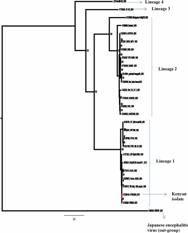
Maximum likelihood phylogeny was obtained using RAxML program under GTR model of
nucleotide substitution and bootstrapping with 1,000 replicates (Figure 1). The Kenyan strain
(KC243146.1) clustered together with other strains from different parts of the world
belonging to WNV Lineage 1.
Figure 1 Maximum likelihood phylogeny of selected WNV sequences. GenBank accession
numbers. Strain abbreviations (isolation source, country, year and accession number): SE-90:
Mimomyia lacustris, Senegal, 1990, DQ318019; ATH002316, R. pulchellus, Kenya, 2010,
KC243146.1; Ug-37: human, Uganda, 1937, AY532665; WNFCG: derivate of Ug-37,
M12294; SA-89: human, South Africa, 1989, EF429197; SA-01: human, South Africa, 2001,
EF429198; Hu-04: Accipiter gentilis, Hungary, 2004, DQ116961; Gr-10: Culex pipiens,
Greece, 2010, HQ537483; SA-58: human, South Africa, 1958, EF429200; SA-00: human,
South Africa, 2000, EF429199; Rus-07: human, Russia, 2007, FJ425721; Rab-97: Cx.

pipiens, Czech Republic, 1997, AY765264.1; CAR-72: Cx. tigripes, Central African
Republic, 1972, DQ318020.1; Sarafend: derivate of Ug-37, AY688948.1; Coracopsis vasa,
Madagascar, 1978, DQ176636.2; Rus-98: Dermacentor marginatus, Russia, 1998,
AY277252.1; human brain in 1999, LEIV-Vlg99-27889, Russia: Volgograd, low Volga,
AY277252.1; NY-99, USA, KC407666.1; total brain RNA (patient NYC99002), HNY1999,
USA: New York, AF202541.1; crow, WN NY 2000-crow3356, USA: New York,
AF404756.1; NY99,385-99, USA, DQ211652.1; Homo sapiens, Italy/2012/Livenza/31.1,
Italy: Veneto region, Venice province, JX556213.1; brain of horse with encephalitis, 2003,
Morocco, AY701413.1; brain, house sparrow (Passer domesticus), France 405/04, France,
2004, DQ786572.1; equine, WN Italy 1998-equine, Italy, AF404757.1; Culex univittatus,
KN3829, AY262283.1 and swine brain, China, 2007, FJ495189.1. Japanese encephalitis
virus isolate GenBank accession number FJ495189.1 was used an out-group.
Evolutionary relationships among WNV strains
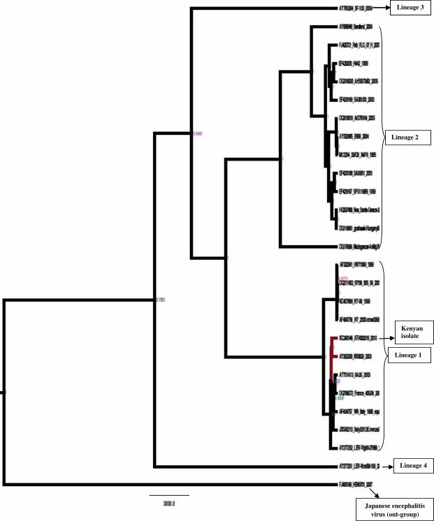
The evolutionary relationship of the Kenyan tick-borne WNV strain and other WNV strains
was assessed using BEAST tool under general time reversible (GTR) model. The clustering
observed under this method was similar to that observed under maximum likelihood
providing further support that the WNV isolate (KC243146.1) from ticks collected in Ijara
district in Northern Kenya clustered closely to Lineage 1 strains from Kenya as well as
Europe, North Africa and the USA. The time to the most recent common ancestor of Lineage
1 viruses was estimated to be approximately 200 years (Figure 2) while the Ijara tick strain
diverged approximately 21 years ago from the other Kenyan strain or European strains and 30
years ago from the USA strains. The strains from North Africa were closest to the strains
studied in France and only diverged 9 years ago, suggesting this may have been the possible
route of introduction to Europe. The Kenyan strain (KC243146.1) was distinct from Lineage
2 strains from southern Africa and diverged more than 200 years ago from these.
Figure 2 Evolutionary relationships among the WNV strains. Maximum Clade Credibility
tree generated under GTR gamma model of nucleotide substitution with 4 gamma categories
of rate heterogeneity and constant size coalescent population model assuming log-normal
priors. GenBank accession numbers: DQ318019, KC243146.1, AY532665, M12294,
EF429197, EF429198, DQ116961, HQ537483, EF429200, EF429199, FJ425721,
AY765264.1, DQ318020.1, AY688948.1, DQ176636.2, AY277252.1, AY277252.1,
KC407666.1, AF202541.1, AF404756.1, DQ211652.1, JX556213.1, AY701413.1,
DQ786572.1, AF404757.1, AY262283.1 and FJ495189.1. Node labels are median heights (in
years)
Selection signatures (amino acid substitutions) in WNV genome
The 27 WNV full genome sequences were first tested for recombination. Two partitions of 1–
980 codons and 981–3650 codon sites were obtained; however, no recombination signal was
detected in any of the genomes used for these analyses. The Hyphy SLAC selection analysis
method predicted 21 codon sites to be under positive selection based on significant p-values.
(Figure 3). Site-specific amino acid changes as predicted from SLAC analysis in Hphy
datamonkey webserver were extracted and recorded as shown in Additional file 1: Table S1.
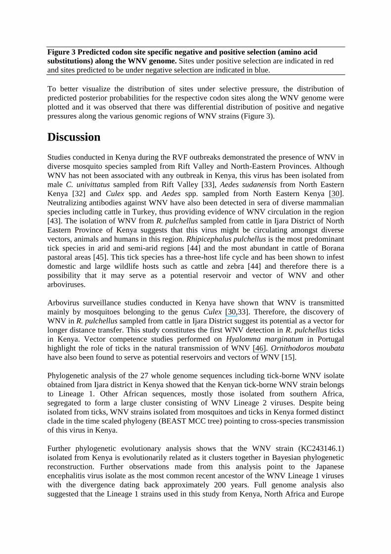
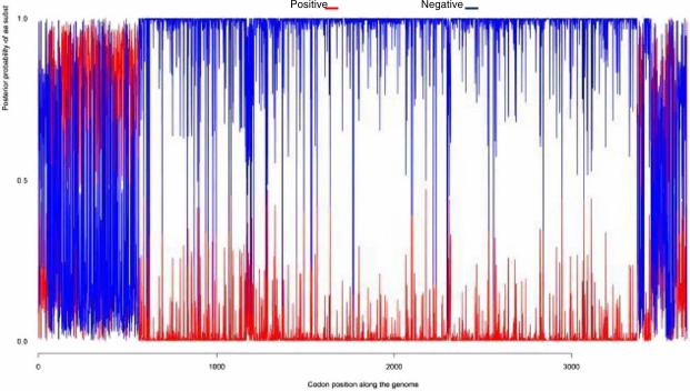
Figure 3 Predicted codon site specific negative and positive selection (amino acid
substitutions) along the WNV genome. Sites under positive selection are indicated in red
and sites predicted to be under negative selection are indicated in blue.
To better visualize the distribution of sites under selective pressure, the distribution of
predicted posterior probabilities for the respective codon sites along the WNV genome were
plotted and it was observed that there was differential distribution of positive and negative
pressures along the various genomic regions of WNV strains (Figure 3).
Discussion
Studies conducted in Kenya during the RVF outbreaks demonstrated the presence of WNV in
diverse mosquito species sampled from Rift Valley and North-Eastern Provinces. Although
WNV has not been associated with any outbreak in Kenya, this virus has been isolated from
male C. univittatus sampled from Rift Valley [33], Aedes sudanensis from North Eastern
Kenya [32] and Culex spp. and Aedes spp. sampled from North Eastern Kenya [30].
Neutralizing antibodies against WNV have also been detected in sera of diverse mammalian
species including cattle in Turkey, thus providing evidence of WNV circulation in the region
[43]. The isolation of WNV from R. pulchellus sampled from cattle in Ijara District of North
Eastern Province of Kenya suggests that this virus might be circulating amongst diverse
vectors, animals and humans in this region. Rhipicephalus pulchellus is the most predominant
tick species in arid and semi-arid regions [44] and the most abundant in cattle of Borana
pastoral areas [45]. This tick species has a three-host life cycle and has been shown to infest
domestic and large wildlife hosts such as cattle and zebra [44] and therefore there is a
possibility that it may serve as a potential reservoir and vector of WNV and other
arboviruses.
Arbovirus surveillance studies conducted in Kenya have shown that WNV is transmitted
mainly by mosquitoes belonging to the genus Culex [30,33]. Therefore, the discovery of
WNV in R. pulchellus sampled from cattle in Ijara District suggest its potential as a vector for
longer distance transfer. This study constitutes the first WNV detection in R. pulchellus ticks
in Kenya. Vector competence studies performed on Hyalomma marginatum in Portugal
highlight the role of ticks in the natural transmission of WNV [46]. Ornithodoros moubata
have also been found to serve as potential reservoirs and vectors of WNV [15].
Phylogenetic analysis of the 27 whole genome sequences including tick-borne WNV isolate
obtained from Ijara district in Kenya showed that the Kenyan tick-borne WNV strain belongs
to Lineage 1. Other African sequences, mostly those isolated from southern Africa,
segregated to form a large cluster consisting of WNV Lineage 2 viruses. Despite being
isolated from ticks, WNV strains isolated from mosquitoes and ticks in Kenya formed distinct
clade in the time scaled phylogeny (BEAST MCC tree) pointing to cross-species transmission
of this virus in Kenya.
Further phylogenetic evolutionary analysis shows that the WNV strain (KC243146.1)
isolated from Kenya is evolutionarily related as it clusters together in Bayesian phylogenetic
reconstruction. Further observations made from this analysis point to the Japanese
encephalitis virus isolate as the most common recent ancestor of the WNV Lineage 1 viruses
with the divergence dating back approximately 200 years. Full genome analysis also
suggested that the Lineage 1 strains used in this study from Kenya, North Africa and Europe

diverged approximately 20 years ago which does not suggest that the Kenyan strain
(KC243146.1) was involved in recent introduction. The closest ancestor to the European
strains was from Morocco which diverged nine years ago from strains of France and Italy.
This does emphasize a possible role of inter- and cross-continental spread for WNV strains.
Transfer of ticks through migratory birds remains a potential mechanism to achieve this.
Lineage 2 strains diverged more than 200 years ago from Lineage 1. The South African
strains diverged 23 years ago from those in Europe. However the Greek strain diverged only
nine years ago from those in Hungary; this may suggest that the Central European strains
were the origin of this outbreak rather than recent introduction from Africa.
WNV Lineage 1 strains have a widespread distribution in Africa, Europe, the Middle East,
America, Australia and India. It is possible that WNV Lineage 1 could have been exported
from Africa to Europe in particular, by migratory birds that might have had ticks attached to
them though evidence of R. pulchellus infesting birds is lacking. The phylogenetic analysis
suggests that Morocco rather than Kenya was the source of recent introduction to Europe.
Both highly and less neuroinvasive strains have been shown to exist in both Lineage 1 and 2
[7]. Experimental or clinical data on the neurovirulence of Lineage 1 strains from Kenya are
not yet available and investigation of WNV as a cause of neurological disease in humans and
animals in Kenya is warranted.
Neurovirulence is associated with specific genotypes and not related to lineage, source of
isolate, geographic distribution, passage level, or year of isolation [47-49]. Selection analyses
carried out in the current study revealed differential selection pressures operating at different
gene regions along the WNV genome. In this analysis it was observed that non-synonymous
amino acid substitutions are more prevalent in 5′ structural genes region and less prevalent in
non-structural gene regions towards the 3′ prime end. Of special interest is the observation
that limited or no diversifying selection pressures seemed to be operating on the polymerase
gene. This may be attributed to its strategic function in the replication of the virus genome
hence being kept under negative selection pressure as seen in Figure 3 (highlighted in blue).
The amino acid changes show the same trend in the viruses belonging to similar lineages
further underscoring the importance of these genes in the maintenance of progeny, i.e.
conferring viral fitness to the various strains. The Kenyan WNV isolate (KC243146.1) from
ticks shows amino acid substitutions commonly observed in other strains belonging to
Lineage 1 isolated from ticks and mosquitoes from different locations worldwide. These
results resonate well with previous studies that have attempted to elucidate genetic markers
for the virulence and pathogenicity of different lineages of the WNV strains. According to a
study conducted by Beasley [50], enhanced virulence of North American WNV strains
compared with other Lineage 1 strains of the Old World was linked to the envelope protein
glycosylation. WNV Lineage 2 strains isolated from patients in South Africa all had these
envelope protein glycosylation sites which have been associated with increased virulence
[51].
The isolation of WNV from R. pulchellus species sampled from cattle in Ijara District might
imply that either this tick species may have acquired the virus after ingesting a viraemic
blood meal from the infected animal or the virus was present in the tick which may imply its
potential as vector, reservoir and disseminator of WNV. In general, birds are known as
reservoirs of WNV while most mammals have relatively low viremia and act as dead-end
hosts [3]. The role of cattle and warthogs as reservoirs for WNV is not known. Although
thought to be unlikely, this could be the source of infection to the tick. Surveillance for
arboviruses such as WNV should be conducted and appropriate prevention and control
strategies be put in place so as to be able to manage arboviruses early enough before they
cause outbreaks [52].
Conclusions
The identification of WNV Lineage 1 in Ijara District further elucidates the geographic range
of Lineage 1 and 2 strains in Africa. Lineage 2 strains have been described in Tanzania and
are almost exclusively found in South Africa [2] while Lineage 1 strains are the major lineage
reported in North Africa, Eurasia, India, the Middle East and the Americas. The close
proximity of Europe to Africa may be the reason that both lineages have emerged in Europe
although the exact origin of importation of Lineage 2 strains to Europe is not known. Tick
vectors pose a feasible transmission/dissemination mechanism for preserving WNV over long
distances and extended time if considering the relatively short viremic period in most
animals. The Kenyan strain identified here was not close to other published WNV strains if
considering the divergence time from known strains. This suggests that a further
uncharacterized source of WNV strains exist in livestock and ticks in Africa.
Competing interests
The authors declare that they have no competing interests.
Authors’ contributions
OWL RS conceived and designed experiments. OWL conducted the experimental work.
OWL GM CR FN SS analyzed the data. OWL MV JL GM CR FG VO CT CO RS
contributed to the manuscript. All authors approved the final version for submission.
Acknowledgements
We wish to thank the Arbovirus Incidence and Diversity Project consortium-led by
International Centre of Insect Physiology and Ecology (ICIPE) and including; International
Livestock Research Institute, Kenya Agricultural Research Institute, Ministry of Livestock
Development-Central Veterinary Laboratories, Kenya Medical Research Institute, Ministry of
Public Health and Sanitation and Kenya Wildlife Service; The director of ICIPE, Prof.
Christian Borgemeister, for providing all the necessary support in spearheading the project.
References
1. Lindenbach BD, Rice C: Flaviviridae: the viruses and their replication. Fields Virol
2001, 1:991–1041.
2. Venter M: Lineage 2 West Nile virus as cause of fatal neurologic disease in horses.
South Africa Emerg Infect Dis 2009, 15(6):877.
3. Artsob H: West Nile virus in the New World: trends in the spread and proliferation of
West Nile virus in the Western Hemisphere. Zoonoses Public Health 2009, 56(6-7):357–
369.
4. Garmendia AE, Van Kruiningen HJ, French RA: The West Nile virus: its recent
emergence in North America. Microb Infect 2001, 3(3):223–229.
5. Roehr B: US hit by massive West Nile virus outbreak centred around Texas. Br J Med
Med Res 2012, 345:e5633. doi: 10.1136/bmj.e5633.
6. Autorino GL: West Nile virus epidemic in horses, Tuscany region Italy. Emerg Infect
Dis 2002, 8(12):1372.
7. Venter M, Swanepoel R: West Nile virus lineage 2 as a cause of zoonotic neurological
disease in humans and horses in southern Africa. Vector-Borne Zoon Dis 2010,
10(7):659–664.
8. Bagnarelli P: Human case of autochthonous West Nile virus lineage 2 infection in
Italy, September 2011. Euro Surveill 2011, 16(43):20002.
9. Danis K: Ongoing outbreak of West Nile virus infection in humans, Greece. Euro
Surveill 2011, 16(34):19951 [pii].
10. Bakonyi T: Explosive spread of a neuroinvasive lineage 2 West Nile virus in
2008/2009, central Europe. Vet Microbiol 2013, 165:61–70.
doi:10.1016/j.vetmic.2013.03.005.
11. Kramer LD, Li J, Shi PY: West Nile virus. Lancet Neur 2007, 6(2):171–181.
12. Sardelis MR: Vector competence of selected North American Culex and
Coquillettidia mosquitoes for West Nile virus. Emerg Infect Dis 2001, 7(6):1018.
13. Kilpatrick AM: Temperature, viral genetics, and the transmission of West Nile virus
by Culex pipiens mosquitoes. PLoS Pathog 2008, 4(6):e1000092.
14. Hamer GL: Host selection by Culex pipiens mosquitoes and West Nile virus
amplification. Am J Trop Med Hyg 2009, 80(2):268.
15. Lawrie CH: Ixodid and argasid tick species and West Nile virus. Emerg Infect Dis
2004, 10(4):653.
16. Komar N: Experimental infection of North American birds with the New York 1999
strain of West Nile virus. Emerg Infect Dis 2003, 9(3):311.
17. Abbassy MM, Stein KJ, Osman M: New artificial feeding technique for experimental
infection of Argas ticks (Acari: Argasidae). J Med Entomol 1994, 31(2):202–205.
18. Petersen LR, Marfin AA: West Nile virus: a primer for the clinician. Ann Intern Med
2002, 137(3):173–179.
19. Fratkin JD: Spinal cord neuropathology in human west NileVirus infection. Arch
Pathol Lab Med 2004, 128(5):533–537.
20. Drummond CL: Impact of trap elevation on estimates of abundance, parity rates, and
body size of Culex pipiens and Culex restuans (Diptera: Culicidae). J Med Entomol 2006,
43(2):177–184.
21. Paddock CD: Fatal hemorrhagic fever caused by West Nile virus in the United States.
Clin Infect Dis 2006, 42(11):1527–1535.
22. Lindsey NP: West Nile virus neuroinvasive disease incidence in the United States,
2002-2006. Vector-Borne Zoon Dis 2008, 8(1):35–40.
23. Asnis DS: West Nile virus outbreak of 1999 in New York: the Flushing hospital
experience. Clin Infect Dis 2000, 30(3):413–418.
24. Granwehr BP: West Nile virus: where are we now? Lancet Infect Dis 2004, 4(9):547–
556.
25. May FJ: Phylogeography of West Nile virus: from the cradle of evolution in Africa to
Eurasia, Australia, and the Americas. J Virol 2011, 85(6):2964–2974.
26. Petersen LR, Roehrig JT: West Nile virus: a reemerging global pathogen. Rev Biomed
2001, 12(3):208–216.
27. Bakonyi T: Novel flavivirus or new lineage of West Nile virus, central Europe. Emerg
Infect Dis 2005, 11(2):225.
28. Lvov D: West Nile virus and other zoonotic viruses in Russia: examples of emerging-
reemerging situations. Arch Virol-suppl 2004, 18:85–96.
29. Bondre VP: West Nile virus isolates from India: evidence for a distinct genetic
lineage. J Gen Virol 2007, 88(3):875–884.
30. LaBeaud AD, Bashir F, King CH: Measuring the burden of arboviral diseases: the
spectrum of morbidity and mortality from four prevalent infections. Popul Health
Metrics 2011, 9(1):1.
31. Lutomiah JL: Ability of selected Kenyan mosquito (Diptera: Culicidae) species to
transmit West Nile virus under laboratory conditions. J Med Entomol 2011, 48(6):1197–
1201.
32. Crabtree M: Arbovirus surveillance of mosquitoes collected at sites of active Rift
Valley fever virus transmission: Kenya, 2006-2007. J Med Entomol 2009, 46(4):961–964.
33. Miller BR: Isolation and Genetic Characterization of Rift Valley fever virus from
Aedes vexans arabiensis, Kingdom. Emerg Infect Dis 2002, 8(12):1493.
34. Lwande OW: Isolation of tick and mosquito-borne arboviruses from ticks sampled
from livestock and wild animal hosts in Ijara district. Kenya Vector Vector-Borne Zoon
Dis 2013, 13(9):637–642.
35. Djikeng A: Viral genome sequencing by random priming methods. BMC Genomics
2008, 9(1):5.
36. Maddison DR, Maddison WP: MacClade 4. State: Sinauer Associates Sunderland; 2000.
37. Posada D: jModelTest: phylogenetic model averaging. Mol Biol Evol 2008,
25(7):1253–1256.
38. Stamatakis A: RAxML-VI-HPC: maximum likelihood-based phylogenetic analyses
with thousands of taxa and mixed models. Bioinformatics 2006, 22(21):2688–2690.
39. Pond SLK, Frost SD: Datamonkey: rapid detection of selective pressure on individual
sites of codon alignments. Bioinformatics 2005, 21(10):2531–2533.
40. Murrell B: FUBAR: a fast, unconstrained bayesian appRoximation for inferring
selection. Mol Biol Evol 2013, 30(5):1196–1205.
41. Pond SLK: GARD: a genetic algorithm for recombination detection. Bioinformatics
2006, 22(24):3096–3098.
42. Westgeest KB: Genome-wide analysis of reassortment and evolution of human
influenza a (H3N2) viruses circulating between 1968 and. J Virol 2011, 2013:02163–13.
43. Ozkul A: Serological evidence of West Nile Virus (WNV) in mammalian species in
Turkey. Epidemiol Infect 2006, 134(04):826–829.
44. Walker JB, Keirans JE, Horak IG: The Genus Rhipicephalus (Acari, Ixodidae): a Guide
to the Brown Ticks of the World. Cambridge University Press; 2000. ISBN 9780521019774.
45. Ayana D, Eshetu E, Abunna F: Survey of Ixodid Ticks on Cattle in Borana Pastoral Area,
Ethiopia. ; 2013.
46. Formosinho P, Santos-Silva M: Experimental infection of Hyalomma marginatum
ticks with West Nile virus. Acta Virol (Praha) 2006, 50(3):175.
47. Venter M: Gene expression in mice infected with West Nile virus strains of different
neurovirulence. Virology 2005, 342(1):119–140.
48. Beasley DW, Barrett AD: Identification of neutralizing epitopes within structural
domain III of the West Nile virus envelope protein. J Virol 2002, 76(24):13097–13100.
49. Burt FJ: Phylogenetic relationships of southern African West Nile virus isolates.
Emerg Infect Dis 2002, 8(8):820–826.
50. Beasley DW: Envelope protein glycosylation status influences mouse neuroinvasion
phenotype of genetic lineage 1 West Nile virus strains. J Virol 2005, 79(13):8339–8347.
51. Botha EM: Genetic determinants of virulence in pathogenic lineage 2 West Nile virus
strains. Emerg Infect Dis 2008, 14(2):222.

52. Bellini R, Zeller H, Bortel VW: A review of the vector management methods to
prevent and control outbreaks of West Nile virus infection and challenge for Europe. Parasites Vectors 2014, 7:323.
Lineage 3
Lineage 2
Lineage 1
Lineage 4
Japanese encephalitis virus (out-group)
Lineage 3
Kenyanisolate
Positive Negative

Addtional files provided with this submission:
Additional file 1: Table S1. Amino acid substitutions of the WNV polyprotein gene that corresponds to the structural (C,E and M) and non-structural (1, 2A, 2B, 3, 4A, 4B and 5) proteins (606 kb)k)http://www.parasitesandvectors.com/content/supplementary/s13071-014-0542-2-s1.pdf
Top Related
Copyright © 2022 FDOKUMEN

