


Bahasa
Halaman
Hukum


JOURNAL OF VIROLOGY, Sept. 2007, p. 9408–9418 Vol. 81, No. 170022-538X/07/$08.00�0 doi:10.1128/JVI.00707-07Copyright © 2007, American Society for Microbiology. All Rights Reserved.
Vaccine Platform for Prevention of Tuberculosis and Mother-to-ChildTransmission of Human Immunodeficiency Virus Type 1
through Breastfeeding�
Eung-Jun Im,1 Narcıs Saubi,2 Goretti Virgili,2 Clare Sander,3 Denise Teoh,1 Jose M. Gatell,2Helen McShane,3 Joan Joseph,2† and Tomas Hanke1†*
MRC Human Immunology Unit, Weatherall Institute of Molecular Medicine, University of Oxford, The John Radcliffe,Oxford OX3 9DS, United Kingdom1; Catalan HIV Vaccine Research and Development Center, AIDS Research Unit,
Infectious Diseases Department, Hospital Clinic, August Pi i Sunyer Biomedical Research Institute, School of Medicine,University of Barcelona, 170 08036 Barcelona, Spain2; and Centre for Clinical Vaccinology and
Tropical Medicine, Churchill Hospital, Oxford OX3 7LJ, United Kingdom3
Received 3 April 2007/Accepted 11 June 2007
Most children in Africa receive their vaccine against tuberculosis at birth. Those infants born to humanimmunodeficiency virus type 1 (HIV-1)-positive mothers are at high risk of acquiring HIV-1 infection throughbreastfeeding in the first weeks of their lives. Thus, the development of a vaccine which would protect newbornsagainst both of these major global killers is a logical yet highly scientifically, ethically, and practicallychallenging aim. Here, a recombinant lysine auxotroph of Mycobacterium bovis bacillus Calmette-Guerin(BCG), a BCG strain that is safer than those currently used and expresses an African HIV-1 clade-derivedimmunogen, was generated and shown to be stable and to induce durable, high-quality HIV-1-specific CD4�-and CD8�-T-cell responses. Furthermore, when the recombinant BCG vaccine was used in a priming-boostingregimen with heterologous components, the HIV-1-specific responses provided protection against surrogatevirus challenge, and the recombinant BCG vaccine alone protected against aerosol challenge with M. tuber-culosis. Thus, inserting an HIV-1-derived immunogen into the scheduled BCG vaccine delivered at or soon afterbirth may prime HIV-1-specific responses, which can be boosted by natural exposure to HIV-1 in the breastmilk and/or by a heterologous vaccine such as recombinant modified vaccinia virus Ankara delivering the sameimmunogen, and decrease mother-to-child transmission of HIV-1 during breastfeeding.
Since the first report of AIDS in 1981, an estimated morethan 60 million people have become infected with human im-munodeficiency virus type 1 (HIV-1), of whom some 25 millionhave died. Over 60% of the global HIV-1-infected populationlives in Africa, and about a half of the infected adults arewomen of childbearing age. Up to 40% of pregnant womenattending large urban-center clinics in Kenya are HIV-1 sero-positive. Despite the fact that approximately half of mother-to-child transmissions (MTCT) are due to prolonged breast-feeding (43), for many HIV-1-positive mothers, bottle feedingis not an option for social, practical, and health reasons. Bottle-fed babies of infected mothers have higher morbidity and mor-tality due to increased exposure and susceptibility to otherinfections (32). Although antiretroviral therapy can signifi-cantly reduce the risk of MTCT at parturition, it is less clearwhether it is practical to use antiretroviral drugs to preventHIV-1 transmission through breast milk. The drugs are veryexpensive and would have to be administered at birth andthroughout the whole period of breastfeeding; in addition,their effectiveness may be compromised by the emergence ofresistant mutants. Thus, the best hope for protecting newborns
against MTCT of HIV-1 in developing countries is the devel-opment of safe, effective, accessible prophylactic vaccines,which would both reduce the adult burden of infection andprotect neonates against vertical HIV-1 transmission.
The development of an HIV-1 vaccine remains on the ho-rizon. Although in principle it is possible, the induction ofbroadly neutralizing antibodies effective across HIV-1 cladeshas been extremely difficult to achieve by active immunization(6). While various technologies are becoming more successfulat inducing HIV-1-specific T-cell responses (33), a single cor-relate of protection against HIV-1 infection and AIDS remainselusive and may be nonexistent, as protection is most likely tobe multifactorial. Therefore, before further progress in thisarea is made, the interim aim of HIV-1 vaccination is to elicitresponses comprising T cells that are high in frequency, mul-tispecific, multifunctional, capable of rapid proliferation, long-lived, and of the central memory phenotype. The in vivo func-tionality of vaccine-induced T cells can be assessed usingsurrogate models of viral challenge. The ultimate goal, vaccineefficacy, can be confirmed only by adequately powered clinicaltrials with humans.
For a number of years, we have been developing an HIV-1vaccine focusing on the induction of protective cell-mediatedresponses. Our starting platform was based on a heterologousregimen consisting of a DNA priming vaccine and a modifiedvaccinia virus Ankara (MVA) boosting vaccine (44) deliveringa common immunogen called HIVA, which is derived fromconsensus HIV-1 clade A Gag protein, i.e., an immunogen
* Corresponding author. Mailing address: Weatherall Institute ofMolecular Medicine, The John Radcliffe, Oxford OX3 9DS, UnitedKingdom. Phone: 44 1865 222355. Fax: 44 1865 222502. E-mail: [email protected].
† These authors have codirected the research work and have con-tributed equally to this work.
� Published ahead of print on 27 June 2007.
9408
derived from an HIV-1 strain prevalent in central and easternAfrica, and a string of CD8�-T-cell epitopes (17). Extensivestudies with mice, nonhuman primates, and more than 400healthy and HIV-1-infected humans have shown that the vac-cines are safe and immunogenic (18). In humans, DNA vaccinepTHr.HIVA weakly but consistently primes HIV-1-specific re-sponses involving mostly CD4� T cells and MVA expressingHIVA (MVA.HIVA) delivers a strong and consistent boost,expanding populations of both CD4� and CD8� HIV-1-spe-cific T cells, particularly if they are well primed, e.g., by HIV-1infection (12, 15, 37).
Infection with Mycobacterium tuberculosis kills about 2 mil-lion people each year (42). M. bovis bacillus Calmette-Guerin(BCG) is the only licensed vaccine and protects significantlyagainst childhood and miliary tuberculosis (52). Globally, 80%percent of children are vaccinated with BCG (50), the majorityof them at birth. Thus, the development of a combined vaccinethat would protect neonates against tuberculosis and MTCT ofHIV-1 through breastfeeding is a logical effort in the fightagainst these two major global killers. One such obvious vac-cine or vaccine component is a recombinant BCG (rBCG)strain expressing an HIV-1-derived immunogen.
BCG as a vaccine vector has a number of attractive features(10). BCG has a proven record of safety as a vaccine againsttuberculosis from its use in more than 2 billion individuals,including neonates (26). BCG infects and colonizes macro-phages and dendritic cells, where it can survive and replicatefor a long period of time. By its persistence and potent adju-vancy through its cell wall components, it can induce long-lasting humoral and cellular immune responses. Immunityagainst tuberculosis following neonatal BCG vaccination lasts10 to 15 years (20) and, therefore, fails to protect adults frompulmonary disease (9). Thus, it should easily cover the dangerperiod of breastfeeding, and a later booster vaccine may offerchildren further protection in adolescence. BCG can be givenat or any time after birth and is not affected by maternalantibodies. The manufacturing of BCG-based vaccines ischeap as live bacteria are easy to purify. Finally, BCG is one ofthe most heat-stable vaccines in present use (14).
It is not known how early in life T cells can be educated tolaunch a protective response against intracellular microorgan-isms, and this factor most likely differs from pathogen to patho-gen. Qualitative and quantitative differences between re-sponses to a number of infections in human newborns andadults have been observed previously (1, 16, 31, 45). WeakerCD8� T-cell responses in HIV-1-infected infants than in adultsmay play an important role in fast disease progression (7, 27,41); children account for 4% of HIV-1 infections, yet theyrepresent 20% of AIDS deaths (16). At the same time, matureresponses to certain infections and vaccines during the post-natal and even fetal periods have been demonstrated previ-ously (28). This observation is particularly true for BCG vac-cine-induced responses, which promote adult-like Th1responses in newborns (21, 29, 38, 49). Therefore, there issome evidence suggesting that protective T-cell-mediated re-sponses could be elicited by vaccines early in life and that BCGas a vaccine vector may be very well suited to prime them (30).
In this study, we have constructed a recombinant lysineauxotroph of BCG expressing the HIVA immunogen fromboth replicative and integrative vectors. After confirmation of
the HIVA gene sequence, plasmid stability, and protein ex-pression, the BCG strain expressing HIVA from an episomalplasmid (BCG.HIVA) alone and BCG.HIVA in a heterolo-gous priming-boosting combination were studied for the induc-tion of HIV-1- and M. tuberculosis-specific immune responsesin a murine model. Protection against both surrogate virusexpressing HIVA and aerosol M. tuberculosis challenge wasachieved.
MATERIALS AND METHODS
Construction of Escherichia coli-mycobacterium shuttle vectors expressingHIVA. Parental plasmids pJH222 and pJH223 were kindly provided by W. R.Jacobs, Jr., B. R. Bloom, and T. Hsu. The coding sequence of the HIVA gene wasfused to the M. tuberculosis nucleotides coding for the 19-kDa lipoprotein signalsequence in a PCR, and the chimeric gene was cloned into the pJH222 andpJH223 plasmids as a HindIII-HindIII fragment under the control of the M.tuberculosis �-antigen promoter by using standard recombinant-DNA tech-niques.
Mycobacterial culture. A lysine auxotroph of BCG, kindly provided by W. R.Jacobs, Jr., B. R. Bloom, and T. Hsu, was transformed by electroporation.Mycobacterial cultures were grown in Middlebrook 7H9 broth medium or onMiddlebrook agar 7H10 medium supplemented with albumin-dextrose complex(ADC; Difco) and containing 0.05% Tween 80 and 25 �g of kanamycin/ml. TheL-lysine monohydrochloride (Sigma) was dissolved in distilled water and used ata concentration of 40 �g/ml. For transformation, BCG cultures were grown to anoptical density at 600 nm of 0.9, transformed using a Bio-Rad gene pulserelectroporator at 2.5 kV, 25 mF, and 1,000 �, and plated onto ADC-supple-mented Middlebrook agar 7H10 medium containing 0.05% Tween 80 and 25 �gof kanamycin/ml.
Sodium dodecyl sulfate-polyacrylamide gel electrophoresis and Western blotanalysis. Cell lysates of mid-logarithmic-phase BCG transformants were pre-pared, separated by sodium dodecyl sulfate-polyacrylamide gel electrophoresis,and electroblotted. HIVA protein was detected using anti-Pk antibodies with anECL kit (Amersham International).
Mice. For all animal experiments, groups of four to five 5- to 8-week-oldfemale BALB/c mice were used unless otherwise specified. All animal proce-dures and care were approved by local ethical committees and strictly conformedto United Kingdom Home Office guidelines.
Immunizations and isolation of splenocytes. Under general anesthesia, micewere immunized intramuscularly with 100 �g of endotoxin-free pTHr.HIVADNA (Cobra Therapeutics, United Kingdom) or 106 PFU of MVA.HIVA in-tramuscularly or intraperitoneally (i.p.) with 106 CFU of parental BCG (BCG.p)or BCG.HIVA. For dose titration, mice received 103 to 107 CFU of BCG.HIVAi.p. On the day of sacrifice, spleens were removed and pressed individuallythrough a cell strainer (Falcon) with a 5-ml syringe rubber plunger. Following theremoval of red blood cells with red blood cell lysing buffer (Sigma), splenocyteswere washed and resuspended in lymphocyte medium (R10 [RPMI 1640 sup-plemented with 10% fetal calf serum {FCS} and penicillin-streptomycin], 20 mMHEPES, and 15 mM 2-mercaptoethanol) at a concentration of 2 �107 cells/ml.
In vivo stability of plasmid pJH222.HIVA. The growth of rBCG and thestability of the extrachromosomal plasmid pJH222.HIVA were established by therecovery of BCG.HIVA colonies from the spleens of mice 15 weeks after im-munization. Spleens were homogenized and plated onto Middlebrook 7H10medium supplemented with ADC (Difco) and containing 0.05% Tween 80 and25 �g of kanamycin/ml. With specific primers, the HIVA gene was detectedamong well-separated mycobacterial colonies by PCR.
Peptides. For assessing the immunogenicity of HIVA in the BALB/c mice, thefollowing peptides were used: H-2Dd-restricted epitope P18-I10 (RGPGRAFVTI) (48), herein designated epitope H, and the subdominant H-2Kd-restrictedP epitope (IFQSSMTKI) (22). Three epitope peptides derived from p24 werepooled to investigate the major histocompatibility complex (MHC) class II-restricted response: MHQALSPRTLNAQVKVIEEK, NPPIPVGDIYKRWIILGLNK, and FRDYVDRFFKTLRAEQATQE.
In vivo killing assay. Naıve syngeneic mice were sacrificed, the splenocyteswere prepared as described above, and the isolated splenocytes were incubatedwithout or with 2 �g of peptides/ml in R10 at 37°C in 5% CO2 for 90 min andwashed three times. Target cells not pulsed with peptides were labeled with5-(and-6)-(((4-chloromethyl)benzoyl)amino)tetramethyl rhodamine (CMTMR;Molecular Probes) only, while peptide-pulsed target cells were labeled withcarboxyfluorescein diacetate succinimidyl ester either without (P peptide) or
VOL. 81, 2007 INFANT VACCINE AGAINST TUBERCULOSIS AND HIV-1 9409

combined with (H peptide) CMTMR. Briefly, H or P peptide-pulsed splenoctyesresuspended in phosphate-buffered saline (PBS) at 2 � 107 cells/ml were incu-bated with 80 nM carboxyfluorescein diacetate succinimidyl ester (MolecularProbes) in the dark at room temperature for 10 min, followed by the quenchingof the reaction with an equal volume of FCS and three washing steps. H peptide-pulsed cells were then resuspended in R10 at 2 � 107 cells/ml and incubated with10 �M CMTMR at 37°C for 15 min and in fresh R10 only for a further 15 min.Three differentially labeled cell cultures were washed, resuspended in PBS, andcombined for intravenous adoptive transfer, with each animal receiving approx-imately 2 � 106 cells of each population. After 12 h, animals were sacrificed, andtheir splenocytes were isolated and analyzed using flow cytometry. Cytotoxicitywas calculated using the following formula: adjusted percentage of survivingcells � 100 � (percentage of surviving peptide-pulsed cells/mean percentage ofsurviving unpulsed cells). Next, the percentage of specific lysis was calculated asfollows: percentage of specific lysis � 100 � adjusted percentage of survivingcells (19).
Intracellular cytokine staining. Two million splenocytes were added to eachwell of a 96-well round-bottomed plate (Falcon) and pulsed with peptides at 2�g/ml (CD8 epitopes) to 5 �g/ml (CD4 epitopes) or a 5-�g/ml concentration ofpurified protein derivative (PPD) tuberculin (Statens Serum Institut, Copenha-gen, Denmark), together with antibodies against lysosome-associated membraneproteins anti-CD107a-fluorescein isothiocyanate (FITC) and anti-CD107b-FITC(BD Biosciences) (2), and kept at 37°C and 5% CO2 for 90 min, followed by theaddition of GolgiStop (BD Biosciences) containing monensin. After a further 5-hincubation, the reaction was terminated and the cells were washed with FACSwash buffer (PBS, 2% FCS, 0.01% azide) and blocked with anti-CD16/32 (BDBiosciences) at 4°C for 30 min. All subsequent antibody staining procedures wereperformed using the same conditions. Cells were then washed and stained withanti-CD8-peridinin chlorophyll protein or anti-CD4-peridinin chlorophyll pro-tein (BD Biosciences), washed again, and permeabilized using the Cytofix/Cytoperm kit (BD Biosciences). Perm/Wash buffer (BD Biosciences) was used towash cells before staining with anti-interleukin-2 (anti-IL-2)-FITC, anti-tumornecrosis factor alpha (anti-TNF-�)-phycoerythrin, and anti-gamma interferon(anti-IFN-�)-allophycocyanin (BD Biosciences). Cells were fixed with CellFIX(BD Biosciences) and stored at 4°C until analysis.
Vaccinia virus challenge and protection assay. Groups of four to five naıve orimmunized BALB/c female mice were challenged i.p. with 4 � 106 PFU ofrecombinant replication-competent vaccinia virus strain Western Reserve ex-pressing the HIVA immunogen (WR.HIVA). Four days later, ovaries werecollected and sonicated. Confluent 143B cells negative for human thymidinekinase in 6-well plates were infected with 10-fold serial dilutions of the homo-genated ovaries. Cells were stained with 0.1% crystal violet in 20% ethanol, andthe plaques were counted.
Fluorescence-activated cell sorter analysis. All chromogen-labeled cells wereanalyzed by flow cytometry using the CellQuest software (BD Biosciences).
M. tuberculosis challenge and protection assay. Specific-pathogen-free 6- to8-week-old BALB/c mice were maintained under barrier conditions in a categoryIII safety facility. Groups of seven to nine mice were vaccinated subcutaneouslyin the left hind foot with 3 � 105 CFU of either BCG.HIVA, BCG.p, or BCGwild-type 1173 p2 12 weeks prior to challenge. M. tuberculosis Erdman wasobtained from M. Brennan (World Health Organization), kept at �80°C, andsonicated before use. Mice were challenged with aerosol spray containing M.tuberculosis by using a modified Henderson apparatus (40) and exposing only thenose during challenge with a system from ADG Developments (United King-dom). Bacterial loads in lungs and spleens were determined 4 weeks afterchallenge by plating 10-fold serial dilutions of organ homogenates onto Middle-brook plates and counting the CFU following a 3-week incubation at 37°C.Deposition in the lungs 24 h after challenge was estimated and determined to bearound 25 CFU/lung.
RESULTS
Construction of recombinant M. bovis BCG expressingHIV-1 clade A immunogen. To construct a candidate HIV-1vaccine with M. bovis BCG as the vector, we used an immu-nologically well-characterized protein, HIVA, containing themonoclonal antibody (MAb) tag Pk (17). The HIVA gene wassynthesized utilizing humanized GC-rich codons (5), which aresimilar to those used by mycobacteria (4). The HIVA openreading frame was fused at its 5 end to nucleotides coding for
the 19-kDa lipoprotein signal sequence, which facilitates theexpression of foreign proteins in the mycobacterial membraneand was shown to increase the foreign protein immunogenicity(47). To facilitate the preclinical development of candidatevaccines, the HIVA immunogen contains an immunodominantH-2Dd-restricted epitope, P18-I10 (48), herein designatedepitope H. In addition, it also contains at least three othersubdominant H-2d epitopes recognized by CD8� T cells, in-cluding epitope P, lipoprotein, and three CD4�-T-helperepitopes (24). The chimeric 19-kDa signal sequence-HIVAgene was expressed from E. coli-mycobacterium shuttleplasmids pJH222 and pJH223 under the control of the M.tuberculosis �-antigen promoter (Fig. 1A). pJH222 is a low-copy-number replicative episomal vector and contains a myco-bacterial origin of replication (oriM). pJH223 is an integrativevector that carries an attachment site (attP) and the integrasegene (int) from the mycobacteriophage L5 and integrates as asingle copy into the mycobacterial chromosome. Both vectorsalso contained the kanamycin resistance gene (aph) as a se-lectable marker, an E. coli origin of replication (oriE), and awild-type lysine A-complementing gene for vector mainte-nance in the BCG auxotroph. The lysine auxotroph of M. bovisBCG host strain Pasteur (39) was transformed with recombi-nant pJH222.HIVA and pJH223.HIVA. The expression of thefull-size chimeric 19-kDa lipoprotein signal sequence-HIVAprotein with an Mr of 65 kDa was confirmed on a Western blotof transformed mycobacterial whole-cell lysates using anti-PkMAb (Fig. 1B). This analysis also revealed that rBCG carryingepisomal pJH222.HIVA expressed moderately higher levels ofHIVA than rBCG with the integrated pJH223.HIVA plasmid.The growth of the transformed mycobacteria and the in vivostability of the episomal plasmid were established by the re-covery of HIVA-expressing rBCG from the spleens of BALB/cmice 15 weeks after immunization (Fig. 1C). For further ex-periments, rBCG harboring episomal vector pJH222.HIVAwas used and referred to as BCG.HIVA.
BCG.HIVA primes and enhances MVA.HIVA-elicited HIV-1-specific CD8�-T-cell responses. The ability of the candidateBCG.HIVA vaccine to induce HIV-1-specific immune re-sponses in BALB/c mice was determined (Fig. 2A). On day 0,mice were immunized using either rBCG with the episomalplasmid or BCG.p or left unimmunized, and on day 102, half ofthe animals received a booster dose of MVA.HIVA. Becauseof the activation induced by BCG, detectable HIVA-specificresponses peaked after 12 weeks (data not shown). On day 151,the mice were sacrificed and the functional quality of the elic-ited T cells in terms of their ability to produce IFN-� andTNF-� and to degranulate (exhibit surface expression ofCD107a/b) in response to peptide stimulation was investigatedin a multicolor flow cytometric analysis (Fig. 2B). A number ofobservations were made. First, BCG.HIVA alone induced un-detectable, HIV-1-specific CD8� T-cell responses with thepossible exception of degranulating (CD107a/b�) cells produc-ing TNF-� (Fig. 2B, middle dot blot). Nevertheless, the sameBCG.HIVA priming increased the MVA.HIVA-elicited fre-quencies of the H- and subdominant P-specific CD8� spleno-cytes producing IFN-� and degranulating by 5 (P � 0.0004)-and 14 (P � 0.02)-fold, respectively. Although some increase inthe H responses after the BCG.p priming was also observed,this increase was not statistically significant. Thus, the BCG.
9410 IM ET AL. J. VIROL.

HIVA enhancement of MVA.HIVA responses was not a non-specific stimulation of bystander T cells by innate anti-BCGresponses but rather a HIVA-specific response. Second, of allthe regimens tested, the BCG.HIVA priming-MVA.HIVAboosting regimen elicited the highest proportion of HIV-1-specific bifunctional cells in all three analyses presented (Fig.2B). Finally, no HIVA-induced CD4�-T-cell responses weredetected (data not shown). HIV-1-specific cellular responses in
MVA.HIVA- and BCG.HIVA-MVA.HIVA-vaccinated micewere also assessed using an in vivo killing assay (Fig. 2C). Thisassay clearly demonstrated strong in vivo lytic activities againstboth the H and P epitopes primed by BCG.HIVA, whichapproximately doubled and tripled, respectively, responseselicited by MVA.HIVA alone.
Priming BCG.HIVA dose affects vigor and quality of CD8�-T-cell response to a subdominant epitope. Next, the effect ofthe BCG.HIVA dose on the vigor and quality of CD8�-T-cellresponses induced by the heterologous BCG.HIVA-MVA.HIVAregimen was investigated. According to a schedule similar tothat described above, BALB/c mice were either left unvacci-nated or primed with increasing doses of BCG.HIVA rangingfrom 103 to 107 CFU per animal and given a constant boosterdose of MVA.HIVA (Fig. 3A). First, the frequencies of bi-functional IFN-�� CD107a/b�, TNF-�� CD107a/b�, andIFN-�� TNF-�� T cells specific for the H and P epitopes wereassessed. We found that for immunodominant epitope H, bi-functional responses steadily increased in magnitude alongwith the dose and reached a maximum of approximately 0.7%CD8� splenocytes at 106 CFU of BCG.HIVA. In contrast, forsubdominant epitope P, 105 CFU of BCG.HIVA appeared tobe the threshold for response induction and the P responseincreased with doses of up to 107 CFU, reaching approximately0.2% CD8� splenocytes (Fig. 3B). Analyzing trifunctional cellsrevealed that for the immunodominant H epitope, theBCG.HIVA dose did not significantly affect the relatively highfraction of CD8� IFN-�� splenocytes that were also capable ofTNF-� production and degranulation. The highest group av-erage proportion of trifunctional cells was 88% CD8� IFN-��
splenocytes, which was detected at 105 CFU of BCG.HIVA(Fig. 3C). In contrast, responses to subdominant epitope Ppeaked at 107 CFU of BCG.HIVA and reached 80% CD8�
IFN-�� splenocytes. Similar results were obtained by the re-ciprocal analysis, i.e., identifying the fractions of CD8�
TNF-�� cells that could both produce IFN-� and degranulate(Fig. 3D). Thus, the priming dose of BCG.HIVA affected boththe strength and quality of CD8�-T-cell responses induced bya heterologous rBCG priming-recombinant MVA (rMVA)boosting regimen. A high priming BCG.HIVA dose was par-ticularly important for improving the quality of responses tothe subdominant P epitope, i.e., for the breadth of the vaccine-induced T-cell responses.
pTHr.HIVA DNA priming-BCG.HIVA boosting protectsagainst a surrogate virus challenge. The relevant functionalityof vaccine-induced responses against viral infection is besttested by an in vivo virus challenge. However, HIV-1 does notreplicate in mice, and there is not a challenge relevant for theHIV-1-derived immunogen HIVA in nonhuman primate mod-els other than the infection of chimpanzees with HIV-1, whichis prohibitively costly. Thus, a surrogate challenge with WR.HIVA was used as described previously (25). To avoid theinduction of antipoxvirus immune responses by MVA.HIVA,we utilized a priming-boosting regimen with heterologous vac-cines consisting of a pTHr.HIVA DNA priming dose on day 0and a BCG.HIVA booster dose on day 33, together with sev-eral control groups (Fig. 4A). Mice were challenged on day 150with WR.HIVA and sacrificed 4 days later, and both theHIVA-specific immune responses and the WR.HIVA loads inthe ovaries were determined. While a 4-day infection with
FIG. 1. Construction of BCG.HIVA. (A) A synthetic GC-richHIVA gene was fused to the region encoding the 19-kDa lipoproteinsignal sequence and inserted into the episomal pJH222 or integrativepJH223 E. coli-mycobacterium shuttle plasmid. These plasmids bothcontained kanamycin resistance (aph) and complementing lysA genesand an E. coli origin of replication (oriE). In addition, pJH222 con-tained the mycobacterial origin of replication (oriM) and pJH223carried the attachment site (attP) and the integrase gene (int) of my-cobacteriophage L5. The BALB/c mouse T-cell and MAb Pk epitopesused in this work are depicted. P�-Ag, M. tuberculosis �-antigen pro-moter; PHSP60, heat shock protein 60 gene promoter. (B) Western blotof lysates of rBCG containing pJH222.HIVA (lanes 1 and 2) andpJH223.HIVA (lanes 3 and 4) and of BCG.p (lane 5) are shown.HIVA was detected using the anti-Pk MAb followed by horseradishperoxidase-protein A and enhanced chemiluminescence. wt, wild type.(C) Stability of rBCG harboring pJH222.HIVA. Mice were injectedi.p. with 107 CFU of BCG.HIVA, and rBCG was recovered fromhomogenized spleens 15 weeks later and plated (without kanamycin).Ten randomly picked mycobacterial colonies were tested for kanamy-cin resistance, and two of these colonies were tested for the presenceof the HIVA gene by HIVA gene-specific PCR. Lanes 1 and 8, mo-lecular markers; lane 2, BCG.p; lane 3, no template; lane 4, plasmidpJH222.HIVA; lanes 5 and 6, kanamycin-resistant colonies; and lane 7,BCG.HIVA vaccine stock. nd, not done.
VOL. 81, 2007 INFANT VACCINE AGAINST TUBERCULOSIS AND HIV-1 9411

WR.HIVA did not elicit any HIVA-specific responses in naıveand BCG.p-vaccinated mice, H- and P-specific responses inmice that had received BCG.HIVA, pTHr.HIVA-BCG.p, andpTHr.HIVA-BCG.HIVA prior to the WR.HIVA challengewere readily detected (Fig. 4B). The highest frequencies of Tcells were detected in the pTHr.HIVA-BCG.HIVA-vaccinatedWR.HIVA-challenged group, in which the T cells producedmainly IFN-� and degranulated. The TNF-�-producing cellswere lower in frequency than the IFN-�� cells, but the major-ity of both the H- and P-specific TNF-�� T cells were trifunc-
tional, again suggesting the generation of high-quality T cells(Fig. 4C and D).
In this series of experiments, CD4� responses to three knownHIVA epitopes in all groups primed with the HIVA immunogenwere also detected. Significantly higher frequencies of bifunc-tional splenocytes were elicited by the pTHr.HIVA DNA-BCG.HIVA immunization and the WR.HIVA challenge than by theother immunization-challenge regimens (Fig. 5A, left panels).Interestingly, for CD4� cells, both BCG.HIVA and BCG.p boost-ing of pTHr.HIVA-primed responses achieved the highest quality
FIG. 2. Induction of multifunctional HIV-1-specific CD8� T cells by the BCG.HIVA priming-MVA.HIVA boosting regimen. (A) Mice wereeither left unimmunized or immunized with 106 CFU of p.BCG or BCG.HIVA and subsequently given a booster dose of 106 PFU of MVA.HIVAas indicated. (B) Analysis of bifunctional vaccine-elicited CD8� T cells. The top panels provide examples of dot blots as generated for group 6 andepitope H, and the bottom panels summarize the data obtained for each vaccination group by using the H (top) and P (bottom) epitopes. For theIFN-�/CD107a/b and TNF-�/CD107a/b analyses, the frequencies of nondegranulating (empty bars) and degranulating (full bars) cells producingcytokine are shown. For the IFN-�/TNF-� analysis, group average frequencies corresponding to dot blot quadrants I, II, and III are plotted. Dataare presented as means standard deviations (SD; n, 4 to 5 mice). (C) Results of a 12-h procedure of in vivo killing of syngeneic peptide-pulsedcells from naıve and vaccinated animals. The left panel is an example dot blot showing data for splenocytes reisolated from naıve mice. Right panelsshow H- and P-specific killing as means SD (n � 5).
9412 IM ET AL. J. VIROL.

of CD4� IFN-�� TNF-�� IL-2� splenocytes observed among alltreatment groups (Fig. 5A, right panels), suggesting, at least forthe CD4�-T-cell response, an augmenting role for the BCG-stimulated innate response.
Indeed, a similar synergism between the HIVA-specific andnonspecific responses in the control of WR.HIVA replicationin the ovaries was observed (Fig. 5B). Compared to no immu-nization, immunization with BCG.HIVA alone, but also thatwith BCG.p alone, decreased the WR.HIVA titer in ovaries by2 orders of magnitude, thus strongly implicating HIVA-non-specific protective mechanisms. While pTHr.HIVA DNA onits own provided no protection, pTHr.HIVA priming improved
the BCG.p-generated control of WR.HIVA replication by afurther 2 orders of magnitude compared to that by BCG.palone. Finally, a complete WR.HIVA clearance from ovariesof five out of five mice was achieved by the combination ofHIVA-specific and nonspecific responses following the pTHr.HIVA priming-BCG.HIVA boosting regimen. Although weused a surrogate virus challenge, the quality of the BCG-elicited responses and the complete protection are encourag-ing for further development of the BCG.HIVA vaccine.
Lysine auxotroph BCG.HIVA gives a level of protectionagainst M. tuberculosis challenge similar to that given by thepresently used BCG vaccine. We also assessed the immuno-
FIG. 3. Effect of the BCG.HIVA priming on the induction of HIV-1-specific CD8� T cells. (A) Immunization groups. Mice were either leftunimmunized or primed with increasing doses of BCG.HIVA and given a booster dose of 106 PFU of MVA.HIVA on a schedule similar to thatshown in Fig. 2A. (B) The top right panels provide examples of dot blots for the analysis of bifunctional vaccine-elicited CD8� T cells as generatedfor group 6 and epitope H. The bottom panels summarize the data obtained for each vaccination group by using the H (top) and P (bottom)epitopes. For the IFN-�/CD107a/b and TNF-�/CD107a/b analyses, the frequencies of nondegranulating (empty bars) and degranulating (full bars)cells producing cytokine are shown. For the IFN-�/TNF-� analysis, average frequencies corresponding to dot blot quadrants I, II, and III areplotted. Data are presented as means SD (n, 4 to 5 mice). (C and D) Analyses of trifunctional vaccine-elicited T cells. The two left panels indicatethe gating. The right panels give the frequencies of trifunctional cells corresponding to the upper right quadrants for individual mice (circles) andgroups (bars; values are means for the groups) as obtained with the H (top) and P (bottom) epitopes. Frequencies are expressed as percentagesof CD8� IFN-�� (C) and CD8� TNF-�� (D) cells.
VOL. 81, 2007 INFANT VACCINE AGAINST TUBERCULOSIS AND HIV-1 9413
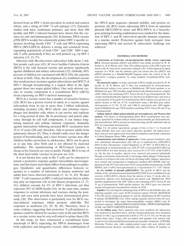
logical response to PPD following the vaccine regimen consist-ing of BCG.HIVA dose response priming and a constantMVA.HIVA booster dose as described in the legend to Fig. 3Aand found PPD-specific cells producing mainly IFN-� andTNF-� at the higher BCG.HIVA doses (Fig. 6A). Upon an M.tuberculosis aerosol challenge, the present BCG vaccine (BCGwild-type 1173 p2), BCG.p, and BCG.HIVA provided equiva-lent levels of protection as demonstrated by a 2-order-of-mag-nitude decrease in M. tuberculosis loads in both lungs andspleens (Fig. 6B). Thus, at least this murine experiment sug-gests that the safer lysine auxotroph BCG.HIVA can replacethe neonatal BCG vaccine against M. tuberculosis without los-
ing the benefits of the BCG vaccination and that, therefore, itsuse will not compromise or interfere with any other scheduledpediatric vaccination.
DISCUSSION
Here, we have engineered a novel candidate vaccine for bothHIV-1 and M. tuberculosis infection that is vectored by a lysineauxotroph of M. bovis BCG (the Pasteur �lysA5::res strain),which expresses the HIV-1 clade A-derived immunogenHIVA. We demonstrated with BALB/c mice that BCG.HIVAcan both prime novel and boost preexisting HIV-1-specific
FIG. 4. Immunogenicity of the pTHr.HIVA DNA priming-BCG.HIVA boosting regimen for CD8� T cells. (A) Mice were left unimmunizedor primed with 106 CFU of BCG.HIVA or BCG.p or 100 �g of pTHr.HIVA DNA, groups 5 and 6 were given booster doses of 106 CFU of BCG.por BCG.HIVA, and then all groups were challenged with 4 � 106 PFU of WR.HIVA. (B) The top panels provide examples of dot blots for theanalysis of bifunctional CD8� T cells as generated for group 6 and epitope H. The bottom panels summarize the data obtained for each vaccinationgroup by using the H (top) and P (bottom) epitopes. For the IFN-�/CD107a/b and TNF-�/CD107a/b analyses, the frequencies of nondegranulating(empty bars) and degranulating (full bars) cells producing cytokine are shown. For the IFN-�/TNF-� analysis, average frequencies correspondingto dot blot quadrants I, II, and III are plotted. Data are presented as means SD (n, 4 to 5 mice). (C and D) Analyses of trifunctionalvaccine-elicited T cells. The two left panels indicate the gating. The right panels give the frequencies of trifunctional cells corresponding to theupper right quadrants for individual mice (circles) and groups (bars; values are means for the groups) as obtained with the H (top) and P (bottom)epitopes. Frequencies are expressed as percentages of CD8� IFN-�� (C) and CD8� TNF-�� (D) cells.
9414 IM ET AL. J. VIROL.

cellular immune responses, which are mediated by CD4� andCD8� T cells of high quality as judged by their long-termpersistence and capacity to proliferate and produce multiplecytokines upon antigenic reexposure. Furthermore, sequentialheterologous immunization involving pTHr.HIVA DNA andBCG.HIVA conferred complete protection of mice against asurrogate challenge with vaccinia virus expressing the HIVAimmunogen, while BCG.HIVA alone provided a level of pro-tection against M. tuberculosis challenge comparable to thatachieved by a presently used BCG vaccine strain.
Here, we constructed rBCG stably expressing the immuno-gen HIVA from both the episomal pJH222.HIVA and theintegrative pJH223.HIVA plasmids. To express the HIVA pro-tein, both plasmids used the �-antigen promoter and a 19-kDalipoprotein signal sequence, which were found to be optimal
for antigen secretion and immunogenicity (10), perhapsthrough the acylation of the signal sequence, which was im-portant for entering into the MHC class I presentation andinteraction with Toll-like receptor 2 (36). Based on higherlevels of HIVA expression by pJH222.HIVA, we chose tofurther characterize the episomal construct. The same plasmidbackbones in a strain of M. smegmatis, a rapidly replicatingmycobacterium expressing the HIV-1 gp120 envelope glyco-protein, were recently compared (8, 53). Despite higher levelsof gp120 expression from the episomal plasmid than from theintegrated plasmid, the levels of immunogenicity in BALB/cmice were identical.
To date, BCG-vectored vaccines against a number of infec-tious agents, such as Leishmania species, Borrelia burgdorferi,Streptococcus pneumoniae, Bordetella pertussis, malaria para-
FIG. 5. Induction of high-quality HIV-1-specific CD4� T cells and complete protection against surrogate virus challenge. The mice and thetreatment groups (1 through 6) were the same as those described in the legend to Fig. 4A. (A) The leftmost panels summarize the data obtainedfor each cytokine and vaccination group. Data are presented as means SD (n, 4 to 5 mice). The middle panels demonstrate the gating for IFN-�-,TNF-�-, and IL-2-producing CD4� T cells as generated by group 6 for a cocktail of three MHC class II epitopes. The rightmost panels give theupper-right-quadrant data for trifunctional HIV-1-specific CD4� T cells from individual mice (circles) and groups (bars). Data are presented asmeans SD (n, 4 to 5 mice). (B) Mice were either left naıve (1) or vaccinated with BCG.p (2), BCG.HIVA (3), pTHr.HIVA DNA (4),pTHr.HIVA DNA and BCG.p (5), or pTHr.HIVA and BCG.HIVA (6) and challenged with WR.HIVA. The WR.HIVA loads in ovaries weredetermined 4 days later. Data for individual mice (circles) and group means (bars; n, 4 to 5 mice) are shown.
VOL. 81, 2007 INFANT VACCINE AGAINST TUBERCULOSIS AND HIV-1 9415

sites, cottontail rabbit papillomavirus, measles virus, and in-deed, HIV and simian immunodeficiency virus (SIV), havebeen successfully constructed (23). Many of these vaccinesshow immunogenicity and protection in murine models, whilesome are also immunogenic in nonhuman primates (3, 51, 53).The only volunteer study conducted to date suggested thatrBCG is only moderately efficient in eliciting immune re-sponses to passenger proteins in humans, but this study did notuse a heterologous priming-boosting regimen (13). In fact, onlya small fraction of the above-mentioned animal experimentsused rBCG strains in heterologous priming-boosting regimens.However, when BCG-SIVGag-induced responses were boostedby defective poxvirus-SIVGag, partial protection against apathogenic simian-human immunodeficiency virus challengewas obtained (3). Here, the inclusion of BCG.HIVA in heter-ologous-vaccine protocols consistently improved the fre-quency, quality, and durability of the generated HIV-1-specificresponses. This improvement was reflected by the detection of
the highest fractions of multifunctional HIVA-specific T cellsin the mice that received BCG.HIVA in a heterologous-vac-cine regimen versus the other regimens tested; these responseswere present over 14 weeks after rBCG administration. Also,the frequency and functionality of CD8� T cells specific for thesubdominant epitope P were significantly elevated by the in-clusion of rBCG, suggesting that BCG may be a useful vaccinevector for the induction of specificity, which may be overshad-owed by immunodominance under other circumstances, al-though this possibility will need to be confirmed for otherepitopes and immunogens. Detectable HIV-1-specific CD4�
T-cell responses were found only in the WR.HIVA challengeexperiment but not following immunization with BCG.HIVAalone or BCG.HIVA-MVA.HIVA. These results may simplyreflect the generally lower frequencies of elicited CD4� T cellsthan of their CD8� counterparts or the fact that in the formerexperiment, the CD4� T-cell responses were also boosted bythe vigorously replicating WR.HIVA. Nevertheless, the ability
FIG. 6. Immune responses and protection against M. tuberculosis challenge by BCG.HIVA. (A) The mice and the treatment groups were thesame as those described in the legend to Fig. 3A (immunized mice were primed with increasing doses of BCG.HIVA). Left panels show examplesof bifunctional analyses using PPD as an antigenic stimulus. On the right, data corresponding to dot blot quadrants I, II, and III for eachimmunization group are shown as means SD (n, 4 to 5 mice). (B) Mice were left naıve or immunized subcutaneously in their left hind legs withthe presently used BCG vaccine, a parental BCG, or lysine auxotrophic BCG.HIVA and challenged with inhaled M. tuberculosis. M. tuberculosisloads in lungs (left) and spleens (right) were determined 4 weeks later. Data for individual mice (circles) and geometric means for each group (n,7 to 9 mice) are shown.
9416 IM ET AL. J. VIROL.
to elicit strong, multispecific, and multifunctional CD8� T-cellresponses is likely to be a critical feature for the success of acandidate HIV-1 vaccine.
The BCG.HIVA vaccine candidate in this study was vec-tored by a lysine auxotroph of BCG that carried an E. coli-mycobacterial shuttle plasmid with a lysine A-complementinggene. This inclusion theoretically increases the plasmid stabil-ity in vivo, prevents genetic rearrangements of the heterolo-gous genes, and adds an additional safety feature to the vac-cine. Here, we demonstrated that BCG.HIVA was as efficientin protecting against M. tuberculosis as the presently used BCGwild-type 1173 p2 vaccine in a mouse model. Also, this is thefirst report of an HIV-1 vaccine vectored by an auxotroph ofBCG and the first study using an rBCG priming-rMVA boost-ing protocol as a potential anti-HIV-1 regimen, which com-bines the benefits of both of these promising vaccine vectors.
Protection against M. tuberculosis challenge offers the pos-sibility to use BCG.HIVA in neonates as a substitute for theBCG vaccine in the first month of their lives (35) and, thus, forBCG.HIVA to serve as a dual vaccine against HIV-1 andtuberculosis. The relevance of the “artificial” consensus cladeA HIVA immunogen to the viruses presently circulating inNairobi has been demonstrated with HIV-1-infected and HIV-1-exposed uninfected infants, of whom 38 to 52% made T-cellresponses that recognized the Gag and polyepitope domains ofHIVA (46). The high frequency of exposed uninfected infantswhose T cells recognize HIVA (52%) is encouraging, as itsuggests that exposure to virus through breast milk does notinevitably lead to infection and can prime or boost T-cell im-munity. Thus, the T-cell responses in these vaccinated infantsmay exceed those to the same vaccines in HIV-1-unexposedadults. HIVA’s usefulness was also shown by boosting HIV-1-specific responses in patients on highly active antiretroviraltherapy who were infected with diverse HIV-1 clades (11, 12,37). Therefore, we propose to further develop this rBCG strainas a pediatric vaccine to protect children born to HIV-1-pos-itive mothers against becoming infected with HIV-1 throughbreastfeeding. The regimen would consist of BCG.HIVA prim-ing at birth, followed by boosting with MVA.HIVA at 1 or 2months of age along with the babies’ scheduled Extended Pro-gramme of Immunization. This booster vaccine could be com-bined with MVA.85A to strengthen the tuberculosis protection(34). In addition, the BCG.HIVA-primed responses can bealso expanded by natural exposure to HIV-1 through breastmilk. The evaluation of the safety and immunogenicity of thisregimen in newborn nonhuman primates is the next aim. How-ever, for a more relevant challenge with SIV, an equivalentimmunogen derived from SIV inner proteins will have to beconstructed.
ACKNOWLEDGMENTS
This work was supported by MRC UK, HIVACAT, the Foundationfor Research and Prevention of AIDS in Spain (FIPSE 36338/02),Fundacion Mutua Madrilena de Automoviles (second call for propos-als), and Fundacio BCN SIDA 2002. H.M. is a Wellcome Trust seniorclinical fellow, C.S. is funded by AFTBVAC (the European Commis-sion, the Fifth Framework Programme), and D.T. is supported by theA*Star Graduate Academy of Singapore. T.H. and H.S. are The Jen-ner Institute Investigators.
REFERENCES
1. Adkins, B., C. Leclerc, and S. Marshall-Clarke. 2004. Neonatal adaptiveimmunity comes of age. Nat. Rev. Immunol. 4:553–564.
2. Alter, G., J. M. Malenfant, and M. Altfeld. 2004. CD107a as a functionalmarker for the identification of natural killer cell activity. J. Immunol. Meth-ods 294:15–22.
3. Ami, Y., Y. Izumi, K. Matsuo, K. Someya, M. Kanekiyo, S. Horibata, N.Yoshino, K. Sakai, K. Shinohara, S. Matsumoto, T. Yamada, S. Yamazaki,N. Yamamoto, and M. Honda. 2005. Priming-boosting vaccination with re-combinant Mycobacterium bovis bacillus Calmette-Guerin and a nonreplicat-ing vaccinia virus recombinant leads to long-lasting and effective immunity.J. Virol. 79:12871–12879.
4. Andersson, G. E., and P. M. Sharp. 1996. Codon usage in the Mycobacte-rium tuberculosis complex. Microbiology 142:915–925.
5. Andre, S., B. Seed, J. Eberle, W. Schraut, A. Bultmann, and J. Haas. 1998.Increased immune response elicited by DNA vaccination with a syntheticgp120 sequence with optimized codon usage. J. Virol. 72:1497–1503.
6. Burton, D. R., R. C. Desrosiers, R. W. Doms, W. C. Koff, P. D. Kwong, J. P.Moore, G. J. Nabel, J. Sodroski, I. A. Wilson, and R. T. Wyatt. 2004. HIVvaccine design and the neutralizing antibody problem. Nat. Immunol. 5:233–236.
7. Buseyne, F., M. Burgard, J. P. Teglas, E. Bui, C. Rouzioux, M. J. Mayaux, S.Blanche, and Y. Riviere. 1998. Early HIV-specific cytotoxic T lymphocytesand disease progression in children born to HIV-infected mothers. AIDSRes. Hum. Retrovir. 14:1435–1444.
8. Cayabyab, M. J., A. H. Hovav, T. Hsu, G. R. Krivulka, M. A. Lifton, D. A.Gorgone, G. J. Fennelly, B. F. Haynes, W. R. Jacobs, Jr., and N. L. Letvin.2006. Generation of CD8� T-cell responses by a recombinant nonpathogenicMycobacterium smegmatis vaccine vector expressing human immunodefi-ciency virus type 1 Env. J. Virol. 80:1645–1652.
9. Colditz, G. A., T. F. Brewer, C. S. Berkey, M. E. Wilson, E. Burdick, H. V.Fineberg, and F. Mosteller. 1994. Efficacy of BCG vaccine in the preventionof tuberculosis. Meta-analysis of the published literature. JAMA 271:698–702.
10. Dennehy, M., and A. L. Williamson. 2005. Factors influencing the immuneresponse to foreign antigen expressed in recombinant BCG vaccines. Vac-cine 23:1209–1224.
11. Dorrell, L., H. Yang, A. K. Iversen, C. Conlon, A. Suttill, M. Lancaster, T.Dong, I. Cebere, A. Edwards, S. Rowland-Jones, T. Hanke, and A. J.McMichael. 2005. Therapeutic immunization of highly active antiretroviraltherapy-treated HIV-1-infected patients: safety and immunogenicity of anHIV-1 gag/poly-epitope DNA vaccine. AIDS 19:1321–1323.
12. Dorrell, L., H. Yang, B. Ondondo, T. Dong, K. di Gleria, A. Suttill, C.Conlon, D. Brown, P. Williams, P. Bowness, N. Goonetilleke, T. Rostron, S.Rowland-Jones, T. Hanke, and A. J. McMichael. 2006. Expansion and di-versification of virus-specific T cells following immunization of human im-munodeficiency virus type 1 (HIV-1)-infected individuals with a recombinantmodified vaccinia virus Ankara/HIV-1 Gag vaccine. J. Virol. 80:4705–4716.
13. Edelman, R., K. Palmer, K. G. Russ, H. P. Secrest, J. A. Becker, S. A.Bodison, J. G. Perry, A. R. Sills, A. G. Barbour, C. J. Luke, M. S. Hanson,C. K. Stover, J. E. Burlein, G. P. Bansal, E. M. Connor, and S. Koenig. 1999.Safety and immunogenicity of recombinant bacille Calmette-Guerin (rBCG)expressing Borrelia burgdorferi outer-surface protein A (OspA) lipoproteinin adult volunteers: a candidate Lyme disease vaccine. Vaccine 17:904–919.
14. Gheorghiu, M., P. H. Lagrange, and C. Fillastre. 1988. The stability andimmunogenicity of a dispersed-grown freeze-dried Pasteur BCG vaccine.J. Biol. Stand. 16:15–26.
15. Goonetilleke, N., S. Moore, L. Dally, N. Winstone, N. Mahmoud, I. Cebere,S. Pinheiro, G. Gillespie, D. Brown, V. Loach, J. Roberts, A. Guimaraes-Walker, P. Hayes, K. Loughran, C. Smith, P. Fast, L. Dorrell, T. Hanke, andA. McMichael. 2006. Induction of multifunctional human immunodeficiencyvirus type 1 (HIV-1)-specific T cells capable of proliferation in healthysubjects by using a prime-boost regimen of DNA- and modified vacciniavirus Ankara-vectored vaccines expressing HIV-1 Gag coupled to CD8�
T-cell epitopes. J. Virol. 80:4717–4728.16. Goulder, P. J., P. Jeena, G. Tudor-Williams, and S. Burchett. 2001. Paedi-
atric HIV infection: correlates of protective immunity and global perspec-tives in prevention and management. Br. Med. Bull. 58:89–108.
17. Hanke, T., and A. J. McMichael. 2000. Design and construction of an ex-perimental HIV-1 vaccine for a year-2000 clinical trial in Kenya. Nat. Med.6:951–955.
18. Hanke, T., A. J. McMichael, and L. Dorrell. 2007. Clinical experience withplasmid DNA- and modified vaccinia vaccine Ankara (MVA)-vectoredHIV-1 clade A vaccine inducing T cells. J. Gen. Virol. 88:1–12.
19. Hermans, I. F., J. D. Silk, J. Yang, M. J. Palmowski, U. Gileadi, C.McCarthy, M. Salio, F. Ronchese, and V. Cerundolo. 2004. The VITALassay: a versatile fluorometric technique for assessing CTL- and NKT-me-diated cytotoxicity against multiple targets in vitro and in vivo. J. Immunol.Methods 285:25–40.
20. Horwitz, M. A., and G. Harth. 2003. A new vaccine against tuberculosis
VOL. 81, 2007 INFANT VACCINE AGAINST TUBERCULOSIS AND HIV-1 9417
affords greater survival after challenge than the current vaccine in the guineapig model of pulmonary tuberculosis. Infect. Immun. 71:1672–1679.
21. Hussey, G. D., M. L. Watkins, E. A. Goddard, S. Gottschalk, E. J. Hughes,K. Iloni, M. A. Kibel, and S. R. Ress. 2002. Neonatal mycobacterial specificcytotoxic T-lymphocyte and cytokine profiles in response to distinct BCGvaccination strategies. Immunology 105:314–324.
22. Im, E.-J., and T. Hanke. 2007. Pre-clinical evaluation of candidate HIV-1vaccines in inbred strains and an outbred stock of mice. AIDS Res. Hum.Retrovir. 33:857–862.
23. Joseph, J., N. Saubi, E. Pezzat, and J. M. Gatell. 2006. Progress towards anHIV vaccine based on recombinant bacillus Calmette-Guerin: failures andchallenges. Exp. Rev. Vaccines 5:827–838.
24. Larke, N., E.-J. Im, R. Wagner, C. Williamson, A.-L. Williamson, A. J.McMichael, and T. Hanke. 2007. Combined single-clade candidate HIV-1vaccines induce T cell responses limited by multiple forms of in vivo immuneinterference. Eur. J. Immunol. 37:566–577.
25. Larke, N., A. Murphy, C. Wirblich, D. Teoh, M. J. Estcourt, A. J. McMichael,P. Roy, and T. Hanke. 2005. Induction of human immunodeficiency virustype 1-specific T cells by a bluetongue virus tubule-vectored vaccine prime-recombinant modified virus Ankara boost regimen. J. Virol. 79:14822–14833.
26. Lotte, A., O. Wasz-Hockert, N. Poisson, N. Dumitrescu, M. Verron, and E.Couvet. 1984. BCG complications. Estimates of the risks among vaccinatedsubjects and statistical analysis of their main characteristics. Adv. Tuberc.Res. 21:107–193.
27. Luzuriaga, K., D. Holmes, A. Hereema, J. Wong, D. L. Panicali, and J. L.Sullivan. 1995. HIV-1-specific cytotoxic T lymphocyte responses in the firstyear of life. J. Immunol. 154:433–443.
28. Marchant, A., V. Appay, M. Van Der Sande, N. Dulphy, C. Liesnard, M.Kidd, S. Kaye, O. Ojuola, G. M. Gillespie, A. L. Vargas Cuero, V. Cerundolo,M. Callan, K. P. McAdam, S. L. Rowland-Jones, C. Donner, A. J. Mc-Michael, and H. Whittle. 2003. Mature CD8(�) T lymphocyte response toviral infection during fetal life. J. Clin. Investig. 111:1747–1755.
29. Marchant, A., T. Goetghebuer, M. O. Ota, I. Wolfe, S. J. Ceesay, D. DeGroote, T. Corrah, S. Bennett, J. Wheeler, K. Huygen, P. Aaby, K. P.McAdam, and M. J. Newport. 1999. Newborns develop a Th1-type immuneresponse to Mycobacterium bovis bacillus Calmette-Guerin vaccination.J. Immunol. 163:2249–2255.
30. Marchant, A., and M. Goldman. 2005. T cell-mediated immune responses inhuman newborns: ready to learn? Clin. Exp. Immunol. 141:10–18.
31. Marchant, A., and M. Newport. 2000. Prevention of infectious diseases byneonatal and early infantile immunization: prospects for the new millen-nium. Curr. Opin. Infect. Dis. 13:241–246.
32. Mbori-Ngacha, D., R. Nduati, G. John, M. Reilly, B. Richardson, A. Mwatha,J. Ndinya-Achola, J. Bwayo, and J. Kreiss. 2001. Morbidity and mortality inbreastfed and formula-fed infants of HIV-1-infected women: a randomizedclinical trial. JAMA 286:2413–2420.
33. McMichael, A. J. 2006. HIV vaccines. Annu. Rev. Immunol. 24:227–255.34. McShane, H., A. A. Pathan, C. R. Sander, S. M. Keating, S. C. Gilbert, K.
Huygen, H. A. Fletcher, and A. V. Hill. 2004. Recombinant modified vacciniavirus Ankara expressing antigen 85A boosts BCG-primed and naturallyacquired antimycobacterial immunity in humans. Nat. Med. 10:1240–1244.
35. Moss, W. J., C. J. Clements, and N. A. Halsey. 2003. Immunization ofchildren at risk of infection with human immunodeficiency virus. Bull.W. H. O. 81:61–70.
36. Neyrolles, O., K. Gould, M. P. Gares, S. Brett, R. Janssen, P. O’Gaora, J. L.Herrmann, M. C. Prevost, E. Perret, J. E. Thole, and D. Young. 2001.Lipoprotein access to MHC class I presentation during infection of murinemacrophages with live mycobacteria. J. Immunol. 166:447–457.
37. Ondondo, B., H. Yang, T. Dong, K. di Gleria, A. Suttill, C. Conlon, D. Brown,P. Williams, P. Bowness, S. L. Rowland-Jones, T. Hanke, A. J. McMichael,and L. Dorrell. 2006. Immunisation with recombinant modified vacciniavirus Ankara expressing HIV-1 gag in HIV-1-infected subjects stimulatesbroad functional CD4� T cell responses. Eur. J. Immunol. 36:2585–2594.
38. Ota, M. O., J. Vekemans, S. E. Schlegel-Haueter, K. Fielding, M. Sanneh, M.
Kidd, M. J. Newport, P. Aaby, H. Whittle, P. H. Lambert, K. P. McAdam,C. A. Siegrist, and A. Marchant. 2002. Influence of Mycobacterium bovisbacillus Calmette-Guerin on antibody and cytokine responses to humanneonatal vaccination. J. Immunol. 168:919–925.
39. Pavelka, M. S., Jr., and W. R. Jacobs, Jr. 1999. Comparison of the construc-tion of unmarked deletion mutations in Mycobacterium smegmatis, Mycobac-terium bovis bacillus Calmette-Guerin, and Mycobacterium tuberculosisH37Rv by allelic exchange. J. Bacteriol. 181:4780–4789.
40. Phillpotts, R. J., T. J. Brooks, and C. S. Cox. 1997. A simple device for theexposure of animals to infectious microorganisms by the airborne route.Epidemiol. Infect. 118:71–75.
41. Pikora, C. A., J. L. Sullivan, D. Panicali, and K. Luzuriaga. 1997. EarlyHIV-1 envelope-specific cytotoxic T lymphocyte responses in vertically in-fected infants. J. Exp. Med. 185:1153–1161.
42. Raviglione, M. C., D. E. Snider, Jr., and A. Kochi. 1995. Global epidemiologyof tuberculosis. Morbidity and mortality of a worldwide epidemic. JAMA273:220–226.
43. Rousseau, C. M., R. W. Nduati, B. A. Richardson, M. S. Steele, G. C.John-Stewart, D. A. Mbori-Ngacha, J. K. Kreiss, and J. Overbaugh. 2003.Longitudinal analysis of human immunodeficiency virus type 1 RNA inbreast milk and of its relationship to infant infection and maternal disease.J. Infect. Dis. 187:741–747.
44. Schneider, J., S. C. Gilbert, T. J. Blanchard, T. Hanke, K. J. Robson, C. M.Hannan, M. Becker, R. Sinden, G. L. Smith, and A. V. S. Hill. 1998. En-hanced immunogenicity for CD8� T cell induction and complete protectiveefficacy of malaria DNA vaccination by boosting with modified vaccinia virusAnkara. Nat. Med. 4:397–402.
45. Siegrist, C. A. 2001. Neonatal and early life vaccinology. Vaccine 19:3331–3346.
46. Slyker, J. A., B. L. Lohman, D. A. Mbori-Ngacha, M. Reilly, E. G. Wee, T.Dong, A. J. McMichael, S. L. Rowland-Jones, T. Hanke, and G. John-Stewart. 2005. Modified vaccinia Ankara expressing HIVA antigen stimu-lates HIV-1-specific CD8 T cells in ELISpot assays of HIV-1 exposed infants.Vaccine 23:4711–4719.
47. Stover, C. K., G. P. Bansal, M. S. Hanson, J. E. Burlein, S. R. Palaszynski,J. F. Young, S. Koenig, D. B. Young, A. Sadziene, and A. G. Barbour. 1993.Protective immunity elicited by recombinant bacille Calmette-Guerin (BCG)expressing outer surface protein A (OspA) lipoprotein: a candidate Lymedisease vaccine. J. Exp. Med. 178:197–209.
48. Takahashi, H., J. Cohen, A. Hosmalin, K. B. Cease, R. Houghten, J. L.Cornette, C. DeLisi, B. Moss, R. N. Germain, and J. A. Berzofsky. 1988. Animmunodominant epitope of the human immunodeficiency virus envelopeglycoprotein gp160 recognized by class I major histocompatibility molecule-restricted murine cytotoxic T lymphocytes. Proc. Natl. Acad. Sci. USA 85:3105–3109.
49. Vekemans, J., A. Amedei, M. O. Ota, M. M. D’Elios, T. Goetghebuer, J.Ismaili, M. J. Newport, G. Del Prete, M. Goldman, K. P. McAdam, and A.Marchant. 2001. Neonatal bacillus Calmette-Guerin vaccination inducesadult-like IFN-gamma production by CD4� T lymphocytes. Eur. J. Immu-nol. 31:1531–1535.
50. World Health Organization. 2001. Outbreak news. Wkly. Epidemiol. Rec.76:33–40.
51. Yasutomi, Y., S. Koenig, S. S. Haun, C. K. Stover, R. K. Jackson, P. Conard,A. J. Conley, E. A. Emini, T. R. Fuerst, and N. L. Letvin. 1993. Immunizationwith recombinant BCG-SIV elicits SIV-specific cytotoxic T lymphocytes inrhesus monkeys. J. Immunol. 150:3101–3107.
52. Young, D., and C. Dye. 2006. The development and impact of tuberculosisvaccines. Cell 124:683–687.
53. Yu, J.-S., J. W. Peacock, S. Vanleeuwen, T. Hsu, W. R. Jacobs, Jr., M. J.Cayabyab, N. L. Letvin, R. Fronthingham, H. F. Staats, H. X. Liao, and B. F.Haynes. 2006. Generation of mucosal anti-human immunodeficiency virustype 1 T-cell responses by recombinant Mycobacterium smegmatis. Clin. Vac-cine Immunol. 13:1204–1211.
9418 IM ET AL. J. VIROL.
Top Related
Copyright © 2022 FDOKUMEN

