


Bahasa
Halaman
Hukum

Use of the Uteroglobin Platform for the Expression of aBivalent Antibody against Oncofetal Fibronectin inEscherichia coliElisa Ventura1, Mattia Riondato2, Gianmario Sambuceti2, Annalisa Salis3, Gianluca Damonte4,
Cinzia Cordazzo5, Huseyin Besir6, Vito Pistoia1, Luciano Zardi5*
1 Laboratory of Oncology, G. Gaslini Institute, Genova, Italy, 2 Department of Health Science, Nuclear Medicine, University of Genoa, IRCCS AOU San Martino - IST, Genova,
Italy, 3 Department of Hearth, Environmental and Life Science, Center for Excellence in Biomedical Research, Genova, Italy, 4 Department of Experimental Medicine and
Center for Excellence in Biomedical Research, Genova, Italy, 5 Sirius-biotech, c/o Advanced Biotechnology Center, Genova, Italy, 6 Protein Expression and Purification Core
Facility, EMBL Heidelberg, Heidelberg, Germany
Abstract
Escherichia coli is a robust, economic and rapid expression system for the production of recombinant therapeutic proteins.However, the expression in bacterial systems of complex molecules such as antibodies and fusion proteins is still affected byseveral drawbacks. We have previously described a procedure based on uteroglobin (UG) for the engineering of very solubleand stable polyvalent and polyspecific fusion proteins in mammalian cells (Ventura et al. 2009. J. Biol. Chem. 284:26646–26654.) Here, we applied the UG platform to achieve the expression in E. coli of a bivalent human recombinant antibody(L19) toward the oncofetal fibronectin (B-FN), a pan-tumor target. Purified bacterial L19-UG was highly soluble, stable, and,in all molecules, the L19 moiety maintained its immunoreactivity. About 50–70% of the molecules were covalenthomodimer, however after refolding with the redox couple reduced-glutathione/oxidized-glutathione (GSH/GSSG), 100% ofmolecules were covalent dimers. Mass spectrometry studies showed that the proteins produced by E. coli and mammaliancells have an identical molecular mass and that both proteins are not glycosylated. L19-UG from bacteria can be freeze-driedwithout any loss of protein and immunoreactivity. In vivo, in tumor-bearing mice, radio-iodinated L19-UG selectivelyaccumulated in neoplastic tissues showing the same performance of L19-UG from mammalian cells. The UG-platform mayrepresent a general procedure for production of various biological therapeutics in E. coli.
Citation: Ventura E, Riondato M, Sambuceti G, Salis A, Damonte G, et al. (2013) Use of the Uteroglobin Platform for the Expression of a Bivalent Antibody againstOncofetal Fibronectin in Escherichia coli. PLoS ONE 8(12): e82878. doi:10.1371/journal.pone.0082878
Editor: Paul D. Riggs, New England BioLabs, United States of America
Received July 31, 2013; Accepted November 6, 2013; Published December 19, 2013
Copyright: � 2013 Ventura et al. This is an open-access article distributed under the terms of the Creative Commons Attribution License, which permitsunrestricted use, distribution, and reproduction in any medium, provided the original author and source are credited.
Funding: This study was partially supported by ‘‘Cinque per mille e Ricerca Corrente, Ministero della Salute ‘‘ from the Italian Ministry of Health. EV wassupported by the ‘‘8 fellowship – Lenino Fontana e Maria Lionello’’ from FIRC (Fondazione Italiana per la Ricerca sul Cancro) and by an ‘‘EMBO Short TermFellowship.’’ CC and MR were supported by post-doctoral fellowships from Regione Liguria-Italy (PO CRO FSE 2007/13). Sirius-biotech S.R.L. financed the article-processing charge. The funders had no role in study design, data collection and analysis, decision to publish, or preparation of the manuscript.
Competing Interests: The authors have read the journal’s policy and have the following conflicts: LZ is CEO of Sirius-biotech a biotechnology stat-up; CC is ascientist of Sirius biotech, a company that markets antibody purification. Sirius-biotech S.R.L. financed the article-processing charge for this study. There are nofurther patents, products in development or marketed products to declare. This does not alter the authors’ adherence to all the PLOS ONE policies on sharing dataand materials, as detailed online in the guide for authors.
* E-mail: [email protected]
Introduction
Fibronectins (FNs) are high molecular mass adhesive glycopro-
teins of the extracellular matrix (ECM). FNs are widely distributed
in normal tissues and body fluids and are involved in several
processes such as cell adhesion and migration, maintenance of
normal cell morphology, cell growth and differentiation [1,2].
Fibronectins are encoded by a single gene localized on chromo-
some 2 [3], but different isoforms arise from the alternative
splicing of the pre-mRNA, a process that for some ECM proteins,
including fibronectin, is modulated by cytokines and extracellular/
intracellular pH [4–6].
B-FN is a FN isoform containing the extra-domain B (ED-B), a
complete type III homology repeat of 91 amino acids in which
exon usage or skipping leads to inclusion or exclusion within the
molecule of this type III repeat. B-FN is undetectable in tissues of
healthy adults (with very rare exceptions, such as the female
reproductive system where recurrent tissue remodeling and
angiogenesis processes take place), it is abundant in fetal tissues
and in different types of pathologies including cancer and all
angiogenesis-associated pathologies [7–12]. The demonstration
that murine monoclonal antibodies to B-FN injected into tumor-
bearing mice selectively accumulate in neoplastic lesions [13]
prompted the generation of high-affinity human recombinant
antibodies for therapeutic and diagnostic purposes [14–21].
L19 is a human high-affinity recombinant antibody that reacts
with the ED-B domain [15]. Because the ED-B domain shares
100% homology in human and mouse, L19 recognizes both
human and murine B-FN. Several studies have demonstrated that
L19 can be used to selectively deliver radionuclides or toxic agents
to tumors both in diagnostic and therapeutic applications. L19 is
presently used in phase I/II clinical trials for therapy of both solid
and hematologic malignancies as a radio-immunoconjugate or as a
fusion protein with the cytokines TNF-alpha and IL-2 [17,22–25].
PLOS ONE | www.plosone.org 1 December 2013 | Volume 8 | Issue 12 | e82878
L19 has been produced in various formats including dimeric
scFv (scFv2) in E. coli, small immunoprotein (SIP) and complete
IgG1 in mammalian cells, and these formats show diverse
pharmacokinetic properties [19]. SIP, produced in CHO cells, is
the currently used format for radioimmuno-therapeutic purposes
because it represents the best compromise of in vivo stability,
blood clearance and performance in tumor targeting [19]. In
particular the performance in tumor targeting of L19 scFv was
very poor since it was unstable giving formation of aggregates
and losing its immunoreactivity few hours after injection [19].
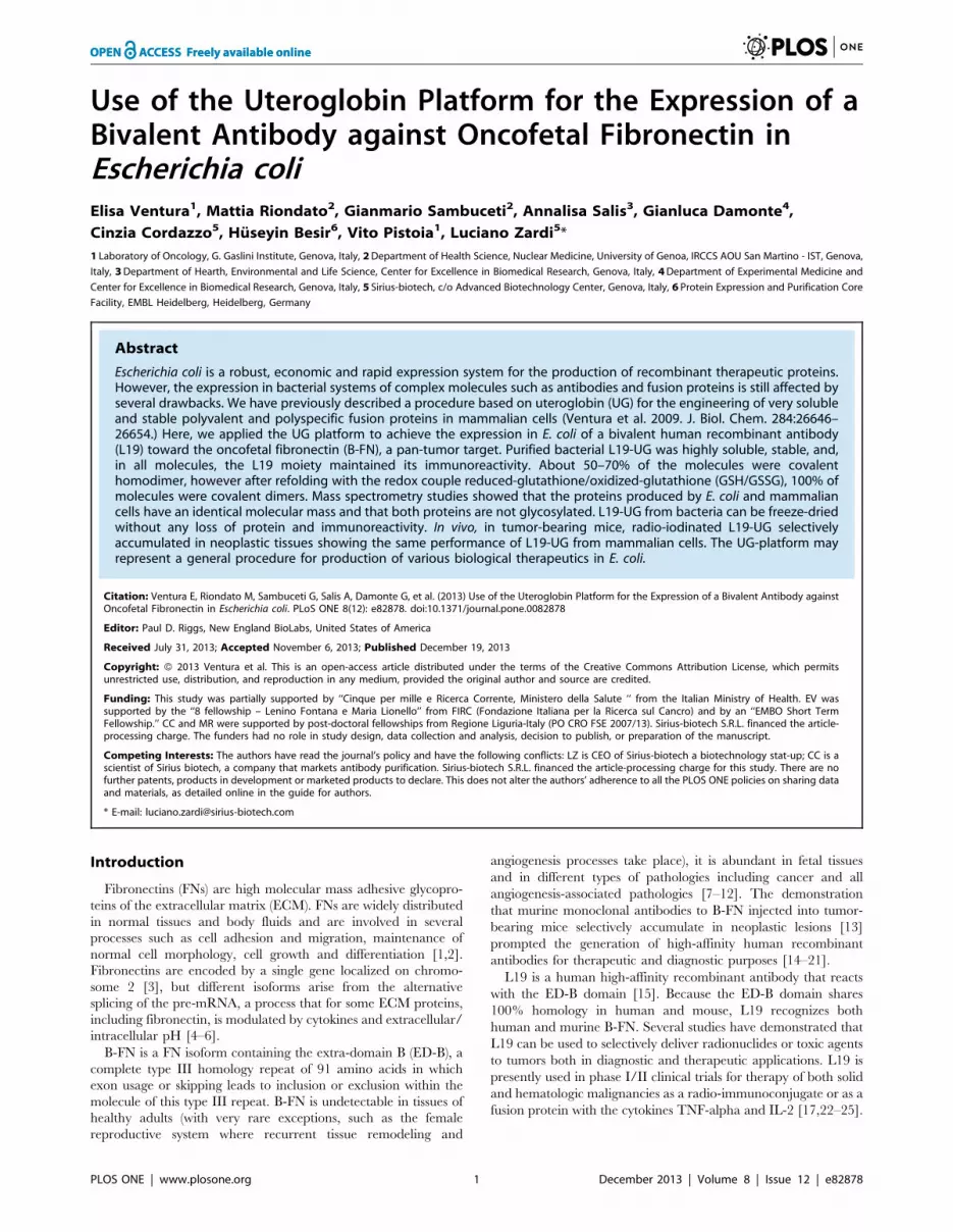
We recently described a novel strategy for the generation of
divalent and dual-specific tetravalent antibodies based on the use
of uteroglobin (UG) [26,27]. UG is a seventy-amino acids globular
and non-glycosylated homodimeric secreted protein [28]. The UG
monomer is organized into a secondary structure containing four
alpha helices; two subunits are then joined in an antiparallel
fashion by disulfide bridges established between two highly
conserved cysteine residues in the amino and carboxyl termini
[28]. The high solubility and stability of UG to variations in pH
and temperature, its resistance to proteases and its homodimeric
structure make UG an ideal linker for the generation of polyvalent
and either monospecific or bispecific recombinant antibodies. The
UG platform (Fig. 1) consists of the fusion of the recombinant
antibody sequence at the amino terminal or alternatively at the
carboxyl terminal or both the amino and carboxyl terminals of
UG; the covalent dimerization of UG allows the dimerization of
the fusion proteins and thus the generation of divalent or dual
specific-tetravalent molecules, which, when compared with similar
fusion proteins without UG, possess enhanced solubility and
stability, factors that would improve their storage and clinical use
[26]. L19-UG is very soluble and stable and has a better
performance with respect to the SIP for in vivo accumulation in
neoplastic tissues in tumor-bearing mice [26]. However, until now,
both the SIP and UG formats of L19 have been produced in
mammalian cells. Their expression and purification from bacteria
would be beneficial because the production of recombinant
therapeutic proteins from E. coli offers several advantages over
mammalian cells including higher yields, faster and simpler
growth, lower costs and easier scale up processes [29]. In fact,
numerous efforts have been made to produce complex molecules
in bacteria, in particular a procedure for isolating full-length
antibodies from libraries expressed in E. coli has been described
[30].
Here we report the expression, purification and characterization
both in vitro and in vivo of L19-UG from E. coli demonstrating the
possibility of using the UG platform for the production of complex
therapeutic fusion proteins in bacterial systems.
Materials and Methods
All experiments involving animals were reviewed and approved
by the Ethical Committee of the National Cancer research
Institute’s Animal Facility and in compliance with the current
National and International guidelines of FELASA, and designated
by the Italian Ministry of Health with Ministerial Decree D.M.S.
nu 146/2009-A and subsequent integration, project nu 282.
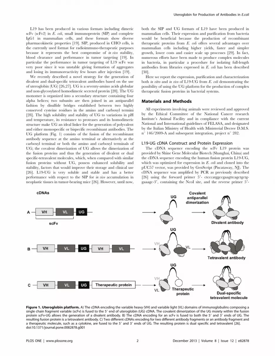
L19-UG cDNA Construct and Protein ExpressionThe cDNA sequence encoding the scFv L19 protein was
provided by Shine Gene Molecular Biotech (Shanghai, China) and
the cDNA sequence encoding the human fusion protein L19-UG,
which was optimized for expression in E. coli and cloned into the
pUC57 vector, was provided by GenScript (Piscataway, NJ). The
cDNA sequence was amplified by PCR as previously described
[26] using the forward primer 59- ctcccatggccgaagttcagctgctg-
gaaagc-39, containing the NcoI site, and the reverse primer 59-
Figure 1. Uteroglobin platform. A) The cDNA encoding the variable heavy (VH) and variable light (VL) domains of immunoglobulins composing asingle chain fragment variable (scFv) is fused to the 59 end of uteroglobin (UG) cDNA. The covalent dimerization of the UG moiety within the fusionprotein scFv-UG allows the generation of a divalent antibody. B) The cDNA encoding for an scFv is fused to both the 59 and 39 ends of UG. Theresulting fusion protein is a tetravalent antibody. C) Two different cDNAs encoding for two different antibody fragments or an antibody fragment anda therapeutic molecule, such as a cytokine, are fused to the 59 and 39 ends of UG. The resulting protein is dual specific and tetravalent [26].doi:10.1371/journal.pone.0082878.g001
Uteroglobin for Production of Antibodies in E.coli
PLOS ONE | www.plosone.org 2 December 2013 | Volume 8 | Issue 12 | e82878
ctcgcggccgcttagttgcacaggctgct-39, containing a stop codon and the
NotI site. The cDNA of L19-UG and the pHEN-1 expression
vector [31] were both digested by NcoI/NotI, ligated and used to
transform DH5a bacteria as previously described [26]. The cDNA
construct was extracted and purified from DH5a, sequenced on
both strands and used to transform the TG-1 and HB2151 E. coli
strains.
L19-UG was expressed in E. coli HB2151 or TG-1 strains
transformed with the pHEN-1 vector containing the cDNA
construct of L19-UG. Bacteria was grown at 37uC with shaking in
2xYT broth (MP Biomedical, Santa Ana, CA) supplemented with
1% D-glucose and 100 mg/ml ampicillin (Sigma, St. Louis, MO).
This overnight culture was diluted 1:100 to prepare a culture in
2xYT broth supplemented with 0.1% D-glucose and 100 mg/ml
ampicillin. Bacteria were incubated at 37uC until the culture
reached an A600 value of 0.6, at which point it was incubated at
18uC for 30 minutes. Expression of the protein was then induced
by adding isopropyl-b-D-thiogalactopyranoside (IPTG) (Inalco,
Italy) at a final concentration of 0.2 mM and by incubating at
18uC for 72 hours. Triton X-100 (Sigma) was also added at the
final concentration of 1% (v/v).
Immunoaffinity, Anion Exchange and Size ExclusionChromatography
After removing bacterial cells by centrifugation at 52006g for 1
hour at 4uC using a Avanti J-25 centrifuge (Beckman Coulter,
Brea, CA), the bacteria culture broth was first passed on a pre-
column of Sepharose-4B and then loaded on an immunoaffinity
chromatography column of recombinant ED-B conjugated to
Sepharose-4B (GE Healthcare, Waukesha, WI). The column was
washed with 3 column volumes of phosphate buffered saline (PBS,
20 mM NaH2PO4, 150 mM NaCl, pH = 7.6) followed by 2
column volumes of 20 mM NaH2PO4 (pH = 7.6) containing 1 M
NaCl and then 2 column volumes of PBS. The protein was then
eluted in 10 mM tri-ethilamine (pH = 11.0) and dialyzed in a
solution containing 20 mM Tris/HCl and 28 mM NaCl
(pH = 8.2).
Anion exchange chromatography was conducted using a
HiTrap DEAE FF column (GE Healthcare) connected to an
AKTA Basic system (GE Healthcare). L19-UG in a solution of
20 mM Tris/HCl and 28 mM NaCl (pH = 8.2) was loaded on the
column, which was previously equilibrated with the same buffer,
and then eluted with a solution containing 20 mM Tris/HCl and
90 mM NaCl (pH = 8.2).
Figure 2. L19-UG from E. coli. A) Schematic of the L19-UG cDNA cloned into the pHEN-1 prokaryotic expression vector and fused to the pelBleader sequence. Depicted on the right is a schematic representation of the resulting dimeric fusion protein. B) SDS-PAGE analysis of L19-UG undernon-reducing conditions after the first (lane 2) and the second purification steps (lane 3). L19-UG after protein refolding under non-reducing (lane 4)and reducing conditions (lane 5). Lane 1 shows the molecular mass standards. C) Size-exclusion chromatography (Superdex200 column) profiles ofL19-UG purified from CHO cells (left panel) and from E. coli (central panel). The column retention volumes are 15.18 ml for the protein purified fromCHO cells and 15.14 ml for the protein purified from E. coli. In the right panel the SEC profile of L19-UG obtained from E. coli after proteinlyophilization and reconstitution. On the right the SDS-PAGE analysis of L19-UG, under non-reducing conditions, before (lane 1) and after (lane 2)protein lyophilization and reconstitution.doi:10.1371/journal.pone.0082878.g002
Uteroglobin for Production of Antibodies in E.coli
PLOS ONE | www.plosone.org 3 December 2013 | Volume 8 | Issue 12 | e82878
Size exclusion chromatography (SEC) was carried out using a
Superdex200 column (GE Healthcare) equilibrated in PBS
(pH = 7.6) and the AKTA Basic System.
Protein RefoldingProtein refolding was performed by adding to the protein in
solution in PBS, 1 mM GSH and 0. 2 mM GSSG (final pH , 8)
and by incubating at 4uC for 24 hours. Refolded L19-UG was
than separated from glutathione by SEC on a Superdex 200
column, equilibrated in PBS.
Sodium Dodecyl Sulfate-polyacrylamide GelElectrophoresis (SDS-PAGE), Enzyme-linkedImmunosorbent Assay (ELISA) and Recombinant FNFragments
4–12% SDS-PAGE gradient analysis in reducing and non-
reducing conditions was carried out as previously described [19].
The 7.ED-B.8.9, ED-B and B-8 recombinant fragments of FN
were prepared as described previously [32].
ELISA was performed as previously described [32]. Briefly,
different concentrations of purified protein were tested against the
fibronectin recombinant fragment 7.ED-B.8.9. Bound L19-UG
was revealed by using a rabbit polyclonal antibody to human
uteroglobin (produced in our laboratory). A peroxidase-conjugated
anti-rabbit IgG was used as the tertiary antibody (Pierce,
Rockford, IL).
Mass SpectrometryThe molecular mass of L19-UG was measured using an Agilent
1100 HPLC system coupled to a MSD Ion Trap XCT mass
spectrometer, equipped with an electrospray ion source (HPLC-
ESI-MS) (Agilent Technologies, Palo Alto, CA, USA). To evaluate
the presence of essential disulfide bridges the samples were
analyzed in the presence or in the absence of the disulfide reducing
agent dithiothreitol (DTT). The reduction was conducted with
150 mM DTT at room temperature for 5 hours. Separations were
performed on a Symmetry C4 column 16150 mm with 3-mm
particle size (Waters Corporation, Milford, MA, USA).
Eluents used were water and acetonitrile added with 0.1%
formic acid. The gradient employed was: 10% acetonitrile for
10 min, then linear to 95% in 50 min. The flow rate was set to
30 ml/min and the column temperature was set at 25uC. Injection
volume was 5 ml. Ions were detected in ion charged control with a
target ions value of 50000 and an accumulation time of 300 ms,
using the following operation parameters: capillary voltage:
2500V; nebulizer pressure: 25 psi; drying gas: 8 l/min; dry
temperature: 325uC; rolling averages 3, averages 8. Mass spectra
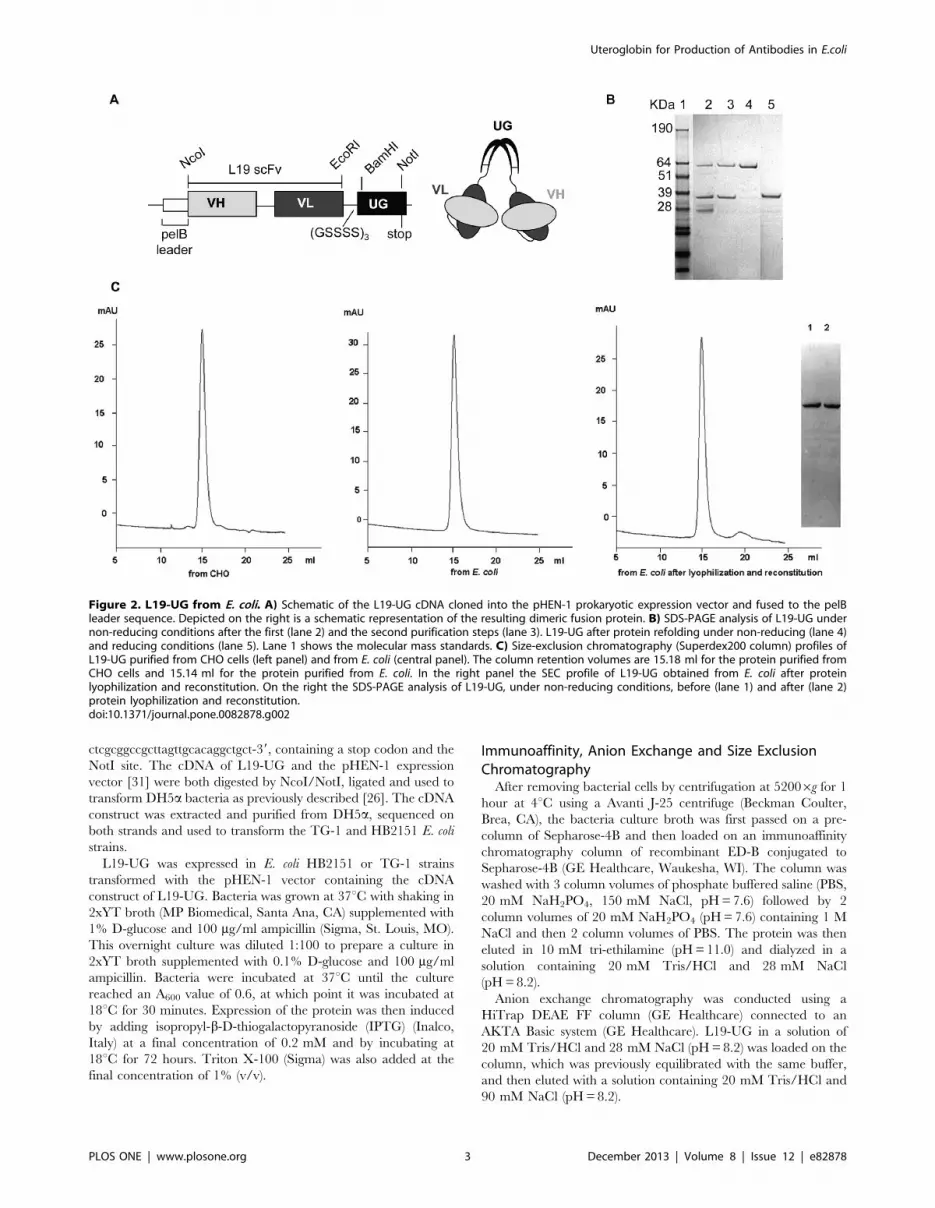
Figure 3. Mass spectrometry analysis of L19-UG from mammalian cells and E.coli. Mass spectrometry analysis of reduced L19-UG obtainedfrom CHO cells (A, C) and from E. coli (B, D). The raw (A–B) and deconvoluted (C–D) mass spectra relative to the chromatographic peaks at 32.5minutes are reported. The calculated average neutral mass of monomeric form of L19-UG is 34670,3 Da for the protein produced in CHO cells and34670,6 Da for the protein produced in E. coli.doi:10.1371/journal.pone.0082878.g003
Uteroglobin for Production of Antibodies in E.coli
PLOS ONE | www.plosone.org 4 December 2013 | Volume 8 | Issue 12 | e82878
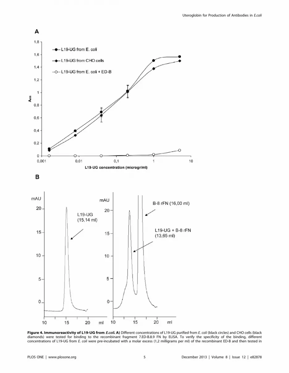
Figure 4. Immunoreactivity of L19-UG from E.coli. A) Different concentrations of L19-UG purified from E. coli (black circles) and CHO cells (blackdiamonds) were tested for binding to the recombinant fragment 7.ED-B.8.9 FN by ELISA. To verify the specificity of the binding, differentconcentrations of L19-UG from E. coli were pre-incubated with a molar excess (1,2 milligrams per ml) of the recombinant ED-B and then tested in
Uteroglobin for Production of Antibodies in E.coli
PLOS ONE | www.plosone.org 5 December 2013 | Volume 8 | Issue 12 | e82878
were acquired in the positive ion mode in the 800–2000 m/z mass
range. Raw spectra were deconvoluted using the LC/MSD Trap
Software revision 5.3.
Cell Lines, Immunohistochemistry (IHC) andImmunofluorescence (IF)
Human melanoma derived cells SK-MEL-28, human SV40
transformed WI38VA fibroblasts and mouse embryonic terato-
carcinoma cells F9 were purchased from ATCC (Rockville, MD)
and cultured in DMEM (Sigma) supplemented with 10% FCS
(Sigma) and 4 mM L-glutamine (Biochrom, Berlin, Germany). For
IF studies, cells were fixed with ice cold (220uC) acetone (Sigma).
For immunohistochemical and immunofluorescence experi-
ments, we used cultured human SV40 transformed WI38VA
fibroblasts and 5-mm cryostat sections of the human melanoma
SK-MEL-28 subcutaneously grown in NOD-SCID mice. IHC
was performed as previously described [10] using biotinylated
L19-UG at a final concentration of 2 mg/ml. For L19-UG
biotinylation, we used the reagent 2X-AH-BIOTIN-NHS (Biospa,
Milano, Italy) following the manufacturer’s instructions. For IF we
used L19-UG at a concentration of 1 mg/ml and a rabbit
polyclonal anti-human uteroglobin antibody (Abcam, Cambridge,
UK) as the secondary antibody. A Cy3-conjugated goat anti-rabbit
IgG (H+L) antibody (Jackson ImmunoResearch, West Grove, PA)
was used as the tertiary antibody. The sections were counter-
stained with DAPI (Invitrogen, Carlsbad, CA). Images were
acquired using a Nikon Digital Sight DS-5Mc camera mounted on
an Olympus BX5 I microscope and the Nikon imaging software
NIS-Elements F.
Radio-labelling of L19-UG with 125I and BiodistributionExperiments
L19-UG was radio-labeled with 125I by using the IODO-GEN
method (Pierce, Rockford, IL) as previously described [19]. The
immunoreactivity of the radiolabeled protein was determined as
previously reported [19]. 125I –L19-UG was analyzed by SEC, by
loading about 10 mCi of radio-labeled protein on a Superdex 200
column equilibrated in PBS and counting the eluted fractions with
ELISA for its binding to 7.ED-B.8.9 (white circles). The mean absorbances at l= 405 nm 6 SD are indicated. B) A shift in the column retention volumeof L19-UG in SEC (Superdex 200) was obtained after incubating L19-UG with a molar excess of the recombinant FN fragment B-8. Left panel: SECprofile of L19-UG showing a single peak at 15.14 ml. Right panel: SEC profile of L19-UG pre-incubated with a molar excess of B-8 rFN showing anelution peak at 13.65 ml, which corresponds to the immunocomplex L19-UG/B-8 rFN, and an elution peak at 16 ml, which corresponds to the excessunbound B-8 rFN.doi:10.1371/journal.pone.0082878.g004
Figure 5. Immunohistochemistry and immunofluorescence experiments. A) IHC analysis of human melanoma SK-MEL-28 specimens grownsubcutaneously in NOD-SCID mice. L19-UG purified from E. coli (red) detects tumor-associated vessels. B–C) IF analysis of SK-MEL-28 specimens: L19-UG from E. coli (red) detects fibrillar structures of the tumor extracellular matrix (B) and vessels (C). D–E) IF analysis of cultured human SV40transformed WI38VA fibroblasts stained with L19-UG purified from E. coli (D) and CHO cells (E). L19-UG detects the typical FN fibrillar structures of theextracellular matrix. Nuclei were stained with DAPI. Bars: 5 mm.doi:10.1371/journal.pone.0082878.g005
Uteroglobin for Production of Antibodies in E.coli
PLOS ONE | www.plosone.org 6 December 2013 | Volume 8 | Issue 12 | e82878
a gamma-counter (Cobra, Packard/Perkin Elmer, Waltham,
USA). The radiochemical purity (RCP) of 125I –L19-UG was
assessed by Instant Thin Layer Chromatography (ITLC). A 20 cm
long ITLC SG (Gelman Sciences, Ann Arbor, MI) and 85%
methanol in water were used as stationary and mobile phases,
respectively. After a run of 15 cm, the stationary phase was used to
expose a super resolution Type SR autoradiography plate
(Packard/PerkinElmer) for 30 minutes. Data were acquired by
using Cyclon (Perkin Elmer).
For biodistribution experiments male NOD-SCID mice were
subcutaneously implanted with 3,56106 F9 cells in 100 ml of PBS.
When tumors reached the volume of approximately 0.1–0.3 cm3
(determined using the formula (d)2 6D60.52 where d and D are
the short and long dimensions respectively, determined using a
calliper), animals were treated intravenously with 4,5 mCi of 125I –
L19-UG. To block nonspecific accumulation of 125I in the
stomach and concentration in thyroid, mice were given orally
20 mg of sodium perchlorate (Carlo Erba, Milano, Italy) in water,
30 minutes before injection of the radio-labeled antibody. Animals
were dived into three groups of three mice each and sacrificed 3
hours and 30 minutes, 24 and 48 hours post injection. Tumors and
organs were excised, weighed and counted in a gamma counter
(Perkin Elmer). The accumulation of the radio-labeled protein in
the different organs is expressed as the percentage of the injected
dose per gram of tissue (%ID/g).
NOD-SCID mice were provided by the IRCCS AOU San
Martino – IST Animal Facility (Genova, Italy). All procedures
involving animals were performed under the supervision and
approval of the Ethical Committee of the National Cancer
Research Institute’s Animal Facility and in compliance with the
current national and international guidelines of FELASA, and
designated by the Italian Ministry of Health with Ministerial
Decree D.M.S. nu 146/2009-A and subsequent integrations,
project nu 282.
Results
Protein Expression, Purification, Characterization andRefolding
For the expression of L19-UG, we used the prokaryotic
expression vector pHEN-1, which includes the bacterial PelB
signal sequence for protein export into the periplasm (Fig. 2A). We
used the HB2151 and TG-1 E. coli strains as expression host strains
and obtained similar yield of L19-UG with similar solubility,
stability and immunoreactivity from both strains. We cultivated
bacteria under different temperatures (from 30uC to 10uC),
inducer concentrations (from 0.01 to 1 mM), incubation times
(from 16 to 72 hours) and additive amounts. We found that the
best conditions for L19-UG expression were as follows: 0.2 mM
IPTG, induction temperature of 18uC, incubation time of 72
hours and supplementation with 1% (v/v) Triton-X100. Under
these conditions, the release of L19-UG from the periplasm into
the culture broth was higher than 95%, while without Triton-
X100 the release from the periplasm into the culture media was
about 50%.
We purified L19-UG from the bacteria culture broth in two
steps. In the first step, the protein was purified by immunoaffinity
chromatography with ED-B conjugated to Sepharose-4B as
described in ‘‘Materials and Methods.’’ This purification produced
a yield of approximately 5–6 mg of protein per liter of bacteria
culture broth. L19-UG was than extensively dialyzed against a
solution of 20 mM Tris/HCl and 28 mM NaCl (pH = 8.2). Fig. 2B
shows the SDS-PAGE of L19-UG obtained after the first
purification step. Under non-reducing conditions, L19-UG
migrates both as a monomer and a dimer with apparent molecular
mass of about 35 kDa and 64 kDa, respectively. Since different
contaminating bands of lower molecular mass were also present,
L19-UG was then loaded on an anion exchange chromatography
HiTrap DEAE FF resin that was previously equilibrated in a
solution of 20 mM Tris/HCl and 28 mM NaCl (pH = 8.2). Under
these conditions, contaminants did not bind to the resin, while
L19-UG was retained by the resin. The protein was than eluted
using 90 mM NaCl in 20 mM Tris/HCl (pH = 8.2). As shown by
the SDS-PAGE analysis (Fig. 2B), this second step of purification
allowed the separation of L19-UG from contaminants.
Since purified L19-UG from E. coli was only 50–70% a covalent
dimer, we treated the protein with the redox couple GSH/GSSG
to achieve 100% covalent dimerization. As shown in Fig. 2B, after
incubation at 4uC for 24 hours in presence of 1 mM GSH and
0.2 mM GSSG, L19-UG became 100% covalent dimer, migrating
in SDS-PAGE under non-reducing conditions as a single band
with an apparent molecular mass of 64 KDa. In reducing
condition the protein migrated as a single band with an apparent
molecular mass of about 35 KDa, as expected (Fig. 2B). Identical
bands under reducing and non-reducing conditions were observed
for L19-UG purified from CHO cells [26]. SEC of the purified
and refolded L19-UG shows a single peak with a retention volume
of approximately 15 ml, which is in accordance with the molecular
mass of dimeric L19-UG and which coincides with the retention
volume obtained with the L19-UG purified from CHO cells
(Fig. 2C).
L19-UG, in solution in PBS, was lyophilized and then
reconstituted with distilled water without any protein loss. As
demonstrated by the SEC and SDS-PAGE analysis, neither
protein aggregation nor degradation were observed after lyoph-
ilization and reconstitution (Fig. 2C). On the contrary L19-SIP, as
well as the L19-scFv from E. coli, has much lower solubility than
L19-UG from E.coli and can not be reconstituted after lyophili-
zation without aggregation and precipitation of protein [26].
Table 1. Biodistribution experiments of 125I-L19-UG in F9tumor-bearing mice.
ORGAN 3 hr 30 min 24 hr 48 hr
Blood 11,1762,39 0,5360,31 0,1360,11
Tumor 14,2362,12 8,4062,15 11,5262,38
Liver 4,0661,21 0,1960,09 0,0860,06
Spleen 4,5761,79 0,2960,17 0,1660.20
Heart 4,5761,6 0,2260,12 0,0760,06
Lung 7,9260,72 0,6460,31 0,7860,66
Kidney 7,1261,52 0,4060,17 0,1860,14
Bone 2,2360,21 0,4760,34 0,2360,19
Stomach 1,8060,23 0,6160,34 0,2760,25
Small intestine 3,0460,60 0,6360,10 0,1860,07
Large intestine 2,0560,71 0,4860,21 0,2560,13
Bladder 10,6366,25 1,7860,63 0, 8360,23
Muscle 0,9960,19 0,2160,24 0,0560,05
Testis 3,1460,11 0,2060,08 0,1060,1
The percentage of the injected dose per gram of tissue (%ID/g) 6 SD in thedifferent organs, at the indicated time post 125I-L19-UG i.v. administration, arereported.doi:10.1371/journal.pone.0082878.t001
Uteroglobin for Production of Antibodies in E.coli
PLOS ONE | www.plosone.org 7 December 2013 | Volume 8 | Issue 12 | e82878
The molecular mass of reduced L19-UG measured by mass
spectrometry was of 34670.6 Da for L19-UG from E. coli and of
34670.3 Da for L19-UG from CHO cells (Fig. 3). The measured
molecular mass coincides with the theoretical one, 34670.6 Da,
calculated on the basis of aminoacid composition. The MS data
indicate that: 1. the leader peptide is correctly removed from the
fusion protein produced in E. coli during the secretion process and
that no proteolytic degradation has occurred; 2) the protein
obtained from CHO cells is not glycosylated.
To test the immunoreactivity of the L19 moiety, different
concentrations of L19-UG were used in ELISA against the human
recombinant FN fragment containing the type III repeats 7, ED-B,
8 and 9 and compared with L19-UG that was purified from
mammalian CHO cells. Fig. 4A shows that the proteins purified
from E. coli and from CHO cells gave identical results, which
demonstrates identical immunoreactivity and avidity of the L19
moiety in both proteins. The reaction was abolished by pre-
incubating L19-UG with a large excess of purified ED-B, which
further demonstrates the specificity of the reaction. To test the
immunoreactivity in solution, we incubated L19-UG with a seven-
fold molar excess of a recombinant FN fragments composed of
ED-B and 8 type III repeats (B-8 rFN), and then analyzed them by
SEC. The peak of L19-UG completely shifted from approximately
15 ml to approximately 13.7 ml (Fig. 4B), indicating a complete
binding of L19-UG to the rFN fragment B-8, thus demonstrating
that 100% of the molecules were immunoreactive. Identical
results, both in ELISA and SEC, were obtained by testing L19-UG
before the refolding step (data not shown). This demonstrates that
the protein obtained from E. coli, even if not 100% covalently
dimer, was 100% immunoreactive.
L19-UG was used in immunohistochemistry and immunofluo-
rescence on cultured transformed human fibroblasts (WI38VA)
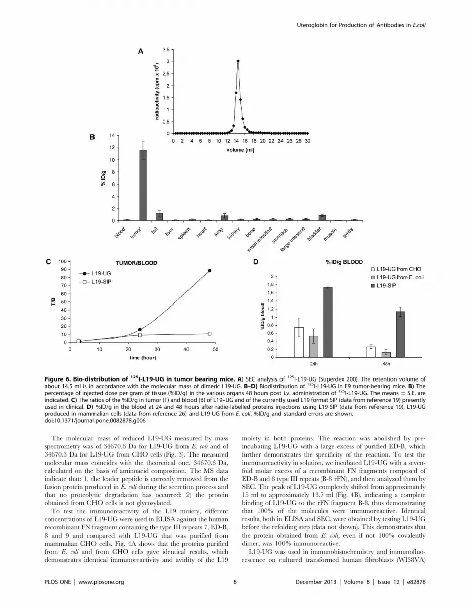
Figure 6. Bio-distribution of 125I-L19-UG in tumor bearing mice. A) SEC analysis of 125I-L19-UG (Superdex 200). The retention volume ofabout 14.5 ml is in accordance with the molecular mass of dimeric L19-UG. B–D) Biodistribution of 125I-L19-UG in F9 tumor-bearing mice. B) Thepercentage of injected dose per gram of tissue (%ID/g) in the various organs 48 hours post i.v. administration of 125I-L19-UG. The means 6 S.E. areindicated. C) The ratios of the %ID/g in tumor (T) and blood (B) of L19–UG and of the currently used L19 format SIP (data from reference 19) presentlyused in clinical. D) %ID/g in the blood at 24 and 48 hours after radio-labelled proteins injections using L19-SIP (data from reference 19), L19-UGproduced in mammalian cells (data from reference 26) and L19-UG from E. coli. %ID/g and standard errors are shown.doi:10.1371/journal.pone.0082878.g006
Uteroglobin for Production of Antibodies in E.coli
PLOS ONE | www.plosone.org 8 December 2013 | Volume 8 | Issue 12 | e82878
and specimens of human melanoma SK-MEL-28 grown subcu-
taneously in NOD-SCID mice. L19-UG detected tumoral vessels
as expected (Fig. 5A–C). Equivalent amounts of L19-UG obtained
from E. coli or CHO gave the same reaction patterns, typical of B-
FN (Fig. 5D–E).
Biodistribution in Tumor-bearing MiceL19-UG was radio-labeled with Iodine 125. After radio-
iodination L19-UG was 95% immunoreactive. The SEC analysis
of 125I-L19-UG shows a single peak with a column retention
volume (14,5 ml), which is in accordance with the molecular mass
of L19-UG, and the absence of any aggregates and proteolytic
fragments (Fig. 6A). The RCP, determined by ITLC, one hour
after radiolabeling, was higher than 95%. 125I-L19-UG was tested
in bioistribution experiments in F9 teratocarcinoma tumor-
bearing mice. As shown in Fig. 6B–C and in Table 1, L19-UG
selectively accumulated in the tumors, reaching a percentage of
the injected dose per gram of tissue (%ID/g) of about 12% 48
hours post-injection (Fig. 6B and Table 1). The radio labeled
molecule was cleared quickly from the blood, showing a %ID/g in
the blood at 24 and 48 from injection of 0,53% and 0,13%,
respectively (Fig. 6D and Table 1). Forty-eight hours post-injection
the %ID/g in the tumor was 88 fold higher than the %ID/g in
blood (T/B) while the T/B was about twelve employing the format
currently in use in clinical trials, L19 SIP [19] (Fig. 6C and 6D).
Discussion
Recombinant antibodies represent one of the most prominent
drugs for imaging and for therapy of cancer and immune
disorders. They include antibodies targeting antigens on cancer
cells as well as antigens associated with cancer ECM and
vasculature. Antigens associated with the tumor ECM and vessels
offer several advantages with respect to cell surface antigens
because they usually are more homogenous, more abundant, more
stable, more accessible from the blood stream and they are shared
by different types of malignancies [33].
B-FN is an oncofetal FN isoform [7,8] and is the prototype
ECM target arising from the deregulation of alternative splicing.
In fact, the altered alternative splicing of many different pre-
mRNA in several types of neoplastic and non-neoplastic diseases is
recognized as a general phenomenon in which the derived
products are considered important therapeutic and diagnostic
targets [34].
The demonstration that B-FN is a cancer- and angiogenesis-
associated ECM component and that murine monoclonal
antibodies against B-FN that are injected i.v. into tumor-bearing
mice selectively accumulate on neoplastic tissues, prompted the
production of human recombinant antibodies from human scFv
libraries [14]. One of these human recombinant antibodies, L19
[15], is currently used by Philogen SPA for various diagnostic and
therapeutic clinical trials as a radio-immunoconjugate or as an
immunocytokine in different tumor types [17,21–25]. For radio
immunotherapy of hematologic cancers, L19 is used in the radio
iodinated small immunoprotein (SIP) format [17,22].
We previously described the UG platform that allows the
generation of very soluble and stable recombinant polyvalent/
polyspecific antibodies in mammalian cells [26]. Here we
demonstrate that this procedure allows the production of the
antibody L19 in the UG format in E. coli. The L19-UG molecule
produced in E. coli is very soluble and stable and maintains
identical immunoreactivity and avidity compared to L19-UG
produced from mammalian cells. The mechanisms for which UG
increases the solubility of the fusion proteins are under investiga-
tion. Further, we demonstrated that the molecules expressed by
mammalian cells and by E. coli have identical molecular mass and
are not glycosylated.
Only 50–70% of L19-UG molecules from E. coli is a covalent
dimer, even if 100% of L19-UG molecules is immunoreactive.
However treating the purified protein with the redox couple
GSH/GSSG is possible to obtain 100% of covalent dimer. After
refolding the protein obtained from E. coli is identical to the
molecule produced in CHO cells.
In tumor-bearing mice L19-UG from E. coli selectively
accumulated in neoplastic tissues and showed a fast blood
clearance. The biodistribution properties of L19-UG reported
here confirm the previously obtained results with the protein
expressed in mammalian cells. L19-UG performs better in vivo
than the currently used SIP format in terms of accumulation in
neoplastic lesions and blood clearance. In fact, in the murine
tumor model F9, forty-eight hours post-injection the %ID/g in the
tumor was 88 fold higher than the %ID/g in blood while it was
about twelve employing L19-SIP, the format currently in use in
clinical trials [19] (Fig. 6C and 6D). We are presently investigating
the reason why fusion proteins containing UG have a faster
clearance with respect to proteins not containing UG. Fast blood
clearance is a very important drug property for radio-immuno-
therapy applications as it reduces the radio-toxicity on non-target
organs. Moreover UG has anti-inflammatory properties and this
could be helpful to contrast some radio-immunotherapy side
effects such as inflammation and fibrosis.
In addition we have demonstrated that it is possible to lyophilize
and reconstitute the protein without any protein precipitation and
aggregation. This property, which is not shared by the antibody
L19 in the SIP and scFv formats, can facilitate the storage and
consequently the potential clinical use of L19-UG. In fact L19-
SIP, as well as L19-scFv, after lyophilization and reconstitution
presents significant amount of aggregates and precipitates [26].
The procedure for the production of L19-UG described here
presents many advantages with respect to the production from
mammalian cells. In fact, in general, recombinant protein
production in E. coli is faster and cheaper than in mammalian
systems. The cost for bacterial cell medium is at least 90% lower
than for mammalian cell medium. Fermentation takes 24 to 72
hours in bacteria versus 14 days to 3 weeks in CHO cells.
Furthermore, we have engineered a cDNA construct and
described a procedure that induces the release of a correctly
folded, soluble, and active recombinant protein directly into the
extracellular fermentation broth, which allows simple and easily
scalable GMP purification procedures. L19-scFv has been
produced in E. coli and after purification it was made up of tow
forms, monomer and a non-covalent dimer, a second step of
purification was required to isolate the latter dimeric form [19].
When radio-labeled and tested in vivo in F9 tumor bearing mice
the performance in tumor targeting of L19-scFv was very low, 3.2
and 2.8%ID/g at 24 and 48 hours after injection respectively,
about three-four times lower with respect to the %ID/g obtained
using L19-UG produced in E. coli [19]. Furthermore Borsi et al.
reported that L19-scFv was unstable giving formation of aggre-
gates and quickly loses its immunoreactivity few hours after
injection in tumor-bearing mice [19].
In conclusion, the two main points reported here are 1) the
possibility of utilizing bacteria to produce dimeric recombinant
antibodies for tumor targeting and 2) that the UG platform could
be useful for the production of complex therapeutic proteins in
bacterial systems.
Uteroglobin for Production of Antibodies in E.coli
PLOS ONE | www.plosone.org 9 December 2013 | Volume 8 | Issue 12 | e82878
Author Contributions
Conceived and designed the experiments: EV HB LZ. Performed the
experiments: EV CC MR AS. Analyzed the data: EV GS GD VP HB LZ.
Contributed reagents/materials/analysis tools: MR AS GD VP HB. Wrote
the paper: EV LZ.
References
1. Hynes RO (1990) Fibronectins. New York: Springer-Verlag.
2. Pankov R, Yamada KM (2002) Fibronectin at a glance. J Cell Sci 115: 3861–
3863.
3. Zardi L, Cianfriglia M, Balza E, Carnemolla B, Siri A, et al. (1982) Species-
specific monoclonal antibodies in the assignment of the gene for human
fibronectin to chromosome 2. EMBO J 1: 929–933.
4. Balza E, Borsi L, Allemanni G, Zardi L (1988) Transforming growth factor beta
regulates the levels of different fibronectin isoforms in normal human cultured
fibroblasts. FEBS Lett 228: 42–44.
5. Borsi L, Balza E, Gaggero B, Allemanni G, Zardi L (1995) The alternative
splicing pattern of the tenascin-C pre-mRNA is controlled by the extracellular
pH. J Biol Chem 270: 6243–6245.
6. Borsi L, Balza E, Castellani P, Carnemolla B, Ponassi M, et al. (1994) Cell-cycle
dependent alternative splicing of the tenascin primary transcript. Cell Adhes
Commun 1: 307–317.
7. Zardi L, Carnemolla B, Siri A, Petersen TE, Paolella G, et al. (1987)
Transformed human cells produce a new fibronectin isoform by preferential
alternative splicing of a previously unobserved exon. EMBO J 6: 2337–2342.
8. Carnemolla B, Balza E, Siri A, Zardi L, Nicotra MR, et al. (1989) A tumor-
associated fibronectin isoform generated by alternative splicing of messenger
RNA precursors. J Cell Biol 108: 1139–1148.
9. Carnemolla B, Leprini A, Allemanni G, Saginati M, Zardi L (1992) The
inclusion of the type III repeat ED-B in the fibronectin molecule generates
conformational modifications that unmask a cryptic sequence. J Biol Chem 267:
24689–24692.
10. Castellani P, Viale G, Dorcaratto A, Nicolo G, Kaczmarek J, et al. (1994) The
fibronectin isoform containing the ED-B oncofetal domain: a marker of
angiogenesis. Int J Cancer 59: 612–618.
11. Kosmehl H, Berndt A, Katenkamp D (1996) Molecular variants of fibronectin
and laminin: structure, physiological occurrence and histopathological aspects.
Virchows Arch 429: 311–322.
12. Castellani P, Borsi L, Carnemolla B, Biro A, Dorcaratto A, et al. (2002)
Differentiation between high- and low-grade astrocytoma using a human
recombinant antibody to the extra domain-B of fibronectin. Am J Pathol 161:
1695–1700.
13. Mariani G, Lasku A, Balza E, Gaggero B, Motta C, et al. (1997) Tumor
targeting potential of the monoclonal antibody BC-1 against oncofetal
fibronectin in nude mice bearing human tumor implants. Cancer 80: 2378–
2384.
14. Carnemolla B, Neri D, Castellani P, Leprini A, Neri G, et al. (1996) Phage
antibodies with pan-species recognition of the oncofoetal angiogenesis marker
fibronectin ED-B domain. Int J Cancer 68: 397–405.
15. Pini A, Viti F, Santucci A, Carnemolla B, Zardi L, et al. (1998) Design and use of
a phage display library. Human antibodies with subnanomolar affinity against a
marker of angiogenesis eluted from a two-dimensional gel. J Biol Chem 273:
21769–21776.
16. Neri D, Bicknell R (2005) Tumor vascular targeting. Nat Rev Cancer 5: 436–
446.
17. Sauer S, Erba PA, Petrini M, Menrad A, Giovannoni G, et al. (2009) Expression
of the oncofetal ED-B-containing fibronectin isoform in hematologic tumors
enables ED-B-targeted 131I-L19SIP radioimmunotherapy in Hodgkin lympho-
ma patients. Blood 113: 2265–2274.
18. Carnemolla B, Borsi L, Balza E, Castellani P, Meazza R, et al. (2002)
Enhancement of the antitumor properties of interleukin-2 by its targeted deliveryto the tumor blood vessel extracellular matrix. Blood 99: 1659–1665.
19. Borsi L, Balza E, Bestagno M, Castellani P, Carnemolla B, et al. (2002) Selectivetargeting of tumoral vasculature: comparison of different formats of an antibody
(L19) to the ED-B domain of fibronectin. Int J Cancer 102: 75–85.
20. Borsi L, Balza E, Carnemolla B, Sassi F, Castellani P, et al. (2003) Selectivetargeted delivery of TNFalpha to tumor blood vessels. Blood 102: 4384–4392.
21. Santimaria M, Moscatelli G, Viale GL, Giovannoni L, Neri G, et al. (2003)Immunoscintigraphic detection of the ED-B domain of fibronectin, a marker of
angiogenesis, in patients with cancer. Clin Cancer Res 9: 571–579.
22. Erba PA, Sollini M, Orciuolo E, Traino C, Petrini M, et al. (2012)Radioimmunotherapy with radretumab in patients with relapsed hematologic
malignancies. J Nucl Med 53: 922–927.23. Papadia F, Basso V, Patuzzo R, Maurichi A, Di Florio A, et al. (2013) Isolated
limb perfusion with the tumor-targeting human monoclonal antibody-cytokinefusion protein L19-TNF plus melphalan and mild hyperthermia in patients with
locally advanced extremity melanoma. Journal of surgical oncology 107: 173–
179.24. Johannsen M, Spitaleri G, Curigliano G, Roigas J, Weikert S, et al. (2010) The
tumor-targeting human L19-IL2 immunocytokine: preclinical safety studies,phase I clinical trial in patients with solid tumors and expansion into patients
with advanced renal cell carcinoma. Eur J Cancer, 46: 2926–2935.
25. Eigentler TK, Weide B, de Braud F, Spitaleri G, Romanini A, et al. (2011) Adose-escalation and signal-generating study of the immunocytokine L19-IL2 in
combination with dacarbazine for the therapy of patients with metastaticmelanoma. Clin Cancer Res 17: 7732–7742.
26. Ventura E, Sassi F, Fossati S, Parodi A, Blalock W, et al. (2009) Use of
uteroglobin for the engineering of polyvalent, polyspecific fusion proteins. J BiolChem 284: 26646–26654.
27. Ventura E, Balza E, Borsi L, Tutolo G, Carnemolla B, et al. (2011) Selectivetargeted delivery of the TNF-alpha receptor p75 and uteroglobin to the
vasculature of inflamed tissues: a preliminary report. BMC biotechnology 11:104.
28. Mukherjee AB, Zhang Z, Chilton BS (2007) Uteroglobin: a steroid-inducible
immunomodulatory protein that founded the Secretoglobin superfamily. EndocrRev 28: 707–725.
29. Huang CJ, Lin H, Yang X (2012) Industrial production of recombinanttherapeutics in Escherichia coli and its recent advancements. J Ind Microbiol
Biotechnol 39: 383–399.
30. Mazor Y, Van Blarcom T, Mabry R, Iverson BL, Georgiou G (2007) Isolation ofengineered, full-length antibodies from libraries expressed in Escherichia Coli. Nat
Biotechnol 25(5): 563–565.31. Hoogenboom HR, Griffiths AD, Johnson KS, Chiswell DJ, Hudson P, et al.
(1991) Multi-subunit proteins on the surface of filamentous phage: methodol-ogies for displaying antibody (Fab) heavy and light chains. Nucleic Acids Res 19:
4133–4137.
32. Balza E, Sassi F, Ventura E, Parodi A, Fossati S, et al. (2009) A novel humanfibronectin cryptic sequence unmasked by the insertion of the angiogenesis-
associated extra type III domain B. Int J Cancer 125: 751–758.33. Zardi L, Neri D (1998) Affinity reagents against tumor-associated extracellular
molecules and newforming vessels. Advanced drug delivery reviews 31: 43–52.
34. Miura K, Fujibuchi W, Unno M (2012) Splice isoforms as therapeutic targets forcolorectal cancer. Carcinogenesis 33: 2311–2319.
Uteroglobin for Production of Antibodies in E.coli
PLOS ONE | www.plosone.org 10 December 2013 | Volume 8 | Issue 12 | e82878
Top Related
Copyright © 2022 FDOKUMEN

