


Bahasa
Halaman
Hukum

Tubulin Tyrosination Is Required for the ProperOrganization and Pathfinding of the Growth ConeSeverine Marcos1,2, Julie Moreau1,2., Stephanie Backer1,2., Didier Job3, Annie Andrieux3, Evelyne Bloch-
Gallego1,2*
1 Institut Cochin, Universite Paris Descartes, CNRS (UMR 8104), Paris, France, 2 Inserm, U567, Departement Genetique et Developpement, Paris, France, 3 Grenoble Institut
des Neurosciences, Centre de Recherche Inserm U.836 – UJF-CEA-CHU, Batiment Edmond J. Safra, Universite Joseph Fourier, Site Sante a La Tronche, Grenoble, France
Abstract
Background: During development, neuronal growth cones integrate diffusible and contact guidance cues that areconveyed to both actin and microtubule (MT) cytoskeletons and ensure axon outgrowth and pathfinding. Although severalpost-translational modifications of tubulin have been identified and despite their strong conservation among species, theirphysiological roles during development, especially in the nervous sytem, are still poorly understood.
Methodology/Findings: Here, we have dissected the role of a post-translational modification of the last amino acid of the a-tubulin on axonal growth by analyzing the phenotype of precerebellar neurons in Tubulin tyrosin ligase knock-out mice(TTL2/2) through in vivo, ex vivo and in vitro analyses. TTL2/2 neurons are devoid of tyrosinated tubulin. Their pathwayshows defects in vivo, ex vivo, in hindbrains open-book preparations or in vitro, in a collagen matrix. Their axons still orienttoward tropic cues, but they emit supernumerary branches and their growth cones are enlarged and exhibit an emission ofmis-oriented filopodia. Further analysis of the TTL2/2 growth cone intracellular organization also reveals that the respectivelocalization of actin and MT filaments is disturbed, with a decrease in the distal accumulation of Myosin IIB, as well as aconcomitant Rac1 over-activation in the hindbrain. Pharmacological inhibition of Rac1 over-activation in TTL2/2 neuronscan rescue Myosin IIB localization.
Conclusions/Significance: In the growth cone, we propose that tubulin tyrosination takes part in the relative arrangementof actin and MT cytoskeletons, in the regulation of small GTPases activity, and consequently, in the proper morphogenesis,organization and pathfinding of the growth cone during development.
Citation: Marcos S, Moreau J, Backer S, Job D, Andrieux A, et al. (2009) Tubulin Tyrosination Is Required for the Proper Organization and Pathfinding of theGrowth Cone. PLoS ONE 4(4): e5405. doi:10.1371/journal.pone.0005405
Editor: Michael Hendricks, Harvard University, United States of America
Received September 11, 2008; Accepted March 18, 2009; Published April 30, 2009
Copyright: � 2009 Marcos et al. This is an open-access article distributed under the terms of the Creative Commons Attribution License, which permitsunrestricted use, distribution, and reproduction in any medium, provided the original author and source are credited.
Funding: This work was partially funded by the Association pour la Recherche sur le Cancer (ARC 3697), the Association Francaise pour la Recherche contre lesMyopathies (AFM) and INSERM. The funders have evaluated the projects prior to providing fundings, and had no role in study design, data collection and analysis,decision to publish, or preparation of the manuscript.
Competing Interests: The authors have declared that no competing interests exist.
* E-mail: [email protected]
. These authors contributed equally to this work.
Introduction
During embryonic development, axons have to make choices in
order to reach their target in the nervous system. The neuronal
growth cone is a dynamic structure at the tip of the axon that
provides the traction force necessary for axon outgrowth and acts
as a sensor. It explores the environment by integrating guidance
cues that influence its direction and remodeling. These functions
depend on the cytoskeleton reorganization and dynamics [1,2].
Although the actin cytoskeleton has been proposed as a major
target of guidance cues due to its peripheral location in the growth
cone and its crucial role in extension and retraction of filopodia
and lamellipodia when they encounter tropic cues, the microtu-
bule (MT) cytoskeleton has also been involved in the guiding
process through intracellular signaling pathways [3]. Changes in
the organization of MTs were proposed to occur later in vivo, as a
consequence of formation and disassembly of actin filaments.
However, recent studies have revealed that MTs do not remain
passively bundled in the growth cone center since a dynamic
population of MTs composed of tyrosinated-tubulin is able to
enter the actin-rich peripheral domain (P-domain) of the growth
cone and to grow along actin filaments in filopodia [4].
We have studied the role of tubulin tyrosination, a post-
translational modification of the tubulin that allows, after
elimination of the C-terminal tyrosine of neo-synthesized a-
tubulin by a carboxypeptidase, its re-addition by the tubulin
tyrosine ligase (TTL). This post-translational modification leads to
the presence of tyrosinated-tubulin (tyr-tubulin) at the dynamic
plus-ends of MTs while older and more stable MTs are mainly
composed of detyrosinated tubulin, called glutamylated-tubulin
(glu-tubulin) [5,6]. Despite the absence of functional links between
the tyrosination state of MTs and their dynamics [7], it appears
that dynamic MTs contain tyr-tubulin, whereas bundled MTs in
the central domain of the growth cone (C-domain) are detyrosinated
and more stable. In TTL knock-out mice (TTL2/2), detyrosinated
tubulin is accumulated and mice die perinatally [8]. A previous
PLoS ONE | www.plosone.org 1 April 2009 | Volume 4 | Issue 4 | e5405
analysis of TTL2/2 mice has revealed that tyr-tubulin is
involved in the control of proper neurite extensions of
hippocampal neurons [8]. In addition, the recruitment of
Clip-170, a plus-end tracking protein (+TIP protein) that binds
to MTs, and of other MT +TIP proteins, which have a
cytoskeleton-associated protein glycine-rich (CAP-Gly) MT
binding domain, such as Clip115 and p150 Glued, is impaired
in TTL2/2 mice [9]. Thus, to better understand how the
presence of the C-terminal tyrosine on a-tubulin influences
neurite outgrowth, a deeper analysis of the intracellular
organization of axons during outgrowth was necessary.
Precerebellar nuclei (PCN) provide an interesting model to
study guidance cues. Indeed, PCN neurons, dorsally located in the
rhombic lips, migrate ventrally through a tangential neurophilic
migration [10], emitting first a leading process, the future axon, and
then translocating their cell bodies in the leading process [11]. The
floor plate is a source of both contact molecules and chemotropic
factors, such as Netrin-1 that can influence both axon guidance and
cell bodies migration of various PCN neurons [12,13,14] and act as
an intermediate target for migrating PCN neurons.
Although admitted that axon growth and guidance depend on
well-coordinated cytoskeletal dynamics, the direct characterization
of specific cues remained a challenge. Only recently have small
GTPases - in particular the Rho/Rho Kinase pathway and myosin
II contractility - been altogether involved in regulation of
microtubule behavior during neuronal growth [15]. It has also
been reported that upon Myosin II inhibition, the movement of
actin filaments and MTs immediately stopped and MTs
unbundled in the gowth cone neck [16].
Here, we explore the role of tubulin re-tyrosination on growth
cone pathfinding through the analysis of TTL2/2 PCN neurons
that are deprived of tyr-tubulin. We report that the pathfinding of
TTL2/2 axons is disturbed in the vicinity of the floor plate in vivo.
Ex vivo, in hindbrain open-book preparations, growth cones are
enlarged and exhibit a complex morphology with numerous mis-
oriented filopodia, especially when reaching the floor plate. In
vitro in a collagen matrix, axon outgrowth is decreased, although
still oriented toward a local attractive Netrin-1 source. Supernu-
merary exploring branches also develop all along the axons. In
addition, the cytoskeletal organization in the growth cone is
disrupted since detyrosinated MTs abnormally enter the periph-
eral actin-rich domain and they exhibit a decreased local
recruitment of Myosin IIB. These observations are concomitant
and consistent with an increased activity of Rac1 small GTPase
observed in TTL2/2 hindbrains. The distal accumulation of
Myosin IIB can be significantly rescued after pharmacological
inhibition of Rac1 activity. Altogether, these results suggest that re-
tyrosination at the MTs plus-ends is required for the formation
and intracellular organization of a functional growth cone at the
tip of the axon.
Results
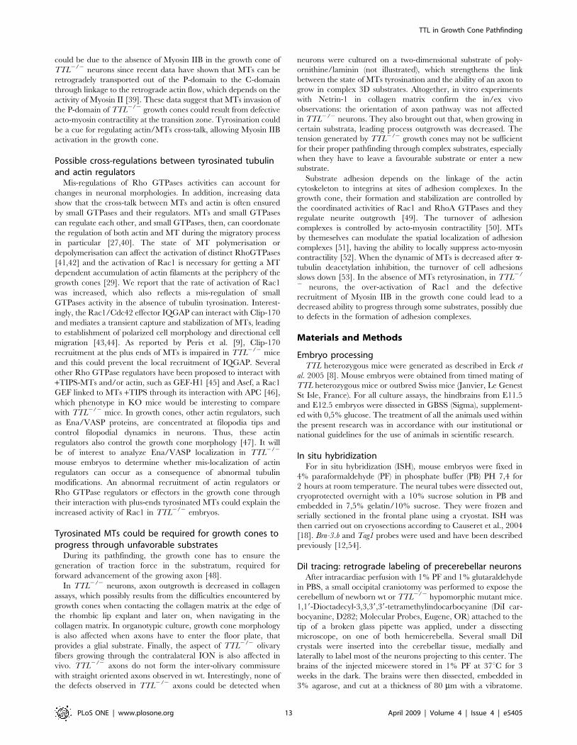
In vivo, the migratory process develops properly in theabsence of TTL, but inferior olivary fibers show anabnormal aspect in the vicinity of the floor plate
Newly synthesized a-tubulin carries a tyrosine residue on its C-
terminal. Then the tyrosine residue is removed by a carboxypep-
tidase and TTL brings an additional tyrosine residue According to
the level of transcription-translation in a cell, the amount in neo-
synthesized tyr-tubulin varies. Thus, in TTL2/2 cells, the amount
of remaining tyr-tubulin varies according to cell types and tissues
and their specific transcription-translation rate [9]. We first
analyzed the tyr-tubulin content in PCN neurons with a specific
antibody that only labels tyrosinated tubulin. In the absence of
retyrosination in TTL2/2 mice, PCN neurons almost lack tyr-
tubulin and mainly contain detyrosinated tubulin, called glutamy-
lated tubulin (glu-tubulin) (supplemental Figure S1). To analyze
the effect of the absence of retyrosination in neurons, we compared
the positioning of PCN cell bodies and axonal projections in wt
and TTL2/2 mice at birth. During development, the growing
axons of all PCN neurons, including those that will form the lateral
reticular nucleus (LRN) as well as the inferior olivary nucleus
(ION), are first attracted toward the floor plate and cross it to
reach their cerebellar target. In contrast, while the cell bodies of
LRN neurons cross the floor plate, those of ION neurons stop
before crossing it and develop an axonal inter-olivary commissure.
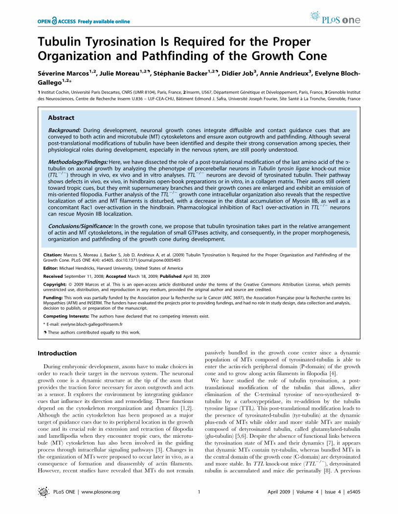
To determine the position of cell bodies, we used a Brn3b
mRNA probe to visualize ION neurons and a Tag1 probe for
neurons of the LRN. No delay was observed in the time ION
neurons needed to get their proper position close to the floor plate
at birth (Fig. 1A, B; n = 3 for each stage). The ION cell bodies
showed their characteristic lamellated organization in both wt
(Fig. 1A) and TTL2/2 mice (Fig. 1B). The LRN cell bodies were
also correctly located laterally to the ION in the absence of TTL
(data not shown).
In parallel, we analyzed the PCN axon development toward
their cerebellar target in vivo. In this purpose, we performed
retrograde tracing of PCN axons after unilateral insertion of a
crystal of DiI in the cerebellum at birth (schema in Fig. 1C). ION
neurons were labeled contralaterally to the injection site, and LRN
neurons ipsilaterally in both wt (n = 8; Fig. 1C) and TTL2/2 mice
(n = 5; Fig. 1D). However, the aspect of post-crossing olivary fibers,
growing through neurons forming the other contralateral ION
mass, showed some defects in TTL2/2 mice. Instead of them
being all perpendicularly oriented to the floor plate as in the wt
(Fig. 1C and 1E at higher magnification), the olivary fibers seemed
to navigate around the cells to leave the ION masses on the
opposite side and to lose their straight orientation in TTL2/2
hindbrains when located in the vicinity of the floor plate (Fig. 1D
and 1F at higher magnification). The ION phenotype is the same
in all cases, excluding the hypothesis of variable penetrance.
These results suggest that, in vivo, the axon pathway is impaired
when axons from TTL2/2 embryos navigate in the vicinity of their
intermediate target, the floor plate, that is a source of both
diffusible and contact cues [11] during ION axon outgrowth and
neuronal migration. Thus, we decided to further investigate the
responses of TTL2/2 neurons in response to guidance factors
using in vitro assays, focusing on axons pathfinding.
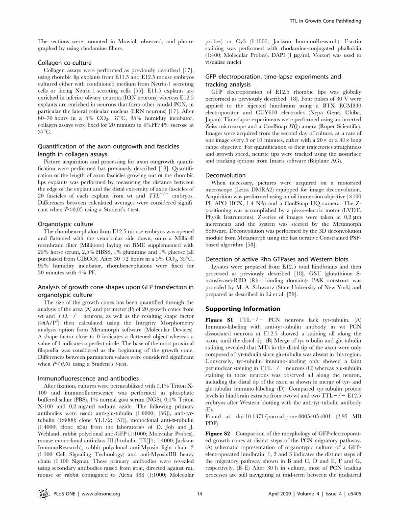
TTL2/2 growth cones show a hypertrophic morphologyin the vicinity of the floor plate in organotypic hindbraincultures
To mimic axon outgrowth in vivo and observe TTL2/2
neurons outgrowth in their physiological environment, we
developed organotypic cultures of hindbrains of E12.5 embryos,
as they only navigate inside the hindbrain as a substrate. To
further compare the aspect of individual axons and growth cones,
a GFP-expression plasmid was electroporated unilaterally at the
rhombic lip (Fig. 2A). To analyze whether the growth speed was
affected by the absence of tyrosination, we fixed GFP-electropo-
rated organotypic cultures after 30, 48 and 72 hours and
confirmed that axon outgrowth could develop ex vivo with a
proper time course and amplitude. After 2DIV, when they
reached the vicinity of the floor plate, wt axons were all oriented
parallel to each other (Fig. 2B) and showed a thin growth cone
with all filopodia oriented straight toward the floor plate (Fig. 2C).
TTL in Growth Cone Pathfinding
PLoS ONE | www.plosone.org 2 April 2009 | Volume 4 | Issue 4 | e5405
On the contrary, in TTL2/2 hindbrains, the trajectory of the axons
was disorganized (Fig. 2D). In addition, at higher magnification, the
growth cone showed a hypertrophic aspect, with randomly oriented
filopodia (Fig. 2E and drawings in Fig. 2F), not at the initiation of
migration but especially when growth cones are in the vicinity of the
floor plate (supplemental Figure S2). Thus, the most obvious
phenotype is observed close to the floor plate, although we cannot
exclude a slighter difference all along the pathway, that would be
hard to detect and evaluate through the present techniques.
To further analyze these morphological changes, we have
quantified the growth cone area and perimeter in wt (n = 20) and
TTL2/2 (n = 20) neurons. Both areas (26 295 a.u. (arbitrary units)
and 38 676 a.u. respectively) and perimeters values (3 825 a.u. and
5 634 a.u. respectively) were significantly increased in TTL2/2
growth cones (P,0,01) (Fig. 2G and H respectively). The resulting
shape factor (see Material and Methods) was 0.15 in wt and 0.08 in
TTL2/2 growth cones (P,0,01) (Fig. 2I), which indicates that the
morphology of TTL2/2 growth cones is more complex than the
wt one. In conclusion, in these physiological conditions, there was
no delay in the leading process outgrowth in TTL2/2 hindbrains,
but the morphology of growth cones appeared to be particularly
affected when they navigate close to the floor plate.
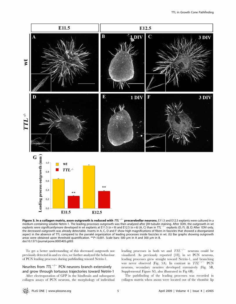
In vitro, TTL2/2 axons that grow in presence of solubleNetrin-1 or toward Netrin-1 in a collagen matrix show adecreased outgrowth
First, we analyzed the possible role of tubulin retyrosination on
leading process outgrowth using collagen assays. Explants of
rhombic lips at E11.5 (n = 9) and E12.5 (n = 6) were first cultured
with conditioned medium from Netrin-1 secreting cells in a
collagen matrix. Netrin-1 was previously shown to promote PCN
axon outgrowth in collagen assays and to be necessary for PCN
nucleokinesis in vitro [17]. Since the leading edges of E11.5
migrating neurons is the future axon and at E12.5, leading
processes have axonal growth cone-like morphology, we will refer
to axons and axon-like as leading processes in quantifications at
E11.5 and E12.5 respectively. When cultured with soluble Netrin-
1, the quantification of the total outgrowth of the leading processes
by TUJ1 immuno-staining and thresholding revealed that their
growth was reduced about 3 times in TTL2/2 explants, after 3
days in vitro (3 DIV) (Fig. 3A, C, D, F and G). In addition, axon-
like fascicles that grew from TTL2/2 explants showed a fuzzy
organization with non parallel leading processes inside the fascicles
(compare high magnifications in wt and TTL2/2 in Fig. 3A, C, D
and F). It is noteworthy that this decrease in leading processes
outgrowth was already observed after 1 DIV (Fig. 3B and E).
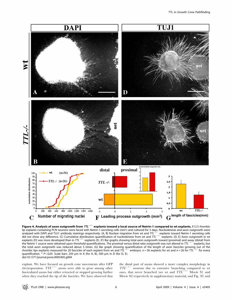
Since Netrin-1 is secreted by the floor plate and because in vivo
and ex vivo defects were observed close to the midline, we tried to
determine whether this alteration was due to a defect in the ability
of TTL2/2 neurons to transduce the Netrin-1 attractive effect. For
that purpose, we faced rhombic lip explants with a local source of
Netrin-1. The rate of nucleokinesis was not significantly affected in
the TTL2/2 explants when compared to the wt ones as illustrated
for E12.5 neurons (Fig. 4A, B, and C). Furthermore, the outgrowth
was similarly reduced in this condition but leading processes still
grew preferentially toward the Netrin-1 source in the proximal
quadrant rather than in the distal quadrant (Fig. 4D, E, and F),
indicating that TTL2/2 neurons can still respond to guidance
cues. These observations were consistant with their globally
normal oriented outgrowth in and ex vivo and the prediction that
there is no defect in guidance. Nevertheless, we observed that
leading processes from TTL2/2 neurons were thicker and shorter
as revealed by individual length measurements (Fig. 3D and F and
Fig. 4E and G) than those growing from wt explants (Fig. 3A, C
and Fig. 4D).
Figure 1. Correct positioning of ION cell bodies but impaired aspect of ION fibers in TTL2/2 newborn mice. (A, B) At birth, a Brn-3bprobe allowed the visualization of the whole ION (Inferior Olivary Nucleus) lamellae in wt (A) and TTL2/2 mice (B) upon ISH. ION rostral lamellae wereproperly developed in TTL2/2 mice. (C–F) Upon unilateral DiI injections in the cerebellum (left side and schema in C) and retrograde PCN labeling atbirth, in wt mice (C), the ION located controlateral to the injection site and the ipsilateral lateral reticular nucleus (LRN) were labeled. Crossing fibers ofthe olivary commissure were visualized ventrally in the olivary region (white arrowhead in E). In TTL2/2 mice, ION neurons were also labeledcontrolaterally to the injection site (D), but showed a disorganization of ION fibers in the ION mass, once they have crossed the floor plate (fp) andlocate ipsilaterally to the injected cerebellum that they reach (white arrowhead in F). LRN was correctly labeled ipsilaterally (D). For each genotype, 4newborn mice were analyzed after DiI injection.doi:10.1371/journal.pone.0005405.g001
TTL in Growth Cone Pathfinding
PLoS ONE | www.plosone.org 3 April 2009 | Volume 4 | Issue 4 | e5405
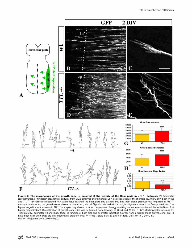
Figure 2. The morphology of the growth cone is impaired at the vicinity of the floor plate in TTL2/2 embryos. (A) Schematicrepresentation of hindbrain organotypic cultures from E12.5 embryos after unilateral GFP-electroporation of the rhombic lip. After 2 DIV, both wt (B)and TTL2/2 (D) GFP-electroporated PCN axons have reached the floor plate (FP, dashed line) but their axonal pathway was impaired in TTL2/2
embryos. In wt axons, the growth cones showed a thin aspect, with all filipodia oriented with a straight alignment toward the floor plate (B and C athigher magnification), whereas in TTL2/2 embryos, they showed a more complex morphology, emitting numerous non-oriented filopodia (D and E athigher magnification). Quantification of growth cones size was performed from drawings of 20 wt and 20 TTL2/2 GFP-electroporated neurons (F).Their area (G), perimeter (H) and shape factor (a function of both area and perimeter indicating how far from a circular shape growth cones are) (I)have been calculated. Data are presented using arbitrary units. ** P,0,01. Scale bars: 30 mm in B (forB, D); 5 mm in C (for C, E).doi:10.1371/journal.pone.0005405.g002
TTL in Growth Cone Pathfinding
PLoS ONE | www.plosone.org 4 April 2009 | Volume 4 | Issue 4 | e5405
To get a better understanding of this decreased outgrowth not
previously detected in and ex vivo, we further analyzed the behaviour
of PCN leading processes during pathfinding toward Netrin-1.
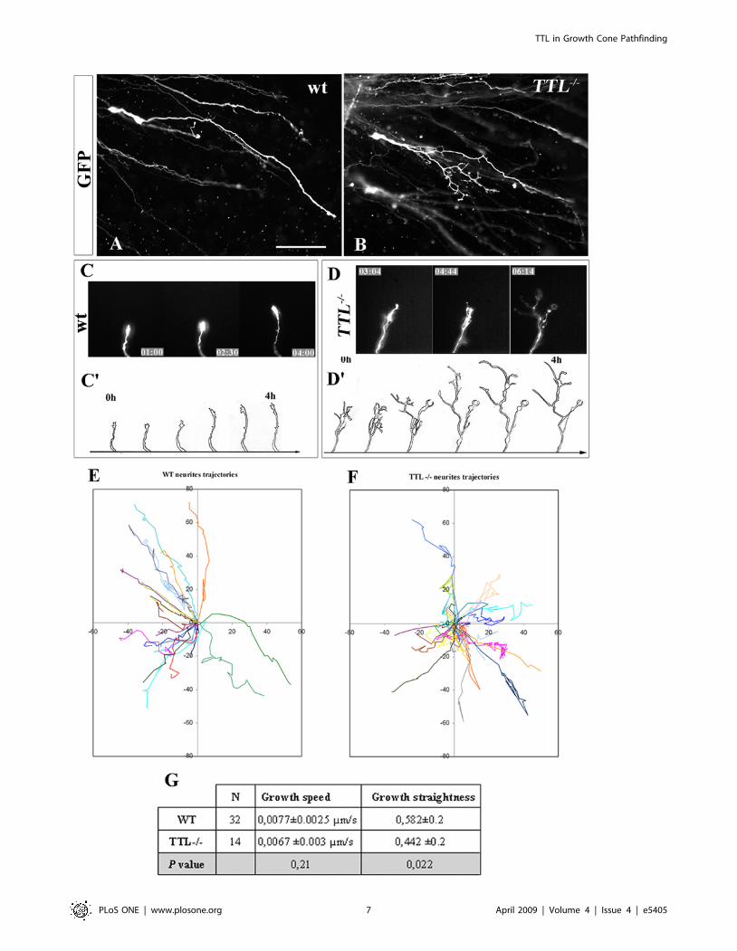
Neurites from TTL2/2 PCN neurons branch extensivelyand grow through tortuous trajectories toward Netrin-1
After electroporation of GFP in the hindbrain and subsequent
collagen assays of PCN neurons, the morphology of individual
leading processes in both wt and TTL2/2 neurons could be
visualized. As previously reported [18], in wt PCN neurons,
leading processes grew straight toward Netrin-1, and branching
was never observed (Fig. 5A). In contrast in TTL2/2 PCN
neurons, secondary neurites developed extensively (Fig. 5B,
Supplemental Figure S3, also illustrated in Fig 6B).
The pathfinding of the leading processes was recorded in
collagen matrix when axons were located out of the rhombic lip
Figure 3. In a collagen matrix, axon outgrowth is reduced with TTL2/2 precerebellar neurons. E11.5 and E12.5 explants were cultured in amedium containing soluble Netrin-1. The leading processes outgrowth was then analyzed after bIII-tubulin staining. After 3DIV, the outgrowth in wtexplants were significantlymore developed in wt explants at E11.5 (n = 9) and E12.5 (n = 6) (A, C) than in TTL2/2 explants (D, F). (B, E) After 1DIV only,the decreased outgrowth was already detectable. Inserts in A, C, D and F show high magnifications of fibers in fascicles that showed a disorganizedaspect in the absence of TTL compared to the parralel organization of leading processes inside fascicles in wt. (G) Bar graphs showing outgrowthareas were obtained upon threshold quantification. **P,0,001. Scale bars: 500 mm in A and 300 mm in B.doi:10.1371/journal.pone.0005405.g003
TTL in Growth Cone Pathfinding
PLoS ONE | www.plosone.org 5 April 2009 | Volume 4 | Issue 4 | e5405
explant. We have focused on growth cone movements after GFP
electroporation. TTL2/2 axons were able to grow among other
fasciculated axons but either retracted or stopped growing further
when they reached the tip of the fascicles. We have observed that
the distal part of axons showed a more complex morphology in
TTL2/2 neurons due to extensive branching compared to wt
ones, that never branched (see wt and TTL2/2 Movie S1 and
Movie S2 respectively in supplementary material, and Fig. 5C and
Figure 4. Analysis of axon outgrowth from TTL2/2 explants toward a local source of Netrin-1 compared to wt explants. E12.5 rhombiclip explants containing PCN neurons were faced with Netrin-1-secreting cells (net1) and cultured for 3 days. Nucleokinesis and axon outgrowth wereanalyzed with DAPI and TUJ1 antibody stainings respectively. (A, B) Nuclear migration from wt and TTL2/2 explants toward Netrin-1 secreting cellsdid not show any difference. (C) Cumulative distribution quantification of nucleokinesis from wt and TTL2/2 explants. (D, E) Axon outgrowth in wtexplants (D) was more developed than in TTL2/2 explants (E). (F) Bar graphs showing total axon outgrowth toward (proximal) and away (distal) fromthe Netrin-1 source were obtained upon threshold quantifications. The proximal versus distal ratio outgrowth was not altered in TTL2/2 explants, butthe total axon outgrowth was reduced about 3 times. (G) Bar graph showing quantification of the length of axon fascicles growing out of therhombic lips explants measured for 20 fascicles of each explant from wt and TTL2/2 embryos. n = 36 explants for wt and n = 26 for TTL2/2 for everyquantification. * P,0,05. Scale bars: 250 mm in A (for A, B), 500 mm in D (for D, E).doi:10.1371/journal.pone.0005405.g004
TTL in Growth Cone Pathfinding
PLoS ONE | www.plosone.org 6 April 2009 | Volume 4 | Issue 4 | e5405
TTL in Growth Cone Pathfinding
PLoS ONE | www.plosone.org 7 April 2009 | Volume 4 | Issue 4 | e5405
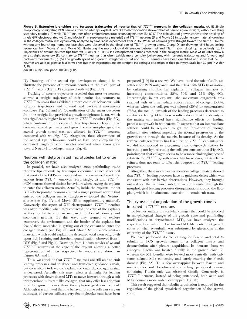
D). Drawings of the axonal tips development along 4 hours
illustrate the presence of numerous neurites in the distal part of
TTL2/2 axons (Fig. 5D’) compared with wt (Fig. 5C’).
Tracking of neurite trajectories revealed that most wt neurons
showed a straight trajectory of their neurite tips, contrary to
TTL2/2 neurons that exhibited a more complex behaviour, with
tortuous trajectories and forward and backward movements
(compare Fig. 5E and F). Quantification of the mean deviation
from the straight line provided a growth straightness factor, which
was significantly higher in wt than in TTL2/2 neurites (Fig. 5G),
which confirms the dispersion of their trajectories. Despite these
complex axonal pathways and growth cones morphologies, the
axonal growth speed was not affected in TTL2/2 neurons
compared with wt (Fig. 5G). Altogether, these observations of
the axonal tips behaviours could at least partly explain the
decreased length of axon fascicles observed when axons grew
toward Netrin-1 in collagen assays (Fig. 4).
Neurons with detyrosinated microtubules fail to enterthe collagen matrix
In parallel, we have also analyzed axon pathfinding inside
rhombic lips explants by time-lapse experiments since it seemed
that most of the GFP-electroporated neurons remained inside the
explant from TTL2/2 embryos. Surprisingly, wt and TTL2/2
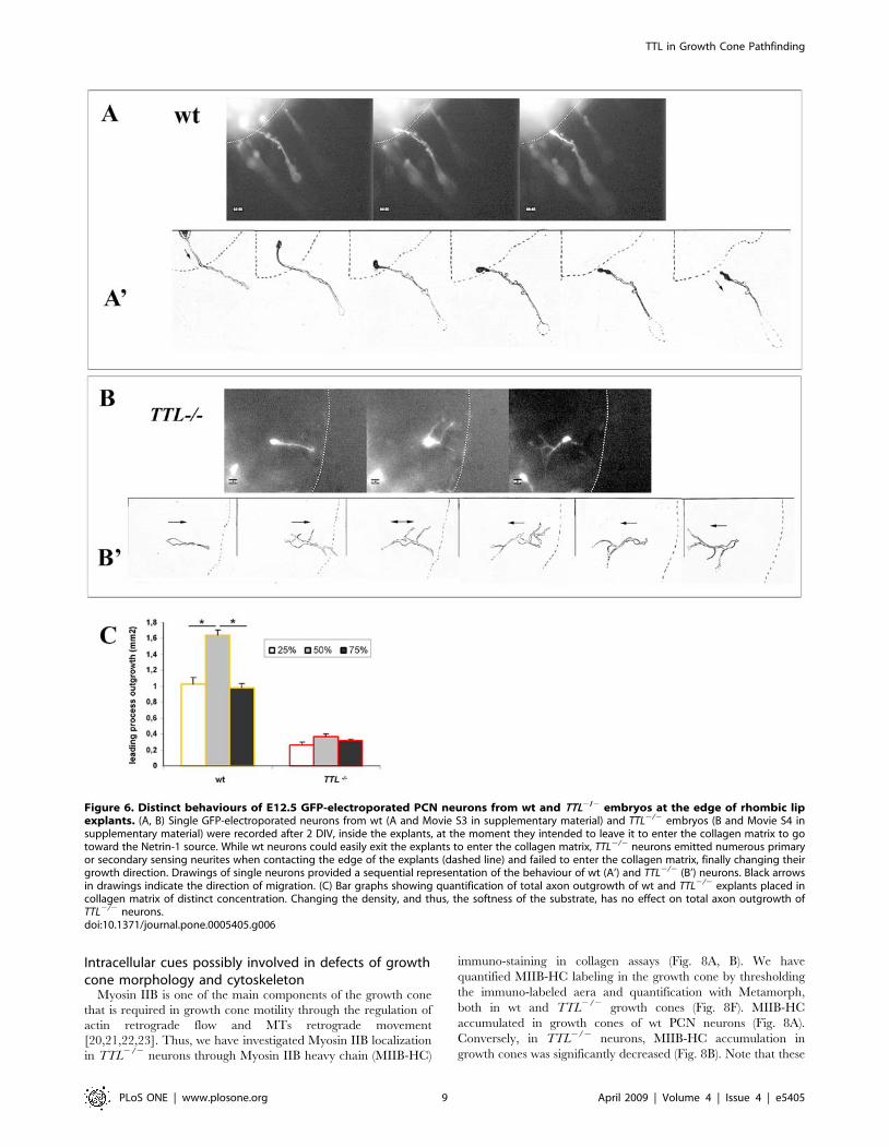
PCN neurons showed distinct behaviours when leaving the explant
to enter the collagen matrix. Actually, inside the explants, the wt
GFP-electroporated neurons emitted a single primary neurite that
entered the collagen matrix straightaway toward the Netrin-1
source (see Fig. 6A and Movie S3 in supplementary material).
Conversely, the aspect of GFP-electroporated TTL2/2 neurites
was often modified when they contacted the edge of the explants
as they started to emit an increased number of primary and
secondary neurites. By this way, they seemed to explore
extensively the environment and the border of the explant, but
few of them succeeded in getting out of the explant to enter the
collagen matrix (see Fig. 6B and Movie S4 in supplementary
material), which could explain the decreased total axon outgrowth
upon TUJ1 staining and threshold quantification, observed from 1
DIV (Fig. 3 and Fig. 4). Drawings from 4 hours movies of wt and
TTL2/2 neurons at the edge of the explant allowing a better
representation of their respective behaviours are shown in
Figures 6A’ and B’.
Thus, we conclude that TTL2/2 neurons are still able to emit
leading processes and to detect and transduce guidance signals,
but their ability to leave the explant and enter the collagen matrix
is decreased. Actually, this may reflect a difficulty for leading
processes with detyrosinated MTs to move forward through a soft
tridimensional substrate like collagen, that may offer less adhesion
sites for growth cones than their physiological environment.
Although it is admitted that the behavior of some cells can vary on
substrates of various stiffness, very few molecular cues have been
proposed ([19] for a review). We have tested the role of stiffness/
softness for PCN outgrowth and their link with MTs tyrosination
by culturing rhombic lip explants in collagen matrices of
increasing concentrations, 25%, 50% and 75% (Fig. 6C).
Interestingly, in wt explants, the maximum outgrowth was
reached with an intermediate concentration of collagen (50%),
whereas when the collagen was diluted (25%) or concentrated
(75%), the total outgrowth of the leading processes decreased to
similar levels (Fig. 6C). These results indicate that the density of
the matrix can indeed have significative effects on leading
process outgrowth in wt neurons. It suggests that an intermediate
softness could be required to get the formation of enough
adhesion sites without impeding the normal progression of the
growth cone through the matrix, because of its density. At the
reverse, collagen density has less effect on TTL2/2 explants since
we did not succeed in increasing their outgrowth neither by
increasing nor by decreasing the collagen concentration (Fig. 6C),
pointing out that collagen seems to be a more challenging type of
substrate for TTL2/2 growth cones than for wt ones, but its relative
softness does not seem to affect the outgrowth of TTL2/2leading
processes.
Altogether, these in vitro experiments in collagen matrix showed
that TTL2/2 leading processes have no guidance defect which was
consistant with our in vivo observations. However, they brought
out a defect that remained subtle in vivo only visible through the
morphological leading processes disorganizations around the floor
plate, which is the abnormal extension in certain substrata.
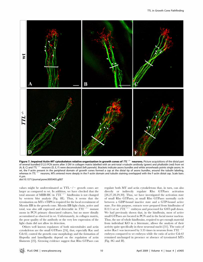
The cytoskeletal organization of the growth cone isimpaired in TTL2/2 neurons
To further analyze intracellular changes that could be involved
in morphological changes of the growth cone and pathfinding
modifications in detyrosinated MTs, we have analyzed the
respective localization of F-actin and MT filaments in wt growth
cones or when tyr-tubulin was substituted by glu-tubulin at the
extremity of the TTL2/2 axons.
We have performed double staining for F-actin and total a-
tubulin in PCN growth cones in a collagen matrix and
deconvolution after picture acquisition. In neurons from wt
embryos, F-actin was located distally in the growth cone [2]
whereas the MT bundles were located more centrally, with only
some isolated MTs contacting and barely entering the F-actin
domain (Fig. 7A). Thus, few overlapping between F-actin and
microtubules could be observed and a large peripheral domain
containing F-actin only was observed distally. Conversely, in
TTL2/2 neurons, instead of being juxtaposed, both actin and
MTs domains more widely overlapped (Fig. 7B).
This result suggested that tubulin tyrosination is required for the
regulation of the global cytoskeletal organization of the growth
cone.
Figure 5. Extensive branching and tortuous trajectories of neurite tips of TTL2/2 neurons in the collagen matrix. (A, B) Singlemorphology of migrating PCN neurons from rhombic lips explants after GFP electroporation showed that wt neurons grew straight, without emittingsecondary neurites (A) while TTL2/2 neurons often emitted numerous secondary neurites (B). (C, D) The behaviour of growth cones at the distal tip ofsingle GFP-electroporated wt (C and Movie S1 in supplementary material) and TTL2/2 neurons (D and Movie S2 in supplementary material) growingin the collagen matrix was dynamically analyzed by time-lapse experiments after 2 DIV. While wt neurons grew straight toward the Netrin-1 sourcewithout any branching, numerous branches were observed in the distal part of TTL2/2 growing axons. C’ and D’ are drawings of 4 hours lastingsequences from Movie S1 and Movie S2, illustrating the morphological differences between wt and TTL2/2 axon distal tip respectively. (E, F)Trajectories of distinct neurites tips from wt (E) or TTL2/2 (F) GFP-electroporated neurons recorded in the collagen matrix. Most wt neurites show avery straight trajectory (E), contrary to TTL2/2 neurites that often exhibit more complex behaviours, with tortuous trajectories and forward andbackward movements (F). (G) The growth speed and growth straightness of wt and TTL2/2 neurites have been quantified and show that TTL2/2
neurites are able to grow as fast as wt ones but their trajectories are less straight, indicating a dispersion of their pathway. Scale bar: 30 mm in A (forA, B).doi:10.1371/journal.pone.0005405.g005
TTL in Growth Cone Pathfinding
PLoS ONE | www.plosone.org 8 April 2009 | Volume 4 | Issue 4 | e5405
Intracellular cues possibly involved in defects of growthcone morphology and cytoskeleton
Myosin IIB is one of the main components of the growth cone
that is required in growth cone motility through the regulation of
actin retrograde flow and MTs retrograde movement
[20,21,22,23]. Thus, we have investigated Myosin IIB localization
in TTL2/2 neurons through Myosin IIB heavy chain (MIIB-HC)
immuno-staining in collagen assays (Fig. 8A, B). We have
quantified MIIB-HC labeling in the growth cone by thresholding
the immuno-labeled aera and quantification with Metamorph,
both in wt and TTL2/2 growth cones (Fig. 8F). MIIB-HC
accumulated in growth cones of wt PCN neurons (Fig. 8A).
Conversely, in TTL2/2 neurons, MIIB-HC accumulation in
growth cones was significantly decreased (Fig. 8B). Note that these
Figure 6. Distinct behaviours of E12.5 GFP-electroporated PCN neurons from wt and TTL2/2 embryos at the edge of rhombic lipexplants. (A, B) Single GFP-electroporated neurons from wt (A and Movie S3 in supplementary material) and TTL2/2 embryos (B and Movie S4 insupplementary material) were recorded after 2 DIV, inside the explants, at the moment they intended to leave it to enter the collagen matrix to gotoward the Netrin-1 source. While wt neurons could easily exit the explants to enter the collagen matrix, TTL2/2 neurons emitted numerous primaryor secondary sensing neurites when contacting the edge of the explants (dashed line) and failed to enter the collagen matrix, finally changing theirgrowth direction. Drawings of single neurons provided a sequential representation of the behaviour of wt (A’) and TTL2/2 (B’) neurons. Black arrowsin drawings indicate the direction of migration. (C) Bar graphs showing quantification of total axon outgrowth of wt and TTL2/2 explants placed incollagen matrix of distinct concentration. Changing the density, and thus, the softness of the substrate, has no effect on total axon outgrowth ofTTL2/2 neurons.doi:10.1371/journal.pone.0005405.g006
TTL in Growth Cone Pathfinding
PLoS ONE | www.plosone.org 9 April 2009 | Volume 4 | Issue 4 | e5405
values might be underestimated as TTL2/2 growth cones are
larger as compared to wt. In addition, we have checked that the
total amount of MIIB-HC in TTL2/2 hindbrains is not changed
by western blot analysis (Fig. 8E). Thus, it seems that the
tyrosination on MTs +TIPS is required for the local recruitment of
Myosin IIB in the growth cone. Myosin IIB light chain, active and
total, was also still expressed and detectable in TTL2/2 mutant
axons in PCN primary dissociated cultures, but no more distally
accumulated as observed in wt. Unfortunately, in collagen matrix,
the poor quality of the antibody or the very low expression of the
light chain did not allow its detection.
Others well known regulators of both microtubules and actin
cytoskeleton are the small GTPases [24], that, especially Rac and
Cdc42, control the growth cone morphology and the formation of
filopodia and lamellipodia depend on the regulation of actin
filaments [25]. Growing evidence suggest that Rho GTPases can
regulate both MT and actin cytoskeletons that, in turn, can also
directly or indirectly regulate Rho GTPases activation
[26,27,28,29,30]. Thus, we have investigated the activation state
of small Rho GTPases, as small Rho GTPases normally cycle
between a GDP-bound inactive state and a GTP-bound active
state. For this purpose, extracts were prepared from hindbrains of
E12.5 wt or TTL2/2 embryos and processed for GST-pull down.
We had previously shown that, in the hindbrain, most of active
small GTPases are located in PCN and in the facial motor nucleus.
Thus, the use of whole hindbrains, required to get enough material
from individual KO in a littermate, allows the analysis of their
activity quite specifically in these neuronal nuclei [31]. The ratio of
active Rac1 was increased by 3.35 times in neurons from TTL2/2
embryos compared to wt embryos while the ratio of active Cdc42
remained unchanged in presence or absence of tyrosinated MTs
(Fig. 8G and H).
Figure 7. Impaired Actin-MT cytoskeleton relative organization in growth cones of TTL2/2 neurons. Picture acquisitions of the distal partof several bundled E12.5 PCN axons after 3 DIV in collagen matrix labelled with an anti-total a-tubulin antibody (green) and phalloidin (red) from wt(A, B, C) and TTL2/2 neurons (D, E, F) were deconvoluted (left pannels). Brackets indicate axons bundles and white arrowheads points single axons. Inwt, the F-actin present in the peripheral domain of growth cones formed a cap at the distal tip of axons bundles, around the tubulin labeling,whereas in TTL2/2 neurons, MTs entered more deeply in the F-actin domain and tubulin staining overlapped with the F-actin distal cap. Scale bars:4 mm.doi:10.1371/journal.pone.0005405.g007
TTL in Growth Cone Pathfinding
PLoS ONE | www.plosone.org 10 April 2009 | Volume 4 | Issue 4 | e5405
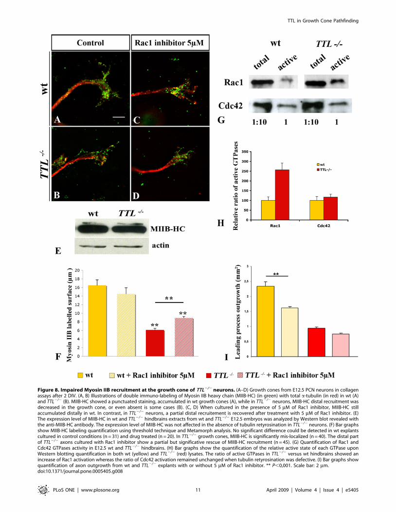
Figure 8. Impaired Myosin IIB recruitment at the growth cone of TTL2/2 neurons. (A–D) Growth cones from E12.5 PCN neurons in collagenassays after 2 DIV. (A, B) Illustrations of double immuno-labeling of Myosin IIB heavy chain (MIIB-HC) (in green) with total a-tubulin (in red) in wt (A)and TTL2/2 (B). MIIB-HC showed a punctuated staining, accumulated in wt growth cones (A), while in TTL2/2 neurons, MIIB-HC distal recruitment wasdecreased in the growth cone, or even absent is some cases (B). (C, D) When cultured in the presence of 5 mM of Rac1 inhibitor, MIIB-HC stillaccumulated distally in wt. In contrast, in TTL2/2 neurons, a partial distal recruitement is recovered after treatment with 5 mM of Rac1 inhibitor. (E)The expression level of MIIB-HC in wt and TTL2/2 hindbrains extracts from wt and TTL2/2 E12.5 embryos was analyzed by Western blot revealed withthe anti-MIIB-HC antibody. The expression level of MIIB-HC was not affected in the absence of tubulin retyrosination in TTL2/2 neurons. (F) Bar graphsshow MIIB-HC labeling quantification using threshold technique and Metamorph analysis. No significant difference could be detected in wt explantscultured in control conditions (n = 31) and drug treated (n = 20). In TTL2/2 growth cones, MIIB-HC is significantly mis-localized (n = 40). The distal partof TTL2/2 axons cultured with Rac1 inhibitor show a partial but significative rescue of MIIB-HC recruitment (n = 45). (G) Quantification of Rac1 andCdc42 GTPases activity in E12.5 wt and TTL2/2 hindbrains. (H) Bar graphs show the quantification of the relative active state of each GTPase uponWestern blotting quantification in both wt (yellow) and TTL2/2 (red) lysates. The ratio of active GTPases in TTL2/2 versus wt hindbrains showed anincrease of Rac1 activation whereas the ratio of Cdc42 activation remained unchanged when tubulin retyrosination was defective. (I) Bar graphs showquantification of axon outgrowth from wt and TTL2/2 explants with or without 5 mM of Rac1 inhibitor. ** P,0,001. Scale bar: 2 mm.doi:10.1371/journal.pone.0005405.g008
TTL in Growth Cone Pathfinding
PLoS ONE | www.plosone.org 11 April 2009 | Volume 4 | Issue 4 | e5405
Thus, the increased activity of Rac1, which can regulate actin
filaments organization in neuronal growth cones, could be partly
responsible for the changes in the growth cone morphology and
cytoskeleton organization and for the impaired regulation of
neurites extension revealed by the increased number of neurites all
along the axons of TTL2/2 neurons.
To further investigate how tubulin tyrosination could be related
to the subcellular localization of MyosinIIB, we have tested the
effect of inhibiting Rac1 over-activation by applying a commercial
Rac1 inhibitor in collagen assays (Fig. 8C and D). We have
established a dose response curve and chosen the concentration
that would allow the minimal defects in outgrowth and no defect
in nucleokinesis on wt neurons that is 5 mM Rac1 inhibitor
(supplemental Figure S4). When applied on explants in collagen
assays, we observed that the outgrowth was decreased by 30% for
wt neurons and only by 20% in TTL2/2 neurons (Fig. 8I). This
suggested that the sensitivity of mutant axons, that carry a Rac1
over activation, was lower than the one of wt. In TTL2/2 growth
cones, upon application of 5 mM Rac1 inhibitor, myosin IIB distal
recruitement was rescued (compare Fig. 8B and D). After
quantification with or without drug application, we observed a
partial and significant recovery of the distal accumulation of
MyosinIIB upon down-regulation of Rac1 over-activation (Fig. 8F).
Thus, Rac1 over-activation was, at least in part, responsible for the
absence of Myosin IIB in TTL2/2 growth cones, suggesting that it
could exist a direct link between myosin delocalization in the
growth cone and Rac1 over-activation. Indeed, in N1E-115 cells,
it has been shown that over-expression of a constitutively active
form of Rac induces cell spreading accompanied by a loss of
cortical Myosin II heavy chain [32]. Here, Myosin distal
recruitment is dependent on the proper regulation of Rac1
activity and occurs downstream of Rac1.
Discussion
In this report, we show that tubulin retyrosination plays a major
role in the morphology and functionality of the growth cone. We
demonstrate that axons in which tubulin retyrosination does not
occur emit supernumerary neurites ex vivo and in vitro. In vitro
assays reveal a reduction of axon length and their axon trajectories
are not straight when they grow in a collagen matrix, or ex vivo
when they approach the floor plate. The pathway defects reported
here in TTL2/2 mutant hindbrains are specially emphasized
around the floor plate, possibly due to its composition in both
adhesion and diffusible guidance molecules, but they could also
reflect a general growth defect or sensitivity in the absence of TTL.
In addition, the absence of tubulin retyrosination prevents the
proper intracellular organization of the growth cone cytoskeletal
components. Nevertheless, TTL2/2 axons can still follow their
global pathway in/ex vivo and reach their target. In the light of
our results, we will discuss the alternative growth strategies used by
axons when their main sensor, i.e the growth cone, is not properly
formed as in TTL2/2 neurons. We will also discuss which
intracellular associated components could link tubulin tyrosination
to the phenotypes of the TTL2/2 PCN neurons.
Tubulin tyrosination is a major cue for growth conemorphology
The growth cone motility depends on the one hand on its
function of detector and transducer of extrinsic guidance cues and
on the other hand, on the traction forces it generates. MT
assembly at the growth cone is important for the proper
organization of the neurite cytoskeleton in growing neurites. Some
of the factors that may influence MT assembly in growth cones
may be linked to post-translational modifications of tubulin itself
[33]. We report here that, in vitro, PCN neurons with only
detyrosinated MTs emit secondary distal neurites in a repeated
and uncontrolled way. Their growth cones show numerous non-
oriented filopodia which confer them a complex morphology.
Interestingly, other mutant mice carrying mutations in microtu-
bule associated proteins, in particular Kif2A MT-based motor
protein and Microtubule Associated Protein 1B (MAP1B), show an
increased growth cone surface in vitro and an increased branching
in various neuronal cell types [34,35]. The extensive branching
reported in kif2a2/2 and Map1b2/2 neurons was suggested to be
partly due to an impaired suppression of collateral branches
extension [34,36]. In both Map1b2/2 and kif2a2/2 mutant mice,
axon branching is also associated with guidance defects of the
migratory process, that lead to impaired formation of PCN nuclei.
Neurons lacking MAP1B have a reduced proportion of tyrosinated
MTs and recently, it has been shown that MAP1B protein
interacts with TTL [37]. Despite similar axon branching and
growth cone morphology defects, PCN migration and axon
responses to guidance factors seem to occur properly in TTL2/2
mutant mice, which shows that all these events are not strictly
linked and may be finely and locally modulated by a post-
translation modification like tyrosination.
In vivo, axons could set up compensatory mechanisms toensure axon outgrowth
Remarkably, in vivo and ex-vivo, the TTL2/2 phenotype is less
dramatic than in collagen assays, since axon outgrowth is not
impaired, but only the morphology of the growth cone at decision
points like the floor plate is abnormal. The phenotypic difference
of growing neurons in vitro versus ex vivo suggests that either
neurons can set up compensatory mechanisms when growing in a
physiological environment or in vivo, physical properties are less
challenging for outgrowth than in vitro. This could allow axons to
avoid the extensive emission of secondary branches and/or favor
the pruning of supernumerary filopodia in response to guidance
cues as observed during development when axons retract in
response to repellent-cues like Semaphorin 3A that coordinates the
activation of Myosin II [38].
Thus, we propose that in vitro, the successive emission and
elimination of supernumerary neurites could allow axons to
explore the environment, when the growth cone dynamic is
impaired in a surrounding environment that cannot provide
compensatory physiological cues. A previous analysis of TTL2/2
mice revealed that the cortico-thalamic loop was not properly
formed [8]. It remains to be established whether the role of
tyrosinated MTs is identical in all types of axons that have to take
decisions during pathfinding processes, independently of the
morphology of the various migrating neurons and of the
environment encountered during their development.
Tyrosinated tubulin regulates the distribution andpolarized organization of the components of the growthcone
During normal development, dynamic MTs can transiently
enter the growth cone periphery. They actively explore the
lamellipodia and even penetrate into the filopodia to integrate
guidance signals [4]. In addition, Myosin IIB accumulates in the
transition zone between the P- and C-domains [23]. In the present
report, we demonstrate that in the absence of tubulin post-
translational modification through the tyrosination cycle, detyr-
osinated MTs not only transiently explore the axonal tips, but
invade the distal actin-rich domain of the growth cone. This defect
TTL in Growth Cone Pathfinding
PLoS ONE | www.plosone.org 12 April 2009 | Volume 4 | Issue 4 | e5405
could be due to the absence of Myosin IIB in the growth cone of
TTL2/2 neurons since recent data have shown that MTs can be
retrogradely transported out of the P-domain to the C-domain
through linkage to the retrograde actin flow, which depends on the
activity of Myosin II [39]. These data suggest that MTs invasion of
the P-domain of TTL2/2 growth cones could result from defective
acto-myosin contractility at the transition zone. Tyrosination could
be a cue for regulating actin/MTs cross-talk, allowing Myosin IIB
activation in the growth cone.
Possible cross-regulations between tyrosinated tubulinand actin regulators
Mis-regulations of Rho GTPases activities can account for
changes in neuronal morphologies. In addition, increasing data
show that the cross-talk between MTs and actin is often ensured
by small GTPases and their regulators. MTs and small GTPases
can regulate each other, and small GTPases, then, can coordonate
the regulation of both actin and MT during the migratory process
in particular [27,40]. The state of MT polymerisation or
depolymerisation can affect the activation of distinct RhoGTPases
[41,42] and the activation of Rac1 is necessary for getting a MT
dependent accumulation of actin filaments at the periphery of the
growth cones [29]. We report that the rate of activation of Rac1
was increased, which also reflects a mis-regulation of small
GTPases activity in the absence of tubulin tyrosination. Interest-
ingly, the Rac1/Cdc42 effector IQGAP can interact with Clip-170
and mediates a transient capture and stabilization of MTs, leading
to establishment of polarized cell morphology and directional cell
migration [43,44]. As reported by Peris et al. [9], Clip-170
recruitment at the plus ends of MTs is impaired in TTL2/2 mice
and this could prevent the local recruitment of IQGAP. Several
other Rho GTPase regulators have been proposed to interact with
+TIPS-MTs and/or actin, such as GEF-H1 [45] and Asef, a Rac1
GEF linked to MTs +TIPS through its interaction with APC [46],
which phenotype in KO mice would be interesting to compare
with TTL2/2 mice. In growth cones, other actin regulators, such
as Ena/VASP proteins, are concentrated at filopodia tips and
control filopodial dynamics in neurons. Thus, these actin
regulators also control the growth cone morphology [47]. It will
be of interest to analyze Ena/VASP localization in TTL2/2
mouse embryos to determine whether mis-localization of actin
regulators can occur as a consequence of abnormal tubulin
modifications. An abnormal recruitment of actin regulators or
Rho GTPase regulators or effectors in the growth cone through
their interaction with plus-ends tyrosinated MTs could explain the
increased activity of Rac1 in TTL2/2 embryos.
Tyrosinated MTs could be required for growth cones toprogress through unfavorable substrates
During its pathfinding, the growth cone has to ensure the
generation of traction force in the substratum, required for
forward advancement of the growing axon [48].
In TTL2/2 neurons, axon outgrowth is decreased in collagen
assays, which possibly results from the difficulties encountered by
growth cones when contacting the collagen matrix at the edge of
the rhombic lip explant and later on, when navigating in the
collagen matrix. In organotypic culture, growth cone morphology
is also affected when axons have to enter the floor plate, that
provides a glial substrate. Finally, the aspect of TTL2/2 olivary
fibers growing through the contralateral ION is also affected in
vivo. TTL2/2 axons do not form the inter-olivary commissure
with straight oriented axons observed in wt. Interestingly, none of
the defects observed in TTL2/2 axons could be detected when
neurons were cultured on a two-dimensional substrate of poly-
ornithine/laminin (not illustrated), which strengthens the link
between the state of MTs tyrosination and the ability of an axon to
grow in complex 3D substrates. Altogether, in vitro experiments
with Netrin-1 in collagen matrix confirm the in/ex vivo
observations: the orientation of axon pathway was not affected
in TTL2/2 neurons. They also brought out that, when growing in
certain substrata, leading process outgrowth was decreased. The
tension generated by TTL2/2 growth cones may not be sufficient
for their proper pathfinding through complex substrates, especially
when they have to leave a favourable substrate or enter a new
substrate.
Substrate adhesion depends on the linkage of the actin
cytoskeleton to integrins at sites of adhesion complexes. In the
growth cone, their formation and stabilization are controlled by
the coordinated activities of Rac1 and RhoA GTPases and they
regulate neurite outgrowth [49]. The turnover of adhesion
complexes is controlled by acto-myosin contractility [50]. MTs
by themeselves can modulate the spatial localization of adhesion
complexes [51], having the ability to locally suppress acto-myosin
contractility [52]. When the dynamic of MTs is decreased after a-
tubulin deacetylation inhibition, the turnover of cell adhesions
slows down [53]. In the absence of MTs retyrosination, in TTL2/
2 neurons, the over-activation of Rac1 and the defective
recruitment of Myosin IIB in the growth cone could lead to a
decreased ability to progress through some substrates, possibly due
to defects in the formation of adhesion complexes.
Materials and Methods
Embryo processingTTL heterozygous mice were generated as described in Erck et
al. 2005 [8]. Mouse embryos were obtained from timed mating of
TTL heterozygous mice or outbred Swiss mice (Janvier, Le Genest
St Isle, France). For all culture assays, the hindbrains from E11.5
and E12.5 embryos were dissected in GBSS (Sigma), supplement-
ed with 0,5% glucose. The treatment of all the animals used within
the present research was in accordance with our institutional or
national guidelines for the use of animals in scientific research.
In situ hybridizationFor in situ hybridization (ISH), mouse embryos were fixed in
4% paraformaldehyde (PF) in phosphate buffer (PB) PH 7,4 for
2 hours at room temperature. The neural tubes were dissected out,
cryoprotected overnight with a 10% sucrose solution in PB and
embedded in 7,5% gelatin/10% sucrose. They were frozen and
serially sectioned in the frontal plane using a cryostat. ISH was
then carried out on cryosections according to Causeret et al., 2004
[18]. Brn-3.b and Tag1 probes were used and have been described
previously [12,54].
DiI tracing: retrograde labeling of precerebellar neuronsAfter intracardiac perfusion with 1% PF and 1% glutaraldehyde
in PBS, a small occipital craniotomy was performed to expose the
cerebellum of newborn wt or TTL2/2 hypomorphic mutant mice.
1,19-Dioctadecyl-3,3,39,39-tetramethylindocarbocyanine (DiI car-
bocyanine, D282; Molecular Probes, Eugene, OR) attached to the
tip of a broken glass pipette was applied, under a dissecting
microscope, on one of both hemicerebella. Several small DiI
crystals were inserted into the cerebellar tissue, medially and
laterally to label most of the neurons projecting to this center. The
brains of the injected micewere stored in 1% PF at 37uC for 3
weeks in the dark. The brains were then dissected, embedded in
3% agarose, and cut at a thickness of 80 mm with a vibratome.
TTL in Growth Cone Pathfinding
PLoS ONE | www.plosone.org 13 April 2009 | Volume 4 | Issue 4 | e5405
The sections were mounted in Mowiol, observed, and photo-
graphed by using rhodamine filters.
Collagen co-cultureCollagen assays were performed as previously described [17],
using rhombic lip explants from E11.5 and E12.5 mouse embryos
cultured either with conditioned medium from Netrin-1 secreting
cells or facing Netrin-1-secreting cells [55]. E11.5 explants are
enriched in inferior olivary neurons (ION neurons) whereas E12.5
explants are enriched in neurons that form other caudal PCN, in
particular the lateral reticular nucleus (LRN neurons) [17]. After
60–70 hours in a 5% CO2, 37uC, 95% humidity incubator,
collagen assays were fixed for 20 minutes in 4%PF/4% sucrose at
37uC.
Quantification of the axon outgrowth and fascicleslength in collagen assays
Picture acquisition and processing for axon outgrowth quanti-
fication were performed has previously described [18]. Quantifi-
cation of the length of axon fascicles growing out of the rhombic
lips explants was performed by measuring the distance between
the edge of the explant and the distal extremity of axon fascicles of
20 fascicles of each explant from wt and TTL2/2 embryos.
Differences between calculated averages were considered signifi-
cant when P,0,05 using a Student’s t-test.
Organotypic cultureThe rhombencephalon from E12.5 mouse embryos was opened
and flattened with the ventricular side down, onto a Millicell
membrane filter (Millipore) laying on BME supplemented with
25% horse serum, 2,5% HBSS, 1% glutamine and 1% glucose (all
purchased from GIBCO). After 30–72 hours in a 5% CO2, 35uC,
95% humidity incubator, rhombencephalons were fixed for
30 minutes with 4% PF.
Analysis of growth cone shapes upon GFP transfection inorganotypic culture
The size of the growth cones has been quantified through the
analysis of the area (A) and perimeter (P) of 20 growth cones from
wt and TTL2/2 neurons, as well as the resulting shape factor
(4pA/P2) then calculated using the Integrity Morphometry
analysis option from Metamorph software (Molecular Devices).
A shape factor close to 0 indicates a flattened object whereas a
value of 1 indicates a perfect circle. The base of the most proximal
filopodia was considered as the beginning of the growth cone.
Differences between parameters values were considered significant
when P,0,01 using a Student’s t-test.
Immunofluorescence and antibodiesAfter fixation, cultures were permeabilized with 0,1% Triton X-
100 and immunofluorescence was performed in phosphate
buffered saline (PBS), 1% normal goat serum (NGS), 0,1% Triton
X-100 and 0,2 mg/ml sodium azide. The following primary
antibodies were used: anti-glu-tubulin (1:6000; [56]), anti-tyr-
tubulin (1:6000; clone YL1/2; [57]), monoclonal anti-a-tubulin
(1:4000; clone a3a) from the laboratories of D. Job and J.
Wehland, rabbit polyclonal anti-GFP (1:1000; Molecular Probes),
mouse monoclonal anti-class III b-tubulin (TUJ1; 1:4000; Jackson
ImmunoResearch), rabbit polyclonal anti-Myosin light chain 2
(1:100 Cell Signaling Technology) and anti-MyosinIIB heavy
chain (1:100 Sigma). These primary antibodies were revealed
using secondary antibodies raised from goat, directed against rat,
mouse or rabbit conjugated to Alexa 488 (1:1000; Molecular
probes) or Cy3 (1:1000; Jackson ImmunoResearch). F-actin
staining was performed with rhodamine-conjugated phalloidin
(1:400; Molecular Probes). DAPI (1 mg/ml, Vector) was used to
visualize nuclei.
GFP electroporation, time-lapse experiments andtracking analysis
GFP electroporation of E12.5 rhombic lips was globally
performed as previously described [18]. Four pulses of 30 V were
applied to the injected hindbrains using a BTX ECM830
electroporator and CUY610 electrodes (Nepa Gene, Chiba,
Japan). Time-lapse experiments were performed using an inverted
Zeiss microscope and a CoolSnap HQ camera (Roper Scientific).
Images were acquired from the second day of culture, at a rate of
one image every 5 or 10 minutes, either with a 206or a 406 long
range objective. For quantification of their trajectories straightness
and growth speed, neurite tips were tracked using the isosurface
and tracking options from Imaris software (Bitplane AG).
DeconvolutionWhen necessary, pictures were acquired on a motorised
microscope (Leica DMRA2) equipped for image deconvolution.
Acquisition was performed using an oil immersion objective (6100
PL APO HCX, 1.4 NA) and a CoolSnap HQ camera. The Z-
positioning was accomplished by a piezo-electric motor (LVDT,
Physik Instruments). Z-series of images were taken at 0,2 mm
increments and the system was steered by the Metamorph
Software. Deconvolution was performed by the 3D deconvolution
module from Metamorph using the fast iterative Constrained PSF-
based algorithm [58].
Detection of active Rho GTPases and Western blotsLysates were prepared from E12.5 total hindbrains and then
processed as previously described [18]. GST (glutathione S-
transferase)-RBD (Rho binding domain)- PAK construct was
provided by M. A. Schwartz (State University of New York) and
prepared as described in Li et al. [59].
Supporting Information
Figure S1 TTL2/2 PCN neurons lack tyr-tubulin. (A)
Immuno-labeling with anti-tyr-tubulin antibody in wt PCN
dissociated neurons at E12.5 showed a staining all along the
axon, until the distal tip. (B) Merge of tyr-tubulin and glu-tubulin
staining revealed that MTs in the distal tip of the axon were only
composed of tyr-tubulin since glu-tubulin was absent in this region.
Conversely, tyr-tubulin immuno-labeling only showed a faint
perinuclear staining in TTL2/2 neurons (C) whereas glu-tubulin
staining in these neurons was observed all along the neuron,
including the distal tip of the axon as shown in merge of tyr- and
glu-tubulin immuno-labeling (D). Compared tyr-tubulin proteic
levels in hindbrain extracts from two wt and two TTL2/2 E12.5
embryos after Western blotting with the anti-tyr-tubulin antibody
(E).
Found at: doi:10.1371/journal.pone.0005405.s001 (2.95 MB
PDF)
Figure S2 Comparison of the morphology of GFP-electroporat-
ed growth cones at disinct steps of the PCN migratory pathway.
(A) schematic representation of organotypic culture of a GFP-
electroporated hindbrain. 1, 2 and 3 indicates the distinct steps of
the migratory pathway shown in B and C, D and E, F and G,
respectively. (B–E) After 30 h in culture, most of PCN leading
processes are still navigating at mid-term between the ipsilateral
TTL in Growth Cone Pathfinding
PLoS ONE | www.plosone.org 14 April 2009 | Volume 4 | Issue 4 | e5405
rhombic lip and the midline (B, C; step 1) while some of them are
already reaching it (dashed line) (D, E: step 2). (F, G) Later on, after
3DIV, some leading processes can be seen approaching the
contralateral rhombic lip (step 3). At each step, white arrow heads
indicate growth cones with hypertrophic morphology or lacking an
obvious direction. These growth cones are more frequently observed
in TTL2/2 hinbrains, but especially when leading processes are
getting close to the floor plate (E). Scale bar: 30 mm in B (for B–G).
Found at: doi:10.1371/journal.pone.0005405.s002 (2.54 MB
PDF)
Figure S3 Comparison of neurites branching in wt and TTL2/
2 neurons growing in a collagen matrix. The aspect of GFP-
electroporated growing neurites from wt (A, B) and TTL2/2 (C,
D) E12 explants toward Netrin-1 were observed after immuno-
labeling with anti-GFP antibody. In wt, neurons extend straight
neurites toward Netrin-1 without branching, neither proximally to
the cell body nor distally along the neuritic extension (arrows in A,
B) whereas in TTL2/2, branches are observed distally along the
neurite, or close to the cell body where the leading process initiates
(white arrows in C, D). Scale bar in A: 125 mm
Found at: doi:10.1371/journal.pone.0005405.s003 (0.56 MB
PDF)
Figure S4 Analysis of the effect of increasing concentrations of
Rac1 inhibitor on leading process outgrowth and nuclear
migration- E12.5 wt rhombic lip explants were cultured in
collagen matrix in presence of a range of concentrations of Rac1
inhibitor : 0 mM (A), 2 mM (B), 5 mM (C), 10 mM (D). Axon
outgrowth and nuclear migration was then analyzed after anti-total
a-tubulin and DAPI immunostainings. Quantifications were ob-
tained using the threshold technique and Metamorph analysis (E).
No significant difference could be observed between control
condition (n = 12) and 2 mM Rac1 inhibitor treated explants
(n = 18), neither for leading process outgrowth nor for nucleokinesis.
(C) At 5 mM of Rac1 inhibitor, the outgrowth was significantly
decreased but nuclear migration was not affected (n = 18). (D) At the
highest concentration, 10 mM, both axon outgrowth and nuclear
migration were significantly impaired compared to control condi-
tions (n = 18). ** P,0,001. Scale bar : 500 mm.
Found at: doi:10.1371/journal.pone.0005405.s004 (0.75 MB
PDF)
Movie S1
Found at: doi:10.1371/journal.pone.0005405.s005 (13.07 MB
AVI)
Movie S2
Found at: doi:10.1371/journal.pone.0005405.s006 (10.69 MB
AVI)
Movie S3 Movie S3 shows a wt PCN neuron getting out of the
rhombic lip explant and entering easily in the collagen matrix
toward the Netrin-1 source.
Found at: doi:10.1371/journal.pone.0005405.s007 (2.48 MB
MPG)
Movie S4 Movie S4 shows a TTL2/2 PCN neuron at the
border of rhombic lip explant and failing to enter the collagen
matrix to further grow toward the Netrin-1 source. Instead, the
neuron emit numerous primary and secondary neurites that
explore the edge of the explant and never succeed in going past it,
finally resulting in change of its migration direction.
Found at: doi:10.1371/journal.pone.0005405.s008 (41.48 MB
MPG)
Acknowledgments
We are grateful to Drs Sonia Garel, Cecile Gauthier-Rouviere and Isabelle
Le Roux for their critical reading of the manuscript. Pr Juergen Wehland is
thanked for providing the TTL2/2 mouse line, Annie Schweitzer and
Dominique Divers-Proietto for their help in the maintenance of the TTL2/2
mice colony and Pierre Bourdoncle for his technical support for the use of the
cell imaging platform (Institut Cochin, Paris). Annie Goldman is thanked for
proof-reading in English.
Author Contributions
Conceived and designed the experiments: SM EBG. Performed the
experiments: SM JM SB. Analyzed the data: SM JM SB DJ AA EBG.
Contributed reagents/materials/analysis tools: DJ AA. Wrote the paper:
SM EBG.
References
1. Zhou FQ, Cohan CS (2004) How actin filaments and microtubules steer growthcones to their targets. J Neurobiol 58: 84–91.
2. Dent EW, Gertler FB (2003) Cytoskeletal dynamics and transport in growth
cone motility and axon guidance. Neuron 40: 209–227.
3. Gordon-Weeks PR (2004) Microtubules and growth cone function. J Neurobiol58: 70–83.
4. Kalil K, Dent EW (2005) Touch and go: guidance cues signal to the growth conecytoskeleton. Curr Opin Neurobiol 15: 521–526.
5. Paturle L, Wehland J, Margolis RL, Job D (1989) Complete separation of
tyrosinated, detyrosinated, and nontyrosinatable brain tubulin subpopulationsusing affinity chromatography. Biochemistry 28: 2698–2704.
6. Webster DR, Gundersen GG, Bulinski JC, Borisy GG (1987) Differential
turnover of tyrosinated and detyrosinated microtubules. Proc Natl Acad Sci U S A84: 9040–9044.
7. Webster DR, Wehland J, Weber K, Borisy GG (1990) Detyrosination of alpha
tubulin does not stabilize microtubules in vivo. J Cell Biol 111: 113–122.8. Erck C, Peris L, Andrieux A, Meissirel C, Gruber AD, et al. (2005) A vital role of
tubulin-tyrosine-ligase for neuronal organization. Proc Natl Acad Sci U S A 102:7853–7858.
9. Peris L, Thery M, Faure J, Saoudi Y, Lafanechere L, et al. (2006) Tubulin
tyrosination is a major factor affecting the recruitment of CAP-Gly proteins atmicrotubule plus ends. J Cell Biol 174: 839–849.
10. Rakic P (1990) Principles of neural cell migration. Experientia 46: 882–891.
11. Bloch-Gallego E, Causeret F, Ezan F, Backer S, Hidalgo-Sanchez M (2005)Development of precerebellar nuclei: instructive factors and intracellular
mediators in neuronal migration, survival and axon pathfinding. Brain Res
Brain Res Rev 49: 253–266.12. Bloch-Gallego E, Ezan F, Tessier-Lavigne M, Sotelo C (1999) Floor plate and
netrin-1 are involved in the migration and survival of inferior olivary neurons.
J Neurosci 19: 4407–4420.
13. Yee KT, Simon HH, Tessier-Lavigne M, O’Leary DM (1999) Extension of long
leading processes and neuronal migration in the mammalian brain directed bythe chemoattractant netrin-1. Neuron 24: 607–622.
14. Alcantara S, Ruiz M, De Castro F, Soriano E, Sotelo C (2000) Netrin 1 acts asan attractive or as a repulsive cue for distinct migrating neurons during the
development of the cerebellar system. Development 127: 1359–1372.
15. Schaefer AW, Schoonderwoert VT, Ji L, Mederios N, Danuser G, et al. (2008)Coordination of actin filament and microtubule dynamics during neurite
outgrowth. Dev Cell 15: 146–162.
16. Burnette DT, Ji L, Schaefer AW, Medeiros NA, Danuser G, et al. (2008) MyosinII activity facilitates microtubule bundling in the neuronal growth cone neck.
Dev Cell 15: 163–169.
17. Causeret F, Danne F, Ezan F, Sotelo C, Bloch-Gallego E (2002) Slit antagonizesnetrin-1 attractive effects during the migration of inferior olivary neurons. Dev
Biol 246: 429–440.18. Causeret F, Hidalgo-Sanchez M, Fort P, Backer S, Popoff MR, et al. (2004)
Distinct roles of Rac1/Cdc42 and Rho/Rock for axon outgrowth and
nucleokinesis of precerebellar neurons toward netrin 1. Development 131:2841–2852.
19. Discher DE, Janmey P, Wang YL (2005) Tissue cells feel and respond to the
stiffness of their substrate. Science 310: 1139–1143.20. Bridgman PC, Dave S, Asnes CF, Tullio AN, Adelstein RS (2001) Myosin IIB is
required for growth cone motility. J Neurosci 21: 6159–6169.
21. Brown J, Bridgman PC (2003) Role of myosin II in axon outgrowth. J HistochemCytochem 51: 421–428.
22. Schaefer AW, Kabir N, Forscher P (2002) Filopodia and actin arcs guide the
assembly and transport of two populations of microtubules with unique dynamicparameters in neuronal growth cones. J Cell Biol 158: 139–152.
23. Medeiros NA, Burnette DT, Forscher P (2006) Myosin II functions in actin-bundle turnover in neuronal growth cones. Nat Cell Biol 8: 215–226.
TTL in Growth Cone Pathfinding
PLoS ONE | www.plosone.org 15 April 2009 | Volume 4 | Issue 4 | e5405
24. Watanabe T, Noritake J, Kaibuchi K (2005) Roles of IQGAP1 in cell
polarization and migration. Novartis Found Symp 269: 92–101; discussion 101–105, 223–130.
25. Kuhn TB, Brown MD, Bamburg JR (1998) Rac1-dependent actin filament
organization in growth cones is necessary for beta1-integrin-mediated advancebut not for growth on poly-D-lysine. J Neurobiol 37: 524–540.
26. Wittmann T, Waterman-Storer CM (2001) Cell motility: can Rho GTPases andmicrotubules point the way? J Cell Sci 114: 3795–3803.
27. Rodriguez OC, Schaefer AW, Mandato CA, Forscher P, Bement WM, et al.
(2003) Conserved microtubule-actin interactions in cell movement andmorphogenesis. Nat Cell Biol 5: 599–609.
28. Kodama A, Lechler T, Fuchs E (2004) Coordinating cytoskeletal tracks topolarize cellular movements. J Cell Biol 167: 203–207.
29. Grabham PW, Reznik B, Goldberg DJ (2003) Microtubule and Rac 1-dependent F-actin in growth cones. J Cell Sci 116: 3739–3748.
30. Watanabe T, Noritake J, Kaibuchi K (2005) Regulation of microtubules in cell
migration. Trends Cell Biol 15: 76–83.31. Backer S, Hidalgo-Sanchez M, Offner N, Portales-Casamar E, Debant A, et al.
(2007) Trio controls the mature organization of neuronal clusters in thehindbrain. J Neurosci 27: 10323–10332.
32. van Leeuwen FNvDS, Kain HE, van der Kammen RA, Collard JG (1999) Rac
regulates phosphorylation of the myosin-II heavy chain, actinomyosindisassembly and cell spreading. Nat Cell Biol 1: 242–248.
33. Mansfield SG, Gordon-Weeks PR (1991) Dynamic post-translational modifica-tion of tubulin in rat cerebral cortical neurons extending neurites in culture:
effects of taxol. J Neurocytol 20: 654–666.34. Homma N, Takei Y, Tanaka Y, Nakata T, Terada S, et al. (2003) Kinesin
superfamily protein 2A (KIF2A) functions in suppression of collateral branch
extension. Cell 114: 229–239.35. Gonzalez-Billault C, Engelke M, Jimenez-Mateos EM, Wandosell F, Caceres A,
et al. (2002) Participation of structural microtubule-associated proteins (MAPs) inthe development of neuronal polarity. J Neurosci Res 67: 713–719.
36. Bouquet C, Soares S, von Boxberg Y, Ravaille-Veron M, Propst F, et al. (2004)
Microtubule-associated protein 1B controls directionality of growth conemigration and axonal branching in regeneration of adult dorsal root ganglia
neurons. J Neurosci 24: 7204–7213.37. Utreras E, Jimenez-Mateos EM, Contreras-Vallejos E, Tortosa E, Perez M, et
al. (2008) Microtubule-associated protein 1B interaction with tubulin tyrosineligase contributes to the control of microtubule tyrosination. Dev Neurosci 30:
200–210.
38. Gallo G (2006) RhoA-kinase coordinates F-actin organization and myosin IIactivity during semaphorin-3A-induced axon retraction. J Cell Sci 119:
3413–3423.39. Zhou FQ, Waterman-Storer CM, Cohan CS (2002) Focal loss of actin bundles
causes microtubule redistribution and growth cone turning. J Cell Biol 157:
839–849.40. Wittmann TW-SC (2001) Cell motility: can Rho GTPases and microtubules
point the way? J Cell Sci 114: 3795–3803.41. Ren YLR, Zheng Y, Busch H (1998) Cloning and characterization of GEF-H1,
a microtubule-associated guanine nucleotide exchange factor for Rac and RhoGTPases. J Biol Chem 273: 34954–34960.
42. Waterman-Storer CMWR, Liu BP, Burridge K, Salmon ED (1999) Microtubule
growth activates Rac1 to promote lamellipodial protrusion in fibroblasts. Nat
Cell Biol 1: 45–50.
43. Gundersen GG (2002) Evolutionary conservation of microtubule-capture
mechanisms. Nat Rev Mol Cell Biol 3: 296–304.
44. Noritake J, Watanabe T, Sato K, Wang S, Kaibuchi K (2005) IQGAP1: a key
regulator of adhesion and migration. J Cell Sci 118: 2085–2092.
45. Krendel M, Zenke FT, Bokoch GM (2002) Nucleotide exchange factor GEF-H1
mediates cross-talk between microtubules and the actin cytoskeleton. Nat Cell
Biol 4: 294–301.
46. Kawasaki Y, Senda T, Ishidate T, Koyama R, Morishita T, et al. (2000) Asef, a
link between the tumor suppressor APC and G-protein signaling. Science 289:
1194–1197.
47. Lebrand C, Dent EW, Strasser GA, Lanier LM, Krause M, et al. (2004) Critical
role of Ena/VASP proteins for filopodia formation in neurons and in function
downstream of netrin-1. Neuron 42: 37–49.
48. Lamoureux P, Buxbaum RE, Heidemann SR (1989) Direct evidence that
growth cones pull. Nature 340: 159–162.
49. Woo S, Gomez TM (2006) Rac1 and RhoA promote neurite outgrowth through
formation and stabilization of growth cone point contacts. J Neurosci 26:
1418–1428.
50. Chrzanowska-Wodnicka M, Burridge K (1996) Rho-stimulated contractility
drives the formation of stress fibers and focal adhesions. J Cell Biol 133:
1403–1415.
51. Small JV, Kaverina I (2003) Microtubules meet substrate adhesions to arrange
cell polarity. Curr Opin Cell Biol 15: 40–47.
52. Kaverina I, Krylyshkina O, Gimona M, Beningo K, Wang YL, et al. (2000)
Enforced polarisation and locomotion of fibroblasts lacking microtubules. Curr
Biol 10: 739–742.
53. Tran AD, Marmo TP, Salam AA, Che S, Finkelstein E, et al. (2007) HDAC6
deacetylation of tubulin modulates dynamics of cellular adhesions. J Cell Sci 120:
1469–1479.
54. Backer S, Sakurai T, Grumet M, Sotelo C, Bloch-Gallego E (2002) Nr-CAM
and TAG-1 are expressed in distinct populations of developing precerebellar and
cerebellar neurons. Neuroscience 113: 743–748.
55. Kennedy TE, Serafini T, de la Torre JR, Tessier-Lavigne M (1994) Netrins are
diffusible chemotropic factors for commissural axons in the embryonic spinal
cord. Cell 78: 425–435.
56. Paturle-Lafanechere L, Manier M, Trigault N, Pirollet F, Mazarguil H, et al.
(1994) Accumulation of delta 2-tubulin, a major tubulin variant that cannot be
tyrosinated, in neuronal tissues and in stable microtubule assemblies. J Cell Sci
107 (Pt 6): 1529–1543.
57. Wehland J, Weber K (1987) Turnover of the carboxy-terminal tyrosine of alpha-
tubulin and means of reaching elevated levels of detyrosination in living cells.
J Cell Sci 88 (Pt 2): 185–203.
58. Sibarita JB (2005) Deconvolution microscopy. Adv Biochem Eng Biotechnol 95:
201–243.
59. Li Z, Aizenman CD, Cline HT (2002) Regulation of rho GTPases by crosstalk
and neuronal activity in vivo. Neuron 33: 741–750.
TTL in Growth Cone Pathfinding
PLoS ONE | www.plosone.org 16 April 2009 | Volume 4 | Issue 4 | e5405
Top Related
Copyright © 2022 FDOKUMEN

